2008 Lab Inv ROS
11
Role of the life span determinant P66 shcA in ethanol-induced liver damage Osvaldo R Koch 1,5 , Salvatore Fusco 2,5 , Sofia Chiatamone Ranieri 2,5 , Giuseppe Maulucci 3 , Paola Palozza 2 , Luigi Maria Larocca 4 , Amerys AM Cravero 1 , Stella M Farre’ 1 , Marco De Spirito 3 , Tommaso Galeotti 2 and Giovambattista Pani 2 Mice lacking the 66 kDa isoform of the adapter molecule shcA (p66 shcA ) display increased resistance to oxidative stress and delayed aging. In cultured cell lines, p66 promotes formation of Reactive Oxygen Species (ROS) in mitochondria, and apoptotic cell death in response to a variety of pro-oxidant noxious stimuli. As mitochondrial ROS and oxidative cell damage are clearly involved in alcohol-induced pathology, we hypothesized that p66 may also have a role in ethanol. In vivo, changes observed in p66 þ / þ mice after 6-week exposure to ethanol in the drinking water, including elevated serum alanine aminotransferase (ALT), liver swelling and evident liver steatosis, were significantly attenuated in p66/ mutant mice. Biochemical analysis of liver tissues revealed induction of the p66 protein by ethanol, whereas p66-deficient livers responded to alcohol with a significant upregulation of the mitochondrial antioxidant enzyme MnSOD, nearly absent in control mice. Evidence of an inverse correlation between expression level of p66 and protection from alcohol-induced oxidative stress was also confirmed in vitro in primary hepatocytes and in HepG2-E47 cells, an ethanol-responsive hepatoma cell line. In fact, MnSOD upregulation by exposure to ethanol in vitro was much more pronounced in p66KO versus wild-type isolated liver cells, and blunted in HepG2 cells overexpressing p66shc. p66 overexpression also prevented the activation of a luciferase reporter gene controlled by the SOD2 promoter, indicating that p66 repression of MnSOD operates at a transcriptional level. Finally, p66 generated ROS in HepG2 cells and potentiated oxidative stress and mitochondrial depolarization by ethanol. Taken together, the above observations clearly indicate a role for p66 in alcohol-induced cell damage, likely via a cell-autonomous mechanism involving reduced expression of antioxidant defenses and mitochondrial dysfunction. Laboratory Investigation advance online publication, 19 May 2008; doi:10.1038/labinvest.2008.44 KEYWORDS: alcohol-related liver damage; reactive oxygen species (ROS); p66shcA; manganese superoxide dismutase; mitochondria; p66 KO mice Ethanol-related pathology represents a worldwide relevant public health issue. 1,2 In the liver, pathological consequences of chronic abuse of ethanol include steatosis, steatohepatitis, fibrosis and cirrhosis. Eventually, primary hepatocarcinoma can develop as a consequence of both the direct carcino- genic effect of ethanol and alcohol-induced cell necrosis/ regeneration. 3 The molecular mechanisms underlying ethanol hepato- toxicity are still under debate. In a current model, hepatocyte accumulation of triglycerides and cholesterol (steatosis) is, at least in part, because of increased lipogenesis by excess NADH and acetyl-CoA derived from ethanol catabolism. Alcohol-induced modifications in the function of key regulators of lipid metabolism, such as PPAR-g and sterol-regulated element-binding protein (SREBP), also have been convincingly shown to take a role in this process. 4–6 As for ethanol-induced hepathocyte apoptosis/necrosis, inflammation, fibrosis and eventually cirrhosis, a major pathogenic role is currently imputed to the action of dele- terious Reactive Oxygen Species (ROS). 1,7 These oxidant intermediates are generated both in the extra mitochondrial compartment by cytochrome p450 2E1 (CyP 2E1), 8 which is part of the Microsomal Ethanol Oxidizing System, and by the mitochondrial respiratory chain 9 where the reducing equivalents produced by ethanol are eventually converted into energy. Received 01 February 2008; revised 03 March 2008; accepted 12 March 2008; published online 19 May 2008 1 Department of Pathology, Faculty of Medicine University of Buenos Aires, Buenos Aires, Argentina; 2 Institute of General Pathology, Catholic University Medical School, Rome, Italy; 3 Institute of Physics, Catholic University Medical School, Rome, Italy and 4 Department of Pathology, Catholic University Medical School, Rome, Italy Correspondence: Dr G Pani, MD, PhD, Institute of General Pathology, Catholic University Medical School, Largo F Vito no. 1, Rome 00168, Italy. E-mail: [email protected] 5 These authors contributed equally to this work. Laboratory Investigation (2008) 1–11 & 2008 USCAP, Inc All rights reserved 0023-6837/08 $30.00 www.laboratoryinvestigation.org | Laboratory Investigation | Volume 00 00 2008 1
Transcript of 2008 Lab Inv ROS

Role of the life span determinant P66shcA inethanol-induced liver damageOsvaldo R Koch1,5, Salvatore Fusco2,5, Sofia Chiatamone Ranieri2,5, Giuseppe Maulucci3, Paola Palozza2, Luigi MariaLarocca4, Amerys AM Cravero1, Stella M Farre’1, Marco De Spirito3, Tommaso Galeotti2 and Giovambattista Pani2
Mice lacking the 66 kDa isoform of the adapter molecule shcA (p66shcA) display increased resistance to oxidative stressand delayed aging. In cultured cell lines, p66 promotes formation of Reactive Oxygen Species (ROS) in mitochondria, andapoptotic cell death in response to a variety of pro-oxidant noxious stimuli. As mitochondrial ROS and oxidative celldamage are clearly involved in alcohol-induced pathology, we hypothesized that p66 may also have a role in ethanol.In vivo, changes observed in p66þ /þ mice after 6-week exposure to ethanol in the drinking water, including elevatedserum alanine aminotransferase (ALT), liver swelling and evident liver steatosis, were significantly attenuated in p66�/�mutant mice. Biochemical analysis of liver tissues revealed induction of the p66 protein by ethanol, whereas p66-deficientlivers responded to alcohol with a significant upregulation of the mitochondrial antioxidant enzyme MnSOD, nearlyabsent in control mice. Evidence of an inverse correlation between expression level of p66 and protection fromalcohol-induced oxidative stress was also confirmed in vitro in primary hepatocytes and in HepG2-E47 cells, anethanol-responsive hepatoma cell line. In fact, MnSOD upregulation by exposure to ethanol in vitro was much morepronounced in p66KO versus wild-type isolated liver cells, and blunted in HepG2 cells overexpressing p66shc. p66overexpression also prevented the activation of a luciferase reporter gene controlled by the SOD2 promoter, indicatingthat p66 repression of MnSOD operates at a transcriptional level. Finally, p66 generated ROS in HepG2 cells andpotentiated oxidative stress and mitochondrial depolarization by ethanol. Taken together, the above observations clearlyindicate a role for p66 in alcohol-induced cell damage, likely via a cell-autonomous mechanism involving reducedexpression of antioxidant defenses and mitochondrial dysfunction.Laboratory Investigation advance online publication, 19 May 2008; doi:10.1038/labinvest.2008.44
KEYWORDS: alcohol-related liver damage; reactive oxygen species (ROS); p66shcA; manganese superoxide dismutase; mitochondria;p66 KO mice
Ethanol-related pathology represents a worldwide relevantpublic health issue.1,2 In the liver, pathological consequencesof chronic abuse of ethanol include steatosis, steatohepatitis,fibrosis and cirrhosis. Eventually, primary hepatocarcinomacan develop as a consequence of both the direct carcino-genic effect of ethanol and alcohol-induced cell necrosis/regeneration.3
The molecular mechanisms underlying ethanol hepato-toxicity are still under debate. In a current model, hepatocyteaccumulation of triglycerides and cholesterol (steatosis) is, atleast in part, because of increased lipogenesis by excessNADH and acetyl-CoA derived from ethanol catabolism.Alcohol-induced modifications in the function of key
regulators of lipid metabolism, such as PPAR-g andsterol-regulated element-binding protein (SREBP), also havebeen convincingly shown to take a role in this process.4–6
As for ethanol-induced hepathocyte apoptosis/necrosis,inflammation, fibrosis and eventually cirrhosis, a majorpathogenic role is currently imputed to the action of dele-terious Reactive Oxygen Species (ROS).1,7 These oxidantintermediates are generated both in the extra mitochondrialcompartment by cytochrome p450 2E1 (CyP 2E1),8 which ispart of the Microsomal Ethanol Oxidizing System, and bythe mitochondrial respiratory chain9 where the reducingequivalents produced by ethanol are eventually convertedinto energy.
Received 01 February 2008; revised 03 March 2008; accepted 12 March 2008; published online 19 May 2008
1Department of Pathology, Faculty of Medicine University of Buenos Aires, Buenos Aires, Argentina; 2Institute of General Pathology, Catholic University Medical School,Rome, Italy; 3Institute of Physics, Catholic University Medical School, Rome, Italy and 4Department of Pathology, Catholic University Medical School, Rome, ItalyCorrespondence: Dr G Pani, MD, PhD, Institute of General Pathology, Catholic University Medical School, Largo F Vito no. 1, Rome 00168, Italy.E-mail: [email protected]
5These authors contributed equally to this work.
Laboratory Investigation (2008) 1–11
& 2008 USCAP, Inc All rights reserved 0023-6837/08 $30.00
www.laboratoryinvestigation.org | Laboratory Investigation | Volume 00 00 2008 1

Regardless of the subcellular source of ROS, ethanol-induced oxidative stress impacts on mitochondrial function,compromising energy metabolism and promoting the cyto-solic release of apoptogenic factors and, as a final con-sequence, cell death by both necrosis and apoptosis.10 Inkeeping with this general scheme, manganese-dependentmitochondrial superoxide dismutase is upregulated by etha-nol treatment in rat liver,11 and modulation of antioxidantenzymes superoxide dismutases 1 (cytosolic) and 2 (mito-chondrial) and catalase have been shown to affect alcoholhepatotoxicity both in vivo12,13 and in vitro.14,15 Moreover, wehave recently reported that, in ethanol-fed mice, hepatocyteapoptosis is largely mediated by the tumor suppressorprotein p53,16 which promotes cell death through themitochondrial generation of oxidant intermediates.17
Importantly, resistance to liver apoptosis in p53-deficientmice correlates with elevated expression of the mitochondrialsuperoxide dismutase.16
In recent years, much attention has been drawn by thesignaling adapter protein p66shcA, as a major determinant ofcell resistance to oxidative stress and to oxidant-induced celldamage and death.18 This molecule is in part localized inmitochondria where it promotes, through mechanisms onlypartially clarified,19 the generation of ROS in response to anarray of noxious stimuli.20 In fact, cell lines derived fromp66-deficient mice have reduced levels of intracellular oxi-dant species, and are resistant to pro-oxidants such as hy-drogen peroxide.21 More importantly, these animals live 30%longer than control, p66-proficient littermates,18 and areremarkably resistant to pathologies known to be mediated byoxidative tissue damage, such as atherosclerosis,22 hind limbischemia-reperfusion23 and, most relevant to the presentstudy, liver necrosis by carbon tetrachloride poisoning.19
p66shc has also been shown to participate in the pro-apop-totic cascade triggered by p53 and to mediate, at least in part,the effect of p53 on the mitochondrial production of radicalspecies.20
Reproduction of the pathological effects of chronic ethanolabuse in alcohol-fed rodents, along with the increasingavailability of genetically modified mouse strains has, in thelast few years, significantly contributed to understanding ofthe molecular basis of ethanol toxicity.12,16,24–27
Prompted by the recent emphasis on the role of p66shc inoxidant-induced cell damage and aging, and in view of therecognized role of mitochondrial oxidative stress in tissueinjury by alcohol, we sought to evaluate the pathologicaleffects of ethanol feeding in mice deficient of p66shc, incomparison to p66-proficient littermates. In parallel, weinvestigated in vitro the response of primary hepatocytesfrom wild-type and p66�/� mice, and of an ethanol-meta-bolizing human hepatocellular carcinoma, to physiologicallyrelevant amounts of ethanol. The aim of this study was toevaluate p66 as a potential molecular mediator of thedeleterious effects of alcohol on cell metabolism andviability, and to obtain fresh evidence on the role of
mitochondrial damage in the pathogenesis of alcohol-de-pendent pathology.
MATERIALS AND METHODSAnimalsSv129-p66shcA WT and p66shcA�/� (henceforth indicated asWT and p66 KO) mice have been described.18 Selective in-activation of the 66 kDa isoform of the adapter molecule Shc(Src Homology and Collagen) has been obtained by targetingpart of the 50 end of exon 2, upstream of ATG2. Mutant miceused in the present study are in the outbreed Sv129 geneticbackground, and appear healthy and fertile. Progeny fromheterozygous p66þ /� breeders was routinely genotyped byallele-specific PCR,18 and p66þ /þ and p66�/� littermateswere utilized for experimental procedures.
Animals were originally provided by Professor PG Pelicci(European Institute of Oncology, Milan, Italy) and weremaintained in a temperature-controlled room with a 12 hlight/dark cycle, in the animal facility of Universita’ Cattolicadel Sacro Cuore, Rome. All experimental procedures invol-ving animals complied with the Guidelines of the ItalianNational Institute of Health, and were approved by theInstitutional Ethical Committee.
Plasmids and Cell LinesThe SODLUC-3340 reporter construct containing a 3340 bpfragment of the MnSOD human promoter upstream of theFirefly luciferase cDNA28 has been kindly provided by Dr MBYim (NIH, Bethesda, MD, USA). The promoter sequence wasmutagenized at position �1245(C-G) to inactivate a pu-tative reverse binding site for the Foxo3A transcription factor,according to Kops et al.29 The retroviral construct encodingthe human p66shc cDNA in the pBabe/Puro backbone, andthe corresponding empty vector, have been provided byDr Marco Giorgio (European Institute of Oncology, IEO,Milan). cDNA encoding the redox-sensitive fluorescent proteinrxYFP[Cys149�Cys202] was kindly provided by Dr JR Winther(Carlsberg Laboratory, Copenhagen, Denmark). The cDNAwas excised from the original plasmid and subcloned in theEcoRI site of the mammalian expression vector (Invitrogen,San Giuliano Milanese, Italy). Insert orientation and cDNAintegrity were verified by automated sequencing.
HepG2 cells expressing the cytochrome P450 2E1 (CyP2E1,clone E47)35 have been provided by Dr A Cederbaum(Mount Sinai School of Medicine, NY, USA). Cells wereroutinely maintained in selective medium (DMEM contain-ing 8% FCS and 400 mg/ml G418) and split 1:5 every week.Cyp2E1 expression was periodically confirmed by enzymaticassay.
HepG2 E47 cells stably overexpressing the p66 cDNA un-der the LTR retroviral promoter (henceforth E47-p66), or thecorresponding empty vector (henceforth E47-pBabe) weregenerated by cell transfection with Effectene- (Qiagen, Milan,Italy) according to the manufacturer’s recommendations,followed by 4 days selection in 2.5 mg/ml Puromycin (Sigma,
Role of p66shcA in alcohol toxicity
OR Koch et al
2 Laboratory Investigation | Volume 00 00 2008 | www.laboratoryinvestigation.org

Milan, Italy). P66 overexpression was initially verified andperiodically confirmed by immunoblot analysis.
Preparation of Primary HepatocytesFor hepatocyte preparation, pairs of male p66þ /þ andp66�/� littermates were killed by cervical dislocation, andliver tissues rapidly removed, minced with scissors and in-cubated in Hepatocyte Complete Medium (HCM, Clone-ticsTM supplemented with Insulin, Transferrin, AscorbicAcid, BSA, Hydrocortisone 21 Hemisuccinate, EGF and An-tibiotics) and containing 2.5 mg/ml. Collagenase IV (Sigmacat. no. C-5138) for 1 h at 371C with occasional shaking.Tubes were left standing for 1 min and supernatants removedand centrifuged at low speed (50 g for 5 min). Pellets con-taining liver cells were resuspended for 2 min at room tem-perature in ACK buffer (150 mM NH4Cl, 1 mM KHCO3,0.1 mM EDTA, pH 7.2) to lyse red cells. After one wash inHCM cells were counted and seeded in 12 well clusters(5� 104 cells per well) for further manipulation. This simpleprocedure yielded hepatocyte suspensions whose viabilitywas always at least than 80% based on Trypan blue exclusion.
Ethanol IntoxicationSix- to eight-week-old, weight- (20–25 g initial body weight)matched male p66þ /þ and p66�/� littermates were al-lowed to drink ad libitum a mixture containing increasingconcentrations of ethanol (10% for 2 days; 15% for 3 daysand 20% thereafter) plus 20% sucrose as the only source fordrinking fluid during the 6-week period of treatment, or justwater and sucrose (control groups of both genotypes).11,16
Mice were monitored daily for clinical appearance, weightgain and drinking rate (about 5 ml per mouse per day withno differences among the four groups). As treatment con-sistently decreased food intake in both WT and p66�/�groups, diet in the ethanol groups was substantially enrichedin vitamins and micronutrients, as previously described,11 tominimize the confounding effect of potential malnutritionon liver parameters. Decreased food intake compensated forcaloric intake from ethanol-sucrose, as confirmed by the factthat weight gain curves between treated and control micewere superimposable (not shown).
Animals in the alcohol groups were kept without Et-OHfor 18 h before death, but were allowed free access to drinkingwater. Animals from all groups were starved for food over-night before killing.
Liver PathologyImmediately after killing the animals, livers were either fixedin paraformaldehyde or flash-frozen in liquid nitrogen andstored at �801C. Fixed specimens were included in paraffin,sectioned and stained with Hematoxylin–Eosin for pathol-ogy. Steatosis score was assigned in a blinded manner bythree of the authors (ORK, AMC and LML), as follows:1r25% hepatocytes containing fat droplets; 2¼ 25–50%;3¼ 50–75%; 4Z75%.30
Determination of Serum ALTBlood specimens were obtained by cardiac puncture; serawere stored at �201C and routinely processed for enzymaticdetermination of alanine aminotransferase (ALT) activity.
Vitamin E MeasurementTissue content of vitamin E in total liver homogenates wasdetermined by HPLC as previously described.31
In Vitro StudiesFor stimulation with ethanol, primary hepatocytes wereseeded immediately after purification in collagen-coated 12-well tissue culture clusters (50� 104 cells per well), in 2 mlHCM buffered with 1 mM Hepes, pH 7.4, Ethanol was addedat a different concentration ranging from 50 to 200 mM andthe plate was sealed with Parafilm M (SPI Supplies Inc., WestChester, PA, USA) to minimize evaporation. After 16 h, themedium was removed, and attached cells lysed directly in thewell, with a standard lysis buffer [Tris-HCl 10 mM; NaCl150 mM; EDTA 5 mM] containing 1% Triton X-100, 0.1%SDS, protease and phosphatase inhibitors. Floating cells wererecovered by centrifugation, lysed separately and pooled withadherent cells.
After 15 min on ice, cell lysates were cleared by cen-trifugation at 14 000 rpm (18 650 g) at 41C, and the super-natants quantified for the protein content (DC protein assay,BIORAD) and mixed with 6X Laemmli’s buffer for anti-MnSOD, anti-CREB and anti-phospho Ser 133 CREB im-munoblotting.
HepG2 E47 cells were seeded at 105 cells per well in 12-wellclusters in DMEM containing 0.5%FCS, Hepes 1 mM, ph 7.4and no selective drugs. Ethanol was added at 50–200 mMfinal concentration in 2 ml medium, and the plate sealed asdescribed above. Cell lysis and protein analysis were per-formed as described for primary hepatocytes.
Luciferase Reporter AssaysThe pSODLUC-3340 reporter construct, containing a3.3 kilobase portion of the human SOD2 promoter upstreamof the Firefly luciferase gene, and its derivative lacking theputative Foxo-responsive element, have been already de-scribed.28 The two constructs (pSODLUC-3340 and pSO-DLUC-3340mt, 0.4 mg each) were cotransfected in the HepG2E47 cell line with the pRL-TK plasmid (0.04 mg) containingRenilla luciferase driven by the thymidine kinase promoter, asan internal control for transfection efficiency, and either theempty pBabe/Puro vector or the p66shc-Puro construct(0.6 mg), using the Effectene reagent (Qiagen) according tothe manufacturer’s recommendations. Forty-eight hours aftertransfection cells were seeded in 12-well cluster plates (105
per well) and, once attached to the plate, serum-starved andexposed to 100 mM Ethanol (0.6%) for additional 18–20hours. The culture plate was sealed with Parafilm, to mini-mize ethanol evaporation. Luciferase activity was measured
Role of p66shcA in alcohol toxicity
OR Koch et al
www.laboratoryinvestigation.org | Laboratory Investigation | Volume 00 00 2008 3

and normalized for transfection efficiency using the DualLuciferase gene reporter assay (Promega s.r.l., Milan, Italy).
Analysis of Intracellular Oxidation by rxYFPHepG2 E47 cells were seeded in 35 mm glass bottom dishes(Ibidi, Integrated Biodiagnostic, Martinsried, Germany) andtransfected with 0.25 mg of the pcDNA3-rxYFP construct,mixed with 0.5 mg of p66shc-Puro or the correspondingempty vector (pBabe-Puro), using the Effectene reagent(Qiagen) according to the manufacturer’s recommendations.After 72 h cells were serum starved and exposed to Ethanolfor additional 18–20 h, or left untreated (PBS). Cell fluores-cence was imaged and quantified by confocal microscopy(Leica, DM-IRE2 Germany).
To detect redox changes, while accounting for sampleconcentration, photobleaching or laser fluctuations, rxYFPemission fluorescence signals of samples excited at 488 nm(F488) and at 458 nm (F458) were measured and theratio (F¼ F488/F458) calculated. Values of F for completelyreduced (Fred) and completely oxidized (Fox) rxYFPwere obtained from literature.36 Pseudocolor images wereconstructed based on I values, defined as
I ¼ ðF � FoxÞ=ðFred � FoxÞand ranging from 0 (complete oxidation) and 1 (completereduction), by means of a dedicated software generatedthrough the Labview 7.1 interface (Maulucci G et al, manu-script in preparation).
For image quantitation, average I values were determinedwithin multiple Regions of Interest (ROIs, single cells orsmall cell clusters) for each sample, and their mean±s.d.(n¼ 7–9) determined and utilized for further statisticalanalysis (two-way ANOVA and Bonferroni’s post hoc com-parison).
Evaluation of Mitochondrial IntegrityIntracellular (intramitochondrial) accumulation of thefluorescent dye Rhodamine 123 was used as an indicator ofmitochondrial polarization and membrane integrity.29
Briefly, cells were exposed for 48 h to different concentrationsof ethanol as described above, and labelled for the last 40 minwith 10 mg/ml Rhodamine 123 (Invitrogen, Molecular Probes1:1000 from a 10 mg/ml stock dissolved in DMSO). Cellswere then washed with PBS, detached from the plate bytrypsinization, resuspended in PBS and analyzed bystandard flow cytometry (green fluorescence, FL-1) using aCOULTER-EPICS Instrument equipped with a 488 nmArgon laser lamp.
Biochemical StudiesLiver homogenates were obtained by standard procedure.Briefly, livers were rinsed and minced in PBS, put in plastictubes and disaggregated with a tissue homogenizer (threecycles of 15 s on ice) in PBS containing protease inhibitors.Homogenates were clarified by centrifugation at 18 500 g and
assayed for protein content by a modified Lowry method,using a commercial kit according to the manufacturer’s re-commendations.
For western blot analysis, equal amounts of protein sam-ples were resolved by SDS–PAGE, transferred onto ni-trocellulose and immunoblotted with the sera of interest.Immunocomplexes were visualized by HRP-conjugates sec-ondary reagents followed by ECL detection. The followingantibodies were used in the present study: anti-p66shc andanti-MnSOD (Upstate Biotechnology); anti-phospho-FKHR(Thr 24)/FKHRL-1 (Thr32) (Cell Signaling Technology);anti-actin (Santa Cruz Biotechnology). Band intensities werequantified by volume densitometry, using a Gel.DOC Ima-ging Station equipped with a dedicated Software (Quanti-tyOne). Densitometric values were normalized for thecorresponding loading control band.
StatisticsAll data from experiments in vivo are expressed asmean±s.d. Data series were compared by two-way ANOVA(http://faculty.vassar.edu/lowry/webtext.html), followed byBonferroni’s post hoc analysis (GraphPad), or by pairedStudent’s t-test, were appropriate. The threshold of sig-nificance was set to a P-value of o0.05.
RESULTSAttenuated Liver Damage by Alcohol in p66�/� MiceC129sv mice harboring the homozygous deletion of thep66 kDa Shc adapter protein,18 and the correspondingp66þ /þ littermates, were fed ethanol in the drinking fluidor plain water for 6 weeks, as previously described.16 Foodand water/ethanol consumption, and weight gain curves didnot present significant differences between the two mousestrains (not shown), ensuring that mutant and normal micewere exposed to comparable amounts of alcohol and to asimilar dietary regimen.
Alcohol-fed wild-type mice presented a significant(roughly 30%) increase in the average liver weight in com-parison to the corresponding control group at killing. Suchan increase, indicative of organ swelling and injury, wasnearly absent in p66�/� mice, providing an initial indicationof a resistance to ethanol hepatotoxicity in this mouse mu-tant (Figure 1a). Accordingly, elevation in serum concentra-tion of ALT, a marker of hepatocyte necrosis, was observed inp66þ /þ mice following ethanol intoxication, but not (ornot significantly) in p66�/� animals (Figure 1b). Althoughwith some individual variability within groups, these data areclearly suggestive of a different response to ethanol feeding inmice lacking the longevity protein p66shc.
Prompted by this evidence we proceeded to histologicalexamination of liver tissues. Hematoxylin–Eosin (H–E)staining of liver sections revealed, in agreement with severalprevious reports,4,6 the presence of marked steatosis in al-cohol-treated wild-type mice (Figure 2a, upper right panel).This was the predominant pathological change induced by
Role of p66shcA in alcohol toxicity
OR Koch et al
4 Laboratory Investigation | Volume 00 00 2008 | www.laboratoryinvestigation.org

ethanol; inflammation and fibrosis likely require a longertreatment or higher doses of alcohol to become evident, atleast in our experimental setting. No or minimal steatosis wasscored in mice fed the control, alcohol-free diet. Importantly,ethanol-induced lipid accumulation to a much lesser extentin the liver of p66�/� mice, in keeping with reduced organswelling and with low serum markers of hepatocyte injury(compare upper right and lower right panels). Quantificationof steatosis, based on the percentage of cells containing fat,indicated that protection from steatosis by p66shc deletion isnot complete, but still highly significant (Figure 2b; Po0.05).This finding supports the idea of a role for p66shc in liverdamage by alcohol, and indicates that p66 may act already atan early stage of the pathological sequence of liver lesions(steatosis-steatohepatitis-fibrosis-cirrhosis) known toentail chronical alcohol intoxication both in humans and inrodent experimental models.
p66 is Induced by Ethanol and Modulates AntioxidantResponsep66 is transcriptionally induced and post-translationally ac-tivated (through phosphorylation on serine 36) by oxidativestress and promotes, in turn, generation of oxygen species,mitochondrial depolarization and apoptosis.18,19 Ethanol-induced oxidative stress may trigger p66 in hepatocytes anddamage cells through a p66-dependent cascade leading tomitochondrial failure and cell death. Alternatively, beneficialeffects of p66 ablation on liver damage by ethanol may beindirect or non cell-autonomous.
To sort among these possibilities, p66 expression level wasassessed by western blotting in liver homogenates from miceexposed to ethanol or to the alcohol-free diet. P66shc wasnearly undetectable in control livers from p66þ /þ mice(Figure 3a; each lane is a pool of two animals), but was clearly
Figure 1 Effect of p66 deletion on macroscopic and hematochemical signs
of chronic ethanol intoxication. (a) Exposure to ethanol increases average
liver weight in p66þ /þ (WT) but not in p66�/� (p66KO) mice.
Experimental groups were of four mice each. Relevant P-values (two-way
ANOVA followed by Bonferroni0s post-hoc comparison) are indicated.
NS¼ not statistically significant. For each genotype data are expressed as
liver weight relative to the average value of the control group, made equal
to one. Average liver weight and liver- to- mouse weight ratio did not differ
between WT and p66�/� animals (not shown). (b) Lack of p66 protects
from ethanol hepatotoxicity. Blood was collected immediately after killing
and processed by routine hematochemistry. ALT¼ alanine
aminotransferase. The composition of the experimental groups is indicated.
Statistics were calculated as in (a). NS¼not statistically significant.
Figure 2 Protection from liver steatosis in p66-deficient mice exposed to
ethanol. (a) Examples of liver sections from control and ethanol-treated
mice of the indicated genotype. Staining is Hematoxylin–Eosin (H–E). Lipid
droplets, evident in treated p66þ /þ (WT) mice and nearly absent in the
corresponding p66�/� group, are indicated by arrows. (b) Liver steatosis
quantification in the four experimental groups. Steatosis score was
assigned by three of the authors, as follows: 1r25% of hepatocytes
containing fat. 2r25–50%; 3¼ 50–75%; 4Z75%. The number of animals
per group is indicated. Statistics were calculated as in Figure 1 by factorial
2� 2 ANOVA.
Role of p66shcA in alcohol toxicity
OR Koch et al
www.laboratoryinvestigation.org | Laboratory Investigation | Volume 00 00 2008 5
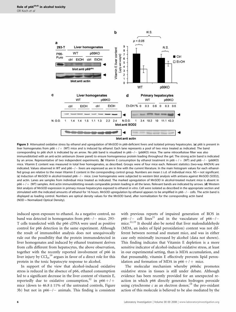
induced upon exposure to ethanol. As a negative control, noband was detected in homogenates from p66�/� mice. 293-T cells transfected with the p66 cDNA were used as positivecontrol for p66 detection in the same experiment. Althoughthe result of immunoblot analysis does not unequivocallyrule out the possibility that the protein immunodetected inliver homogenates and induced by ethanol treatment derivesfrom cells different from hepatocytes, the above observation,together with the recently reported involvement of p66 inliver injury by CCl4,19 argues in favor of a direct role for thisprotein in the toxic hepatocyte response to alcohol.
In support of the view that alcohol-induced oxidativestress is reduced in the absence of p66, ethanol consumptionled to a significant decrease in the liver content of vitamin E,reportedly due to oxidative consumption,32 in p66þ /þmice (down to 46.8±11% of the untreated controls, Figure3b) but not in p66�/� animals. This finding is consistent
with previous reports of impaired generation of ROS inp66�/� cell lines21 and in the vasculature of p66�/�mice.22,33 It should also be noted that liver malondialdehyde(MDA, an index of lipid peroxidation) content was not dif-ferent between normal and mutant mice, and was in eithercase only minimally increased by alcohol (data not shown).This finding indicates that Vitamin E depletion is a moresensitive indicator of alcohol-induced oxidative stress, at leastin our experimental setting, than is MDA accumulation, andthat presumably, vitamin E effectively prevents lipid perox-idation and formation of MDA in p66þ /þ mice.
The molecular mechanism whereby p66shc promotesoxidative stress in tissues is still under debate. Althoughevidence has been recently provided for an unexpected re-action in which p66 directly generates hydrogen peroxideusing cytochrome c as an electron donor,19 the pro-oxidantaction of this molecule is believed to be also mediated by the
Figure 3 Attenuated oxidative stress by ethanol and upregulation of MnSOD in p66-deficient livers and isolated primary hepatocytes. (a) p66 is present in
liver homogenates from p66þ /þ (WT) mice and is induced by ethanol. Each lane represents a pool of two mice treated as indicated. The band
corresponding to p66 shcA is indicated by an arrow. No p66 band is visualized in p66�/� (p66KO) mice. The same nitrocellulose filter was also
immunoblotted with an anti-actin antiserum (lower panel) to ensure homogeneous protein loading throughout the gel. The strong actin band is indicated
by an arrow. Representative of two independent experiments. (b) Vitamin E consumption by ethanol treatment in p66þ /þ (WT) and p66�/� (p66KO)
mice. Vitamin E content was measured in total liver homogenates, as described. Groups were of four mice each. Relevant statistics (two-way ANOVA) are
indicated. Values observed in WT and p66�/� livers are expressed as are in line with the current literature. In the main histogram values for each ethanol-
fed group are relative to the mean Vitamin E content in the corresponding control group. Numbers are mean±s.d. of individual mice. NS¼not significant.
(c) Induction of MnSOD in alcohol-treated p66�/� mice. Liver homogenates were subjected to western blot analysis with antisera against MnSOD (SOD2),
and actin. Lanes are samples from individual mice treated as indicated. The marked upregulation of MnSOD in alcohol-treated mutant mice is absent in
p66þ /þ (WT) samples. Anti actin immunoblotting reveals comparable protein loading in all the lanes. Relevant bands are indicated by arrows. (d) Western
blot analysis of MnSOD expression in primary mouse hepatocytes exposed to ethanol in vitro. Cell were isolated as described in the appropriate section and
stimulated with the indicated amounts of ethanol for 16 hours. MnSOD upregulation by ethanol appears to be amplified in p66�/� cells. The actin band is
displayed as loading control. Numbers are optical density values for the MnSOD band, after normalization for the corresponding actin band
(NOD¼Normalized Optical Density).
Role of p66shcA in alcohol toxicity
OR Koch et al
6 Laboratory Investigation | Volume 00 00 2008 | www.laboratoryinvestigation.org

phosphorylation/inactivation of the FOXO family transcrip-tion factor FKHR-L1 (Foxo 3a),34 and by the downregulationof FOXO-regulated antioxidant enzymes, namely catalase andmitochondrial superoxide dismutase (SOD2).34,29 Accordingto the latter model, SOD2 is expected to be hyper-expressedin p66-deficient tissues; importantly, several lines of evidenceindicate a protective role for MnSOD in ethanol-related liverpathology.11,13,14,16
In search for a mechanism linking lack of p66shc and in-creased hepatocyte resistance to alcohol injury, we assessedthe expression level of SOD2 in the liver of p66�/� andp66þ /þ mice. As indicated by the western blot analysisshown in Figure 3c, mice from the two control groups(p66þ /þ ctrl and p66�/� ctrl) expressed comparableamounts of immunoreactive SOD2; however, exposure toethanol increased SOD2 expression in mutant mice, whereasenzyme upregulation was nearly absent in p66þ /þ animals.This finding indicates that active scavenging of mitochon-drial ROS may contribute, at least in part, to attenuatealcohol-induced damage in p66�/� hepatocytes.
Effects of p66 on Cell Response to Alcohol areCell-Autonomous and can be Reproduced In VitroTo gain a more mechanistic insight into the functional linkbetween p66shc, MnSOD and cell response to alcohol injury,we decided to investigate the effect of cell exposure to ethanolin vitro. To this end, primary hepatocytes were freshlyisolated from the liver of male p66�/� mice and theirp66þ /þ littermates, maintained in complete hepatocytemedium for 2–3 h and then exposed to 0.3 or 0.6% ethanol(50–100 mM roughly) for additional 16 h; culture dishes weresealed to prevent alcohol evaporation.
In keeping with evidence in vivo of MnSOD upregulationby alcohol in p66�/� liver tissue, SOD2 content wasmarkedly increased, in a dose-dependent fashion, in p66�/�cultured liver cells; unlike in vivo, immunoreactive SOD2 wasalso induced, but to a lesser extent, in p66þ /þ hepatocytes(Figure 3d). SOD2 content was constitutively higher inp66-deficient versus p66þ /þ cells, even in the absence ofethanol challenge. Thus, in isolated liver cells, p66 inhibitsMnSOD expression also in the absence of ethanol challenge,likely as part of a cellular response to purification andculture stress.
Human Hepatoma HepG2 cells (clone E47) geneticallyengineered to express the ethanol-metabolizing cytochromep450 2E1 represent an established model for hepatocyteresponse to alcohol in vitro35 in alternative to primaryhepatocytes, with the obvious advantages of an indefinitelygrowing cell line and the possibility to be further geneticallymanipulated.
HepG2 E47 cells exposed to physiologically relevantconcentrations of Ethanol (0.3 or 0.6% v/v, for 24 h) re-capitulated the biochemical responses observed in liver tis-sues and in primary hepatocytes in vitro, that is, upregulationof p66shc and induction of MnSOD (Figure 4a, left panels).
More importantly, LTR-driven overexpression of p66shccDNA in the same cell line led to a moderate accumulation ofthe protein and, in parallel, inhibited ethanol-inducedupregulation of MnSOD (Figure 4a, right panels). Thesefindings are consistent with the possibility, supported also byevidence from Figure 3d, that p66 inhibits a protectiveantioxidant response to ethanol brought about by the mi-tochondrial superoxide dismutase. Multiple transcriptional
Figure 4 MnSOD induction by ethanol and modulation by p66shc in
human hepatoma cells expressing CYP2E1 (HepG2-E47). (a) (left) HEPG2 E47
cells recapitulate in vitro biochemical aspects of hepatocyte response to
alcohol. Induction of p66shc and upregulation of the mitochondrial
superoxide in HepG2 E47 cells exposed to increasing concentrations of
ethanol. (a) (right) Inhibitory effect of p66 overexpression on MnSOD
response to ethanol challenge. FKHRL-1 phosphorylation on Threonine 32
is increased by Ethanol in parallel with p66 accumulation (left panels) and is
constitutively high in p66 overexpressing cells (right panels). Blots
representative of several (at least two) independent experiments. Relevant
protein bands are indicated by arrows. Band densitometric values are
reported as raw data (OD) or fold induction (FI), as indicated. (b) Gene
reporter assay for MnSOD promoter activation in HEPG2 E47 cells exposed
to ethanol (0.6%, about 100 mM). Values are fold induction of luciferase
activity by 100 mM ethanol. The SODLUC-3334 reporter activation by
ethanol is significantly repressed by coexpression of p66shc (Po0.005 by
two-tailed paired t-test n ¼ 6). Response of a similar reporter lacking a
Foxo3A-binding site29 (SODLUC-mut) to ethanol is reduced and largely
insensitive to p66 shc. Values are mean±s.d. of multiple experiments
(between two and six) performed independently.
Role of p66shcA in alcohol toxicity
OR Koch et al
www.laboratoryinvestigation.org | Laboratory Investigation | Volume 00 00 2008 7

regulators control MnSOD expression in response to oxida-tive challenge. These include the cytokine-activated factorsAP-1 and NF-kB, the tumor suppressor protein p53, and, asmore recently reported, the FOXO-family factors FOXO-1(FKHR) and FOXO3a (FKHRL1); FOXO factors, in parti-cular, increase the expression of MnSOD in cells deprived ofexogenous growth factors,29 and have been shown, alongdifferent lines of evidence, to be phosphorylated and in-activated in a p66- and ROS-dependent fashion in NIH-3T3cells.34 Collectively, these data qualify FOXOs as potentialmediators of p66 effects on MnSOD in hepatocytes.
In H47 pBabe cells cultured in serum-free medium, basallevel phosphorylation of Foxo3a on Thr 32 was barely de-tectable, but could be increased by cell exposure to ethanol(Figure 4a). Notably, FKHRL-1 phosphorylation is associatedwith factor retention in the cell cytosol and decreased trans-criptional activity.29 Foxo phosphorylation was found con-stitutively high in E47-p66 cells, and moderately increased byethanol, consistent with the notion that this molecule re-presents a target for p66 in oxidant-challenged cells.34 Theabove finding therefore implicates Foxo3A in the inhibitoryaction exerted by p66 on MnSOD induction by ethanol.
To gain further insight in the modulation of MnSOD byp66, HepG2 cells were transiently transfected with a reporterconstruct encoding Renilla luciferase under the control of theSOD2 promoter, alone or in combination with the plasmidencoding human p66shc. In parallel cultures, a mutant formof the same reporter, lacking a putative Foxo3A target se-quence (see experimental procedures) was transfected, withor without p66shc. As indicated in panel 4B, exposure toethanol led to a significant increase of the SOD reporteractivity, which was nearly completely abolished by thecoexpression of p66 (1.59±0.28 fold induction versus1.01±0.21, Po0.001). Interestingly, the mutant reporter,unresponsive to Foxo3a, was markedly less sensitive to bothethanol induction and modulation by p66 (4C). Takentogether, these findings indicate that MnSOD induction byethanol and repression by p66 operate, at least in part,at a transcriptional level, through a member of the Foxotranscription factor family.29
p66 Exacerbates Oxidative Stress and MitochondrialDamage by Ethanol in Cultured Hepatoma CellsFinally, to verify the functional relevance of MnSOD in-hibition by p66 in the cell exposed to ethanol, HepG2E47cells were cotransfected with the p66 cDNA or the corre-sponding empty control vector (pBabe), and a plasmid encod-ing for a redox sensitive mutant of the Yellow FluorescentProtein (rxYFP), originally described by Ostergaard et al.36 Anet decrease in fluorescent emission of this YFP derivativereveals protein oxidation through the formation of an in-tramolecular disulphide bond between engrafted cysteineresidues 143 and 204,36 but the sensitivity of the probe can befurther enhanced by ratiometric calculation, that is, by nor-malizing changes in emission intensity for the fluorescence
values elicited at a less redox-sensitive excitation wavelength(Methods, and Maulucci G et al, manuscript in preparation).Confocal microscopy analysis of rxYFP and rxYFP/p66shcdouble transfectant HepG2E47 cells demonstrated that bothoverexpression of p66 (confirmed by immunoblot analysis oftotal protein lysates) and exposure to 100 mM ethanol led,separately, to a significant oxidation of HepG2E47 cells, asrevealed by a yellow-to-red shift of cell pseudocolor in se-lected microscopic fields (Figure 5a), and by the quantifica-tion of signal ratios in multiple regions of interest (5b).Importantly, superexpression of p66 exacerbated oxidativestress by ethanol (Po0.01, two-way ANOVA). However,the combined effect was less-than-additive, suggestingthat shared biochemical mechanisms, likely centered onmitochondria, underlie generation of ROS by alcoholand p66shc. In keeping with this view and with several previousreports, cell exposure to ethanol led to a decrease in mi-tochondrial integrity, as assessed by cell capacity to con-centrate the fluorescent dye Rhodamine 1,2,3 (Figure 5d).Overexpression of p66 lowered the baseline mitochondrialpotential of HepG2 E47 cells and amplified depolarization byethanol, suggesting that, in this cell model at least, mi-tochondrial damage by alcohol is worsened by p66 via in-hibition of MnSOD.
DISCUSSIONIn spite of extensive research in the field, the molecularmechanisms underlying damage to hepatocytes and other celltypes by ethanol are still incompletely understood, and fur-ther information is critically needed to open novel preventiveand therapeutic opportunities. The present work identifiesthe pp66kDa isoform of the adapter protein shc (p66shcA) asa molecule involved in the establishment of liver damage byalcohol, in a murine experimental model of chronic ethanolintoxication. Main findings in support of this conclusion aredisplayed in Figures 1 and 2, showing reduced liver swelling,serum ALT levels (a marker of hepatocyte necrosis) and at-tenuated fatty changes in p66�/� mice fed an alcohol-richdiet, in comparison to p66þ /þ controls. Importantly,steatosis is the main pathological change observed in ourmodel in alcohol-treated animals, with minimal signs of in-flammation and fibrosis. This is probably due to the limitedlength of the treatment (6 weeks) and to the modality ofanimal exposure to ethanol (ad libitum feeding in thedrinking water). This is a milder regime than other morerobust models of ethanol feeding such as the liquid Lieber-DeCarli diet, or intragastric infusion.37 Therefore, although itis not possible to conclude that late stage aspects of alcohol-related pathology also would be attenuated by the absence ofp66, this finding indicates an early role for p66shc in alcohol-induced hepatotoxicity.
Our data also address the mechanism underlying the roleof p66 in liver damage by ethanol. In keeping with the re-cognized function of p66 as an inducer of oxidative stress andof mitochondrial damage in a number of ROS-related
Role of p66shcA in alcohol toxicity
OR Koch et al
8 Laboratory Investigation | Volume 00 00 2008 | www.laboratoryinvestigation.org

pathologies and presumably during the aging process,18,22,23
we provide indirect evidence, in vivo, that the establishmentof oxidative stress in response to ethanol is attenuated in p66-deficient livers. In fact, vitamin E consumption, a reliablemarker of tissue challenge by oxygen species, is marginal orabsent in p66�/� mice exposed to ethanol, whereas sig-nificant in p66þ /þ animals in parallel with the increase inthe liver content of p66. Moreover, the manganese-dependentsuperoxide dismutase (MnSOD) is upregulated in p66-defi-cient livers in response to ethanol, but not in control mice,thus implying in the mutant mouse strain a better capacity ofactively detoxifying mitochondrial ROS13 (Figure 3). Im-portantly, these data are corroborated by data provided inFigure 5, that forced expression of p66 in HepG2 E47 he-patoma cells in vitro induces ROS and potentiates oxidativestress and loss of mitochondrial membrane integrity inducedby alcohol. Collectively these data provide a mechanistic linkbetween reduced oxidative stress and protection from ethanoldamage in p66�/� mice. Experiments based on antioxidantdietary supplementation during ethanol treatment, are on-going, to further evaluate this link.
Experiments in vitro on primary hepatocytes and HepG2-E47 cells, expressing the cytochrome P450 2E1 cDNA underthe transcriptional control of a viral promoter, have beendesigned to gain a more mechanistic insight in the molecularconnection between alcohol-induced stress, p66shc and mi-tochondrial antioxidant response. The use of Cyp450 2E1transfected cells, a well established in vitro model for cellresponse to ethanol, has been made necessary by technicallimitations in genetically manipulating primary hepatocytes.This cell model has been already successfully employed to
Figure 5 Effect of p66 on ethanol-induced oxidative stress and
mitochondrial dysfunction in HepG2 E47 cells. (a) Representative
pseudocolor images of HepG2 E47 cells 48 h after transfection of the redox-
sensitive fluorescent protein rxYFP, in combination with p66shc or the
corresponding empty vector (pBabe). Treatments are indicated. Cell color is
function of the I parameter (see experimental procedures), whose value
ranges from 0 (complete oxidation, red) to 1 (complete reduction, yellow).
Yellow-to-red shift in cell color indicates cell oxidation. (b) Image
quantitation based on multiple randomly chosen ROIs (Regions Of Interest,
single cells or small cell clusters) for each treatment. Average I values within
single ROIs were calculated. Columns are mean±s.d. of the average I values
for the different treatments. N¼ 7 for PBS, N¼ 9 for Et-OH, N¼ 7 for p66,
N¼ 8 for p66 þ EtOH. Figure representative of two independent
experiments. Relevant statistics (Two-way ANOVA followed by Bonferroni’s
post hoc test) are indicated. (c) Western blot analysis confirming expression
of rxYFP and overexpression of p66shc in HepG2 E47 cells. (d)
Mitochondrial membrane integrity monitored by the fluorescent dye
Rhodamine 1,2,3. E47-p66 cells and the corresponding control line
(E47-pBabe) were treated with ethanol, loaded with Rhodamine 123 and
analyzed by standard flow cytometry as described in experimental
procedures. Dose-dependent decrease of mean cell fluorescence in
response to ethanol indicates loss of mitochondrial polarization. P66
reduces mitochondrial potential in untreated cells and exacerbates the
effect of alcohol. Numbers are mean±s.d. of duplicate samples. Figure
representative of two independent experiments.
Role of p66shcA in alcohol toxicity
OR Koch et al
www.laboratoryinvestigation.org | Laboratory Investigation | Volume 00 00 2008 9

study mechanisms of ethanol toxicity at concentrations veryclose to those (100 mM) here utilized.38
Results displayed in Figures 3d and 4a, showing, respec-tively, enhanced induction of MnSOD by ethanol inp66-deficient hepatocytes and, vice versa, attenuated SOD2increase in HepG2E47 cells overexpressing p66, are in agree-ment with findings in vivo and indicate that p66 effects onantioxidant response to alcohol are largely cell-autonomous,and independent from tissue factors (ie, inflammatory cyto-kines) presumably present in the complex microenvironmentof an ethanol-metabolizing organ. Although the inhibitoryaction of p66shc on MnSOD expression in vitro confirmsin vivo evidence, a number of differences emerge between thetwo experimental models. First, unlike in liver tissues,MnSOD expression in p66�/� isolated hepatocytes wasfound to be higher than in normal control cells. Second, invitro, MnSOD is induced by ethanol in both p66þ /þ andp66�/� cells (Figures 3d and 4a), although to a larger extentin the latter population, whereas evidence for enzyme in-duction in the p66þ /þ livers in vivo is missing (Figure 3c).As for the first difference, it is possible that a threshold-levelof cell stress is necessary to disclose p66 activity on MnSOD.With this respect, it should be acknowledged that procedureof cell enzymatic isolation and seeding in culture (at a higheroxygen concentration than is found in living tissues) re-presents a severe challenge for primary liver cells. It is note-worthy, on the other hand, that overexpression of p66shc inhuman hepatoma cells inhibits ethanol-induced SOD2 up-regulation with little or no effect on the basal expression levelof the enzyme (4A).
Along similar lines, culture conditions (ie, the modalityof exposure to alcohol, which recapitulates more an acuteintoxication than a chronic abuse) may explain MnSODresponse to ethanol in p66-proficient cells (primaryhepatocytes and hepatoma cells) in vitro.
Although molecular details of MnSOD regulation by p66in response to ethanol challenge still need to be elucidated,evidence of Foxo3a hyperphosphorylation in E47-p66 cells(Figure 4a) and reporter assay data presented in Figure 4bsuggest that a Foxo factor may have a role in this circuitry. AsFoxo3a/FKHR-L1 appears to be phosphorylated/inactivatedupon cell exposure to ethanol, this is unlikely to be the onlytranscriptional regulator mediating MnSOD induction byalcohol. Still, residual activity of this factor may be critical,within the MnSOD transcriptosome, to allow promoteractivation by ethanol challenge, thereby justifying theinhibitory action of p66.
Unlike in HepG2 cells, ethanol-dependent phosphoryla-tion of Foxo3a could not be detected in primary hepatocytes.Although technical limitations may underlie this result, me-chanisms distinct from phosphorylation may underlie foxoregulation by ethanol and p66 in this experimental setting.For instance, NADH accumulation due to ethanol meta-bolism and to mitochondrial damage may interfere with Foxodeacetylation/activation by sirtuins.39 Further experimental
work is required to adequately address the potential con-nection between p66 and Foxo factors in ethanol-induced celldamage.
Based on the available information, a tentative pathophy-siological cascade linking ethanol and p66 to alcohol-inducedliver injury may be outlined as follows. Ethanol metabolisminduces p66shc in liver cells (Figures 3a and 4); such in-duction may be mediated by the p53 tumor suppressorprotein, based on our and others’ previous work.16,21
Whatever the mechanism, increased cell content of p66 canlead to increased ROS generation in mitochondria by twodistinct and parallel mechanisms: (1) direct generation ofoxygen species by transfer of electrons from cytochrome c tomolecular oxygen;19 and (2) inhibition of the mitochondrialscavenger MnSOD, via phosphorylation of a Forkheadtranscription factor. (Figure 4a and b). Eventually, mi-tochondrial oxidative stress would result in organelle da-mage, fat accumulation in hepatocytes (Figure 2a) and,ultimately, in cell death (1B).
In keeping with the above hypothesized model, we havebeen able to detect, by means of a novel and sensitivefluorescent protein-based methodology, intracellularoxidation in hepatoma cells challenged with ethanol andexacerbation of oxidative burden by p66 coexpression.(Figure 5a and b). Importantly, oxidative stress in the aboveconditions is closely paralleled by mitochondrial functionaldamage (5d).
Notwithstanding these data, and although our findingsand current literature suggest that modulation of ethanoltoxicity by p66 is mediated by a redox mechanism, ROS-independent effects of p66 could also be operative. For in-stance, as in our experimental model of ethanol intoxication,a dramatic reduction of steatosis represents the most evidentaspect of liver protection by p66 deficiency, a specific role forp66 in fat biosynthesis/storage cannot be excluded. Althoughprotection from oxidative stress is per se sufficient to preventalcohol-induced steatosis in mice,13 it is worth noting thatoverexpression of p66 also enhances insulin signaling andpromotes adipocyte differentiation of 3T3-L1 cells (Pani et al,manuscript submitted). Therefore, purely metabolic func-tions of p66 related to lipid metabolism could also contributeto the establishment of liver steatosis in ethanol-fed wild-typemice. With respect to this possibility, preliminary evidenceexcludes major differences in the liver expression of lipogenictranscription factors PPAR-a4 and -g between the two mousestrains (data not shown), whereas the potential role of othermolecules involved in lipid metabolism and in alcoholicsteatosis, such as CREB,40 the SREBP6 and the AMP-activatedprotein kinase AMPK41 is currently under evaluation.
In conclusion, our data invoke the longevity proteinp66shc as a molecular determinant of alcohol cytotoxicityand of liver injury by chronic ethanol intoxication, already atthe stage of lipid accumulation (liver steatosis); moreover, theabove findings indicate a novel potential molecular me-chanism of damage by p66 in the inhibition of mitochondrial
Role of p66shcA in alcohol toxicity
OR Koch et al
10 Laboratory Investigation | Volume 00 00 2008 | www.laboratoryinvestigation.org

antioxidant responses, and in particular of the upregulationof MnSOD. Although mechanistic interactions underlyingthe above phenomena still need to be clarified, these ob-servations are potentially relevant for the understanding ofhow p66 regulates general cell response to oxidative chal-lenge, and encourage intense pharmacological research aimedto block the deleterious effect of the p66 protein for theprevention and treatment of alcohol-related and other ROS-dependent diseases.
ACKNOWLEDGEMENTS
We thank Professor Piergiuseppe Pelicci and Drs Enrica Migliaccio and
Marco Giorgio for providing the p66�/� mouse strain and for critically
reading the article; Dr MB Yim (NIH, Bethesda) for the gift of the SODLUC-
3340 reporter constructs; Dr A Cederbaum for HepG2-E47 cells transfected
with the Cytochrome P450 2E1 cDNA; Dr JR Winther for the rxYFP cDNA; Dr
E Stigliano for excellent technical assistance with liver pathology; Drs Silvia
Borrello, Ambra Valdannini and Emiliano Panieri for experimental
contribution members of the laboratory for helpful discussions and Dr
Renata Colavitti for editing the article. This work is supported by grants from
Ministero Affari Esteri, Italian–Argentinian collaborative Research Program
(L.401/1990) and University of Buenos Aires M042.
1. Mendez-Sanchez N, Almeda-Valdes P, Uribe M. Alcoholic liver disease.An update. Ann Hepatol 2005;4:32–42.
2. Lieber CS. Alcoholic fatty liver: its pathogenesis and mechanism ofprogression to inflammation and fibrosis. Alcohol 2004;34:9–19.
3. Mufti SI. Alcohol acts to promote incidence of tumors. Cancer DetectPrev 1992;16:157–162.
4. Crabb DW, Galli A, Fischer M, et al. Molecular mechanisms of alcoholicfatty liver: role of peroxisome proliferator-activated receptor alpha.Alcohol 2004;34:35–38.
5. You M, Crabb DW. Molecular mechanisms of alcoholic fatty liver: roleof sterol regulatory element-binding proteins. Alcohol 2004;34:39–43.
6. You M, Fischer M, Deeg MA, et al. Ethanol induces fatty acid synthesispathways by activation of sterol regulatory element-binding protein(SREBP). J Biol Chem 2002;277:29342–29347.
7. Hoek JB, Cahill A, Pastorino JG. Alcohol and mitochondria: adysfunctional relationship. Gastroenterology 2002;122:2049–2063.
8. Caro AA, Cederbaum AI. Oxidative stress, toxicology, andpharmacology of CYP2E1. Annu Rev Pharmacol Toxicol 2004;44:27–42.
9. Bailey SM, Cunningham CC. Contribution of mitochondria to oxidativestress associated with alcoholic liver disease. Free Radic Biol Med2002;32:11–16.
10. Adachi M, Ishii H. Role of mitochondria in alcoholic liver injury. FreeRadic Biol Med 2002;32:487–491.
11. Koch OR, De Leo ME, Borrello S, et al. Ethanol treatment up-regulatesthe expression of mitochondrial manganese superoxide dismutase inrat liver. Biochem Biophys Res Commun 1994;201:1356–1365.
12. Kessova IG, Ho YS, Thung S, et al. Alcohol-induced liver injury in micelacking Cu, Zn-superoxide dismutase. Hepatology 2003;38:1136–1145.
13. Wheeler MD, Nakagami M, Bradford BU, et al. Overexpression ofmanganese superoxide dismutase prevents alcohol-induced liverinjury in the rat. J Biol Chem 2001;276:36664–36672.
14. Perez MJ, Cederbaum AI. Adenovirus-mediated expression of Cu/Zn-or Mn-superoxide dismutase protects against CYP2E1-dependenttoxicity. Hepatology 2003;38:1146–1158.
15. Bai J, Cederbaum AI. Adenovirus-mediated overexpression of catalasein the cytosolic or mitochondrial compartment protects againstcytochrome P450 2E1-dependent toxicity in HepG2 cells. J Biol Chem2001;276:4315–4321.
16. Pani G, Fusco S, Colavitti R, et al. Abrogation of hepatocyte apoptosisand early appearance of liver dysplasia in ethanol-fed p53-deficientmice. Biochem Biophys Res Commun 2004;325:97–100.
17. Polyak K, Xia Y, Zweier JL, et al. A model for p53-induced apoptosis.Nature 1997;389:300–305.
18. Migliaccio E, Giorgio M, Mele S, et al. The p66shc adaptor proteincontrols oxidative stress response and life span in mammals. Nature1999;402:309–313.
19. Giorgio M, Migliaccio E, Orsini F, et al. Electron transfer betweencytochrome c and p66Shc generates reactive oxygen species thattrigger mitochondrial apoptosis. Cell 2005;122:221–233.
20. Orsini F, Migliaccio E, Moroni M, et al. The life span determinantp66Shc localizes to mitochondria where it associates withmitochondrial heat shock protein 70 and regulates trans-membranepotential. J Biol Chem 2004;279:25689–25695.
21. Trinei M, Giorgio M, Cicalese A, et al. A p53-p66Shc signalling pathwaycontrols intracellular redox status, levels of oxidation-damaged DNAand oxidative stress-induced apoptosis. Oncogene 2002;21:3872–3878.
22. Napoli C, Martin-Padura I, de Nigris F, et al. Deletion of the p66Shclongevity gene reduces systemic and tissue oxidative stress, vascularcell apoptosis, and early atherogenesis in mice fed a high-fat diet. ProcNatl Acad Sci USA 2003;100:2112–2116.
23. Zaccagnini G, Martelli F, Fasanaro P, et al. p66ShcA modulates tissueresponse to hindlimb ischemia. Circulation 2004;109:2917–2923.
24. Koch OR, Galeotti T, Bartoli GM, et al. Alcohol-induced oxidative stressin rat liver. Xenobiotica 1991;21:1077–1084.
25. Morgan K, French SW, Morgan TR. Production of a cytochrome P4502E1 transgenic mouse and initial evaluation of alcoholic liver damage.Hepatology 2002;36:122–134.
26. Bradford BU, Kono H, Isayama F, et al. Cytochrome P450 CYP2E1, butnot nicotinamide adenine dinucleotide phosphate oxidase, is requiredfor ethanol-induced oxidative DNA damage in rodent liver.Hepatology 2005;41:336–344.
27. McKim SE, Gabele E, Isayama F, et al. Inducible nitric oxide synthase isrequired in alcohol-induced liver injury: studies with knockout mice.Gastroenterology 2003;125:1834–1844.
28. Kim HP, Roe JH, Chock PB, et al. Transcriptional activation of thehuman manganese superoxide dismutase gene mediated bytetradecanoylphorbol acetate. J Biol Chem 1999;274:37455–37460.
29. Kops GJ, Dansen TB, Polderman PE, et al. Forkhead transcription factorFOXO3a protects quiescent cells from oxidative stress. Nature2002;419:316–321.
30. Nanji AA, Mendenhall CL, French SW. Beef fat prevents alcoholic liverdisease in the rat. Alcohol Clin Exp Res 1989;13:15–19.
31. Palozza P, Calviello G, Serini S, et al. Supplementation withcanthaxanthin affects plasma and tissue distribution of alpha- andgamma-tocopherols in mice. J Nutr 1998;128:1989–1994.
32. Koch O, Farre S, De Leo ME, et al. Regulation of manganese superoxidedismutase (MnSOD) in chronic experimental alcoholism: effects ofvitamin E-supplemented and -deficient diets. Alcohol Alcohol2000;35:159–163.
33. Francia P, delli Gatti C, Bachschmid M, et al. Deletion of p66shc geneprotects against age-related endothelial dysfunction. Circulation2004;110:2889–2895.
34. Nemoto S, Finkel T. Redox regulation of forkhead proteins througha p66shc-dependent signaling pathway. Science 2002;295:2450–2452.
35. Wu D, Cederbaum AI. Ethanol cytotoxicity to a transfected HepG2 cellline expressing human cytochrome P4502E1. J Biol Chem1996;271:23914–23919.
36. Ostergaard H, Henriksen A, Hansen FG, et al. Shedding light ondisulfide bond formation: engineering a redox switch in greenfluorescent protein. EMBO J 2001;20:5853–5862.
37. Lieber CS, DeCarli LM, Sorrell MF. Experimental methods of ethanoladministration. Hepatology 1989;10:501–510.
38. Chen Q, Cederbaum AI. Cytotoxicity and apoptosis produced bycytochrome P450 2E1 in Hep G2 cells. Mol Pharmacol 1998;53:638–648.
39. Brunet A, Sweeney LB, Sturgill JF, et al. Stress-dependent regulation ofFOXO transcription factors by the SIRT1 deacetylase. Science2004;303:2011–2015.
40. Herzig S, Hedrick S, Morantte I, et al. CREB controls hepatic lipidmetabolism through nuclear hormone receptor PPAR-gamma. Nature2003;426:190–193.
41. You M, Matsumoto M, Pacold CM, et al. The role of AMP-activatedprotein kinase in the action of ethanol in the liver. Gastroenterology2004;127:1798–1808.
Role of p66shcA in alcohol toxicity
OR Koch et al
www.laboratoryinvestigation.org | Laboratory Investigation | Volume 00 00 2008 11