Constitutive activation of AMP-activated protein kinase (AMPK) propel mitochondrial biogenesis
10.1128/MCB.24.1.71-83.2004.
2004, 24(1):71. DOI:Mol. Cell. Biol. Tohyama, Jolinda A. Traugh and Hirohei YamamuraJun Ling, Koichiro Maeno, Shinkou Kyo, Xiujuan Qu, Yumi S. M. Shahjahan Miah, Kiyonao Sada, Polygena T. Tuazon,
-PAKγKinase Pak2/Interaction with p21-Activated Proteinin Response to Osmotic Stress Requires Activation of Syk Protein Tyrosine Kinase
http://mcb.asm.org/content/24/1/71Updated information and services can be found at:
These include:
REFERENCEShttp://mcb.asm.org/content/24/1/71#ref-list-1at:
This article cites 41 articles, 26 of which can be accessed free
CONTENT ALERTS more»articles cite this article),
Receive: RSS Feeds, eTOCs, free email alerts (when new
http://journals.asm.org/site/misc/reprints.xhtmlInformation about commercial reprint orders: http://journals.asm.org/site/subscriptions/To subscribe to to another ASM Journal go to:
on March 12, 2014 by guest
http://mcb.asm
.org/D
ownloaded from
on M
arch 12, 2014 by guesthttp://m
cb.asm.org/
Dow
nloaded from
MOLECULAR AND CELLULAR BIOLOGY, Jan. 2004, p. 71–83 Vol. 24, No. 10270-7306/04/$08.00�0 DOI: 10.1128/MCB.24.1.71–83.2004Copyright © 2004, American Society for Microbiology. All Rights Reserved.
Activation of Syk Protein Tyrosine Kinase in Response to OsmoticStress Requires Interaction with p21-Activated Protein
Kinase Pak2/�-PAKS. M. Shahjahan Miah,1 Kiyonao Sada,1* Polygena T. Tuazon,2 Jun Ling,2 Koichiro Maeno,1
Shinkou Kyo,1 Xiujuan Qu,1 Yumi Tohyama,1 Jolinda A. Traugh,2 and Hirohei Yamamura1*Department of Genome Sciences, Kobe University Graduate School of Medicine, Kobe 650-0017, Japan,1 and
Department of Biochemistry, University of California, Riverside, California 925212
Received 9 June 2003/Returned for modification 29 July 2003/Accepted 24 September 2003
The p21-activated serine/threonine protein kinase Pak2/�-PAK and the nonreceptor type of protein tyrosinekinase Syk are known to be activated when the cells are exposed to osmotic stress. The purpose of the presentstudy was to examine whether Pak2 and Syk functionally cooperate in cellular signaling. Cotransfection studiesrevealed that Pak2 associates with Syk in COS cells. The constitutively active form of Cdc42 increases theassociation of Pak2 with Syk. Pak2 coexpressed with an inactive form of Cdc42 or kinase-inactive Pak2interacts to a lesser extent with Syk, suggesting that Pak2-Syk association is enhanced by Pak2 activation.Interaction with Pak2 enhances the intrinsic kinase activity of Syk. This is supported by in vitro studiesshowing that Pak2 phosphorylates and activates Syk. Treatment of cells with sorbitol to induce hyperosmo-larity results in the translocation of Pak2 and Syk to the region surrounding the nucleus and in dramaticenhancement of their association. Furthermore, cotransfection of Pak2 and Syk leads to the activation of c-JunN-terminal kinase (JNK) under hyperosmotic conditions. Pak2 short interfering RNA suppresses sorbitol-mediated activation of endogenous Syk and JNK, thus identifying a novel pathway for JNK activation by Cdc42.These results demonstrate that Pak2 and Syk positively cooperate to regulate cellular responses to stress.
The p21-activated protein kinase (Pak) family is composedof three isoforms: Pak1 (�-PAK), Pak2 (�-PAK or PAK I), andPak3 (�-PAK) (3, 15). Pak family protein kinases contain ahighly conserved kinase domain and an N-terminal regulatorydomain consisting of proline-rich regions and the Cdc42/Racbinding and autoinhibitory domains. A variety of receptorsgenerate signals that can activate Pak family protein kinasesvia distinct mechanisms. Association of the GTP-bound formof Cdc42/Rac reverses an autoinhibitory intramolecular inter-action inducing enzymatic activation and autophosphorylationof Pak family protein kinases.
Pak2 is activated in response to hyperosmolarity, irradiation,UV light, and DNA-damaging chemotherapeutic drugs such ascytosine �-D-arabinofuranoside and cis-platinum(II)diamminedichloride (cisplatin) (24, 25). The activation of Pak2 in re-sponse to irradiation or cytosine �-D-arabinofuranoside is de-pendent on protein tyrosine kinases (PTKs) and phosphatidyl-inositol 3-kinase (PI 3-kinase) activity. Cleavage of Pak2 bycaspase during Fas ligand-induced apoptosis results in the gen-eration of an active 34-kDa C-terminal fragment of Pak2 thatmediates morphological and biochemical changes seen in apo-ptosis (18, 28, 38). Caspase-mediated proteolytic activation ofPak2 is also observed in the cells stimulated with tumor necro-sis factor alpha or C2 ceramide. These findings indicate thatPak2 is a proapoptotic effector. In contrast, Pak2 associateswith the Nef protein of human immunodeficiency virus and is
required for Nef-mediated antiapoptotic signaling (23, 39).Expression of wild-type (WT) Pak2 induces cytostasis inCOS-7 and 293T cells, whereas kinase-inactive mutants ofPak2 do not alter cell division (7). Therefore, Pak2 seems to beinvolved in both cell death and survival pathways. This is sup-ported by a study by Jakobi et al. with a cell line containingstably transfected Pak2 (11).
The nonreceptor type of PTK Syk is widely expressed inhematopoietic cells. Syk is essential for lymphocyte develop-ment and is critical for B-cell receptor or Fc receptor-mediatedcell activation or integrin-mediated signaling (8, 21, 31, 37).Recent findings reveal that Syk is also expressed in endothelialand epithelial cells and has tumor-suppressive activity in breastcancer cells (4, 40). Induction of Syk results in the suppressionof tumor growth and metastasis formation. Syk is composed ofa C-terminal kinase domain and tandem N-terminal Src ho-mology 2 (SH2) domains that bind to the phosphorylated im-munoreceptor tyrosine-based activating motif (ITAM) of theimmune receptor. Conformational changes induced by bindingto phosphorylated ITAM and autophosphorylation increasethe enzymatic activity of Syk (14, 17). Phosphorylation of ty-rosines in the linker region, which separates the SH2 andkinase domains, creates docking sites for other signaling mol-ecules to propagate the downstream signals (6, 13). Similar tothe immune receptor stimulation, it was found that oxidativestress and osmotic stress induce the activation of Syk in avianB cells (34).
Pak2 interacts with nonreceptor PTK c-Abl (26). Tyrosinephosphorylation of Pak2 by c-Abl results in the suppression ofPak2 activity and causes accumulation and stabilization ofPak2 by inhibiting protein turnover through the proteasome
* Corresponding author. Mailing address: 7-5-1 Kusunoki-cho,Chuo-ku, Kobe 650-0017, Japan. Phone: 81-78-382-5400. Fax: 81-78-382-5418. E-mail for Kiyonao Sada: [email protected]. E-mailfor Hirohei Yamamura: [email protected].
71
on March 12, 2014 by guest
http://mcb.asm
.org/D
ownloaded from
degradation pathway. The experiments reported here havedemonstrated that Pak2 complexes with another member ofthe nonreceptor type of PTK Syk. This association is enhancedby the activation of Pak2. Moreover, the association of Pak2with Syk stimulates the kinase activity of Syk. In vitro, Pak2phosphorylates Syk and stimulates Syk activity. Treatment ofcells with sorbitol increases the formation of the Pak2-Sykcomplex, translocates both kinases from cytosol to the regionsurrounding the nucleus, and enhances Syk kinase activity.Furthermore, Pak2 and Syk functionally cooperate to activatethe downstream c-Jun N-terminal kinase (JNK) in osmoticstress-mediated cellular signaling.
MATERIALS AND METHODS
Materials and antibodies. Protein A-agarose, histone H2B, anti-Flag mono-clonal antibody (MAb), and anti-�-tubulin were purchased from Sigma (St.Louis, Mo.). Antiphosphotyrosine (anti-pTyr) MAb (4G10), anti-JNK antibody,and anti-Syk MAb were obtained from Upstate Biotechnology (Lake Placid,N.Y.). Antihemagglutinin epitope (anti-HA) (Y-11), anti-Syk, and anti-Pak2/�-PAK antibodies were from Santa Cruz Biotechnology (Santa Cruz, Calif.). Anti-phospho-SAPK/JNK, anti-phospho-Syk (Tyr525/526), and anti-Syk antibodieswere from Cell Signaling Technology (Beverly, Mass.). FuGENE 6 transfectionreagent was from Roche Molecular Biochemicals (Indianapolis, Ind.). The HA-tagged WT Pak2, HA-Pak2 K278R (kinase inactive), HA-Pak2 T402A (domi-nant negative), and HA-Cdc42L61 (constitutively active) and HA-Cdc42N17(constitutively inactive) (provided by J. S. Gutkind, National Institutes of Health,Bethesda, Md.) cDNAs were subcloned into the expression vector pcDNA3.1�(7, 26). Flag-Cdc42L61 cDNA was generated by PCR. Human Syk cDNA (a giftfrom B. Mueller-Hilke, Deutsches RheumaForschungs-Zentrum, Berlin, Ger-many) was subcloned into pApuro expression vector. The Flag-tagged kinase-inactive form of human Syk cDNA was created by replacement of Lys402 by Arg(K402R) by using a PCR-based method. The cDNA of CAK�/Pyk2 (a gift fromT. Sasaki, Sapporo Medical University, Sapporo, Japan) was subcloned intopSVL expression vector. The expression constructs pSVL-Lyn and pApuro-T7-Btk were kindly provided by R. P. Siraganian (NIH) and T. Kurosaki (KansaiMedical School, Osaka, Japan), respectively. [�-32P]ATP from NEN Life ScienceProducts (Boston, Mass.) and Val5-angiotensin II from Calbiochem (San Diego,Calif.) were used for in vitro studies. Glutathione S-transferase (GST)–Cdc42was expressed in Escherichia coli and purified as described previously (10).Sorbitol was purchased from Wako (Osaka, Japan), and 30% hydrogen peroxidewas from Santoku (Tokyo, Japan).
Cell culture and transient transfection. COS-7 cells were maintained as mono-layer cultures in Dulbecco’s modified Eagle’s medium (Sigma) supplementedwith 10% heat-inactivated fetal bovine serum and 100 U of penicillin per ml at37°C. For transient transfection, 6 �l of FuGENE 6 reagent and 2 to 3 �g ofcDNA were added to 105 of COS-7 cells seeded in six-well plate, according to themanufacturer’s instructions. The amount of transfected cDNA was normalizedby the empty vector. In some experiments, cells were incubated with Dulbecco’smodified Eagle’s medium containing 400 mM sorbitol for 30 min or with 100 �Mof H2O2 for 10 min at 37°C. Ramos B cells were maintained in RPMI 1640medium (Sigma) supplemented with 10% heat-inactivated fetal bovine serumand 100 U of penicillin per ml at 37°C. Ramos B cells expressing simian virus 40T antigen (Ramos-T cells) were kindly provided by H. Band (Harvard MedicalSchool, Boston, Mass.) and were maintained in the same manner as the parentalRamos B cells. For transient transfection, 10 �g of cDNA was transfected into107 Ramos-T cells by electroporation (310 V, 950 �F). At 48 h after transfection,cells were utilized for the experiments.
Immunoprecipitation and immunoblotting. At 48 h after transfection, cellswere washed twice with ice-cold phosphate-buffered saline (PBS) and solubilizedin lysis buffer (1% Nonidet P-40, 50 mM Tris [pH 7.4], 150 mM NaCl, 10 mMEDTA, 1 mM NaF, 1 mM Na3VO4, and protease inhibitors [1 mM phenylmeth-ylsulfonyl fluoride and 2 �g of aprotinin per ml]) (29, 30). The cell lysates wereprecleared by centrifugation, and the resultant supernatants were incubated with1 �g of each primary antibody prebound to protein A-agarose. Aliquots from thefirst centrifugation are referred to as detergent-soluble lysate. After incubationfor 1 h at 4°C, the beads were washed four times with lysis buffer. The immu-noprecipitated proteins were eluted by heat treatment at 100°C for 5 min in 2�sample buffer, separated by sodium dodecyl sulfate-polyacrylamide gel electro-phoresis (SDS-PAGE), and electrophoretically transferred to polyvinylidene di-
fluoride membranes (Millipore, Bedford, Mass.). The membranes were blockedwith 5% milk in TBST (25 mM Tris [pH 8.0], 150 mM NaCl, and 0.1% Tween 20)for 1 h at room temperature. The blots were incubated with the indicatedprimary antibodies for 1 h at 4°C. The membranes were washed four times withTBST and incubated with horseradish peroxidase-conjugated secondary antibod-ies for 30 min at room temperature. After extensive washing with TBST, proteinswere visualized by enhanced chemiluminescence (Perkin-Elmer Life Science,Boston, Mass.). For preparation of total cell lysates, cells were washed twice withPBS and directly analyzed by SDS-PAGE after addition of 2� sample buffer.Densitometric analysis of immunoblotting was performed with NIH Image soft-ware.
In vitro protein kinase assay. The washed anti-Syk immunoprecipitates fromthe differentially transfected cells were incubated in 40 �l of kinase buffer (30mM HEPES [pH 7.5], 10 mM MgCl2, 2 mM MnCl2, 4 �M ATP, 4 �Ci of[�-32P]ATP) and 10 �g of the substrate histone H2B for the indicated times atroom temperature (32). Reactions were terminated by heat treatment at 100°Cfor 5 min in 2� sample buffer, and the proteins were separated by SDS-PAGE.The gels were treated with 1 N KOH for 1 h at 56°C to remove phosphoserineand most of the phosphothreonine. After gel drying, radiolabeled proteins werevisualized by autoradiography.
Expression and purification of Syk from insect cells. Syk cDNA was subclonedinto the baculovirus expression vector pAcG2T by PCR, using the 5� primerGGATCCATGGCAGACAGTG (underlining indicates the BamHI site) andthe 3� primer GAATTCTTAATTAACCACATCGTAGTAG (the EcoRI site isunderlined). The PCR product obtained with Pfu DNA polymerase was sub-cloned into the BamHI (5�) and EcoRI (3�) sites of pAcG2T (BD Biosciences,Lexington, Ky.) to make Syk in frame with the upstream GST tag. Transfectionof insect cells (TN5B-4) and establishment of the recombinant virus were per-formed with the BaculoGold Transfection kit (BD Biosciences), and overexpres-sion clones were identified. GST-Syk was purified on glutathione-Sepharose 4B(Amersham, Piscataway, N.J.) and eluted with glutathione.
Phosphorylation of Syk and Pak2 in vitro. GST-Pak2 was obtained by expres-sion of the cDNA in insect cells (TN5B-4) and purified by binding to glutathione-Sepharose 4B. GST-Pak2 was eluted with glutathione, and Pak2 was obtained bycleavage with thrombin as described previously (10). Cdc42 was preloaded with0.18 mM GTP�S prior to addition of Pak2 (10). Phosphorylation was carried outin 30-�l reaction mixtures containing GST-Syk (0.5 �g) and Pak2 (0.5 to 1.0 �g)in 20 mM Tris (pH 7.4)–5 mM MnCl2–5 mM MgCl2–30 mM 2-mercaptoethanolwith 0.2 mM [�-32P]ATP (specific activity, 2,000 to 4,000 cpm/pmol) and 2 �g ofleupeptin and aprotinin per ml. Incubation was at 30°C for 30 min. Reactionswere terminated by the addition of 5 �l of 100 mM ATP and SDS sample bufferand analyzed by SDS-PAGE on 10% gels. The radiolabeled proteins were de-tected by autoradiography with X-ray film or a PhosphorImager (MolecularDynamics, Sunnyvale, Calif.). The 32P incorporated was quantified by countingproteins excised from the gel in a liquid scintillation counter.
To determine the protein kinase activity of Syk, reactions were carried out asdescribed above except that Val5-angiotensin II (1 mM) and bovine serumalbumin (0.2 mg/ml) were added. After termination of the reaction with 5 �l of100 mM ATP, an aliquot (3 to 5 �l) of each sample was analyzed by thin-layerelectrophoresis and an aliquot of 20 �l was analyzed by precipitation on What-man P81 phosphocellulose filter paper as described previously (36). 32P incor-poration into Syk precipitated in the filter paper was quantified by liquid scin-tillation counting. 32P incorporation when analyzed by thin-layer electrophoresiswas quantified with the ImageQuant software of the PhosphorImager.
Phosphoamino acid analysis. GST-Syk and Pak2 phosphorylated with[�-32P]ATP were analyzed by SDS-PAGE as described above. Phosphoaminoacid analysis was carried out on phosphorylated proteins excised from the gel asdescribed previously (35).
Immunofluorescence microscopy. COS-7 cells transiently transfected with dif-ferent cDNAs were fixed with 4% paraformaldehyde in PBS for 10 min. Cellswere washed twice with PBS, permeabilized with 0.2% Triton X-100 in PBS for10 min, washed three times with PBS, and blocked with 3% bovine serumalbumin in PBS, all at room temperature. For double staining, cells were primedwith anti-HA MAb and rabbit polyclonal anti-Syk antibody for 1 h at roomtemperature, washed three times with 0.5% Triton X-100 in PBS, and thenincubated with secondary antibody (Cy3-labeled goat anti-mouse immunoglob-ulin G or fluorescein isothiocyanate-labeled goat anti-rabbit immunoglobulin G)for 30 min. The cells were washed as described above, mounted by using aSlowFade antifade kit (Molecular Probes, Eugene, Oreg.), and analyzed byconfocal microscopy with LSM 5 Pascal and LSM 510 META instruments (CarlZeiss, Jena, Germany) (20).
RNA interference. Two sets of synthetic oligonucleotides for the sense andantisense target sequences of the human Pak2-coding sequences including bp 17
72 MIAH ET AL. MOL. CELL. BIOL.
on March 12, 2014 by guest
http://mcb.asm
.org/D
ownloaded from
to 35 and 472 to 490 with stem-loop sequence to form the small hairpin RNA(5�-GATCCGCTGGAAGATAAGCCTCCAGTTCAAGAGACTGGAGGCTTATCTTCCAGTTTTTTGGAAA-3� and 5�-AGCTTTTCCAAAAAACTGGAAGATAAGCCTCCAGTCTCTTGAACTGGAGGCTTATCTTCCAGCG-3�
[bp 17 to 35] and 5�-GATCCGTGCCAAGGGAACAGAAGCATTCAAGAGATGCTTCTGTTCCCTTGGCATTTTTTGGAAA-3� and 5�-AGCTTTTCCAAAAAATGCCAAGGGAACAGAAGCATCTCTTGAATGCTTCTGTTCCCTTGGCACG-3� [bp 472 to 490]) were phosphorylated with T4 kinase(Takara, Tokyo, Japan), annealed, and ligated into BamHI- and HindIII-cleaved backbone of pSilencer 2.1-U6 (Ambion, Austin, Tex.). Both se-quences are specific for Pak2. Ten micrograms of the empty vector, pSilencer-Pak2 short interfering RNA (siRNA) (bp 17 to 35), or pSilencer-Pak2 siRNA(bp 472 to 490) was transiently transfected to Ramos-T cells (107) by elec-troporation (310 V, 950 �F). At 48 h after transfection, cells were utilized forthe experiments.
RESULTS
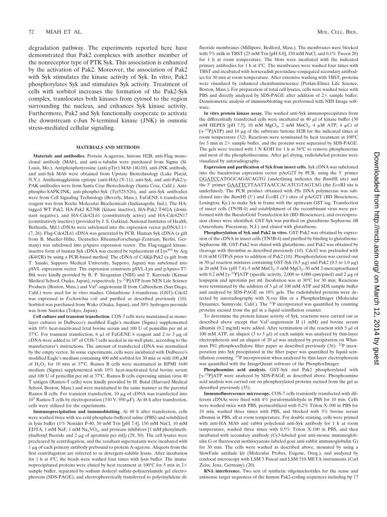
Activation of Pak2 induces the association with Syk. Thep21-activated protein kinase Pak2 and Syk are known to beactivated when the cells are exposed to osmotic stress (24, 34).To examine whether Pak2 and Syk cooperate in cellular sig-naling, HA-tagged Pak2 and Syk cDNAs were transientlytransfected into COS-7 cells. Pak2 was immunoprecipitated byanti-HA antibody, and the immunoprecipitates were analyzedby immunoblotting with anti-pTyr MAb (Fig. 1, top panel).Tyrosine-phosphorylated proteins migrating with the molecu-lar weight of Syk were obtained in the anti-HA immunopre-cipitates of Pak2, and the level of phosphorylation of Syk wassignificantly increased with constitutively active Cdc42. Immu-noblotting experiments confirmed that Syk coimmunoprecipi-tated with WT Pak2, but only a low level of Syk was associatedwith the kinase-inactive mutant (K278R) (Fig. 1, secondpanel). This association of Pak2 with Syk was enhanced by
FIG. 1. Association of Pak2 with Syk. The cDNAs encoding WTHA-Pak2, the kinase-inactive mutant Pak2 K278R, Syk, and HA-Cdc42L61 were transiently cotransfected into COS-7 cells. After 48 hof transfection, cells were solubilized with lysis buffer and Pak2 wasimmunoprecipitated (IP) with anti-HA antibody. Immunoprecipitatedproteins and detergent-soluble lysates were separated by SDS-PAGE,electrophoretically transferred to polyvinylidene difluoride mem-branes and then analyzed by immunoblotting with anti-pTyr MAb andanti-Syk and anti-HA (Pak2 and Cdc42) antibodies. Positions of mo-lecular mass markers are shown on the left in kilodaltons.
FIG. 2. Cdc42 regulates the association of Pak2 with Syk. COS-7cells were transiently transfected with the cDNAs for WT HA-Pak2,Syk, HA-Cdc42L61, HA-Cdc42N17, and Flag-Cdc42L61, as indicated.At 48 h after transfection, cells were solubilized with lysis buffer andPak2 was immunoprecipitated (IP) with anti-HA antibody. Immuno-precipitated proteins and detergent-soluble lysates were analyzed byimmunoblotting with anti-pTyr, anti-Syk, anti-HA (Pak2 and Cdc42),and anti-Flag (Cdc42) antibodies.
VOL. 24, 2004 ASSOCIATION OF Pak2 WITH Syk 73
on March 12, 2014 by guest
http://mcb.asm
.org/D
ownloaded from
coexpression with Cdc42L61, the constitutively active form ofCdc42 (Fig. 1, lane 6).
Quantitative analysis revealed that Cdc42L61 increased theamount of Syk protein (1.2-fold) and the extent of tyrosinephosphorylation of Syk (4.2-fold) in the immunoprecipitates ofPak2 (Fig. 1, top and second panels, lane 4 versus lane 6). Inthe immunoprecipitates of Pak2 K278R, the amounts of co-
precipitated Syk and its tyrosine phosphorylation were de-creased (0.3- and 0.4-fold, respectively) compared to those ofWT Pak2 (Fig. 1, top and second panels, lane 2 versus lane 4).HA-Cdc42 was also precipitated with anti-HA antibody; how-ever, Cdc42 alone could not precipitate Syk (data not shown).In addition, a different HA-tagged protein, HA-Cbl, could notprecipitate significant amounts of Syk, suggesting that Pak2
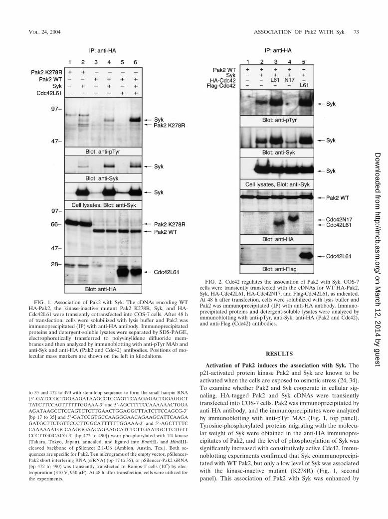
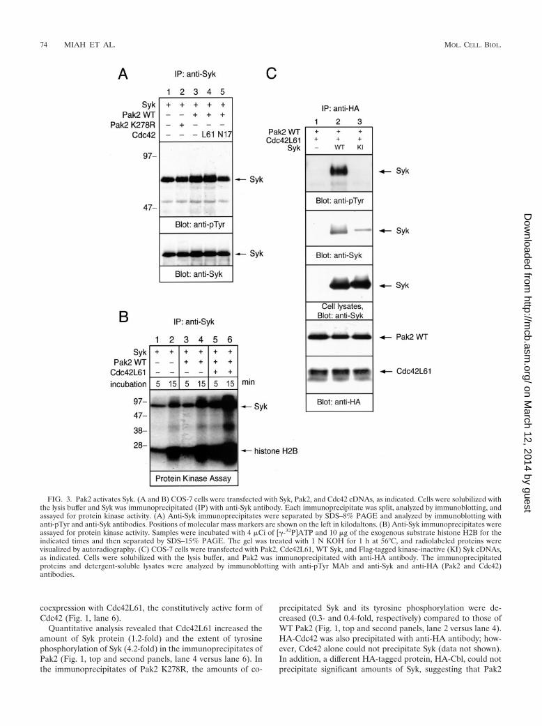
FIG. 3. Pak2 activates Syk. (A and B) COS-7 cells were transfected with Syk, Pak2, and Cdc42 cDNAs, as indicated. Cells were solubilized withthe lysis buffer and Syk was immunoprecipitated (IP) with anti-Syk antibody. Each immunoprecipitate was split, analyzed by immunoblotting, andassayed for protein kinase activity. (A) Anti-Syk immunoprecipitates were separated by SDS–8% PAGE and analyzed by immunoblotting withanti-pTyr and anti-Syk antibodies. Positions of molecular mass markers are shown on the left in kilodaltons. (B) Anti-Syk immunoprecipitates wereassayed for protein kinase activity. Samples were incubated with 4 �Ci of [�-32P]ATP and 10 �g of the exogenous substrate histone H2B for theindicated times and then separated by SDS–15% PAGE. The gel was treated with 1 N KOH for 1 h at 56°C, and radiolabeled proteins werevisualized by autoradiography. (C) COS-7 cells were transfected with Pak2, Cdc42L61, WT Syk, and Flag-tagged kinase-inactive (KI) Syk cDNAs,as indicated. Cells were solubilized with the lysis buffer, and Pak2 was immunoprecipitated with anti-HA antibody. The immunoprecipitatedproteins and detergent-soluble lysates were analyzed by immunoblotting with anti-pTyr MAb and anti-Syk and anti-HA (Pak2 and Cdc42)antibodies.
74 MIAH ET AL. MOL. CELL. BIOL.
on March 12, 2014 by guest
http://mcb.asm
.org/D
ownloaded from
specifically associates with Syk in COS cells (data not shown).Immunoblotting of the detergent-soluble lysates indicated thatequal amounts of Syk were expressed in the cells transfectedwith the different cDNAs (Fig. 1, third panel). As reportedpreviously (26), the expression level of WT Pak2 was muchlower than that of the kinase-inactive mutant K278R, becauseWT Pak2 can be down-regulated by the proteasome degrada-tion pathway (Fig. 1, bottom panel). In addition, the migration
of K278R was slower than that of the WT, as reported previ-ously (7, 26). These results demonstrated that the associationof Pak2 with Syk is enhanced by the activation of Pak2.
Cotransfection of Pak2 with Syk elicited slight tyrosine phos-phorylation of K278R (Fig. 1, top panel). Tyrosine phosphor-ylation of WT Pak2 was observed only when the membranewas exposed for longer times (data not shown).
The dominant-negative form of Cdc42 inhibits the associa-tion of Pak2 with Syk. The serine/threonine kinase activity ofPak family protein kinases is stimulated by binding of activatedGTP bound to Cdc42/Rac (3). To examine the functional in-teraction between Pak2 and Syk, the cDNAs of HA-tagged WTPak2 and Syk were cotransfected into COS-7 cells along witheither constitutively active Cdc42L61 or inactive Cdc42N17(Fig. 2). The expression of Cdc42L61 increased the associationof Pak2 with Syk, as shown in Fig. 1. On the other hand,expression of inactive Cdc42N17 blocked the coprecipitationof Syk with Pak2 (Fig. 2, lane 4). It is suggested that overex-pression of the dominant-negative form of Cdc42 inhibited theactivation of Pak2 by endogenous Cdc42 and the subsequentinteraction with Syk (Fig. 2, lane 2). The amounts of Sykprotein expressed in cells transfected with different cDNAswere similar (Fig. 2, third panel). To confirm that the increasein the coprecipitation of Syk with HA-Pak2 was not due to thesame epitope tag on Cdc42, we tested Cdc42L61 with a differ-ent epitope tag. As shown (Fig. 2, lane 5), cotransfection withFlag-tagged Cdc42L61 also resulted in a similar increase in thecoprecipitation of Syk with Pak2. These findings suggested thatCdc42 functionally regulates the association of Pak2 with Sykin vivo.
Association with Pak2 induces the activation of Syk. Sinceactivation of Pak2 resulted in its association with Syk, we ex-amined the effects of Pak2 association on Syk kinase activity.Tyrosine phosphorylation and the intrinsic kinase activity ofSyk were measured by immunoblotting with anti-pTyr and by aSyk activity assay in vitro (Fig. 3A and B, respectively). Addi-tion of WT Pak2 slightly elevated the tyrosine phosphorylationof Syk. This Pak2-mediated enhancement of tyrosine phos-phorylation of Syk was enhanced further by cotransfection of
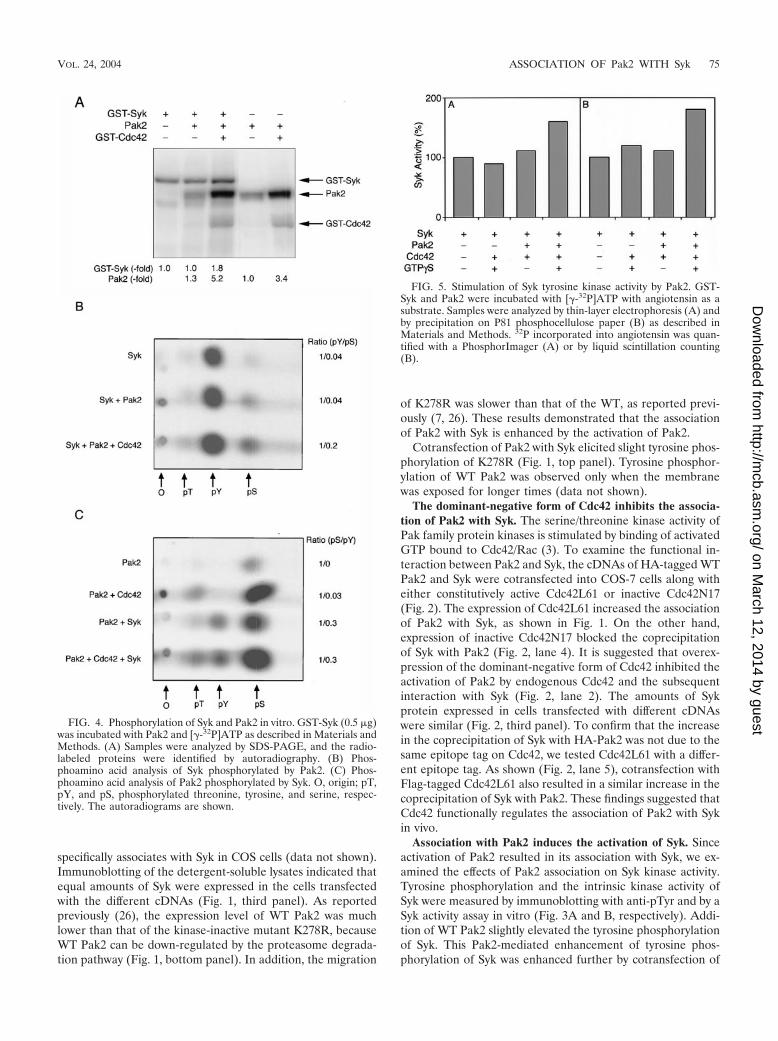
FIG. 4. Phosphorylation of Syk and Pak2 in vitro. GST-Syk (0.5 �g)was incubated with Pak2 and [�-32P]ATP as described in Materials andMethods. (A) Samples were analyzed by SDS-PAGE, and the radio-labeled proteins were identified by autoradiography. (B) Phos-phoamino acid analysis of Syk phosphorylated by Pak2. (C) Phos-phoamino acid analysis of Pak2 phosphorylated by Syk. O, origin; pT,pY, and pS, phosphorylated threonine, tyrosine, and serine, respec-tively. The autoradiograms are shown.
FIG. 5. Stimulation of Syk tyrosine kinase activity by Pak2. GST-Syk and Pak2 were incubated with [�-32P]ATP with angiotensin as asubstrate. Samples were analyzed by thin-layer electrophoresis (A) andby precipitation on P81 phosphocellulose paper (B) as described inMaterials and Methods. 32P incorporated into angiotensin was quan-tified with a PhosphorImager (A) or by liquid scintillation counting(B).
VOL. 24, 2004 ASSOCIATION OF Pak2 WITH Syk 75
on March 12, 2014 by guest
http://mcb.asm
.org/D
ownloaded from
WT Pak2 and Cdc42L61. Expression of the kinase-inactivePak2 K278R had no effect on Syk phosphorylation. Coexpres-sion with Cdc42N17, which inhibited the association of Pak2with Syk, suppressed tyrosine phosphorylation of Syk (Fig. 3A,lane 5). To examine the intrinsic kinase activity of Syk, anti-Sykimmunoprecipitates were subjected to an in vitro protein ki-nase assay (Fig. 3B). The samples were separated by SDS-PAGE, and gels were incubated with KOH to remove phos-phoserine and most phosphothreonine. Coexpression withPak2 elevated autophosphorylation of Syk and phosphoryla-tion of exogenous substrate histone H2B. There was no de-tectable signal in an in vitro kinase assay with Pak2 immuno-precipitated by anti-HA after the KOH treatment (data notshown). This ruled out the possibility that phosphorylation ofH2B histone was due to the coprecipitated Pak2. Also, wecould not detect Pak2 in the Syk immunoprecipitates by im-munoblotting (data not shown). Moreover, activation of Pak2by Cdc42L61 significantly enhanced the kinase activity of Syk(Fig. 3B). Cdc42N17 showed a negative effect on the kinaseactivity of Syk (data not shown). Thus, this result indicated thatassociation of the active form of Pak2 enhances tyrosine phos-phorylation and the intrinsic kinase activity of Syk. It suggestedthat association with Pak2 stimulates the kinase activity of Syk.
We then tested whether the kinase activity of Syk is requiredfor the interaction between Pak2 and Syk. Coprecipitation ofPak2 with the kinase-inactive form of Syk was less than that ofWT Syk (Fig. 3C). Therefore, the association of Pak2 with Sykrequires the kinase activity of Syk.
Phosphorylation and activation of Syk by Pak2 in vitro. Theassociation of Pak2 and Syk, with the resultant enhancement ofautophosphorylation and activity of Syk when Syk was coex-pressed with Pak2 and Cdc42, suggested that Syk could be asubstrate for Pak2. To examine this, purified GST-Syk ex-pressed in insect cells was incubated with purified Pak2 and[�-32P]ATP in the presence of Mg2� and Mn2�. Phosphoryla-tion of GST-Syk was increased 1.8-fold when phosphorylationwas by Pak2 activated with Cdc42 (GTP�S) (Fig. 4A). Asexpected, phosphoamino acid analysis of autophosphorylatedSyk showed that the phosphorylation was exclusively on ty-rosine (Fig. 4B). There was little effect on phosphorylationwith inactive Pak2. When activated Pak2 was added to theassay mixture, there was additional phosphorylation of Syk onboth serine and tyrosine. Quantification of the data showedthat the phosphotyrosine/phosphoserine ratio decreased five-
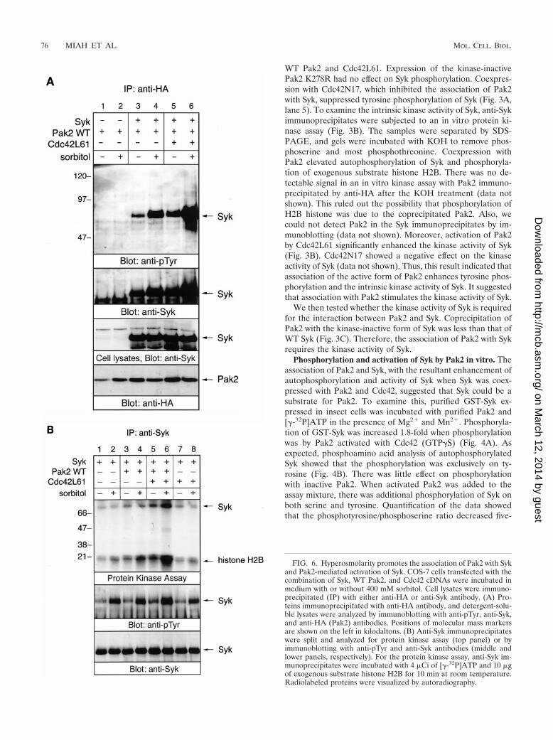
FIG. 6. Hyperosmolarity promotes the association of Pak2 with Sykand Pak2-mediated activation of Syk. COS-7 cells transfected with thecombination of Syk, WT Pak2, and Cdc42 cDNAs were incubated inmedium with or without 400 mM sorbitol. Cell lysates were immuno-precipitated (IP) with either anti-HA or anti-Syk antibody. (A) Pro-teins immunoprecipitated with anti-HA antibody, and detergent-solu-ble lysates were analyzed by immunoblotting with anti-pTyr, anti-Syk,and anti-HA (Pak2) antibodies. Positions of molecular mass markersare shown on the left in kilodaltons. (B) Anti-Syk immunoprecipitateswere split and analyzed for protein kinase assay (top panel) or byimmunoblotting with anti-pTyr and anti-Syk antibodies (middle andlower panels, respectively). For the protein kinase assay, anti-Syk im-munoprecipitates were incubated with 4 �Ci of [�-32P]ATP and 10 �gof exogenous substrate histone H2B for 10 min at room temperature.Radiolabeled proteins were visualized by autoradiography.
76 MIAH ET AL. MOL. CELL. BIOL.
on March 12, 2014 by guest
http://mcb.asm
.org/D
ownloaded from
fold with activated Pak2. Taken together, the data indicatedthat there was a substantial increase in tyrosine phosphoryla-tion when Syk was phosphorylated on serine by Pak2.
Pak2 was also phosphorylated by Syk (Fig. 4A). There was a1.3-fold increase in phosphorylation of inactive Pak2 and a1.5-fold increase in Cdc42-activated Pak2. Pak2 was autophos-phorylated only on serine in the absence of Cdc42 and onserine and threonine in the presence of Cdc42 (Fig. 4C). Ad-dition of Syk resulted in phosphorylation of tyrosine in bothcases. Phosphorylation of tyrosine did not result in activationof Pak2, as shown by a lack of phosphate in the activation loopthreonine in inactive Pak2 (Fig. 4B). In fact, the ratio of phos-phoserine to phosphotyrosine was similar with both active andinactive Pak2.
The effects of phosphorylation of Syk by Pak2 were examinedby using Val5-angiotensin II as the substrate. As shown in Fig. 5,phosphorylation of angiotensin was increased when Syk was in-cubated with Cdc42-activated Pak2. An increase of 1.6-fold wasobserved when 32P incorporation into Syk was analyzed by thin-layer electrophoresis (Fig. 5A). This was consistent with the re-sults obtained by precipitation of phosphorylated Syk on filterpaper (Fig. 5B).
Hyperosmotic stress promotes the association of Pak2 withSyk. We have demonstrated that treatment of avian B cellswith sorbitol activates Syk, although the receptor for the os-motic stress has not yet been identified (22). Sorbitol alsoresults in the translocation and subsequent activation of the
protein kinase activity of Pak2 in the particulate fraction byassociation with Cdc42 (24). Therefore, we examined whetherthe interaction between Pak2 and Syk was affected by osmoticstress. Sorbitol stimulation resulted in an increase in the asso-ciation of Pak2 with Syk (Fig. 6A, lanes 3 and 4). This associ-ation was dramatically increased in cells transfected withCdc42L61 (Fig. 6A, lanes 5 and 6). Equal amounts of Syk wereexpressed in cells transfected with different cDNAs (Fig. 6A,third panel). This result indicated that hyperosmotic stress andCdc42 synergistically promote the association of Pak2 with Syk.
We then tested the effect of osmotic stress on Pak2-medi-ated activation of Syk (Fig. 6B). In unstimulated cells, coex-pression of Pak2 elevated the kinase activity of Syk, and addi-tion of Cdc42 further stimulated Pak2-mediated activation ofSyk (Fig. 6B, upper panel, lanes 1, 3, and 5). Cdc42L61 aloneinduced the kinase activity of Syk to a lesser extent, perhaps byactivating endogenous Pak kinases (Fig. 6B, lane 7). Stimula-tion of cells with sorbitol increased the kinase activity of Syk,and cotransfection of WT Pak2 enhanced the sorbitol-medi-ated activation of Syk (Fig. 6B, lanes 1 to 4). Addition ofCdc42L61 could further stimulate sorbitol-mediated activationof Syk (Fig. 6B, lanes 5 and 6). This result demonstrated thathyperosmolarity increases the association of Pak2 with Syk andPak2-mediated activation of Syk. A similar effect was observedwhen cells were treated with 400 mM sucrose (data notshown).
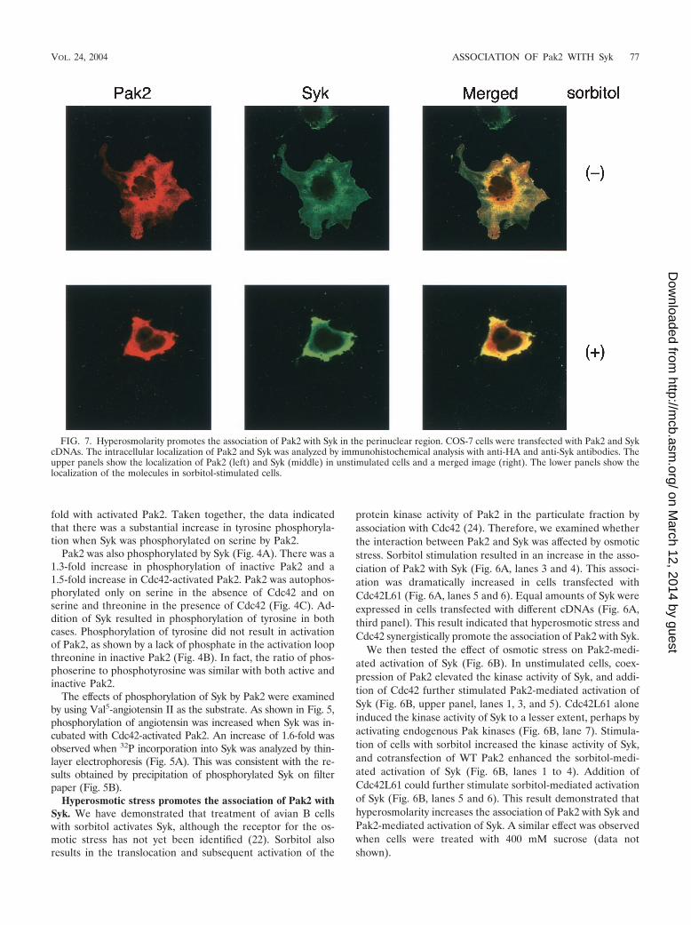
FIG. 7. Hyperosmolarity promotes the association of Pak2 with Syk in the perinuclear region. COS-7 cells were transfected with Pak2 and SykcDNAs. The intracellular localization of Pak2 and Syk was analyzed by immunohistochemical analysis with anti-HA and anti-Syk antibodies. Theupper panels show the localization of Pak2 (left) and Syk (middle) in unstimulated cells and a merged image (right). The lower panels show thelocalization of the molecules in sorbitol-stimulated cells.
VOL. 24, 2004 ASSOCIATION OF Pak2 WITH Syk 77
on March 12, 2014 by guest
http://mcb.asm
.org/D
ownloaded from

Hyperosmotic stress induces the association of Pak2 withSyk in the perinuclear region. Endogenous Pak2 is present inboth the cytosolic and particulate fractions in mouse fibroblast3T3-L1 cells. Moreover, recent findings demonstrate that lo-calization of Pak2 in the endoplasmic reticulum is required forthe induction of cytostasis (7). Therefore, we examined theintracellular localization of Pak2 and Syk in COS-7 cells. Im-munohistochemical analysis using confocal microscopy con-firmed the association of Pak2 and Syk (Fig. 7). Pak2 and Sykwere concentrated in a region near the nucleus and were alsolocated elsewhere in the cytosolic region, but not in the nucleus(Fig. 7, top panels). Sorbitol treatment resulted in shrinkage ofthe cells and an increase in the association of Pak2 with Syk inthe perinuclear region (Fig. 7, bottom panels).
Pak2 and Syk functionally cooperate to enhance sorbitol-mediated activation of JNK. It was previously demonstratedthat Syk and Rac1 lead to synergistic JNK activation in T cells
(9). In addition, Syk is critical for antigen-mediated activationof JNK in avian B cells (12). Therefore, we examined theeffects of Pak2 and Syk in sorbitol-mediated activation of JNK(Fig. 8). As shown by immunoblotting with antibody to phos-pho-JNK, sorbitol-mediated activation of JNK was enhancedin the cells transfected with Syk. The JNK activity was dramat-ically enhanced in cells coexpressing Syk, Pak2, and Cdc42L61(Fig. 8A). Osmotic stress had no effect on activation of extra-cellular signal-regulated kinase (ERK) and p38 kinase in alltransfected cells (data not shown). Furthermore, we testedwhether the different kinds of nonreceptor PTKs cooperatewith Pak2 to activate JNK. Dramatic activation of JNK wasobserved only in cells cotransfected with Syk PTK and not inthose transfected with Lyn, Btk, or CAK�/Pyk2 (Fig. 8B).Thus, hyperosmolarity dramatically increased the associationof Pak2 with Syk, leading to the activation of Syk and therebydownstream JNK phosphorylation.
FIG. 8. Pak2 and Syk cooperate in hyperosmolarity-induced activation of JNK. COS-7 cells were transfected with the combination of Syk, Pak2(WT and K278R), and Cdc42 cDNAs as indicated (A) or Pak2, Cdc42L61, and the various PTK cDNAs were cotransfected into COS-7 cells (B).Cells were incubated in medium with or without the addition of 400 mM sorbitol. Total cell lysates were analyzed by immunoblotting withanti-phospho-JNK and anti-JNK antibodies. Positions of molecular mass markers are shown on the left in kilodaltons.
78 MIAH ET AL. MOL. CELL. BIOL.
on March 12, 2014 by guest
http://mcb.asm
.org/D
ownloaded from
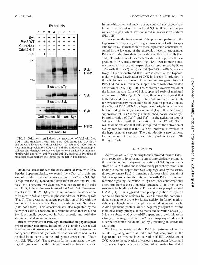
Oxidative stress induces the association of Pak2 with Syk.Besides hyperosmolarity, we tested the effect of a differentkind of cellular stress on the association of Pak2 with Syk. Sykis required for H2O2-mediated activation of Akt and PI 3-ki-nase (34). Therefore, we examined whether treatment of cellswith H2O2 induces the association of Pak2 with Syk. Treatmentof cells with 100 �M H2O2 for 10 min induced the associationof Pak2 with Syk and tyrosine phosphorylation of Pak2 by Syk(Fig. 9). There was no apparent precipitation of Syk with theantibody to HA when the cells were transfected with Syk alone(data not shown). This association was also regulated by theactivity of Cdc42. Therefore, this result indicated that Pak2 andSyk functionally cooperated in both osmotic and oxidativestress-mediated signaling in vivo.
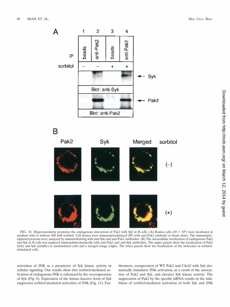
Direct involvement of Pak2-Syk interaction in physiologicalhyperosmolar responses. Based on these findings, we testedwhether osmotic stress can induce the interaction between theendogenous Pak2 and Syk. Sorbitol treatment of Ramos B cellsresulted in an increase in the endogenous association of Pak2with Syk (Fig. 10A). These results further emphasize the bio-logical significance of the interaction of the two molecules.
Immunohistochemical analysis using confocal microscopy con-firmed the association of Pak2 and Syk in B cells in the pe-rinuclear region, which was enhanced in response to sorbitol(Fig. 10B).
To examine the involvement of the proposed pathway in thehyperosmolar response, we designed two kinds of siRNAs spe-cific for Pak2. Transfection of these expression constructs re-sulted in the lowering of the expression level of endogenousPak2 and sorbitol-mediated activation of JNK in B cells (Fig.11A). Transfection of Pak2 siRNA did not suppress the ex-pression of JNK and �-tubulin (Fig. 11A). Densitometric anal-ysis revealed that protein expression was suppressed by 90 or70% with the Pak2(17-35) or Pak2(472-490) siRNA, respec-tively. This demonstrated that Pak2 is essential for hyperos-molarity-induced activation of JNK in B cells. In addition tothe siRNA, overexpression of the dominant-negative form ofPak2 (T402A) resulted in the suppression of sorbitol-mediatedactivation of JNK (Fig. 11B) (7). Moreover, overexpression ofthe kinase-inactive form of Syk suppressed sorbitol-mediatedactivation of JNK (Fig. 11C). Thus, these results suggest thatboth Pak2 and its associating protein Syk are critical in B cellsfor hyperosmolarity-mediated physiological responses. Finally,the effect of Pak2 siRNA on hyperosmolarity-induced activa-tion of endogenous Syk was examined (Fig. 11D). As shown,suppression of Pak2 directly inhibits phosphorylation of Syk.Phosphorylation of Tyr525 and Tyr526 in the activation loop ofSyk is correlated with the activation of Syk (17, 41). Theseresults demonstrated that Pak2 is required for the activation ofSyk by sorbitol and that the Pak2-Syk pathway is involved inthe hyperosmolar response. The data identify a new pathwayfor activation of the stress-activated protein kinase JNKthrough Cdc42.
DISCUSSION
Activation of Pak2 by binding to the activated form of Cdc42or in response to hyperosmotic stress synergistically promotesthe association and enzymatic activation of Syk. Syk is a sub-strate of Pak2 in vitro and is activated by phosphorylation. Ourfinding is the first report that Syk is up-regulated by the serine/threonine kinase Pak2. It remains unknown which domain ofSyk is responsible for the interaction with Pak2. In immunereceptor signaling, activation of Syk requires conformationalalteration from a closed inactive structure to an open activestructure by binding of the SH2 domains to phosphorylatedITAM (14). It is suggested that phosphorylation of Syk onserine or threonine residues by Pak2 induces the conforma-tional change to activate Syk kinase activity. In formyl methio-nyl-leucyl-phenylalanine receptor-mediated signaling, cyclicAMP-dependent protein kinase negatively regulates formylmethionyl-leucyl-phenylalanine-mediated activation of Syk (2).Syk is a substrate of cyclic AMP-dependent protein kinase invitro (2). It is suggested that Pak2 may phosphorylate differenta serine/threonine residue(s) on Syk, resulting in enzymaticactivation.
We have demonstrated that Pak2 is upstream of Syk incellular signaling and that Pak2 and Syk cooperate in thehyperosmotic stress-mediated activation of JNK. Activation ofJNK leads to the activation of various transcription factors andexpression of specific genes (5). We utilized sorbitol-mediated
FIG. 9. Oxidative stress induces the association of Pak2 with Syk.COS-7 cells transfected with Syk, Pak2, Cdc42L61, and Cdc42N17cDNAs were incubated with or without 100 �M H2O2. Cell lysateswere immunoprecipitated (IP) with anti-HA antibody. Immunopre-cipitates and detergent-soluble cell lysates were analyzed by immuno-blotting with anti-pTyr, anti-Syk, and anti-HA antibodies. Positions ofmolecular mass markers are shown on the left in kilodaltons.
VOL. 24, 2004 ASSOCIATION OF Pak2 WITH Syk 79
on March 12, 2014 by guest
http://mcb.asm
.org/D
ownloaded from
activation of JNK as a parameter of Syk kinase activity incellular signaling. Our results show that sorbitol-mediated ac-tivation of endogenous JNK is enhanced by the overexpressionof Syk (Fig. 8). Expression of the kinase-inactive form of Syksuppresses sorbitol-mediated activation of JNK (Fig. 11). Fur-
thermore, coexpression of WT Pak2 and Cdc42 with Syk dra-matically stimulates JNK activation, as a result of the associa-tion of Pak2 and Syk, and elevates Syk kinase activity. Thesuppression of Pak2 by the specific siRNA results in the inhi-bition of sorbitol-mediated activation of both Syk and JNK
FIG. 10. Hyperosmolarity promotes the endogenous interaction of Pak2 with Syk in B cells. (A) Ramos cells (20 � 106) were incubated inmedium with or without 400 mM sorbitol. Cell lysates were immunoprecipitated (IP) with anti-Pak2 antibody or beads alone. The immunopre-cipitated proteins were analyzed by immunoblotting with anti-Syk and anti-Pak2 antibodies. (B) The intracellular localization of endogenous Pak2and Syk in B cells was analyzed immunohistochemically with anti-Pak2 and anti-Syk antibodies. The upper panels show the localization of Pak2(left) and Syk (middle) in unstimulated cells and a merged image (right). The lower panels show the localization of the molecules in sorbitol-stimulated cells.
80 MIAH ET AL. MOL. CELL. BIOL.
on March 12, 2014 by guest
http://mcb.asm
.org/D
ownloaded from

FIG. 11. Suppression of Pak2 reduces hyperosmolarity-induced ac-tivation of endogenous Syk and JNK in B cells. At 48 h after transfec-tion, cells were incubated in serum-free medium without or with 400mM sorbitol for 30 min. Equal amounts of the cell lysates were ana-lyzed by immunoblotting with anti-Pak2, anti-phospho-JNK, anti-JNK,anti-�-tubulin, and anti-HA, anti-Flag, anti-pTyr, anti-phospho-Syk(Tyr525/526), and anti-Syk antibodies, as indicated. (A) Ramos cellswere transiently transfected with the pSilencer vector, pSilencer-siRNA for Pak2(17-35) or for Pak2(472-490). (B) Ramos cells weretransiently transfected with pcDNA3.1� vector or the dominant-neg-ative form of Pak2 (T402A). (C) Ramos cells were transiently trans-fected with pcDNA3.1� vector or the kinase-inactive form of Syk (SykKI). (D) Ramos cells were transiently transfected with pSilencer vectoror pSilencer-siRNA for Pak2(17-35).
VOL. 24, 2004 ASSOCIATION OF Pak2 WITH Syk 81
on March 12, 2014 by guest
http://mcb.asm
.org/D
ownloaded from
(Fig. 11). We have not tested whether caspase 3 cleavageproducts of Pak2 could associate with Syk and stimulate itskinase activity. In our experimental system, osmotic stress didnot cause any increase in the caspase cleavage of Pak2 inCOS-7 cells (data not shown). It remains unknown whether theextreme stress that results in cleavage and activation of Pak2could induce the association of Pak2 with Syk to stimulate JNKactivity. Collectively, Syk could be responsible for osmoticstress-mediated JNK activation in mammalian cells, cooperat-ing with Pak2 and Rho family GTP binding proteins. Activa-tion of Syk in response to osmotic stress requires the interac-tion with Pak2.
Pak2 has been shown to have cytostatic properties, as shownby microinjection of Pak2 into early frog embryos (27) andoverexpression of Pak2 in COS-7 and HEK 293T cells (7).Activation of Pak2 by moderate cell stress inhibits cell division,resulting in inhibition of cell growth, whereas high levels ofstress or irreparable damage results in cell death. Both Pak2and Syk are activated in response to oxidative stress. Syk acti-vation by moderate oxidative stress leads to the activation ofthe PI 3-kinase-Akt survival pathway (34). On the other hand,extreme oxidative stress induces Syk-dependent phospholipaseC-� activation, which accelerates oxidative stress-mediated apo-ptosis (34).
Both Pak2 and Syk were originally isolated as cytosolic ki-nases. Various cellular ligands induce the translocation ofthese enzymes to the perinuclear region. Active Pak2, but notthe inactive enzyme, is tightly associated with the endoplasmicreticulum, and the amount of Pak2 associated with the endo-plasmic reticulum is significantly enhanced in cells that arestressed and/or not dividing (7). In COS-7 cells, Pak2 and Sykare primarily cytosolic, and sorbitol treatment increases trans-location of both kinases to the perinuclear region (Fig. 7).Unlike that of c-Abl, tyrosine phosphorylation of Pak2 by Sykdid not result in an accumulation of inactive Pak2 or inhibit itsprotein turnover through the proteasome degradation pathway(Fig. 1). It is suggested that Pak2 and Syk transmit the activat-ing signals to the downstream molecules from the perinuclearregion. In immune receptor signaling, Syk translocates to theplasma membrane to interact with receptors in the glycolipid-enriched microdomain (33). Syk phosphorylates Vav, a gua-nine nucleotide exchange factor of Rac1, to regulate calciummobilization and transcription of cytokine messages (1, 6, 19).In T cells, activation of Rac1 and subsequently Pak1 requires aSyk family PTK (16). Our findings demonstrate that there is adistinct distribution and function of Syk that is different fromthat established in the immune cells.
It is interesting that although both ERK and JNK are down-stream of Syk in the immune receptor signaling, only JNK isactivated in nonhematopoietic cells (12). In breast cancer cells,induction of Syk protein results in a suppression of tumor cellproliferation resulting from the abnormalities of cell division(14). Perhaps Pak2 and Syk cooperate to stimulate the tran-scription and translation of both proapoptotic and antiapopto-tic proteins which are downstream of JNK.
ACKNOWLEDGMENTS
This study was supported in part by research funding from theUehara Memorial Foundation and the Hyogo Science and TechnologyAssociation; the 21st Century COE Program and a Grant-in-Aid for
Scientific Research from the Japan Society for the Promotion of Sci-ence and the Ministry of Education, Culture, Sports, Science andTechnology of Japan; and a grant from the National Institutes ofHealth (GM26738) to J.A.T.
REFERENCES
1. Arudchandran, R., M. J. Brown, M. J. Peirce, J. S. Song, J. Zhang, R. P.Siraganian, U. Blank, and J. Rivera. 2000. The Src homology 2 domain ofVav is required for its compartmentation to the plasma membrane andactivation of c-Jun NH(2)-terminal kinase 1. J. Exp. Med. 191:47–60.
2. Asahi, M., Y. Tanaka, S. Qin, M. Tsubokawa, K. Sada, Y. Minami, and H.Yamamura. 1995. Cyclic AMP-elevating agents negatively regulate the acti-vation of p72syk in N-formyl-methionyl-leucyl-phenylalanine receptor signal-ing. Biochem. Biophys. Res. Commun. 212:887–893.
3. Bagrodia, S., and R. A. Cerione. 1999. Pak to the future. Trends Cell Biol.9:350–355.
4. Coopman, P. J., M. T. Do, M. Barth, E. T. Bowden, A. J. Hayes, E. Basyuk,J. K. Blancato, P. R. Vezza, S. W. McLeskey, P. H. Mangeat, and S. C.Mueller. 2000. The Syk tyrosine kinase suppresses malignant growth ofhuman breast cancer cells. Nature 406:742–747.
5. Davis, R. J. 2000. Signal transduction by the JNK group of MAP kinases.Cell 103:239–252.
6. Deckert, M., S. Tartare-Deckert, C. Couture, T. Mustelin, and A. Altman.1996. Functional and physical interactions of Syk family kinases with the Vavproto-oncogene product. Immunity 5:591–604.
7. Huang, Z., J. Ling, and J. A. Traugh. 2003. Localization of p21-activatedprotein kinase gamma-PAK/Pak2 in the endoplasmic reticulum is requiredfor induction of cytostasis. J. Biol. Chem. 278:13101–13109.
8. Inoue, O., K. Suzuki-Inoue, W. L. Dean, J. Frampton, and S. P. Watson.2003. Integrin alpha2beta1 mediates outside-in regulation of platelet spread-ing on collagen through activation of Src kinases and PLCgamma2. J. CellBiol. 160:769–780.
9. Jacinto, E., G. Werlen, and M. Karin. 1998. Cooperation between Syk andRac1 leads to synergistic JNK activation in T lymphocytes. Immunity 8:31–41.
10. Jakobi, R., C. J. Chen, P. T. Tuazon, and J. A. Traugh. 1996. Molecularcloning and sequencing of the cytostatic G protein-activated protein kinasePAK I. J. Biol. Chem. 271:6206–6211.
11. Jakobi, R., E. Moertl, and M. A. Koeppel. 2001. p21-activated protein kinasegamma-PAK suppresses programmed cell death of BALB3T3 fibroblasts.J. Biol. Chem. 276:16624–16634.
12. Jiang, A., A. Craxton, T. Kurosaki, and E. A. Clark. 1998. Different proteintyrosine kinases are required for B cell antigen receptor-mediated activationof extracellular signal-regulated kinase, c-Jun NH2-terminal kinase 1, andp38 mitogen-activated protein kinase. J. Exp. Med. 188:1297–1306.
13. Keshvara, L. M., C. C. Isaacson, T. M. Yankee, R. Sarac, M. L. Harrison,and R. L. Geahlen. 1998. Syk- and Lyn-dependent phosphorylation of Syk onmultiple tyrosines following B cell activation includes a site that negativelyregulates signaling. J. Immunol. 161:5276–5283.
14. Kimura, T., H. Sakamoto, E. Appella, and R. P. Siraganian. 1996. Confor-mational changes induced in the protein tyrosine kinase p72syk by tyrosinephosphorylation or by binding of phosphorylated immunoreceptor tyrosine-based activation motif peptides. Mol. Cell. Biol. 16:1471–1478.
15. Knaus, U. G., and G. M. Bokoch. 1998. The p21Rac/Cdc42-activated kinases(PAKs). Int. J. Biochem. Cell Biol. 30:857–862.
16. Ku, G. M., D. Yablonski, E. Manser, L. Lim, and A. Weiss. 2001. A PAK1-PIX-PKL complex is activated by the T-cell receptor independent of Nck,Slp-76 and LAT. EMBO J. 20:457–465.
17. Kurosaki, T., S. A. Johnson, L. Pao, K. Sada, H. Yamamura, and J. C.Cambier. 1995. Role of the Syk autophosphorylation site and SH2 domainsin B cell antigen receptor signaling. J. Exp. Med. 182:1815–1823.
18. Lee, N., H. MacDonald, C. Reinhard, R. Halenbeck, A. Roulston, T. Shi, andL. T. Williams. 1997. Activation of hPAK65 by caspase cleavage inducessome of the morphological and biochemical changes of apoptosis. Proc. Natl.Acad. Sci. USA 94:13642–13647.
19. Manetz, T. S., C. Gonzalez-Espinosa, R. Arudchandran, S. Xirasagar, V.Tybulewicz, and J. Rivera. 2001. Vav1 regulates phospholipase C� activationand calcium responses in mast cells. Mol. Cell. Biol. 21:3763–3774.
20. Mitsui, N., R. Inatome, S. Takahashi, Y. Goshima, H. Yamamura, and S.Yanagi. 2002. Involvement of Fes/Fps tyrosine kinase in semaphorin 3Asignaling. EMBO J. 21:3274–3285.
21. Poole, A., J. M. Gibbins, M. Turner, M. J. van Vugt, J. G. van de Winkel, T.Saito, V. L. Tybulewicz, and S. P. Watson. 1997. The Fc receptor gamma-chain and the tyrosine kinase Syk are essential for activation of mouseplatelets by collagen. EMBO J. 16:2333–2341.
22. Qin, S., Y. Minami, M. Hibi, T. Kurosaki, and H. Yamamura. 1997. Syk-dependent and -independent signaling cascades in B cells elicited by osmoticand oxidative stress. J. Biol. Chem. 272:2098–2103.
23. Renkema, G. H., A. Manninen, and K. Saksela. 2001. Human immunodefi-ciency virus type 1 Nef selectively associates with a catalytically active sub-
82 MIAH ET AL. MOL. CELL. BIOL.
on March 12, 2014 by guest
http://mcb.asm
.org/D
ownloaded from
population of p21-activated kinase 2 (PAK2) independently of PAK2 bind-ing to Nck or beta-PIX. J. Virol. 75:2154–2160.
24. Roig, J., Z. Huang, C. Lytle, and J. A. Traugh. 2000. p21-activated proteinkinase gamma-PAK is translocated and activated in response to hyperosmo-larity. Implication of Cdc42 and phosphoinositide 3-kinase in a two-stepmechanism for gamma-PAK activation. J. Biol. Chem. 275:16933–16940.
25. Roig, J., and J. A. Traugh. 1999. p21-activated protein kinase gamma-PAKis activated by ionizing radiation and other DNA-damaging agents. Similar-ities and differences to alpha-PAK. J. Biol. Chem. 274:31119–31122.
26. Roig, J., P. T. Tuazon, P. A. Zipfel, A. M. Pendergast, and J. A. Traugh. 2000.Functional interaction between c-Abl and the p21-activated protein kinasegamma-PAK. Proc. Natl. Acad. Sci. USA 97:14346–14351.
27. Rooney, R. D., P. T. Tuazon, W. E. Meek, E. J. Carroll, J. J. Hagen, E. L.Gump, C. A. Monnig, T. Lugo, and J. A. Traugh. 1996. Cleavage arrest ofearly frog embryos by the G protein-activated protein kinase PAK I. J. Biol.Chem. 271:21498–21504.
28. Rudel, T., and G. M. Bokoch. 1997. Membrane and morphological changesin apoptotic cells regulated by caspase-mediated activation of PAK2. Science276:1571–1574.
29. Sada, K., S. M. S. Miah, K. Maeno, S. Kyo, X. Qu, and H. Yamamura. 2002.Regulation of Fcepsilon RI-mediated degranulation by an adaptor protein3BP2 in rat basophilic leukemia RBL-2H3 cells. Blood 100:2138–2144.
30. Sada, K., Y. Minami, and H. Yamamura. 1997. Relocation of Syk protein-tyrosine kinase to the actin filament network and subsequent association withFak. Eur. J. Biochem. 248:827–833.
31. Sada, K., T. Takano, S. Yanagi, and H. Yamamura. 2001. Structure andfunction of Syk protein-tyrosine kinase. J. Biochem. (Tokyo) 130:177–186.
32. Sada, K., J. Zhang, and R. P. Siraganian. 2000. Point mutation of a tyrosinein the linker region of Syk results in a gain of function. J. Immunol. 164:338–344.
33. Sada, K., J. Zhang, and R. P. Siraganian. 2001. SH2 domain-mediated
targeting, but not localization, of Syk in the plasma membrane is critical forFcepsilonRI signaling. Blood 97:1352–1359.
34. Takano, T., K. Sada, and H. Yamamura. 2002. Role of protein-tyrosinekinase Syk in oxidative stress signaling in B cells. Antioxid. Redox Signal.4:533–540.
35. Tuazon, P. T., M. Chinwah, and J. A. Traugh. 1998. Autophosphorylationand protein kinase activity of p21-activated protein kinase gamma-PAK aredifferentially affected by magnesium and manganese. Biochemistry37:17024–17029.
36. Tuazon, P. T., W. C. Spanos, E. L. Gump, C. A. Monnig, and J. A. Traugh.1997. Determinants for substrate phosphorylation by p21-activated proteinkinase (gamma-PAK). Biochemistry 36:16059–16064.
37. Turner, M., E. Schweighoffer, F. Colucci, J. P. Di Santo, and V. L. Ty-bulewicz. 2000. Tyrosine kinase SYK: essential functions for immunorecep-tor signalling. Immunol. Today 21:148–154.
38. Walter, B. N., Z. Huang, R. Jakobi, P. T. Tuazon, E. S. Alnemri, G. Litwack,and J. A. Traugh. 1998. Cleavage and activation of p21-activated proteinkinase gamma-PAK by CPP32 (caspase 3). Effects of autophosphorylationon activity. J. Biol. Chem. 273:28733–28739.
39. Wolf, D., V. Witte, B. Laffert, K. Blume, E. Stromer, S. Trapp, P. d’Aloja, A.Schurmann, and A. S. Baur. 2001. HIV-1 Nef associated PAK and PI3-kinases stimulate Akt-independent Bad-phosphorylation to induce anti-apoptotic signals. Nat. Med. 7:1217–1224.
40. Yanagi, S., R. Inatome, J. Ding, H. Kitaguchi, V. L. Tybulewicz, and H.Yamamura. 2001. Syk expression in endothelial cells and their morphologicdefects in embryonic Syk-deficient mice. Blood 98:2869–2871.
41. Zhang, J., M. L. Billingsley, R. L. Kincaid, and R. P. Siraganian. 2000.Phosphorylation of Syk activation loop tyrosines is essential for Syk function.An in vivo study using a specific anti-Syk activation loop phosphotyrosineantibody. J. Biol. Chem. 275:35442–35447.
VOL. 24, 2004 ASSOCIATION OF Pak2 WITH Syk 83
on March 12, 2014 by guest
http://mcb.asm
.org/D
ownloaded from
Copyright © 2022 FDOKUMEN