
The challenge of selecting protein kinase assays for lead discovery optimization
22
The challenge of selecting protein kinase assays for lead discovery optimization Haiching Ma, PhD 1,† , Sean Deacon, PhD 2 , and Kurumi Horiuchi, PhD 3 1 Chief Technology Officer, Reaction Biology Corporation, One Great Valley Parkway, Suite 8, Malvern, PA 19355, USA, Tel: +1 610 722 0247; Fax: +1 610 722 0246; E-mail: [email protected] 2 Senior Scientist, Reaction Biology Corporation, One Great Valley Parkway, Suite 8, Malvern, PA 19355, USA 3 Director of Biochemistry, Reaction Biology Corporation, One Great Valley Parkway, Suite 8, Malvern, PA 19355, USA Abstract Background—Protein kinases represent one of the most promising groups of drug targets owing to their involvement in such pathological conditions as cancer, inflammatory diseases, neural disorders, and metabolism problems. In the last few years, numerous pharmaceutical and biotech companies have established kinase high-throughput screening (HTS) programs, and the reagent and service industries for kinase assay platforms, kits, and profiling services have begun to thrive. Objective—The plethora of different assay formats available today poses a great challenge to scientists who want to select a technology that will be cost efficient, convenient to use, and have low false positive and false negative rates. Methods—In the current review, we summarize the most commonly used kinase assay methods in the drug discovery process, present the advantages and disadvantages of each of these methods, and discuss the challenges of discovering kinase inhibitors by using these technologies. Conclusions—The decision of selecting the assay formats for HTS or service platform for profiling should take into account not only the final goals of the screens but also the limitation of resources. Keywords drug discovery; drug profiling; drug screening; ELISA; fluorescence polarization; fluorescence resonance energy transfer; kinase assay; kinase profiling; luminescence assays; radioisotope filtration binding assay; time-resolved fluorescence resonance energy transfer 1. Introduction Protein kinases play an integral role in many cell signaling pathways and comprise one of the largest families of homologous proteins, with ~ 518 members in the human kinome [1]. As such, protein kinases represent the largest ‘druggable’ gene family within the human genome [2]. Overexpression and/or dysregulation of protein kinases result in many diseases, thus providing numerous targets for drug development [3]. Since 2001, the FDA has approved nine kinase inhibitors for oncology targets (Table 1), and many more are now in clinical trials for †Author for correspondence. NIH Public Access Author Manuscript Expert Opin Drug Discov. Author manuscript; available in PMC 2009 August 5. Published in final edited form as: Expert Opin Drug Discov. 2008 June ; 3(6): 607–621. doi:10.1517/17460441.3.6.607. NIH-PA Author Manuscript NIH-PA Author Manuscript NIH-PA Author Manuscript
-
Upload
independent -
Category
Documents
-
view
2 -
download
0
Transcript of The challenge of selecting protein kinase assays for lead discovery optimization
The challenge of selecting protein kinase assays for leaddiscovery optimization
Haiching Ma, PhD1,†, Sean Deacon, PhD2, and Kurumi Horiuchi, PhD31 Chief Technology Officer, Reaction Biology Corporation, One Great Valley Parkway, Suite 8,Malvern, PA 19355, USA, Tel: +1 610 722 0247; Fax: +1 610 722 0246; E-mail:[email protected] Senior Scientist, Reaction Biology Corporation, One Great Valley Parkway, Suite 8, Malvern, PA19355, USA3 Director of Biochemistry, Reaction Biology Corporation, One Great Valley Parkway, Suite 8,Malvern, PA 19355, USA
AbstractBackground—Protein kinases represent one of the most promising groups of drug targets owingto their involvement in such pathological conditions as cancer, inflammatory diseases, neuraldisorders, and metabolism problems. In the last few years, numerous pharmaceutical and biotechcompanies have established kinase high-throughput screening (HTS) programs, and the reagent andservice industries for kinase assay platforms, kits, and profiling services have begun to thrive.
Objective—The plethora of different assay formats available today poses a great challenge toscientists who want to select a technology that will be cost efficient, convenient to use, and have lowfalse positive and false negative rates.
Methods—In the current review, we summarize the most commonly used kinase assay methods inthe drug discovery process, present the advantages and disadvantages of each of these methods, anddiscuss the challenges of discovering kinase inhibitors by using these technologies.
Conclusions—The decision of selecting the assay formats for HTS or service platform for profilingshould take into account not only the final goals of the screens but also the limitation of resources.
Keywordsdrug discovery; drug profiling; drug screening; ELISA; fluorescence polarization; fluorescenceresonance energy transfer; kinase assay; kinase profiling; luminescence assays; radioisotope filtrationbinding assay; time-resolved fluorescence resonance energy transfer
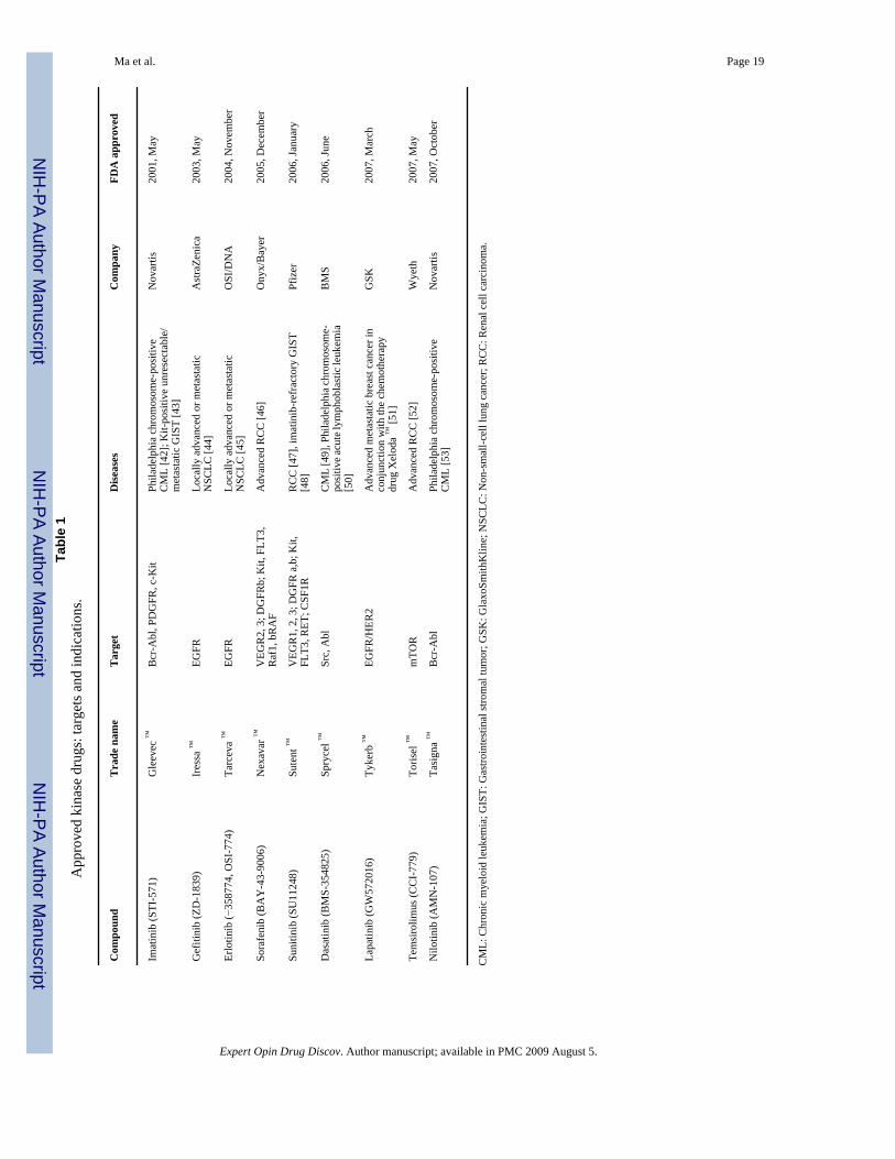
1. IntroductionProtein kinases play an integral role in many cell signaling pathways and comprise one of thelargest families of homologous proteins, with ~ 518 members in the human kinome [1]. Assuch, protein kinases represent the largest ‘druggable’ gene family within the human genome[2]. Overexpression and/or dysregulation of protein kinases result in many diseases, thusproviding numerous targets for drug development [3]. Since 2001, the FDA has approved ninekinase inhibitors for oncology targets (Table 1), and many more are now in clinical trials for
†Author for correspondence.
NIH Public AccessAuthor ManuscriptExpert Opin Drug Discov. Author manuscript; available in PMC 2009 August 5.
Published in final edited form as:Expert Opin Drug Discov. 2008 June ; 3(6): 607–621. doi:10.1517/17460441.3.6.607.
NIH
-PA Author Manuscript
NIH
-PA Author Manuscript
NIH
-PA Author Manuscript
the treatment of such diseases as cancer and cardiovascular and inflammatory diseases [4]. Atpresent, ~ 24% of all research spending on drug discovery and development is focused onkinases [5]. Given the huge demand for small molecules that target this class of proteins, manytechnologies and platforms have been developed for discovering kinase inhibitors, includingbiochemical based functional assays and compound binding competition assays.
Protein kinases are phosphoryl transferases that transfer the γ-phosphate of ATP to conservedserine, threonine, or tyrosine residues on specific substrate proteins. Classical methods to assaykinase activity involve the quantification of this phosphoryl transfer by detection of theproduction of the phosphorylated product or the change in the ratio of ATP to ADP. Thetraditional biochemical based method used to achieve this goal is the radioisotope filtrationbinding assay, in which the reactions are performed using radioisotope labeled γ-ATP. Theincorporation of this radiolabeled phosphate into the kinase substrate is then assayed after aseries of binding and washing steps to remove unincorporated radioisotope. This allows forthe detection of kinase phosphoryl transfer activity, which is directly proportional to the amountof phosphorylated substrate. Despite the sensitivity and resolution of this assay, this methodis not commonly used in a high-throughput screening (HTS) format owing to the drawbacksassociated with the use of radioisotope materials, such as waste disposal, radiation safety, andthe cumbersome washing and detection process.
To facilitate the HTS for kinase inhibitors, many convenient and automation friendly ‘mix andread’ assays have been recently developed that use fluorescence emission as a detectionmethod. Technologies such as fluorescence intensity (FI), fluorescence polarization (FP),fluorescence resonance energy transfer (FRET), time-resolved fluorescence (TRF), time-resolved fluorescence resonance energy transfer (TR-FRET), and chemiluminescence offer theadvantage of reagents that contain no radioactive materials. However, these new methods alsopose new challenges in areas such as assay development, data analysis, and interpretation. Inaddition, these assays are susceptible to fluorescence interference caused by fluorescentcompounds and fluorescently-labeled substrates, antibodies, and tracers [6–8]. More critically,owing to the modified reaction components and the variety of detection methods, assays withdifferent formats for screening the same library and target could create strikingly different setsof inhibitors [9–16]. Therefore, extensive confirmation assays to validate hits are necessary toreduce the false positive and false negative rates. In addition to these common fluorescenceand luminescence methods, there exist several special platforms that monitor the productionof phosphorylated substrates or the binding of potential chemical inhibitors to target kinases.Such platforms include the scintillation proximity assay, ELISA, mobility shift assay, andprotein binding assay. A common advantage of these assays is that there is little to nointerference from compound or probe labeling [7,8,14].
The myriad of screening technologies have given scientists the freedom to pick the screeningparameters that suit their individual drug discovery platforms or personal preferences.However, the plethora of assay formats also poses a great challenge to scientists who mustdecide on a technology that will be cost efficient and convenient to use for internal screening,as well as providing low false positive and false negative results. In the sections below, we willdiscuss the commonly used kinase assay methods along with their pros and cons in generaldrug screening applications. The common kinase profiling models used at present are alsodiscussed in detail with reference to data reliability and how data obtained from kinase profilingmay be used to guide the drug discovery process.
2. Radiometric based assaysRadiometric based assays are the most reliable methods for detecting kinase reactions;therefore, they are the preferred method for kinase profiling [8].
Ma et al. Page 2
Expert Opin Drug Discov. Author manuscript; available in PMC 2009 August 5.
NIH
-PA Author Manuscript
NIH
-PA Author Manuscript
NIH
-PA Author Manuscript
2.1 Filtration binding assayThe radiometric based filtration binding assay is considered the ‘gold standard’ to which non-radiometric methods are compared. In this assay format, the kinase reaction is performed inthe presence of 32 P-γ-ATP or 33 P-γ-ATP, followed by binding of the final radioisotope labeledproducts to filters after which unreacted phosphate can be washed away without interferingwith the detection of real phosphorylated products. The major advantage of this method is thatit is a true universal kinase assay method which can be used for any kinase and substrate withoutlimitations. This method does not require any special substrate labeling or modification, anddetection is free from interference from compounds and unreacted radioisotope. However, theuse of radioisotopes and the complex washing and separation steps present a major limitationto applying this technology for large-scale HTS. Nevertheless, filtration binding assaysrepresent one of the most favorable assay methods for kinase profiling work, owing to its error-free detection. Both Reaction Biology Corporation’s HotSpotSM [17] and Millipore’sKinaseProfiler [18] use this technology for profiling large kinase panels. The HotSpotSM
technology is a miniaturized kinase assay platform which reduces the consumption ofradioisotope materials, kinase targets, substrates, and chemical compounds. As such, thistechnology is easily adaptable for use in HTS.
2.2 Scintillation proximity assayThe separation and washing steps used in the radiometric filtration binding assays limit itsapplication in large-scale screenings. To circumvent the need for separation steps, GE andPerkinElmer have developed the scintillation proximity assay (SPA), which is a mix and readmethod similar to homogenous fluorescence-based detections. GE’s SPA assay is ahomogenous system that uses microscopic beads containing a scintillant that can be stimulatedby beta particles or auger electrons to emit light. This stimulation event occurs only whenradiolabeled molecules of interest are bound to the surface of the beads, which results in theemission of light that can be detected using standard scintillation counters or with a CCD(charge coupled device) camera-based imaging instrumentation, such as the LEADseeker™
made by GE. Unbound radiolabeled molecules are not in close enough proximity to stimulatethe bead to emit light. Therefore, the washing of unbound radiolabeled materials from the assaysystem is unnecessary. To reduce interference from intrinsic emission and absorptionproperties of chemical compounds used in the assays, GE has developed beads that emit red-shifted light [19]. PerkinElmer takes a different approach by offering two types of scintillant-coated microtiter plates, Scintiplates® and Flashplates®, for direct assays that eliminate theneed to add scintillation cocktail as with bead-based assays. The interior of each well ispermanently coated with a thin layer of polystyrene-based scintillant, which provides aplatform for non-separation assays using a variety of isotopes (e.g., 3H, 125I, 14C, and 33P)[20]. ProQinase (Freiburg, Germany) uses the FlashPlate® technology for its 33 PanQinase®
profiling services [21].
3. Fluorescence-based detection assaysKinase assay formats that use fluorescence-based detection are the most widely used methodsfor HTS-based kinase drug discovery because they are automation friendly, easy to use,relatively low cost, and widely available. In this type of assay format, some of the morecommonly applied techniques use FP, FRET, and TRF to identify lead compounds.
3.1 Fluorescence Intensity assaysFI is arguably the most common fluorescence-based method for detecting enzyme activity, andis widely used among assays that use protease-based detection reactions. However, to detectthe transferase activity of a kinase using this assay, one or more coupling reactions must beused. One such FI-based method is to detect the production of ADP by using linked reactions
Ma et al. Page 3
Expert Opin Drug Discov. Author manuscript; available in PMC 2009 August 5.
NIH
-PA Author Manuscript
NIH
-PA Author Manuscript
NIH
-PA Author Manuscript
involving pyruvate kinase, pyruvate oxidase, and horseradish peroxidase. After completion ofthe kinase reaction, pyruvate kinase is added to convert ADP to ATP and phosphoenolpyruvateto pyruvate. Subsequently, the pyruvate oxidase converts pyruvate to hydrogen peroxide(H2O2), which is then detected by using the fluorescent substrate Amplex Red (10-acetyl-3,7-dihydroxyphenoxazine) and horseradish peroxidase, resulting in the production of highlyfluorescent resorufin [22]. This approach is commercially available from DiscoveRx as ADPHunter™, which is a universal and sensitive assay. However, the multistep reactions involvemany enzymes, which can complicate the hit conformation process. Another FI method isPromega’s ProFluor® approach, in which a rhodamine-110 fluorophore is conjugated with apeptide substrate. The non-phosphorylated peptide can be digested by a proprietaryendopeptidase to release free fluorescent dye rhodamine-110, but the phosphorylated peptideis resistant to such digestion [23]. We have tested the ProFluor® PKA assay kit using theDisocoveryDot™ chemical microarray technology and found that the assay is very robust andsensitive, with faithful detection attained in a 1 nl reaction volume by using a CCD imager orDNA microarray scanner [24]. However, at present only a few assay kits are available for afew kinases, simply because it is difficult to develop enough peptide substrates that can be usedby both the kinases and the special peptidase in the reaction.
3.2 Fluorescence polarization assaysFP, a technique that monitors molecular movement and rotation, is a widely employed detectionmethod used in HTS for kinase inhibitors. The principle of this assay is that when excited withpolarized light, a molecule with high molecular weight will have a slower rotational movementcompared with a molecule with low molecular weight. Similarly, when a molecule is linkedto a fluorescence tracer, the polarized fluorescent signal will be dominated by the size of themolecule. Fluorescence polarization (P) is defined by the following equation: P = (I|| − I⊥)//(I|| + I⊥)[25], where I|| is the emission intensity parallel to the excitation plane and I⊥ is theemission intensity perpendicular to the excitation plane of a fluorophore when excited bypolarized light. The polarization value (P) is not dependent on the intensity of the emitted lightor on the concentration of the fluorophore. When the fluorophore is free to rotate, thepolarization is a smaller number, which is called low FP, and after the fluorophore binds to alarger molecule that limits the rotation of molecule, the polarization becomes a larger number,which is called high FP.
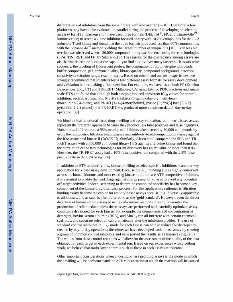
Many companies have developed assay kits that use this method, including BellBrook’sTranscreener™ FP assay, DiscoveRx’s HitHunter™ FP Assay, Invitrogen’s Far-RedPolarScreen™ FP Assay, Millipore’s KinEASE™ FP Assay, and Molecular Devices’IMAP™ FP assay. This larger group of assays can be further divided into two groups based onthe method used to monitor kinase activity. The first method, which is used in theTranscreener™ Kinase FP assay, uses an ADP-specific antibody to detect the conversion ofATP to ADP by the target kinase (Figure 1A). By contrast, the second method detects theproduction of phosphorylated substrates as in the HitHunter™, PolarScreen™, andKinEASE™ systems, which use substrate-specific antibodies (Figure 1B), and in theIMAP™ assay, which uses the high affinity of trivalent metal ions to phosphate (Figure 1C).
The advantages of the FP assay include a mix and read approach that is automation friendly,low cost of materials, and the availability of many assay formats. However, the cost ofdeveloping new assays is high, especially for the methods that use specific antibodies andtracers. Therefore, the approaches adapted by IMAP™ and Transcreener™ are more generallyapplicable to a variety of kinases. Generally speaking, the Transcreener™ method can also beused for other types of enzymes that generate ADP in the reaction systems.
Similar to other types of fluorescence-based detection methods (described later), FP detectioncan generate both false positive and false negative results because of the fluorescence
Ma et al. Page 4
Expert Opin Drug Discov. Author manuscript; available in PMC 2009 August 5.
NIH
-PA Author Manuscript
NIH
-PA Author Manuscript
NIH
-PA Author Manuscript
interferences from fluorescent tracers, labeled substrates, and colored and fluorescencecompounds. For example, Kashem et al. used the competition FP method to screen a 10,280compound library against the IL-2 inducible T cell kinase and found nine florescent compoundsthat were false positive, six of which were also colored [16]. In an early study by Beasley etal.[11], many strong inhibitors generated from the FP-based assay were shown as weakinhibitors in the TRF and TR-FRET assays. The authors suggest that a few situations couldcause the observed high mP values. The first was the low concentration of phosphorylatedsubstrate and high concentration of antibody–tracer complex used in the assays; the secondwas the aggregation of fluorescent compounds interfering with the signal from the antibody–tracer complex; the third was that compound aggregation could increase light-scattering duringdetection or sequester the kinase or tracer. Invitrogen has studied the compound and tracerinterference by mixing 10 μM of Sigma’s LOPAC1280 library compounds with 1 nM of green,red, and far-red tracers individually, and found that using green tracer produced the mostnumber of compounds that could interfere with detection, with 1.5% compounds showing >50% signal intensity. The red tracer followed with 0.7% compounds showing > 50% signalintensity and the far-red tracer had the least problems with 0.2% compounds showing > 50%signal intensity [26]. Based on this information, the Far-Red PolarScreen™ FP Assay may bea good choice for a library with a high incidence of fluorescent compounds.
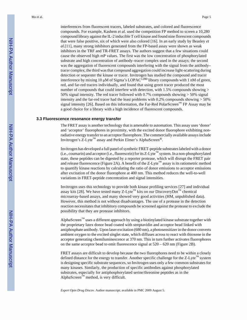
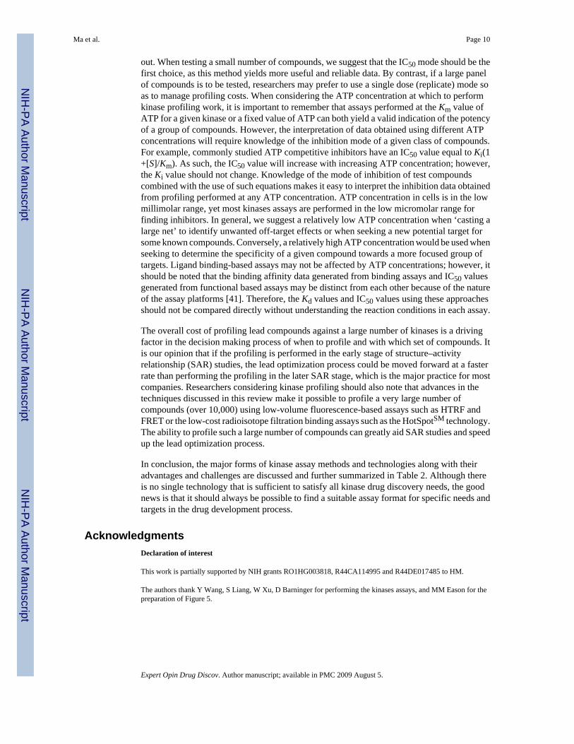
3.3 Fluorescence resonance energy transferThe FRET assay is another technology that is amenable to automation. This assay uses ‘donor’and ‘acceptor’ fluorophores in proximity, with the excited donor fluorophore exhibiting non-radiative energy transfer to an acceptor fluorophore. The commercially available assays includeInvitrogen’s Z-Lyte™ assay and Perkin Elmer’s AlphaScreen®.
Invitrogen has developed a full panel of synthetic FRET-peptide substrates labeled with a donor(i.e., coumarin) and acceptor (i.e., fluorescein) for its Z-Lyte™ system. In a non-phosphorylatedstate, these peptides can be digested by a reporter protease, which will disrupt the FRET pairand release fluorescence (Figure 2A). A benefit of the Z-Lyte™ assay is its ratiometric methodto quantify kinase reactions by calculating the ratio of donor emissions to acceptor emissionsafter excitation of the donor fluorophore at 400 nm. This method reduces the well-to-wellvariations in FRET-peptide concentration and signal intensities.
Invitrogen uses this technology to provide both kinase profiling services [27] and individualassay kits [28]. We have tested many Z-Lyte™ kits on our DiscoveryDot™ chemicalmicroarray-based assays, and many showed very good activities (HM, unpublished data).However, this method is not without disadvantages. The use of a protease in the detectionreaction necessitates that inhibitory compounds be screened against the protease to exclude thepossibility that they are protease inhibitors.
AlphaScreen™ uses a different approach by using a biotinylated kinase substrate together withthe proprietary latex donor bead coated with streptavidin and acceptor bead linked withantiphosphate antibody. Upon laser excitation (680 nm), a photosensitizer in the donor convertsambient oxygen to the excited singlet state, which diffuses across to react with thioxene in theacceptor generating chemiluminescence at 370 nm. This in turn further activates fluorophoreson the same acceptor bead to emit fluorescence signal at 520 – 620 nm (Figure 2B).
FRET assays are difficult to develop because the two fluorophores need to be within a closelydefined distance for the energy to transfer. Another specific challenge for the Z-Lyte™ systemis designing specific substrate sequences, so Invitrogen uses only a few common substrates formany kinases. Similarly, the production of specific antibodies against phosphorylatedsubstrates, especially for antiphosphorylated serine/threonine peptides as in theAlphaScreen™ method, is very difficult.
Ma et al. Page 5
Expert Opin Drug Discov. Author manuscript; available in PMC 2009 August 5.
NIH
-PA Author Manuscript
NIH
-PA Author Manuscript
NIH
-PA Author Manuscript
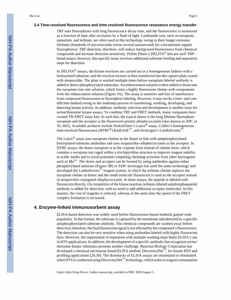
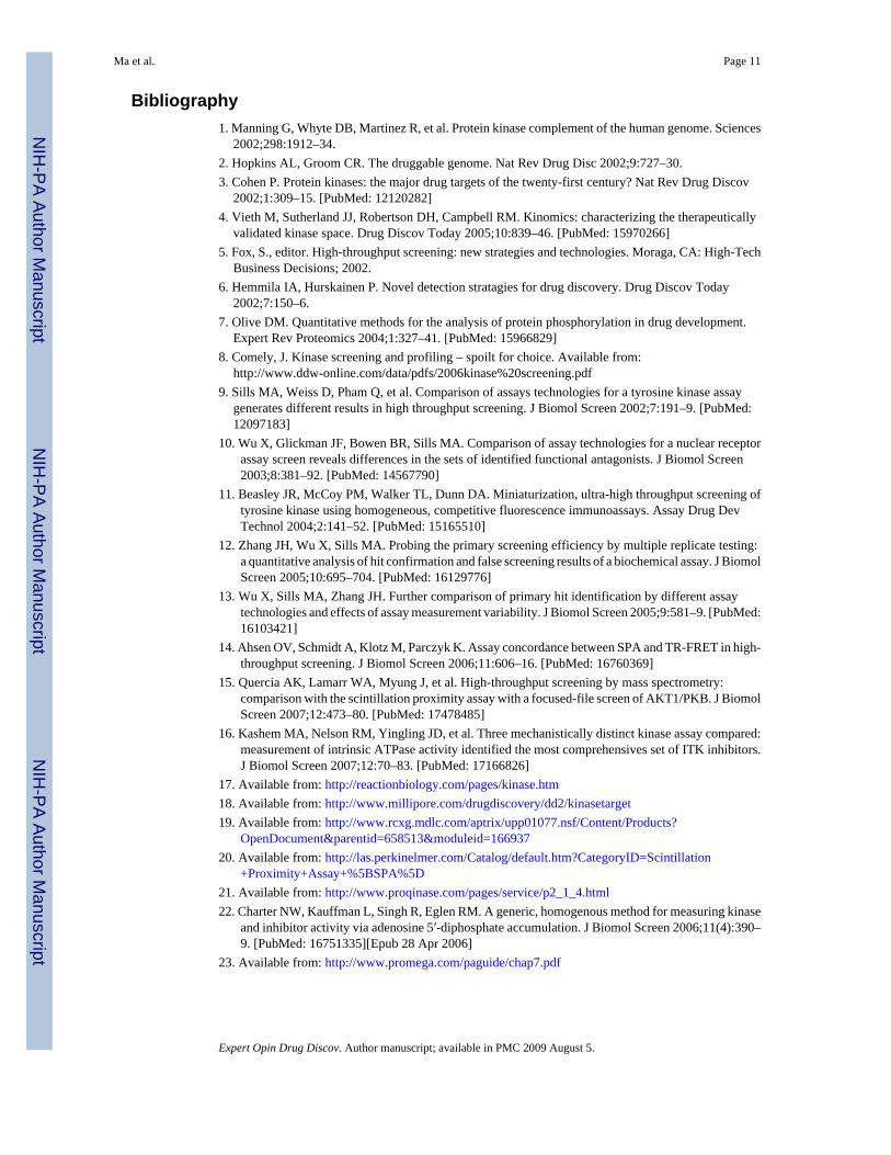
3.4 Time-resolved fluorescence and time-resolved fluorescence resonance energy transferTRF uses fluorophores with long fluorescence decay time, and the fluorescence is monitoredas a function of time after excitation by a flash of light. Lanthanide ions, such as europium,samarium, and terbium, are often used in this technology owing to their longer emissionlifetimes (hundreds of microseconds versus several nanoseconds for conventional organicfluorophores). TRF detection, therefore, will reduce background fluorescence from chemicalcompounds and increase detection sensitivity. Perkin Elmer’s DELFIA® kits are such TRF-based assays; however, this specific assay involves additional substrate binding and separationsteps for detection.
In DELFIA® assays, the kinase reactions are carried out in a homogeneous fashion with abiotinylated substrate, and the reaction mixture is then transferred into the capture plate coatedwith streptavidin. The plate is washed multiple times before europium-labeled antibody isadded to detect phosphorylated substrates. An enhancement solution is then added to dissociatethe europium ions into solution, which forms a highly fluorescent chelate with componentsfrom the enhancement solution (Figure 3A). The assay is sensitive and free of interferencefrom compound fluorescence or fluorophore labeling. However, it may not be a time- and cost-efficient method owing to the multistep process of transferring, washing, developing, anddetecting kinase activity. In addition, antibody selection and development is another issue forserine/threonine kinase assays. To combine TRF and FRET methods, many companies havecreated TR-FRET assay kits. In such kits, the typical donor is the long lifetime fluorophoreeuropium and the acceptor is the fluorescent protein allophycocyanin (also known as APC orXL-665). Available products include PerkinElmer’s Lance® assay, CisBio’s homogeneoustime-resolved fluorescence (HTRF®) KinEASE™, and Invitrogen’s LanthaScreen™.
The Lance® assay uses europium chelate as the donor to link with antiphosphorylatedbiotinylated-substrate antibodies and uses straptavidin–allophycocyanin as the acceptor. InHTRF assays, the donor europium is in the cryptate form instead of chelate form, whichcontains a europium ion caged within a tris-bipyridine structure to improve reagent stabilityin acidic media and to avoid potential competing chelating activities from other bioreagentssuch as Mn2+. The donor and acceptor can be formed by using antibodies against eitherphosphorylated substrate (Figure 3B) or ADP. Invitrogen has used the same technology anddeveloped the LanthaScreen™ reagent system, in which the terbium chelate replaces theeuropium chelate as donor and the small molecule fluorescein is used as the acceptor insteadof streptavidin conjugated allophycocyanin. In these assays, the peptide is labeled withfluorescein directly. On completion of the kinase reaction, terbium-labeled antiphosphopeptideantibody is added for detection, with no need to add additional acceptor molecules. In thismanner, the cost of reagents is reduced, whereas at the same time the speed of the FRETcomplex formation is increased.
4. Enzyme-linked immunosorbent assayELISA-based detection was widely used before fluorescence-based methods gained widepopularity. In this format, the substrate is captured by the membrane and detected by a specificantiphosphorylated substrate antibody. The chemical compounds are washed away beforedetection; therefore, the final fluorescent signal is not affected by the compound’s fluorescence.The detection can also be very sensitive when using antibodies labeled with highly fluorescentdyes. However, the requirement of separation with multiple washing steps limits ELISA’s usein HTS applications. In addition, the development of a specific antibody that recognizes serine/threonine kinase substrates presents another challenge. Reaction Biology Corporation hasdeveloped a chemical microarray-based ELISA method, DiscoveryDot™, for kinase HTS andprofiling applications [29,30]. The drawbacks of ELISA assays are minimized or eliminatedwhen HTS is conducted using DiscoveryDot™ technology, which reduces reagent consumption
Ma et al. Page 6
Expert Opin Drug Discov. Author manuscript; available in PMC 2009 August 5.
NIH
-PA Author Manuscript
NIH
-PA Author Manuscript
NIH
-PA Author Manuscript
(compound, kinase, substrate, and detector reagents), eliminates compound interference, andallows automation of multiple wash steps [16]. Carna Biosciences (Kobe, Japan) is alsooffering tyrosine kinase profiling and assay kits with ELISA detection [31].
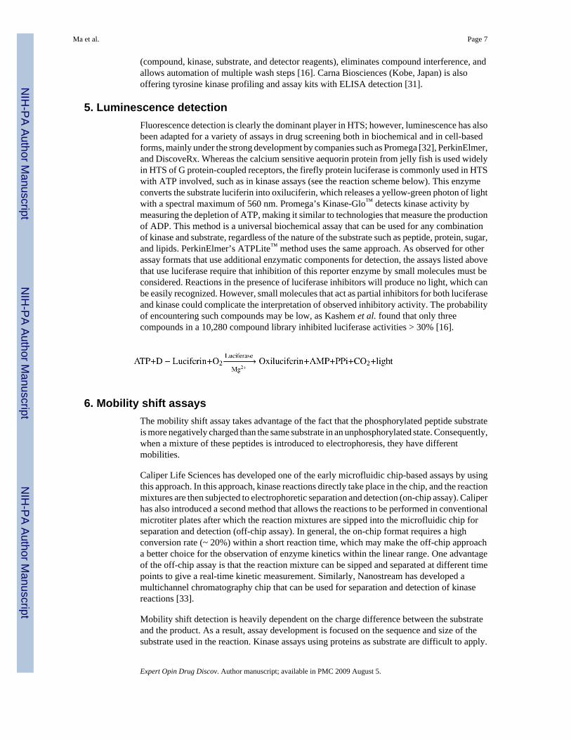
5. Luminescence detectionFluorescence detection is clearly the dominant player in HTS; however, luminescence has alsobeen adapted for a variety of assays in drug screening both in biochemical and in cell-basedforms, mainly under the strong development by companies such as Promega [32], PerkinElmer,and DiscoveRx. Whereas the calcium sensitive aequorin protein from jelly fish is used widelyin HTS of G protein-coupled receptors, the firefly protein luciferase is commonly used in HTSwith ATP involved, such as in kinase assays (see the reaction scheme below). This enzymeconverts the substrate luciferin into oxiluciferin, which releases a yellow-green photon of lightwith a spectral maximum of 560 nm. Promega’s Kinase-Glo™ detects kinase activity bymeasuring the depletion of ATP, making it similar to technologies that measure the productionof ADP. This method is a universal biochemical assay that can be used for any combinationof kinase and substrate, regardless of the nature of the substrate such as peptide, protein, sugar,and lipids. PerkinElmer’s ATPLite™ method uses the same approach. As observed for otherassay formats that use additional enzymatic components for detection, the assays listed abovethat use luciferase require that inhibition of this reporter enzyme by small molecules must beconsidered. Reactions in the presence of luciferase inhibitors will produce no light, which canbe easily recognized. However, small molecules that act as partial inhibitors for both luciferaseand kinase could complicate the interpretation of observed inhibitory activity. The probabilityof encountering such compounds may be low, as Kashem et al. found that only threecompounds in a 10,280 compound library inhibited luciferase activities > 30% [16].
6. Mobility shift assaysThe mobility shift assay takes advantage of the fact that the phosphorylated peptide substrateis more negatively charged than the same substrate in an unphosphorylated state. Consequently,when a mixture of these peptides is introduced to electrophoresis, they have differentmobilities.
Caliper Life Sciences has developed one of the early microfluidic chip-based assays by usingthis approach. In this approach, kinase reactions directly take place in the chip, and the reactionmixtures are then subjected to electrophoretic separation and detection (on-chip assay). Caliperhas also introduced a second method that allows the reactions to be performed in conventionalmicrotiter plates after which the reaction mixtures are sipped into the microfluidic chip forseparation and detection (off-chip assay). In general, the on-chip format requires a highconversion rate (~ 20%) within a short reaction time, which may make the off-chip approacha better choice for the observation of enzyme kinetics within the linear range. One advantageof the off-chip assay is that the reaction mixture can be sipped and separated at different timepoints to give a real-time kinetic measurement. Similarly, Nanostream has developed amultichannel chromatography chip that can be used for separation and detection of kinasereactions [33].
Mobility shift detection is heavily dependent on the charge difference between the substrateand the product. As a result, assay development is focused on the sequence and size of thesubstrate used in the reaction. Kinase assays using proteins as substrate are difficult to apply.
Ma et al. Page 7
Expert Opin Drug Discov. Author manuscript; available in PMC 2009 August 5.
NIH
-PA Author Manuscript
NIH
-PA Author Manuscript
NIH
-PA Author Manuscript
Dunne et al. performed a comparison study for the on-chip assay and off-chip assay. Usingcommercially available inhibitors and multiple kinases such as PKA, GSK3β, AKT1, theynoticed that the two methods can identify inhibitors with a 70% overlap but the inhibition valuefor the on-chip assay was lower than that of the off-chip assay [34]. These results indicate thatwhen running a HTS campaign with an on-chip-based method, a different inhibition cutoff forthe follow-up assay may have to be considered to avoid losing too many potential hits.Similarly, the 30% difference in hit identification and confirmation could pose a great challengefor profiling work using this method, especially for compounds with solubility problems.
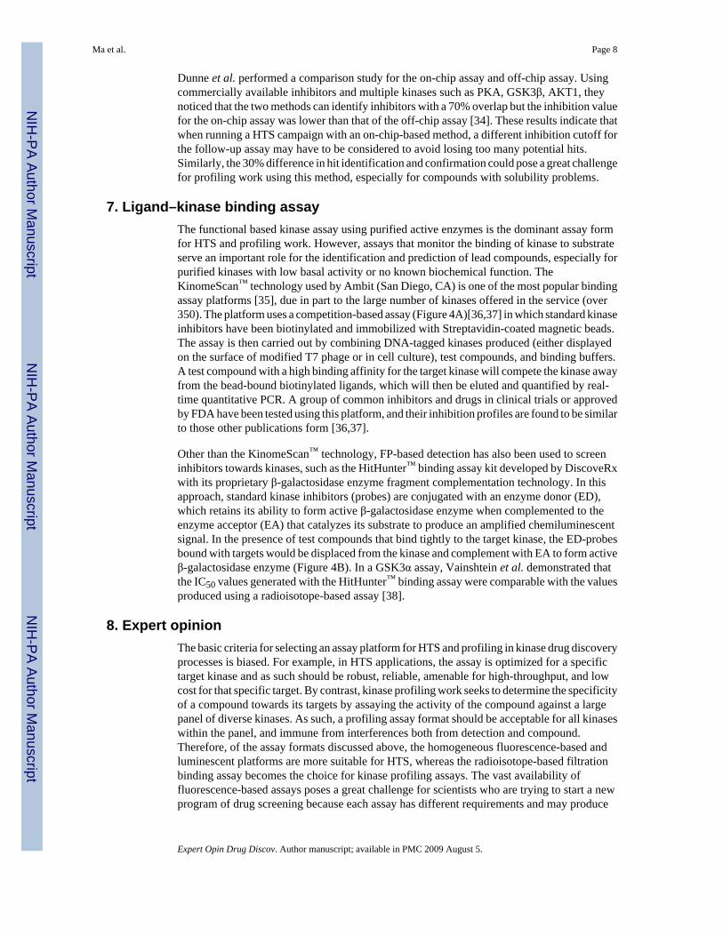
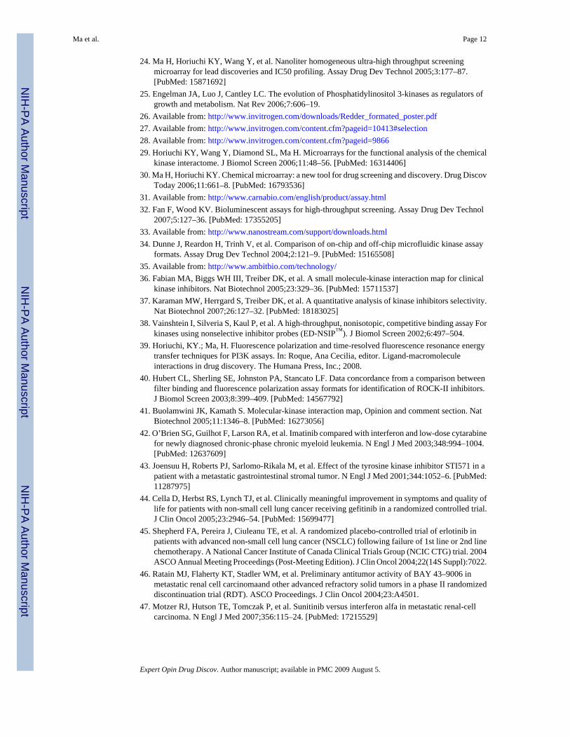
7. Ligand–kinase binding assayThe functional based kinase assay using purified active enzymes is the dominant assay formfor HTS and profiling work. However, assays that monitor the binding of kinase to substrateserve an important role for the identification and prediction of lead compounds, especially forpurified kinases with low basal activity or no known biochemical function. TheKinomeScan™ technology used by Ambit (San Diego, CA) is one of the most popular bindingassay platforms [35], due in part to the large number of kinases offered in the service (over350). The platform uses a competition-based assay (Figure 4A)[36,37] in which standard kinaseinhibitors have been biotinylated and immobilized with Streptavidin-coated magnetic beads.The assay is then carried out by combining DNA-tagged kinases produced (either displayedon the surface of modified T7 phage or in cell culture), test compounds, and binding buffers.A test compound with a high binding affinity for the target kinase will compete the kinase awayfrom the bead-bound biotinylated ligands, which will then be eluted and quantified by real-time quantitative PCR. A group of common inhibitors and drugs in clinical trials or approvedby FDA have been tested using this platform, and their inhibition profiles are found to be similarto those other publications form [36,37].
Other than the KinomeScan™ technology, FP-based detection has also been used to screeninhibitors towards kinases, such as the HitHunter™ binding assay kit developed by DiscoveRxwith its proprietary β-galactosidase enzyme fragment complementation technology. In thisapproach, standard kinase inhibitors (probes) are conjugated with an enzyme donor (ED),which retains its ability to form active β-galactosidase enzyme when complemented to theenzyme acceptor (EA) that catalyzes its substrate to produce an amplified chemiluminescentsignal. In the presence of test compounds that bind tightly to the target kinase, the ED-probesbound with targets would be displaced from the kinase and complement with EA to form activeβ-galactosidase enzyme (Figure 4B). In a GSK3α assay, Vainshtein et al. demonstrated thatthe IC50 values generated with the HitHunter™ binding assay were comparable with the valuesproduced using a radioisotope-based assay [38].
8. Expert opinionThe basic criteria for selecting an assay platform for HTS and profiling in kinase drug discoveryprocesses is biased. For example, in HTS applications, the assay is optimized for a specifictarget kinase and as such should be robust, reliable, amenable for high-throughput, and lowcost for that specific target. By contrast, kinase profiling work seeks to determine the specificityof a compound towards its targets by assaying the activity of the compound against a largepanel of diverse kinases. As such, a profiling assay format should be acceptable for all kinaseswithin the panel, and immune from interferences both from detection and compound.Therefore, of the assay formats discussed above, the homogeneous fluorescence-based andluminescent platforms are more suitable for HTS, whereas the radioisotope-based filtrationbinding assay becomes the choice for kinase profiling assays. The vast availability offluorescence-based assays poses a great challenge for scientists who are trying to start a newprogram of drug screening because each assay has different requirements and may produce
Ma et al. Page 8
Expert Opin Drug Discov. Author manuscript; available in PMC 2009 August 5.
NIH
-PA Author Manuscript
NIH
-PA Author Manuscript
NIH
-PA Author Manuscript
different sets of inhibitors from the same library with low overlap [9–16]. Therefore, a fewplatforms may have to be evaluated in parallel during the process of developing or selectingan assay for HTS. Kashem et al. have used three formats (DELFIA®, FP, and Kinase-Glo™
luminescence) to screen a kinase inhibitor focused library with 10,208 compounds for the IL-2inducible T cell kinase and found that the three formats produced less than 60% common hits,with the Kinase-Glo™ method yielding the largest number of unique hits [16]. Even less hitoverlap was observed when a 30,000 compound library was screened using three technologies(SPA, TR-FRET, and FP) by Sills et al.[9]. The reasons for the discrepancy among assays arealso hard to determine because the capability to find hits involves many factors such as substratesequence, the labeling of fluorescent probes, the conjugation of biotin/streptavidin beads,buffer composition, pH, enzyme quality, library quality, compound background, detectionsensitivity, excitation range, reaction steps. Based on others’ and our own experiences, westrongly recommend that scientists test a few different assay formats for assay developmentand validation before making a final decision. For example, we have tested both FP (EchelonBiosciences, Inc., UT) and TR-FRET (Millipore, CA) assay kits for PI3K reactions and small-scale HTS and found that although both assays produced consistent IC50 values for controlinhibitors such as wortmannin, PI3-Kγ Inhibitor (5-quinoxalin-6-ylmethylene-thiazolidine-2,4-dione), and PI-103 (3-(4-(4-morpholinyl) pyrido [3′,2′:4,5] furo [3,2-d]pyrimidin-2-yl) phenol), the TR-FRET kits produced more consistent data in day-to-dayoperation [39].
For biochemical functional based drug profiling and assay validation, radiometric based assaysrepresent the preferred approach because they produce less false positives and false negatives.Hubert et al.[40] reported a 95% overlap of inhibitors after screening 30,000 compounds byusing the radiometric filtration binding assay and antibody-based competition FP assay againstthe Rho-associated kinase II (ROCK-II). Similarly, Ahsen et al. compared the SPA and TR-FRET assays with a 300,000 compound library HTS against a tyrosine kinase and found thatthe correlation of the two technologies for hit discovery has an R2 value of more than 0.85.However, the TR-FRET assay had a 10% false positive rate compared with the 2.5% falsepositive rate in the SPA assay [14].
In addition to HTS to identify hits, kinase profiling to select specific inhibitors is another keyapplication for kinase assay development. Because the ATP binding site is highly conservedacross the human kinome, and most existing kinase inhibitors are ATP competitive inhibitors,it is essential to profile the lead drugs against a large panel of kinases to avoid any potentialoff-target activities. Indeed, screening to determine compound specificity has become a keycomponent of the kinase drug discovery process. For this application, radiometric filtrationbinding assays become the choice for activity-based assays because it is universally applicableto all kinases, and as such is often referred to as the ‘gold standard’. However, even the directdetection of kinase activity assayed using radiometric methods does not guarantee theproduction of reliable data unless these assays are performed with carefully optimized assayconditions developed for each kinase. For example, the components and concentration ofdetergent, bovine serum albumin (BSA), and MnCl2 can all interfere with certain chemicalscaffolds, and substrate selection can dramatically alter the inhibition profiles. The use ofstandard control inhibitors in IC50 mode for each kinase can help to reduce the discrepancycreated by day-to-day operations; therefore, we have developed each kinase assay by runninga group of common control inhibitors and have posted the results as a reference (Figure 5).The values from these control reactions will allow for the assessment of the quality of the dataobtained for each target in each experimental run. Based on our experiences with profilingwork, we believe that multi-layer controls such as these in each assay are essential.
Other important considerations when choosing kinase profiling assays is the mode in whichthe profiling will be performed and the ATP concentration at which the reaction will be carried
Ma et al. Page 9
Expert Opin Drug Discov. Author manuscript; available in PMC 2009 August 5.
NIH
-PA Author Manuscript
NIH
-PA Author Manuscript
NIH
-PA Author Manuscript
out. When testing a small number of compounds, we suggest that the IC50 mode should be thefirst choice, as this method yields more useful and reliable data. By contrast, if a large panelof compounds is to be tested, researchers may prefer to use a single dose (replicate) mode soas to manage profiling costs. When considering the ATP concentration at which to performkinase profiling work, it is important to remember that assays performed at the Km value ofATP for a given kinase or a fixed value of ATP can both yield a valid indication of the potencyof a group of compounds. However, the interpretation of data obtained using different ATPconcentrations will require knowledge of the inhibition mode of a given class of compounds.For example, commonly studied ATP competitive inhibitors have an IC50 value equal to Ki(1+[S]/Km). As such, the IC50 value will increase with increasing ATP concentration; however,the Ki value should not change. Knowledge of the mode of inhibition of test compoundscombined with the use of such equations makes it easy to interpret the inhibition data obtainedfrom profiling performed at any ATP concentration. ATP concentration in cells is in the lowmillimolar range, yet most kinases assays are performed in the low micromolar range forfinding inhibitors. In general, we suggest a relatively low ATP concentration when ‘casting alarge net’ to identify unwanted off-target effects or when seeking a new potential target forsome known compounds. Conversely, a relatively high ATP concentration would be used whenseeking to determine the specificity of a given compound towards a more focused group oftargets. Ligand binding-based assays may not be affected by ATP concentrations; however, itshould be noted that the binding affinity data generated from binding assays and IC50 valuesgenerated from functional based assays may be distinct from each other because of the natureof the assay platforms [41]. Therefore, the Kd values and IC50 values using these approachesshould not be compared directly without understanding the reaction conditions in each assay.
The overall cost of profiling lead compounds against a large number of kinases is a drivingfactor in the decision making process of when to profile and with which set of compounds. Itis our opinion that if the profiling is performed in the early stage of structure–activityrelationship (SAR) studies, the lead optimization process could be moved forward at a fasterrate than performing the profiling in the later SAR stage, which is the major practice for mostcompanies. Researchers considering kinase profiling should also note that advances in thetechniques discussed in this review make it possible to profile a very large number ofcompounds (over 10,000) using low-volume fluorescence-based assays such as HTRF andFRET or the low-cost radioisotope filtration binding assays such as the HotSpotSM technology.The ability to profile such a large number of compounds can greatly aid SAR studies and speedup the lead optimization process.
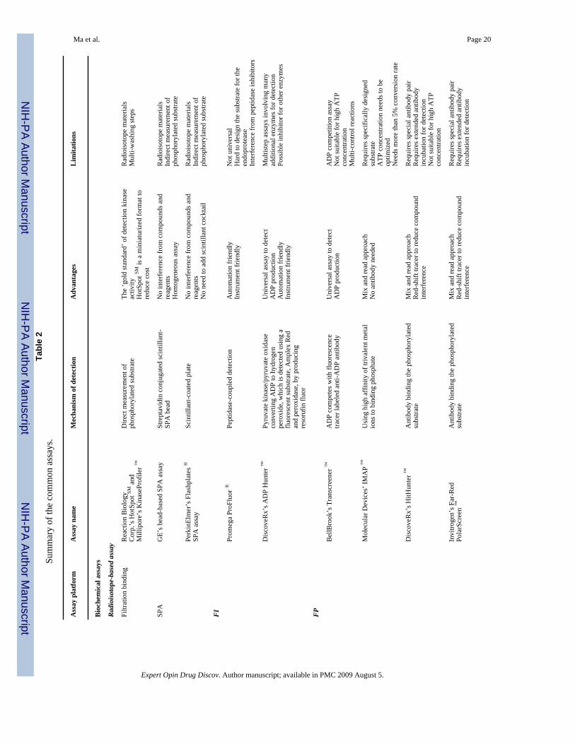
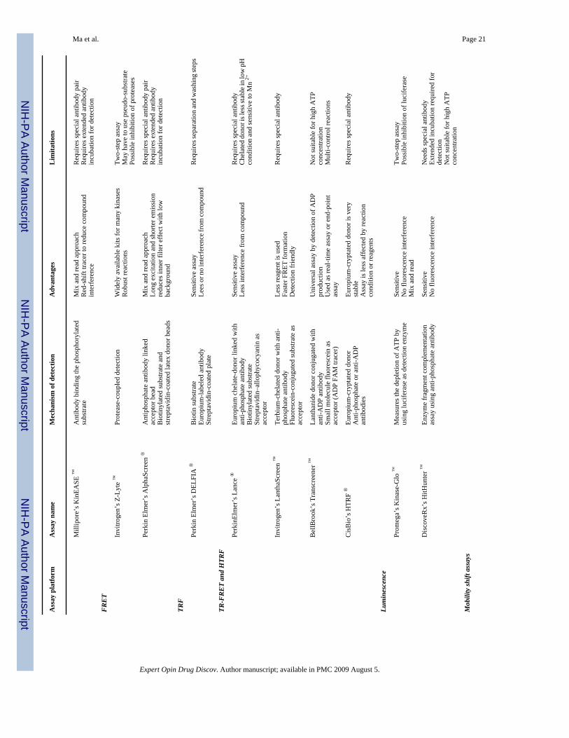
In conclusion, the major forms of kinase assay methods and technologies along with theiradvantages and challenges are discussed and further summarized in Table 2. Although thereis no single technology that is sufficient to satisfy all kinase drug discovery needs, the goodnews is that it should always be possible to find a suitable assay format for specific needs andtargets in the drug development process.
AcknowledgmentsDeclaration of interest
This work is partially supported by NIH grants RO1HG003818, R44CA114995 and R44DE017485 to HM.
The authors thank Y Wang, S Liang, W Xu, D Barninger for performing the kinases assays, and MM Eason for thepreparation of Figure 5.
Ma et al. Page 10
Expert Opin Drug Discov. Author manuscript; available in PMC 2009 August 5.
NIH
-PA Author Manuscript
NIH
-PA Author Manuscript
NIH
-PA Author Manuscript
Bibliography1. Manning G, Whyte DB, Martinez R, et al. Protein kinase complement of the human genome. Sciences
2002;298:1912–34.2. Hopkins AL, Groom CR. The druggable genome. Nat Rev Drug Disc 2002;9:727–30.3. Cohen P. Protein kinases: the major drug targets of the twenty-first century? Nat Rev Drug Discov
2002;1:309–15. [PubMed: 12120282]4. Vieth M, Sutherland JJ, Robertson DH, Campbell RM. Kinomics: characterizing the therapeutically
validated kinase space. Drug Discov Today 2005;10:839–46. [PubMed: 15970266]5. Fox, S., editor. High-throughput screening: new strategies and technologies. Moraga, CA: High-Tech
Business Decisions; 2002.6. Hemmila IA, Hurskainen P. Novel detection stratagies for drug discovery. Drug Discov Today
2002;7:150–6.7. Olive DM. Quantitative methods for the analysis of protein phosphorylation in drug development.
Expert Rev Proteomics 2004;1:327–41. [PubMed: 15966829]8. Comely, J. Kinase screening and profiling – spoilt for choice. Available from:
http://www.ddw-online.com/data/pdfs/2006kinase%20screening.pdf9. Sills MA, Weiss D, Pham Q, et al. Comparison of assays technologies for a tyrosine kinase assay
generates different results in high throughput screening. J Biomol Screen 2002;7:191–9. [PubMed:12097183]
10. Wu X, Glickman JF, Bowen BR, Sills MA. Comparison of assay technologies for a nuclear receptorassay screen reveals differences in the sets of identified functional antagonists. J Biomol Screen2003;8:381–92. [PubMed: 14567790]
11. Beasley JR, McCoy PM, Walker TL, Dunn DA. Miniaturization, ultra-high throughput screening oftyrosine kinase using homogeneous, competitive fluorescence immunoassays. Assay Drug DevTechnol 2004;2:141–52. [PubMed: 15165510]
12. Zhang JH, Wu X, Sills MA. Probing the primary screening efficiency by multiple replicate testing:a quantitative analysis of hit confirmation and false screening results of a biochemical assay. J BiomolScreen 2005;10:695–704. [PubMed: 16129776]
13. Wu X, Sills MA, Zhang JH. Further comparison of primary hit identification by different assaytechnologies and effects of assay measurement variability. J Biomol Screen 2005;9:581–9. [PubMed:16103421]
14. Ahsen OV, Schmidt A, Klotz M, Parczyk K. Assay concordance between SPA and TR-FRET in high-throughput screening. J Biomol Screen 2006;11:606–16. [PubMed: 16760369]
15. Quercia AK, Lamarr WA, Myung J, et al. High-throughput screening by mass spectrometry:comparison with the scintillation proximity assay with a focused-file screen of AKT1/PKB. J BiomolScreen 2007;12:473–80. [PubMed: 17478485]
16. Kashem MA, Nelson RM, Yingling JD, et al. Three mechanistically distinct kinase assay compared:measurement of intrinsic ATPase activity identified the most comprehensives set of ITK inhibitors.J Biomol Screen 2007;12:70–83. [PubMed: 17166826]
17. Available from: http://reactionbiology.com/pages/kinase.htm18. Available from: http://www.millipore.com/drugdiscovery/dd2/kinasetarget19. Available from: http://www.rcxg.mdlc.com/aptrix/upp01077.nsf/Content/Products?
OpenDocument&parentid=658513&moduleid=16693720. Available from: http://las.perkinelmer.com/Catalog/default.htm?CategoryID=Scintillation
+Proximity+Assay+%5BSPA%5D21. Available from: http://www.proqinase.com/pages/service/p2_1_4.html22. Charter NW, Kauffman L, Singh R, Eglen RM. A generic, homogenous method for measuring kinase
and inhibitor activity via adenosine 5′-diphosphate accumulation. J Biomol Screen 2006;11(4):390–9. [PubMed: 16751335][Epub 28 Apr 2006]
23. Available from: http://www.promega.com/paguide/chap7.pdf
Ma et al. Page 11
Expert Opin Drug Discov. Author manuscript; available in PMC 2009 August 5.
NIH
-PA Author Manuscript
NIH
-PA Author Manuscript
NIH
-PA Author Manuscript
24. Ma H, Horiuchi KY, Wang Y, et al. Nanoliter homogeneous ultra-high throughput screeningmicroarray for lead discoveries and IC50 profiling. Assay Drug Dev Technol 2005;3:177–87.[PubMed: 15871692]
25. Engelman JA, Luo J, Cantley LC. The evolution of Phosphatidylinositol 3-kinases as regulators ofgrowth and metabolism. Nat Rev 2006;7:606–19.
26. Available from: http://www.invitrogen.com/downloads/Redder_formated_poster.pdf27. Available from: http://www.invitrogen.com/content.cfm?pageid=10413#selection28. Available from: http://www.invitrogen.com/content.cfm?pageid=986629. Horiuchi KY, Wang Y, Diamond SL, Ma H. Microarrays for the functional analysis of the chemical
kinase interactome. J Biomol Screen 2006;11:48–56. [PubMed: 16314406]30. Ma H, Horiuchi KY. Chemical microarray: a new tool for drug screening and discovery. Drug Discov
Today 2006;11:661–8. [PubMed: 16793536]31. Available from: http://www.carnabio.com/english/product/assay.html32. Fan F, Wood KV. Bioluminescent assays for high-throughput screening. Assay Drug Dev Technol
2007;5:127–36. [PubMed: 17355205]33. Available from: http://www.nanostream.com/support/downloads.html34. Dunne J, Reardon H, Trinh V, et al. Comparison of on-chip and off-chip microfluidic kinase assay
formats. Assay Drug Dev Technol 2004;2:121–9. [PubMed: 15165508]35. Available from: http://www.ambitbio.com/technology/36. Fabian MA, Biggs WH III, Treiber DK, et al. A small molecule-kinase interaction map for clinical
kinase inhibitors. Nat Biotechnol 2005;23:329–36. [PubMed: 15711537]37. Karaman MW, Herrgard S, Treiber DK, et al. A quantitative analysis of kinase inhibitors selectivity.
Nat Biotechnol 2007;26:127–32. [PubMed: 18183025]38. Vainshtein I, Silveria S, Kaul P, et al. A high-throughput, nonisotopic, competitive binding assay For
kinases using nonselective inhibitor probes (ED-NSIP™). J Biomol Screen 2002;6:497–504.39. Horiuchi, KY.; Ma, H. Fluorescence polarization and time-resolved fluorescence resonance energy
transfer techniques for PI3K assays. In: Roque, Ana Cecilia, editor. Ligand-macromoleculeinteractions in drug discovery. The Humana Press, Inc.; 2008.
40. Hubert CL, Sherling SE, Johnston PA, Stancato LF. Data concordance from a comparison betweenfilter binding and fluorescence polarization assay formats for identification of ROCK-II inhibitors.J Biomol Screen 2003;8:399–409. [PubMed: 14567792]
41. Buolamwini JK, Kamath S. Molecular-kinase interaction map, Opinion and comment section. NatBiotechnol 2005;11:1346–8. [PubMed: 16273056]
42. O’Brien SG, Guilhot F, Larson RA, et al. Imatinib compared with interferon and low-dose cytarabinefor newly diagnosed chronic-phase chronic myeloid leukemia. N Engl J Med 2003;348:994–1004.[PubMed: 12637609]
43. Joensuu H, Roberts PJ, Sarlomo-Rikala M, et al. Effect of the tyrosine kinase inhibitor STI571 in apatient with a metastatic gastrointestinal stromal tumor. N Engl J Med 2001;344:1052–6. [PubMed:11287975]
44. Cella D, Herbst RS, Lynch TJ, et al. Clinically meaningful improvement in symptoms and quality oflife for patients with non-small cell lung cancer receiving gefitinib in a randomized controlled trial.J Clin Oncol 2005;23:2946–54. [PubMed: 15699477]
45. Shepherd FA, Pereira J, Ciuleanu TE, et al. A randomized placebo-controlled trial of erlotinib inpatients with advanced non-small cell lung cancer (NSCLC) following failure of 1st line or 2nd linechemotherapy. A National Cancer Institute of Canada Clinical Trials Group (NCIC CTG) trial. 2004ASCO Annual Meeting Proceedings (Post-Meeting Edition). J Clin Oncol 2004;22(14S Suppl):7022.
46. Ratain MJ, Flaherty KT, Stadler WM, et al. Preliminary antitumor activity of BAY 43–9006 inmetastatic renal cell carcinomaand other advanced refractory solid tumors in a phase II randomizeddiscontinuation trial (RDT). ASCO Proceedings. J Clin Oncol 2004;23:A4501.
47. Motzer RJ, Hutson TE, Tomczak P, et al. Sunitinib versus interferon alfa in metastatic renal-cellcarcinoma. N Engl J Med 2007;356:115–24. [PubMed: 17215529]
Ma et al. Page 12
Expert Opin Drug Discov. Author manuscript; available in PMC 2009 August 5.
NIH
-PA Author Manuscript
NIH
-PA Author Manuscript
NIH
-PA Author Manuscript
48. Demetri GD, van Oosterom AT, Garrett CR, et al. Efficacy and safety of sunitinib in patients withadvanced gastrointestinal stromal tumour after failure of imatinib: a randomised controlled trial.Lancet 2006;368:1329–38. [PubMed: 17046465]
49. Talpaz M, Shah NP, Kantarjian H, et al. Dasatinib in imatinib-resistant philadelphia chromosome–positive leukemias. N Engl J Med 2006;354:2531–41. [PubMed: 16775234]
50. Shah N, Sawyers CL, Kantarjian HM, et al. Correlation of clinical response to BMS-354825 withBCR-ABL mutation status in imatinib-resistant patients with chronic myeloid leukemia (CML) andPhiladelphia chromosome-associated acute lymphoblastic leukemia (Ph+ ALL). 2005 ASCO AnnualMeeting Proceedings. J Clin Oncol 2005;23(16S Suppl Pt I of II):565s .[Abstract 6521]
51. Geyer CE, Forster J, Lindquist D, et al. Lapatinib plus capecitabine for HER2-positive advancedbreast cancer. N Engl J Med 2006;355:2733–43. [PubMed: 17192538]
52. Fazio N, Dettori M, Lorizzo K, et al. Temsirolimus for advanced renal-cell carcinoma. N Engl J Med2007;357:1050–10. [PubMed: 17804854]
53. Kantarjian H, Giles F, Wunderle L, et al. Nilotinib in imatinib-resistant CML and Philadelphiachromosome-positive AL. N Engl J Med 354:2542–51. [PubMed: 16775235]
Ma et al. Page 13
Expert Opin Drug Discov. Author manuscript; available in PMC 2009 August 5.
NIH
-PA Author Manuscript
NIH
-PA Author Manuscript
NIH
-PA Author Manuscript
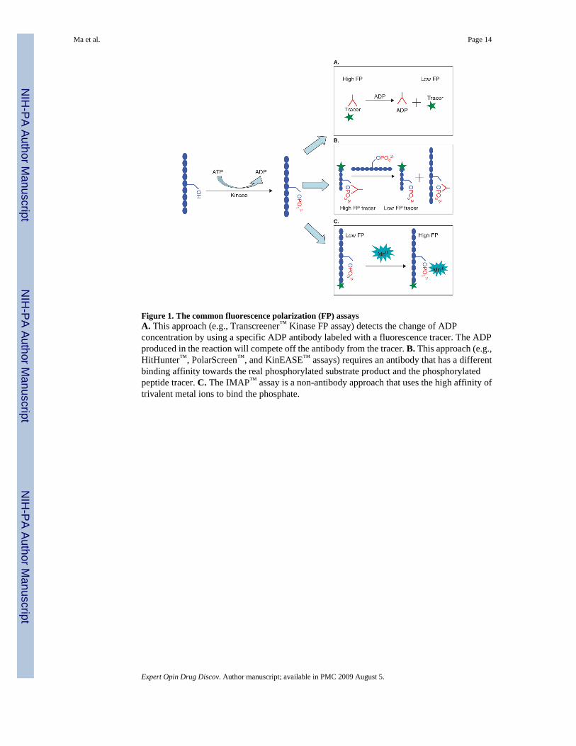
Figure 1. The common fluorescence polarization (FP) assaysA. This approach (e.g., Transcreener™ Kinase FP assay) detects the change of ADPconcentration by using a specific ADP antibody labeled with a fluorescence tracer. The ADPproduced in the reaction will compete off the antibody from the tracer. B. This approach (e.g.,HitHunter™, PolarScreen™, and KinEASE™ assays) requires an antibody that has a differentbinding affinity towards the real phosphorylated substrate product and the phosphorylatedpeptide tracer. C. The IMAP™ assay is a non-antibody approach that uses the high affinity oftrivalent metal ions to bind the phosphate.
Ma et al. Page 14
Expert Opin Drug Discov. Author manuscript; available in PMC 2009 August 5.
NIH
-PA Author Manuscript
NIH
-PA Author Manuscript
NIH
-PA Author Manuscript
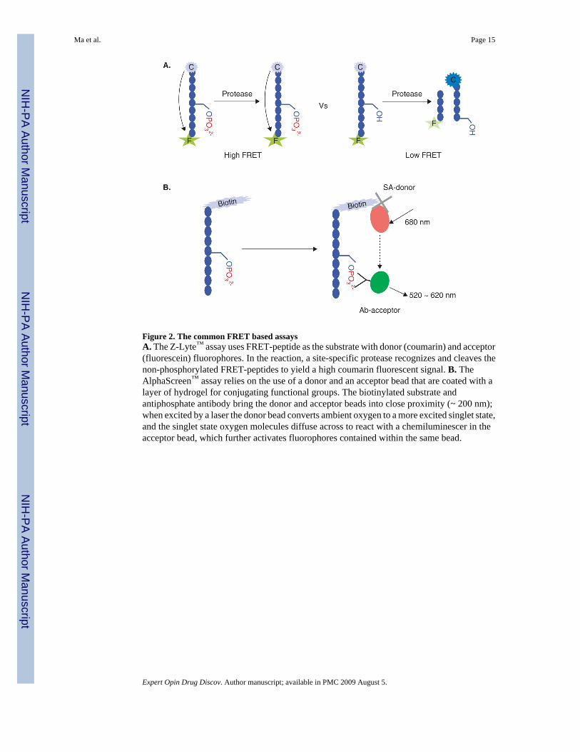
Figure 2. The common FRET based assaysA. The Z-Lyte™ assay uses FRET-peptide as the substrate with donor (coumarin) and acceptor(fluorescein) fluorophores. In the reaction, a site-specific protease recognizes and cleaves thenon-phosphorylated FRET-peptides to yield a high coumarin fluorescent signal. B. TheAlphaScreen™ assay relies on the use of a donor and an acceptor bead that are coated with alayer of hydrogel for conjugating functional groups. The biotinylated substrate andantiphosphate antibody bring the donor and acceptor beads into close proximity (~ 200 nm);when excited by a laser the donor bead converts ambient oxygen to a more excited singlet state,and the singlet state oxygen molecules diffuse across to react with a chemiluminescer in theacceptor bead, which further activates fluorophores contained within the same bead.
Ma et al. Page 15
Expert Opin Drug Discov. Author manuscript; available in PMC 2009 August 5.
NIH
-PA Author Manuscript
NIH
-PA Author Manuscript
NIH
-PA Author Manuscript

Figure 3. The common TRF assaysA. DELFIA® assay is very similar to ELISA-based assays but with a lanthanide-labeledantibody for detection. The lanthanide can be dissociated by a low pH enhancement solutionand forms a stable fluorescent chelate inside a protective micelle in the enhancer solution. B.The HTRF assay is a combination of the TRF and FRET assays. The europium cryptatecomplex can transfer excitation energy to an acceptor molecule, XL665, when they are broughtinto close proximity with one another. The energy captured by the cryptate europium duringexcitation is transferred to the acceptor and then released as a FRET signal.
Ma et al. Page 16
Expert Opin Drug Discov. Author manuscript; available in PMC 2009 August 5.
NIH
-PA Author Manuscript
NIH
-PA Author Manuscript
NIH
-PA Author Manuscript
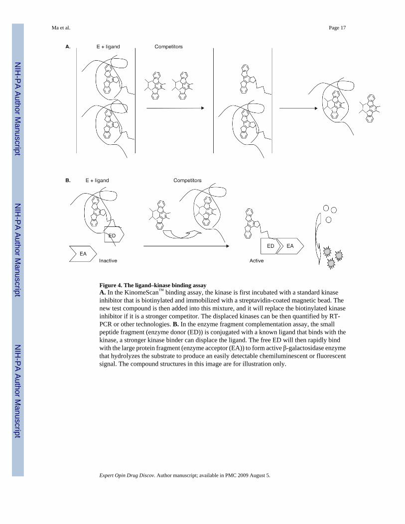
Figure 4. The ligand–kinase binding assayA. In the KinomeScan™ binding assay, the kinase is first incubated with a standard kinaseinhibitor that is biotinylated and immobilized with a streptavidin-coated magnetic bead. Thenew test compound is then added into this mixture, and it will replace the biotinylated kinaseinhibitor if it is a stronger competitor. The displaced kinases can be then quantified by RT-PCR or other technologies. B. In the enzyme fragment complementation assay, the smallpeptide fragment (enzyme donor (ED)) is conjugated with a known ligand that binds with thekinase, a stronger kinase binder can displace the ligand. The free ED will then rapidly bindwith the large protein fragment (enzyme acceptor (EA)) to form active β-galactosidase enzymethat hydrolyzes the substrate to produce an easily detectable chemiluminescent or fluorescentsignal. The compound structures in this image are for illustration only.
Ma et al. Page 17
Expert Opin Drug Discov. Author manuscript; available in PMC 2009 August 5.
NIH
-PA Author Manuscript
NIH
-PA Author Manuscript
NIH
-PA Author Manuscript
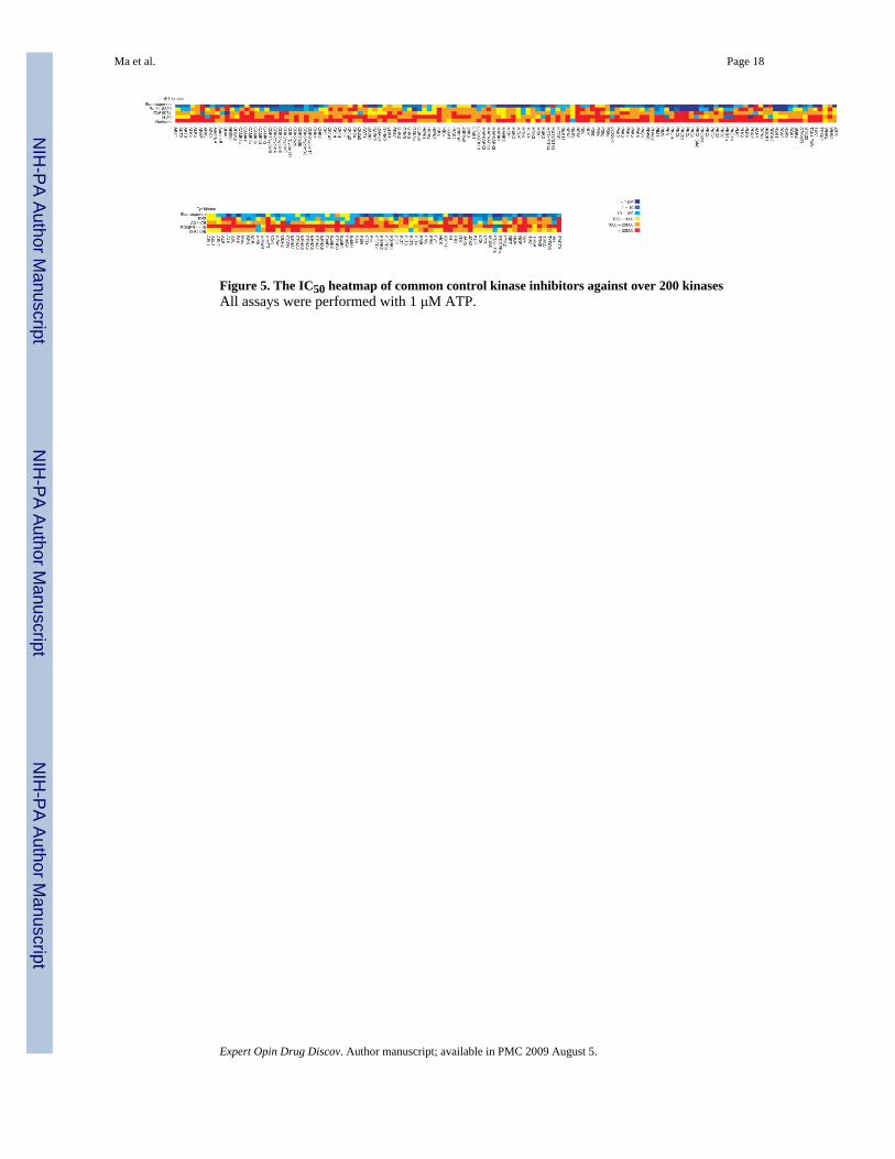
Figure 5. The IC50 heatmap of common control kinase inhibitors against over 200 kinasesAll assays were performed with 1 μM ATP.
Ma et al. Page 18
Expert Opin Drug Discov. Author manuscript; available in PMC 2009 August 5.
NIH
-PA Author Manuscript
NIH
-PA Author Manuscript
NIH
-PA Author Manuscript
NIH
-PA Author Manuscript
NIH
-PA Author Manuscript
NIH
-PA Author Manuscript
Ma et al. Page 19Ta
ble
1A
ppro
ved
kina
se d
rugs
: tar
gets
and
indi
catio
ns.
Com
poun
dT
rade
nam
eT
arge
tD
isea
ses
Com
pany
FDA
app
rove
d
Imat
inib
(STI
-571
)G
leev
ec ™
Bcr
-Abl
, PD
GFR
, c-K
itPh
ilade
lphi
a ch
rom
osom
e-po
sitiv
eC
ML
[42]
; Kit-
posi
tive
unre
sect
able
/m
etas
tatic
GIS
T [4
3]
Nov
artis
2001
, May
Gef
itini
b (Z
D-1
839)
Ires
sa ™
EGFR
Loca
lly a
dvan
ced
or m
etas
tatic
NSC
LC [4
4]A
stra
Zeni
ca20
03, M
ay
Erlo
tinib
(−35
8774
, OSI
-774
)Ta
rcev
a ™
EGFR
Loca
lly a
dvan
ced
or m
etas
tatic
NSC
LC [4
5]O
SI/D
NA
2004
, Nov
embe
r
Sora
feni
b (B
AY
-43-
9006
)N
exav
ar ™
VEG
R2,
3; D
GFR
b; K
it, F
LT3,
Raf
1, b
RA
FA
dvan
ced
RC
C [4
6]O
nyx/
Bay
er20
05, D
ecem
ber
Suni
tinib
(SU
1124
8)Su
tent
™V
EGR
1, 2
, 3; D
GFR
a,b
; Kit,
FLT3
, RET
; CSF
1RR
CC
[47]
, im
atin
ib-r
efra
ctor
y G
IST
[48]
Pfiz
er20
06, J
anua
ry
Das
atin
ib (B
MS-
3548
25)
Spry
cel ™
Src,
Abl
CM
L [4
9], P
hila
delp
hia c
hrom
osom
e-po
sitiv
e acu
te ly
mph
obla
stic
leuk
emia
[50]
BM
S20
06, J
une
Lapa
tinib
(GW
5720
16)
Tyke
rb ™
EGFR
/HER
2A
dvan
ced
met
asta
tic b
reas
t can
cer i
nco
njun
ctio
n w
ith th
e ch
emot
hera
pydr
ug X
elod
a ™
[51]
GSK
2007
, Mar
ch
Tem
siro
limus
(CC
I-77
9)To
risel
™m
TOR
Adv
ance
d R
CC
[52]
Wye
th20
07, M
ay
Nilo
tinib
(AM
N-1
07)
Tasi
gna
™B
cr-A
blPh
ilade
lphi
a ch
rom
osom
e-po
sitiv
eC
ML
[53]
Nov
artis
2007
, Oct
ober
CM
L: C
hron
ic m
yelo
id le
ukem
ia; G
IST:
Gas
troin
test
inal
stro
mal
tum
or; G
SK: G
laxo
Smith
Klin
e; N
SCLC
: Non
-sm
all-c
ell l
ung
canc
er; R
CC
: Ren
al c
ell c
arci
nom
a.
Expert Opin Drug Discov. Author manuscript; available in PMC 2009 August 5.
NIH
-PA Author Manuscript
NIH
-PA Author Manuscript
NIH
-PA Author Manuscript
Ma et al. Page 20Ta
ble
2Su
mm
ary
of th
e co
mm
on a
ssay
s.
Ass
ay p
latfo
rmA
ssay
nam
eM
echa
nism
of d
etec
tion
Adv
anta
ges
Lim
itatio
ns
Bio
chem
ical
ass
ays
Radi
oiso
tope
-bas
ed a
ssay
Filtr
atio
n bi
ndin
gR
eact
ion
Bio
logy
Cor
p.’s
Hot
Spot
SM a
ndM
illip
ore’
s Kin
aseP
rofil
er ™
Dire
ct m
easu
rem
ent o
fph
osph
oryl
ated
subs
trate
The
‘gol
d st
anda
rd’ o
f det
ectio
n ki
nase
activ
ityH
otSp
ot SM
is a
min
iatu
rized
form
at to
redu
ce c
ost
Rad
iois
otop
e m
ater
ials
Mul
ti-w
ashi
ng st
eps
SPA
GE’
s bea
d-ba
sed
SPA
ass
aySt
rept
avid
in c
onju
gate
d sc
intil
lant
-SP
A b
ead
No
inte
rfer
ence
from
com
poun
ds a
ndre
agen
tsH
omog
eneo
us a
ssay
Rad
iois
otop
e m
ater
ials
Indi
rect
mea
sure
men
t of
phos
phor
ylat
ed su
bstra
te
Perk
inEl
mer
’s F
lash
plat
es ®
SPA
ass
aySc
intil
lant
-coa
ted
plat
eN
o in
terf
eren
ce fr
om c
ompo
unds
and
reag
ents
No
need
to a
dd sc
intil
lant
coc
ktai
l
Rad
iois
otop
e m
ater
ials
Indi
rect
mea
sure
men
t of
phos
phor
ylat
ed su
bstra
te
FI
Prom
ega
ProF
luor
®Pe
ptid
ase-
coup
led
dete
ctio
nA
utom
atio
n fr
iend
lyIn
stru
men
t frie
ndly
Not
uni
vers
alH
ard
to d
esig
n th
e su
bstra
te fo
r the
endo
prot
ease
Inte
rfer
ence
from
pep
tidas
e in
hibi
tors
Dis
cove
Rx’
s AD
P H
unte
r ™Py
ruva
te k
inas
e/py
ruva
te o
xida
seco
nver
ting
AD
P to
hyd
roge
npe
roxi
de, w
hich
is d
etec
ted
usin
g a
fluor
esce
nt su
bstra
te, A
mpl
ex R
edan
d pe
roxi
dase
, by
prod
ucin
gre
soru
fin fl
uor
Uni
vers
al a
ssay
to d
etec
tA
DP
prod
uctio
nA
utom
atio
n fr
iend
lyIn
stru
men
t frie
ndly
Mul
tiste
p as
says
invo
lvin
g m
any
addi
tiona
l enz
ymes
for d
etec
tion
Poss
ible
inhi
bito
r for
oth
er e
nzym
es
FP
Bel
lBro
ok’s
Tra
nscr
eene
r ™A
DP
com
pete
s with
fluo
resc
ence
trace
r lab
eled
ant
i-AD
P an
tibod
yU
nive
rsal
ass
ay to
det
ect
AD
P pr
oduc
tion
AD
P co
mpe
titio
n as
say
Not
suita
ble
for h
igh
ATP
conc
entra
tion
Mul
ti-co
ntro
l rea
ctio
ns
Mol
ecul
ar D
evic
es’ I
MA
P ™
Usi
ng h
igh
affin
ity o
f triv
alen
t met
alio
ns to
bin
ding
pho
spha
teM
ix a
nd re
ad a
ppro
ach
No
antib
ody
need
edR
equi
res s
peci
fical
ly d
esig
ned
subs
trate
ATP
con
cent
ratio
n ne
eds t
o be
optim
ized
Nee
ds m
ore
than
5%
con
vers
ion
rate
Dis
cove
Rx’
s HitH
unte
r ™A
ntib
ody
bind
ing
the
phos
phor
ylat
edsu
bstra
teM
ix a
nd re
ad a
ppro
ach
Red
-shi
ft tra
cer t
o re
duce
com
poun
din
terf
eren
ce
Req
uire
s spe
cial
ant
ibod
y pa
irR
equi
res e
xten
ded
antib
ody
incu
batio
n fo
r det
ectio
nN
ot su
itabl
e fo
r hig
h A
TPco
ncen
tratio
n
Invi
troge
n’s F
ar-R
edPo
larS
cree
n ™
Ant
ibod
y bi
ndin
g th
e ph
osph
oryl
ated
subs
trate
Mix
and
read
app
roac
hR
ed-s
hift
trace
r to
redu
ce c
ompo
und
inte
rfer
ence
Req
uire
s spe
cial
ant
ibod
y pa
irR
equi
res e
xten
ded
antib
ody
incu
batio
n fo
r det
ectio
n
Expert Opin Drug Discov. Author manuscript; available in PMC 2009 August 5.
NIH
-PA Author Manuscript
NIH
-PA Author Manuscript
NIH
-PA Author Manuscript
Ma et al. Page 21
Ass
ay p
latfo
rmA
ssay
nam
eM
echa
nism
of d
etec
tion
Adv
anta
ges
Lim
itatio
ns
Mill
ipor
e’s K
inEA
SE ™
Ant
ibod
y bi
ndin
g th
e ph
osph
oryl
ated
subs
trate
Mix
and
read
app
roac
hR
ed-s
hift
trace
r to
redu
ce c
ompo
und
inte
rfer
ence
Req
uire
s spe
cial
ant
ibod
y pa
irR
equi
res e
xten
ded
antib
ody
incu
batio
n fo
r det
ectio
n
FRET
Invi
troge
n’s Z
-Lyt
e ™
Prot
ease
-cou
pled
det
ectio
nW
idel
y av
aila
ble
kits
for m
any
kina
ses
Rob
ust r
eact
ions
Two-
step
ass
ayM
ay h
ave
to u
se p
seud
o-su
bstra
tePo
ssib
le in
hibi
tion
of p
rote
ases
Perk
in E
lmer
’s A
lpha
Scre
en ®
Ant
ipho
spha
te a
ntib
ody
linke
dac
cept
or b
ead
Bio
tinyl
ated
subs
trate
and
stre
ptav
idin
-coa
ted
late
x do
nor b
eads
Mix
and
read
app
roac
hLo
ng e
xcita
tion
and
shor
ter e
mis
sion
redu
ces i
nner
filte
r eff
ect w
ith lo
wba
ckgr
ound
Req
uire
s spe
cial
ant
ibod
y pa
irR
equi
res e
xten
ded
antib
ody
incu
batio
n fo
r det
ectio
n
TRF
Perk
in E
lmer
’s D
ELFI
A ®
Bio
tin su
bstra
teEu
ropi
um-la
bele
d an
tibod
ySt
rept
avid
in-c
oate
d pl
ate
Sens
itive
ass
ayLe
es o
r no
inte
rfer
ence
from
com
poun
dR
equi
res s
epar
atio
n an
d w
ashi
ng st
eps
TR-F
RET
and
HTR
F
Perk
inEl
mer
’s L
ance
®Eu
ropi
um c
hela
te-d
onor
link
ed w
ithan
ti-ph
osph
ate
antib
ody
Bio
tinyl
ated
subs
trate
Stre
ptav
idin
–allo
phyc
ocya
nin
asac
cept
or
Sens
itive
ass
ayLe
ss in
terf
eren
ce fr
om c
ompo
und
Req
uire
s spe
cial
ant
ibod
yC
hela
ted
dono
r is l
ess s
tabl
e in
low
pH
cond
ition
and
sens
itive
to M
n 2+
Invi
troge
n’s L
anth
aScr
een
™Te
rbiu
m-c
hela
ted
dono
r with
ant
i-ph
osph
ate
antib
ody
Fluo
resc
ein-
conj
ugat
ed su
bstra
te a
sac
cept
or
Less
reag
ent i
s use
dFa
ster
FR
ET fo
rmat
ion
Det
ectio
n fr
iend
ly
Req
uire
s spe
cial
ant
ibod
y
Bel
lBro
ok’s
Tra
nscr
eene
r ™La
ntha
nide
don
or c
onju
gate
d w
ithan
ti-A
DP
antib
ody
Smal
l mol
ecul
e flu
ores
cein
as
acce
ptor
(AD
P FA
M tr
acer
)
Uni
vers
al a
ssay
by
dete
ctio
n of
AD
Ppr
oduc
tion
Use
d as
real
-tim
e as
say
or e
nd-p
oint
assa
y
Not
suita
ble
for h
igh
ATP
conc
entra
tion
Mul
ti-co
ntro
l rea
ctio
ns
Cis
Bio
’s H
TRF
®Eu
ropi
um-c
rypt
ated
don
orA
nti-p
hosp
hate
or a
nti-A
DP
antib
odie
s
Euro
pium
-cry
ptat
ed d
onor
is v
ery
stab
leA
ssay
is le
ss a
ffec
ted
by re
actio
nco
nditi
on o
r rea
gent
s
Req
uire
s spe
cial
ant
ibod
y
Lum
ines
cenc
e
Prom
ega’
s Kin
ase-
Glo
™M
easu
res t
he d
eple
tion
of A
TP b
yus
ing
luci
fera
se a
s det
ectio
n en
zym
eSe
nsiti
veN
o flu
ores
cenc
e in
terf
eren
ceM
ix a
nd re
ad
Two-
step
ass
ayPo
ssib
le in
hibi
tion
of lu
cife
rase
Dis
cove
Rx’
s HitH
unte
r ™En
zym
e fr
agm
ent c
ompl
emen
tatio
nas
say
usin
g an
ti-ph
osph
ate
antib
ody
Sens
itive
No
fluor
esce
nce
inte
rfer
ence
Nee
ds sp
ecia
l ant
ibod
yEx
tend
ed in
cuba
tion
requ
ired
for
dete
ctio
nN
ot su
itabl
e fo
r hig
h A
TPco
ncen
tratio
n
Mob
ility
shift
ass
ays
Expert Opin Drug Discov. Author manuscript; available in PMC 2009 August 5.
NIH
-PA Author Manuscript
NIH
-PA Author Manuscript
NIH
-PA Author Manuscript
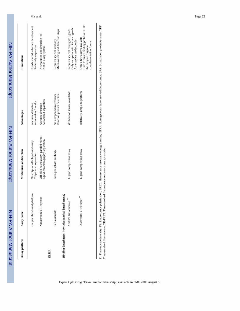
Ma et al. Page 22
Ass
ay p
latfo
rmA
ssay
nam
eM
echa
nism
of d
etec
tion
Adv
anta
ges
Lim
itatio
ns
Cal
iper
chi
p-ba
sed
plat
form
On-
chip
- or o
ff-c
hip-
base
d as
say
Chi
p-ba
sed
sepa
ratio
nA
ccur
ate
dete
ctio
nA
utom
atio
n fr
iend
lyN
eeds
spec
ial s
ubst
rate
dev
elop
men
tR
elat
ivel
y ex
pens
ive
Nan
ostre
am’s
LD
syst
emO
ff-c
hip
base
d as
say,
par
alle
l mic
ro-
liqui
d ch
rom
atog
raph
y se
para
tion
Acc
urat
e de
tect
ion
Aut
omat
ed se
para
tion
A se
para
tion
and
dete
ctio
n to
olN
ot a
n as
say
syst
em
ELIS
A
Self-
asse
mbl
eA
nti-p
hosp
hate
ant
ibod
yN
o co
mpo
und
inte
rfer
ence
Rea
ctio
n pr
oduc
t det
ectio
nR
equi
res s
peci
al a
ntib
ody
Mul
ti-w
ashi
ng a
nd d
etec
tion
step
s
Bind
ing-
base
d as
say
(non
-bio
chem
ical
bas
ed a
ssay
s)
Am
bit’s
Kin
omeS
can
™Li
gand
com
petit
ion
assa
yW
ith b
road
kin
ases
ava
ilabl
eR
equi
res s
peci
al c
onju
gate
liga
nds
Onl
y co
mpe
tes w
ith k
now
n lig
ands
As a
serv
ice
prod
uct o
nly
Dis
cove
Rx’
s HitH
unte
r ™Li
gand
com
petit
ion
assa
yR
elat
ivel
y si
mpl
e to
per
form
Onl
y a
few
ass
ays a
vaila
ble
Har
d to
cre
ate
bind
ing
prob
e to
fit i
nto
the
enzy
me
frag
men
tco
mpl
emen
tatio
n fo
rmat
FI: F
luor
esce
nce
inte
nsity
; FP:
Flu
ores
cenc
e po
lariz
atio
n; F
RET
: Flu
ores
cenc
e re
sona
nce
ener
gy tr
ansf
er; H
TRF:
Hom
ogen
eous
tim
e-re
solv
ed fl
uore
scen
ce; S
PA: S
cint
illat
ion
prox
imity
ass
ay; T
RF:
Tim
e-re
solv
ed fl
uore
scen
ce; T
R-F
RET
: Tim
e-re
solv
ed fl
uore
scen
ce re
sona
nce
ener
gy tr
ansf
er.
Expert Opin Drug Discov. Author manuscript; available in PMC 2009 August 5.


![Samenvatting urban challenge[1]](https://static.fdokumen.com/doc/165x107/6313d00f3ed465f0570ad8b4/samenvatting-urban-challenge1.jpg)