Roles interpersonales • Representante • Líder • Enlace • Roles informativos • Monitor • Difusor

Multiple Roles for the Receptor Tyrosine Kinase Axl
in Tumor Formation
Sacha J. Holland,1Mark J. Powell,
1Christian Franci,
1Emily W. Chan,
1Annabelle M. Friera,
1
Robert E. Atchison,1John McLaughlin,
1Susan E. Swift,
1Erlina S. Pali,
1George Yam,
1Stephen Wong,
1
Joe Lasaga,1Mary R. Shen,
1Simon Yu,
1Weiduan Xu,
1Yasumichi Hitoshi,
1Jakob Bogenberger,
1
Jacques E. Nor,2Donald G. Payan,
1and James B. Lorens
1
1Rigel, Inc., South San Francisco, California and 2University of Michigan School of Dentistry, Ann Arbor, Michigan
Abstract
A focus of contemporary cancer therapeutic development isthe targeting of both the transformed cell and the supportingcellular microenvironment. Cell migration is a fundamentalcellular behavior required for the complex interplay betweenmultiple cell types necessary for tumor development. Wetherefore developed a novel retroviral-based screening tech-nology in primary human endothelial cells to discover genesthat control cell migration. We identified the receptor tyrosinekinase Axl as a novel regulator of endothelial cell haptotacticmigration towards the matrix factor vitronectin. Using smallinterfering RNA–mediated silencing and overexpression ofwild-type or mutated receptor proteins, we show that Axl is akey regulator of multiple angiogenic behaviors includingendothelial cell migration, proliferation, and tube formationin vitro . Moreover, using sustained, retrovirally delivered shorthairpin RNA (shRNA) Axl knockdown, we show that Axl isnecessary for in vivo angiogenesis in a mouse model.Furthermore, we show that Axl is also required for humanbreast carcinoma cells to form a tumor in vivo . These findingsindicate that Axl regulates processes vital for both neo-vascularization and tumorigenesis. Disruption of Axl signalingusing a small-molecule inhibitor will hence simultaneouslyaffect both the tumor and stromal cell compartments and thusrepresents a unique approach for cancer therapeutic devel-opment. (Cancer Res 2005; 65(20): 9294-303)
Introduction
Cancer is not a single-cell disease and tumor developmentinvolves complex reciprocal interactions among neoplastic, stro-mal, and immune cells (1). Cell migration is a central cellularfunction used by these cell types for angiogenesis, inflammation,and metastasis (2). Although it is likely that inhibitors of cellmigration will complement existing cancer drugs, this possibilityhas been relatively neglected in the search for new cancertherapeutics.Angiogenesis, a physiologic and strictly regulated process in
healthy adults, is aberrantly induced by hypoxic tumors thussecuring an adequate blood supply that feeds tumor growth and
facilitates metastasis. The assembly of functional vessels requiresthe precise coordination of multiple cellular events and inter-actions (3). Compelling evidence shows that vascular endothelialgrowth factor (VEGF) can initiate angiogenic sprouting in vivo ,increasing permeability and supporting endothelial cell prolifera-tion and survival (4). Sprouting migration of activated endothelialcells is accompanied by up-regulation of integrin matrix factorreceptors (e.g., avh3 and a5h1). Nascent endothelial tubes thusformed are stabilized by pericytes and basement membraneformation. Targeted inhibition of angiogenesis using an anti-VEGFantibody (bevacizumab) has shown efficacy in treating humancancer (5). However, as tumors frequently advanced while patientswere receiving therapy, inhibition of VEGF signaling alone may notbe sufficient to block neovascularization in human malignancies(6). Blocking processes complementary to those driven by VEGFmay therefore be important in fully inhibiting angiogenesis inhuman disease.Retroviral-based functional screening has been established as an
effective, versatile, and unbiased method for identifying novel drugtargets and regulators of specific cellular and/or disease processes(7). The use of retroviral expression vectors allows the efficientdelivery and stable expression of complex and diverse libraries ofgenetic effectors (e.g., cDNAs and peptides) in a multitude of celltypes including primary human cells. This allows design ofscreening assays in cell types that reflect physiologically relevantdisease responses. We reasoned that this approach would be wellsuited to probing a complex and technically challenging cellularfunction such as cell migration.As part of an effort to develop novel therapeutic strategies
for cancer treatment, we developed a unique retroviral-basedfunctional genetic screening protocol to discover genes thatregulate cell migration in primary human endothelial cells. Weidentified Axl as a novel regulator of endothelial cell haptotacticmigration towards the matrix factor vitronectin. Axl (UFO/ARK/Tyro7) is a receptor tyrosine kinase (RTK) that is stimulated bythe f76-kDa secreted protein Gas6 (growth arrest specific 6;refs. 8, 9). We show that Axl signaling affects multiple cellularbehaviors required for neovascularization in vitro and regulatesangiogenesis in vivo . Furthermore, we show that loss of Axlexpression in tumor cells blocks the growth of solid humanneoplasms in an in vivo MDA-MB-231 breast carcinoma xenograftmodel. This suggests a model in which Gas6-Axl signalingsupports tumor-stroma interactions necessary for tumor growthand angiogenesis.Taken together, these data indicate that Axl signaling can
independently regulate neovascularization and tumor growth andthus represents a novel target class for tumor therapeuticdevelopment.
Note: J.B. Lorens is currently at the Department of Biomedicine, University ofBergen, N-5009 Bergen, Norway.
Supplementary data for this article are available at Cancer Research Online (http://cancerres.aacrjournals.org/).
Requests for reprints: Sacha J. Holland, Rigel, Inc., 1180 Veteran’s Boulevard,South San Francisco, CA 94080. Phone: 650-624-1283; E-mail: [email protected].
I2005 American Association for Cancer Research.doi:10.1158/0008-5472.CAN-05-0993
Cancer Res 2005; 65: (20). October 15, 2005 9294 www.aacrjournals.org
Research Article
Research. on February 18, 2016. © 2005 American Association for Cancercancerres.aacrjournals.org Downloaded from

Materials and Methods
Antibodies. Antibodies were purchased from the following suppliers:
mouse monoclonal anti-human Axl (MAB154, R&D Systems, Minneapolis,
MN), rabbit polyclonal anti-actin (Cytoskeleton, Denver, CO), unconjugated
anti-human CD31 (BioCare, Walnut Creek, CA), and FITC-conjugated anti-
human CD31 (BD Biosciences PharMingen, Mansfield, MA). Mouse mono-
clonal anti-VEGFR2 was a gift from Dr. Jeanette Wood (Novartis, Basel,
Switzerland). Rhodamine-conjugated Ulex europaeus agglutinin 1 (UEA-1)
lectin was from Vector Labs (Burlingame, CA).
Cell culture. All cells were cultured at 37jC, 5% CO2. Phoenix A cells
(Dr. Gary Nolan, Stanford) were maintained in DMEM supplemented with
10% fetal bovine serum (FBS), antibiotics, and glutamine. MDA-MB-231
human breast epithelial carcinoma cells (American Type Culture Collection,
Rockville, MD) were maintained in F12K supplemented with 10% FBS.
Primary human umbilical vein endothelial cells (HUVEC), human dermal
microvascular endothelial cells (HMVEC), and pulmonary artery smooth
muscle cells (PASMC) were cultured as vendor’s recommendations
(Cambrex, Walkersville, MD).Retroviral haptotaxis screen. The HUVEC green fluorescent protein
(GFP)-cDNA library was prepared in the retroviral CRU5-GFP fusion vector
(10). mRNA was isolated from VEGF, basic fibroblast growth factor, and
epidermal growth factor–stimulated HUVECs with poly-dT magnetic beads
(Dynal, Lake Success, NY) and normalized with biotinylated first strand
HUVEC cDNA and streptavidin beads (Invitrogen, Carlsbad, CA). Double-
stranded cDNA was synthesized using standard methods (Smart cDNA
System, Clontech/BD Biosciences, Mansfield, MA) and cloned into the
library vector bidirectionally using BstXI adapters (Invitrogen). The
complexity of the final library was 1.5 � 106. Retroviral packaging cells
(Phoenix A) were transfected with the cDNA library to produce infectious
virus sufficient to infect 7 � 106 subconfluent HUVEC at 10% transduction
efficiency in T75 flasks. GFP-expressing HUVECS were sorted by fluores-
cence-activated cell sorting (FACS) and replated. The following day, the cells
were harvested and migrated thrice in 75-mm Boyden chambers (Transwell,
Corning-Costar, Acton, MA) coated on the underside with 15 Ag/mL
vitronectin (Chemicon, Temecula, CA). Each migration was conducted
for 3 hours. Nonmigrating cells were harvested with 0.05% Trypsin for
90 seconds at 37jC and remigrated. The cells were infected with wild type
Moloney murine leukemia virus (MMTV, American Type Culture Collection)
before the third migration step to produce library clone viruses from the
enriched nonmigratory cell population. The final selected nonmigratory
HUVEC population was plated and library viruses were harvested the
following day and used to infect PG13 packaging cells at a low multiplicity of
infection (0.5%). Single GFP+ PG13 cells were arrayed into microtiter plates
by FACS. cDNA inserts were isolated by reverse transcription-PCR (RT-PCR,
Clontech/BD Biosciences) from purified retrovirus (12). To confirm the
library insert phenotype, naive HUVECs were transduced using supernatants
from each PG13 clone. GFP-expressing cells were purified by FACS and
tested in individual haptotaxis assays (see below).
Small interfering RNA design and transfection. Luciferase control
(GL2), VEGFR2 (D-003148-05), and custom small interfering RNA (siRNA)
oligos were purchased from Dharmacon (Lafayette, CO). siRNA sequences
(Supplementary Table 1) were designed according to established criteria
and examined to ensure minimal homology with other sequences. For
siRNA transfection, 60,000 HUVECs were plated per well in a six-well plate.
The following day, cells were transfected with 3.2 Ag siRNAs using
Oligofectamine reagent in Opti-MEM (both Invitrogen) for 4 hours after
which 3� volume of culture medium containing 6% FBS was added.
Constructs. The shRNA vector (EFS-U3/U6) was derived from the TRA
retroviral vector (10). The EFS-U3/U6 vector comprises retroviral elements
required for stable integration into the genome of infected cells, a modified
U6 RNA promoter (�230 to +1) and terminator (TTTTT) embedded into the
NheI site of the TRA vector 3V long terminal repeat for conditional
expression of hp-siRNA and an internal EF1-a expression cassette (11) that
drives expression of a destabilized version (COOH-terminal PEST sequence)
of the Renilla GFP (dsRMG) for independent monitoring of transfection/
infection efficiencies. Specific shRNA vectors were created by PCR. COOH-
terminally myc-tagged Axl cDNA (480-3140) was expressed from an IRES-
GFP vector (10) derivative where GFP is replaced with Renilla GFP.Transfection/retroviral infection. Phoenix A cells were transfected
using the calcium phosphate method (12). Approximately 30 hours after
transfection, the medium was changed to growth medium for the cells to be
infected, supplemented with 10% FBS. Infectious supernatant was collected
f48 hours after transfection. Target cells were exposed to supernatant
containing 5 Ag/mL protamine sulfate for 6 hours to overnight before being
returned to regular growth medium.
Taqman. Total RNA was isolated using RNeasy Mini Kit (Qiagen,Valencia, CA) and quantified using Ribo Green Quantification kit (Molecular
Probes, Eugene, OR). Human Gas6 primers and probe were designed using
Primer Express software: sense primer, ATGTGGCAGACAATCTCTG;
reverse primer, ACAGCATCCCTGTTGACCT; probe, AGCTGGCGCGG-AATCTGGTCA. Reactions were run using a one-step RT-PCR kit (Qiagen)
in an ABI Prism 7900HT sequence analyzer. An 18S rRNA reaction kit (ABI,
Foster City, CA) was used as the reference/loading control reaction. Data
was normalized to 18S rRNA and plotted as percentage of luciferasecontrol.
Western blot. Cells were lyzed in PLC lysis buffer containing 1% Triton
X-100 (13). Protein concentrations were determined using the bicinchoninicacid protein assay kit (Pierce, Rockford, IL) and equalized. SDS-PAGE and
Western blotting were carried out according to standard procedures.
Boyden chamber haptotaxis assay. Migration assays were done 48
hours after siRNA transfection, or 1 to 2 days after sorting of infected
populations. The undersides of 8 Am 24-well Transwell filters (Costar) were
coated with 15 Ag/mL vitronectin (Chemicon). Six replicate wells were run
per sample. The following day, unbound vitronectin was eluted in migration
medium [0.5% bovine serum albumin (BSA, Sigma, St. Louis, MO) in EGM-2
medium (Cambrex)]. Migration medium (500 AL) was added into the lower
chamber. HUVECs resuspended in migration medium at 2,000 cells/300 ALper well were added to the upper chamber. Cells were allowed to migrate
for 3 hours at 37jC after which they were removed from either the upper or
lower surface of the membrane (three wells each) using a cotton swab.
Following fixation in 3.7% formaldehyde, swabbing was repeated and nuclei
were stained using 2 Ag/mL 4V,6-diamidino-2-phenylindole. Cells remaining
on the upper (nonmigrated cells) or lower (migrated cells) surface of
the membrane were imaged using a Cellomics Arrayscan instrument and
processed using ImagePro software. Migration is presented as mean
haptotactic index (% nonmigrated, control / % nonmigrated, test) F SD: 1,
no effect; >1, enhanced migration; <1, retarded migration. Representative
examples of at least two replicate experiments are shown. Statistical
analysis was done using the m2 test.
Proliferation assay. Cells were seeded sparsely in six-well dishes,
infected with retroviral supernatants, and maintained in complete
endothelial cell medium. The percentage of infected, GFP-positive cells inthe population was monitored at regular intervals by FACS. Cells were
passaged during the course of the experiment as necessary to keep them
subconfluent. Data was analyzed using FlowJo software. The relative
number of GFP-positive cells for all samples at the beginning of theexperiment (day 3) was set to 100%. Subsequent changes in relative % GFP
over time were normalized to the in-day vector control value.
Coculture assay. Early-passage HUVECs (Vp6) were mixed 1:10 withPASMC in HUVEC growth medium (EGM-2) and seeded onto collagen-
coated (Sigma) LabTekII chamber slides (Nalge Nunc, Rochester, NY).
Medium was replaced every 2 days. Five days after seeding, the cocultures
were washed in PBS, fixed using �20jC/70% ethanol at room temperaturefor 15 minutes, blocked in PBS + 5% BSA, and stained using FITC-
conjugated anti-CD31 antibodies or Rhodamine-conjugated UEA-1 lectin
(1:200) to reveal endothelial cell tubes. Cultures were photographed and
mean tube formation over 16 images covering an entire representative wellwas quantified using ImagePro software. Representative examples of at least
two replicate experiments are shown. Statistical analysis was done using the
t test.
In vivo angiogenesis assay. HMVECs were infected with shRNA vectorsand GFP-positive cells were enriched by FACS. Poly L-lactic acid (PLLA)
sponge matrices (5 � 5 � 1 mm) were seeded with 700,000 sorted HMVEC
Multiple Roles for Axl in Tumor Formation
www.aacrjournals.org 9295 Cancer Res 2005; 65: (20). October 15, 2005
Research. on February 18, 2016. © 2005 American Association for Cancercancerres.aacrjournals.org Downloaded from

cells in a 1:1 mixture of EGM-2 MV and Matrigel (BD Biosciences). Up tofour sponges were implanted s.c. in female CB-17 severe combined
immunodeficient (SCID) mice according to Nor et al. (14). Sponges were
harvested after 14 days for biochemical or immunohistochemical analysis.
To specifically label perfused human endothelial cells in the sponges, 200AL of rhodamine-conjugated UEA-1 lectin (1 Ag/AL in sterile 0.9% NaCl)
was injected into the tail vein 30 minutes before sacrifice. Excised sponges
were homogenized using a handheld mincer (Biospec Products, Inc.,
Bartlesville, OK) in 200 AL of modified radioimmunoprecipitation assaybuffer. Fluorescence in filtered (0.45-Am GHP syringe filter, Pall-Gelman
Corp., Inc., East Hills, NY) supernatant was quantified on a Fluorskan
machine with a 530/590-nm excitation/emission filter pair and normalized
to plasma lectin content. Tie-2 levels present in sponge lysates (100 AL)were measured using a human Tie-2 ELISA (R&D Systems) and normalized
to protein content.
Statistical analysis between groups was done using the Mann-Whitneyrank sum test.
Xenograft assay. MDA-MB-231 cells were infected with shRNA vectors
and GFP positive populations were enriched to 98% to 100% by FACS. RNA
interference (RNAi)–mediated knockdown of cell surface Axl expressionwas confirmed on the day of implantation. Cells (1 � 107) were injected in
0.2 mL of F12K medium + 10% FBS/high concentration Matrigel (1:1 ratio,
BD Biosciences) into the right flank of female CB-17 SCID mice. Animals in
which tumors measured at least 0.3 to 0.5 cm were selected for the study.Tumor volume [V = (tumor length � tumor width2) / 2] was measured
every 2 to 3 days for 30 days, or until it reached a volume of 2,000 mm3.
Tumor growth was expressed as V/Vo, where V = tumor volume and Vo =
initial volume of tumor. Mean V/Vo and SDs were calculated using LabcatSoftware. Statistical analysis between groups was done using the t test.
Immunohistochemistry. Staining reagents were from BioCare. Heat-
induced epitope retrieval was done on 4 Am paraformaldehyde/formalin-
fixed, paraffin-embedded tissue sections using Medical DecloakingChamber in Borg high-pH buffer for 2 minutes followed by rinses in Hot
Rinse Solution to remove residual paraffin. Nonspecific staining was
eliminated using SNIPER blocking reagent. Primary antibody (mouse
monoclonal anti-human CD31; 1:50 or mouse monoclonal anti-human Axl,1:100) incubations were followed by biotinylated goat anti-mouse/rabbit
secondary (BioCare) and then alkaline phosphatase or horseradish
peroxidase–conjugated streptavidin. Development was done according to
the manufacturer’s instructions. Slides were counterstained with hematox-ylin, mounted, and coverslipped. Brightfield pictures were taken using a
Nikon Eclipse 400 microscope with a Spot Insight QE imaging system.
Results
Genetic screen to identify modulators of vitronectinhaptotaxis. We established a retroviral-based functional screento select for genes that regulate endothelial migration (Fig. 1A).Similar screens, based on cell survival, proliferation, and surfacemarker expression, have been done in transformed cell lines(7, 15–20). To institute a physiologically relevant screening assay,we used early-passage primary HUVECs. A GFP-fused HUVECcDNA library was constructed in a retroviral vector. The GFP
Figure 1. Antisense cDNA effector correspondingto Axl was identified in a retroviral-basedfunctional screen for inhibitors of endothelial cellhaptotaxis. A, haptotaxis screen protocol.Phoenix A packaging cells were transfected with aretroviral GFP-cDNA fragment library.Retrovirus-containing medium was collectedand used to infect HUVECs. The infected,GFP-expressing HUVEC population was enrichedby FACS and subjected to three rounds ofmigration in Boyden chambers coated on theunderside of the membrane with 15 Ag/mL ofvitronectin. To rescue the library inserts, selectedHUVECs were infected with WT virus andinfectious supernatant was used to infect PG13packaging cells. PG13 clones containing a singlelibrary insert each were arrayed in microtiter platesas single cells by FACS and expanded. Libraryvirus RNA from individual PG13 clones was usedas a template for RT-PCR to isolate cDNA inserts.Retroviruses from selected clones were used toinfect naive HUVECs and individual migrationexperiments were carried out to reconfirm insertphenotypes. B, haptotaxis towards vitronectin.HUVECs were infected with virus from PG13 clone#420 or control vector. GFP-positive cells wereenriched by FACS and subjected to haptotaxistowards 15 Ag/mL of vitronectin. Expression ofGFP-Axl antisense cDNA fragment reducedhaptotaxis compared with control. Columns, meanhaptotactic index; bars, FSD. % Nonmigrated,control / % nonmigrated, test: 1, no effect; >1,enhanced migration; <1, retarded migration.C-D, siRNAs targeting Axl inhibit endothelial cellhaptotaxis. HUVECs were transfected with acontrol siRNA targeting luciferase or twoindependent siRNAs targeting different sequenceswithin the endogenous Axl message (Axl-2 andAxl-4). After 48 hours, cells were assayed (C ) fortarget knockdown by Western blot (showingreduction of Axl expression to near backgroundlevels) or (D ) haptotaxis towards 15 Ag/mL ofvitronectin. *, P < 0.001 (m2 test) compared withcontrol.
Cancer Research
Cancer Res 2005; 65: (20). October 15, 2005 9296 www.aacrjournals.org
Research. on February 18, 2016. © 2005 American Association for Cancercancerres.aacrjournals.org Downloaded from

moiety stabilizes fusion proteins, reveals subcellular localization,and facilitates identification of vector-infected cells. Furthermore,the bidirectional cloning strategy yields potential antisenseeffectors (21). This library was introduced into HUVECs at a lowinfection rate (10%) to avoid multiple viral integrations per cell.The infected, GFP-expressing cell population was enriched by FACSand cells with impaired haptotactic motility towards 15 Ag/mLvitronectin were selected by three rounds of migration in amodified Boyden chamber assay (see Materials and Methods).As the HUVEC life span is limited, we developed a technique to
rescue genetic library information from the selected cells andtransfer it to a more robust, transformed cell type. NonmigratingHUVECs were infected with wild-type (WT) MMLV to packageresident library viruses (15). Virion-containing medium washarvested and used to infect the NIH3T3-derived PG13 packagingcell line at a very low infection rate to ensure a single infection percell. Single-cell PG13 clones were arrayed in microtiter plates byFACS and expanded. Library inserts were isolated by PCR using theretroviral DNA as the template and sequenced.Two hundred fifty four independent library inserts were iden-
tified. Library clone #420 encoded a fragment from the COOH-terminal region of the RTK Axl (nucleotides 2884-2964), which wasoriented in the reverse direction indicating that it may function asan antisense effector.Genetic effectors targeting Axl impair vitronectin hapto-
taxis. To reconfirm that the isolated clone #420 virus couldreproduce the phenotype selected for in the screen, retroviralsupernatant from clone 420 was used to infect naive HUVECs. GFP-expressing cell populations were enriched by FACS and assayed bymigration towards 15 Ag/mL of vitronectin. HUVECs infected withthe Axl genetic effector indeed showed reduced haptotacticmigration towards vitronectin (Fig. 1B).We used siRNA-mediated silencing to independently verify that
Axl knockdown could inhibit vitronectin haptotaxis. Comparedwith HUVECs transfected with a control siRNA-targeting luciferase,those transfected with either of two independent siRNAs targetingAxl (Fig. 1C) phenocopied the effect of the Axl antisense screeninginsert and exhibited reduced migration towards vitronectin (Fig. 1D).Furthermore, GFP-expressing retroviral vectors designed to pro-duce shRNAs corresponding to these Axl-silencing sequencesgenerated a similar reduction in haptotaxis (data not shown). Axlexpression in HUVECs is thus required for haptotaxis towardsvitronectin.Axl regulates human umbilical vein endothelial cell
proliferation. An important characteristic of angiogenic endothe-lial cells is their high proliferative capacity. As Gas6 is a weakmitogen in some cell types, we asked whether Axl signalingregulates HUVEC proliferation. HUVECs infected with Axl shRNAvectors or a positive control construct expressing the cell cycleinhibitor p21 (22) exhibited a marked growth disadvantagecompared with controls (Supplementary Fig. 1). Furthermore,although we did not detect significant apoptosis in Axl shRNA-expressing cell populations under the assay conditions, Axlsilencing also reduced DNA replication as assayed by bromodeox-yuridine incorporation (data not shown). Taken together, thesedata show that Axl expression is important in driving HUVECproliferation.Axl knockdown impairs endothelial tube formation in vitro.
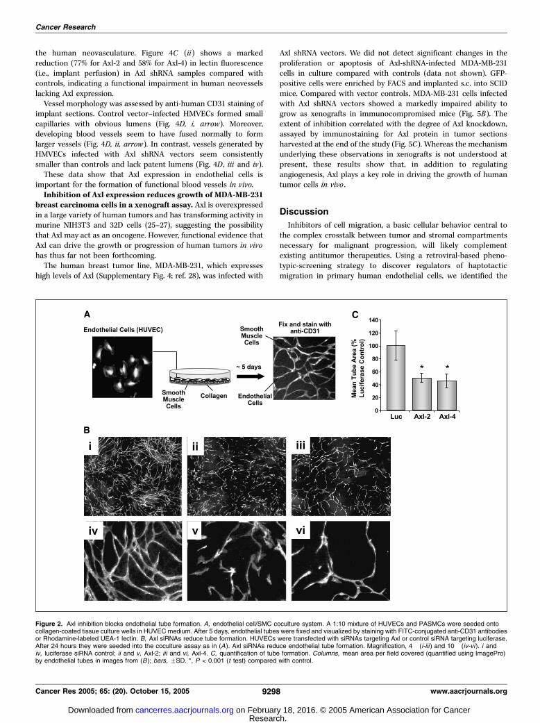
We next addressed whether Axl regulates endothelial tubeformation. To mimic the complex matricellular environmentencountered by angiogenic endothelial cells in vivo , we designed
a HUVEC/primary PASMC coculture branching morphogenesis/tube formation assay (Fig. 2A). Endothelial morphogenesis in thisassay is VEGF dependent and proliferation independent (seeSupplementary Fig. 2).HUVECs were transfected with Axl siRNAs and seeded into a
coculture assay (Fig. 2B). Axl knockdown was similar to that inFig. 1C (data not shown). Total tube formation, fiber length, andbranching were reduced by at least f50 % in HUVECstransfected with Axl siRNAs (Fig. 2B , ii, iii, v, and vi and C ;data not shown) compared with those transfected with theluciferase control siRNA (Fig. 2B , i and iv and C). These resultsshow that Axl is involved in the regulation of endothelial tubemorphogenesis.Endothelial cell Gas6 expression and Axl catalytic activity
are required for regulation of proangiogenic processes. Wenext asked whether ligand-stimulated Axl catalytic activity isrequired for its proangiogenic functions. HUVECs synthesize theAxl ligand, Gas6 (23). Two independent siRNAs designed to targetGas6 depressed HUVEC Gas6 message levels and reduced HUVEChaptotaxis towards vitronectin compared with controls (Supple-mentary Fig. 3). Gas6 silencing thus phenocopies the effect of Axlknockdown on vitronectin haptotaxis.We next focused on the catalytic activity of Axl. Overexpression
of WT Axl in HUVECs (Fig. 3A) induced a growth advantagerelative to controls (Fig. 3B). Conversely, overexpression of acatalytically inactive Axl mutant (AxlKD; K567E substitution; ref. 24;Fig. 3A) conferred a f30% growth disadvantage over the course ofthe assay (Fig. 3B). Lastly, sorted, GFP-positive WT Axl or GFP-infected HUVECs were seeded into a coculture assay. Over-expression of WT Axl led to f40% increase in the number ofendothelial cell tubes formed compared with control vector(Fig. 3C , ii and v and D). In contrast, overexpression of AxlKDdid not enhance tube morphogenesis (Fig. 3C , iii and vi and D).Similar trends were noted for fiber length and branching (data notshown). The autocrine/paracrine ligand-induced kinase activity ofAxl therefore controls several distinct proangiogenic processes andthus represents an attractive target for small-molecule inhibition.Axl knockdown impairs blood vessel formation and func-
tion in a mouse angiogenesis model. Because our observationssuggest that Axl signaling may regulate angiogenesis in vivo , weinvestigated this possibility using the SCID mouse model of humanangiogenesis (Fig. 4A ; ref. 14).Primary HMVECs were infected with the Axl shRNA vectors, an
shRNA vector that silences VEGFR2 or the luciferase shRNAcontrol vector. FACS-enriched GFP-expressing cells were mixedwith Matrigel, seeded into PLLA scaffolds, and subsequentlyimplanted into SCID mice. Over 14 to 22 days, the humanendothelial cells form vessels that anastamose with recruitedmouse vasculature, connecting them to the mouse circulation.HMVECs not incorporated into vessels typically undergoapoptosis by around 14 days (J.E. Nor, data not shown). Vesselformation was analyzed 14 days after implantation by measuringhuman Tie-2 levels in implant lysates by ELISA. In implantscontaining HMVECs infected with VEGFR2 or Axl shRNA vectors,human Tie-2 levels were reduced by 42% and over 50%,respectively (Fig. 4C, i) indicating that Axl silencing impairsneovascularization.Fluorescent UEA-1 lectin, which binds specifically to human
endothelial cells, was injected into the mouse bloodstream beforesacrifice. The relative fluorescence in implant lysates comparedwith mouse plasma provides a measure of functional circulation in
Multiple Roles for Axl in Tumor Formation
www.aacrjournals.org 9297 Cancer Res 2005; 65: (20). October 15, 2005
Research. on February 18, 2016. © 2005 American Association for Cancercancerres.aacrjournals.org Downloaded from
the human neovasculature. Figure 4C (ii) shows a markedreduction (77% for Axl-2 and 58% for Axl-4) in lectin fluorescence(i.e., implant perfusion) in Axl shRNA samples compared withcontrols, indicating a functional impairment in human neovesselslacking Axl expression.Vessel morphology was assessed by anti-human CD31 staining of
implant sections. Control vector–infected HMVECs formed smallcapillaries with obvious lumens (Fig. 4D, i, arrow). Moreover,developing blood vessels seem to have fused normally to formlarger vessels (Fig. 4D, ii, arrow). In contrast, vessels generated byHMVECs infected with Axl shRNA vectors seem consistentlysmaller than controls and lack patent lumens (Fig. 4D , iii and iv).These data show that Axl expression in endothelial cells is
important for the formation of functional blood vessels in vivo.Inhibition of Axl expression reduces growth of MDA-MB-231
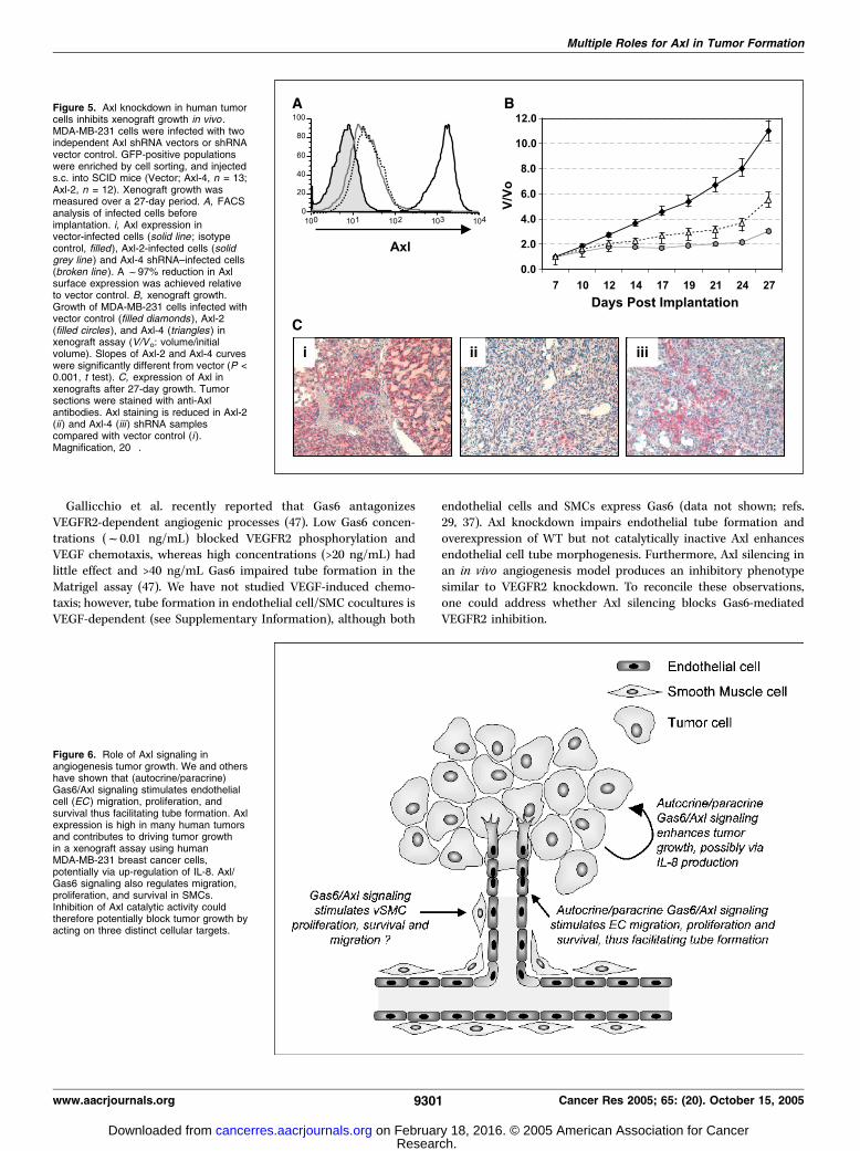
breast carcinoma cells in a xenograft assay. Axl is overexpressedin a large variety of human tumors and has transforming activity inmurine NIH3T3 and 32D cells (25–27), suggesting the possibilitythat Axl may act as an oncogene. However, functional evidence thatAxl can drive the growth or progression of human tumors in vivohas thus far not been forthcoming.The human breast tumor line, MDA-MB-231, which expresses
high levels of Axl (Supplementary Fig. 4; ref. 28), was infected with
Axl shRNA vectors. We did not detect significant changes in theproliferation or apoptosis of Axl-shRNA-infected MDA-MB-231cells in culture compared with controls (data not shown). GFP-positive cells were enriched by FACS and implanted s.c. into SCIDmice. Compared with vector controls, MDA-MB-231 cells infectedwith Axl shRNA vectors showed a markedly impaired ability togrow as xenografts in immunocompromised mice (Fig. 5B). Theextent of inhibition correlated with the degree of Axl knockdown,assayed by immunostaining for Axl protein in tumor sectionsharvested at the end of the study (Fig. 5C). Whereas the mechanismunderlying these observations in xenografts is not understood atpresent, these results show that, in addition to regulatingangiogenesis, Axl plays a key role in driving the growth of humantumor cells in vivo .
Discussion
Inhibitors of cell migration, a basic cellular behavior central tothe complex crosstalk between tumor and stromal compartmentsnecessary for malignant progression, will likely complementexisting antitumor therapeutics. Using a retroviral-based pheno-typic-screening strategy to discover regulators of haptotacticmigration in primary human endothelial cells, we identified the
Figure 2. Axl inhibition blocks endothelial tube formation. A, endothelial cell/SMC coculture system. A 1:10 mixture of HUVECs and PASMCs were seeded ontocollagen-coated tissue culture wells in HUVEC medium. After 5 days, endothelial tubes were fixed and visualized by staining with FITC-conjugated anti-CD31 antibodiesor Rhodamine-labeled UEA-1 lectin. B, Axl siRNAs reduce tube formation. HUVECs were transfected with siRNAs targeting Axl or control siRNA targeting luciferase.After 24 hours they were seeded into the coculture assay as in (A). Axl siRNAs reduce endothelial tube formation. Magnification, 4� (i-iii ) and 10� (iv-vi). i andiv, luciferase siRNA control; ii and v, Axl-2; iii and vi, Axl-4. C, quantification of tube formation. Columns, mean area per field covered (quantified using ImagePro)by endothelial tubes in images from (B); bars, FSD. *, P < 0.001 (t test) compared with control.
Cancer Research
Cancer Res 2005; 65: (20). October 15, 2005 9298 www.aacrjournals.org
Research. on February 18, 2016. © 2005 American Association for Cancercancerres.aacrjournals.org Downloaded from

RTK Axl as a key modulator of multiple endothelial cell processes(proliferation, migration, endothelial cell survival, and tubeformation) required for angiogenesis and tumor growth.The receptor tyrosine kinase Axl was first identified from the
DNA of chronic myelogenous leukemia patients due to itstransforming activity in vitro (25, 26). Expression of Axl isdetected in many cell types but is prominent in the vasculature(in both endothelial cells and vascular SMCs, VSMC) and in cellsof the myeloid lineage (29–31). Axl and its two close relatives Mer/Nyk and Sky (Tyro3/Rse/Dtk) all bind and are stimulated tovarying degrees by Gas6 (8, 9). Axl signaling in culture protectscells from serum starvation– or tumor necrosis factor-a-inducedapoptosis and mediates ligand-induced chemotaxis and celldifferentiation (24, 31–33). However, Axl�/� mice exhibit no overtphenotype and the physiologic function of Axl in vivo is not clearlyestablished (34). The overexpression of Axl and/or its ligand, Gas6,has been reported in a wide variety of solid human tumor types(ref. 35 and references therein) and myeloid leukemias (30). Axlsignaling is also functionally implicated in the response tovascular injury and kidney disease (29, 36). Axl may thuspotentially represent a therapeutic target for diverse pathologicconditions.Our data suggest that Axl acts as a central facilitator of
endothelial cell activation. Axl may support pathologic neo-
vascularization as impaired developmental angiogenesis has notbeen reported in Axl�/� mice. Axl and Gas6 are coexpressed inendothelial cells and silencing of either gene impairs vitronectinhaptotaxis. Thus, a constitutive autocrine/paracrine signalingloop may exist in these cells (ref. 37; data not shown). Axl maymodulate activated endothelial cell functions through one ofseveral possible mechanisms: (a) Axl signaling in endothelial cellsmay support the activated state, potentially by altering geneexpression, thus maintaining a proliferative, motile phenotype.Axl inhibition may therefore alter the angiogenic balance, drivingendothelial cells towards a more quiescent, nonmotile, differen-tiated state. (b) Axl signaling modulates integrin function.Angelillo-Scherrer et al. recently showed that Gas6-dependentsignaling results in phosphorylation of h3 integrin in platelets(38) resulting in defective aIIbh3-dependent ‘‘outside-in’’ signalingin Axl�/� platelets. Similar crosstalk has been described betweengrowth factor receptors (e.g., VEGFR2 and FGFR) and integrins(e.g., avh3 and avh5; ref. 39). Integrin-mediated substrateadhesion is required for cell migration, proliferation, and survivalof growth factor–stimulated cells. Thus, blocking Axl signalingmay alter the function of certain integrins necessary for thegrowth, migration, and survival of endothelial cell and tumorcells. (c) Constitutive Gas6 stimulation may maintain the steadystate activity of Axl effectors such as Rac and Akt (40, 41) that
Figure 3. Axl catalytic activity is required for regulation of pro-angiogenic phenotypes. A and B, Axl catalytic activity regulates HUVEC proliferation. A, expressionof Axl in FACS-enriched GFP + HUVEC populations infected with wild type Axl, AxlKD, or control vector was assayed by Western blot. B, % GFP in the infectedpopulations was monitored over time by FACS as in Supplementary Fig. 1. AxlWT (broken dashed line, squares ) increases and AxlKD (broken dotted line, triangles )reduces HUVEC growth compared with control vector (solid line, diamonds ). Initial absolute % GFP on day 3 was Vector, 69.3%; AxlWT, 43.2%; and AxlKD, 60.3%.C and D, Axl catalytic activity regulates endothelial tube formation. C, sorted, GFP-positive infected populations were seeded into the coculture as in Fig. 2. AxlAxlWT enhances endothelial cell tube formation, whereas Axl KD does not. Magnification, 4� (i-iii ) and 10� (iv-vi ). i and iv, Vector control; ii and v, AxlWT; iii andvi, AxlKD. D, columns, mean area per field (quantified using ImagePro as in Fig. 2C ) covered by endothelial tubes in images from (C ); bars, FSD. *, P < 0.001(t test) compared with vector.
Multiple Roles for Axl in Tumor Formation
www.aacrjournals.org 9299 Cancer Res 2005; 65: (20). October 15, 2005
Research. on February 18, 2016. © 2005 American Association for Cancercancerres.aacrjournals.org Downloaded from

control matrix- and chemoattractant-induced cell motility. Thedefective lumen morphology observed in vessels lacking Axlmay reflect regulation of the Rho/Rac-dependent fusion ofintracellular vacuoles thought to control lumen generation(42, 43). Akt is a positive regulator of angiogenesis and is centralto both Gas6- and VEGF-induced endothelial cell survival(41, 44). Moreover, the pivotal role of Akt in tumor developmentis well documented (45).
Axl knockdown diminished vitronectin haptotaxis, proliferation,and blunted endothelial cell tube morphogenesis in vitro . Converse-ly, overexpression of WT Axl but not AxlKD enhanced endothelialcell growth and tube formation. Whereas we cannot at present ruleout a contribution from homophilic adhesive interactions via theligand-binding domain (46), these observations show the kinasedependency of proangiogenic Axl signaling and imply that thisprocess is a classic target for small-molecule drug inhibition.
Figure 4. Axl knockdown in endothelial cells inhibits angiogenesis in vivo. A, experimental protocol. HMVEC cells were infected with two independent Axl shRNAvectors, VEGFR2 shRNA vector, or luciferase shRNA vector control. GFP-positive populations were enriched by cell sorting, mixed with growth factor–reduced Matrigel,and seeded into PLLA scaffolds. Scaffolded HMVECs were implanted s.c. into SCID mice and harvested after 14 days in vivo . Thirty minutes before harvest,Rhodamine-conjugated UEA-1 lectin was administered by tail vein injection. B, FACS analysis of infected cells before implantation. i, Axl expression in vector infected(solid line ; isotype control, filled ) and Axl-2 (solid grey line ) and Axl-4 (broken line ) shRNA infected cells. A f97% reduction in Axl surface expression wasachieved relative to vector control. ii, VEGFR2 expression in vector-infected cells (solid line ) and VEGFR2 shRNA–infected cells (broken line ). A f92% reductionin VEGFR2 surface expression was achieved relative to vector control. C, quantification of angiogenesis in sponges. i, total human endothelial cell content wasquantified using human Tie-2 ELISA on sponge lysates. A450 nm for an implanted blank sponge control = 0.018. *, P V 0.05 compared with vector. ii, human endothelialcells in functional vessels perfused with mouse blood quantified by absorbance at 590 nm in sponge lysates (Rhodamine-conjugated UEA-1 lectin). Emission 590 nm fora blank sponge control = 0.017. *, P V 0.05 compared with vector. D, anti-CD31 staining of human vessels in sponges. i and ii, vector control; iii and iv, Axl-2. i/iiiand ii/iv, samples from two independent mice.
Cancer Research
Cancer Res 2005; 65: (20). October 15, 2005 9300 www.aacrjournals.org
Research. on February 18, 2016. © 2005 American Association for Cancercancerres.aacrjournals.org Downloaded from
Gallicchio et al. recently reported that Gas6 antagonizesVEGFR2-dependent angiogenic processes (47). Low Gas6 concen-trations (f0.01 ng/mL) blocked VEGFR2 phosphorylation andVEGF chemotaxis, whereas high concentrations (>20 ng/mL) hadlittle effect and >40 ng/mL Gas6 impaired tube formation in theMatrigel assay (47). We have not studied VEGF-induced chemo-taxis; however, tube formation in endothelial cell/SMC cocultures isVEGF-dependent (see Supplementary Information), although both
endothelial cells and SMCs express Gas6 (data not shown; refs.29, 37). Axl knockdown impairs endothelial tube formation andoverexpression of WT but not catalytically inactive Axl enhancesendothelial cell tube morphogenesis. Furthermore, Axl silencing inan in vivo angiogenesis model produces an inhibitory phenotypesimilar to VEGFR2 knockdown. To reconcile these observations,one could address whether Axl silencing blocks Gas6-mediatedVEGFR2 inhibition.
Figure 5. Axl knockdown in human tumorcells inhibits xenograft growth in vivo .MDA-MB-231 cells were infected with twoindependent Axl shRNA vectors or shRNAvector control. GFP-positive populationswere enriched by cell sorting, and injecteds.c. into SCID mice (Vector; Axl-4, n = 13;Axl-2, n = 12). Xenograft growth wasmeasured over a 27-day period. A, FACSanalysis of infected cells beforeimplantation. i, Axl expression invector-infected cells (solid line ; isotypecontrol, filled), Axl-2-infected cells (solidgrey line ) and Axl-4 shRNA–infected cells(broken line ). A f97% reduction in Axlsurface expression was achieved relativeto vector control. B, xenograft growth.Growth of MDA-MB-231 cells infected withvector control (filled diamonds ), Axl-2(filled circles ), and Axl-4 (triangles ) inxenograft assay (V/Vo: volume/initialvolume). Slopes of Axl-2 and Axl-4 curveswere significantly different from vector (P <0.001, t test). C, expression of Axl inxenografts after 27-day growth. Tumorsections were stained with anti-Axlantibodies. Axl staining is reduced in Axl-2(ii ) and Axl-4 (iii ) shRNA samplescompared with vector control (i ).Magnification, 20�.
Figure 6. Role of Axl signaling inangiogenesis tumor growth. We and othershave shown that (autocrine/paracrine)Gas6/Axl signaling stimulates endothelialcell (EC ) migration, proliferation, andsurvival thus facilitating tube formation. Axlexpression is high in many human tumorsand contributes to driving tumor growthin a xenograft assay using humanMDA-MB-231 breast cancer cells,potentially via up-regulation of IL-8. Axl/Gas6 signaling also regulates migration,proliferation, and survival in SMCs.Inhibition of Axl catalytic activity couldtherefore potentially block tumor growth byacting on three distinct cellular targets.
Multiple Roles for Axl in Tumor Formation
www.aacrjournals.org 9301 Cancer Res 2005; 65: (20). October 15, 2005
Research. on February 18, 2016. © 2005 American Association for Cancercancerres.aacrjournals.org Downloaded from

Pronounced Axl expression has been documented in a variety ofhuman cancers (ref. 35 and references therein). Furthermore, Axlpossesses transforming activity in NIH3T3 and 32D cells (25–27). Inendothelial cells, Axl controls diverse processes including growth,survival, migration, and morphologic differentiation linked to theproliferative and invasive changes required for angiogenic activa-tion (31, 37, 41). These mechanisms are also central to tumorformation and progression in vivo . Although no significantimpairment of growth was noted in culture, sustained, RNAi-mediated inhibition of Axl expression in tumor cells blocked thegrowth of solid human neoplasms in an in vivo MDA-MB-231human breast cancer xenograft model. This experiment providesfunctional evidence that Axl signaling may contribute to drivinghuman tumor growth. The recently reported induction of theprotumorigenic cytokine interleukin-8 (IL-8) by Axl family RTKs(48) is compatible with a model in which Gas6-Axl signalingenhances tumor growth and supportive angiogenesis in part via thelocal induction of cytokine secretion. Confluent, serum-deprivedMDA-MB-231 cells up-regulate Gas6 message (data not shown)consistent with the finding that these cells produce IL-8 underserum starvation conditions (49). Correspondingly, we haveobserved expression of human Gas6 in MDA-MB-231 xenograftsamples (data not shown).In addition to angiogenic endothelial cells and neoplastic cells,
solid tumors offer perivascular cells as potential cellular
therapeutic targets (50). Disruption of pericyte function usingplatelet-derived growth factor receptor inhibitors synergizeswith VEGFR2 inhibition in endothelial cells resulting inenhanced inhibition of angiogenesis and tumor (51). Interesting-ly, Axl expression is up-regulated in VSMCs of developing bloodvessels (31) and Axl-Gas6 signaling plays key roles in activationof SMCs after vascular injury (29). Whereas the function of Axlin pericytes or VSMCs in developing vessels is not established,inhibition of Axl may also potentially disrupt the supportfunction of such perivascular cells providing a third level atwhich Axl targeting could intervene to suppress tumor growth(Fig. 6). We have recently discovered small-molecule inhibitors ofAxl signaling (data not shown). We hypothesize that disruptionof Axl signaling using a small-molecule inhibitor may simulta-neously affect the endothelial cell, pericyte, and tumor cellcompartments, independently targeting angiogenesis and tumorgrowth and providing a highly effective method to treat solidhuman tumors.
Acknowledgments
Received 3/24/2005; revised 7/19/2005; accepted 8/4/2005.The costs of publication of this article were defrayed in part by the payment of page
charges. This article must therefore be hereby marked advertisement in accordancewith 18 U.S.C. Section 1734 solely to indicate this fact.
We thank Hua Wang for statistical analysis.
Cancer Research
Cancer Res 2005; 65: (20). October 15, 2005 9302 www.aacrjournals.org
References1. Mueller MM, Fusenig NE. Friends or foes: bipolareffects of the tumour stroma in cancer. Nat Rev Cancer2004;4:839–49.
2. Ridley AJ, Schwartz MA, Burridge K, et al. Cellmigration: integrating signals from front to back.Science 2003;302:1704–9.
3. Jain RK. Molecular regulation of vessel maturation.Nat Med 2003;9:685–93.
4. Ferrara N, Gerber HP, LeCouter J. The biology of VEGFand its receptors. Nat Med 2003;9:669–76.
5. Hurwitz H, Fehrenbacher L, Novotny W, et al.Bevacizumab plus irinotecan, fluorouracil, and leuco-vorin for metastatic colorectal cancer. N Engl J Med 2004;350:2335–42.
6. Ferrara N, Hillan KJ, Gerber HP, Novotny W. Discoveryand development of bevacizumab, an anti-VEGF anti-body for treating cancer. Nat Rev Drug Discov 2004;3:391–400.
7. Lorens JB, Sousa C, Bennett MK, Molineaux SM, PayanDG. The use of retroviruses as pharmaceutical tools fortarget discovery and validation in the field of functionalgenomics. Curr Opin Biotechnol 2001;12:613–21.
8. Varnum BC, Young C, Elliott G, et al. Axl receptortyrosine kinase stimulated by the vitamin K-dependentprotein encoded by growth-arrest-specific gene 6.Nature 1995;373:623–6.
9. Stitt TN, Conn G, Gore M, et al. The anticoagulationfactor protein S and its relative, Gas6, are ligands for theTyro 3/Axl family of receptor tyrosine kinases. Cell 1995;80:661–70.
10. Lorens JB, Jang Y, Rossi AB, Payan DG, BogenbergerJM. Optimization of regulated LTR-mediated expression.Virology 2000;272:7–15.
11. Wakabayashi-Ito N, Nagata S. Characterization of theregulatory elements in the promoter of the humanelongation factor-1a gene. J Biol Chem 1994;269:29831–7.
12. Swift SE, Lorens JB, Achacoso P, Nolan GP. Rapidproduction of retroviruses for efficient gene delivery tomammalian cells using 293T cell-based systems. In:Coligan JE, Kruisbeek AM, Margulies DH, Shevach EM,Strober W, editors. Current protocols in immunology.Vol. 10.17C. New York: John Wiley and Sons, Inc.; 1999.p. 1–17.
13. Henkemeyer M, Marengere LE, McGlade J, et al.Immunolocalization of the Nuk receptor tyrosine kinasesuggests roles in segmental patterning of the brain andaxonogenesis. Oncogene 1994;9:1001–14.
14. Nor JE, Peters MC, Christensen JB, et al. Engineeringand characterization of functional human microvesselsin immunodeficient mice. Lab Invest 2001;81:453–63.
15. Perez OD, Kinoshita S, Hitoshi Y, et al. Activation ofthe PKB/AKT pathway by ICAM-2. Immunity 2002;16:51–65.
16. Hitoshi Y, Gururaja T, Pearsall DM, et al. Cellularlocalization and antiproliferative effect of peptidesdiscovered from a functional screen of a retrovirallydelivered random peptide library. Chem Biol 2003;10:975–87.
17. Holland SJ, Liao XC, Mendenhall MK, et al. Func-tional cloning of Src-like adapter protein-2 (SLAP-2), anovel inhibitor of antigen receptor signaling. J Exp Med2001;194:1263–76.
18. Kinsella TM, Ohashi CT, Harder AG, et al. Retrovirallydelivered random cyclic peptide libraries yield inhibitorsof interleukin-4 signaling in human B cells. J Biol Chem2002;277:37512–8.
19. Chu P, Pardo J, Zhao H, et al. Systematic identifica-tion of regulatory proteins critical for T-cell activation.J Biol 2003;2:21.
20. Xu X, Leo C, Jang Y, et al. Dominant effector geneticsin mammalian cells. Nat Genet 2001;27:23–9.
21. Pimentel-Muinos FX, Seed B. Regulated commitmentof TNF receptor signaling: a molecular switch for deathor activation. Immunity 1999;11:783–93.
22. Lorens JB, Bennett MK, Pearsall DM, et al. Retroviraldelivery of peptide modulators of cellular functions. MolTher 2000;1:438–47.
23. Avanzi GC, Gallicchio M, Bottarel F, et al. GAS6inhibits granulocyte adhesion to endothelial cells. Blood1998;91:2334–40.
24. Fridell YW, Villa J, Jr., Attar EC, Liu ET. GAS6 inducesAxl-mediated chemotaxis of vascular smooth musclecells. J Biol Chem 1998;273:7123–6.
25. O’Bryan JP, Frye RA, Cogswell PC, et al. Axl, atransforming gene isolated from primary humanmyeloid leukemia cells, encodes a novel receptortyrosine kinase. Mol Cell Biol 1991;11:5016–31.
26. Janssen JW, Schulz AS, Steenvoorden AC, et al. A
novel putative tyrosine kinase receptor with oncogenicpotential. Oncogene 1991;6:2113–20.
27. McCloskey P, Pierce J, Koski RA, Varnum B, Liu ET.Activation of the Axl receptor tyrosine kinase inducesmitogenesis and transformation in 32D cells. CellGrowth Differ 1994;5:1105–17.
28. Meric F, Lee WP, Sahin A, Zhang H, Kung HJ, HungMC. Expression profile of tyrosine kinases in breastcancer. Clin Cancer Res 2002;8:361–7.
29. Melaragno MG, Fridell YW, Berk BC. The Gas6/Axlsystem: a novel regulator of vascular cell function.Trends Cardiovasc Med 1999;9:250–3.
30. Neubauer A, Fiebeler A, Graham DK, et al. Expressionof axl, a transforming receptor tyrosine kinase, innormal and malignant hematopoiesis. Blood 1994;84:1931–41.
31. O’Donnell K, Harkes IC, Dougherty L, Wicks IP.Expression of receptor tyrosine kinase Axl and its ligandGas6 in rheumatoid arthritis: evidence for a novelendothelial cell survival pathway. Am J Pathol 1999;154:1171–80.
32. Bellosta P, Zhang Q, Goff SP, Basilico C. Signalingthrough the ARK tyrosine kinase receptor protects fromapoptosis in the absence of growth stimulation.Oncogene 1997;15:2387–97.
33. Collett G, Wood A, Alexander MY, et al. Receptortyrosine kinase Axl modulates the osteogenic differen-tiation of pericytes. Circ Res 2003;92:1123–9.
34. Lu Q, Gore M, Zhang Q, et al. Tyro-3 family receptorsare essential regulators of mammalian spermatogenesis.Nature 1999;398:723–8.
35. van Ginkel PR, Gee RL, Shearer RL, et al. Expressionof the receptor tyrosine kinase Axl promotes ocularmelanoma cell survival. Cancer Res 2004;64:128–34.
36. Yanagita M. The role of the vitamin K-dependentgrowth factor Gas6 in glomerular pathophysiology. CurrOpin Nephrol Hypertens 2004;13:465–70.
37. Healy AM, Schwartz JJ, Zhu X, Herrick BE, Varnum B,Farber HW. Gas 6 promotes Axl-mediated survival inpulmonary endothelial cells. Am J Physiol Lung Cell MolPhysiol 2001;280:L1273–81.
38. Angelillo-Scherrer A, Burnier L, Flores N, et al. Role ofGas6 receptors in platelet signaling during thrombusstabilization and implications for antithrombotic ther-apy. J Clin Invest 2005;115:237–46.
Research. on February 18, 2016. © 2005 American Association for Cancercancerres.aacrjournals.org Downloaded from

Multiple Roles for Axl in Tumor Formation
www.aacrjournals.org 9303 Cancer Res 2005; 65: (20). October 15, 2005
39. Eliceiri BP. Integrin and growth factor receptorcrosstalk. Circ Res 2001;89:1104–10.
40. Allen MP, Linseman DA, Udo H, et al. Novelmechanism for gonadotropin-releasing hormone neuro-nal migration involving Gas6/Ark signaling to p38mitogen-activated protein kinase. Mol Cell Biol 2002;22:599–613.
41. Hasanbasic I, Cuerquis J, Varnum B, Blostein MD.Intracellular signaling pathways involved in Gas6-Axl-mediated survival of endothelial cells. Am J PhysiolHeart Circ Physiol 2004;287:H1207–13.
42. Hoang MV, Whelan MC, Senger DR. Rho activitycritically and selectively regulates endothelial cellorganization during angiogenesis. Proc Natl Acad SciU S A 2004;101:1874–9.
43. Davis GE, Bayless KJ. An integrin and Rho GTPase-dependent pinocytic vacuole mechanism controls cap-illary lumen formation in collagen and fibrin matrices.Microcirculation 2003;10:27–44.
44. Shiojima I, Walsh K. Role of Akt signaling invascular homeostasis and angiogenesis. Circ Res 2002;90:1243–50.
45. Luo J, Manning BD, Cantley LC. Targeting the PI3K-Akt pathway in human cancer: rationale and promise.Cancer Cell 2003;4:257–62.
46. Bellosta P, Costa M, Lin DA, Basilico C. The receptortyrosine kinase ARK mediates cell aggregation byhomophilic binding. Mol Cell Biol 1995;15:614–25.
47. Gallicchio M, Mitola S, Valdembri D, et al. Inhibitionof vascular endothelial growth factor receptor 2-
mediated endothelial cell activation by Axl tyrosinekinase receptor. Blood 2005;105:1970–6.
48. WuYM, RobinsonDR, KungHJ. Signal pathways in up-regulation of chemokines by tyrosine kinase MER/NYKin prostate cancer cells. Cancer Res 2004;64:7311–20.
49. Schneider GP, Salcedo R, Welniak LA, Howard OM,Murphy WJ. The diverse role of chemokines in tumorprogression: prospects for intervention. Int J Mol Med2001;8:235–44.
50. Saharinen P, Alitalo K. Double target for tumor massdestruction. J Clin Invest 2003;111:1277–80.
51. Bergers G, Song S, Meyer-Morse N, Bergsland E,Hanahan D. Benefits of targeting both pericytes andendothelial cells in the tumor vasculature with kinaseinhibitors. J Clin Invest 2003;111:1287–95.
Research. on February 18, 2016. © 2005 American Association for Cancercancerres.aacrjournals.org Downloaded from

2005;65:9294-9303. Cancer Res Sacha J. Holland, Mark J. Powell, Christian Franci, et al. Tumor FormationMultiple Roles for the Receptor Tyrosine Kinase Axl in
Updated version
http://cancerres.aacrjournals.org/content/65/20/9294
Access the most recent version of this article at:
Material
Supplementary
http://cancerres.aacrjournals.org/content/suppl/2006/02/14/65.20.9294.DC1.html
Access the most recent supplemental material at:
Cited articles
http://cancerres.aacrjournals.org/content/65/20/9294.full.html#ref-list-1
This article cites 49 articles, 21 of which you can access for free at:
Citing articles
http://cancerres.aacrjournals.org/content/65/20/9294.full.html#related-urls
This article has been cited by 30 HighWire-hosted articles. Access the articles at:
E-mail alerts related to this article or journal.Sign up to receive free email-alerts
Subscriptions
Reprints and
To order reprints of this article or to subscribe to the journal, contact the AACR Publications
Permissions
To request permission to re-use all or part of this article, contact the AACR Publications
Research. on February 18, 2016. © 2005 American Association for Cancercancerres.aacrjournals.org Downloaded from
Copyright © 2022 FDOKUMEN