Interleukin7: physiological roles and mechanisms of action
20
Survey Interleukin-7: physiological roles and mechanisms of action R. Hofmeister a , A.R. Khaled a , N. Benbernou b , E. Rajnavolgyi a , K. Muegge b , S.K. Durum a, * a Laboratory of Molecular Immunoregulation, NCI, USA b SAIC, Frederick, MD 21702–1201, USA Abstract Interleukin-7 (IL-7), a product of stromal cells, provides critical signals to lymphoid cells at early stages in their development. Two types of cellular responses to IL-7 have been identified in lymphoid progenitors: (1) a trophic eect and (2) an eect supporting V(D)J recombination. The IL-7 receptor is comprised of two chains, IL-7Ra and g c . Following receptor crosslinking, rapid activation of several classes of kinases occurs, including members of the Janus and Src families and PI3-kinase. A number of transcription factors are subsequently activated including STATs, c-myc, NFAT and AP-1. However, it remains to be determined which, if any, previously identified pathway leads to the trophic or V(D)J endpoints. The trophic response to IL-7 involves protecting lymphoid progenitors from a death process that resembles apoptosis. This protection is partly mediated by IL-7 induction of Bcl-2, however other IL-7-induced events are probably also involved in the trophic response. The V(D)J response to IL-7 is partly mediated through increased production of Rag proteins (which cleave the target locus) and partly by increasing the accessibility of a target locus to cleavage through chromatin remodeling. Published by Elsevier Science Ltd. Keywords: Interleukin-7; Interleukin-7 receptor; Signal transduction; Apoptosis; V(D)J-recombination 1. Introduction Interleukin-7 (IL-7) is a cytokine that performs criti- cal functions during lymphoid development. In this review, we will discuss the physiological role of IL-7 with particular emphasis on what is known about the biological basis of its action at dierent lymphoid stages. We will discuss what has been learned about IL-7 receptor engagement, describe subsequent signal transduction pathways, and elaborate on potential in- tracellular mechanisms by which IL-7 may function. In the final sections of this review we will focus on two aspects of the biological requirements for IL-7 in lym- phoid cells. IL-7 has a trophic action on lymphoid progenitors [1,2], maintaining viability of precursor cells independent of cell division. In addition, IL-7 promotes V(D)J recombination in several dierent ways [3–7]. Fig. 1 illustrates the major points we will discuss regarding the response of cells to IL-7. Beyond development, mature lymphoid cells also respond to IL-7 [8] and this has suggested promising therapeutic possibilities for IL-7. However, the reader should be aware that in the quest of understanding how IL-7 functions in physiological processes, we have had to neglect the many promising studies of its therapeutic potential. 2. Role of IL-7 in lymphoid development IL-7 was originally discovered based on its activity in inducing proliferation of murine pro-B cells [9]. It is now recognized that IL-7 plays a critical role in the development of both T and B cells in mice, and of T cells (but not B cells) in man. Early work using recom- binant IL-7 protein revealed that this cytokine is capable of greatly expanding lymphocyte populations, leading to profound increases in both the B and T cell Cytokine & Growth Factor Reviews 10 (1999) 41–60 1359-6101/99/$ - see front matter Published by Elsevier Science Ltd. PII: S1359-6101(98)00025-2 * Corresponding author. Tel.: 301–846–1545, fax: 301–846–6720. E-mail address: [email protected] (S.K. Durum)
Transcript of Interleukin7: physiological roles and mechanisms of action

Survey
Interleukin-7: physiological roles and mechanisms of action
R. Hofmeistera, A.R. Khaleda, N. Benbernoub, E. Rajnavolgyia, K. Mueggeb,S.K. Duruma,*
aLaboratory of Molecular Immunoregulation, NCI, USAbSAIC, Frederick, MD 21702±1201, USA
Abstract
Interleukin-7 (IL-7), a product of stromal cells, provides critical signals to lymphoid cells at early stages in their development.Two types of cellular responses to IL-7 have been identi®ed in lymphoid progenitors: (1) a trophic e�ect and (2) an e�ect
supporting V(D)J recombination. The IL-7 receptor is comprised of two chains, IL-7Ra and gc. Following receptor crosslinking,rapid activation of several classes of kinases occurs, including members of the Janus and Src families and PI3-kinase. A numberof transcription factors are subsequently activated including STATs, c-myc, NFAT and AP-1. However, it remains to be
determined which, if any, previously identi®ed pathway leads to the trophic or V(D)J endpoints. The trophic response to IL-7involves protecting lymphoid progenitors from a death process that resembles apoptosis. This protection is partly mediated byIL-7 induction of Bcl-2, however other IL-7-induced events are probably also involved in the trophic response. The V(D)Jresponse to IL-7 is partly mediated through increased production of Rag proteins (which cleave the target locus) and partly by
increasing the accessibility of a target locus to cleavage through chromatin remodeling. Published by Elsevier Science Ltd.
Keywords: Interleukin-7; Interleukin-7 receptor; Signal transduction; Apoptosis; V(D)J-recombination
1. Introduction
Interleukin-7 (IL-7) is a cytokine that performs criti-cal functions during lymphoid development. In thisreview, we will discuss the physiological role of IL-7with particular emphasis on what is known about thebiological basis of its action at di�erent lymphoidstages. We will discuss what has been learned aboutIL-7 receptor engagement, describe subsequent signaltransduction pathways, and elaborate on potential in-tracellular mechanisms by which IL-7 may function. Inthe ®nal sections of this review we will focus on twoaspects of the biological requirements for IL-7 in lym-phoid cells. IL-7 has a trophic action on lymphoidprogenitors [1,2], maintaining viability of precursorcells independent of cell division. In addition, IL-7promotes V(D)J recombination in several di�erent
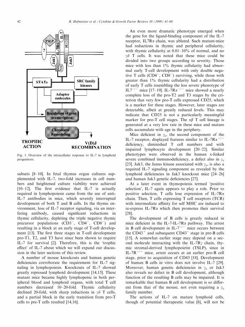
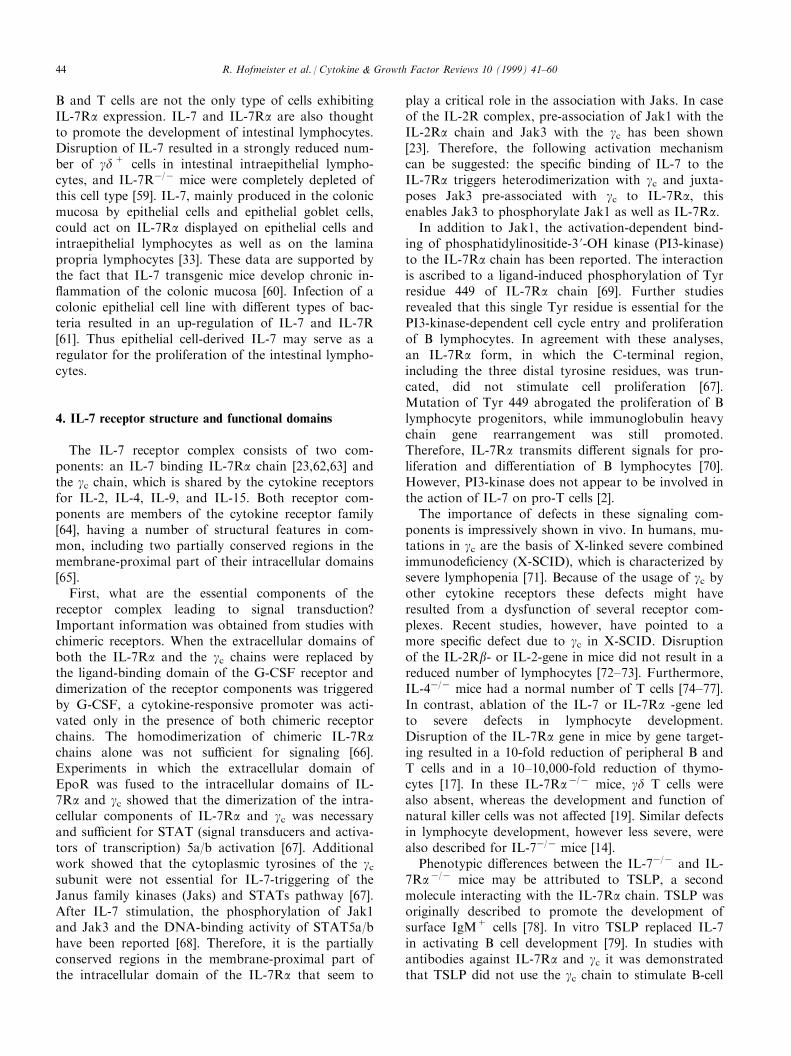
ways [3±7]. Fig. 1 illustrates the major points we willdiscuss regarding the response of cells to IL-7. Beyonddevelopment, mature lymphoid cells also respond toIL-7 [8] and this has suggested promising therapeuticpossibilities for IL-7. However, the reader should beaware that in the quest of understanding how IL-7functions in physiological processes, we have had toneglect the many promising studies of its therapeuticpotential.
2. Role of IL-7 in lymphoid development
IL-7 was originally discovered based on its activityin inducing proliferation of murine pro-B cells [9]. It isnow recognized that IL-7 plays a critical role in thedevelopment of both T and B cells in mice, and of Tcells (but not B cells) in man. Early work using recom-binant IL-7 protein revealed that this cytokine iscapable of greatly expanding lymphocyte populations,leading to profound increases in both the B and T cell
Cytokine & Growth Factor Reviews 10 (1999) 41±60
1359-6101/99/$ - see front matter Published by Elsevier Science Ltd.
PII: S1359-6101(98 )00025 -2
* Corresponding author. Tel.: 301±846±1545, fax: 301±846±6720.
E-mail address: [email protected] (S.K. Durum)
subsets [8±10]. In fetal thymus organ cultures sup-plemented with IL-7, two-fold increases in cell num-bers and heightened culture viability were achieved[10±12]. The ®rst evidence that IL-7 is actuallyrequired in lymphopoiesis came from the use of anti-IL-7 antibodies in mice, which severely interrupteddevelopment of both T and B cells. In the thymic en-vironment, loss of IL-7 receptor signaling, via an inter-fering antibody, caused signi®cant reductions inthymic cellularity, depleting the triple negative thymicprecursor populations (CD3ÿ, CD4ÿ, CD8ÿ) andresulting in a block at an early stage of T-cell develop-ment [13]. The ®rst three stages in T-cell developmentpro-T1, T2, and T3 have since been shown to requireIL-7 for survival [2]. Therefore, this is the `trophice�ect' of IL-7 about which we will expand our discus-sion in the later sections of this review.
A number of mouse knockouts and human geneticde®ciencies corroborate the requirement for IL-7 sig-naling in lymphopoiesis. Knockouts of IL-7 showedgreatly repressed lymphoid development [14,15]. Thesemutant mice became highly lymphopenic in both per-ipheral blood and lymphoid organs, with total T cellnumbers decreased 10±20-fold. Thymic cellularitydeclined 20-fold, with sharp reductions in gd T cells,and a partial block in the early transition from pro-Tcells to pre-T cells resulted [14,16].
An even more dramatic phenotype emerged whenthe gene for the ligand-binding component of the IL-7receptor, IL7Ra chain, was ablated. Such mutant-micehad reductions in thymic and peripheral cellularity,with thymic cellularity at 0.01±10% of normal, and nogd T cells. It was noted that these mice could bedivided into two groups according to severity. Thosemice with less than 1% thymic cellularity had abnor-mal early T-cell development with only double nega-tive T cells (CD4ÿ, CD8ÿ) surviving, while those withgreater than 1% thymic cellularity had a distributionof early T cells resembling the less severe phenotype ofIL7ÿ/ÿ mice [17±19]. IL-7Raÿ/ÿ mice showed a nearlycomplete loss of the pro-T2 and T3 stages by the cri-terion that very few pro-T cells expressed CD25, whichis a marker for these stages. However, later stages aredetectable, albeit at greatly reduced levels. This mayindicate that CD25 is not a particularly meaningfulmarker for pro-T cell stages. The ab T cell lineage isgenerated at a very low rate in these mice and maturecells accumulate with age in the periphery.
Mice de®cient in gc, the second component of theIL-7 receptor, displayed features similar to IL-7Raÿ/ÿ
de®ciency, diminished T cell numbers and withimpaired lymphocyte development [20±22]. Similarphenotypes were observed in the human x-linkedsevere combined immunode®ciency, a defect also in gc[23]. Jak3, the Janus kinase associated with gc, is also arequired IL-7 signaling component as revealed by thelymphoid de®ciencies in Jak3 knockout mice [24±26]and human Jak3 genetic de®ciencies [27].
At a later event in thymopoiesis termed `positiveselection', IL-7 again appears to play a role. Prior topositive selection, T cells lose expression of IL-7Rachain. Then, T cells expressing T cell receptors (TCR)with intermediate a�nity for self MHC are induced tore-express IL-7Ra which then promotes their survival[28].
The development of B cells is greatly reduced inmice de®cient in the IL-7-IL-7Ra pathway. The arrestin B cell development in IL-7ÿ/ÿ mice occurs betweenthe CD43+ and subsequent CD43ÿ stage in pre-B cells[15]. A somewhat earlier stage may depend on a sec-ond molecule interacting with the IL-7Rg chain, thy-mic stromal-derived lymphopoietin (TSLP), since inIL-7Rÿ/ÿ mice, arrest occurs at an earlier pro-B cellstage, prior to acquisition of CD43 [18]. Developmentof human B cells in vitro does not involve IL-7 [29].Moreover, human genetic de®ciencies in gc or Jak3also reveals no defect in B cell development, althoughfunction of the resulting B cells may be impaired. It isremarkable that human B cell development is so di�er-ent from that of the mouse, not even requiring a gcfamily member.
The actions of IL-7 on mature lymphoid cells,though of potential therapeutic value [8], will not be
Fig. 1. Overview of the intracellular response to IL-7 in lymphoid
progenitors.
R. Hofmeister et al. / Cytokine & Growth Factor Reviews 10 (1999) 41±6042
further discussed since this review is restricted to thephysiological actions of IL-7 which are currentlybelieved to occur primarily in lymphopoiesis.
3. Production of IL-7 and IL-7Raa expression
3.1. Production of IL-7
The thymus has been reported as the anatomical siteof the highest IL-7 production [9,30]. The thymic celltype producing IL-7 expresses MHC class II and pre-sumably corresponds to the cortical epithelial cell sub-set [16,31]. This same subset also produces stem cellfactor (SCF) which probably has an important syner-gistic action, along with IL-7, in inducing thymocyteproliferation. The bone marrow cell producing IL-7has been classi®ed as a reticular stromal cell thatexpresses VCAM-1 and, like the thymic IL-7 producer,also makes SCF [32].
Other sites of IL-7 production include the intestinalepithelium [33], where it appears to support extrathy-mic development of gd T cells [34]. Follicular dendriticcells produce IL-7, suggesting possible roles in germ-inal center formation [35]. Keratinocytes also produceIL-7 [36], presumably promoting gd T cell survival inthe skin. Cutaneous IL-7 levels are increased in psoria-tic plaques [37] and after Schistosoma mansoni infec-tion [38]. Cutaneous IL-7 may act as a trophic factorfor Sezary lymphoma cells in the skin [39]. Other neo-plastic associations with IL-7 include its production byCLL cells [40] and Burkitts's lymphoma cells [41] andits elevation in the serum of Hodgkin's patients [42].
Several transgenic mouse lines have been producedthat express IL-7 under di�erent promoters.Phenotypes range from a benign increase in T and Bcells [43] to lymphoproliferative disorders [44] particu-larly in the skin [45±47].
Regulation of the IL-7 gene is not well understood.There are no apparent upstream TATA or CAATsequences typical of inducible genes [9]. The promoterregion that regulates thymic or bone marrow ex-pression has not been studied. However, a keratinocyteline has been used to identify some upstream regulat-ory regions, including constitutive promoter and in-hibitor regions and an interferon (IFN) inducibleregion containing an interferon-g speci®c response el-ement (ISRE) [48]. Multiple start sites, multiple spliceforms, polyadenylation sites, and a 5 'UTR translationinhibitory region have been identi®ed. Expression inbone marrow was impaired in mice de®cient for thetranscription factor C/EBPb [49]. Transforming growthfactor b1 (TGFb1) was observed to downregulate IL-7production by bone marrow stroma [50].
The IL-7 protein has been placed in the hematopoie-tin family. Up to four isoforms have been described as
products of alternative splicing, and predictions oftheir structure have been generated [51,52]. The predo-minant active isoform is 25 kDa and predicted to con-tain four a helices and to be internally disulphidecrosslinked. Murine IL-7 does not have activity onhuman cells, although human IL-7 is active on mousecells. IL-7 protein associates with extracellular matrixin the thymus [53] and has been shown to bind ®bro-nectin in vitro [54].
3.2. Expression of IL-7Ra-chain
IL-7Ra is displayed on B and T cell lineages.Surface IgMÿ B-cell progenitors in the bone marrowexpress IL-7Ra on the surface. In the thymus,CD4ÿCD8ÿ double negative (DN) cells but notCD4+CD8+ double positive (DP) cells bear the IL-7Ra chain [55]. It is very likely that the IL-7Ra chaintransduces survival signals to DN cells and supportstheir expansion. The loss of IL-7Ra at the DP stage isperhaps important, in that, were these cells capable ofresponding to IL-7 it might protect cells that need tobe deleted. IL-7 might not be the only factor keepingDN cells alive. C-kit de®cient mice also showed a sig-ni®cant reduction in thymic cellularity [56]. After dis-ruption of both the gc and the c-kit genes in mice,thymic development was completely blocked, indicat-ing that both of these receptors are required to trans-duce strong survival signals [57]. Recently, twopathways for the positive selection of thymocytes,involving c-kit and the IL-7R complex, have beendescribed, a c-kit+ and a c-kitÿ pathway. DPint
TCRloc-kit+ cells begin the process of self-MHC rec-ognition and undergo maturation to CD4 and CD8single positive cells via the c-kit+ pathway. IL-7R sig-nals induced Bcl-2 expression, which was associatedwith keeping the cells alive. Cells which fail this posi-tive selection can enter the c-kitÿ salvage pathway,thereby down-regulating c-kit, IL-7R, and Bcl-2 andup-regulating CD4 and CD8 to become DPhiTCRloc-kitÿ blast cells. The majority of this cell populationdies, but a small subset of cells, which has successfullyrearranged the TCRa/b in an alternative pathway,becomes IL-7Ra-positive again. Therefore, IL-7Ra isthought to be important for the survival of the cells inboth pathways through the up-regulation of Bcl-2 [28].
In the peripheral lymphoid tissue, single-positiveCD4 or CD8 cells were the major IL-7Ra positive cells[55]. CD34+CD19ÿ bone marrow progenitor cells alsoexpressed IL-7R. These cells demonstrated markedlyenhanced lymphoid clonogenic capacity and the abilityto di�erentiate into pro-B cells in short-term culturewhereas the same cell type lacking IL-7Ra expressiondid not di�erentiate into pro-B-cells. The onset of IL-7Ra expression shortly before expression of CD19suggested the involvement of IL-7 in this process [58].
R. Hofmeister et al. / Cytokine & Growth Factor Reviews 10 (1999) 41±60 43
B and T cells are not the only type of cells exhibitingIL-7Ra expression. IL-7 and IL-7Ra are also thoughtto promote the development of intestinal lymphocytes.Disruption of IL-7 resulted in a strongly reduced num-ber of gd+ cells in intestinal intraepithelial lympho-cytes, and IL-7Rÿ/ÿ mice were completely depleted ofthis cell type [59]. IL-7, mainly produced in the colonicmucosa by epithelial cells and epithelial goblet cells,could act on IL-7Ra displayed on epithelial cells andintraepithelial lymphocytes as well as on the laminapropria lymphocytes [33]. These data are supported bythe fact that IL-7 transgenic mice develop chronic in-¯ammation of the colonic mucosa [60]. Infection of acolonic epithelial cell line with di�erent types of bac-teria resulted in an up-regulation of IL-7 and IL-7R[61]. Thus epithelial cell-derived IL-7 may serve as aregulator for the proliferation of the intestinal lympho-cytes.
4. IL-7 receptor structure and functional domains
The IL-7 receptor complex consists of two com-ponents: an IL-7 binding IL-7Ra chain [23,62,63] andthe gc chain, which is shared by the cytokine receptorsfor IL-2, IL-4, IL-9, and IL-15. Both receptor com-ponents are members of the cytokine receptor family[64], having a number of structural features in com-mon, including two partially conserved regions in themembrane-proximal part of their intracellular domains[65].
First, what are the essential components of thereceptor complex leading to signal transduction?Important information was obtained from studies withchimeric receptors. When the extracellular domains ofboth the IL-7Ra and the gc chains were replaced bythe ligand-binding domain of the G-CSF receptor anddimerization of the receptor components was triggeredby G-CSF, a cytokine-responsive promoter was acti-vated only in the presence of both chimeric receptorchains. The homodimerization of chimeric IL-7Rachains alone was not su�cient for signaling [66].Experiments in which the extracellular domain ofEpoR was fused to the intracellular domains of IL-7Ra and gc showed that the dimerization of the intra-cellular components of IL-7Ra and gc was necessaryand su�cient for STAT (signal transducers and activa-tors of transcription) 5a/b activation [67]. Additionalwork showed that the cytoplasmic tyrosines of the gcsubunit were not essential for IL-7-triggering of theJanus family kinases (Jaks) and STATs pathway [67].After IL-7 stimulation, the phosphorylation of Jak1and Jak3 and the DNA-binding activity of STAT5a/bhave been reported [68]. Therefore, it is the partiallyconserved regions in the membrane-proximal part ofthe intracellular domain of the IL-7Ra that seem to
play a critical role in the association with Jaks. In caseof the IL-2R complex, pre-association of Jak1 with theIL-2Ra chain and Jak3 with the gc has been shown[23]. Therefore, the following activation mechanismcan be suggested: the speci®c binding of IL-7 to theIL-7Ra triggers heterodimerization with gc and juxta-poses Jak3 pre-associated with gc to IL-7Ra, thisenables Jak3 to phosphorylate Jak1 as well as IL-7Ra.
In addition to Jak1, the activation-dependent bind-ing of phosphatidylinositide-3 '-OH kinase (PI3-kinase)to the IL-7Ra chain has been reported. The interactionis ascribed to a ligand-induced phosphorylation of Tyrresidue 449 of IL-7Ra chain [69]. Further studiesrevealed that this single Tyr residue is essential for thePI3-kinase-dependent cell cycle entry and proliferationof B lymphocytes. In agreement with these analyses,an IL-7Ra form, in which the C-terminal region,including the three distal tyrosine residues, was trun-cated, did not stimulate cell proliferation [67].Mutation of Tyr 449 abrogated the proliferation of Blymphocyte progenitors, while immunoglobulin heavychain gene rearrangement was still promoted.Therefore, IL-7Ra transmits di�erent signals for pro-liferation and di�erentiation of B lymphocytes [70].However, PI3-kinase does not appear to be involved inthe action of IL-7 on pro-T cells [2].
The importance of defects in these signaling com-ponents is impressively shown in vivo. In humans, mu-tations in gc are the basis of X-linked severe combinedimmunode®ciency (X-SCID), which is characterized bysevere lymphopenia [71]. Because of the usage of gc byother cytokine receptors these defects might haveresulted from a dysfunction of several receptor com-plexes. Recent studies, however, have pointed to amore speci®c defect due to gc in X-SCID. Disruptionof the IL-2Rb- or IL-2-gene in mice did not result in areduced number of lymphocytes [72±73]. Furthermore,IL-4ÿ/ÿ mice had a normal number of T cells [74±77].In contrast, ablation of the IL-7 or IL-7Ra -gene ledto severe defects in lymphocyte development.Disruption of the IL-7Ra gene in mice by gene target-ing resulted in a 10-fold reduction of peripheral B andT cells and in a 10±10,000-fold reduction of thymo-cytes [17]. In these IL-7Raÿ/ÿ mice, gd T cells werealso absent, whereas the development and function ofnatural killer cells was not a�ected [19]. Similar defectsin lymphocyte development, however less severe, werealso described for IL-7ÿ/ÿ mice [14].
Phenotypic di�erences between the IL-7ÿ/ÿ and IL-7Raÿ/ÿ mice may be attributed to TSLP, a secondmolecule interacting with the IL-7Ra chain. TSLP wasoriginally described to promote the development ofsurface IgM+ cells [78]. In vitro TSLP replaced IL-7in activating B cell development [79]. In studies withantibodies against IL-7Ra and gc it was demonstratedthat TSLP did not use the gc chain to stimulate B-cell
R. Hofmeister et al. / Cytokine & Growth Factor Reviews 10 (1999) 41±6044
proliferation. Analyses of downstream signaling eventsclearly distinguished TSLP from IL-7 in that, whileboth activated STAT5a/b isoforms, TSLP did it inde-pendently of Jak1 and Jak3 [A. Farr, University ofWashington, Seattle, USA]. This implies the existenceof a novel (and as yet unreported) component of theTSLP receptor that serves as a counterpart to gc in theIL-7 receptor. But thus far, the role of TSLP in thy-mocyte development remains unclear.
5. IL-7R signal transduction
The cytokines IL-7 and IL-2 both act on lympho-cytes and use the gc chain as part of their respectivereceptors. Though IL-7 probably shares many of itsfunctions with other cytokines like IL-2, IL-7 has aunique role in the development of the immune system,especially during the early stages of T cell develop-ment. The intracellular mechanisms mediating signal-ing for the various e�ects of IL-7 are not clearlyestablished. However, phosphorylation of proteins ontyrosine residues is an important early step in manycytokine receptor signal transduction pathways. IL-7induces the tyrosine phosphorylation of a number ofintracellular proteins in leukemic T cells [80,81], pre-Bcells [82,83], and in thymocytes [84,85]. Neither the IL-7R [62], nor the gc chain with which it heterodimerizes[23,63], have an intrinsic tyrosine kinase activity. TheIL-7-induced tyrosine phosphorylation is thereforecompletely due to the recruitment of intracellularkinases.
5.1. Src kinases
The Src family of non-receptor protein tyrosinekinases (PTK) are membrane-associated proteins. Eachmember of this family of PTKs contains a unique N-terminal domain that confers kinase-speci®c functionto each enzyme, as well as SH3 and SH2 domains, anda conserved SH1 kinase domain. The SH2 domainsinteract with phosphorylated tyrosine residues, and theSH3 domains interact with proline-rich regions.Stimulation by IL-7 activates the Src family kinasesp59fyn and p53lyn in pre B cells [81], and in myeloidcell lines [86]. In contrast to p53/56lyn, p59fyn wasfound to be associated constitutively with IL-7R inthese cells [81,86]. In mature human T cells, p56lck isactivated by IL-7, and the IL-7R was shown to bephysically associated distinctly with both p59fyn andp56lck [87]. Signaling through p59fyn is unlikely to med-iate all of the responses generated by IL-7, as mice de-®cient for p59fyn do not show the defects in T and Bcell development that are seen following the in vivoblockage of the IL-7/IL-7R pathway [13,55,88].Additionally, Lck was reported to have a critical role
in the regulation of TCR signal transduction in murinethymocytes [89]. Thymuses of mice de®cient for p56lck
expression were severely reduced in total cell numberand exhibited impaired development of the DP subset[90]. However, a recent report revealed that the recep-tor-associated p59fyn and p56lck activity induced by IL-7 does not correlate with IL-7-induced proliferation inT cells [87], thus suggesting that IL-7 may be able toutilize signaling pathway(s) other than Src signaling.The physiological signi®cance of this physical inter-action between p56lck, p59fyn, and IL-7R, as well asthe role of these IL-7-induced Src family kinases in cellgrowth, survival and/or thymocyte maturation,remains to be clearly established.
5.2. Shc-Ras-ERK kinase pathway
p52shc is tyrosine phosphorylated by IL-2 in B cellsand T cells [83,91] and has been shown to be criticallyinvolved in Ras activation by tyrosine kinase receptors[92] as well as other non-receptor tyrosine kinases [93].Studies in T cells [94] and in pre-B cells [83] failed toshow IL-7-induced tyrosine phosphorylation of Shc.This suggests that IL-7 does not induce the activationof p21ras or other intermediate elements in this path-way. It is unlikely that the lack of Shc phosphorylationis due to a defect in p56lck activation, since IL-7 wasable to induce this relevant kinase in human T cellsand thymocytes [87,95]. In contrast to IL-2, IL-7 wasunable to activate MAP/ERK kinases [94], and the ac-tivation of MAP/ERK kinase seemed not to berequired for IL-7-induced T cell proliferation [96].However, IL-7 was shown to be able to activate JNK(c-Jun N-terminal kinase) and p38 kinase during T cellproliferation [97].
Recent studies using the GTPase Rho de®cient miceshowed a dramatic defect in early T cell developmentwith a marked decrease in thymic cellularity, a promi-nent depletion of CD25+CD44+ cells and a decreasein CD25ÿCD44+ cell population [98], a phenotypesimilar to mice lacking IL-7R. Interestingly, the selec-tive apoptosis defect in Rhoÿ pro-thymocytes was res-cued by expression of a bcl-2 transgene [99]. Therefore,Rho GTPase has been suggested to have a role in deli-vering IL-7 receptor induced proliferation and cell sur-vival signals [98].
5.3. Jak-STAT pathway
Recent studies have indicated that the Jaks andtyrosine-phosphorylated transcription factors, STATs,are fundamentally important in cytokine signal trans-duction [100,101]. These signaling pathways seem to beimportant for early gene activation and the develop-ment of the immune system. Recently, it was estab-lished that the cytokine receptors are functionally
R. Hofmeister et al. / Cytokine & Growth Factor Reviews 10 (1999) 41±60 45
coupled to Jak kinases. The Jak kinases have no trans-membrane domains or SH2 and SH3 domains, buthave seven regions of sequence homology (JH1±7)domains. The C-terminal catalytic domain JH1 is theenzymatically active domain. The JH7 and JH6domains are necessary for binding to the cytokinereceptor subunit gc [102,103].
Jak3 was found to be activated by IL-7 as well asby IL-2, IL-4, IL-9 and IL-15 [104±109]. This can beexplained by the fact that Jak3 is physically associatedwith the gc chain shared by all these receptors. The im-portance of Jak3 was recently demonstrated in theautosomal recessive TÿB+ severe combined immuno-de®ciency, patients with this disorder had homozygouspoint or deletion mutations in Jak3 [27,110].Additionally, mice lacking Jak3 also displayed dra-matic immunode®ciency [24,26,111], a phenotype thatresembles the gc-de®cient mice phenotype [21], as wellas the IL-7 and IL-7 receptor knockout mice pheno-type. This strengthens the interpretation that it is thelack of IL-7 receptor signaling that is the cause of thethymic defect observed in Jak3ÿ/ÿ and gc
ÿ/ÿ mice.IL-7 induced a rapid tyrosine phosphorylation of
Jak1 concomitantly to Jak3 in murine and human Tcells [68,95]. It was demonstrated that Jak1 is associ-ated with the IL-2Rb chain [112] and with the a chainsof receptors for IL-7, IL-4, and IL-9 [113]. The mech-anisms by which IL-7 and the gc-containing cytokinesreceptors regulate their target genes via Jak3 are notyet clearly established. Jak3 is activated by IL-7 inde-pendently of Src family kinase (Lck or Fyn) activity[95]. It is proposed that the speci®c binding of IL-7 tothe IL-7Ra chain triggers heterodimerization with thegc chain and phosphorylation of the Jaks, and tyrosineresidues in the cytoplasmic tail of the receptor thusprovide docking sites for proteins with phosphotyro-sine binding SH2 domains, which in turn are also Jaksubstrates. The STAT family of transcription factors isone example of Jak3 substrates [114]. The STATsthemselves are phosphorylated, dissociate from thereceptor, form homo- or hetereo-dimers, and rapidlytranslocate to the nucleus where they can bind toDNA sequences and regulate target gene transcription.Various studies have shown that IL-7 induced thephosphorylation and activation of STAT3 [115] andSTAT5 [68,75,115,116]. In human peripheral blood Tlymphoblasts, IL-2 and IL-7 were shown to be poten-tially equivalent in their ability to induce tyrosinephosphorylation of both isoforms, STAT5a andSTAT5b, and facilitate binding of these STATs to animmobilized GAS motif [117]. STAT5a and STAT5bwere shown to bind to related but distinct dockingsites on the IL-7 receptor a chain [75]. IL-7 inducesSTAT5a/STAT5b heterodimerization, and STAT3seems to be associated constitutively with each STAT5isoform [117]. STAT1 was also shown to be activated
upon stimulation of precursor B cells by IL-7 [118].One mechanism for speci®city of STATs may be re-lated to the fact that the di�erent STATs are charac-terized by speci®c SH2 domains that recognizedi�erent phosphorylated motifs on the cytokine recep-tors. Additionally, the fact that IL-7 activates the sameSTAT proteins as IL-2 and IL-15 could be explainedby the existence of similar tyrosine phosphorylatedmotifs in the cytoplasmic domains of IL-2Rb and IL-7Ra that constitute docking sites for STAT activation[75].
The induction of both Jak1 and Jak 3 activity wasreported to correlate with the growth-promoting e�ectsof IL-7 in murine T cells [68], suggesting that this sig-nal transduction mechanism could play a key role inIL-7-induced proliferation. Accordingly, the generationof mice lacking Jak1 showed a severely impaired lym-phocytic development with a critical reduction in thy-mic cellularity [119]. Furthermore, IL-7 failed toinduce cell colony formation in Jak1ÿ/ÿ mice. Thusone explanation of the lymphopoietic de®ciencies inJak1ÿ/ÿ mice could be related to the lack of IL-7receptor function. However, a recent study, usingMOLT-4 transfectants with a mutant IL-2Rb chainlacking Jak1 association, showed that Jak1 is notrequired for activation of Jak3 and STAT5, or IL-2-stimulated cell growth signaling [113], which raises thequestion of whether IL-7 receptor signaling reallyrequires Jak1.
The role of IL-7-induced-STAT activation is not yetclear. Induction of STAT1 and STAT5 complexes wasshown to occur in cases of B cell precursor acute lym-phoblastic leukemia (BCP-ALL) that respond to IL-7in proliferation assays [118]. However, IL-7 alsoinduced STAT/DNA binding in BCP-ALL cases thatfailed to proliferate in response to IL-7, thussuggesting that the ability of IL-7 to activate the Jak/STAT pathway is not su�cient for cell growth.Additionally, disruption of both STAT5a and STAT5bin mutant mice did not a�ect thymus development orperipheral B cells [120], demonstrating that IL-7 recep-tor must use other signaling pathways independent ofSTAT5 for the survival of thymocytes and T cell devel-opment in vivo. Accordingly, it was recently reportedthat the signaling of IL-4±induced survival of resting Tcells is independent of the STAT6 pathway [121].
While Jak3 is crucial for IL-7 signaling, it seemsclear that STATs can not by themselves determine theunique action of IL-7. Other proteins that werereported to interact with Jaks including Shc, Grb2,SHP-2, Vav, IRS, PI3-kinase, STAM, Pyk2, and/orother unidenti®ed proteins that could mediate IL-7 sig-naling. STAM (signal transducing adaptor molecule) isan adaptor molecule and was initially identi®ed as atyrosine phosphorylated protein that was induced byIL-7, as well as by IL-2, IL-4, IL-3 and GM-CSF
R. Hofmeister et al. / Cytokine & Growth Factor Reviews 10 (1999) 41±6046
[122]. STAM contains an SH3 domain and an ITAM,which was originally known to interact with the Zap-70 family tyrosine kinases. STAM has been shown tobe associated with Jak3 or Jak2 [123], and to be phos-phorylated by Jak3. A deletion mutant form of STAMlacking its SH3 domain inhibits IL-2-induced prolifer-ation as well as c-myc promoter activity, suggestingthat STAM may be the component downstream ofJak3 that is involved in the regulation of c-myc geneexpression.
Pyk2, a member of focal adhesion kinase (FAK)family PTK, has been shown to physically associatewith Jak3 and to mediate IL-2-induced cell prolifer-ation without a�ecting STAT5 pathway [124], thusimplicating Pyk2 as a new component of Jak3 signal-ing.
5.4. PI3-kinase pathway
IL-7 induces the activation of PI3-kinase [116] andinositol 1,4,5 triphosphate production [80,82,84].Activation of PI3-kinase has been demonstrated in anumber of cytokine and growth factor systems. PI3-kinase activation was involved in transducing prolif-erative signals [125,126]. IL-7-induced PI3 kinase acti-vation, mediated by tyrosine phosphorylation of thePI3-kinase p85 subunit, occurs in the absence of Srcfamily kinase activity [95]. Furthermore, Jak3 is associ-ated with the p85 subunit of PI3-kinase in IL-7 stimu-lated T cells and seems to regulate PI3-kinaseactivation by mediating tyrosine phosphorylation ofthe p85 subunit [95].
The PI3-kinase pathway is not yet clearly de®ned.IL-7 stimulation of human thymocytes resulted in therapid tyrosine phosphorylation of insulin receptor sub-strate-1 (IRS-1) and IRS-2 proteins. It was shown thatthe Jak1 and Jak3 kinases were associated with bothIRS-1 and IRS-2 in thymocytes. IL-7 preferentiallyinduced the association of IRS-1 with Jak3, while thephosphorylation level of Jak3-associated IRS-2 seemedto be much higher than IRS-1 [127]. Furthermore, the160±185 kDa IRS proteins associate with the p85 regu-latory subunit of the PI3-kinase [127], thus suggestinga close regulation between PI3-kinase and IRS proteinsafter IL-7 stimulation. In addition, the serine/threoninekinase Akt encoded by the akt proto-oncogene (pro-tein kinase B) was also shown to be a downstreamsubstrate of PI3-kinase [128]. IL-2 induced membranetranslocation and activation of Akt via the PI3-kinasein T cells, and studies using a catalytically active Aktshowed promotion of cellular growth and survival[235]. However the trophic action of IL-7 on pro-Tcells could not be shown to depend on PI3-kinasebased on experiments with the inhibitor wortmannin[2].
Additionally, the adaptor protein SLP-76 should be
also considered in IL-7 signaling, as the mice lackingSLP-76 showed a profound block in thymic develop-ment and expansion of DP thymocytes, thussuggesting that SLP-76 is crucial for T cell develop-ment [129].
6. Target genes in IL-7 signaling
The complexity of signaling pathways that appearsto be triggered by the IL-7 receptor complex leads tothe next question of how these signals are integrated ingene induction, and which genes are the targets ofaction. The key regulators of intracellular events in cellfate decisions are transcription factors that directlyregulate gene expression and act as multimolecularcomplexes consisting of widely expressed and/or cell-speci®c proteins. Some of these play an architecturalrole by DNA bending or by modulating protein/pro-tein interactions or nucleosome structure. Others areDNA sequence-speci®c and bind to their recognitionsite in the promoter or enhancer regions of the targetgenes and act either as activators or repressors of genetranscription [130,131]. The expression of nuclear fac-tors, their organization to molecular complexes, andtheir functional activity is regulated at multiple levelsand may be modulated by extrinsic factors such asgrowth factors, cell-cell or cell-matrix interactions, andalso by the stage of the cell cycle. Signaling by IL-7receptor, indispensable during the early stages of T celldevelopment, may directly induce the transcription ofgenes encoding certain nuclear factors or may modu-late the functional activity of transcription factors byaltering their post-translational modi®cation. Amongthe few genes identi®ed so far, the anti-apoptotic pro-tein Bcl-2 is one of the likelier, but not exclusive, tar-gets of IL-7 regulation. The following sectionsummarizes recent data as examples to demonstratehow IL-7-mediated Bcl-2 regulation may be connectedto the action of transcription factors.
6.1. Bcl-2
The anti-apoptotic Bcl-2 protein belongs to a familyof 15 members that are important regulators of pro-grammed cell death. Based on their function these pro-teins are divided into pro-survival and pro-apoptoticmembers [132]. An important element of apoptosis in-duction is based on the regulated ratio of the proteinsof opposing functions [133]. Bcl-2 is regulated by cyto-kines, and other survival factors, at a transcriptionallevel and also by post-translational phosphorylation.The bcl-2 gene has been suggested to be controlled bythe transcription factors NFkB, AP-1, Oct-1 [134],NF-AT [135] and c-Myb [136], all known to beinvolved in controlling T cell di�erentiation and acti-
R. Hofmeister et al. / Cytokine & Growth Factor Reviews 10 (1999) 41±60 47

vation. The promoter of the pro-apoptotic bax gene[137] binds to and is directly regulated by G®-1, atranscriptional repressor restricted to the thymus,spleen and testis. Overexpression of G®-1 in IL-2-dependent T cells or in murine thymocytes was shownto repress both Bax and Bak expression without in¯u-encing Bcl-2 levels, indicating that apoptotic cell deathcould be delayed without a�ecting Bcl-2 [138].
Phosphorylation of Bcl-2 at a de®ned position(Ser70) results in activation [139], while phosphoryl-ation of other serine residues by JNK leads to inacti-vation of the protein [236]. Sustained activation ofJNK or p38 kinase was implicated in apoptosis [140],and the suppression of JNK signaling was shown to becritical for Bcl-2 induction [4,141].
One of the means by which interleukins that sharethe common gc chain could regulate apoptosis isthrough the induction of Bcl-2 [142,143].Overexpression of Bcl-2 protects against apoptosisinduced by IL-2 deprivation [144] or blockage of JNKactivation, which promotes the survival of neuronalcells during trophic factor withdrawal [145]. Recently anovel Rho/PI3K/zPKC pathway was demonstrated forIL-2-mediated Bcl-2 expression, which is regulated bythe nuclear factor of activated T cells (NFAT) [146],raising the possibility that this nuclear factor, widelyused in T lymphocyte activation, might also be regu-lated by IL-7.
6.2. NFAT/AP-1
NFAT consists of a pre-existing cytoplasmic com-ponent and inducible nuclear components composed ofAP-1 family members such as c-jun/c-fos. AP-1 andNFAT factors collaborate by binding to calcium/PKC-responsive composite enhancer binding sites [147]. Thenuclear import of cytoplasmic NFAT is regulated viacytoplasmic dephosphorylation by the calcium-depen-dent serine/threonine phosphatase calcineurin [148]and by rapid nuclear rephosphorylation and shuttlingto the cytoplasm [149,150]. Bcl-2 forms a tight complexwith calcineurin via the BH4 domain, a characteristicdomain of the anti-apoptotic family members[151,152]. This interaction inhibits the phosphoryl-ation-dependent translocation of NFAT4/NFATx tothe nucleus but retains the enzymatic activity of thephosphatase. The pro-apoptotic Bax protein interfereswith the calcineurin/Bcl-2 interaction [152]. TherebyNFAT activity can be negatively regulated by Bcl-2,while Bax has an opposing e�ect [152,153]. The BH4domain of Bcl-2 also interacts with Raf-1 and directsthe protein kinase to intracellular membranes [154].This results in enhanced protection against apoptosisand re¯ects the connection between the Ras/Raf path-way and the Bcl-2-mediated regulation of cell death.
IL-2 activates AP-1 by the tyrosine kinase-induced
Ras pathway via the common gc-induced activation ofthe protein kinase Jak3 [146]. Although the role of thispathway in IL-7-mediated signaling has not beendemonstrated, both AP-1 and NFAT are constitutivelynuclear in thymocytes responding to intrathymic sig-nals [155]. DNA binding to and transactivation byAP-1 was shown in fetal thymus at day 15 of embryo-nic life [156]. The NFAT4/NFATx member of theNFAT family is expressed predominantly in immaturethymocytes [157], and it is regulated di�erently fromthe other NFAT proteins in that its nuclear accumu-lation is opposed by JNK, casein kinase-1 andMEKK1 [158,159].
The components of AP-1, especially c-Jun, wereimplicated in those death processes that required denovo protein synthesis [160], reviewed in [161]. Fos in-duction in apoptotic cells is presumably part of a stressresponse that is compensated by rapid degradation ofthe protein controlled by the ubiquitin-proteasome sys-tem and regulated by Bcl-2 through c-Fos stabilization[162]. The transcriptional activation of c-Fos wasshown to require STAT5 [163] which is activated byIL-7 through heterodimerization of STAT5 isoforms[117]. In IL-7-dependent bone marrow cultures, theregulatory role of c-Fos/c-Jun, induced by IL-7 inearly B cells, was demonstrated [164,165]. IL-7enhanced IL-2 mRNA accumulation by increasing theDNA binding activity of both NFAT and AP-1 [166],suggesting that IL-7-mediated signaling may involvethe activation of the c-Fos/c-Jun proteins. The di�er-ential role of IL-4 and IL-7 was shown in transformedpre-B cells where IL-7 but not IL-4 was able to delayapoptosis and the reduction of c-myc expression byupregulating the anti-apoptotic proteins, Bcl-2 andBcl-XL, in the absence of v-Abl kinase activity [167].Recently IL-4 was shown to inhibit apoptosis by theinhibition of JNK without any e�ect on Bcl-2, Bcl-Xor Bax levels, and PI3-kinase/Akt activation wasdemonstrated in this pathway [168]. In contrast to IL-2, which induced Bcl-2 and AP-1/NFAT activity, IL-4downregulated Bcl-2 and inhibited the DNA-bindingactivity of AP-1 and NFAT in the same cells. Theseexamples demonstrate how distinct gc-receptors,expressed on the same cell, can deliver survival/prolif-erative signals through separate pathways [146].
6.3. c-Myb
Bcl-2 and c-Myb are coexpressed in DN thymocytes.Recent studies implicated the importance of c-Myb, atranscription factor essential for the maintenance ofimmature hematopoietic cells, in the regulation of thesurvival of thymoma and IL-2- dependent T cells viabcl-2 gene induction [136,169]. Induced expression ofdominant interfering c-Myb mutants caused downregu-lation of Bcl-2 via trans-activation of the bcl-2 promo-
R. Hofmeister et al. / Cytokine & Growth Factor Reviews 10 (1999) 41±6048
ter, while, in another system, the c-Myb-speci®c trans-activation of the bax gene was also postulated [170]. Ingood correlation with the expression pattern of c-Mybin fetal thymocytes and the strong requirement of IL-7for the development of gd T cells, an intact c-Mybbinding site was required for the activation of V(D)Jrecombination in both the TCRg and TCRd enhancers[171,172]. The possibility that IL-7 directly regulates c-Myb remains to be elucidated.
6.4. c-myc
The involvement of di�erent signaling pathways[173±175] and the role of gc-associated Jak3 weredemonstrated in the induction of IL-2-mediated c-mycgene expression [176]. Both IL-2 and IL-7 induced c-myc expression in lymphokine-dependent T cells [94].IL-7 regulated transcription of the c-myc gene in pre-Band in T cell lines and N-Myc in pre-B, but not inmature B or T cells [177,178]. IL-7 mediated signalingof pre-B cells required the B lineage-speci®c pax-5gene, encoding the transcription factor BSAP, for N-myc expression [179]. IL-7 was also shown to promoteneurite outgrowth and induce rapid c-myc expressionin neurons [180].
In addition to bcl-2, IL-7 induced genes identi®ed todate include the nuclear factors c-jun/c-fos and c-myc,which are regarded as ubiquitous regulators of cellgrowth, di�erentiation, and programmed cell death,the function of which may be connected to bcl-2 regu-lation. Since most of the information on the aboveoutlined mechanisms derives from data obtained fromIL-2 or IL-4 signaling, the question as to whether IL-7utilizes a unique or a general combination of thesepathways to exert its survival and/or proliferative func-tion remains open.
7. The trophic action of IL-7
Engagement of the IL-7 receptor by its ligand in-itiates a cascade of signaling events, ultimately promot-ing the survival of lymphoid cells. This trophic e�ectof IL-7 is well-described in the development of early Tcells. Thymic precursors, in particular, require IL-7 forsurvival, as shown by the signi®cant repression of Tcell development in the IL-7ÿ/ÿ and IL-7Raÿ/ÿ
knockout mice [14,17,19]. The mechanism by whichIL-7 maintains cell viability is not clear, but in recentyears signi®cant strides have been made in elucidatingpotential mechanisms. In itself, cell death is a compli-cated process, a full description being beyond thescope of this review. The most physiologically relevantform of death is that of apoptosis or programmed celldeath. This is an ordered multi-step process by whicheither engagement of a death receptor by its ligand
(i.e., Fas, TNF) or loss of an obligate growth factor(i.e., cytokines, hormones) is the trigger that initiates acomplex cascade of events requiring modulators (i.e.,kinases, transcription factors, adaptor molecules) ande�ectors (i.e., cysteine proteases or caspases) ending inthe measurable manifestation of apoptotic death (i.e.,protein fragmentation, DNA fragmention, membraneblebbing) [181,182]. That nuclear events are not necess-arily required for apoptosis to occur has been demon-strated with enucleated cytoplasts induced to undergodeath via the Fas pathway or by granzyme-mediatedcytotoxicity [183]. Though not completely ruling outnuclear contributions in all forms of apoptotic death,such work has suggested that cytoplasmic regulatorswith multiple intracellular targets could be key me-diators in the induction of apoptosis [184]. In particu-lar, mitochondria and mitochondrial products (i.e.,cytochrome c) have cardinal roles in apoptosis, withdisruption of mitochondrial structure and functionbeing irreversible events preceding cell death [185,186].Though it remains to be irrevocably proven that IL-7exerts its trophic e�ects by inhibiting apoptosis, thismechanism is the most likely possibility and the onethat will be addressed in subsequent discussion.
7.1. IL-7 withdrawal leads to apoptosis
Is the essential function of IL-7 during lymphoiddevelopment to promote cell division, di�erentiationor to support survival? Studies evaluating the func-tional competence of T cells from IL-7Raÿ/ÿ micerevealed that such T cells failed to proliferate in re-sponse to alloantigen or phorbal myristate acetate andionomycin, and that indeed the majority of these cellsinstead underwent programmed cell death or apoptosis[187]. In fact, it appeared that a large fraction of thetriple negative early thymocytes from IL-7ÿ/ÿ or gc
ÿ/ÿ
mice bound annexin V, indicating that without IL-7signaling apoptosis was occurring at a higher rate[22,188]. When apoptosis was blocked, such as by thetargeted disruption of the p53 tumor suppressor genein Rag-1ÿ/ÿ mice, early T cells survived much longereven without a functional TCR [189]. Therefore, it ismore plausible, and likely probable, that a key func-tion of IL-7 is to prevent thymic precursors fromundergoing apoptosis, promoting initial expansion asthese pass through the ®rst stages of development.
That IL-7 stimulates cell survival rather than pro-liferation is not without precedent among cytokines.For example, insulin-like growth factor I promotessurvival and not proliferation of glial cells [190], andmore recently erythropoietin was shown to provide asupportive function for red blood cell developmentrather than induce di�erentiation [191]. Cytokineswhich share the gc chain in their receptors, such as IL-2 or IL-4, are able to enhance survival under certain
R. Hofmeister et al. / Cytokine & Growth Factor Reviews 10 (1999) 41±60 49
conditions (i.e., g-irradiation) rather than promote celldivision [142,192]. IL-7 may be maintaining the viabi-lity of cells by repressing a `death-inducing' factorand/or activating a `life-promoting' factor. As evidencefor the latter, IL-7, as well as other gc cytokines, canrescue activated T cells from apoptosis due to IL-2withdrawal or dexamethasone-induction and concomi-tantly raise the levels of the anti-apoptotic proteins,Bcl-2 and Bcl-XL, suggesting this as a possible mech-anism by which cell survival is achieved [143,193].
7.2. IL-7 and the anti-apoptotic proteins, Bcl-2 and Bcl-XL
The Bcl-2 family of related proteins are attractivecandidates for agents of IL-7-mediated cell survival.These proteins share multiple homology domains andfunction as either anti-apoptotic members (i.e., Bcl-2,Bcl-XL) or pro-apoptotic members (i.e., Bax, Bak,Bad, Bid), having distinct and varied functional ca-pacities [194]. The founding member, Bcl-2, is knownto be a powerful inhibitor of apoptosis, maintainingcellular viability in the face of many cell death triggers[195]. In particular, Bcl-2 can act on the mitochondria,stabilizing membrane integrity, preventing the loss ofpermeability, thereby inhibiting the release of factorslike cytochrome c [186,196,197].
Expression of Bcl-2 is found in lymphocyte progeni-tor cells as wells as mature T and B cells. During Tcell development, Bcl-2 is expressed at a high level inDN cells, it declines in DP cells, then is reexpressed inthe SP populations [198,199]. This biphasic expressionof Bcl-2, in early precursors and then in later maturecells, coincides with IL-7Ra expression, suggesting thatBcl-2 could mediate IL-7 receptor e�ects on T cellmaturation and survival. In Bcl-2 de®cient mice, a gra-dual loss both of thymocytes and mature T cells isseen after the second postnatal week, indicating thatBcl-2 protects mature cells, as well as thymocyteundergoing selection, from apoptosis [200].
IL-7 sustained the expression of Bcl-2 in pro-T cells[2]. Studies in IL-7ÿ/ÿ mice revealed that Bcl-2 ex-pression became dependent upon IL-7 during the earlydevelopmental stage at which a thymocyte commits tothe T cell lineage [188]. The severe lymphopenia andloss of T cell function seen in IL-7Rÿ/ÿmice was sub-stantially abrogated in mice over-expressing Bcl-2,resulting in 4- to 10-fold increases in total T cell num-bers [201,202]. Likewise, in gc-de®cient mice, T cellnumbers were restored with a bcl-2 transgene [203],reinforcing the concepts that survival signals for thy-mic precursors are mediated through the IL-7R andthat Bcl-2 can in part compensate for the loss of suchsignaling by inhibiting apoptosis.
However, Bcl-2 may not be the sole agent of IL-7-mediated survival. The experiments using Bcl-2 trans-
genes cannot be taken as conclusive proof that Bcl-2replaces IL-7. The described increases in cell numberscould result, for example, from increases in ab T cellsrather than expanded progenitor populations [22].Moreover, the B cell and gd lineages were not restoredby over-expression of Bcl-2 in IL-7R de®cient animals[202,203]. In another study with gc-de®cient mice, abcl-2 transgene could not completely rescue thymic cel-lularity [204], and in another, only a two-fold increasewas observed comparing Bcl-2+gc
ÿ to Bcl-2ÿgcÿ mice
[22]. In addition, c-kitÿgcÿ mice with or without intro-
duction of the Bcl-2 transgene were all devoid of thy-mocytes [22]. In Bcl-2-de®cient mice, unlike IL7Rÿ/ÿ
mice, distinct phenotypes emerged with early T celldevelopment occurring in the absence of Bcl-2 ex-pression [1,200]. Therefore, the possibility arises thatBcl-2 is not the only survival factor induced by theanti-apoptotic function of IL-7. It has been noted thatIL-7 can promote the survival of Bcl-2-de®cientmature T cells, suggesting that aspects of the anti-apoptotic functions of IL-7 are independent of Bcl-2[205].
If not Bcl-2, then what other anti-apoptotic mechan-ism is induced by IL-7? Within the Bcl-2 family of pro-teins other members, such as Bcl-XL, have been shownto prevent apoptosis during growth factor withdrawal.In activated T cells starved of IL-2, the levels of Bcl-XL dropped just prior to the onset of apoptosis,suggesting that the presence of Bcl-XL prevented celldeath [206]. Recently, IL-7 was shown to upregulateBcl-XL and so rescue activated, primary human T cellsfrom apoptosis induced by IL-2 withdrawal [207]. Buttriple negative thymocytes express only low levels ofBcl-XL, whereas DP cells express high levels [208], apattern that does not coincide with the developmentalstages when IL-7 expression is critical for maintainingsurvival. Bcl-XL may be a signi®cant survival factor atthe later T cell stages, but it seems not to have as criti-cal a role during early thymocyte development.Therefore, Bcl-XL is not a likely candidate for theanti-apoptotic activity of IL-7 in thymic precursors.
7.3. IL-7 and pro-apoptotic proteins
Apoptosis induced by loss of IL-7 signal transduc-tion may be in part due to the action of a death pro-moting factor. Death resulting from IL-7 withdrawalin pro-T cells does not seem to rely on either the p53pathway or the Fas/Fas ligand pathway, since blockingof these death e�ectors still leads to apoptotic death[2]. But in such thymocytes, IL-7 withdrawal did a�ectthe levels of the family of Bcl-2 proteins, suggestingthat a balance of anti-apoptotic against pro-apoptoticproteins is an important consideration [2,209]. It ispossible that another mechanism, through which IL-7protects from death, is the inhibition of a pro-apopto-
R. Hofmeister et al. / Cytokine & Growth Factor Reviews 10 (1999) 41±6050

tic protein like Bid, Bad or Bax. Recent work hasrevealed much of the regulatory mechanisms leading tocell death by way of these proteins.
Full-length Bid is a cytosolic protein that is cleavedby caspase-8 upon engagement of the Fas death path-way. The truncated form of Bid translocates to themitochondria, precipitating the demise of this organelleand releasing cytochrome c which triggers a host ofdestructive responses contributing to cell death[210,211].
A di�erent mechanism regulates Bad, whereby acti-vation of PI3-kinase (possibly induced by IL-7) [128]leads to the triggering of Akt, a serine-threonine kinasewhich phosphorylates Bad, promoting survival. Thephosphorylated form of Bad is sequestered by the tauform of 14-3-3 proteins, suppressing its apoptotic ac-tivity [212,213]. However, Bad was not detectable byintracellular staining in pro-T cells, lending no supportto a role in death from IL-7 withdrawal [2].
Bax is an attractive candidate for IL-7 regulation,since in Bax-de®cient mice lymphoid hyperplasia wasdescribed with total thymocyte numbers increasingalmost two-fold over normal controls [214]. Moreover,thymic overexpression of a bax transgene led to a sub-stantial loss of mature T cells [209]. Although theexact mechanism by which Bax induces cell death isnot known, several key ®ndings have been recentlypresented. Bax is itself primarily a cytosolic proteinuntil apoptosis is induced, there upon it translocates tothe mitochondria [215]. What targets this translocationis unclear. Recent studies suggest various mechanismssuch as homodimerizing of the Bax protein [216], clea-vage of Bax by calpains [217] or derepression of atransmembrane anchoring domain in the Bax protein[218]. Once inserted into the mitochondrial membrane,Bax initiates a cascade of death promoting eventswhich include the release of cytochrome c, triggeringthe activation of degradative caspases [219]. Bcl-2 hasbeen shown to interfere with the Bax-induced releaseof cytochrome c [220], suggesting that IL-7 may func-tion at various levels regulating the activities of anti-apoptotic and pro-apoptotic factors.
8. Role of IL-7 in V(D)J recombination
Mice that have a targeted deletion of receptor com-ponents that are required for IL-7 signal transductionare severely de®cient in T and B lymphocytes, as men-tioned above. Since survival is necessary for V(D)Jrecombination and V(D)J recombination is necessaryfor survival, it has been di�cult to determine what thenature of the immunode®ciency in these animals is.Several experimental systems in mice and man showthat IL-7 can promote V(D)J recombination in vitro.Studies in knockout mice that lack part of the IL-7
signal transduction pathway reveal that IL-7 isrequired for V(D)J recombination for at least some ofthe immune receptor loci. Finally, we will discussrecent work about the mechanism of IL-7 action oncontrol of locus accessibility for V(D)J recombination.
8.1. E�ect of IL-7 on V(D)J recombination in vitro
IL-7 can act as a co-factor for V(D)J recombinationin vitro in murine lymphoid precursors. Fetal thymo-cytes that lack IL-7 gene expression do not expressRag genes in vitro and do not undergo gene rearrange-ment of the TCRb locus unless recombinant exogenousIL-7 is added back to the culture [3]. Fetal murineliver cell cultures express speci®c transcripts of re-arranged TCRg chains after culture with recombinantIL-7 [221,222]. Only MHC class II epithelial cells thatproduce IL-7 can induce TCRb and TCRd rearrange-ments in uncommitted fetal liver cell progenitorswhereas thymic stromal cells derived from mice with atargeted deletion of the IL-7 gene fail to do so [31].Murine stem cells derived from the bone marrow canperform full V-J joining of the TCRg chain in the pre-sence of recombinant IL-7 [223]. Furthermore, IL-7and IL-7 receptor gene expression brie¯y precedes re-arrangement of the Ig heavy and k light loci, as wellas TCR g and d loci in embryonic stem cell cultures[224]. Thus a number of experimental systems in vitrosuggest a role for IL-7 in the control of V(D)J recom-bination.
8.2. Suppression of V(D)J recombination in mice thatlack components of the IL-7 signal transductionpathway
In vivo observations in knockout mice imply a rolefor IL-7 in control of locus accessibility for V(D)Jrecombination. Mice that have a targeted deletion ofthe IL-7Ra chain show an impaired V(D)J rearrange-ment of the Ig heavy chain locus. Recombination ofvariable gene segments is progressively suppressed thefurther their location is upstream of the DJ-constantregion [6]. Mice lacking the IL-7Ra chain show also asevere reduction of gene rearrangement of the TCRglocus [1,4]. In addition, mice lacking other componentsof the IL-7 signal transduction pathway, such as the gcchain or Jak3, also show a reduction of recombinationat the TCRg cluster1 [7,225]. This suppression of TCRg chain rearrangement cannot be explained by the lackof gd T cells reported in these mice, since TCRg chainrearrangement is not speci®c for the gd lineage andoccurs also in ab T cells and precursor cells. On theother hand, a defect in VJ recombination at the TCRg locus is not su�cient to explain why gd T cells fail tothrive, since a gd transgene cannot fully reconstitutethe gd T cell compartment [225]. Furthermore, g chain
R. Hofmeister et al. / Cytokine & Growth Factor Reviews 10 (1999) 41±60 51
rearrangement that is detectable in fetal thymus tissue(in contrast to adult animals) still does not result inany detectable gd T cell compartment. Thus, IL-7 mustprovide a signal for gd T cells in addition to VJ recom-bination. Rearrangement of the other TCR loci (a, band d ) can be detected (by sensitive PCR methods) inthe absence of the IL-7 signal [1,4]. However, initiationof Rag-mediated cleavage appears to also be sup-pressed at the TCRb chain locus (Durum, Schlisseland Muegge unpublished) suggesting a defect inrecombination at several loci, with the most dramaticsuppression for the g locus. Human SCID (based onmutations in gc) is reported to show an arrest at thestage of V to DJ joining in the TCRb locus [226].Thus in vivo, a variety of studies show that IL-7 con-trols recombination of many of the immune receptorgenes.
8.3. Molecular mechanism of IL-7 action on V(D)Jrecombination
The e�ect of IL-7 on recombination may be partiallyexplained by regulation of rag gene expression.Addition of recombinant IL-7 to fetal liver or thymiccell cultures promotes rag gene expression [3,222].Treatment of mice with speci®c antibodies against theIL-7 receptor results in a suppression of rag expressionin the germinal centers of these animals [227].Comparison of rag gene expression in thymocytesfrom IL-7Raÿ/ÿ mice with wild type controls revealeda greatly reduced expression of rag-1 and rag-2 genes[228]. This reduced rag gene expression level couldexplain a lower rate of cleavage initiation at the TCRblocus in these mice (as mentioned above), but it couldnot explain the dramatic reduction in recombinationspeci®cally at the TCRg locus, suggesting in addition,a locus speci®c control conducted by IL-7.Furthermore, pre-B cells in IL-7Raÿ/ÿ mice appear toexpress normal levels of rag mRNA, but showimpaired gene rearrangement of distal heavy chain seg-ments [6]. Thus, IL-7 may also control accessibility ofa speci®c locus for V(D)J recombination. Sterile tran-scripts of the immune receptor gene segments precedeV(D)J recombination and thus can serve as an indi-cator for `open' accessible chromatin. IL-7Raÿ/ÿ miceshow indeed a suppression of sterile transcripts of theregions that are most severely suppressed for recombi-nation, that is the distal Ig heavy chain segments [6]and the TCRg chain variable and constant regions [7].What is the mechanism of repressed locus accessibilityin the absence of IL-7? In the case of the Ig heavychain, the transcription factor Pax5 is thought to beresponsible: pax5ÿ/ÿ mice also show a reduction in Vto DJ joining of the Ig heavy chain [229] and pax5mRNA levels are reduced in IL-7Raÿ/ÿ pre-B cells [6].However, pro-T cells have normal pax5 mRNA levels
in the absence of the IL-7 signal and show no di�er-ence in nuclear factors binding to known enhancer el-ements of the TCRg gene [7]. Instead the TCRg locusappears to be heavily methylated in thymocytes of IL-7Raÿ/ÿ and gc
ÿ/ÿ mice. Since methylated recombina-tion substrates are unable to perform V(D)J recombi-nation in lymphoid precursor cells [230], methylationof the TCRg locus could be responsible for suppres-sion of locus accessibility. MecP2 can bind to methyl-ated DNA and via association with histonedeacetylases render histones deacetylated, thought torepresent a repressive form of chromatin [231].Trichostatin A, a speci®c inhibitor of histone deacety-lases, can enhance histone acetylation and can over-come the block caused by methylation. Trichostatin Acan also circumvent the need for the IL-7 signal andcan promote recombination at the TCRg locus in lym-phoid precursor cells from IL-7Raÿ/ÿ mice [7]. ThusIL-7 appears to induce demethylation of the TCRglocus and thus, via subsequent histone acetylation,open the chromatin for Rag-mediated cleavage. Asimilar mechanism involving the transcription factorPax5 could be responsible for regulation of distal IgHchain segments. The cis-acting elements and transact-ing factors controlling TCRg recombination are cur-rently not known.
9. Conclusions and questions
Lessons from knockout mice and human mutationsclearly pointed to IL-7 as contributing somethingessential during T cell development. Several processes,including the trophic and V(D)J e�ects, have beenimplicated as biological endpoints of the action of IL-7. However in this section we will raise some of themany questions remaining at the level of cell biologyas to how IL-7 achieves these e�ects.
9.1. Trophic e�ects
Unravelling the trophic action could have impli-cations for the mechanism of action of all trophic fac-tors. Many of the cells in the body depend on trophicsignals from their environment, and little is understoodof their mechanism. The nature of the death processthat follows trophic withdrawal has some hallmarks ofapoptosis, but other pathways may also be involved,and this key sequence of events remains to be de®ned.Moreover, the trophic process is subverted in carcino-genesis, since metastatic cells are able to survive inmicroenvironments that lack their normal trophic sig-nals, which underscores a need to understand this pro-cess. Thus we need to determine how IL-7 fully exertsits trophic e�ects. Does IL-7 repress death signals inways unregulated to Bcl-2 induction? How does cell
R. Hofmeister et al. / Cytokine & Growth Factor Reviews 10 (1999) 41±6052
death induced by IL-7 withdrawal relate to apoptoticdeath triggered by TNF or Fas? To what extent doesgene induction participate in this trophic action, andwhat genes are they? Do IL-7 receptor signals co-operate with lymphocyte lineage-speci®c transcriptionfactors?
9.2. VDJ e�ects
Having concluded that IL-7 in¯uenced chromatinaccessibility of the TCRg locus o�ers a model of locusactivation. Understanding how IL-7 receptor signalsalter the chromatin around a speci®c gene should shedlight on this basic question in di�erentiation of cellsregarding how accessibility of di�erent loci (to tran-scription factors for example) is regulated. IL-7 alsoserves as a precedent for an extrinsic signal that ren-ders a particular locus amenable to V(D)J rearrange-ment, and there may be other cytokines that controlother loci. What type of DNA binding proteins areinvolved in opening chromatin?
9.3. Is IL-7 signaling really unique?
While IL-7's trophic action is probably not unique,the V(D)J signal, one is led to believe, ought to beunique. An important biological issue is whether IL-7really delivers unique signals to cells, or whether allreceptors that incorporate gc (IL-2, 4, 7, 9 and 15) deli-ver identical signals. It may seem implausible thatnature would retain ®ve di�erent ligand-receptor sys-tems that all do the same thing. Moreover, how couldthe knockout of IL-7Ra have a unique phenotype if all®ve receptors signaled identically? However, the IL-7system could be comparable to that of erythropoietin.Clearly erythropoiesis requires erythropoietin receptor.Nevertheless, the intracellular domain of the prolactin[232], G-CSF, thrombopoietin or growth hormonereceptors [191], all members of the same receptorfamily, completely substitute for the intracellulardomain of the erythropoietin receptor. Thus theuniqueness of the erythropoietin system lies, not inreceptor signaling, but in stringent expression of onlyone type of receptor on erythroid progenitors. Byextension to the IL-7 system, it is possible that thepro-T cell only expresses the IL-7 receptor (not the IL-2, 4, 9 and 15 receptors), or that the thymic microen-vironment only produces IL-7 and not the otherligands. This would result in a stringent requirementfor IL-7, even if all the gc receptors had identical signaltransduction pathways. In fact, the b chain of the IL-2receptor is switched o� at the pro-T2 stage [233],whereas, if a full IL-2 receptor were expressed, and ifIL-2 were expressed in the right place in the thymus,perhaps IL-2 would replace IL-7.
On the other hand, it has long been known that
IL-2 and IL-4 activate at least some di�erent pathwayswithin the same cell, as well as some of the same ones[234] and IL-7 receptor has also been shown to elicit,some cellular responses distinct from IL-2 receptor(noted above). It is clear that the other receptor chainsthat couple with gc, such as IL-2Rb, can impart aspeci®c element to signaling, partly by bringing indi�erent substrates for Jak3 into the crosslinkedreceptor complex.
It would not be surprising if the anti-apoptoticmechanisms of IL-7 will be the same as for IL-2 andIL-4. However, the stringent requirement for IL-7 inrearrangement of the TCRg locus in pro-T cells leadsus to suspect there is a di�erence among these recep-tors in impinging on that locus. We have found thatIL-7R signals control chromatin remodeling of theTCRg locus through DNA methylation and acety-lation of histones and it would be surprising if IL-2 orIL-4 induce the same chromatin remodeling of theTCRg locus.
What domains of the IL-7Ra chain control thedi�erent signal pathways? Are di�erent domains oper-ative in T versus B cell development, trophic versusV(D)J e�ects? What proteins interact with the IL-7receptor and initiate the signaling cascades?
References
[1] Candeias S, Peschon JJ, Muegge K, Durum SK. Defective T-
cell receptor g gene rearrangement in interleukin-7 receptor
knockout mice. Immunol Lett 1997;57:9±14.
[2] Kim K, Lee CK, Sayers TJ, Muegge K, Durum SK. The
trophic action of IL-7 on pro-T cells: inhibition of apoptosis
of pro-T1, -T2, -T3 cells correlates with Bcl-2 and Bax levels
and is independent of Fas and p53 pathways. J Immunol
1998;160:5735±41.
[3] Muegge K, Vila MP, Durum SK. Interleukin-7: a cofactor for
V(D)J rearrangement of the T cell receptor b gene. Science
1993;261:93±5.
[4] Maki K, Sunaga S, Ikuta K. The V-J recombination of T cell
receptor-g genes is blocked in interleukin-7 receptor-de®cient
mice. J Exp Med 1996;184:2423±7.
[5] Maki K, Sunaga S, Komagata Y, Kodaira Y, Mabuchi A,
Karasuyama H, Yokomuro K, Miyazaki JI, Ikuta K.
Interleukin 7 receptor-de®cient mice lack gd T cells. Proc Natl
Acad Sci USA 1996;93:7172±7.
[6] Corcoran AE, Riddell A, Krooshoop D, Venkitaraman AR.
Impaired immunoglobulin gene rearrangement in mice lacking
the IL-7 receptor. Nature 1998;391:904±7.
[7] Durum SK, Lee CK, Geiman TM, Murphy WJ, Muegge K.
CD16 cross-linking blocks rearrangement of the TCR b locus
and development of a b T cells and induces development of
NK cells from thymic progenitors. J Immunol 1998;161:3325±
9.
[8] Komschlies KL, Gregorio TA, Gruys ME, Back TC, Faltynek
CR, Wiltrout RH. Administration of recombinant human IL-7
to mice alters the composition of B-lineage cells and T cell
subsets, enhances T cell function, induces regression of estab-
lished metastases. J Immunol 1994;152:5776±84.
R. Hofmeister et al. / Cytokine & Growth Factor Reviews 10 (1999) 41±60 53
[9] Namen AE, Lupton S, Hjerrild K, Wignall J, Mochizuki DY,
Schmierer A, Mosley B, March CJ, Urdal D, Gillis S.
Stimulation of B-cell progenitors by cloned murine interleukin-
7. Nature 1988;333:571±3.
[10] Watson JD, Morrissey PJ, Namen AE, Conlon PJ, Widmer
MB. E�ect of IL-7 on the growth of fetal thymocytes in cul-
ture. J Immunol 1989;143:1215±22.
[11] Suda T, Zlotnik A. IL-7 maintains the T cell precursor poten-
tial of CD3±CD4±CD8± thymocytes. J Immunol
1991;146:3068±73.
[12] Plum J, De-Smedt M, Leclercq G. Exogenous IL-7 promotes
the growth of CD3±CD4±CD8±CD44+CD252 precursor cells
and blocks the di�erentiation pathway of TCR-a b cells in
fetal thymus organ culture. J Immunol 1993;150:2706±16.
[13] Grabstein KH, Waldschmidt TJ, Finkelman FD, Hess BW,
Alpert AR, Boiani NE, Namen AE, Morrissey PJ. Inhibition
of murine B and T lymphopoiesis in vivo by an anti-interleu-
kin 7 monoclonal antibody. J Exp Med 1993;178:257±64.
[14] von-Freeden-Je�ry U, Vieira P, Lucian LA, McNeil T,
Burdach SE, Murray R. Lymphopenia in interleukin (IL)-7
gene-deleted mice identi®es IL-7 as a nonredundant cytokine.
J Exp Med 1995;181:1519±26.
[15] von Freeden-Je�ry U, Moore TA, Zlotnik A, Murray R. IL-7
knockout mice and the generation of lymphocytes. In: Durum
SK, Muegge K, editors. Cytokine knockouts. Totowa, NJ:
Humana Press Inc, 1998. p. 21±36.
[16] Moore TA, von-Freeden-Je�ry U, Murray R, Zlotnik A.
Inhibition of g d T cell development and early thymocyte
maturation in IL-7 ÿ/ÿ mice. J Immunol 1996;157:2366±73.
[17] Peschon JJ, Morrissey PJ, Grabstein KH, Ramsdell FJ,
Maraskovsky E, Gliniak BC, Park LS, Ziegler SF, Williams
DE, Ware CB, et al. Early lymphocyte expansion is severely
impaired in interleukin 7 receptor-de®cient mice. J Exp Med
1994;180:1955±60.
[18] Peschon JJ, Gliniak BC, Morrissey PJ, Maraskovsky E.
Lymphoid development and function in IL-7R-de®cient mice.
In: Durum SK, Muegge K, editors. Cytokine knockouts.
Totowa, NJ: Humana Press Inc, 1998. p. 37±52.
[19] He YW, Malek TR. Interleukin-7 receptor a is essential for
the development of g d +T cells, but not natural killer cells.
J Exp Med 1996;184:289±93.
[20] DiSanto JP, Muller W, Guy-Grand D, Fischer A, Rajewsky
K. Lymphoid development in mice with a targeted deletion of
the interleukin 2 receptor g chain. Proc Natl Acad Sci USA
1995;92:377±81.
[21] Cao X, Shores EW, Hu, Li J, Anver MR, Kelsall BL, Russell
SM, Drago J, Noguchi M, Grinberg A, Bloom ET, et al.
Defective lymphoid development in mice lacking expression of
the common cytokine receptor g chain. Immunity 1995;2:223±
38.
[22] Di Santo JP, Rodewald HR. In vivo roles of receptor tyrosine
kinases and cytokine receptors in early thymocyte develop-
ment. Curr Opin Immunol 1998;10:196±207.
[23] Noguchi M, Nakamura Y, Russell SM, Ziegler SF, Tsang M,
Cao X, Leonard WJ. Interleukin-2 receptor g chain: a func-
tional component of the interleukin-7 receptor. Science
1993;262:1877±80.
[24] Nosaka T, van-Deursen JM, Tripp RA, Thierfelder WE,
Witthuhn BA, McMickle AP, Doherty PC, Grosveld GC, Ihle
JN. Defective lymphoid development in mice lacking Jak3.
Science 1995;270:800±2 published erratum appears in Science
1996 Jan 5, 271(5245):17.
[25] Park SY, Saijo K, Takahashi T, Osawa M, Arase H,
Hirayama N, Miyake K, Nakauchi H, Shirasawa T, Saito T.
Developmental defects of lymphoid cells in Jak3 kinase-
de®cient mice. Immunity 1995;3:771±82.
[26] Thomis DC, Gurniak CB, Tivol E, Sharpe AH, Berg LJ.
Defects in B lymphocyte maturation and T lymphocyte acti-
vation in mice lacking Jak3. Science 1995;270:794±7.
[27] Macchi P, Villa A, Gillani S, Sacco MG, Frattini A, Porta F,
Ugazio AG, Johnston JA, Candotti F, O'Shea JJ, et al.
Mutations of Jak-3 gene in patients with autosomal severe
combined immune de®ciency (SCID). Nature 1995;377:65±8.
[28] Akashi K, Kondo M, Weissman IL. Two distinct pathways of
positive selection for thymocytes. Proc Natl Acad Sci USA
1998;95:2486±91.
[29] Prieyl JA, LeBien TW. Interleukin 7 independent develop-
ment of human B cells. Proc Natl Acad Sci USA
1996;93:10348±53.
[30] Wiles MV, Ruiz P, Imhof BA. Interleukin-7 expression during
mouse thymus development. Eur J Immunol 1992;22:1037±42.
[31] Oosterwegel MA, Haks MC, Je�ry U, Murray R, Kruisbeek
AM. Induction of TCR gene rearrangements in uncommitted
stem cells by a subset of IL-7 producing, MHC class-II-expres-
sing thymic stromal cells. Immunity 1997;6:351±60.
[32] Funk PE, Stephan RP, Witte PL. Vascular cell adhesion mol-
ecule 1-positive reticular cells express interleukin-7 and stem
cell factor in the bone marrow. Blood 1995;86:2661±71.
[33] Watanabe M, Ueno Y, Yajima T, Iwao Y, Tsuchiya M,
Ishikawa H, Aiso S, Hibi T, Ishii H. Interleukin 7 is produced
by human intestinal epithelial cells and regulates the prolifer-
ation of intestinal mucosal lymphocytes. J Clin Invest
1995;95:2945±53.
[34] Laky K, Lefrancois L, von Freeden-Je�ry U, Murray R,
Puddington L. The role of IL-7 in thymic and extrathymic
development of TCR g d cells. J Immunol 1998;161:707±13.
[35] Kroncke R, Loppnow H, Flad HD, Gerdes J. Human follicu-
lar dendritic cells and vascular cells produce interleukin-7: a
potential role for interleukin-7 in the germinal center reaction.
Eur J Immunol 1996;26:2541±4.
[36] Heu¯er C, Topar G, Grasseger A, Stanzl U, Koch F, Romani
N, Namen AE, Schuler G. Interleukin 7 is produced by mur-
ine and human keratinocytes. J Exp Med 1993;178:1109±14.
[37] Bonifati C, Trento E, Cordiali Fei P, Carducci M, Mussi A,
D'Auria L, Pimpinelli F, Fazio M, Ameglio F. Increased inter-
leukin-7 concentrations in lesional skin and in the sera of
patients with plaque-type psoriasis. Clin Immunol
Immunopathol 1997;83:41±4.
[38] Roye O, Delhem N, Trottein F, Remoue F, Nutten S, Decavel
JP, Delacre M, Martinot V, Cesbron JY, Auriault C,
Wolowczuk I. Dermal endothelial cells and keratinocytes pro-
duce IL-7 in vivo after human Schistosoma mansoni percuta-
neous infection. J Immunol 1998;161:4161±8.
[39] Dalloul A, Laroche L, Bagot M, Mossalayi MD, Fourcade C,
Thacker DJ, Hogge DE, Merle-Beral H, Debre P, Schmitt C.
Interleukin-7 is a growth factor for Sezary lymphoma cells.
J Clin Invest 1992;90:1054±60.
[40] Frishman J, Long B, Knospe W, Gregory S, Plate J. Genes
for interleukin 7 are transcribed in leukemic cell subsets of in-
dividuals with chronic lymphocytic leukemia. J Exp Med
1993;177:955±64.
[41] Benjamin D, Sharma V, Knobloch TJ, Armitage RJ, Dayton
MA, Goodwin RG. B cell IL-7 Human B cell lines constitu-
tively secrete IL-7 and express IL-7 receptors. J Immunol
1994;152:4749±57.
[42] Trumper L, Jung W, Dahl G, Diehl V, Gause A,
Pfreundschuh M. Interleukin-7, interleukin-8, soluble TNF
receptor, p53 protein levels are elevated in the serum of
patients with Hodgkin's disease. Ann Oncol 1994;5
(Suppl 1):93±6.
[43] Samaridis J, Casorati G, Traunecker A, Iglesias A, Gutierrez
JC, Muller U, Palacios R. Development of lymphocytes in
interleukin-7 transgenic mice. Eur J Immunol 1991;21:453±60.
[44] Fisher AG, Burdet C, LeMeur M, Haasner D, Gerber P,
R. Hofmeister et al. / Cytokine & Growth Factor Reviews 10 (1999) 41±6054

Ceredig R. Lymphoproliferative disorders in an IL-7 trans-
genic mouse line. Leukemia 1993;7(Suppl 2):S66±S68.
[45] Rich BE, Campos-Torres J, Tepper RI, Moreadith RW, Leder
P. Cutaneous lymphoproliferation and lymphomas in interleu-
kin 7 transgenic mice. J Exp Med 1993;177:305±16.
[46] Uehira M, Matsuda H, Hikita I, Sakata T, Fujiwara H,
Nishimoto H. The development of dermatitis in®ltrated by g dT cells in IL- 7 transgenic mice. Int Immunol 1993;5:1619±27.
[47] Williams IR, Rawson EA, Manning L, Karaoli T, Rich BE,
Kupper TS. IL-7 overexpression in transgenic mouse keratino-
cytes causes a lymphoproliferative skin disease dominated by
intermediate TCR cells: evidence for a hierarchy in IL-7
responsiveness among cutaneous T cells. J Immunol
1997;159:3044±56.
[48] Ariizumi K, Meng Y, Bergstresser PR, Takashima A. IFN-g-dependent IL-7 gene regulation in keratinocytes. J Immunol
1995;154:6031±9.
[49] Chen X, Liu W, Ambrosino C, Ruocco MR, Poli V, Romani
L, Quinto I, Barbieri S, Holmes KL, Venuta S, Scala G.
Impaired generation of bone marrow B lymphocytes in mice
de®cient in C/EBPb. Blood 1997;90:156±64.
[50] Tang J, Nuccie BL, Ritterman I, Liesveld JL, Abboud CN,
Ryan DH. TGF-b down-regulates stromal IL-7 secretion and
inhibits proliferation of human B cell precursors. J Immunol
1997;159:117±25.
[51] Goodwin RG, Lupton S, Schmierer A, Hjerrild KJ, Jerzy R,
Clevenger W, Gillis S, Cosman D, Namen AE. Human inter-
leukin 7: molecular cloning and growth factor activity on
human and murine B-lineage cells. Proc Natl Acad Sci USA
1989;86:302±6.
[52] Kroemer RT, Kroncke R, Gerdes J, Richards WG.
Comparison of the 3D models of four di�erent human IL-7
isoforms with human and murine IL-7. Protein Eng
1998;11:31±40.
[53] Kitazawa H, Muegge K, Badolato R, Wang JM, Fogler WE,
Ferris DK, Lee CK, Candeias S, Smith MR, Oppenheim JJ,
Durum SK. IL-7 activates a4b1 integrin in murine thymocytes.
J Immunol 1997;159:2259±64.
[54] Ariel A, Hershkoviz R, Cahalon L, Williams DE, Akiyama
SK, Yamada KM, Chen C, Alon R, Lapidot T, Lider O.
Induction of T cell adhesion to extracellular matrix or endo-
thelial cell ligands by soluble or matrix-bound interleukin-7.
Eur J Immunol 1997;27:2562±70.
[55] Sudo T, Nishikawa S, Ohno N, Akiyama N, Tamakoshi M,
Yoshida H. Expression and function of the interleukin 7 recep-
tor in murine lymphocytes. Proc Natl Acad Sci USA
1993;90:9125±9.
[56] Rodewald HR, Kretzschmar K, Swat W, Takeda S.
Intrathymically expressed c-kit ligand (stem cell factor) is a
major factor driving expansion of very immature thymocytes
in vivo. Immunity 1995;3:313±9.
[57] Rodewald HR, Ogawa M, Haller C, Waskow C, DiSanto JP.
Pro-thymocyte expansion by c-kit and the common cytokine
receptor g chain is essential for repertoire formation.
Immunity 1997;6:265±72.
[58] Ryan DH, Nuccie BL, Ritterman I, Liesveld JL, Abboud CN,
Insel RA. Expression of interleukin-7 receptor by lineage-nega-
tive human bone marrow progenitors with enhanced lymphoid
proliferative potential and B- lineage di�erentiation capacity.
Blood 1997;89:929±40.
[59] Fujihashi K, McGhee JR, Yamamoto M, Peschon JJ, Kiyono
H. An interleukin-7 internet for intestinal intraepithelial T cell
development: knockout of ligand or receptor reveal di�erences
in the immunode®cient state. Eur J Immunol 1997;27:2133±8.
[60] Watanabe M, Ueno Y, Yajima T, Okamoto S, Hayashi T,
Yamazaki M, Iwao Y, Ishii H, Habu S, Uehira M, Nishimoto
H, Ishikawa H, Hata J, Hibi T. Interleukin 7 transgenic mice
develop chronic colitis with decreased interleukin 7 protein ac-
cumulation in the colonic mucosa. J Exp Med 1998;187:389±
402.
[61] Yamada K, Shimaoka M, Nagayama K, Hiroi T, Kiyono H,
Honda T. Bacterial invasion induces interleukin-7 receptor ex-
pression in colonic epithelial cell line, T84. Eur J Immunol
1997;27:3456±60.
[62] Goodwin RG, Friend D, Ziegler SF, Jerzy R, Falk BA,
Gimpel S, Cosman D, Dower SK, March CJ, Namen AE.
Cloning of the human and murine interleukin-7 receptors:
demonstration of a soluble form and homology to a new
receptor superfamily. Cell 1990;60:941±51.
[63] Kondo M, Takeshita T, Higuchi M, Nakamura M, Sudo T,
Nishikawa S, Sugamura K. Functional participation of the IL-
2 receptor g chain in IL-7 receptor complexes. Science
1994;263:1453±4.
[64] Bazan JF. Structural design and molecular evolution of a cyto-
kine receptor superfamily. Proc Natl Acad Sci USA
1990;87:6934±8.
[65] Murakami M, Narazaki M, Hibi M, Yawata H, Yasukawa K,
Hamaguchi M, Taga T, Kishimoto T. Critical cytoplasmic
region of the interleukin 6 signal transducer gp130 is conserved
in the cytokine receptor family. Proc Natl Acad Sci USA
1991;88:11349±53.
[66] Ziegler SE, Morella KK, Anderson D, Kumaki N, Leonard
WJ, Cosman D, Baumann H. Reconstitution of a functional
interleukin (IL)-7 receptor demonstrates that the IL-2 receptor
g chain is required for IL-7 signal transduction. Eur J
Immunol 1995;25:399±404.
[67] Lai SY, Molden J, Goldsmith MA. Shared g(c) subunit withinthe human interleukin-7 receptor complex. A molecular basis
for the pathogenesis of X-linked severe combined immunode®-
ciency. J Clin Invest 1997;99:169±77.
[68] Foxwell BM, Beadling C, Guschin D, Kerr I, Cantrell D.
Interleukin-7 can induce the activation of Jak 1, Jak 3 and
STAT 5 proteins in murine T cells. Eur J Immunol
1995;25:3041±6.
[69] Venkitaraman AR, Cowling RJ. Interleukin-7 induces the as-
sociation of phosphatidylinositol 3±kinase with the a chain of
the interleukin-7 receptor. Eur J Immunol 1994;24:2168±74.
[70] Corcoran AE, Smart FM, Cowling RJ, Crompton T, Owen
MJ, Venkitaraman AR. The interleukin-7 receptor a chain
transmits distinct signals for proliferation and di�erentiation
during B lymphopoiesis. EMBO J 1996;15:1924±32.
[71] Di Santo JP, Kuhn R, Muller W. Common cytokine receptor
g chain (g c)-dependent cytokines: understanding in vivo func-
tions by gene targeting. Immunol Rev 1995;148:19±34.
[72] Schorle H, Holtschke T, Hunig T, Schimpl A, Horak I.
Development and function of T cells in mice rendered interleu-
kin-2 de®cient by gene targeting. Nature 1991;352:621±4.
[73] Suzuki H, Kundig TM, Furlonger C, Wakeham A, Timms E,
Matsuyama T, Schmits R, Simard JJ, Ohashi PS, Griesser H,
et al. Deregulated T cell activation and autoimmunity in mice
lacking interleukin-2 receptor b. Science 1995;268:1472±6.
[74] Keegan AD, Johnston JA, Tortolani PJ, McReynolds LJ,
Kinzer C, O'Shea JJ, Paul WE. Similarities and di�erences in
signal transduction by interleukin 4 and interleukin 13: analy-
sis of Janus kinase activation. Proc Natl Acad Sci USA
1995;92:7681±5.
[75] Lin JX, Migone TS, Tsang M, Friedmann M, Weatherbee JA,
Zhou L, Yamauchi A, Bloom ET, Mietz J, John S, et al. The
role of shared receptor motifs and common Stat proteins in
the generation of cytokine pleiotropy and redundancy by IL-2,
IL-4, IL-7, IL-13, IL-15. Immunity 1995;2:331±9.
[76] Lai SY, Molden J, Liu KD, Puck JM, White MD, Goldsmith
MA. Interleukin-4±speci®c signal transduction events are dri-
R. Hofmeister et al. / Cytokine & Growth Factor Reviews 10 (1999) 41±60 55

ven by homotypic interactions of the interleukin-4 receptor asubunit. EMBO J 1996;15:4506±14.
[77] Palmer-Crocker RL, Hughes CC, Pober JS. IL-4 and IL-13
activate the JAK2 tyrosine kinase and Stat6 in cultured
human vascular endothelial cells through a common pathway
that does not involve the g c chain. J Clin Invest 1996;98:604±
9.
[78] Friend SL, Hosier S, Nelson A, Foxworthe D, Williams DE,
Farr A. A thymic stromal cell line supports in vitro develop-
ment of surface IgM+B cells and produces a novel growth
factor a�ecting B and T lineage cells. Exp Hematol
1994;22:321±8.
[79] Ray RJ, Furlonger C, Williams DE, Paige CJ.
Characterization of thymic stromal-derived lymphopoietin
(TSLP) in murine B cell development in vitro. Eur J Immunol
1996;26:10±6.
[80] Dibirdik I, Langlie MC, Ledbetter JA, Tuel Ahlgren L, Obuz
V, Waddick KG, Gajl Peczalska K, Schieven GL, Uckun FM.
Engagement of interleukin-7 receptor stimulates tyrosine phos-
phorylation, phosphoinositide turnover, clonal proliferation of
human T-lineage acute lymphoblastic leukemia cells. Blood
1991;78:564±70.
[81] Seckinger P, Fougereau M. Activation of src family kinases in
human pre-B cells by IL-7. J Immunol 1994;153:97±109.
[82] Uckun FM, Dibirdik I, Smith R, Tuel Ahlgren L, Chandan
Langlie M, Schieven GL, Waddick KG, Hanson M, Ledbetter
JA. Interleukin 7 receptor ligation stimulates tyrosine phos-
phorylation, inositol phospholipid turnover, clonal prolifer-
ation of human B-cell precursors. Proc Natl Acad Sci USA
1991;88:3589±93.
[83] Dorsch M, Hock H, Diamantstein T. Gene transfer of the
interleukin (IL)-2 receptor b chain into an IL-7-dependent pre-
B cell line permits IL-2-driven proliferation: tyrosine phos-
phorylation of Shc is induced by IL-2 but not IL-7. Eur J
Immunol 1994;24:2049±54.
[84] Uckun FM, Tuel-Ahlgren L, Obuz V, Smith R, Dibirdik I,
Hanson M, Langlie MC, Ledbetter JA. Interleukin 7 receptor
engagement stimulates tyrosine phosphorylation, inositol phos-
pholipid turnover, proliferation, selective di�erentiation to the
CD4 lineage by human fetal thymocytes. Proc Natl Acad Sci
USA 1991;88:6323±7.
[85] Roifman CM, Wang GX, Freedman M, Pan ZQ. IL-7 recep-
tor mediates tyrosine phosphorylation but does not activate
the phosphatidylinositol-phospholipase C-g 1 pathway. J
Immunol 1992;148:1136±42.
[86] Venkitaraman AR, Cowling RJ. Interleukin 7 receptor func-
tions by recruiting the tyrosine kinase p59fyn through a seg-
ment of its cytoplasmic tail. Proc Natl Acad Sci USA
1992;89:12083±7.
[87] Page TH, Lali FV, Foxwell BM. Interleukin-7 activates p56lck
and p59fyn, two tyrosine kinases associated with the p90 inter-
leukin-7 receptor in primary human T cells. Eur J Immunol
1995;25:2956±60.
[88] Stein PL, Lee HM, Rich S, Soriano P. pp59fyn mutant mice
display di�erential signaling in thymocytes and peripheral T
cells. Cell 1992;70:741±50.
[89] van-Oers NS, Killeen N, Weiss A. Lck regulates the tyrosine
phosphorylation of the T cell receptor subunits and ZAP-70 in
murine thymocytes. J Exp Med 1996;183:1053±62.
[90] Wallace VA, Kawai K, Levelt CN, Kishihara K, Molina T,
Timms E, Pircher H, Penninger J, Ohashi PS, Eichmann K, et
al. T lymphocyte development in p56lck de®cient mice: allelic
exclusion of the TCR b locus is incomplete but thymocyte
development is not restored by TCR b or TcR a b transgenes.
Eur J Immunol 1995;25:1312±8.
[91] Burns LA, Karnitz LM, Sutor SL, Abraham RT. Interleukin-
2±induced tyrosine phosphorylation of p52shc in T lympho-
cytes. J Biol Chem 1993;268:17659±61.
[92] Skolnik EY, Lee CH, Batzer A, Vicentini LM, Zhou M, Daly
R, Myers Jr MJ, Backer JM, Ullrich A, White MF, et al. The
SH2/SH3 domain-containing protein GRB2 interacts with
tyrosine-phosphorylated IRS1 and Shc: implications for insulin
control of ras signalling. EMBO J 1993;12:1929±36.
[93] McGlade J, Cheng A, Pelicci G, Pelicci PG, Pawson T. Shc
proteins are phosphorylated and regulated by the v-Src and v-
Fps protein-tyrosine kinases. Proc Natl Acad Sci USA
1992;89:8869±73.
[94] Crawley JB, Willcocks J, Foxwell BM. Interleukin-7 induces T
cell proliferation in the absence of Erk/MAP kinase activity.
Eur J Immunol 1996;26:2717±23.
[95] Sharfe N, Dadi HK, Roifman CM. JAK3 protein tyrosine
kinase mediates interleukin-7±induced activation of phosphati-
dylinositol-3 ' kinase. Blood 1995;86:2077±85.
[96] Dudley DT, Pang L, Decker SJ, Bridges AJ, Saltiel AR. A
synthetic inhibitor of the mitogen-activated protein kinase cas-
cade. Proc Natl Acad Sci USA 1995;92:7686±9.
[97] Crawley JB, Rawlinson L, Lali FV, Page TH, Saklatvala J,
Foxwell BM. T cell proliferation in response to interleukins 2
and 7 requires p38MAP kinase activation. J Biol Chem
1997;272:15023±7.
[98] Henning SW, Galandrini R, Hall A, Cantrell DA. The
GTPase Rho has a critical regulatory role in thymus develop-
ment. EMBO J 1997;16:2397±407.
[99] Galandrini R, Henning SW, Cantrell DA. Di�erent functions
of the GTPase Rho in prothymocytes and late pre-T cells.
Immunity 1997;7:163±74.
[100] Ihle JN. Cytokine receptor signalling. Nature 1995;377:591±4.
[101] Darnell Jr JE. STATs and gene regulation. Science
1997;277:1630±5.
[102] Leonard WJ, O'Shea JJ. Jaks and STATs: biological impli-
cations. Annu.Rev Immunol 1998;16:293±322.
[103] Witthuhn BA, Silvennoinen O, Miura O, Lai KS, Cwik C, Liu
ET, Ihle JN. Involvement of the Jak-3 Janus kinase in signal-
ling by interleukins 2 and 4 in lymphoid and myeloid cells.
Nature 1994;370:153±7.
[104] Zeng YX, Takahashi H, Shibata M, Hirokawa K. JAK3
Janus kinase is involved in interleukin 7 signal pathway. FEBS
Lett 1994;353:289±93.
[105] Johnston JA, Kawamura M, Kirken RA, Chen YQ, Blake TB,
Shibuya K, Ortaldo JR, McVicar DW, O'Shea JJ.
Phosphorylation and activation of the Jak-3 Janus kinase in
response to interleukin-2. Nature 1994;370:151±3.
[106] Yin T, Yang L, Yang YC. Tyrosine phosphorylation and acti-
vation of JAK family tyrosine kinases by interleukin-9 in
MO7E cells. Blood 1995;85:3101±6.
[107] Rolling C, Treton D, Beckmann P, Galanaud P, Richard Y.
JAK3 associates with the human interleukin 4 receptor and is
tyrosine phosphorylated following receptor triggering.
Oncogene 1995;10:1757±61.
[108] Beadling C, Guschin D, Witthuhn BA, Ziemiecki A, Ihle JN,
Kerr IM, Cantrell DA. Activation of JAK kinases and STAT
proteins by interleukin-2 and interferon a, but not the T cell
antigen receptor, in human T lymphocytes. EMBO J
1994;13:5605±15.
[109] Johnston JA, Bacon CM, Finbloom DS, Rees RC, Kaplan D,
Shibuya K, Ortaldo JR, Gupta S, Chen YQ, Giri JD, et al.
Tyrosine phosphorylation and activation of STAT5, STAT3,
Janus kinases by interleukins 2 and 15. Proc Natl Acad Sci
USA 1995;92:8705±9.
[110] Russell SM, Tayebi N, Nakajima H, Riedy MC, Roberts JL,
Aman MJ, Migone TS, Noguchi M, Markert ML, Buckley
RH, et al. Mutation of Jak3 in a patient with SCID: essential
R. Hofmeister et al. / Cytokine & Growth Factor Reviews 10 (1999) 41±6056
role of Jak3 in lymphoid development. Science 1995;270:797±
800.
[111] Baird AM, Thomis DC, Berg LJ. T cell development and acti-
vation in Jak3±de®cient mice. J Leukoc.Biol 1998;63:669±77.
[112] Miyazaki T, Kawahara A, Fujii H, Nakagawa Y, Minami Y,
Liu ZJ, Oishi I, Silvennoinen O, Witthuhn BA, Ihle JN, et al.
Functional activation of Jak1 and Jak3 by selective association
with IL-2 receptor subunits. Science 1994;266:1045±7.
[113] Higuchi M, Asao H, Tanaka N, Oda K, Takeshita T,
Nakamura M, Van-Snick J, Sugamura K. Dispensability of
Jak1 tyrosine kinase for interleukin-2±induced cell growth sig-
naling in a human T cell line. Eur J Immunol 1996;26:1322±7.
[114] Darnell Jr JE, Kerr IM, Stark GR. Jak-STAT pathways and
transcriptional activation in response to IFNs and other extra-
cellular signaling proteins. Science 1994;264:1415±21.
[115] Pernis A, Gupta S, Yopp J, Garfein E, Kashleva H, Schindler
C, Rothman P. g chain-associated cytokine receptors signal
through distinct transducing factors. J Biol Chem
1995;270:14517±22.
[116] Dadi H, Ke S, Roifman CM. Activation of phosphatidylinosi-
tol-3 kinase by ligation of the interleukin-7 receptor is depen-
dent on protein tyrosine kinase activity. Blood 1994;84:1579±
86.
[117] Rosenthal LA, Winestock KD, Finbloom DS. IL-2 and IL-7
induce heterodimerization of STAT5 isoforms in human per-
ipheral blood T lymphoblasts. Cell Immunol 1997;181:172±81.
[118] van der Plas DC, Smiers F, Pouwels K, Hoefsloot LH,
Lowenberg B, Touw IP. Interleukin-7 signaling in human B
cell precursor acute lymphoblastic leukemia cells and murine
BAF3 cells involves activation of STAT1 and STAT5
mediated via the interleukin-7 receptor a chain. Leukemia
1996;10:1317±25.
[119] Rodig SJ, Meraz MA, White JM, Lampe PA, Riley JK,
Arthur CD, King KL, Sheehan KC, Yin L, Pennica D,
Johnson Jr EM, Schreiber RD. Disruption of the Jak1 gene
demonstrates obligatory and nonredundant roles of the Jaks in
cytokine-induced biologic responses. Cell 1998;93:373±83.
[120] Teglund S, McKay C, Schuetz E, van-Deursen JM,
Stravopodis D, Wang D, Brown M, Bodner S, Grosveld G,
Ihle JN. Stat5a and Stat5b proteins have essential and nones-
sential, or redundant, roles in cytokine responses. Cell
1998;93:841±50.
[121] Vella A, Teague TK, Ihle J, Kappler J, Marrack P. Interleukin
4 (IL-4) or IL-7 prevents the death of resting T cells: stat6 is
probably not required for the e�ect of IL-4. J Exp Med
1997;186:325±30.
[122] Takeshita T, Arita T, Asao H, Tanaka N, Higuchi M, Kuroda
H, Kaneko K, Munakata H, Endo Y, Fujita T, Sugamura K.
Cloning of a novel signal-transducing adaptor molecule con-
taining an SH3 domain and ITAM. Biochem Biophys Res
Commun 1996;225:1035±9.
[123] Takeshita T, Arita T, Higuchi M, Asao H, Endo K, Kuroda
H, Tanaka N, Murata K, Ishii N, Sugamura K. STAM, signal
transducing adaptor molecule, is associated with Janus kinases
and involved in signaling for cell growth and c-myc induction.
Immunity 1997;6:449±57.
[124] Miyazaki T, Takaoka A, Nogueira L, Dikic I, Fujii H,
Tsujino S, Mitani Y, Maeda M, Schlessinger J, Taniguchi T.
Pyk2 is a downstream mediator of the IL-2 receptor-coupled
Jak signaling pathway. Genes Dev 1998;12:770±5.
[125] Carpenter CL, Cantley LC. Phosphoinositide 3±kinase and the
regulation of cell growth. Biochim Biophys Acta
1996;1288:M11±M16.
[126] Zvelebil MJ, MacDougall L, Leevers S, Volinia S,
Vanhaesebroeck B, Gout I, Panayotou G, Domin J, Stein R,
Pages F, et al. Structural and functional diversity of phosphoi-
nositide 3±kinases. Philos Trans R Soc Lond B Biol Sci
1996;351:217±23.
[127] Sharfe N, Roifman CM. Di�erential association of phosphati-
dylinositol 3±kinase with insulin receptor substrate (IRS)-1
and IRS-2 in human thymocytes in response to IL-7. J
Immunol 1997;159:1107±14.
[128] Franke TF, Kaplan DR, Cantley LC. PI3 K: downstream
AKTion blocks apoptosis. Cell 1997;88:435±7.
[129] Pivniouk V, Tsitsikov E, Swinton P, Rathbun G, Alt FW,
Geha RS. Impaired viability and profound block in thymocyte
development in mice lacking the adaptor protein SLP-76. Cell
1998;94:229±38.
[130] Grosschedl R. Higher-order nucleoprotein complexes in tran-
scription: analogies with site-speci®c recombination. Curr Opin
Cell Biol 1995;7:362±70.
[131] Clevers HC, Grosschedl R. Transcriptional control of lym-
phoid development: lessons from gene targeting. Immunol
Today 1996;17:336±43.
[132] Adams JM, Cory S. The Bcl-2 protein family: arbiters of cell
survival. Science 1998;281:1322±6.
[133] Wyllie AH. The genetic regulation of apoptosis. Curr Opin
Genet.Dev 1995;5:97±104.
[134] Ivanov VN, Deng G, Podack ER, Malek TR. Pleiotropic
e�ects of Bcl-2 on transcription factors in T cells: potential
role of NF-kB p50±p50 for the anti-apoptotic function of Bcl-
2. Int Immunol 1995;7:1709±20.
[135] Gomez J, Martinez A, Gonzalez A, Garcia A, Rebollo A. The
Bcl-2 gene is di�erentially regulated by IL-2 and IL-4: role of
the transcription factor NF-AT. Oncogene 1998;17:1235±43.
[136] Salomoni P, Perrotti D, Martinez R, Franceschi C, Calabretta
B. Resistance to apoptosis in CTLL-2 cells constitutively
expressing c-Myb is associated with induction of BCL-2 ex-
pression and Myb-dependent regulation of bcl-2 promoter ac-
tivity. Proc Natl Acad Sci USA 1997;94:3296±301.
[137] Miyashita T, Reed JC. Tumor suppressor p53 is a direct tran-
scriptional activator of the human bax gene. Cell 1995;80:293±
9.
[138] Grimes HL, Gilks CB, Chan TO, Porter S, Tsichlis PN. The
G®-1 protooncoprotein represses Bax expression and inhibits
T-cell death. Proc Natl Acad Sci USA 1996;93:14569±73.
[139] Ito T, Deng X, Carr B, May WS. Bcl-2 phosphorylation
required for anti-apoptosis function. J Biol Chem
1997;272:11671±3.
[140] Ip YT, Davis RJ. Signal transduction by the c-Jun N-terminal
kinase (JNK)-from in¯ammation to development. Curr Opin
Cell Biol 1998;10:205±19.
[141] Park J, Kim I, Oh YJ, Lee K, Han PL, Choi EJ. Activation of
c-Jun N-terminal kinase antagonizes an anti-apoptotic action
of Bcl-2. J Biol Chem 1997;272:16725±8.
[142] Boise LH, Minn AJ, June CH, Lindsten T, Thompson CB.
Growth factors can enhance lymphocyte survival without com-
mitting the cell to undergo cell division. Proc Natl Acad Sci
USA 1995;92:5491±5.
[143] Akbar AN, Borthwick NJ, Wickremasinghe RG, Panayoitidis
P, Pilling D, Bo®ll M, Krajewski S, Reed JC, Salmon M.
Interleukin-2 receptor common g-chain signaling cytokines
regulate activated T cell apoptosis in response to growth factor
withdrawal: selective induction of anti-apoptotic (bcl-2, bcl-
xL) but not pro-apoptotic (bax, bcl-xS) gene expression. Eur J
Immunol 1996;26:294±9.
[144] Deng G, Podack ER. Suppression of apoptosis in a cytotoxic
T-cell line by interleukin 2±mediated gene transcription and
deregulated expression of the protooncogene bcl-2. Proc Natl
Acad Sci USA 1993;90:2189±93.
[145] Rao L, White E. Bcl-2 and the ICE family of apoptotic regula-
tors: making a connection. Curr Opin Genet Dev 1997;7:52±8.
[146] Gomez J, Martinez AC, Gonzalez A, Rebollo A. Dual role of
R. Hofmeister et al. / Cytokine & Growth Factor Reviews 10 (1999) 41±60 57
Ras and Rho proteins: at the cutting edge of life and death.
Immunol Cell Biol 1998;76:125±34.
[147] Miner JN, Yamamoto KR. Regulatory crosstalk at composite
response elements. Trends Biochem Sci 1991;16:423±6.
[148] Clipstone NA, Crabtree GR. Identi®cation of calcineurin as a
key signalling enzyme in T-lymphocyte activation. Nature
1992;357:695±7.
[149] Shibasaki F, Price ER, Milan D, McKeon F. Role of kinases
and the phosphatase calcineurin in the nuclear shuttling of
transcription factor NF-AT4. Nature 1996;382:370±3.
[150] Beals CR, Clipstone NA, Ho SN, Crabtree GR. Nuclear local-
ization of NF-ATc by a calcineurin-dependent, cyclosporin-
sensitive intramolecular interaction. Genes Dev 1997;11:824±
34.
[151] Wallach D, Boldin MP, Kovalenko AV, Malinin NL, Mett
IL, Camonis JH. The yeast two-hybrid screening technique
and its use in the study of protein-protein interactions in apop-
tosis. Curr Opin Immunol 1998;10:131±6.
[152] Shibasaki F, Kondo E, Akagi T, McKeon F. Suppression of
signalling through transcription factor NF-AT by interactions
between calcineurin and Bcl-2. Nature 1997;386:728±31.
[153] Linette GP, Li Y, Roth K, Korsmeyer SJ. Cross talk between
cell death and cell cycle progression: BCL-2 regulates NFAT-
mediated activation. Proc Natl Acad Sci USA 1996;93:9545±
52.
[154] Wang HG, Rapp UR, Reed JC. Bcl-2 targets the protein
kinase Raf-1 to mitochondria. Cell 1996;87:629±38.
[155] Sen J, Shinkai Y, Alt FW, Sen R, Burako� SJ. Nuclear factors
that mediate intrathymic signals are developmentally regulated.
J Exp Med 1994;180:2321±7.
[156] Zuniga-P¯ucker JC, Lenardo MJ. Regulation of thymocyte
development from immature progenitors. Curr Opin Immunol
1996;8:215±24.
[157] Masuda ES, Naito Y, Tokumitsu H, Campbell D, Saito F,
Hannum C, Arai K, Arai N. NFATx, a novel member of the
nuclear factor of activated T cells family that is expressed pre-
dominantly in the thymus. Mol.Cell Biol 1995;15:2697±706.
[158] Chow CW, Rincon M, Cavanagh J, Dickens M, Davis RJ.
Nuclear accumulation of NFAT4 opposed by the JNK signal
transduction pathway. Science 1997;278:1638±41.
[159] Zhu J, Shibasaki F, Price R, Guillemot JC, Yano T, Dotsch
V, Wagner G, Ferrara P, McKeon F. Intramolecular masking
of nuclear import signal on NF-AT4 by casein kinase I and
MEKK1. Cell 1998;93:851±61.
[160] Kyriakis JM, Avruch J. Protein kinase cascades activated by
stress and in¯ammatory cytokines. Bioessays 1996;18:567±77.
[161] Karin M, Liu Zg, Zandi E. AP-1 function and regulation.
Curr Opin Cell Biol 1997;9:240±6.
[162] He H, Qi XM, Grossmann J, Distelhorst CW. c-Fos degra-
dation by the proteasome. An early, Bcl-2±regulated step in
apoptosis. J Biol Chem 1998;273:25015±9.
[163] Muli AL, Wakao H, Kinoshita T, Kitamura T, Miyajima A.
Suppression of interleukin-3±induced gene expression by a C-
terminal truncated Stat5: role of Stat5 in proliferation. EMBO
J 1996;15:2425±33.
[164] Fujita K, Imoto S, Phuchareon J, Tokuhisa T. c-fos and jun
gene expression in murine precursor B lymphocytes developed
in the interleukin-7±dependent bone marrow cell culture.
Immunobiology 1994;190:13±22.
[165] Imoto S, Hu L, Tomita Y, Phuchareon J, Ruther U, Tokuhisa
T. A regulatory role of c-Fos in the development of precursor
B lymphocytes mediated by interleukin-7. Cell Immunol
1996;169:67±74.
[166] Gringhuis SI, de-Leij LF, Verschuren EW, Borger P, Vellenga
E. Interleukin-7 upregulates the interleukin-2±gene expression
in activated human T lymphocytes at the transcriptional level
by enhancing the DNA binding activities of both nuclear fac-
tor of activated T cells and activator protein-1. Blood
1997;90:2690±700.
[167] Banerjee A, Rothman P. IL-7 reconstitutes multiple aspects of
v-Abl-mediated signaling. J Immunol 1998;161:4611±7.
[168] Cerezo A, Martinez A, Lanzarot D, Fischer S, Franke TF,
Rebollo A. Role of akt and c-Jun N-terminal kinase 2 in
apoptosis induced by interleukin-4 deprivation. Mol Biol Cell
1998;9:3107±18 In Process Citation.
[169] Taylor D, Badiani P, Weston K. A dominant interfering Myb
mutant causes apoptosis in T cells. Genes Dev 1996;10:2732±
44.
[170] Sala A, Casella I, Grasso L, Bellon T, Reed JC, Miyashita T,
Peschle C. Apoptotic response to oncogenic stimuli: coopera-
tive and antagonistic interactions between c-myb and the
growth suppressor p53. Cancer Res 1996;56:1991±6.
[171] Hsiang YH, Goldman JP, Raulet DH. The role of c-Myb or a
related factor in regulating the T cell receptor g gene enhancer.
J Immunol 1995;154:5195±204.
[172] Hernandez-Munain C, Lauzurica P, Krangel MS. Regulation
of T cell receptor d gene rearrangement by c-Myb. J Exp Med
1996;183:289±93.
[173] Minami Y, Nakagawa Y, Kawahara A, Miyazaki T, Sada K,
Yamamura H, Taniguchi T. Protein tyrosine kinase Syk is as-
sociated with and activated by the IL-2 receptor: possible link
with the c-myc induction pathway. Immunity 1995;2:89±100.
[174] Miyazaki T, Liu ZJ, Kawahara A, Minami Y, Yamada K,
Tsujimoto Y, Barsoumian EL, Permutter RM, Taniguchi T.
Three distinct IL-2 signaling pathways mediated by bcl-2, c-
myc, lck cooperate in hematopoietic cell proliferation. Cell
1995;81:223±31.
[175] Asao H, Tanaka N, Ishii N, Higuchi M, Takeshita T,
Nakamura M, Shirasawa T, Sugamura K. Interleukin 2±
induced activation of JAK3: possible involvement in signal
transduction for c-myc induction and cell proliferation. FEBS
Lett 1994;351:201±6.
[176] Goldsmith MA, Lai SY, Xu W, Amaral MC, Kuczek ES,
Parent LJ, Mills GB, Tarr KL, Longmore GD, Greene WC.
Growth signal transduction by the human interleukin-2 recep-
tor requires cytoplasmic tyrosines of the b chain and non-tyro-
sine residues of the g c chain. J Biol Chem 1995;270:21729±37.
[177] Morrow MA, Lee G, Gillis S, Yancopoulos GD, Alt FW.
Interleukin-7 induces N-myc and c-myc expression in normal
precursor B lymphocytes. Genes Dev 1992;6:61±70.
[178] Seckinger P, Milili M, Schi� C, Fougereau M. Interleukin-7
regulates c-myc expression in murine T cells and thymocytes: a
role for tyrosine kinase(s) and calcium mobilization. Eur J
Immunol 1994;24:716±22.
[179] Nutt SL, Morrison AM, Dor¯er P, Rolink A, Busslinger M.
Identi®cation of BSAP (Pax-5) target genes in early B-cell
development by loss- and gain-of-function experiments.
EMBO J 1998;17:2319±33.
[180] Michaelson MD, Mehler MF, Xu H, Gross RE, Kessler JA.
Interleukin-7 is trophic for embryonic neurons and is
expressed in developing brain. Dev Biol 1996;179:251±63.
[181] Rowan S, Fisher DE. Mechanisms of apoptotic cell death.
Leukemia 1997;11:457±65.
[182] Penninger JM, Kroemer G. Molecular and cellular mechan-
isms of T lymphocyte apoptosis. Adv Immunol 1998;68:51±
144.
[183] Nakajima H, Golstein P, Henkart PA. The target cell nucleus
is not required for cell-mediated granzyme- or Fas-based cyto-
toxicity. J Exp Med 1995;181:1905±9.
[184] Jacobson MD, Burne JF, Ra� MC. Programmed cell death
and Bcl-2 protection in the absence of a nucleus. EMBO J
1994;13:1899±910.
[185] Skulachev VP. Cytochrome c in the apoptotic and antioxidant
cascades. FEBS Lett 1998;423:275±80.
R. Hofmeister et al. / Cytokine & Growth Factor Reviews 10 (1999) 41±6058
[186] Susin SA, Zamzami N, Kroemer G. Mitochondria as regula-
tors of apoptosis: doubt no more. Biochim Biophys Acta
1998;1366:151±65.
[187] Maraskovsky E, Teepe M, Morrissey PJ, Braddy S, Miller
RE, Lynch DH, Peschon JJ. Impaired survival and prolifer-
ation in IL-7 receptor-de®cient peripheral T cells. J Immunol
1996;157:5315±23.
[188] von-Freeden-Je�ry U, Solvason N, Howard M, Murray R.
The earliest T lineage-committed cells depend on IL-7 for Bcl-
2 expression and normal cell cycle progression. Immunity
1997;7:147±54.
[189] Jiang D, Lenardo MJ, Zuniga-P¯ucker C. p53 prevents matu-
ration to the CD4+CD8+stage of thymocyte di�erentiation
in the absence of T cell receptor rearrangement. J Exp Med
1996;183:1923±8.
[190] Barres BA, Hart IK, Coles HS, Burne JF, Voyvodic JT,
Richardson WD, Ra� MC. Cell death and control of cell sur-
vival in the oligodendrocyte lineage. Cell 1992;70:31±46.
[191] Goldsmith MA, Mikami A, You Y, Liu KD, Thomas L,
Pharr P, Longmore GD. Absence of cytokine receptor-depen-
dent speci®city in red blood cell di�erentiation in vivo. Proc
Natl Acad Sci USA 1998;95:7006±11.
[192] Collins MK, Perkins GR, Rodriguez-Tarduchy G, Nieto MA,
Lopez Rivas A. Growth factors as survival factors: regulation
of apoptosis. Bioessays 1994;16:133±8.
[193] Hernandez-Caselles T, Martinez-Esparza M, Sancho D, Rubio
G, Aparicio P. Interleukin-7 rescues human activated T lym-
phocytes from apoptosis induced by glucocorticoesteroids and
regulates bcl-2 and CD25 expression. Hum Immunol
1995;43:181±9.
[194] Chao DT, Korsmeyer SJ. BCL-2 family: regulators of cell
death. Annu Rev Immunol 1998;16:395±419.
[195] Reed JC. Double identity for proteins of the Bcl-2 family.
Nature 1997;387:773±6.
[196] Yang J, Liu X, Bhalla K, Kim CN, Ibrado AM, Cai J, Peng
TI, Jones DP, Wang X. Prevention of apoptosis by Bcl-2:
release of cytochrome c from mitochondria blocked. Science
1997;275:1129±32.
[197] Kluck RM, Bossy-Wetzel E, Green DR, Newmeyer DD. The
release of cytochrome c from mitochondria: a primary site for
Bcl-2 regulation of apoptosis. Science 1997;275:1132±6.
[198] Gratiot-Deans J, Ding L, Turka LA, Nunez G. bcl-2 proto-
oncogene expression during human T cell development.
Evidence for biphasic regulation. J Immunol 1993;151:83±91.
[199] Veis DJ, Sentman CL, Bach EA, Korsmeyer SJ. Expression of
the Bcl-2 protein in murine and human thymocytes and in per-
ipheral T lymphocytes. J Immunol 1993;151:2546±54.
[200] Veis DJ, Sorenson CM, Shutter JR, Korsmeyer SJ. Bcl-2±de-
®cient mice demonstrate fulminant lymphoid apoptosis, poly-
cystic kidneys, hypopigmented hair. Cell 1993;75:229±40.
[201] Maraskovsky E, O'Reilly LA, Teepe M, Corcoran LM,
Peschon JJ, Strasser A. Bcl-2 can rescue T lymphocyte devel-
opment in interleukin-7 receptor-de®cient mice but not in
mutant rag-1ÿ/ÿ mice. Cell 1997;89:1011±9.
[202] Akashi K, Kondo M, von Freeden-Je�ry U, Murray R,
Weissman IL. Bcl-2 rescues T lymphopoiesis in interleukin-7
receptor-de®cient mice. Cell 1997;89:1033±41.
[203] Kondo M, Akashi K, Domen J, Sugamura K, Weissman IL.
Bcl-2 rescues T lymphopoiesis, but not B or NK cell develop-
ment, in common g chain-de®cient mice. Immunity
1997;7:155±62.
[204] Blom B, Spits H, Krimpenfort P. The role of the common gchain of the IL-2, IL-4, IL-7 and IL-15 receptors in develop-
ment of lymphocytes. Constitutive expression of bcl-2 does not
rescue the developmental defects in common g de®cient mice.
In: Smit Sibinga S, Das PC, Loewenberg D, editors. Cytokines
and growth factors in blood transfusion. Dordrecht: Kluwer
Academic Publishers, 1997. p. 3±11.
[205] Nakayama K, Dustin LB, Loh DY. T-B cell interaction inhi-
bits spontaneous apoptosis of mature lymphocytes in Bcl-2±
de®cient mice. J Exp Med 1995;182:1101±9.
[206] Broome HE, Dargan CM, Krajewski S, Reed JC. Expression
of Bcl-2, Bcl-x, Bax after T cell activation and IL-2 withdra-
wal. J Immunol 1995;155:2311±7.
[207] Amos CL, Woetmann A, Nielsen M, Geisler C, Odum N,
Brown BL, Dobson PRM. The role of caspase 3 and BclxL in
the action of interleukin-7 (IL-7): a survival factor in activated
human T cells. Cytokine 1998;10:662±8.
[208] Behrens TW, Mueller DL. Bcl-x and the regulation of survival
in the immune system. Immunol Res 1997;16:149±60.
[209] Williams O, Norton T, Halligey M, Kioussis D, Brady HJM.
The action of Bax and bcl-2 on T cell selection. J Exp Med
1998;188:1125±33.
[210] Luo X, Budihardjo I, Zou H, Slaughter C, Wang X. Bid, a
Bcl2 interacting protein, mediates cytochrome c release from
mitochondria in response to activation of cell surface death
receptors. Cell 1998;94:481±90.
[211] Li H, Zhu H, Xu CJ, Yuan J. Cleavage of BID by caspase 8
mediates the mitochondrial damage in the Fas pathway of
apoptosis. Cell 1998;94:491±501.
[212] Datta SR, Dudek H, Tao X, Masters S, Fu H, Gotoh Y,
Greenberg ME. Akt phosphorylation of BAD couples survival
signals to the cell-intrinsic death machinery. Cell 1997;91:231±
41.
[213] del-Peso L, Gonzalez-Garcia M, Page C, Herrera R, Nunez G.
Interleukin-3±induced phosphorylation of BAD through the
protein kinase Akt. Science 1997;278:687±9.
[214] Knudson CM, Tung KS, Tourtellotte WG, Brown GA,
Korsmeyer SJ. Bax-de®cient mice with lymphoid hyperplasia
and male germ cell death. Science 1995;270:96±9.
[215] Wolter KG, Hsu YT, Smith CL, Nechushtan A, Xi XG,
Youle RJ. Movement of Bax from the cytosol to mitochondria
during apoptosis. J Cell Biol 1997;139:1281±92.
[216] Gross A, Jockel J, Wei MC, Korsmeyer SJ. Enforced dimeri-
zation of BAX results in its translocation, mitochondrial dys-
function and apoptosis. EMBO J 1998;17:3878±85.
[217] Wood DE, Thomas A, Devi LA, Berman Y, Beavis RC, Reed
JC, Newcomb EW. Bax cleavage is mediated by calpain during
drug-induced apoptosis. Oncogene 1998;17:1069±78.
[218] Goping IS, Gross A, Lavoie JN, Nguyen M, Jemmerson R,
Roth K, Korsmeyer SJ, Shore GC. Regulated targeting of
BAX to mitochondria. J Cell Biol 1998;143:207±15.
[219] Jurgensmeier JM, Xie Z, Deveraux Q, Ellerby L, Bredesen D,
Reed JC. Bax directly induces release of cytochrome c from
isolated mitochondria. Proc Natl Acad Sci USA 1998;95:4997±
5002.
[220] Rosse T, Olivier R, Monney L, Rager M, Conus S, Fellay I,
Jansen B, Borner C. Bcl-2 prolongs cell survival after Bax-
induced release of cytochrome c. Nature 1998;391:496±9.
[221] Appasamy PM. IL 7±induced T cell receptor-g gene expression
by pre-T cells in murine fetal liver cultures. J Immunol
1992;149:1649±56.
[222] Appasamy PM, Kenniston Jr TW, Weng Y, Holt EC, Kost J,
Chambers WH. Interleukin 7±induced expression of speci®c T
cell receptor g variable region genes in murine fetal liver cul-
tures. J Exp Med 1993;178:2201±6.
[223] Solo� RS, Wang TG, Dempsey D, Jennings SR, Wolcott RM,
Chervenak R. Interleukin 7 induces TCR gene rearrangement
in adult marrow-resident murine precursor T cells. Mol
Immunol 1997;34:453±62.
[224] Potocnik AJ, Nielsen PJ, Eichmann K. In vitro generation of
lymphoid precursors from embryonic stem cells. EMBO J
1994;13:5274±83.
R. Hofmeister et al. / Cytokine & Growth Factor Reviews 10 (1999) 41±60 59
[225] Malissen M, Pereira P, Gerber DJ, Malissen B, DiSanto JP.
The common cytokine receptor g chain controls survival of g/dT cells. J Exp Med 1997;186:1277±85.
[226] Sleasman JW, Harville TO, White GB, George JF, Barrett DJ,
Goodenow MM. Arrested rearrangement of TCR V b genes in
thymocytes from children with X-linked severe combined
immunode®ciency disease. J Immunol 1994;153:442±8.
[227] Hikida M, Nakayama Y, Yamashita Y, Kumazawa Y,
Nishikawa SI, Ohmori H. Expression of recombination acti-
vating genes in germinal center B cells: involvement of inter-
leukin 7 (IL-7) and the IL-7 receptor. J Exp Med
1998;188:365±72.
[228] Crompton T, Outram SV, Buckland J, Owen MJ. A transgenic
T cell receptor restores thymocyte di�erentiation in interleu-
kin-7 receptor a chain-de®cient mice. Eur J Immunol
1997;27:100±4.
[229] Nutt SL, Urbanek P, Rolink A, Busslinger M. Essential func-
tions of Pax5 (BSAP) in pro-B cell development: di�erence
between fetal and adult B lymphopoiesis and reduced V-to-DJ
recombination at the IgH locus. Genes Dev 1997;11:476±91.
[230] Hsieh CL, Lieber MR. CpG methylated minichromosomes
become inaccessible for V(D)J recombination after undergoing
replication. EMBO J 1992;11:315±25.
[231] Nan X, Ng HH, Johnson CA, Laherty CD, Turner BM,
Eisenman RN, Bird A. Transcriptional repression by the
methyl-CpG-binding protein MeCP2 involves a histone deace-
tylase complex. Nature 1998;393:386±9.
[232] Socolovsky M, Lodish HF, Daley GQ. Control of hemato-
poietic di�erentiation: lack of speci®city in signaling by cyto-
kine receptors. Proc Natl Acad Sci USA 1998;95:6573±5.
[233] Takeuchi Y, Tanaka T, Hamamura K, Sugimoto T, Miyasaka
M, Yagita H, Okumura K. Expression and role of interleukin-
2 receptor b chain on CD4±CD8± T cell receptor a b+cells.
Eur J Immunol 1992;22:2929±35 published erratum appears in
Eur J Immunol 1993 Jan, 23(1):311.
[234] Wang HY, Paul WE, Keegan AD. IL-4 function can be trans-
ferred to the IL-2 receptor by tyrosine containing sequences
found in the IL-4 receptor a chain. Immunity 1996;4:113±21.
[235] Ahmed NN, Grimes HL, Bellacosa A, Chan TO, Tsichlis PN.
Transduction of interleukin-2 antiapoptotic and proliferative
signals via Akt protein kinase. Proc Natl Acad Sci USA
1997;94:3627±3632.
[236] Maundrell K, Antonsson B, Magnenat E, Camps M, Muda
M, Chabert C, Gillieron C, Boschert U, Vial-Knecht E,
Martinou JC, Arkinstall S, Bcl-2 undergoes phosphorylation
by c-Jun N-terminal kinase/stress-activated protein kinases in
the presence of the constitutively active GTP-binding protein
Rac1. J Biol Chem 1997;272:25238±25242.
R. Hofmeister et al. / Cytokine & Growth Factor Reviews 10 (1999) 41±6060