Physiological responses to social and physical challenges in children: Quantifying mechanisms...
12
Physiological Responses to Social and Physical Challenges in Children: Quantifying Mechanisms Supporting Social Engagement and Mobilization Behaviors Keri J. Heilman Elgiz Bal Olga V. Bazhenova Yevgeniya Sorokin Susan B. Perlman Mary C. Hanley Stephen W. Porges Brain Body Center (MC 912) Department of Psychiatry University of Illinois at Chicago 1601 W. Taylor Street Chicago, IL 60612 E-mail: [email protected] ABSTRACT: Physiological response patterns to laboratory-based social and physical challenges were investigated in 37 typically-developing 3- to 5-year-old children. The study was conducted to determine whether the response profiles during each challenge were similar and whether individual differences in the response profiles to the challenges were correlated. Results demonstrated challenge specific physiological response strategies. In response to the social challenge, respiratory sinus arrhythmia and heart period increased and motor activity decreased. In contrast, in response to the physical challenge, respiratory sinus arrhythmia and heart period decreased and motor activity increased. Neither challenge reliably elicited changes in salivary cortisol. Only heart period responses were correlated between the challenges. ß 2008 Wiley Periodicals, Inc. Dev Psychobiol 50: 171–182, 2008. Keywords: respiratory sinus arrhythmia; heart rate; heart rate variability; cortisol; children; exercise INTRODUCTION Children physiologically react to social and physical challenges. Studies have demonstrated that young children react with reliable physiological responses to both social (Alkon, Goldstein, Smider, Essex, Kupfer, & Boyce, 2003; Boyce, Quas, Alkon, Smider, Essex, Kupfer & MacArthur Assessment Battery Working Group of the MacArthur Foundation Research Network on Psychopathology and Development, 2001; Bruce, Davis, & Gunnar, 2002; Davis, Donzella, Krueger, & Gunnar, 1999; Dettling, Gunnar, & Donzella, 1999; Doussard- Roosevelt, Montgomery, & Porges, 2003; Kagan, Reznik, & Snidman, 1999; Rubin, Hastings, Stewart, Henderson, & Chen, 1997; Schmidt, Fox, Rubin, Sternberg, & Gold, 1999; Zimmermann & Stansbury, 2004) and physical challenges (Filaire, Bonis, & Lac, 2004; Jansen et al., 1999). However, no published study has contrasted the physiological response profiles during social and physical challenges in children. Since similar physiological param- eters can be monitored during both classes of challenges (e.g., respiratory sinus arrhythmia (RSA), heart rate, salivary cortisol), it is possible to determine whether the response magnitude (i.e., reactivity) elicited by these two classes of challenges are correlated or if the res- ponse profiles represent task-specific neurophysiological adjustments. The Polyvagal Theory (Porges, 1995, 2001a, 2003) provides a framework to interpret physiological responses during various challenges. The theory describes the evolution of the neural regulation of the autonomic nervous system in vertebrates and how phylogenetic changes in the Received 11 October 2005; Accepted 7 July 2007 Correspondence to: S. W. Porges Published online in Wiley InterScience (www.interscience.wiley.com). DOI 10.1002/dev.20257 ß 2008 Wiley Periodicals, Inc.
Transcript of Physiological responses to social and physical challenges in children: Quantifying mechanisms...
Physiological Responses toSocial and Physical Challengesin Children: QuantifyingMechanisms SupportingSocial Engagement andMobilization Behaviors
Keri J. Heilman
Elgiz Bal
Olga V. Bazhenova
Yevgeniya Sorokin
Susan B. Perlman
Mary C. Hanley
Stephen W. Porges
Brain Body Center (MC 912)Department of Psychiatry
University of Illinois at Chicago1601 W. Taylor Street
Chicago, IL 60612E-mail: [email protected]
ABSTRACT: Physiological response patterns to laboratory-based social andphysical challenges were investigated in 37 typically-developing 3- to 5-year-oldchildren. The study was conducted to determine whether the response profilesduring each challenge were similar and whether individual differences in theresponse profiles to the challenges were correlated. Results demonstrated challengespecific physiological response strategies. In response to the social challenge,respiratory sinus arrhythmia and heart period increased and motor activitydecreased. In contrast, in response to the physical challenge, respiratory sinusarrhythmia and heart period decreased and motor activity increased. Neitherchallenge reliably elicited changes in salivary cortisol. Only heart period responseswere correlated between the challenges. � 2008 Wiley Periodicals, Inc. DevPsychobiol 50: 171–182, 2008.
Keywords: respiratory sinus arrhythmia; heart rate; heart rate variability;cortisol; children; exercise
INTRODUCTION
Children physiologically react to social and physical
challenges. Studies have demonstrated that young
children react with reliable physiological responses
to both social (Alkon, Goldstein, Smider, Essex, Kupfer,
& Boyce, 2003; Boyce, Quas, Alkon, Smider, Essex,
Kupfer & MacArthur Assessment Battery Working Group
of the MacArthur Foundation Research Network on
Psychopathology and Development, 2001; Bruce, Davis,
& Gunnar, 2002; Davis, Donzella, Krueger, & Gunnar,
1999; Dettling, Gunnar, & Donzella, 1999; Doussard-
Roosevelt, Montgomery, & Porges, 2003; Kagan, Reznik,
& Snidman, 1999; Rubin, Hastings, Stewart, Henderson,
& Chen, 1997; Schmidt, Fox, Rubin, Sternberg, & Gold,
1999; Zimmermann & Stansbury, 2004) and physical
challenges (Filaire, Bonis, & Lac, 2004; Jansen et al.,
1999). However, no published study has contrasted the
physiological response profiles during social and physical
challenges in children. Since similar physiological param-
eters can be monitored during both classes of challenges
(e.g., respiratory sinus arrhythmia (RSA), heart rate,
salivary cortisol), it is possible to determine whether the
response magnitude (i.e., reactivity) elicited by these
two classes of challenges are correlated or if the res-
ponse profiles represent task-specific neurophysiological
adjustments.
The Polyvagal Theory (Porges, 1995, 2001a, 2003)
provides a framework to interpret physiological responses
during various challenges. The theory describes the
evolution of the neural regulation of the autonomic nervous
system in vertebrates and how phylogenetic changes in the
Received 11 October 2005; Accepted 7 July 2007Correspondence to: S. W. PorgesPublished online in Wiley InterScience
(www.interscience.wiley.com). DOI 10.1002/dev.20257
� 2008 Wiley Periodicals, Inc.
autonomic nervous system are related to social behavior.
Specifically, based on vertebrate phylogeny, the Polyvagal
Theory describes three hierarchically organized neural
‘‘circuits’’ that regulate the autonomic nervous system and
foster distinct behavioral strategies. In the most phyloge-
netically recent circuit, a myelinated vagus provides
the neural regulation of the heart to support behavioral
strategies associated with social engagement. The
myelinated vagus can be conceptualized as a ‘‘brake,’’
(Porges, Doussard-Roosevelt, Portales, & Greenspan,
1996) that decreases heart rate below the intrinsic rate of
the pacemaker to promote a calm physiological state during
normal conditions. When the vagal brake is released, heart
rate increases. An index of the dynamic influence of
the myelinated vagus on the heart can be estimated by
quantifying the changing amplitude of RSA. Thus, it is
hypothesized that a social challenge would recruit a neural
circuit to support social engagement behaviors by increas-
ing RSA and the physical challenge would recruit a neural
circuit to support mobilization behaviors by decreasing
RSA. Both neural circuits are related to the sympathetic-
adrenal system, as the neural circuit which supports social
engagement behaviors suppresses sympathetic-adrenal
activity, while the neural circuit which supports mobiliza-
tion increases sympathetic-adrenal activity. Increased
sympathetic-adrenal activity is often indexed by increased
heart rate, decreased RSA, and increased glucocorticoids,
such as cortisol.
Cortisol plays an important role in supporting
mobilization behavior. In general, the functioning of the
adrenal cortex and the secretion of cortisol appears to be
integrated into the mobilization function of the autonomic
nervous system by increasing sympathetic activation
and circulating catecholamines. These effects suggest
that, consistent with the phylogenetic approach described
in the Polyvagal Theory (see Porges, 1995, 2001a,b),
cortisol secretion may be related to the maintenance of
mobilization (i.e., the conversion of norepinephrine into
epinephrine) for flight–flight behaviors and in the
recovery from the lactate build up that may contribute to
a functional oxygen debt (i.e., gluconeogenesis). Vagal
activity has been implicated in the function of the adrenal
cortex, which produces cortisol. Reports suggest that
afferents originating in the subdiaphragmatic vagus
exhibit an inhibitory influence on the HPA-axis and
reduce cortisol secretion (e.g., Bueno et al., 1989; Miao,
Janig, Green, & Levine, 1997). Other research has
demonstrated a covariation between increases in cortisol
and decreases in cardiac vagal tone (Gunnar, Porter, Wolf,
Rigatuso, & Larson, 1995), which, consistent with the
removal of the vagal brake and the stimulation of the
sympathetic nervous system, would promote mobiliza-
tion. Similarly, psychological stressors that reduce cardiac
vagal tone have been reported to increase cortisol plasma
level (e.g., Cacioppo et al., 1995). Thus, in several
situations there appears to be a coordinated response that
functions to promote metabolic activity to support
mobilization behaviors by withdrawing of the vagal
‘‘brake’’ and activating both the sympathetic nervous
system and the HPA-axis.
The current study was conducted to examine the
relation between physiological responses to social and
physical challenges. The study addresses five primary
questions: (1) Do social and physical challenges
elicit similar physiological response patterns? (2) Is there
a synergistic relation between decreases in vagal influen-
ces on the heart and increases in salivary cortisol in
response to the physical challenge? (3) Are physiological
base levels related to response patterns? (4) Are the
systematic changes in heart period and RSA due to
challenge related changes in activity? (5) Are individual
differences in response strategies to the social challenge
related to parental perception of a child’s behavior?
Children between the ages of 3 and 5 years were
tested, since children of this age have rapid developmental
shifts in their social skills (e.g., Koblinsky, Gordon, &
Anderson, 2000; National Research Council Institute of
Medicine, 2000) and display a broad range of individual
differences in regulating emotion in social settings (e.g.,
Denham, 1998; Eisenberg & Fabes, 1992; National
Research Council Institute of Medicine, 2000; Sroufe,
1996). The broad range of individual differences in social
behavior and emotional regulation commonly observed in
children (e.g., Cole, Zahn-Waxler, Fox, Usher, & Welsh,
1997; Galyer & Evans, 2002; National Research
Council Institute of Medicine, 2000; Rubin, Coplan,
Fox, & Calkins, 1995) provides an optimal opportunity to
evaluate the relation between physiological response
profiles during different domains of challenge and also
to relate these physiological response parameters to
individual differences in behavior.
Both a social and a physical challenge were included to
determine whether there was a common physiological
reaction to both challenges, whether responses to the
social and physical challenges use similar mechanisms, or
whether the response profile was context-specific. The
social challenge was designed to place children in a
physically safe environment that could nonetheless elicit a
broad range of reactions to a novel adult. In this study, the
social challenge consisted of the child remaining in the
research playroom, while the parent exited the room and
a researcher (i.e., novel adult) entered the room and
conducted hearing tests. Through this design, children
were placed in a physically safe environment (i.e.,
research play-room), but subjected to an experience of
the parent being absent and an unfamiliar adult being
present. The physical challenge consisted of rapid pedal-
ing of a stationary bicycle. RSA, heart period (i.e., the
Developmental Psychobiology172 Heilman et al.
reciprocal of heart rate), and motor activity were
continuously monitored during both challenges. Saliva
was sampled six times during the study. Heart period, was
recorded as an overall measure of cardiac function and
metabolic processes. Heart period was used instead of
heart rate because the duration between sequential heart
beats monotonically increases with vagal influences to
the cardiac pacemaker (Berntson, Cacioppo, & Quigley,
1995). RSA, an index of the functioning of the myelinated
vagus at the level of the heart (i.e., the vagal brake) was
statistically derived from the beat-to-beat heart period
data. Since the myelinated vagal efferent fibers to the
pacemaker have a respiratory rhythm (Jordan, Khalid,
Schneiderman, & Spyer, 1982), the functional impact
of the neural transmission through these vagal fibers
can be assessed dynamically by evaluating the amplitude
of RSA. Salivary cortisol was collected as an indicator
of HPA-axis activation convergent with the mobilization
strategy described in the Polyvagal Theory (see Porges,
2001b) and because salivary cortisol has been reported
to be responsive to social challenges, such as the Trier
Social Stress Test (Kirschbaum, Pirke, & Hellhammer,
1993). Since the neural regulation of the heart is
influenced by changing metabolic demands required
by increases and decreases in movement, motor activity
level was recorded. In addition, the Child Behavior
Checklist (CBCL; Achenbach & Rescorla, 2000) was
administered, since child behavior patterns and clinical
dimensions have been related to physiological reactivity
(e.g., Boyce et al., 2001; Borger & van der Meere,
2000; Garralda, Connell, & Taylor, 1991; Mangeot
et al., 2001; Monk et al., 2001; Ortiz & Raine, 2004; Pine
et al., 1998, 2000; Sampei, Dakeishi, Wood, & Murata,
2006).
METHODS
Participants
Fifty eight 3- to 5-year olds were recruited from the
Chicago area via public solicitation (e.g., newspaper,
magazine, announcements at area preschools). Analyses
were conducted on participants with complete physio-
logical and parent questionnaire data (N¼ 37, M¼53.1 months, SD¼ 10.3). The most common sources of
incomplete data were insufficient amount of saliva for
hormone analysis (n¼ 10), uneditable heart period data for
one or more conditions (n¼ 8) or equipment problems
(n¼ 3). There were no differences in gender, age or
parental assessment of anxiety for children with complete
and incomplete data. Inclusion criteria included normal
hearing and physical ability to use arms to manipulate a
bicycle-like device. Participants were excluded if they were
taking medications or had a medical condition that could
interfere with the physiological data.
The participants with complete data (19 females,
18 males) represented a broad range of socioeconomic
levels and culturally diverse backgrounds. Fifty four
percent of the participants were Caucasian, 13.5% were
African-American, 13.5% were Hispanic, 8.1% were
Asian, 8.1% were Caucasian and Hispanic and 2.7%
were described as ‘‘other.’’ Three percent of the primary
caregivers had completed high school, 24.3% had
completed some college, 37.8% held a bachelor’s degree,
2.7% held a master’s degree and 32.4% held advanced
degrees (Ph.D., J.D., or M.D.) Eighty-one percent of
parents reported an annual household income above
$50,000, 16.2% reported an annual income below $50,000
and 2.7% reported receiving public assistance.
Physiological Measures
Cardiac. Heart period was continuously recorded with a
Mini-Logger (Bend, OR, Mini-Mitter), a small device the
size of a personal digital assistant (PDA) that stores
sequential R-R intervals timed to the nearest millisecond.
Two Ag/AgCl self-adhering electrodes (Meditrace) were
placed on the chest forming a plane across the heart. Heart
period data were visually inspected and edited off-line
with MXedit software (Brain-Body Center, University
of Illinois at Chicago). Editing consisted of integer
arithmetic (i.e., dividing intervals when detections were
missed or adding intervals when spuriously invalid
detections occurred). RSA was calculated with MXedit
consistent with the procedures developed by Porges
(1985). The Porges method quantifies the amplitude of
RSA with age-specific parameters, sensitive to the
maturational shifts in the frequency of spontaneous
breathing. Steps include: (1) R–R intervals are timed to
the nearest millisecond to produce a time series of
sequential heart periods; (2) sequential heart periods are
resampled into 250 ms intervals to produce time-based
data; (3) the time-based series is detrended by a 21-point
cubic moving polynomial (Porges & Bohrer, 1990) that is
stepped through the data to create a smoothed template
and the template is subtracted from the original time-
based series to generate a detrended residual series; (4) the
detrended time series is bandpassed to extract the variance
in the heart period pattern associated with spontaneous
breathing (i.e., 0.24–1.04 Hz); and (f) the natural
logarithm of the variance of the bandpassed time series
is calculated as the measure of the amplitude of RSA
(Riniolo & Porges, 2000). These procedures are mathe-
matically equivalent to frequency domain methods (i.e.,
spectral analysis) for the calculation of the amplitude of
RSA when heart period data are stationary (Porges &
Byrne, 1992). Five minutes of heart period data during
Developmental Psychobiology Social and Physical Challenges 173

each challenge condition and 2 min of heart period data
during each baseline condition were edited. RSA and
heart period were quantified during each sequential 30-s
epoch and the averages within each condition were used in
the data analyses.
Activity. Activity level was recorded via an Actigraph
(Manufacturing Technology, Inc., Fort Walton Beach,
FL). The Actigraph contains an accelerometer and records
movements that range in acceleration from .05 to 2 G’s.
The acceleration signal is band pass filtered (between
0.25 and 2.5 Hz) and digitized (8 bit A/D converter at
10 samples per second). The Actigraph was fastened
around the wrist of the child’s dominant hand to record
movements. Movement data were converted to move-
ments per minute within each condition.
Salivary Cortisol. Saliva was collected to assay cortisol.
A minimum of 1 ml of saliva was collected at six time
points: (1) at the beginning of the protocol; (2) pre-social
challenge; (3) post-social challenge; (4) pre-physical
challenge; (5) post-physical challenge; and (6) at the
conclusion of the protocol. Children were asked to chew
on small pieces of cotton dental rolls for approximately
20–30 s. The cotton pieces were placed in a plastic
needleless syringe and the saliva was expressed in a
cryogenic vial. Children were asked to chew multiple
pieces of cotton until 1 ml of saliva had been collected.
Most children chewed 4–5 cotton pieces to obtain the 1 ml
saliva. Sufficient saliva was obtained in approximately
3–5 min. HS Cortisol High Sensitivity Salivary
Cortisol Enzyme Immunoassay Kits (Salimetrics LLC)
were used to assay the saliva samples. All samples were
assayed in duplicate using multiple plates. Samples
differing by 5% were re-assayed. The lower limit of
sensitivity was <.007 mg/dl and the intra- and inter-assay
coefficients of variation were less than 6% and 3%,
respectively.
Behavioral Measure
Parental perception of child behavior was assessed
with the Child Behavior Checklist 1½–5. The checklist
is well validated, has high test-retest reliability, and has
high stability over a 12-month period (Achenbach &
Rescorla, 2000). The CBCL is a 99-problem item
checklist designed to assess the parental perception of
the child’s typical behaviors. The checklist produces
seven syndrome scales: Emotionally Reactive, Anxious/
Depressed, Somatic Complaints, Withdrawn, Sleep
Problems, Attention Problems, and Aggressive Behavior.
The Internalizing Scale is a sum of scores on all the
problem items within the following subscales: emotion-
ally reactive, anxious/depressed, somatic complaints, and
withdrawn. The Externalizing Scale is a sum of scores on
all the problem items within the following subscales:
attention problems and aggressive behavior. The Total
Problems Scale composite score is the sum of scores on all
the problem items of a form.
Procedure
Parental consent was obtained prior to start of the study.
The study was conducted in a research room at the Brain-
Body Center and began at approximately 9:30 a.m. Upon
entering the research room the physiological monitoring
equipment and method for saliva collection were
demonstrated to the parents and children. After demon-
strating the physiological monitoring equipment and the
method for collecting saliva, sensors were attached to the
child. The researcher attached the activity monitor around
the wrist of the child’s dominant hand with a Velcro strap
and placed two self-adhesive electrodes on either side of
the child’s heart. After ensuring that the parent/guardian
and child were comfortable with the equipment, the study
proceeded. An initial baseline measure of heart period
(2 min) was collected while the child sat in a chair at a
table. During this and subsequent baselines, the child was
encouraged to minimize motor activity by engaging in the
‘‘quiet game’’ with the researcher. The object of the ‘‘quiet
game’’ was to remain quieter with less movement than the
other player. After each quiet game and at the end of the
study, the child was asked to provide a saliva sample.
The protocol sequence is outlined in Table 1. At the
beginning of the social challenge, the parent/guardian
was asked to temporarily leave the testing room and
accompany the familiar researcher to an adjacent room,
wherein the parent/guardian was invited to watch the child
via video monitoring equipment. The social challenge
began when the child realized that the parent would be
leaving the room and an unfamiliar researcher entered
the room to conduct 2 hearing tests (results not presented
in this paper). During the hearing tests, children were
asked to sit motionless during administration of a
tympanometer test and an audiogram. The duration of
Developmental Psychobiology
Table 1. Timeline for Experimental Procedures
9:30 Baseline heart rate and saliva collection
9:35 Quiet play for 20 min
9:55 Pre-social challenge heart rate and saliva collection
10:00 Social challenge for 15 min
10:15 Post-social challenge heart rate and saliva collection
10:20 Quiet play for 20 min
10:40 Pre-physical challenge heart rate and saliva collection
10:45 Physical challenge for 10 min
11:05 Post-physical challenge heart rate and saliva collection
11:10 Quiet play for 25 min
11:35 Final saliva collection
174 Heilman et al.

the social challenge was 10 min. The social challenge
ended when the parent returned to the room with the
familiar researcher.
During the physical challenge, the child was asked to
pedal a stationary bicycle (SimCycle, Eloton 2001) with
his/her arms for 8–10 min. To motivate the child to pedal
the bicycle fast, the bicycle was connected to an
interactive video game (Need for Speed: Porsche
Unleashed, EA Sports 2002) that requires rapid pedaling
in order to play the game. The video game was presented
on a computer monitor that was positioned in front of
the stationary bicycle, thus allowing the child to watch
the video screen while pedaling. Due to the nature and
duration of the task, as well as the age group of the
participants, some children fatigued before the challenge
session was over. If the child reported feeling fatigued
before the end of the physical challenge, the child was
allotted a 5 s break from pedaling and then encouraged
to continue. Fewer than 10% of the children requested a
break, and each child was allotted no more than 2 breaks.
During quiet play, videos, books, and toys were provided
to entertain the child without requiring excessive physical
activity that could interfere with the cortisol measure-
ments. The timing and duration of events in the protocol
were selected to capture cortisol reactivity to specific
events. After completion of the study, the physiological
sensors were removed from the child and the parent/
guardian and child were thanked. Parents/Guardians were
compensated for their time ($10) and for their travel
expenses. Children were given a $25 bookstore gift
certificate.
RESULTS
Heart period, RSA, and activity were quantified during
seven conditions (initial baseline, social pre-baseline,
social challenge, social post-baseline, physical pre-
baseline, physical challenge, and physical post-baseline).
A square root transformation was used to normalize
the distribution of activity data. Salivary cortisol was
quantified at six time points (beginning of the protocol,
pre-social challenge, post-social challenge, pre-physical
challenge, post-physical challenge, end of protocol). A
logarithm transformation was used to normalize the
distribution of cortisol values.
The means and standard deviations of the physiolog-
ical variables during each condition are listed in Table 2.
There were no gender effects in any of the physiological
variables during either challenge or during the baseline.
There were no age effects in heart period, RSA, activity, or
cortisol during baseline or social challenge. During
the physical challenge, there were age effects for activity,
F(4, 68)¼ 9.82, p< .001. The youngest children were
most active during the pre- and post-baselines, while the
oldest children were most active during the physical
challenge.
Stability Across Baselines
To evaluate the stability of each variable across baselines
within the experimental session, correlations were
calculated. All baseline measures of RSA, heart period
and activity were highly correlated across the session (see
Tab. 3). Cortisol level at the beginning of the protocol was
highly correlated with cortisol level before and after the
social challenge, but was not correlated with cortisol level
before or after the physical challenge. The other cortisol
measures were highly correlated with one another.
Response Patterns During Challenge Conditions
Repeated measures ANOVAs were calculated to assess
response pattern of each variable (RSA, heart period,
Developmental Psychobiology
Table 2. Means (Standard Deviations) for Respiratory Sinus Arrhythmia, Heart Period,
Activity, and Cortisol (n¼ 37)
Respiratory Sinus
Arrhythmiaa Heart Periodb Activityc Cortisold
Initial baseline 6.55 (1.24) 614.46 (85.52) 20.14 (14.71) �2.33 (.56)
Social pre-BL 6.28 (1.28) 606.65 (71.81) 30.19 (16.90) �2.49 (.59)
Social challenge 6.70 (1.18) 623.04 (76.56) 22.73 (10.12)
Social post-BL 6.38 (1.27) 610.94 (75.99) 30.25 (13.73) �2.54 (.60)
Physical pre-BL 6.52 (1.19) 614.81 (67.56) 33.40 (13.75) �2.61 (.66)
Physical challenge 3.59 (1.18) 467.39 (48.05) 111.79 (17.79)
Physical post-BL 6.24 (1.09) 598.34 (56.27) 35.57 (14.46) �2.59 (.63)
Final baseline �2.57 (.64)
aln (ms)2.bms.cSquare root (movements per minute).dln (nmol/L).
Social and Physical Challenges 175
activity, and cortisol) during each challenge (social and
physical). When necessary, a Huynh-Feldt correction was
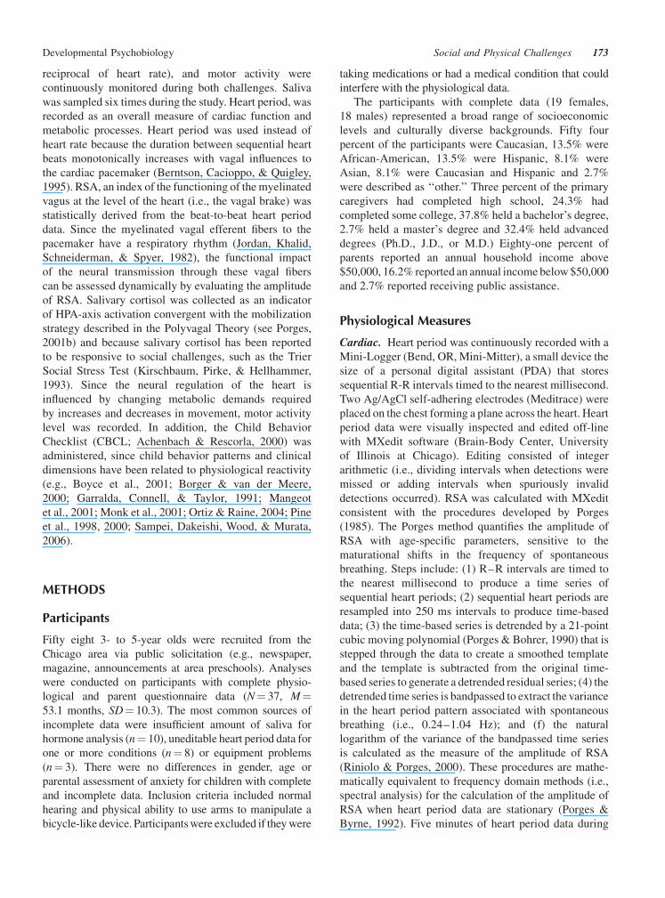
used to adjust for sphericity violations. As illustrated in
Figure 1, there were condition effects during the social
challenge for RSA, F(2, 72)¼ 9.57, p< .001, heart
period, F(1.60, 57.53)¼ 4.33, p< .03, and activity, F(2,
72)¼ 5.52, p< .006. During the social challenge, RSA
and heart period increased from pre-baseline to challenge
and decreased from challenge to post-baseline. Activity
increased from pre-baseline to challenge and decreased
from challenge to post-baseline. Cortisol did not signi-
ficantly change from pre-social challenge to post-social
challenge, F(1, 36)¼ .07, p< .80.
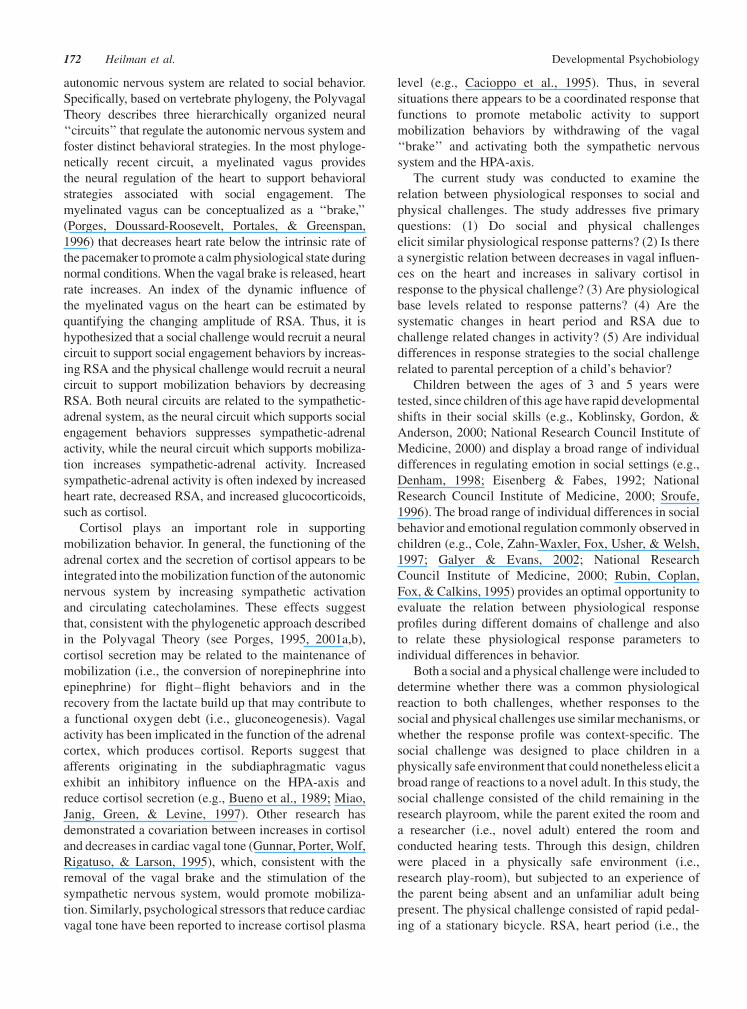
As illustrated in Figure 2, there were significant
condition effects during the physical challenge for RSA,
F(1.43, 51.51)¼ 172.81, p< .001, heart period, F(1.31,
47.24)¼ 135.63, p< .001, and activity, F(1.32,
47.49)¼ 322.99, p< .001. During the physical challenge,
RSA and heart period decreased from pre-baseline to
challenge and increased from challenge to baseline and
activity increased from pre-baseline to challenge and
decreased from challenge to post-baseline. Cortisol did
not significantly change from pre-physical challenge to
post-physical challenge, F(1, 36)¼ .64, p< .43.
Response Patterns Among VariablesWithin Challenges
To evaluate reactivity of RSA, heart period, and activity
to each challenge, change scores (challenge minus pre-
baseline) were calculated. To evaluate cortisol reactivity,
change scores (post-challenge minus pre-challenge) were
calculated. The means (standard deviations) of the change
scores during the social challenge were: RSA [.42 (.67)],
heart period [16.39 (39.73)], activity [�7.46 (17.56)]
and cortisol [�.05 (.37)]. The means (standard deviations)
of the change scores during the physical challenge
were: RSA [�2.93 (1.18)], heart period [�147.42
(73.36)], activity [78.39 (22.32)], and cortisol [.02
(.45)]. In general, the change scores in response to each
challenge for RSA, heart period, and activity were
significantly correlated with their respective baselines
(social challenge: RSA, r¼�.41, p< .01, heart period,
r¼�.15, p< .37, activity, r¼ .83, p< .001; physical
challenge: RSA, r¼�.51, p< .001, heart period,
r¼�.77, p< .001, activity, r¼�.60, p< .001). Thus, to
remove the statistical dependence between baseline
and change, residualized change scores adjusted for the
pre-baseline value were calculated with regression
analyses. To maintain the metric for each variable,
unstandardized residuals were used.
Correlations were calculated among the residualized
change scores within each challenge condition to
Developmental Psychobiology
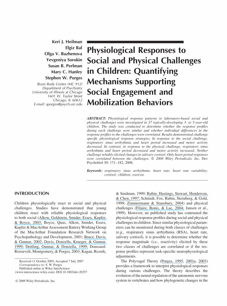
Table 3. Correlations Among Baseline Measures (n¼ 37)
Initial
BL
Social
Pre-BL
Social
Post-BL
Physical
Pre-BL
RSA
Social pre-BL .91*
Social post-BL .87* .91*
Physical pre-BL .84* .88* .88*
Physical post-BL .80* .83* .85* .86*
Heart period
Social pre-BL .93*
Social post-BL .93* .95*
Physical pre-BL .85* .89* .90*
Physical post-BL .83* .84* .84* .89*
Activity
Social pre-BL .59*
Social post-BL .49* .54*
Physical pre-BL .48* .38** .50*
Physical post-BL .57* .59* .57* .64*
Cortisol
Pre-social .70*
Post-social .51* .80*
Pre-physical .11 .49* .53*
Post-physical .05 .39** .44* .76*
*p< .01.
**p< 0.05.
FIGURE 1 RSA, heart period (HP) and activity during social
challenge (n¼ 37).
FIGURE 2 RSA, heart period (HP), and activity during
physical challenge (n¼ 37).
176 Heilman et al.

determine whether there was a coordinated response
among the variables. During the social challenge, RSA
reactivity was significantly correlated to heart period
reactivity, r¼ .68, p< .001; a greater increase in RSAwas
related to a greater increase in heart period (see Tab. 4).
During the social challenge, heart period reactivity
was significantly correlated to cortisol reactivity,
r¼�.34, p< .04; a greater increase in heart period was
related to a greater decrease in cortisol (see Tab. 4). During
the physical challenge, all correlations among RSA,
heart period and activity were significant (see Tab. 4).
RSA reactivity was correlated to heart period reactivity,
r¼ .50, p< .001 and to activity reactivity, r¼�.57,
p< .001; a greater decrease in RSA was related to a
greater decrease in heart period and greater increase
in activity. Heart period reactivity was significantly
correlated to activity reactivity during the physical
challenge, r¼�.62, p< .001; a greater decrease in heart
period was related to a greater increase in activity. Thus, as
individual differences in activity increased, there was a
parallel decrease in heart period and RSA. Cortisol
reactivity was not significantly correlated with the other
variables.
Response Patterns Among PhysiologicalMeasures Between Challenges
To determine if the response patterns to the two challenges
were similar, correlations were calculated between the
residualized change scores for each variable on each
challenge. Residualized heart period reactivity was
correlated between the social and physical challenges,
r¼ .40, p< .01; a greater reactivity to the social challenge
was related to greater reactivity to the physical challenge.
The remaining measures of reactivity were not related
between challenges (RSA reactivity, r¼ .09, p< .61;
activity reactivity, r¼ .31, p< .06; cortisol reactivity,
r¼ .08, p< .63).
Evaluation of Activity as a Potential Covariate ofPhysiological Patterns During Challenges
To determine whether systematic changes in heart period
and RSA were due to challenge-related changes in
activity, a repeated-measures ANCOVA was conducted
for RSA and heart period during each of the challenges,
using baseline activity for each respective challenge as a
covariate. During the social challenge, activity was not a
significant covariate for RSA, F(1, 35)¼ .05, p< .82, or
for heart period, F(1, 35)¼ .01, p< .93. During the
physical challenge, activity was not a significant covariate
for RSA, F(1, 35)¼ 1.14, p< .29 or for heart period, F(1,
35)¼ .05, p< .83.
Relationship Between ResponsePatterns and Behavior
To determine the relation between response patterns
during the challenges and parental perception of child
behaviors assessed by the CBCL, correlations were
calculated between each residualized change score and
each syndrome scale on the CBCL (i.e., anxious-
depressed, emotionally reactive, somatic complaints,
withdrawn, sleep problems, attention problems, internal-
izing subscale, externalizing subscale, and total problems)
(see Tab. 5). In response to the social challenge, there were
significant correlations between change in RSA and
several of the scales. Greater decreases in RSA during
the social challenge were significantly correlated with
higher values on the anxious-depressed, r¼�.43,
p< .007, sleep problems, r¼�.41, p< .01, internalizing
problems, r¼�.39, p< .02, and total problems scales,
r¼�.39, p< .02. Consistent with these findings, greater
decreases/less of an increase in heart period during the
social challenge was significantly correlated with higher
values on the anxious-depressed, r¼�.40, p< .01, sleep
problems, r¼�.40, p< .02, and total problems scales,
r¼�.33, p< .05. In addition, decreases in heart period
were correlated with higher scores on the emotionally
reactive scale, r¼�.34, p< .04, increases in cortisol were
correlated with higher scores on the anxious-depressed
subscale, r¼ .40, p< .01, and increases in activity were
correlated with higher scores on the attention problems
subscale, r¼ .39, p< .02.
In response to the physical challenge, only change in
heart period was correlated with aggressive behavior,
r¼ .37, p< .03, as individuals with more aggressive
behaviors demonstrated smaller decreases in heart period
in response to the physical challenge.
An exploratory stepwise regression, sequentially
applying additional variables, was used to determine the
most parsimonious model for predicting scores on the
syndrome scales of the CBCL that were correlated with
Developmental Psychobiology
Table 4. Correlations Among Change Scores (Residua-
lized) During the Social and Physical Challenges (n¼ 37)
DRSA DHP DActivity
Social challenge
DHP .68*
DActivity �.14 �.23
DCortisol �.21 �.34** �.16
Physical challenge
DHP .50*
DActivity �.57* �.62*
DCortisol �.28 �.09 .00
*p< .01.
**p< .05.
Social and Physical Challenges 177

response patterns during either challenge. For the CBCL
scales, the following predictors were entered into the
model: residualized changes in RSA, heart period,
activity, and cortisol for each challenge separately. Only
for the anxious-depressed scale did the stepwise regres-
sion analysis select more than one variable and in this case
change in both RSA and cortisol change during the social
challenge were the strongest predictors F(2, 36)¼ 6.93,
p< .003.
DISCUSSION
The current study investigated several important ques-
tions regarding our understanding of child autonomic and
cortisol reactions to two common classes of challenges.
Below, the results of the study are evaluated within the
context of each of the five primary questions, as described
in the Introduction.
Is There a Common Physiological ResponseStrategy to Both Social and Physical Challenges?
Although both challenges could be described opera-
tionally as ‘‘stressing,’’ each challenge elicited a different
response profile. In response to the physical challenge,
vagal withdrawal, characterized by significant decreases
in heart period and RSA, paralleled an increase in activity.
These findings reflect the active withdrawal of the vagal
‘‘brake,’’ which is necessary to support the increased
metabolic requirements associated with increased activ-
ity. Relative to the physical challenge, the response profile
to the social challenge was inverted. Heart period and
RSA increased and activity decreased. These findings
are consistent with the active increase of the vagal
brake during social engagement (see Porges, 2001a) and
the reduced cardiac output required during quiescent
behavioral states (see Porges et al., 1996). However,
cortisol did not respond to either challenge.
Although the pattern of reactivity differed between
the tasks, there is the possibility that the magnitude of
reactivity might be related across the two challenges. For
example, a dampened response to physical exercise might
be related to a dampened response to a social challenge.
Only changes in heart period were correlated between the
tasks. Participants who had the greatest increase in heart
period to the social challenge had a dampened decrease in
heart period during the physical challenge. In contrast,
RSA reactivity, a sensitive measure of the dynamic
regulation of the vagal brake, was not consistent across
the challenges. Likewise, neither activity nor cortisol
reactivity were consistent across challenges. The data
Developmental Psychobiology
Table 5. Correlations Between Residualized Change Scores (Social/Physical Challenges)
and Syndrome Scales on the CBCL (t-Scores; n¼ 37)
DRSA DHP DActivity DCortisol
Social challenge
Emotionally reactive �.25 �.34** �.13 .30
Anxious/depressed �.43* �.40** �.02 .40**
Somatic complaints �.21 �.12 �.01 .17
Withdrawn �.28 �.25 .13 �.05
Sleep problems �.41** �.40** .11 .04
Attention problems .08 .08 .39** �.18
Aggressive behavior �.01 �.05 .01 .13
Internalizing problems �.39** �.21 �.06 .19
Externalizing problems �.25 �.27 .18 .04
Total problems �.39** �.33** .07 .13
Physical challenge
Emotionally reactive �.06 .04 �.04 .06
Anxious/depressed �.18 �.11 .06 .16
Somatic complaints .10 .04 �.15 .06
Withdrawn .06 �.07 .06 �.13
Sleep problems .19 �.13 �.18 �.10
Attention problems .14 �.02 .08 .07
Aggressive behavior .07 .37** �.11 �.25
Internalizing problems .06 .05 �.06 �.03
Externalizing problems .13 .24 �.23 �.07
Total problems .16 .17 �.21 �.07
*p< .01.
**p< .05.
178 Heilman et al.

illustrate that, although the same variables respond to both
challenges, the patterns of reactivity were different. Thus,
challenges that appear to be stressful in a social context
may not be related to the responses elicited by a physical
challenge. There may be an important distinction between
the neurophysiological mechanisms mediating reactions
to these two broad classes of challenges. For example,
challenges that require mobilization may elicit well
understood neurophysiological systems that promote
increases in metabolic resources to fight and to flee.
Alternatively, challenges associated with social interac-
tion may be more dependent on a ‘‘neural’’ evaluation
of safety in the environment and the dampening of
mechanisms mediating mobilization. Thus, if the ‘‘neu-
ral’’ evaluation (i.e., neuroception) detects risk and not
safety, the primitive limbic fight/flight mechanisms are
not dampened and a physiological state is maintained to
support fight/flight behaviors (see Porges, 2003).
Is There a Synergistic Relation BetweenReduced Vagal Efferent Influences on theHeart and Increases in Salivary Cortisol?
Unexpectantly, in the current study, salivary cortisol did
not parallel changes in activity or cardiac function.
Changes in cortisol were not anticipated during the social
challenge, as the challenge required only mild cardiac and
activity demands. However, it was assumed that the
mobilization demands of the physical challenge would
elicit a reliable release of cortisol. There are several
possible reasons for these negative findings. First, the
duration and intensity of the physical challenge may
not have been sufficient to trigger the cortisol response.
While there are no published studies examining the
relation between physical activity and cortisol responses
in young children, studies examining this relation in older
children and adults have employed a physical challenge
of longer durations requiring greater aerobic demands
(e.g., Furlan, DeMartinis, Schweizer, Rickels, & Lucki,
2001; Gonzalez-Bono, Moya-Albiol, Martinez-Sanchis,
& Salvador, 2002; Jansen et al., 1999). While several
participants pedaled intensely throughout the physical
challenge, most participants did not pedal with maximal
effort. Interestingly, correlations between movement
and cortisol reactivity to the physical challenge were not
significant although there were significant parallels
between the increases activity and decreases in RSA and
heart period.
A second possibility for the lack of a reliable cortisol
response to the physical challenge might be related to
the time of day during which cortisol levels were sampled
(morning) and the general effect of circadian rhythms.
Due to the circadian rhythm of HPA-axis activity, the
pattern of cortisol release fluctuates throughout the day.
While there is a marked variability in the circadian profile
of cortisol release in healthy, pre-pubertal children,
researchers have found that the majority of children
demonstrate a sharp peak of cortisol release at approx-
imately 6:00 am. This peak is followed by a rapid decline
in cortisol release until approximately 10:00 am, and
then by a slower decline until approximately 2:00 am
(Knutsson et al., 1997). By scheduling all participants in
the morning (i.e., time of peak cortisol release), the
experiment might have challenged the system during a
period when it was close to maximal output. Thus, the
cortisol reactions might have been dampened. Or, even
if the challenges altered cortisol release, the amplitude
of the response might not be detectable when it is
superimposed on the individual’s circadian rhythm. Given
the highly stable and reproducible pattern of cortisol
release within individuals (Knutsson et al., 1997), future
studies should assess cortisol reactions to challenges
with the circadian rhythm of cortisol levels on a non-
experimental day.
Are Individual Differences in BaselinePhysiological State (i.e., RSA) Related tothe Response Patterns?
Higher baseline RSA was related to greater reductions in
RSA during the physical challenge and attenuated
increases during the social challenge. Higher baseline
activity was related to greater decreases in activity during
the physical challenge and to greater increases in activity
during the social challenge. Longer baseline heart period
(i.e., slower heart rate) was related to larger decreases in
heart period during the physical challenge. Baseline
salivary cortisol was not related to changes during either
challenge. Thus, the initial levels of the autonomic
variables were most clearly related to the magnitude of
the reactivity to the physical challenge, with greater
cardiac vagal tone (i.e., higher amplitude RSA and longer
heart periods) during baseline being related to larger
decreases during the physical challenge. These findings
provide additional support for the use of residualized
change scores to enable the study of reactivity independ-
ent of initial levels. The importance of base level on
the magnitude of autonomic reactivity was initially
described by Wilder (1931) in his ‘‘law of initial values’’
(1931/1976), and regression methods were proposed as a
corrective strategy several years ago by Benjamin (1967).
Are Individual Differences in the PhysiologicalResponses Due to Changes in Activity?
Since the autonomic response profiles to each challenge
covaried with activity, we investigated whether changes in
activity determined the changes in RSA and heart period.
Developmental Psychobiology Social and Physical Challenges 179

Only during the physical challenge did the individual
differences in the autonomic variables covary with
changes in activity, as increases in activity correlated
with decreases in RSA and heart period. These findings
are consistent with the physiological mechanisms
required to increase cardiac output to support mobiliza-
tion behaviors by withdrawing vagal influences. To
explore further the possibility that autonomic reactivity
was driven by activity, change in activity was used as a
covariate in analyses of variance evaluating the challenge
effects on heart period and RSA. However, activity was
not a significant covariate for either variable during either
challenge, thus demonstrating that autonomic reactivity
was not driven by activity.
Are Individual Differences in ResponseStrategies to the Challenges Related toParental Perception of a Child’s Behavior?
Physiological reactivity to the challenges was related to
parental perceptions of anxiety/depression, emotional
reactivity, sleep problems, attention problems, aggressive
behavior, internalizing problems, and total problems. The
findings replicate previous studies which have likewise
demonstrated a relationship between physiology and
anxiety (Boyce et al., 2001; Monk et al., 2001; Pine
et al., 1998, 2000), emotional disorders (Garralda et al.,
1991), sleep duration (Sampei et al., 2006), ADHD
(Borger & van der Meere, 2000), and antisocial behavior
(which includes aggressive behavior; Ortiz & Raine,
2004).
However, only scores on the anxious/depressed sub-
scale were predicted by RSA and cortisol reactivity to the
social challenge (less of an increase in RSA and a
greater increase in cortisol during the social challenge
predicted more symptoms of anxiety-depression). The
behavioral dimension of anxiety is of theoretical interest,
as the Polyvagal Theory interprets social behavior as an
emergent property of a specific neurophysiological state.
The neurophysiological state of individuals higher on the
anxiety-depression scale may inappropriately promote
mobilization behaviors, instead of social engagement
behaviors. While all individuals in the current study were
within the typical range for all syndrome scales, including
the anxious/depressed syndrome scale, the data indicate
that individual differences in physiological reactivity
specifically to the social challenge are related to the
parent perception of child behavior. Importantly, the data
suggest that reactivity to a social challenge can yield
important information regarding the mechanisms of
typical behavior in children that could potentially be
applied to study atypical behavior in children and adults
(i.e., anxiety and/or depression). Future studies could also
include behavioral data to examine the relationship
among parental perception of a child’s behavior, the
child’s behavior, and the child’s physiological reactivity
during the social challenge.
The study has several limitations. First, due to the
difficulty of collecting physiological data from young
children, the final sample size was small. The small
sample size limits the generalizability of the findings,
especially for cortisol which has a small effect size.
Second, the social challenge used in the study might not
generalize to the social interactions that the child
would normally experience. During the social challenge,
children were not only exposed to an unfamiliar person in
the absence of parents, but were also exposed to the
unfamiliar equipment and procedures used to conduct the
hearing tests. Thus, the physiological reactivity monitored
during the social challenge represents a response to the
complex features of the task. Future studies investigating
social behavior could address this confound and evaluate
whether the social interaction between the child and an
unfamiliar person in the absence of novel equipment
would elicit a similar physiological response pattern.
To summarize, the study demonstrates several important
points. First, physiological reactions to social and physi-
cal challenges were not correlated. Second, although both
challenges reliably produced effects in heart period and
RSA, cortisol was insensitive. Third, as proposed approx-
imately 75 years ago by Wilder (1931/1976), the magnitude
of autonomic reactions to challenge was dependent
on baseline values (i.e., law of initial values). Thus, the
data provide additional evidence for the importance of
applying residualized change scores or other regression
methods (e.g., partial correlation, analysis of covariance,
regression transformation) to study specific ‘‘reactivity’’
processes. These strategies to study autonomic reactivity
to psychological manipulations, proposed at the time
psychophysiology was emerging as a discipline, are still
valid and crucial to current research (e.g., Benjamin, 1967).
Fourth, the changes in autonomic reactivity to social and
physical challenges are, in part, dependent on the changing
metabolic demands associated with task related activity.
These findings suggest that measurement of movement is
critical when studying autonomic responses and useful in
providing a functional interpretation of reported changes
in autonomic state. Fifth, autonomic reactivity to the social
challenge may provide an experimental portal to study the
mechanisms of anxiety and/or depression in children.
NOTES
The research was supported, in part, by NIH research grant
MH060625 awarded to S.W. Porges. K.J. Heilman was
supported by the Biomedical Neuroscience Training Program
T32 MH067631. The contents of the manuscript are solely the
Developmental Psychobiology180 Heilman et al.
responsibility of the authors and do not necessarily represent the
official views of NIH. We would like to thank the parents and
children who participated in the study.
REFERENCES
Achenbach, T. M., & Rescorla, L. A. (2000). Manual for the
ASEBA preschool forms & profiles. Burlington, VT:
University of Vermont, Research Center for Children, Youth
& Families.
Alkon, A., Goldstein, L. H., Smider, N., Essex, M. J., Kupfer,
D. J., & Boyce, W. T. (2003). Developmental
and contextual influences on autonomic reactivity in
young children. Developmental Psychobiology, 42(1), 64–
78.
Benjamin, L. S. (1967). Facts and artifacts in using analysis of
covariance to ‘‘undo’’ the law of initial values. Psychophysi-
ology, 2, 187–206.
Berntson, G. G., Cacioppo, J. T., & Quigley, K. S. (1995). The
metrics of cardiac chronotropism Biometric perspectives.
Psychophysiology, 32(2), 162–171.
Borger, N., & van der Meere, J. (2000). Motor control and state
regulation in children with ADHD: A cardiac response study.
Biological Psychology, 51(2–3), 247–267.
Boyce, W. T., Quas, J., Alkon, A., Smider, N. A., Essex, M. J.,
Kupfer, D. J., & MacArthur Assessment Battery Working
Group of the MacArthur Foundation Research Network on
Psychopathology and Development. (2001). Autonomic
reactivity and psychopathology in middle childhood. British
Journal of Psychiatry, 179, 144–150.
Bruce, J., Davis, E. P., & Gunnar, M. R. (2002). Individual
differences in children’s cortisol response to the beginning of
a new school year. Psychoneuroendocrinology, 27(6), 635–
650.
Bueno, L., Gue, M., Fargeas, M. J., Alvinerie, M., Junien, J. L.,
& Fioramonti, J. (1989). Vagally mediated inhibition of
acoustic stress-induced cortisol release by orally adminis-
tered kappa-opiod substances in dogs. Endocrinology, 123,
1788–1793.
Cacioppo, J. T., Malarkey, W. B., Kiecolt-Glaser, J. K., Uchino,
B. N., Sgoutas-Emch, S. A., Sheridan, J. F., Berntson, G. G.,
& Glaser, R. (1995). Heterogeneity in neuroendocrine and
immune response to brief psychological stressors as a
function of autonomic cardiac activation. Psychosomatic
Medicine, 57, 154–164.
Cole, P. M., Zahn-Waxler, C., Fox, N. A., Usher, B. A., &
Welsh, J. D. (1997). Individual differences in emotion
regulation and behavior problems in preschool children.
Journal of Abnormal Psychology, 105(4), 518–529.
Davis, E. P., Donzella, B., Krueger, W. K., & Gunnar, M. R.
(1999). The start of a new school year: Individual differences
in salivary cortisol response in relation to child temperament.
Developmental Psychobiology, 35(3), 188–196.
Denham, S. A. (1998). Emotional development in young
children. New York: Guilford Press.
Dettling, A. C., Gunnar, M. R., & Donzella, B. (1999). Cortisol
levels of young children in full-day childcare centers:
Relations with age and temperament. Psychoneuroendocri-
nology, 24(5), 519–536.
Doussard-Roosevelt, J. A., Montgomery, L. A., & Porges, S. W.
(2003). Short-term stability of physiological measures in
kindergarten children: Respiratory sinus arrhythmia, heart
period, and cortisol. Developmental Psychobiology, 43(3),
231–242.
Eisenberg N. & Fabes R. A. (Eds.), (1992). Emotion and its
regulation in early development. New York: Jossey-Bass.
Filaire, E., Bonis, J., & Lac, G. (2004). Relationships between
physiological and psychological stress and salivary immu-
noglobulin A among young female gymnasts. Perceptual and
Motor Skills, 99(2), 605–617.
Furlan, P. M., DeMartinis, N., Schweizer, E., Rickels, K., &
Lucki, I. (2001). Abnormal salivary cortisol levels in social
phobic patients in response to acute psychological but not
physical stress. Biological Psychiatry, 50, 254–259.
Galyer, K. T., & Evans, I. M. (2002). Pretend play and the
development of emotion regulation in preschool children.
Early Child Development & Care, 166, 93–108.
Garralda, M. E., Connell, J., & Taylor, D. C. (1991).
Psychophysiological anomalies in children with emotional
and conduct disorders. Psychological Medicine, 21(4), 947–
957.
Gonzalez-Bono, E., Moya-Albiol, L., Martinez-Sanchis, S., &
Salvador, A. (2002). Salivary testosterone and cortisol
responses to cycle ergometry in basketball players with
different training volume. Journal of Psychophysiology, 16,
158–166.
Gunnar, M. R., Porter, F. L., Wolf, C. M., Rigatuso, J., &
Larson, M. C. (1995). Neonatal stress reactivity: Predictions
to later emotional temperament. Child Development, 66, 1–
13.
Jansen, L. M. C., Gispen-de Wied, C. C., Jansen, M. A., van der
Gaag, R.-J., Matthys, W., & van Engeland, H. (1999).
Pituitary-adrenal reactivity in a child psychiatric population:
Salivary cortisol response to stressors. European Neuro-
psychopharmacology, 9, 67–75.
Jordan, D., Khalid, M. E. M., Schneiderman, N., & Spyer, K.
M. (1982). The location and properties of preganglionic
vagal cardio-motor neurones in the rabbit. Pflugers Archive,
395, 244–250.
Kagan, J., Reznik, J. S., & Snidman, N. (1999). Biological basis
of childhood shyness. In: A. Slater & D. Muir (Eds.), The
Blackwell reader in development psychology (pp. 65–78.).
Malden, MA: Blackwell Publishing.
Kirschbaum, C., Pirke, K. M., & Hellhammer, D. H. (1993).
The Trier Social Stress Test—A tool for investigating
psychobiological stress responses in a laboratory setting.
Neuropsychobiology, 28(1–2), 76–81.
Knutsson, U., Dahlgren, J., Marcus, C., Rosberg, S., Bronne-
gard, M., Stierna, P., & Albertsson-Wikland, K. (1997).
Circadian cortisol rhythms in healthy boys and girls:
Relationship with age, growth, body composition and
pubertal development. Journal of Clinical Endocrinology
and Metabolism, 82, 536–540.
Koblinsky, S. A., Gordon, A. L., & Anderson, E. A. (2000).
Changes in the social skills and behavior problems of
Developmental Psychobiology Social and Physical Challenges 181

homeless and housed children during the preschool year.
Early Education & Development, 11(3), 321–338.
Mangeot, S. D., Miller, L. J., McIntosh, D. N., McGrath-Clarke,
J., Simon, J., Hagerman, R. J., & Goldson, E. (2001). Sensory
modulation dysfunction in children with attention-deficit-
hyperactivity disorder. Developmental Medicine and Child
Neurology, 43(6), 399–406.
Miao, F. J.-P., Janig, W., Green, P. G., & Levine, J. D. (1997).
Inhibition of bradykinin-induced plasma extravasation pro-
duced by noxious cutaneous and visceral stimuli and its
modulation by vagal activity. Journal of Neurophysiology,
78, 1285–1292.
Monk, C., Kovelenko, P., Ellman, L. M., Sloan, R. P., Bagiella,
E., Gorman, J. M., & Pine, D. S. (2001). Enhanced stress
reactivity in paediatric anxiety disorders: Implications for
future cardiovascular health. International Journal of Neuro-
psychopharmacology, 4, 199–206.
National Research Council and Institute of Medicine. (2000).
From Neurons to Neighborhoods: The Science of Early
Child Development. Committee on Integrating the Science
of Early Childhood Development. Jack P. Shonkoff and
Deborah A. Phillips, eds. Board on Children, Youth, and
Families, Commission on Behavioral and Social Sciences
and Education. Washington, D.C.: National Academy Press.
Ortiz, J., & Raine, A. D. (2004). Heart rate level and antisocial
behavior in children and adolescents: A Meta-analysis.
Journal of the American Academy of Child and Adolescent
Psychiatry, 43(2), 154–162.
Pine, D. S., Coplan, J. D., Papp, L. A., Klein, R. G., Martinez, J.
M., Kovalenko, P., Tancer, N., Moreau, D., Dummit, E. S.,
Shaffer, D., Klein, D. F., & Gorman, J. M. (1998). Ventilatory
physiology of children and adolescents with anxiety
disorders. Archives of General Psychiatry, 55(2), 123–129.
Pine, D. S., Klein, R. G., Coplan, J. D., Papp, L. A., Hoven, C.
W., Martinez, J., Kovalenko, P., Mandell, D. J., Moreau, D.,
Klein, D. F., & Gorman, J. M. (2000). Differential carbon
dioxide sensitivity in childhood anxiety disorders and nonill
comparison group. Arch Gen Psychiatry, 57, 960–967.
Porges, S. W. (1985). Method and apparatus for evaluating
rhythmic oscillations in a periodic physiological response
systems. US Patent 4,510,944.
Porges, S. W. (1995). Orienting in a defensive world:
Mammalian modifications of our evolutionary heritage: A
Polyvagal Theory. Psychophysiology, 32(4), 301–318.
Porges, S. W. (2001a). The polyvagal theory: Phylogenetic
substrates of a social nervous system. International Journal of
Psychophysiology, 42(2), 123–146.
Porges, S. W. (2001). Is there a major stress system at the
periphery other than the adrenals? In: D. M. Broom (Ed.),
Dahlem workshop on coping with challenge: Welfare in
animals including humans. Dahlem University Press,
pp.135–149.
Porges, S. W. (2003). Social engagement and attachment: A
phylogenetic perspective. Annals of the New York Academy
of Science, 1008, 31–47.
Porges, S. W., & Bohrer, R. E. (1990). The analysis of periodic
processes in psychophysiological research. In: J. Cacioppo &
L. G. Tassinary (Eds.), Principles of psychophysiology:
Physical, social, and inferential elements. (pp. 708–753.).
New York, NY: Cambridge University Press.
Porges, S. W., & Byrne, E. A. (1992). Research methods for
measurement of heart rate and respiration. Biological
Psychology, 34(2–3), 93–130.
Porges, S. W., Doussard-Roosevelt, J. A., Portales, A. L., &
Greenspan, S. I. (1996). Infant regulation of the vagal
‘‘brake’’ predicts child behavior problems: A psychobiolog-
ical model of social behavior. Developmental Psychobiology,
29(8), 697–712.
Riniolo, T. C., & Porges, S. W. (2000). Evaluating group
distributional characteristics: Why psychophysiologists
should be interested in qualitative departures from the
normal distribution. Psychophysiology, 37(1), 21–28.
Rubin, K. H., Coplan, R. J., Fox, N. A., & Calkins, S. D. (1995).
Emotionality, emotion regulation, and preschoolers’ social
adaptation. Development & Psychopathology, 7(1), 49–62.
Rubin, K. H., Hastings, P. D., Stewart, S. L., Henderson, H. A.,
& Chen, X. (1997). The consistency and concomitants of
inhibition: Some of the children, all of the time. Child
Development, 68(3), 467–483.
Sampei, M., Dakeishi, M., Wood, D. C., & Murata, K. (2006).
Impact of total sleep duration on blood pressure in preschool
children. Biomedical Research, 27(3), 111–115.
Schmidt, L. A., Fox, N. A., Schulkin, J., & Gold, P. W. (1999).
Behavioral and psychophysiological correlates of self-
presentation in temperamentally shy children. Developmen-
tal Psychobiology, 35, 119–135.
Sroufe, L. A. (1996). Emotional development: The organization
of emotional life in the early years. New York: Cambridge
University Press.
Wilder, J. (1931). The ‘‘Law of Initial Values,’’ a neglected
biological law and its significance for research and practice.
Zeitschrift fur die gesammete Neurologie und Psychiatrie,
137, 317–338. [Reprinted in English in Porges & Coles,
1976].
Zimmermann, L. K., & Stansbury, K. (2004). The influence of
emotion regulation, level of shyness, and habituation on the
neuroendocrine responses of three-year-old children. Psy-
choneuroendocrinology, 29, 973–982.
Developmental Psychobiology182 Heilman et al.