Kainate receptor trafficking: physiological roles and molecular mechanisms
10
Kainate receptor trafficking: physiological roles and molecular mechanisms John T.R. Isaac a,b, * , Jack Mellor b , David Hurtado a , Katherine W. Roche a a National Institute for Neurological Disorders and Stroke, National Institutes of Health, Bethesda, MD 20892-3701, USA b MRC Centre for Synaptic Plasticity, Department of Anatomy, University of Bristol, Bristol BS8 1TD, UK Abstract Recently, there has been intense interest in the mechanisms regulating the trafficking and synaptic targeting of kainate receptors in neurons. This topic is still in its infancy when compared with studies of trafficking of other ionotropic glutamate receptors; however, it is already clear that mechanisms exist for subunit- and splice variant-specific trafficking of kainate receptors. There is also enormous diversity of kainate receptor targeting, with the best-studied neurons in this regard being hippocampal CA3 pyramidal neurons and CA1 GABAergic interneurons. This review summarizes the current state of knowledge on this topic, focusing on the molecular mechanisms of kainate receptor trafficking and the potential for these mechanisms to regulate neuronal kainate receptor function. D 2004 Elsevier Inc. All rights reserved. Keywords: Kainate receptor; Glutamate; Hippocampus; ER retention motif; Receptor targeting; Synaptic plasticity Abbreviations: AMPA, a-amino-3-hydroxy-5-methyl-4-isoxazolepropionic acid; A/C, associational commissural; Eph, ephrin; EPSC, excitatory postsynaptic current; ER, endoplasmic reticulum; GFP, green flourescent protein; GRIP, glutamate receptor interacting protein; IPSC, inhibitory postsynaptic current; NMDA, N-methyl-D-aspartate; PDZ, postsynaptic density 95/disc large/zonula occludens-1; PICK: protein interacting with C-kinase 1; PM, plasma membrane; PSD-95/SAP90, postsynaptic density protein-95/synapse associated protein 90; TM, transmembrane. Contents 1. Introduction ........................................ 164 2. Diverse locations and functions of kainate receptors in the hippocampus ........ 164 3. Kainate receptors in CA3 pyramidal neurons....................... 165 3.1. Postsynaptic kainate receptors in CA3 pyramidal cells .............. 165 3.2. Presynaptic kainate receptors in CA3 pyramidal neurons ............. 166 4. CA1 interneurons ..................................... 166 4.1. Somatodendritic kainate receptors in CA1 interneurons.............. 166 4.2. Presynaptic kainate receptors in CA1 interneurons ................ 167 5. Kainate receptor targeting in neurons........................... 167 6. Molecular mechanisms regulating kainate receptor expression at the synapse ...... 168 6.1. Kainate receptors en route to the plasma membrane ............... 168 6.2. Beyond the C-terminus .............................. 169 6.3. Kainate receptor binding proteins and synaptic targeting ............. 170 0163-7258/$ - see front matter D 2004 Elsevier Inc. All rights reserved. doi:10.1016/j.pharmthera.2004.08.006 * Corresponding author. Tel.: 44 117 928 7400; fax: 44 117 929 1687. E-mail address: [email protected] (J.T.R. Isaac). Pharmacology & Therapeutics 104 (2004) 163 – 172 www.elsevier.com/locate/pharmthera
-
Upload
independent -
Category
Documents
-
view
4 -
download
0
Transcript of Kainate receptor trafficking: physiological roles and molecular mechanisms

Pharmacology & Therapeutics 104 (2004) 163–172
www.elsevier.com/locate/pharmthera
Kainate receptor trafficking: physiological roles and
molecular mechanisms
John T.R. Isaaca,b,*, Jack Mellorb, David Hurtadoa, Katherine W. Rochea
aNational Institute for Neurological Disorders and Stroke, National Institutes of Health, Bethesda, MD 20892-3701, USAbMRC Centre for Synaptic Plasticity, Department of Anatomy, University of Bristol, Bristol BS8 1TD, UK
Abstract
Recently, there has been intense interest in the mechanisms regulating the trafficking and synaptic targeting of kainate receptors in
neurons. This topic is still in its infancy when compared with studies of trafficking of other ionotropic glutamate receptors; however, it is
already clear that mechanisms exist for subunit- and splice variant-specific trafficking of kainate receptors. There is also enormous diversity
of kainate receptor targeting, with the best-studied neurons in this regard being hippocampal CA3 pyramidal neurons and CA1 GABAergic
interneurons. This review summarizes the current state of knowledge on this topic, focusing on the molecular mechanisms of kainate receptor
trafficking and the potential for these mechanisms to regulate neuronal kainate receptor function.
D 2004 Elsevier Inc. All rights reserved.
Keywords: Kainate receptor; Glutamate; Hippocampus; ER retention motif; Receptor targeting; Synaptic plasticity
Abbreviations: AMPA, a-amino-3-hydroxy-5-methyl-4-isoxazolepropionic acid; A/C, associational commissural; Eph, ephrin; EPSC, excitatory postsynaptic
current; ER, endoplasmic reticulum; GFP, green flourescent protein; GRIP, glutamate receptor interacting protein; IPSC, inhibitory postsynaptic current;
NMDA, N-methyl-D-aspartate; PDZ, postsynaptic density 95/disc large/zonula occludens-1; PICK: protein interacting with C-kinase 1; PM, plasma
membrane; PSD-95/SAP90, postsynaptic density protein-95/synapse associated protein 90; TM, transmembrane.
0163-7258/$ - see f
doi:10.1016/j.pharm
* Correspondin
E-mail address:
Contents
ron
the
g au
isa
1. Introduction. . . . . . . . . . . . . . . . . . . . . . . . . . . . . . . . . . . . . . . . 164
2. Diverse locations and functions of kainate receptors in the hippocampus . . . . . . . . 164
3. Kainate receptors in CA3 pyramidal neurons. . . . . . . . . . . . . . . . . . . . . . . 165
3.1. Postsynaptic kainate receptors in CA3 pyramidal cells . . . . . . . . . . . . . . 165
3.2. Presynaptic kainate receptors in CA3 pyramidal neurons . . . . . . . . . . . . . 166
4. CA1 interneurons . . . . . . . . . . . . . . . . . . . . . . . . . . . . . . . . . . . . . 166
4.1. Somatodendritic kainate receptors in CA1 interneurons. . . . . . . . . . . . . . 166
4.2. Presynaptic kainate receptors in CA1 interneurons . . . . . . . . . . . . . . . . 167
5. Kainate receptor targeting in neurons. . . . . . . . . . . . . . . . . . . . . . . . . . . 167
6. Molecular mechanisms regulating kainate receptor expression at the synapse . . . . . . 168
6.1. Kainate receptors en route to the plasma membrane . . . . . . . . . . . . . . . 168
6.2. Beyond the C-terminus . . . . . . . . . . . . . . . . . . . . . . . . . . . . . . 169
6.3. Kainate receptor binding proteins and synaptic targeting . . . . . . . . . . . . . 170
t matter D 2004 Elsevier Inc. All rights reserved.
ra.2004.08.006
thor. Tel.: 44 117 928 7400; fax: 44 117 929 1687.
[email protected] (J.T.R. Isaac).

J.T.R. Isaac et al. / Pharmacology & Therapeutics 104 (2004) 163–172164
7. Conclusions and prospects . . . . . . . . . . . . . . . . . . . . . . . . . . . . . . . . 171
Acknowledgments . . . . . . . . . . . . . . . . . . . . . . . . . . . . . . . . . . . . . . . 171
References . . . . . . . . . . . . . . . . . . . . . . . . . . . . . . . . . . . . . . . . . . . 171
Table 1
mRNA expression levels for kainate receptor subunits in neurons in the
hippocampal formation
Cell type GluR5 GluR6 GluR7 KA1 KA2
Dentate granule + +++ ++ +++ +++
CA3 pyramidal + +++ � +++ +++
CA3 interneuron +++ ++ ++ � �CA1 pyramidal � ++ � � ++
CA1 interneuron +++ ++ ++ � �For references, see accompanying text. Key: +++, high; ++, moderate; +,
low; �, not detected.
1. Introduction
Kainate receptors are 1 of the 3 ionotropic glutamate
receptor subtypes. They are widely expressed in the central
nervous system (Wisden & Seeburg, 1993; Bahn et al.,
1994; Bettler & Mulle, 1995), critically involved in
excitatory and inhibitory neurotransmission and the induc-
tion and expression of long-term potentiation, and heavily
implicated in diseases such as epilepsy (Frerking & Nicoll,
2000; Lerma et al., 2001). Kainate receptors are tetrameric
combinations of GluR5–7 and KA1–2 subunits (Wisden &
Seeburg, 1993; Bettler & Mulle, 1995). The GluR5–7
subunits are functional ion channels either as homomers or
in heteromeric combination with other GluR5–7 and KA1–2
subunits. The KA subunits are not thought to produce
functional homomeric channels (Ren et al., 2003a, 2003b)
and are thought to form part of heteromers in combination
with the GluR5–7 subunits.
Much more so than the other ionotropic glutamate
receptors (AMPA and N-methyl-d-aspartate [NMDA]),
kainate receptors are targeted to a variety of presynaptic
and postsynaptic locations often within the same neuron. At
these locations, they perform specific tasks related to the
regulation of presynaptic function (of glutamate trans-
mission and transmission mediated by other transmitters,
most notably GABA), postsynaptic neurotransmission, or
regulation of membrane excitability (Frerking & Nicoll,
2000; Kullmann, 2001; Lerma et al., 2001; Lerma, 2003).
Moreover, there is evidence that kainate receptors at
different locations have distinct subunit compositions and
in some cases, in addition to their classic ionotropic
function, may couple with G protein-dependent signaling
mechanisms (Rodriguez-Moreno & Lerma, 1998; Frerking
et al., 2001; Melyan et al., 2002; Rozas et al., 2003).
Furthermore, there is evidence that kainate receptor expres-
sion at synapses may be acutely regulated during long-term
synaptic plasticity (Kidd & Isaac, 1999) and during
development (Bahn et al., 1994; Kidd & Isaac, 1999; Kidd
et al., 2002). Thus, from a physiological point of view, the
trafficking and targeting mechanisms regulating kainate
receptor subunits are required to fulfill a variety of tasks.
This review will describe the recent advances in under-
standing kainate receptor trafficking and targeting mecha-
nisms in the context of the current understanding of the
diverse physiological functions of kainate receptors.
In situ hybridization studies show that mRNA for kainate
receptor subunits is highly expressed throughout the brain
and spinal cord (Wisden & Seeburg, 1993; Bahn et al.,
1994; Bettler & Mulle, 1995). However, due to a paucity of
high-quality subunit-selective antibodies, few studies of the
expression of kainate receptor subunits at the protein level
have been performed. Therefore, most information regard-
ing the localization of kainate receptors and specific
subunits in certain locations in neurons has been obtained
from functional studies combined with pharmacology and/
or the use of kainate receptor subunit-deficient mice. The
most detailed functional analysis of kainate receptors has
been performed on hippocampal circuits for both glutama-
tergic and GABAergic neurons. We will therefore use the
hippocampus as an example to illustrate the diverse
locations at which kainate receptors are found.
2. Diverse locations and functions
of kainate receptors in the hippocampus
In the hippocampus, mRNA for all kainate receptor
subunits is found, but expression levels differ greatly
between different regions and cell types (Table 1; Bahn et
al., 1994; Bureau et al., 1999; Paternain et al., 2000).
Principal cells of the CA3 region and dentate gyrus show
high levels of message for GluR6, KA2, and KA1 but low
levels for GluR5. GluR7 is found in dentate granule cells.
Interneurons in the CA3 region express high levels of GluR5
and populations also express GluR6 and GluR7. KA1–2
subunits have low levels of expression in CA3 interneurons.
In the CA1 region, principal cells generally show lower
levels of expression for kainate receptor subunits compared
with CA3 but have detectable levels of transcripts for GluR6
and KA2. A population of CA1 interneurons have high levels
of GluR5, and another potentially overlapping population
show high levels of GluR6. GluR7 is selectively expressed in
a population of stratum oriens interneurons. KA1–2 subunits
do not appear to be expressed in interneurons.
Thus, simply from the mRNA expression data, there is
potential for considerable cell type-specific differences in the
subunit combination of kainate receptor complexes. Because
mRNA data only provide information on the amount of new
protein synthesized, regional and cell-specific information at

Fig. 1. Locations of kainate receptors in hippocampal CA3 pyramidal neurons. For this and Fig. 2, subunit information is provided where there is positive
evidence for the presence of a particular subunit. See text for references and details.
J.T.R. Isaac et al. / Pharmacology & Therapeutics 104 (2004) 163–172 165
the protein level for the different kainate receptor subunits is
required to get a clear picture of exactly what subunits are
expressed and where. In the absence of such information,
however, functional studies using selective pharmacological
agents and kainate receptor subunit-deficient mouse strains
have provided most of the information on the location of
kainate receptors and their subunit compositions. A sum-
mary of kainate receptor locations and subunit information
where known is provided in Figs. 1 and 2.
3. Kainate receptors in CA3 pyramidal neurons
3.1. Postsynaptic kainate receptors in CA3 pyramidal cells
CA3 pyramidal cells differentially target kainate recep-
tors to postsynaptic locations. They are found postsynapti-
cally at mossy fiber synapses where they mediate a slow
synaptic current but remarkably not at associational
commissural (A/C) synapses also located on the apical
dendrites of the same neurons (Castillo et al., 1997; Vignes
& Collingridge, 1997). Evidence from electrophysiological
studies suggests that postsynaptic kainate receptors are
located at the postsynaptic density. This is based on studies
on mossy fiber–CA3 synapses and in other brain areas
Fig. 2. Locations of kainate receptors in hippocampal GABAerg
showing a lack of effect on the size and kinetics of the
kainate receptor-mediated excitatory postsynaptic current
(EPSC) for manipulations that modulate glutamate clear-
ance from the synaptic cleft (Castillo et al., 1997; Vignes &
Collingridge, 1997; Bureau et al., 2000; Kidd & Isaac,
2001; but see Min et al., 1998). In addition, studies using
biochemical techniques and/or immunogold localization of
kainate receptor subunits also support the view that kainate
receptors are located at the postsynaptic density (Darstein et
al., 2003; Hirbec et al., 2003).
Studies of kainate receptor subunit-deficient mice have
proven to be very informative in understanding the subunit
composition of specific functionally identified kainate
receptors. At mossy fiber synapses, such genetic manipu-
lations provide evidence for GluR6-containing receptors
mediating the kainate receptor-mediated EPSC (Mulle et al.,
1998). This is supported by pharmacological evidence
demonstrating a low sensitivity to GluR5 selective ligands
in CA3 pyramidal cells (Lauri et al., 2001a), indicating a lack
of the involvement of this subunit postsynaptically. More
recent studies also provide immunogold and biochemical
evidence for postsynaptic GluR6/KA2 heteromers (Darstein
et al., 2003) and a role for KA2 in determining the kinetics of
the kainate receptor-mediated EPSC (Contractor et al.,
2003). Thus, the current data all point to GluR6/KA2
ic CA1 interneurons. See text for references and details.

J.T.R. Isaac et al. / Pharmacology & Therapeutics 104 (2004) 163–172166
heteromers as the major postsynaptic kainate receptor
subunit composition at mossy fiber synapses.
3.2. Presynaptic kainate
receptors in CA3 pyramidal neurons
The A/C input onto CA3 pyramidal neurons, connections
between CA3 pyramidal neurons, is regulated by presy-
naptic kainate receptors. A/C synapses are negatively
regulated by kainate receptor agonists (Bortolotto et al.,
1999; Contractor et al., 2000; Lauri et al., 2001b), and
pharmacological studies primarily performed on rat tissue
indicate the involvement of GluR5-containing receptors
(Bortolotto et al., 1999; Lauri et al., 2001b). However,
controversy exists in this regard because genetic evidence in
mice points to a critical role for GluR6 subunits and
provides evidence against the involvement of GluR5
(Contractor et al., 2000). The reason for this discrepancy
is at present unclear but could suggest that GluR5
trafficking to synapses is impaired in the GluR6 knockout
or that kainate receptor stoichiometry is different in rats
compared with mice.
The postsynaptic effects of kainate receptor agonists, at
doses that cause strong inhibition of A/C transmission, are
unlikely to account for the presynaptic effects on trans-
mission (Vignes et al., 1998; Contractor et al., 2000). This
indicates that there is a population of presynaptic kainate
receptors located close to A/C terminals. In support of this
idea, the frequency of action potential independent mini-
ature EPSCs is increased by kainate receptor agonists
(Contractor et al., 2000; but see Castillo et al., 1997).
Puzzlingly, however, Contractor et al. (2000) reported that
the effect on miniature EPSC frequency was selectively lost
in GluR5 receptor subunit-deficient mice. This was despite
evidence provided in the same study against a selective role
of GluR5 in regulating evoked release at any glutamatergic
input onto CA3 pyramidal neurons. This apparent paradox
is still unresolved but could indicate that there is more than
1 presynaptic kainate receptor subtype. Thus, although
several unresolved issues concerning the exact location and
subunit identity of the presynaptic kainate receptor(s)
remain, the data suggest that CA3 pyramidal neurons target
GluR6 and GluR5 subunit containing kainate receptors to
A/C terminals. The possibility also exists for at least 2
different types of presynaptic kainate receptors of different
subunit composition being present.
Kainate receptors also regulate transmission at CA3-CA1
synapses, the Schaffer collateral/commissural input. This
has been studied in CA1 pyramidal neurons where kainate
receptor agonists are found to depress excitatory trans-
mission (Chittajallu et al., 1996; Kamiya & Ozawa, 1998;
Vignes et al., 1998; Bortolotto et al., 1999; Frerking et al.,
2001; Clarke & Collingridge, 2002). Pharmacological
evidence suggests that GluR5-containing receptors are
involved (Vignes et al., 1998; Bortolotto et al., 1999; Clarke
& Collingridge, 2002), although this issue has not been
addressed in kainate receptor subunit-deficient mice. The
evidence suggests that the presynaptic kainate receptor
directly regulates transmission at the terminal (Frerking et
al., 2001; Clarke & Collingridge, 2002) and that the
mechanism involves a novel metabotropic action of the
kainate receptor through a pertussis toxin-sensitive G
protein-mediated mechanism (Frerking et al., 2001).
Schaffer collateral/commissural axons also synapse onto
CA1 interneurons; however, to date, it has not been reported
if these synapses are also regulated by kainate receptors.
Overall, the available evidence indicates that CA3 pyrami-
dal neurons target kainate receptors to their terminals in the
CA1 region. These kainate receptors appear to contain
GluR5 subunits and also couple to a metabotropic cascade
involving a pertussis toxin-sensitive G protein. The degree
of similarity of these presynaptic kainate receptors to those
at A/C terminals in CA3 is not fully explored; therefore, it is
not clear if CA3 neurons target different kainate receptor
subtypes to distinct terminals.
4. CA1 interneurons
GABAergic interneurons in CA1 are a second cell type
for which there is considerable evidence that kainate
receptors of different subunit compositions can be targeted
to diverse locations. GABAergic interneurons are found
throughout the CA1 region of the hippocampus and can be
subdivided into many subtypes based on their anatomical,
neurochemical, and electrophysiological signature (Macca-
ferri & Lacaille, 2003). All CA1 interneuron subtypes
studied thus far exhibit functional kainate receptor-mediated
responses (Cossart et al., 1998). However, the majority of
the work on this topic does not distinguish between
interneuron subtype; therefore, the degree of intersubtype
variation in kainate receptor expression and function is not
well explored.
4.1. Somatodendritic kainate receptors in CA1 interneurons
Kainate receptors are found postsynaptically at glutama-
tergic synapses on CA1 interneurons where they mediate a
slow EPSC (Cossart et al., 1998; Frerking et al., 1998).
Pharmacological evidence suggests that these receptors
contain GluR5 subunits (Cossart et al., 1998), although
genetic investigation of the subunit composition of these
synaptic receptors has yet to be reported.
There is considerable controversy concerning the mech-
anism(s) by which kainate receptors regulate GABAergic
interneuron function (Kullmann, 2001). However, it is
generally agreed that kainate receptors located on somato-
dendritic membranes mediate the intense excitation that is
induced in interneurons by kainate receptor agonists
(Rodriguez-Moreno et al., 1997, 2000; Cossart et al.,
1998; Frerking et al., 1998, 1999; Rodriguez-Moreno &
Lerma, 1998; Bureau et al., 1999; Mulle et al., 2000).

J.T.R. Isaac et al. / Pharmacology & Therapeutics 104 (2004) 163–172 167
Activation of these receptors causes a profound increase in
the frequency of spontaneous (action potential-dependent)
inhibitory postsynaptic currents (sIPSCs) recorded in CA1
pyramidal cells and interneurons. It is not clear if these
somatodendritic receptors are the same population as those
present at glutamatergic synapses or if they are an additional
or overlapping population. Pharmacological evidence indi-
cates that these receptors contain GluR5 subunits (Cossart et
al., 1998; Rodriguez-Moreno et al., 2000), and genetic
studies indicate that most are GluR5/6 heteromers (Bureau
et al., 1999; Mulle et al., 2000).
4.2. Presynaptic kainate receptors in CA1 interneurons
It is also generally agreed that kainate receptor agonists
cause a depression of evoked IPSCs recorded in CA1
pyramidal neurons (Clarke et al., 1997; Rodriguez-Moreno
et al., 1997, 2000; Rodriguez-Moreno & Lerma, 1998;
Bureau et al., 1999; Frerking et al., 1999; Min et al., 1999;
Mulle et al., 2000). However, the mechanism for this is
controversial. One possibility is that there is a distinct
population of kainate receptors located presynaptically at
GABAergic terminals onto pyramidal neurons, which
inhibit GABA release. Evidence for this has been provided
in studies investigating the effects of kainate receptor
agonists on mIPSCs (Rodriguez-Moreno & Lerma, 1998;
Rodriguez-Moreno et al., 2000) and differential dose-
dependent effects of different agonists on somatodendritic
excitation and evoked IPSC depression (Rodriguez-Moreno
et al., 2000). These studies suggest that receptors at the
terminal have a different makeup from the somatodendritic
receptors in that they do not contain GluR5 (Rodriguez-
Moreno et al., 2000). However, there is also pharmaco-
logical and genetic evidence that much of the depression is a
consequence of the intense excitation by somatodendritic
receptors (Frerking et al., 1999; Mulle et al., 2000) and that
additional axonal and postsynaptic mechanisms also con-
tribute (Frerking et al., 1999; Kang et al., 2004). One final
twist to this debate has come from a more recent study
(Jiang et al., 2001), providing evidence for a kainate
receptor located close to the synaptic terminal for which
the physiological mode of action is to facilitate GABA
release rather than depress it as suggested previously.
Resolution of this debate is required for it to become clear
if CA1 interneurons do indeed target kainate receptors of a
specific subunit composition to terminals onto pyramidal
cells in addition to GluR5/6 receptors at somatodendritic
locations.
There is also evidence that CA1 interneurons can target
kainate receptors to axons and presynaptic terminals
contacting other interneurons (Mulle et al., 2000; Cossart
et al., 2001; Semyanov & Kullmann, 2001). Pharmaco-
logical activation of these presynaptic kainate receptors
decreases mIPSC frequency and evidence suggests that this
is GluR6 dependent with no involvement of GluR5 (Mulle
et al., 2000; Cossart et al., 2001). In addition, pharmaco-
logical or synaptic activation of kainate receptors causes a
direct depolarization of axons between interneurons result-
ing in increased excitability (Semyanov & Kullmann,
2001), although the subunit composition of these receptors
remains to be explored. This raises the possibility that
interneurons target kainate receptors to both terminals and
axons; however, it is still unclear whether they can select
between interneuron-interneuron and interneuron-pyrami-
dal cell connections. A simple scheme would be that
interneurons target GluR5/6 heteromers somatodendriti-
cally (including the postsynaptic density) and GluR6-
containing (but GluR5-lacking) receptors to axons and
terminals.
One final point of interest for kainate receptors in CA1
interneurons is that there is evidence that they too can
couple to a G protein-mediated metabotropic cascade. The
depression of evoked IPSCs in CA1 pyramidal neurons is
blocked by pertussis toxin and protein kinase C inhibitors
(Rodriguez-Moreno & Lerma, 1998). This is very similar
to the mechanism reported at glutamatergic synapses onto
CA1 pyramidal cells (Frerking et al., 2001).
5. Kainate receptor targeting in neurons
CA3 pyramidal neurons and CA1 interneurons provide
excellent illustrations of the complexity of kainate receptor
targeting. Although these 2 cell types are best characterized
in this regard, it is clear that kainate receptors are found in
presynaptic and postsynaptic locations in many other cell
types in the mammalian brain (Lerma et al., 2001). This
suggests that selective, subunit-specific targeting of kainate
receptors in neurons is a general principle. Considerable
controversy remains, even in the well-studied cell types, as
to exactly where kainate receptors are found. This is
primarily due to the reliance on electrophysiological data,
which can be ambiguous in its interpretation. Systematic
immunolocalization studies in native brain samples will be
required to fully elucidate the precise locations of kainate
receptors in neurons. In addition, there is the intriguing
finding that kainate receptors can couple to G protein-
dependent metabotropic cascades. This has been reported
for presynaptic receptors regulating GABAergic synapses
onto CA1 pyramidal neurons (Rodriguez-Moreno & Lerma,
1998), for presynaptic and postsynaptic receptors at
glutamatergic synapses onto CA1 pyramidal neurons
(Frerking et al., 2001; Melyan et al., 2002, 2004), and for
postsynaptic receptors in dorsal root ganglion cells (Rozas
et al., 2003). Consistently, this mechanism involves a
pertussis toxin-sensitive G protein and protein kinase C. It
would of great interest to understand the molecular
mechanisms by which these ionotropic receptors can couple
directly to a G protein and whether this coupling is subunit
specific. Clues to this may be provided by understanding
the trafficking and targeting mechanisms of kainate receptor
subunits.
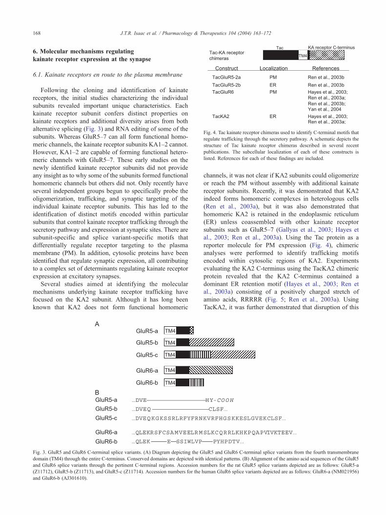
Fig. 4. Tac kainate receptor chimeras used to identify C-terminal motifs that
regulate trafficking through the secretory pathway. A schematic depicts the
structure of Tac kainate receptor chimeras described in several recent
publications. The subcellular localization of each of these constructs is
listed. References for each of these findings are included.
J.T.R. Isaac et al. / Pharmacology & Therapeutics 104 (2004) 163–172168
6. Molecular mechanisms regulating
kainate receptor expression at the synapse
6.1. Kainate receptors en route to the plasma membrane
Following the cloning and identification of kainate
receptors, the initial studies characterizing the individual
subunits revealed important unique characteristics. Each
kainate receptor subunit confers distinct properties on
kainate receptors and additional diversity arises from both
alternative splicing (Fig. 3) and RNA editing of some of the
subunits. Whereas GluR5–7 can all form functional homo-
meric channels, the kainate receptor subunits KA1–2 cannot.
However, KA1–2 are capable of forming functional hetero-
meric channels with GluR5–7. These early studies on the
newly identified kainate receptor subunits did not provide
any insight as to why some of the subunits formed functional
homomeric channels but others did not. Only recently have
several independent groups begun to specifically probe the
oligomerization, trafficking, and synaptic targeting of the
individual kainate receptor subunits. This has led to the
identification of distinct motifs encoded within particular
subunits that control kainate receptor trafficking through the
secretory pathway and expression at synaptic sites. There are
subunit-specific and splice variant-specific motifs that
differentially regulate receptor targeting to the plasma
membrane (PM). In addition, cytosolic proteins have been
identified that regulate synaptic expression, all contributing
to a complex set of determinants regulating kainate receptor
expression at excitatory synapses.
Several studies aimed at identifying the molecular
mechanisms underlying kainate receptor trafficking have
focused on the KA2 subunit. Although it has long been
known that KA2 does not form functional homomeric
Fig. 3. GluR5 and GluR6 C-terminal splice variants. (A) Diagram depicting the G
domain (TM4) through the entire C-terminus. Conserved domains are depicted with
and GluR6 splice variants through the pertinent C-terminal regions. Accession n
(Z11712), GluR5-b (Z11713), and GluR5-c (Z11714). Accession numbers for the h
and GluR6-b (AJ301610).
channels, it was not clear if KA2 subunits could oligomerize
or reach the PM without assembly with additional kainate
receptor subunits. Recently, it was demonstrated that KA2
indeed forms homomeric complexes in heterologous cells
(Ren et al., 2003a), but it was also demonstrated that
homomeric KA2 is retained in the endoplasmic reticulum
(ER) unless coassembled with other kainate receptor
subunits such as GluR5–7 (Gallyas et al., 2003; Hayes et
al., 2003; Ren et al., 2003a). Using the Tac protein as a
reporter molecule for PM expression (Fig. 4), chimeric
analyses were performed to identify trafficking motifs
encoded within cytosolic regions of KA2. Experiments
evaluating the KA2 C-terminus using the TacKA2 chimeric
protein revealed that the KA2 C-terminus contained a
dominant ER retention motif (Hayes et al., 2003; Ren et
al., 2003a) consisting of a positively charged stretch of
amino acids, RRRRR (Fig. 5; Ren et al., 2003a). Using
TacKA2, it was further demonstrated that disruption of this
luR5 and GluR6 C-terminal splice variants from the fourth transmembrane
identical patterns. (B) Alignment of the amino acid sequences of the GluR5
umbers for the rat GluR5 splice variants depicted are as follows: GluR5-a
uman GluR6 splice variants depicted are as follows: GluR6-a (NM021956)

Fig. 5. Schematic of kainate receptor C-termini depicting the location of
defined motifs that regulate trafficking through the secretory pathway. The
positively charged motifs within GluR5 and KA2 regulate ER retention,
whereas the positively charged motif in GluR6 regulates egress from the ER.
J.T.R. Isaac et al. / Pharmacology & Therapeutics 104 (2004) 163–172 169
stretch of 5 arginines abolished ER retention. Surprisingly,
PM expression of TacKA2 was still quite low, and
additional mutations revealed a putative C-terminal di-
leucine endocytic motif that mediated the rapid endocytosis
and low steady-state PM expression of TacKA2 even
following mutations of the ER retention motif. The
disruption of both the ER retention motif and the putative
endocytic motif increased PM expression of TacKA2;
however, the same mutations in full-length KA2 did not
yield functional homomeric channels (Ren et al., 2003a).
Thus, for KA2, both egress from the ER and the formation
of functional kainate receptors depend on heterooligomeri-
zation with other kainate receptor subunits (Gallyas et al.,
2003; Hayes et al., 2003; Ren et al., 2003a).
Using similar approaches, additional studies have now
defined trafficking motifs that regulate PM expression of
both GluR5 and GluR6 (Ren et al., 2003b; Jaskolski et al.,
2004; Yan et al., 2004). Unlike KA2, GluR6 is known to
form functional homomeric channels. However, in a recent
study, GluR6 was also demonstrated to have trafficking
information encoded within its C-terminus (Yan et al.,
2004). Systematic analysis of the GluR6 C-terminus using
truncation analyses of a GFP-tagged GluR6 subunit
revealed that a region between amino acids 868 and 880
was critical for GluR6 egress from the ER. Further site-
directed mutagenesis led to the identification of a positively
charged stretch of amino acids, CQRRLKH, that is essential
for efficient PM expression of GluR6 (Fig. 5). Disruption of
this motif using site-directed mutagenesis results in the ER
retention of a TacGluR6 chimera (Fig. 4), and similar
mutations in full-length GluR6 resulted in the reduction of
functional homomeric GluR6 channels expressed on the
PM. Interestingly, GluR6, which contains point mutations
within the CQRRLKH motif causing it to be ER retained,
still traffics to the PM when coexpressed with wild-type
GluR6 or KA2. Therefore, the GluR6 egress motif is not
essential, and oligomerization ultimately controls PM
expression. Jaskolski et al. also tested the possibility that
the PDZ-binding domain on GluR6 (-ETMA) described
previously (Garcia et al., 1998) could promote PM
expression and actively relieve ER retention as has been
described previously for the NR1–3 splice variant of the
NR1 subunit of NMDA receptors (Standley et al., 2000;
Scott et al., 2001; Xia et al., 2001). However, unlike NR1–3,
mutations in the PDZ-binding domain played no role in PM
expression of GluR6 (Jaskolski et al., 2004).
GluR5, like GluR6, is capable of forming homomeric
channels and also of combining with KA1–2 to form
heteromeric channels. Also like GluR6, GluR5 has multiple
isoforms due to alternative splicing (Fig. 3) and mRNA
editing. Two new studies demonstrate that alternative
splicing of GluR5 plays a critical role in PM expression
(Ren et al., 2003b; Jaskolski et al., 2004). Although all GluR5
splice variants are predominantly retained in the ER
(Jaskolski et al., 2004), the GluR5-2c splice variant displays
particularly robust ER retention. Interestingly, the 29 amino
acid insert in GluR5-2c contains a RXR ER retention motif
similar to that described for KA2, NMDA receptors, and K+
channels (Fig. 5). When this RLR motif is mutated to ALA,
GluR5-2c traffics to the PM in both heterologous cells and
neurons (Jaskolski et al., 2004). In addition, positively
charged residues in GluR5-2b that are conserved with
GluR5-2c have also been demonstrated to regulate egress
from the ER (Fig. 5; Ren et al., 2003b). Using the TacGluR5-
2b construct, it was demonstrated that mutation of R896, as
well as R900 and K901, promotes surface expression in
heterologous cells and hippocampal neurons. Furthermore, as
described for GluR6 (Jaskolski et al., 2004), mutations in the
GluR5 C-terminal PDZ binding domain do not regulate PM
expression (Ren et al., 2003b). Therefore, for GluR5, GluR6,
and KA2, the C-terminus has been found to contain essential
cis-acting elements that regulate trafficking to the PM.
6.2. Beyond the C-terminus
Whereas these recent studies have conclusively demon-
strated that the C-termini of kainate receptors, like all
glutamate receptors, contain essential motifs for trafficking
to the PM and regulation of synaptic expression, it is also
likely that the smaller intracellular loops and the trans-
membrane domains contain information critical for oligo-
merization, PM expression, and synaptic targeting. Indeed,
oligomerization has been shown previously to be dependent
on the extracellular N-terminus and possibly the trans-
membrane regions of AMPA and NMDA receptors
(Leuschner & Hoch, 1999; Ayalon & Stern-Bach, 2001;
Meddows et al., 2001). In addition, the pore lining region,
TM2, of the AMPA receptor subunit GluR2 contains a
critical arginine residue that is determined by alternative
splicing (the Q/R site). This amino acid determines the
calcium permeability of GluR2-containing AMPA receptors.
A study characterizing the trafficking of GluR2 through the
secretory pathway has demonstrated that the Q/R site also

J.T.R. Isaac et al. / Pharmacology & Therapeutics 104 (2004) 163–172170
regulates the egress of GluR2 from the ER (Greger et al.,
2002) as well as the tetramerization of AMPA receptors
(Greger et al., 2003). Furthermore, metabotropic glutamate
receptors have important protein binding and trafficking
information encoded in their intracellular loops. For
example, the specific interactions with G proteins are via
the metabotropic glutamate receptor second and third
intracellular loops (reviewed in Conn & Pin, 1997).
Although no trafficking motifs have been described within
the smaller loops of the ionotropic glutamate receptors,
other neurotransmitter receptors have trafficking informa-
tion encoded in regions other than the C-termini (Boyd et
al., 2003; Tan et al., 2004).
6.3. Kainate receptor binding
proteins and synaptic targeting
In recent years, several glutamate receptor binding
proteins have been identified. Many of these proteins were
isolated with the help of a yeast 2-hybrid screen in which the
intracellular C-termini of individual glutamate receptor
subunits were used as bbaitQ in screens of yeast 2-hybrid
libraries. Whereas some of the identified binding proteins
are subtype specific or display unique binding to a particular
subunit of a glutamate receptor subtype, other binding
proteins are more promiscuous and interact with KA,
AMPA, and NMDA receptors. We will discuss several of
the kainate receptor binding proteins, which have been
characterized in recent years and have been implicated in the
trafficking and synaptic expression of kainate receptors (for
related reviews on the subject, see De La Rue & Henley,
2002; Collingridge & Isaac, 2003).
Like many glutamate receptors, kainate receptors contain
a PDZ-binding motif at their extreme C-terminus. Studies on
the prototypic PDZ domain-containing family of proteins
including PSD-95/SAP90 have demonstrated that these
proteins bind to kainate receptors, cluster them, and
modulate their desensitization (Garcia et al., 1998; Mehta
et al., 2001). In another study, 4 PDZ domain-containing
proteins were isolated that bind to kainate receptors, PSD-
95, GRIP, PICK1, and syntenin (Hirbec et al., 2003). None
of these proteins are specific for kainate receptors among the
ionotropic glutamate receptors, as they also interact with
AMPA receptor and in some cases NMDA receptors (Kim et
al., 1995; Kornau et al., 1995; Dong et al., 1997; Hirbec et
al., 2002). Although at first glance the kainate receptor
binding proteins appear to lack binding specificity, studies
demonstrate functionally distinct roles for these proteins in
regulating kainate and AMPA receptors. Using an approach
that takes advantage of fusion proteins of kainate receptor
C-termini or synthetic peptides that differentially disrupt
binding of kainate receptors to PSD-95, GRIP, PICK1, and
syntenin, Hirbec et al. described the differential regulation
of AMPA and kainate receptors at hippocampal mossy
fiber–CA3 synapses of the hippocampus. Infusion of
GluR5-2b C-terminal fusion proteins resulted in the loss
of kainate receptor-mediated synaptic currents without
affecting the AMPA component. Using fusion proteins with
specific point mutations in the GluR5-2b C-terminus or
peptides to compete off endogenous interactions with
kainate and AMPA receptors, they provide evidence that
interactions with GRIP decrease synaptic kainate receptor
function while simultaneously increasing AMPA receptor
function at synapses. In addition, blockade of PICK1
interactions caused a depression of the kainate receptor-
mediated EPSC but had no effect on the AMPA receptor-
mediated EPSC. Although some of these peptides have also
been reported to affect presynaptic release probability at
mossy fiber–CA3 synapses through a mechanism involving
GRIP-Eph receptor interactions (Contractor et al., 2002), the
simultaneous differential regulation of AMPA and kainate
receptor-mediated responses at the same population of
inputs cannot be explained by such a mechanism. Rather,
these data indicate distinct roles for PICK1 and GRIP in the
regulation of synaptic AMPA and kainate receptors.
Previously, these proteins have been shown to regulate
AMPA receptor function by regulating surface expression;
thus, this is also a likely mechanism for the regulation of
kainate receptors by these interactors. Overall, these studies
point to a complex interplay between synaptic scaffolding
proteins and the various glutamate receptors at the
postsynaptic density.
In addition to PDZ domain-containing proteins, kainate
receptors have also been shown to interact with cadherin/
catenin complexes (Coussen et al., 2002), specifically h-catenin. Although cadherins/catenins do not possess PDZ
domains, they are known to interact with the PDZ domain-
containing proteins CASK and PSD-95 (Perego et al.,
2000), thereby still implicating PDZ domain interactions in
kainate receptor trafficking and synaptic targeting. It is
intriguing that these findings demonstrate that the GluR6
PDZ ligand (-ETMA) at the extreme C-terminus is required
for the h-catenin interaction, yet the GluR6-h-catenininteraction is indirect and not dependent on PSD-95 or
CASK (Coussen et al., 2002). More work is required to
determine the precise molecular basis for the GluR6
association with cadherin/catenin complexes and to define
the PDZ domain-containing proteins that link these
proteins.
In addition to the kainate receptor binding proteins
characterized thus far, it is likely that additional components
of the intracellular trafficking machinery will eventually be
shown to interact with kainate receptors. The numerous
subunit- and splice variant-specific trafficking motifs that
have been identified in the past year (Fig. 5) most likely
mediate specific protein-protein interactions regulating
particular steps in the secretory pathway. As future research
reveals additional binding partners, we will undoubtedly
learn more about the mechanisms regulating the precise
targeting of kainate receptors to both presynaptic and
postsynaptic sites and the functional consequences of such
interactions.

J.T.R. Isaac et al. / Pharmacology & Therapeutics 104 (2004) 163–172 171
7. Conclusions and prospects
A consistent theme in the study of kainate receptors is
that neurons clearly have the ability to traffic and target
kainate receptors differentially from the closely related
AMPA receptors that coexist in the same cells. Moreover,
neurons can target kainate receptors in a subunit specific
manner, so that receptor complexes of specific compositions
and functions can be located at numerous different locations
within the same cell. How this is achieved is at present
largely unknown. Trafficking studies of kainate receptors
are still in their infancy in comparison with the better-
studied ionotropic AMPA and NMDA glutamate receptor
subtypes. However, it is already evident that mechanisms
exist for the subunit- and splice variant-specific trafficking
of kainate receptors. The challenge is now on to further
elucidate these mechanisms and understand how such
processes can lead to the complex differential and regulated
targeting of kainate receptors within neurons.
Acknowledgments
Supported by the Wellcome Trust (J.M. and J.T.R.I.),
MRC (J.M. and J.T.R.I.), and NINDS/NIH (K.W.R., D.H.,
and J.T.R.I.).
References
Ayalon, G., & Stern-Bach, Y. (2001). Functional assembly of AMPA and
kainate receptors is mediated by several discrete protein-protein
interactions. Neuron 31, 103–113.
Bahn, S., Volk, B., & Wisden, W. (1994). Kainate receptor gene expression
in the developing rat brain. J Neurosci 14, 5525–5547.
Bettler, B., & Mulle, C. (1995). Neurotransmitter receptors. II. AMPA and
kainate receptors. Neuropharmacology 34, 123–139.
Bortolotto, Z. A., Clarke, V. R. J., Delany, C. M., Parry, M. C., Smolders, I.,
Vignes, M., et al. (1999). Kainate receptors are involved in synaptic
plasticity. Nature 402, 297–301.
Boyd, G. W., Doward, A. I., Kirkness, E. F., Millar, N. S., & Connolly, C.
N. (2003). Cell surface expression of 5-hydroxytryptamine type 3
receptors is controlled by an endoplasmic reticulum retention signal. J
Biol Chem 278, 27681–27687.
Bureau, B., Bischoff, B., Heinemann, S. F., & Mulle, C. (1999). Kainate
receptor-mediated responses in the CA1 field of wild-type and
GluR6-.deficient mice. J Neurosci 19, 653–663.
Bureau, I., Dieudonne, S., Coussen, F., & Mulle, C. (2000). Kainate
receptor-mediated synaptic currents in cerebellar Golgi cells are
not shaped by diffusion of glutamate. Proc Natl Acad Sci USA 97,
6838–6843.
Castillo, P. E., Malenka, R. C., & Nicoll, R. A. (1997). Kainate receptors
mediate a slow postsynaptic current in hippocampal CA3 neurons.
Nature 388, 182–186.
Chittajallu, R., Vignes, M., Dev, K. K., Barnes, J. M., Collingridge, G. L.,
& Henley, J. M. (1996). Regulation of glutamate release by presynaptic
kainate receptors in the hippocampus. Nature 379, 78–81.
Clarke, V. R. J., & Collingridge, G. L. (2002). Characterisation of the
effects of ATPA, a GLUK5 receptor selective agonist, on excitatory
synaptic transmission in area CA1 of rat hippocampal slices. Neuro-
pharmacology 42, 889–902.
Clarke, V. R., Ballyk, B. A., Hoo, K. H., Mandelzys, A., Pellizzari, A.,
Bath, C. P., et al. (1997). A hippocampal GluR5 kainate receptor
regulating inhibitory synaptic transmission. Nature 389, 599–603.
Collingridge, G. L., & Isaac, J. T. R. (2003). Functional roles of protein
interactions with AMPA and kainate receptors. Neurosci Res 47, 3–15.
Conn, P. J., & Pin, J. P. (1997). Pharmacology and functions of
metabotropic glutamate receptors. Annu Rev Pharmacol Toxicol 37,
205–237.
Contractor, A., Swanson, G. T., Sailer, A., O’Gorman, S., & Heinemann, S.
F. (2000). Identification of the kainate receptor subunit underlying
modulation of excitatory synaptic transmission in the CA3 region of the
hippocampus. J Neurosci 15, 8269–8278.
Contractor, A., Rogers, C., Maron, C., Henkemeyer, M., Swanson, G. T., &
Heinemann, S. F. (2002). Trans-synaptic Eph receptor-ephrin signaling
in hippocampal mossy fiber LTP. Science 296, 1864–1869.
Contractor, A., Sailer, A. W., Darstein, M., Maron, C., Xu, J., Swanson, G.
T., et al. (2003). Loss of kainate receptor-mediated heterosynaptic
facilitation of mossy-fiber synapses in KA2�/� mice. J Neurosci 23,
422–429.
Cossart, R., Esclapez, M., Hirsch, J. C., Bernard, C., & Ben-Ari, Y. (1998).
GluR5 kainate receptor activation in interneurons increases tonic
inhibition of pyramidal cells. Nat Neurosci 1, 470–478.
Cossart, R., Tyzio, R., Dinocourt, C., Esclapez, M., Hirsch, J. C., Ben-Ari,
Y., et al. (2001). Presynaptic kainate receptors that enhance the release
of GABA on CA1 hippocampal interneurons. Neuron 29, 497–508.
Coussen, F., Normand, E., Marchal, C., Costet, P., Choquet, D., Lambert,
M., et al. (2002). Recruitment of the kainate receptor subunit
glutamate receptor 6 by cadherin/catenin complexes. J Neurosci 22,
6426–6436.
Darstein, M., Petralia, R. S., Swanson, G. T., Wenthold, R. J., &
Heinemann, S. F. (2003). Distribution of kainate receptor subunits at
hippocampal mossy fiber synapses. J Neurosci 23, 8013–8019.
De La Rue, S. A., & Henley, J. M. (2002). Proteins involved in the
trafficking and functional synaptic expression of AMPA and KA
receptors. Sci World J 2, 461–482.
Dong, H., O’Brien, R. J., Fung, E. T., Lanahan, A. A., Worley, P. F., et al.
(1997). GRIP: A synaptic PDZ domain-containing protein that interacts
with AMPA receptors. Nature 386, 279–284.
Frerking, M., & Nicoll, R. A. (2000). Synaptic kainate receptors. Curr Opin
Neurobiol 10, 342–351.
Frerking, M., Malenka, R. C., & Nicoll, R. A. (1998). Synaptic activation
of kainate receptors on hippocampal interneurons. Nat Neurosci 1,
479–486.
Frerking, M., Petersen, C. C. H., & Nicoll, R. A. (1999). Mechanisms
underlying kainate receptor-mediated disinhibition in the hippocampus.
Proc Natl Acad Sci USA 96, 12917–12922.
Frerking, M., Schmitz, D., Zhou, Q., Johansen, J., & Nicoll, R. A. (2001).
Kainate receptors depress excitatory synaptic transmission at CA3-CA1
synapses in the hippocampus via a direct presynaptic action. J Neurosci
21, 2958–2966.
Gallyas, F., Jr., Ball, S. M., & Molnar, E. (2003). Assembly and cell surface
expression of KA-2 subunit-containing kainate receptors. J Neurochem
86, 1414–1427.
Garcia, E. P., Mehta, S., Blair, L. A., Wells, D. G., Shang, J., Fukushima, T.,
et al. (1998). SAP90 binds and clusters kainate receptors causing
incomplete desensitization. Neuron 21, 727–739.
Greger, I. H., Khatri, L., & Ziff, E. B. (2002). RNA editing at arg607
controls AMPA receptor exit from the endoplasmic reticulum. Neuron
34, 759–772.
Greger, I. H., Khatri, L., Kong, X., & Ziff, E. B. (2003). AMPA receptor
tetramerization is mediated by Q/R editing. Neuron 40, 763–774.
Hayes, D. M., Braud, S., Hurtado, D. E., McCallum, J., Standley, S.,
Isaac, J. T., et al. (2003). Trafficking and surface expression of the
glutamate receptor subunit, KA2. Biochem Biophys Res Commun 310,
8–13.
Hirbec, H., Perestenko, O., Nishimune, A., Meyer, G., Nakanishi, S.,
Henley, J. M., et al. (2002). The PDZ proteins PICK1, GRIP, and

J.T.R. Isaac et al. / Pharmacology & Therapeutics 104 (2004) 163–172172
syntenin bind multiple glutamate receptor subtypes. Analysis of PDZ
binding motifs. J Biol Chem 277, 15221–15224.
Hirbec, H., Francis, J. C., Lauri, S. E., Braithwaite, S. P., Coussen, F.,
Mulle, C., et al. (2003). Rapid and differential regulation of AMPA and
kainate receptors at hippocampal mossy fibre synapses by PICK1 and
GRIP. Neuron 37, 625–638.
Jaskolski, F., Coussen, F., Nagarajan, N., Normand, E., Rosenmund, C., &
Mulle, C. (2004). Subunit composition and alternative splicing regulate
membrane delivery of kainate receptors. J Neurosci 24, 2506–2515.
Jiang, L., Xu, J., Nedergaard, M., & Kang, J. (2001). A kainate receptor
increases the efficacy of GABAergic synapses. Neuron 30, 503–513.
Kamiya, H., & Ozawa, S. (1998). Kainate receptor-mediated inhibition of
presynaptic Ca2+ influx and EPSP in area CA1 of the rat hippocampus.
J Physiol 509, 833–845.
Kang, N., Jiang, L., He, W., Xu, J., Nedergaard, M., & Kang, J. (2004).
Presynaptic inactivation of action potentials and postsynaptic inhibition
of GABAA currents contribute to KA-induced disinhibition in CA1
pyramidal neurons. J Neurophysiol 92, 873–882.
Kidd, F. L., & Isaac, J. T. R. (1999). Developmental and activity-dependent
regulation of kainate receptors at thalamocortical synapses. Nature 400,
569–573.
Kidd, F. L., & Isaac, J. T. R. (2001). Kinetics and activation of postsynaptic
kainate receptors at thalamocortical synapses: role of glutamate
clearance. J Neurophysiol 86, 1139–1148.
Kidd, F. L., Coumis, U., Collingridge, G. L., Crabtree, J. W., & Isaac, J. T.
R. (2002). A kainate autoreceptor is involved in regulating the dynamic
properties of thalamocortical synapses during development. Neuron 34,
635–646.
Kim, E., Niethammer, M., Rothschild, A., Jan, Y. N., & Sheng, M. (1995).
Clustering of Shaker-type K+ channels by interaction with a family of
membrane-associated guanylate kinases. Nature 378, 85–88.
Kornau, H. C., Schenker, L. T., Kennedy, M. B., & Seeburg, P. H. (1995).
Domain interaction between NMDA receptor subunits and the
postsynaptic density protein PSD-95. Science 269, 1737–1740.
Kullmann, D. M. (2001). Presynaptic kainate receptors in the hippocampus:
slowly emerging from obscurity. Neuron 32, 561–564.
Lauri, S. E., Bortolotto, Z. A., Bleakman, D., Ornstein, P. L., Lodge, D.,
Isaac, J. T. R., et al. (2001a). A critical role of a facilitatory kainate
autoreceptor in mossy fibre LTP. Neuron 32, 697–709.
Lauri, S. E., Delaney, C., Clarke, V., Bortolotto, Z. A., Ornstein, P. L.,
Isaac, J. T. R., et al. (2001b). Synaptic activation of a presynaptic
kainate receptor facilitates AMPA receptor-mediated synaptic trans-
mission at hippocampal mossy fibre synapses. Neuropharmacology 41,
907–915.
Lerma, J. (2003). Roles and rules of kainate receptors in synaptic
transmission. Nat Rev Neurosci 4, 481–495.
Lerma, J., Paternain, A. V., Rodriguez-Moreno, A., & Lopez-Garcia, J. C.
(2001). Molecular physiology of kainate receptors. Physiol Rev 81,
971–998.
Leuschner, W. D., & Hoch, W. (1999). Subtype-specific assembly of
alpha-amino-3-hydroxy-5-methyl-4-isoxazole propionic acid receptor
subunits is mediated by their N-terminal domains. J Biol Chem 274,
16907–16916.
Maccaferri, G., & Lacaille, J. -C. (2003). Interneuron Diversity series:
hippocampal interneuron classifications—making things as simple as
possible, not simpler. Trends Neurosci 26, 564–671.
Meddows, E., Le Bourdelles, B., Grimwood, S., Wafford, K., Sandhu, S.,
Whiting, P., et al. (2001). Identification of molecular determinants that
are important in the assembly of N-methyl-d-aspartate receptors. J Biol
Chem 276, 18795–18803.
Mehta, S., Wu, H., Garner, C. C., & Marshall, J. (2001). Molecular
mechanisms regulating the differential association of kainate
receptor subunits with SAP90/PSD-95 and SAP97. J Biol Chem
276, 16092–16099.
Melyan, Z., Wheal, H. V., & Lancaster, B. (2002). Metabotropic-mediated
kainate receptor regulation of IsAHP and excitability in pyramidal cells.
Neuron 34, 107–114.
Melyan, Z., Lancaster, B., & Wheal, H. V. (2004). Metabotropic regulation
of intrinsic excitability by synaptic activation of kainate receptors. J
Neurosci 24, 4530–4534.
Min, M. -Y., Rusakov, D. A., & Kullmann, D. M. (1998). Activation of
AMPA, kainate and metabotropic receptors at hippocampal mossy fiber
synapses: role of glutamate diffusion. Neuron 21, 561–570.
Min, M. -Y., Melyan, Z., & Kullmann, D. M. (1999). Synaptically released
glutamate reduces gamma-aminobutyric acid (GABA)ergic inhibition in
the hippocampus via kainate receptors. Proc Natl Acad Sci USA 96,
9932–9937.
Mulle, C., Sailer, A., Perez-Otano, I., Dickinson-Anson, H., Castillo, P. E.,
Bureau, I., et al. (1998). Altered synaptic physiology and reduced
susceptibility to kainate-induced seizures in GluR6-deficient mice.
Nature 98, 601–605.
Mulle, C., Sailer, A., Swanson, G. T., Brana, C., O’Gorman, S., Bettler, B.,
et al. (2000). Subunit composition of kainate receptors in hippocampal
interneurons. Neuron 28, 475–484.
Paternain, A. V., Herrera, M. T., Nieto, M. A., & Lerma, J. (2000). GluR5
and GluR6 kainate receptor subunits coexist in hippocampal neurons
and coassemble to form functional receptors. J Neurosci 20, 196–205.
Perego, C., Vanoni, C., Massari, S., Longhi, R., & Pietrini, G. (2000).
Mammalian LIN-7 PDZ proteins associate with beta-catenin at the cell-
cell junctions of epithelia and neurons. EMBO J 19, 3978–3989.
Ren, Z., Riley, N. J., Garcia, E. P., Sanders, J. M., Swanson, G. T., &
Marshall, J. (2003a). Multiple trafficking signals regulate kainate
receptor KA2 subunit surface expression. J Neurosci 23, 6608–6616.
Ren, Z., Riley, N. J., Needleman, L. A., Sanders, J. M., Swanson, G. T., &
Marshall, J. (2003b). Cell surface expression of GluR5 kainate receptors
is regulated by an endoplasmic reticulum retention signal. J Biol Chem
278, 52700–52709.
Rodriguez-Moreno, A., & Lerma, J. (1998). Kainate receptor modu-
lation of GABA release involves a metabotropic function. Neuron
20, 1211–1218.
Rodriguez-Moreno, A., Herreras, O., & Lerma, J. (1997). Kainate receptors
presynaptically downregulate GABAergic inhibition in the rat hippo-
campus. Neuron 19, 893–901.
Rodriguez-Moreno, A., Lopez-Garcia, J. C., & Lerma, J. (2000). Two
populations of kainate receptors with separate signaling mechanisms in
hippocampal interneurons. Proc Natl Acad Sci USA 97, 1293–1298.
Rozas, J. L., Paternain, A. V., & Lerma, J. (2003). Noncanonical signaling
by ionotropic kainate receptors. Neuron 39, 543–553.
Scott, D. B., Blanpied, T. A., Swanson, G. T., Zhang, C., & Ehlers, M. D.
(2001). An NMDA receptor ER retention signal regulated by
phosphorylation and alternative splicing. J Neurosci 21, 3063–3072.
Semyanov, A., & Kullmann, D. M. (2001). Kainate receptor-dependent
axonal depolarization and action potential initiation in interneurons. Nat
Neurosci 4, 718–723.
Standley, S., Roche, K. W., McCallum, J., Sans, N., & Wenthold, R. J.
(2000). PDZ domain suppression of an ER retention signal in NMDA
receptor NR1 splice variants. Neuron 28, 887–898.
Tan, C. M., Brady, A. E., Nickols, H. H., Wang, Q., & Limbird, L. E.
(2004). Membrane trafficking of G protein-coupled receptors. Annu Rev
Pharmacol Toxicol 44, 559–609.
Vignes, M., & Collingridge, G. L. (1997). The synaptic activation of
kainate receptors. Nature 388, 179–182.
Vignes, M., Clarke, V. R. J., Parry, M. J., Bleakman, D., Lodge, D.,
Ornstein, P. L., et al. (1998). The GluR5 subtype of kainate receptor
regulates excitatory synaptic transmission in areas CA1 and CA3 of the
rat hippocampus. Neuropharmacology 37, 1269–1277.
Wisden, W., & Seeburg, P. H. (1993). A complex mosaic of high-affinity
kainate receptors in rat brain. J Neurosci 13, 3582–3598.
Xia, H., Hornby, Z. D., & Malenka, R. C. (2001). An ER retention signal
explains differences in surface expression of NMDA and AMPA
receptor subunits. Neuropharmacology 41, 714–723.
Yan, S., Sanders, J. M., Xu, J., Zhu, Y., Contractor, A., & Swanson, G. T.
(2004). A C-terminal determinant of GluR6 kainate receptor trafficking.
J Neurosci 24, 679–691.