
1973 Editing Manual - and Reference Information - Deep Blue ...
Upload
khangminh22Category
view
3download
0
Kv1.1 RNA Editing: Physiological Roles and its Implications for
Episodic Ataxia Type-1
By
Elizabeth Ferrick Kiddie
Dissertation
Submitted to the Faculty of the
Graduate School of Vanderbilt University
in partial fulfillment of the requirements
for the degree of
DOCTOR OF PHILOSOPHY
in
Molecular Physiology and Biophysics
May, 2017
Nashville, Tennessee
Approved:
Ronald B. Emeson, Ph.D.
Charles E. Cobb, Ph.D.
Roger J. Colbran, Ph.D.
Aurelio A. Galli, Ph.D.
Jennifer A. Kearney, Ph.D
.

ii
This dissertation is dedicated to my family, friends, and especially my lab and my
husband- who all in their own ways encouraged me forward scientifically,
spiritually, and emotionally.

iii
ACKNOWLEDGEMENTS
First and foremost, I would like to thank my adviser, Dr. Ron Emeson,
for your mentorship that made my dissertation research possible and set me up
with opportunities that will surely propel my scientific career forward. Thank you
for demonstrating scientific curiosity and rigor for replicability. Dr. Chuck Cobb,
thank you for being more than just my committee chair- you were a key support
for helping me navigate my time at Vanderbilt. Thank you to Dr. Roger Colbran,
Dr. Aurelio Galli, and Dr. Jennifer Kearney for serving on my committee and
spurring me to think critically about the progression of my research. Thank you to
the members of the Molecular Physiology & Biophysics department for asking
thoughtful questions at seminars and for being a community of scientific rigor.
Thank you to the members of my lab, past and present, Dr. Randi Ulbricht, Dr.
Jennifer Hood, Dr. Richard O’Neil, Hussain Jinnah, Turnee Malik, Kayla
Shumate, Chris Hofmann, Li Peng, Beth Sims, and Katie Patterson. You were
my advisers, teachers of techniques, co-laborers of experiments, and listening
ears for troubleshooting. Thank you to Li Peng for your expertise in cell culture
and for aiding me in the cell experiments of my research. In particular, thanks to
Katie Patterson whose superb mouse husbandry, expertise of behavioral tests,
and peripheral tissue dissection skills made all the mouse experiments possible.
Thank you to the many, many collaborators who have contributed to
the success of my project. Thank you to Dr. Roberts Reenan (Brown University)
for developing and sharing the non-edited [Kv1.1(I)] and edited [Kv1.1(V)] mice

iv
with us. Thank you to Dr. James Maylie (Oregon Health & Science University)
who provided the Kv1.1 V408A/+ mice and was generous with his time by email
and in person to discuss his expertise in the Episodic ataxia type-1 field. Thank
you to Dr. Danny Winder and Dr. Samuel Centanni for your expertise in
performing hippocampal slice electrophysiology for me. Huge thanks to Dr.
Joshua Rosenthal (Woods Hole and University of Puerto Rico Medical Sciences
Campus) for hosting me at his laboratory in Puerto Rico. You provided key
mentorship for performing, analyzing, and writing all the data derived from the
Xenopus oocyte electrophysiological experiments. Thank you to the members of
Dr. Rosenthal’s lab for being so welcoming and teaching and advising me in all
the techniques I needed to learn in a short amount of time, Dr. Laura Fernández-
Alacid, Dr. Nuria Vazquez, Dr. Maria Fernanda Montiel-Gonzalez. Isabel
Vallecillo-Viejo, and Sonia Soto (who in particular isolated the Xenopus oocytes
for my experiments). Thank you to Dan Ayers, the master statistician who helped
build complex statistical models when he could and advised me on the statistics
for the rest- you have made me care more about statistics then I ever thought I
would. Thank you to the friends across departments who helped me find and
fine-tune my dissertation story, in particular Amanda Meyer, Dr. Andrea Belovich,
Britney Lizama-Manibusan, and, of course, my husband Dr. Bradley Kiddie.
Funding for this work was provided by the Vanderbilt Molecular
Endocrinology Training Program (T32DK007563), the Ruth L. Kirschstein
National Research Service Award (F31NS087911), a Vanderbilt Dissertation
Enhancement Grant, and the Vanderbilt Joel G. Hardman Chair in

v
Pharmacology. Additional support for the research, which occurred in Dr. Joshua
Rosenthal’s lab, included NINDS (R0111223855, R01NS64259), the Cystic
Fibrosis Foundation Therapeutics (Rosent14XXO), NIGMS (P20GM103642),
NIMH (G12-MD007600), and NSF (DBI 0115825, DBI 1337284).
Finally, thank you to my supportive family and friends who made the
time I spent at Vanderbilt and in Nashville a pleasant and formative time of life.

vi
TABLE OF CONTENTS
Page
DEDICATION ........................................................................................................ ii
ACKNOWLEDGEMENTS ..................................................................................... iii
LIST OF TABLES ................................................................................................. ix
LIST OF FIGURES ............................................................................................... x
LIST OF EQUATIONS ........................................................................................ xiii
LIST OF ABBREVIATIONS ................................................................................ xiv
Chapter
I. INTRODUCTION .............................................................................................. 1
A brief history of our understanding of the variety of influences on genetic flow .................................................................................................... 1 A-to-I RNA Editing ............................................................................................ 2 Voltage-gated potassium channels................................................................... 5 Kv1.1 and the Kv1 family .................................................................................. 9 Episodic Ataxia type-1 .................................................................................... 16 Modulation of Kv1.1 function by RNA editing ................................................. 18 Summary ........................................................................................................ 26 II. FUNCTIONAL CONSEQUENCES OF KV1.1 RNA EDITING IN MOUSE
MODELS ........................................................................................................ 29 Introduction ..................................................................................................... 29 Materials & Methods ....................................................................................... 34 Mouse Experiment Approval ........................................................................ 34 Characterization of Kv1.1 editing in wild-type mouse tissues ...................... 35 Generation of non-edited [Kv1.1(I)] and edited [Kv1.1(V)] mutant mice ....... 35
Kv1.1 RNA Expression Characterization in non-edited [Kv1.1(I)] and edited [Kv1.1(V)] mutant mice ................................................................... 38
Mendelian distribution of progeny ................................................................ 39 Behavioral Testing ....................................................................................... 40 Spontaneous Seizure Monitoring ................................................................. 42 Drug-induced Seizure Susceptibility ............................................................ 43 Hippocampal Slice Electrophysiology .......................................................... 46 Statistical analysis ........................................................................................ 47

vii
Results ........................................................................................................... 48 Characterization of Kv1.1 editing in wild-type mouse tissues ...................... 48 Verification of non-edited [Kv1.1(I)] and edited [Kv1.1(V)] mutant mouse models ........................................................................................... 49 Mendelian distribution of progeny ................................................................ 53 Behavioral characterization .......................................................................... 56 Spontaneous Seizure Monitoring ................................................................. 67 Drug-induced Seizure Susceptibility ............................................................ 67 Preliminary Electrophysiological characterization ........................................ 77 Discussion ...................................................................................................... 79 Characterization of Kv1.1 editing in wild-type mouse tissues ...................... 79 Verification of non-edited [Kv1.1(I)] and edited [Kv1.1(V)] mutant mouse models ........................................................................................... 81 Mendelian distribution of progeny ................................................................ 84 Behavioral characterization .......................................................................... 85 Spontaneous Seizure Monitoring and Drug-induced Seizure Susceptibility ............................................................................................. 91 Preliminary Electrophysiological characterization ........................................ 95 Summary ..................................................................................................... 96 III. INTERACTIONS BETWEEN KV1.1 RNA EDITING AND EPISODIC ATAXIA TYPE-1 MUTATIONS ....................................................................... 98 Introduction ..................................................................................................... 98 Materials & Methods ....................................................................................... 99
Kv1.1 and Kv1.1 constructs ....................................................................... 99 In vitro analysis of RNA editing .................................................................. 100 In vivo analysis of RNA editing .................................................................. 102 Electrophysiological recording in Xenopus oocytes ................................... 103 Statistical analysis ...................................................................................... 105 Results ......................................................................................................... 107 EA1-associated mutations alter RNA editing in vitro .................................. 107 A mouse model of EA1 (V408A/+) alters RNA editing in vivo .................... 110 Gating properties are altered between non-edited and edited Kv1.1 channels harboring EA1 mutations ......................................................... 112 Inactivation kinetics are altered between non-edited and edited EA1 mutant proteins ................................................................................ 122 Discussion .................................................................................................... 129 Proof of concept that existing mutations alter RNA editing ........................ 129 Edited Isoforms of EA1 mutants alter electrophysiological properties ........ 131 Incorporating RNA editing into the complex interactions involved in the EA1 disorder ..................................................................................... 133 IV. CONCLUSIONS......................................................................................... 135 Significance .................................................................................................. 135

viii
Future Directions .......................................................................................... 138 Utilizing the newly developed mouse models of Kv1.1 editing ................... 138 Continuing the study of RNA editing effects on EA1 .................................. 145 Appendix A. Comparative analysis of editing rates determined by Sanger sequencing
versus High-throughput sequence analysis .................................................. 150 B. Additional p-value tables .............................................................................. 153 C. Human variants found in the Kv1.1 mRNA duplex........................................ 157 REFERENCES ................................................................................................. 161

ix
LIST OF TABLES
Table
Page
2.1 Seizure threshold scale for scoring PTZ-induced seizures ..................... 44
2.2 Seizure threshold scale for scoring 4-AP-induced seizures ................... 46
2.3 Conditional non-Mendelian distribution of non-edited [Kv1.1(I)] mutant offspring ................................................................................................. 55
2.4 Conditional non-Mendelian distribution of edited [Kv1.1(V)] mutant offspring ................................................................................................. 55
3.1 Free energy calculations of wild-type and EA1 human mutant duplexes ............................................................................................... 108
3.2 Voltage-dependence of activation ........................................................ 115
3.3 Kinetics of recovery from Kv1.1 inactivation ....................................... 129
B.1 P-values corresponding to Figure 3.6 (time to half-activation) ............. 154
B.2 P-values corresponding to Figure 3.8 (closing kinetics) ....................... 155
B.3 P-values corresponding to Figure 3.10 (kinetics of Kv1.1 inactivation) .......................................................................................... 156
C.1 Human variants found in the Kv1.1 mRNA duplex ............................... 158

x
LIST OF FIGURES
Figure
Page
1.1 ADAR Enzymes convert adenosine residues to inosine by hydrolytic deamination ............................................................................................ 3
1.2 Domain topology for the Kv1.1 -subunit and accessory Kv-subunit .... 7
1.3 Conformational states of Kv channels ..................................................... 9
2.1 Targeting strategy to solely generate non-edited [Kv1.1(I)] or edited [Kv1.1(V)] isoforms in mutant mice ....................................................... 36
2.2 Design of duplex mutations to solely generate non-edited [Kv1.1(I)] or edited [Kv1.1(V)] transcripts in mutant mice ......................................... 37
2.3 Quantitative analysis of Kv1.1 RNA editing (I/V site) in mouse ............. 49
2.4 Confirmation of Kv1.1 isoform identity in mutant mice .......................... 51
2.5 Steady-state Kv1.1 RNA expression is unchanged in non-edited [Kv1.1(I)] and edited [Kv1.1(V)] mutant mice compared to wild-type mice ...................................................................................................... 53
2.6 Initial behavioral analysis of non-edited [Kv1.1(I)] and edited [Kv1.1(V)] mice ...................................................................................................... 57
2.7 Non-edited [KV1.1(I)] and edited [KV1.1(V)] heterozygous and homozygous mutant mice do not display locomotor dysfunction under the unstressed traditional rotarod paradigm .......................................... 58
2.8 Non-edited [Kv1.1(I)] and edited [Kv1.1(I)] mutant mice do not display locomotor dysfunction under the unstressed modified rotarod paradigm ............................................................................................... 60
2.9 Non-edited [Kv1.1(I)] mutant mice display stress-induced locomotor dysfunction under the stressed modified rotarod paradigm .................. 61
2.10 Non-edited [Kv1.1(I)] mice display reproducible stress-induced gait alterations ............................................................................................. 63
2.11 Non-edited [Kv1.1(I)] and edited [Kv1.1(V)] mice do not display stress-induced alterations by the inverted screen test ..................................... 65

xi
2.12 Open-field analysis of non-edited [Kv1.1(I)] and edited [Kv1.1(V)]
mice ...................................................................................................... 66
2.13 Flurothyl-induced seizures .................................................................... 68
2.14 PTZ-induced seizure threshold scores .................................................. 69
2.15 Male versus female comparison of PTZ-induced seizure threshold scores ................................................................................................... 70
2.16 Latency to hindlimb extension following PTZ-induced seizures ............ 71
2.17 4-AP-induced seizure threshold scores ................................................ 73
2.18 Male versus female comparison of 4-AP-induced seizure threshold scores ................................................................................................... 74
2.19 Latency to hindlimb extension following 4-AP-induced seizures ........... 76
2.20 Electrophysiological characterization of dentate gyrus granule cells of non-edited [Kv1.1(I)] and wild-type mice ............................................... 78
2.21 Stress-enhancement of rotarod performance ....................................... 89
3.1 Time course analysis to determine the linear range for the in vitro RNA editing rates ........................................................................................ 102
3.2 The proximity of the Kv1.1 editing site compared to the EA1 mutations responsible for V404I, I407M, and V408A .......................................... 108
3.3 Quantitative analysis of in vitro RNA editing rates for wild-type and mutant Kv1.1 transcripts ..................................................................... 110
3.4 Quantitative analysis of allele-specific Kv1.1 editing in V408A mutant mice .................................................................................................... 111
3.5 Voltage-dependence of non-edited compared to edited mutant channels ............................................................................................. 114
3.6 Editing alters opening (activation) kinetics of I407M channels ............ 116
3.7 I407M E and V408A E channels display decreased currents at the most positive voltages ................................................................................. 117
3.8 Editing alters closing (deactivating) kinetics of V404I channels .......... 119

xii
3.9 Long pulse characterization of WT and EA1 channels ........................ 121
3.10 Editing slows Kv1.1-induced inactivation kinetics of I407M and V408A channels ............................................................................................. 124
3.11 Long pulse characterization of Kv1.1-inactivation of WT and EA1 channels ............................................................................................. 126
3.12 Editing alters the recovery from Kv1.1-induced inactivation in V404I, I407M, and V408A channels ............................................................... 128
4.1 Human variants found in the Kv1.1 mRNA duplex .............................. 148
A.1 Comparative analysis of editing quantification by Sanger and High-Throughput Sequencing ...................................................................... 152

xiii
LIST OF EQUATIONS
Equation
Page
3.1 Ohm’s Law ...................................................................................... 105
3.2 Boltzmann Function ........................................................................ 113
xiv
LIST OF ABBREVIATIONS
G Gibb's free energy
4-AP 4-aminopyridine
5HT2C 2C-subtype of 5-hydroxytryptamine (serotonin) receptor
A adenosine
A-type rapidly inactivating
ADMS autosomal dominant myokymia and seizures rat model
AP action potential
ADAR Adenosine Deaminase acting on RNA
AMPA -amino-3-hydroxy-5-methyl-isoxazole-4-propionate
ANOVA analysis of variance
BCNU 1-3-bischloroethyl-nitrosurea
bp base pair
C cytidine
C-terminal Carboxyl-terminal
CA1 Cornu Ammonis region 1
CA3 Cornu Ammonis region 3
CaCl2 calcium chloride
Cav1.3 voltage-gated calcium channel, -subunit 1D
cDNA complementary deoxyribonucleic acid
DNA deoxyribonucleic acid
dsRNA double stranded ribonucleic acid

xv
DTT dithiothreitol
EEG electroencephalogram
E# embryonic day #
E edited Kv1.1 isoform
EDTA ethylenediaminetetraacetic acid
EGTA ethylene glycol-bis(β-aminoethyl ether)-N,N,N',N'-tetraacetic acid
EMG electromyography
EA1 Episodic ataxia type-1
ER endoplasmic reticulum
FAAH fatty acid amide hydrolase
Flna filamin A, alpha
G guanosine
GluA2 AMPA Glutamate Receptor subunit 2
Gabra3 GABAA receptor 3 subunit
Gapdh glyceraldehyde 3-phosphate dehydrogenase
GABA -aminobutyric acid
HEPES 4-(2-hydroxyethyl)-1-piperazineethanesulfonic acid
HgTx rHongotoxin-1
HLE hindlimb extension
I inosine
I400V isoleucine to valine change at amino acid position 400
I407M isoleucine to methionine change at amino acid position 407
k voltage sensitivity
xvi
K+ potassium ion
K-glutamate potassium glutamate
KCNA1 human gene encoding Kv1.1
Kcna1 mouse gene encoding Kv1.1
Kv voltage-gated potassium channel
Kv1.1 voltage-gated potassium channel, subfamily A member 1 (older names MBK1 and RCK1)
Kv1.x heterotetramers of Kv1 channel subunits
Kv1.1(I) protein encoding the non-edited isoform of Kv1.1 with isoleucine at amino acid position 400
Kv1.1(V) protein encoding the edited isoform of Kv1.1 with valine at amino acid position 400
Kv potassium channel, voltage-dependent, beta subunit
Kv1.1 potassium channel, voltage-dependent, beta subunit 1.1
Lgi1 leucine-rich glioma inactivated gene 1
LGN lateral geniculate nucleus
loxP locus of crossover X in P1
MAP mitogen-activated protein kinase
mceph truncation mutation in Kv1.1 causing megencephaly
mg/kg mg drug administered per kg of mouse’s body weight
MgCl2 magnesium chloride
miRNA micro ribonucleic acid
MNTB medial nucleus of the trapezoid body
mRNA messenger ribonucleic acid
xvii
MTLE+HS mesial temporal lobe epilepsy with hippocampal sclerosis
N non-edited Kv1.1 isoform
n sample size number
N-terminal amino-terminal
NAB N-terminal A and B box domain
NaCl sodium chloride
NADP+ nicotinamide adenin dinucleotide phosphate
NaOH sodium hydroxide
P# postnatal day #
Pin1 peptidyl-prolyl cis-trans isomerase NIMA-interacting 1
PIP2 phosphatidylinositol 4,5-bisphosphate
PCR polymerase chain reaction
PMSF phenylmethylsulfonyl fluoride
PTZ pentylenetetrazole
qRT-PCR quantitative reverse-transcriptase polymerase chain reaction
RNA ribonucleic acid
RT-PCR reverse transcriptase-polymerase chain reaction
S# transmembrane domain #
SEM standard error of the mean
sIPSC spontaneous inhibitory postsynaptic current
T thymidine
T1 tetramerization domain
tRNA transfer ribonucleic acid

xviii
U uridine
V1/2 voltage corresponding to the midpoint of channel activation
V404I valine to isoleucine change at amino acid position 404
V408A valine to alanine change at amino acid position 408
V408A/+ mouse heterozygous for the V408A mutation in Kv1.1
WT
wild-type
WWP2 WW domain containing E3 ubiquitin protein ligase 2 (also known as NEDD4-like E3-ubiquitin protein ligase)
1
CHAPTER I
INTRODUCTION
A brief history of our understanding of the variety of influences on genetic flow
Debates raged in the first half of the twentieth century- what is the
molecular makeup of our genetic code? Proteins or DNA? Although DNA rightly
won the title for our genetic material through elegant discoveries of its
transformative properties and information-storing structure, the story of genetic
flow was still incomplete (Avery et al., 1944; Franklin and Gosling, 1953; Griffith,
1928; Hershey and Chase, 1952; Watson and Crick, 1953). The central dogma in
its simplest form lays the groundwork for genetics: DNA is the central genetic
material that is stably housed in the nucleus of (almost) every eukaryotic cell and
in the nucleoid of prokaryotic cells. Because DNA is (generally) stationary, our
genetic material is copied into smaller, portable forms, RNAs, which can travel to
the ribosome for translating genetic information into proteins. Each of these
steps, though faithfully copied into every textbook, has a caveat associated with
it- genetics are not actually this simple.
Indeed, when the human genome was completed, the gene count was
surprisingly low compared to original estimates and nodded to our current
understanding that our DNA sequences alone are not enough to explain life’s

2
vast complexity (Consortium, 2004; Lander et al., 2001; Pertea and Salzberg,
2010; Venter et al., 2001). Beyond the simple DNA sequence, epigenetic
discoveries of histone structure, its modification, and DNA modifications began to
explain how post-translationally modified proteins and DNA modifications could
shape our genetic landscape, even revealing how environmental impacts could
alter gene expression (Feil and Fraga, 2012; Felsenfeld, 2014). RNA regulation
was recognized as a key way to shape and change the flow of genetic
information, through cell-specific RNA processing and splicing, miRNA-induced
repression of RNA expression, piwi-interacting RNA-mediated protection from
transposable elements, long non-coding RNAs functioning as epigenetic
controllers, and post-transcriptional modification, including RNA editing
(Licatalosi and Darnell, 2010).
A-to-I RNA Editing
RNA editing, in particular, involves endogenous RNA processing events,
which add complexity to the process of genetic flow by altering genetic
information post-transcriptionally. One form of RNA editing is adenosine-to-
inosine (A-to-I) RNA editing, which is an enzyme-mediated, hydrolytic
deamination of adenosine residues to inosine within regions of double stranded
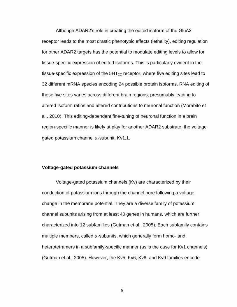
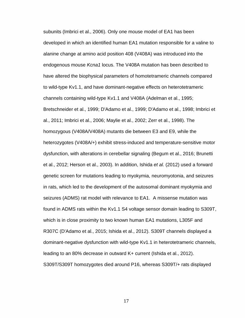
RNA (dsRNA) (Bass and Weintraub, 1988; Wagner et al., 1989) (Figure 1.1).
This conversion can lead to recoding when it occurs within the open reading
frame of an mRNA, because inosine base pairs with cytidine (in a similar manner

3
as guanosine) in the tRNA anticodon loop, thus A-to-I editing functions like an A-
to-G conversion (Basilio et al., 1962).
Figure 1.1. ADAR Enzymes convert adenosine residues to inosine by hydrolytic deamination.
This enzymatic reaction is catalyzed by the ADAR enzymes (Adenosine
Deaminase acting on RNA), which are a family of dsRNA-binding proteins,
including ADAR1, ADAR2 and ADAR3. ADAR1 and ADAR2 are the only
catalytically active enzymes of the family, functioning as homodimers, and
possessing varying selectivity regarding which RNAs, as well as which specific
sites, they target (Chen et al., 2000; Cho et al., 2003; Gallo et al., 2003;
Lehmann and Bass, 2000). ADAR1 and ADAR2 are found ubiquitously
throughout the human body, whereas ADAR3 is brain-specific, and ADAR
homologs are conserved from humans to the earliest branching metazoan
lineages (including sponges, C. elegans, D. melanogaster, squid, rat, mouse)
(Chen et al., 2000; Feng et al., 2006; Gerber et al., 1997; Grice and Degnan,

4
2015; Kim et al., 1994a). In shorter, imperfect duplexes, ADARs deaminate A-to-I
in a site-specific manner, whereas in the context of extensive duplexes, ADARs
are known to edit non-specifically, such as with invading viral dsRNA genomes
(George et al., 2011; Nishikura, 2010). In mammalian tissues, A-to-I editing
occurs prominently in the brain where it serves to modulate the function of many
proteins important for nervous system function including the ion-selectivity and
biophysical properties of the AMPA Glutamate Receptor subunit 2 (GluA2),
altering activation and deactivation kinetics of the GABAA receptor 3 subunit
(Gabra3), regulating G-protein coupling efficacy and constitutive activity for the
serotonin 5-HT2C receptor (5HT2C), and modulating the calcium-dependent
inhibition of a voltage-gated calcium channel subunit (Cav1.3) (Huang et al.,
2012; Niswender et al., 1999; Rula et al., 2008; Sommer et al., 1991). The
importance of these and additional editing events also is highlighted by the
effects observed in mutant mice where the expression of ADAR1 or ADAR2 has
been selectively ablated. ADAR1 knockout mice exhibit embryonic lethality
between embryonic day 11 (E11) and E12.5, exhibiting widespread apoptosis
and defects in hematopoiesis that can be partially rescued by inhibiting
components of the innate immune system (Hartner et al., 2004; Mannion et al.,
2014; Wang et al., 2004). ADAR2 knockout mice display postnatal lethality
between postnatal day 0 (P0) and P20, coinciding with progressive seizures,
which can be rescued through expression of the genomically edited GluA2
receptor (Higuchi et al., 2000).
5
Although ADAR2’s role in creating the edited isoform of the GluA2
receptor leads to the most drastic phenotypic effects (lethality), editing regulation
for other ADAR2 targets has the potential to modulate editing levels to allow for
tissue-specific expression of edited isoforms. This is particularly evident in the
tissue-specific expression of the 5HT2C receptor, where five editing sites lead to
32 different mRNA species encoding 24 possible protein isoforms. RNA editing of
these five sites varies across different brain regions, presumably leading to
altered isoform ratios and altered contributions to neuronal function (Morabito et
al., 2010). This editing-dependent fine-tuning of neuronal function in a brain
region-specific manner is likely at play for another ADAR2 substrate, the voltage
gated potassium channel -subunit, Kv1.1.
Voltage-gated potassium channels
Voltage-gated potassium channels (Kv) are characterized by their
conduction of potassium ions through the channel pore following a voltage
change in the membrane potential. They are a diverse family of potassium
channel subunits arising from at least 40 genes in humans, which are further
characterized into 12 subfamilies (Gutman et al., 2005). Each subfamily contains
multiple members, called -subunits, which generally form homo- and
heterotetramers in a subfamily-specific manner (as is the case for Kv1 channels)
(Gutman et al., 2005). However, the Kv5, Kv6, Kv8, and Kv9 families encode

6
subunits which act as modifier subunits within Kv2 heterotetramers (Gutman et
al., 2005).
Structural insights regarding these six helical transmembrane domain
proteins has been gained through determination of the Kv1.2 crystal structure
(Chen et al., 2010; Long et al., 2005). Each -subunit is composed of six -
helical transmembrane domains (S1-S6): S1 through S4 together form the
voltage-sensor domain while S5, the P loop, and S6 together form the pore
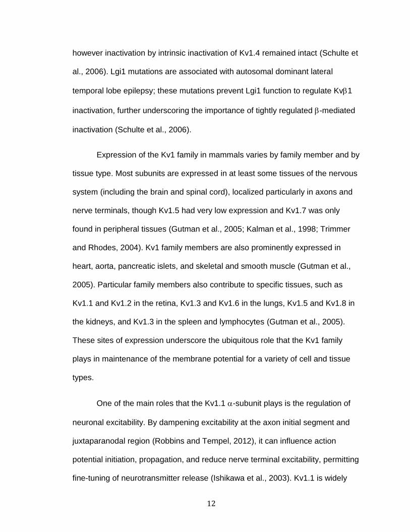
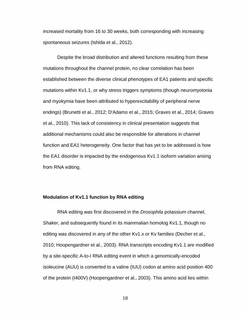
domain (Sands et al., 2005) (Figure 1.2). The extracellular face of the pore is
composed of a selectivity filter which allows for the selective flow of potassium
(K+) ions down their electrochemical gradient, with conformational control over
the intracellular face of the pore conferred by the charged residues of the S4
helix, in the voltage sensing domain, which twists upon changes in the
membrane potential (Glauner et al., 1999). A cytoplasmic N-terminal
tetramerization domain (T1, also called the N-terminal A and B box domain, or
NAB) of Kv channels is responsible for allowing the four -subunits to associate
with each other as well as with four accessory -subunits (Yu et al., 1996).

7
Figure 1.2. Domain topology for the Kv1.1 -subunit and accessory Kv-subunit. The voltage-sensing domain is composed of the S1-S4 helices, with conformational control of the voltage sensor mediated by the positively charged S4 helix. The pore domain is composed of the S5-P loop-S6 region, and the I400V residue (determined by RNA editing) is contained within the S6 helix,
within a hydrophobic region (shaded blue). Tetramerization of four -subunits
with each other and with four accessory Kv-subunits is mediated by the N-
terminal T1 domain. The -subunit contains an inactivation domain composed of hydrophobic (shaded blue) and positively charged residues which interact with the hydrophobic residues of the channel pore and the negatively charged residues of the T1 domain (Adapted from Hood and Emeson, 2012).
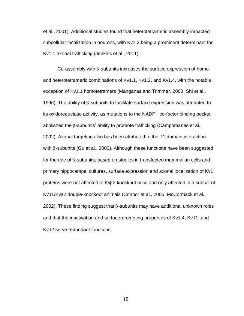
Potassium channels can exist in several states: closed, open, or
inactivated (Figure 1.3). The closed and inactivated states occur when the pore is
constricted or blocked at different locations within the central pore. The closed
versus opened state occurs when the S6 transmembrane domains of each -
subunit constrict on the intracellular face of the channel, in a region called the
bundle-crossing and the activation gate (Holmgren et al., 1998; Long et al., 2005;

8
Yellen, 2002). In addition to closing, there are two types of inactivation that can
occur following opening of the channel, despite maintaining a continuous
depolarizing voltage, slow (C-type) and fast (N-type) inactivation. Both types of
inactivation are distinct from the closed state and represent stable, non-
conducting states which are maintained for periods of time before the channels
are closed and available to be opened again by depolarization (a process
quantified as the recovery from inactivation) (Kurata and Fedida, 2006). C-type
inactivation is generally slower than N-type and involves a change in the
extracellular pore of the channel, proposed to be mediated either by pore
constriction or pore dilation at the outermost face of the selectivity filter (Choi et
al., 1991; Hoshi and Armstrong, 2013). N-type inactivation is a rapid inactivation
which can be mediated by two types of auto-inhibitory peptides, either N-terminal
domains which are part of some -subunits, such as Shaker and Kv1.4 (Hoshi et
al., 1990; Lee et al., 1996; Po et al., 1992; Zagotta et al., 1990), or from the N-
terminal domains of accessory -subunits (Rettig et al., 1994; Zhou et al., 2001).
N-type inactivation is proposed to be a docking of these N-terminal inactivation
peptides within the inner channel pore, representing approximately 20 amino
acids (Zagotta et al., 1990). The peptides contain a string of 10 hydrophobic
followed by 10 positively charged or hydrophilic amino acids which interact with
the negatively charged amino acids within the T1 domain and the hydrophobic
residues within the intracellular pore of the channel (Long et al., 2005).

9
Figure 1.3. Conformational states of Kv channels. Kv channels (composed of
four -subunits) can exist in several conformational states, including closed, open, and inactivated either by slow (C-type) inactivation or fast (N-type) inactivation. Slow inactivation has been proposed to be either a pore dilation or pore constriction. Fast inactivation can be mediated by the inactivation domain of
an accessory -subunit (shown in red) or by an intrinsic inactivation domain of
some Kv -subunits (not shown) (Adapted from Bezanilla, 2004).
Kv1.1 and the Kv1 family
The homolog of the mammalian Kv1 family was first discovered in
Drosophila melanogaster, and named Shaker, describing the rapid shaking
phenotype of Shaker mutant flies upon treatment with ether (Kaplan and Trout,
1969). Cloning of the Shaker locus revealed it encoded a potassium channel and
subsequent studies characterized its electrophysiological properties (Kamb et al.,
1987; Tempel et al., 1987). The Kv1.1 mammalian Shaker-homolog, originally
named MBK1 and RCK1, was first discovered by hybridizing Shaker

10
complementary DNA (cDNA) probes, directed against the transmembrane
segments, under low-stringency to a cDNA library composed of either mouse
brain or rat cortex mRNA (Baumann et al., 1988; Tempel et al., 1988). Rat and
mouse Kv1.1 were found to have 68% and 65% sequence homology with
Drosophila Shaker, respectively, highlighting the selective pressure to maintain
these proteins’ functions in evolutionarily distant organisms (Baumann et al.,
1988; Tempel et al., 1988).
The Kv1 family is composed of eight Shaker-related family members,
Kv1.1 through Kv1.8 (human gene names KCNA1 through KCNA10), each with
unique biophysical characteristics of activation, deactivation, and inactivation
(Bardien-Kruger et al., 2002; Grupe et al., 1990; Gutman et al., 2005; Heinemann
et al., 1996; Kalman et al., 1998; Kirsch et al., 1991; Lang et al., 2000; McKinnon,
1989; Stühmer et al., 1989; Swanson et al., 1990; Yao et al., 1995). These family
members can be subdivided based upon their inactivation characteristics. For
example, Kv1.4 is a rapidly inactivating (A-type) channel with N-type inactivation
conferred by its intrinsic N-terminal inactivation domain (Gutman et al., 2005; Lee
et al., 1996). The other Kv1 family members are delayed rectifiers that
intrinsically inactivate only by C-type inactivation (Gutman et al., 2005). N-type
inactivation can be conferred to channels containing most of the delayed rectifier
subunits, by co-assembly with -subunits or heterotetramerization with Kv1.4,
though Kv1.6 contains an N-type inactivation prevention (NIP) domain (Gutman
et al., 2005; Heinemann et al., 1996; Po et al., 1993; Roeper et al., 1998).

11
Kv1 channels can co-assemble with accessory -subunits, in an 44
configuration (Parcej et al., 1992). Multiple isoforms of -subunits include Kv1
(alternative splicing isoforms: 1.1, 1.2, and 1.3), Kv2 (alternative splicing
isoforms: 2.1 and 2.2), and Kv3 (England et al., 1995; Pongs and Schwarz,
2010). These isoforms differ predominantly in their N-terminal domains, which
are responsible for mediating inactivation, whereas high homology is found
throughout the rest of the proteins (Dolly and Parcej, 1996). Kv2.1 is the -
subunit with the highest abundance in the mammalian brain, whereas Kv2.2 has
only been found in the C6 rat glioblastoma cell line, reactive astrocytes from a rat
model of gliosis, and rabbit portal vein, but not normal rat brain (Akhtar et al.,
1999; Rhodes et al., 1996; Thorneloe et al., 2001). Of these -subunits, isoforms
of Kv1 has been shown to interact and inactivate Kv1.1 channels, whereas
Kv2 and Kv3 can co-assemble with Kv1.1 but do not inactivate it (Heinemann
et al., 1996; Heinemann et al., 1995; Rettig et al., 1994). Kv2 is proposed to
modulate the fast inactivation properties of other inactivating proteins, as it
prevents Kv1 fast-inactivation when both -subunits are co-expressed with Kv1
channels and speeds Kv1.4-induced inactivation (McCormack et al., 1995; Xu
and Li, 1997). Pulldown studies have isolated Kv1.1-containing heterotetramers
with Kv1.1 and Kv2.1 subunits in the human brain, whereas the Kv1.2 and
Kv1.3 isoforms are predominantly found in the heart. One study did find Kv1.3
expression in the brain, but it was weakly inactivating for Kv1.1 (Coleman et al.,
1999; England et al., 1995; Wang et al., 1996). Interestingly, another accessory
subunit, Lgi1, can oppose Kv1 inactivation in tetramers of Kv1.1 and Kv1.4,
12
however inactivation by intrinsic inactivation of Kv1.4 remained intact (Schulte et
al., 2006). Lgi1 mutations are associated with autosomal dominant lateral
temporal lobe epilepsy; these mutations prevent Lgi1 function to regulate Kv1
inactivation, further underscoring the importance of tightly regulated -mediated
inactivation (Schulte et al., 2006).
Expression of the Kv1 family in mammals varies by family member and by
tissue type. Most subunits are expressed in at least some tissues of the nervous
system (including the brain and spinal cord), localized particularly in axons and
nerve terminals, though Kv1.5 had very low expression and Kv1.7 was only
found in peripheral tissues (Gutman et al., 2005; Kalman et al., 1998; Trimmer
and Rhodes, 2004). Kv1 family members are also prominently expressed in
heart, aorta, pancreatic islets, and skeletal and smooth muscle (Gutman et al.,
2005). Particular family members also contribute to specific tissues, such as
Kv1.1 and Kv1.2 in the retina, Kv1.3 and Kv1.6 in the lungs, Kv1.5 and Kv1.8 in
the kidneys, and Kv1.3 in the spleen and lymphocytes (Gutman et al., 2005).
These sites of expression underscore the ubiquitous role that the Kv1 family
plays in maintenance of the membrane potential for a variety of cell and tissue
types.
One of the main roles that the Kv1.1 -subunit plays is the regulation of
neuronal excitability. By dampening excitability at the axon initial segment and
juxtaparanodal region (Robbins and Tempel, 2012), it can influence action
potential initiation, propagation, and reduce nerve terminal excitability, permitting
fine-tuning of neurotransmitter release (Ishikawa et al., 2003). Kv1.1 is widely

13
expressed throughout the mammalian brain, with high levels of expression in the
hippocampus (notably in CA3 pyramidal cells and dentate gyrus granule cells)
and cerebellum (particularly in basket cell nerve terminals synapsing to Purkinje
neurons) (Kirchheim et al., 2013; Tsaur et al., 1992; Wang et al., 1993).
Underscoring its physiological importance, genetic knockout studies have
revealed that mice lacking Kv1.1 expression develop spontaneous seizures,
cold-sensitive neuromyotonia, decreased motor coordination, hyperalgesia, and
neurogenic cardiac dysfunction (Clark and Tempel, 1998; Glasscock et al., 2010;
Smart et al., 1998). Kv1.1-knockout mice display an incompletely penetrant
lethality due to increased developmental seizure susceptibility, with
approximately half of the homozygous mutants dying before the sixth postnatal
week (Rho et al., 1999; Smart et al., 1998; Zhang et al., 1999; Zhou et al., 1998).
Although Kv1.1 can form functional homotetrameric channels, Kv1.1 is
predominantly found in heterotetramers with other Kv1 family members (denoted
Kv1.x) that contribute to the large diversity of Kv1 channel kinetics and
pharmacology throughout the mammalian central nervous system (Coleman et
al., 1999; Koch et al., 1997; Rasband et al., 2001; Sokolov et al., 2007). Most
often Kv1.1 is found in various heterotetrameric combinations with Kv1.2 and/or
Kv1.4, as these three subunits are the most predominant Kv1 -subunits in the
mammalian brain (Coleman et al., 1999; Trimmer and Rhodes, 2004; Wang et
al., 1999; Wang et al., 1993; Wang et al., 1994). Kv1.1 also is co-expressed with
subunits of lesser abundance, co-localized with Kv1.3 in the choroid plexus and
cerebellar cortex and with Kv1.6 in interneurons (Rhodes et al., 1997; Speake et

14
al., 2004; Trimmer and Rhodes, 2004; Wang et al., 1999). Heterotetrameric
assembly leads to channels with biophysical properties intermediate to the
characteristics of homotetrameric -subunit channels, and the ratios of each type
of -subunit lead to dose-dependent contribution to the overall channel
characteristics (Bagchi et al., 2014; Ovsepian et al., 2016; Sokolov et al., 2007).
Kv1.1 has a unique role in influencing heterotetrameric voltage-dependence and
the kinetics of activation, as it opens at an especially low voltage threshold
(Kv1.1 V1/2:-35 mV; Kv1.2 V1/2: 5 and 27 mV; Kv1.4 V1/2: 22 and 34 mV) and
faster than other highly co-expressed Kv1.x -subunits (Kv1.1 : 5 ms; Kv1.2 : 6
ms; Kv1.4 : 16.5 ms) (Ovsepian et al., 2016).
Subunit composition not only alters the biophysical characteristics of the
heteromeric channel, but may also alter the trafficking of Kv1 channels to the
plasma membrane. When subunits were expressed as homotetramers in
transfected cell lines and primary hippocampal cultures, Kv1.1 was
predominantly localized to the endoplasmic reticulum (ER), Kv1.4 to the cell
surface, and Kv1.2 expression was split between the ER and the plasma
membrane (Manganas and Trimmer, 2000). Increasing the ratio of Kv1.1
subunits in Kv1.x heterotetramers shifted the expression towards greater
intracellular localization, whereas Kv1.4 had the opposite effect, and Kv1.2 had a
neutral effect (Manganas and Trimmer, 2000). Extracellular pore residues within
the P loop of homotetrameric Kv1.1 channels, which differ from other Kv1.x
family members (A352P and Y379K), are thought to prevent Kv1.1 cell surface
trafficking from the ER, though a mechanism has not been elucidated (Manganas
15
et al., 2001). Additional studies found that heterotetrameric assembly impacted
subcellular localization in neurons, with Kv1.2 being a prominent determinant for
Kv1.1 axonal trafficking (Jenkins et al., 2011).
Co-assembly with -subunits increases the surface expression of homo-
and heterotetrameric combinations of Kv1.1, Kv1.2, and Kv1.4, with the notable
exception of Kv1.1 homotetramers (Manganas and Trimmer, 2000; Shi et al.,
1996). The ability of -subunits to facilitate surface expression was attributed to
its oxidoreductase activity, as mutations to the NADP+ co-factor binding pocket
abolished the -subunits’ ability to promote trafficking (Campomanes et al.,
2002). Axonal targeting also has been attributed to the T1 domain interaction
with -subunits (Gu et al., 2003). Although these functions have been suggested
for the role of -subunits, based on studies in transfected mammalian cells and
primary hippocampal cultures, surface expression and axonal localization of Kv1
proteins were not affected in Kv2 knockout mice and only affected in a subset of
Kv1/Kv2 double-knockout animals (Connor et al., 2005; McCormack et al.,
2002). These finding suggest that -subunits may have additional unknown roles
and that the inactivation and surface-promoting properties of Kv1.4, Kv1, and
Kv2 serve redundant functions.
16
Episodic Ataxia type-1
The role of Kv1.1 and its effects on human health have also been studied
in the context of a related genetic disorder. Genetic linkage analyses have ident-
ified over 30 heterozygous mutations within the gene encoding human Kv1.1
(KCNA1) that have been associated with the sporadic and autosomal dominant
neurological disorder, episodic ataxia type-1 (EA1) (D'Adamo et al., 2015). EA1
occurs with an age of onset in patients between 2 and 15 years old and is
characterized by stress-induced motor discoordination, involuntary, repetitive
muscle contraction (myokymia), and can coincide with seizures (Graves et al.,
2014). Types of stressors found to trigger ataxia vary between patients, most
commonly including exercise/exertion, stress/emotional upset, and environmental
temperature; additional triggers can include, but are not limited to, fever, caffeine,
alcohol, startle, prolonged rest, sudden movement, and diet (Graves et al., 2014).
The most common symptoms include imbalance, slurred speech, incoordination
of hands, weakness, tremors, and muscle twitching or stiffness (Graves et al.,
2014).
The majority of EA1-related mutations result in a loss of channel function,
reduced surface expression, or a change in biophysical properties where the
mutant subunits can exert a dominant-negative effect by association with wild-
type -subunits (Chen et al., 2016; D'Adamo et al., 2015; Eunson et al., 2000;
Imbrici et al., 2006; Mestre et al., 2016; Petitjean et al., 2015; Tomlinson et al.,
2013; Zerr et al., 1998). Deficits have also been observed in heterotetramers with
other Kv1 family members as well as when co-expressed with auxiliary -
17
subunits (Imbrici et al., 2006). Only one mouse model of EA1 has been
developed in which an identified human EA1 mutation responsible for a valine to
alanine change at amino acid position 408 (V408A) was introduced into the
endogenous mouse Kcna1 locus. The V408A mutation has been described to
have altered the biophysical parameters of homotetrameric channels compared
to wild-type Kv1.1, and have dominant-negative effects on heterotetrameric
channels containing wild-type Kv1.1 and V408A (Adelman et al., 1995;
Bretschneider et al., 1999; D'Adamo et al., 1999; D'Adamo et al., 1998; Imbrici et
al., 2011; Imbrici et al., 2006; Maylie et al., 2002; Zerr et al., 1998). The
homozygous (V408A/V408A) mutants die between E3 and E9, while the
heterozygotes (V408A/+) exhibit stress-induced and temperature-sensitive motor
dysfunction, with alterations in cerebellar signaling (Begum et al., 2016; Brunetti
et al., 2012; Herson et al., 2003). In addition, Ishida et al. (2012) used a forward
genetic screen for mutations leading to myokymia, neuromyotonia, and seizures
in rats, which led to the development of the autosomal dominant myokymia and
seizures (ADMS) rat model with relevance to EA1. A missense mutation was
found in ADMS rats within the Kv1.1 S4 voltage sensor domain leading to S309T,
which is in close proximity to two known human EA1 mutations, L305F and
R307C (D'Adamo et al., 2015; Ishida et al., 2012). S309T channels displayed a
dominant-negative dysfunction with wild-type Kv1.1 in heterotetrameric channels,
leading to an 80% decrease in outward K+ current (Ishida et al., 2012).
S309T/S309T homozygotes died around P16, whereas S309T/+ rats displayed
18
increased mortality from 16 to 30 weeks, both corresponding with increasing
spontaneous seizures (Ishida et al., 2012).
Despite the broad distribution and altered functions resulting from these
mutations throughout the channel protein, no clear correlation has been
established between the diverse clinical phenotypes of EA1 patients and specific
mutations within Kv1.1, or why stress triggers symptoms (though neuromyotonia
and myokymia have been attributed to hyperexcitability of peripheral nerve
endings) (Brunetti et al., 2012; D'Adamo et al., 2015; Graves et al., 2014; Graves
et al., 2010). This lack of consistency in clinical presentation suggests that
additional mechanisms could also be responsible for alterations in channel
function and EA1 heterogeneity. One factor that has yet to be addressed is how
the EA1 disorder is impacted by the endogenous Kv1.1 isoform variation arising
from RNA editing.
Modulation of Kv1.1 function by RNA editing
RNA editing was first discovered in the Drosophila potassium channel,
Shaker, and subsequently found in its mammalian homolog Kv1.1, though no
editing was discovered in any of the other Kv1.x or Kv families (Decher et al.,
2010; Hoopengardner et al., 2003). RNA transcripts encoding Kv1.1 are modified
by a site-specific A-to-I RNA editing event in which a genomically-encoded
isoleucine (AUU) is converted to a valine (IUU) codon at amino acid position 400
of the protein (I400V) (Hoopengardner et al., 2003). This amino acid lies within

19
the S6 transmembrane domain predicted to line the ion-conducting pore of the
channel (Figure 1.2). Editing of Kv1.1 transcripts is dependent upon a region of
dsRNA within exon 2 which forms intramolecular base-pairing interactions
between imperfect, inverted repeat elements surrounding the targeted adenosine
moiety (Bhalla et al., 2004). This process was determined to be catalyzed by
ADAR2 through exogenous expression in HEK293 cells as well as the almost
complete absence of Kv1.1 RNA editing in ADAR2-null mice (Bhalla et al., 2004;
Horsch et al., 2011).
Although an isoleucine to valine substitution does not seem substantial,
only resulting in an amino acid with one fewer methyl group, previous studies
have revealed that Kv1.1 channels containing edited [Kv1.1(V)] subunits display
a 20-fold faster rate of recovery from Kv1.1-inactivation, compared to non-
edited channels [Kv1.1(I)] (Bhalla et al., 2004). More recent studies have
indicated that only small alterations in the hydrophobicity of the editing site amino
acid were needed to reproduce the editing-dependent alterations in recovery
kinetics (Gonzalez et al., 2011). In addition, the extent of inactivation was heavily
influenced by the accessible surface area of the hydrophobic amino acid at the
editing site; thus the non-edited [Kv1.1(I)] isoform has a greater extent of
inactivation than the edited [Kv1.1(V)] isoform (Gonzalez et al., 2011).
Interestingly, the alteration in the recovery from inactivation following a slight
change in the hydrophobic interactions has been recapitulated previously in a
complimentary experiment, where the hydrophobic residues of the N-terminus of
the auxiliary -subunit were substituted for alanine or valine (Zhou et al., 2001).
20
The dissociation constants of the mutant -subunits were generally faster
compared to wild-type, further supporting the role of these hydrophobic
interactions, between the - and -subunits, in setting the dissociation rate of -
inactivation (Zhou et al., 2001).
-subunits are not alone in conferring hydrophobic-mediated inactivation
of potassium channels, as endogenous lipids can not only induce fast inactivation
(arachidonic acid induces fast inactivation in Kv1.1), but they also can prevent -
inactivation (such as PIP2 preventing Kv1.1 inactivation by Kv1.1) (Honoré et
al., 1994; Oliver et al., 2004). Extensive drug studies have elucidated that the
editing status of Kv1.1 has a prominent effect on the efficacy of drug-mediated
channel blockers. Edited Kv1.1 was insensitive to the channel block mediated by
important endogenous signaling lipids, including arachidonic acid,
docosahexaenoic acid, and the endocannabinoid anandamide, whereas non-
edited channels were readily blocked (Decher et al., 2010). Although the binding
rate of arachidonic acid was not different between non-edited [Kv1.1(I)] and
edited [Kv1.1(V)] isoforms, the recovery from arachidonic acid-induced block was
rapid for edited [Kv1.1(V)] and slow for non-edited [Kv1.1(I)] channels, mirroring
the recovery from inactivation kinetics of the -subunit (Bhalla et al., 2004;
Decher et al., 2010). Several pharmacological open channel blockers displayed
similar selectivity for the non-edited [Kv1.1(I)] channel, including the known Kv1
channel blockers, Psora-4 and 4-aminopyridine (4-AP) (Decher et al., 2010).
When different ratios of non-edited [Kv1.1I(I)] and edited [Kv1.1(V)] channels
were co-expressed to make a tetramer, only one edited [Kv1.1(V)] subunit was
21
needed to suppress the sensitivity to endogenous and pharmacological blockers,
reducing the inhibition by 60% for arachidonic acid and 65% for Psora-4,
compared to channels made entirely of non-edited [Kv1.1(I)] subunits (Decher et
al., 2010). Similarly, heterotetrameric assembly of edited [Kv1.1(V)] with other
Kv1.x family members significantly reduced the affinity of the channels to open
channel blockers (Decher et al., 2010). These studies emphasize that even
tissues with low levels of Kv1.1 RNA editing may be able to impact signaling
kinetics through the effects of a single edited [Kv1.1(V)] subunit within a
heterotetramer.
Kv1.1 editing also has been implicated in altering trafficking to the plasma
membrane. Although homotetrameric Kv1.1 is known to be predominantly
localized to the ER, some non-edited [Kv1.1(I)] channels still express at the cell
surface. By contrast, edited [Kv1.1(V)] surface expression was significantly
reduced compared to the non-edited [Kv1.1(I)] channels (Streit et al., 2014). Co-
expression with Kv1.4 trafficked both the non-edited [Kv1.1(I)] and edited
[Kv1.1(V)] channels to the plasma membrane, equalizing the surface expression
of both isoforms (Streit et al., 2014). A caveat to these trafficking experiments
was that they were performed in HeLa, HEK293, and CHO cells and in Xenopus
oocytes, rather than in primary hippocampal cultures like previous Kv1.x
trafficking experiments. It is unknown whether these cell lines contain all of the
necessary components for Kv1.1 trafficking.
Kv1.1 editing has been found predominantly in nervous tissue, though
some editing has been found in human aorta (but not in the heart) (Decher et al.,
22
2010; Hoopengardner et al., 2003; Li et al., 2009). The percentage of Kv1.1
editing is found to vary in dissected portions of the mammalian nervous system,
with approximately 20% editing in human hippocampus compared to 70% in
human spinal cord (Hoopengardner et al., 2003). In addition, Kv1.1 RNA editing
is developmentally regulated in the mouse brain, with low editing (approximately
5-7%) in whole brain samples at E15, E19, P0, and P2, but by P21 levels rise to
approximately 30%, near the adult levels of 45% (Jacobs et al., 2009; Wahlstedt
et al., 2009). Although many other editing targets share similar trends in editing
regulation, with low embryonic editing (except the GluA2 RNA which is always
highly edited), most other targets have increases in editing at birth, whereas
Kv1.1 RNA editing lags behind these other targets (Flna RNA has a similar
lagging RNA editing trend to Kv1.1) (Jacobs et al., 2009; Wahlstedt et al., 2009).
The same Kv1.1 developmental RNA editing trend was observed by
transcriptome-wide RNA-seq, detecting editing patterns across development for
human and mouse tissues, and these studies also further resolved that Kv1.1
editing began increasing at P7 in mice (Hwang et al., 2016). At this time, it is
unknown whether adult neurogenesis occurring following a seizure leads to a
similar lag in Kv1.1 RNA editing that has been observed during embryonic
development.
As is the case with the quantification of Kv1.1 editing described above,
most editing profiles are quantified from entire tissues or brain regions that may
be composed of many different cell types. The heterogeneity of these tissues
leads to different interpretations of what the editing percentages mean. Are

23
tissues composed of a few cell types with high levels of editing in the midst of
other cells with no editing, or does editing occur in every cell at a variable ratio?
Although Kv1.1 RNA editing has not been determined in defined cell
types, patch-clamp studies on acutely isolated thalamic LGN and hippocampal
CA1 neurons by Decher et al. (2010) provide an indirect method for inferring
single-cell editing. Using the differential pharmacological sensitivity to Psora-4
(blocks nonedited [Kv1.1(I)] and Kv1.x channels, but not edited [Kv1.1(V)] or
Kv1.4) versus rHongotoxin-1 (HgTX) (blocks both nonedited [Kv1.1(I)] and edited
[Kv1.1(V)] and Kv1.x, but not Kv1.4 or Kv1.5), investigators were able to
determine estimates of edited Kv1.1 expression in individual neurons of each cell
type (Decher et al., 2010; Koschak et al., 1998; Vennekamp et al., 2004). The
currents which were Psora-4-resistant, but HgTX-sensitive, indicated the
presence of the edited [Kv1.1(V)] protein isoform in these cell types. Sequencing
reverse transcriptase-polymerase chain reaction (RT-PCR) amplicons derived
from these isolated neuronal populations revealed that the average Kv1.1 RNA
editing correlated well with the edited [Kv1.1(V)] protein expression estimated
from the single-cell electrophysiological characterization; 63% of the current from
LGN neurons was Psora-4 resistant, indicative of edited Kv1.1 expression, and
52% of the Kv1.1 RNA was edited in the population of these isolated cells
(Decher et al., 2010). By contrast, 15% of the current from CA1 pyramidal cells
was Psora-4 resistant, correlating with a 7% editing profile for Kv1.1 RNA in
these cells (Decher et al., 2010). These studies provide evidence that
intermediate editing ratios are observed within single cells, though they do not

24
exclude the possibility that editing could still vary by cell type within tissues.
Little is known about how Kv1.1 RNA editing is regulated in vivo, however,
Kv1.1 editing levels are altered in two mouse models of epilepsy. In the first
model, control rats displayed low Kv1.1 editing levels in the entorhinal cortex
(5.1%), whereas, following epileptic induction by kainic acid, editing was
increased (21.5%) (Streit et al., 2011). In normal rat entorhinal/hippocampal
slices, 4-AP induces seizure-like events, however slices from kainic acid epileptic
rats are resistant to 4-AP effects (Streit et al., 2011). These data suggest that
increased editing may be associated with a decreased seizure-susceptibility in
rats. The second model is a genetic model of epilepsy where editing of the GluA2
receptor was decreased in the mouse forebrain (in particular, GluA2 Q/R site
editing in the hippocampus was decreased approximately 25%) (Krestel et al.,
2013; Krestel et al., 2004). In this model, Kv1.1 RNA editing was also increased
in the epileptic mice compared to controls (Krestel et al., 2013).
There are several possible mechanisms for these increases in Kv1.1 RNA
editing following seizure induction. First, increases in editing may result from
increased expression of ADAR2, as previous studies have indicated that,
following kainic acid seizure induction, rats had a 40% increase in ADAR2 protein
expression (O'Leary et al., 2016). Second, the kainic acid-induced seizure rat
model has also been associated with increased activity of two MAP kinases, and
the nuclear localization of ADAR2 is positively regulated by a phosphorylation-
dependent interaction with the phosphorylation-dependent prolyl-isomerase, Pin1
(whereas localization in the cytoplasm can lead to degradation mediated by the

25
E3-ubiquitin ligase, WWP2) (Kim et al., 1994b; Marcucci et al., 2011). Third,
ADAR2 gene expression is regulated by the CREB transcription factor and
seizure-induction can activate CREB (in these cases, seizures were induced by
pentylenetetrazole (PTZ) or 1-3-bischloroethyl-nitrosurea (BCNU)) (Moore et al.,
1996; Peng et al., 2006; Pennacchio et al., 2015). Although editing regulation
may occur by altering ADAR2 expression and activity, as detailed above, other
unknown factors may be involved as well.
Aside from these mouse models displaying alterations in Kv1.1 RNA
editing, one study has correlated Kv1.1 editing in epileptic patients. Tissue
samples obtained from patients undergoing surgery for mesial temporal lobe
epilepsy revealed an editing-dependent association, where levels of Kv1.1 RNA
editing were inversely correlated with the duration of years that the patients had
experienced epileptic activity, such that lower editing was correlated with a longer
epilepsy duration (Krestel et al., 2013). These results could indicate that
increased editing of Kv1.1 has a protective effect, dampening future seizures in
chronic epilepsy, and that decreased editing could represent a risk factor for
long-term seizures, though further experiments are necessary to test this
hypothesis.
Of note, in two brain regions of interest, the hippocampus and the
cerebellum, Kv1.1 expression is lower than other Kv1.x subunits (including
combinations of Kv1.2, Kv1.4, and/or Kv1.6), thus Kv1.1-containing
heterotetramers in these tissues may contain only one Kv1.1 subunit (Scott et al.,
1994). Kv1.1 is an important subunit, even in heterotetramers, because it opens
26
at lower voltages and faster than other Kv1.x subunits (Ovsepian et al., 2016).
This Kv1.1 subunit could be non-edited or edited, thus heterotetramers may be
further stratified into groups defined by the unique and dominant characteristics
conferred by the absence or presence of an edited subunit. The role of Kv1.1 in
defining the opening kinetics of heterotetramers, and editing conferring rapid
recovery from inactivation, may underlie the particular vulnerability of mutations
and altered editing patterns in leading to dysfunctions related to hippocampal and
cerebellar function.
Summary
Kv1.1 is an important regulator of neuronal signaling in normal physiology
and its dysregulation leads to EA1, yet the role of Kv1.1 RNA editing as it
contributes to healthy and EA1 physiology are still largely unknown. Although
several studies have indicated that edited [Kv1.1(V)] channels have altered
biophysical characteristics, these studies have been performed predominantly in
cell culture and Xenopus oocytes, rather than in an endogenous context. In
addition, no studies have been performed to understand how RNA editing
impacts the human disorder, EA1, though several EA1 mutations are in close
proximity to the editing site.
The data in Chapter II describe our attempts to understand Kv1.1 RNA
editing in a physiological context. Thus we have generated two mutant mouse
lines which solely express either the non-edited Kv1.1 [Kv1.1(I)] or edited Kv1.1
[Kv1.1(V)] isoform. We describe how globally regulating Kv1.1 editing leads to

27
phenotypic alterations in each mouse line compared to wild-type littermates. Both
the non-edited [Kv1.1(I)] and edited [Kv1.1(V)] mice exhibited an incompletely
penetrant postnatal lethality that appeared to be dependent upon environmental
factors. Non-edited [Kv1.1(I)] mice displayed EA1-like stress-induced ataxia, with
alterations in gait and locomotor activity. In addition, editing altered drug-induced
seizure susceptibility of both mouse lines, with the non-edited [Kv1.1(I)] mice
displaying an increased susceptibility and faster latency to 4-AP-induced
seizures and the edited [Kv1.1(V)] mice displaying a decreased susceptibility to
both 4-AP and PTZ-induced seizures. Initial electrophysiological analysis of
Kv1.1 in mouse hippocampal slices did not find differences in signaling
parameters for non-edited [Kv1.1(I)] mice that would explain this alteration in
seizure susceptibility.
In Chapter III, we detail the impact of RNA editing on EA1 mutant RNAs
and proteins. Although numerous Kv1.1 mutations have been associated with the
human disorder EA1, three EA1 mutations, V404I, I407M, and V408A, are
located within the RNA duplex structure required for Kv1.1 RNA editing. Each
EA1 mutation decreased RNA editing in vitro and the V408A mutation was
confirmed to decrease RNA editing in vivo using a mouse model bearing the
heterozygous V408A allele. Editing of transcripts encoding mutant channels
affected numerous biophysical properties including channel opening, closing, and
inactivation. Thus, EA1 symptoms could be influenced not only by the direct
effects of the mutations on channel properties, but also by their influence on RNA
editing. These studies provide the first evidence that mutations associated with a

28
human genetic disorder can affect cis-regulatory elements to alter RNA editing.
Finally, Chapter IV summarizes the conclusions of these studies and
describes future directions to address new questions spurred on by the
discoveries made thus far.

29
CHAPTER II
FUNCTIONAL CONSEQUENCES OF KV1.1 RNA EDITING
IN MOUSE MODELS
Introduction
Although initial studies have characterized the effects of Kv1.1 RNA
editing on channel function, most have been performed in exogenously
expressing Xenopus oocytes. Thus, further studies are needed to better
understand the physiological impact of Kv1.1 RNA editing in vivo. We have
developed mutant mouse lines that have been genetically modified to solely
express either the non-edited [Kv1.1(I)] or edited [Kv1.1(V)] isoforms. These
mutant mouse lines have allowed us to assess the phenotypic effects of
dysregulating normal editing patterns for Kv1.1 transcripts, and serve as
important tools to examine the native electrophysiological properties of Kv1.1-
expressing neurons.
These novel mouse lines have been developed in the context of several
other related mouse models, whose characterized defects could indicate that our
editing mouse models will have similar dysfunctions. Three models of Kv1.1
dysfunction have been created thus far including a Kv1.1-null mouse in which
expression of the Kcna1 gene encoding Kv1.1 is ablated (Smart et al., 1998).
Although Kv1.1-null mice exhibit a normal Mendelian distribution at birth, no overt
30
ataxia, and normal Kv1.2 distribution in the cerebellum, they display spontaneous
seizures starting at 3 weeks of age, hyperalgesia, neurogenic cardiac
dysfunction, and have an enlarged hippocampus and ventral cortex (Clark and
Tempel, 1998; Glasscock et al., 2010; Persson et al., 2007; Smart et al., 1998).
Half of Kv1.1-null mice die within 3-5 weeks after birth, often immediately
following seizures, though surviving mice can live up to 12 months of age (Smart
et al., 1998). Kv1.1-null mice also displayed a shorter latency to flurothyl-induced
seizure onset, with seizures occurring 60% sooner than wild-type animals, and
heterozygous null mice showed a modest change in the onset of seizures (9%
sooner than wild-type) (Rho et al., 1999; Smart et al., 1998). Surprisingly, no
differences were found in the intrinsic membrane properties of CA3 pyramidal
cells, though other electrophysiological changes were observed, including lower
threshold for antidromic action potentials and a subset of cells which displayed
epileptic late burst discharges upon mossy fiber stimulation (Smart et al., 1998).
In the cerebella of Kv1.1-null mice, Purkinje neurons displayed increased
frequency of GABAergic spontaneous inhibitory postsynaptic currents (sIPSCs),
and these mice displayed motor deficits walking across a stationary thin rod, but
were not different from wild-type animals using the rotarod test (Zhang et al.,
1999). Peripheral nerve transmission also was altered in Kv1.1-null mice,
including recordings in the sciatic nerve that indicated that loss of Kv1.1 leads to
a prolonged repolarization and a longer recovery period and hyperexcitable
neuromuscular transmission following cooling of isolated phrenic nerve (Smart et
al., 1998; Zhou et al., 1998).

31
The mceph/mceph mouse is a megencephaly mouse model resulting from
an 11-bp deletion in Kv1.1, leading to a frame-shifted, C-terminally truncated
protein (truncated at amino acid 230 out of 495) (Petersson et al., 2003). These
mice have a 25% increase in brain size, shakiness in gait, and display complex
partial seizures starting at 3 weeks of age (Donahue et al., 1996; Petersson et
al., 2003). Electrophysiological characterization in mossy cells of the
hippocampus revealed increased frequency of stimulus-induced pulse trains, but
no change in action potential shape or membrane resistance (Petersson et al.,
2003). The truncated protein was expressed in mice, though localized to the ER,
and Kv1.2 and Kv1.3 protein expression was decreased in mceph/mceph
hippocampus (Persson et al., 2005; Petersson et al., 2003).
Another Kv1.1 mutant mouse was developed which incorporated a human
EA1 mutation, V408A, into the endogenous Kcna1 locus (Herson et al., 2003).
Mice homozygous for the V408A mutation die during embryonic development
between E3 and E9, while heterozygous V408A mice (V408A/+) display stress-
induced motor discoordination (Herson et al., 2003). Electrophysiological
characterization of V408A/+ cerebellar Purkinje neurons indicated this mutation
increased the frequency and amplitude of sIPSCs, attributed to action potential
broadening at basket cell boutons leading to increased GABA release (Begum et
al., 2016; Herson et al., 2003). Recordings from V408A/+ motor nerves also
revealed spontaneous bursting activity, which was triggered by fatigue, ischemia,
and low temperature (Brunetti et al., 2012).

32
In addition to mouse lines with introduced mutations in the Kv1.1 -
subunit, four -subunit knockout models also have been developed. Kv1.1-null
mice exhibited no changes in lifespan, brain morphology, seizure activity, or
alterations in open field or bar hang behavioral tasks (Connor et al., 2005; Giese
et al., 1998). However, electrophysiological characterization of CA1 pyramidal
neurons in Kv1.1-null mice revealed several alterations compared to wild-type
controls. Kv1.1-nulls showed a decreased amplitude of inactivation, faster
action potential repolarization during trains of depolarizing pulses (a decrease in
the frequency-dependent spike broadening), and a reduced amplitude of the slow
after-hyperpolarization phase of the action potential following spike trains (Giese
et al., 1998). Kv1.1-null mice did not have any overt impairment in spatial or
contextual learning and extracellular field recordings in the CA1 region did not
reveal any differences in synaptic plasticity, though subtle learning impairments
were observed in some cases (including in an altered paradigm of the Morris
water maze, where previously trained mice had to find a new hidden platform
location, and in the social transmission of food preference test) (Giese et al.,
1998; Need et al., 2003).
Kv2-null mice were found to have phenotypic alterations similar to the
Kv1.1-null mice, including cold swim-induced tremors, shortened life span
(though normal Mendelian distribution at weaning), and the presence of sporadic
seizures, but no differences in PTZ-induced seizure susceptibility (Connor et al.,
2005; McCormack et al., 2002). Phenotypes were altered in a strain-specific
manner, with Kv2-null mice on a C57B/6J background demonstrating more
33
severe phenotypes than the 129/SvEv background, which could result from a
strain-specific 5-fold higher expression of Kv2 in C57B/6J versus 129/SvEv
wild-type mice (Connor et al., 2005; Sandberg et al., 2000). The presence of
seizures in this model is consistent with human patient data where a deletion
including the region of Kv2 was a significant risk factor for epilepsy or
epileptiform activity observed by EEG analysis (Heilstedt et al., 2001).
Surprisingly, none of the observed mouse phenotypes could be attributed to
presumed functions of Kv2. Previous characterization of Kv2 in heterologous
expression systems suggested that Kv2, which does not induce fast-
inactivation, was involved in potassium channel trafficking and axonal targeting
(Gu et al., 2003; Shi et al., 1996). However, characterization of Kv2-null mice
indicated no change in trafficking of Kv1.1 and Kv1.2 in the cerebellum or in the
juxtaparanodal localization of Kv1.1 and Kv1.2 within the sciatic nerve
(McCormack et al., 2002). In addition to Kv2-null animals, an additional mutant
line was developed in which the oxidoreductase catalytic domain was targeted,
but no observable phenotype resulted (McCormack et al., 2002).
Double mutant animals, in which the expression of both Kv1.1 and Kv2
were ablated, were developed to assess whether these subunits perform
redundant functions (McCormack et al., 2002). Kv1.1/Kv2 double mutants had
decreased lifespan compared to the Kv2-null animals, however Kv1.1 loss did
not further exacerbate the cold swim-induced tremors (Connor et al., 2005).
Localization of Kv1.2 within the pinceau region of cerebellar basket cells was
more variable for the double mutants, with 5/8 mutants displaying less robust and
34
more diffuse expression of Kv1.2 (Connor et al., 2005). However, 3/8 mutants
had comparable expression to wild-type controls, still indicating that proper
localization could occur without either -subunit (Connor et al., 2005).
Mouse models containing alterations in either the Kv1.1 -subunit or Kv
subunits serve as important references to compare the phenotype of mutant
animals in which the editing profile for Kv1.1 RNAs has been fixed. For these
studies, we have focused specifically on phenotypic alterations in survival,
stress-induced motor discoordination, seizures and seizure susceptibility, and
brain slice electrophysiology. Our initial characterization indicates that Kv1.1
editing serves a modulatory function for motor coordination and seizure
susceptibility.
Materials & Methods
Mouse Experiment Approval
All animal care and experimental procedures involving mice were
approved by the Vanderbilt University Medical Center Institutional Animal Care
and Use Committee and were performed in accordance with relevant guidelines
and regulations.
35
Characterization of Kv1.1 editing in wild-type mouse tissues
To characterize wild-type Kv1.1 RNA editing levels, wild-type C57BL/6
mice were euthanized by cervical dislocation under anesthesia followed by
decapitation. Seven tissues (whole brain, spinal cord, lung, heart, bladder,
skeletal muscle, kidney) were dissected from four male wild-type adult mice. Six
brain regions (cerebellum, hippocampus, hypothalamus, cortex, striatum,
olfactory bulb) were dissected from three additional male wild-type adult mice.
Tissues were flash-frozen in liquid nitrogen and RNA was isolated by sonication
in TRIzol (Ambion) according to the manufacturer’s instructions. RNA was
reverse-transcribed with random primers using the High Capacity cDNA Reverse
Transcription kit (Applied Biosystems) and Kv1.1 editing was quantified by high-
throughput sequence analysis as described previously (Hood et al., 2014)
(VANTAGE, Vanderbilt University).
Generation of non-edited [Kv1.1(I)] and edited [Kv1.1(V)] mutant mice
Our collaborator, Dr. Robert Reenan (Brown University), generated mice
that were genetically modified to solely express either the non-edited [Kv1.1(I)] or
the edited [Kv1.1(V)] subunit isoform. Mutations were incorporated at the
endogenous Kcna1 locus (knock-in) by homologous recombination with a
replacement-type targeting vector in C57BL/6J embryonic stem cells (Figure 2.1).
This targeting vector included a positive selectable marker (neomycin cassette
flanked by loxP sites) within and a negative selectable marker (thymidine kinase)

36
outside the region of homology, respectively. Mice with the mutant-neo allele
were crossed with a mutant line expressing Cre recombinase to excise the
neomycin resistance cassette, leaving a single loxP site downstream of exon 2.
The non-edited [Kv1.1(I)] targeting vector incorporated two point mutations
surrounding the editing site, which have previously been shown to disrupt the
critical RNA duplex structure required for editing (Bhalla et al., 2004), yet
maintain the codon identities in this region (Figure 2.2). The targeting vector to
generate edited [Kv1.1(V)] transcripts was designed to introduce a guanosine at
the editing site to mimic the coding potential of the inosine normally generated by
A-to-I conversion (Figure 2.2).
Figure 2.1. Targeting strategy to solely generate non-edited [Kv1.1(I)] or edited [Kv1.1(V)] isoforms in mutant mice. The targeting strategy for creating the non-edited [Kv1.1(I)] and edited [Kv1.1(V)] mutant mice is outlined. The Kcna1 mouse gene is labeled with the editing site (indicated as a red A) within the RNA duplex region, indicated by arrows. Mutant constructs included the mutations required for either the non-edited or the edited isoform (indicated as a red asterisk) in conjunction with a positive selectable marker (neomycin cassette flanked by loxP sites) within the region of homology and a negative selectable marker (thymidine kinase) outside the region of homology. Mice with the mutant-neo allele were crossed with a Cre recombinase-expressing mouse to excise the neomycin resistance cassette, leaving a single loxP site behind (blue triangle).

37
Figure 2.2. Design of duplex mutations to solely generate non-edited [Kv1.1(I)] or edited [Kv1.1(V)] transcripts in mutant mice. The predicted secondary structure for the wild-type mouse Kv1.1 pre-mRNA, in the proximal region of exon 2, is shown with the position of the I/V editing site indicated. The introduction of mutations to generate mutant mouse lines, solely expressing the non-edited and edited isoforms of the channel, are indicated in red.
Genotyping was performed by polymerase chain reaction (PCR)
amplification of a 534 base pair (bp) region surrounding the editing site using
sense (5’- TCCTCTTCATTGGGGTCATACTGT-3’) and antisense (5’-
GGGGTTTGTTTGGGGCTTTTGTTG-3’) oligonucleotide primers in exon 2. The
presence of the mutations incorporated into these mouse lines creates or
destroys restriction sites for the non-edited (Sfc I) and edited (Mfe I) alleles,
respectively, allowing accurate genotyping to be distinguished from heterozygous
breeding crosses. Genotyping was performed using tail biopsies with the

38
REDExtract-N-Amp Tissue PCR Kit (Sigma-Aldrich).
To ensure expression of the mutant allele at the RNA level, RNA from
mouse cerebellum for each genotype was isolated with TRIzol and subjected to
RT-PCR, as described previously, and directly sequenced by Sanger sequencing
(VANTAGE, Vanderbilt University). To further ensure that the non-edited
[Kv1.1(I)] mice did not have undetectable RNA editing occurring, spinal cord
cDNA was subjected to high-throughput sequence analysis as described
previously (Hood et al., 2014) (VANTAGE, Vanderbilt University).
Kv1.1 RNA Expression Characterization in non-edited [Kv1.1(I)] and edited [Kv1.1(V)] mutant mice
Six adult mice each (mixed sex) of non-edited [Kv1.1(I)], edited [Kv1.1(V)],
and wild-type animals were euthanized by cervical dislocation under anesthesia
followed by decapitation. Six brain regions (cerebellum, hippocampus,
hypothalamus, cortex, striatum, olfactory bulb) were dissected from each mouse.
RNA was extracted and used as a template for reverse transcription with random
primers using the High Capacity cDNA Reverse Transcription kit (Applied
Biosystems). Steady-state RNA expression was assayed from each isolated
cDNA using Taqman probes directed against mouse Kcna1 (Mm00439977_s1,
ThermoFisher Scientific) and using mouse Gapdh as an internal normalization
control (Mm99999915_g1, ThermoFisher Scientific). Quantitative RT-PCR (qRT-
PCR) reactions were prepared using Taqman Universal PCR mastermix (2X)
(ThermoFisher Scientific) according to the manufacturer’s protocol. Technical

39
replicates for all cDNA samples were performed in triplicate and average values
used for data analyses. Gene efficiencies were calculated using the PCR miner
algorithm (Zhao and Fernald, 2005). All data were normalized to internal Gapdh
expression using the CFX Manager Software (Bio-Rad). In addition, all samples
were normalized to a standard wild-type sample (of the same tissue type), re-
assayed on each qRT-PCR plate. This ensured normalization consistency, as the
number of replicate tissue samples required assaying over multiple qRT-PCR
plates.
Mendelian distribution of progeny
Heterozygous crosses of Kv1.1 non-edited mice and heterozygous
crosses of Kv1.1 edited mice were set up in multiple mouse housing facilities
(MCNII Facility, on multiple floors, and the Barrier Facility) and the offspring tailed
for genotyping. These offspring were generated for subsequent experimental
analyses. Separate heterozygous crosses were set up for prenatal lethality
analysis. Pregnant heterozygous female mice with embryos at E18.5 were
anesthetized and euthanized.

40
Behavioral Testing
Initial behavioral characterization was performed using adult male non-
edited [Kv1.1(I)] and edited [Kv1.1(V)] mice, and their wild-type littermates
(Brooks and Dunnett, 2009). Grip strength was assessed with a force gauge
(San Diego Instruments, San Diego, CA). The pole climb test involved placing
the mice face upwards on the top of a vertical pole. The time to turn 180º and
climb down the pole was measured for each animal.
Muscle endurance and motor coordination were assessed using an
accelerating rotarod test (Ugo Basile, Comerio, Italy) on adult male mice: non-
edited [Kv1.1(I)], edited [Kv1.1(V)], and their wild-type littermates (Brooks and
Dunnett, 2009). Mice were placed on an accelerating rotating rod (rod diameter:
3.2 cm; acceleration: 5 to 40 revolution/minute over a 5-minute period). Latency
to fall off the rod was measured by the device’s automatic stopwatch. Mice that
failed to walk on the rotarod were scored as fallen after two successive stationary
rotations. Original unstressed trials were performed with the traditional rotarod
paradigm: 3 trials per day for 3 days. A modified rotarod paradigm was
performed for follow-up unstressed and stressed rotarod trials: trials were
performed daily for 9 days with either control animals or mice that were stressed
for 30 minutes by conical tube restraint immediately preceding the rotarod trial.
Gait analysis was assessed using the footprint test on adult male non-
edited [Kv1.1(I)] mice, edited [Kv1.1(V)] mice, and their wild-type littermates
(Carter et al., 2001). Forepaws and hindpaws were painted with non-toxic red or

41
blue paint, respectively, and mice were allowed to run through an opaque,
plexiglass tunnel on standard legal paper (8.5 x 14 inches) to record the position
of the paw prints. A light positioned above the entrance to the tunnel was used to
motivate the mice to run to a dark, enclosed box at the end of the tunnel.
Analysis of gait metrics included forelimb and hindlimb stride length, front and
hind base width, and right and left overlap measurements. One group of mice
performed the footprint test unstressed and another group were stressed for 30
minutes by conical tube restraint prior to the test.
The inverted screen task was performed on adult male non-edited
[Kv1.1(I)] mice, edited [Kv1.1(V)] mice, and their wild-type littermates (Brooks
and Dunnett, 2009). Prior to the task, mice were either unstressed or stressed by
30 minutes of conical tube restraint. Latency to fall from the inverted screen was
measured, to a maximum of 60 s, and the task was repeated three times for the
unstressed and stressed conditions.
The open-field task was performed to assess the locomotion and anxiety
of adult male non-edited [Kv1.1(I)], edited [Kv1.1(V)], and their wild-type
littermates (Brooks and Dunnett, 2009). Mice were tested under either the
unstressed or stressed paradigm (30 minutes of conical tube restraint
immediately prior the task), and then observed in the open field chamber for 30
minutes. Analysis of locomotion was achieved through measurements of
distance traveled and velocity. Analysis of anxiety was achieved through
measurements of center time, center entries, rearing counts, and grooming time.

42
Spontaneous Seizure Monitoring
Spontaneous seizures were analyzed by electroencephalogram (EEG)
analysis. Preliminary studies were performed on adult male non-edited [Kv1.1(I)]
mice, edited [Kv1.1(V)] mice, and their wild-type littermates by Alison Miller in the
laboratory of Dr. Jennifer Kearney, which suggested that the non-edited [Kv1.1(I)]
mice had abnormal recordings. Therefore, we performed an additional pilot study
comparing a non-edited [Kv1.1(I)] mouse to a wild-type littermate, using cortical
surface electrodes to allow for more precise measurements. Following isoflurane
anesthesia, mice were implanted with cortical surface electrodes. Six positions
were marked for electrode and screw placement- two positions above bregma on
either side of the midline and four positions between lambda and bregma (two on
either side of the midline). A microdrill was used to gently carve away an opening
at each pre-marked position. Two screws were gently placed in the holes closest
to lambda; these screws were used to anchor the prefabricated head mount.
Surface electrodes attached to the head mount were gently threaded through the
skull openings and secured with a small amount of cyanoacrylate.
Electromyography (EMG) electrodes were placed in the muscle at the base of
the skull. Following electrode placement, dental acrylic was used to seal and
anchor the implant and the skin was sutured (sutures were later removed 10 to
14 days post-surgery). Ketoprofen was administered following surgery and once
a day for 72 hours. Mice were returned to a singly housed holding cage that was
maintained at a constant 39°C temperature during recovery with a Deltaphase
isothermal pad (Braintree Scientific) placed under half of the cage. After

43
approximately 3 weeks of recovery, a continuous video-EEG recording was
recorded for approximately 6 hours using Sirenia Acquisition software and data
analyzed with Sirenia Seizure Basic software (Pinnacle Technology). Mice were
returned to animal housing (singly housed) when not performing EEG analysis
and euthanized within 2 months after initial EEG surgery.
Drug-induced Seizure Susceptibility
Flurothyl-induced seizures were performed by Alison Miller in the lab of
Dr. Jennifer Kearney, comparing the seizure thresholds of adult non-edited
[Kv1.1(I)] mice, edited [Kv1.1(V)] mice, and their wild-type littermates. Mice were
individually placed in a clear plexiglass container and exposed to the volatile
convulsant flurothyl (2,2,2-trifluroethyl ether) (Sigma-Aldrich), which was
introduced via a syringe pump at a rate of 20 L/min and allowed to volatize from
a suspended platform. Latency to tonic-clonic seizure onset was measured as
the seizure threshold.
Pentylenetetrazole (PTZ)-induced seizures were performed to compare
the seizure threshold scores of the following genotypes using adult mice (mixed
sex): non-edited [Kv1.1(I)], edited [Kv1.1(V)], V408A/+, and V408A/+ wild-type
littermates. PTZ (Sigma-Aldrich) was dissolved in sterile saline (0.9% NaCl) and
administered intraperitoneally at a volume of 0.1 mL/10 g mouse’s body weight
(De Sarro et al., 2004). Several doses were prepared by serial dilution to create a
dose response curve based on previously determined seizure threshold data for
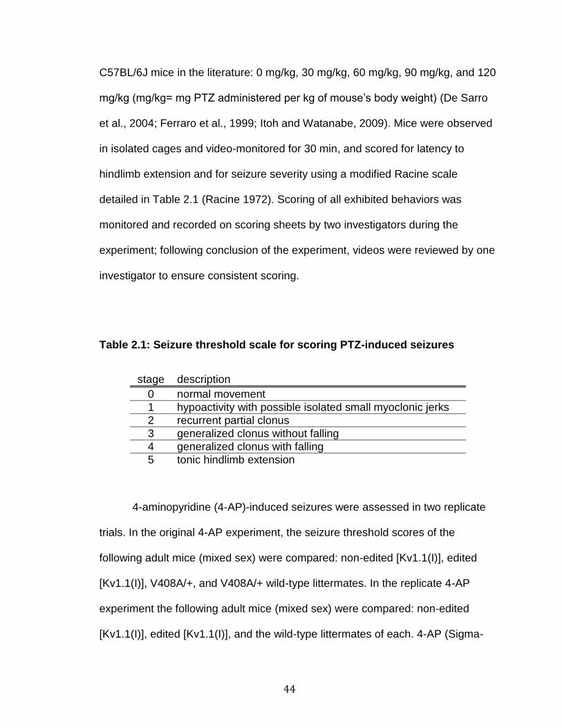
44
C57BL/6J mice in the literature: 0 mg/kg, 30 mg/kg, 60 mg/kg, 90 mg/kg, and 120
mg/kg (mg/kg= mg PTZ administered per kg of mouse’s body weight) (De Sarro
et al., 2004; Ferraro et al., 1999; Itoh and Watanabe, 2009). Mice were observed
in isolated cages and video-monitored for 30 min, and scored for latency to
hindlimb extension and for seizure severity using a modified Racine scale
detailed in Table 2.1 (Racine 1972). Scoring of all exhibited behaviors was
monitored and recorded on scoring sheets by two investigators during the
experiment; following conclusion of the experiment, videos were reviewed by one
investigator to ensure consistent scoring.
Table 2.1: Seizure threshold scale for scoring PTZ-induced seizures
stage description
0 normal movement
1 hypoactivity with possible isolated small myoclonic jerks
2 recurrent partial clonus
3 generalized clonus without falling
4 generalized clonus with falling
5 tonic hindlimb extension
4-aminopyridine (4-AP)-induced seizures were assessed in two replicate
trials. In the original 4-AP experiment, the seizure threshold scores of the
following adult mice (mixed sex) were compared: non-edited [Kv1.1(I)], edited
[Kv1.1(I)], V408A/+, and V408A/+ wild-type littermates. In the replicate 4-AP
experiment the following adult mice (mixed sex) were compared: non-edited
[Kv1.1(I)], edited [Kv1.1(I)], and the wild-type littermates of each. 4-AP (Sigma-

45
Aldrich) was dissolved in sterile saline (0.9% NaCl), the pH adjusted to 7 with
HCl, and administered subcutaneously at a volume of 0.1 mL/10 g mouse’s body
weight (De Sarro et al., 2004). Several doses were prepared by serial dilution to
create a dose response curve based on previously determined seizure threshold
data for C57BL/6J mice in the literature: 0 mg/kg, 2 mg/kg, 6 mg/kg, 10 mg/kg,
and 14 mg/kg (mg/kg= mg 4-AP administered per kg of mouse’s body weight)
(Chung et al., 2009; De Sarro et al., 2004; Dhir et al., 2011; Kim et al., 2001).
Mice were observed in isolated cages and video-monitored for 1 hour and scored
for latency to hindlimb extension and for seizure severity using a modified Racine
scale detailed in Table 2.2 (Racine 1972). Scoring of all exhibited behaviors was
monitored and recorded on scoring sheets by two investigators during the
original experiment and 3 during the replicate experiment, however following the
conclusion of the experiment videos were reviewed by one investigator to ensure
consistent scoring. Additional precautions were taken in the replicate 4-AP
experiment, where all investigators were blinded toward mouse genotype during
the scoring and the final review of all the scores was performed by an
investigator who was not part of the original test and who was unfamiliar with the
original results.

46
Table 2.2: Seizure threshold scale for scoring 4-AP-induced seizures
stage description
0 normal movement
1 hypoactivity with possible isolated small myoclonic jerks
2 recurrent partial clonus
3 episodes of generalized clonus without loss of righting ability
4 continuous generalized clonus with or without loss of righting ability and/or motoric control
5 tonic hindlimb extension
Hippocampal Slice Electrophysiology
We collaborated with Dr. Sam Centanni in the laboratory of Dr. Danny
Winder to perform electrophysiological recordings of dentate granule cells in
hippocampal slices of non-edited [Kv1.1(I)] mice and their wild-type littermates.
Recordings were performed on mice which had either been unstressed or had
been subjected to 30 minutes of conical tube restraint prior to brain isolation.
Acute, 300 µm slices were obtained containing the hippocampus and recordings
performed solely from dentate gyrus granule cells. The recordings were
conducted using a potassium gluconate internal solution. All recordings were
conducted in a current clamp configuration where the cell was injected with the
necessary current required to keep the cell held at -70 mV. To analyze firing
characteristics and intrinsic properties, current steps were applied for 1 second
and increased in 25 pA increments (starting with -150 pA and going up to +350
pA).

47
Statistical analysis
Statistical tests were performed using Graphpad Prism (Graphpad Soft-
ware). In experiments that have a single independent variable (genotype), one-
way ANOVA was performed with Tukey’s multiple comparisons test to determine
genotype comparisons (grip strength, pole climb, and flurothyl-induced seizures).
When two independent variables were present (genotype and type of brain
region; genotype and stress), 2-way ANOVA was performed with Tukey’s or
Sidak’s multiple comparisons test (type of multiple comparisons test was chosen
based on recommendations by the Prism software) (qRT-PCR, gait analysis,
inverted screen, open field, PTZ and 4-AP-induced seizures, and dentate granule
cell electrophysiology). Rotarod data, which involved two independent variables
(genotype and trial day), were analyzed by 2-way repeated measures ANOVA
with Tukey’s multiple comparisons test of the main effects, to determine
genotype differences regardless of trial day. Deviations from normal Mendelian
inheritance patterns were determined by 2 analysis. Statistical significance was
defined as p ≤ 0.05.

48
Results
Characterization of Kv1.1 editing in wild-type mouse tissues
Previous studies have characterized the levels of Kv1.1 editing in human
nervous and peripheral tissues, but only a few mouse and rat tissues were
examined (Decher et al., 2010; Hoopengardner et al., 2003; Li et al., 2009). We
confirmed and extended these previous characterizations, demonstrating that
Kv1.1 RNA editing occurs most substantially in the nervous system and that
distinct RNA editing profiles are observed when comparing different brain regions
(Figure 2.3).

49
Figure 2.3. Quantitative analysis of Kv1.1 RNA editing (I/V site) in mouse. The extent of editing for Kv1.1 transcripts isolated from (a) mouse tissues or (b) dissected brain regions in adult animals was determined by high-throughput sequence analysis as described previously (Hood et al., 2014); data represent the analysis of 4 and 3 individual animals for tissue and brain regions, respectively (mean ± SEM).
Verification of non-edited [Kv1.1(I)] and edited [Kv1.1(V)] mutant mouse models
Although the function of the non-edited [Kv1.1(I)] and edited [Kv1.1(V)]
isoforms of Kv1.1 have been studied in heterologous expression systems, no
studies have been performed to examine the physiological significance of these
editing-dependent changes in vivo. To address this issue, in collaboration with
Dr. Robert Reenan, we have taken advantage of homologous recombination in
C57BL/6J embryonic stem cells to generate genetically-modified mice that solely

50
expressed either the non-edited [Kv1.1(I)] or edited [Kv1.1(V)] subunit isoforms
(Figures 2.1 and 2.2).
To confirm that mutant mice bearing modified Kv1.1 alleles expressed
transcripts solely encoding the non-edited [Kv1.1(I)] or edited [Kv1.1(V)] isoform
of the channel, cDNA was generated by RT-PCR amplification of cerebellar RNA
and the resulting amplicons were sequenced directly. Sequences from wild-type
animals exhibited overlapping adenosine/guanosine peaks in electropherogram
traces resulting from a mixture of non-edited and edited Kv1.1 transcripts, as
inosine base pairs similarly to guanosine during reverse transcription. As
expected, sequences obtained from mutant animals showed only an adenosine
or guanosine moiety, for the non-edited and edited transcripts, respectively
(Figure 2.4).

51
Figure 2.4. Confirmation of Kv1.1 isoform identity in mutant mice. Sequence analysis of Kv1.1 RNA isoforms in mutant mice. RNA was isolated from cerebella dissected from wild-type, non-edited [Kv1.1(I)], and edited [Kv1.1(V)] adult mice. A region spanning the editing site was amplified by RT-PCR and the amplicons were analyzed by Sanger sequencing. Electropherogram traces are shown with the editing site indicated in inverse lettering; mutant nucleotides introduced to produce the non-edited and edited isoforms are underlined.
Although the results in Figure 2.4 indicated that the expected Kv1.1
isoforms are being expressed in both mouse models, Sanger sequencing is
qualitative by nature. To generate the edited [Kv1.1(V)] mouse model, the Kcna1
gene was genomically altered at the editing site to a guanosine; this target
cannot be altered by the adenosine-specific ADAR2 enzyme, and thus needed
no further validation. In contrast, the non-edited [Kv1.1(I)] mouse model was
generated by incorporating mutations around the editing site, relying on these
synonymous mutations to sufficiently disrupt the Kv1.1 RNA duplex structure to
prevent ADAR2 from recognizing and editing the RNA. As Sanger sequencing
may not be sufficiently sensitive to determine whether low levels of editing may

52
have been occurring in the non-edited [Kv1.1(I)] mutant animals, we further
examined Kv1.1 RNA expression using a high-throughput sequence strategy
previously developed in our laboratory (Hood et al., 2014). We quantified RNA
editing levels in the spinal cord, which contains one of the highest RNA editing
levels, and thus would be the most likely of our tissue targets to display aberrant
editing. Three mouse spinal cord samples were analyzed where isolated RNA
was subjected to RT-PCR amplification and the resulting amplicons were
sequenced by high-throughput sequence analysis (Hood et al., 2014). The mean
RNA editing detected in the non-edited [Kv1.1(I)] spinal cord samples was 0.045
± 0.0046 %, which corresponded to approximately 28 out of an average 60879
total reads.
After confirming that our mouse models solely expressed their intended
RNA isoform, we sought to determine whether the isoform identity affected the
steady-state RNA expression level encoding the Kv1.1 channel. We performed
qRT-PCR to determine the relative expression of Kv1.1 RNA in six brain regions
from non-edited [Kv1.1(I)], edited [Kv1.1(V)] and wild-type mice (Figure 2.5).
Results from this analysis revealed no differences in steady-state Kv1.1 RNA
expression between the non-edited [Kv1.1(I)], edited [Kv1.1(V)], and wild-type
animals for all brain regions examined.
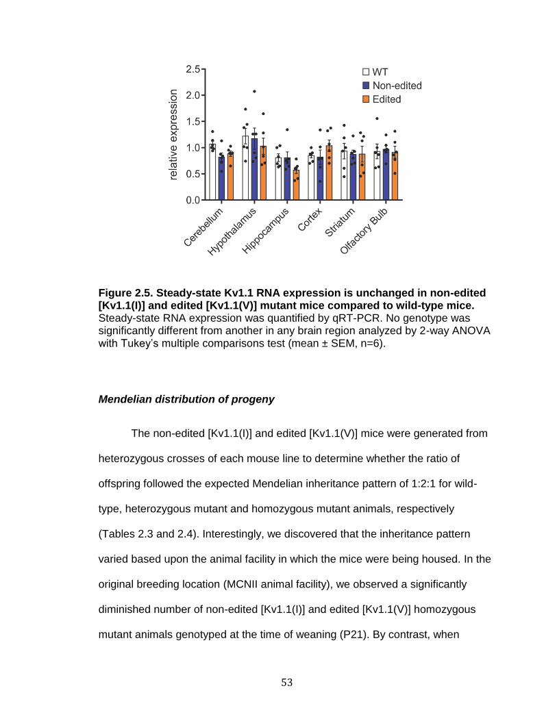
53
Figure 2.5. Steady-state Kv1.1 RNA expression is unchanged in non-edited [Kv1.1(I)] and edited [Kv1.1(V)] mutant mice compared to wild-type mice. Steady-state RNA expression was quantified by qRT-PCR. No genotype was significantly different from another in any brain region analyzed by 2-way ANOVA with Tukey’s multiple comparisons test (mean ± SEM, n=6).
Mendelian distribution of progeny
The non-edited [Kv1.1(I)] and edited [Kv1.1(V)] mice were generated from
heterozygous crosses of each mouse line to determine whether the ratio of
offspring followed the expected Mendelian inheritance pattern of 1:2:1 for wild-
type, heterozygous mutant and homozygous mutant animals, respectively
(Tables 2.3 and 2.4). Interestingly, we discovered that the inheritance pattern
varied based upon the animal facility in which the mice were being housed. In the
original breeding location (MCNII animal facility), we observed a significantly
diminished number of non-edited [Kv1.1(I)] and edited [Kv1.1(V)] homozygous
mutant animals genotyped at the time of weaning (P21). By contrast, when
54
embryos from the original breeding location were genotyped at E18.5,
immediately prior to birth, homozygous mutants from each mutant line did not
differ from the expected Mendelian inheritance pattern. These results indicated
that both strains demonstrate an incompletely penetrant postnatal lethality.
However, when the mouse colony was moved to a new floor in the same facility,
both mouse lines no longer exhibited the lethality observed for homozygous
mutants. While undefined environmental factors such as background noise or
access to the room by multiple investigators could produce stressors that affect
the survival of mutant Kv1.1 homozygotes, we rederived both mouse lines into
the pathogen-free barrier facility. Surprisingly, both lines regained the
incompletely penetrant postnatal lethality. We observed fewer homozygous
mutants at P21 in the original MCNII and Barrier facilities, yet genotype analyses
of mice that died between P0 and P21 did not identify an overrepresentation of
homozygous mutants which could be explained by the small sample size of
mouse bodies that were found.

55
Non-edited: Kv1.1(I)/+ x Kv1.1(I)/+
genotype
E18.5 Original
P21 Original
P21 Barrier
Obs (Exp) Obs (Exp) Obs (Exp)
Wild-type +/+
13 (12.25) 19 (17.75) 43 (34)
Heterozygous Kv1.1(I)/+
28 (24.5) 49 (35.5) 75 (68)
Homozygous Kv1.1(I)/Kv1.1(I)
8 (12.25) 3 (17.75) 18 (34)
total 49 71 136 p-value p≤0.3641 p≤0.0002 p≤0.0049
Table 2.3. Conditional non-Mendelian distribution of non-edited [Kv1.1(I)] mutant offspring. Upon breeding heterozygous non-edited [Kv1.1 (I)] mice (Kv1.1(I)/+), either in the original facility or the barrier facility, offspring were genotyped at either E18.5 or by P21. Observed (Obs) progeny versus the
expected (Exp) progeny were compared by 2 analysis to determine significance.
Edited: Kv1.1(V)/+ x Kv1.1(V)/+
genotype
E18.5 Original
P21 Original
P21 Barrier
Obs (Exp) Obs (Exp) Obs (Exp)
Wild-type +/+
12 (14.75) 46 (29) 55 (37.25)
Heterozygous Kv1.1(V)/+
34 (29.5) 60 (58) 67 (74.5)
Homozygous Kv1.1(V)/Kv1.1(V)
13 (14.75) 10 (29) 27 (37.25)
total 59 116 149 p-value p≤0.4949 p≤0.0001 p≤0.0024
Table 2.4. Conditional non-Mendelian distribution of edited [Kv1.1(V)] mutant offspring. Upon breeding heterozygous edited [Kv1.1 (V)] mice (Kv1.1(V)/+), either in the original facility or the barrier facility, offspring were genotyped at either E18.5 or by P21. Observed (Obs) progeny versus the
expected (Exp) progeny were compared by 2 analysis to determine significance.
56
Behavioral characterization
We have used a variety of tasks to distinguish behavioral differences
between the non-edited [Kv1.1(I)] and edited [Kv1.1(V)] mutant mouse models
compared to their wild-type littermates (Brooks and Dunnett, 2009). Initial
behavioral tasks showed that these mutant animals did not have deficits in grip
strength, but that the edited [Kv1.1(V)] mice displayed enhanced motor
coordination on the pole climb task (Figure 2.6). The locomotor coordination of
the non-edited [Kv1.1(I)] and edited [Kv1.1(V)] mice was tested using the
accelerating rotarod behavioral task, first using the traditional paradigm: three
trials/day for three days (Figure 2.7). In preliminary studies, we compared non-
edited [Kv1.1(I)] and edited [Kv1.1(V)] mutants to the performance of wild-type
littermates, as well as their heterozygous mutant littermates [Kv1.1(I)/+ and
Kv1.1(V)/+]. There were no significant differences between either type of mutant
mice compared to the wild-type littermates or their respective heterozygous
mutant mice (Figure 2.7). This lack of any impairment phenotype under normal
conditions was similar to the EA1 mouse model (V408A/+), which only displays
locomotor deficits under stressed conditions. All further behavioral tests were
performed under control conditions (unstressed) as well as in response to a well
characterized stressor involving 30 minutes of conical tube restraint immediately
prior to the tests (Buynitsky and Mostofsky, 2009).

57
Figure 2.6. Initial behavioral analysis of non-edited [Kv1.1(I)] and edited [Kv1.1(V)] mice. Adult non-edited [Kv1.1(I)], edited [Kv1.1(V0], and wild-type littermate male mice were subjected to the (a) grip strength and (b) pole climb behavioral tests. No significant differences were found in the grip strength test, but the edited [Kv1.1(V)] mice were significantly faster to descend on the pole climb test compared to wild-type and non-edited [Kv1.1(I)] mice (mean ± SEM, n=7-13, *p<0.05, 1-way ANOVA with Tukey’s multiple comparisons test).
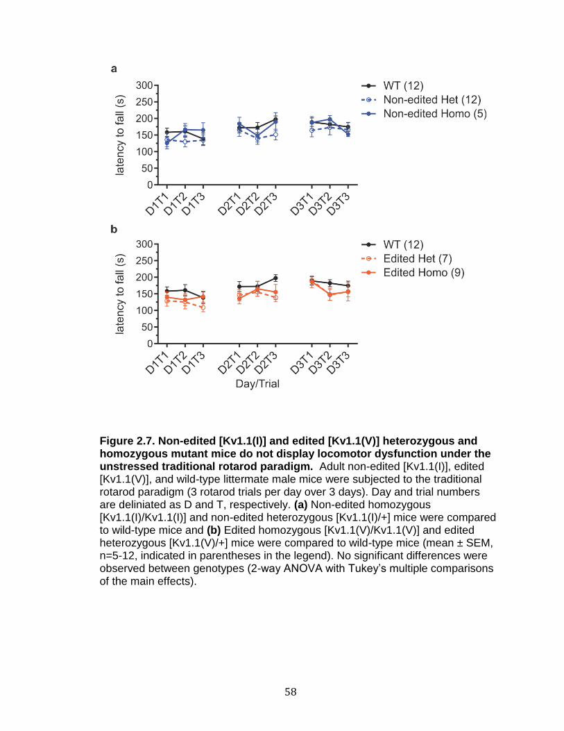
58
Figure 2.7. Non-edited [Kv1.1(I)] and edited [Kv1.1(V)] heterozygous and homozygous mutant mice do not display locomotor dysfunction under the unstressed traditional rotarod paradigm. Adult non-edited [Kv1.1(I)], edited [Kv1.1(V)], and wild-type littermate male mice were subjected to the traditional rotarod paradigm (3 rotarod trials per day over 3 days). Day and trial numbers are deliniated as D and T, respectively. (a) Non-edited homozygous [Kv1.1(I)/Kv1.1(I)] and non-edited heterozygous [Kv1.1(I)/+] mice were compared to wild-type mice and (b) Edited homozygous [Kv1.1(V)/Kv1.1(V)] and edited heterozygous [Kv1.1(V)/+] mice were compared to wild-type mice (mean ± SEM, n=5-12, indicated in parentheses in the legend). No significant differences were observed between genotypes (2-way ANOVA with Tukey’s multiple comparisons of the main effects).
59
To incorporate a stressor paradigm in conjunction with the rotarod test, a
modified rotarod paradigm was developed where mice were run on the rotarod
one trial per day for nine days, with or without 30 minutes of conical tube
restraint, immediately preceding the rotarod test. This modified rotarod paradigm
was replicated in three unstressed replicate trials (Figure 2.8) and two stressed
replicate trials (Figure 2.9). In the absence of a stressor, no differences were
observed between the mutant and wild-type mouse lines, whereas, following 30
minutes of conical tube restraint, the non-edited [Kv1.1(I)] mice consistently
underperformed compared to both the wild-type and the edited [Kv1.1(V)] mice.
These studies were replicated and continued to show that homozygous mice
solely expressing non-edited [Kv1.1(I)] underperformed compared to the wild-
type and edited [Kv1.1(V)] lines.
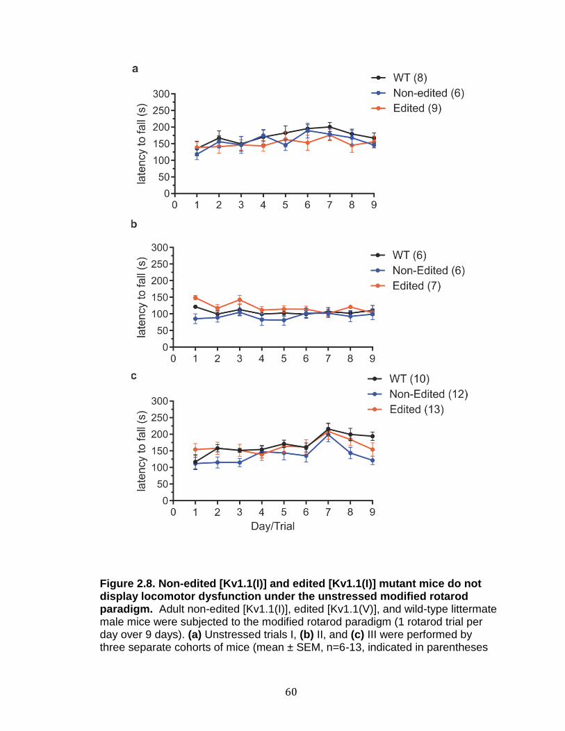
60
Figure 2.8. Non-edited [Kv1.1(I)] and edited [Kv1.1(I)] mutant mice do not display locomotor dysfunction under the unstressed modified rotarod paradigm. Adult non-edited [Kv1.1(I)], edited [Kv1.1(V)], and wild-type littermate male mice were subjected to the modified rotarod paradigm (1 rotarod trial per day over 9 days). (a) Unstressed trials I, (b) II, and (c) III were performed by three separate cohorts of mice (mean ± SEM, n=6-13, indicated in parentheses

61
in the legend). No significant differences were observed between non-edited [Kv1.1(I)], edited [Kv1.1(V)], or wild-type genotypes (2-way repeated measures ANOVA with Tukey’s multiple comparisons of the main effects).
Figure 2.9. Non-edited [Kv1.1(I)] mutant mice display stress-induced locomotor dysfunction under the stressed modified rotarod paradigm. Adult non-edited [Kv1.1(I)], edited [Kv1.1(V)], and wild-type littermate male mice were subjected to a stressor (30 minutes of conical tube restraint) followed by the modified rotarod paradigm (1 rotarod trial per day over 9 days). (a) Stressed trials I, (b) II were performed by two separate cohorts of mice (mean ± SEM, n=6-8, indicated in parentheses in the legend). Significant differences were observed between the non-edited [Kv1.1(I)] mice and the edited [Kv1.1(V)] and wild-type genotypes (*p<0.05, ***p<0.001, 2-way repeated measures ANOVA with Tukey’s multiple comparisons of the main effects).

62
To determine the underlying factors responsible for alterations in the
rotarod task in response to stress, additional behavioral studies were employed
to examine alterations in gait directly, similar to the deficits observed in EA1
patients. Alterations in gait were assessed using the footprint test, under
unstressed or stressed conditions (30 min of conical tube restraint prior the task),
and each condition was replicated twice using the same cohort of mice (Figure
2.10). No differences were seen between the replicate trials of either the
unstressed or the stressed paradigm. In addition, no differences were found
comparing the genotypes by the forelimb stride length and front base
parameters. The hindlimb stride length, hind base, and right overlap parameters
contained significant differences in one unstressed or stressed trial, but not both
trials, suggesting that these differences were not replicable in our preliminary
studies. In both stressed trials of the left overlap parameter, however, the non-
edited [Kv1.1(I)] mice had a significantly smaller left overlap parameter as
compared to wild-type littermates.

63
Figure 2.10. Non-edited [Kv1.1(I)] mice display reproducible stress-induced gait alterations. Adult non-edited [Kv1.1(I)], edited [Kv1.1(V)], and wild-type littermate male mice were subjected to either an unstressed or stressed paradigm (30 minutes of conical tube restraint) followed by footprinting gait analyses. Two trials were performed on the same cohort of mice (Un1=Unstressed trial 1, Un2=Unstressed trial 2, S1=Stressed trial 1, S2=Stressed trial 2). Several metrics were determined: (a) Forelimb Stride Length, (b) Hindlimb Stride Length, (c) Front Base, (d) Hind Base, (e) Left Overlap, and (f) Right Overlap. Several significant differences were observed (mean ± SEM, n=5-17, *p<0.05, **p<0.01, 2-way ANOVA with Tukey’s multiple

64
comparisons test, comparing genotypes within each trial). Replicate trials were not different from each other by 2-way ANOVA wtih Sidak’s multiple comparisons test.
Stressed effects to grip strength were determined by performing the
inverted screen task, under the unstressed and stressed conditions (30 min of
conical tube restraint prior the task) (Figure 2.11). Both the unstressed and
stressed paradigms were replicated three times each. No significant differences
were seen for the non-edited [Kv1.1(I)], edited [Kv1.1(V)], or the wild-type
littermates, when comparing genotypes within the same trial.

65
Figure 2.11. Non-edited [Kv1.1(I)] and edited [Kv1.1(V)] mice do not display stress-induced alterations by the inverted screen test. Adult non-edited [Kv1.1(I)], edited [Kv1.1(V)], and wild-type littermate male mice were subjected to either an unstressed or stressed paradigm (30 minutes of conical tube restraint) followed by the inverted screen test for a maximum of 60 s. Three trials were performed for the unstressed and stressed paradigms (Un1=Unstressed trial 1, Un2=Unstressed trial 2, Un3=Unstressed trial 3, S1=Stressed trial 1, S2=Stressed trial 2, S3=Stressed trial 3). No significant differences were observed between the genotypes within the same trial (mean ± SEM, n=9-13, 2-way ANOVA with Tukey’s multiple comparisons test).
Finally, locomotion and anxiety were tested using an open field behavioral
test under both unstressed and stressed conditions. Mice were observed for 30
min during the task and many parameters were observed (Figure 2.12).
Although, many parameters were altered by stress when comparing each
individual genotype (such as distance traveled, velocity, center time, center
entries, and grooming time), differences between genotypes were rare. The only
identified difference observed between genotypes was a decrease in thigmotaxis
for non-edited [Kv1.1(I)] animals compared to wild-type mice.

66
Figure 2.12. Open-field analysis of non-edited [Kv1.1(I)] and edited [Kv1.1(V)] mice. Adult non-edited [Kv1.1(I)], edited [Kv1.1(V)], and wild-type littermate male mice were subjected to either an unstressed or stressed paradigm (30 minutes of conical tube restraint) followed by 30 min of open-field analysis. Several metrics were determined: (a) Distance Traveled, (b) Velocity, (c) Center Time, (d) Center Entries, (e) Rearing Counts, and (f) Grooming Time. Several significant differences were observed between the Unstressed and
67
Stressed trials of the same genotype (mean ± SEM, n=8-13; *p<0.05, ***p<0.001, ****p<0.0001, 2-way ANOVA with Sidak’s multiple comparisons test). When comparing genotypes within each trial, only the non-edited [KV1.1(I)] center entries were different from WT in (d) (#p<0.05, 2-way ANOVA with Tukey’s multiple comparisons test).
Spontaneous Seizure Monitoring
Although collaborative EEG recordings, performed by Alison Miller in Dr.
Jennifer Kearney’s lab, qualitatively suggested abnormal activity in the non-
edited [Kv1.1(I)] mutants, this data was not conclusive due to a high background
in the collected data. To decrease the background for our EEGs, we performed a
pilot study comparing a non-edited [Kv1.1(I)] mouse to its wild-type littermate
using surface cortical electrodes, instead of screws, to decrease any noise that
could be interfering with the measurements. The non-edited [Kv1.1(I)] mouse did
not display any epileptiform activity visible in the EEG traces during six hours of
recording, but video analysis revealed two tonic events in the non-edited
[Kv1.1(I)] animal during sleep.
Drug-induced Seizure Susceptibility
Drug-induced seizure susceptibility was investigated using three different
convulsant drugs: flurothyl, PTZ, and 4-AP in homozygous Kv1.1 mutant lines
and wild-type mice. No differences were seen for flurothyl-induced seizures,
however genotype-specific differences were observed in response to PTZ and 4-
AP (Figure 2.13).
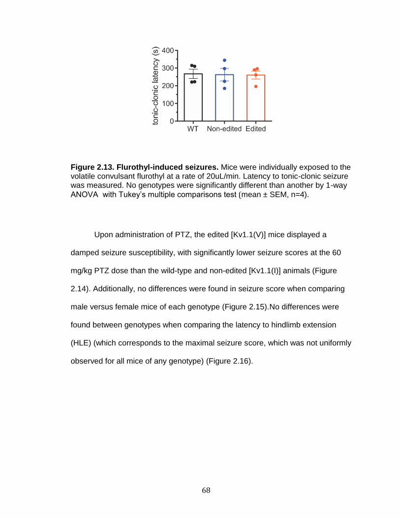
68
Figure 2.13. Flurothyl-induced seizures. Mice were individually exposed to the volatile convulsant flurothyl at a rate of 20uL/min. Latency to tonic-clonic seizure was measured. No genotypes were significantly different than another by 1-way
ANOVA with Tukey’s multiple comparisons test (mean ± SEM, n=4).
Upon administration of PTZ, the edited [Kv1.1(V)] mice displayed a
damped seizure susceptibility, with significantly lower seizure scores at the 60
mg/kg PTZ dose than the wild-type and non-edited [Kv1.1(I)] animals (Figure
2.14). Additionally, no differences were found in seizure score when comparing
male versus female mice of each genotype (Figure 2.15).No differences were
found between genotypes when comparing the latency to hindlimb extension
(HLE) (which corresponds to the maximal seizure score, which was not uniformly
observed for all mice of any genotype) (Figure 2.16).

69
Figure 2.14. PTZ-induced seizure threshold scores. Mice were intraperitoneally administered increasing doses of PTZ, observed for 30 minutes, and scored for seizure severity on a scale from 0 to 5 (as described in the Materials & Methods). Non-edited [Kv1.1(I)], edited [Kv1.1(V)], and V408A/+ mice were compared to the wild-type littermates of the V408A/+ mice. Edited [Kv1.1 (V)] mice differed significantly from wild-type and non-edited [Kv1.1 (I)] animals at the 60mg/kg PTZ dose by 2-way ANOVA with Tukey’s mulitple comparisons test (mean ± SEM, n=7-9 mice for each genotype per dose; Edited to WT: *p<0.05; Edited to Non-edited: #p<0.05).
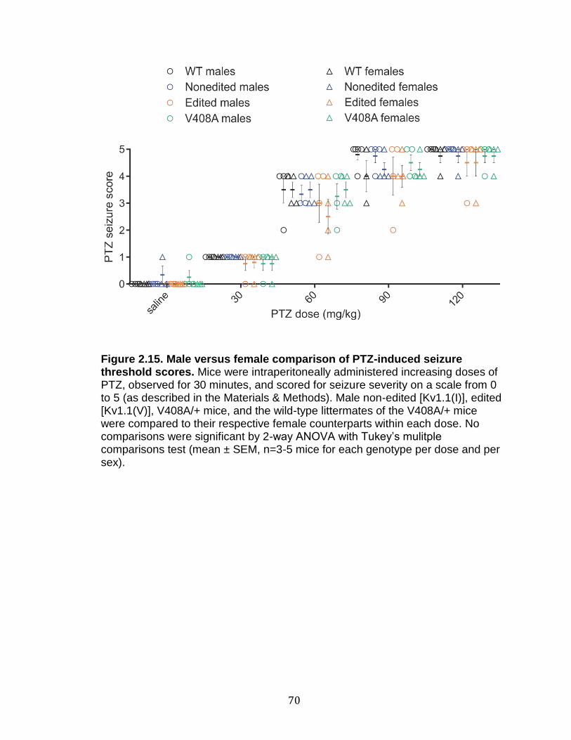
70
Figure 2.15. Male versus female comparison of PTZ-induced seizure threshold scores. Mice were intraperitoneally administered increasing doses of PTZ, observed for 30 minutes, and scored for seizure severity on a scale from 0 to 5 (as described in the Materials & Methods). Male non-edited [Kv1.1(I)], edited [Kv1.1(V)], V408A/+ mice, and the wild-type littermates of the V408A/+ mice were compared to their respective female counterparts within each dose. No comparisons were significant by 2-way ANOVA with Tukey’s mulitple comparisons test (mean ± SEM, n=3-5 mice for each genotype per dose and per sex).

71
Figure 2.16. Latency to hindlimb extension following PTZ-induced seizures. Mice were subcutaneously administered increasing doses of 4-AP, observed for 1 hour, measuring the latency to first hindlimb extension (HLE), if it occurred. Non-edited [Kv1.1(I)], edited [Kv1.1(V)], and V408A/+ mice were compared to the wild-type littermates of the V408A/+ mice (mean ± SEM, n=7-9 mice for each genotype per dose; not every mouse reached HLE). No statistical comparisons were significant by 2-way ANOVA with Tukey’s multiple comparisons test.
In the original 4-AP-induced seizure threshold test, the non-edited
[Kv1.1(I)] mice displayed an increased seizure susceptibility, displaying higher
scoring seizures at the 6mg/kg 4-AP dose, compared to all other genotypes
(Figure 2.17a). Due to experimental limitations, the PTZ and original 4-AP
seizure threshold tests used wild-type littermates from the V408A/+ mice as
control animals. To ensure consistency however, the 4-AP seizure threshold test
was replicated with the appropriate wild-type littermates for non-edited [Kv1.1(I)]
and edited [Kv1.1(V)] mice (Figure 2.17b). In the replicate 4-AP analysis, the
non-edited [Kv1.1(I)] mice demonstrated a significantly higher score at the 6
mg/kg 4-AP dose compared to wild-type animals, yet edited [Kv1.1(V)] mice

72
showed a significantly lower score. Furthermore, the edited [Kv1.1(V)] mice had
a significantly dampened seizure susceptibility compared to the non-edited
[Kv1.1(I)] mice at the higher (6 mg/kg and 10 mg/kg) 4-AP doses. Although male
versus female differences were observed in both 4-AP trials, replicate tests did
not agree on the type of sex differences, and likely were not biologically
significant (Figure 2.18). The non-edited [Kv1.1(I)] mice, of both replicate trials,
had a significantly shorter latency to HLE at the 10 mg/kg dose compared to all
the other genotypes (Figure 2.19). In addition, many non-edited [Kv1.1(I)] mice
exhibited HLE at the 6 mg/kg dose in both replicate trials (though not enough
individuals of the other genotypes exhibited HLE for statistical analysis at this
dose).
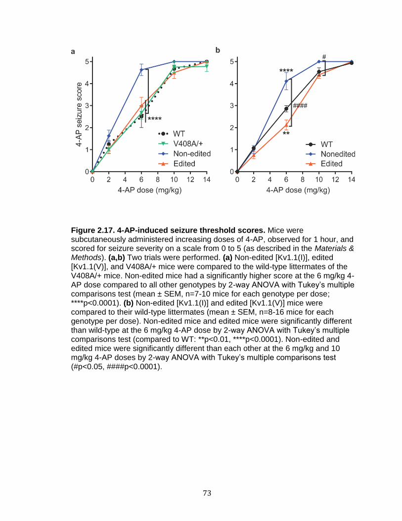
73
Figure 2.17. 4-AP-induced seizure threshold scores. Mice were subcutaneously administered increasing doses of 4-AP, observed for 1 hour, and scored for seizure severity on a scale from 0 to 5 (as described in the Materials & Methods). (a,b) Two trials were performed. (a) Non-edited [Kv1.1(I)], edited [Kv1.1(V)], and V408A/+ mice were compared to the wild-type littermates of the V408A/+ mice. Non-edited mice had a significantly higher score at the 6 mg/kg 4-AP dose compared to all other genotypes by 2-way ANOVA with Tukey’s multiple comparisons test (mean ± SEM, n=7-10 mice for each genotype per dose; ****p<0.0001). (b) Non-edited [Kv1.1(I)] and edited [Kv1.1(V)] mice were compared to their wild-type littermates (mean ± SEM, n=8-16 mice for each genotype per dose). Non-edited mice and edited mice were significantly different than wild-type at the 6 mg/kg 4-AP dose by 2-way ANOVA with Tukey’s multiple comparisons test (compared to WT: **p<0.01, ****p<0.0001). Non-edited and edited mice were significantly different than each other at the 6 mg/kg and 10 mg/kg 4-AP doses by 2-way ANOVA with Tukey’s multiple comparisons test (#p<0.05, ####p<0.0001).

74
Figure 2.18. Male versus female comparison of 4-AP-induced seizure threshold scores. Mice were subcutaneously administered increasing doses of 4-AP, observed for 1 hour, and scored for seizure severity on a scale from 0 to 5 (as described in the Materials & Methods). (a,b) Two trials were performed. (a) Male non-edited [Kv1.1(I)], edited [Kv1.1(V)], V408A/+ mice, and the wild-type littermates of the V408A/+ mice were compared to their respective female counterparts within each dose. At the 6 mg/kg 4-AP dose, edited [Kv1.1(V)] male mice were significantly different than the respective female mice by 2-way ANOVA with Tukey’s multiple comparisons test (mean ± SEM, n=3-6 mice for each genotype per dose and per sex; *p<0.05). (b) Male non-edited [Kv1.1(I)],

75
edited [Kv1.1(V)], and their wild-type littermate mice were compared to their respective female counterparts within each dose. At the 6 mg/kg 4-AP dose, non-edited [Kv1.1(I)] male mice were significantly different than the respective female mice by 2-way ANOVA with Tukey’s multiple comparisons test (mean ± SEM, n=3-9 mice for each genotype per dose and per sex; **p<0.01).

76
Figure 2.19. Latency to hindlimb extension following 4-AP-induced seizures. Mice were subcutaneously administered increasing doses of 4-AP, observed for 1 hour, measuring the latency to first hindlimb extension (HLE), if it occurred. (a,b) Two trials were performed. (a) Non-edited [Kv1.1(I)], edited [Kv1.1(V)], and V408A/+ mice were compared to the wild-type littermates of the V408A/+ mice (mean ± SEM, n=7-10 mice for each genotype per dose; not every mouse reached HLE). No statistical comparisons were possible at the 6 mg/kg 4-AP dose, because only the non-edited [Kv1.1(I)] mice had more than one individual mouse reaching HLE. Non-edited [Kv1.1(I)] mice had a significantly shorter latency to HLE at the 10 mg/kg 4-AP dose compared to all other genotypes by 2-way ANOVA with Tukey’s multiple comparisons test (*p<0.05,**p<0.01). (b) Non-edited [Kv1.1(I)] and edited [Kv1.1(V)] mice were compared to their wild-type littermates (mean ± SEM, n=8-16 mice for each genotype per dose; not every mouse reached HLE). No statistical comparisons were possible at the 6 mg/kg 4-AP dose, because only the non-edited [Kv1.1(I)]

77
mice had mice reaching HLE. Non-edited [Kv1.1(I)] mice had a significantly shorter latency to HLE at the 10 mg/kg 4-AP dose compared to all other genotypes by 2-way ANOVA with Tukey’s multiple comparisons test (***p<0.001, ****p<0.0001).
Preliminary Electrophysiological characterization
Due to the observed alterations in drug-induced seizure susceptibility, we
collaborated with Dr. Sam Centanni in the laboratory of Dr. Danny Winder to
perform a pilot study of slice electrophysiology in homozygous non-edited
[Kv1.1(I)] mutant mice. We focused our pilot recordings on dentate granule cells
in the hippocampus of non-edited [Kv1.1(I)] mice compared to wild-type
littermates because of a recent study which observed a dramatic upregulation of
Kv1.1 in these neurons in a model of temporal lobe epilepsy (Kirchheim et al.,
2013). Results from this analysis showed no differences between non-edited
[Kv1.1(I)] and wild-type animals, comparing the intrinsic properties and single
action potential firing characteristics of dentate granule cells. Since we observed
phenotypic differences only in non-edited [Kv1.1(I)] mice following conical tube
restraint, Dr. Centanni also performed recordings on mice that had just
undergone conical tube restraint and again, there were no significant differences
in any signaling parameters between the genotypes (Figure 2.20).
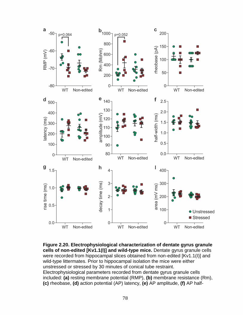
78
Figure 2.20. Electrophysiological characterization of dentate gyrus granule cells of non-edited [Kv1.1(I)] and wild-type mice. Dentate gyrus granule cells were recorded from hippocampal slices obtained from non-edited [Kv1.1(I)] and wild-type littermates. Prior to hippocampal isolation the mice were either unstressed or stressed by 30 minutes of conical tube restraint. Electrophysiological parameters recorded from dentate gyrus granule cells included: (a) resting membrane potential (RMP), (b) membrane resistance (Rm), (c) rheobase, (d) action potential (AP) latency, (e) AP amplitude, (f) AP half-

79
width, (g) AP rise time, (h) AP decay, (i) AP area. No significant differences were found between genotypes or unstressed versus stressed conditions, though p-values close to 0.05 are marked (mean ± SEM, n=5-9, 2-way ANOVA with Sidak’s multiple comparisons test).
Discussion
Characterization of Kv1.1 editing in wild-type mouse tissues
Our studies extending characterization of Kv1.1 editing in wild-type mouse
tissues highlight the importance of Kv1.1 regulation throughout the nervous
system. We did not see substantial Kv1.1 editing in any peripheral tissues
examined, however these dissections were of large tissues and do not discount a
small subset of cells that might still undergo RNA editing, which could have been
diluted out in our broad studies. The specific and reproducible levels of Kv1.1
editing characterized in each of the brain regions tested may indicate that
regulating expression of the non-edited versus edited Kv1.1 transcripts allows for
unique signaling properties for each tissue type. Further studies will be required
on single cells to determine whether the RNA editing levels we observed occur
across multiple cell types or whether the level of editing in a subset of the cellular
populations drives the average.
In general, the levels of RNA editing that we observed in these mouse
tissues were comparable to the levels other investigators have seen in mice, rats,
and humans, especially considering the known variation in editing between
80
different wild-type mouse strains (Morabito et al., 2010). One notable exception
was Decher et al. (2010), where a high level of editing in human striatum was
observed (approximately 65%), whereas levels of editing in mouse striatum were
approximately 14%. These results suggest that mouse and human Kv1.1 editing
profiles could vary more in the striatum as compared to other tissues. It is also
important to note that RNA editing levels are necessarily more precise in mouse
lines, which are genetically identical, compared to wide variability in human RNA
editing. This variability has been previously characterized for Kv1.1 in human
tissues obtained from patients undergoing surgery for mesial temporal lobe
epilepsy with hippocampal sclerosis (MTLE+HS) (Krestel et al., 2013). Krestel et
al. (2013) observed normal Kv1.1 RNA editing levels in control human
hippocampal RNA at 23% and 20%, respectively, representing commercially
available human hippocampal RNA (pooled sample from 19 individuals) and from
autopsy controls (pooled from 5 individuals). Conversely, their samples from
MTLE+HS patients varied widely, with Kv1.1 RNA editing ranging from 0% to
approximately 47%. The variability of editing levels in epilepsy patients could
result from their underlying disorder or might represent normal variability between
individuals, whereas the controls were pooled samples that could artificially mask
the population’s variability. A second interpretation is that the dissections are not
of comparable regions of the striatum. In Decher et al. (2010), human brain RNA
was obtained commercially and consisted of pooled RNA from 100 to 200
individuals. Unfortunately, no detail is given for which part of the striatum was
obtained, whether a particular region or a homogenate of the entire brain region,
81
or whether the RNA was obtained from clinically healthy humans or from patients
with an underlying disorder. In a similar manner, the wide variability in MTLE+HS
patients also could stem from differences in which part of the hippocampus was
surgically removed. For these reasons, we can only speculate as to whether the
difference in editing frequency between mouse and human striatum is biologically
consequential or an experimental artifact. Future research is needed to describe
the normal human variation in Kv1.1 RNA editing, in order to understand if it is as
variable as observed in MTLE+HS tissue samples, and if this variability could
underlie a risk factor for seizures or whether variability is a resulting
consequence of seizures (Krestel et al., 2013). This could particularly be studied
by comparing Kv1.1 RNA editing in focal tissue versus the surrounding/non-focal
tissue from epileptic patient samples. Our high-throughput sequencing method
would be key to detecting accurate measurements of RNA editing, as other
studies of human variation of RNA editing have lacked robustness, with low read
levels covering the editing site (O'Neil and Emeson, 2012).
Verification of non-edited [Kv1.1(I)] and edited [Kv1.1(V)] mutant mouse
models
Our mutant mouse lines can serve as important model systems to assess
the consequences resulting from editing dysregulation of Kv1.1 transcripts,
including alterations in survival, behavior, seizure-susceptibility, and
electrophysiological properties of Kv1.1-expressing neurons in their native
82
environment. However, to make meaningful assertions regarding the roles of the
non-edited [Kv1.1(I)] and edited [Kv1.1(V)] isoforms, we needed to first validate
that our mouse models only differed in Kv1.1 isoform identity, not steady-state
Kv1.1 mRNA levels. First, we confirmed that the intended isoform was being
expressed and not altered at the RNA level by qualitative Sanger sequencing of
RT-PCR amplicons generated from cerebellar RNAs from the wild-type, non-
edited [Kv1.1(I)], and edited [Kv1.1(V)] mouse lines. Editing of the Sanger
sequenced WT cerebellum sample in Figure 2.4 appears to be greater than the
editing determined by high-throughput sequence analysis in Figure 2.4; for a
comparative analysis of editing quantification by Sanger versus high-throughput
sequencing, see Appendix A and Figure A.1.
Additional verification was performed for non-edited [Kv.1.1(I)] mice to
ensure that the mutations introduced around the editing site were sufficient to
prevent RNA editing from occurring. Although a low level of RNA editing was
detected for the non-edited [Kv1.1(I)] mice in RNAs isolated from spinal cord
(0.045 ± 0.0046 %), it is likely to have little physiological significance. These
edited reads are likely the outcome of the low level of erroneous nucleotide
incorporation, arising from the error rate of the polymerase during RT-PCR
amplification, as well as the error rate for accurate detection on the Illumina
sequencing platform. Two contributing factors to the error profiles using Illumina
sequencing are the presence of the motif GGC (or GCC when sequencing the
reverse strand) within 10 bases of the sequence specific error (SSE) as well as
the presence of inverted repeats in the vicinity of the SSE (Nakamura et al.,
83
2011). Interestingly, there are two GCC motifs within 10 bases downstream of
the editing site and the editing site naturally resides within an extended inverted
repeat. These type of errors also are heavily influenced by the sample
preparation method as well as primers used, with different motifs influencing the
types of errors that occur (Schirmer et al., 2015). They also noted that amplicon-
based sequencing is more prone to motif-based errors due to the high degree of
similarity in sequenced reads. Our high-throughput sequencing technique is able
to limit the effects of errors by excluding reads containing any errors that differ
from the reference sequence, but cannot differentiate necessarily between G-to-
A and A-to-G sequencing errors at the editing site. This represents a small
amount of error with little effect on the overall editing percentage, but can explain
the very small number of edited reads found in spinal cord RNA isolated from
non-edited [Kv1.1(I)] mice.
To make accurate conclusions about our mouse models, we also needed
to verify whether our efforts to express a fixed Kv1.1 isoform led to alterations in
steady-state Kv1.1 RNA expression. Quantitative RT-PCR experiments validated
that isoform identity did not alter Kv1.1 RNA levels for either isoform from that
observed in wild-type mice. These controls further support the idea that any
variation in phenotype that we might observe would result from restricting Kv1.1
expression to a single edited isoform, either non-edited [Kv1.1(I)] or edited
[Kv1.1(V)], and not due to unpredicted isoform expression or alterations in Kv1.1
RNA expression levels.
84
Mendelian distribution of progeny
We have observed that both homozygous non-edited [Kv1.1(I)] and
edited[Kv1.1(V)] mutant alleles can result in early postnatal lethality with
incomplete penetrance, but that such postnatal lethality may be dependent on
environmental factors such as stress. Mice were bred in both a traditional
(MCNII) and pathogen-free Barrier facility to assess whether the observed
postnatal lethality in homozygous mutant mice resulted from an external
pathogen. However, both facilities demonstrated lethality for a subset of the
homozygous mutants. Although we cannot rule out that there was a difference in
the gut microbiota of the two breeding colonies in the different floors of the MCNII
facilities, prior studies of gut microbiota variability have demonstrated that raising
mice in individually ventilated cages in separate rooms of the same facility led to
mice with statistically similar cecal microbiomes (Hufeldt et al., 2010). Because
mutations in Kv1.1 underlie the etiology of EA1, which involves stress-induced
symptoms, it is possible that there are stress-induced environmental factors that
lead to the difference in survival. Such environmental factors might include a
difference in animal care technicians, levels of noise, and/or levels of activity in
each room. Informal characterization of the two MCNII rooms indicates that one
had a higher level of noise and activity because it contained a commonly used
procedural hood. When we genotyped the corpses of dead pups found in the
breeding facilities, we did not see an overrepresentation of homozygous mutants
for any genotype. There are several possibilities for why we never observed the
postnatal lethality. One possibility is that the offspring are dying sometime after

85
birth, triggered by an environmental stressor, at an early enough stage that the
mother can cannibalize the dead pup without leaving any trace. Another
possibility is that some of the homozygous pups are dying during the stress of
birth, followed by maternal cannibalization. Finally, environmental stressors could
be affecting the mother rather than the pup, leading to selective maternal
cannibalism of the homozygous mutants. Although it seems unlikely that the
mothers are using some unknown differentiating factor to distinguish
homozygous mutants versus heterozygotes and wild-type littermates, this has
been previously documented in the literature for other homozygous mutant
animals (Kastner et al., 1996; Lohnes et al., 1993; Lugt et al., 1994). More
detailed video monitoring and cross-fostering analyses will be required to
determine when these homozygous mutant animals are lost and what the
underlying factors could be (Kuroda and Tsuneoka, 2013; Weber et al., 2013).
Behavioral characterization
We observed that the non-edited [Kv1.1(I)] mice show locomotor deficits
under stressed conditions, similar to those observed in the human disorder, EA1.
While rotarod results for all animals were within the range of normal literature
values obtained from C57BL/6J mice, the mice of some unstressed trials did not
appear to increase their improvement on the task (Figure 2.8b) (Matsuo et al.,
2010). This apparent lack of learning in one of the unstressed trials likely results
from random sampling and increasing the number of animals used in the trial
86
could better display this trend. Local maxima are seen before the last trial day for
the first and third trials, but this is in agreement with studies by Matsuo et al.
(2010), which also observed this kind of trend for data with smaller animal
numbers, whereas when they increased their sample size from 13 to 1087
animals, the curve smoothed to a steady plateau.
We observed a statistical difference between left overlap parameters of
the non-edited [Kv1.1(I)] mice compared to the wild-type littermates in gait
analyses, though not in any other parameters. This result could be indicative of a
stress-induced gait difference, though it is generally held that decreases in the
paw overlap indicate improvements in motor coordination rather than
impairments (Carter et al., 1999; Carter et al., 2001). However, there are several
examples of mouse models with locomotor deficits in the rotarod task that do not
display an ataxic gait by footprinting analysis (Jayabal et al., 2015; Kayakabe et
al., 2014). Furthermore, the observed decrease in left paw overlap could
potentially lead to dysfunction because it is an unbalanced change not reflected
in the right overlap.
Importantly, all behavioral tasks where we observed stress-mediated
differences were performed on mice originating from the second MCNII facility,
where no lethality was observed for either mutant genotype. As we have
hypothesized that the conditional lethality of the original MCNII and barrier
facilities may be associated with an environmental stressor, using mice from
these facilities could result in inadvertently selecting for mutants that are
hardened to stress, which could confound our interpretation. In contrast, by using

87
mice from the facility with no observed lethality, we avoided this possible
selection.
Unfortunately, additional studies of grip strength, motor coordination by
the pole climb test, inverted screen, and open field analysis did not indicate
dysfunction for homozygous non-edited [Kv1.1(I)] mice. The results of open field
analysis also indicate that the non-edited [Kv1.1(I)] mice do not have increased
anxiety, although direct assessments of anxiety-like behavior (e.g. elevated plus
maze) were not employed. The rotarod task may be uniquely suited to promoting
the stress-induced ataxia because it is a more strenuous task than the other
behavioral studies used for these animals.
The interaction between stress and genotype is complex and leads to
further questions about how stress is leading to genotype differences. Stress
appears to increase performance for all genotypes, but also reveals a differential
genotypic effect of stress between different trial days. To compare the effects of
stress, the data described in Figures 2.8 and 2.9 were combined; this was done
in order to minimize the differences due to each trial being performed at a
different time on separate, naive cohorts (trials would need to be simultaneous
for all groups for the most accurate comparisons) (Figure 2.21). Stress led to
significant increases in rotarod performance for all genotypes, when comparing
the performance over the entire test (significant main effects indicated by #
symbols in Figure 2.21). Significant differences comparing among the unstressed
genotypes or among the stressed genotypes followed the insignificant and
significant trends described for the individual cohort data in Figures 2.8 and 2.9,

88
respectively (significant main effects indicated by + symbols in Figure 2.21).
Interestingly, when the significance was tested for each trial day separately, the
edited [Kv1.1(V)] and wild-type stressed groups performed significantly longer
than the corresponding unstressed edited or wild-type groups starting on days 2
and 3, respectively (indicated by * symbols in Figure 2.21). In contrast, the non-
edited [Kv1.1(I)] stressed group was not significantly increased compared to the
non-edited [Kv1.1(I)] unstressed group until day 6. This indicates that the non-
edited [Kv1.1(I)] mice gain less from the stressed performance enhancement
compared to the other genotypes and the stress performance enhancement
begins later.
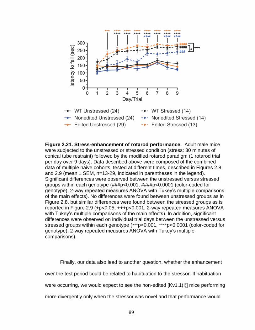
89
Figure 2.21. Stress-enhancement of rotarod performance. Adult male mice were subjected to the unstressed or stressed condition (stress: 30 minutes of conical tube restraint) followed by the modified rotarod paradigm (1 rotarod trial per day over 9 days). Data described above were composed of the combined data of multiple naive cohorts, tested at different times, described in Figures 2.8 and 2.9 (mean ± SEM, n=13-29, indicated in parentheses in the legend). Significant differences were observed between the unstressed versus stressed groups within each genotype (###p<0.001, ####p<0.0001 (color-coded for genotype), 2-way repeated measures ANOVA with Tukey’s multiple comparisons of the main effects). No differences were found between unstressed groups as in Figure 2.8, but similar differences were found between the stressed groups as is reported in Figure 2.9 (+p<0.05, +++p<0.001, 2-way repeated measures ANOVA with Tukey’s multiple comparisons of the main effects). In addition, significant differences were observed on individual trial days between the unstressed versus stressed groups within each genotype (***p<0.001, ****p<0.0001 (color-coded for genotype), 2-way repeated measures ANOVA with Tukey’s multiple comparisons).
Finally, our data also lead to another question, whether the enhancement
over the test period could be related to habituation to the stressor. If habituation
were occurring, we would expect to see the non-edited [Kv1.1(I)] mice performing
more divergently only when the stressor was novel and that performance would

90
not differ as the mice became habituated to the stressor. As the non-edited
[Kv1.1(I)] unstressed versus stressed mice do not significantly differ until day 6,
this could indicate a delay in habituation compared to the other genotypes
(Figure 2.21). Unfortunately, for the comparisons between genotypes in Figures
2.8 and 2.9, the statistics were limited to main effects, because comparisons at
individual trials were underpowered for most trial days (though in the combined
data in Figure 2.21, the non-edited [Kv1.1(I)] mice were significantly decreased
compared to the edited [Kv1.1(V)] mice on days 2, 3, 4, 5 and 7). Thus, in order
to determine significance between genotypes at each trial day in data consisting
of a single cohort, a mixed-model should be developed for more precise
statistical comparisons. An important caveat to the rotarod paradigm is that the
maximum run time for the mice is 300 s, thus the apparent plateau in rotarod
performance over time is likely an artifact of this ceiling effect, which could
confound statistical tests at the later trial days, and future experiments should
increase the maximum time to determine the exact latency to fall value. In order
to specifically address any stressor habituation, a follow-up study could be
performed, using a chronic unpredictable stress protocol (Monteiro et al., 2015).
Varying the stressor may reveal additional deficits, possibly leading to further
decreases in rotarod performance for the non-edited [Kv1.1(I)] mice compared to
the unstressed paradigm or the other genotypes. Although a chronic
unpredictable stress paradigm may provide additional insights into genotypic
differences, the nature of EA1 as a paroxysmal stimulus-induced disorder
suggests that patients do not habituate to the triggering stressors, and using a

91
chronic unpredictable stress may reveal no more information than only using the
conical tube restraint stressor (Dreissen and Tijssen, 2012).
Spontaneous Seizure Monitoring and Drug-induced Seizure Susceptibility
Initial EEG analyses did not reveal epileptiform activity, though the
homozygous non-edited [Kv1.1(I)] mouse exhibited two tonic events during
sleep. These abnormal movements could not be classified as seizures, because
there were no concurrent alterations in the EEG traces, yet they suggested that
the non-edited [Kv1.1(I)] mutants may have underlying signaling deficits that
could lead to altered seizure-susceptibility. Additional studies are required to
discern if this observed activity was consistent for this genotype.
Subsequent studies assessed whether Kv1.1 mutant animals displayed
differences in drug-induced seizure susceptibility using the pro-convulsant drugs,
flurothyl, PTZ, and 4-AP. These convulsants were chosen due to their
widespread use, as well as prior use in Kv1.1 studies in the literature. PTZ has
been shown to alter the voltage dependence and decrease the maximal current
of Kv1.1, when exogenously expressed in Xenopus oocytes, and Kv1.1 editing
has been implicated in reducing the 4-AP-seizure susceptibility (Madeja et al.,
1996; Madeja et al., 1994; Streit et al., 2011). These drugs each have different
mechanisms of action, therefore it is not surprising that one has no effect
(flurothyl) and the others have opposite effects (PTZ and 4-AP) on homozygous
Kv1.1 mutant animals.

92
The mechanism of action for flurothyl is not well understood, so the lack of
a genotype difference is less informative. It has been previously demonstrated
that Kv1.1-null mice experience flurothyl-induced seizures 60% faster than their
wild-type littermates (Smart et al., 1998). In contrast, we found no difference in
the onset of flurothyl-induced seizures between our mutant mouse lines,
indicating that any effect of flurothyl is independent of Kv1.1 editing. The loss of
Kv1.1 in null mice might lead to an increase in seizure susceptibility by increasing
the expression of Kv1.x channels at the plasma membrane (since Kv1.1 can lead
to greater internalization of heterotetrameric Kv1.x channels) (Manganas and
Trimmer, 2000). Our characterization of the steady-state levels of Kv1.1 RNA
indicates similar expression of either isoform in mutant lines compared to wild-
type mice, so an expression-dependent flurothyl effect would not be expected to
alter seizure-susceptibility. We did not follow up with a larger cohort study
because power analyses (at 80% power and 5% type-I error rate) predicted that
we would need to test 2472 non-edited [Kv1.1(I)] and 1185 edited [Kv1.1(V)]
animals in order to see significant differences compared to wildtype (using the
average standard deviation derived from our flurothyl data described above). By
contrast, it is interesting that 4-AP and PTZ are each able to reveal differences
between the homozygous non-edited [Kv1.1(I)] and edited[Kv1.1(V)] mutant
mice, respectively.
PTZ is proposed to weaken GABAA receptor mediated inhibition and PTZ
has been shown to alter the voltage-dependence of Kv1.1 in exogenously
expressing Xenopus oocytes (Madeja et al., 1994; Ramanjaneyulu and Ticku,

93
1984). We observed that the edited [Kv1.1(V)] mice displayed a dampened
seizure-susceptibility towards PTZ-induced seizures. The main biophysical
difference between the non-edited [Kv1.1(I)] and edited[Kv1.1(V)] mutant
channels is the speed of the recovery from fast-inactivation. Whereas faster
recovery from inactivation is epileptogenic for sodium channels (R1460H
mutation in SCN1A) (Alekov et al., 2000), we have observed that the edited
[Kv1.1(V)] isoform is resistant to PTZ-induced seizures, indicating that faster
recovery from inactivation for potassium channels is anti-epileptogenic. The
opposing effects of faster recovery from inactivation for the sodium channel
mutant and potassium channel edited isoform mentioned above may alter the
frequency and shape of the repetitive firing of action potentials. Faster recovery
from inactivation for a sodium channel may overall lead to greater sodium influx
and hyperexcitability, whereas faster recovery for a potassium channel may lead
to less potassium efflux and hypoexcitability. Our studies indicating that the
edited channel confers dampened seizure susceptibility also are consistent with
studies indicating that drugs, which open potassium channels or prevent -
inactivation, counteract PTZ-induced seizures (Lu et al., 2008; Pozo et al., 1990).
4-AP is a known Kv channel blocker with high-affinity for the Kv1 channel
family (Decher et al., 2010; Grissmer et al., 1994). It was hypothesized that
homozygous non-edited [Kv1.1(I)] mice would have a greater 4-AP-induced
impairment, given that 4-AP efficiently blocks the non-edited [Kv1.1(I)] isoform,
but not the edited [Kv1.1(V)] isoform when exogenously expressed in Xenopus
oocytes (IC50: nonedited= 98 µM; edited= 6.7 mM) (Decher et al., 2010).
94
Recordings from entorhinal slices from chronic epileptic rats, observed to have
increases in Kv1.1 RNA editing, revealed a resistance to 4-AP-induced seizure-
like events (Streit et al., 2011). Thus our results, demonstrating that the
homozygous non-edited [Kv1.1(I)] mutant mice were more susceptible to 4-AP-
induced seizures agrees well with previous studies. The 4-AP experiment was
replicated twice and the increase in seizure susceptibility was still observed,
though interestingly our second experiment, using the wild-type littermates of the
non-edited [Kv1.1(I)] and edited [Kv1.1(V)] mice demonstrated further differences
that were not seen in the original experiment using the wild-type littermates of the
V408A/+ mice. The latency to HLE did differ between genotypes for the 4-AP-
induced seizures, unlike the results with PTZ (in which genotype had no effect on
seizure latency). This difference in the seizure latency was only observed for the
non-edited [Kv1.1(I)] mice, which exhibited faster maximal seizure onset (HLE)
than the other genotypes. The presence of edited channels in the edited
[Kv1.1(V)], wild-type, and V408A/+ mice may have a dominant effect on seizure
latency, which all displayed similar seizure latency, whereas the complete lack of
edited channels in the non-edited [Kv1.1(I)] mice led to faster and more severe
seizures.
It is noteworthy that the wild-type littermates had an intermediate 4-AP
seizure-susceptibility curve between the non-edited [Kv1.1(I)] and edited
[Kv1.1(V)] mice, especially given the lower rates of editing in the hippocampus in
particular (approximately 18%). This could also indicate that the edited [Kv1.1(V)]
isoform exerts a dominant effect, even when it is expressed at a lower ratio
95
compared to the non-edited [Kv1.1(I)] channel. This interpretation is consistent
with studies by Decher et al. (2010), which demonstrated that edited [Kv1.1(V)]
exerts dominant effects on drug and fatty acid binding when in tetramers with
non-edited [Kv1.1(I)] as well as in tetramers with other Kv1.x family members.
Another possible explanation is that a small subset of neurons has a high
expression of edited [Kv1.1(V)] and these neurons set the epileptic tone of the
predominantly nonedited [Kv1.1(I)] isoform expressing tissue. This is possible but
is less likely, given that editing has been quantified in CA1 pyramidal neurons
and these neurons had low levels of editing (7%), similar to the average of region
(Decher et al., 2010).
Preliminary Electrophysiological characterization
Collaborations with the Winder lab performed the first electrophysiological
characterization of the homozygous non-edited [Kv1.1(I)] mutant mice. We chose
to focus on the non-edited [Kv1.1(I)] animals to probe whether these mice have
altered signaling properties which could explain the heightened seizure-
susceptibility seen in the 4-AP experiments. In addition, recordings were
performed under stressed conditions, due to the observed stress-induced motor
discoordination of non-edited [Kv1.1(I)] mutant animals upon conical tube
restraint. Kv1.1 has been implicated in alterations in dentate granule cells’
signaling, as Kv1.1 was upregulated in dentate granule cells in a model of
temporal lobe epilepsy (Kirchheim et al., 2013). When Kv1.1 was reversibly

96
knocked down by antisense oligonucleotides, electrophysiological recordings of
dentate granule cells revealed action potential broadening and an abolished
hyperpolarization phase, though no alterations in other parameters were
observed, such as the resting membrane potential and action potential amplitude
(Kirchheim et al., 2013; Meiri et al., 1997). Unfortunately, our studies did not
identify any differences between the homozygous non-edited [Kv1.1(I)] mice and
their wild-type littermates. This outcome may result from our choice of recording
in the hippocampus where wild-type mice express approximately 80% non-edited
Kv1.1 transcripts, which could explain why the signaling in the hippocampus of
non-edited [Kv1.1(I)] mutant animals was very similar to that observed in control
mice. The lack of a difference in signaling for the non-edited [Kv1.1(I)] mice may
indicate that the presence or absence of the edited [Kv1.1(V)] isoform may be
more informative for understanding Kv1.1 editing effects in vivo.
Summary
Until now, characterization of Kv1.1 editing has been restricted to
heterologous expression systems. Our newly developed mouse models allow for
phenotypic contrasting of the non-edited [Kv1.1(I)] and edited [Kv1.1(V)] mice
and the opportunity to observe the electrophysiological changes upon solely
expressing either isoform in vivo. Our characterization has revealed that
disrupting the normal ratios of Kv1.1 editing has surprising effects on postnatal
lethality that are related to environmental factors. Furthermore, our phenotypic

97
characterization has indicated that the non-edited [Kv1.1(I)] mouse shares
similarities with the V408A EA1 mouse model, posing new questions as to how
these two mouse models are similar and whether altered Kv1.1 editing could be
a contributing factor in the V408A/+ mouse model as well.

98
CHAPTER III
INTERACTIONS BETWEEN KV1.1 RNA EDITING AND EPISODIC ATAXIA TYPE-1 MUTATIONS
The contents of this chapter were adapted from the published work,
“Mutations underlying Episodic Ataxia type-1 antagonize Kv1.1 RNA editing,” in
the journal Scientific Reports in February 2017, under a Creative Contributions
Attribution v4.0 International License (Ferrick-Kiddie et al., 2017).
Introduction
At least three EA1-associated mutations (V404I, I407M, and V408A) have
been identified within the duplex region required for editing of Kv1.1 RNAs
(Adelman et al., 1995; Eunson et al., 2000; Imbrici et al., 2006; Maylie et al.,
2002; Tomlinson et al., 2013; Zerr et al., 1998). Due to the proximity of the
mutant amino acids to the editing site (I400V), we sought to determine whether
such EA1 mutations affect the editing profiles for Kv1.1 transcripts and to
determine the resultant biophysical properties of edited, mutant channels.
Our studies reveal an antagonistic relationship between these EA1
mutations and RNA editing. Each EA1 mutation decreased the rate of RNA
editing using an in vitro editing system and the V408A mutation decreased the
extent of editing in a previously characterized mouse model of EA1. Furthermore,
99
we show that editing can have a variable effect on each type of EA1 mutant
protein, altering voltage sensitivity, activation and deactivation, and inactivation
with a Kv subunit. Not only do our studies demonstrate that EA1 mutations can
impede RNA editing and alter resulting protein function, but also represent the
first examples of existing disease-associated human mutations which act to
disrupt cis-regulatory elements required for RNA editing.
Materials & Methods
Kv1.1 and Kv1.1 constructs
A 463 bp-region encompassing the duplex required for Kv1.1 editing was
amplified by PCR from human genomic DNA using sense (5’-GC-
GAAGCTTCCTCTTCATCGGGGTCATCCT-3’) and antisense (5’-
GCGGCGGCCGCAGTTTTGGTTAGCAGTGG-3’) oligonucleotide primers in
exon 2. To aid in subcloning, the primers incorporated Hind III and Not I
restriction sites on their 5’-ends for the sense and antisense primers,
respectively. The PCR amplicon was subcloned into the mammalian expression
vector, pRc-CMV (Thermo Fisher) to generate a wild-type Kv1.1 minigene. To
generate the V404I, I407M, and V408A minigenes, the wild-type Kv1.1 construct
was mutagenized using the QuikChange II Site-Directed Mutagenesis kit (Agilent
Technologies), where the PCR reactions were supplemented with 5% DMSO.
Full-length mouse Kv1.1 (Addgene) and mouse Kv1.1 (Thermo Scientific)
100
cDNAs were subcloned into the Xenopus expression vector, pGEM HE (Liman et
al., 1992). The following full-length constructs were created by PCR mutagenesis
from the full-length mouse non-edited [Kv1.1(I)] cDNA and validated by sequence
analysis: wild-type edited [Kv1.1(V)] and V404I, I407M, and V408A mutant Kv1.1
(non-edited [Kv1.1(I)] and edited [Kv1.1(I)]) cDNAs.
In vitro analysis of RNA editing
RNAs were transcribed in vitro from the wild-type Kv1.1 minigene, as well
as corresponding minigenes harboring the V404I, I407M, and V408A mutations
using the MAXIscript kit (Ambion) with T7 RNA polymerase according to
manufacturer’s instructions. Nuclear extracts were prepared from transiently
transfected HEK293 cells expressing rat ADAR2, as described previously, and
stored at -80º C until required (Schreiber et al., 1989; Stefl et al., 2010).
Immediately prior to in vitro editing analysis, nuclear extracts were diluted 1:10 in
dialysis buffer [20 mM HEPES, 1 mM EDTA, 1 mM EGTA, 10% glycerol, 300 mM
NaCl, 1 mM PMSF, 1 mM DTT, 1X complete, EDTA-free protease inhibitor
cocktail (Roche)], before a 2-hour incubation at 30°C with RNase inhibitors and
RNA substrates varying in concentration from 0.125 to 2 nM. Nuclear extracts
represented one-third of the total 50 L reaction volume which was diluted with
the RNA substrate and water to reduce the glycerol concentration into a range
necessary for ADAR2 activity. The incubation time was determined empirically by
time-course analyses to ensure that editing of the wild-type Kv1.1 minigene was
101
within the linear range of the reaction (Figure 3.1). In Figure 3.1, editing was
determined by Sanger sequencing electropherogram peak height analysis (see
Appendix A for additional description of the accuracy of Sanger compared to
high-throughput sequencing analyses). By the limitations of this technique, very
low editing levels were undetectable, especially at the 1 hour time point for the
mutant duplexes (and editing was undetectable for all RNA duplexes in pilot trials
at 20 minutes (data not shown)). Reactions were terminated by the addition of
TRIzol (Ambion) and RNA was extracted according to the manufacturer’s
protocol. RNA was reverse-transcribed with random primers using the High
Capacity cDNA Reverse Transcription kit (Applied Biosystems) and the extent of
RNA editing was quantified by high-throughput multiplexed sequence analysis as
described previously (Hood et al., 2014). The editing rate was calculated as the
fmol RNA converted to the edited isoform divided by the duration of the reaction.

102
Figure 3.1. Time course analysis to determine the linear range for the in vitro RNA editing rates. Wild-type (WT) and mutant Kv1.1 RNA minigenes, encompassing the duplex region required for editing, were in vitro transcribed and incubated between 1 and 4 hours with nuclear extracts prepared from HEK293 cells transiently expressing rat ADAR2. The extent of editing was quantified by the relative A and G peak heights derived from Sanger sequence analysis, used to calculate the approximate editing rate.
In vivo analysis of RNA editing
All animal care and experimental procedures involving mice were
approved by the Vanderbilt University Medical Center Institutional Animal Care
and Use Committee and were performed in accordance with relevant guidelines
and regulations. Mice harboring the heterozygous V408A mutation (V408A/+)
were generously provided by Dr. James Maylie (Oregon Health & Science
University) (Herson et al., 2003). At approximately 6 weeks of age, male V408A/+
and wild-type littermates were euthanized by cervical dislocation under
anesthesia followed by decapitation. Six brain regions (cerebellum,
hippocampus, hypothalamus, cortex, striatum, olfactory bulb) and spinal cord
were dissected from each mouse. Tissues were flash-frozen in liquid nitrogen
and RNA was isolated by sonication in TRIzol (Ambion) according to the
103
manufacturer’s instructions. RNA was reverse-transcribed and Kv1.1 editing was
quantified by high-throughput sequence analysis as described for in vitro RNA
editing analyses.
Electrophysiological recording in Xenopus oocytes
All animal care and experimental procedures involving Xenopus laevis
were approved by the University of Puerto Rico Institutional Animal Care and
Use Committee. Kv1.1 and full-length, wild-type, V404I, I407M, and V408A
Kv1.1 RNAs were transcribed in vitro, capped, and polyadenylated using the T7
mScript Standard mRNA Production System (CELLSCRIPT). Ovary sections
containing several hundred oocytes were removed from adult specimens of
Xenopus laevis obtained from Xenopus Express (Brooksville, FL). Oocytes were
dispersed with type II collagenase and manually defolliculated. Stage V and VI
oocytes were then selected by manual inspection for subsequent RNA injection.
On day 1, oocytes were injected with 38.6 nL of one of the eight full-length Kv1.1
RNAs encoding non-edited [Kv1.1(I)] and edited [Kv1.1(V)] isoforms of wild-type,
V404I, I407M, and V408A channels, with or without the Kv1.1 RNA. Injection
concentrations were optimized individually for each construct, with greater
concentrations required for the I407M and V408A RNAs due to protein
expression differences previously described in the literature (Tomlinson et al.,
2013; Zerr et al., 1998). Each -subunit was injected at a concentration from 2
ng/L to 1 g/L and co-injected with Kv1.1 when applicable; concentrations for

104
the Kv1.1 constructs were 10-fold more than each -subunit, up to a maximum
injection concentration of 500 ng/L. Electrophysiological analysis of oocytes
were performed between day 3-5 post-injection using the cut-open oocyte
voltage-clamp technique (Stefani and Bezanilla, 1998). The external solution
consisted of: 20 mM K-glutamate, 100 mM L-glutamate, 2.5 mM MgCl2, 2.5 mM
CaCl2, 10 mM HEPES, pH 7.4. The internal solution consisted of: 120 mM K-
glutamate, 2.5 mM EGTA, 10 mM HEPES, pH 7.4. The pH of the solutions was
adjusted using N-methyl-D-glucamine, as an alternative to NaOH, to limit the
introduction of sodium ions into the solutions. To gain electrical access to the
oocyte interior, the internal solution was supplemented with 0.3% saponin and
used for a brief permeabilization prior to recording. The oocyte membrane
potential was controlled using a CA-1B High Performance Oocyte Clamp (Dagan
Corporation). Analog current signals were digitized at 100 kHz using an
SBC6711 A/D D/A board (Innovative Integration, Simi Valley CA) and filtered at 5
kHz. To avoid errors introduced by series resistance, only traces exhibiting less
than 10 A were used for analysis. GPATCH M software, kindly provided by Dr.
F. Bezanilla (University of Chicago), was used for data collection and clamp
control. Leak currents were subtracted using a linear P/4 procedure. Data were
analyzed using ANALYSIS software, also provided by Dr. F. Bezanilla, for fitting
data with exponential functions and measuring current amplitudes. In addition,
single exponential curves were fitted to recovery from the inactivation data using
Graphpad Prism (Graphpad Software) to determine the rate constant, . As the
channels encoded by edited V408A transcripts closed too rapidly for
105
measurements of tail current amplitude, conductance (G) was calculated using
Ohm’s law (Equation 3.1),
rVV
IG
(3.1)
where I represents the maximal current at the test potential (V) and Vr signifies
the reversal potential, determined empirically. In primary traces displayed, points
arising from brief capacity transients were removed for clarity.
Statistical analysis
Statistical comparisons for in vitro and in vivo editing analyses were
determined by 2-way ANOVA with Tukey’s multiple comparisons test (Figures
3.3 and 3.4). Boltzmann functions were fitted using non-linear regression to
model conductance-voltage curves and to determine V1/2 and k values
associated with each replicate. Two-sample Student’s t-tests were used to
compare the voltage-dependent parameters, long pulse characterization with and
without Kv1.1, and recovery from inactivation values (Tables 3.2 and 3.3;
Figures 3.9 and 3.11). The above analyses were conducted using Graphpad
Prism (Graphpad Software). To maintain the type I error rate for each experiment
at 5%, a Bonferroni correction was applied to each test based on the number of
comparisons within each experiment and statistical significance for any pair of
106
treatment comparisons was redefined according to these adjusted p-values. For
Table 3.2 and Table 3.3, 10 comparisons were made and the significance was
adjusted to p ≤ 0.005. For Figure 3.9b, 42 comparisons were made and the
significance was adjusted to p ≤ 0.0012. For Figures 3.9c and 3.11b, 30
comparisons were made and the significance was adjusted to p ≤ 0.0017.
Analysis of activation and deactivation, and inactivation kinetics (Figures 3.6, 3.8,
and 3.10) were performed with linear mixed models using the natural log of the
acquired data to better meet model assumptions (developed by Dan Ayers).
Individual group comparisons for p-values were based on the Wald tests of
model-based predicted (least square) means and appropriate standard errors
(and reported in Appendix B). Because these data indicate that the
measurements of activation, deactivation, and -inactivation were dependent on
voltage, comparisons were made only between values obtained at the same volt-
age. Data are presented with their original scale to allow for easier interpretation
and comparison with the existing EA1 literature. All statistical tests were two-
sided and statistical significance was defined as p ≤ 0.05. Unless specified, no
adjustments for multiple comparisons were performed.
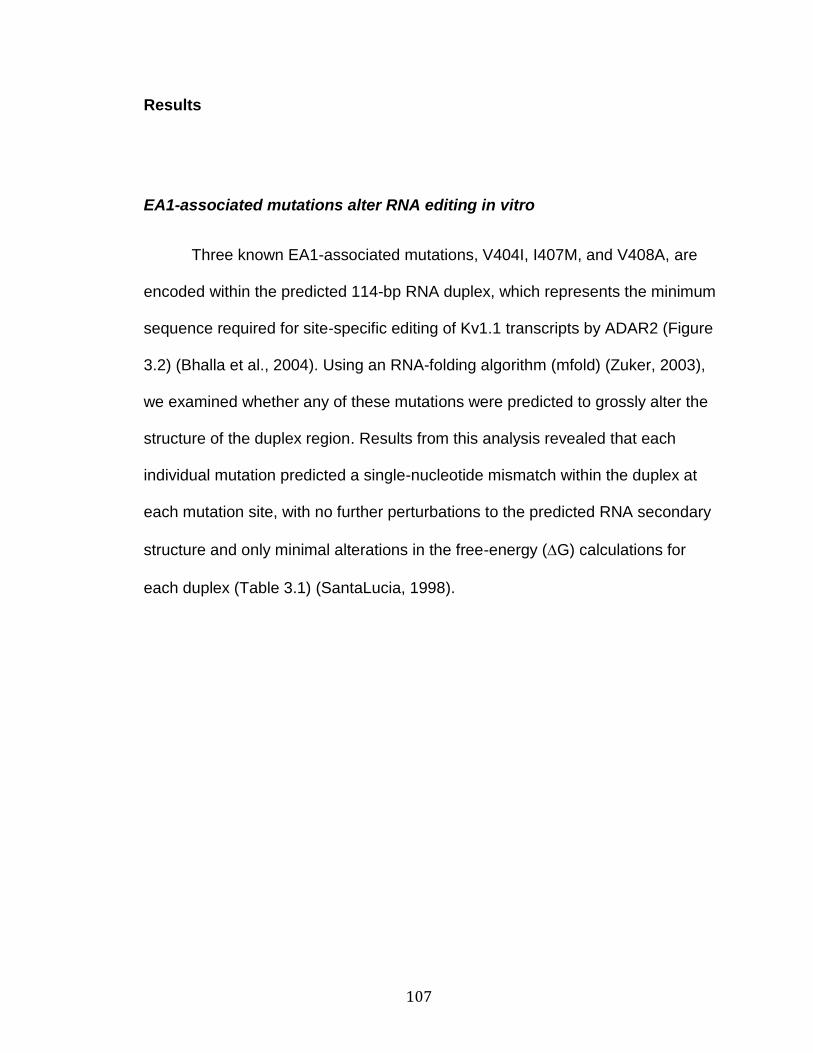
107
Results
EA1-associated mutations alter RNA editing in vitro
Three known EA1-associated mutations, V404I, I407M, and V408A, are
encoded within the predicted 114-bp RNA duplex, which represents the minimum
sequence required for site-specific editing of Kv1.1 transcripts by ADAR2 (Figure
3.2) (Bhalla et al., 2004). Using an RNA-folding algorithm (mfold) (Zuker, 2003),
we examined whether any of these mutations were predicted to grossly alter the
structure of the duplex region. Results from this analysis revealed that each
individual mutation predicted a single-nucleotide mismatch within the duplex at
each mutation site, with no further perturbations to the predicted RNA secondary
structure and only minimal alterations in the free-energy (G) calculations for
each duplex (Table 3.1) (SantaLucia, 1998).
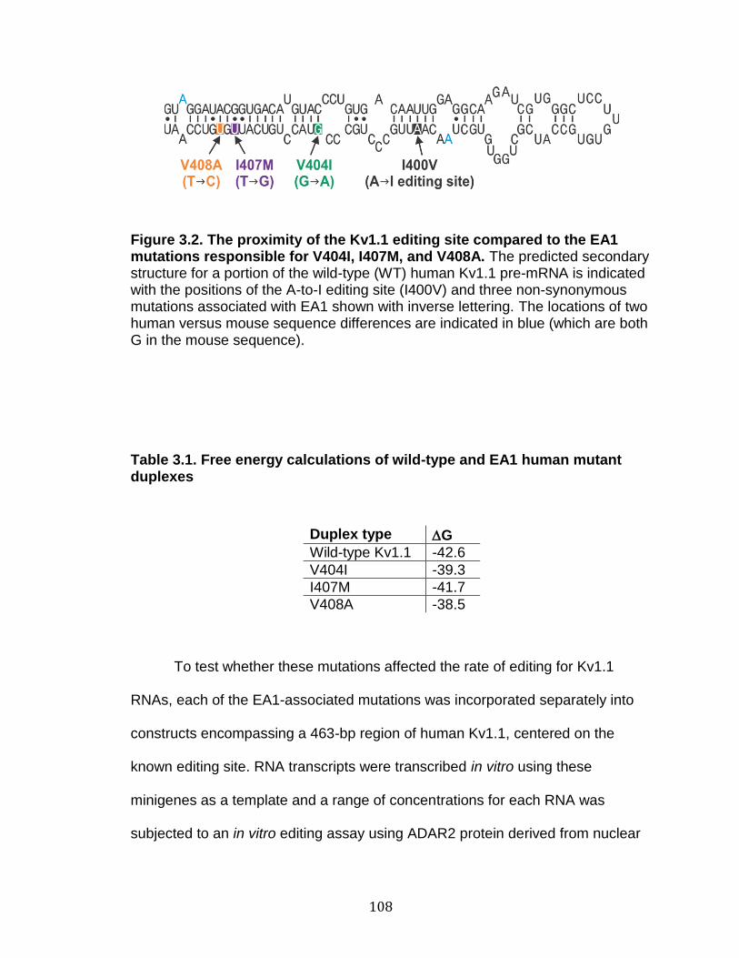
108
Figure 3.2. The proximity of the Kv1.1 editing site compared to the EA1 mutations responsible for V404I, I407M, and V408A. The predicted secondary structure for a portion of the wild-type (WT) human Kv1.1 pre-mRNA is indicated with the positions of the A-to-I editing site (I400V) and three non-synonymous mutations associated with EA1 shown with inverse lettering. The locations of two human versus mouse sequence differences are indicated in blue (which are both G in the mouse sequence).
Table 3.1. Free energy calculations of wild-type and EA1 human mutant duplexes
Duplex type G
Wild-type Kv1.1 -42.6
V404I -39.3
I407M -41.7
V408A -38.5
To test whether these mutations affected the rate of editing for Kv1.1
RNAs, each of the EA1-associated mutations was incorporated separately into
constructs encompassing a 463-bp region of human Kv1.1, centered on the
known editing site. RNA transcripts were transcribed in vitro using these
minigenes as a template and a range of concentrations for each RNA was
subjected to an in vitro editing assay using ADAR2 protein derived from nuclear

109
extracts isolated from HEK293 cells transiently expressing ADAR2 (Stefl et al.,
2010). The extent of editing was quantified by high-throughput sequence analysis
as described previously and used to calculate the rate of editing (Figure 3.3)
(Hood et al., 2014). Results from these studies clearly demonstrated that
introduction of any of these EA1-associated point mutations into the wild-type
sequence was sufficient to decrease the editing rate for Kv1.1 transcripts in vitro.
Furthermore, the magnitude of this rate decrease corresponded to the proximity
of the mutation to the editing site (I400V), with the most severe deficit observed
for the V404I mutation (81% rate reduction at 2 nM RNA), and a 58% and 17%
reduction in editing rate for the I407M and V408A mutations, respectively.
110
Figure 3.3. Quantitative analysis of in vitro RNA editing rates for wild-type and mutant Kv1.1 transcripts. Wild-type (WT) and mutant human Kv1.1 RNA minigenes, encompassing the duplex region required for editing, were in vitro transcribed and incubated with nuclear extracts prepared from HEK293 cells transiently expressing rat ADAR2. The extent of editing was quantified by high-throughput sequence analysis and used to calculate the editing rate, as described previously (Hood et al., 2014). Statistical differences were determined for replicates at the 2 nM RNA concentration by 2-way ANOVA with Tukey’s multiple comparisons test (mean ± SEM, n=4 replicate reactions, *p ≤ 0.05; ****p ≤ 0.0001). Small error bars were obscured by the data symbol for the V404I 2nM data point.
A mouse model of EA1 (V408A/+) alters RNA editing in vivo
To date, only one mouse model of EA1 has been developed (Herson et
al., 2003). Mutant mice homozygous for the V408A allele die between E3 and
E9, whereas V408A/+ heterozygotes are characterized by stress-induced ataxia
as well as attenuated cerebellar Purkinje signaling, which has been attributed to
action potential broadening at basket cell boutons leading to increased GABA
release (Begum et al., 2016; Herson et al., 2003). To determine whether the
presence of the V408A mutation inhibited Kv1.1 editing in vivo, we isolated RNA

111
from multiple dissected brain regions and spinal cord of wild-type and V408A/+
mutant animals to determine RNA editing profiles by high-throughput sequence
analysis of Kv1.1 transcripts. Since this deep-sequencing approach generates
sequence reads covering both the V408A mutation and the editing site, it was
possible to quantify allele-specific editing profiles in V408A/+ heterozygotes.
Results from this analysis indicated that the extent of editing for the wild-type
allele in V408A/+ mutant mice was similar to that observed in wild-type animals.
Editing for the mutant V408A allele showed a 59% reduction in site-specific
editing efficiency in all tissues examined when compared to either wild-type
littermates or the V408A/+ wild-type allele (Figure 3.4).
Figure 3.4. Quantitative analysis of allele-specific Kv1.1 editing in V408A mutant mice. The extent of editing for the wild-type and mutant alleles in heterozygous V408A adult mice (V408A/+), compared to wild-type littermates, was determined for RNA isolated from dissected brain regions and spinal cord by high-throughput sequence analysis. Significance was determined by 2-way ANOVA with Tukey’s multiple comparisons test (mean ± SEM, n=4, ***p ≤ 0.001, ****p ≤ 0.0001). Cbl, cerebellum; Hyp, hypothalamus; Hip, hippocampus; Ctx, cortex; Str, striatum; Olf, olfactory bulb; Sp C, spinal cord.
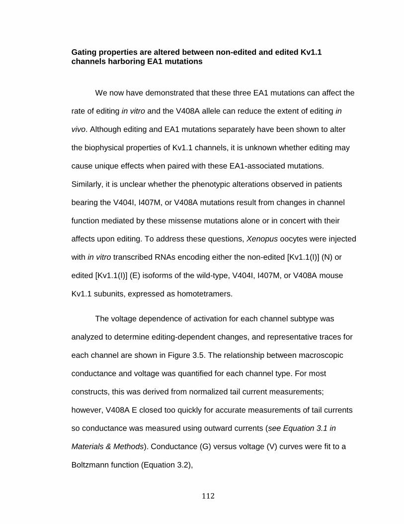
112
Gating properties are altered between non-edited and edited Kv1.1 channels harboring EA1 mutations
We now have demonstrated that these three EA1 mutations can affect the
rate of editing in vitro and the V408A allele can reduce the extent of editing in
vivo. Although editing and EA1 mutations separately have been shown to alter
the biophysical properties of Kv1.1 channels, it is unknown whether editing may
cause unique effects when paired with these EA1-associated mutations.
Similarly, it is unclear whether the phenotypic alterations observed in patients
bearing the V404I, I407M, or V408A mutations result from changes in channel
function mediated by these missense mutations alone or in concert with their
affects upon editing. To address these questions, Xenopus oocytes were injected
with in vitro transcribed RNAs encoding either the non-edited [Kv1.1(I)] (N) or
edited [Kv1.1(I)] (E) isoforms of the wild-type, V404I, I407M, or V408A mouse
Kv1.1 subunits, expressed as homotetramers.
The voltage dependence of activation for each channel subtype was
analyzed to determine editing-dependent changes, and representative traces for
each channel are shown in Figure 3.5. The relationship between macroscopic
conductance and voltage was quantified for each channel type. For most
constructs, this was derived from normalized tail current measurements;
however, V408A E closed too quickly for accurate measurements of tail currents
so conductance was measured using outward currents (see Equation 3.1 in
Materials & Methods). Conductance (G) versus voltage (V) curves were fit to a
Boltzmann function (Equation 3.2),

113
k
VV
e
G
2/1
1
1 (3.2)
to estimate the midpoint of channel activation (V1/2) and the relative voltage
sensitivity (k) (Table 3.2). Consistent with previous reports for I407M N
channels(Tomlinson et al., 2013), we observed a 30 mV positive shift in the V1/2,
but this change was not influenced by editing (Figure 3.5 and Table 3.2). By
contrast, V404I also caused a positive shift, but it was more pronounced for the
non-edited channel (a shift of 22.1 mV for the non-edited channel, 14.2 mV for
the edited channel). Thus, editing partially ameliorated the alteration in channel
function caused by the V404I mutation. V408A channels did not exhibit altered
voltage-dependence for either the non-edited or edited isoforms. Editing had little
effect on voltage sensitivity for any of the wild-type or mutant homotetrameric
channels.
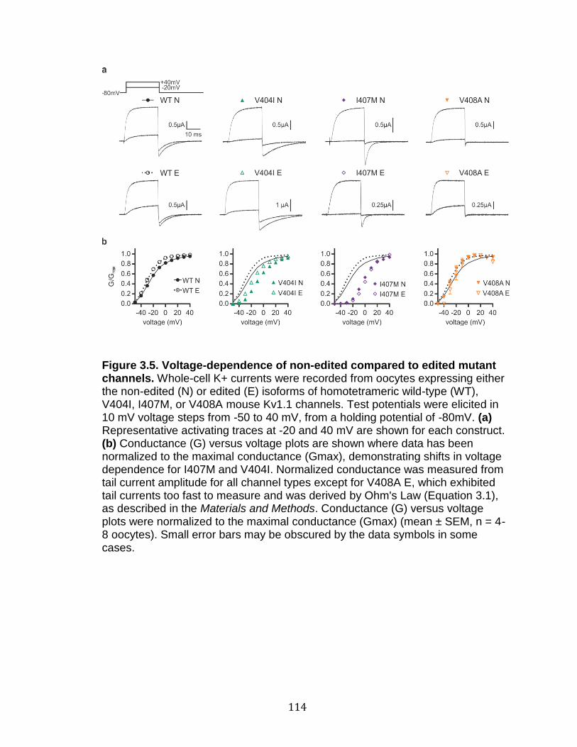
114
Figure 3.5. Voltage-dependence of non-edited compared to edited mutant channels. Whole-cell K+ currents were recorded from oocytes expressing either the non-edited (N) or edited (E) isoforms of homotetrameric wild-type (WT), V404I, I407M, or V408A mouse Kv1.1 channels. Test potentials were elicited in 10 mV voltage steps from -50 to 40 mV, from a holding potential of -80mV. (a) Representative activating traces at -20 and 40 mV are shown for each construct. (b) Conductance (G) versus voltage plots are shown where data has been normalized to the maximal conductance (Gmax), demonstrating shifts in voltage dependence for I407M and V404I. Normalized conductance was measured from tail current amplitude for all channel types except for V408A E, which exhibited tail currents too fast to measure and was derived by Ohm's Law (Equation 3.1), as described in the Materials and Methods. Conductance (G) versus voltage plots were normalized to the maximal conductance (Gmax) (mean ± SEM, n = 4-8 oocytes). Small error bars may be obscured by the data symbols in some cases.

115
Table 3.2. Voltage-dependence of activation. Voltage-dependence of activation was determined by fitting data to a Boltzmann function (Equation 3.2), to determine the mid-point of channel activation (V1/2) and relative voltage sensitivity (k). All data are represented as mean ± SEM, n=4-8 oocytes for each channel type. Non-edited (N) and edited (E) isoforms of the mutant channels were compared to WT E and WT N channels, respectively: ***p≤0.001; ****p<0.0001. All types of N channels were compared to their respective E channels: ++p≤0.005; +++p≤0.001. Significance was determined by two-sample t-tests; due to multiple comparisons, significance was set at p≤0.005.
To examine how editing affected channel opening kinetics, the time to
reach half-maximal activation across a range of voltages was determined (Figure
3.6). The only editing-dependent change was observed for the I407M mutation.
Both I407M channels opened more slowly than their wild-type counterparts, but
the slowing was more severe for I407M E channels, leading to channels with an
exacerbated slow opening phenotype. In addition, I407M E and V408A E
channels demonstrated non-linearity in their voltage dependence for outward
currents (Figure 3.7). This was particularly evident at very positive voltages,
where I407M E and V408A E channels reached peak current amplitudes at 50
and 40 mV respectively, with further voltage steps resulting in decreasing current
amplitudes.
116
Figure 3.6. Editing alters opening (activation) kinetics of I407M channels. (a) Example trace overlay of I407M editing dependent alteration of opening kinetics at the 10mV activating pulse (b) Representative activation traces, depicting whole-cell currents, were recorded from oocytes expressing either a non-edited (N) or edited (E) isoform of the wild-type (WT) or mutant mouse Kv1.1 channel. Test potentials were elicited in 10 mV voltage steps from -10 to 80 mV, from a holding potential of -80 mV. (c) Opening kinetics were measured as the time to reach half-maximal current amplitude and individual group comparisons for p-values were based on the Wald tests of model-based predicted (least square) means (mean ± SEM, n=3-7 oocytes). I407M N and I407M E channels were significantly slowed in their time to half-activation compared to each other, in the voltage range -10 to 70 mV (0.05 > p ≥ 0.0008). I407M N was significantly slower than WT N at all voltages (p ≤ 0.0001) and I407M E was significantly

117
slower than WT E at all voltages (0.01 > p ≥ 0.0001). Small error bars were obscured by the data symbols in some cases.
Figure 3.7. I407M E and V408A E channels display decreased currents at the most positive voltages. Whole-cell K+ currents were recorded from oocytes expressing either the I407M E or V408A E mouse Kv1.1 channel. Test potentials were elicited in 10 mV voltage steps from -50 to 80 mV, from a holding potential of -80 mV. (a) Representative traces for I407M E and V408A E channels are shown, indicating loss of current amplitude at the highest voltages. Voltages at 60mV (blue) and 80mV (red) are colored for clarity. (b) Current (I) versus voltage plots, normalized to the maximal current (Imax), are shown; note the unusual bell-shaped curves. Data points represent mean ± SEM (N=7-8 oocytes). Small error bars were obscured by the data symbols in some cases.
118
Closing (deactivation) kinetics were measured by fitting single exponential
curves to tail current traces to obtain estimates of tau (), the reciprocal of the
closing rate constant (Figure 3.8). Editing resulted in wild-type channels closing
slightly faster. In addition, the I407M and V408A mutations greatly increased
closing speeds on their own. The editing of I407M channels had only a small
effect on deactivation kinetics, while the edited V408A channels closed so quickly
that the closing rate could not be accurately measured. Unlike the other
mutations, however, V404I led to slower closing speeds and editing partially
ameliorated this phenotype.
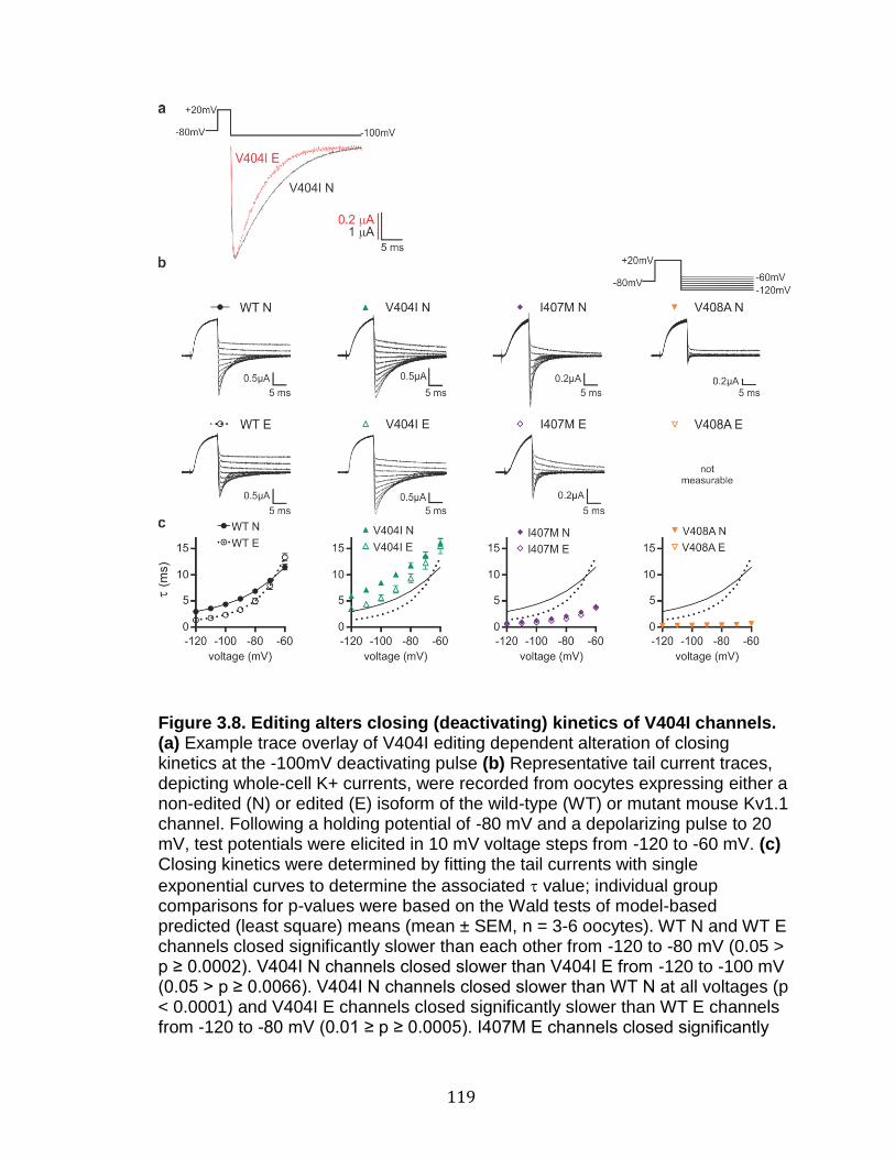
119
Figure 3.8. Editing alters closing (deactivating) kinetics of V404I channels. (a) Example trace overlay of V404I editing dependent alteration of closing kinetics at the -100mV deactivating pulse (b) Representative tail current traces, depicting whole-cell K+ currents, were recorded from oocytes expressing either a non-edited (N) or edited (E) isoform of the wild-type (WT) or mutant mouse Kv1.1 channel. Following a holding potential of -80 mV and a depolarizing pulse to 20 mV, test potentials were elicited in 10 mV voltage steps from -120 to -60 mV. (c) Closing kinetics were determined by fitting the tail currents with single
exponential curves to determine the associated value; individual group comparisons for p-values were based on the Wald tests of model-based predicted (least square) means (mean ± SEM, n = 3-6 oocytes). WT N and WT E channels closed significantly slower than each other from -120 to -80 mV (0.05 > p ≥ 0.0002). V404I N channels closed slower than V404I E from -120 to -100 mV (0.05 > p ≥ 0.0066). V404I N channels closed slower than WT N at all voltages (p < 0.0001) and V404I E channels closed significantly slower than WT E channels from -120 to -80 mV (0.01 ≥ p ≥ 0.0005). I407M E channels closed significantly
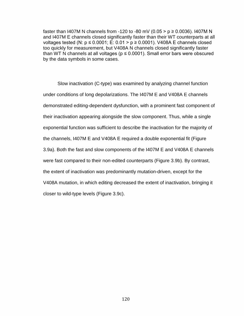
120
faster than I407M N channels from -120 to -80 mV (0.05 > p ≥ 0.0036). I407M N and I407M E channels closed significantly faster than their WT counterparts at all voltages tested (N: p ≤ 0.0001; E: 0.01 > p ≥ 0.0001). V408A E channels closed too quickly for measurement, but V408A N channels closed significantly faster than WT N channels at all voltages (p ≤ 0.0001). Small error bars were obscured by the data symbols in some cases.
Slow inactivation (C-type) was examined by analyzing channel function
under conditions of long depolarizations. The I407M E and V408A E channels
demonstrated editing-dependent dysfunction, with a prominent fast component of
their inactivation appearing alongside the slow component. Thus, while a single
exponential function was sufficient to describe the inactivation for the majority of
the channels, I407M E and V408A E required a double exponential fit (Figure
3.9a). Both the fast and slow components of the I407M E and V408A E channels
were fast compared to their non-edited counterparts (Figure 3.9b). By contrast,
the extent of inactivation was predominantly mutation-driven, except for the
V408A mutation, in which editing decreased the extent of inactivation, bringing it
closer to wild-type levels (Figure 3.9c).
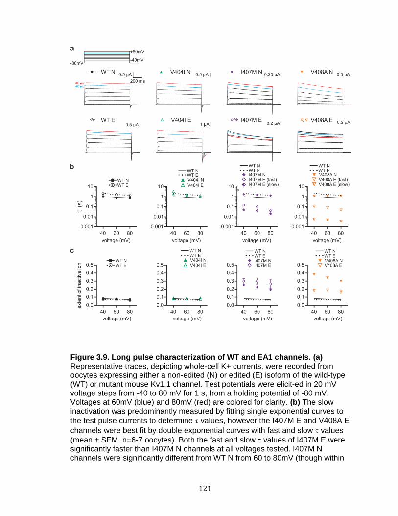
121
Figure 3.9. Long pulse characterization of WT and EA1 channels. (a) Representative traces, depicting whole-cell K+ currents, were recorded from oocytes expressing either a non-edited (N) or edited (E) isoform of the wild-type (WT) or mutant mouse Kv1.1 channel. Test potentials were elicited in 20 mV voltage steps from -40 to 80 mV for 1 s, from a holding potential of -80 mV. Voltages at 60mV (blue) and 80mV (red) are colored for clarity. (b) The slow inactivation was predominantly measured by fitting single exponential curves to
the test pulse currents to determine values, however the I407M E and V408A E
channels were best fit by double exponential curves with fast and slow values
(mean ± SEM, n=6-7 oocytes). Both the fast and slow values of I407M E were significantly faster than I407M N channels at all voltages tested. I407M N channels were significantly different from WT N from 60 to 80mV (though within
122
the range of WT E). Both the fast and slow values of V408A E were significantly faster than V408A N channels at all voltages tested. (c) Extent of inactivation was measured by dividing the amplitude of inactivation by the total amplitude (mean ± SEM, n=3-7 oocytes). I407M N channels had significantly greater inactivation that WT N channels at all voltages tested. V408A N channels had significantly increased inactivation compared to V408A E at all voltages tested. V408A N had significantly greater inactivation that WT N at all voltages tested and V408A E was significantly different from WT E channels from 40 to 60mV. Significance was determined by two-sample t-tests; due to multiple comparisons, significance was set at p ≤ 0.0012 for (b) and p ≤ 0.0017 for (c). Small error bars were obscured by the data symbols in some cases.
Inactivation kinetics are altered between non-edited and edited EA1 mutant proteins
Previous characterization of non-edited and edited wild-type Kv1.1
channels found that the most profound difference was a change in the rate of
recovery from channel inactivation, presumably by altering interactions with an
inactivating Kv subunit (Bhalla et al., 2004). To determine the effect of EA1
mutations on this biophysical property, non-edited and edited isoforms of the
wild-type, V404I, I407M, and V408A channels were co-expressed with Kv1.1 to
measure N-type, fast inactivation kinetics and recovery from inactivation.
Oocytes expressing each channel subtype, along with Kv1.1, were
subjected to short depolarizing pulses to different voltages and the resulting fast
inactivation traces were fit to a single exponential (Figure 3.10). These studies
identified a previously uncharacterized difference in wild-type channels where
editing modestly slowed the rate of channel inactivation. V404I N and E channels
inactivated within the wild-type range, without exhibiting any editing-dependent
123
changes. Inactivation for non-edited isoforms of the I407M and V408A mutants
resembled edited, wild-type channels, however, editing of I407M and V408A
resulted in drastically slower rates of inactivation for both channels. This effect
was most extreme and apparent at all voltages for I407M E, whereas slowing
was only observed for V408A E with shallow depolarizations. Interestingly, the
V404I N channels also exhibited a low extent of inactivation where inactivation
could not be measured in over half the oocytes tested. This variability in the
extent of inactivation for the mutant channels is consistent with previous studies
demonstrating that the extent of inactivation could be manipulated by varying the
aliphatic amino acid residues at the position of the editing site (Gonzalez et al.,
2011).

124
Figure 3.10. Editing slows Kv1.1-induced inactivation kinetics of I407M and V408A channels. (a) Example trace overlays of I407M and V408A editing-
dependent alterations of Kv1.1-inactivation kinetics at the 80 and 10 mV
activating pulses, respectively (b) Representative Kv1.1-inactivation traces, depicting whole-cell currents, were recorded from oocytes expressing either a non-edited (N) or edited (E) isoform of the wild-type (WT) or mutant mouse Kv1.1
channel, co-expressed with mouse Kv1.1. Test potentials were elicited in 10 mV voltage steps from 10 to 80 mV, from a holding potential of -80 mV. (c) Inactivation kinetics were measured by fitting single exponential curves to the
test pulse currents, to determine the associated value; individual group comparisons for p-values were based on the Wald tests of model-based predicted (least square) means (mean ± SEM, n=3-6 oocytes). WT E channels were significantly slower than WT N channels at every voltage (p ≤ 0.0001). V404I N and V404I E were not significantly different from each other, though

125
each was significantly different from their respective WT counterpart (N: 0.05 > p ≥ 0.0052; E: 0.01 > p ≥ 0.0002). I407M E channels were significantly slower to inactivate than I407M N channels at every voltage (p ≤ 0.0001) and both I407M N and I407M E channels were slower than WT N and WT E channels, respectively, at every voltage (p ≤ 0.0001). V408A E channels were significantly slower than V408A N channels from 10 to 50mV (0.05 > p ≥ 0.0005). V408A E channels were slower than WT E channels from 10 to 60mV (0.05 > p ≥ 0.0001). V408A N channels were significantly slower than WT N channels at all voltages (p ≤ 0.0001). Small error bars were obscured by the data symbols in some cases.
Long depolarizing pulses were measured to determine the fast and slow
components of Kv1.1 channels when co-expressed with Kv1.1 (Figure 3.11).
Double exponential curves were fit to the inactivating traces, to determine the
fast and slow values and the relative amplitude of the fast component of the
inactivation (compared to the slow component). The fast and slow values
largely corresponded to the results described for the -inactivation of the short
pulses and the slow inactivation of the long pulses without Kv1.1 (data not
shown). In wild-type channels, editing led to an increase in the relative amplitude
of the fast component of inactivation. An editing-dependent change also was
observed for the V404I channels, where editing brought the relative amplitude of
the fast component closer to that of the wild-type channel.

126
Figure 3.11. Long pulse characterization of Kv1.1-inactivation of WT and
EA1 channels. (a) Representative Kv1.1-inactivation traces, depicting whole-cell K+ currents, were recorded from oocytes expressing either a non-edited (N) or edited (E) isoform of the wild-type (WT) or mutant mouse Kv1.1 channel, co-
expressed with mouse Kv1.1. Test potentials were elicited in 20 mV voltage steps from -40 to 80 mV for 1 s, from a holding potential of -80 mV. Voltages at 60mV (blue) and 80mV (red) are colored for clarity. (b) The slow and fast inactivation was measured by fitting double exponential curves to the test pulse currents to determine the relative amplitude of the fast component of the inactivation (fast amplitude as a ratio of the total inactivation amplitude) (mean ± SEM, n=5-7 oocytes). Significance was determined by two-sample t-tests; due to multiple comparisons, significance was set at p ≤ 0.0017. WT E channels were significantly slower than WT N channels from 40 to 60mV. V404I N and V404I E were significantly different from each other at every voltage tested, as well as compared to their respective WT counterpart. I407M N and I407M E channels were not significantly different from each other, though I407M E was significantly lower than WT E at 80mV. V408A N and V408AE channels only differed from one another at 40mV, and V408A E channels differed from WT E at 60 and 80mV. Small error bars were obscured by the data symbols in some cases.

127
Finally, the rate of recovery from fast inactivation was measured using a
two-pulse protocol, where the fractional recovery at specific time intervals was
assessed after the onset of inactivation. A representative experiment for oocytes
expressing either I407M N or E channels is presented in Figure 3.12a. As
previously reported in Bhalla et al. (2004), editing increased the rate of recovery
when comparing non-edited and edited isoforms of the wild-type channel (Figure
3.12b) (Bhalla et al., 2004). All edited isoforms of the mutant channels exhibited
a significantly faster rate of recovery than their respective non-edited
counterparts (Figure 3.12b and Table 3.3). Recovery from inactivation for the
V404I E channel was significantly slower than that of the WT E channel, whereas
for the I407M N, I407M E, and V408A N channels it was faster compared to its
corresponding wild-type channel. Although the extent of the effect differed for
each mutation, editing resulted in a unique and substantial contribution to the
rate of recovery from fast-inactivation for each channel type.
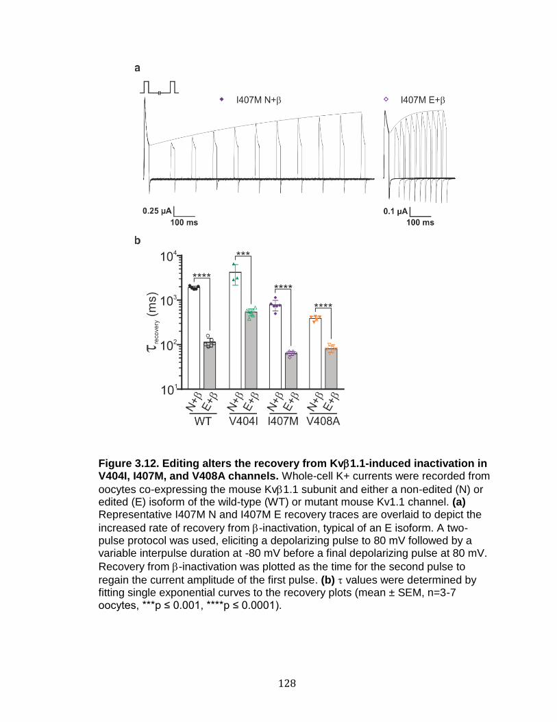
128
Figure 3.12. Editing alters the recovery from Kv1.1-induced inactivation in V404I, I407M, and V408A channels. Whole-cell K+ currents were recorded from
oocytes co-expressing the mouse Kv1.1 subunit and either a non-edited (N) or edited (E) isoform of the wild-type (WT) or mutant mouse Kv1.1 channel. (a) Representative I407M N and I407M E recovery traces are overlaid to depict the
increased rate of recovery from -inactivation, typical of an E isoform. A two-pulse protocol was used, eliciting a depolarizing pulse to 80 mV followed by a variable interpulse duration at -80 mV before a final depolarizing pulse at 80 mV.
Recovery from -inactivation was plotted as the time for the second pulse to
regain the current amplitude of the first pulse. (b) values were determined by fitting single exponential curves to the recovery plots (mean ± SEM, n=3-7 oocytes, ***p ≤ 0.001, ****p ≤ 0.0001).

129
Table 3.3. Kinetics of recovery from Kv1.1 inactivation. All data represent mean ± SEM, n=3-7 oocytes for each channel type. Mutant N and E channels were compared to their WT N and E channels, respectively: ***p ≤ 0.001; ****p ≤ 0.0001. All types of N channels were compared to their respective E channels: +++p ≤ 0.001; ++++p ≤ 0.0001. Significance was determined by two-sample t-tests; due to multiple comparisons, significance was set at p ≤ 0.005.
Discussion
Proof of concept that existing mutations alter RNA editing
The conversion of A-to-I by RNA editing has been shown to represent an
important post-transcriptional modification by which to modulate the function of
numerous proteins critical for nervous system function (Hood and Emeson,
2012). Previous studies have shown that site-selective editing of transcripts
encoding the Kv1.1 channel can affect the rate of recovery from channel
inactivation, the binding of drugs and highly unsaturated fatty acids, the
regulation of homotetrameric Kv1.1 channel trafficking, and seizure-susceptibility
in chronic epileptic rats (Bhalla et al., 2004; Decher et al., 2010; Gonzalez et al.,
130
2011; Streit et al., 2011; Streit et al., 2014). While numerous EA1-associated
mutations have been identified throughout the KCNA1 coding region, several of
these mutations (V404I, I407M, and V408A) are within close proximity to the
Kv1.1 editing site (I400V) and also are predicted to disrupt the critical RNA
duplex structure required for this post-transcriptional modification.
To our knowledge, the present studies represent the first demonstration
that disease-associated mutations can disrupt critical cis-regulatory elements to
change their gene’s RNA editing profile, by altering the RNA structure required
for site-selective A-to-I conversion. It is well established that artificially
incorporating mutations in an RNA duplex can abolish editing by disrupting
duplex pairing; indeed, this technique was used to experimentally verify the RNA
duplex responsible for Kv1.1 editing (Bhalla et al., 2004; Dawson et al., 2004).
However, this is the first evidence that existing human mutations in an RNA can
disrupt its own editing and represents a unique advancement in our
understanding of the mutational consequences of human disorders involving
RNA editing targets. Results using both in vitro and in vivo model systems have
shown significant reductions in the extent and rate of editing for Kv1.1 transcripts
harboring specific EA1 mutations (Figures 3.3 and 3.4). Importantly, because the
wild-type allele RNA was unchanged in the V408A/+ mouse model, it is likely that
the observed changes in the editing of the V408A allele-derived RNA were solely
due to the V408A mutation and not due to any developmental, compensatory
changes. These in vitro and in vivo data serve as a proof of concept that even
single nucleotide changes in an RNA can alter its RNA editing potential.
131
Furthermore, our studies suggest that both synonymous and non-
synonymous duplex-disrupting mutations and single nucleotide polymorphisms
within Kv1.1 and other edited RNA targets may also affect the expression of their
specific edited isoforms, thus altering the activity of the encoded protein
products. And more broadly, the mutations we studied affect Kv1.1 RNA
interactions with the editing enzyme and dsRNA-binding protein, ADAR2, and
could indicate that mutational consequences in other genes should be studied
not only for the amino acid changes, but also for their alterations on RNA-binding
protein interactions.
Edited Isoforms of EA1 mutants alter electrophysiological properties
These studies also have revealed that the effects of EA1 mutations on
Kv1.1 function are far more complex than originally anticipated, as each mutation
produces channels with unique biophysical properties that depend on the I400V
amino acid identity, mediated by RNA editing. The V404I mutation altered
several electrophysiological parameters on its own, but the edited isoform
demonstrated less drastic changes than the non-edited isoform, as observed for
channel voltage sensitivity, closing kinetics, and the amplitude of -inactivation
(Figures 3.5, 3.8, and 3.11). Although it is tempting to speculate that editing could
dampen the defects in channel function resulting from this point mutation, it also
should be noted that this mutation largely prevents the RNA from being edited in
the first place (Figure 3.3). Thus, it is anticipated that edited V404I isoforms

132
contribute little to the electrophysiological properties of Kv1.1 channels in those
tissue where they are expressed.
Unlike the V404I channel, however, editing combined with the I407M or
V408A mutations led to more severe channel dysfunctions than the non-edited
isoforms. Edited isoforms of both I407M and V408A exhibited unusually slow -
dependent inactivation kinetics (Figure 3.10) and severe defects in activation at
higher voltages (Figure 3.7). The current-voltage curves of I407M E and V408A E
were bell-shaped, resembling the ‘rapid’ delayed rectifier activation kinetics
typical of Kv11.1 (hERG) channels, though reaching peak amplitudes at
markedly higher voltages than Kv11.1 (Figure 3.7) (Sanguinetti et al., 1995).
These defects could possibly be caused by a significantly faster entry into, or
slower recovery from, C-type inactivation (Figure 3.9) (Sanguinetti et al., 1995).
In addition, while the I407M mutation slowed the kinetics of channel opening, the
effect was greater for the edited isoform (Figure 3.6). These studies also
extended the characterization of the I407M mutation, as previous studies of the
non-edited I407M channel reported only alterations in expression and voltage-
sensitivity, while the present study also shows changes in kinetics (Tomlinson et
al., 2013).
Although our studies suggest that edited isoforms of mutant channels
represent a smaller portion of the total Kv1.1 population, they may still exert
functional effects, particularly in tissues with higher editing levels (such as
cerebellum and spinal cord) (Figure 3.4). This is supported by previous studies,
which have shown that incorporating even one edited subunit into a Kv1.x

133
heterotetramer was sufficient to alter its sensitivity to open-channel blocking
molecules (Decher et al., 2010). Alternatively, despite the many functional
differences observed between edited isoforms of the mutant channels, all
recovered from fast inactivation significantly faster than their non-edited
counterparts (Figure 3.12). As these EA1 mutations reduced their own isoform
editing, it is predicted that the overall recovery from fast inactivation in vivo will be
comparatively slow, possibly resulting in unanticipated effects that could prevent
normal neuronal signaling.
Incorporating RNA editing into the complex interactions involved in the EA1 disorder
While no clear correlation has been established between the diverse
clinical phenotypes of EA1 patients and specific mutations within Kv1.1 (Graves
et al., 2014; Graves et al., 2010), part of the observed variability in symptoms
might be explained by differences in RNA editing. These phenotypic differences
could arise from EA1 mutations that disrupt the editing duplex, or from overall
changes in Kv1.1 editing regulation. Although the mechanisms regulating Kv1.1
RNA editing are largely unknown, recent studies have demonstrated that
inducing rats with chronic epilepsy led to a 4-fold increase in Kv1.1 editing in the
entorhinal cortex (Streit et al., 2011). Interestingly, once Kv1.1 editing was
increased, recordings in isolated rat brain slices demonstrated that these animals
had a decreased sensitivity to 4-AP-induced seizure-like events, suggesting that
134
increasing editing might dampen seizure susceptibility. Similarly, analyses of
patients undergoing surgery for mesial temporal lobe epilepsy revealed that
having increased levels of Kv1.1 RNA editing was negatively correlated with the
period of years that the patients had experienced epileptic activity (Krestel et al.,
2013), suggesting that decreased Kv1.1 editing may represent a risk factor for
long-term seizures. Graves et al. (2010) clinically surveyed two families
containing the same EA1 mutation (F414S), and found that one family exhibited
seizures while the other did not, raising the possibility that additional factors,
such as differences in editing regulation, could represent an explanation for these
phenotypic differences (Graves et al., 2010). As previous studies have shown
that open-channel blocking drugs interact less with edited Kv1.1 homo- and
heterotetramers (Decher et al., 2010), a precise therapeutic strategy for the
treatment of Kv1.1-dependent seizures may require not only a knowledge of the
specific mutation(s) involved, but also the editing profiles of Kv1.1 transcripts.
135
CHAPTER IV
CONCLUSIONS
Significance
Kv1.1 is an important voltage gated -subunit and human mutations have
revealed that its misregulation contributes to epilepsy and the EA1 disorder.
Although Kv1.1 RNA editing is known to be regulated differentially in the brain,
until now, its impacts were mainly proposed from exogenous expression studies
within cell lines and Xenopus oocytes. This work has begun the process of
understanding the role of Kv1.1 RNA editing within the context of normal
physiology, as well as its implications for the EA1 disorder.
By utilizing two newly developed mouse models, we have characterized
the effects of solely expressing either the non-edited [Kv1.1(I)] or edited
[Kv1.1(V)] isoforms in mice. We have observed that our editing mouse models
share similarities with previously characterized Kv1.1 and Kv mouse models,
relating to ataxia and seizure-susceptibility. The non-edited [Kv1.1(I)] mouse
displays EA1-like stress-induced ataxia, similar to the V408A/+ mouse (Herson et
al., 2003). This is particularly salient because our EA1 experiments have
demonstrated that the V408A mutation leads to decreased editing in vivo; thus
ataxia might have a multifaceted onset arising from either mutant Kv1.1 or

136
editing-deficient Kv1.1. The non-edited [Kv1.1(I)] and edited [Kv1.1(V)] mice
display diverging susceptibilities to 4-AP-induced seizures, where the non-edited
[Kv1.1(I)] mice required a smaller concentration of drug for seizure symptoms
compared to the wild-type littermates and the edited [Kv1.1(V)] mice required a
greater concentration. This is in agreement with the studies of Streit et al. (2011),
which compared the 4-AP susceptibility of rats with higher versus lower Kv1.1
RNA editing. Importantly, we have also observed that the dampening of seizure
susceptibility observed in the edited [Kv1.1(V)] mice is translatable to PTZ
seizure induction as well, indicating that Kv1.1 RNA editing may have a
protective effect against seizures arising from disparate pathways of initiation and
propagation. Although no changes in signaling were observed upon
electrophysiological characterization of dentate granule cells of the hippocampus,
interestingly other mouse models of Kv1 and Kv alterations also do not show
overt changes in electrophysiology, such as the intrinsic membrane properties of
the Kv1.1-null mice.
Our characterization of the complex interplay between RNA editing and
EA1 mutations have revealed new mechanisms of channel dysfunctions to factor
into our broader understanding of this human disorder. First, we have
demonstrated that at least three documented EA1 mutations contain the ability to
inhibit Kv1.1 RNA editing in vitro, and confirmed that the V408A mutation inhibits
editing in vivo. We observed that the in vivo decreases in editing were more
severe than would be predicted by our in vitro editing assay- indicative of the
presence of further regulatory factors that mediate the rate of editing in vivo.

137
Unfortunately, little is known about the regulation of RNA editing, though
particular nucleotide motif biases and structural determinants play a dominant
role in setting the rates (Dawson et al., 2004). It was surprising that only single-
nucleotide changes in the Kv1.1 duplex were necessary to impair editing to such
a high degree, as well as that the level of editing impairment was related to the
proximity to the editing site (with the closest mutation,V404I, leading to the
greatest impairment in editing). This may be indicative that the Kv1.1 duplex is
particularly susceptible to mutation-driven decreases in RNA editing because it is
a relatively poor ADAR2 substrate; this is supported by data which shows that
Kv1.1 editing is low compared to other ADAR2 substrates sequences isolated
from the same tissue dissections, across development and in adult mice (ex.
GluA2 Q/R site and the 5HT2C D site) (Hood et al., 2014; Jacobs et al., 2009). In
addition, we experimentally observed that the wild-type Kv1.1 duplex was a
poorer substrate compared to the 5HT2C RNA duplex, which was used as a
positive control for the in vitro editing assay (data not shown). The susceptibility
of mutations leading to RNA disruptions signifies that other mutations, including
silent mutations or single-nucleotide polymorphisms within the Kv1.1 duplex may
also be able to disrupt Kv1.1 editing.
And, finally, edited isoforms of EA1 mutant proteins were shown to have
altered electrophysiological properties compared to their non-edited counterparts.
The type and severity of the defects were unique to each mutation. The edited
V404I mutant channels seemed to normalize the biophysical properties that were
dysfunctional in the non-edited V404I channels. In contrast, the edited I407M and

138
V408A mutant channels often displayed more severe defects compared to their
non-edited counterparts. The role of editing on setting the fast rate of recovery
from -inactivation was noticeably similar between wild-type and mutant
channels, indicating that the I400V amino acid has a particularly important role in
this capability, regardless of mutations. However, the expression of these edited
channels will depend on each mutation’s disruption of normal RNA editing. If
RNA editing is decreased substantially in patients with duplex mutations, then the
altered edited mutant channels may have little effect on overall physiology. In
contrast, the editing-dependent fast rate of the recovery from -inactivation would
be prominently lost if editing is lowered substantially. As the EA1 disorder is a
multifaceted disorder with incomplete penetrance, additional factors, including
the presence of edited mutant isoforms, may be able to explain the clinical and
genetic heterogeneity in symptom severity.
Future Directions
Utilizing the newly developed mouse models of Kv1.1 editing
Although our studies have brought new insights into the role of Kv1.1 RNA
editing for normal physiology and in the context of EA1, new questions can now
be raised in response. First of all, we have developed new mouse models of
Kv1.1 editing, the non-edited [Kv1.1(I)] and edited [Kv1.1(V)] mice, which should
continue to be characterized in-depth. And our studies of the implications of
139
Kv1.1 RNA editing on the complexity of EA1 provide a new avenue for
investigating this multifaceted disorder.
Our mouse models offer a unique opportunity to study the impacts of the
non-edited [Kv1.1(I)] and edited [Kv1.1(V)] isoforms in vivo, starting with a
detailed study of each isoform’s localization and heterotetrameric assembly in the
brain. Immunohistochemistry of non-edited [Kv1.1(I)], edited [Kv1.1(V)], and wild-
type brain slices can be performed to determine whether editing alters Kv1.1
localization in different brain regions, as well as the impacts on other Kv1 family
members and -subunits. In addition, subcellular localization may be determined
by differential centrifugation strategies to isolate different membrane protein
fractions. Alterations in localization at the plasma membrane versus the ER could
be determined by Western blot analysis and performed in concert with Kv1.1-
pull-down studies to probe for other Kv1.x and -subunits (particularly for co-
assembly with Kv1.2, Kv1.4, Kv1, Kv2). These studies will be important for
determining whether localization and heterotetrameric assembly differs for non-
edited and edited Kv1.1.
Although we have performed many behavioral tests to compare the non-
edited [Kv1.1(I)] and edited [Kv1.1(V)] mice, there are several others that could
give important insights. Kv1.1 has known roles in mechanosensation and
nociception (Clark and Tempel, 1998; Galeotti et al., 1997; Hao et al., 2013), and
editing effects on these signaling pathways could be tested by observing
alterations between the non-edited [Kv1.1(I)] and edited [Kv1.1(V)] mice in von
Frey filament stimulation and thermal stimulation (such as the hotplate test).

140
Alterations in ananamide effects would be interesting to study, given the known
differential in anandamide block for the non-edited [Kv1.1(I)] versus edited
[Kv1.1(V)] isoforms (Decher et al., 2010) and the locomotor deficits and
antinociception observed following anandamide administration (Stein et al.,
1996). The non-edited [Kv1.1(I)] and edited [Kv1.1(V)] mice could be
administered anadamide and monitored for alterations in locomotion and
nociception, in order to determine whether editing has a role in regulation of
these anadamide responses. For testing prolonged effects of anandamide, these
experiments could be done in mutants crossed with fatty acid amide hydrolase
(FAAH) knock-out mice, as FAAH is the enzyme responsible for hydrolyzing
anandamide, and FAAH-null mice display magnified anandamide effects (Cravatt
et al., 2001). Finally, the role of Kv1.1 editing on metabolic function could be
probed, as Kv1.1 has been implicated in contributing to glucose-induced insulin
release, with islets isolated from both mceph/mceph and Kv1.1-null mice
secreting greater insulin upon glucose challenge (Ma et al., 2011). Ma et al.
(2011) detail that, while there are discrepancies in the literature concerning
detecting Kv1.1 in islets, their expression analyses and electrophysiological
characterization supports functional expression in mouse -cells, and expression
was further confirmed in rat and human islets (Ma et al., 2011; MacDonald and
Wheeler, 2003). In addition, ADAR2 expression has been shown to be
upregulated in pancreatic islets following a high-fat diet, leading to an increase in
GluA2 RNA editing (unfortunately Kv1.1 expression was undetectable in this
study) (Gan et al., 2006). First, editing should first be fully characterized in
141
pancreas and islets from normal and type-2 diabetic mice (we have preliminary
studies that Kv1.1 editing might be affected, however these preliminary studies
need to be replicated to determine precise quantification). Following this, the
non-edited [Kv1.1(I)] and edited [Kv1.1(V)] mice could be subjected to metabolic
studies starting with glucose tolerance tests, compared to wild-type littermates, to
see whether editing alters glucose homeostasis.
The altered seizure susceptibility to PTZ and 4-AP-induced seizures has
indicated that editing plays a role as a contributing factor in seizure severity.
While we did not observe spontaneous seizures in either mouse model, the
alteration in seizure-susceptibility suggests that, although editing may not be an
originating cause for seizures, it can act as a downstream modifier to heighten or
dampen seizures. Now that we have characterized this function by drug-induced
seizures, seizure-susceptibility could be further investigated in chronically
induced-seizure models (such as the kainic acid model) or by genetic
mechanisms- breeding non-edited [Kv1.1(I)] and edited [Kv1.1(V)] mice with
genetic mouse models of epilepsy to see if editing status can affect seizure
susceptibility and resistance. A relevant epilepsy mouse model to begin with
could be breeding the non-edited [Kv1.1(I)] or edited [Kv1.1(V)] mice with Kv1.2-
null mice, which are postnatal lethal from P16 to P19 and coincide with
spontaneous seizures (Brew et al., 2007). The severe phenotype of the Kv1.2-
null mouse is in sharp contrast to the mild phenotypes of other knock-out models
of Kv1 family members, which have normal life spans and no (Kv1.3-null and
Kv1.5-null) or limited seizures (Kv1.4-null) (Archer et al., 2001; London et al.,

142
1998; Xu et al., 2003). Unexpectedly, hypoactivity and larger K+ currents were
observed in auditory neurons of the medial nucleus of the trapezoid body
(MNTB) in the brainstem of Kv1.2-null mice (hyperexcitability was found in the
same neurons of Kv1.1-null mice) (Brew et al., 2007; Brew et al., 2003). As the
brainstem is a location of high levels of Kv1.1 RNA editing (60% in mice, 80% in
humans) (Decher et al., 2010; Hoopengardner et al., 2003), this could be an
indication that Kv1.1 editing may serve a more dominant role in Kv1.2-null mice.
Knowing that editing can dampen seizure-susceptibility also begs the
question: how can this knowledge be utilized in a pharmacological approach to
treat epilepsy (or at least dampen its severity)? The predominant biophysical
difference noted between the non-edited [Kv1.1(I)] and edited [Kv1.1(V)]
channels is that the edited isoform promotes rapid recovery from -subunit
inactivation. Thus, a non-edited [Kv1.1(I)] channel could be pharmacologically
similar to an edited [Kv1.1(V)] channel if the inactivation domain of the -subunit
was prevented from interacting. Interestingly, studies of this topic have already
been performed and fit well with the editing-modifier effects on seizures that we
have observed (Lu et al., 2008; Pozo et al., 1990). In particular, Lu et al. (2008)
performed in-depth drug screens looking for drugs that could disrupt -
inactivation while not altering interactions responsible for the assembly of the -
subunit with the Kv1 T1 domain (while leaving Kv1.4 inactivation intact). Drugs
discovered in this screen were then administered prior to PTZ-induced seizures.
Three drugs dampened seizure-susceptibility, indicating that decreasing -
inactivation pharmacologically could dampen seizure susceptibility (Lu et al.,

143
2008). These drugs could be further tested with our mice to determine whether
editing-dependent alterations mediating the -interaction are involved in
mediating this dampened seizure-susceptibility.
Finally, a key reason for developing our non-edited [Kv1.1(I)] and edited
[Kv1.1(V)] mouse models was to allow for electrophysiological characterization of
neurons in slices. Our initial studies in dentate granule cells of the non-edited
[Kv1.1(I)] mutants should be compared next to signaling dynamics of the same
cells in edited [Kv1.1(V)] mice. In addition, as Kv1.1 editing is regulated during
embryonic development, our mice could be used as controls for a study of
alterations in Kv1.1 editing during adult neurogenesis of dentate granule cells
following seizure-induction in wild-type mice. Alterations in signaling in immature
versus mature dentate granule cells may correlate with the Kv1.1 editing status,
and our non-edited [Kv1.1(I)] and edited [Kv1.1(V)] mice may serve as important
controls. As the non-edited [Kv1.1(I)] mice display stress-induced motor
dyscoordination, cerebellar recordings of basket cells and Purkinje neurons will
be important for determining if editing in basket cells is altering the GABA
release, controlling the rate of Purkinje neuron firing. Since MNTB neurons have
already been described in Kv1.1-null and Kv1.2-null mice, characterizing the
excitability of these neurons in our mouse models may be able to differentiate
whether editing is a key contributor to the normally low excitability of these
neurons (Brew et al., 2007; Brew et al., 2003). Peripheral nerve studies can also
be performed, particularly investigating whether the non-edited [Kv1(I)] or edited
[Kv1.1(V)] mutants have similarities to the Kv1.1-null mice, which have

144
temperature-sensitive neuromuscular transmission (Zhou et al., 1998). Particular
attention should be devoted to probing for the type of altered signaling dynamics
that have been observed in other Kv1-related mouse models, not only assessing
passive membrane properties and action potential shape. This could include
probing for alterations in repetitive firing properties such as the frequency of
stimulus-induced pulse trains (mceph/mceph mice) and the presence of
frequency-dependent spike broadening (Kv1.1-null mice) (Giese et al., 1998;
Petersson et al., 2003).
Recording from neurons in slices also offers a convenient model for
epileptic drug administration, using drugs we have already determined to have an
effect (4-AP and PTZ) as well as other widely used pharmacological agents, such
as picrotoxin (Gonzalez-Sulser et al., 2012; Marsh et al., 1999; Piredda et al.,
1985). We expect that experiments using these drugs will give comparable
results consistent with our seizure studies as well as the previous findings using
4-AP to induce seizure-like events in slices from chronic epileptic rats (which
displayed higher editing levels) (Streit et al., 2011). We predict that seizure-like
events will be observed to the greatest extent in the slices from non-edited
[Kv1.1(I)] mice, absent in the edited [Kv1.1(V)] mice, and intermediate in severity
in the wild-type mice (or possibly similar to the non-edited mice). Finally, aside
from incorporating a stressor prior to hippocampal slice isolation (as is described
in the electrophysiology of Chapter 2), other methods of stressing the slices
during recording could include oxygen-glucose deprivation, hypo- and hyper-
osmotic challenges, and incubation with corticosterone (Alfarez et al., 2002;
145
Karst et al., 1993; Lipski et al., 2006; Risher et al., 2009). These stressor
experiments could help define mechanisms for the stress-vulnerability for the
non-edited [Kv1.1(I)] mice.
Continuing the study of RNA editing effects on EA1
We have identified that RNA editing is impacted by the presence of EA1
mutations and that edited EA1 isoforms have altered homotetrameric properties.
However, additional experiments are needed to further understand the role of
EA1 RNA editing in heterotetramers. The V408A/+ mice can also be further
studied for in vivo effects of EA1 altered RNA editing.
Further characterizations of edited EA1 mutant channels could help us
better understand their physiological defects in vivo. These include stimulating
the channels with action potential-like commands (trains of depolarizing pulses)
to assess cumulative inactivation, as well as probing the voltage-dependence of
their inactivation. As Kv1.1 is rarely found in vivo in homotetrameric complexes,
the alterations in heterotetrameric properties need to be investigated. Previous
studies have indicated that the V404I and V408A mutations alter the kinetics of
heterotetramers composed of tandem-linked Kv1.1-Kv1.2 or Kv1.1-Kv1.4 -
subunits (D'Adamo et al., 1999; Imbrici et al., 2006; Manganas and Trimmer,
2000). These experiments could be performed with the edited EA1 isoforms,
tandem-linked with Kv1.2 or Kv1.4 (or a combination of both), with or without co-
expression with -subunits. These heterotetrameric studies will also be important

146
for determining whether the co-expression with Kv1.2 and Kv1.4 improves the
expression of I407M and V408A non-edited and edited isoforms. Wild-type and
edited EA1 characterization in tandemly-linked channels containing Kv1.2
subunits should also be specifically studied for editing-dependent changes in the
use-dependent activation property (also called prepulse potentiation), which has
recently been attributed to Kv1.2-containing heterotetramers (Baronas et al.,
2015).
Now that we have established that the V408A/+ mice are an in vivo model
of reduced Kv1.1 editing, it can be used as a unique model for testing editing
effects and therapeutic strategies targeted towards editing. The plasma
membrane versus ER expression of the different wild-type and V408A non-edited
and edited isoforms could be determined by isolating different membrane
fractions by differential centrifugation of brain regions and then subjecting them
to mass spectrometry for protein isoform identification. Also, as described
previously, there are several known drugs that can prevent the fast-inactivation
mediated by -subunits and could potentially abolish the slow recovery from
inactivation of non-edited [Kv1.1(I)] (Lu et al., 2008). These drugs could be
administered prior to rotarod analyses or electrophysiological recordings to
determine if the decreased Kv1.1 editing of V408A/+ mice is a contributing factor
to stress-induced motor dyscoordination and altered Purkinje neuron firing.
These experiments could be performed on the V408A/+ mice as well as V408A
mutant mice that have been bred with the non-edited [Kv1.1(I)] or edited
[Kv1.1(V)] mice, for additional alterations in the wild-type allele editing level.
147
These further breeding experiments may reveal whether restricting the wild-type
isoform to non-edited or edited could lead to dominant-negative effects on motor
dyscoordination or electrophysiology.
Finally, we have also detailed how several EA1 mutations had
consequences on the RNA editing process, presumably by altering the enzymatic
recognition of the RNA structure. Expanding the implications of our studies for
the wider scientific community, we hope that this will encourage others to see
whether mutations alter the RNA editing of other substrates or, more broadly, if
other mutations affect RNA interactions with other RNA-binding proteins. This
could include other heritable mutations, somatic mutations arising in cancer, or
synonymous variants, which could alter RNA structures while leaving the coding
unchanged. Interestingly, there are many known single nucleotide
polymorphisms in the region of the Kv1.1 RNA duplex (which encodes amino
acids 373 through 410) as well as many documented mutations arising in a
variety of cancers (Ensembl release 87 (Aken et al., 2017)) and recent studies
have linked Kv1.1 expression with apoptosis and sensitivity to drug-induced cell
death (Leanza et al., 2014; Szabò et al., 2010). Our studies suggest that the
Kv1.1 duplex is sensitive to small perturbations, and the presence of variants in
the duplex could account for RNA editing variation among the population, which
could be risk factors for patients that have additional germline or somatic
mutations. Of particular interest would be to test whether several known
synonymous human variants around the editing site could alter Kv1.1 RNA
editing similarly to the EA1 mutations we have described. To date, there are 13

148
synonymous variants that have been documented within the minimal duplex
required for ADAR2-mediated RNA editing, on either side of the duplex, and
several coincide with locations of EA1 mutations as well (Figure 4.1 and Table
C.1). These silent variants may be additional factors regulating RNA editing in
vivo. The EA1 mutations’ impacts on Kv1.1 RNA editing may not be an isolated
phenomenon, but could rather be an initial case-study revealing the
consequences of mutations and variants on RNA-RNA interactions (such as with
miRNAs) and RNA-protein interactions.
Figure 4.1. Human variants found in the Kv1.1 mRNA duplex. The predicted secondary structure for the minimal duplex required for ADAR2 editing of the wild-type human Kv1.1 pre-mRNA. Human variants are located in various positions of the duplex and color-coded for variant type, checkered letters indicate multiple types of variants at a given location (blue: missense somatic mutation, purple: synonymous somatic mutation, yellow: missense genetic variant, red: synonymous genetic variant). The position of the A-to-I editing site (I400V) is indicated with a black box and the four non-synonymous mutations associated with EA1 indicated with dark grey boxes. The numbered arrows indicate the codon positions of the associated amino acids. The locations of two human versus mouse sequence differences are indicated in light grey letters (which are both G in the mouse sequence).
149
We hope our studies will encourage others to continue studying the
editing-dependent effects of Kv1.1 function in normal physiology and in the EA1
disorder, as well as inspire the broader scientific community to study the impacts
of mutations, not only for their amino acid alterations, but also in terms of their
possible altered RNA interactions.

150
Appendix A: Comparative analysis of editing rates determined by Sanger sequencing versus High-throughput sequence analysis
The wild-type ratio of the peak heights described in Figure 2.4 appears to
diverge from the wild-type cerebellar RNA editing rate reported in (Figure 2.3).
This discrepancy highlights the importance of using our high-throughput
sequencing approach to quantify RNA editing rather than relying on the ratio of
electropherogram peak heights, which are known to vary based on the
polymerase incorporation preference for dideoxynucleotide type, sequencing
direction, and sequence context (Carr et al., 2009; Li et al., 1999; Rinkevich et
al., 2012). In order to highlight the differences in quantification by these two
methods, I have compared three data sets where I quantified the same samples
using both methods and displayed the differences in the percent editing or the
rate of percent editing (Figure A.1). In Figure A.1a and b, I have detailed how the
quantification methods compared for Kv1.1 editing from mouse tissues
(described previously in Figures 2.3 and 2.4) and from my in vitro editing assay
(Figure 3.3). In these data, the Sanger sequencing peak height quantification
mainly overestimates the editing level or editing rate quantified by high-
throughput sequencing, though it gives close to accurate measurements for the
hippocampal editing level. However, Figure A.1c demonstrates that Sanger
sequencing can underestimate editing levels in different contexts (these data are
derived from a collaborative study I contributed to for Dr. Martin Gallagher,
quantifying Gabra3 RNA editing in the thalamic reticular nucleus (nRT) dissected
from mouse brain) (Zhou et al., 2015). This variation in underestimating versus

151
overestimating the percent editing has been documented previously (Rinkevich
et al., 2012).
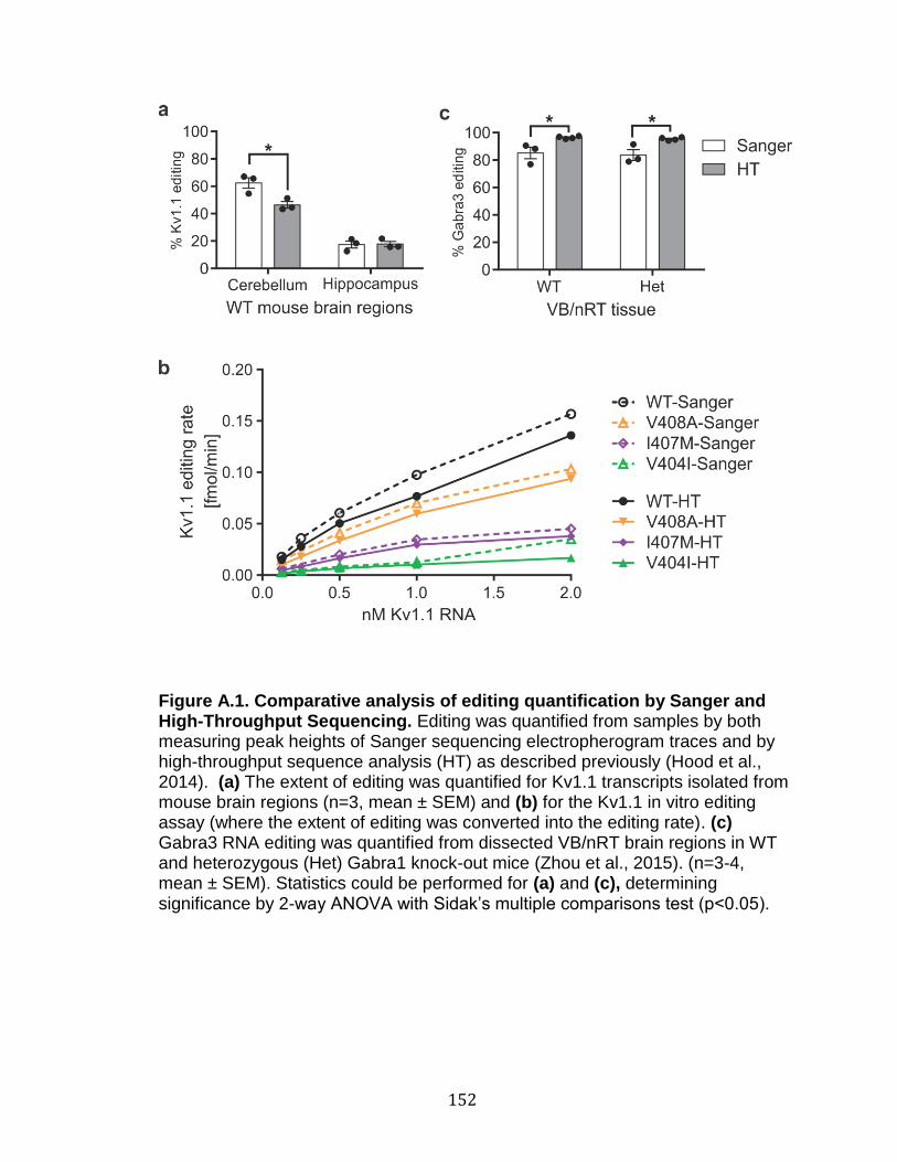
152
Figure A.1. Comparative analysis of editing quantification by Sanger and High-Throughput Sequencing. Editing was quantified from samples by both measuring peak heights of Sanger sequencing electropherogram traces and by high-throughput sequence analysis (HT) as described previously (Hood et al., 2014). (a) The extent of editing was quantified for Kv1.1 transcripts isolated from mouse brain regions (n=3, mean ± SEM) and (b) for the Kv1.1 in vitro editing assay (where the extent of editing was converted into the editing rate). (c) Gabra3 RNA editing was quantified from dissected VB/nRT brain regions in WT and heterozygous (Het) Gabra1 knock-out mice (Zhou et al., 2015). (n=3-4, mean ± SEM). Statistics could be performed for (a) and (c), determining significance by 2-way ANOVA with Sidak’s multiple comparisons test (p<0.05).

153
Appendix B: Additional p-value tables
For the sake of brevity, ranges of p-values were reported in the legends
for Figures 3.6, 3.8, and 3.10. P-values associated with these figures are
presented below.

154
Table B.1: P-values corresponding to Figure 3.6 (time to half-activation)
voltage -10 0 10 20 30 40 50 60 70 80 significant range
Non-edited vs Edited (4
comparisons)
WT N vs WT E 0.0266 0.0443 0.0631 0.0766 0.0791 0.0695 0.052 0.0334 0.0185 0.009 0.0443>p>0.009
significant * * ns ns ns ns ns * * *
V404I N vs V404I E 0.099 0.0574 0.0397 0.0325 0.0316 0.0364 0.0497 0.0813 0.1576 0.3391 0.0497>p>0.0316
significant ns ns * * * * * ns ns ns
I407M N vs I407M E 0.0055 0.0018 0.001 0.0008 0.0009 0.0013 0.0029 0.0089 0.0397 0.2249 0.0397>p>0.0008
significant * * * * * * * * * ns
V408A N vs V408A E 0.0004 0.0048 0.0432 0.2189 0.5883 0.9986 0.7067 0.5621 0.555 0.681 0.0432>p>0.0004
significant * * * ns ns ns ns ns ns ns
WT N vs mutant N (3
comparisons)
WT N vs V404I N 0.0006 0.0017 0.0043 0.0093 0.0165 0.0246 0.0309 0.0336 0.0322 0.028 0.0336>p>0.0006
significant * * * * * * * * * *
WT N vs I407M N <0.0001 <0.0001 <0.0001 <0.0001 <0.0001 <0.0001 <0.0001 <0.0001 <0.0001 <0.0001 all p<0.0001
significant * * * * * * * * * *
WT N vs V408A N 0.0778 0.0804 0.0906 0.1096 0.1407 0.1905 0.2701 0.3962 0.587 0.847 none
significant ns ns ns ns ns ns ns ns ns ns
WT E vs mutant E (3
comparisons)
WT E vs V404I E 0.2704 0.1292 0.0664 0.0377 0.0236 0.0165 0.0131 0.0128 0.0165 0.0292 0.0377>p>0.0165
significant ns ns ns * * * * * * *
WT E vs I407M E <0.0001 <0.0001 <0.0001 <0.0001 <0.0001 <0.0001 <0.0001 <0.0001 0.0001 0.0027 0.027>p>0.0001
significant * * * * * * * * * *
WT E vs V408A E 0.1427 0.0592 0.026 0.0122 0.006 0.0031 0.0017 0.0011 0.001 0.0013 0.026>p>0.001
significant ns ns * * * * * * * *
Individual group comparisons for p-values were based on the Wald tests of model-based predicted (least square) means. Significance was defined as p<0.05.

155
Table B.2: P-values corresponding to Figure 3.8 (closing kinetics)
voltage -120 -110 -100 -90 -80 -70 -60 significant range
Nonedited vs Edited (4
comparisons)
WT N vs WT E 0.0002 0.0004 0.001 0.0042 0.034 0.4182 0.2903 0.034>p>0.0002
significant * * * * * ns ns
V404I N vs V404I E 0.0066 0.0122 0.0254 0.0586 0.1447 0.3556 0.7789 0.0586>p>0.0066
significant * * * ns ns ns ns
I407M N vs I407M E 0.0086 0.0041 0.0036 0.0054 0.015 0.0807 0.6194 0.015>p>0.0036
significant * * * * * ns ns
V408A N vs V408A E N/A N/A N/A N/A N/A N/A N/A
significant ns ns ns ns ns ns ns N/A
WT N v other N (3
comparisons)
WT N vs V404I N <0.0001 <0.0001 <0.0001 <0.0001 <0.0001 <0.0001 <0.0001 all p<0.0001
significant * * * * * * *
WT N vs I407M N <0.0001 <0.0001 <0.0001 <0.0001 <0.0001 <0.0001 <0.0001 all p<0.0001
significant * * * * * * *
WT N vs V408A N <0.0001 <0.0001 <0.0001 <0.0001 <0.0001 <0.0001 <0.0001 all p<0.0001
significant * * * * * * *
WT E v other E (3
comparisons)
WT E vs V404I E 0.0005 0.0005 0.0009 0.0024 0.01 0.0637 0.45 0.01>p>0.0005
significant * * * * * ns ns
WT E vs I407M E 0.0019 0.001 0.0005 0.0003 0.0001 <0.0001 <0.0001 0.0019>p>0.0001
significant * * * * * * *
WT E vs V408A E N/A N/A N/A N/A N/A N/A N/A
significant ns ns ns ns ns ns ns N/A
Individual group comparisons for p-values were based on the Wald tests of model-based predicted (least square) means. Significance was defined as p<0.05.

156
Table B.3: P-values corresponding to Figure 3.10 (kinetics of Kv1.1 inactivation)
voltage 10 20 30 40 50 60 70 80 significant range
Nonedited vs Edited (4
comparisons)
WT N vs WT E <0.0001 <0.0001 <0.0001 <0.0001 <0.0001 <0.0001 <0.0001 <0.0001 all p<0.0001
significant * * * * * * * *
V404I N vs V404I E N/A N/A N/A N/A 0.6925 0.947 0.6908 0.5607 none
significant ns ns ns ns ns ns ns ns
I407M N vs I407M E N/A N/A <0.0001 <0.0001 <0.0001 <0.0001 <0.0001 <0.0001 all p<0.0001
significant ns ns * * * * * *
V408A N vs V408A E 0.0005 0.001 0.002 0.0051 0.017 0.0683 0.3027 0.9997 0.017>p>0.0005
significant * * * * * ns ns ns
WT N v other N (3
comparisons)
WT N vs V404I N N/A N/A N/A N/A 0.0052 0.0138 0.0263 0.0386 0.0386>p>0.0052
significant ns ns ns ns * * * *
WT N vs I407M N N/A <0.0001 <0.0001 <0.0001 <0.0001 <0.0001 <0.0001 <0.0001 all p<0.0001
significant ns * * * * * * *
WT N vs V408A N <0.0001 <0.0001 <0.0001 <0.0001 <0.0001 <0.0001 <0.0001 <0.0001 all p<0.0001
significant * * * * * * * *
WT E v other E (3
comparisons)
WT E vs V404I E 0.0002 0.0003 0.0006 0.0012 0.0018 0.0023 0.0024 0.0025 0.0025>p>0.0002
significant * * * * * * * *
WT E vs I407M E N/A N/A <0.0001 <0.0001 <0.0001 <0.0001 <0.0001 <0.0001 all p<0.0001
significant ns ns * * * * * *
WT E vs V408A E <0.0001 <0.0001 <0.0001 <0.0001 0.001 0.0194 0.2455 0.8719 0.0194>p>0.0001
significant * * * * * * ns ns
Individual group comparisons for p-values were based on the Wald tests of model-based predicted (least square) means. Significance was defined as p<0.05.

157
Appendix C: Human variants found in the Kv1.1 mRNA duplex
Table C.1 was compiled from data accessed from Ensembl release
87(Aken et al., 2017) and the Human Gene Mutation Database (Stenson et al.,
2014). These data further describe the information found in Figure 4.1.
http://uswest.ensembl.org/Homo_sapiens/Gene/Variation_Gene/Table?db=core;
g=ENSG00000111262;r=12:4909905-4931361;t=ENST00000382545
http://www.hgmd.cf.ac.uk/ac/index.php
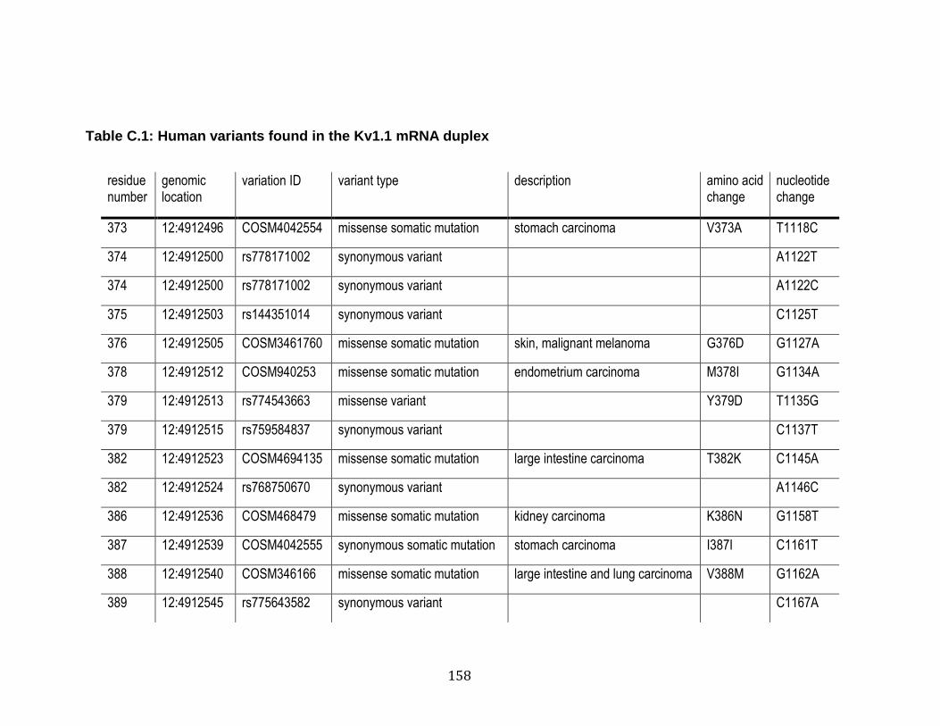
158
Table C.1: Human variants found in the Kv1.1 mRNA duplex
residue number
genomic location
variation ID variant type description amino acid change
nucleotide change
373 12:4912496 COSM4042554 missense somatic mutation stomach carcinoma V373A T1118C
374 12:4912500 rs778171002 synonymous variant A1122T
374 12:4912500 rs778171002 synonymous variant A1122C
375 12:4912503 rs144351014 synonymous variant C1125T
376 12:4912505 COSM3461760 missense somatic mutation skin, malignant melanoma G376D G1127A
378 12:4912512 COSM940253 missense somatic mutation endometrium carcinoma M378I G1134A
379 12:4912513 rs774543663 missense variant Y379D T1135G
379 12:4912515 rs759584837 synonymous variant C1137T
382 12:4912523 COSM4694135 missense somatic mutation large intestine carcinoma T382K C1145A
382 12:4912524 rs768750670 synonymous variant A1146C
386 12:4912536 COSM468479 missense somatic mutation kidney carcinoma K386N G1158T
387 12:4912539 COSM4042555 synonymous somatic mutation stomach carcinoma I387I C1161T
388 12:4912540 COSM346166 missense somatic mutation large intestine and lung carcinoma V388M G1162A
389 12:4912545 rs775643582 synonymous variant C1167A
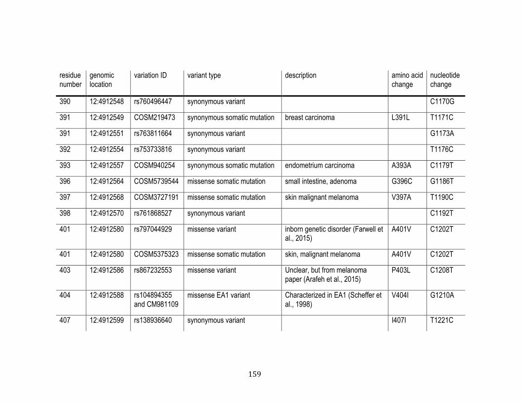
159
residue number
genomic location
variation ID variant type description amino acid change
nucleotide change
390 12:4912548 rs760496447 synonymous variant C1170G
391 12:4912549 COSM219473 synonymous somatic mutation breast carcinoma L391L T1171C
391 12:4912551 rs763811664 synonymous variant G1173A
392 12:4912554 rs753733816 synonymous variant T1176C
393 12:4912557 COSM940254 synonymous somatic mutation endometrium carcinoma A393A C1179T
396 12:4912564 COSM5739544 missense somatic mutation small intestine, adenoma G396C G1186T
397 12:4912568 COSM3727191 missense somatic mutation skin malignant melanoma V397A T1190C
398 12:4912570 rs761868527 synonymous variant C1192T
401 12:4912580 rs797044929 missense variant inborn genetic disorder (Farwell et al., 2015)
A401V C1202T
401 12:4912580 COSM5375323 missense somatic mutation skin, malignant melanoma A401V C1202T
403 12:4912586 rs867232553 missense variant Unclear, but from melanoma paper (Arafeh et al., 2015)
P403L C1208T
404 12:4912588 rs104894355 and CM981109
missense EA1 variant Characterized in EA1 (Scheffer et al., 1998)
V404I G1210A
407 12:4912599 rs138936640 synonymous variant I407I T1221C

160
residue number
genomic location
variation ID variant type description amino acid change
nucleotide change
407 12:4912599 CM130980 Missense EA1 variant Characterized in EA1 (Tomlinson et al., 2013)
408 12:4912600 rs113994117 and CM092472
missense EA1 variant Characterized in EA1 (Demos et al., 2009)
V408L G1222T
408 12:4912601 rs104894352 and CM940995
missense EA1 variant Characterized in EA1 (Browne et al., 1994)
V408A T1223C
408 12:4912602 rs142055425 synonymous variant G1224A
409 12:4912603 COSM4042556 missense somatic mutation stomach carcinoma S409T T1225A
409 12:4912605 rs757958737 synonymous variant C1227A
410 12:4912608 COSM1747035 missense somatic mutation urinary tract carcinoma N410K T1230A

161
REFERENCES
Adelman, J.P., Bond, C.T., Pessia, M., and Mayliet, J. (1995). Episodic ataxia results from voltage-dependent potassium channels with altered functions. Neuron 15, 1449-1454. Aken, B.L., Achuthan, P., Akanni, W., Amode, M.R., Bernsdorff, F., Bhai, J., Billis, K., Carvalho-Silva, D., Cummins, C., Clapham, P., et al. (2017). Ensembl 2017. Nucleic Acids Res 45, D635-D642. Akhtar, S., McIntosh, P., Bryan-Sisneros, A., Barratt, L., Robertson, B., and Dolly, J.O. (1999). A Functional Spliced-Variant of β2 Subunit of Kv1 Channels in C6 Glioma Cells and Reactive Astrocytes from Rat Lesioned Cerebellum. Biochemistry 38, 16984-16992. Alekov, A.K., Rahman, M.M., Mitrovic, N., LehmannHorn, F., and Lerche, H. (2000). A sodium channel mutation causing epilepsy in man exhibits subtle defects in fast inactivation and activation in vitro. The Journal of Physiology 529, 533-540. Alfarez, D.N., Wiegert, O., Joels, M., and Krugers, H.J. (2002). Corticosterone and stress reduce synaptic potentiation in mouse hippocampal slices with mild stimulation. Neuroscience 115, 1119-1126. Arafeh, R., Qutob, N., Emmanuel, R., Keren-Paz, A., Madore, J., Elkahloun, A., Wilmott, J.S., Gartner, J.J., Di Pizio, A., Winograd-Katz, S., et al. (2015). Recurrent inactivating RASA2 mutations in melanoma. Nat Genet 47, 1408-1410. Archer, S.L., London, B., Hampl, V., Wu, X., Nsair, A., Puttagunta, L., Hashimoto, K., Waite, R.E., and Michelakis, E.D. (2001). Impairment of hypoxic pulmonary vasoconstriction in mice lacking the voltage-gated potassium channel Kv1.5. FASEB J 15, 1801-1803. Avery, O.T., MacLeod, C.M., and McCarty, M. (1944). STUDIES ON THE CHEMICAL NATURE OF THE SUBSTANCE INDUCING TRANSFORMATION OF PNEUMOCOCCAL TYPES. The Journal of Experimental Medicine 79, 137-158. Bagchi, B., Al-Sabi, A., Kaza, S., Scholz, D., O'Leary, V.B., Dolly, J.O., and Ovsepian, S.V. (2014). Disruption of myelin leads to ectopic expression of K(V)1.1 channels with abnormal conductivity of optic nerve axons in a cuprizone-induced model of demyelination. PLoS ONE 9, e87736.

162
Bardien-Kruger, S., Wulff, H., Arieff, Z., Brink, P., Chandy, K.G., and Corfield, V. (2002). Characterisation of the human voltage-gated potassium channel gene, KCNA7, a candidate gene for inherited cardiac disorders, and its exclusion as cause of progressive familial heart block I (PFHBI). European Journal of Human Genetics 10, 36-43. Baronas, V.A., McGuinness, B.R., Brigidi, G.S., Gomm Kolisko, R.N., Vilin, Y.Y., Kim, R.Y., Lynn, F.C., Bamji, S.X., Yang, R., and Kurata, H.T. (2015). Use-dependent activation of neuronal Kv1.2 channel complexes. J Neurosci 35, 3515-3524. Basilio, C., Wahba, A.J., Lengyel, P., Speyer, J.F., and Ochoa, S. (1962). Synthetic Polynucleotides and the Amino Acid Code, V. Proceedings of the National Academy of Sciences of the United States of America 48. Bass, B.L., and Weintraub, H. (1988). An unwinding activity that covalently modifies its double-stranded RNA substrate. Cell 55, 1089-1098. Baumann, A., Grupe, A., Ackermann, A., and Pongs, O. (1988). Structure of the voltage-dependent potassium channel is highly conserved from Drosophila to vertebrate central nervous systems. The EMBO Journal 7, 2457-2463. Begum, R., Bakiri, Y., Volynski, K.E., and Kullmann, D.M. (2016). Action potential broadening in a presynaptic channelopathy. Nat Commun 7, 12102. Bezanilla, F. (2004). RNA editing of a human potassium channel modifies its inactivation. Nat Struct Mol Biol 11, 915-916. Bhalla, T., Rosenthal, J.J.C., Holmgren, M., and Reenan, R. (2004). Control of human potassium channel inactivation by editing of a small mRNA hairpin. Nat Struct Mol Biol 11, 950-956. Bretschneider, F., Wrisch, A., Lehmann-Horn, F., and Grissmer, S. (1999). Expression in mammalian cells and electrophysiological characterization of two mutant Kv1.1 channels causing episodic ataxia type 1 (EA-1). European Journal of Neuroscience 11, 2403-2412. Brew, H.M., Gittelman, J.X., Silverstein, R.S., Hanks, T.D., Demas, V.P., Robinson, L.C., Robbins, C.A., McKee-Johnson, J., Chiu, S.Y., Messing, A., et al. (2007). Seizures and reduced life span in mice lacking the potassium channel subunit Kv1.2, but hypoexcitability and enlarged Kv1 currents in auditory neurons. J Neurophysiol 98, 1501-1525. Brew, H.M., Hallows, J.L., and Tempel, B.L. (2003). Hyperexcitability and reduced low threshold potassium currents in auditory neurons of mice lacking the channel subunit Kv1.1. The Journal of Physiology 548, 1-20.
163
Brooks, S.P., and Dunnett, S.B. (2009). Tests to assess motor phenotype in mice: a user's guide. Nat Rev Neurosci 10, 519-529. Browne, D.L., Gancher, S.T., Nutt, J.G., Brunt, E.R.P., Smith, E.A., Kramer, P., and Litt, M. (1994). Episodic ataxia/myokymia syndrome is associated with point mutations in the human potassium channel gene, KCNA1. Nature Genetics 8, 136-140. Brunetti, O., Imbrici, P., Botti, F.M., Pettorossi, V.E., D'Adamo, M.C., Valentino, M., Zammit, C., Mora, M., Gibertini, S., Di Giovanni, G., et al. (2012). Kv1.1 knock-in ataxic mice exhibit spontaneous myokymic activity exacerbated by fatigue, ischemia and low temperature. Neurobiol Dis 47, 310-321. Buynitsky, T., and Mostofsky, D.I. (2009). Restraint stress in biobehavioral research: Recent developments. Neuroscience & Biobehavioral Reviews 33, 1089-1098. Campomanes, C.R., Carroll, K.I., Manganas, L.N., Hershberger, M.E., Gong, B., Antonucci, D.E., Rhodes, K.J., and Trimmer, J.S. (2002). Kvβ Subunit Oxidoreductase Activity and Kv1 Potassium Channel Trafficking. Journal of Biological Chemistry 277, 8298-8305. Carr, I.M., Robinson, J.I., Dimitriou, R., Markham, A.F., Morgan, A.W., and Bonthron, D.T. (2009). Inferring relative proportions of DNA variants from sequencing electropherograms. Bioinformatics 25, 3244-3250. Carter, R.J., Lione, L.A., Humby, T., Mangiarini, L., Mahal, A., Bates, G.P., Dunnett, S.B., and Morton, A.J. (1999). Characterization of Progressive Motor Deficits in Mice Transgenic for the Human Huntington’s Disease Mutation. Journal of Neuroscience 19, 3248-3257. Carter, R.J., Morton, J., and Dunnett, S.B. (2001). Motor Coordination and Balance in Rodents. In Current Protocols in Neuroscience (John Wiley & Sons, Inc.). Chen, C.X., Cho, D.S., Wang, Q., Lai, F., Carter, K.C., and Nishikura, K. (2000). A third member of the RNA-specific adenosine deaminase gene family, ADAR3, contains both single- and double-stranded RNA binding domains. RNA 6, 755-767. Chen, S.H., Fu, S.J., Huang, J.J., and Tang, C.Y. (2016). The episodic ataxia type 1 mutation I262T alters voltage-dependent gating and disrupts protein biosynthesis of human Kv1.1 potassium channels. Sci Rep 6, 19378.
164
Chen, X., Wang, Q., Ni, F., and Ma, J. (2010). Structure of the full-length Shaker potassium channel Kv1.2 by normal-mode-based X-ray crystallographic refinement. Proceedings of the National Academy of Sciences of the United States of America 107, 11352-11357. Cho, D.S.C., Yang, W., Lee, J.T., Shiekhattar, R., Murray, J.M., and Nishikura, K. (2003). Requirement of dimerization for RNA editing activity of adenosine deaminases acting on RNA. Journal of Biological Chemistry 278. Choi, K.L., Aldrich, R.W., and Yellen, G. (1991). Tetraethylammonium blockade distinguishes two inactivation mechanisms in voltage-activated K+ channels. Proceedings of the National Academy of Sciences of the United States of America 88, 5092. Chung, W.K., Shin, M., Jaramillo, T.C., Leibel, R.L., LeDuc, C.A., Fischer, S.G., Tzilianos, E., Gheith, A.A., Lewis, A.S., and Chetkovich, D.M. (2009). Absence epilepsy in apathetic, a spontaneous mutant mouse lacking the h channel subunit, HCN2. Neurobiol Dis 33, 499-508. Clark, J.D., and Tempel, B.L. (1998). Hyperalgesia in mice lacking the Kv1.1 potassium channel gene. Neurosci Lett 251, 121-124. Coleman, S.K., Newcombe, J., Pryke, J., and Dolly, J.O. (1999). Subunit Composition of Kv1 Channels in Human CNS. Journal of Neurochemistry 73, 849-858. Connor, J.X., McCormack, K., Pletsch, A., Gaeta, S., Ganetzky, B., Chiu, S.Y., and Messing, A. (2005). Genetic modifiers of the Kvβ2-null phenotype in mice. Genes, Brain and Behavior 4, 77-88. Consortium, I.H.G.S. (2004). Finishing the euchromatic sequence of the human genome. Nature 431, 931-945. Cravatt, B.F., Demarest, K., Patricelli, M.P., Bracey, M.H., Giang, D.K., Martin, B.R., and Lichtman, A.H. (2001). Supersensitivity to anandamide and enhanced endogenous cannabinoid signaling in mice lacking fatty acid amide hydrolase. Proc Natl Acad Sci U S A 98, 9371-9376. D'Adamo, M.C., Hasan, S., Guglielmi, L., Servettini, I., Cenciarini, M., Catacuzzeno, L., and Franciolini, F. (2015). New insights into the pathogenesis and therapeutics of episodic ataxia type 1. Front Cell Neurosci 9, 317.

165
D'Adamo, M.C., Imbrici, P., Sponcichetti, F., and Pessia, M. (1999). Mutations in the KCNA1 gene associated with episodic ataxia type-1 syndrome impair heteromeric voltage-gated K(+) channel function. FASEB journal : official publication of the Federation of American Societies for Experimental Biology 13, 1335-1345. D'Adamo, M.C., Liu, Z., Adelman, J.P., Maylie, J., and Pessia, M. (1998). Episodic ataxia type-1 mutations in the hKv1.1 cytoplasmic pore region alter the gating properties of the channel. EMBO J 17, 1200-1207. Dawson, T.R., Sansam, C.L., and Emeson, R.B. (2004). Structure and Sequence Determinants Required for the RNA Editing of ADAR2 Substrates. Journal of Biological Chemistry 279, 4941-4951. De Sarro, G., Ibbadu, G.F., Marra, R., Rotiroti, D., Loiacono, A., Donato Di Paola, E., and Russo, E. (2004). Seizure susceptibility to various convulsant stimuli in dystrophin-deficient mdx mice. Neuroscience Research 50, 37-44. Decher, N., Streit, A.K., Rapedius, M., Netter, M.F., Marzian, S., Ehling, P., Schlichthörl, G., Craan, T., Renigunta, V., Köhler, A., et al. (2010). RNA editing modulates the binding of drugs and highly unsaturated fatty acids to the open pore of Kv potassium channels. EMBO J 29, 2101-2113. Demos, M.K., Macri, V., Farrell, K., Nelson, T.N., Chapman, K., Accili, E., and Armstrong, L. (2009). A novel KCNA1 mutation associated with global delay and persistent cerebellar dysfunction. Movement Disorders 24, 778-782. Dhir, A., Zolkowska, D., Murphy, R.B., and Rogawski, M.A. (2011). Seizure Protection by Intrapulmonary Delivery of Propofol Hemisuccinate. J Pharmacol Exp Ther 336, 215-222. Dolly, J.O., and Parcej, D.N. (1996). Molecular properties of voltage-gated K+ channels. Journal of bioenergetics and biomembranes 28, 231-253. Donahue, L.R., Cook, S.A., Johnson, K.R., Bronson, R.T., and Davisson, M.T. (1996). Megencephaly: a new mouse mutation on chromosome 6 that causes hypertrophy of the brain. Mamm Genome 7, 871-876. Dreissen, Y.E., and Tijssen, M.A. (2012). The startle syndromes: physiology and treatment. Epilepsia 53 Suppl 7, 3-11. England, S.K., Uebele, V.N., Kodali, J., Bennett, P.B., and Tamkun, M.M. (1995). A novel K+ channel beta-subunit (hKv beta 1.3) is produced via alternative mRNA splicing. J Biol Chem 270, 28531-28534.

166
Eunson, L.H., Rea, R., Zuberi, S.M., Youroukos, S., Panayiotopoulos, C.P., Liguori, R., Avoni, P., McWilliam, R.C., Stephenson, J.B., Hanna, M.G., et al. (2000). Clinical, genetic, and expression studies of mutations in the potassium channel gene KCNA1 reveal new phenotypic variability. Ann Neurol 48, 647-656. Farwell, K.D., Shahmirzadi, L., El-Khechen, D., Powis, Z., Chao, E.C., Tippin Davis, B., Baxter, R.M., Zeng, W., Mroske, C., Parra, M.C., et al. (2015). Enhanced utility of family-centered diagnostic exome sequencing with inheritance model-based analysis: results from 500 unselected families with undiagnosed genetic conditions. Genetics in medicine : official journal of the American College of Medical Genetics 17, 578-586. Feil, R., and Fraga, M.F. (2012). Epigenetics and the environment: emerging patterns and implications. Nature Reviews Genetics 13, 97-109. Felsenfeld, G. (2014). A Brief History of Epigenetics. Cold Spring Harbor Perspectives in Biology 6, a018200. Feng, Y., Sansam, C.L., Singh, M., and Emeson, R.B. (2006). Altered RNA Editing in Mice Lacking ADAR2 Autoregulation. Mol Cell Biol 26, 480-488. Ferraro, T.N., Golden, G.T., Smith, G.G., Jean, P.S., Schork, N.J., Mulholland, N., Ballas, C., Schill, J., Buono, R.J., and Berrettini, W.H. (1999). Mapping Loci for Pentylenetetrazol-Induced Seizure Susceptibility in Mice. J Neurosci 19, 6733-6739. Ferrick-Kiddie, E.A., Rosenthal, J.J., Ayers, G.D., and Emeson, R.B. (2017). Mutations underlying Episodic Ataxia type-1 antagonize Kv1.1 RNA editing. Sci Rep 7, 41095. Franklin, R.E., and Gosling, R.G. (1953). Molecular Configuration in Sodium Thymonucleate. Nature 171, 740-741. Galeotti, N., Ghelardini, C., Papucci, L., Capaccioli, S., Quattrone, A., and Bartolini, A. (1997). An Antisense Oligonucleotide on the MouseShaker-like Potassium Channel Kv1.1 Gene Prevents Antinociception Induced by Morphine and Baclofen. J Pharmacol Exp Ther 281, 941-949. Gallo, A., Keegan, L.P., Ring, G.M., and O'Connell, M.A. (2003). An ADAR that edits transcripts encoding ion channel subunits functions as a dimer. The EMBO Journal 22, 3421-3430. Gan, Z., Zhao, L., Yang, L., Huang, P., Zhao, F., Li, W., and Liu, Y. (2006). RNA Editing by ADAR2 Is Metabolically Regulated in Pancreatic Islets and β-Cells. Journal of Biological Chemistry 281, 33386-33394.

167
George, C.X., Gan, Z., Liu, Y., and Samuel, C.E. (2011). Adenosine Deaminases Acting on RNA, RNA Editing, and Interferon Action. J Interferon Cytokine Res 31, 99-117. Gerber, A., O'Connell, M.A., and Keller, W. (1997). Two forms of human double-stranded RNA-specific editase 1 (hRED1) generated by the insertion of an Alu cassette. RNA 3, 453. Giese, K.P., Storm, J.F., Reuter, D., Fedorov, N.B., Shao, L.-R., Leicher, T., Pongs, O., and Silva, A.J. (1998). Reduced K+ Channel Inactivation, Spike Broadening, and After-Hyperpolarization in Kvβ1.1-Deficient Mice with Impaired Learning. Learn Mem 5, 257-273. Glasscock, E., Yoo, J.W., Chen, T.T., Klassen, T.L., and Noebels, J.L. (2010). Kv1.1 potassium channel deficiency reveals brain-driven cardiac dysfunction as a candidate mechanism for sudden unexplained death in epilepsy. J Neurosci 30, 5167-5175. Glauner, K.S., Mannuzzu, L.M., Gandhi, C.S., and Isacoff, E.Y. (1999). Spectroscopic mapping of voltage sensor movement in the Shaker potassium channel. Nature 402, 813-817. Gonzalez-Sulser, A., Wang, J., Queenan, B.N., Avoli, M., Vicini, S., and Dzakpasu, R. (2012). Hippocampal neuron firing and local field potentials in the in vitro 4-aminopyridine epilepsy model. J Neurophysiol 108, 2568-2580. Gonzalez, C., Lopez-Rodriguez, A., Srikumar, D., Rosenthal, J.J.C., and Holmgren, M. (2011). Editing of human KV1.1 channel mRNAs disrupts binding of the N-terminus tip at the intracellular cavity. Nat Commun 2. Graves, T.D., Cha, Y.-H., Hahn, A.F., Barohn, R., Salajegheh, M.K., Griggs, R.C., Bundy, B.N., Jen, J.C., Baloh, R.W., and Hanna, M.G. (2014). Episodic ataxia type 1: clinical characterization, quality of life and genotype–phenotype correlation. Brain 137, 1009-1018. Graves, T.D., Rajakulendran, S., Zuberi, S.M., Morris, H.R., Schorge, S., Hanna, M.G., and Kullmann, D.M. (2010). Nongenetic factors influence severity of episodic ataxia type 1 in monozygotic twins(Video). Neurology 75, 367-372. Grice, L.F., and Degnan, B.M. (2015). The origin of the ADAR gene family and animal RNA editing. BMC Evolutionary Biology 15, 4. Griffith, F. (1928). The Significance of Pneumococcal Types. J Hyg (Lond) 27, 113-159.
168
Grissmer, S., Nguyen, A.N., Aiyar, J., Hanson, D.C., Mather, R.J., Gutman, G.A., Karmilowicz, M.J., Auperin, D.D., and Chandy, K.G. (1994). Pharmacological characterization of five cloned voltage-gated K+ channels, types Kv1.1, 1.2, 1.3, 1.5, and 3.1, stably expressed in mammalian cell lines. Mol Pharmacol 45, 1227-1234. Grupe, A., Schröter, K.H., Ruppersberg, J.P., Stocker, M., Drewes, T., Beckh, S., and Pongs, O. (1990). Cloning and expression of a human voltage-gated potassium channel. A novel member of the RCK potassium channel family. The EMBO Journal 9, 1749. Gu, C., Jan, Y.N., and Jan, L.Y. (2003). A Conserved Domain in Axonal Targeting of Kv1 (Shaker) Voltage-Gated Potassium Channels. Science 301, 646-649. Gutman, G.A., Chandy, K.G., Grissmer, S., Lazdunski, M., McKinnon, D., Pardo, L.A., Robertson, G.A., Rudy, B., Sanguinetti, M.C., Stühmer, W., et al. (2005). International Union of Pharmacology. LIII. Nomenclature and Molecular Relationships of Voltage-Gated Potassium Channels. Pharmacol Rev 57, 473-508. Hao, J., Padilla, F., Dandonneau, M., Lavebratt, C., Lesage, F., Noel, J., and Delmas, P. (2013). Kv1.1 channels act as mechanical brake in the senses of touch and pain. Neuron 77, 899-914. Hartner, J.C., Schmittwolf, C., Kispert, A., Müller, A.M., Higuchi, M., and Seeburg, P.H. (2004). Liver Disintegration in the Mouse Embryo Caused by Deficiency in the RNA-editing Enzyme ADAR1. Journal of Biological Chemistry 279, 4894-4902. Heilstedt, H.A., Burgess, D.L., Anderson, A.E., Chedrawi, A., Tharp, B., Lee, O., Kashork, C.D., Starkey, D.E., Wu, Y.-Q., Noebels, J.L., et al. (2001). Loss of the Potassium Channel β-Subunit Gene, KCNAB2, Is Associated with Epilepsy in Patients with 1p36 Deletion Syndrome. Epilepsia 42, 1103-1111. Heinemann, S.H., Rettig, J., Graack, H.R., and Pongs, O. (1996). Functional characterization of Kv channel beta-subunits from rat brain. The Journal of Physiology 493, 625. Heinemann, S.H., Rettig, J., Wunder, F., and Pongs, O. (1995). Molecular and functional characterization of a rat brain Kv beta 3 potassium channel subunit. FEBS Lett 377, 383-389. Hershey, A.D., and Chase, M. (1952). INDEPENDENT FUNCTIONS OF VIRAL PROTEIN AND NUCLEIC ACID IN GROWTH OF BACTERIOPHAGE. J Gen Physiol 36, 39-56.

169
Herson, P.S., Virk, M., Rustay, N.R., Bond, C.T., Crabbe, J.C., Adelman, J.P., and Maylie, J. (2003). A mouse model of episodic ataxia type-1. Nat Neurosci 6, 378-383. Higuchi, M., Maas, S., Single, F.N., Hartner, J., Rozov, A., Burnashev, N., Feldmeyer, D., Sprengel, R., and Seeburg, P.H. (2000). Point mutation in an AMPA receptor gene rescues lethality in mice deficient in the RNA-editing enzyme ADAR2. Nature 406, 78-81. Holmgren, M., Shin, K.S., and Yellen, G. (1998). The Activation Gate of a Voltage-Gated K+ Channel Can Be Trapped in the Open State by an Intersubunit Metal Bridge. Neuron 21, 617-621. Honoré, E., Barhanin, J., Attali, B., Lesage, F., and Lazdunski, M. (1994). External blockade of the major cardiac delayed-rectifier K+ channel (Kv1.5) by polyunsaturated fatty acids. Proceedings of the National Academy of Sciences 91, 1937-1941. Hood, J.L., and Emeson, R.B. (2012). Editing of Neurotransmitter Receptor and Ion Channel RNAs in the Nervous System. In Adenosine Deaminases Acting on RNA (ADARs) and A-to-I Editing, C.E. Samuel, ed. (Springer Berlin Heidelberg), pp. 61-90. Hood, J.L., Morabito, M.V., Martinez, C.R., Gilbert, J.A., Ferrick, E.A., Ayers, G.D., Chappell, J.D., Dermody, T.S., and Emeson, R.B. (2014). Reovirus-mediated induction of ADAR1 (p150) minimally alters RNA editing patterns in discrete brain regions. Mol Cell Neurosci. Hoopengardner, B., Bhalla, T., Staber, C., and Reenan, R. (2003). Nervous System Targets of RNA Editing Identified by Comparative Genomics. Science 301, 832-836. Horsch, M., Seeburg, P.H., Adler, T., Aguilar-Pimentel, J.A., Becker, L., Calzada-Wack, J., Garrett, L., Gotz, A., Hans, W., Higuchi, M., et al. (2011). Requirement of the RNA-editing enzyme ADAR2 for normal physiology in mice. J Biol Chem 286, 18614-18622. Hoshi, T., and Armstrong, C.M. (2013). C-type inactivation of voltage-gated K+ channels: Pore constriction or dilation? J Gen Physiol 141, 151-160. Hoshi, T., Zagotta, W.N., and Aldrich, R.W. (1990). Biophysical and Molecular Mechanisms of Shaker Potassium Channel Inactivation. Science 250, 533. Huang, H., Tan, B.Z., Shen, Y., Tao, J., Jiang, F., Sung, Y.Y., Ng, C.K., Raida, M., Köhr, G., Higuchi, M., et al. (2012). RNA Editing of the IQ Domain in Cav1.3 Channels Modulates Their Ca2+-Dependent Inactivation. Neuron 73, 304-316.
170
Hufeldt, M.R., Nielsen, D.S., Vogensen, F.K., Midtvedt, T., and Hansen, A.K. (2010). Variation in the Gut Microbiota of Laboratory Mice Is Related to Both Genetic and Environmental Factors. Comp Med 60, 336-342. Hwang, T., Park, C.K., Leung, A.K., Gao, Y., Hyde, T.M., Kleinman, J.E., Rajpurohit, A., Tao, R., Shin, J.H., and Weinberger, D.R. (2016). Dynamic regulation of RNA editing in human brain development and disease. Nat Neurosci 19, 1093-1099. Imbrici, P., D'Adamo, M.C., Grottesi, A., Biscarini, A., and Pessia, M. (2011). Episodic ataxia type 1 mutations affect fast inactivation of K+ channels by a reduction in either subunit surface expression or affinity for inactivation domain. Am J Physiol Cell Physiol 300, C1314-C1322. Imbrici, P., D'Adamo, M.C., Kullmann, D.M., and Pessia, M. (2006). Episodic ataxia type 1 mutations in the KCNA1 gene impair the fast inactivation properties of the human potassium channels Kv1.4-1.1/Kvβ1.1 and Kv1.4-1.1/Kvβ1.2. Eur J Neurosci 24, 3073-3083. Ishida, S., Sakamoto, Y., Nishio, T., Baulac, S., Kuwamura, M., Ohno, Y., Takizawa, A., Kaneko, S., Serikawa, T., and Mashimo, T. (2012). Kcna1-mutant rats dominantly display myokymia, neuromyotonia and spontaneous epileptic seizures. Brain Research 1435, 154-166. Ishikawa, T., Nakamura, Y., Saitoh, N., Li, W.-B., Iwasaki, S., and Takahashi, T. (2003). Distinct Roles of Kv1 and Kv3 Potassium Channels at the Calyx of Held Presynaptic Terminal. J Neurosci 23, 10445-10453. Itoh, K., and Watanabe, M. (2009). Paradoxical facilitation of pentylenetetrazole-induced convulsion susceptibility in mice lacking neuronal nitric oxide synthase. Neuroscience 159, 735-743. Jacobs, M.M., Fogg, R.L., Emeson, R.B., and Stanwood, G.D. (2009). ADAR1 and ADAR2 Expression and Editing Activity during Forebrain Development. Dev Neurosci-Basel 31, 223-237. Jayabal, S., Ljungberg, L., Erwes, T., Cormier, A., Quilez, S., Jaouhari, S.E., and Watt, A.J. (2015). Rapid Onset of Motor Deficits in a Mouse Model of Spinocerebellar Ataxia Type 6 Precedes Late Cerebellar Degeneration. eNeuro 2, ENEURO.0094-0015.2015. Jenkins, P.M., McIntyre, J.C., Zhang, L., Anantharam, A., Vesely, E.D., Arendt, K.L., Carruthers, C.J.L., Kerppola, T.K., Iñiguez-LluhÍ, J.A., Holz, R.W., et al. (2011). Subunit-Dependent Axonal Trafficking of Distinct α Heteromeric Potassium Channel Complexes. Journal of Neuroscience 31, 13224-13235.
171
Kalman, K., Nguyen, A., Tseng-Crank, J., Dukes, I.D., Chandy, G., Hustad, C.M., Copeland, N.G., Jenkins, N.A., Mohrenweiser, H., Brandriff, B., et al. (1998). Genomic Organization, Chromosomal Localization, Tissue Distribution, and Biophysical Characterization of a Novel MammalianShaker-related Voltage-gated Potassium Channel, Kv1.7. Journal of Biological Chemistry 273, 5851-5857. Kamb, A., Iverson, L.E., and Tanouye, M.A. (1987). Molecular characterization of Shaker, a Drosophila gene that encodes a potassium channel. Cell 50, 405-413. Kaplan, W.D., and Trout, W.E. (1969). The Behavior of Four Neurological Mutants of Drosophila. Genetics 61, 399-409. Karst, H., Wadman, W.J., and Joels, M. (1993). Long-term control by corticosteroids of the inward rectifier in rat CA1 pyramidal neurons, in vitro. Brain Res 612, 172-179. Kastner, P., Mark, M., Leid, M., Gansmuller, A., Chin, W., Grondona, J.M., Décimo, D., Krezel, W., Dierich, A., and Chambon, P. (1996). Abnormal spermatogenesis in RXR beta mutant mice. Genes & Dev 10, 80-92. Kayakabe, M., Kakizaki, T., Kaneko, R., Sasaki, A., Nakazato, Y., Shibasaki, K., Ishizaki, Y., Saito, H., Suzuki, N., and Yanagawa, Y. (2014). Motor dysfunction in cerebellar Purkinje cell-specific vesicular GABA transporter knockout mice. Front Cell Neurosci 7, 286. Kim, D., Song, I., Keum, S., Lee, T., Jeong, M.-J., Kim, S.-S., McEnery, M.W., and Shin, H.-S. (2001). Lack of the Burst Firing of Thalamocortical Relay Neurons and Resistance to Absence Seizures in Mice Lacking α1G T-Type Ca2+ Channels. Neuron 31, 35-45. Kim, U., Wang, Y., Sanford, T., Zeng, Y., and Nishikura, K. (1994a). Molecular cloning of cDNA for double-stranded RNA adenosine deaminase, a candidate enzyme for nuclear RNA editing. Proceedings of the National Academy of Sciences 91, 11457-11461. Kim, Y.S., Hong, K.S., Seong, Y.S., Park, J.B., Kuroda, S., Kishi, K., Kaibuchi, K., and Takai, Y. (1994b). Phosphorylation and activation of mitogen-activated protein kinase by kainic acid-induced seizure in rat hippocampus. Biochem Biophys Res Commun 202, 1163-1168. Kirchheim, F., Tinnes, S., Haas, C.A., Stegen, M., and Wolfart, J. (2013). Regulation of action potential delays via voltage-gated potassium Kv1.1 channels in dentate granule cells during hippocampal epilepsy. Front Cell Neurosci 7.
172
Kirsch, G.E., Drewe, J.A., Verma, S., Brown, A.M., and Joho, R.H. (1991). Electrophysiological characterization of a new member of the RCK family of rat brain K+ channels. FEBS Letters 278, 55-60. Koch, R.O., Wanner, S.G., Koschak, A., Hanner, M., Schwarzer, C., Kaczorowski, G.J., Slaughter, R.S., Garcia, M.L., and Knaus, H.-G. (1997). Complex Subunit Assembly of Neuronal Voltage-gated K+Channels Basis for High-Affinity Toxin Interactions and Pharmacology. J Biol Chem 272, 27577-27581. Koschak, A., Bugianesi, R.M., Mitterdorfer, J., Kaczorowski, G.J., Garcia, M.L., and Knaus, H.-G. (1998). Subunit Composition of Brain Voltage-gated Potassium Channels Determined by Hongotoxin-1, a Novel Peptide Derived fromCentruroides limbatus Venom. Journal of Biological Chemistry 273, 2639-2644. Krestel, H., Raffel, S., von Lehe, M., Jagella, C., Moskau-Hartmann, S., Becker, A., Elger, C.E., Seeburg, P.H., and Nirkko, A. (2013). Differences between RNA and DNA due to RNA editing in temporal lobe epilepsy. Neurobiol Dis 56, 66-73. Krestel, H.E., Shimshek, D.R., Jensen, V., Nevian, T., Kim, J., Geng, Y., Bast, T., Depaulis, A., Schonig, K., Schwenk, F., et al. (2004). A genetic switch for epilepsy in adult mice. J Neurosci 24, 10568-10578. Kurata, H.T., and Fedida, D. (2006). A structural interpretation of voltage-gated potassium channel inactivation. Progress in Biophysics and Molecular Biology 92, 185-208. Kuroda, K.O., and Tsuneoka, Y. (2013). Assessing Postpartum Maternal Care, Alloparental Behavior, and Infanticide in Mice: With Notes on Chemosensory Influences. In Pheromone Signaling: Methods and Protocols, K. Touhara, ed. (Totowa, NJ: Humana Press), pp. 331-347. Lander, E.S., Linton, L.M., Birren, B., Nusbaum, C., Zody, M.C., Baldwin, J., Devon, K., Dewar, K., Doyle, M., FitzHugh, W., et al. (2001). Initial sequencing and analysis of the human genome. Nature 409, 860-921. Lang, R., Lee, G., Liu, W., Tian, S., Rafi, H., Orias, M., Segal, A.S., and Desir, G.V. (2000). KCNA10: a novel ion channel functionally related to both voltage-gated potassium and CNG cation channels. American Journal of Physiology - Renal Physiology 278, F1013-F1021. Leanza, L., O'Reilly, P., Doyle, A., Venturini, E., Zoratti, M., Szegezdi, E., and Szabo, I. (2014). Correlation between potassium channel expression and sensitivity to drug-induced cell death in tumor cell lines. Curr Pharm Des 20, 189-200.

173
Lee, T.E., Philipson, L.H., and Nelson, D.J. (1996). N-type Inactivation in the Mammalian Shaker K+ Channel Kv1.4. J Membrane Biol 151, 225-235. Lehmann, K.A., and Bass, B.L. (2000). Double-Stranded RNA Adenosine Deaminases ADAR1 and ADAR2 Have Overlapping Specificities. Biochemistry 39, 12875-12884. Li, J.B., Levanon, E.Y., Yoon, J.-K., Aach, J., Xie, B., LeProust, E., Zhang, K., Gao, Y., and Church, G.M. (2009). Genome-Wide Identification of Human RNA Editing Sites by Parallel DNA Capturing and Sequencing. Science 324, 1210-1213. Li, Y., Mitaxov, V., and Waksman, G. (1999). Structure-based design of Taq DNA polymerases with improved properties of dideoxynucleotide incorporation. Proceedings of the National Academy of Sciences 96, 9491-9496. Licatalosi, D.D., and Darnell, R.B. (2010). RNA processing and its regulation: global insights into biological networks. Nature Reviews Genetics 11, 75-87. Liman, E.R., Tytgat, J., and Hess, P. (1992). Subunit stoichiometry of a mammalian K+ channel determined by construction of multimeric cDNAs. Neuron 9, 861-871. Lipski, J., Park, T.I., Li, D., Lee, S.C., Trevarton, A.J., Chung, K.K., Freestone, P.S., and Bai, J.Z. (2006). Involvement of TRP-like channels in the acute ischemic response of hippocampal CA1 neurons in brain slices. Brain Res 1077, 187-199. Lohnes, D., Kastner, P., Dierich, A., Mark, M., LeMeur, M., and Chambon, P. (1993). Function of retinoic acid receptor γ in the mouse. Cell 73, 643-658. London, B., Wang, D.W., Hill, J.A., and Bennett, P.B. (1998). The transient outward current in mice lacking the potassium channel gene Kv1.4. The Journal of Physiology 509 ( Pt 1), 171-182. Long, S.B., Campbell, E.B., and MacKinnon, R. (2005). Crystal Structure of a Mammalian Voltage-Dependent Shaker Family K+ Channel. Science 309, 897-903. Lu, Q., Peevey, J., Jow, F., Monaghan, M.M., Mendoza, G., Zhang, H., Wu, J., Kim, C.Y., Bicksler, J., Greenblatt, L., et al. (2008). Disruption of Kv1.1 N-type inactivation by novel small molecule inhibitors (disinactivators). Bioorganic & Medicinal Chemistry 16, 3067-3075. Lugt, N.M.v.d., Domen, J., Linders, K., Roon, M.v., Robanus-Maandag, E., Riele, H.t., Valk, M.v.d., Deschamps, J., Sofroniew, M., and Lohuizen, M.v. (1994).

174
Posterior transformation, neurological abnormalities, and severe hematopoietic defects in mice with a targeted deletion of the bmi-1 proto-oncogene. Genes & Dev 8, 757-769. Ma, Z., Lavebratt, C., Almgren, M., Portwood, N., Forsberg, L.E., Bränström, R., Berglund, E., Falkmer, S., Sundler, F., Wierup, N., et al. (2011). Evidence for Presence and Functional Effects of Kv1.1 Channels in β-Cells: General Survey and Results from mceph/mceph Mice. PLoS ONE 6. MacDonald, P.E., and Wheeler, M.B. (2003). Voltage-dependent K(+) channels in pancreatic beta cells: role, regulation and potential as therapeutic targets. Diabetologia 46, 1046-1062. Madeja, M., Mußhoff, U., Lorra, C., Pongs, O., and Speckmann, E.-J. (1996). Mechanism of action of the epileptogenic drug pentylenetetrazol on a cloned neuronal potassium channel. Brain Research 722, 59-70. Madeja, M., Stocker, M., Muβhoff, U., Pongs, O., and Speckmann, E.-J. (1994). Potassium currents in epilepsy: effects of the epileptogenic agent pentylenetetrazol on a cloned potassium channel. Brain Research 656, 287-294. Manganas, L.N., and Trimmer, J.S. (2000). Subunit Composition Determines Kv1 Potassium Channel Surface Expression. J Biol Chem 275, 29685-29693. Manganas, L.N., Wang, Q., Scannevin, R.H., Antonucci, D.E., Rhodes, K.J., and Trimmer, J.S. (2001). Identification of a trafficking determinant localized to the Kv1 potassium channel pore. Proceedings of the National Academy of Sciences of the United States of America 98, 14055-14059. Mannion, Niamh M., Greenwood, S.M., Young, R., Cox, S., Brindle, J., Read, D., Nellåker, C., Vesely, C., Ponting, Chris P., McLaughlin, Paul J., et al. (2014). The RNA-Editing Enzyme ADAR1 Controls Innate Immune Responses to RNA. Cell reports 9, 1482-1494. Marcucci, R., Brindle, J., Paro, S., Casadio, A., Hempel, S., Morrice, N., Bisso, A., Keegan, L.P., Sal, G.D., and O'Connell, M.A. (2011). Pin1 and WWP2 regulate GluR2 Q/R site RNA editing by ADAR2 with opposing effects. The EMBO Journal 30, 4211-4222. Marsh, D.J., Baraban, S.C., Hollopeter, G., and Palmiter, R.D. (1999). Role of the Y5 neuropeptide Y receptor in limbic seizures. Proc Natl Acad Sci U S A 96, 13518-13523. Matsuo, N., Takao, K., Nakanishi, K., Yamasaki, N., Tanda, K., and Miyakawa, T. (2010). Behavioral profiles of three C57BL/6 substrains. Front Behav Neurosci 4, 29.

175
Maylie, B., Bissonnette, E., Virk, M., Adelman, J.P., and Maylie, J.G. (2002). Episodic Ataxia Type 1 Mutations in the Human Kv1.1 Potassium Channel Alter hKvβ1-Induced N-Type Inactivation. J Neurosci 22, 4786-4793. McCormack, K., Connor, J.X., Zhou, L., Ho, L.L., Ganetzky, B., Chiu, S.-Y., and Messing, A. (2002). Genetic Analysis of the Mammalian K+ Channel β Subunit Kvβ2 (Kcnab2). Journal of Biological Chemistry 277, 13219-13228. McCormack, K., McCormack, T., Tanouye, M., Rudy, B., and Stühmer, W. (1995). Alternative splicing of the human Shaker K+ channel β1 gene and functional expression of the β2 gene product. FEBS Letters 370, 32-36. McKinnon, D. (1989). Isolation of a cDNA clone coding for a putative second potassium channel indicates the existence of a gene family. Journal of Biological Chemistry 264, 8230-8236. Meiri, N., Ghelardini, C., Tesco, G., Galeotti, N., Dahl, D., Tomsic, D., Cavallaro, S., Quattrone, A., Capaccioli, S., Bartolini, A., et al. (1997). Reversible antisense inhibition of Shaker-like Kv1.1 potassium channel expression impairs associative memory in mouse and rat. Proceedings of the National Academy of Sciences 94, 4430-4434. Mestre, T.A., Manole, A., MacDonald, H., Riazi, S., Kraeva, N., Hanna, M.G., Lang, A.E., Mannikko, R., and Yoon, G. (2016). A novel KCNA1 mutation in a family with episodic ataxia and malignant hyperthermia. Neurogenetics. Monteiro, S., Roque, S., de Sa-Calcada, D., Sousa, N., Correia-Neves, M., and Cerqueira, J.J. (2015). An efficient chronic unpredictable stress protocol to induce stress-related responses in C57BL/6 mice. Frontiers in psychiatry 6, 6. Moore, A.N., Waxham, M.N., and Dash, P.K. (1996). Neuronal activity increases the phosphorylation of the transcription factor cAMP response element-binding protein (CREB) in rat hippocampus and cortex. J Biol Chem 271, 14214-14220. Morabito, M.V., Ulbricht, R.J., O'Neil, R.T., Airey, D.C., Lu, P., Zhang, B., Wang, L., and Emeson, R.B. (2010). High-Throughput Multiplexed Transcript Analysis Yields Enhanced Resolution of 5-Hydroxytryptamine2C Receptor mRNA Editing Profiles. Mol Pharmacol 77, 895-902. Nakamura, K., Oshima, T., Morimoto, T., Ikeda, S., Yoshikawa, H., Shiwa, Y., Ishikawa, S., Linak, M.C., Hirai, A., Takahashi, H., et al. (2011). Sequence-specific error profile of Illumina sequencers. Nucleic Acids Research 39, e90-e90.

176
Need, A.C., Irvine, E.E., and Giese, K.P. (2003). Learning and memory impairments in Kvβ1.1-null mutants are rescued by environmental enrichment or ageing. European Journal of Neuroscience 18, 1640-1644. Nishikura, K. (2010). Functions and Regulation of RNA Editing by ADAR Deaminases. Annual review of biochemistry 79, 321-349. Niswender, C.M., Copeland, S.C., Herrick-Davis, K., Emeson, R.B., and Sanders-Bush, E. (1999). RNA Editing of the Human Serotonin 5-Hydroxytryptamine 2C Receptor Silences Constitutive Activity. Journal of Biological Chemistry 274, 9472-9478. O'Leary, H., Bernard, P.B., Castano, A.M., and Benke, T.A. (2016). Enhanced long term potentiation and decreased AMPA receptor desensitization in the acute period following a single kainate induced early life seizure. Neurobiol Dis 87, 134-144. O'Neil, R.T., and Emeson, R.B. (2012). Quantitative analysis of 5HT(2C) receptor RNA editing patterns in psychiatric disorders. Neurobiol Dis 45, 8-13. Oliver, D., Lien, C.-C., Soom, M., Baukrowitz, T., Jonas, P., and Fakler, B. (2004). Functional Conversion Between A-Type and Delayed Rectifier K+ Channels by Membrane Lipids. Science 304, 265-270. Ovsepian, S.V., LeBerre, M., Steuber, V., O'Leary, V.B., Leibold, C., and Oliver Dolly, J. (2016). Distinctive role of KV1.1 subunit in the biology and functions of low threshold K(+) channels with implications for neurological disease. Pharmacol Ther 159, 93-101. Parcej, D.N., Scott, V.E., and Dolly, J.O. (1992). Oligomeric properties of alpha-dendrotoxin-sensitive potassium ion channels purified from bovine brain. Biochemistry 31, 11084-11088. Peng, P.L., Zhong, X., Tu, W., Soundarapandian, M.M., Molner, P., Zhu, D., Lau, L., Liu, S., Liu, F., and Lu, Y. (2006). ADAR2-dependent RNA editing of AMPA receptor subunit GluR2 determines vulnerability of neurons in forebrain ischemia. Neuron 49, 719-733. Pennacchio, P., Noe, F., Gnatkovsky, V., Moroni, R.F., Zucca, I., Regondi, M.C., Inverardi, F., de Curtis, M., and Frassoni, C. (2015). Increased pCREB expression and the spontaneous epileptiform activity in a BCNU-treated rat model of cortical dysplasia. Epilepsia 56, 1343-1354.
177
Persson, A.-S., Klement, G., Almgren, M., Sahlholm, K., Nilsson, J., Petersson, S., Århem, P., Schalling, M., and Lavebratt, C. (2005). A truncated Kv1.1 protein in the brain of the megencephaly mouse: expression and interaction. BMC Neurosci 6, 65. Persson, A.-S., Westman, E., Wang, F.-H., Khan, F.H., Spenger, C., and Lavebratt, C. (2007). Kv1.1 null mice have enlarged hippocampus and ventral cortex. BMC Neurosci 8, 10. Pertea, M., and Salzberg, S.L. (2010). Between a chicken and a grape: estimating the number of human genes. Genome Biology 11, 206. Petersson, S., Persson, A.-S., Johansen, J.E., Ingvar, M., Nilsson, J., Klement, G., Århem, P., Schalling, M., and Lavebratt, C. (2003). Truncation of the Shaker-like voltage-gated potassium channel, Kv1.1, causes megencephaly. European Journal of Neuroscience 18, 3231-3240. Petitjean, D., Kalstrup, T., Zhao, J., and Blunck, R. (2015). A Disease Mutation Causing Episodic Ataxia Type I in the S1 Links Directly to the Voltage Sensor and the Selectivity Filter in Kv Channels. J Neurosci 35, 12198-12206. Piredda, S., Yonekawa, W., Whittingham, T.S., and Kupferberg, H.J. (1985). Potassium, Pentylenetetrazol, and Anticonvulsants in Mouse Hippocampal Slices. Epilepsia 26, 167-174. Po, S., Roberds, S., Snyders, D.J., Tamkun, M.M., and Bennett, P.B. (1993). Heteromultimeric assembly of human potassium channels. Molecular basis of a transient outward current? Circulation Research 72, 1326-1336. Po, S., Snyders, D.J., Baker, R., Tamkun, M.M., and Bennett, P.B. (1992). Functional expression of an inactivating potassium channel cloned from human heart. Circulation Research 71, 732-736. Pongs, O., and Schwarz, J.R. (2010). Ancillary subunits associated with voltage-dependent K+ channels. Physiol Rev 90, 755-796. Pozo, E.D., Barrios, M., and Baeyens, J.M. (1990). Effects of Potassium Channel Openers on Pentylenetetrazole-Induced Seizures in Mice. Pharmacology & Toxicology 67, 182-184. Ramanjaneyulu, R., and Ticku, M.K. (1984). Interactions of pentamethylenetetrazole and tetrazole analogues with the picrotoxinin site of the benzodiazepine-GABA receptor-ionophore complex. Eur J Pharmacol 98, 337-345.
178
Rasband, M.N., Park, E.W., Vanderah, T.W., Lai, J., Porreca, F., and Trimmer, J.S. (2001). Distinct potassium channels on pain-sensing neurons. Proc Natl Acad Sci USA 98, 13373-13378. Rettig, J., Heinemann, S.H., Wunder, F., Lorra, C., Parcej, D.N., Oliver Dolly, J., and Pongs, O. (1994). Inactivation properties of voltage-gated K+ channels altered by presence of β-subunit. Nature 369, 289-294. Rho, J.M., Szot, P., Tempel, B.L., and Schwartzkroin, P.A. (1999). Developmental Seizure Susceptibility of Kv1.1 Potassium Channel Knockout Mice. Dev Neurosci-Basel 21, 320-327. Rhodes, K.J., Monaghan, M.M., Barrezueta, N.X., Nawoschik, S., Bekele-Arcuri, Z., Matos, M.F., Nakahira, K., Schechter, L.E., and Trimmer, J.S. (1996). Voltage-gated K+ channel beta subunits: expression and distribution of Kv beta 1 and Kv beta 2 in adult rat brain. J Neurosci 16, 4846-4860. Rhodes, K.J., Strassle, B.W., Monaghan, M.M., Bekele-Arcuri, Z., Matos, M.F., and Trimmer, J.S. (1997). Association and Colocalization of the Kvβ1 and Kvβ2 β-Subunits with Kv1 α-Subunits in Mammalian Brain K+Channel Complexes. J Neurosci 17, 8246-8258. Rinkevich, F.D., Schweitzer, P.A., and Scott, J.G. (2012). Antisense sequencing improves the accuracy and precision of A-to-I editing measurements using the peak height ratio method. BMC Research Notes 5, 63. Risher, W.C., Andrew, R.D., and Kirov, S.A. (2009). Real-time passive volume responses of astrocytes to acute osmotic and ischemic stress in cortical slices and in vivo revealed by two-photon microscopy. Glia 57, 207-221. Robbins, C.A., and Tempel, B.L. (2012). Kv1.1 and Kv1.2: Similar channels, different seizure models. Epilepsia 53, 134-141. Roeper, J., Sewing, S., Zhang, Y., Sommer, T., Wanner, S.G., and Pongs, O. (1998). NIP domain prevents N-type inactivation in voltage-gated potassium channels. Nature 391, 390-393. Rula, E.Y., Lagrange, A.H., Jacobs, M.M., Hu, N., Macdonald, R.L., and Emeson, R.B. (2008). Developmental modulation of GABA(A) receptor function by RNA editing. J Neurosci 28, 6196-6201. Sandberg, R., Yasuda, R., Pankratz, D.G., Carter, T.A., Rio, J.A.D., Wodicka, L., Mayford, M., Lockhart, D.J., and Barlow, C. (2000). Regional and strain-specific gene expression mapping in the adult mouse brain. Proceedings of the National Academy of Sciences 97, 11038-11043.

179
Sands, Z., Grottesi, A., and Sansom, M.S. (2005). Voltage-gated ion channels. Current biology : CB 15, R44-47. Sanguinetti, M.C., Jiang, C., Curran, M.E., and Keating, M.T. (1995). A mechanistic link between an inherited and an acquired cardiac arrhythmia: HERG encodes the IKr potassium channel. Cell 81, 299-307. SantaLucia, J., Jr. (1998). A unified view of polymer, dumbbell, and oligonucleotide DNA nearest-neighbor thermodynamics. Proc Natl Acad Sci USA 95, 1460-1465. Scheffer, H., Brunt, E.R.P., Mol, G.J.J., van der Vlies, P., Stulp, Verlind, E., Mantel, G., Averyanov, Y.N., Hofstra, R.M.W., and Buys, C.H.C.M. (1998). Three novel KCNA1 mutations in episodic ataxia type I families. Human Genetics 102, 464-466. Schirmer, M., Ijaz, U.Z., D'Amore, R., Hall, N., Sloan, W.T., and Quince, C. (2015). Insight into biases and sequencing errors for amplicon sequencing with the Illumina MiSeq platform. Nucleic Acids Research, gku1341. Schreiber, E., Matthias, P., Müller, M.M., and Schaffner, W. (1989). Rapid detection of octamer binding proteins with ‘mini extracts’, prepared from a small number of cells. Nucleic Acids Res 17, 6419-6419. Schulte, U., Thumfart, J.-O., Klöcker, N., Sailer, C.A., Bildl, W., Biniossek, M., Dehn, D., Deller, T., Eble, S., Abbass, K., et al. (2006). The Epilepsy-Linked Lgi1 Protein Assembles into Presynaptic Kv1 Channels and Inhibits Inactivation by Kvβ1. Neuron 49, 697-706. Scott, V.E.S., Muniz, Z.M., Sewing, S., Lichtinghagen, R., Parcej, D.N., Pongs, O., and Dolly, J.O. (1994). Antibodies specific for distinct Kv subunits unveil a heterooligomeric basis for subtypes of .alpha.-dendrotoxin-sensitive potassium channels in bovine brain. Biochemistry 33, 1617-1623. Shi, G., Nakahira, K., Hammond, S., Rhodes, K.J., Schechter, L.E., and Trimmer, J.S. (1996). βSubunits Promote K+ Channel Surface Expression through Effects Early in Biosynthesis. Neuron 16, 843-852. Smart, S.L., Lopantsev, V., Zhang, C.L., Robbins, C.A., Wang, H., Chiu, S.Y., Schwartzkroin, P.A., Messing, A., and Tempel, B.L. (1998). Deletion of the KV1.1 Potassium Channel Causes Epilepsy in Mice. Neuron 20, 809-819. Sokolov, M.V., Shamotienko, O., Dhochartaigh, S.N., Sack, J.T., and Dolly, J.O. (2007). Concatemers of brain Kv1 channel alpha subunits that give similar K+ currents yield pharmacologically distinguishable heteromers. Neuropharmacol 53, 272-282.
180
Sommer, B., Köhler, M., Sprengel, R., and Seeburg, P.H. (1991). RNA editing in brain controls a determinant of ion flow in glutamate-gated channels. Cell 67, 11-19. Speake, T., Kibble, J.D., and Brown, P.D. (2004). Kv1.1 and Kv1.3 channels contribute to the delayed-rectifying K+ conductance in rat choroid plexus epithelial cells. Am J Physiol Cell Physiol 286, C611-C620. Stefani, E., and Bezanilla, F. (1998). [17] Cut-open oocyte voltage-clamp technique. In Meth Enzymol, P.M. Conn, ed. (Academic Press), pp. 300-318. Stefl, R., Oberstrass, F.C., Hood, J.L., Jourdan, M., Zimmermann, M., Skrisovska, L., Maris, C., Peng, L., Hofr, C., Emeson, R.B., et al. (2010). The Solution Structure of the ADAR2 dsRBM-RNA Complex Reveals a Sequence-Specific Readout of the Minor Groove. Cell 143, 225-237. Stein, E.A., Fuller, S.A., Edgemond, W.S., and Campbell, W.B. (1996). Physiological and behavioural effects of the endogenous cannabinoid, arachidonylethanolamide (anandamide), in the rat. Br J Pharmacol 119, 107-114. Stenson, P.D., Mort, M., Ball, E.V., Shaw, K., Phillips, A., and Cooper, D.N. (2014). The Human Gene Mutation Database: building a comprehensive mutation repository for clinical and molecular genetics, diagnostic testing and personalized genomic medicine. Hum Genet 133, 1-9. Streit, A.K., Derst, C., Wegner, S., Heinemann, U., Zahn, R.K., and Decher, N. (2011). RNA editing of Kv1.1 channels may account for reduced ictogenic potential of 4‐ aminopyridine in chronic epileptic rats. Epilepsia 52, 645-648. Streit, A.K., Matschke, L.A., Dolga, A.M., Rinné, S., and Decher, N. (2014). RNA Editing in the Central Cavity as a Mechanism to Regulate Surface Expression of the Voltage-gated Potassium Channel Kv1.1. J Biol Chem 289, 26762-26771. Stühmer, W., Ruppersberg, J.P., Schröter, K.H., Sakmann, B., Stocker, M., Giese, K.P., Perschke, A., Baumann, A., and Pongs, O. (1989). Molecular basis of functional diversity of voltage-gated potassium channels in mammalian brain. The EMBO Journal 8, 3235. Swanson, R., Marshall, J., Smith, J.S., Williams, J.B., Boyle, M.B., Folander, K., Luneau, C.J., Antanavage, J., Oliva, C., Buhrow, S.A., et al. (1990). Cloning and expression of cDNA and genomic clones encoding three delayed rectifier potassium channels in rat brain. Neuron 4, 929-939. Szabò, I., Zoratti, M., and Gulbins, E. (2010). Contribution of voltage-gated potassium channels to the regulation of apoptosis. FEBS Lett 584, 2049-2056.
181
Tempel, B.L., Jan, Y.N., and Jan, L.Y. (1988). Cloning of a probable potassium channel gene from mouse brain. Nature 332, 837-839. Tempel, B.L., Papazian, D.M., Schwarz, T.L., Jan, Y.N., and Jan, L.Y. (1987). Sequence of a probable potassium channel component encoded at Shaker locus of Drosophila. Science 237, 770-775. Thorneloe, K.S., Chen, T.T., Kerr, P.M., Grier, E.F., Horowitz, B., Cole, W.C., and Walsh, M.P. (2001). Molecular Composition of 4-Aminopyridine-Sensitive Voltage-Gated K+ Channels of Vascular Smooth Muscle. Circulation Research 89, 1030-1037. Tomlinson, S.E., Rajakulendran, S., Tan, S.V., Graves, T.D., Bamiou, D.-E., Labrum, R.W., Burke, D., Sue, C.M., Giunti, P., Schorge, S., et al. (2013). Clinical, genetic, neurophysiological and functional study of new mutations in episodic ataxia type 1. J Neurol Neurosurg Psychiatry 84, 1107-1112. Trimmer, J.S., and Rhodes, K.J. (2004). Localization of Voltage-Gated Ion Channels IN Mammalian Brain. Annual Review of Physiology 66, 477-519. Tsaur, M.L., Sheng, M., Lowenstein, D.H., Jan, Y.N., and Jan, L.Y. (1992). Differential expression of K+ channel mRNAs in the rat brain and down-regulation in the hippocampus following seizures. Neuron 8, 1055-1067. Vennekamp, J., Wulff, H., Beeton, C., Calabresi, P.A., Grissmer, S., Hänsel, W., and Chandy, K.G. (2004). Kv1.3-Blocking 5-Phenylalkoxypsoralens: A New Class of Immunomodulators. Mol Pharmacol 65, 1364-1374. Venter, J.C., Adams, M.D., Myers, E.W., Li, P.W., Mural, R.J., Sutton, G.G., Smith, H.O., Yandell, M., Evans, C.A., Holt, R.A., et al. (2001). The Sequence of the Human Genome. Science 291, 1304-1351. Wagner, R.W., Smith, J.E., Cooperman, B.S., and Nishikura, K. (1989). A double-stranded RNA unwinding activity introduces structural alterations by means of adenosine to inosine conversions in mammalian cells and Xenopus eggs. Proceedings of the National Academy of Sciences 86, 2647-2651. Wahlstedt, H., Daniel, C., Ensterö, M., and Öhman, M. (2009). Large-scale mRNA sequencing determines global regulation of RNA editing during brain development. Genome Research 19, 978-986. Wang, F.C., Parcej, D.N., and Dolly, J.O. (1999). α Subunit compositions of Kv1.1-containing K+ channel subtypes fractionated from rat brain using dendrotoxins. Eur J Biochem 263, 230-237.

182
Wang, H., Kunkel, D.D., Martin, T.M., Schwartzkroin, P.A., and Tempel, B.L. (1993). Heteromultimeric K+ channels in terminal and juxtaparanodal regions of neurons. Nature 365, 75-79. Wang, H., Kunkel, D.D., Schwartzkroin, P.A., and Tempel, B.L. (1994). Localization of Kv1.1 and Kv1.2, two K channel proteins, to synaptic terminals, somata, and dendrites in the mouse brain. J Neurosci 14, 4588-4599. Wang, Q., Miyakoda, M., Yang, W., Khillan, J., Stachura, D.L., Weiss, M.J., and Nishikura, K. (2004). Stress-induced Apoptosis Associated with Null Mutation of ADAR1 RNA Editing Deaminase Gene. Journal of Biological Chemistry 279, 4952-4961. Wang, Z., Kiehn, J., Yang, Q., Brown, A.M., and Wible, B.A. (1996). Comparison of Binding and Block Produced by Alternatively Spliced Kvβ1 Subunits. Journal of Biological Chemistry 271, 28311-28317. Watson, J.D., and Crick, F.H.C. (1953). Molecular Structure of Nucleic Acids: A Structure for Deoxyribose Nucleic Acid. Nature 171, 737-738. Weber, E.M., Algers, B., Hultgren, J., and Olsson, I.A.S. (2013). Pup mortality in laboratory mice – infanticide or not? Acta Vet Scand 55, 83. Xu, J., Koni, P.A., Wang, P., Li, G., Kaczmarek, L., Wu, Y., Li, Y., Flavell, R.A., and Desir, G.V. (2003). The voltage-gated potassium channel Kv1.3 regulates energy homeostasis and body weight. Hum Mol Genet 12, 551-559. Xu, J., and Li, M. (1997). Kvβ2 Inhibits the Kvβ1-mediated Inactivation of K+ Channels in Transfected Mammalian Cells. Journal of Biological Chemistry 272, 11728-11735. Yao, X., Segal, A.S., Welling, P., Zhang, X., McNicholas, C.M., Engel, D., Boulpaep, E.L., and Desir, G.V. (1995). Primary structure and functional expression of a cGMP-gated potassium channel. Proceedings of the National Academy of Sciences 92, 11711-11715. Yellen, G. (2002). The voltage-gated potassium channels and their relatives. Nature 419, 35-42. Yu, W., Xu, J., and Li, M. (1996). NAB Domain Is Essential for the Subunit Assembly of both α–α and α–β Complexes of Shaker-like Potassium Channels. Neuron 16, 441-453. Zagotta, W.N., Hoshi, T., and Aldrich, R.W. (1990). Restoration of inactivation in mutants of Shaker potassium channels by a peptide derived from ShB. Science 250, 568-571.

183
Zerr, P., Adelman, J.P., and Maylie, J. (1998). Episodic Ataxia Mutations in Kv1.1 Alter Potassium Channel Function by Dominant Negative Effects or Haploinsufficiency. J Neurosci 18, 2842-2848. Zhang, C.-L., Messing, A., and Chiu, S.Y. (1999). Specific Alteration of Spontaneous GABAergic Inhibition in Cerebellar Purkinje Cells in Mice Lacking the Potassium Channel Kv1.1. J Neurosci 19, 2852-2864. Zhao, S., and Fernald, R.D. (2005). Comprehensive Algorithm for Quantitative Real-Time Polymerase Chain Reaction. J Comput Biol 12, 1047-1064. Zhou, C., Ding, L., Deel, M.E., Ferrick, E.A., Emeson, R.B., and Gallagher, M.J. (2015). Altered intrathalamic GABAA neurotransmission in a mouse model of a human genetic absence epilepsy syndrome. Neurobiol Dis 73, 407-417. Zhou, L., Zhang, C.-L., Messing, A., and Chiu, S.Y. (1998). Temperature-Sensitive Neuromuscular Transmission in Kv1.1 Null Mice: Role of Potassium Channels under the Myelin Sheath in Young Nerves. J Neurosci 18, 7200-7215. Zhou, M., Morais-Cabral, J.H., Mann, S., and MacKinnon, R. (2001). Potassium channel receptor site for the inactivation gate and quaternary amine inhibitors. Nature 411, 657-661. Zuker, M. (2003). Mfold web server for nucleic acid folding and hybridization prediction. Nucleic Acids Res 31, 3406-3415.
Copyright © 2022 FDOKUMEN