Dissecting the splicing mechanism of the Drosophila editing ...
10
Edinburgh Research Explorer Dissecting the splicing mechanism of the Drosophila editing enzyme; dADAR Citation for published version: Marcucci, R, Romano, M, Feiguin, F, O'Connell, MA & Baralle, FE 2009, 'Dissecting the splicing mechanism of the Drosophila editing enzyme; dADAR', Nucleic Acids Research, vol. 37, no. 5, pp. 1663-71. https://doi.org/10.1093/nar/gkn1080 Digital Object Identifier (DOI): 10.1093/nar/gkn1080 Link: Link to publication record in Edinburgh Research Explorer Document Version: Publisher's PDF, also known as Version of record Published In: Nucleic Acids Research Publisher Rights Statement: This is an Open Access article distributed under the terms of the Creative Commons Attribution Non-Commercial License (http://creativecommons.org/licenses/ by-nc/2.0/uk/) which permits unrestricted non-commercial use, distribution, and reproduction in any medium, provided the original work is properly cited. General rights Copyright for the publications made accessible via the Edinburgh Research Explorer is retained by the author(s) and / or other copyright owners and it is a condition of accessing these publications that users recognise and abide by the legal requirements associated with these rights. Take down policy The University of Edinburgh has made every reasonable effort to ensure that Edinburgh Research Explorer content complies with UK legislation. If you believe that the public display of this file breaches copyright please contact [email protected] providing details, and we will remove access to the work immediately and investigate your claim. Download date: 10. Feb. 2022
-
Upload
khangminh22 -
Category
Documents
-
view
0 -
download
0
Transcript of Dissecting the splicing mechanism of the Drosophila editing ...
Edinburgh Research Explorer
Dissecting the splicing mechanism of the Drosophila editingenzyme; dADAR
Citation for published version:Marcucci, R, Romano, M, Feiguin, F, O'Connell, MA & Baralle, FE 2009, 'Dissecting the splicing mechanismof the Drosophila editing enzyme; dADAR', Nucleic Acids Research, vol. 37, no. 5, pp. 1663-71.https://doi.org/10.1093/nar/gkn1080
Digital Object Identifier (DOI):10.1093/nar/gkn1080
Link:Link to publication record in Edinburgh Research Explorer
Document Version:Publisher's PDF, also known as Version of record
Published In:Nucleic Acids Research
Publisher Rights Statement:This is an Open Access article distributed under the terms of the Creative Commons Attribution Non-CommercialLicense (http://creativecommons.org/licenses/by-nc/2.0/uk/) which permits unrestricted non-commercial use, distribution, and reproduction in any medium,provided the original work is properly cited.
General rightsCopyright for the publications made accessible via the Edinburgh Research Explorer is retained by the author(s)and / or other copyright owners and it is a condition of accessing these publications that users recognise andabide by the legal requirements associated with these rights.
Take down policyThe University of Edinburgh has made every reasonable effort to ensure that Edinburgh Research Explorercontent complies with UK legislation. If you believe that the public display of this file breaches copyright pleasecontact [email protected] providing details, and we will remove access to the work immediately andinvestigate your claim.
Download date: 10. Feb. 2022
Published online 19 January 2009 Nucleic Acids Research, 2009, Vol. 37, No. 5 1663–1671doi:10.1093/nar/gkn1080
Dissecting the splicing mechanism of theDrosophila editing enzyme; dADARRoberto Marcucci1, Maurizio Romano1,2, Fabian Feiguin1,
Mary A. O’Connell3 and Francisco E. Baralle1,*
1International Centre for Genetic Engineering and Biotechnology, Padriciano 99, I-34012, 2Department ofPhysiology and Pathology, University of Trieste, Via A. Fleming 22, 34127, Trieste, Italy and 3MRC HumanGenetics Unit Western General Hospital, Edinburgh EH4 2X U, UK
Received September 11, 2008; Revised December 19, 2008; Accepted December 23, 2008
ABSTRACT
In Drosophila melanogaster, the expression of aden-osine deaminase acting on RNA is regulated bytranscription and alternative splicing so that atleast four different isoforms are generated thathave a tissue-specific splicing pattern. Eventhough dAdar has been extensively studied, thecomplete adult expression pattern has yet to beelucidated. In the present study, we investigatemature transcripts of dAdar arising from differentpromoters. Two predominant isoforms of dAdarare expressed in gonads and dAdar is transcribedfrom both the embryonic and the adult promoters.Furthermore, full-length transcripts containingthe alternatively spliced exon-1 are expressedin a tissue-specific manner. The splicing factorB52/SRp55 binds within the alternative splicedexon 3a and plays a role in this alternative splicingevent.
Drosophila adenosine deaminase acting on RNA (dAdar)is an RNA editing enzyme that catalyzes the hydrolyticdeamination of adenosine to inosine. Inosine base pairswith cytosine and is translated as if it were guanosine.Therefore, RNA editing can alter the codon compositionand thereby increase the coding capacity of the RNA (1).Alternative splicing and RNA editing are the mainpathways which increase proteome complexity post-transcriptionally. Editing can alter the protein codingsequence, thereby affecting the function of the protein oraffect the splicing of pre-mRNA (2–4). It can also influ-ence its own protein expression such as in the negativeautoregulation of rodent ADAR2 (4) and DrosophilaAdar (5). Moreover, the loss of Adar activity has been
shown to be important for normal behavior both inCaenorhabditis elegans and Drosophila (6) and to be crit-ical for mouse development (7,8).Drosophila Adar lies at the tip of the X chromosome
within a region that resides in an ecdysone inducible chro-mosomal puff. Enhanced transcription from the 4A pro-moter was observed during pupal development andproposed to be due to an increase in ecdysone levels (9).In adult flies, the ovary and the supporting cells ofthe oocyte were shown to be the major steroidogenictissue. (10).There are multiple promoters for the dAdar gene.
Expression occurs from the 4A promoter during all thestages of development, whereas 4B is only actively tran-scribed from the pupal stage and in adults. The mRNAsthat are expressed encode a complex combination of alter-native spliced isoforms (9). There are two mutuallyexcluded alternatively spliced exons, -4a, and -4b at the50-end, in addition to alternatively spliced exons -1 andexon 3a. Exon 3a arises from the extension of exon 3due to the selection of a distal non-canonical GC at the50 splice site. Inclusion or exclusion of exon 3a changes thespacing between the dsRNA binding domain of dADARresulting in altered binding capability on the dsRNA tar-gets (11). In addition, transcripts including 3a exon wereshown in vivo to lack self-editing of exon 7 that lies withinthe deaminase domain. By editing its own transcript,dADAR generates an enzyme that is catalytically lessactive (5). The biological function of alternative exon-1has yet to be determined.Members of SR family of proteins have been shown to
be required for splice site selection. Here, we identify abinding site for B52/SRp55 within exon 3a of dAdar anddemonstrate its role in splicing this exon. In Drosophila,B52/SRp55 was shown to control eye development and toregulate the alternative splicing of many transcriptsinvolved in brain organogenesis (12,13).
*To whom correspondence should be addressed. Tel: +39 040 3757337; Fax: +39 040 3757361; Email: [email protected]
� 2009 The Author(s)This is an Open Access article distributed under the terms of the Creative Commons Attribution Non-Commercial License (http://creativecommons.org/licenses/by-nc/2.0/uk/) which permits unrestricted non-commercial use, distribution, and reproduction in any medium, provided the original work is properly cited.
To date, all known transcripts that are edited byADARs have been detected in the CNS (14). Althoughediting in Drosophila occurs primarily in adult nervoussystem, ADAR is expressed in other organs and the com-plete adult expression pattern has not been defined. In thisarticle, we investigate the expression pattern and the alter-native splicing of dAdar in the gonads. dAdar is expressedfrom both the early and late promoter and there is atissue-specific inclusion of exon-1.
EXPERIMENTAL PROCEDURES
Primers
dAdar Exon 2 sense50-TACACGCACCTCTATTCACG-30
dAdar Exon 4 antisense50-TGACCGTCAACAGTAATAGC-30
dAdar Exon -4a sense50-TCAATAATTAGAGCAAAAAG-30
dAdar Exon -4b sense50-GTGTGTAGTGCACTTTTGGCCAGCC-30
rp 49 sense50-GCGGGTGCGCTTGTTCGATCC-30
rp 49 antisense50-CCAAGGACTTCATCCGCCACC-30
egl Exon 2 sense50-TGACGTTCGTCTTCTTCCTGG-30
egl Exon 4 antisense50- ATCCGTCATGTTCTCGCAGCC-30
B52/SRp55 Binding site WT sense 50-GAATTAATACGACTCACTATAGGGAGACTACCTCGCTTGAACAACCCACGTTTTGCATGAGTCAG-30
B52/SRp55 binding site WT as 50-TCTGACTCATGCAAAACGTGGGTTGTTCAAGCGAGGTAG-30
B52/SRp55 Binding site MUT sense 50-GAATTAATACGACTCACTATAGGGAGACTACCTCGCTTGTACATCTCACTTTTTTTCATCAGTCAGA-30
B52/SRp55 Binding site MUT antisense50-TCTGACTCATGAAAAAAGTGAGATGT ACAAGCGAGGTAG-30
T7 B52/SRp55 dsRNA sense 50-GAATTAATACGACTCACTATAGGGAGACATCAAAAATGGCTACGGCT-30
T7 B52/SRp55 dsRNA as 50-GAATTAATACGACTC ACTATAGGGAGAAGCTCGGTGTCATCCAACTT-30
Preparation of RNA templates for pull-down and dsRNAexperiments
RNA templates for pull-down experiments were made byannealing the oligonucleotides listed above, that encode aT7 polymerase sequence followed by either the wild-typeor mutant binding site for B52/SRp55. The annealedoligos were then transcribed with T7 RNA Polymerase(Stratagene) as previously described (15). Two specific oli-gonucleotides also containing a T7 polymerase sequencewere used to amplify 426 bp of B52/SRp55 with cDNAfrom S2 cells for the dsRNA experiment. The amplifiedproduct was then purified and �5 mg of DNA was tran-scribed with T7 Megascript kit (Ambion). The two RNAstrands were annealed in 100mM Tris–HCl, 10mMMgCl2 50mM NaCl, 1mM dithiothreitol by incubating
at 658C for 20min followed by a slow cooling to roomtemperature.
Drosophila stock
All RT–PCR experiments were performed with Oregon Rflies, obtained from the Bloomington Stock Center. Thestock was maintained on standard cornmeal–agar mediumat 258C. Ovaries, oocytes and testes were meticulously dis-sected in 1� PBS for the light microscopy studies (seeSupplementary material), and images were taken withthe stereo microscope (Leica 12MZ 125).
Cell culture, transfections and RNAi experiment
Drosophila S2 cell lines were cultured in Schneider’sDrosophila media (GIBCO) with glutamax (Invitrogen)supplemented with 10% fetal bovine serum and gentami-cin (100mg/ml) at room temperature. For the RNAiexperiments, 15 mg of dsRNA specific for B52/SRp55were transfected into S2 cells that were plated at 1� 106/well in six-well culture dishes with Fugene 6 TransfectionReagent (Roche). The cells were maintained in culture for3 days before RNA was extracted.
RNA extraction and RT–PCR
Total RNA was prepared with TRI Reagent (SigmaAldrich) from cultured S2 cells or from homogenized sam-ples that had been dissected from flies. cDNA was synthe-sized with First Strand cDNA kit (Amersham Pharmacia)with random-hexamer primers. PCR products were sepa-rated by electrophoresis on an agarose gel and visualizedby staining with ethidium bromide. To amplify the alter-native spliced region within exon 3/3a, RT–PCR wasperformed with dAdar exon 2 as sense primer, and dAdarexon 4 as antisense primer. To amplify the dAdar -4a/-4bspliced region, primer specific for either exon -4a or -4bwere used as sense primer and exon 4 as antisense.
The PCR conditions were the following: 948C for 3minfor the initial denaturation, 948C for 30 s, 558C for 30 s,728C for 50 s for 35 cycles and 728C for 7min for the finalextension. The PCRs were optimized to be in the expo-nential phase of amplification. The results of the transfec-tions represent at least three independent experiments.The relative amount of different splice variants was quan-tified by optical densitometry with ImageJ software(Rasband, W.S., ImageJ, National Institutes of Health,Bethesda, MD, USA, http://rsb.info.nih.gov/ij/, 1997–2004). Data were expressed as percentage of mean andSD. The significance of differences was determined byStudent’s t-test and P< 0.05 was considered to be statis-tically significant.
RNA affinity purification and western blot analysis
RNA affinity purification was performed as previouslydescribed (16). The immunoblots were hybridized withmouse anti-SR antibody 1H4 (Zymed Laboratories) andrevealed with chemilumiscent detection of HRP (ECLdetection kit; Amersham-Pharmacia) followed by expo-sure to X-ray film (Kodak X-OMAT AR).
1664 Nucleic Acids Research, 2009, Vol. 37, No. 5
RESULTS
Alternative splicing of exon 3/3a of dAdar in tagmata
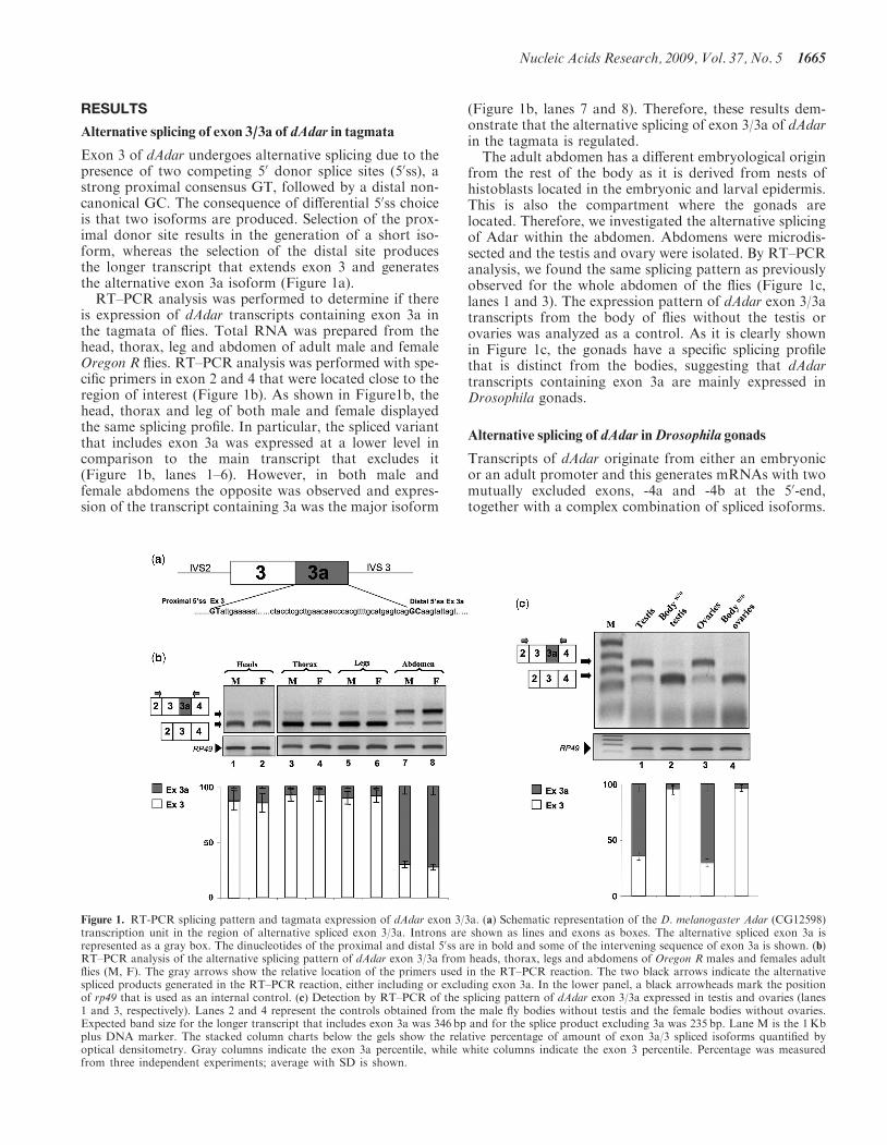
Exon 3 of dAdar undergoes alternative splicing due to thepresence of two competing 50 donor splice sites (50ss), astrong proximal consensus GT, followed by a distal non-canonical GC. The consequence of differential 50ss choiceis that two isoforms are produced. Selection of the prox-imal donor site results in the generation of a short iso-form, whereas the selection of the distal site producesthe longer transcript that extends exon 3 and generatesthe alternative exon 3a isoform (Figure 1a).
RT–PCR analysis was performed to determine if thereis expression of dAdar transcripts containing exon 3a inthe tagmata of flies. Total RNA was prepared from thehead, thorax, leg and abdomen of adult male and femaleOregon R flies. RT–PCR analysis was performed with spe-cific primers in exon 2 and 4 that were located close to theregion of interest (Figure 1b). As shown in Figure1b, thehead, thorax and leg of both male and female displayedthe same splicing profile. In particular, the spliced variantthat includes exon 3a was expressed at a lower level incomparison to the main transcript that excludes it(Figure 1b, lanes 1–6). However, in both male andfemale abdomens the opposite was observed and expres-sion of the transcript containing 3a was the major isoform
(Figure 1b, lanes 7 and 8). Therefore, these results dem-onstrate that the alternative splicing of exon 3/3a of dAdarin the tagmata is regulated.The adult abdomen has a different embryological origin
from the rest of the body as it is derived from nests ofhistoblasts located in the embryonic and larval epidermis.This is also the compartment where the gonads arelocated. Therefore, we investigated the alternative splicingof Adar within the abdomen. Abdomens were microdis-sected and the testis and ovary were isolated. By RT–PCRanalysis, we found the same splicing pattern as previouslyobserved for the whole abdomen of the flies (Figure 1c,lanes 1 and 3). The expression pattern of dAdar exon 3/3atranscripts from the body of flies without the testis orovaries was analyzed as a control. As it is clearly shownin Figure 1c, the gonads have a specific splicing profilethat is distinct from the bodies, suggesting that dAdartranscripts containing exon 3a are mainly expressed inDrosophila gonads.
Alternative splicing of dAdar inDrosophila gonads
Transcripts of dAdar originate from either an embryonicor an adult promoter and this generates mRNAs with twomutually excluded exons, -4a and -4b at the 50-end,together with a complex combination of spliced isoforms.
Figure 1. RT-PCR splicing pattern and tagmata expression of dAdar exon 3/3a. (a) Schematic representation of the D. melanogaster Adar (CG12598)transcription unit in the region of alternative spliced exon 3/3a. Introns are shown as lines and exons as boxes. The alternative spliced exon 3a isrepresented as a gray box. The dinucleotides of the proximal and distal 50ss are in bold and some of the intervening sequence of exon 3a is shown. (b)RT–PCR analysis of the alternative splicing pattern of dAdar exon 3/3a from heads, thorax, legs and abdomens of Oregon R males and females adultflies (M, F). The gray arrows show the relative location of the primers used in the RT–PCR reaction. The two black arrows indicate the alternativespliced products generated in the RT–PCR reaction, either including or excluding exon 3a. In the lower panel, a black arrowheads mark the positionof rp49 that is used as an internal control. (c) Detection by RT–PCR of the splicing pattern of dAdar exon 3/3a expressed in testis and ovaries (lanes1 and 3, respectively). Lanes 2 and 4 represent the controls obtained from the male fly bodies without testis and the female bodies without ovaries.Expected band size for the longer transcript that includes exon 3a was 346 bp and for the splice product excluding 3a was 235 bp. Lane M is the 1Kbplus DNA marker. The stacked column charts below the gels show the relative percentage of amount of exon 3a/3 spliced isoforms quantified byoptical densitometry. Gray columns indicate the exon 3a percentile, while white columns indicate the exon 3 percentile. Percentage was measuredfrom three independent experiments; average with SD is shown.
Nucleic Acids Research, 2009, Vol. 37, No. 5 1665
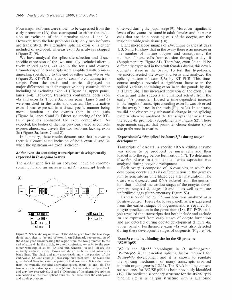
Four major isoforms were shown to be expressed from theearly promoter (4A) that correspond to either the inclu-sion or exclusion of the alternative exons -1 and 3a.However, from the late promoter (4B), only two isoformsare transcribed. By alternative splicing exon -1 is eitherincluded or excluded, whereas exon 3a is always skipped(Figure 2) (9).We have analyzed the splice site selection and tissue-
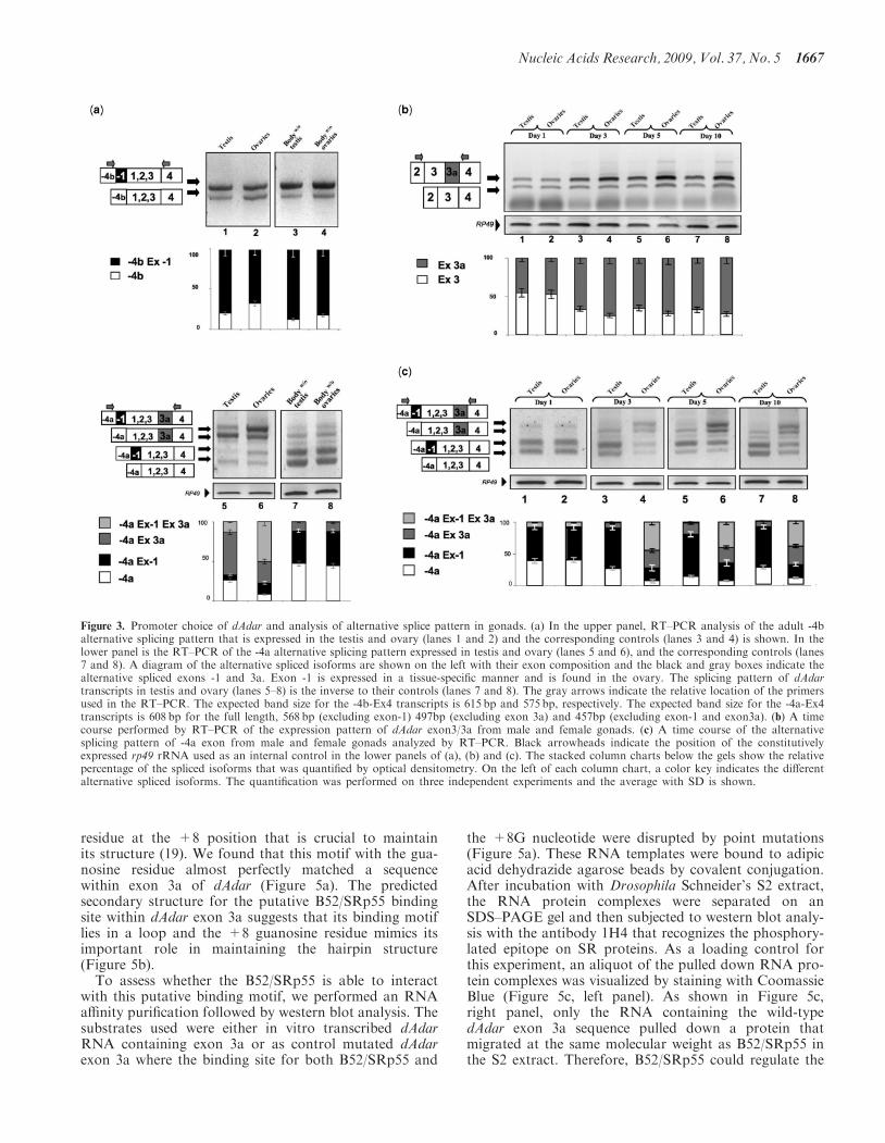
specific expression of the two mutually excluded alterna-tively spliced exons, -4a -4b in the testis and ovaries.Promoter-specific transcripts were amplified with primersannealing specifically to the end of either exon -4b or -4a(Figure 3). RT–PCR analysis of exon -4b containing tran-scripts from the testis and ovaries displayed nomajor differences to their respective body controls eitherincluding or excluding exon -1 (Figure 3a, upper panel,lanes 1–4). However, transcripts containing both exon-4a and exon 3a (Figure 3a, lower panel, lanes 5 and 6)were enriched in the testis and ovaries. The alternativeexon -1 was expressed in a tissue-specific manner beingmore abundant in the ovaries than in the testis(Figure 3a, lanes 5 and 6). Direct sequencing of the RT–PCR products confirmed the exon composition. Asexpected, the bodies of the flies previously used as controlsexpress almost exclusively the two isoforms lacking exon3a (Figure 3a, lanes 7 and 8).In summary, these results demonstrate that in ovaries
there is a coordinated inclusion of both exon -1 and 3awhen the upstream -4a exon is chosen.
dAdar exon -4a containing transcripts are developmentallyexpressed inDrosophila ovaries
The dAdar gene lies in an ecdysone inducible chromo-somal puff and an increase in dAdar transcript levels is
observed during the pupal stage (9). Moreover, significantlevels of ecdysone are found in adult females and the nursecells that are the supporting cells of the oocyte, are themajor steroidogenic tissue (10).
Light microscopy images of Drosophila ovaries at days1, 3, 5 and 10, show that in the ovary there is an increase inthe number of mature oocytes and consequently thenumber of nurse cells from eclosion through to day 10(Supplementary Figure S1). Therefore, exon 3a could bedifferently expressed in the adult females during this devel-opmental stage in the ovary. To test this hypothesis,we microdissected the ovary and testis and analyzed thesplicing pattern of exon 3/3a by RT–PCR. This time-course analysis revealed a significant increase in thespliced variants containing exon 3a in the gonads by day3 (Figure 3b). This increased inclusion of the exon 3a inovaries and testis suggests an enhanced activity from theearly -4A promoter. Indeed a time-dependent increasein the length of transcripts encoding exon 3a was observedin the ovary but not in the testis (Figure 3c). In contrast,we did not observe any substantial change in the splicingpattern when we analyzed the transcripts that arise fromthe adult 4B promoter (Supplementary Figure S2). Theseexperiments suggest that promoter choice dictates splicesite preference in ovaries.
Expression of dAdar spliced isoforms 3/3a during oocytedevelopment
Transcripts of dAdat1, a specific tRNA editing enzymewas shown to be produced by nurse cells and thenloaded into the egg before fertilization (17). To determineif dAdar behaves in a similar manner its expression wasanalyzed during oocyte development.
Each ovary is composed of 16 ovarioles, in which thedeveloping oocyte starts its differentiation in the germar-ium to generate an unfertilized egg after maturation. Theovary was dissected and RNA isolated from the germar-ium that included the earliest stages of the oocytes devel-opment; stages 4–8, stages 10 and 11 as well as matureunfertilized eggs (Supplementary Figure S3).
Expression of the Egalitarian gene was analyzed as apositive control (Figure 4a, lower panel), as it is expressedfrom the earliest stages of oogenesis and is required foroocyte specification in the germarium (18). RT–PCR anal-ysis revealed that transcripts that both include and exclude3a are expressed from early stages of oocyte formationand are detected during oocyte development (Figure 4a,upper panel). Furthermore exon -4a was also detectedduring these development stages of oogenesis (Figure 4b).
Exon 3a contains a binding site for the SR proteinsB52/SRp55
B52 is the SRp55 homologue in D. melanogaster.B52/SRp55 is an essential splicing factor required forDrosophila development and it is known to regulatethe splicing mechanism of many transcripts involvedin brain organogenesis (12,13). The RNA binding consen-sus sequence for B52/SRp55 has been previously identified(19). The predicted secondary structure for the B52/SRp55binding site is a hairpin structure with a guanosine
Figure 2. Schematic organization of the dAdar gene from the transcrip-tional start sites to the end of exon 4. (a) Schematic representation ofthe dAdar gene encompassing the region from the two promoter to theend of exon 4. In the article, to avoid confusion, we refer to the pro-moter with capital letters (4A and 4B), whereas -4a and -4b are themutually excluded exons. Exons are shown as boxes and introns asblack lines. The black and gray arrowheads mark the position of theembryonic (4A) and adult (4B) transcriptional start sites. The black andgray dashed lines indicate the pattern of alternative splicing that arisefrom the mutually excluded alternative spliced exons -4a and -4b. Thetwo other alternative spliced exon (-1 and 3a) are indicated by a blackand gray box respectively. (b and c) Diagrams of the alternative splicingcomposition of the main spliced variants that arise from the embryonicand adult promoters.
1666 Nucleic Acids Research, 2009, Vol. 37, No. 5
residue at the +8 position that is crucial to maintainits structure (19). We found that this motif with the gua-nosine residue almost perfectly matched a sequencewithin exon 3a of dAdar (Figure 5a). The predictedsecondary structure for the putative B52/SRp55 bindingsite within dAdar exon 3a suggests that its binding motiflies in a loop and the +8 guanosine residue mimics itsimportant role in maintaining the hairpin structure(Figure 5b).
To assess whether the B52/SRp55 is able to interactwith this putative binding motif, we performed an RNAaffinity purification followed by western blot analysis. Thesubstrates used were either in vitro transcribed dAdarRNA containing exon 3a or as control mutated dAdarexon 3a where the binding site for both B52/SRp55 and
the +8G nucleotide were disrupted by point mutations(Figure 5a). These RNA templates were bound to adipicacid dehydrazide agarose beads by covalent conjugation.After incubation with Drosophila Schneider’s S2 extract,the RNA protein complexes were separated on anSDS–PAGE gel and then subjected to western blot analy-sis with the antibody 1H4 that recognizes the phosphory-lated epitope on SR proteins. As a loading control forthis experiment, an aliquot of the pulled down RNA pro-tein complexes was visualized by staining with CoomassieBlue (Figure 5c, left panel). As shown in Figure 5c,right panel, only the RNA containing the wild-typedAdar exon 3a sequence pulled down a protein thatmigrated at the same molecular weight as B52/SRp55 inthe S2 extract. Therefore, B52/SRp55 could regulate the
Figure 3. Promoter choice of dAdar and analysis of alternative splice pattern in gonads. (a) In the upper panel, RT–PCR analysis of the adult -4balternative splicing pattern that is expressed in the testis and ovary (lanes 1 and 2) and the corresponding controls (lanes 3 and 4) is shown. In thelower panel is the RT–PCR of the -4a alternative splicing pattern expressed in testis and ovary (lanes 5 and 6), and the corresponding controls (lanes7 and 8). A diagram of the alternative spliced isoforms are shown on the left with their exon composition and the black and gray boxes indicate thealternative spliced exons -1 and 3a. Exon -1 is expressed in a tissue-specific manner and is found in the ovary. The splicing pattern of dAdartranscripts in testis and ovary (lanes 5–8) is the inverse to their controls (lanes 7 and 8). The gray arrows indicate the relative location of the primersused in the RT–PCR. The expected band size for the -4b-Ex4 transcripts is 615 bp and 575 bp, respectively. The expected band size for the -4a-Ex4transcripts is 608 bp for the full length, 568 bp (excluding exon-1) 497bp (excluding exon 3a) and 457bp (excluding exon-1 and exon3a). (b) A timecourse performed by RT–PCR of the expression pattern of dAdar exon3/3a from male and female gonads. (c) A time course of the alternativesplicing pattern of -4a exon from male and female gonads analyzed by RT–PCR. Black arrowheads indicate the position of the constitutivelyexpressed rp49 rRNA used as an internal control in the lower panels of (a), (b) and (c). The stacked column charts below the gels show the relativepercentage of the spliced isoforms that was quantified by optical densitometry. On the left of each column chart, a color key indicates the differentalternative spliced isoforms. The quantification was performed on three independent experiments and the average with SD is shown.
Nucleic Acids Research, 2009, Vol. 37, No. 5 1667
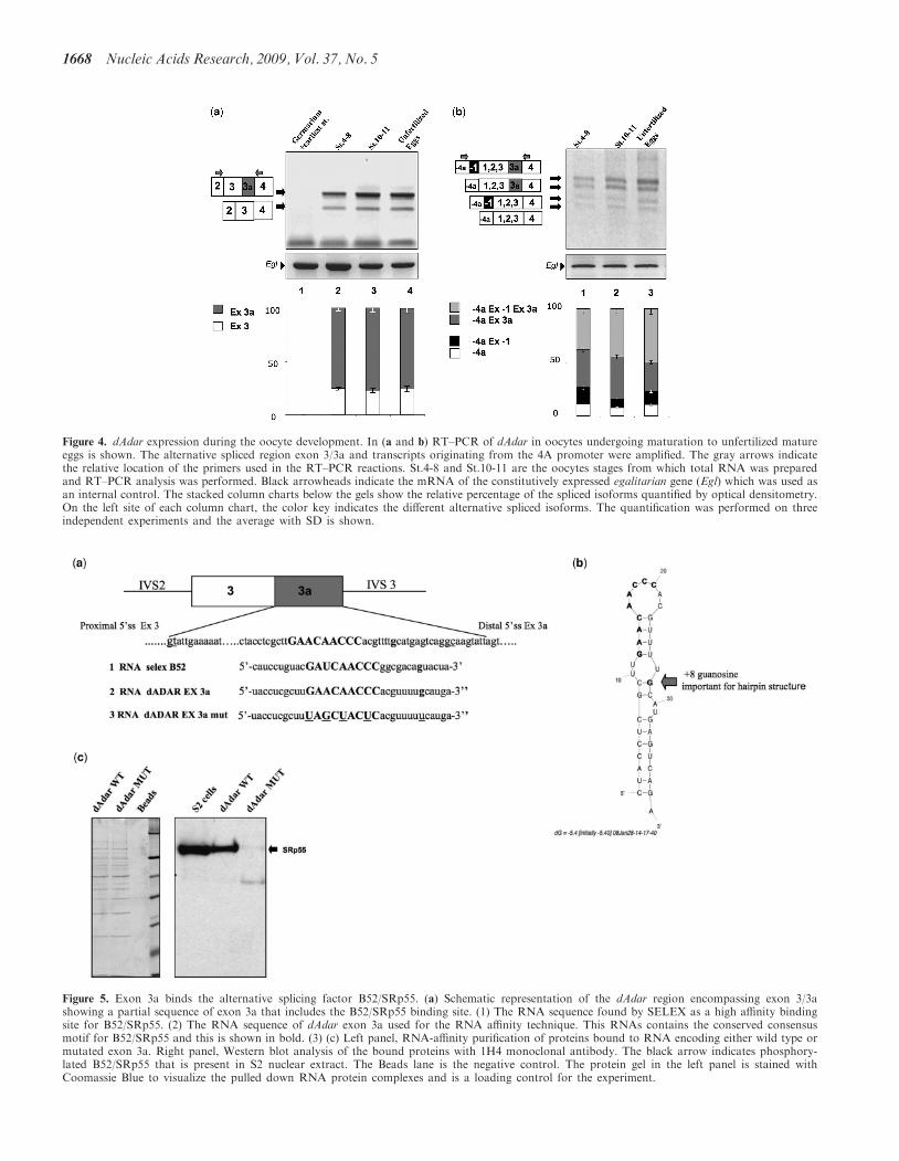
Figure 5. Exon 3a binds the alternative splicing factor B52/SRp55. (a) Schematic representation of the dAdar region encompassing exon 3/3ashowing a partial sequence of exon 3a that includes the B52/SRp55 binding site. (1) The RNA sequence found by SELEX as a high affinity bindingsite for B52/SRp55. (2) The RNA sequence of dAdar exon 3a used for the RNA affinity technique. This RNAs contains the conserved consensusmotif for B52/SRp55 and this is shown in bold. (3) (c) Left panel, RNA-affinity purification of proteins bound to RNA encoding either wild type ormutated exon 3a. Right panel, Western blot analysis of the bound proteins with 1H4 monoclonal antibody. The black arrow indicates phosphory-lated B52/SRp55 that is present in S2 nuclear extract. The Beads lane is the negative control. The protein gel in the left panel is stained withCoomassie Blue to visualize the pulled down RNA protein complexes and is a loading control for the experiment.
Figure 4. dAdar expression during the oocyte development. In (a and b) RT–PCR of dAdar in oocytes undergoing maturation to unfertilized matureeggs is shown. The alternative spliced region exon 3/3a and transcripts originating from the 4A promoter were amplified. The gray arrows indicatethe relative location of the primers used in the RT–PCR reactions. St.4-8 and St.10-11 are the oocytes stages from which total RNA was preparedand RT–PCR analysis was performed. Black arrowheads indicate the mRNA of the constitutively expressed egalitarian gene (Egl) which was used asan internal control. The stacked column charts below the gels show the relative percentage of the spliced isoforms quantified by optical densitometry.On the left site of each column chart, the color key indicates the different alternative spliced isoforms. The quantification was performed on threeindependent experiments and the average with SD is shown.
1668 Nucleic Acids Research, 2009, Vol. 37, No. 5
alternative splicing of exon 3a in Drosophila by bindingwithin exon 3a.
B52/SRp55 is required for the dAdar exon3/3a splicingregulation
To demonstrate that B52/SRp55 is important for the alter-native splicing of dAdar exon 3/3a, we took advantage ofdouble-stranded RNA-mediated interference for B52/SRp55 proteins in a Drosophila cell line (20).
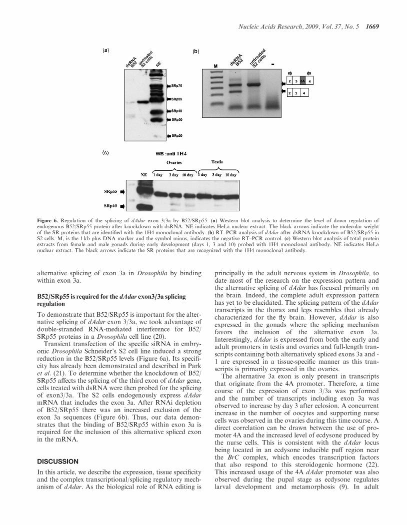
Transient transfection of the specific siRNA in embry-onic Drosophila Schneider’s S2 cell line induced a strongreduction in the B52/SRp55 levels (Figure 6a). Its specifi-city has already been demonstrated and described in Parket al. (21). To determine whether the knockdown of B52/SRp55 affects the splicing of the third exon of dAdar gene,cells treated with dsRNA were then probed for the splicingof exon3/3a. The S2 cells endogenously express dAdarmRNA that includes the exon 3a. After RNAi depletionof B52/SRp55 there was an increased exclusion of theexon 3a sequences (Figure 6b). Thus, our data demon-strates that the binding of B52/SRp55 within exon 3a isrequired for the inclusion of this alternative spliced exonin the mRNA.
DISCUSSION
In this article, we describe the expression, tissue specificityand the complex transcriptional/splicing regulatory mech-anism of dAdar. As the biological role of RNA editing is
principally in the adult nervous system in Drosophila, todate most of the research on the expression pattern andthe alternative splicing of dAdar has focused primarily onthe brain. Indeed, the complete adult expression patternhas yet to be elucidated. The splicing pattern of the dAdartranscripts in the thorax and legs resembles that alreadycharacterized for the fly brain. However, dAdar is alsoexpressed in the gonads where the splicing mechanismfavors the inclusion of the alternative exon 3a.Interestingly, dAdar is expressed from both the early andadult promoters in testis and ovaries and full-length tran-scripts containing both alternatively spliced exons 3a and -1 are expressed in a tissue-specific manner as this tran-scripts is primarily expressed in the ovaries.The alternative 3a exon is only present in transcripts
that originate from the 4A promoter. Therefore, a timecourse of the expression of exon 3/3a was performedand the number of transcripts including exon 3a wasobserved to increase by day 3 after eclosion. A concurrentincrease in the number of oocytes and supporting nursecells was observed in the ovaries during this time course. Adirect correlation can be drawn between the use of pro-moter 4A and the increased level of ecdysone produced bythe nurse cells. This is consistent with the dAdar locusbeing located in an ecdysone inducible puff region nearthe BrC complex, which encodes transcription factorsthat also respond to this steroidogenic hormone (22).This increased usage of the 4A dAdar promoter was alsoobserved during the pupal stage as ecdysone regulateslarval development and metamorphosis (9). In adult
Figure 6. Regulation of the splicing of dAdar exon 3/3a by B52/SRp55. (a) Western blot analysis to determine the level of down regulation ofendogenous B52/SRp55 protein after knockdown with dsRNA. NE indicates HeLa nuclear extract. The black arrows indicate the molecular weightof the SR proteins that are identified with the 1H4 monoclonal antibody. (b) RT–PCR analysis of dAdar after dsRNA knockdown of B52/SRp55 inS2 cells. M, is the 1 kb plus DNA marker and the symbol minus, indicates the negative RT–PCR control. (c) Western blot analysis of total proteinextracts from female and male gonads during early development (days 1, 3 and 10) probed with 1H4 monoclonal antibody. NE indicates HeLanuclear extract. The black arrows indicate the SR proteins that are recognized with the 1H4 monoclonal antibody.
Nucleic Acids Research, 2009, Vol. 37, No. 5 1669
females, ecdysone is also produced in the nurse cells in theovary. Therefore, we propose that enhanced activity fromthe 4A promoter is a consequence of an increased level ofthe ecdysone produced by these nurse cells.As transcripts of a related protein dAdat (the Drosophila
t-RNA editing enzyme) were shown to be produced by thenurse cells and loaded into the egg before the fertilization(17), we wanted to determine if dAdar could be detectedduring oogenesis. Full-length transcripts of dAdar weredetected from early stages in oocyte development throughto unfertilized mature eggs. One question that needs to beaddress is whether the predominant transcripts expressedin gonads are translated into functional proteins.However, this will remain unanswered until betterdADAR antibodies are available. In Drosophila, theoocyte nucleus has been shown to be transcriptionallyinactive during most of stages of oogenesis (23). Theembryo receives maternally provided transcriptionalinput from the two other cell sources in the oocyte: thenurse cells and the somatic follicle cells. Therefore, in thefuture it will be interesting to understand the contributionto dAdar expression both from the nurse cells and thesomatic follicle cells.Recently, additional roles for editing enzymes have been
proposed in relation to Alu elements, siRNA and miRNAfunction. ADARs can edit noncoding RNAs and canaffect the processing of miRNAs such as pri-miR-142(24) or affect the seed sequence as occurs in miR-376(25), so that the miRNA is redirected to silence a differentset of transcripts. Therefore, it is interesting to considerthat dADAR could interact with noncoding RNAs andretarget the silencing machinery to transcripts that arerequired to be inactivated during the oocyte development.To determine which splicing factor could regulate the
alternative splicing of exon 3/3a, we analyzed the nucleo-tide sequence of exon 3a. The B52/SRp55 bindingsequence obtained by SELEX (19) is very similar to thesequence we found in exon 3a that has a hairpin configu-ration with the protein binding site exposed in a loop andthe guanosine residue that is critical for RNA structure.We demonstrated that B52/SRp55 specifically bindswithin exon 3a. Depletion of B52/SRp55 in DrosophilaS2 cell line by RNAi demonstrates that this splicingfactor controls the inclusion of the exon 3a as its down-regulation promotes the exclusion of exon 3a. However,we cannot conclude that the increase level of inclusion ofexon 3a present in dAdar transcripts in the gonads is dueto an increase in B52/SRp55 protein. Indeed, in testis andovary the expression level of B52/SRp55 does not changeduring the developmental stages that have been analyzed(Figure 6c). This result suggests that in Drosophila gonadsadditional splicing factors participate in the complex splic-ing mechanism that regulates the alternative splicing ofexon 3a. These results confirm previous reports thatB52/SRp55 together with other splicing factors participatein the recognition of the distal 50ss (20). B52/SRp55 waspreviously been shown to control eye development and toregulate the splicing of many transcripts involved in brainorganogenesis (12,13). Here, we describe a novel functionfor B52/SRp55 to regulate the splicing of the exon3/3a inAdar transcripts in testis and ovaries (26).
The inclusion or exclusion of exon 3 in dAdar tran-scripts was demonstrated to affect both the binding andediting activity of the protein (5). In vivo no transcriptshave been isolated that contain exon 3a and are edited inthe deaminase domain. However, in vitro transcriptsincluding the 3a isoform were shown to have reducedself-editing in exon 7 that lies within the deaminasedomain. In addition, the exon 3a containing transcriptintroduces an additional 38 amino acids betweendsRBM 1 and dsRBM 2 that generates two distinctenzymes. The spacing between the binding domains withexon 3a is very similar to the distance between the bindingdomains of ADAR2, whereas when this exon is excludedthe distance is reminiscence of the distance between thedsRNA binding domains found in ADAR1. This conser-vation in the spacing of the dsRNA binding domains sug-gests that it is important for function. It has also beenshown that the different isoforms of dAdar can form het-erodimers and this can change its binding capability on thedsRNA target (11). Therefore, in the gonads different iso-forms of dAdar with different editing activity could specif-ically edit transcripts that are expressed there and as aconsequence modify the properties of the encodedproteins.
In ovaries, the inclusion of the alternative exon-1 resultsin a protein being expressed that has an additional 12amino acids at the amino-terminal. This exon introducesan alternative in-frame translational start site that is pre-ferentially transcribed from the early promoter. It has pre-viously been shown that the use of alternativetranslational starts can regulate several cytokine familyproteins whose expression level has to be fine tuned tomaintain their physiological activity (16,27). Therefore,the question arises could this be a form of regulation ofdAdar in the ovary that is in addition to autoediting?This regulation would represent a coordinated transla-tional–transcriptional control that regulates alternativesplicing and therefore the activity of the editing enzymein the ovary.
New editing targets have recently been found (28).Several of these novel edited transcripts were shown tobe involved in ion homeostasis, cytoskeletal componentsand signal transduction pathway and they may be editedin the gonads and have a biological role there.
In the gonads, development continues throughout thelifetime of the animal and the pathway involves the coor-dinated division and differentiation of a large number ofcells. This site of extensive adult development probablyrequires a fine tune regulation of dAdar.
It is possible that the presence of different spliced iso-forms of dAdar, may reflect differences in dAdar substratespecificity. Indeed, the tissue-specific alternative splicingof dAdar in Drosophila gonads may expand the repertoireof ADAR targets beyond the brain to Drosophilagonads (29).
SUPPLEMENTARY DATA
Supplementary Data are available at NAR Online.
1670 Nucleic Acids Research, 2009, Vol. 37, No. 5
ACKNOWLEDGEMENTS
We would like to thank Bret S.E. Heale for helpful dis-cussion and comments on the article. We also thank LisaSessi for proofreading the article.
FUNDING
Telethon Onlus Foundation, Italy (GGP06147); EuropeanCommunity Grant (EURASNET-LSHG-CT-2005-518238); MRC (U.1275.01.005.00001.01). Funding foropen access charge: ICGEB core funding.
Conflict of interest statement. None declared.
REFERENCES
1. Bass,B.L. (2002) RNA editing by adenosine deaminases that act onRNA. Annu. Rev. Biochem., 71, 817–846.
2. Flomen,R., Knight,J., Sham,P., Kerwin,R. and Makoff,A. (2004)Evidence that RNA editing modulates splice site selection in the5-HT2C receptor gene. Nucleic Acids Res., 32, 2113–2122.
3. Laurencikiene,J., Kallman,A.M., Fong,N., Bentley,D.L. andOhman,M. (2006) RNA editing and alternative splicing: theimportance of co-transcriptional coordination. EMBO Rep., 7,303–307.
4. Rueter,S.M., Dawson,T.R. and Emeson,R.B. (1999) Regulation ofalternative splicing by RNA editing. Nature, 399, 75–80.
5. Keegan,L.P., Brindle,J., Gallo,A., Leroy,A., Reenan,R.A. andO’Connell,M.A. (2005) Tuning of RNA editing by ADAR isrequired in Drosophila. EMBO J., 24, 2183–2193.
6. Tonkin,L.A., Saccomanno,L., Morse,D.P., Brodigan,T., Krause,M.and Bass,B.L. (2002) RNA editing by ADARs is important fornormal behavior in Caenorhabditis elegans. EMBO J., 21,6025–6035.
7. Palladino,M.J., Keegan,L.P., O’Connell,M.A. and Reenan,R.A.(2000) A-to-I pre-mRNA editing in Drosophila is primarilyinvolved in adult nervous system function and integrity. Cell, 102,437–449.
8. Wang,Q., Khillan,J., Gadue,P. and Nishikura,K. (2000)Requirement of the RNA editing deaminase ADAR1 gene forembryonic erythropoiesis. Science, 290, 1765–1768.
9. Palladino,M.J., Keegan,L.P., O’Connell,M.A. and Reenan,R.A.(2000) dADAR, a Drosophila double-stranded RNA-specificadenosine deaminase is highly developmentally regulated and isitself a target for RNA editing. RNA, 6, 1004–1018.
10. Soller,M., Bownes,M. and Kubli,E. (1999) Control of oocytematuration in sexually mature Drosophila females. Dev. Biol., 208,337–351.
11. Gallo,A., Keegan,L.P., Ring,G.M. and O’Connell,M.A. (2003)An ADAR that edits transcripts encoding ion channel subunitsfunctions as a dimer. EMBO J., 22, 3421–3430.
12. Fic,W., Juge,F., Soret,J. and Tazi,J. (2007) Eye development underthe control of SRp55/B52-mediated alternative splicing of eyeless.PLoS ONE, 2, e253.
13. Gabut,M., Dejardin,J., Tazi,J. and Soret,J. (2007) The SR familyproteins B52 and dASF/SF2 modulate development of the
Drosophila visual system by regulating specific RNA targets.Mol. Cell Biol., 27, 3087–3097.
14. Hoopengardner,B., Bhalla,T., Staber,C. and Reenan,R. (2003)Nervous system targets of RNA editing identified by comparativegenomics. Science, 301, 832–836.
15. Romano,M., Marcucci,R., Buratti,E., Ayala,Y.M., Sebastio,G. andBaralle,F.E. (2002) Regulation of 3’ splice site selection in the844ins68 polymorphism of the cystathionine Beta -synthase gene.J. Biol. Chem., 277, 43821–43829.
16. Marcucci,R., Baralle,F.E. and Romano,M. (2007) Complex splicingcontrol of the human Thrombopoietin gene by intronic G runs.Nucleic Acids Res., 35, 132–142.
17. Keegan,L.P., Gerber,A.P., Brindle,J., Leemans,R., Gallo,A.,Keller,W. and O’Connell,M.A. (2000) The properties of atRNA-specific adenosine deaminase from Drosophila melanogastersupport an evolutionary link between pre-mRNA editing and tRNAmodification. Mol. Cell Biol., 20, 825–833.
18. Mach,J.M. and Lehmann,R. (1997) An Egalitarian-BicaudalDcomplex is essential for oocyte specification and axis determinationin Drosophila. Genes Dev., 11, 423–435.
19. Shi,H., Hoffman,B.E. and Lis,J.T. (1997) A specific RNA hairpinloop structure binds the RNA recognition motifs of the DrosophilaSR protein B52. Mol. Cell Biol., 17, 2649–2657.
20. Park,J.W. and Graveley,B.R. (2005) Use of RNA interference todissect the roles of trans-acting factors in alternative pre-mRNAsplicing. Methods, 37, 341–344.
21. Park,J.W., Parisky,K., Celotto,A.M., Reenan,R.A. andGraveley,B.R. (2004) Identification of alternative splicing regulatorsby RNA interference in Drosophila. Proc. Natl Acad. Sci. USA,101, 15974–15979.
22. Wilson,T.G., Yerushalmi,Y., Donnell,D.M. and Restifo,L.L.(2006) Interaction between hormonal signaling pathways inDrosophila melanogaster as revealed by genetic interactionbetween methoprene-tolerant and broad-complex. Genetics, 172,253–264.
23. King,R.C. and Burnett,R.G. (1959) Autoradiographic study ofuptake of tritiated glycine, thymidine, and uridine by fruit flyovaries. Science, 129, 1674–1675.
24. Yang,W., Chendrimada,T.P., Wang,Q., Higuchi,M., Seeburg,P.H.,Shiekhattar,R. and Nishikura,K. (2006) Modulation of microRNAprocessing and expression through RNA editing by ADAR deami-nases. Nat. Struct. Mol. Biol., 13, 13–21.
25. Kawahara,Y., Zinshteyn,B., Sethupathy,P., Iizasa,H.,Hatzigeorgiou,A.G. and Nishikura,K. (2007) Redirection of silenc-ing targets by adenosine-to-inosine editing of miRNAs. Science,315, 1137–1140.
26. Kraus,M.E. and Lis,J.T. (1994) The concentration of B52, anessential splicing factor and regulator of splice site choicein vitro, is critical for Drosophila development. Mol. Cell Biol., 14,5360–5370.
27. Marcucci,R. and Romano,M. (2008) Thrombopoietin and itssplicing variants: Structure and functions in thrombopoiesis andbeyond. Biochim. Biophys. Acta., 1782, 427–432.
28. Stapleton,M., Carlson,J.W. and Celniker,S.E. (2006) RNA editingin Drosophila melanogaster: new targets and functional conse-quences. RNA, 12, 1922–1932.
29. Keegan,L.P., Gallo,A. and O’Connell,M.A. (2001) The many rolesof an RNA editor. Nat. Rev. Genet., 2, 869–878.
30. Zuker,M. (1989) Computer prediction of RNA structure. MethodsEnzymol., 180, 262–288.
Nucleic Acids Research, 2009, Vol. 37, No. 5 1671