Matrix Stiffness Affects Endocytic Uptake of MK2-Inhibitor Peptides
9
Matrix Stiffness Affects Endocytic Uptake of MK2- Inhibitor Peptides Jamie L. Brugnano, Alyssa Panitch* Weldon School of Biomedical Engineering, Purdue University, West Lafayette, Indiana, United States of America Abstract In this study, the role of substrate stiffness on the endocytic uptake of a cell-penetrating peptide was investigated. The cell- penetrating peptide, an inhibitor of mitogen-activated protein kinase activated protein kinase II (MK2), enters a primary mesothelial cell line predominantly through caveolae. Using tissue culture polystyrene and polyacrylamide gels of varying stiffness for cell culture, and flow cytometry quantification and enzyme-linked immunoassays (ELISA) for uptake assays, we showed that the amount of uptake of the peptide is increased on soft substrates. Further, peptide uptake per cell increased at lower cell density. The improved uptake seen on soft substrates in vitro better correlates with in vivo functional studies where 10–100 mM concentrations of the MK2 inhibitor cell penetrating peptide demonstrated functional activity in several disease models. Additional characterization showed actin polymerization did not affect uptake, while microtubule polymerization had a profound effect on uptake. This work demonstrates that cell culture substrate stiffness can play a role in endocytic uptake, and may be an important consideration to improve correlations between in vitro and in vivo drug efficacy. Citation: Brugnano JL, Panitch A (2014) Matrix Stiffness Affects Endocytic Uptake of MK2-Inhibitor Peptides. PLoS ONE 9(1): e84821. doi:10.1371/ journal.pone.0084821 Editor: Adam J. Engler, University of California, San Diego, United States of America Received August 14, 2013; Accepted November 21, 2013; Published January 6, 2014 Copyright: ß 2014 Brugnano, Panitch. This is an open-access article distributed under the terms of the Creative Commons Attribution License, which permits unrestricted use, distribution, and reproduction in any medium, provided the original author and source are credited. Funding: This work was supported in part by the National Institutes of Health grant R01HL106792. JLB was support by an National Science Foundation graduate research fellowship. The funders had no role in study design, data collection and analysis, decision to publish, or preparation of the manuscript. Competing Interests: Alyssa Panitch is a founder of Moerae Matrix, Inc. and holds greater than 5% interest in the company. Moerae Matrix Inc. holds and exclusive license to the cell-penetrating peptide described in this work. This does not alter the authors’ adherence to all the PLOS ONE policies on sharing data and materials. The following patent has been issued on the composition of the YARA peptide described herein (MK2 inhibitor peptide/HSP27 kinase inhibitor). ‘‘Polypeptide inhibitors of HSP27 kinase and uses therefor.’’ United States Patent 8,536,303. The patent also discloses medical uses of the peptide. The in vitro delivery of the peptide, as describe herein, is not subject to any patents or patent applications. * E-mail: [email protected] Introduction Matrix stiffness is an important regulator of cell behavior [1]. Stiffness has been shown to affect cell morphology and spreading [2,3], proliferation [4], migration [5], apoptosis rate [4,6], and differentiation [7,8]. However, most cell studies are performed on tissue culture plastic, which largely fails to replicate the mechanics and microenvironment that cells experience in vivo. Tissue culture plastic is commonly cited as having an elastic modulus of approximately 1 GPa, whereas tissues in the body are less than 100 kPa, with brain having an elasticity less than 1 kPa, muscle around 10 kPa, and bone around 100 kPa [7]. The effects of matrix stiffness are typically evaluated by analyzing cell behavior in different gel systems. Stiffness or elasticity can be varied by simply changing the crosslinking density. Several different hydrogel systems have been investigated including polyacrylamide gels [9–11], alginate [12], collagen [13], matrigel [14], chitosan [15], and hyaluronic acid [16]. Because substrate stiffness regulates so many cellular functions, we wanted to investigate its role in the uptake of cell-penetrating peptides. Although the exact mechanism of cell-penetrating peptide uptake is still debated, investigators generally agree that uptake occurs via one or more of the endocytic pathways: clathrin-mediated endocytosis, caveolae-mediated endocytosis, and macropinocytosis [17–20], or through membrane destabilization or formation of transient pores [21–23]. Our lab has designed and reported on a family of peptide inhibitors of mitogen kinase activated protein kinase- activated protein kinase 2 (MAPKAPK2 or MK2), a kinase important in regulating inflammation through the regulation of proinflammatory cytokines [24–26]. These inhibitors consist of a cell-penetrating peptide domain for intracellular delivery and a therapeutic domain that inhibits MK2. Recently, we demonstrated that the peptide variant YARAAARQARAKALARQLGVAA (YARA) was taken up primarily through caveolae-mediated endocytosis in mesothelial cells [27]. However, when comparing between data obtained from in vitro cell and in vivo animal models, we observed an unusual effect: concentrations of the YARA MK2 inhibitor peptide required for efficacy in cells ranged from 1000–3000 mM [24,26]; however, the concentration re- quired for efficacy in animal models was ten to one hundred-fold less, in the range of 10–100 mM [25,28–31]. This phenomenon opposes what is normally observed in the pharmaceutical industry, as drug concentrations must usually increase to demonstrate efficacy when moving from cell culture to animal models due to metabolism and non-uniform distribution within the body. We hypothesized that the discrepancy observed in peptide concentra- tion required to achieve efficacy in studies in vitro as compared to studies in vivo was due to the unrealistic stiffness of tissue culture polystyrene. Using a technique pioneered by Pelham and Wang [11] and refined by others [10], the role of substrate stiffness in the uptake of the MK2-inhibitor peptides was investigated. Polyacrylamide gels were chosen as the model substrate for this experiment PLOS ONE | www.plosone.org 1 January 2014 | Volume 9 | Issue 1 | e84821
Transcript of Matrix Stiffness Affects Endocytic Uptake of MK2-Inhibitor Peptides
Matrix Stiffness Affects Endocytic Uptake of MK2-Inhibitor PeptidesJamie L. Brugnano, Alyssa Panitch*
Weldon School of Biomedical Engineering, Purdue University, West Lafayette, Indiana, United States of America
Abstract
In this study, the role of substrate stiffness on the endocytic uptake of a cell-penetrating peptide was investigated. The cell-penetrating peptide, an inhibitor of mitogen-activated protein kinase activated protein kinase II (MK2), enters a primarymesothelial cell line predominantly through caveolae. Using tissue culture polystyrene and polyacrylamide gels of varyingstiffness for cell culture, and flow cytometry quantification and enzyme-linked immunoassays (ELISA) for uptake assays, weshowed that the amount of uptake of the peptide is increased on soft substrates. Further, peptide uptake per cell increasedat lower cell density. The improved uptake seen on soft substrates in vitro better correlates with in vivo functional studieswhere 10–100 mM concentrations of the MK2 inhibitor cell penetrating peptide demonstrated functional activity in severaldisease models. Additional characterization showed actin polymerization did not affect uptake, while microtubulepolymerization had a profound effect on uptake. This work demonstrates that cell culture substrate stiffness can play a rolein endocytic uptake, and may be an important consideration to improve correlations between in vitro and in vivo drugefficacy.
Citation: Brugnano JL, Panitch A (2014) Matrix Stiffness Affects Endocytic Uptake of MK2-Inhibitor Peptides. PLoS ONE 9(1): e84821. doi:10.1371/journal.pone.0084821
Editor: Adam J. Engler, University of California, San Diego, United States of America
Received August 14, 2013; Accepted November 21, 2013; Published January 6, 2014
Copyright: � 2014 Brugnano, Panitch. This is an open-access article distributed under the terms of the Creative Commons Attribution License, which permitsunrestricted use, distribution, and reproduction in any medium, provided the original author and source are credited.
Funding: This work was supported in part by the National Institutes of Health grant R01HL106792. JLB was support by an National Science Foundation graduateresearch fellowship. The funders had no role in study design, data collection and analysis, decision to publish, or preparation of the manuscript.
Competing Interests: Alyssa Panitch is a founder of Moerae Matrix, Inc. and holds greater than 5% interest in the company. Moerae Matrix Inc. holds andexclusive license to the cell-penetrating peptide described in this work. This does not alter the authors’ adherence to all the PLOS ONE policies on sharing dataand materials. The following patent has been issued on the composition of the YARA peptide described herein (MK2 inhibitor peptide/HSP27 kinase inhibitor).‘‘Polypeptide inhibitors of HSP27 kinase and uses therefor.’’ United States Patent 8,536,303. The patent also discloses medical uses of the peptide. The in vitrodelivery of the peptide, as describe herein, is not subject to any patents or patent applications.
* E-mail: [email protected]
Introduction
Matrix stiffness is an important regulator of cell behavior [1].
Stiffness has been shown to affect cell morphology and spreading
[2,3], proliferation [4], migration [5], apoptosis rate [4,6], and
differentiation [7,8]. However, most cell studies are performed on
tissue culture plastic, which largely fails to replicate the mechanics
and microenvironment that cells experience in vivo. Tissue culture
plastic is commonly cited as having an elastic modulus of
approximately 1 GPa, whereas tissues in the body are less than
100 kPa, with brain having an elasticity less than 1 kPa, muscle
around 10 kPa, and bone around 100 kPa [7].
The effects of matrix stiffness are typically evaluated by
analyzing cell behavior in different gel systems. Stiffness or
elasticity can be varied by simply changing the crosslinking
density. Several different hydrogel systems have been investigated
including polyacrylamide gels [9–11], alginate [12], collagen [13],
matrigel [14], chitosan [15], and hyaluronic acid [16]. Because
substrate stiffness regulates so many cellular functions, we wanted
to investigate its role in the uptake of cell-penetrating peptides.
Although the exact mechanism of cell-penetrating peptide uptake
is still debated, investigators generally agree that uptake occurs via
one or more of the endocytic pathways: clathrin-mediated
endocytosis, caveolae-mediated endocytosis, and macropinocytosis
[17–20], or through membrane destabilization or formation of
transient pores [21–23]. Our lab has designed and reported on a
family of peptide inhibitors of mitogen kinase activated protein
kinase- activated protein kinase 2 (MAPKAPK2 or MK2), a kinase
important in regulating inflammation through the regulation of
proinflammatory cytokines [24–26]. These inhibitors consist of a
cell-penetrating peptide domain for intracellular delivery and a
therapeutic domain that inhibits MK2. Recently, we demonstrated
that the peptide variant YARAAARQARAKALARQLGVAA
(YARA) was taken up primarily through caveolae-mediated
endocytosis in mesothelial cells [27]. However, when comparing
between data obtained from in vitro cell and in vivo animal
models, we observed an unusual effect: concentrations of the
YARA MK2 inhibitor peptide required for efficacy in cells ranged
from 1000–3000 mM [24,26]; however, the concentration re-
quired for efficacy in animal models was ten to one hundred-fold
less, in the range of 10–100 mM [25,28–31]. This phenomenon
opposes what is normally observed in the pharmaceutical industry,
as drug concentrations must usually increase to demonstrate
efficacy when moving from cell culture to animal models due to
metabolism and non-uniform distribution within the body. We
hypothesized that the discrepancy observed in peptide concentra-
tion required to achieve efficacy in studies in vitro as compared to
studies in vivo was due to the unrealistic stiffness of tissue culture
polystyrene.
Using a technique pioneered by Pelham and Wang [11] and
refined by others [10], the role of substrate stiffness in the uptake
of the MK2-inhibitor peptides was investigated. Polyacrylamide
gels were chosen as the model substrate for this experiment
PLOS ONE | www.plosone.org 1 January 2014 | Volume 9 | Issue 1 | e84821
because stiffness can be modulated by changing the percentage of
bisacrylamide crosslinker within the system. Additionally, poly-
acrylamide gels are clear, non-fluorescent, and have the ability to
covalently link proteins to the surface. Unlike most other systems,
polyacrylamide gels are inert to protein adsorption and cell
adhesion; thus, cellular adhesion can be controlled by functiona-
lizing the gels with an extracellular matrix (ECM) protein. The
adhesion of cells to the gel is then solely attributed to cellular
binding to the ECM protein.
Methods
2.1 Polyacrylamide Gel Substrate PreparationSubstrates of different stiffness were prepared on 18 mm circle
glass coverslips (VWR) following a modified protocol from Tse and
Engler [10]. A uniform film of sodium hydroxide (Sigma) was
formed on the coverslips by evaporation of 600 ml of 0.1 M
sodium hydroxide in a 60uC oven. In the case that uniform
coverage was not achieved, 600 ml of water was added to the
coverslips and evaporated in a 60uC oven. The coverslips were
reacted with 200 ml of (3-aminopropyl) triethoxysilane (Sigma) for
five minutes at room temperature under a nitrogen tent, followed
by extensive washing with water. The coverslips were then
incubated for 30 minutes at room temperature with 0.5%
glutaraldehyde (Polysciences). After allowing the coverslips to air
dry, polyacrylamide gels were formed on the coverslips under a
nitrogen tent. Glass slides (VWR) were covered with 200 mldichlorodimethylsilane (DCDMS, TCI America) for 5–10 minutes
and then washed extensively with water. The polyacrylamide gel
stocks were made up from a mixture of millipore water, 2% bis
solution (Bio-Rad Laboratories, Inc), and 40% acrylamide solution
(Bio-Rad Laboratories, Inc). The ‘‘soft’’ polyacrylamide gel had a
final concentration of 0.03% bis solution and 10% acrylamide.
The ‘‘stiff’’ polyacrylamide gel had a final concentration of 0.5%
bis solution and 10% acrylamide. To these stock solutions, 1/100
volume of 10% ammonium persulfate (APS; Sigma) and 1/1000
volume of N,N,N9,N9-Tetramethylethylenediamine (TEMED;
Sigma) was added. A 40 ml volume of the polyacrylamide solution
was sandwiched between the coverslip and the DCDMS-treated
cover slide. After the polyacrylamide gel polymerized, the
coverslips were washed 3 times with water for five minutes. To
each coverslip, 200 ml of a 0.8 mg/ml solution of sulfosuccinimi-
dyl-6-[49-azido-29-nitrophenylamino]hexanoate (Sulfo-SANPAH,
Thermo-Scientific) in millipore water was added. The coverslips
were exposed to a 365-nm UV light for 30 minutes to covalently
attach the sulfo-SANPAH to the polyacrylamide gels. After
washing three times for five minutes each with 50 mM HEPES
buffer (Mediatech Inc), pH 8.5, the coverslips were incubated with
0.14 mg/ml fibronectin (BD Biosciences) overnight at 4uC. Afterthree washes with sterile DPBS, the coverslips were sterilized
under UV light in a biosafety cabinet and transferred to 12-well
plates for cell seeding.
2.2 Protein CharacterizationTo ensure that both soft and hard substrates had equivalent
amounts of extracellular matrix attached to their surface,
extracellular matrix protein was quantified using a BCA assay
Protein Kit (Pierce) with the enhanced test tube protocol according
to manufacturer’s instructions. Substrates were made as described,
except that they were not sterilized under UV light. Substrates
were transferred to sterile 12-well plates (Greiner One), and
incubated with 2 ml working reagent (50 parts reagent A to 1 part
reagent B) at 60uC for 30 minutes. After cooling to room
temperature, the liquid in each well was transferred to a cuvette
and absorbance was measured at 562 nm an M5 Spectropho-
tometer (Molecular Devices) equipped with SoftMax Pro Software
(Molecular Devices).
2.3 RheologyThe mechanical properties of the polyacrylamide gels were
characterized using an AR-G2 rheometer (TA instruments) with a
20 mm standard steel parallel plate geometry. Polyacrylamide gels
were made as described and 250 ml of solution was used with a
770 mm gap. A solvent trap was used for all experiments to
minimize evaporation. The gelation properties of the polyacryl-
amide gels were monitored over 45 minutes using an oscillatory
stress of 10 Pa and a frequency of 1 Hz. During gelation, the
temperature was held constant at 25uC. Because temperature of
polymerization has been shown to affect the storage modulus (G9)
of polyacrylamide gels [32], the temperature during mechanical
characterization closely followed the temperature during gel
synthesis. Once gelation was complete, the viscoelastic properties
of the gel were tested at 37uC to better simulate the environment
that cells experience. Frequency and stress sweeps were performed
to determine the linear viscoelastic range of the system. Frequency
sweeps occurred at 37uC following a ten minute equilibration.
Using an oscillatory stress of 10 Pa, frequency was varied from
0.01 to 100 Hz, measuring 10 points per decade. Stress sweeps
occurred at 37uC. Using a frequency of 1 Hz, the oscillatory stress
was varied between 0.01 to 100 Pa measuring 10 points per
decade. The results obtained were plotted in Origin. Each data
point is averaged across 3 independently prepared samples.
2.4 Peptide Synthesis and PurificationThe MK2-inhibitor peptide YARA, YARAAARQARAKA-
LARQLGVAA, was synthesized on Knorr-amide resin (Synbiosci
Corp.) using standard FMOC chemistry. Two different chemis-
tries were used to couple each amino acid. The first coupling
reagents were N-hydroxybenzotriazole (HoBt)/N, N9-diisopropyl-
carbodiimide (DIC) and the second coupling reagents were 2-
(1Hbenzotriazole-1-yl)-1,1,3,3-tetramethyluronium hexafluoro-
phosphate (HBTU) and lutidine. For FITC labeled YARA, an
aminohexanoic acid spacer was added to N-terminus to serve as a
spacer for the addition of FITC isomer 1 (Molecular Probes). The
FITC isomer was solubilized in 12:7:5 pyridine/DMF/DCM and
incubated with the deprotected peptide overnight. A ninhydrin test
was used to check complete coupling of FITC to the peptide.
Following synthesis, the peptide was cleaved from the resin with a
trifluoroacetic acid-based cocktail, precipitated in ether, and
recovered by centrifugation. The recovered peptide was dried in
vacuo, resuspended in MilliQ purified water, and purified using an
FPLC (AKTA Explorer, GE Healthcare) equipped with a 22/250
C18 prep-scale column (Grace Davidson). An acetonitrile gradient
with a constant concentration of 0.1% trifluoroacetic acid was
used to achieve purification. Desired molecular weight was
confirmed by time-of-flight MALDI mass spectrometry using a
4800 Plus MALDI TOF/TOFTM Analyzer (Applied Biosystems).
2.5 Cell CultureHuman pleural mesothelial cells were obtained from ATCC
(CRL-9444). Cells were maintained at 37uC with 5% CO2 and
were used between passages 3 and 12. Three different media
formulations were used on the mesothelial cells. Two of the three
media formulations were complete media formulations and
differed only in their base media. Cells were passaged and grown
in Media 199 with Earle’s basic salt solution and 0.75 mM L-
glutamine (Mediatech, Inc.) supplemented with 1.25 g/L sodium
bicarbonate (Sigma), 3.3 nM epidermal growth factor (EGF; MBL
Matrix Stiffness Affects Endocytic Uptake
PLOS ONE | www.plosone.org 2 January 2014 | Volume 9 | Issue 1 | e84821
International), 20 mM HEPES (Sigma), trace elements mixture B
(Mediatech, Inc.), 10% fetal bovine serum (FBS; Mediatech, Inc.),
and 1% penicillin/streptomycin (Mediatech, Inc.). Cells were
seeded for the 10-plex ELISA experiment in Media 199 without
phenol red (Gibco) with the same supplements as mentioned
above. The third media formulation was a serum free media
consisting of Media 199 without phenol red supplemented with
20 mM HEPES, trace elements mixture B, and 1% penicillin/
streptomycin.
For all experiments, mesothelial cells were seeded at either
80,000 cells/well (21,000 cells/cm2) or 300,000 cells/well (71,000
cells/cm2) in 12-well tissue culture plates containing the poly-
acrylamide gel substrates or nothing (control). Cells were allowed
to adhere and grow overnight. The following day, the substrates
were transferred to new 12-well plates to ensure that the response
from only those cells grown on the polyacrylamide substrates
would be measured and the media was changed to the serum-free
media formulation. The following day, cells were treated with a
final concentration of 1 ng/ml IL-b (positive control), 1 ng/ml IL-
1b+YARA peptide (various concentrations –see figures), or PBS
(negative control). After 24 hours, media was collected for cytokine
analysis. The number of living cells was determined using the
CellTiter 96 AQueous One Proliferation Assay Reagent (Promega)
according to the manufacturer instructions. Briefly, 100 ml of
reagent was added directly to 500 ml of cells and media. After one
hour of incubation in the cell culture incubator, the absorbance
was read at 490 nm with a correction at 650 nm using an M5
Spectrophotometer equipped with Softmax Pro software, and
cytokine production was normalized to cell number as described
above.
2.6 Cytokine AnalysisA 10-spot Human Demonstration kit (Meso Scale Discoveries)
was used to analyze TNFa production of mesothelial cells
according to manufacturer’s instructions. Briefly, plates were
warmed to room temperature and incubated with 25 ml of samples
and standards for 2 hours at room temperature with vigorous
shaking. The detection antibody was then added to the plate and
incubated for 2 hours at room temperature with vigorous shaking.
After washing three times with PBS (Mediatech, Inc) +0.05%Tween 20 (Sigma), 2X Read buffer was added to the plate and
imaged using a Sector Imager 2400A (Meso Scale Discovery).
Data was analyzed using the MSD Discovery Workbench
Software.
2.7 Uptake Characterization: Flow CytometryFlow cytometry was used to characterize the uptake of FITC-
YARA. Mesothelial cells were seeded on gel substrates or tissue
culture plastic, and cultured as previously described. After treating
overnight with serum free media, cells were treated with various
concentrations of FITC-YARA (refer to figures) or PBS (untreated
control) in serum free media and incubated for 24 hours. To
collect cells for flow cytometry, cells were washed with PBS,
trypsin treated for 5–10 minutes, neutralized with serum-
containing media, collected in a 15-ml conical tube and spun
down at 3006g for seven minutes. Cells were washed with PBS,
quenched with trypan blue, then washed four to five times with
PBS. Once excess trypan blue was washed away, cells were fixed
using 1X cytofix buffer (BD). Samples were stored protected from
light at 4uC until samples were run on the Cytomics FC500 MPL
flow cytometer (Beckman Coulter) equipped with MXP Cytom-
etry List Mode Data Acquisition and Analysis Software. Data
acquisition required 10,000 events. Figures were obtained using
Flow Jo software.
To characterize uptake on soft substrates, cells were pretreated
with 10 mM methyl-b-cyclodextrin (MbCD) for 1 hour or
incubated at 4uC for 30 minutes, then treated with 3 mM YARA
for 1 hour. Cells were then collected and assayed by flow
cytometry as previously described.
2.8 Effects of Cytochalasin D and Nocodazole on YARAUptakeTo elucidate the role of the cytoskeleton in the uptake of FITC-
YARA, mesothelial cells were treated with various chemicals
known to alter the cytoskeleton. Cytochalasin D (Sigma; 5 mg/ml)
was used as an inhibitor of stress fiber formation on tissue culture
plastic, lysophosphatidic acid (LPA; Sigma; 1,5,and 25 mg/ml )
was used to induce stress fiber formation on soft substrates and
tissue culture plastic, and nocodozole (Calbiochem; 1 and 10 mg/ml) was used to inhibit microtubule polymerization on soft
substrates. Cells were seeded and grown on substrates or tissue
culture plastic as previously described. Following one hour
pretreatment with the various inhibitors, cells were incubated
with FITC-YARA for 1 hour then processed for flow cytometry.
2.9 Uptake Characterization: Confocal ImagingFor confocal images, cells were treated with FITC-YARA for 1
hour, then washed five times with media before imaging live using
an FV1000 confocal microscopy equipped with ASW-10 software
(Olympus). For all confocal imaging experiments, controls
consisting of live, unlabeled cells were included to ensure cells
did not autofluoresce.
2.10 Staining for F-actinFor F-actin staining, Mesothelial cells were grown as previously
described on polyacrylamide substrates, or on tissue culture
polystyrene. Cells were fixed in 4% formalin, washed 3X with
PBS, permeabilized with a 10 minute incubation with 0.1%
Triton-X, then washed 3X with PBS. Cells were incubated with
with Phalloidin Alexafluor 488 (Invitrogen) diluted 1:25 overnight
at 4C. Samples were washed 3X with PBS and imaged with a
fluorescent microscope.
2.11 StatisticsData is presented as mean 6 standard deviation. Statistical
analysis was performed with the program Origin. Data was
subjected to a single factor ANOVA (a ,0.05) and if a significant
p-value was found, was processed with a Tukey post-hoc test.
Results
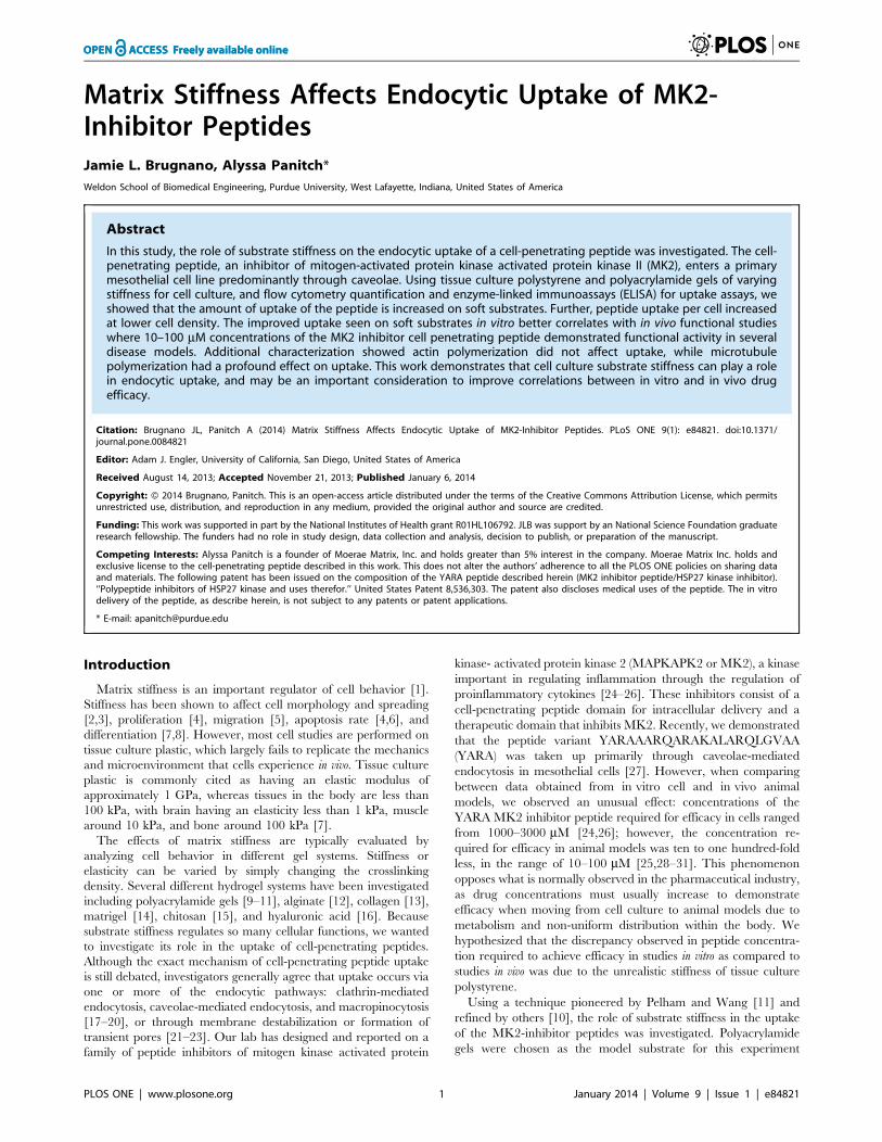
3.1 Rheological CharacterizationTo determine the optimal polyacrylamide gel system to use for
studies, mechanical testing was performed to characterize
substrate stiffness with changes in crosslink density (Figure 1).
The amount of acrylamide monomer remained at a constant 10%
while the bis-acrylamide crosslinker was varied from 0.01% to
1.0%. Substrate stiffness was characterized by measuring the
storage modulus G9. By changing the crosslink density of the
polyacrylamide gels, the storage modulus was varied from 2.5 kPa
to 25 kPa (Figure 1). The choice of which gel system to move
forward with was based upon maximizing the difference in
mechanical properties between the softest and stiffest gel.
However, the stiffest gel with 1.0% bis-acrylamide crosslinker
was opaque and would not accommodate light microscopy images
of cell attachment. The softest gel with 0.01% bis-acrylamide
swelled so much that it prevented the attachment of cells to the
Matrix Stiffness Affects Endocytic Uptake
PLOS ONE | www.plosone.org 3 January 2014 | Volume 9 | Issue 1 | e84821
extracellular matrix protein grafted to the surface. Thus, the two
polyacrylamide gels that were used for further experimentation
were the 0.03% bis-acrylamide for the ‘‘soft’’ gel and the 0.5% bis-
acrylamide for the ‘‘stiff’’ gel, which equate to a stiffness of
approximately 4 and 22 kPa respectively.
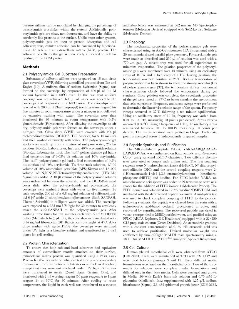
3.2 Protein Characterization on Polyacrylamide GelsFibronectin was covalently grafted to the surface of the
polyacrylamide gels to promote cellular adhesion. To determine
if there was a difference in the amount of fibronectin present on
the gels, a BCA assay was used to quantify total protein
concentration on the gels (Figure 2). There was a significant
increase in the concentration of protein on the gels grafted with
fibronectin (modified) compared to unmodified controls (p,0.05;
one-way ANOVA+Tukey post-hoc test), while no difference was
found in protein concentration between the modified ‘‘soft’’ and
‘‘stiff’’ gels (p.0.05; one-way ANOVA).
3.3 Cellular Morphology on Polyacrylamide GelsOther investigators have reported a change in cellular
morphology in response to matrix stiffness; therefore, we assessed
whether mesothelial cells behaved similarly with the stiffnesses
examined in this study. Light microscopy images demonstrated a
difference in cellular morphology in response to matrix stiffness
(data not shown). On tissue culture plastic, mesothelial cells
displayed a spread cobblestone appearance. However, on soft
substrates, mesothelial cells exhibited a round morphology. Cells
on stiff substrates showed an intermediate morphology. This
change in cellular morphology was correlated to differences in the
actin cytoskeleton (Figure 3). Cells on stiff substrates display
prominent F-actin fibers, with small protrusions extending from
the periphery of the cells (Figure 3A), similar to what is observed
on tissue culture plastic (Figure 3C), while cells on soft substrates
are generally smaller (more rounded) and show predominantly
cortical actin and actin surrounding the nucleus of the cells
(Figure 3B).
3.4 Functional Differences on Polyacrylamide GelsHaving demonstrated that mesothelial cells responded morpho-
logically to the change in substrate stiffness, we next characterized
whether stiffness affected the uptake and function of the MK2-
inhibitor peptide. The MK2-inhibitor peptide was previously
shown to regulate proinflammatory cytokine production in
mesothelial cells using enzyme-linked immunoassays (ELISA)
Figure 1. Rheological characterization of polyacrylamide gels that contain 10% acrylamide and various concentrations of bis-acrylamide crosslinker. Data is displayed as mean 6 standard deviation.doi:10.1371/journal.pone.0084821.g001
Figure 2. Quantification of fibronectin protein on the surfaceof polyacrylamide gels. Data is displayed as mean 6 standarddeviation. Asterisks indicate statistical significance from unmodifiedcontrol (p,0.05; one-way ANOVA+Tukey post-hoc test).doi:10.1371/journal.pone.0084821.g002
Matrix Stiffness Affects Endocytic Uptake
PLOS ONE | www.plosone.org 4 January 2014 | Volume 9 | Issue 1 | e84821

[26], thus, ELISAs were again used to characterize TNFasecretion in these cells when seeded on the polyacrylamide gels
and stimulated with IL-1b and with or without treatment with the
MK2-inhibitor peptide YARA (Figures 4 and 5). TNFa produc-
tion was monitored after 24 hours of treatment. The ELISA results
showed increased efficacy of the MK2-inhibitor peptide on soft
substrates (Figure 4). On soft substrates, TNF-a production was
significantly reduced from the stimulated positive control at a
concentration of 10 mM compared to 100 mM on tissue culture
plastic (Figure 4; p,0.05; one-way ANOVA+Tukey post-hoc test).In addition, statistical analysis showed that the stiff polyacrylamide
substrate is substantially similar to tissue culture polystyrene with
respect to TNFa expression. Statistics also show that cells on stiff
substrates are less responsive to YARA treatment with respect to
suppression of TNFa production. For example, 10 mM YARA
treatment on stiff substrates shows a TNFa release level that is
statistically higher than untreated stiff and untreated soft
substrates, while 10 mM YARA treatment on soft substrates shows
a TNFa research that is statistically the same as untreated soft and
untreated stiff substrate levels.
These results are dependent upon cell number. When cells were
seeded at a high cell density, 71,000 cells/cm2, the increased
efficacy of the MK2-inhibitor peptide observed on soft substrates
disappeared (Figure 5). On tissue culture plastic, cells showed the
expected behavior with an increase in cytokine production with
IL-1b treatment, however, the concentrations of MK2-inhibitor
peptide used did not reverse this effect.
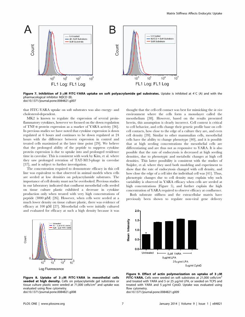
3.5 Intracellular Uptake on Polyacrylamide GelsTo determine if uptake of the MK2-inhibitor peptide differed as
a result of substrate stiffness, the peptide was modified with an N-
terminal FITC-label and intracellular uptake was quantified using
flow cytometry (Figure 6A), and evaluated qualitatively using
fluorescent microscopy (Figure 6B and C). Cells were seeded on
tissue culture polystyrene (6B), or substrates (6C) at low density
(21,000 cells/cm2) to mimic the conditions from the functional
results and treated with FITC-YARA for 24 hours. Cells treated
on soft substrates show approximately double the uptake of FITC-
YARA compared to cells treated on tissue culture plastic, while
uptake of FITC-YARA on stiff substrates is similar to that on tissue
culture plastic (Figure 6A). The increased uptake of FITC-YARA
on soft substrates compared to tissue culture plastic was confirmed
visually with confocal imaging, where more peptide (green
fluorescence) can be seen in cells on soft substrates (Figure 6C)
as compared to cells on tissue culture polystyrene (Figure 6B). All
cells, regardless of substrate, treated with FITC-YARA showed
increased intracellular uptake compared to untreated controls.
To confirm that uptake occurs through similar mechanisms on
the soft polyacrylamide gel compared to tissue culture plastic [27],
we characterized the mechanism of uptake by treating the cells
with FITC-peptide at 4uC to determine if uptake is energy-
dependent (a requirement of endocytic uptake) and with a
pharmacological inhibitor of caveolae-mediated endocytosis,
methyl-b-cyclodextrin (MbCD). Figure 7A and B shows that
uptake of FITC-YARA on soft substrates is inhibited at low
temperatures and is inhibited with MbCD pretreatment, similar to
what has been observed on tissue culture plastic [27].
In addition, we were interested in evaluating uptake as a
function of initial seeding density. As shown in Figure 8, seeding
density does change the uptake of FITC-YARA. A high initial cell
seeding density decreased the amount of FITC-YARA that was
endocytosed by mesothelial cells seeded on soft substrates
compared to tissue culture plastic. This was completely opposite
Figure 3. Mesothelial cells stained for F-actin with phalloidin on stiff (A) and soft (B) substrates, and on tissue culture polystyrene(C). Scale bars are 50 mm.doi:10.1371/journal.pone.0084821.g003
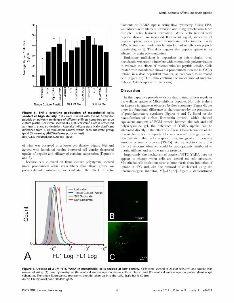
Figure 4. TNF-a cytokine production of mesothelial cellsseeded at low density. Cells were treated with the MK2-inhibitorpeptide on polyacrylamide gels of different stiffness compared to tissueculture plastic. Cells were seeded at 21,000 cells/cm2. Data is presentedas mean 6 standard deviation. Asterisks indicate statistically significantdifference from IL-1b stimulated control within each substrate group(p,0.05; one-way ANOVA+Tukey post-hoc test).doi:10.1371/journal.pone.0084821.g004
Matrix Stiffness Affects Endocytic Uptake
PLOS ONE | www.plosone.org 5 January 2014 | Volume 9 | Issue 1 | e84821
of what was observed at a lower cell density (Figure 6A) and
agreed with functional results: increased cell density decreased
uptake of peptide and efficacy of cytokine suppression (Figures 4
and 5).
Because cells cultured on tissue culture polystyrene showed
more pronounced actin stress fibers than those grown on
polyacrylamide substrates, we evaluated the effect of actin
filaments on YARA uptake using flow cytometry. Using LPA,
we induced actin filament formation and using cytochalasin D we
disrupted actin filament formation. While cells treated with
peptide showed an increased fluorescent signal, indicative of
peptide uptake, as compared to untreated cells, treatment with
LPA, or treatment with cytochalasin D, had no effect on peptide
uptake (Figure 9). This data suggests that peptide uptake is not
affected by actin polymerization.
Endosome trafficking is dependent on microtubules, thus,
nocodazole was used to interfere with microtubule polymerization
to evaluate the effects of microtubules on peptide uptake. Cells
treated with nocodazole showed a pronounced increase in YARA
uptake, in a dose dependent manner, as compared to untreated
cells (Figure 10). This data confirms the importance of microtu-
bules in YARA uptake or trafficking.
Discussion
In this paper, we provide evidence that matrix stiffness regulates
intracellular uptake of MK2-inhibitor peptides. Not only is there
an increase in uptake as observed by flow cytometry (Figure 6), but
there is a functional difference as characterized by the production
of proinflammatory cytokines (Figures 4 and 5). Based on the
quantification of surface fibronectin protein, which showed
equivalent amounts of ECM protein between the soft and stiff
polyacrylamide gel, the difference in YARA uptake can be
attributed directly to the effect of stiffness. Characterization of the
fibronectin protein is important because several investigators have
demonstrated that cells respond morphologically to varying
amounts of matrix proteins [33–35]. We wanted to ensure that
the cell response observed could be appropriately attributed to
matrix stiffness and not the matrix proteins.
Importantly, the mechanism of uptake of FITC-YARA does not
appear to change when cells are seeded on soft substrates.
Mesothelial cells seeded on tissue culture plastic show inhibition of
uptake at 4uC and with the removal of cholesterol using the
pharmacological inhibitor, MbCD [27]. Figure 7 demonstrated
Figure 5. TNF-a cytokine production of mesothelial cellsseeded at high density. Cells were treated with the MK2-inhibitorpeptide on polyacrylamide gels of different stiffness compared to tissueculture plastic. Cells were seeded at 71,000 cells/cm2. Data is presentedas mean 6 standard deviation. Asterisks indicate statistically significantdifference from IL-1b stimulated control within each substrate group(p,0.05; one-way ANOVA+Tukey post-hoc test).doi:10.1371/journal.pone.0084821.g005
Figure 6. Uptake of 3 mM FITC-YARA in mesothelial cells seeded at low density. Cells were seeded at 21,000 cells/cm2 and uptake wasevaluated using (A) flow cytometry or (B) confocal microscope on tissue culture plastic, and (C) confocal microscope on polyacrylamide gelsubstrates. The green fluorescence represents peptide taken up into the cells. Scale bar is 50 mm.doi:10.1371/journal.pone.0084821.g006
Matrix Stiffness Affects Endocytic Uptake
PLOS ONE | www.plosone.org 6 January 2014 | Volume 9 | Issue 1 | e84821
that FITC-YARA uptake on soft substrates was also energy- and
cholesterol-dependent.
MK2 is known to regulate the expression of several proin-
flammatory cytokines, however we focused on the down regulation
of TNF-a protein expression as a marker of YARA activity [36].
In previous studies we have noted that cytokine expression is down
regulated at 6 hours and continues to be down regulated at 24
hours with the difference between expression in control and
treated cells maximized at the later time point [29]. We believe
that the prolonged ability of the peptide to suppress cytokine
protein expression is due to uptake into and prolonged residence
time in caveolae. This is consistent with work by Kim, et al. where
they saw prolonged retention of TAT-M13-phage in caveolae
[37], and is subject to further investigation.
The concentration required to demonstrate efficacy in this cell
line was equivalent to that observed in animal models when cells
are seeded at low densities on polyacrylamide substrates. The
importance of cell density must not be overlooked. Previous studies
in our laboratory indicated that confluent mesothelial cells seeded
on tissue culture plastic exhibited a decrease in cytokine
production only when treated with very high concentrations of
peptide (3000 mM) [26]. However, when cells were seeded at a
much lower density on tissue culture plastic, there was evidence of
efficacy at 100 mM [27]. Mesothelial cells were initially cultured
and evaluated for efficacy at such a high density because it was
thought that the cell-cell contact was best for mimicking the in vivo
environment where the cells form a monolayer called the
mesothelium [38]. However, based on the results presented
herein, this assumption is clearly incorrect. Cell context is critical
to cell behavior, and cells change their genetic profile base on cell-
cell contacts, how close to the edge of a culture they are, and even
cell density [39]. Similar to other mammalian cells, mesothelial
cells have the ability to change phenotype [40], and it is possible
that at high seeding concentrations the mesothelial cells are
differentiating and are thus not as responsive to YARA. It is also
possible that the rate of endocytosis is decreased at high seeding
densities, due to phenotypic and metabolic changes at high cell
densities. This latter possibility is consistent with the studies of
Snijder, et al. where they used both modeling and experiment to
show that the rate of endocytosis changed with cell density, and
how close the edge of a cell islet the individual cell was [41]. Thus,
phenotypic changes due to cell density may explain why such
variability is observed in YARA efficacy when cells are seeded at
high concentrations (Figure 5), and further explain the high
concentration of YARA required to observe efficacy at confluence.
Both substrate stiffness and the extracellular matrix have
previously been shown to regulate non-viral gene delivery
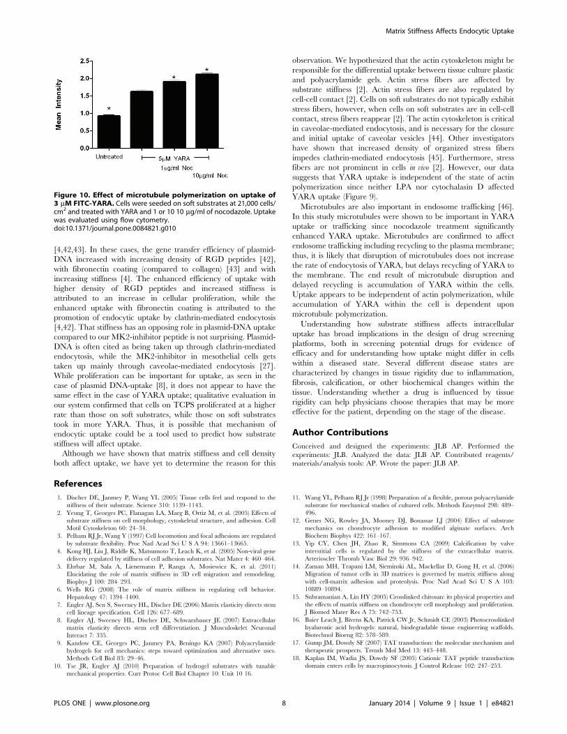
Figure 7. Inhibition of 3 mM FITC-YARA uptake on soft polyacrylamide gel substrates. Uptake is inhibited at 4uC (A) and with thepharmacological inhibitor MbCD (B).doi:10.1371/journal.pone.0084821.g007
Figure 8. Uptake of 3 mM FITC-YARA in mesothelial cellsseeded at high density. Cells on polyacrylamide gel substrates ortissue culture plastic were seeded at 71,000 cells/cm2 and uptake wasevaluated using flow cytometry.doi:10.1371/journal.pone.0084821.g008
Figure 9. Effect of actin polymerization on uptake of 3 mMFITC-YARA. Cells were seeded on soft substrates at 21,000 cells/cm2
and treated with YARA and 5 or 25 mg/ml LPA, or seeded on TCPS andtreated with YARA and 5 mg/ml CytoD. Uptake was evaluated usingflow cytometry.doi:10.1371/journal.pone.0084821.g009
Matrix Stiffness Affects Endocytic Uptake
PLOS ONE | www.plosone.org 7 January 2014 | Volume 9 | Issue 1 | e84821
[4,42,43]. In these cases, the gene transfer efficiency of plasmid-
DNA increased with increasing density of RGD peptides [42],
with fibronectin coating (compared to collagen) [43] and with
increasing stiffness [4]. The enhanced efficiency of uptake with
higher density of RGD peptides and increased stiffness is
attributed to an increase in cellular proliferation, while the
enhanced uptake with fibronectin coating is attributed to the
promotion of endocytic uptake by clathrin-mediated endocytosis
[4,42]. That stiffness has an opposing role in plasmid-DNA uptake
compared to our MK2-inhibitor peptide is not surprising. Plasmid-
DNA is often cited as being taken up through clathrin-mediated
endocytosis, while the MK2-inhibitor in mesothelial cells gets
taken up mainly through caveolae-mediated endocytosis [27].
While proliferation can be important for uptake, as seen in the
case of plasmid DNA-uptake [8], it does not appear to have the
same effect in the case of YARA uptake; qualitative evaluation in
our system confirmed that cells on TCPS proliferated at a higher
rate than those on soft substrates, while those on soft substrates
took in more YARA. Thus, it is possible that mechanism of
endocytic uptake could be a tool used to predict how substrate
stiffness will affect uptake.
Although we have shown that matrix stiffness and cell density
both affect uptake, we have yet to determine the reason for this
observation. We hypothesized that the actin cytoskeleton might be
responsible for the differential uptake between tissue culture plastic
and polyacrylamide gels. Actin stress fibers are affected by
substrate stiffness [2]. Actin stress fibers are also regulated by
cell-cell contact [2]. Cells on soft substrates do not typically exhibit
stress fibers, however, when cells on soft substrates are in cell-cell
contact, stress fibers reappear [2]. The actin cytoskeleton is critical
in caveolae-mediated endocytosis, and is necessary for the closure
and initial uptake of caveolar vesicles [44]. Other investigators
have shown that increased density of organized stress fibers
impedes clathrin-mediated endocytosis [45]. Furthermore, stress
fibers are not prominent in cells in vivo [2]. However, our data
suggests that YARA uptake is independent of the state of actin
polymerization since neither LPA nor cytochalasin D affected
YARA uptake (Figure 9).
Microtubules are also important in endosome trafficking [46].
In this study microtubules were shown to be important in YARA
uptake or trafficking since nocodazole treatment significantly
enhanced YARA uptake. Microtubules are confirmed to affect
endosome trafficking including recycling to the plasma membrane;
thus, it is likely that disruption of microtubules does not increase
the rate of endocytosis of YARA, but delays recycling of YARA to
the membrane. The end result of microtubule disruption and
delayed recycling is accumulation of YARA within the cells.
Uptake appears to be independent of actin polymerization, while
accumulation of YARA within the cell is dependent upon
microtubule polymerization.
Understanding how substrate stiffness affects intracellular
uptake has broad implications in the design of drug screening
platforms, both in screening potential drugs for evidence of
efficacy and for understanding how uptake might differ in cells
within a diseased state. Several different disease states are
characterized by changes in tissue rigidity due to inflammation,
fibrosis, calcification, or other biochemical changes within the
tissue. Understanding whether a drug is influenced by tissue
rigidity can help physicians choose therapies that may be more
effective for the patient, depending on the stage of the disease.
Author Contributions
Conceived and designed the experiments: JLB AP. Performed the
experiments: JLB. Analyzed the data: JLB AP. Contributed reagents/
materials/analysis tools: AP. Wrote the paper: JLB AP.
References
1. Discher DE, Janmey P, Wang YL (2005) Tissue cells feel and respond to the
stiffness of their substrate. Science 310: 1139–1143.
2. Yeung T, Georges PC, Flanagan LA, Marg B, Ortiz M, et al. (2005) Effects of
substrate stiffness on cell morphology, cytoskeletal structure, and adhesion. Cell
Motil Cytoskeleton 60: 24–34.
3. Pelham RJ Jr, Wang Y (1997) Cell locomotion and focal adhesions are regulated
by substrate flexibility. Proc Natl Acad Sci U S A 94: 13661–13665.
4. Kong HJ, Liu J, Riddle K, Matsumoto T, Leach K, et al. (2005) Non-viral gene
delivery regulated by stiffness of cell adhesion substrates. Nat Mater 4: 460–464.
5. Ehrbar M, Sala A, Lienemann P, Ranga A, Mosiewicz K, et al. (2011)
Elucidating the role of matrix stiffness in 3D cell migration and remodeling.
Biophys J 100: 284–293.
6. Wells RG (2008) The role of matrix stiffness in regulating cell behavior.
Hepatology 47: 1394–1400.
7. Engler AJ, Sen S, Sweeney HL, Discher DE (2006) Matrix elasticity directs stem
cell lineage specification. Cell 126: 677–689.
8. Engler AJ, Sweeney HL, Discher DE, Schwarzbauer JE (2007) Extracellular
matrix elasticity directs stem cell differentiation. J Musculoskelet Neuronal
Interact 7: 335.
9. Kandow CE, Georges PC, Janmey PA, Beningo KA (2007) Polyacrylamide
hydrogels for cell mechanics: steps toward optimization and alternative uses.
Methods Cell Biol 83: 29–46.
10. Tse JR, Engler AJ (2010) Preparation of hydrogel substrates with tunable
mechanical properties. Curr Protoc Cell Biol Chapter 10: Unit 10 16.
11. Wang YL, Pelham RJ Jr (1998) Preparation of a flexible, porous polyacrylamide
substrate for mechanical studies of cultured cells. Methods Enzymol 298: 489–
496.
12. Genes NG, Rowley JA, Mooney DJ, Bonassar LJ (2004) Effect of substrate
mechanics on chondrocyte adhesion to modified alginate surfaces. Arch
Biochem Biophys 422: 161–167.
13. Yip CY, Chen JH, Zhao R, Simmons CA (2009) Calcification by valve
interstitial cells is regulated by the stiffness of the extracellular matrix.
Arterioscler Thromb Vasc Biol 29: 936–942.
14. Zaman MH, Trapani LM, Sieminski AL, Mackellar D, Gong H, et al. (2006)
Migration of tumor cells in 3D matrices is governed by matrix stiffness along
with cell-matrix adhesion and proteolysis. Proc Natl Acad Sci U S A 103:
10889–10894.
15. Subramanian A, Lin HY (2005) Crosslinked chitosan: its physical properties and
the effects of matrix stiffness on chondrocyte cell morphology and proliferation.
J Biomed Mater Res A 75: 742–753.
16. Baier Leach J, Bivens KA, Patrick CW Jr, Schmidt CE (2003) Photocrosslinked
hyaluronic acid hydrogels: natural, biodegradable tissue engineering scaffolds.
Biotechnol Bioeng 82: 578–589.
17. Gump JM, Dowdy SF (2007) TAT transduction: the molecular mechanism and
therapeutic prospects. Trends Mol Med 13: 443–448.
18. Kaplan IM, Wadia JS, Dowdy SF (2005) Cationic TAT peptide transduction
domain enters cells by macropinocytosis. J Control Release 102: 247–253.
Figure 10. Effect of microtubule polymerization on uptake of3 mM FITC-YARA. Cells were seeded on soft substrates at 21,000 cells/cm2 and treated with YARA and 1 or 10 10 mg/ml of nocodazole. Uptakewas evaluated using flow cytometry.doi:10.1371/journal.pone.0084821.g010
Matrix Stiffness Affects Endocytic Uptake
PLOS ONE | www.plosone.org 8 January 2014 | Volume 9 | Issue 1 | e84821

19. Vendeville A, Rayne F, Bonhoure A, Bettache N, Montcourrier P, et al. (2004)
HIV-1 Tat enters T cells using coated pits before translocating from acidifiedendosomes and eliciting biological responses. Mol Biol Cell 15: 2347–2360.
20. Ferrari A, Pellegrini V, Arcangeli C, Fittipaldi A, Giacca M, et al. (2003)
Caveolae-mediated internalization of extracellular HIV-1 tat fusion proteinsvisualized in real time. Mol Ther 8: 284–294.
21. Herce HD, Garcia AE (2007) Molecular dynamics simulations suggest amechanism for translocation of the HIV-1 TAT peptide across lipid membranes.
Proc Natl Acad Sci U S A 104: 20805–20810.
22. Palm-Apergi C, Lorents A, Padari K, Pooga M, Hallbrink M (2009) Themembrane repair response masks membrane disturbances caused by cell-
penetrating peptide uptake. The FASEB J 23: 214–223.23. Yandek LE, Pokorny A, Floren A, Knoelke K, Langel U, et al. (2007)
Mechanism of the Cell-Penetrating Peptide Transportan 10 Permeation of LipidBilayers. Biophysical J 92: 2434–2444.
24. Brugnano JL, Chan BK, Seal BL, Panitch A (2011) Cell-penetrating peptides
can confer biological function: regulation of inflammatory cytokines in humanmonocytes by MK2 inhibitor peptides. J Control Release 155: 128–133.
25. Kavalukas SL, Uzgare AR, Panitch A, Ward BC, Barbul A (2009) MK2inhibitor peptide reduces adhesion formation without affecting colonic
anastomotic healing. J Am Col Surg 209: S17.
26. Ward BC, Kavalukas S, Brugnano J, Barbul A, Panitch A (2011) Peptideinhibitors of MK2 show promise for inhibition of abdominal adhesions. J Surg
Res 169: e27–36.27. Brugnano J, McMasters J, Panitch A (2013) Characterization of Endocytic
uptake of MK2-Inhibitor Peptides. J Peptide Sci 19(10): 629–638.28. Muto A, Panitch A, Kim N, Park K, Komalavilas P, et al. (2012) Inhibition of
Mitogen Activated Protein Kinase Activated Protein Kinase II with MMI-0100
reduces intimal hyperplasia ex vivo and in vivo. Vasc pharm 56: 47–55.29. Ward BC, Kavalukas S, Brugnano J, Barbul A, Panitch A (2011) Peptide
inhibitors of MK2 show promise for inhibition of abdominal adhesions. J of SurgRes 169: e27–e36.
30. Muto A, Panitch A, Kim N, Park K, Komalavilas P, et al. (2012) Inhibition of
Mitogen Activated Protein Kinase Activated Protein Kinase II with MMI-0100reduces intimal hyperplasia ex vivo and in vivo. Vascul Pharmacol 56: 47–55.
31. Vittal R, Fisher A, Gu H, Mickler EA, Panitch A, et al. (2013) Peptide-mediatedinhibition of mitogen-activated protein kinase-activated protein kinase-2
ameliorates bleomycin-induced pulmonary fibrosis. Am J Respir Cell Mol Biol49: 47–57.
32. Calvet D, Wong JY, Giasson S (2004) Rheological monitoring of polyacrylamide
gelation: Importance of cross-link density and temperature. Macromolecules 37:7762–7771.
33. Kuhlman W, Taniguchi I, Griffith LG, Mayes AM (2007) Interplay between
PEO tether length and ligand spacing governs cell spreading on RGD-modifiedPMMA-g-PEO comb copolymers. Biomacromolecules 8: 3206–3213.
34. Massia SP, Hubbell JA (1991) An RGD spacing of 440 nm is sufficient forintegrin alpha V beta 3-mediated fibroblast spreading and 140 nm for focal
contact and stress fiber formation. J Cell Biol 114: 1089–1100.
35. Maheshwari G, Brown G, Lauffenburger DA, Wells A, Griffith LG (2000) Celladhesion and motility depend on nanoscale RGD clustering. J Cell Sci 113 (Pt
10): 1677–1686.36. Gaestel M (2006) MAPKAP kinases - MKs – two’s company, three’s a crowd.
Nat Rev Mol Cell Biol 7: 120–130.37. Kim A, Shin TH, Shin SM, Pham CD, Choi DK, et al. (2012) Cellular
internalization mechanism and intracellular trafficking of filamentous M13
phages displaying a cell-penetrating transbody and TAT peptide. PLoS One 7:e51813.
38. Mutsaers SE (2004) The mesothelial cell. Int J Biochem Cell Biol 36: 9–16.39. Sacher R, Stergiou L, Pelkmans L (2008) Lessons from genetics: interpreting
complex phenotypes in RNAi screens. Curr Opin Cell Biol 20: 483–489.
40. Lansley SM, Searles RG, Hoi A, Thomas C, Moneta H, et al. (2011) Mesothelialcell differentiation into osteoblast- and adipocyte-like cells. J Cell Mol Med 15:
2095–2105.41. Snijder B, Sacher R, Ramo P, Damm EM, Liberali P, et al. (2009) Population
context determines cell-to-cell variability in endocytosis and virus infection.Nature 461: 520–523.
42. Kong HJ, Hsiong S, Mooney DJ (2007) Nanoscale cell adhesion ligand
presentation regulates nonviral gene delivery and expression. Nano Lett 7: 161–166.
43. Dhaliwal A, Maldonado M, Han Z, Segura T (2010) Differential uptake ofDNA-poly(ethylenimine) polyplexes in cells cultured on collagen and fibronectin
surfaces. Acta Biomater 6: 3436–3447.
44. Pelkmans L, Helenius A (2002) Endocytosis via caveolae. Traffic 3: 311–320.45. Liu AP, Loerke D, Schmid SL, Danuser G (2009) Global and Local Regulation
of Clathrin-Coated Pit Dynamics Detected on Patterned Substrates. Biophysical J97: 1038–1047.
46. Maxfield FR, McGraw TE (2004) Endocytic recycling. Nat Rev Mol Cell Biol 5:121–132.
Matrix Stiffness Affects Endocytic Uptake
PLOS ONE | www.plosone.org 9 January 2014 | Volume 9 | Issue 1 | e84821