Assessment of Arterial Stiffness by Cardio-Ankle Vascular ...
Upload
independentCategory
view
0download
0
The Focal Adhesion: A Regulated Component of AorticStiffnessRobert J. Saphirstein1,2, Yuan Z. Gao1,2, Mikkel H. Jensen3,4, Cynthia M. Gallant2, Susanne Vetterkind2,
Jeffrey R. Moore4, Kathleen G. Morgan2*
1Department of Biomedical Engineering, Boston University, Boston, Massachusetts, United States of America, 2Department of Health Sciences, Boston University, Boston,
Massachusetts, United States of America, 3Department of Physics, Boston University, Boston, Massachusetts, United States of America, 4Department of Physiology and
Biophysics, Boston University Medical School, Boston, Massachusetts, United States of America
Abstract
Increased aortic stiffness is an acknowledged predictor and cause of cardiovascular disease. The sources and mechanisms ofvascular stiffness are not well understood, although the extracellular matrix (ECM) has been assumed to be a majorcomponent. We tested here the hypothesis that the focal adhesions (FAs) connecting the cortical cytoskeleton of vascularsmooth muscle cells (VSMCs) to the matrix in the aortic wall are a component of aortic stiffness and that this component isdynamically regulated. First, we examined a model system in which magnetic tweezers could be used to monitor cellularcortical stiffness, serum-starved A7r5 aortic smooth muscle cells. Lysophosphatidic acid (LPA), an activator of myosin thatincreases cell contractility, increased cortical stiffness. A small molecule inhibitor of Src-dependent FA recycling, PP2, wasfound to significantly inhibit LPA-induced increases in cortical stiffness, as well as tension-induced increases in FA size. Todirectly test the applicability of these results to force and stiffness development at the level of vascular tissue, we monitoredmouse aorta ring stiffness with small sinusoidal length oscillations during agonist-induced contraction. The alpha-agonistphenylephrine, which also increases myosin activation and contractility, increased tissue stress and stiffness in a PP2- andFAK inhibitor 14-attenuated manner. Subsequent phosphotyrosine screening and follow-up with phosphosite-specificantibodies confirmed that the effects of PP2 and FAK inhibitor 14 in vascular tissue involve FA proteins, including FAK, CAS,and paxillin. Thus, in the present study we identify, for the first time, the FA of the VSMC, in particular the FAK-Src signalingcomplex, as a significant subcellular regulator of aortic stiffness and stress.
Citation: Saphirstein RJ, Gao YZ, Jensen MH, Gallant CM, Vetterkind S, et al. (2013) The Focal Adhesion: A Regulated Component of Aortic Stiffness. PLoS ONE 8(4):e62461. doi:10.1371/journal.pone.0062461
Editor: Adam J. Engler, University of California, San Diego, United States of America
Received November 30, 2012; Accepted March 21, 2013; Published April 23, 2013
Copyright: � 2013 Saphirstein et al. This is an open-access article distributed under the terms of the Creative Commons Attribution License, which permitsunrestricted use, distribution, and reproduction in any medium, provided the original author and source are credited.
Funding: NIH HL086655, NIH Quantitative Biology and Physiology Training Grant, Evans Center for Interdisciplinary Biomedical Research ARC on Aortic Stiffnessat Boston University. The funders had no role in study design, data collection and analysis, decision to publish, or preparation of the manuscript.
Competing Interests: The authors have declared that no competing interests exist.
* E-mail: [email protected]
Introduction
Normal cardiovascular function depends on the biomechanical
properties of blood vessels, and changes in these properties are
characteristic of disease. Increased aortic stiffness precedes and
predicts for the development of hypertension and subsequent
negative cardiovascular outcomes, including myocardial infarc-
tion, stroke, and heart disease [1,2]. A stiffer aorta deforms less in
response to pulse pressure, absorbing less of the pulse while
transmitting a greater portion of the potentially harmful pulsatile
energy downstream to delicate vessels of the microcirculation in
the heart, lung, brain and kidneys [1].
The mechanical properties of a blood vessel, like any material or
tissue, are determined by its components and their organization.
Accordingly, the stiffness of vascular tissue can be derived from the
composition of the extracellular matrix (ECM) and the cells in the
tissue, the organization of the matrix and cells, and the
connections that link these components (cell-matrix and cell-cell
contacts). The prevailing view is that the ECM is the principal
determinant of vascular stiffness [3]; however, the extent to which
other tissue components contribute is unclear.
Vascular smooth muscle cells (VSMCs), the predominant cell
type in the wall of large blood vessels, influence tissue stiffness
when modulating contractile tone or when depositing matrix or
matrix-degrading enzymes [3,4]. Recently, the stiffness of
VSMCs themselves has been implicated as an important
determinant of tissue stiffness [5], with the actin cytoskeleton
suggested as the specific cellular component responsible. Based
on this finding, as well as the recent finding that the cortical
actin cytoskeleton is dynamic in vascular smooth muscle tissue
[6], we hypothesized that the cortical focal adhesions (FAs) (also
known as adhesion plaques or dense plaques in smooth muscle
tissues [7]) may be an important, and possibly regulated,
component of vascular tissue stiffness. Here we define the FA to
include its attachments to the cortical actin cytoskeleton as well
as the integrins that attach the cell to the ECM.
In the present study, we examine the mechanical properties of
blood vessels across multiple length scales and identify, for the first
time, the Src-FAK signaling complex of FAs of the non-migratory,
non-proliferating VSMCs embedded in the blood vessel wall as
a significant regulator of aortic stiffness. Finally, we show that FA
size, cortical stiffness, and VSMC contractility influence the FA-
regulated component of aortic stiffness.
PLOS ONE | www.plosone.org 1 April 2013 | Volume 8 | Issue 4 | e62461
Methods
Ethical ApprovalAll procedures were performed in accordance with protocols
approved by the Boston University Institutional Animal Care and
Use Committee (Permit Number: A3316-01). The animals were
maintained according to guidelines in the NIH Guide for the Care
and Use of Laboratory Animals and were obtained and used in
compliance with federal, state, and local laws. Tissue removal was
quickly performed after isoflurane euthanasia.
Cell CultureA7r5 vascular smooth muscle cells, originally derived from rat
aorta (American Type Culture Collection, Manassas, VA), were
maintained as described previously [8]. A7r5 cells proliferate as
myoblasts and, when they reach the stationary phase, upon serum
deprivation, differentiate into a phenotype that resembles adult
smooth muscle cells [9,10]. The cells express many key markers of
smooth muscle, including smooth muscle a-actin, smooth muscle
myosin, smooth muscle tropomyosin isoforms, h1 calponin, and
SM22 a [11]. This approach was chosen over primary cultured
cells because of the variability of differentiation state with passages.
For all experiments, A7r5 cells were seeded onto coverslips or, for
magnetic tweezers experiments, 35-mm glass-bottom culture
dishes (MatTek, Ashland, MA), grown to 75–90% confluence in
the presence of serum and then serum-starved for 24 hours to
trigger differentiation to the quiescent (non-proliferating, non-
migrating) smooth muscle phenotype.
Bead Coating and AttachmentFor magnetic tweezer studies, an arginine-glycine-aspartic acid
(RGD) peptide (GRGDNP, Enzo Life Sciences, Farmingdale, NY)
was covalently coupled to amine-modified superparamagnetic
Dynabeads M-270 (2.8 mm, Invitrogen, Grand Island, NY)
according to a protocol from Bangs Laboratories (TechNote
205: Covalent Coupling, Bangs Laboratories, Inc., Fishers, IN).
Briefly, amine-modified Dynabeads were suspended in phosphate
buffered saline (10 mg/ml), activated with 10% glutaraldehyde,
reacted with RGD peptide (0.5 mg/ml), quenched with 40 mM
glycine and blocking agent, and stored with blocking agent and
preservative in phosphate buffered saline.
RGD is an ECM protein motif that is a minimal sequence
required for binding by transmembrane integrin receptors [12].
The GRGDNP peptide has affinity for the a5b1 and avb3integrins, which are present in vascular smooth muscle tissues and
recognize fibronectin and vitronectin, respectively [12–14].
Before making measurements with the magnetic tweezers, the
beads were washed and sonicated, and then 6.76105 beads
were added to the 35-mm glass bottom dish containing cells.
The beads were incubated with the cells for 30 min at 37uCwith 5% CO2 to facilitate their attachment to the cells.
Following this incubation, the serum-free medium was ex-
changed twice to remove unbound beads. Measurements of
cortical stiffness were performed at room temperature. Measur-
ing time was limited to 30 minutes per dish.
Measurement of Stiffness with Magnetic TweezersThe magnetic tweezers (MT) apparatus is modeled after
a system designed by the Fabry group [15], and it consists of
a magnetic microneedle made from a cylindrical rod of high-
permeability, low-hysteresivity nickel alloy (HyMU-80, Carpenter
Technology, Reading, PA) with a 4.5-mm core diameter and its
needle-end sharpened to a 40-mm radius (courtesy of the Boston
University machine shop); a solenoid (200 turns of 0.5 mm copper
wire) surrounding the microneedle; and a power supply (MPJA,
Lake Park, FL) connected to the solenoid.
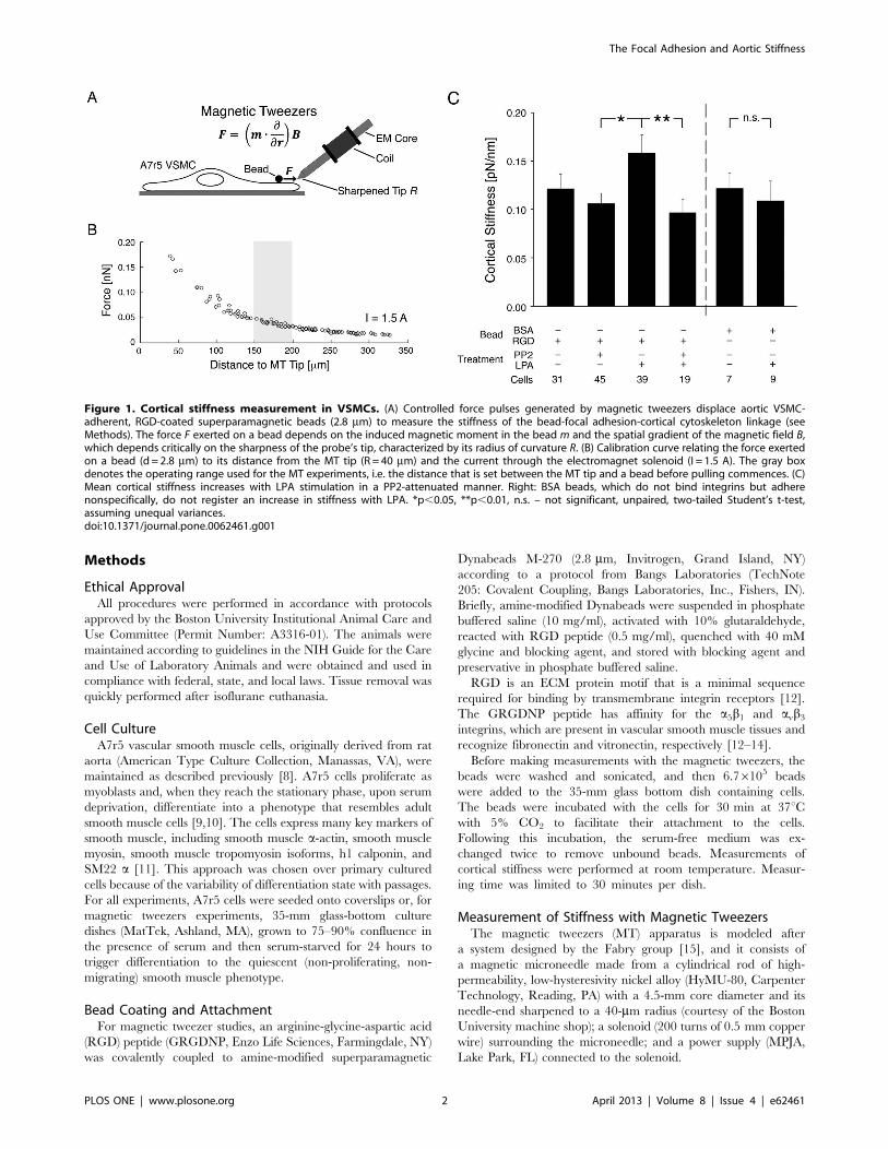
Figure 1. Cortical stiffness measurement in VSMCs. (A) Controlled force pulses generated by magnetic tweezers displace aortic VSMC-adherent, RGD-coated superparamagnetic beads (2.8 mm) to measure the stiffness of the bead-focal adhesion-cortical cytoskeleton linkage (seeMethods). The force F exerted on a bead depends on the induced magnetic moment in the bead m and the spatial gradient of the magnetic field B,which depends critically on the sharpness of the probe’s tip, characterized by its radius of curvature R. (B) Calibration curve relating the force exertedon a bead (d= 2.8 mm) to its distance from the MT tip (R = 40 mm) and the current through the electromagnet solenoid (I = 1.5 A). The gray boxdenotes the operating range used for the MT experiments, i.e. the distance that is set between the MT tip and a bead before pulling commences. (C)Mean cortical stiffness increases with LPA stimulation in a PP2-attenuated manner. Right: BSA beads, which do not bind integrins but adherenonspecifically, do not register an increase in stiffness with LPA. *p,0.05, **p,0.01, n.s. – not significant, unpaired, two-tailed Student’s t-test,assuming unequal variances.doi:10.1371/journal.pone.0062461.g001
The Focal Adhesion and Aortic Stiffness
PLOS ONE | www.plosone.org 2 April 2013 | Volume 8 | Issue 4 | e62461

Calibration of the magnetic tweezers was performed similarly to
what has been done by others [15]. Briefly, the movements of
beads through a viscous mixture of 3:1 glycerol to ddH2O
(viscosity g=0.052 Pa s at room temperature) were used to
compute forces on the beads for different solenoid currents using
Stokes’ formula for viscous drag, F=6pgrv, where g is the
viscosity, r is the radius of the bead, and v is the velocity of the bead
through the mixture. The calibration data for all beads were then
fit to a simple empirical equation relating the force on a bead to
both the bead’s distance from the magnetic tweezers tip and the
current through the solenoid [15]. This relationship was then used
to analyze data from cortical stiffness experiments and calculate
forces exerted on cell-bound beads based on their distance from
the MT tip and the set current.
To measure cortical stiffness, the MT tip is positioned
approximately 150–200 mm from a cell-adherent bead using
a micromanipulator. This distance was chosen because the force-
distance relationship is highly nonlinear at short bead-tip distances
but flattens out at distances greater than 100 mm. Thus, at an
operating distance of 150–200 mm, a pull on a bead will displace it
towards the tip and only negligibly increase the force exerted by
the MT on the bead (i.e., force is essentially constant, F , 50 pN).
An operating current of 1.5 A was chosen for experiments because
it produces low forces appropriate for cortical probing [16–19]. At
this low current (and for the short pull durations employed), there
is no significant heating of the rod or the sample. Between
measurements, the stage is displaced a distance of at least 0.5 mm
so that cells do not experience forces from previous measurements.
Beads selected for analysis are less than 50 mm from the cell edge
and bound on the MT-side of the cell, sufficiently far from the
nucleus, to avoid pulling the bead uphill. Additionally, beads were
selected for pulling only if there were no overlapping cells in the
vicinity of the bead that could influence the local apical
topography of the cell.
Cortical stiffness, which we define as the stiffness of the bead-
integrin attachment and the underlying FA and cortical cytoskel-
eton, was measured by employing a priming protocol described by
others that consists of a series of five identical square-wave cycles
that are 4 sec in duration and 4 sec apart [17,20]. Each pull
displaces the bead towards the MT probe, and in between pulls
the bead recoils and reaches equilibrium. The bead does not
return to its exact start position with each pull, leading to a shift in
the baseline with each pulse that stabilizes within about three pulls.
The fifth and final pull is analyzed to measure cortical stiffness
(calculated as a ratio of the force applied to the displacement of the
cell-adherent bead).
Experiments were performed at 206 magnification (NA 0.5)
with a Nikon Plan Fluor objective under bright-field illumination
Figure 2. LPA stimulation increases FA size in a PP2-sensitive manner. Top: Deconvolution microscopy of FAs. Representative FAs are shownin an expanded view in the insets. All FAs in each cell, not just those featured in the insets, were analyzed to determine the mean FA area. Scale bars:top, 20 mm; bottom, 5 mm. Bottom: Mean FA area is significantly greater in cells stimulated with LPA (n = 82 cells). ***p,0.001, unpaired, two-tailedStudent’s t-test.doi:10.1371/journal.pone.0062461.g002
The Focal Adhesion and Aortic Stiffness
PLOS ONE | www.plosone.org 3 April 2013 | Volume 8 | Issue 4 | e62461

on an Eclipse TE2000-E inverted microscope (Nikon Instruments).
Videos of beads and the MT tip were recorded in real time with
a charge-coupled device camera (CoolSNAP HQ2, Photometrics)
at 10 frames per second using the Nikon NIS Elements imaging
software (Nikon Instruments). Positions of beads were tracked with
an intensity-weighted center-of-mass algorithm using the MTrackJ
plug-in for ImageJ [21] and later analyzed in Matlab (Natick,
MA).
Lysophosphatidic acid (LPA, 10 mM) was added for 15 minutes,
PP2 (10 mM) was added for 30 minutes, or PP2 was added for 30
minutes followed by LPA for 15 minutes. Experiments were
performed at room temperature, and measurement time with the
MT for each sample was limited to 30 minutes to ensure cell
viability.
Immunofluorescence Imaging and AnalysisA7r5 cells were fixed and stained as previously described [8].
Nuclei were stained with 4,6-diamidino-2-phenylindole (Sigma-
Aldrich, St. Louis, MO), and filamentous actin was stained with
Alexa Fluor 568 and 488 phalloidin (1:3000, Invitrogen). Cells
were examined with an Eclipse TE2000-E fluorescence micro-
scope (Nikon Instruments) equipped with a Nikon Plan Apoc-
hromat 60X (NA 1.4) oil immersion objective, a charge-coupled
device camera (CoolSNAP HQ2, Photometrics), and filters
optimized for double-label experiments. NIS-Element Advanced
Research software (Nikon Instruments) was used to capture images
and for removal of out-of-focus fluorescent blur by deconvolution
of Z-stacks (Richardson-Lucy algorithm, constrained iterative-
maximum likelihood estimation algorithm) as previously described
[22]. Subsequent processing was completed with Photoshop CS3
software (Adobe Systems, Mountain View, CA).
Areas of individual FAs were determined using NIS-Elements
Advanced Research software. FAs were measured for 5 to 13 A7r5
cells per condition (control, PP2, LPA, PP2+ LPA) per experiment
(total n = 82 cells). In each cell that was selected for analysis, all
peripheral FAs were examined for analysis because these
adhesions are most relevant to the question of force transmission,
as numerous studies have reported that the largest traction forces
are found in the cell periphery [23]. Cells with overlapping
neighbors that would obscure measurements were excluded from
the analysis.
Aorta Tissue PreparationC57bl/6 Mice (Mus musculus) (3 months old, n= 31) (Charles
River, Wilmington, MA) were euthanized with an overdose of
isoflurane by inhalation. The aorta was promptly excised, rinsed
six times to remove blood within or around the vessel, which can
harm the smooth muscle cells and induce vasoconstriction [24],
and stored in an oxygenated (95% O2–5% CO2) physiological salt
solution (PSS) (in mM: 120 NaCl, 5.9 KCl, 1.2 NaH2PO4, 25
NaHCO3, 11.5 dextrose, 1CaCl2, and 1.4 MgCl2; pH=7.4).
Following careful dissection in oxygenated PSS to remove
connective tissue and adventitia, aortic rings were cut (4 mm
long) then suspended in vitro in tissue baths containing oxygenated
PSS at 37uC. Smaller rings immediately proximal and distal to
these suspended segments were also cut for determining ring
thickness. Following the measurements made in the tissue baths,
aortic rings were quick-frozen at 278.5uC in a slurry of dry ice
and liquid acetone containing 10 mM dithiothreitol and 10%
TCA for subsequent biochemical analysis [25]. The TCA
functions to denature and deactivate endogenous kinases,
phosphatases, and proteolytic enzymes, thereby preserving the
levels of phosphorylation present at the time of freezing [26,27].
Figure 3. Aortic tissue stiffening during contractile stimulation is decreased by inhibition of Src, FAK, or MLCK. (A) Tissue stiffness Emeasured in vitro during PE-induced contraction at optimum length LO with small-amplitude (1%), high frequency (40Hz) sinusoidal stretches DL. Boxheight and width for magnified traces: 0.5 mN, 5 mm, and 0.02 s. Upper Inset: Stiffness calculation. Lower Inset: Fluorescent micrograph of aortic ringin cross-section, used to determine ring thickness for calculation of cross-sectional area A. Green: autofluorescent elastic laminae. Blue: cell nuclei.Scale bar, 100 mm. (B) PE-induced stress is significantly lower when pre-treated with MLCK inhibitor ML-9, confirming the importance of myosinactivation and contraction to vascular stiffness (n = 4). PE-induced stress is also significantly reduced when pre-treated with Src inhibitor PP2 and FAKinhibitor 14. (C) PE-induced stiffening is significantly reduced when pre-treated with MLCK inhibitor ML-9, confirming the importance of myosinactivation to aortic stiffness. Stiffening is also significantly lower when pre-treated with Src inhibitor PP2 and FAK inhibitor 14, indicating a role for Src,FAK, and FA proteins in aortic stiffness. n = 10 untreated, 6 ML-9, 4 PP2, 5 FI-14 rings. *p,0.05,**p,0.01, ***p,0.001, unpaired, two-tailed Student’st-test.doi:10.1371/journal.pone.0062461.g003
The Focal Adhesion and Aortic Stiffness
PLOS ONE | www.plosone.org 4 April 2013 | Volume 8 | Issue 4 | e62461
Freezing of the thin-walled mouse aorta rings occurs rapidly in
comparison to the minute timescale for phosphorylation events,
contraction, or relaxation.
Measurement of Tissue ThicknessTissue thickness was measured to determine cross-sectional area
for stiffness calculations. Thin tissue rings were cut from both ends
of an aorta ring segment prior to measuring its stiffness in the
tissue bath. The thin rings were incubated with 2 drops/ml of
NucBlue (Life Technologies, Grand Island, NY), a vital nuclear
stain, for 30 minutes. Each ring was placed in a small PSS-filled
chamber on a slide with the major axis of the vessel perpendicular
to the slide for examination in cross-section. Images were taken
under the blue channel (cell nuclei) and the green channel
(autofluorescent internal elastic laminae) at 20x, moving around
the ring to acquire a total of six images for thickness measure-
ments. For each of the six images, four line segments were
arbitrarily drawn to measure stiffness at different positions on the
vessel wall. All twenty-four measurements per ring were averaged.
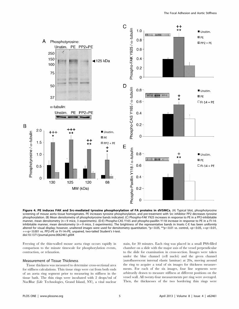
Then, the thicknesses of the two bordering thin rings were
Figure 4. PE induces FAK and Src-mediated tyrosine phosphorylation of FA proteins in dVSMCs. (A) Typical blot, phosphotyrosinescreening of mouse aorta tissue homogenates. PE increases tyrosine phosphorylation, and pre-treatment with Src inhibitor PP2 decreases tyrosinephosphorylation. (B) Mean densitometry of phosphotyrosine bands indicated. (C) Phospho-FAK Y925 increases in response to PE in a PP2-inhibitablemanner, mean densitometry (n = 9 mice, 3 experiments). (D-E) Phospho-CAS Y165 and phospho-paxillin Y118 increase in response to PE in a FI-14-inhibitable manner, mean densitometry (n = 9 mice, 3 experiments). The brightness of the representative bands in Insets C-E has been uniformlyaltered for visual display; however, unaltered images were used for densitometry quantitation. *p,0.05, **p,0.01 vs. control, +p,0.05, ++p,0.01,+++p,0.001 vs. PP2+PE or FI-14+PE, unpaired, two-tailed Student’s t-test.doi:10.1371/journal.pone.0062461.g004
The Focal Adhesion and Aortic Stiffness
PLOS ONE | www.plosone.org 5 April 2013 | Volume 8 | Issue 4 | e62461
averaged to determine the mean thickness of the aorta ring
segment (,60 mm).
Tissue Stiffness Measurements with SinusoidalPerturbationsFor in vitro force and stiffness measurements, thin triangular
shaped wire clasps (d = 0.005 in) were threaded through the lumen
of the aorta rings then attached at one end to a fixed hook and at
the other end to a computer-controlled motorized lever arm
(Dual-Mode Lever Arm System, Model 300C, Aurora Scientific,
Ontario, Canada) capable of setting tissue length while simulta-
neously measuring force. The rings were stretched uniaxially in the
circumferential direction, as vascular smooth muscle cells in the
aorta wall are oriented primarily in this direction, and this is the
primary direction of strain induced by pulsatile flow in vivo.
Rings were stretched to optimal length LO (1.8 x slack length, or
equivalently a strain e=80%) for 30 minutes to allow adequate
time for stress relaxation (i.e., for tensile force to stabilize at
a steady state level). The optimal length for maximal contraction
was predetermined by length-tension curves (data not shown); for
mouse aorta, LO is near the upper end of the tissue’s physiologic
strain range during systolic/diastolic pressure transients [28].
Following this step, the tissue was stimulated to contract for 15
minutes by depolarization with physiologic saline solution in which
51 mM NaCl had been replaced by KCl, followed by a series of
three washouts with regular PSS and a 30 minute relaxation
period. The observation of the magnitude of the KCl contractions
confirms viability and adequate equilibration of the tissue. Then
rings were incubated with vehicle (DMSO, 1:1,000; or ethanol
1:1,000), PP2 (10 mM), FAK inhibitor-14 (FI-14) (10 mM), or ML-
9 (10 mM) for an additional 30 minutes. Stiffness measurements at
optimal length LO were collected continuously during ten minutes
of stimulation with the alpha-agonist phenylephrine (PE, 10 mM)
at a maximally effective concentration.
Tissue stiffness was measured by small-amplitude (1%), high-
frequency (40 Hz) sinusoidal length perturbations as previously
described [4,29]. This regime has previously been shown to
measure total tissue stiffness without breaking actin-myosin
crossbridges [4]. With oscillatory stretching, tissue stiffness
E=E’+iE’’, where E’ is the elastic or storage modulus (the in-
phase component of the stiffness), E’’ is the viscous or loss modulus
(the out-of-phase component of the stiffness), and i is the unit
imaginary number. As reported previously [4], with this protocol
the force and length signals are in-phase, and therefore the out-of-
phase viscous component of the stiffness is negligible. Thus, the
tissue’s elastic modulus, or material stiffness, E is calculated as the
ratio of the stress s to the strain e, E= (DF/A)/(DL/LO), where DFis the amplitude of the force response to the cyclic stretches, A is
the cross-sectional area, and DL is the amplitude of the cyclic
stretches. When stretched to optimal length, the walls of the ring
collapse, allowing the cross-sectional area A to be calculated as
twice the product of the wall thickness h and the axial length of the
ring l, A= 2hl.
Western Blot Analysis and Measurement of LC20PhosphorylationQuick-frozen tissues were homogenized and processed at 4uC as
previously described [25]. The homogenization buffer was
designed to preserve phosphoproteins, and it consists of 20 mM
MOPS, 4% SDS, 10% glycerol, 10 mM DTT, 20 mM b-glycerophosphate, 5.5 mM leupeptin, 5.5 mM pepstatin, 20 KIU
aprotinin, 2 mM Na3VO4, 1 mM NaF, 100 mM ZnCl2, 20 mMAEBSF, and 5 mM EGTA. Western blot densitometry was
performed using Odyssey Infrared Imaging System (LI-COR
Biosciences, Lincoln, NE). Glycerol-urea gel electrophoresis was
performed to separate nonphosphorylated, monophosphorylated,
and diphosphorylated light chains as previously described [22],
and LC20 phosphorylation was calculated as a ratio of phosphor-
ylated LC20 to total LC20. LC20 phosphorylation was examined
30s into stimulation with PE, before both stiffness and contractile
force reach their maximal values, as we have shown previously
that this time point corresponds to maximum LC20 phosphory-
lation in aortic smooth muscle [30].
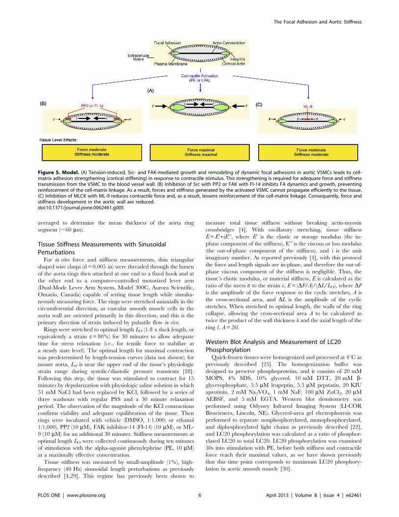
Figure 5. Model. (A) Tension-induced, Src- and FAK-mediated growth and remodeling of dynamic focal adhesions in aortic VSMCs leads to cell-matrix adhesion strengthening (cortical stiffening) in response to contractile stimulus. This strengthening is required for adequate force and stiffnesstransmission from the VSMC to the blood vessel wall. (B) Inhibition of Src with PP2 or FAK with FI-14 inhibits FA dynamics and growth, preventingreinforcement of the cell-matrix linkage. As a result, forces and stiffness generated by the activated VSMC cannot propagate efficiently to the tissue.(C) Inhibition of MLCK with ML-9 reduces contractile force and, as a result, lessens reinforcement of the cell-matrix linkage. Consequently, force andstiffness development in the aortic wall are reduced.doi:10.1371/journal.pone.0062461.g005
The Focal Adhesion and Aortic Stiffness
PLOS ONE | www.plosone.org 6 April 2013 | Volume 8 | Issue 4 | e62461
Reagents and AntibodiesAgonists used were LPA from Cayman Chemical (Ann
Arbor, MI) and phenylephrine from Sigma-Aldrich. PP2 was
purchased from EMD Biosciences (La Jolla, CA); FAK inhibitor
14 was purchased from Santa Cruz Biotechnology (Santa Cruz,
CA); and ML-9 was purchased from Calbiochem (La Jolla, CA).
General laboratory reagents were of analytical grade or better
and purchased from Sigma-Aldrich and Bio-Rad Laboratories
(Hercules, CA). The following primary antibodies were used:
phospho-tyrosine (mouse, 1:500) from BD Biosciences (San Jose,
CA); phospho-FAK Y925 (rabbit, 1:200), phospho-paxillin Y118
(rabbit, 1:250), and phospho-CAS Y165 (rabbit, 1:250) from
Cell Signaling Technology (Danvers, MA); vinculin (mouse
V4505, 1:400) from Sigma-Aldrich; and a-tubulin (rabbit,
1:50,000 from Abcam (Cambridge, MA). For immunofluores-
cence experiments, goat anti-rabbit and goat anti-mouse Alexa
Fluor 488 and Alexa Fluor 568 (1:1,000; Invitrogen) were used
as secondary antibodies. For Western blot experiments, Goat
Oregon Green 488 or Alexa Fluor 568 labeled anti-rabbit or
anti-mouse IgGs were used as secondary antibodies (1:1,000; LI-
COR Biosciences, Lincoln, NE).
Statistical AnalysisData are reported as means +/2 standard error. Differences
between individual means were determined by two-tailed
Student’s t-test. Differences were considered significant at p,0.05.
Results
VSMC Cortical Stiffness is Modulated by FAs andContractile ActivationTo test the role of FAs in regulating cell stiffness, minimizing the
contribution of the ECM, we measured cortical stiffness of serum-
starved A7r5 VSMCs with MT (Figure 1A). For these experi-
ments, the position of the magnetic probe tip was adjusted to be in
the more linear range of the force-distance calibration curve
(Figure 1B). The low forces used here (,50 pN) confine stiffness
measurements to the cortex of the cell [16–19], encompassing the
bead-integrin attachment as well as the underlying FA and cortical
cytoskeleton.
Mean cortical stiffness (Figure 1C) was measured with RGD-
coated beads in order to bind integrins (0.12260.015 pN/nm).
A7r5 cells do not respond to classical vasoconstrictors, but
stimulation with non-mitogenic LPA mimics vasoconstrictors such
as PE since it increases myosin phosphorylation to activate myosin
[22,31,32]. LPA increased stiffness (0.15960.018 pN/nm) signif-
icantly (p,0.01), but pre-treatment before LPA stimulation with
PP2, a specific small molecule inhibitor that prevents FA
remodeling by inhibiting Src family kinases [22,33–35], reduced
cortical stiffness to untreated baseline levels. These results directly
implicate the FA and the surrounding cortical cytoskeleton in
agonist-induced increases in stiffness of the bead-ECM-cytoskel-
eton linkage in A7r5 VSMCs.
As a control, we performed experiments with bovine serum
albumin (BSA)-coated beads, which adhere nonspecifically to cell
membranes [36]. There was no difference between cortical
stiffness measured with RGD beads and BSA beads in the absence
of a stimulus; however, measurements of stiffness with the BSA
beads did not exhibit the increase in stiffness observed with RGD
beads following LPA stimulation of the A7r5 VSMCs (Figure 1C),
thereby confirming the existence of a functional focal adhesion
linkage between the RGD beads and the underlying cortical
cytoskeleton.
LPA Stimulation Increases FA Size in a PP2-attenuatedMannerA larger FA would have greater stiffness as measured by the
MT. We hypothesized that changes in FA dimensions might
underlie our cortical stiffness observations since FAs of migrating
cells grow in a force-dependent manner [37], and Src, together
with FAK, is known to affect FA recycling and growth in
nonmuscle cells [34]. Thus, we quantitated FA sizes using
deconvolution immunofluorescent imaging of vinculin, a marker
of FAs (Figure 2).
Mean FA size did not change with PP2 treatment alone. FAs
were significantly larger in cells stimulated with LPA (4.9760.26
mm2 versus 3.2460.14 mm2 in control cells, a 55% increase)
(p,0.001). This is consistent with effects reported in migrating
cells upon myosin activation and suggests that a tension-induced
increase in FA size in the presence of LPA increases cortical
stiffness. Additionally, the growth in FA size with LPA was
prevented by PP2, which is also consistent with the known effect of
PP2 to prevent FA remodeling and growth in other cell types
[33,34]. Vinculin has multiple actin binding sites and is known to
crosslink actin and the FA [38]; hence, increased vinculin staining
may reflect additional actin-FA interactions and concurrent
stiffening and strengthening of the FA and the surrounding
cortical cytoskeleton. There was no statistically significant differ-
ence between the number of focal adhesions per unit area
(0.05460.003 mm22) in any of the conditions examined.
Contractile Filament Activation is a Component ofAgonist-induced Aortic Tissue StiffnessTo extend our vascular smooth muscle cell studies to vascular
tissue, we measured stiffness of mouse aorta rings in vitro with
small-amplitude sinusoidal stretches that do not break crossbridges
[4]. This stretching produces an in-phase force response that
simplifies the stiffness calculation to a normalized ratio of the force
response to the imposed length cycling (see Methods). The sample
force trace (Figure 3A) illustrates the continuous measurement of
both force generation (the rise of the trace) and stiffening (the
widening of the force response with constant amplitude length
perturbations) during stimulation with the vasoconstrictor PE.
Additionally, the sample trace demonstrates the importance of
smooth muscle cell activation as a modulator of aortic wall
stiffness, as it represents roughly 20% of total stiffness. The
remaining 80% of the stiffness of activated tissue equals the
baseline, unstimulated stiffness (213+/27 kPa), which is likely
determined largely by the ECM. Changes in stiffness (‘‘stiffening’’)
are plotted in Figure 3 as opposed to absolute stiffness, as the focus
of this study is to examine the contribution of the smooth muscle
cell component rather than the matrix component to stiffness.
Smooth muscle contractility is regulated by myosin light chain
kinase (MLCK)-dependent myosin phosphorylation [39]. Rings
pre-treated with the MLCK inhibitor ML-9 before PE stimulation
exhibited a significant decrease in stress generation (43–53%)
(p,0.05) (Figure 3B) and a significant decrease in stiffening (47–
57%) (p,0.01–0.05) (Figure 3C). These results demonstrate that
the actomyosin contractile apparatus of the VSMC is a major
component of regulated blood vessel stiffness.
Aortic Tissue Stiffening is Decreased by Inhibition of Srcor FAKTo test the role of FA remodeling in regulating stiffness of the
intact vessel wall, we measured the stiffness of aortic rings pre-
treated with PP2 before PE activation. PP2 significantly decreased
PE-induced stiffening (71–84%) (p,0.001–0.05) (Figure 3C).
The Focal Adhesion and Aortic Stiffness
PLOS ONE | www.plosone.org 7 April 2013 | Volume 8 | Issue 4 | e62461
These results are consistent with the concept that Src-dependent
FA recycling is a significant regulator of aortic stiffness.
Unexpectedly, PP2 pre-treatment also significantly decreased
PE-induced stress generation (78–88%) (p,0.001–0.05)
(Figure 3B). At first glance, the magnitude of this decrease
suggests that PP2 might directly alter smooth muscle contrac-
tility, possibly by an effect on Rho (via p190RhoGAP). RhoA/
Rho-kinase signals downstream to MLC (myosin light chain)
phosphatase, increasing MLC phosphorylation [40,41]. We have
previously published that the alpha-agonist PE increases myosin
phosphorylation and activation [42] and that the concentration
of PP2 employed here does not affect MLC phosphorylation in
aortic tissue of the ferret [22]. We have now confirmed by urea
gel analysis that PE induces MLC phosphorylation in mouse
aorta, unaffected by PP2 (unstimulated, untreated: 0.218+/20.046 mol PO4/mol LC; PE: 0.375+/20.049 mol PO4/mol
LC, p,0.05; PP2+PE: 0.362+/20.039 mol PO4/mol LC, n.s.,
n = 5). Src is also known in some systems to regulate actin
polymerization [40]. In vascular smooth muscle tissue, actin
remodeling induced by PE has been shown to be largely cortical
[6]. Thus, the inhibition of Src function by PP2 seems to
involve solely the focal adhesion and the associated cortical
cytoskeleton, presumably hindering the transmission of force
from the contractile filaments across the focal adhesion.
We also measured stress and stiffness for aortic rings pre-treated
with the specific small molecule inhibitor FAK inhibitor 14 before
PE activation [43]. As with PP2, FI-14 also significantly decreased
PE-induced stiffening (40–73%) (p,0.01–0.05) (Figure 3C) and
stress generation (47–96%) (p,0.01–0.05) (Figure 3B). Taken
together, these results implicate the FAK-Src signaling complex of
the focal adhesion as a regulator of tension and stiffness in the
aortic wall.
Src- and FAK- Mediated Tyrosine Phosphorylation of FAProteins Parallels Increases in Aortic StiffnessTo confirm that PP2 inhibits Src-dependent FA signaling in
aortic tissue, we performed phosphotyrosine screens of aorta
homogenates. As seen in Figures 4A and 4B, PE increases tyrosine
phosphorylation, and PE-induced increases in phosphotyrosine are
inhibited by PP2, notably for bands at 130, 125, 120 and 68 kDa
(p,0.001–0.05). These bands have been previously shown to
correspond to focal adhesion proteins, including CAS, FAK, and
paxillin, in aorta tissue from the ferret [22].
The p125 band co-stained with an antibody against 125-kDa
FAK, a major FA signaling protein and Src binding protein [44].
A FAK Y925-site-specific phospho-antibody confirmed the
presence of a significant PE-induced increase in phosphorylation
and a significant inhibition of the PE effect by PP2 (p,0.01)
(Figure 4C). FAK Y925 is a Src substrate that signals downstream
to ERK [44], which is also known to be activated by PE in aortic
tissue [22,45,46].
To further explore agonist-induced FA signaling, we examined
phosphotyrosine levels in the presence of FAK inhibitor 14.
Phospho-specific antibodies against p130 CAS Y165 and p68
paxillin Y118, both substrates of the FAK-Src signaling complex
[44], demonstrated statistically significant PE-induced increases in
phosphorylation that are significantly inhibited by FI-14 (p,0.01–
0.05) (Figures 4D–E).
Thus, taken together, these results are consistent with the
concept that, at the tissue level, vasoconstrictor activation initiates
Src- and FAK-dependent FA signaling, which, in turn, regulates
aortic stiffness and contractility.
Discussion
The main finding of this study is that the FA, as a cell-matrix
linker, via the FAK-Src signaling network, is a significant and
regulated component of aortic stiffness and tone. As a first step
towards determining whether the cytoskeleton-FA linkage mod-
ulates vascular tissue stiffness, we measured cortical stiffness on the
surface of aortic smooth muscle cells. To the best of our
knowledge, our use of MT to measure cortical stiffness in VSMCs
is novel but complementary to other techniques employing larger
forces to sample whole cell stiffness [36,47]. Others have shown
that the nanometer lengths and piconewton forces we used with
the MT are sufficiently small and weak to restrict perturbations to
several hundred nanometers into only the cortex of the cell [16–
19]. The 2.8-mm beads used for these experiments, roughly the
size of individual FAs in these cells, allow selective probing of
individual FAs. Both our bead and tissue deflections reflect strains
that are less than 1%, insufficient to disrupt crossbridges [4].
Although the MT force range can evoke local responses from
individual FA proteins in vitro [48–51], these forces are in-
sufficient to activate large-scale biological responses, such as
micromyogenic responses in VSMCs [36], which could obscure
cortical measurements. Pulling with the MT is in the lateral
direction, in contrast to the perpendicular indentation or re-
traction of atomic force microscopy or the torsion of magnetic
twisting cytometry. Lateral pulls are closer to the direction of
insertion of actin stress fibers into the cortex [52] and, therefore,
sample cortical stiffness in a physiologically relevant direction.
The MT experiments revealed that cortical stiffness is increased
by contractile stimulation with LPA in a Src-dependent manner.
Cell immunofluorescence imaging was employed to explain the
effects of LPA and PP2 on cortical stiffness. The imaging data
demonstrate that contractile activation with LPA induces FA
growth, similar to what has been observed in migrating and
spreading cells [31], likely due to increased intracellular load on
FAs [37]. The A7r5s cells are strongly adherent to the coverslips
and do not shorten when stimulated, although they do activate
contractile signaling pathways as shown by myosin light chain
phosphorylation [53]. Therefore, the increase in FA area is not
due to a decrease in cell size, which might bring distant FAs into
close proximity or merge them into a single unit. Additionally, our
measurements in the tissue were made at constant length, which
rules out such effects at that length scale. Other investigators have
reported that the relationship between FA size (based on vinculin
staining) and traction forces in non-migrating cells is linear, i.e.,
larger FAs correlate with larger traction forces [37,54]. Since in
our system of non-migrating smooth muscle cells we examined FA
size using vinculin as a marker, larger FAs may also reflect
increased vinculin cross-linking of actin to the FA [38]. Thus, one
may speculate that larger FAs, and the additional actin associated
with them, form a stiffer, stronger link between the inside and
outside of the cell, which is necessary to stably transmit greater
forces to a cell’s substrate [31]. Additionally, we have previously
shown by differential centrifugation in aorta of the ferret that CAS
and Src redistribute in response to agonist stimulation; therefore,
similar remodeling of the FA likely accompanies FA growth in
response to agonist in rodent aorta [22].
After demonstrating that FAs and especially the FAK-Src
signaling network are important to the cortical stiffening of a VSM
cell model during contractile stimulation, we scaled up to in vitro
stiffness measurements of rodent aorta. We found that smooth
muscle contraction accounts for roughly 20% of stiffness, with the
remaining 80% likely being due to extracellular matrix. Others
have published that the ECM plays an important role in
The Focal Adhesion and Aortic Stiffness
PLOS ONE | www.plosone.org 8 April 2013 | Volume 8 | Issue 4 | e62461

determining tissue stiffness and that changes in the ECM that
occur with age or disease can affect tissue stiffness [3], but the
workings of the smooth muscle component of stiffness are not as
well understood. We found that FI-14 inhibited ,60% of vascular
stiffening and ,50–95% of stress generation during stimulation,
and PP2 inhibited ,70% of vascular stiffening and,80% of stress
generation during stimulation without affecting myosin light chain
phosphorylation, which, together with our examination of
phosphotyrosine signaling in the FAK-Src pathway in this tissue,
is consistent with the concept that the FA, including cortical
cytoskeletal connections, is a major regulator of stiffness. This
finding is noteworthy given that until recently FAs in contractile
vascular smooth muscle embedded in blood vessel walls were
considered to be relatively static structures, in contrast to their
highly dynamic analogues in migrating cell types [37]. Similarly,
tyrosine phosphorylation of FA proteins, particularly by FAK and
Src, is the hallmark of their turnover in migrating, cultured cells
[34,37] but has only recently been studied in contractile smooth
muscle [22,55,56]. Since essentially all other known signaling
events regulating contractile filament activation in contractile
VSMCs involve serine or threonine phosphorylations [39],
tyrosine phosphorylation events in these cells appear to be largely
linked to the dynamics of FA proteins and the associated cortical
actin cytoskeleton.
Taking these findings together, it appears that agonist-induced
FA growth and remodeling entails strengthening of cytoskeleton-
matrix linkages in VSM, permitting adequate force transmission
from contracting cells to the vascular wall (see Model, Figure 5A).
Prevention of FA/cortical cytoskeletal recycling via FAK-Src
signaling, and by preventing FA growth, inhibits both force
transmission and stiffness development (Figure 5B). Conversely,
prevention of myosin activation directly inhibits force transmission
and, by decreasing cytoskeletal tension, appears to also prevent FA
growth, which decreases stiffness and further inhibits force
transmission (Figure 5C). The fact that cortical actin and FA
remodeling are concurrent in this system [6] might lead to changes
in output via a FA-actin cytoskeleton ‘‘clutch’’ mechanism
described for other cell types [57] that may control the efficiency
of force transfer to the blood vessel wall.
In summary, our results point to the FA of the VSMC,
specifically through interactions of FAK and Src, as a regulator of
aortic vessel stiffness. We report that the FAs of these nonmigrat-
ing cells embedded in the wall of the aorta are regulated by
vasoconstrictors and coordinate changes in force and stiffness
development in the tissue. Given that increases in aortic stiffness
are linked to cardiovascular disease, these results also point to the
FA as a potential novel therapeutic target.
Acknowledgments
We would like to thank Dr. Richard Cohen at Boston University for his
invaluable input and enlightening discussions; Howard Cohen at Boston
University for assisting in the assembly of the magnetic tweezers apparatus;
the Boston University Scientific Instrument Facility for sharpening the
electromagnet tip for the magnetic tweezers; and Dr. Fabry’s group at the
University of Erlangen-Nuremberg for guidance in the design of the
magnetic tweezers apparatus.
Author Contributions
Conceived and designed the experiments: RJS YZG MHJ SV JRM KGM.
Performed the experiments: RJS CMG SV. Analyzed the data: RJS CMG
SV JRM KGM. Wrote the paper: RJS YZG MHJ CMG SV JRM KGM.
References
1. Mitchell GF, Hwang SJ, Vasan RS, Larson MG, Pencina MJ, et al. (2010)
Arterial stiffness and cardiovascular events: the Framingham Heart Study.
Circulation 121: 505–511.
2. Kaess BM, Rong J, Larson MG, Hamburg NM, Vita JA, et al. (2012) Aortic
stiffness, blood pressure progression, and incident hypertension. JAMA 308:
875–881.
3. Greenwald SE (2007) Ageing of the conduit arteries. J Pathol 211: 157–172.
4. Brozovich FV, Morgan KG (1989) Stimulus-specific changes in mechanical
properties of vascular smooth muscle. Am J Physiol 257: H1573–1580.
5. Qiu H, Zhu Y, Sun Z, Trzeciakowski JP, Gansner M, et al. (2010) Short
communication: vascular smooth muscle cell stiffness as a mechanism for
increased aortic stiffness with aging. Circ Res 107: 615–619.
6. Kim HR, Gallant C, Leavis PC, Gunst SJ, Morgan KG (2008) Cytoskeletal
remodeling in differentiated vascular smooth muscle is actin isoform dependent
and stimulus dependent. Am J Physiol Cell Physiol 295: C768–778.
7. Small JV (1985) Geometry of actin-membrane attachments in the smooth
muscle cell: the localisations of vinculin and alpha-actinin. EMBO J 4: 45–49.
8. Vetterkind S, Saphirstein RJ, Morgan KG (2011) Stimulus-specific activation
and actin dependency of distinct, spatially separated ERK1/2 fractions in A7r5
smooth muscle cells. PLoS One 7: e30409.
9. Kimes BW, Brandt BL (1976) Characterization of two putative smooth muscle
cell lines from rat thoracic aorta. Exp Cell Res 98: 349–366.
10. Firulli AB, Han D, Kelly-Roloff L, Koteliansky VE, Schwartz SM, et al. (1998)
A comparative molecular analysis of four rat smooth muscle cell lines. In Vitro
Cell Dev Biol Anim 34: 217–226.
11. Gimona M, Kaverina I, Resch GP, Vignal E, Burgstaller G (2003) Calponin
repeats regulate actin filament stability and formation of podosomes in smooth
muscle cells. Mol Biol Cell 14: 2482–2491.
12. Martinez-Lemus LA, Wu X, Wilson E, Hill MA, Davis GE, et al. (2003)
Integrins as unique receptors for vascular control. J Vasc Res 40: 211–233.
13. Mogford JE, Davis GE, Meininger GA (1997) RGDN peptide interaction with
endothelial alpha5beta1 integrin causes sustained endothelin-dependent vaso-
constriction of rat skeletal muscle arterioles. J Clin Invest 100: 1647–1653.
14. Pierschbacher MD, Ruoslahti E (1987) Influence of stereochemistry of the
sequence Arg-Gly-Asp-Xaa on binding specificity in cell adhesion. J Biol Chem
262: 17294–17298.
15. Kollmannsberger P, Fabry B (2007) High-force magnetic tweezers with force
feedback for biological applications. Rev Sci Instrum 78: 114301.
16. Kasas S, Wang X, Hirling H, Marsault R, Huni B, et al. (2005) Superficial and
deep changes of cellular mechanical properties following cytoskeleton disassem-
bly. Cell Motil Cytoskeleton 62: 124–132.
17. Matthews BD, Overby DR, Alenghat FJ, Karavitis J, Numaguchi Y, et al. (2004)
Mechanical properties of individual focal adhesions probed with a magnetic
microneedle. Biochem Biophys Res Commun 313: 758–764.
18. Oberleithner H, Callies C, Kusche-Vihrog K, Schillers H, Shahin V, et al.
(2009) Potassium softens vascular endothelium and increases nitric oxide release.
Proc Natl Acad Sci U S A 106: 2829–2834.
19. Rotsch C, Jacobson K, Radmacher M (1999) Dimensional and mechanical
dynamics of active and stable edges in motile fibroblasts investigated by using
atomic force microscopy. Proc Natl Acad Sci U S A 96: 921–926.
20. Matthews BD, Overby DR, Mannix R, Ingber DE (2006) Cellular adaptation to
mechanical stress: role of integrins, Rho, cytoskeletal tension and mechan-
osensitive ion channels. J Cell Sci 119: 508–518.
21. Meijering E, Dzyubachyk O, Smal I (2012) Methods for cell and particle
tracking. Methods Enzymol 504: 183–200.
22. Min J, Reznichenko M, Poythress RH, Gallant CM, Vetterkind S, et al. (2012)
Src modulates contractile vascular smooth muscle function via regulation of focal
adhesions. J Cell Physiol.
23. Cai Y, Sheetz MP (2009) Force propagation across cells: mechanical coherence
of dynamic cytoskeletons. Curr Opin Cell Biol 21: 47–50.
24. Bulter WE, Peterson JW, Zervas NT, Morgan KG (1996) Intracellular calcium,
myosin light chain phosphorylation, and contractile force in experimental
cerebral vasospasm. Neurosurgery 38: 781–787; discussion 787–788.
25. Marganski WA, Gangopadhyay SS, Je HD, Gallant C, Morgan KG (2005)
Targeting of a novel Ca+2/calmodulin-dependent protein kinase II is essential
for extracellular signal-regulated kinase-mediated signaling in differentiated
smooth muscle cells. Circ Res 97: 541–549.
26. Driska SP, Aksoy MO, Murphy RA (1981) Myosin light chain phosphorylation
associated with contraction in arterial smooth muscle. Am J Physiol 240: C222–
233.
27. Barany M (1996) Biochemistry of smooth muscle contraction. San Diego:
Academic Press.
28. Wagenseil JE, Mecham RP (2009) Vascular extracellular matrix and arterial
mechanics. Physiol Rev 89: 957–989.
29. Rhee AY, Brozovich FV (2000) The smooth muscle cross-bridge cycle studied
using sinusoidal length perturbations. Biophys J 79: 1511–1523.
The Focal Adhesion and Aortic Stiffness
PLOS ONE | www.plosone.org 9 April 2013 | Volume 8 | Issue 4 | e62461

30. Jiang MJ, Morgan KG (1989) Agonist-specific myosin phosphorylation and
intracellular calcium during isometric contractions of arterial smooth muscle.
Pflugers Arch 413: 637–643.
31. Chrzanowska-Wodnicka M, Burridge K (1996) Rho-stimulated contractility
drives the formation of stress fibers and focal adhesions. J Cell Biol 133: 1403–
1415.
32. Jones LG, Ella KM, Bradshaw CD, Gause KC, Dey M, et al. (1994) Activations
of mitogen-activated protein kinases and phospholipase D in A7r5 vascular
smooth muscle cells. J Biol Chem 269: 23790–23799.
33. Fincham VJ, Frame MC (1998) The catalytic activity of Src is dispensable for
translocation to focal adhesions but controls the turnover of these structures
during cell motility. EMBO J 17: 81–92.
34. Webb DJ, Donais K, Whitmore LA, Thomas SM, Turner CE, et al. (2004)
FAK-Src signalling through paxillin, ERK and MLCK regulates adhesion
disassembly. Nat Cell Biol 6: 154–161.
35. Bain J, Plater L, Elliott M, Shpiro N, Hastie CJ, et al. (2007) The selectivity of
protein kinase inhibitors: a further update. Biochem J 408: 297–315.
36. Sun Z, Martinez-Lemus LA, Hill MA, Meininger GA (2008) Extracellular
matrix-specific focal adhesions in vascular smooth muscle produce mechanically
active adhesion sites. Am J Physiol Cell Physiol 295: C268–278.
37. Bershadsky AD, Balaban NQ, Geiger B (2003) Adhesion-dependent cell
mechanosensitivity. Annu Rev Cell Dev Biol 19: 677–695.
38. Mierke CT (2009) The Role of Vinculin in the Regulation of the Mechanical
Properties of Cells. Cell Biochemistry and Biophysics 53: 115–126.
39. Horowitz A, Menice CB, Laporte R, Morgan KG (1996) Mechanisms of smooth
muscle contraction. Physiol Rev 76: 967–1003.
40. Huveneers S, Danen EH (2009) Adhesion signaling - crosstalk between integrins,
Src and Rho. J Cell Sci 122: 1059–1069.
41. Somlyo AP, Somlyo AV (2003) Ca2+ sensitivity of smooth muscle and
nonmuscle myosin II: modulated by G proteins, kinases, and myosin
phosphatase. Physiol Rev 83: 1325–1358.
42. Shin HM, Je HD, Gallant C, Tao TC, Hartshorne DJ, et al. (2002) Differential
association and localization of myosin phosphatase subunits during agonist-
induced signal transduction in smooth muscle. Circ Res 90: 546–553.
43. Golubovskaya VM, Nyberg C, Zheng M, Kweh F, Magis A, et al. (2008) A small
molecule inhibitor, 1,2,4,5-benzenetetraamine tetrahydrochloride, targeting the
y397 site of focal adhesion kinase decreases tumor growth. Journal of Medicinal
Chemistry 51: 7405–7416.44. Mitra SK, Hanson DA, Schlaepfer DD (2005) Focal adhesion kinase: in
command and control of cell motility. Nat Rev Mol Cell Biol 6: 56–68.
45. Khalil RA, Menice CB, Wang CL, Morgan KG (1995) Phosphotyrosine-dependent targeting of mitogen-activated protein kinase in differentiated
contractile vascular cells. Circ Res 76: 1101–1108.46. Dessy C, Kim I, Sougnez CL, Laporte R, Morgan KG (1998) A role for MAP
kinase in differentiated smooth muscle contraction evoked by alpha-adreno-
ceptor stimulation. Am J Physiol 275: C1081–1086.47. Deng L, Trepat X, Butler JP, Millet E, Morgan KG, et al. (2006) Fast and slow
dynamics of the cytoskeleton. Nat Mater 5: 636–640.48. Wang Y, Botvinick EL, Zhao Y, Berns MW, Usami S, et al. (2005) Visualizing
the mechanical activation of Src. Nature 434: 1040–1045.49. Geiger B, Bershadsky A, Pankov R, Yamada KM (2001) Transmembrane
crosstalk between the extracellular matrix–cytoskeleton crosstalk. Nat Rev Mol
Cell Biol 2: 793–805.50. Choquet D, Felsenfeld DP, Sheetz MP (1997) Extracellular matrix rigidity causes
strengthening of integrin-cytoskeleton linkages. Cell 88: 39–48.51. Felsenfeld DP, Schwartzberg PL, Venegas A, Tse R, Sheetz MP (1999) Selective
regulation of integrin–cytoskeleton interactions by the tyrosine kinase Src. Nat
Cell Biol 1: 200–206.52. North AJ, Gimona M, Lando Z, Small JV (1994) Actin isoform compartments in
chicken gizzard smooth muscle cells. J Cell Sci 107 (Pt 3): 445–455.53. Vetterkind S, Lee E, Sundberg E, Poythress RH, Tao TC, et al. (2010) Par-4:
a new activator of myosin phosphatase. Mol Biol Cell 21: 1214–1224.54. Balaban NQ, Schwarz US, Riveline D, Goichberg P, Tzur G, et al. (2001) Force
and focal adhesion assembly: a close relationship studied using elastic
micropatterned substrates. Nat Cell Biol 3: 466–472.55. Zhang W, Gunst SJ (2008) Interactions of airway smooth muscle cells with their
tissue matrix: implications for contraction. Proc Am Thorac Soc 5: 32–39.56. Li Y, Gallant C, Malek S, Morgan KG (2007) Focal adhesion signaling is
required for myometrial ERK activation and contractile phenotype switch
before labor. Journal of Cellular Biochemistry 100: 129–140.57. Hu K, Ji L, Applegate KT, Danuser G, Waterman-Storer CM (2007)
Differential transmission of actin motion within focal adhesions. Science 315:111–115.
The Focal Adhesion and Aortic Stiffness
PLOS ONE | www.plosone.org 10 April 2013 | Volume 8 | Issue 4 | e62461
Copyright © 2022 FDOKUMEN