Three-Dimensional In Vitro Effects of Compression and Time in Culture on Aggregate Modulus and on...
10
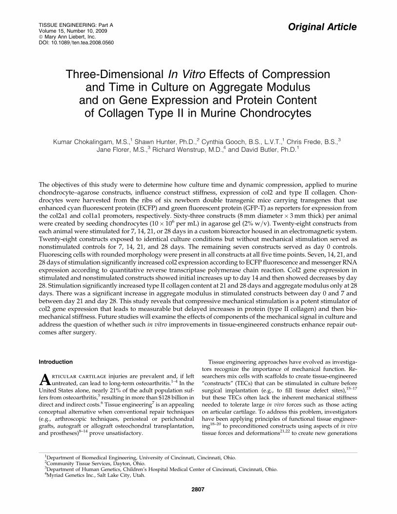
Original Article Three-Dimensional In Vitro Effects of Compression and Time in Culture on Aggregate Modulus and on Gene Expression and Protein Content of Collagen Type II in Murine Chondrocytes Kumar Chokalingam, M.S., 1 Shawn Hunter, Ph.D., 2 Cynthia Gooch, B.S., L.V.T., 1 Chris Frede, B.S., 3 Jane Florer, M.S., 3 Richard Wenstrup, M.D., 4 and David Butler, Ph.D. 1 The objectives of this study were to determine how culture time and dynamic compression, applied to murine chondrocyte–agarose constructs, influence construct stiffness, expression of col2 and type II collagen. Chon- drocytes were harvested from the ribs of six newborn double transgenic mice carrying transgenes that use enhanced cyan fluorescent protein (ECFP) and green fluorescent protein (GFP-T) as reporters for expression from the col2a1 and col1a1 promoters, respectively. Sixty-three constructs (8 mm diameter 3 mm thick) per animal were created by seeding chondrocytes (10 10 6 per mL) in agarose gel (2% w=v). Twenty-eight constructs from each animal were stimulated for 7, 14, 21, or 28 days in a custom bioreactor housed in an electromagnetic system. Twenty-eight constructs exposed to identical culture conditions but without mechanical stimulation served as nonstimulated controls for 7, 14, 21, and 28 days. The remaining seven constructs served as day 0 controls. Fluorescing cells with rounded morphology were present in all constructs at all five time points. Seven, 14, 21, and 28 days of stimulation significantly increased col2 expression according to ECFP fluorescence and messenger RNA expression according to quantitative reverse transcriptase polymerase chain reaction. Col2 gene expression in stimulated and nonstimulated constructs showed initial increases up to day 14 and then showed decreases by day 28. Stimulation significantly increased type II collagen content at 21 and 28 days and aggregate modulus only at 28 days. There was a significant increase in aggregate modulus in stimulated constructs between day 0 and 7 and between day 21 and day 28. This study reveals that compressive mechanical stimulation is a potent stimulator of col2 gene expression that leads to measurable but delayed increases in protein (type II collagen) and then bio- mechanical stiffness. Future studies will examine the effects of components of the mechanical signal in culture and address the question of whether such in vitro improvements in tissue-engineered constructs enhance repair out- comes after surgery. Introduction A rticular cartilage injuries are prevalent and, if left untreated, can lead to long-term osteoarthritis. 1–4 In the United States alone, nearly 21% of the adult population suf- fers from osteoarthritis, 5 resulting in more than $128 billion in direct and indirect costs. 6 Tissue engineering 7 is an appealing conceptual alternative when conventional repair techniques (e.g., arthroscopic techniques, periosteal or perichondral grafts, autograft or allograft osteochondral transplantation, and prostheses) 8–14 prove unsatisfactory. Tissue engineering approaches have evolved as investiga- tors recognize the importance of mechanical function. Re- searchers mix cells with scaffolds to create tissue-engineered ‘‘constructs’’ (TECs) that can be stimulated in culture before surgical implantation (e.g., to fill tissue defect sites), 15–17 but these TECs often lack the inherent mechanical stiffness needed to tolerate large in vivo forces such as those acting on articular cartilage. To address this problem, investigators have been applying principles of functional tissue engineer- ing 18–20 to preconditioned constructs using aspects of in vivo tissue forces and deformations 21,22 to create new generations 1 Department of Biomedical Engineering, University of Cincinnati, Cincinnati, Ohio. 2 Community Tissue Services, Dayton, Ohio. 3 Department of Human Genetics, Children’s Hospital Medical Center of Cincinnati, Cincinnati, Ohio. 4 Myriad Genetics Inc., Salt Lake City, Utah. TISSUE ENGINEERING: Part A Volume 15, Number 10, 2009 ª Mary Ann Liebert, Inc. DOI: 10.1089=ten.tea.2008.0560 2807
-
Upload
independent -
Category
Documents
-
view
0 -
download
0
Transcript of Three-Dimensional In Vitro Effects of Compression and Time in Culture on Aggregate Modulus and on...
Original Article
Three-Dimensional In Vitro Effects of Compressionand Time in Culture on Aggregate Modulus
and on Gene Expression and Protein Contentof Collagen Type II in Murine Chondrocytes
Kumar Chokalingam, M.S.,1 Shawn Hunter, Ph.D.,2 Cynthia Gooch, B.S., L.V.T.,1 Chris Frede, B.S.,3
Jane Florer, M.S.,3 Richard Wenstrup, M.D.,4 and David Butler, Ph.D.1
The objectives of this study were to determine how culture time and dynamic compression, applied to murinechondrocyte–agarose constructs, influence construct stiffness, expression of col2 and type II collagen. Chon-drocytes were harvested from the ribs of six newborn double transgenic mice carrying transgenes that useenhanced cyan fluorescent protein (ECFP) and green fluorescent protein (GFP-T) as reporters for expression fromthe col2a1 and col1a1 promoters, respectively. Sixty-three constructs (8 mm diameter� 3 mm thick) per animalwere created by seeding chondrocytes (10� 106 per mL) in agarose gel (2% w=v). Twenty-eight constructs fromeach animal were stimulated for 7, 14, 21, or 28 days in a custom bioreactor housed in an electromagnetic system.Twenty-eight constructs exposed to identical culture conditions but without mechanical stimulation served asnonstimulated controls for 7, 14, 21, and 28 days. The remaining seven constructs served as day 0 controls.Fluorescing cells with rounded morphology were present in all constructs at all five time points. Seven, 14, 21, and28 days of stimulation significantly increased col2 expression according to ECFP fluorescence and messenger RNAexpression according to quantitative reverse transcriptase polymerase chain reaction. Col2 gene expression instimulated and nonstimulated constructs showed initial increases up to day 14 and then showed decreases by day28. Stimulation significantly increased type II collagen content at 21 and 28 days and aggregate modulus only at 28days. There was a significant increase in aggregate modulus in stimulated constructs between day 0 and 7 andbetween day 21 and day 28. This study reveals that compressive mechanical stimulation is a potent stimulator ofcol2 gene expression that leads to measurable but delayed increases in protein (type II collagen) and then bio-mechanical stiffness. Future studies will examine the effects of components of the mechanical signal in culture andaddress the question of whether such in vitro improvements in tissue-engineered constructs enhance repair out-comes after surgery.
Introduction
Articular cartilage injuries are prevalent and, if leftuntreated, can lead to long-term osteoarthritis.1–4 In the
United States alone, nearly 21% of the adult population suf-fers from osteoarthritis,5 resulting in more than $128 billion indirect and indirect costs.6 Tissue engineering7 is an appealingconceptual alternative when conventional repair techniques(e.g., arthroscopic techniques, periosteal or perichondralgrafts, autograft or allograft osteochondral transplantation,and prostheses)8–14 prove unsatisfactory.
Tissue engineering approaches have evolved as investiga-tors recognize the importance of mechanical function. Re-searchers mix cells with scaffolds to create tissue-engineered‘‘constructs’’ (TECs) that can be stimulated in culture beforesurgical implantation (e.g., to fill tissue defect sites),15–17
but these TECs often lack the inherent mechanical stiffnessneeded to tolerate large in vivo forces such as those actingon articular cartilage. To address this problem, investigatorshave been applying principles of functional tissue engineer-ing18–20 to preconditioned constructs using aspects of in vivotissue forces and deformations21,22 to create new generations
1Department of Biomedical Engineering, University of Cincinnati, Cincinnati, Ohio.2Community Tissue Services, Dayton, Ohio.3Department of Human Genetics, Children’s Hospital Medical Center of Cincinnati, Cincinnati, Ohio.4Myriad Genetics Inc., Salt Lake City, Utah.
TISSUE ENGINEERING: Part AVolume 15, Number 10, 2009ª Mary Ann Liebert, Inc.DOI: 10.1089=ten.tea.2008.0560
2807
of reparative tissues.23–27 Such preconditioning improvesconstruct material properties by increasing synthesis of ex-tracellular matrix proteins like collagens I and II24–29 and re-pair biomechanics after implantation into defect sites.23,25
Unfortunately, cartilage TECs often require months of in-termittent mechanical conditioning to achieve mechanicalproperties and protein composition even remotely appro-aching those of native cartilage.28 For example, Mauck et al.29
determined that cartilage TECs required a full 8 weeks ofdynamic loading in culture to achieve 75% and 25% of anormal tissue’s Young’s modulus and unconfined dynamicmodulus, respectively. Even with these mechanical improve-ments, total collagen content was still only 12% of nativetissue values.29 Although such long development cycles maybe satisfactory in a research setting, these intervals are notpractical for patients in a clinical setting.
To shorten the development cycle and reduce produc-tion costs, new technologies are needed to rapidly and re-peatedly assess the evolving TEC in culture. During the earlydevelopment period, tissue engineers should be able to non-destructively assess the TEC in culture to avoid creating in-appropriate collagen types (e.g. type I vs type II collagen in aTEC that could adversely affect the repair of articular carti-lage). For example, if investigators could monitor how me-chanical signaling influences col2 gene expression in near realtime, they could dramatically improve their ability to synthe-size type II collagen protein. Such technologies could also al-low the tissue engineer to determine which mechanical signalsmost increase col2 gene expression and compressive stiffness.
To address these needs, we have developed two innovativetechnologies. We have bred specialized double transgenic(DT) mice that carry transgenes that use enhanced cyan fluo-rescent protein (ECFP) and green fluorescent protein (GFP-T)as reporters for col2a1 and col1a1 expression, respectively.ECFP and GFP-T each have a half life of 24 h and serve asvisual reporters for cells that express the genes for types II andI collagen, respectively. ECFP linked to col2a1 offers the po-tential to ultimately track in near real time how various stimuli(mechanical and chemical) influence col2 gene activity in cul-ture. We have also designed and fabricated a bioreactor todeliver compressive displacement profiles with micron preci-sion to constructs in an incubator setting over 4-week intervals.This bioreactor is described elsewhere.30
Using these two technologies, our objectives in this studywere to test four hypotheses related to compressive stimula-tion of double transgenic chondrocyte–agarose constructs. Wehypothesized that compressive stimulation significantly in-creases ECFP fluorescence and col2 messenger RNA (mRNA)expression, type II collagen production, and TEC aggregatemodulus up to 28 days in culture. We also hypothesized thatECFP fluorescence and col2 mRNA expression are positivelycorrelated.
Experimental Design
Rib chondrocytes were obtained from six newborn DTmice (one cell line per mouse). Sixty-three cell–agarose con-structs (8 mm diameter� 3 mm thick) were created per animalusing one cell density (10�106 cells=mL)31 in a 2% w=v ratioto maintain chondrocyte phenotype32 and to achieve a powerof 85% to detect treatment-related differences if they existed.We compared the responses of 28 stimulated constructs and
28 nonstimulated controls at 7, 14, 21, and 28 days in culture(7 per time period). These results were also compared tofindings from seven additional nonstimulated controls percell line at day 0. Stimulated constructs all received the samecompressive stimulation profile (sinusoidal displacementpattern at 1 Hz to 10% peak strain for 1 h followed by 1 h ofrest repeated three times a day) adapted from Mauck et al.31
The seven stimulated and seven nonstimulated constructs ateach time point were assigned as follows: one for ECFP fluo-rescence in a spectrophotometer (measured in relative fluo-rescence units (RFUs)),33 one to evaluate changes in mRNAexpression for col2 and glyceraldehyde-3-phosphate dehy-drogenase (GAPDH) using real-time quantitative reversetranscriptase polymerase chain reaction (qRT-PCR),24 one forconfined compression to determine aggregate modulus,34 onefor cell viability using an 3-(4,5-dimethylthiazol-2-yl)-2,5-diphenyltetrazolium bromide (MTT) assay,35 and three con-structs for type II collagen content using enzyme-linked im-munosorbent assay (ELISA).36
Materials
Chondrocyte isolation and expansion
Creation of the murine DT model is described elsewhere.Newborn mice were euthanized using carbon dioxide (CO2)using protocols approved by the Institutional Animal Careand Use Committee. Chondrocytes were harvested from therib cages using previously published procedures.37 Briefly, ribcages from each mouse were repeatedly washed in phosphatebuffered saline (PBS) and digested in 2.5 mL of protease(Sigma P-8811; 2 mg=mL, St. Louis, MO) for 30 min in an in-cubator (Steri-Cult Model 3033, Forma Scientific, Marietta, OH).After incubating overnight in collagenase B solution (Sigma;1 mg=mL), the digest was washed with medium (phenol red–free Dulbecco’s modified Eagle medium, 10% fetal bovineserum (FBS), penicillan=streptomycin), filtered (70mm) to re-move remaining bone pieces, sorted using flow cytometry toexclude non-ECFP cells, plated in a 100-mm tissue culture dish(Falcon, Bedford, MA), and incubated for 1 day at passage 0(P0). Intracellular cyan fluorescence was evident in thesechondrocytes when visualized under a fluorescence micro-scope (Axiovert 25, Carl Zeiss Inc., Gottingen, Germany)equipped with ECFP and GFP-T filter sets (Omega, Brat-tleboro, VT). The monolayer chondrocyte culture was thentrypsinized using 0.25% EDTA. replated at 1�106 cells per 100-mm plate at P1, and cultured for 7 to 10 days. Chondrocyteswere passaged until P3. No intracellular GFP-T expressionwas seen in cultured chondrocytes from P0 through P3.
Construct preparation
Chondrocytes at P3 were suspended at 20�106 cells=mL inmedium (BGjB, Invitrogen-Gibco BRL=Life Technologies Inc.,Gaithersburg, MD) containing 1% antibiotic=antimycotic and10% FBS (Atlanta Biologicals, Lawrenceville, GA). Cell sus-pensions were mixed in equal volumes with 4% (w=v) agarose(type VII, low gelling temperature, Fisher Scientific, Pittsburgh,PA) and PBS to produce a cell–gel solution of 10�106 cells=mLin 2% (w=v) agarose. This solution was then cast for 20 minbetween sterile glass plates separated by 3-mm spacers. Disks(8 mm diameter) were cored using a sterile dermal punch andeach cultured in 5 mL of medium in a multiwell plate for 2 days
2808 CHOKALINGAM ET AL.
(henceforth denoted as day 0) in a standard incubator (Steri-Cult Model 3033, Forma Scientific) to allow cells to adjust totheir new culture environment. Medium was replaced daily.
Methods
Construct preparation for mechanical stimulation
All constructs were transferred from multiwell plates toindividual 35-mm glass-bottom tissue culture dishes (P35G-0-10-C, MatTek Corp, Ashland, MA) covered with TeflonFEP film (100 A; DuPont, Circleville, OH) using a customDelrin ring. The film, permeable to oxygen (O2) and CO2 butnot to water vapor, ensured sterility and prevented evapo-ration. The dish design is provided elsewhere (Fig. 3c30).
Dynamic compressive stimulation
Constructs were subjected to dynamic compression in acustom-built bioreactor housed in an electromagnetic testingsystem (ElectroForce 3200; BOSE Corp., Eden Prairie, MN).30
Briefly, this bioreactor consisted of 12 testing stations and achamber to sustain a cell culture environment (378C, 5% CO2,�75% relative humidity). Six of the 12 stations were equip-ped with loading rods to stimulate the constructs; the re-maining six were devoid of loading rods and housed thenonstimulated controls.
Before starting dynamic compression, the environmentalchamber of the bioreactor was aseptically cleaned, and thesystem’s culture environment was allowed to achieve steady-state culture conditions. Culture dishes were then placed intoeach holding station. Consistent with previous patterns,31
constructs were subjected to the compressive strain profiledescribed above. Feeding medium was replaced daily. Atspecified time points, dishes containing treated and controlconstructs were removed from the bioreactor.
Fluorescence microscopy
At each time point after the end of stimulation, the con-structs subjected to RFU analysis were washed in PBS for 1 hwith gentle shaking to remove medium and then visualized forECFP and GFP-T fluorescence under a fluorescence micro-scope (Axiovert 25, Carl Zeiss Inc.). To exclude cell auto-fluorescence, constructs were also visualized for rhodamineusing a 11002VZ (Chroma, Rockingham, VT) filter set.
Spectrophotometric analysis
After imaging, constructs assigned for gene expressionmeasurements were mechanically homogenized using a glassmortar and Teflon pestle. The homogenate was suspendedin 1 mL of PBS and pipetted into a black-bottom microplate(200mL per well in 5 wells). ECFP fluorescence in these di-gests was quantified in RFUs,33 reading the microplate in aspectrophotometer (Spectra Max M2, Molecular Devices, Sun-nyvale, CA) using excitation and emission wavelengths of436 nm and 486 nm, respectively, with a cut off filter of 475 nm.ECFP RFU was normalized to day 0 values for all samples.
Real-time qRT-PCR
RNA extraction, conventional gene expression analysis,and real-time qRT-PCR were performed according to pub-
lished protocols.24 Mouse-specific primers were used for typeII collagen and GAPDH gene expression. Before use in theexperiment, all primers were extensively tested under con-ventional and real-time qRT-PCR conditions to ensure thatthey were specific for each gene and that we obtained a singleclean band when running the electrophoresis gel (with theassociated base pair size for each gene). The absolute amountof the corresponding gene mRNA in each construct was ob-tained from the corresponding gene standard curve. Expres-sion was normalized by calculating the ratio between type IIcollagen and GAPDH genes for each sample. GAPDH wasused as the housekeeping gene, because it did not change withtreatment conditions when we mechanically stimulated cell–scaffold constructs in tension-containing mesenchymal stemcells from 10-week-old double transgenic mice and fromskeletally mature New Zealand white rabbits.24
Determination of cell viability
At each time point, cell viability in constructs was deter-mined using an MTT assay (Vybrant MTT Cell ProliferationAssay Kit, Invitrogen, Carlsbad, CA), in which live cells re-duce MTT to a strongly pigmented formazan product toprovide colorimetric indicators of cell viability, following themanufacturer’s instructions. Briefly the cell–agarose con-structs were homogenized using a mortar and pestle. Thehomogenate was incubated for 4 h in an incubator in 500mLof 12-mM MTT solution. This was followed by incubation for18 h in 5 mL sodium dodecyl sulfate–hydrochloric acid tocompletely dissolve the formazan. The absorbance of for-mazan was measured at 570 nm in a microplate spectropho-tometer (Spectra Max M2, Molecular Devices).35 The viabilityresults were normalized to day 0 constructs.
Determination of type II collagen content
A native type II collagen detection kit (Catalog #6009;Chondrex Inc., Redmond, WA) was used to quantify type IIcollagen. The three constructs from each treatment groupand animal were pooled. Each construct was washed in PBSfor 1 h with gentle shaking to remove medium. The con-structs were then lyophilized and their dry weight measuredand then incubated in cold water at 48C. Constructs weresubjected to pepsin digestion and elastase digestion follow-ing the manufacturer’s protocols. ELISA was measured ac-cording to these protocols.36
Biomechanical evaluationof the agarose–cell constructs
Aggregate modulus of the cell–agarose constructs wasdetermined using confined compression testing. Constructswere placed in cryovials and stored at �808C. Before testing,each tissue specimen was thawed in room-temperature PBSfor 10 min. A 6-mm-diameter core was created from thecenter of each construct and its height measured using alight-force micrometer (Model ID-C1012CE, Mitutoyo Corp.,Kawasaki, Japan). Each sample was placed into a 6-mm-diameter�4.3-mm-deep well and subjected to a constantcontact stress of 0.066 MPa until equilibrium displacementwas achieved. During testing, specimen height and appliedload were continuously measured using a fine-resolutionLinear variable differential transformer (�0.25@, Sensotec
COMPRESSIVE STIMULATION INCREASES GENE, PROTEIN, AND MECHANICAL PROPERTIES 2809
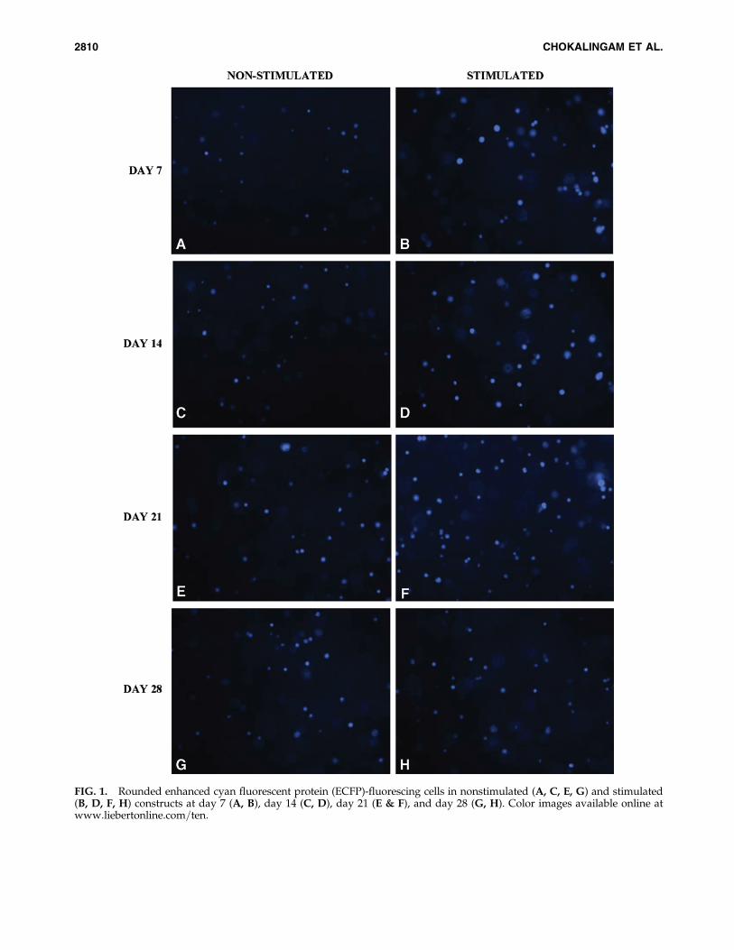
FIG. 1. Rounded enhanced cyan fluorescent protein (ECFP)-fluorescing cells in nonstimulated (A, C, E, G) and stimulated(B, D, F, H) constructs at day 7 (A, B), day 14 (C, D), day 21 (E & F), and day 28 (G, H). Color images available online atwww.liebertonline.com=ten.
2810 CHOKALINGAM ET AL.
Corp., Columbus, OH) and load cell (5 lb Sensotec Corp.,Columbus, OH), respectively. Tissue aggregate modulus(HA) was calculated using the formula HA¼ (Fo=pa2)=(u=h),where Fo is applied compressive force, a is specimen radius,h is specimen thickness, and u is equilibrium displacement.34
Statistical analysis
We determined the presence of treatment-related differ-ences between values for all response measures (ECFP RFUs,col2 mRNA, aggregate modulus, cell viability, and type IIcollagen content) using a two-way analysis of variance withTukey’s honestly significant difference post hoc testing (SASInstitute, Inc., Cary, NC)). We chose animal as a randomfactor, culture time and compressive stimulation as fixedfactors, and response measures as dependent variables.38
Significance was set at p< 0.05.
Results
Qualitative inspection of constructs showed cells withrounded morphology distributed homogenously in the con-
structs. Fluorescence microscopy revealed ECFP fluorescenceat day 0 in all three-dimensional (3-D) constructs that wasmaintained in 7-, 14-, 21-, and 28-day constructs (Fig. 1). Noqualitative differences were apparent in the amount or in-tensity of ECFP fluorescence between the images for non-stimulated and stimulated constructs at the four time points(Fig. 1A-G). Only cyan fluorescing cells were observed in thesections examined. No cells in any of the treatment groupsexpressed GFP-T and hence we did not measure RFUs orperform qRT-PCR for GFP-T and col1a1 expression, respec-tively. No autofluorescence was detected in samples usingrhodamine filter sets.
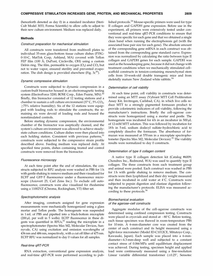
Quantitative results showed that applying a dynamiccompressive displacement significantly increased ECFP ex-pression in the 3-D constructs. Compressive stimulation sig-nificantly increased RFU values by 1.34,1.8,1.27, and 1.24times at 7,14, 21, and 28 days, respectively ( p< 0.05; Fig. 2A).There was a significant increase in RFUs in stimulated con-structs between days 7 and 14 and a significant decreasebetween days 21 and 28 ( p< 0.05; Fig. 2A). There was a sig-nificant decrease in RFUs in nonstimulated constructs be-tween days 21 and 28 ( p< 0.05; Fig. 2A).
Dynamic compressive displacement also significantly in-creased col2 mRNA according to qRT-PCR. Compressivestimulation significantly increased mRNA expression 1.5,1.32, 1.36, and 1.45 times at 7, 14, 21, and 28 days, respectively( p< 0.05; Fig. 2B). There was a significant increase in col2mRNA in stimulated constructs between days 7 and 14 and asignificant decrease between days 21 and 28 ( p< 0.05; Fig.2B). There was a significant increase in Col2 mRNA in non-stimulated constructs between days 7 and 14 and a significantdecrease between days 21 and 28 ( p< 0.05; Fig. 2B). Therewere also no significant differences in GAPDH values be-tween treatment conditions. The results from qRT-PCR andfluorescence analysis were also positively correlated (Fig. 3,coefficient of variation (r2)¼ 0.92 with a slope for the linearregression curve of 2.09).
Compressive stimulation also increased type II collagencontent of stimulated constructs but only after 21 and 28 daysof stimulation. Stimulated constructs showed 1.40 and 1.24times greater type II collagen content at 21 and 28 days,
Day 7 Day 14 Day 21 Day 280.0
0.5
1.0
1.5
2.0
2.5
3.0
3.5
4.0
4.5 Non-StimulatedStimulated
*,**
*,****
**
EC
FP
RF
U
A
Day 7 Day 14 Day 21 Day 280
2
4
6
8 Non-Stimulated
Stimulated
*
*,**
*,**
***
**
Typ
e II
Co
llag
enG
ene
Exp
ress
ion
Rel
ativ
e to
GA
PD
H
B
FIG. 2. Dynamic stimulation increases (A) ECFP expressionby relative fluorescence units (RFU) and (B) Col2 messengerRNA expression according to quantitative reverse transcrip-tase polymerase chain reaction (qRT-PCR) at all four timeperiods of observation. *Significantly greater than nonsti-mulated control at same time point ( p< 0.05). **Significantdifference from same treatment at previous time point. Datarepresented as mean� standard deviation. N¼ 6 for allgroups.
y = 2.0931x - 0.8998R2 = 0.9204
0
1
2
3
4
5
6
0 0.5 1 1.5 2 2.5 3 3.5RFU
qR
T-P
CR
DAY 7
DAY 14
DAY 21
DAY 28
FIG. 3. RFU fluorescence of ECFP expression and qRT-PCRare positively correlated over the four time periods of eval-uation (days 7, 14, 21, and 28). (Slope¼ 2.09; r2¼ 0.92). Circledenotes day 0 constructs, triangles denote nonstimulatedconstructs, and squares denote stimulated constructs.
COMPRESSIVE STIMULATION INCREASES GENE, PROTEIN, AND MECHANICAL PROPERTIES 2811
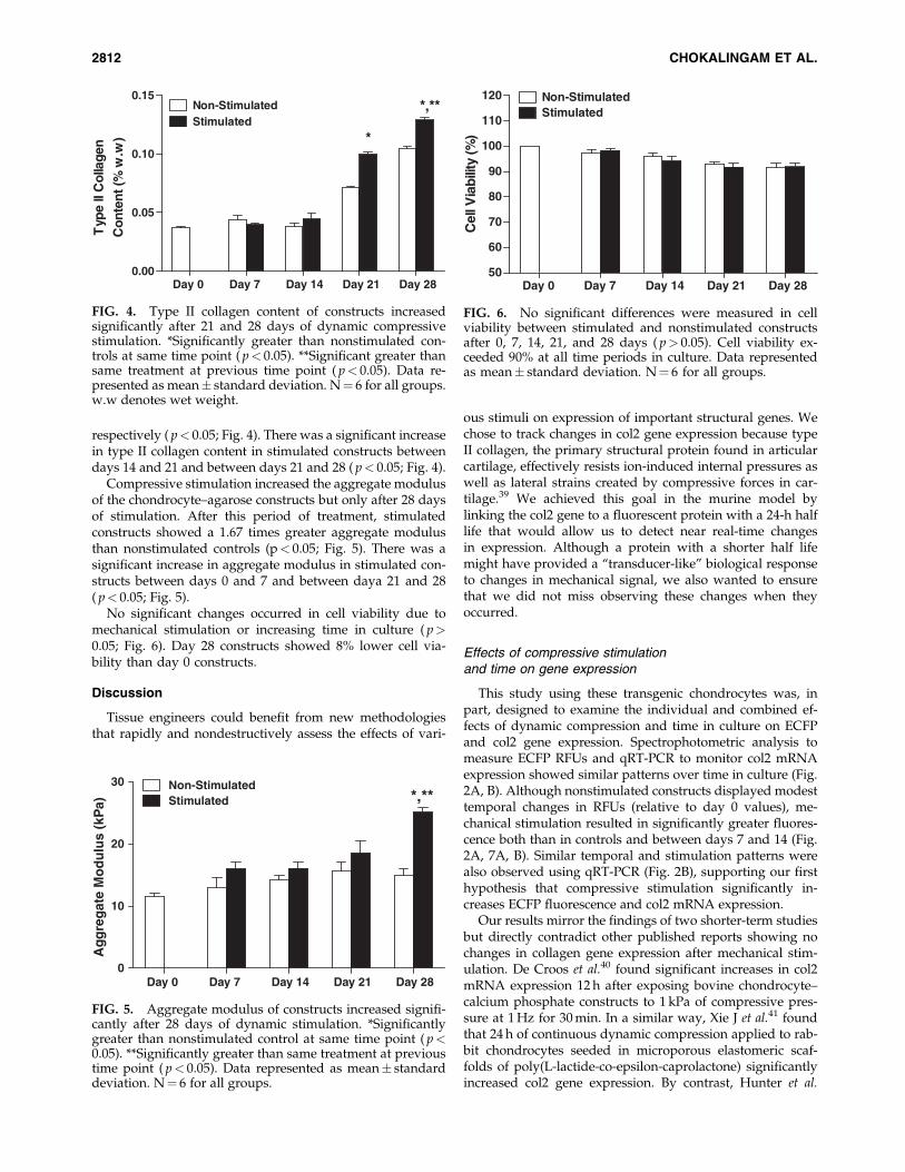
respectively ( p< 0.05; Fig. 4). There was a significant increasein type II collagen content in stimulated constructs betweendays 14 and 21 and between days 21 and 28 ( p< 0.05; Fig. 4).
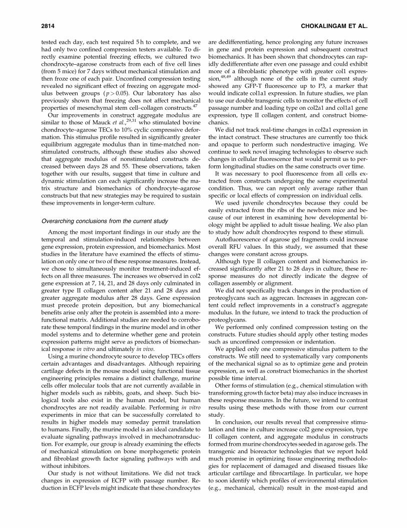
Compressive stimulation increased the aggregate modulusof the chondrocyte–agarose constructs but only after 28 daysof stimulation. After this period of treatment, stimulatedconstructs showed a 1.67 times greater aggregate modulusthan nonstimulated controls (p< 0.05; Fig. 5). There was asignificant increase in aggregate modulus in stimulated con-structs between days 0 and 7 and between daya 21 and 28( p< 0.05; Fig. 5).
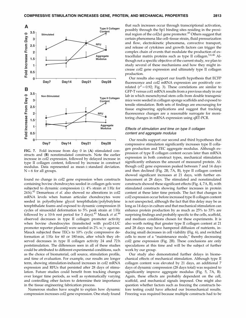
No significant changes occurred in cell viability due tomechanical stimulation or increasing time in culture ( p>0.05; Fig. 6). Day 28 constructs showed 8% lower cell via-bility than day 0 constructs.
Discussion
Tissue engineers could benefit from new methodologiesthat rapidly and nondestructively assess the effects of vari-
ous stimuli on expression of important structural genes. Wechose to track changes in col2 gene expression because typeII collagen, the primary structural protein found in articularcartilage, effectively resists ion-induced internal pressures aswell as lateral strains created by compressive forces in car-tilage.39 We achieved this goal in the murine model bylinking the col2 gene to a fluorescent protein with a 24-h halflife that would allow us to detect near real-time changesin expression. Although a protein with a shorter half lifemight have provided a ‘‘transducer-like’’ biological responseto changes in mechanical signal, we also wanted to ensurethat we did not miss observing these changes when theyoccurred.
Effects of compressive stimulationand time on gene expression
This study using these transgenic chondrocytes was, inpart, designed to examine the individual and combined ef-fects of dynamic compression and time in culture on ECFPand col2 gene expression. Spectrophotometric analysis tomeasure ECFP RFUs and qRT-PCR to monitor col2 mRNAexpression showed similar patterns over time in culture (Fig.2A, B). Although nonstimulated constructs displayed modesttemporal changes in RFUs (relative to day 0 values), me-chanical stimulation resulted in significantly greater fluores-cence both than in controls and between days 7 and 14 (Fig.2A, 7A, B). Similar temporal and stimulation patterns werealso observed using qRT-PCR (Fig. 2B), supporting our firsthypothesis that compressive stimulation significantly in-creases ECFP fluorescence and col2 mRNA expression.
Our results mirror the findings of two shorter-term studiesbut directly contradict other published reports showing nochanges in collagen gene expression after mechanical stim-ulation. De Croos et al.40 found significant increases in col2mRNA expression 12 h after exposing bovine chondrocyte–calcium phosphate constructs to 1 kPa of compressive pres-sure at 1 Hz for 30 min. In a similar way, Xie J et al.41 foundthat 24 h of continuous dynamic compression applied to rab-bit chondrocytes seeded in microporous elastomeric scaf-folds of poly(L-lactide-co-epsilon-caprolactone) significantlyincreased col2 gene expression. By contrast, Hunter et al.
Day 0 Day 7 Day 14 Day 21 Day 280.00
0.05
0.10
0.15Non-StimulatedStimulated
*
*,**T
ype
II C
olla
gen
Con
tent
(% w
.w)
FIG. 4. Type II collagen content of constructs increasedsignificantly after 21 and 28 days of dynamic compressivestimulation. *Significantly greater than nonstimulated con-trols at same time point ( p< 0.05). **Significant greater thansame treatment at previous time point ( p< 0.05). Data re-presented as mean� standard deviation. N¼ 6 for all groups.w.w denotes wet weight.
Day 0 Day 7 Day 14 Day 21 Day 280
10
20
30 Non-StimulatedStimulated *,**
Ag
gre
gat
e M
od
ulu
s (k
Pa)
FIG. 5. Aggregate modulus of constructs increased signifi-cantly after 28 days of dynamic stimulation. *Significantlygreater than nonstimulated control at same time point ( p<0.05). **Significantly greater than same treatment at previoustime point ( p< 0.05). Data represented as mean� standarddeviation. N¼ 6 for all groups.
Day 0 Day 7 Day 14 Day 21 Day 2850
60
70
80
90
100
110
120 Non-StimulatedStimulated
Cel
l Via
bilit
y (%
)
FIG. 6. No significant differences were measured in cellviability between stimulated and nonstimulated constructsafter 0, 7, 14, 21, and 28 days ( p> 0.05). Cell viability ex-ceeded 90% at all time periods in culture. Data representedas mean� standard deviation. N¼ 6 for all groups.
2812 CHOKALINGAM ET AL.
found no change in col2 gene expression when constructscontaining bovine chondrocytes seeded in collagen gels weresubjected to dynamic compression (� 4% strain at 1 Hz for24 h).42 Demarteau et al. also showed no alterations in col2mRNA levels when human articular chondrocytes wereseeded in polyethylene glycol terephthalate=polybutyleneterephthalate foams and exposed to dynamic compression (6cycles of sinusoidal deformation to 5% peak strain at 1 Hzfollowed by a 10-h rest period for 3 days).43 Mauck et al.44
observed decreases in type II collagen promoter activitywhen bovine chondrocytes (transfected with pcol2-LUCpromoter reporter plasmid) were seeded in 2% w=v agarose.Mauck subjected these TECs to 10% cyclic compressive de-formation at 1 Hz for 60 or 180 min, after which they ob-served decreases in type II collagen activity 24 and 72 hpoststimulation. The differences seen in all of these studiescould be attributed to different experimental conditions, suchas the choice of biomaterial, cell source, stimulation profile,and time of evaluation. For example, our results are longerterm, showing stimulation-induced increases in col2 mRNAexpression and RFUs that persisted after 28 days of stimu-lation. Future studies could benefit from tracking changesover longer time periods, as well as systematically varyingand controlling other factors to determine their importancein the tissue engineering fabrication process.
Numerous studies have sought to explain how dynamiccompression increases col2 gene expression. One study found
that such increases occur through transcriptional activation,possibly through the Sp1 binding sites residing in the proxi-mal region of the col2a1 gene promoter.45 Others suggest thatcertain phenomena like cell–tissue strain, fluid pressurizationand flow, electrokinetic phenomena, convective transport,and release of cytokines and growth factors can trigger thecomplex chain of events that modulate the production of ex-tracellular matrix proteins such as type II collagen.5,6,46 Al-though not a specific objective of the current study, we plan tostudy several of these mechanisms and how they might in-crease col2 gene expression and ultimately type II collagenproduction.
Our results also support our fourth hypothesis that ECFPfluorescence and col2 mRNA expression are positively cor-related (r2¼ 0.92; Fig. 3). These correlations are similar toGFP-T versus col1 mRNA results from a previous study in ourlab in which mesenchymal stem cells from double transgenicmice were seeded in collagen sponge scaffolds and exposed totensile stimulation. Both sets of findings are encouraging fortissue engineering applications and suggest that trackingfluorescence changes are a reasonable surrogate for moni-toring changes in mRNA expression using qRT-PCR.
Effects of stimulation and time on type II collagencontent and aggregate modulus
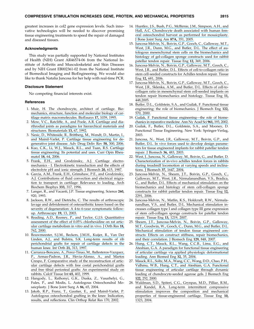
Our results support our second and third hypotheses thatcompressive stimulation significantly increases type II colla-gen production and TEC aggregate modulus. Although ex-pression of type II collagen content occurs later than mRNAexpression in both construct types, mechanical stimulationsignificantly enhances the amount of measured protein. Al-though col2 gene expression peaked between 7 and 14 daysand then declined (Fig. 2B, 7A, B), type II collagen contentshowed significant increases at 21 days, with further en-hancement at 28 days. The stimulated and nonstimulatedconstructs showed these significant effects (Fig. 4, 7A, B), withstimulated constructs showing further increases in proteincontent at these later time periods. The fact that changes incol2 expression occur before increased type II collagen contentis not unexpected, although the fact that this delay may be aslong as 14 days in culture and that mechanical stimulation canenhance protein production by as much as 25% to 33% aresurprising findings and probably specific to the cells, scaffold,and medium conditions chosen for these experiments. It isalso worth noting that greater type II collagen content at 21and 28 days may have hampered diffusion of nutrients, in-ducing small decreases in cell viability (Fig. 6), and switchedcells to more of a ‘‘maintenance mode,’’ thereby decreasingcol2 gene expression (Fig. 2B). These conclusions are onlyspeculations at this time and will be the subject of furtherwork by our group.
Our study also demonstrated further delays in biome-chanical effects of mechanical stimulation. Although type IIcollagen content was elevated by 21 days, an additional 7days of dynamic compression (28 days total) was required tosignificantly improve aggregate modulus (Fig. 5, 7A, B).Again, these effects are probably dependent on the cell,scaffold, and mechanical signals imposed. One might alsoquestion whether factors such as freezing the constructs be-fore testing could have affected our biomechanical results.Freezing was required because multiple constructs had to be
Stimulated
0
0.5
1
1.5
2
2.5
3
3.5
Fo
ld In
crea
se F
rom
Day
0Type II Collagen Content
AggregateModulus
RFU
Non-Stimulated
0
0.5
1
1.5
2
2.5
3
3.5
Day7 Day14 Day21 Day28
Day7 Day14 Day21 Day28
Fo
ld In
crea
se F
rom
Day
0
Type II Collagen Content
AggregateModulus
RFU
A
B
FIG. 7. Fold increase from day 0 in (A) stimulated con-structs and (B) nonstimulated constructs. Note the earlierincrease in col2 expression, followed by delayed increase intype II collagen content, followed by increase in constructmodulus. Data represented as mean� standard deviation.N¼ 6 for all groups.
COMPRESSIVE STIMULATION INCREASES GENE, PROTEIN, AND MECHANICAL PROPERTIES 2813
tested each day, each test required 5 h to complete, and wehad only two confined compression testers available. To di-rectly examine potential freezing effects, we cultured twochondrocyte–agarose constructs from each of five cell lines(from 5 mice) for 7 days without mechanical stimulation andthen froze one of each pair. Unconfined compression testingrevealed no significant effect of freezing on aggregate mod-ulus between groups ( p> 0.05). Our laboratory has alsopreviously shown that freezing does not affect mechanicalproperties of mesenchymal stem cell–collagen constructs.47
Our improvements in construct aggregate modulus aresimilar to those of Mauck et al.,29,31 who stimulated bovinechondrocyte–agarose TECs to 10% cyclic compressive defor-mation. This stimulus profile resulted in significantly greaterequilibrium aggregate modulus than in time-matched non-stimulated constructs, although these studies also showedthat aggregate modulus of nonstimulated constructs de-creased between days 28 and 55. These observations, takentogether with our results, suggest that time in culture anddynamic stimulation can each significantly increase the ma-trix structure and biomechanics of chondrocyte–agaroseconstructs but that new strategies may be required to sustainthese improvements in longer-term culture.
Overarching conclusions from the current study
Among the most important findings in our study are thetemporal and stimulation-induced relationships betweengene expression, protein expression, and biomechanics. Moststudies in the literature have examined the effects of stimu-lation on only one or two of these response measures. Instead,we chose to simultaneously monitor treatment-induced ef-fects on all three measures. The increases we observed in col2gene expression at 7, 14, 21, and 28 days only culminated ingreater type II collagen content after 21 and 28 days andgreater aggregate modulus after 28 days. Gene expressionmust precede protein deposition, but any biomechanicalbenefits arise only after the protein is assembled into a more-functional matrix. Additional studies are needed to corrobo-rate these temporal findings in the murine model and in othermodel systems and to determine whether gene and proteinexpression patterns might serve as predictors of biomechan-ical response in vitro and ultimately in vivo.
Using a murine chondrocyte source to develop TECs offerscertain advantages and disadvantages. Although repairingcartilage defects in the mouse model using functional tissueengineering principles remains a distinct challenge, murinecells offer molecular tools that are not currently available inhigher models such as rabbits, goats, and sheep. Such bio-logical tools also exist in the human model, but humanchondrocytes are not readily available. Performing in vitroexperiments in mice that can be successfully correlated toresults in higher models may someday permit translationto humans. Finally, the murine model is an ideal candidate toevaluate signaling pathways involved in mechanotransduc-tion. For example, our group is already examining the effectsof mechanical stimulation on bone morphogenetic proteinand fibroblast growth factor signaling pathways with andwithout inhibitors.
Our study is not without limitations. We did not trackchanges in expression of ECFP with passage number. Re-duction in ECFP levels might indicate that these chondrocytes
are dedifferentiating, hence prolonging any future increasesin gene and protein expression and subsequent constructbiomechanics. It has been shown that chondrocytes can rap-idly dedifferentiate after even one passage and could exhibitmore of a fibroblastic phenotype with greater col1 expres-sion,48,49 although none of the cells in the current studyshowed any GFP-T fluorescence up to P3, a marker thatwould indicate col1a1 expression. In future studies, we planto use our double transgenic cells to monitor the effects of cellpassage number and loading type on col2a1 and col1a1 geneexpression, type II collagen content, and construct biome-chanics.
We did not track real-time changes in col2a1 expression inthe intact construct. These structures are currently too thickand opaque to perform such nondestructive imaging. Wecontinue to seek novel imaging technologies to observe suchchanges in cellular fluorescence that would permit us to per-form longitudinal studies on the same constructs over time.
It was necessary to pool fluorescence from all cells ex-tracted from constructs undergoing the same experimentalcondition. Thus, we can report only average rather thanspecific or local effects of compression on individual cells.
We used juvenile chondrocytes because they could beeasily extracted from the ribs of the newborn mice and be-cause of our interest in examining how developmental bi-ology might be applied to adult tissue healing. We also planto study how adult chondrocytes respond to these stimuli.
Autofluorescence of agarose gel fragments could increaseoverall RFU values. In this study, we assumed that thesechanges were constant across groups.
Although type II collagen content and biomechanics in-creased significantly after 21 to 28 days in culture, these re-sponse measures do not directly indicate the degree ofcollagen assembly or alignment.
We did not specifically track changes in the production ofproteoglycans such as aggrecan. Increases in aggrecan con-tent could reflect improvements in a construct’s aggregatemodulus. In the future, we intend to track the production ofproteoglycans.
We performed only confined compression testing on theconstructs. Future studies should apply other testing modessuch as unconfined compression or indentation.
We applied only one compressive stimulus pattern to theconstructs. We still need to systematically vary componentsof the mechanical signal so as to optimize gene and proteinexpression, as well as construct biomechanics in the shortestpossible time interval.
Other forms of stimulation (e.g., chemical stimulation withtransforming growth factor beta) may also induce increases inthese response measures. In the future, we intend to contrastresults using these methods with those from our currentstudy.
In conclusion, our results reveal that compressive stimu-lation and time in culture increase col2 gene expression, typeII collagen content, and aggregate modulus in constructsformed from murine chondrocytes seeded in agarose gels. Thetransgenic and bioreactor technologies that we report holdmuch promise in optimizing tissue engineering methodolo-gies for replacement of damaged and diseased tissues likearticular cartilage and fibrocartilage. In particular, we hopeto soon identify which profiles of environmental stimulation(e.g., mechanical, chemical) result in the most-rapid and
2814 CHOKALINGAM ET AL.
greatest increases in col2 gene expression levels. Such inno-vative technologies will be needed to discover promisingtissue engineering treatments to speed the repair of damagedand diseased tissues.
Acknowledgments
This study was partially supported by National Institutesof Health (NIH) Grant AR46574-06 from the National In-stitute of Arthritis and Musculoskeletal and Skin Diseasesand by NIH Grant EB002361-02 from the National Instituteof Biomedical Imaging and BioEngineering. We would alsolike to thank Natalia Juncosa for her help with real-time PCR.
Disclosure Statement
No competing financial interests exist.
References
1. Muir, H. The chondrocyte, architect of cartilage. Bio-mechanics, structure, function and molecular biology of car-tilage matrix macromolecules. BioEssays 17, 1039, 1995.
2. Mow, V.C., Ratcliffe, A., and Poole, A.R. Cartilage and dia-rthrodial joints as paradigms for hierarchical materials andstructures. Biomaterials 13, 67, 1992.
3. Nesic, D., Whiteside, R., Brittberg, M., Wendt, D., Martin, I.,and Mainil-Varlet, P. Cartilage tissue engineering for de-generative joint disease. Adv Drug Deliv Rev 58, 300, 2006.
4. Kuo, C.K., Li, W.J., Mauck, R.L., and Tuan, R.S. Cartilagetissue engineering: Its potential and uses. Curr Opin Rheu-matol 18, 64, 2006.
5. Frank, E.H., and Grodzinsky, A.J. Cartilage electro-mechanics - I. Electrokinetic transduction and the effects ofelectrolyte pH and ionic strength. J Biomech 20, 615, 1987.
6. Garcia, A.M., Frank, E.H., Grimshaw, P.E., and Grodzinsky,A.J. Contributions of fluid convection and electrical migra-tion to transport in cartilage: Relevance to loading. ArchBiochem Biophys 333, 317, 1996.
7. Langer, R., and Vacanti, J.P. Tissue engineering. Science 260,
920, 1993.8. Jackson, R.W., and Dietrichs, C. The results of arthroscopic
lavage and debridement of osteoarthritic knees based on theseverity of degeneration: A 4-to 6-year symptomatic follow-up. Arthroscopy 19, 13, 2003.
9. Reading, A.D., Rooney, P., and Taylor, G.J.S. Quantitativeassessment of the effect of 0.05% chlorhexidine on rat artic-ular cartilage metabolism in vitro and in vivo. J Orth Res 18,
762, 2000.10. Bouwmeester, S.J.M., Beckers, J.M.H., Kuijer, R., Van Der
Linden, A.J., and Bulstra, S.K. Long-term results of ribperichondrial grafts for repair of cartilage defects in thehuman knee. Int Orth 21, 313, 1997.
11. Carranza-Bencano, A., Perez-Tinao, M., Ballesteros-Vazquez,P., Armas-Padron, J.R., Hevia-Alonso, A., and MartosCrespo, F. Comparative study of the reconstruction of artic-ular cartilage defects with free costal perichondrial graftsand free tibial periosteal grafts: An experimental study onrabbits. Calcif Tissue Int 65, 402, 1999.
12. Hangody, L., Rathonyi, G.K., Duska, Z., Vasarhelyi, G.,Fules, P., and Modis, L. Autologous Osteochondral Mo-saicplasty. J Bone Joint Surg A 86, 65, 2004.
13. Jakob, R.P., Franz, T., Gautier, E., and Mainil-Varlet, P.Autologous osteochondral grafting in the knee: Indication,results, and reflections. Clin Orthop Relat Res 170, 2002.
14. Huntley, J.S., Bush, P.G., McBirnie, J.M., Simpson, A.H., andHall, A.C. Chondrocyte death associated with human fem-oral osteochondral harvest as performed for mosaicplasty.J Bone Joint Surg Am 87A, 351, 2005.
15. Juncosa-Melvin, N., Boivin, G.P., Gooch, C., Galloway, M.T.,West, J.R., Dunn, M.G., and Butler, D.L. The effect of au-tologous mesenchymal stem cells on the biomechanics andhistology of gel-collagen sponge constructs used for rabbitpatellar tendon repair. Tissue Eng 12, 369, 2006.
16. Juncosa-Melvin, N., Boivin, G.P., Galloway, M.T., Gooch, C.,West, J.R., and Butler, D.L. Effects of cell-to-collagen ratio instem cell-seeded constructs for Achilles tendon repair. TissueEng 12, 681, 2006.
17. Juncosa-Melvin, N., Boivin, G.P., Galloway, M.T., Gooch, C.,West, J.R., Sklenka, A.M., and Butler, D.L. Effects of cell-to-collagen ratio in mesenchymal stem cell-seeded implants ontendon repair biomechanics and histology. Tissue Eng 11,
448,2005.18. Butler, D.L., Goldstein, S.A., and Guilak, F. Functional tissue
engineering: the role of biomechanics. J Biomech Eng 122,
570, 2000.19. Guilak, F. Functional tissue engineering—the role of biome-
chanics in reparative medicine. Ann Ny Acad Sci 961, 193, 2002.20. Guilak, F., Butler, D.L., Goldstein, S.A., and Mooney, D.
Functional Tissue Engineering. New York: Springer-Verlag,2003.
21. Juncosa, N., West, J.R., Galloway, M.T., Boivin, G.P., andButler, D.L. In vivo forces used to develop design parame-ters for tissue engineered implants for rabbit patellar tendonrepair. J Biomech 36, 483, 2003.
22. West, J., Juncosa, N., Galloway, M., Boivin, G., and Butler, D.Characterization of in-vivo achilles tendon forces in rabbitsduring treadmill locomotion at varying speeds and inclina-tions. J Biomech 37, 1647, 2004.
23. Juncosa-Melvin, N., Shearn, J.T., Boivin, G.P., Gooch, C.,Galloway, M.T., West, J.R., Nirmalanandhan, V.S., Bradica,G., and Butler, D.L. Effects of mechanical stimulation on thebiomechanics and histology of stem cell-collagen spongeconstructs for rabbit patellar tendon repair. Tissue Eng 12,
2291, 2006.24. Juncosa-Melvin, N., Matlin, K.S., Holdcraft, R.W., Nirmala-
nandhan, V.S., and Butler, D.L. Mechanical stimulation in-creases collagen type I and collagen type III gene expressionof stem cell-collagen sponge constructs for patellar tendonrepair. Tissue Eng 13, 1219, 2007.
25. Shearn, J.T., Juncosa-Melvin, N., Boivin, G.P., Galloway,M.T., Goodwin, W., Gooch, C., Dunn, M.G., and Butler, D.L.Mechanical stimulation of tendon tissue engineered con-structs: Effects on construct stiffness, repair biomechanics,and their correlation. J Biomech Eng 129, 848, 2007.
26. Hung, C.T., Mauck, R.L., Wang, C.C.B., Lima, E.G., andAteshian, G.A. A paradigm for functional tissue engineeringof articular cartilage via applied physiologic deformationalloading. Ann Biomed Eng 32, 35, 2004.
27. Mauck, R.L., Soltz, M.A., Wang, C.C., Wong, D.D., Chao, P.H.,Valhmu, W.B., Hung, C.T., and Ateshian, G.A. Functionaltissue engineering of articular cartilage through dynamicloading of chondrocyte-seeded agarose gels. J Biomech Eng122, 252, 2000.
28. Waldman, S.D., Spiteri, C.G., Grynpas, M.D., Pilliar, R.M.,and Kandel, R.A. Long-term intermittent compressivestimulation improves the composition and mechanicalproperties of tissue-engineered cartilage. Tissue Eng 10,
1323, 2004.
COMPRESSIVE STIMULATION INCREASES GENE, PROTEIN, AND MECHANICAL PROPERTIES 2815
29. Mauck, R.L., Wang, C.C.B., Oswald, E.S., Ateshian, G.A.,and Hung, C.T. The role of cell seeding density and nutrientsupply for articular cartilage tissue engineering with defor-mational loading. Osteoarthritis Cartilage 11, 879, 2003.
30. Butler, D.L., Hunter, S.A., Chokalingam, K., Cordray, M.J.,Shearn, J.T., Juncosa-Melvin, N., Nirmalanandhan, S., andJain, A. Using functional tissue engineering and bioreactorsto mechanically stimulate tissue engineered constructs. Tis-sue Eng 15, 741, 2009.
31. Mauck, R.L., Nicoll, S.B., Seyhan, S.L., Ateshian, G.A., andHung, C.T. Synergistic action of growth factors and dynamicloading for articular cartilage tissue engineering. Tissue Eng9, 597, 2003.
32. Benya, P.D., and Shaffer, J.D. Dedifferentiated chondrocytesreexpress the differentiated collagen phenotype when cul-tured in agarose gels. Cell 30, 215, 1982.
33. Molecular Devices Spectramax Gemini-XS Application Note44: Measurement of green fluorescent protein in the Spec-tramax Gemini XS Fluorescence Microplate Reader. http:==www.moleculardevices.com=product_literature=family_links.php?familyid=1. Product literature from Molecular Devices,June 1, 2008.
34. Hunter, S.A., Noyes, F.R., Haridas, B., Levy, M.S., and Butler,D.L. Effects of matrix stabilization when using glutaralde-hyde on the material properties of porcine meniscus. J BiomedMater Res A 67, 1245, 2003.
35. Stabler, C.L., Long, R.C. Jr, Sambanis, A., and Constantinidis,I. Noninvasive measurement of viable cell number in tissue-engineered constructs in vitro, using 1H nuclear magneticresonance spectroscopy. Tissue Eng 11, 404, 2005.
36. http:==www.chondrex.com=products=captureelisa.htm. Col-lagen Assay Kits, June 1, 2008.
37. Thirion, S., and Berenbaum, F. Culture and phenotyping ofchondrocytes in primary culture. Methods Molec Med 100, 1,2004.
38. Montgomery, D. Design and Analysis of Experiments, 5thEd. New York: John Wiley and Sons, 2001.
39. Wilkins, R.J., Browning, J.A., and Urban, J.P.G. Chondrocyteregulation by mechanical load. Biorheology 37, 67, 2000.
40. De Croos, J.N.A., Dhaliwal, S.S., Grynpas, M.D., Pilliar,R.M., and Kandel, R.A. Cyclic compressive mechanicalstimulation induces sequential catabolic and anabolic genechanges in chondrocytes resulting in increased extracellularmatrix accumulation. Matrix Biol 25, 323, 2006.
41. Xie, J., Han, Z., Kim, S.H., Kim, Y.H., and Matsuda, T.Mechanical loading-dependence of mRNA expressions ofextracellular matrices of chondrocytes inoculated into elas-
tomeric microporous poly(L-lactide-co- b-caprolactone) scaf-fold. Tissue Eng 13, 29, 2007.
42. Hunter, C.J., Imler, S.M., Malaviya, P., Nerem, R.M., andLevenston, M.E. Mechanical compression alters gene ex-pression and extracellular matrix synthesis by chondrocytescultured in collagen I gels. Biomaterials 23, 1249, 2002.
43. Demarteau, O., Wendt, D., Braccini, A., Jakob, M., Schafer, D.,Heberer, M., and Martin, I. Dynamic compression of cartilageconstructs engineered from expanded human articularchondrocytes. Biochem Biophys Res Commun 310, 580, 2003.
44. Mauck, R.L., Byers, B.A., Yuan, X., and Tuan, R.S. Regula-tion of cartilaginous ECM gene transcription by chon-drocytes and MSCs in 3D culture in response to dynamicloading. Biomech Model Mechanobiol 6, 113, 2007.
45. Xie, J., Han, Z.Y., and Matsuda, T. Mechanical compressiveloading stimulates the activity of proximal region of humanCOL2A1 gene promoter in transfected chondrocytes. Bio-chem Biophys Res Commun 344, 1192, 2006.
46. Kim, Y.J., Bonassar, L.J., and Grodzinsky, A.J. The role ofcartilage streaming potential, fluid flow and pressure in thestimulation of chondrocyte biosynthesis during dynamiccompression. J Biomech 28, 1055, 1995.
47. Nirmalanandhan, V.S., Rao, M., Shearn, J.T., Juncosa-Melvin, N., Gooch, C., and Butler, D.L. Effect of scaffoldmaterial, construct length and mechanical stimulation on thein vitro stiffness of the engineered tendon construct. J Bio-mech 41, 822, 2008.
48. Darling, E.M., and Athanasiou, K.A. Rapid phenotypicchanges in passaged articular chondrocyte subpopulations.J Orthop Res 23, 425, 2005.
49. Lefebvre, V., Peeters-Joris, C., and Vaes, G. Production ofcollagens, collagenase and collagenase inhibitor during thededifferentiation of articular chondrocytes by serial subcul-tures. Biochim Biophys Acta 1051, 266, 1990.
Address correspondence to:David L. Butler, Ph.D.
Department of Biomedical EngineeringUniversity of Cincinnati
2901 Woodside DriveCincinnati, OH 45221–0048
E-mail: [email protected]
Received: October 8, 2008Accepted: February 19, 2009
Online Publication Date: June 12, 2009
2816 CHOKALINGAM ET AL.