Patterns of Pneumatization of the Posterior Nasal Roof - MDPI
Upload
unispital-baselCategory
view
1download
0
ARTHRITIS & RHEUMATISMVol. 58, No. 1, January 2008, pp 197–208DOI 10.1002/art.23155© 2008, American College of Rheumatology
Engineered Cartilage Generated by Nasal Chondrocytes IsResponsive to Physical Forces Resembling Joint Loading
C. Candrian,1 D. Vonwil,1 A. Barbero,1 E. Bonacina,2 S. Miot,1 J. Farhadi,1 D. Wirz,3
S. Dickinson,4 A. Hollander,4 M. Jakob,1 Z. Li,5 M. Alini,5 M. Heberer,1 and I. Martin1
Objective. To determine whether engineered car-tilage generated by nasal chondrocytes (ECN) is respon-sive to different regimens of loading associated withjoint kinematics and previously shown to be stimulatoryof engineered cartilage generated by articular chondro-cytes (ECA).
Methods. Human nasal and articular chondro-cytes, harvested from 5 individuals, were expanded andcultured for 2 weeks into porous polymeric scaffolds.The resulting ECN and ECA were then maintainedunder static conditions or exposed to the followingloading regimens: regimen 1, single application of cyclicdeformation for 30 minutes; regimen 2, intermittentapplication of cyclic deformation for a total of 10 days,followed by static culture for 2 weeks; regimen 3,application of surface motion for a total of 10 days.
Results. Prior to loading, ECN constructs con-tained significantly higher amounts of glycosaminogly-can (GAG) and type II collagen compared with ECAconstructs. ECN responded to regimen 1 by increasingcollagen and proteoglycan synthesis, to regimen 2 byincreasing the accumulation of GAG and type II colla-gen as well as the dynamic modulus, and to regimen 3 by
increasing the expression of superficial zone protein, atthe messenger RNA level and the protein level, as well asthe release of hyaluronan. ECA constructs were overallless responsive to all loading regimens, likely due to thelower extracellular matrix content.
Conclusion. Human ECN is responsive to physi-cal forces resembling joint loading and can up-regulatemolecules typically involved in joint lubrication. Thesefindings should prompt future in vivo studies exploringthe possibility of using nasal chondrocytes as a cellsource for articular cartilage repair.
Cell-based therapies currently in clinical applica-tion for the treatment of articular cartilage lesionstypically rely on the use of autologous chondrocytesharvested from a small biopsy specimen of articularcartilage. A cartilage biopsy specimen from a joint, evenif harvested from a non–load-bearing site, represents anadditional injury to the cartilage surface and has beenreported to be detrimental to the surrounding healthyarticular cartilage (1). To overcome this problem, sev-eral groups of investigators proposed the use of mesen-chymal progenitor cells from bone marrow (2–4), syno-vial membrane (5–8), or periosteum (9,10) or the use ofchondrocytes from nonarticular cartilage, such as the ear(11,12), rib (11), or nasal septum (11,13).
Nasal cartilage would be a particularly interestingsource of cells, because the tissue is characterized as ahyaline cartilage and contains differentiated chondro-cytes expressing the collagen types typical of articularcartilage (14). Biopsy specimens of nasal cartilage can beharvested under local anesthesia and by a procedure thatis less invasive than removing tissue from specific areasof the joint. Morbidity associated with nasal cartilagebiopsy is also reduced, because the donor site is notsubjected to high levels of physical force (15). Indeed,nasal septal cartilage (sometimes in substantial
Supported in part by the European Union (Project FP6STEPS, contract NMP3-CT-2005-500465), the Swiss National ScienceFoundation (grant 3200B0-110054), and the “Deutsche Arthrose-Hilfee.V.”
1C. Candrian, MD, D. Vonwil, MSc, A. Barbero, PhD, S.Miot, PhD, J. Farhadi, MD, M. Jakob, MD, M. Heberer, MD, I.Martin, PhD: University Hospital Basel, Basel, Switzerland; 2E. Bona-cina, MSc: University Hospital Basel, Basel, Switzerland and Politec-nico di Milano, Milan, Italy; 3D. Wirz, MD: University of Basel, Basel,Switzerland; 4S. Dickinson, PhD, A. Hollander, PhD: University ofBristol, Southmead Hospital, Bristol, UK; 5Z. Li, PhD, M. Alini, PhD:AO Research Institute, Davos, Switzerland.
Address correspondence and reprint requests to Ivan Martin,PhD, Institute for Surgical Research and Hospital Management,University Hospital Basel, Hebelstrasse 20, ZLF, Room 405, 4031Basel, Switzerland. E-mail: [email protected].
Submitted for publication May 21, 2007; accepted in revisedform September 21, 2007.
197
amounts) is routinely resected during septoplasty oper-ations, with few sequelae or complications (15).
Several studies have indicated that human nasalseptum chondrocytes, even after culture expansion andassociated dedifferentiation, retain the capacity to re-differentiate and generate hyaline-like tissue (11,13,16),with mechanical properties approaching those of nativecartilage (17,18). In a comparative study using humanarticular chondrocytes from age-matched donors, it wasalso established that nasal chondrocytes proliferatefaster and have a higher and more reproducible chon-drogenic capacity, both in vitro and in an ectopic modelin vivo (13). Furthermore, although chondrogenesis byexpanded human articular chondrocytes has the ten-dency to decrease with donor age (19), the quality ofengineered cartilage generated by human nasal chon-drocytes (ECN) does not appear to be dependent on theage of the donor (16).
In order for nasal chondrocytes to be consideredfor implantation at an articular cartilage site, it is crucialto determine not only their chondrogenic capacity butalso whether they are responsive to forces that aretypically associated with joint loading. However, to thebest of our knowledge, nothing is yet known aboutwhether nasal chondrocytes and articular chondrocytes,which have a different embryologic origin and arephysiologically exposed to a markedly different bio-chemical and biomechanical environment, share a simi-lar mode of response to physical forces.
The goal of this study was to assess the responseof human nasal chondrocytes to different regimens ofloading, using experimental setups and readout para-meters previously shown by different groups of investi-gators to enhance matrix synthesis or production oflubrication proteins by articular chondrocytes, with stat-ically cultured specimens as controls. In particular, weapplied the following modalities of loading: 1) a singleapplication of cyclic deformation for 30 minutes (20), 2)intermittent application of cyclic deformation for a totalof 10 days (21), or 3) intermittent application of surfacemotion for a total of 10 days (22). As a model toinvestigate cell response to loading, we engineered car-tilage tissue obtained by seeding and culturing expandedchondrocytes into 3-dimensional (3-D) porous scaffolds.The response of ECN was compared with that ofengineered cartilage generated by articular chondro-cytes (ECA), using cells isolated from different sites inthe same individuals.
MATERIALS AND METHODS
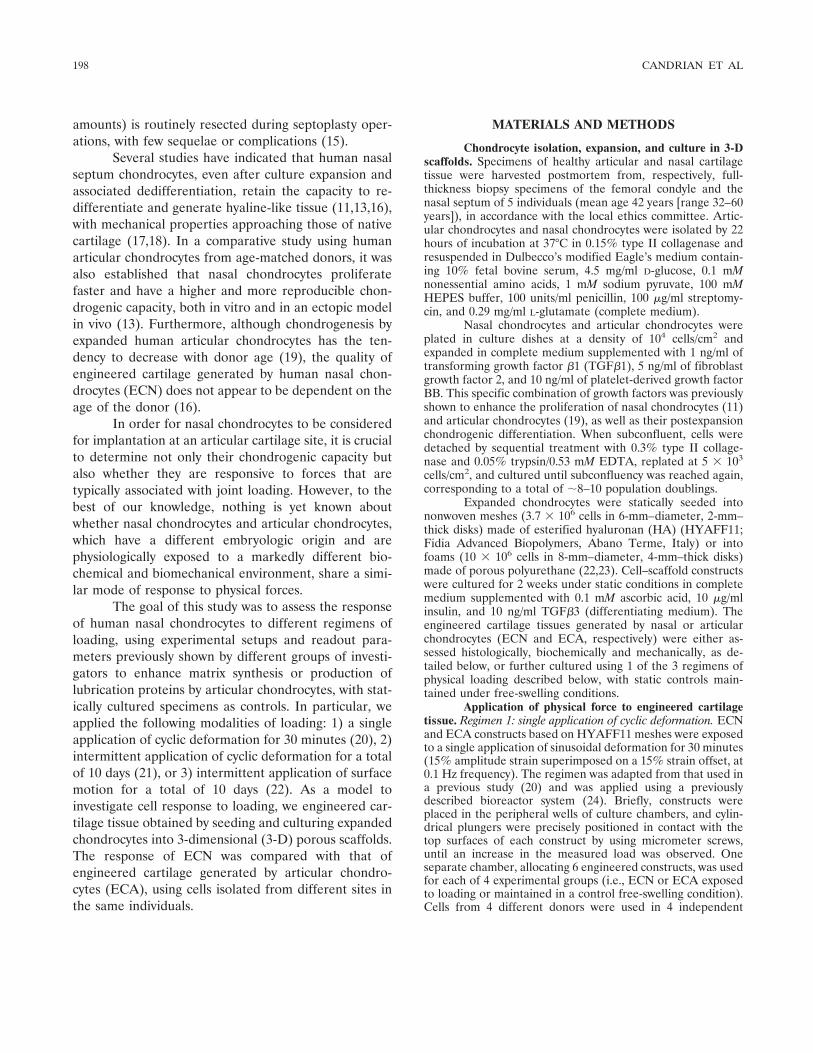
Chondrocyte isolation, expansion, and culture in 3-Dscaffolds. Specimens of healthy articular and nasal cartilagetissue were harvested postmortem from, respectively, full-thickness biopsy specimens of the femoral condyle and thenasal septum of 5 individuals (mean age 42 years [range 32–60years]), in accordance with the local ethics committee. Artic-ular chondrocytes and nasal chondrocytes were isolated by 22hours of incubation at 37°C in 0.15% type II collagenase andresuspended in Dulbecco’s modified Eagle’s medium contain-ing 10% fetal bovine serum, 4.5 mg/ml D-glucose, 0.1 mMnonessential amino acids, 1 mM sodium pyruvate, 100 mMHEPES buffer, 100 units/ml penicillin, 100 �g/ml streptomy-cin, and 0.29 mg/ml L-glutamate (complete medium).
Nasal chondrocytes and articular chondrocytes wereplated in culture dishes at a density of 104 cells/cm2 andexpanded in complete medium supplemented with 1 ng/ml oftransforming growth factor �1 (TGF�1), 5 ng/ml of fibroblastgrowth factor 2, and 10 ng/ml of platelet-derived growth factorBB. This specific combination of growth factors was previouslyshown to enhance the proliferation of nasal chondrocytes (11)and articular chondrocytes (19), as well as their postexpansionchondrogenic differentiation. When subconfluent, cells weredetached by sequential treatment with 0.3% type II collage-nase and 0.05% trypsin/0.53 mM EDTA, replated at 5 � 103
cells/cm2, and cultured until subconfluency was reached again,corresponding to a total of �8–10 population doublings.
Expanded chondrocytes were statically seeded intononwoven meshes (3.7 � 106 cells in 6-mm–diameter, 2-mm–thick disks) made of esterified hyaluronan (HA) (HYAFF11;Fidia Advanced Biopolymers, Abano Terme, Italy) or intofoams (10 � 106 cells in 8-mm–diameter, 4-mm–thick disks)made of porous polyurethane (22,23). Cell–scaffold constructswere cultured for 2 weeks under static conditions in completemedium supplemented with 0.1 mM ascorbic acid, 10 �g/mlinsulin, and 10 ng/ml TGF�3 (differentiating medium). Theengineered cartilage tissues generated by nasal or articularchondrocytes (ECN and ECA, respectively) were either as-sessed histologically, biochemically and mechanically, as de-tailed below, or further cultured using 1 of the 3 regimens ofphysical loading described below, with static controls main-tained under free-swelling conditions.
Application of physical force to engineered cartilagetissue. Regimen 1: single application of cyclic deformation. ECNand ECA constructs based on HYAFF11 meshes were exposedto a single application of sinusoidal deformation for 30 minutes(15% amplitude strain superimposed on a 15% strain offset, at0.1 Hz frequency). The regimen was adapted from that used ina previous study (20) and was applied using a previouslydescribed bioreactor system (24). Briefly, constructs wereplaced in the peripheral wells of culture chambers, and cylin-drical plungers were precisely positioned in contact with thetop surfaces of each construct by using micrometer screws,until an increase in the measured load was observed. Oneseparate chamber, allocating 6 engineered constructs, was usedfor each of 4 experimental groups (i.e., ECN or ECA exposedto loading or maintained in a control free-swelling condition).Cells from 4 different donors were used in 4 independent
198 CANDRIAN ET AL

experiments. After loading, the constructs were assessed forproteoglycan and collagen synthesis, as described below.
Regimen 2: intermittent application of cyclic deforma-tion. ECN and ECA constructs based on HYAFF11 mesheswere exposed to intermittent application of sinusoidal defor-mation for 30 minutes (7.5% amplitude strain superimposedon 7.5% strain offset, at 0.1 Hz frequency), every second dayfor 10 days. The amplitude and frequency were derived fromprevious studies and were reported to be stimulatory ofproteoglycan synthesis in articular cartilage explants (25,26)and also in engineered cartilage, if it was sufficiently developed(21). The duration of the application was modified accordingto the protocol used by Waldman et al (20).
Constructs (6 per experimental group) were placed inthe bioreactor chambers described above, under gentle mixing(30 revolutions per minute), as previously described (24). Aftereach 30-minute application, the plungers were raised 500 �mover the constructs, in order to allow for recovery of the fullthickness and to reduce mass transport limitations. Theconstruct–plunger contact position was reestablished beforeeach application of loading. Unloaded constructs were cul-tured in the same chambers in free-swelling conditions ascontrol. Differentiating medium was changed twice a week andcollected for determination of the amount of glycosaminogly-can (GAG) released. Constructs were assessed histologically,biochemically, and mechanically, as described below, eitherimmediately after the last loading cycle or following an addi-tional 2 weeks of static culture in differentiating medium. Cellsfrom 5 different donors were used in 5 independent experi-ments.
Regimen 3: intermittent application of surface motion.ECN and ECA constructs based on polyurethane foams wereattached to specimen holders using bone cement (NorianSkeletal Repair System; Norian, Cupertino, CA) and exposedto intermittent surface motion by oscillation of a ceramic hipball over their surface for 30 minutes (�60° amplitude rotationsuperimposed on 10% strain offset, at 0.1 Hz frequency), everysecond day for 10 days, using a previously described bioreactorsystem (22). Between the loading periods, constructs did nothave any contact with the ball. Unloaded constructs werecultured in free-swelling conditions as a control.
The response of engineered cartilage to surface motionwas assessed for what was previously reported to be thepredominant effect on bovine articular chondrocytes culturedon polyurethane scaffolds, namely, the expression of genesencoding for lubricating proteins (22). Differentiating mediumwas changed daily and collected for quantification of theamount of superficial zone protein (SZP) and HA released.Immediately after the last cycle of loading, constructs wereassessed for the messenger RNA (mRNA) expression of SZP,HA synthase 1 (HAS-1), and HAS-2, as described below. Cellsfrom 4 different donors were used in 4 independent experi-ments.
Analytic techniques. Proteoglycan and collagen synthe-sis. Immediately after the single application of cyclic loading(regimen 1), constructs were incubated for 24 hours in thepresence of L-3H-proline (5 �Ci/construct) and 35S-SO4 (4�Ci/construct) to quantify the amount of newly synthesizedcollagen and proteoglycans, respectively. After radioactive
labeling, the amount of newly synthesized collagen and pro-teoglycans was quantified both in medium (released fraction),as previously described (20), and in the constructs (accumu-lated fraction), following digestion with proteinase K (0.5 ml of1 mg/ml proteinase K in 50 mM Tris with 1 mM EDTA, 1 mMiodoacetamide, and 10 �g/ml pepstatin A) for 15 hours at56°C. The amount of newly synthesized components wasnormalized to the DNA content of the constructs (determinedas described below).
Biochemical analyses. For determination of GAG andDNA, constructs were digested with proteinase K as describedabove. The amount of GAG was measured spectrophotometri-cally after reaction with dimethylmethylene blue (27), withchondroitin sulfate as a standard. DNA was measured spec-trofluorometrically using the CyQUANT Cell ProliferationAssay Kit (Molecular Probes, Eugene, OR), with calf thymusDNA as a standard (28). The amount of GAG released fromthe constructs into the medium during the 10-day cultureperiod was quantified from the media collected from eachgroup and normalized to the mean DNA content of a singleconstruct. For determination of type II collagen, constructswere digested with tosylamide-2-phenylethyl chloromethylketone–treated bovine pancreatic trypsin at 2 mg/ml (in 50 mMTris HCl, pH 7.6), using an initial incubation of 15 hours at37°C followed by a further 2 hours of incubation at 65°C afterthe addition of fresh trypsin. Samples were boiled for 15minutes to inactivate the enzyme (29), and the amounts of typeII collagen were assayed by inhibition enzyme-linked immu-nosorbent assay (ELISA) using a mouse IgG monoclonalantibody to denatured type II collagen (30).
Histologic and immunohistochemical analyses. Con-structs were fixed in 4% formalin for 24 hours at 4°C,embedded in paraffin, and cross-sectioned (7 �m thick).Sections were stained with Safranin O for GAG or for type IIcollagen (antibody II-II6B3; Hybridoma Bank, University ofIowa, Iowa City), as previously described (31).
Mechanical analyses. Mechanical assessments of ECNand ECA from regimen 2 as well as of cell-free HYAFF11scaffolds were performed in a standard miniature test instru-ment in unconfined compression (Synergie 100; MTS Systems,Eden Prairie, MN), within 4 hours after termination of theculture. The thickness of each specimen was measured first, bymoving the crosshead down at a rate of 0.1 mm/second until aload was detected, and then the specimen was assessed tomeasure the following parameters.
Equilibrium modulus. Constructs were exposed to 5incremental strains of 5% each, accomplished at a crossheadspeed of 0.17 mm/second, and were considered fully relaxedafter each strain increment when the rate of change in the loadwas lower than 0.001 N/second (corresponding to �35 Pa/second). The equilibrium modulus was determined as the slopeof the equilibrium load normalized to the specimen area,plotted versus the applied strain (32).
Pulsatile dynamic modulus. Constructs were subjectedto 5 cycles of compressive loading/unloading at a rate of 0.17mm/second, reaching a maximum of 20% strain and separatedby a no-load period of time equal to that for loading, aspreviously described (33). The mean pulsatile dynamic modu-lus was computed by performing linear regressions through the
RESPONSE OF HUMAN NASAL CHONDROCYTES TO DIFFERENT LOADING REGIMENS 199

stress–strain curve of each cycle, outside the toe region.Because cartilage displays poroviscoelastic material properties,the cyclic nature of the tests means that the pulsatile dynamicmodulus is actually a form of the compressive dynamic elasticmodulus, which is the vector sum of the storage modulus (thein-phase, elastic component) and the loss modulus (the out-of-phase, poroviscous component).
Gene expression at the mRNA level. Gene expressionanalysis of ECN and ECA from regimen 3 was performed byreal-time reverse transcription (RT)–polymerase chain reac-tion (PCR) (GeneAmp 5700; Applied Biosystems, Foster City,CA), following RNA extraction using TRI Reagent (MolecularResearch Center, Cincinnati, OH) and RT with TaqManreagents (Applied Biosystems, Foster City, CA). The oligo-nucleotide primers and fluorescent probes for human SZP,HAS-1, and HAS-2 were described previously (22). Primersand probes for amplification of 18S ribosomal RNA for use as
endogenous control were from Applied Biosystems. Real-timeRT-PCRs were not multiplexed.
Quantification of HA and SZP in culture media. Theamounts of released HA and SZP were quantified in the mediacollected from loaded and free-swelling ECN and ECA con-structs from regimen 3. The HA concentration was measuredin a competitive ELISA system (Echelon Biosciences, SaltLake City, UT). For analysis of SZP, media were fractionatedby anion-exchange chromatography on a 1-ml column ofResource Q (Amersham Biosciences, Uppsala, Sweden) asdescribed previously (34). SZP was then eluted, dialyzed, andseparated by gel electrophoresis as previously described (22).Immunoreactive bands were detected with the ECL PlusWestern blotting detection system (Amersham Biosciences),following incubation with the polyclonal antibody 06A10 (1:1,000 dilution; kindly provided by C. R. Flannery, WyethResearch, Cambridge, MA) against human megakaryocyte-
Figure 1. Properties of engineered cartilage tissue generated by nasal chondrocytes (ECN) orarticular chondrocytes (ECA) prior to mechanical loading. A and B, Safranin O and type IIcollagen immunohistochemical staining of representative ECN or ECA after 2 weeks of culture inchondrogenic medium. Top rows (bar � 500 �m) show lower-magnification views of bottom rows(bar � 100 �m). C, Glycosaminoglycan (GAG) and type II collagen content of ECN and ECA. D,Equilibrium modulus (EEQ, left y-axis) and pulsatile dynamic modulus (EPD, right y-axis) of ECNand ECA. Values are the mean and SEM results from 5 independent experiments. o � significantdifference versus ECA.
200 CANDRIAN ET AL

stimulating factor, previously reported to be homologous toSZP or lubricin (34).
Statistical analysis. All values are presented as themean � SEM. Measurements were obtained from at least 4independent experiments (i.e., with cells from at least 4different donors). For each experiment and experimentalgroup, at least duplicate specimens were assessed. Differencesbetween experimental groups were statistically assessed byWilcoxon’s 2-tailed tests, using SigmaStat software version 13(SPSS Inc., Chicago, IL). P values less than 0.05 were consid-ered significant.
RESULTS
Properties of the tissue-engineered cartilageprior to mechanical loading. After 2 weeks of cultureusing nonwoven HYAFF11 scaffolds, prior to mechani-cal loading, ECN constructs showed more intense anduniform staining for GAG and type II collagen than didECA tissues (Figures 1A and B). Consistently, thebiochemically quantified amounts of GAG and type IIcollagen were significantly higher (71% and 73%, re-spectively) for ECN compared with ECA tissues (Figure1C). Mechanical tests indicated that the equilibriummodulus and the pulsatile dynamic modulus were also
significantly higher (35% and 34%, respectively) forECN than for ECA (Figure 1D).
Regimen 1: single application of cyclic deforma-tion. The application of cyclic deformation for 30 min-utes significantly increased synthesis of collagen andproteoglycans (44% and 39%, respectively) in ECNconstructs, whereas no significant effect was measured inECA constructs (Figure 2). In particular, in ECN tissue,the increased proteoglycan synthesis was reflected by asignificantly higher release (86%) of the newly synthe-sized molecules, and the increased collagen synthesisresulted in a significantly higher accumulation (47%) ofthe newly synthesized molecules. As compared withECA, ECN constructs had significantly higher proteo-glycan and collagen synthesis after both free swellingand cyclic deformation. Interestingly, for both ECN andECA, the percentage of newly synthesized proteoglycansreleased into the media (40–60%) was markedly higherthan the corresponding percentage of newly synthesizedcollagen (5–6%).
Regimen 2: intermittent application of cyclicdeformation. Assessment after 10 days of cyclic deforma-tion. Intermittent application of cyclic deformation for10 days induced a significant increase (66%) in the total
Figure 2. Biosynthetic activity of ECN or ECA following a single application of cyclic loading (regimen 1). Amounts of newlysynthesized proteoglycan (A) and collagen (B) were measured by incorporation of 35S-SO4 and 3H-proline, respectively, inconstructs maintained under free-swelling conditions or 24 hours following a single application of cyclic loading. The upper andlower parts of the columns represent newly synthesized molecules released in the medium or accumulated in the extracellularmatrix, respectively. Values are the mean and SEM results from 4 independent experiments. o � significant difference versusECA cultured under the same condition; � � significant difference versus the free-swelling condition of the same cell source.See Figure 1 for definitions.
RESPONSE OF HUMAN NASAL CHONDROCYTES TO DIFFERENT LOADING REGIMENS 201

amount of GAG released by ECN constructs but not byECA constructs (Figure 3), consistent with the resultsfrom regimen 1. The wet weight fractions of accumu-lated GAG and type II collagen were higher in ECNconstructs than in ECA constructs and were not affectedsignificantly by the application of loading in eithergroup. These findings were consistent with the fact thatloaded and free-swelling constructs displayed similarstaining intensities for GAG and type II collagen, as wellas similar mechanical properties (data not shown).
Assessment after 10 days of cyclic deformationfollowed by 2 weeks of static culture. In order to assesswhether cyclic deformation would influence the devel-opment of engineered cartilage tissue after terminatingits application, ECN and ECA constructs cultured underloading or free-swelling conditions for 10 days werestatically cultivated for 2 additional weeks. At this stage,ECN constructs previously exposed to loading werestained at higher intensity and uniformity for both GAGand type II collagen as compared with the free-swellingcontrols (Figure 4A). Instead, no apparent effect ofloading was observed for ECA constructs, where stain-ing patterns were generally faint and scattered in dis-crete areas (Figure 4B). Biochemical quantification ofGAG and type II collagen, as a percentage of the tissuewet weight, confirmed that 1) in ECN, cyclic deforma-tion significantly increased the content of both mole-cules (by 55% and 60%, respectively), 2) in ECA, cyclicdeformation had no significant effect on the content ofeither molecule, and 3) ECA constructs had signifi-cantly lower content of both molecules than did ECNtissue (Figure 4C). Mechanical properties were gener-ally consistent with the tissue composition: ECN hadsignificantly higher levels of equilibrium modulus andpulsatile dynamic modulus than ECA and, unlike thatin ECA constructs, the response to loading in ECNconstructs was an increase in the pulsatile dynamicmodulus (by 33%, in 5 of 5 donors; P � 0.04) and theequilibrium modulus (by 26%, in 4 of 5 donors; P �0.08) (Figure 4D).
We next investigated whether the increased re-sponse to cyclic deformation of ECN as compared withECA could be related to the higher degree of tissuedevelopment at the time of loading. The fold differencein the GAG content of ECN and ECA constructs ofdifferent donors in response to loading was shown to besignificantly positively correlated with the GAG contentof the same constructs prior to loading (Figure 5). Thus,the constructs with the lowest GAG content had themost detrimental response to loading, whereas tissuewith the highest GAG content had the most stimulatoryresponse to loading. Importantly, ECN and ECA ap-peared to follow a similar trend.
Regimen 3: intermittent application of surfacemotion. Effect of surface motion on mRNA expression ofSZP, HAS-1, and HAS-2. Intermittent application ofsurface motion to ECN constructs for 10 days induced asignificant and marked increase (32.9-fold) in themRNA expression of SZP but not HAS-1 and HAS-2.Only low levels and nonsignificant up-regulation of SZP
Figure 3. Properties of ECN and ECA after intermittent applicationof cyclic deformation for 10 days (regimen 2). Columns show theamounts of GAG released into the culture medium or the amount ofGAG or type II collagen accumulated in the engineered cartilage,normalized to the DNA content or expressed as a percentage of tissuewet weight, respectively. Values are the mean and SEM results from 5independent experiments. o � significant difference versus ECA; � �significant difference versus free swelling. See Figure 1 for definitions.
202 CANDRIAN ET AL

and HAS-1 mRNA (3.4-fold and 1.8-fold, respectively)were measured in ECA following application of surfacemotion. However, the expression of these genes re-
mained higher in ECA constructs as compared withstimulated ECN constructs (15.4-fold for SZP and 51.9-fold for HAS-1) (Figure 6A). The expression of HAS-2
Figure 4. Properties of ECN or ECA after intermittent application of cyclic deformation for 10days (regimen 2) followed by 2 weeks of static culture. A and B, Safranin O and type II collagenimmunohistochemical staining of ECN or ECA constructs. Top rows (bar � 500 �m) showlower-magnification views of bottom rows (bar � 100 �m). C, Amounts of GAG and type IIcollagen accumulated, expressed as a percentage of tissue wet weight. D, Equilibrium modulus andpulsatile dynamic modulus of ECN and ECA constructs. Broken lines indicate the mechanicalproperties of the cell-free HYAFF11 scaffold. Values are the mean and SEM results from 5independent experiments. o � significant difference versus ECA cultured under the samecondition; � � significant difference versus the free-swelling condition of the same cell source. SeeFigure 1 for definitions.
RESPONSE OF HUMAN NASAL CHONDROCYTES TO DIFFERENT LOADING REGIMENS 203

mRNA was similar in ECN and ECA tissue and was notregulated by surface motion.
Effect of surface motion on the release of SZP andHA in culture media. In the absence of loading, thesecretion of SZP and HA protein into the culturemedium by ECA cultures was significantly higher thanthat by ECN cultures (Figure 6B), although the differ-ence was less marked than at the mRNA level. SZP andHA protein amounts released into the medium wereenhanced by applied surface motion in both ECA andECN constructs (for SZP, 1.8-fold and 1.4-fold, respec-tively; for HA, 1.3-fold and 1.4-fold, respectively). Con-sistent with the results for mRNA expression, evenfollowing application of surface motion, SZP and HAwere released in larger amounts (1.8-fold and 2.1-fold,respectively) by ECA constructs than ECN constructs(Figure 6B).
DISCUSSION
In the present study, we demonstrate that humannasal chondrocytes, expanded and subsequently culturedinto 3-D porous scaffolds, are responsive to physicalforces resembling joint loading. In particular, cyclicdeformation increased GAG and collagen synthesis andenhanced the mechanical properties of the resultingECN. Moreover, surface motion up-regulated the ex-pression of genes involved in joint lubrication, at both
the mRNA level (i.e., for SZP) and the protein level(i.e., for SZP and HA).
The model used to investigate chondrocyte re-sponse to loading was based on the expansion (and thusdedifferentiation) of nasal chondrocytes and articularchondrocytes, followed by culture into HA- orpolyurethane-based porous scaffolds for 2 weeks. Basedon previously reported calculations, the extent of expan-sion would be sufficient to produce enough cells forclinically relevant graft sizes (i.e., a few square centime-ters), starting from biopsy specimens weighing only a fewmilligrams (11). The culture time in scaffolds was previ-ously reported to be sufficient to activate the process ofcell redifferentiation (35) and to generate constructswith relevant mechanical properties (36), with the ca-pacity to further develop in vivo (37). The resulting ECNwas histologically, biochemically, and mechanically su-perior to ECA. This finding is consistent with a previousreport that human nasal chondrocytes generally have ahigher and more reproducible postexpansion chondro-genic capacity than articular chondrocytes (13), but thisstudy is the first in which the analysis was performed bya paired comparison using cells harvested from differentsites in the same individuals.
The different loading regimens used in thepresent study to characterize the response of humannasal chondrocytes to specific physical forces only re-motely mimic the mechanical conditioning of joint load-ing and thus cannot be used to predict directly thecapacity of nasal chondrocytes to respond to joint load-ing when implanted in an articular cartilage defect inpatients. Conversely, the models used allowed us toinvestigate the intrinsic capacity of nasal chondrocytes ofhuman origin to respond to defined and controlledphysical conditioning, in a way that is decoupled by thecomplex interactions between biologic, biochemical, andbiomechanical cues of a large-sized animal joint.
A single application of cyclic loading (regimen 1)induced a significant and immediate increase in collagenand proteoglycan synthesis by nasal chondrocytes. Incontrast to a previous study in which a similar regimenwas used (20), ECA constructs in this study were notresponsive to a single application of cyclic deformation.The discrepancy could result from the fact that Wald-man et al (20) used primary (i.e., nonexpanded) bovinearticular chondrocytes from young animals, seeded di-rectly as a thin layer on the top surface of the scaffolds,at a higher cell density. In our system, consisting ofexpanded human articular chondrocytes from adult in-dividuals, seeded at a relatively lower density into the
Figure 5. Response to loading of engineered cartilage versus thematrix content of the tissue. The response to loading (regimen 2followed by 2 weeks of static culture) of the wet weight percentage ofGAG, calculated as the fold difference from the unloaded constructs,was correlated with the GAG content at the time of loading. The Pvalue was determined using Pearson’s 2-tailed correlation test. SeeFigure 1 for definitions.
204 CANDRIAN ET AL

scaffolds, chondrogenesis is expected to proceed lessefficiently, and thus the extracellular and pericellularmatrices, which are critical to determine chondrocyteresponse to loading (21,38), were most likely less devel-oped. This interpretation is consistent with the findingthat the more developed constructs based on nasalchondrocytes were indeed responsive to the loadingregimen.
Intermittent application of cyclic deformation for10 days (regimen 2) increased the amount of GAGreleased from ECN during loading, possibly as a directeffect of increased convection (38,39) and/or as a con-sequence of increased activity of degradative enzymes(40). Because the amounts of accumulated GAG were
not affected by loading, the higher amounts of GAGreleased indicate an overall increase in GAG synthesisby nasal chondrocytes, which is consistent with resultsobtained by application of regimen 1. A significantincrease in GAG accumulation in ECN in response toloading was observed only following an additional 2weeks of static culture. Considering that the net amountof accumulated GAG is critically determined by thecombination of GAG synthesis and GAG release, theresult may reflect that the effect of loading on GAGrelease decays more rapidly than that on GAG synthesis.A similar consideration could apply for type II collagen,although in this case the amounts released in mediumwere below levels of detection for the specific assay used
Figure 6. Messenger RNA expression and protein release of molecules involved in joint lubrication by ECN or ECA, followingintermittent application of surface motion (regimen 3). A, Real-time reverse transcription–polymerase chain reaction analysisof the expression of superficial zone protein (SZP) and hyaluronan synthase 1 (HAS-1) and HAS-2 mRNA in ECN and ECA.E � exponential in base 10. B, Quantification of the total SZP and HA proteins released in the culture medium. Values arethe mean and SEM results from 4 independent experiments. o � significant difference versus ECA cultured under the samecondition; � � significant difference versus the free-swelling condition of the same cell source. See Figure 1 for otherdefinitions.
RESPONSE OF HUMAN NASAL CHONDROCYTES TO DIFFERENT LOADING REGIMENS 205

and thus could not be quantified. The increased accu-mulation of GAG and type II collagen is consistent witha significant increase in the pulsatile dynamic modulus(Figure 4D) and establishes that nasal chondrocytes areindeed positively responsive to a repeated application ofcyclic loading.
The fact that cyclic loading of ECA did notsignificantly increase the amount of GAG released oraccumulated, as well as the amount of type II collagenaccumulated, may (as for regimen 1) be explained by thelower amount of matrix deposited at the time of loading.This would be consistent with previous findings that theresponse of engineered cartilage tissue to mechanicalloading is also directly related to the stage of tissuedevelopment (21). In fact, we observed a significant andpositive correlation in GAG deposition between theGAG content prior to loading and the response toloading (Figure 5), similar to what was previously de-scribed for ECA (21). Interestingly, the correlation wasdetermined by combining data obtained using ECN andECA, suggesting common matrix-mediated mechanismsof response to loading.
Intermittent application of surface motion for 10days (regimen 3) to ECN induced a significant increasein the expression of SZP mRNA and the release of SZPand HA protein into the medium. This indicates thatnasal chondrocytes exposed to a joint-specific modalityof loading can up-regulate the expression of proteinsconsidered to play a fundamental role in joint lubrica-tion. The expression of HAS-1 and HAS-2, in botharticular chondrocytes and nasal chondrocytes, was notregulated by the regimen of surface motion applied. Thelack of response in those 2 genes by articular chondro-cytes, in contrast to previous studies (22,41,42), couldagain result from differences in the cell source (i.e.,human versus bovine), as well as by slight modificationsof the loading protocol (i.e., lower frequency and num-ber of cycles of stimulation).
The levels of SZP mRNA expression and proteinrelease by nasal chondrocytes remained significantlylower than those by articular chondrocytes, confirmingthat the gene is associated with a cellular function typicalof cells derived from the joint. Interestingly, however,expression levels significantly increased in ECN follow-ing application of surface motion and entered the rangeof those measured in ECA under free-swelling condi-tions. This finding indicates a certain plasticity of ex-panded nasal chondrocytes and the potential to adapt toa typical joint environment. Similar cell plasticity to jointkinematics regimens was recently observed in chondro-
cytes from the middle and deep zones of articularcartilage, which normally do not express SZP but wereable to up-regulate its expression following stimulationby surface motion (43).
In this study, we did not address whether thecapacity of nasal chondrocytes to respond to joint-specific mechanical forces was intrinsic in the isolatedcells or was acquired following dedifferentiation. Thelatter possibility was advocated in a recent study,whereby rabbit expanded auricular chondrocytes (i.e.,from elastic cartilage) implanted into the intervertebraldiscs of rabbits survived and produced extracellularmatrix lacking elastic fibers and displaying hyaline-likecharacteristics (44). Despite the different ontogeny ofnasal cartilage and articular cartilage (i.e., ectodermalversus endodermal) and their different topographic lo-cations in the body, no markers specific of human cellsresident in these 2 tissue types (i.e., expressed only bynasal chondrocytes or articular chondrocytes) areknown. The identification of human cartilage subtype–specific genes will be instrumental to investigatingwhether nasal chondrocytes isolated from their physio-logic tissue milieu and dedifferentiated in vitro would orwould not maintain the biologic memory of their originalenvironment.
In conclusion, the present study demonstratesthat expanded human nasal chondrocytes cultured in a3-D scaffold are responsive to different physical forcestypical of a joint, and suggests that the resulting ECN,implanted in an articular cartilage defect, could furthermature and improve the biochemical and mechanicalproperties under stimulation of the physical forces of thejoint. Most obviously, further preclinical investigationsare necessary to validate the possibility of using nasalchondrocytes as a cell source for articular cartilagerepair.
ACKNOWLEDGMENTS
We are grateful to Anna Marsano, David Wendt,Francine Wolf (University Hospital Basel), and Robert Peter(AO Research Institute) for the technical support provided,and to Alma U. Daniels (University of Basel) for the helpfuldiscussions on cartilage mechanics.
AUTHOR CONTRIBUTIONS
Dr. Martin had full access to all of the data in the study andtakes responsibility for the integrity of the data and the accuracy of thedata analysis.Study design. Candrian, Hollander, Jakob, Martin.Acquisition of data. Candrian, Vonwil, Bonacina, Miot, Dickinson, Li.
206 CANDRIAN ET AL

Analysis and interpretation of data. Candrian, Barbero, Farhadi, Wirz,Martin.Manuscript preparation. Candrian, Barbero, Alini, Heberer, Martin.Statistical analysis. Candrian, Bonacina.
REFERENCES
1. Lee CR, Grodzinsky AJ, Hsu HP, Martin SD, Spector M. Effectsof harvest and selected cartilage repair procedures on the physicaland biochemical properties of articular cartilage in the canineknee. J Orthop Res 2000;18:790–9.
2. Kuroda R, Ishida K, Matsumoto T, Akisue T, Fujioka H, MizunoK, et al. Treatment of a full-thickness articular cartilage defect inthe femoral condyle of an athlete with autologous bone-marrowstromal cells. Osteoarthritis Cartilage 2006;15:226–31.
3. Shao X, Goh JC, Hutmacher DW, Lee EH, Zigang G. Repair oflarge articular osteochondral defects using hybrid scaffolds andbone marrow-derived mesenchymal stem cells in a rabbit model.Tissue Eng 2006;12:1539–51.
4. Tuan RS. Stemming cartilage degeneration: adult mesenchymalstem cells as a cell source for articular cartilage tissue engineering.Arthritis Rheum 2006;54:3075–8.
5. De Bari C, Dell’Accio F, Tylzanowski P, Luyten FP. Multipotentmesenchymal stem cells from adult human synovial membrane.Arthritis Rheum 2001;44:1928–42.
6. Mao JJ. Stem-cell-driven regeneration of synovial joints. Biol Cell2005;97:289–301.
7. Rahaman MN, Mao JJ. Stem cell-based composite tissue constructsfor regenerative medicine. Biotechnol Bioeng 2005;91:261–84.
8. Yokoyama A, Sekiya I, Miyazaki K, Ichinose S, Hata Y, Muneta T.In vitro cartilage formation of composites of synovium-derivedmesenchymal stem cells with collagen gel. Cell Tissue Res 2005;322:289–98.
9. De Bari C, Dell’Accio F, Luyten FP. Human periosteum-derivedcells maintain phenotypic stability and chondrogenic potentialthroughout expansion regardless of donor age. Arthritis Rheum2001;44:85–95.
10. Shinomiya R, Ochi M, Adachi N, Hachisuka H, Natsu K, Yasu-naga Y. The cellular origin of cartilage-like tissue after periostealtransplantation of full-thickness articular cartilage defects: anexperimental study using transgenic rats expressing green fluores-cent protein. Acta Orthop 2005;76:920–6.
11. Tay AG, Farhadi J, Suetterlin R, Pierer G, Heberer M, Martin I.Cell yield, proliferation, and postexpansion differentiation capac-ity of human ear, nasal, and rib chondrocytes. Tissue Eng 2004;10:762–70.
12. Van Osch GJ, Mandl EW, Jahr H, Koevoet W, Nolst-Trenite G,Verhaar JA. Considerations on the use of ear chondrocytes asdonor chondrocytes for cartilage tissue engineering. Biorheology2004;41:411–21.
13. Kafienah W, Jakob M, Demarteau O, Frazer A, Barker MD,Martin I, et al. Three-dimensional tissue engineering of hyalinecartilage: comparison of adult nasal and articular chondrocytes.Tissue Eng 2002;8:817–26.
14. Wachsmuth L, Soder S, Fan Z, Finger F, Aigner T. Immunolocal-ization of matrix proteins in different human cartilage subtypes.Histol Histopathol 2006;21:477–85.
15. Siegel NS, Gliklich RE, Taghizadeh F, Chang Y. Outcomes ofseptoplasty. Otolaryngol Head Neck Surg 2000;122:228–32.
16. Rotter N, Bonassar LJ, Tobias G, Lebl M, Roy AK, Vacanti CA. Agedependence of biochemical and biomechanical properties of tissue-engineered human septal cartilage. Biomaterials 2002;23:3087–94.
17. Duda GN, Haisch A, Endres M, Gebert C, Schroeder D, Hoff-mann JE, et al. Mechanical quality of tissue engineered cartilage:results after 6 and 12 weeks in vivo. J Biomed Mater Res2000;53:673–7.
18. Haisch A, Duda GN, Schroeder D, Groger A, Gebert C, Leder K,et al. The morphology and biomechanical characteristics of sub-cutaneously implanted tissue-engineered human septal cartilage.Eur Arch Otorhinolaryngol 2005;262:993–7.
19. Barbero A, Grogan S, Schafer D, Heberer M, Mainil-Varlet P,Martin I. Age related changes in human articular chondrocyteyield, proliferation and post-expansion chondrogenic capacity.Osteoarthritis Cartilage 2004;12:476–84.
20. Waldman SD, Couto DC, Grynpas MD, Pilliar RM, Kandel RA.A single application of cyclic loading can accelerate matrix depo-sition and enhance the properties of tissue-engineered cartilage.Osteoarthritis Cartilage 2006;14:323–30.
21. Demarteau O, Wendt D, Braccini A, Jakob M, Schafer D, HebererM, et al. Dynamic compression of cartilage constructs engineeredfrom expanded human articular chondrocytes. Biochem BiophysRes Commun 2003;310:580–8.
22. Grad S, Lee CR, Gorna K, Gogolewski S, Wimmer MA, Alini M.Surface motion upregulates superficial zone protein and hyaluro-nan production in chondrocyte-seeded three-dimensional scaf-folds. Tissue Eng 2005;11:249–56.
23. Gorna K, Gogolewski S. Preparation, degradation, and calcifica-tion of biodegradable polyurethane foams for bone graft substi-tutes. J Biomed Mater Res A 2003;67:813–27.
24. Demarteau O, Jakob M, Schafer D, Heberer M, Martin I.Development and validation of a bioreactor for physical stimula-tion of engineered cartilage. Biorheology 2003;40:331–6.
25. Grodzinsky AJ, Levenston ME, Jin M, Frank EH. Cartilage tissueremodeling in response to mechanical forces. Annu Rev BiomedEng 2000;2:691–713.
26. Sah RL, Kim YJ, Doong JY, Grodzinsky AJ, Plaas AH, Sandy JD.Biosynthetic response of cartilage explants to dynamic compres-sion. J Orthop Res 1989;7:619–36.
27. Farndale RW, Buttle DJ, Barrett AJ. Improved quantitation anddiscrimination of sulphated glycosaminoglycans by use of dimeth-ylmethylene blue. Biochim Biophys Acta 1986;883:173–7.
28. Handley CJ, Buttle DJ. Assay of proteoglycan degradation. Meth-ods Enzymol 1995;248:47–58.
29. Dickinson SC, Sims TJ, Pittarello L, Soranzo C, Pavesio A, HollanderAP. Quantitative outcome measures of cartilage repair in patientstreated by tissue engineering. Tissue Eng 2005;11:277–87.
30. Hollander AP, Heathfield TF, Webber C, Iwata Y, Bourne R,Rorabeck C, et al. Increased damage to type II collagen inosteoarthritic articular cartilage detected by a new immunoassay.J Clin Invest 1994;93:1722–32.
31. Grogan SP, Rieser F, Winkelmann V, Berardi S, Mainil-Varlet P.A static, closed and scaffold-free bioreactor system that permitschondrogenesis in vitro. Osteoarthritis Cartilage 2003;11:403–11.
32. Korhonen RK, Laasanen MS, Toyras J, Rieppo J, Hirvonen J,Helminen HJ, et al. Comparison of the equilibrium response ofarticular cartilage in unconfined compression, confined compres-sion and indentation. J Biomech 2002;35:903–9.
33. Miot S, Scandiucci de Freitas P, Wirz D, Daniels AU, Sims TJ,Hollander AP, et al. Cartilage tissue engineering by expanded goatarticular chondrocytes. J Orthop Res 2006;24:1078–85.
34. Flannery CR, Hughes CE, Schumacher BL, Tudor D, Aydelotte MB,Kuettner KE, et al. Articular cartilage superficial zone protein (SZP)is homologous to megakaryocyte stimulating factor precursor and is amultifunctional proteoglycan with potential growth-promoting, cyto-protective, and lubricating properties in cartilage metabolism. Bio-chem Biophys Res Commun 1999;254:535–41.
RESPONSE OF HUMAN NASAL CHONDROCYTES TO DIFFERENT LOADING REGIMENS 207

35. Grigolo B, Lisignoli G, Piacentini A, Fiorini M, Gobbi P, MazzottiG, et al. Evidence for redifferentiation of human chondrocytesgrown on a hyaluronan-based biomaterial (HYAff 11): molecular,immunohistochemical and ultrastructural analysis. Biomaterials2002;23:1187–95.
36. Farhadi J, Fulco I, Miot S, Wirz D, Haug M, Dickinson SC, et al.Precultivation of engineered human nasal cartilage enhances themechanical properties relevant for use in facial reconstructivesurgery. Ann Surg 2006;244:978–85.
37. Moretti M, Wendt D, Dickinson SC, Sims TJ, Hollander AP, KellyDJ, et al. Effects of in vitro preculture on in vivo development ofhuman engineered cartilage in an ectopic model. Tissue Eng2005;11:1421–8.
38. Thibault M, Poole AR, Buschmann MD. Cyclic compression ofcartilage/bone explants in vitro leads to physical weakening, me-chanical breakdown of collagen and release of matrix fragments.J Orthop Res 2002;20:1265–73.
39. Hunter CJ, Imler SM, Malaviya P, Nerem RM, Levenston ME.Mechanical compression alters gene expression and extracellular
matrix synthesis by chondrocytes cultured in collagen I gels.Biomaterials 2002;23:1249–59.
40. Blain EJ, Gilbert SJ, Wardale RJ, Capper SJ, Mason DJ, DuanceVC. Up-regulation of matrix metalloproteinase expression andactivation following cyclical compressive loading of articular car-tilage in vitro. Arch Biochem Biophys 2001;396:49–55.
41. Grad S, Gogolewski S, Alini M, Wimmer MA. Effects of simpleand complex motion patterns on gene expression of chondrocytesseeded in 3D scaffolds. Tissue Eng 2006;12:3171–9.
42. Grad S, Lee CR, Wimmer MA, Alini M. Chondrocyte gene expres-sion under applied surface motion. Biorheology 2006;43:259–69.
43. Li Z, Yao S, Alini M, Grad S. Different response of articularchondrocyte subpopulations to surface motion. Osteoarthritis Car-tilage 2007;15:1034–41.
44. Gorensek M, Jaksimovic C, Kregar-Velikonja N, Gorensek M,Knezevic M, Jeras M, et al. Nucleus pulposus repair with culturedautologous elastic cartilage derived chondrocytes. Cell Mol BiolLett 2004;9:363–73.
208 CANDRIAN ET AL
Copyright © 2022 FDOKUMEN