Zicam-Induced Damage to Mouse and Human Nasal Tissue
10
Zicam-Induced Damage to Mouse and Human Nasal Tissue Jae H. Lim 1 , Greg E. Davis 1 , Zhenshan Wang 2 , Vicky Li 2 , Yuping Wu 2 , Tessa C. Rue 3 , Daniel R. Storm 2 * 1 Department of Otolaryngology-Head and Neck Surgery, University of Washington, Seattle, Washington, United States of America, 2 Department of Pharmacology, University of Washington, Seattle, Washington, United States of America, 3 Department of Biostatistics, University of Washington, Seattle, Washington, United States of America Abstract Intranasal medications are used to treat various nasal disorders. However, their effects on olfaction remain unknown. Zicam (zinc gluconate; Matrixx Initiatives, Inc), a homeopathic substance marketed to alleviate cold symptoms, has been implicated in olfactory dysfunction. Here, we investigated Zicam and several common intranasal agents for their effects on olfactory function. Zicam was the only substance that showed significant cytotoxicity in both mouse and human nasal tissue. Specifically, Zicam-treated mice had disrupted sensitivity of olfactory sensory neurons to odorant stimulation and were unable to detect novel odorants in behavioral testing. These findings were long-term as no recovery of function was observed after two months. Finally, human nasal explants treated with Zicam displayed significantly elevated extracellular lactate dehydrogenase levels compared to saline-treated controls, suggesting severe necrosis that was confirmed on histology. Our results demonstrate that Zicam use could irreversibly damage mouse and human nasal tissue and may lead to significant smell dysfunction. Citation: Lim JH, Davis GE, Wang Z, Li V, Wu Y, et al. (2009) Zicam-Induced Damage to Mouse and Human Nasal Tissue. PLoS ONE 4(10): e7647. doi:10.1371/ journal.pone.0007647 Editor: Hiroaki Matsunami, Duke Unviersity, United States of America Received July 25, 2009; Accepted October 8, 2009; Published October 30, 2009 Copyright: ß 2009 Lim et al. This is an open-access article distributed under the terms of the Creative Commons Attribution License, which permits unrestricted use, distribution, and reproduction in any medium, provided the original author and source are credited. Funding: This work was supported by grants from the National Institutes of Health to: J.H.L (2T32DC000018-26) G.E.D (1KL2RR025015-02), and D.R.S(DC04156). The funders had no role in study design, data collection and analysis, decision to publish, or preparation of the manuscript. Competing Interests: The authors have declared that no competing interests exist. * E-mail: [email protected] Introduction As one of the five senses, the ability to smell plays a crucial role in defining the quality of life. Indeed, loss of sense of smell or anosmia can have detrimental consequences as our ability to detect and process noxious chemosensory stimuli is impaired [1–3]. For instance, failure to detect smoke from a house fire while asleep is one such life-threatening situation. Because the sense of smell is intimately linked to gustatory function, smell dysfunction can negatively impact our sense of taste [4,5]. Hence, even a small loss or alteration of smell can significantly disrupt one’s quality of life. Nonetheless, the etiology and mechanism underlying the development of olfactory dysfunction is unclear. One possible cause for the development of smell dysfunction is the use of various intranasal medications. Numerous intranasal drugs are available to treat various nasal disorders, including sinusitis, allergic rhinitis and nasal congestion. Although the safety and efficacy of the majority of these agents are well known, their effects on olfaction are not established. Moreover, some intranasal agents classified as ‘‘homeopathic’’ are marketed to treat common nasal disorders such as symptoms associated with the common cold. These agents are gaining popularity with consumers despite the lack of scientific data on their safety and efficacy [6]. Given the importance of the sense of smell in an individual’s quality of life, the effects of various intranasal drugs – both conventional and ‘‘homeopathic’’ on olfaction need to be determined. In this study, we examined the short-term and long-term effects of several commonly used intranasal agents using mouse and organotypic cultures from human nasal tissue. Specifically, we tested saline, Afrin (Schering-Plough, Kenilworth, NJ), Nasacort (Sanofi-Aventis, Bridgewater, NJ), lidocaine (Hospira, Inc., Lake Forest, IL) and epinephrine (Hospira, Inc., Lake Forest, IL), which are frequently utilized by physicians to treat various nasal disorders. Additionally, we tested one of the ‘‘homeopathic’’, zinc-based intranasal agents, Zicam (Matrixx Initiatives, Inc., Phoenix, AZ) as it has been previously implicated in smell dysfunction [7,8]. We used a combination of electrophysiological, biochemical and behavioral assays to determine whether applica- tion of these intranasal preparations leads to the development of nasal dysfunction. Results Odorant-induced activity of mouse olfactory sensory neurons following intranasal agent administration To determine the effects of various intranasal agents on the functional properties of olfactory sensory neurons (OSN), we performed electro-olfactogram (EOG) analysis on mouse main olfactory epithelium (MOE) 3 and 9 days after intranasal administration of either saline, Afrin, Nasacort, epinephrine, lidocaine or Zicam. Following odorant stimulation, the activation of OSN in vitro was observed in all animal groups except for those treated with Zicam (Figure 1a,b). Specifically, Zicam-treated MOE failed to elicit any measurable response from the OSN following odorant stimulation. We also consistently observed atrophic MOE in Zicam-treated animals as compared to the animals treated with other intranasal agents (Figure S1,S2). PLoS ONE | www.plosone.org 1 October 2009 | Volume 4 | Issue 10 | e7647
Transcript of Zicam-Induced Damage to Mouse and Human Nasal Tissue

Zicam-Induced Damage to Mouse and Human NasalTissueJae H. Lim1, Greg E. Davis1, Zhenshan Wang2, Vicky Li2, Yuping Wu2, Tessa C. Rue3, Daniel R. Storm2*
1 Department of Otolaryngology-Head and Neck Surgery, University of Washington, Seattle, Washington, United States of America, 2 Department of Pharmacology,
University of Washington, Seattle, Washington, United States of America, 3 Department of Biostatistics, University of Washington, Seattle, Washington, United States of
America
Abstract
Intranasal medications are used to treat various nasal disorders. However, their effects on olfaction remain unknown. Zicam(zinc gluconate; Matrixx Initiatives, Inc), a homeopathic substance marketed to alleviate cold symptoms, has beenimplicated in olfactory dysfunction. Here, we investigated Zicam and several common intranasal agents for their effects onolfactory function. Zicam was the only substance that showed significant cytotoxicity in both mouse and human nasaltissue. Specifically, Zicam-treated mice had disrupted sensitivity of olfactory sensory neurons to odorant stimulation andwere unable to detect novel odorants in behavioral testing. These findings were long-term as no recovery of function wasobserved after two months. Finally, human nasal explants treated with Zicam displayed significantly elevated extracellularlactate dehydrogenase levels compared to saline-treated controls, suggesting severe necrosis that was confirmed onhistology. Our results demonstrate that Zicam use could irreversibly damage mouse and human nasal tissue and may leadto significant smell dysfunction.
Citation: Lim JH, Davis GE, Wang Z, Li V, Wu Y, et al. (2009) Zicam-Induced Damage to Mouse and Human Nasal Tissue. PLoS ONE 4(10): e7647. doi:10.1371/journal.pone.0007647
Editor: Hiroaki Matsunami, Duke Unviersity, United States of America
Received July 25, 2009; Accepted October 8, 2009; Published October 30, 2009
Copyright: � 2009 Lim et al. This is an open-access article distributed under the terms of the Creative Commons Attribution License, which permits unrestricteduse, distribution, and reproduction in any medium, provided the original author and source are credited.
Funding: This work was supported by grants from the National Institutes of Health to: J.H.L (2T32DC000018-26) G.E.D (1KL2RR025015-02), and D.R.S(DC04156).The funders had no role in study design, data collection and analysis, decision to publish, or preparation of the manuscript.
Competing Interests: The authors have declared that no competing interests exist.
* E-mail: [email protected]
Introduction
As one of the five senses, the ability to smell plays a crucial role
in defining the quality of life. Indeed, loss of sense of smell or
anosmia can have detrimental consequences as our ability to
detect and process noxious chemosensory stimuli is impaired
[1–3]. For instance, failure to detect smoke from a house fire while
asleep is one such life-threatening situation. Because the sense of
smell is intimately linked to gustatory function, smell dysfunction
can negatively impact our sense of taste [4,5]. Hence, even a small
loss or alteration of smell can significantly disrupt one’s quality of
life. Nonetheless, the etiology and mechanism underlying the
development of olfactory dysfunction is unclear.
One possible cause for the development of smell dysfunction is
the use of various intranasal medications. Numerous intranasal
drugs are available to treat various nasal disorders, including
sinusitis, allergic rhinitis and nasal congestion. Although the safety
and efficacy of the majority of these agents are well known, their
effects on olfaction are not established. Moreover, some intranasal
agents classified as ‘‘homeopathic’’ are marketed to treat common
nasal disorders such as symptoms associated with the common
cold. These agents are gaining popularity with consumers despite
the lack of scientific data on their safety and efficacy [6]. Given the
importance of the sense of smell in an individual’s quality of life,
the effects of various intranasal drugs – both conventional and
‘‘homeopathic’’ on olfaction need to be determined.
In this study, we examined the short-term and long-term effects
of several commonly used intranasal agents using mouse and
organotypic cultures from human nasal tissue. Specifically, we
tested saline, Afrin (Schering-Plough, Kenilworth, NJ), Nasacort
(Sanofi-Aventis, Bridgewater, NJ), lidocaine (Hospira, Inc., Lake
Forest, IL) and epinephrine (Hospira, Inc., Lake Forest, IL), which
are frequently utilized by physicians to treat various nasal
disorders. Additionally, we tested one of the ‘‘homeopathic’’,
zinc-based intranasal agents, Zicam (Matrixx Initiatives, Inc.,
Phoenix, AZ) as it has been previously implicated in smell
dysfunction [7,8]. We used a combination of electrophysiological,
biochemical and behavioral assays to determine whether applica-
tion of these intranasal preparations leads to the development of
nasal dysfunction.
Results
Odorant-induced activity of mouse olfactory sensoryneurons following intranasal agent administration
To determine the effects of various intranasal agents on the
functional properties of olfactory sensory neurons (OSN), we
performed electro-olfactogram (EOG) analysis on mouse main
olfactory epithelium (MOE) 3 and 9 days after intranasal
administration of either saline, Afrin, Nasacort, epinephrine,
lidocaine or Zicam. Following odorant stimulation, the activation
of OSN in vitro was observed in all animal groups except for those
treated with Zicam (Figure 1a,b). Specifically, Zicam-treated
MOE failed to elicit any measurable response from the OSN
following odorant stimulation. We also consistently observed
atrophic MOE in Zicam-treated animals as compared to the
animals treated with other intranasal agents (Figure S1,S2).
PLoS ONE | www.plosone.org 1 October 2009 | Volume 4 | Issue 10 | e7647
AC3, b-tubulin and OMP expression following intranasalagent administration
We also examined the MOE for the expression of several
proteins expressed in OSN. In all MOE examined, except those
treated with Zicam, immunofluorescence staining demonstrated
robust expression of AC3 (adenylyl cyclase 3) in the olfactory cilia,
neural-specific b-tubulin and OMP (olfactory marker protein) in
the cell bodies and processes of OSN (Figure 2a). Zicam-treated
MOE showed significant reductions in the expression of above
biochemical markers. Specifically, 9 days after Zicam treatment,
there was an 86% reduction in AC3 signal intensity (p,0.01;
Figure 2b), and 95% decrease in both b-tubulin (p,0.005;
Figure 2c) and OMP (p,0.005; Figure 2d) immunopositive
cells compared to the saline-treated MOE. The intensity of AC3
immunofluorescence signal and the number of immunopositive b-
tubulin and OMP cells were not statistically significant in MOE
treated with either Afrin, Nasacort, lidocaine or epinephrine
compared to the saline administered group (Figure. 2b,c,d)
Long-term suppression of odorant-induced activity ofOSN by Zicam treatment
Given that MOE maintains regenerative capacity following
injury [9,10], we investigated the possibility of functional recovery
in the MOE following Zicam treatment. We performed EOG
recordings 31 and 65 days after intranasal administration of Zicam
in mice. Odorant stimulation failed to elicit any response from the
OSN at either of these time points (Figure. 3b,c,e,f). Again, we
observed significant atrophy of the MOE in all Zicam-treated
mice, and often had difficulty capturing the baseline electrical
signal (Figure 3a,d; Figure S2). Furthermore, we examined the
Figure 1. Odorant detection defects in the MOE of Zicam-treated mice. (a) Odorant-stimulated EOG responses from mice treated with eithersaline, Afrin, epinephrine, lidocaine, Nasacort or Zicam, 3 and 9 days following intranasal administration. Red arrowhead indicates the time at whichthe odorant was delivered to the MOE. (b) Summary of the mean EOG amplitudes in response to odorants on day 3 and 9 following intranasal agentadministration. Only Zicam-treated MOE failed to elicit odorant-stimulated EOG response (n = 5 for all groups).doi:10.1371/journal.pone.0007647.g001
Nasal Agents and Olfaction
PLoS ONE | www.plosone.org 2 October 2009 | Volume 4 | Issue 10 | e7647
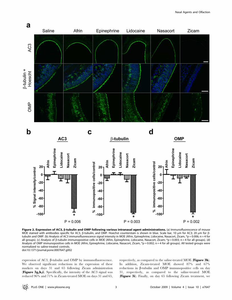
expression of AC3, b-tubulin and OMP by immunofluorescence.
We observed significant reductions in the expression of these
markers on days 31 and 65 following Zicam administration
(Figure 3g,h,i). Specifically, the intensity of the AC3 signal was
reduced 96% and 71% in Zicam-treated MOE on days 31 and 65,
respectively, as compared to the saline-treated MOE (Figure 3h).
In addition, Zicam-treated MOE showed 87% and 67%
reductions in b-tubulin and OMP immunopositive cells on day
31, respectively, as compared to the saline-treated MOE
(Figure 3i). Finally, on day 65 following Zicam treatment, we
Figure 2. Expression of AC3, b-tubulin and OMP following various intranasal agent administrations. (a) Immunofluorescence of mouseMOE stained with antibodies specific for AC3, b-tubulin, and OMP. Hoechst counterstain is shown in blue. Scale bar, 10 mm for AC3; 20 mm for b-tubulin and OMP. (b) Analysis of AC3 immunofluorescence signal intensity in MOE (Afrin, Epinephrine, Lidocaine, Nasacort, Zicam, *p = 0.006; n = 4 forall groups). (c) Analysis of b-tubulin immunopositive cells in MOE (Afrin, Epinephrine, Lidocaine, Nasacort, Zicam; *p = 0.003; n = 4 for all groups). (d)Analysis of OMP immunopositive cells in MOE (Afrin, Epinephrine, Lidocaine, Nasacort, Zicam, *p = 0.002; n = 4 for all groups). All tested groups werenormalized to saline-treated controls.doi:10.1371/journal.pone.0007647.g002
Nasal Agents and Olfaction
PLoS ONE | www.plosone.org 3 October 2009 | Volume 4 | Issue 10 | e7647

Figure 3. Long-term odorant detection defects in the MOE of Zicam-treated mice. (a) Appearance of MOE on day 31 in saline or Zicam-treated mice. Black arrowheads depict significant atrophy in endoturbinates of Zicam-treated MOE. (b) Odorant-stimulated EOG responses from salineor Zicam-treated mice, 31 days following intranasal administration. Red arrowhead indicates the time at which the odorant was delivered to the MOE.(c) Summary of the mean EOG amplitudes in response to odorants on day 31 in saline or Zicam-treated mice. Zicam-treated MOE failed to elicitodorant-stimulated EOG response (n = 4, saline; n = 5, Zicam). (d) Appearance of MOE 65 days after intranasal administration of either saline or Zicam.(e) Odorant-stimulated EOG responses from mice treated with either saline or Zicam, 65 days following intranasal administration. (f) Summary of themean EOG amplitudes in response to odorants 65 days following either saline or Zicam treatment (n = 5 for all groups). (g) Immunofluorescence ofmouse MOE stained with antibodies specific for AC3, b-tubulin, and OMP 31 and 65 days after saline or Zicam treatment. Hoechst counterstain isshown in blue. Scale bar, 10 mm for AC3; 20 mm for b-tubulin and OMP. (h) Analysis of AC3 immunofluorescence signal intensity in MOE of Zicam-treated mice normalized to saline-treated controls (p = 0.05, day 31; p,0.005, day 65; n = 4 for all groups). (i) Analysis of b-tubulin (p,0.05, day 31;p,0.0005, day 65) and OMP (p,0.01, day 31; p,0.05, day 65) immunopositive cells in the MOE treated with Zicam normalized to saline-treatedcontrols (n = 4 for all groups).doi:10.1371/journal.pone.0007647.g003
Nasal Agents and Olfaction
PLoS ONE | www.plosone.org 4 October 2009 | Volume 4 | Issue 10 | e7647
observed 87% and 83% reductions in b-tubulin and OMP
immunopositive cells, respectively (Figure 3i), as compared to the
saline-treated controls. The data collectively indicate a remarkable
damage to the olfactory epithelium and a significant loss of
regenerative capacity in Zicam-treated mice.
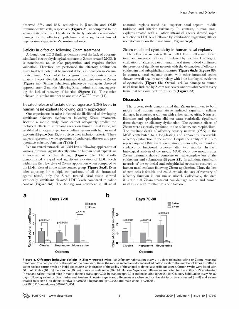
Deficits in olfaction following Zicam treatmentAlthough our EOG findings demonstrated the lack of odorant-
stimulated electrophysiological response in Zicam-treated MOE, it
is nonetheless an in vitro preparation and requires further
validation. Therefore, we performed the olfactory habituation
assay to detect potential behavioral deficits in olfaction in Zicam-
treated mice. Mice failed to recognize novel odorants approx-
imately 1 week after bilateral intranasal administration of Zicam
(Figure 4a). Similar behavioral phenotype was again observed
approximately 2 months following Zicam administration, suggest-
ing the lack of recovery of function (Figure 4b). These mice
behaved in similar manner to anosmic AC32/2 mice [11].
Elevated release of lactate dehydrogenase (LDH) levels inhuman nasal explants following Zicam application
Our experiments in mice indicated the likelihood of developing
significant olfactory dysfunction following Zicam treatment.
Because a mouse study alone cannot adequately predict the
biological effects of intranasal agents on human nasal tissue, we
established an organotypic tissue culture system with human nasal
explants (Figure 5a). Eight subjects met inclusion criteria. These
subjects represent a wide spectrum of pathologic diseases and pre-
operative olfactory function (Table 1).
We measured extracellular LDH levels following application of
various intranasal agents directly onto the human nasal explants as
a measure of cellular damage (Figure 5b). The results
demonstrated a rapid and significant elevation of LDH levels
within the first few days of Zicam application when compared to
the LDH released in the saline control group (Figure 5c,d). Even
after adjusting for multiple comparisons, of all the intranasal
agents tested, only the Zicam treated nasal tissue showed
statistically significant elevated LDH levels compared to saline
control (Figure 5d). The finding was consistent in all nasal
anatomic regions tested (i.e., superior nasal septum, middle
turbinate and inferior turbinate). In contrast, human nasal
explants treated with all other intranasal agents showed rapid
reduction in LDH level followed by stabilization suggesting little or
no cytotoxicity on the nasal tissue (Figure 5c,d).
Zicam mediated cytotoxicity in human nasal explantsThe elevation in extracellular LDH levels following Zicam
treatment suggested cell death mediated by necrosis. Histological
evaluation of Zicam-treated human nasal tissue indeed confirmed
the presence of significant necrosis with the destruction of olfactory
epithelium and subepithelial structures (Figure 6a,b; Figure S3).
In contrast, nasal explants treated with other intranasal agents
showed overall healthy morphology with little histological evidence
of cytotoxicity (Figure 6b). Overall, cellular damage to human
nasal tissue induced by Zicam was severe and was observed in every
tissue that we examined for this study (Figure S3).
Discussion
The present study demonstrated that Zicam treatment to both
mouse and human nasal tissue induced significant cellular
damage. In contrast, treatment with either saline, Afrin, Nasacort,
lidocaine and epinephrine did not cause statistically significant
tissue damage or olfactory dysfunction. The cytotoxic effects of
Zicam were especially profound in the olfactory neuroepithelium.
The resultant death of olfactory sensory neurons (OSN) in the
MOE contributed to a long-lasting and apparently irreversible
olfactory dysfunction in the mouse. Despite the ability of MOE to
replace injured OSN via differentiation of stem cells, we found no
evidence of functional recovery after two months. In fact,
histological analysis of the mouse MOE about two months after
Zicam treatment showed complete or near-complete loss of the
epithelium and submucosa (Figure S2). In addition, significant
necrosis of the epithelial and subepithelial structures occurred in
human nasal explants following Zicam application. Thus, the loss
of stem cells is feasible and could explain the lack of recovery of
olfactory function in our mouse model. Collectively, the data
illustrate that Zicam treatment can damage mouse and human
nasal tissue with resultant loss of olfaction.
Figure 4. Olfactory behavior deficits in Zicam-treated mice. (a) Olfactory habituation assay 7–10 days following saline or Zicam intranasaltreatment. The comparison of the ratio of the number of times the mouse sniffed an odorant-soaked cotton swab to the number of times it sniffed awater-soaked cotton swab on initial exposure is an indication of the ability of the animal to detect a specific substance. Cotton swabs were laced with50 ml of citralva (10 mm), heptanone (50 mm) or mouse male urine (50-fold dilution). Significant differences are noted for the ability of Zicam-treated(n = 8) and saline-treated mice (n = 8) to detect citralva (p,0.05), heptanone (p,0.01) and male urine (p,0.05). (b) Olfactory habituation assay 70–80days following saline or Zicam intranasal treatment. Again, significant differences are observed for the ability of Zicam-treated (n = 8) and saline-treated mice (n = 8) to detect citralva (p,0.0005), heptanone (p,0.005) and male urine (p,0.0005).doi:10.1371/journal.pone.0007647.g004
Nasal Agents and Olfaction
PLoS ONE | www.plosone.org 5 October 2009 | Volume 4 | Issue 10 | e7647
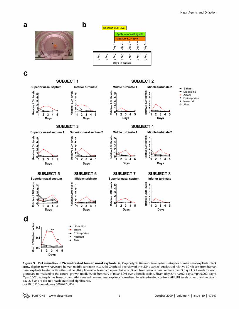
Figure 5. LDH elevation in Zicam-treated human nasal explants. (a) Organotypic tissue culture system setup for human nasal explants. Blackarrow depicts newly harvested human middle turbinate tissue. (b) Graphical overview of the LDH assay. (c) Analysis of relative LDH levels from humannasal explants treated with either saline, Afrin, lidocaine, Nasacort, epinephrine or Zicam from various nasal regions over 5 days. LDH levels for eachgroup are normalized to the control growth medium. (d) Summary of mean LDH levels from lidocaine, Zicam (day 2, *p,0.02; day 3,**p,0.002; day 4,**p,0.002), epinephrine, Nasacort and Afrin-treated human nasal explants normalized to saline-treated controls. All LDH levels other than the Zicamday 2, 3 and 4 did not reach statistical significance.doi:10.1371/journal.pone.0007647.g005
Nasal Agents and Olfaction
PLoS ONE | www.plosone.org 6 October 2009 | Volume 4 | Issue 10 | e7647

Our findings contradict a previous report on the effects of
intranasal treatment of Zicam in mice [12]. The study showed that
intranasal administrations with smaller volumes (i.e., 2 ml or 8 ml)
of Zicam did not significantly compromise the integrity of
olfactory epithelium and olfactory function. However, we find
several caveats in their methodology and interpretation of the
data. First, it is difficult to inject small volumes (,10 ml) of
intranasal agents in unanesthetized mice, even if they are
restrained. Based on our experiences with intranasal dye
injections, such small volumes did not distribute thoroughly in
the MOE, and sometimes missed the MOE entirely. Therefore,
we performed slow, controlled intranasal injections in anesthetized
mice with a targeted volume of about 15 ml that consistently
distributed throughout the MOE. Since our study aimed to
determine the role of various intranasal agents on olfaction, such a
technique is necessary in order to ensure the effective delivery of
the agents to the MOE. While this may reflect a higher dosage
(i.e., based on nasal cavity volume alone) than that recommended
for human use, it is difficult to determine the effects of intranasal
agents on olfaction if smaller volumes are used in an in vivo mouse
model system. Furthermore, we used the same volume for all other
intranasal agents (i.e., saline, Afrin, Nasacort, lidocaine and
epinephrine), and none of these drugs showed any significant signs
of cytotoxicity to either mouse or human nasal tissue, confirming
their safety profile.
Second, Slotnick and colleagues utilized anterograde tracing
with wheat germ agglutinin horseradish peroxidase to determine
the integrity of olfactory epithelium [12]. They suggested that
despite the noticeable injury to the olfactory epithelium following
Table 1. Characteristics of Human Subjects.
Age (range, mean) 20–58, 44.6
Male 4
Female 4
UPSIT (range, mean) 13–39, 27
LM CT (range, mean) 1–24, 5.4
SNOT (range, mean) 0–3.15, 1.50
Indication for surgery (N)
Chronic sinusitis 5a
Pituitary tumor 1b
Nasal congestion 2c
LM CT, Lund-Mackay staging system; SNOT-20, Sino-NasalOutcomes Test; UPSIT, University of Pennsylvania SmellIdentification Test).
aFunctional endoscopic sinus surgery.bTransnasal endoscopic approach to the pituitary tumor removal.cInferior turbinate submucosal resection.doi:10.1371/journal.pone.0007647.t001
Figure 6. Cellular necrosis in Zicam-treated human nasal explants. (a) Hematoxylin and eosin (H&E) stained human nasal explants fromsuperior nasal septum following saline or Zicam application. (b) H&E stained human nasal explants from various nasal regions treated with eithersaline, lidocaine, Afrin, epinephrine, Nasacort or Zicam. Black arrows depict dead epithelial cells. Red arrows indicate basal surface of the remainingepithelium. Note the infiltration of inflammatory cells and severe necrosis in Zicam treated-nasal explants.doi:10.1371/journal.pone.0007647.g006
Nasal Agents and Olfaction
PLoS ONE | www.plosone.org 7 October 2009 | Volume 4 | Issue 10 | e7647
Zicam treatment, the effects are transient and that recovery of
olfaction is possible. However, the study failed to show any
statistical analysis of their findings and lacked direct evidence of
functional recovery at the level of olfactory epithelium. Notably,
the observation that disruption of olfactory circuitry occurred with
Zicam treatment (even with lower doses) in the mouse model
should be worrisome because human OSN occupies a much
smaller area in the nasal cavity than mice OSN [13]. Therefore,
the risk of developing smell dysfunction could be significant if
Zicam contacts the olfactory neuroepithelium in the human nasal
cavity. Although most studies have focused on the effects of Zicam
on olfactory neuroepithelium, and hence smell dysfunction, we
emphasize that the cytotoxic effects of Zicam encompass a wider
region of the nasal tissue. These include many subepithelial
structures such as mucous glands, capillary networks and
trigeminal nerve fibers. While these components do not directly
contribute to smell dysfunction, they are critical to maintaining
healthy nasal function [14,15]. Presently, the long-term conse-
quences of damage to these subepithelial structures are unknown.
Many studies have demonstrated the cytotoxic effects of zinc
cation on various cell types, including cultured cortical neurons
[16–20], neuronal PC12 cells [21] and olfactory epithelium [22–
25]. The mechanism underlying zinc-mediated cell death appears
to be oxidative necrosis [16,21]. As demonstrated in our study,
human nasal explants undergo rapid necrosis as evidenced by
elevated extracellular LDH levels within first few days of treatment
with Zicam (Figure 5,6). We have also performed the TUNEL
(terminal dUTP nick-end labeling) assay with mouse MOE and
did not find much apoptosis following Zicam administration (data
not shown). Therefore, necrosis is likely to be the key mechanism
underlying Zicam-mediated cell death in the nasal tissue.
The regenerative capacity of MOE will depend on the extent of
necrosis. While our study showed a high degree of cellular damage
into deep tissue layers of MOE, variability in clinical presentation
(i.e., severity and recovery) is possible given the variable anatomy of
the human nasal cavity. Indeed, published case reports indicate
varying outcomes of smell dysfunction associated with Zicam use
[7,8]. Similar variations in experimental outcomes of olfactory
dysfunction are also shown in animals treated with zinc sulfate [25].
The recurring theme from these studies is that various zinc-based
solutions, including Zicam (at the concentration currently being
marketed) are cytotoxic. Interestingly, the cytotoxic effect of Zicam
on cultured human nasal tissue appears to be independent of patient
disease or anatomic location, and specific to Zicam treatment. The
human nasal tissues utilized in our study represent patients with
varied degree of olfactory dysfunction (Table 1). The elevation of
LDH and necrosis of nasal tissue was evident in all examined tissues.
This finding has critical importance to hyposmic patients because
Zicam use in this group could put them at a higher risk for
developing anosmia than a normosmic person. Lastly, the efficacy of
Zicam in treating common cold symptoms remains controversial
[26,27]. Although the adverse effects may be transient for some, the
unpredictable nature of its use by the public, together with the
current and previous findings [7,8,26] indicate that the risks of zinc-
based therapy far outweigh any benefits that it may offer.
In this paper, we demonstrated the effects of several commonly
used intranasal agents on olfaction using mouse and human nasal
tissue. Our study is the most comprehensive examination, to date, of
the role that intranasal medications have on short-term and long-
term olfactory function. Intranasal administration of Zicam, unlike
other tested agents, resulted in significant cytotoxicity to both mouse
and human nasal tissue. This is a concerning finding given the
potential development of long-lasting, and perhaps irreversible smell
dysfunction. Because Zicam was previously classified as a
homeopathic substance, it was not required to undergo stringent
safety or efficacy evaluation as other conventional drugs. However,
the Food and Drug Administration (FDA) recently issued a public
health advisory cautioning against the use of some Zicam cold
remedy nasal products. The FDA also issued a warning letter to the
manufacturers of Zicam reclassifying these products as ‘‘drugs’’ that
would require additional safety and efficacy testing to continue to
market these products. The potential health risks of intranasal
Zicam use demonstrated in our study stresses the need for stringent
oversight of homeopathic remedies to protect the public from
potentially unknown and dangerous side effects.
Materials and Methods
AnimalsAdult male C57BL/6 mice (Charles River, Wilmington, MA)
were used for all experiments. All work with animals was approved
by the University of Washington Institutional Animal Care and
Use Committee.
Human subjectsHuman subjects were recruited as a consecutive sample from a
rhinology clinic at a tertiary medical center. Written consent was
obtained from all subjects donating the nasal tissue. Inclusion
criteria included any patient over age 18 already scheduled for an
endoscopic nasal procedure. Patients were excluded if they had a
history of blood borne pathogens. The recruitment of patients and
the study protocol was approved by the Institutional Review Board
at the University of Washington (#35031).
Intranasal agent administrationFor EOG and biochemical studies, about 15 ml of above
intranasal agents were delivered slowly into the right nasal cavity
of an adult mouse using a blunted 27-gauge needle. For behavioral
testing, about 15 ml of intranasal agent was administered into both
nasal cavities using the blunted 27-gauge needle. Animals were
sedated with intraperitoneal administration of 80 mg/kg ketamine
and 8 mg/kg xylazine prior to intranasal delivery of various agents.
Electro-olfactogram (EOG)EOG recordings from the MOE were performed as described
previously [11]. Briefly, the olfactory endoturbinate was exposed by
dissecting the mouse head through the septum. The recordings were
performed with an agar- and saline-filled glass microelectrode in
contact with apical surface of the MOE in the open circuit
configuration. Odorant solutions were puffed onto the exposed
epithelium for 1 second, followed by a stream of moisturized oxygen.
Traces were captured and digitized using a Digidata 1200 A
(Molecular Devices, Union City, CA). The traces were low-filtered
at 30 Hz and sampled at 100 Hz. Multiple regions were sampled, but
EOG recordings from two separate regions of endoturbinate 1 were
used for the analysis. Odorants tested were isoamyl acetate (100 mm,
diluted in mineral oil) and citralva (50 mm, diluted in mineral oil),
vanillin (100 mm, diluted in dH2O), mineral oil and dH2O.
Odorant habituation assayThe odorant habituation assay of adult male mice was
performed as described previously [28]. The data are presented
as a ratio of the number of sniffs an animal took when the odorant-
laced cotton swab was first introduced to the number of sniffs
observed when the water-laced swab was first introduced. Male
mice urine was stored at 280C until use. All chemical odorants
and male mouse urine were diluted in water.
Nasal Agents and Olfaction
PLoS ONE | www.plosone.org 8 October 2009 | Volume 4 | Issue 10 | e7647
Tissue processingMouse MOE and human nasal tissues were immersed in 4%
paraformaldehyde overnight at 4C, followed by cryoprotection in
30% sucrose overnight at 4C. Mouse MOE was decalcified in
0.5 M EGTA at 4C for 3–4 days prior to cryoprotection with 30%
sucrose. The tissue was subsequently embedded in OCT (Sakura
Finetek USA, Inc., Torrance, CA), frozen at 220C, cryosectioned
at 30 mM and mounted onto slides.
Immunofluorescence confocal microscopy and analysisImmunofluorescence was performed as described before with the
following modifications [11]. AC3 (Santa Cruz Biotechnology Inc.,
Santa Cruz, CA), OMP (Dako, Carpinteria, CA), and b-tubulin
(Promega, Madison, WI) were used at 1:500, 1:5,000, and 1:2,000
dilutions, respectively. Images were taken with Zeiss confocal
microscope using either 20x or 63x objectives. AC3 immunofluores-
cence intensity density was measured using ImageJ software (NIH,
Bethesda, MD). OMP and b-tubulin immunopositive cells were
manually counted using ImageJ software in 200 mm6200 mm region.
Four animals were analyzed for each intranasal agent. Both AC3
immunofluorescence intensity density, OMP and b-tubulin immu-
nopositive cell numbers were normalized to saline-treated group.
Hematoxylin and Eosin (H+E) stainingThe tissues were processed as above and air dried for 30
minutes before staining in hematoxylin and eosin (Surgipath
Medical Industries, Inc, Richmond, IL) for 1 min each. Tissues
were then dehydrated in ascending series of ethanol, cleared in
xylene and coverslipped with DPX (Fluka, Milwaukee, WI) and
visualized with light microscope.
Human nasal tissue biopsySubject disease severity was assessed with several instruments
including a sinus CT scan (evaluated with Lund-Mackay staging
system; LM CT) [29], nasal endoscopy and the Sino-Nasal
Outcomes Test (SNOT-20) [30]. Subjects also took the University
of Pennsylvania Smell Identification Test (UPSIT; Sensonics, Inc.,
Haddon Heights, NJ) preoperatively to assess their olfactory
function. The human nasal tissue was collected during each
subjects’ surgery while under general anesthesia. We collected
various nasal tissues from the middle turbinate, inferior turbinate
or superior nasal septum based on the planned operation. The
harvested nasal tissue was immediately processed as below for
establishing an organotypic tissue culture system.
Human nasal explants cultureThe biopsied nasal explants were removed of any tissue debris or
blood, and divided into small pieces (approximately 2–4 mm62–4 mm61–2 mm) using a 15-blade scalpel and a fine forcep under a
dissecting scope. It was then placed on 0.4 mm polytetrafluoroethane
(PTFE) membrane (Millicell-CM; Millipore, Cork, Ireland) immersed
in 1.7 ml of culture medium consisting of 50% Dulbecco’s Modified
Eagle Medium High Glucose (Invitrogen, Carlsbad, CA); 25% Hank
solution (Invitrogen, Carlsbad, CA); 50 U/mL penicillin G and
40 mg/mL streptomycin (Invitrogen, Carlsbad, CA) in a 60615 mm
tissue culture dish (Corning, Corning, NY). The tissue was placed on
the membrane such that epithelial side was exposed to the air, and
placed in humidified incubator at 37uC with 5% CO2. The culture
media was changed every day.
Lactate dehydrogenase (LDH) assayLDH assay was performed with CytoTox 96 Non-Radioactive
Cytotoxicity Assay Kit (Promega, Madison, WI) with the following
modifications. On the day of the assay, 50 ml of culture medium
was removed and added to 50 ml of ‘substrate mix’. Following 30
minute incubation at room temperature, the reaction was stopped
with 50 ml of ‘stop solution’. Absorbance at 490 nm was obtained
with Epx Precision Microplate Reader (Molecular Devices,
Sunnyvale, CA). For measuring LDH levels in the human nasal
explants, we stabilized the nasal tissue in the cultured environment
for approximately 48 hours. We then measured the baseline LDH
levels, and applied 2 ml of either 0.9% saline, Afrin (Schering-
Plough HealthCare Products, Inc., Kenilworth, NJ), Nasacort
(Sanofi-Aventis, Bridgewater, NJ), lidocaine (Hospira, Inc., Lake
Forest, IL), 1:100,000 epinephrine (diluted with 0.9% saline;
Hospira, Inc., Lake Forest, IL) or Zicam (Matrixx Initiatives, Inc.,
Phoenix, AZ) directly onto the tissue. The levels of LDH were
again measured 24 hours later. We repeated the addition of the
intranasal agent and LDH measurement for five days.
Statistical analysisData are reported as mean +/2 S.E.M. We performed statistical
analyses using the two-tailed Student t-test. Statistical significance was
defined as P,0.05. For the human tissue analysis, a power calculation
was performed that showed a sample size of three would give 80%
power to detect a significant difference (based on two pilot samples
that showed a large effect size). We used linear mixed regression
modeling to account for the repeated measures of the LDH assays on
successive days for each tissue sample. To account for multiple
comparisons, we used Bonferroni adjusted P-values to determine
statistical significance (P = 0.05/25 = 0.002 for significance).
Supporting Information
Figure S1 Gross appearance of mouse MOE 9 days after various
intranasal agent administrations. Note the atrophy of MOE
in Zicam-treated mouse. Black arrowheads indicate atrophic
endoturbinates.
Found at: doi:10.1371/journal.pone.0007647.s001 (3.15 MB TIF)
Figure S2 Damage to mouse olfactory epithelium following
Zicam treatment at various time points. H&E staining of mouse
MOE depicting a significant loss of epithelium and submucosal
damage 9 days after intranasal administration of Zicam as
compared to saline treatment. Much greater damages to the
epithelium and submucosal structure are observed without
evidence of regeneration 31 and 35 days after intranasal
administration of Zicam. Black arrows indicate damaged and
remnants of MOE with fibrosis (e.g., days 31 and 65) in Zicam-
treated mice. Scale bar, 100 mm.
Found at: doi:10.1371/journal.pone.0007647.s002 (2.94 MB TIF)
Figure S3 Cell death in human nasal explants following Zicam
treatment from various regions of nasal cavity. H&E staining is
shown. (a) Subject 1, inferior turbinate. (b) Subject 2, middle
turbinate. (c) Subject 3, superior nasal septum. (d) Subject 4,
middle turbinate.
Found at: doi:10.1371/journal.pone.0007647.s003 (2.87 MB TIF)
Acknowledgements
We thank Carolyn Bea for excellent technical assistance. We also thank the
Storm laboratory for critical reading of this manuscript.
Author Contributions
Conceived and designed the experiments: JHL GED DRS. Performed the
experiments: JHL ZW VL YW. Analyzed the data: JHL GED ZW TCR.
Contributed reagents/materials/analysis tools: JHL GED DRS. Wrote the
paper: JHL GED DRS.
Nasal Agents and Olfaction
PLoS ONE | www.plosone.org 9 October 2009 | Volume 4 | Issue 10 | e7647

References
1. Leopold DA, Bartels S (2002) Evaluation of olfaction. J Otolaryngol 31: Suppl 1:
S18–23.2. Holbrook EH, Leopold DA (2006) An updated review of clinical olfaction. Curr
Opin Otolaryngol Head Neck Surg 14: 23–28.3. Doty RL (2009) The olfactory system and its disorders. Semin Neurol 29: 74–81.
4. Jones LM, Fontanini A, Katz DB (2006) Gustatory processing: a dynamicsystems approach. Curr Opin Neurobiol 16: 420–428.
5. Simon SA, de Araujo IE, Gutierrez R, Nicolelis MA (2006) The neural
mechanisms of gustation: a distributed processing code. Nat Rev Neurosci 7:890–901.
6. Angell M, Kassirer JP (1998) Alternative medicine–the risks of untested andunregulated remedies. N Engl J Med 339: 839–841.
7. Jafek BW, Linschoten MR, Murrow BW (2004) Anosmia after intranasal zinc
gluconate use. Am J Rhinol 18: 137–141.8. Alexander TH, Davidson TM (2006) Intranasal zinc and anosmia: the zinc-
induced anosmia syndrome. Laryngoscope 116: 217–220.9. Calof AL, Hagiwara N, Holcomb JD, Mumm JS, Shou J (2006) Neurogenesis
and cell death in olfactory epithelium. J Neurobiol 30: 67–81.
10. Henion TR, Schwarting GA (2007) Patterning the developing and regeneratingolfactory system. J Cell Physiol 210: 290–297.
11. Wong ST, Trinh K, Hacker B, Chan GC, Lowe G, et al. (2000) Disruption ofthe type III adenylyl cyclase gene leads to peripheral and behavioral anosmia in
transgenic mice. Neuron 27: 487–497.12. Slotnick B, Sanguino A, Husband S, Marquino G, Silberberg A (2007) Olfaction
and olfactory epithelium in mice treated with zinc gluconate. Laryngoscope 117:
743–749.13. Feron F, Perry C, McGrath JJ, Mackay-Sim A (1998) New techniques for biopsy
and culture of human olfactory epithelial neurons. Arch Otolaryngol Head NeckSurg 124: 861–866.
14. Widdicombe JG (1986) The physiology of the nose. Clin Chest Med 7: 159–170.
15. Widdicombe J (1997) Microvascular anatomy of the nose. Allergy 52: 7–11.16. Kim EY, Koh JY, Kim YH, Sohn S, Joe E, et al. (1999) Zn2+ entry produces
oxidative neuronal necrosis in cortical cell cultures. Eur J Neurosci 11: 327–334.17. Manev H, Kharlamov E, Uz T, Mason RP, Cagnoli CM (1997) Characteriza-
tion of zinc-induced neuronal death in primary cultures of rat cerebellar granulecells. Exp Neurol 146: 171–178.
18. Sheline CT, Behrens MM, Choi DW (2000) Zinc-induced cortical neuronal
death: contribution of energy failure attributable to loss of NAD(+) and
inhibition of glycolysis. J Neurosci 20: 3139–3146.
19. Chen CJ, Liao SL (2003) Zinc toxicity on neonatal cortical neurons: involvement
of glutathione chelation. J Neurochem 85: 443–453.
20. Capasso M, Jeng JM, Malavolta M, Mocchegiani E, Sensi SL (2005) Zinc
dyshomeostasis: a key modulator of neuronal injury. J Alzheimers Dis 8: 93–108.
21. Pavlica S, Gaunitz F, Gebhardt R (2009) Comparative in vitro toxicity of seven
zinc-salts towards neuronal PC12 cells. Toxicol In Vitro 23: 653–659.
22. Matulionis DH (1975) Ultrastructural study of mouse olfactory epithelium
following destruction by ZnSO4 and its subsequent regeneration. Am J Anat
142: 67–89.
23. Harding JW, Getchell TV, Margolis FL (1978) Denervation of the primary
olfactory pathway in mice. V. Long-term effect of intranasal ZnSO4 irrigation on
behavior, biochemistry and morphology. Brain Res 140: 271–285.
24. Burd GD (1993) Morphological study of the effects of intranasal zinc sulfate
irrigation on the mouse olfactory epithelium and olfactory bulb. Microsc Res
Tech 24: 195–213.
25. McBride K, Slotnick B, Margolis FL (2003) Does intranasal application of zinc
sulfate produce anosmia in the mouse? An olfactometric and anatomical study.
Chem Senses 28: 659–670.
26. Eby GA, Halcomb WW (2006) Ineffectiveness of zinc gluconate nasal spray and
zinc orotate lozenges in common-cold treatment: a double-blind, placebo-
controlled clinical trial. Altern Ther Health Med 12: 34–38.
27. Mossad SB (2003) Effect of zincum gluconicum nasal gel on the duration and
symptom severity of the common cold in otherwise healthy adults. Qjm 96:
35–43.
28. Trinh K, Storm DR (2003) Vomeronasal organ detects odorants in absence of
signaling through main olfactory epithelium. Nat Neurosci 6: 519–525.
29. Lund VJ, Mackay IS (1993) Staging in rhinosinusitus. Rhinology 31: 183–184.
30. Piccirillo JF, Merritt MG, Jr., Richards ML (2002) Psychometric and clinimetric
validity of the 20-Item Sino-Nasal Outcome Test (SNOT-20). Otolaryngol Head
Neck Surg 126: 41–47.
Nasal Agents and Olfaction
PLoS ONE | www.plosone.org 10 October 2009 | Volume 4 | Issue 10 | e7647