European Position Paper on Rhinosinusitis and Nasal Polyps ...
137
International Rhinology Rhinologie Internationale S U P P L E M E N T 2 0 EPOS 3 2007 European Position Paper on Rhinosinusitis and Nasal Polyps 2007 Wytske Fokkens, Valerie Lund, Joaquim Mullol, on behalf of the European Position Paper on Rhinosinusitis and Nasal Polyps group.
-
Upload
khangminh22 -
Category
Documents
-
view
0 -
download
0
Transcript of European Position Paper on Rhinosinusitis and Nasal Polyps ...
InternationalRhinologyRhinologieInternationale
S U P P L E M E N T 2 0
EPOS32007
European Position Paper onRhinosinusitis and Nasal Polyps 2007Wytske Fokkens, Valerie Lund, Joaquim Mullol, on behalf of theEuropean Position Paper on Rhinosinusitis and Nasal Polyps group.
Rhinology
supplement20
EPO
S2007
71388_oms_rhinology:71388_oms_rhinology 30-05-2007 11:26 Pagina 1
Wilfred
Corrected version October 16, 2007
W.J. Fokkens, V.J. Lund, J. Mullol et al., European Position Paper on Nasal Polyps 2007.Rhinology 45; suppl. 20: 1-139.
* corresponding author: Wytske Fokkens, Department of Otorhinolaryngology, Amsterdam Medical Centre, PO box 22660, 1100 DD Amsterdam,The Netherlands. Email: [email protected]
Rhinosinusitis is a significant and increasing health problem which results in a large financial burden on society. This evidence basedposition paper describes what is known about rhinosinusitis and nasal polyps, offers evidence based recommendations on diagnosisand treatment, and considers how we can make progress with research in this area.Rhinitis and sinusitis usually coexist and are concurrent in most individuals; thus, the correct terminology is now rhinosinusitis.Rhinosinusitis (including nasal polyps) is defined as inflammation of the nose and the paranasal sinuses characterised by two or moresymptoms, one of which should be either nasal blockage/obstruction/congestion or nasal discharge (anterior/posterior nasal drip), ±facial pain/pressure, ± reduction or loss of smell; and either endoscopic signs of polyps and/or mucopurulent discharge primarilyfrom middle meatus and/or; oedema/mucosal obstruction primarily in middle meatus, and/or CT changes showing mucosal changeswithin the ostiomeatal complex and/or sinuses.The paper gives different definitions for epidemiology, first line and second line treatment and for research.Furthermore the paper describes the anatomy and (patho)physiology, epidemiology and predisposing factors, inflammatory mecha-nisms, evidence based diagnosis, medical and surgical treatment in acute and chronic rhinosinusitis and nasal polyposis in adults andchildren. Evidence based schemes for diagnosis and treatment are given for the first and second line clinicians. Moreover attention isgiven to complications and socio-economic cost of chronic rhinosinusitis and nasal polyps. Last but not least the relation to the lowerairways is discussed.
ABSTRACT
2 Supplement 20
Participants:
Wytske Fokkens, Chair. Department of Otorhinolaryngology,Amsterdam Medical Centre, Amsterdam
Valerie Lund, Co-Chair. Institute of Laryngology andOtolaryngology. University College London, London
Joaquim Mullol, Co-Chair. Rhinology Unit & Smell Clinic, ENTDepartment, Hospital Clínic – IDIBAPS, Barcelona, Spain
Claus Bachert. Upper Airway Research Laboratory, Departmentof Otorhinolaryngology, Ghent University, Belgium
Noam Cohen, Department of Otorhinolaryngology: Head andNeck Surgery, University of Pennsylvania, Philadelphia, USA
Roxanna Cobo, Department of Otolaryngology, CentroMédico Imbanaco, Cali, Colombia
Martin Desrosiers, Department of Otolaryngology-Head andNeck Surgery, University of Montreal, Montreal, Canada
Peter Hellings, Department of Otorhinolaryngology, UniversityHospitals Leuven, Catholic University Leuven, Leuven,Belgium
Mats Holmstrom, Department of Otorhinolaryngology,Uppsala University Hospital Uppsala, Sweden
Maija Hytönen, Department of Otorhinolaryngology, HelsinkiUniversity Central Hospital, Helsinki, Finland
Nick Jones, Department of Otorhinolaryngology, Head andNeck Surgery, Queen's Medical Centre, University ofNottingham, Nottingham, UK
Livije Kalogjera, Department of Otorhinolaryngology/Headand Neck Surgery, University Hospital "Sestre Milosrdnice",Zagreb, Croatia
David Kennedy, Department of Otorhinolaryngology: Head andNeck Surgery, University of Pennsylvania, Philadelphia, USA
Jean Michel Klossek, Department ENT and Head and necksurgery, CHU Poitiers: Univ Poitiers Hopital Jean Bernard,Poitiers, France
Marek Kowalski, Department of Immunology, Rheumatologyand Allergy, Medical University of Lodz, Lodz, Poland
Eli Meltzer, Allergy and Asthma Medical Group and ResearchCenter, University of California at San Diego, San Diego,California
Bob Naclerio, Section of Otolaryngology-Head and NeckSurgery, The Pritzker School of Medicine, The University ofChicago, Chicago, USA
Desiderio Passali, ENT Department, Policlinico Le Scotte -University of Siena, Siena, Italy
David Price, Dept of General Practice and Primary Care,University of Aberdeen, Aberdeen, UK
Herbert Riechelmann, University Hospital for Ear, Nose andThroat Diseases, University Hospital Center Ulm, Ulm,Germany
Glenis Scadding, Allergy & Medical Rhinology Department,Royal National TNE Hospital, London, UK.
Heinz Stammberger, University Ear, Nose and ThroatHospital, Graz, Austria
Mike Thomas, Department of General Practice and PrimaryCare, University of Aberdeen, Aberdeen, UK
Richard Voegels, Rhinology - Clinics, University of Sao PauloMedical School, Sao Paulo, Brazil
De-Yun Wang. Department of Otolaryngology, NationalUniversity of Singapore, Singapore
Acknowledgements:The chairs of EP3OS would like to express their gratitude for
the great help in preparing this document:Fenna Ebbens, AmsterdamChristos Georgalas, LondonHanneke de Bakker, AmsterdamJosep Maria Guilemany, Barcelona

CONTENTS
1 Introduction 5
2 Definition of rhinosinusitis and nasal polyps 62-1 Introduction 62-2 Clinical definition 62-3 Definition for use in epidemiology studies/
General Practice 62-4 Definition for research 7
3 Chronic rhinosinusitis with or without nasal polyps 83-1 Anatomy and (patho)physiology 83-2 Rhinosinusitis 83-3 Chronic rhinosinusitis with or without nasal polyps 8
4 Epidemiology and predisposing factors 104-1 Introduction. 104-2 Acute bacterial rhinosinusitis. 104-3 Factors associated with acute rhinosinusitis. 104-4 Chronic rhinosinusitis (CRS) without nasal polyps 124-5 Factors associated with chronic rhinosinusitis (CRS)
without nasal polyps 124-6 Chronic rhinosinusitis with nasal polyps 154-7 Factors associated with Chronic rhinosinusitis
with nasal polyps 164-8 Epidemiology and predisposing factors for
rhinosinusitis in children 184-9 Conclusion 18
5 Inflammatory mechanisms in acute and chronicrhinosinusitis with or without nasal polyposis 19
5-1 Introduction 195-2 Acute rhinosinusitis 195-3 Chronic rhinosinusitis without nasal polyps 205-4 Chronic rhinosinusitis with nasal polyps 265-5 Aspirin sensitivity – Inflammatory mechanisms in
acute and chronic rhinosinusitis 325-6 Conclusion 34
6 Diagnosis 356-1 Assessment of rhinosinusitis symptoms 356-2 Examination 376-3 Quality of Life 40
7 Management 437-1 Treatment of rhinosinusitis with corticosteroids 437-2 Treatment of rhinosinusitis with antibiotics 517-3 Other medical management for rhinosinusitis 567-4 Evidence based surgery for rhinosinusitis 647-5 Influence of age concomitant diseases on sinus
surgery outcome 717-6 Complications of surgical treatment 76
8 Complications of rhinosinusitis and nasal polyps 808-1 Introduction 808-2 Epidemiology of complications 808-3 Orbital complications 808-4 Endocranial complications 018-5 Cavernous sinus thrombosis 828-6 Osseous complications 828-7 Unusual complications of rhinosinusitis 83
9 Special considerations: Rhinosinusitis in children 849-1 Introduction 849-2 Anatomy 849-3 Epidemiology and pathophysiology 849-4 Symptoms and signs 859-5 Clinical examination 859-6 Investigations 859-7 Management 87
10 Chronic rhinosinusitis with or without nasal polypsin relation to the lower airways 90
10-1 Introduction 9010-2 Asthma and Chronic Rhinosinusitis without NP 9010-3 Asthma and Chronic Rhinosinusitis with NP 9110-4 COPD and rhinosinusitis 91
11 Socio-economic cost of chronic rhinosinusitis andnasal polyps 92
11-1 Direct Costs 9211-2 Indirect Costs 92
12 Outcomes measurements in research 94
13 Evidence based schemes for diagnostic and treatment 9513-1 Introduction 9513-2 Introduction 9713-3 Evidence based management scheme for adults with
acute rhinosinusitis 9813-4 Evidence based management scheme for adults with
chronic rhinosinusitis without nasal polyps 10013-5 Evidence based schemes for therapy in children 103
14 Research needs and priorities 10514-1 Epidemiology: Identifying the risk factors for
development of CRS an NP 10514-2 Beyond infection: New roles for bacteria 10514-3 Host response 10514-4 Genetics 10514-5 Clinical trials 105
15 GLOSARRY TERMS 107
16 Information on QOL instruments: 10816-1 General health status instruments: 10816-2 Disease specific health status instruments: 108
17 Survey of published olfactory tests 109
18 References 111
European Position Paper on Rhinosinusitis and Nasal Polyps 2007

4 Supplement 20
European Position Paper on Rhinosinusitis and Nasal Polyps 2007 5
Rhinosinusitis is a significant health problem which seems tomirror the increasing frequency of allergic rhinitis and whichresults in a large financial burden on society (1-3). Data on(chronic) rhinosinusitis are limited and the disease entity isbadly defined. Therefore, the available data are difficult tointerpret and extrapolate.
The last decade has seen the development of a number ofguidelines, consensus documents and position papers on theepidemiology, diagnosis and treatment of rhinosinusitis andnasal polyposis (4-7). In 2005 the first European Position Paperon Rhinosinusitis and Nasal Polyps (EP3OS) was published (8, 9).This first evidence based position paper was initiated by theEuropean Academy of Allergology and Clinical Immunology(EAACI) to consider what was known about rhinosinusitis andnasal polyps, to offer evidence-based recommendations ondiagnosis and treatment, and to consider how we can makeprogress with research in this area. The paper has beenapproved by the European Rhinologic Society.
Evidence-based medicine is an important method of preparingguidelines (10, 11). Moreover, the implementation of guidelines isequally important.
Table 1-1. Category of evidence (11)
Ia evidence from meta-analysis of randomised controlled trialsIb evidence from at least one randomised controlled trialIIa evidence from at least one controlled study without
randomisationIIb evidence from at least one other type of quasi-experimental studyIII evidence from non-experimental descriptive studies, such as
comparative studies, correlation studies, and case-control studiesIV evidence from expert committee reports or opinions or clinical
experience of respected authorities, or both
Table 1-2. Strength of recommendation
A directly based on category I evidenceB directly based on category II evidence or extrapolated
recommendation from category I evidenceC directly based on category III evidence or extrapolated
recommendation from category I or II evidenceD directly based on category IV evidence or extrapolated
recommendation from category I, II or III evidence
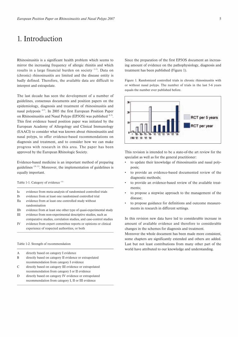
Since the preparation of the first EP3OS document an increas-ing amount of evidence on the pathophysiology, diagnosis andtreatment has been published (Figure 1).
Figure 1. Randomized controlled trials in chronic rhinosinusitis withor without nasal polyps. The number of trials in the last 5-6 yearsequals the number ever published before.
This revision is intended to be a state-of-the art review for thespecialist as well as for the general practitioner:• to update their knowledge of rhinosinusitis and nasal poly-
posis;• to provide an evidence-based documented review of the
diagnostic methods;• to provide an evidence-based review of the available treat-
ments;• to propose a stepwise approach to the management of the
disease;• to propose guidance for definitions and outcome measure-
ments in research in different settings.
In this revision new data have led to considerable increase inamount of available evidence and therefore to considerablechanges in the schemes for diagnosis and treatment.Moreover the whole document has been made more consistent,some chapters are significantly extended and others are added.Last but not least contributions from many other part of theworld have attributed to our knowledge and understanding.
1. Introduction
6 Supplement 20
2-1 Introduction
Rhinitis and sinusitis usually coexist and are concurrent in mostindividuals; thus, the correct terminology is now rhinosinusitis.The diagnosis of rhinosinusitis is made by a wide variety ofpractitioners, including allergologists, otolaryngologists, pulmo-nologists, primary care physicians and many others. Therefore,an accurate, efficient, and accessible definition of rhinosinusitisis required. A number of groups have published reports on rhi-nosinusitis and its definition. In most of these reports defini-tions are based on symptomatology and duration of disease anda single definition is aimed at all practitioners (4, 5, 12, 13).Due to the large differences in technical possibilities to diag-nose and treat rhinosinusitis/nasal polyps by various disci-plines, the need to differentiate between subgroups varies. Onone hand the epidemiologist wants a workable definition thatdoes not impose too many restrictions to study larger popula-tions. On the other hand researchers in a clinical setting are inneed of a set of clearly defined items that describes theirpatient population accurately and avoids the comparison of‘apples and oranges’ in studies that relate to diagnosis andtreatment. The taskforce tried to accommodate these differentneeds by offering definitions that can be applied in differentcircumstances. In this way the taskforce hopes to improve thecomparability of studies, thereby enhancing the evidencebased diagnosis and treatment of patients with rhinosinusitisand nasal polyps.
2-2 Clinical definition
2-2-1 Clinical definition of rhinosinusitis/nasal polyps
2-2-1-1 BacteriaRhinosinusitis (including nasal polyps) is defined as:• inflammation of the nose and the paranasal sinuses charac-
terised by two or more symptoms, one of which should beeither nasal blockage/obstruction/congestion or nasal dis-charge (anterior/posterior nasal drip):- ± facial pain/pressure,- ± reduction or loss of smell;
and either• endoscopic signs of:
- polyps and/or;- mucopurulent discharge primarily from middle meatus
and/or; oedema/mucosal obstruction primarily in mid-dle meatus,
and/or• CT changes:
- mucosal changes within the ostiomeatal complex and/orsinuses.
2-2-2 Severity of the diseaseThe disease can be divided into MILD, MODERATE andSEVERE based on total severity visual analogue scale (VAS)score (0 - 10 cm):
- MILD = VAS 0-3- MODERATE = VAS >3-7- SEVERE = VAS >7-10
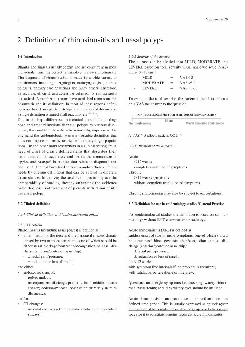
To evaluate the total severity, the patient is asked to indicateon a VAS the answer to the question:
HOW TROUBLESOME ARE YOUR SYMPTOMS OF RHINOSINUSITIS?
A VAS > 5 affects patient QOL (14).
2-2-3 Duration of the disease
Acute< 12 weekscomplete resolution of symptoms.
Chronic> 12 weeks symptomswithout complete resolution of symptoms.
Chronic rhinosinusitis may also be subject to exacerbations
2-3 Definition for use in epidemiology studies/General Practice
For epidemiological studies the definition is based on sympto-matology without ENT examination or radiology.
Acute rhinosinusitis (ARS) is defined as:sudden onset of two or more symptoms, one of which shouldbe either nasal blockage/obstruction/congestion or nasal dis-charge (anterior/posterior nasal drip):
± facial pain/pressure,± reduction or loss of smell;
for < 12 weeks;with symptom free intervals if the problem is recurrent;with validation by telephone or interview.
Questions on allergic symptoms i.e. sneezing, watery rhinor-rhea, nasal itching and itchy watery eyes should be included.
Acute rhinosinusistis can occur once or more than once in adefined time period. This is usually expressed as episodes/yearbut there must be complete resolution of symptoms between epi-sodes for it to constitute genuine recurrent acute rhinosinusitis.
2. Definition of rhinosinusitis and nasal polyps
European Position Paper on Rhinosinusitis and Nasal Polyps 2007 7
Common cold/ acute viral rhinosinusitis is defined as:duration of symptoms for less than 10 days.
Acute non-viral rhinosinusitis is defined as:increase of symptoms after 5 days or persistent symptoms after10 days with less than 12 weeks duration.
Chronic rhinosinusitis with or without nasal polyps is defined as:presence of two or more symptoms one of which should beeither nasal blockage/obstruction/congestion or nasal dis-charge (anterior/posterior nasal drip):
± facial pain/pressure;± reduction or loss of smell;
for > 12 weeks;with validation by telephone or interview.
Questions on allergic symptoms i.e. sneezing, watery rhinor-rhea, nasal itching and itchy watery eyes should be included.
2-4 Definition for research
For research purposes acute rhinosinusitis is defined as above.Bacteriology (antral tap, middle meatal tap) and/or radiology(X-ray, CT) are advised, but not obligatory.
For research purposes chronic rhinosinusitis (CRS) is definedas above. CRS is the major finding and nasal polyposis (NP) isconsidered a subgroup of this entitiy. For the purpose of astudy, the differentiation between CRS and NP must be basedon out-patient endoscopy. The research definition is based onthe presence of polyps and prior surgery.
2-4-1 Definition of chronic rhinosinusitis when no earlier sinussurgery has been performed
Chronic rhinosinusitis with nasal polyposis:polyps bilateral, endoscopically visualised in middle meatusChronic rhinosinusitis without nasal polyps:no visible polyps in middle meatus, if necessary followingdecongestant
This definition accepts that there is a spectrum of disease inCRS which includes polypoid change in the sinuses and/ormiddle meatus but excludes those with polypoid disease pre-senting in the nasal cavity to avoid overlap.
2-4-2 Definition of chronic rhinosinusitis when sinus surgery hasbeen performedOnce surgery has altered the anatomy of the lateral wall, thepresence of polyps is defined as bilateral pedunculated lesionsas opposed to cobblestoned mucosa > 6 months after surgeryon endoscopic examination. Any mucosal disease withoutovert polyps should be regarded as CRS.
2-4-3 Conditions for sub-analysisThe following conditions should be considered for sub-analy-sis:1. aspirin sensitivity based on positive oral, bronchial or nasal
provocation or an obvious history;2. asthma/bronchial hyper-reactivity /COPD/ bronchiectasies
based on symptoms, respiratory function tests;3. allergy based on specific serum IgE or SPT’s.
2-4-4 Exclusion from general studiesPatients with the following diseases should be excluded fromgeneral studies, but may be the subject of a specific study onchronic rhinosinusitis and/or nasal polyposis:1. cystic fibrosis based on positive sweat test or DNA alleles;2. gross immunodeficiency (congenital or acquired);3. congenital mucociliary problems eg primary ciliary dyskine-
sia (PCD);4. non-invasive fungal balls and invasive fungal disease;5. systemic vasculitis and granulomatous diseases;6. cocaine abuse;7. neoplasia.
8 Supplement 20
3-1 Anatomy and (patho)physiology
The nose and paranasal sinuses constitute a collection of air-filled spaces within the anterior skull. The paranasal sinusescommunicate with the nasal cavity through small apertures.The nasal cavity and its adjacent paranasal sinuses are lined bypseudostratified columnar ciliated epithelium. This containsgoblet cells and nasal glands, producers of nasal secretions thatkeep the nose moist and form a “tapis roulant” of mucus.Particles and bacteria can be caught in this mucus, renderedharmless by enzymes like lysozyme and lactoferrin, and betransported down towards the oesophagus. Cilia play an impor-tant role in mucus transport. All paranasal sinuses are normallycleared by this mucociliary transport, even though transportfrom large areas of sinuses passes through small openingstowards the nasal cavity.
A fundamental role in the pathogenesis of rhinosinusitis isplayed by the ostiomeatal complex, a functional unit that com-prises maxillary sinus ostia, anterior ethmoid cells and theirostia, ethmoid infundibulum, hiatus semilunaris and middlemeatus. The key element is the maintenance of the ostialpatency. Specifically, ostial patency significantly affects mucuscomposition and secretion; moreover, an open ostium allowsmucociliary clearance to easily remove particulate matter andbacteria. Problems occur if the orifice is too small for theamount of mucus, if mucus production is increased, forinstance during an upper respiratory tract infection (URTI), orif ciliary function is impaired. Stasis of secretions follows andbacterial export ceases, causing or exacerbating inflammationof the mucosa whilst aeration of the mucosa is decreased,causing even more ciliary dysfunction. This vicious cycle canbe difficult to break, and if the condition persists, it can resultas chronic rhinosinusitis. In chronic rhinosinusitis the role ofostium occlusion seems to be less pronounced than in ARS.
3-2 Rhinosinusitis
Rhinosinusitis is an inflammatory process involving themucosa of the nose and one or more sinuses. The mucosa ofthe nose and sinuses form a continuum and thus more oftenthan not the mucous membranes of the sinus are involved indiseases which are primarily caused by an inflammation of thenasal mucosa. Chronic rhinosinusitis is a multifactorial disease(15). Factors contributing can be mucociliairy impairment (16, 17),(bacterial) infection (18), allergy (19), swelling of the mucosa foranother reason, or rarely physical obstructions caused by mor-phological/anatomical variations in the nasal cavity orparanasal sinuses (20, 21). A role in the pathogenesis of rhinosi-nusitis is certainly played by the ostiomeatal complex, a func-
tional unit that comprises maxillary sinus ostia, anterior eth-moid cells and their ostia, ethmoid infundibulum, hiatus semi-lunaris and middle meatus. The key element is the mainte-nance of the ostial patency. An in depth discussion on factorscontributing to chronic rhinosinusitis and nasal polyps can befound in chapter 4.
3-3 Chronic rhinosinusitis with or without nasal polyps
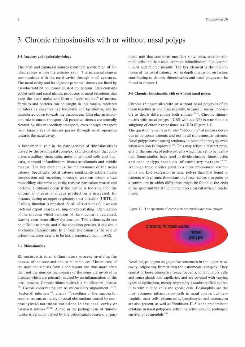
Chronic rhinosinusitis with or without nasal polyps is oftentaken together as one disease entity, because it seems impossi-ble to clearly differentiate both entities (22-24). Chronic rhinosi-nusitis with nasal polyps (CRS without NP) is considered asubgroup of chronic rhinosinusitis (CRS) (Figure 3-1).The question remains as to why “ballooning” of mucosa devel-ops in polyposis patients and not in all rhinosinusitis patients.Nasal polyps have a strong tendency to recur after surgery evenwhen aeration is improved (25). This may reflect a distinct prop-erty of the mucosa of polyp patients which has yet to be identi-fied. Some studies have tried to divide chronic rhinosinusitisand nasal polyps based on inflammatory markers (26-30).Although these studies point to a more pronounced eosino-philia and IL-5 expression in nasal polyps than that found inpatients with chronic rhinosinusitis, these studies also point toa continuum in which differences might be found at the endsof the spectrum but at the moment no clear cut division can bemade.
Figure 3-1. The spectrum of chronic rhinosinusitis and nasal polyps
Nasal polyps appear as grape-like structures in the upper nasalcavity, originating from within the ostiomeatal complex. Theyconsist of loose connective tissue, oedema, inflammatory cellsand some glands and capillaries, and are covered with varyingtypes of epithelium, mostly respiratory pseudostratified epithe-lium with ciliated cells and goblet cells. Eosinophils are themost common inflammatory cells in nasal polyps, but neu-trophils, mast cells, plasma cells, lymphocytes and monocytesare also present, as well as fibroblasts. IL-5 is the predominantcytokine in nasal polyposis, reflecting activation and prolongedsurvival of eosinophils (31).
3. Chronic rhinosinusitis with or without nasal polyps

European Position Paper on Rhinosinusitis and Nasal Polyps 2007 9
The reason why polyps develop in some patients and not inothers remains unknown. There is a definite relationship inpatients with 'Samter triad': asthma, NSAID sensitivity andnasal polyps. However, not all patients with NSAID sensitivityhave nasal polyps, and vice-versa. In the general population,the prevalence of nasal polyps is 4% (32). In patients with asth-ma, a prevalence of 7 to 15% has been noted whereas, inNSAID sensitivity, nasal polyps are found in 36 to 60% of
patients (33, 34). It had long been assumed that allergy predis-posed to nasal polyps because the symptoms of watery rhinor-rhoea and mucosal swelling are present in both diseases, andeosinophils are abundant. However, epidemiological data pro-vide no evidence for this relationship: polyps are found in 0.5to 1.5% of patients with positive skin prick tests for commonallergens (34, 35).
10 Supplement 20
4-1 Introduction
Rhinosinusitis in its many forms, constitutes one of the com-monest conditions encountered in medicine and may presentto a wide range of clinicians from primary care to accident andemergency, pulmonologists, allergists, otorhinolaryngologistsand even intensivists and neurosurgeons when severe compli-cations occur.
The incidence of acute viral rhinosinusitis (common cold) isvery high. It has been estimated that adults suffer 2 to 5 coldsper year, and school children may suffer 7 to 10 colds per year.The exact incidence is difficult to measure because mostpatients with common cold do not consult a doctor. Recently acase control study in the Dutch population concluded that anestimated 900000 consultations take place annually for acuterespiratory tract infection. Rhinovirus (24%) and Influenzae(11%) were the most common agents isolated. (36). More reliabledata are available on ARS. As mentioned earlier acute non-viral rhinosinusitis (ARS) is defined as an increase of symp-toms after 5 days or persistent symptoms after 10 days after asudden onset of two or more of the symptoms: nasal block-age/congestion, anterior discharge/postnasal drip, facialpain/pressure, and/or reduction/loss of smell. It is estimatedthat only 0.5% to 2% of viral URTIs are complicated by bacteri-al infection; however, the exact incidence is unknown giventhe difficulty distinguishing viral from bacterial infection with-out invasive sinus-puncture studies. Bacterial culture results insuspected cases of acute community-acquired sinusitis are pos-itive in only 60% of cases (37). Signs and symptoms of bacterialinfection may be mild and often resolve spontaneously (38, 39).
In spite of the high incidence of ARS and prevalence and signifi-cant morbidity of chronic rhinosinusitis (CRS), with and withoutnasal polyps, there is only limited accurate data on the epidemi-ology of these conditions. This observation mainly relates to thelack of a uniformly accepted definition for CRS. In addition,patient selection criteria greatly differ between epidemiologicstudies complicating comparison of studies. When interpretingepidemiologic data, one should be aware of a significant selec-tion bias of the different studies presented below. The purposeof this section of the EP3OS document is to give an updatedoverview of the currently available epidemiologic data on ARSand CRS with and without nasal polyps, and illustrate the fac-tors which are believed to predispose to their development.
4-2 Acute bacterial rhinosinusitis.
When describing the incidence of acute bacterial rhinosinusitisthere has been a lot of debate about the actual definition of the
condition. For example in the Cochrane Review on antibioticsfor ARS, studies were included if sinusitis was proven by a con-sistent clinical history, and radiographic or aspiration evidenceof ARS (40). However, most guidelines on the diagnosis of acutebacterial rhinosinusitis base the diagnosis on symptoms andclinical examination. However, if the diagnosis is based on clin-ical examination alone, the rate of false positive results is high.In patients with a clinical diagnosis of ARS, less than half havesignificant abnormalities at X-ray examination (41). Based onsinus puncture/aspiration (considered the most accurate), 49-83% of symptomatic patients had ARS (42). Compared withpuncture/aspiration, radiography offered moderate ability todiagnose sinusitis Using sinus opacity or fluid as the criterionfor sinusitis, radiography had sensitivity of 0.73 and specificityof 0.80 (42).
An average of 8.4 % of the Dutch population reported at leastone episode of ARS per year in 1999 (43). The incidence of visitsto the general practitioner for acute sinusitis in theNetherlands in 2000 was 20 per 1000 men and 33.8 per 1000women (44). According to National Ambulatory Medical CareSurvey (NAMCS) data in the USA, sinusitis is the fifth mostcommon diagnosis for which an antibiotic is prescribed.Written in 2002, sinusitis accounted for 9% and 21% of all pae-diatric and adult antibiotic prescriptions respectively (4).
4-3 Factors associated with acute rhinosinusitis.
4-3-1 PathogensSuperinfection by bacteria on mucosa damaged by viral infec-tion (common cold) is the most important cause of ARS. Themost common bacterial species isolated from the maxillarysinuses of patients with ARS are Streptococcus pneumoniae,Haemophilus influenzae, and Moraxella catarrhalis, the latterbeing more common in children (45, 46). Other streptococcalspecies, anaerobic bacteria and Staphylococcus aureus cause asmall percentage of cases. Resistance patterns of the predomi-nant pathogens vary considerably (47, 48). The prevalence anddegree of antibacterial resistance in common respiratorypathogens are increasing worldwide. In France, increases inresistance have been observed during the last twenty years inthe same geographical area for H. influenzae and S. pneumoniae(49). The association between inappropriate antibiotic consump-tion and the prevalence of resistance is widely assumed basedon in vitro experience (50). Pathogens may also influence theseverity of symptomatology (51).
4-3-2 Ciliary impairmentNormal mucociliary flow is a significant non-specific defencemechanism in the prevention of ARS. Viral rhinosinusitis
4. Epidemiology and predisposing factors

European Position Paper on Rhinosinusitis and Nasal Polyps 2007 11
results in the loss of cilia and ciliated cells, reaching a maxi-mum around one week after the infection. Three weeks afterthe beginning of the infection the number of cilia and ciliatedcells increases to nearly normal. However, as a sign of regener-ation, immature short cilia (0.7 to 2.5 µm in length) were oftenseen (52). The impaired mucociliary function during viral rhinos-inusitis results in an increased sensitivity to bacterial infection.
Also in animal experiments it was shown that shortly afterexposure to pathogenic bacteria, like Streptococcus pneumoniae,Hemophilus influenzae, Pseudomonas aeruginosa, a significantloss of ciliated cells from sinus mucosa and a correspondingdisruption of normal mucociliary flow occurred (53).
4-3-3 AllergyReview articles on sinusitis have suggested that atopy predis-poses to rhinosinusitis (54). This theory is attractive given thepopularity of the concept that disease in the ostiomeatal areacontributes to the development of sinus disease. Mucosa in anindividual with allergic rhinitis might be expected to beswollen and therefore more liable to obstruct sinus ostia,reduce ventilation, leading to mucus retention which in turnmight be more prone to infection. Furthermore there has beenan increase in the body of opinion that regard the mucosa ofthe nasal airway as being in a continuum with the paranasalsinuses reflected in the term ‘rhinosinusitis’ (55). However, thenumber of studies determining the occurrence of ARS inpatients with and without allergy are very limited.
Savolainen studied the occurrence of allergy in 224 patientswith verified ARS by means of an allergy questionnaire, skintesting, and nasal smears. Allergy was found in 25% of thepatients and considered probable in another 6.5%. The corre-sponding percentages in the control group were 16.5 and 3,respectively. There were no differences between allergic andnon-allergic patients in the number of previous ARS episodesnor of previously performed sinus irrigations. Bacteriologicaland radiological findings did not differ significantly betweenthe groups (56). Alho showed that subjects with allergic IgE-mediated rhinitis had more severe paranasal sinus changes onCT scanning than non-allergic subjects during viral colds.These changes indicate impaired sinus functioning and mayincrease the risk of bacterial sinusitis (57). Alho studied cellularmodifications during acute viral rhinitis in three differentgroups (allergic, recurrent sinusitis, and healthy patients). Nosignificant difference in inflammatory cells was found in anygroup during acute (D0) and convalescent (D21) phases.
In a small prospective study, no difference in prevalence ofpurulent rhinosinusitis was found between patients with andwithout allergic rhinitis (58). Furthermore, allergy was found in31.5% of patients with verified acute maxillary sinusitis andthere were no differences between allergic and non-allergicpatients in the number of prior ARS episodes (56). Newman et
al. reported that whilst 39% of patients with CRS had asthma,raised specific IgE or an eosinophilia, only 25% had true mark-ers to show they were atopic (59). Finally, Emanuel et al. (60)
found relatively lower percentages of allergic patients in thegroup of patients with the most severe sinus disease on CTscan and Iwens et al. (61) reported that the prevalence and extentof sinus mucosa involvement on CT was not determined bythe atopic state.
Radiologic studies are unhelpful in unravelling the correlationbetween allergy and rhinosinusitis. High percentages of sinusmucosa abnormalities are found on radiologic images of aller-gic patients, e.g. 60% incidence of abnormalities on CT scansamong subjects with ragweed allergy during the season (62).However, one should interpret this data with caution in viewof the fact that high percentages of incidental findings arefound on radiologic images of the sinus mucosa in individualswithout nasal complaints, ranging from 24.7 % to 49.2 % (63-66),that the normal nasal cycle induces cyclical changes in thenasal mucosa volume (67), and that radiological abnormalities donot correlate well with patient's symptoms (62).
Holzmann reported an increased prevalence of allergic rhinitisin children with orbital complications of ARS, and these com-plications occurred especially during the pollen season (68). In astudy involving 8723 children, Chen and colleagues found theprevalence of sinusitis to be significantly higher in childrenwith allergic rhinitis than in children without allergies (69).
In conclusion, although an attractive hypothesis, we can repeatthe statement made a decade ago that there are no publishedprospective reports on the incidence of infective rhinosinusitisin populations with and without clearly defined allergic rhinos-inusitis (70).
4-3-4 Helicobacter pylori and laryngopharyngeal refluxVery few articles can be found in the literature regarding therole of the laryngopharyngeal reflux (LPR) and/or Helicobacterpylori infection in the pathogenesis of ARS. More have investi-gated a possible role in CRS but without significant results.Wise described a correlation between LPR (detected eitherwith pH sensor and/or with symptom scores), and postnasaldrip without the typical findings of CRS, a problem whichmight predispose a subject to an acute bacterial infection (71). Ina case report, Dinis underlines the presence of Helicobacter inthe sphenoid sinus of a patient with severe sphenoid sinusitisthat was also treated with Helicobacter pylori therapy (72).Therefore, even if there is no clear correlation between refluxdisease and/or Helicobacter pylori infection and ARS, this isundoubtedly a field for future investigation when one consid-ers the increase of this gastrointestinal problem in developedcountries and the fact that the acid content of reflux and theHelicobacter infection itself can cause mucociliary impairment.
12 Supplement 20
4-3-5 Other risk factors : ventilation, naso-gastric tubeNosocomial sinusitis are frequently observed in intensive care(73, 74) and have been generally linked to naso-tracheal intubation(75) or presence of naso-gastric tube (76). The maxillary sinus isfrequently involved. Endoscopy of the middle meatus is usefulto determine the presence of purulence in the middle meatusand if the culture is possible. Bacteriology differs from com-munity acquired cases with a more frequent isolation of anaer-obic bacteria (77). Treatment may include, complementary toadjustment of antibiotic treatment, drainage, daily lavage andremoval of the grastic tube (78).
4-4 Chronic rhinosinusitis (CRS) without nasal polyps
The paucity of accurate epidemiologic data on CRS with orwithout nasal polyps contrasts with the more abundant infor-mation on microbiology, diagnosis and treatment options forthese conditions. When reviewing the current literature onCRS, it becomes clear that giving an accurate estimate of theprevalence of CRS remains speculative, because of the hetero-geneity of the disorder and the diagnostic imprecision oftenused in publications. In a survey on the prevalence of chronicconditions, it was estimated that CRS, defined as having ‘sinustrouble’ for more than 3 months in the year before the inter-view, affects 15.5% of the total population in the United States(79), ranking this condition second in prevalence among allchronic conditions. Subsequently the high prevalence of CRSwas confirmed by another survey suggesting that 16% of theadult US population has CRS (80). However, the prevalence ofdoctor-diagnosed CRS is much lower; a prevalence of 2% wasfound using ICD-9 codes as an identifier (81). Corroboration ofthe definitive diagnosis of CRS should be done with nasalendoscopy (82) or CT (83). As the diagnosis of CRS has primarilybeen based on symptoms, often excluding dysosmia, thismeans that the diagnosis of CRS is often overestimated (83). Themajority of primary care physicians do not have the training orequipment to perform nasal endoscopy that also leads to over-diagnosis (84).
Interestingly, the prevalence rate of CRS was substantiallyhigher in females with a female/male ratio of 6/4 (79). InCanada, the prevalence of CRS, defined as an affirmativeanswer to the question ‘Has the patient had sinusitis diagnosedby a health professional lasting for more than 6 months?’ranged from 3.4% in male to 5.7% in female subjects (85). Theprevalence increased with age, with a mean of 2.7% and 6.6%in the age groups of 20-29 and 50-59 years, respectively. Afterthe age of 60 years, prevalence levels of CRS levelled off to4.7% (85). In a nationwide survey in Korea, the overall preva-lence of CRS, defined as the presence of at least 3 nasal symp-toms lasting more than 3 months together with the endoscopicfinding of nasal polyps and/or mucopurulent discharge withinthe middle meatus, was 1.01% (86), with no differences betweenage groups or gender. By screening a non-ENT population,
which may be considered representative of the general popula-tion in Belgium, Gordts et al. (87) reported that 6% of subjectssuffered from chronic nasal discharge. A comparative study inthe north of Scotland and the Caribbean found that in ORLclinics in both populations there was a similar prevalence ofCRS (9.6% and 9.3% respectively) (88). Not withstanding theshortcomings of epidemiologic studies on CRS, it represents acommon disorder of multifactorial origin. A list of factors willbe discussed in the following chapter which are believed to beetiologically linked to CRS.
4-5 Factors associated with chronic rhinosinusitis (CRS) withoutnasal polyps
4-5-1 Ciliary impairmentAs may be concluded from the section on anatomy and patho-physiology, ciliary function plays an important role in the clear-ance of the sinuses and the prevention of chronic inflamma-tion. Secondary ciliary dyskinesia is found in patients withchronic rhinosinusitis, and is probably reversible, althoughrestoration takes some time (89). As expected in patients withKartagener’s syndrome and primary ciliary dyskinesia, CRS is acommon problem and these patients usually have a long histo-ry of respiratory infections. In patients with cystic fibrosis (CF),the inability of the cilia to transport the viscous mucus causesciliary malfunction and consequently CRS. Nasal polyps arepresent in about 40% of patients with CF (90). These polyps aregenerally more neutrophilic than eosinophilic in nature butmay respond to steroids nonetheless, as inhaled steroids inpatients with CF reduce neutrophilic inflammation (91-93).
4-5-2 AllergyReview articles on rhinosinusitis have suggested that atopypredisposes to its development (54, 94). It is tempting to speculatethat allergic inflammation in the nose predisposes the atopicindividual to the development of CRS. Both conditions sharethe same trend of increasing prevalence (95, 96) and are frequentlyassociated.
It has been postulated (97) that swelling of the nasal mucosa inallergic rhinitis at the site of the sinus ostia may compromiseventilation and even obstruct sinus ostia, leading to mucusretention and infection. Futhermore, there has been anincrease in the body of opinion that regard the mucosa of thenasal airway as being in a continuum with the paranasal sinus-es and hence the term ‘rhinosinusitis’ was introduced (55).However, critical analysis of the papers linking atopy as a riskfactor to infective rhinosinusitis (chronic or acute) reveal thatwhilst many of the studies suggest a higher prevalence of aller-gy in patients presenting with symptoms consistent with sinusi-tis than would be expected in the general population, theremay well have been a significant selection process, because thedoctors involved often had an interest in allergy (30, 98-102). A num-ber of studies report that markers of atopy are more prevalent

European Position Paper on Rhinosinusitis and Nasal Polyps 2007 13
in populations with CRS. Benninger reported that 54% of out-patients with CRS had positive skin prick tests (103). AmongCRS patients undergoing sinus surgery, the prevalence of posi-tive skin prick tests ranges from 50% to 84% (56, 60, 104), of whichthe majority (60%) have multiple sensitivities (60). As far back as1975, Friedman reported an incidence of atopy in 94% ofpatients undergoing sphenoethmoidectomies (105).However, the role of allergy in CRS is questioned by other epi-demiologic studies showing no increase in the incidence ofinfectious rhinosinusitis during the pollen season in pollen-sensitized patients (70). Taken together, epidemiologic datashow an increased prevalence of allergic rhinitis in patientswith CRS, but the role of allergy in CRS remains unclear.Notwithstanding the lack of hard epidemiologic evidence for aclear causal relationship between allergy and CRS, it is clearthat failure to address allergy as a contributing factor to CRSdiminishes the probability of success of a surgical intervention(106). Among allergy patients undergoing immunotherapy, thosewho felt most helped by immunotherapy were the subjectswith a history of recurrent rhinosinusitis, and about half of thepatients, who had had sinus surgery before, believed that thesurgery alone was not sufficient to completely resolve therecurrent episodes of infection (106).
4-5-3 AsthmaRecent evidence suggests that allergic inflammation in theupper and lower airways coexist and should be seen as a con-tinuum of inflammation, with inflammation in one part of theairway influencing its counterpart at a distance. The argumentsand consequences of this statement are summerized in theARIA document (107). Rhinosinusitis and asthma are also fre-quently associated in the same patients, but their inter-rela-tionship is poorly understood. The evidence that treatment ofrhinosinusitis improves asthma symptoms and hence reducesthe need for medication to control asthma, mainly results fromresearch in children and will be discussed below (Chapter 9-7).In short, improvements in both asthma symptoms and medica-tion have been obtained after surgery for rhinosinusitis in chil-dren with both conditions (108-110).
Studies on radiographic abnormalities of the sinuses in asth-matic patients have shown a high prevalence of abnormal sinusmucosa (111, 112). All patients with steroid-dependant asthma hadabnormal mucosal changes on CT compared to 88% with mildto moderate asthma (113). Again caution should be exercised inthe interpretation of these studies. Radiographically detectedsinus abnormalities in sensitized patients may reflect inflam-mation related to the allergic state rather than to sinus infec-tion.
4-5-4 Immunocompromised stateAmong conditions associated with dysfunction of the immunesystem, congenital immunodeficiencies manifest themselveswith symptoms early in life and will be dealt with in the paedi-
atric CRS section (see Chapter 7-6). However, dysfunction ofthe immune system may occur later in life and present withCRS. In a retrospective review of refractory sinusitis patients,Chee et al. found an unexpectedly high incidence of immunedysfunction (114). Of the 60 patients with in vitro T-lymphocytefunction testing, 55% showed abnormal proliferation inresponse to recall antigens. Low immunoglobulin G, A, and Mtitres were found in 18%, 17%, and 5%, respectively, of patientswith refractory sinusitis. Common variable immunodeficiencywas diagnosed in 10% and selective IgA deficiency in 6% ofpatients. Therefore, immunological testing should be an inte-gral part of the diagnostic pathway of patients with CRS. In across-sectional study to assess the overall prevalence of oto-laryngologic diseases in patients with HIV infection, Porter etal. (115) reported that sinusitis was present in more than half ofthe HIV-positive population, ranking this condition one of themost prevalent diseases in HIV-positive individuals. However,the relevance of these data is questioned as there was no dif-ference in sinonasal symptom severity between HIV-positiveand AIDS patients nor was there a correlation between CD4+cell counts and symptom severity. In a more detailed study,Garcia-Rodrigues et al. (116) reported a lower incidence of rhi-nosinusitis (34%), but with a good correlation between lowCD4+ cell count and the probability of rhinosinusitis. Itshould also be mentioned here that atypical organisms likeAspergillus spp, Pseudomonas aeruginosa and microsporidia areoften isolated from affected sinuses and that neoplasms suchas non-Hodgkin lymphoma and Kaposi’s sarcoma, mayaccount for sinonasal problems in patients with AIDS (117).
4-5-5 Genetic factorsAlthough chronic sinus disease has been observed in familymembers, no genetic abnormality has been identified linked toCRS. However, the role of genetic factors in CRS has beenimplicated in patients with cystic fibrosis (CF) and primary cil-iary dyskinesia (Kartagener's syndrome). CF is one of the mostfrequent autosomal recessive disorders of the Caucasian popu-lation, caused by mutations of the CFTR gene on chromo-some 7 (118). The most common mutation, F508, is found in 70to 80% of all CFTR genes in Northern Europe (119, 120). Upper air-way manifestations of CF patients include chronic rhinosinusi-tis and nasal polyps, which are found in 25 to 40 % of CFpatients above the age of 5 (121-124). Interestingly, Jorissen et al.(125) reported that F508 homozygosity represents a risk factorfor paranasal sinus disease in CF and Wang reported thatmutations in the gene responsible for CF may be associatedwith the development of CRS in the general population (126).
4-5-6 Pregnancy and endocrine stateDuring pregnancy, nasal congestion occurs in approximatelyone-fifth of women (127). The pathogenesis of this disorderremains unexplained, but there have been a number of pro-posed theories. Besides direct hormonal effects of oestrogen,progesterone and placental growth hormone on the nasal
14 Supplement 20
mucosa, indirect hormonal effects like vascular changes maybe involved. Whether pregnancy rhinitis predisposes to thedevelopment of sinusitis, is not clear. In a small prospectivestudy, Sobol et al. (128) report that 61% of pregnant women hadnasal congestion during the first trimester, whereas only 3%had sinusitis. In this study, a similar percentage of non-preg-nant women in the control group developed sinusitis duringthe period of the study. Also in an earlier report, the incidenceof sinusitis in pregnancy was shown to be quite low, i.e. 1.5%(129). In addition, thyroid dysfunction has been implicated inCRS, b there is only limited data on the prevalence of CRS inpatients with hypothyroidism.
4-5-7 Local host factorsCertain anatomic variations such as concha bullosa, nasal sep-tal deviation and a displaced uncinate process, have been sug-gested as potential risk factors for developing CRS (130).However, some studies that have made this assertion haveequated mucosal thickening on CT with CRS (131) when it hasbeen shown that incidental mucosal thickening occurs inapproximately a third of an asymptomatic population (20).However, Bolger et al. (132) found no correlation between CRSand bony anatomic variations in the nose. Holbrook et al. alsofound no correlation between sinus opacification, anatomicalvariations and symptom scores (133). However, one should men-tion here that no study has so far investigated whether a partic-ular anatomic variation can impair drainage of the ostiomeatalcomplex per se. Whilst some authors have postulated thatanatomical variations of the paranasal sinuses can contribute toostial obstruction (134) there are several studies that show theprevalence of anatomical variations is no more common inpatients with rhinosinusitis or polyposis than in a control pop-ulation (20, 21, 135). One area where conjecture remains is the effectof a deviated septum. There are a number of studies that showno correlation between septal deviation and the prevalence ofCRS. (136, 137). Whilst there is no recognised method of objective-ly defining the extent of a deviated septum, some studies havefound a deviation of more than 3mm from the midline to bemore prevalent in rhinosinusitis (138, 139) whilst others have not (21,
137, 140). Taken together, there is no evidence for a causal correla-tion between nasal anatomic variations in general and the inci-dence of CRS. In spite of the observation that sinonasal com-plaints often resolve after surgery, this does not necessarilyimply that anatomic variation is etiologically involved.
CRS of dental origin should not be overlooked when consider-ing the etiology of CRS. Obtaining accurate epidemiologic dataon the incidence of CRS of dental origin is not possible as theliterature is limited to anecdotal reports.
4-5-8 Micro-organisms
4-5-8-1 BacteriaAlthough it is often hypothesized that CRS evolves from ARS,this has never been proven. Furthermore, the role of bacteriain CRS is far from clear. A number of authors have describedthe microbiology of the middle meatus and sinuses. Howeverif and which of these pathogen are contributory to the diseaseremains a matter of debate. Bhattacharyya (2005) found thatboth anaerobes and aerobic species could be recovered fromboth diseased and the non-diseased contralateral side ofpatients with chronic rhinosinusitis casting doubt on the aetio-logic role of bacteria in CRS (141). Anaerobes are more prevalentin infections secondary to dental problems.
Arouja isolated aerobes from 86% of the middle meatus samplesof CRS patients, whereas anaerobes were isolated in 8%. Themost frequent microorganisms were Staphylococcus aureus(36%), coagulase-negative Staphylococcus (20%), and Streptococcuspneumoniae (17%). Middle meatus and maxillary sinus culturespresented the same pathogens in 80% of cases. In healthy indi-viduals, coagulase-negative Staphylococcus (56%), S. aureus (39%),and S. pneumoniae (9%) were the most frequent isolates. (142).Some authors suggest that as chronicity develops, the aerobicand facultative species are gradually replaced by anaerobes (143, 144).This change may result from the selective pressure of antimicro-bial agents that enable resistant organisms to survive and fromthe development of conditions appropriate for anaerobic growth,which include the reduction in oxygen tension and an increasein acidity within the sinus. Often polymicrobial colonisation isfound; the contribution to the disease of the different pathogensremains unclear. The presence of intracellularS. aureus in epithelial cells of the nasal mucosa has been sug-gested to pay a significant risk factor for recurrent episodes ofrhinosinusitis due to persistent bacterial clonotypes, whichappear refractory to antimicrobial and surgical therapy (145).
4-5-8-2 FungiFungi have been cultured from human sinuses (146). Their pres-ence may be relatively benign, colonizing normal sinuses orforming saprophytic crusts. They may also cause a range ofpathology, ranging from non-invasive fungus balls to invasive,debilitating disease (147).
There is an increasing interest in the concept that the mostcommon form of sinus disease induced by fungus may becaused by the inflammation stimulated by airborne fungal anti-gens. In 1999 it was proposed that most patients with CRSexhibit eosinophilic infiltration and the presence of fungi byhistology or culture (148). This assertion was based on findingpositive fungal culture by using a new culture technique in 202of 210 (96%) patients with CRS who prospectively were evaluat-ed in a cohort study. No increase in type I sensitivity was foundin patients as compared with controls. The term ‘‘eosinophilic

European Position Paper on Rhinosinusitis and Nasal Polyps 2007 15
chronic rhinosinusitis’’ was proposed to replace previously usednomenclature (allergic chronic rhinosinusitis). Using this newculture technique, the same percentage of positive fungi cul-tures was also found in normal controls (149).
Pant et al. suggest that fungal-specific immunity is charac-terised by serum IgG3 and not IgE distinguished patients withCRS and eosinophilic mucus from healthy controls, regardlessof whether fungi were found within the mucus. They found nodifferences between those with CRS and the eosinophilicmucus group and a group with allergic fungal rhinosinusitis (150).
Some authors suggest that non IgE mediated mechanisms tofungal spores might be responsible for eosinophilic inflamma-tion seen in some individuals (151) Shin et al. found that patientswith CRS had an exaggerated humoral and TH1 and TH2 cel-lular response to common airborne fungi, particularlyAlternaria. No increase in type I sensitivity was found inpatients as compared with controls (152). In another study nocorrelation was found between fungal parameters and the clini-cal parameters of CRS or the presence of eosinophilia (153) andthe use of quantitative PCR produced a recovery rate of fungiof 46% in a group with CRS and a control group (154).
A broad array of fungi has been identified in the sinus cavitiesof patients with sinusitis through varied staining and culturetechniques (148, 149). As with the isolation of bacteria in sinus cavi-ties in these patients, the presence of fungi does not prove thatthese pathogens directly create or perpetuate disease. The useof topical or systemic antifungal agents have not consistentlybeen shown to help patients with CRS (155, 156).
4-5-9 “Osteitis”—the role of boneAreas of increased bone density and irregular bony thickeningare frequently seen on CT in areas of chronic inflammationand may be a marker of the chronic inflammatory process (157).However, the effect during the initial phases of a severe CRSfrequently appears as rarefaction of the bony ethmoid parti-tions. Although to date bacterial organisms have not beenidentified in the bone in either humans or animal models ofCRS, it has been suggested that that this irregular bony thick-ening is a sign of inflammation of the bone which in turnmight maintain mucosal inflammation (158).
In rabbit studies it was demonstrated that not only the boneadjacent to the involved maxillary sinus become involved, butthat the inflammation typically spreads through the Haversiancanals and may result in bone changes consistent with somedegree of chronic osteomyelitis at a distance from the primaryinfection (159, 160). It is certainly possible that these changes, if fur-ther confirmed in patients, may at least in part, explain whyCRS is relatively resistant to therapy.
4-5-10 Environmental factorsCigarette smoking was associated with a higher prevalence ofrhinosinusitis in Canada (85), whereas this observation was notconfirmed in a nationwide survey in Korea (86). Other lifestyle-related factors are undoubtedly involved in the chronic inflam-matory processes of rhinosinusitis. For instance, low incomewas associated with a higher prevalence of CRS (85). In spite ofin vitro data on the toxicity of pollutants on respiratory epithe-lium, there exists no convincing evidence for the etiologic roleof pollutants and toxins such as ozone in CRS.
4-5-11 Iatrogenic factorsAmong risk factors of CRS, iatrogenic factors should not beforgotten as they may be responsible for the failure of sinussurgery. The increasing number of sinus mucocoeles seems tocorrelate with the increase in endoscopic sinus surgery proce-dures. Among a group of 42 patients with mucocoeles, 11 hadprior surgery within 2 years prior to presentation (161). Anotherreason for failure after surgery can be the recirculation ofmucus out of the natural maxillary ostium and back through aseparate surgically created antrostomy resulting in an increasedrisk of persistent sinus infection (162).
4-5-12 Helicobacter pylori and laryngopharyngeal refluxHeliobacter pylori DNA has been detected in between 11% (163)
and 33% (164) of sinus samples from patients with CRS but notfrom controls. However, as in ARS this does not prove acausal relationship.
4-6 Chronic rhinosinusitis with nasal polyps
Epidemiologic studies rely on nasal endoscopy and/or ques-tionnaires to report on the prevalence of nasal polyps (NP).Large NP can be visualized by anterior rhinoscopy, whereasnasal endoscopy is warranted for the diagnosis of smaller NP.Nasal endoscopy appears to be a prerequisite for an accurateestimate of the prevalence of NP, as not all patients that claimto have NP actually have polyps on nasal endoscopy (165).Therefore, surveys based on questionnaires asking for the pres-ence of NP, may provide us with an overestimation of the self-reported prevalence of NP. Recently, a French expert panel ofENT specialists elaborated a diagnostic questionnaire/algo-rithm with 90 % sensitivity and specificity (166).
In the light of epidemiologic research, a distinction needs to bemade between clinically silent NP or preclinical cases, andsymptomatic NP. Asymptomatic polyps may transiently be pre-sent or persist, and hence remain undiagnosed until they arediscovered by clinical examination. On the other hand, polypsthat become symptomatic may remain undiagnosed, eitherbecause they are missed during anterior rhinoscopy and/orbecause patients do not see their doctor for this problem.Indeed, one third of patients with CRS with NP do not seekmedical advice for their sinonasal symptoms (167). Compared to

16 Supplement 20
patients with CRS with NP not seeking medical attention,those actively seeking medical care for CRS with NP had moreextensive NP with more reduction of peak nasal inspiratoryflow and greater impairment of the sense of smell (168).
In a population-based study in Skövde, Sweden, Johansson et al.(165) reported a prevalence of nasal polyps of 2.7% of the total pop-ulation. In this study, NP were diagnosed by nasal endoscopyand were more frequent in men (2.2 to 1), the elderly (5% at 60years of age and older) and asthmatics. In a nationwide surveyin Korea, the overall prevalence of polyps diagnosed by nasalendoscopy was 0.5% of the total population (136). Based on apostal questionnaire survey in Finland, Hedman et al. (32) foundthat 4.3% of the adult population answered positively to thequestion as to whether polyps had been found in their nose.Using a disease-specific questionnaire, Klossek et al. (167) reporteda prevalence of NP of 2.1% in France.
From autopsy studies, a prevalence of 2% has been foundusing anterior rhinoscopy (169). After removing whole naso-eth-moidal blocks, nasal polyps were found in 5 of 19 cadavers (170),and in 42% of 31 autopsy samples combining endoscopy withendoscopic sinus surgery (171). The median age of the cases inthe 3 autopsy studies by Larsen and Tos ranged from 70 to 79years. From these cadaver studies, one may conclude that asignificant number of patients with NP do not feel the need toseek medical attention or that the diagnosis of NP is oftenmissed by doctors.
It has been stated that between 0.2% and 1% of people developNP at some stage (172). In a prospective study on the incidence ofsymptomatic NP, Larsen and Tos (173) found an estimated inci-dence of 0.86 and 0.39 patients per thousand per year for malesand females, respectively. The incidence increased with age,reaching peaks of 1.68 and 0.82 patients per thousand per yearfor males and females respectively in the age group of 50-59years. When reviewing data from patient records of nearly 5,000patients from hospitals and allergy clinics in the US in 1977, theprevalence of NP was found to be 4.2% (174), with a higher preva-lence (6.7%) in the asthmatic patients.In general, NP occur in all races and becomes more commonwith age (167, 175-178). The average age of onset is approximately 42years, which is 7 years older than the average age of the onsetof asthma (179-181). NP are uncommon under the age of 20 (182) andare more frequently found in men than in women (32, 173, 183),except in the studies by Settipane (174) and Klossek (167).
4-7 Factors associated with chronic rhinosinusitis with nasal polyps
4-7-1 AllergyFrom 0.5 to 4.5% of subjects with allergic rhinitis have NP (34, 35,
184), which compares with the normal population (172). In childrenthe prevalence of CRS with NP has been reported to be 0.1%(34) and Kern found NP in 25.6% of patients with allergy com-
pared to 3.9% in a control population (185). On the other hand,the prevalence of allergy in patients with NP has been reportedas varying from 10% (186), to 54% (187) and 64% (188). Contrary toreports that have implicated atopy as being more prevalent inpatients with NP, others have failed to show this (34, 184, 189-191).Recently, Bachert et al. (192) found an association between levelsof both total and specific IgE and eosinophilic infiltration inNP. These findings were unrelated to skin prick test results.Although intradermal test to food allergens are known to beunreliable,, positive intradermal tests to food allergens havebeen reported in 70% (193) and 81% (194) of NP patients comparedto respectively 34% and 11% of controls. Based on question-naires, food allergy was reported by 22% (167) and 31% (177) ofpatients with NP, which was significantly higher than in non-NP controls (167). Pang et al. found a higher prevalence of posi-tive intradermal food tests (81%) in patients with NP comparedto 11% in a small control group (194). Further research is neededto investigate a possible role for food allergy in the initiationand perpetuation of NP.
4-7-2 AsthmaBronchial symptoms are associated with NP in a subgroup ofpatients (195). Wheezing and respiratory discomfort are presentin 31% and 42% of patients with NP, and asthma is reported by26% of patients with NP, compared to 6% of controls (167).Alternatively, 7% of asthmatic patients have NP (34), with aprevalence of 13% in non-atopic asthma (skin prick test andtotal and specific IgE negative) and 5% in atopic asthma (182).Late onset asthma is associated with the development of nasalpolyps in 10-15% (34). Asthma develops first in approximately69% of patients with both asthma and CRS with NP. NP takebetween 9 and 13 years to develop, only two years in aspirininduced asthma (196) Ten percent develop both polyps and asth-ma simultaneously and the remainder develop polyps first andasthma later (between 2 and 12 years) (175). Generally NP aretwice as prevalent in men although the proportion of thosewith polyps and asthma is twice that in women than men.Women that have nasal polyps are 1.6 times more likely to beasthmatic and 2.7 times to have allergic rhinitis (178).
4-7-3 Aspirin sensitivityIn patients with aspirin sensitivity 36-96% have CRS with NP (35,
182, 197-202) and up to 96% have radiographic changes affecting theirparanasal sinuses (203). Patients with aspirin sensitivity, asthmaand NP are usually non-atopic and the prevalence increases overthe age of 40 years. The children of patients with asthma, NP,and aspirin sensitivity had NP and rhinosinusitis more oftenthan the children of controls (204). Concerning hereditary factors,HLA A1/B8 has been reported as having a higher incidence inpatients with asthma and aspirin sensitivity (205) although Klosseket al (167) found no difference between gender in 10033 patients.Zhang found that IgE antibodies to enterotoxins can be foundin the majority of patients polyps who are aspirin sensitive (206).
European Position Paper on Rhinosinusitis and Nasal Polyps 2007 17
4-7-4 Genetics predisposition of chronic rhinosinusitis with nasalpolypsAlthough the mechanisms involved in the pathogenesis ofnasal polyps (NP) remain largely unclear, there are reports sug-gesting an underlying genetic predisposition. This concept issupported by some clinical data and genetic studies. This chap-ter does not include NP in cystic fibrosis (CF), which is knownto be a hereditary disease with multi-systemic involvementwith genetic variations, presenting with defect in chloridetransport across membranes and dehydrated secretions.
4-7-4-1 Family and twin studiesAn interesting observation is that NP are frequently found torun in families, suggesting a hereditary or with shared environ-mental factor. In the study by Rugina et al. (177), more than halfof 224 NP patients (52%) had a positive family history of NP.The presence of NP was considered when NP had been diag-nosed by an ENT practitioner or the tients had undergonesinus surgery for NP. A lower percentage (14%) of familialoccurrence of NP was reported earlier by Greisner et al. (86) insmaller group (n = 50) of adult patients with NP. Thus, theseresults strongly suggest the existence of a hereditary factor inthe pathogenesis of NP.
However, studies of monozygotic twins have not shown thatboth siblings always develop polyps, indicating that environ-mental factors are likely to influence the occurrence of NP (207,
208). NPs have been described in identical twins, but given theprevalence of nasal polyps it might be expected that therewould be more than a rare report of this finding (209).
4-7-4-2 Linkage analysis and association studiesIn the literature, some studies were able to show linkage ofcertain phenotypes of NP to candidate gene polymorphisms.Karjalainen et al. reported that subjects with a single G-to-Tpolymorphism in exon 5 at +4845 of the gene encoding IL-1alpha (IL-1A) were found to have less risk of developing NPas compared to subjects with common G/G genotype (210). Inanother study, polymorphism of IL-4 (IL-4/-590 C-T), a poten-tial determinant of IgE mediated allergic disease, was found tobe associated with a protective mechanism against NPs in theKorean populations (211).
A number of genetic association studies found a significantcorrelation between certain HLA (human leukocyte antigen)alleles and NP. HLA is the general name of a group of genes inthe human major histocompatibility complex (MHC) regionon the human chromosome 6 that encodes the cell-surfaceantigen-presenting proteins. Luxenberger et al. (212) reported anassociation between HLA-A74 and NPs, whereas Molnar-Gabor et al. (213) reported that subjects carrying HLA-DR7-DQA1*0201 and HLA-DR7-DQB1*0202 haplotype had a 2 to 3times odds ratio of developing NP. The risk of developing NPcan be as high as 5.53 times in subjects with HLA-DQA1*0201-
DQB1*0201 haplotype (214). Although several HLA alleles werefound to be associated with NP, such susceptibility can beinfluenced by ethnicity. In the Mexican Mestizo population,increased frequency of the HLA-DRB1*03 allele and of theHLA-DRB1*04 allele were found in patients with NP as com-pared to healthy controls (215).
4-7-4-3 Multiple gene expressions in nasal polypsThe development and persistence of mucosal inflammation inNPs have been reported to be associated with numerous genesand potential single nucleotide polymorphisms (SNPs). Theproducts of these genes determine various disease processes,such as immune modulation or immuno-pathogenesis, inflam-matory cells (e.g., lymphocytes, eosinophils, neutrophils) devel-opment, activation, migration and life span, adhesion moleculeexpression, cytokine synthesis, cell-surface receptor display, andprocesses governing fibrosis and epithelial remodelling.
In the literature, gene expression profiles in nasal polyp havebeen performed by many studies, including the major reper-toire of disease-related susceptibility genes or genotypic mark-ers (Table 4-1). With the advance of microassay technique,expression profiles of over 10000 of known and novel genescan be detected. A recent study showed that in NP tissues, 192genes were upregulated by at least 2-fold, and 156 genes weredownregulated by at least 50% in NP tissues as compared tosphenoid sinuses mucosa (216). In another study (217), microarrayanalysis was used to investigate the expression profile of 491immune-associated genes in nasal polyps. The results showedthat 87 genes were differentially expressed in the immune-associated gene profile of nasal polyps, and 15 genes showeddifferential expression in both NP and controls (turbinate).These seemingly conflicting results are likely due to the het-erogeneity of inflammatory cells within nasal polyps and thedifferences in study designs and analytic approaches. In addi-tion, in most of the published studies, the functional signifi-cance of aberrant gene expression with respective to the patho-genesis of NP is yet to be determined.
The expression of gene products is regulated at multiple levels,such as during transcription, mRNA processing, translation,phosphorylation and degradation. Although some studies wereable to show certain NP associated polymorphisms and geno-types, the present data is still fragmented. Same as for manycommon human diseases, inherited genetic variation appearsto be critical but yet still largely unexplained. Future studiesare needed to identify the key genes underlying the develop-ment or formation of NP and to investigate the interactionsbetween genetic and environmental factors that influence thecomplex traits of this disease. Identifying the causal genes andvariants in NP is important in the path towards improved pre-vention, diagnosis and treatment of NPs.

18 Supplement 20
4-7-5 Environmental factorsThe role of environmental factors in the development of CRSwith NP is unclear. No difference in the prevalence of CRSwith NP has been found related to the patient's habitat or pol-lution at work (177). One study found that a significantly smallerproportion of the population with polyps were smokers com-pared to an unselected population (15% vs. 35%) (177), whereasthis was not confirmed by others (167). One study reports on theassociation between the use of a woodstove as a primarysource of heating and the development of NP (218).
4-8 Epidemiology and predisposing factors for rhinosinusitis inchildren
4-8-1 EpidemiologyFew prospective population studies exist (see Table 4.1). Thefirst longitudinal study was performed by Maresh andWashburn (219) who followed 100 healthy children from birth tomaturity, looking at history, physical examination and routinepostero-anterior radiograph of the paranasal sinuses 4 times ayear. Postero-anterior standard X-ray of the sinuses in a childgives only information about the maxillary sinuses. There exist-ed a relatively constant percentage (30 %) of "pathologic" antrain the films taken between 1 and 6 years of age. From 6 to 12years, this percentage dropped steadily to approximately 15%.Variations in size of the sinuses occurred frequently, withoutany relation to infections. When there was an upper respiratorytract infection ("URTI") in the previous 2 weeks, less than 50%showed clear sinuses. Tonsillectomy had no demonstrableeffect on the radiographic appearance of the sinuses.Since the introduction of CT scanning, it has become clearthat a runny nose in a child is not only due to limited rhinitisor adenoid hypertrophy, but that in the majority of the casesthe sinuses are involved as well – 64% in a CT scan study ofchildren with a history of chronic purulent rhinorrhea andnasal obstruction (220). In an MRI study of a non-ENT paediatricpopulation (87) it was shown that the overall prevalence ofsinusitis signs in children was 45%. This prevalence increasedin the presence of a history of nasal obstruction to 50%, to 80%when bilateral mucosal swelling was present on rhinoscopy, to81% after a recent upper respiratory tract infection (URI), andto 100% in the presence of purulent secretions. Kristo et al
found a similar overall percentage (50 %) of abnormalities onMRI in 24 school children (221). At follow-up after 6 to 7 monthsabout half of the abnormal sinuses on MRI findings hadresolved or improved without any intervention.
Therefore, in younger children with CRS, there exists a sponta-neous tendency towards recovery after the age of 6 to 8 years. Adecrease in prevalence of rhinosinusitis in older children wasalso confirmed by other authors in patient populations (223).
4-8-2 Predisposing factorsThese include day care (224, 225), nasal obstruction and passivesmoking (226-228)). No protective effect of breast- feeding has beendemonstrated (229, 230). Urban atmospheric pollution in Sao Paulowas associated with a higher prevalence of rhinitis, sinusitis andURTIs in 1000 schoolchildren aged 7-14 years than that seen in1000 rural children (231). Children with tonsillitis or otitis mediaare more likely to suffer from sinusitis than those without sug-gesting that immunological deficiencies are involved (232). CRS ismore common in children with mucociliary dysfunction due toCF (often plus NP) or primary ciliary dyskinesia and in thosewith humoral immune deficiencies (233). Heterozygotes for CFgenes occur more commonly than expected in the CRS popula-tion suggesting that this may be a predisposing factor (234).Anatomical variations of the lateral nasal wall are common inchildren but bear no relationship to sinusitis (235).
4-9 Conclusion
The overview of the currently available literature illustrates thepaucity of accurate information on the epidemiology of ARS,and CRS with and without NP, especially in European coun-tries, and highlights the need for large-scale epidemiologicresearch exploring their prevalence and incidence. Only by theuse of well standardized definitions for ARS, CRS and NP, andwell-defined inclusion criteria for epidemiologic research, will itbe possible to obtain accurate epidemiologic data on the naturalevolution of CRS and NP, the influence of ethnic backgroundand genetic factors on CRS and NP, and the factors associatedwith the disease manifestation. Such studies need to be per-formed in order to make significant progress in the developmentof diagnostic and therapeutic strategies for affected patients.
Table 4-1. Results of epidemiologic studies in rhinosinusitis is children.
author/year included group examination method result conclusionMaresh, Washburn, 1940 100 healthy children ENT-examination 30% “pathologic antra” overall high rate of pathology, can be(219) from birth to maturity and pa-Xray of sinuses >50% “pathologic antra” with under or over estimated because
previous upper airway infection of the examination technique(URTI) in the last two weeks
Bagatsch, 1980 (222) 24,000 children in the one or more URTI in increased betweenarea of Rostock the year: November and Februaryfollowed up for 1 year 0-2 years: 84%
4-6 years: 74%> 7 years: 80%
European Position Paper on Rhinosinusitis and Nasal Polyps 2007 19
5-1 Introduction
Rhinosinusitis is a heterogeneous group of diseases, with dif-ferent underlying aetiologies and pathomechanisms, and mayindeed represent an umbrella, covering different disease enti-ties. It is currently not understood whether acute recurrent rhi-nosinusitis necessarily develops into chronic rhinosinusitis,which then possibly gives rise to polyp growth, or whetherthese entities develop independently from each other. All ofthese items may be referred to as “rhinosinusitis”, meaning“inflammation of the nose and sinuses”; however, for didacticreasons and for future clinical and research purposes, a differ-entiation of these entities is preferred. For this purpose, we dif-ferentiate between acute rhinosinusitis (ARS), chronic rhinosi-nusitis (CRS) without polyps and chronic rhinosinusitis withnasal polyposis (NP), and omit an ill-defined group of “hyper-plastic chronic rhinosinusitis”, which might be included inCRS, or represent an overlap between CRS and NP.
5-2 Acute rhinosinusitis
The pathophysiology of ARS remains underexplored becauseof the difficulty of obtaining mucosal samples during thecourse of the disease. Few experimental models have beendedicated to bacterial infections though experimental modelsof viral rhinosinusitis in animals and man exist.(236-238). The com-mon cold is commonly presumed to be implicated in oppor-tunistic bacterial infections due to impairment of mechanical,humoral and cellular defences and epithelial damage. Usuallytwo phases of reaction are described: a non-specific phasewhere the mucus and its contents (eg: lysozyme, defensin) playa major role and a second including the immune response andinflammatory reaction. Common cold symptoms are usuallyshort- lived with a peak of severity at 48 hours; the course ofbacterial infection appears longer. Some previous studies haveconfirmed preferential association and cooperation betweenvirus and bacteria eg, Influenzae A virus and streptococcalinfection, HRV-14 and S. pneumoniae (239). The mechanism ofthis superinfection may be in relation to viral replication whichincreases bacterial adhesion. However rhinovirus, the most fre-quent cause of the common cold is not associated with majorepithelial destruction nor immunosuppression. An initialmechanism involving release of IL-6 and IL-8 and overexpres-sion of ICAM may be relevant.
5-2-1 Histopathology: inflammatory cells and mediatorsFrom single case reports or a single study including 10 patientswith complications, neutrophils are mainly found in the
mucosa and the sinus fluid (240). Epithelial cells are the first bar-rier in contact with virus or bacteria. These release and expressseveral mediators and receptors to initiate different viral elimi-nation mechanisms Recently evidence of biofilms has beensuggested in experimentally-induced bacterial (Pseudomonas)sinusitis in rabbits (241).
5-2-1-1 Epithelial cellsNo specific studies are available concerning the role of epithe-lial cells in ARS. In cases of experimental induced viral rhinos-inusitis, epithelial damage is observed. In vitro release of Il-6after rhinovirus innoculation has been found (242). Epithelialcells in contact with human rhinovirus express intracellularadhesion molecule 1 (ICAM-1) which belongs to theimmunoglobulin supergene family. Membranous (m-ICAM)and circulating (s-ICAM) forms are detected during commoncolds and expressed in vitro by epithelial cells (243).
5-2-1-2 GranulocytesNeutrophils are responsible for proteolytic degradation due tothe action of protease (244). In vitro leucocytes produce lacticacid during S. pneumoniae induced rhinosinusitis (245).Neutrophils are a likely source of IL-8 and TNF-! (246).
5-2-1-3 T lymphocytesThese are stimulated during ARS by pro-inflammatorycytokines such as IL-1ß, IL-6 and TNF-! (247). Experimentally,antigen stimulated TH2 seems active in the augmentedresponse to bacterial with S. pneumoniae in allergic mice (236).
5-2-1-4 CytokinesMucosal tissue sampled from the maxillary sinus in ARS(n=10), demonstrated significantly elevated IL-8 concentra-tions compared to 7 controls (240). IL-8 belongs to the CXC-chemokine group and is a potent neutrophil chemotactic pro-tein, which is constantly synthesized in the nasal mucosa (247).Similar results, though not reaching significance, wereobtained for IL-1ß and IL-6, whereas other cytokines such asGM-CSF, IL-5 and IL-4 were not upregulated. Another studyconfirm that some specific cytokines were more implicated inARS (IL-12, IL-4, IL-10, IL-13) (248). Recently, IL-8, TNF-! andtotal protein content were increased in nasal lavage from sub-jects with ARS compared to controls and allergic rhinitis sub-jects (246). The cytokine pattern found in ARS resembles that inlavage from naturally acquired viral rhinitis (249).
5-2-1-5 Adhesion molecules
5. Inflammatory mechanisms in acute and chronic rhinosinusitiswith or without nasal polyposis
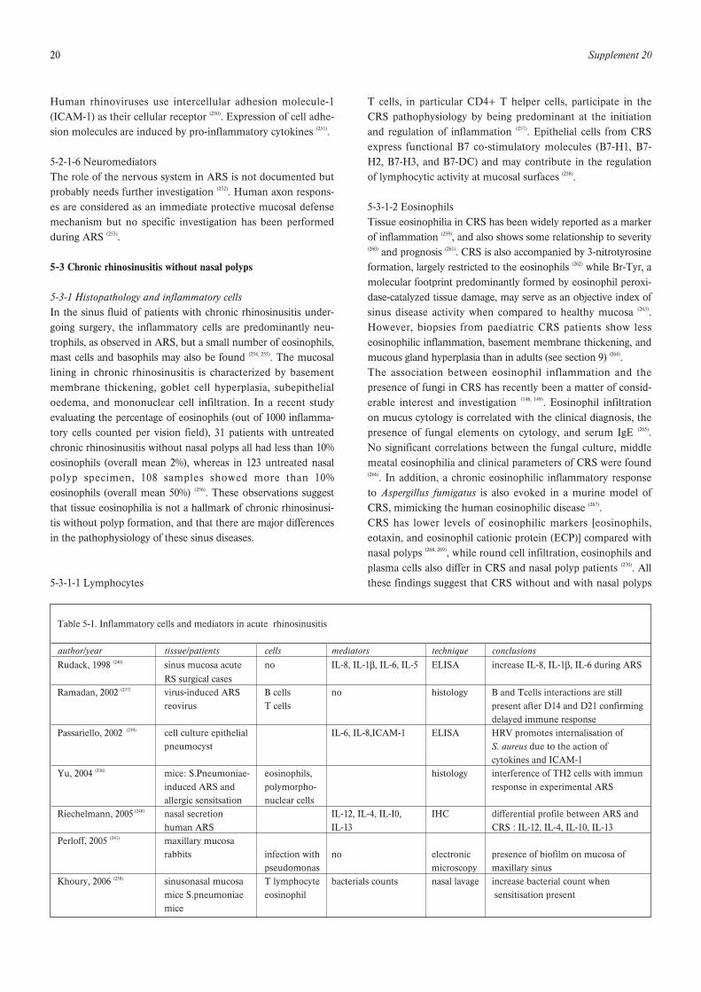
20 Supplement 20
Human rhinoviruses use intercellular adhesion molecule-1(ICAM-1) as their cellular receptor (250). Expression of cell adhe-sion molecules are induced by pro-inflammatory cytokines (251).
5-2-1-6 NeuromediatorsThe role of the nervous system in ARS is not documented butprobably needs further investigation (252). Human axon respons-es are considered as an immediate protective mucosal defensemechanism but no specific investigation has been performedduring ARS (253).
5-3 Chronic rhinosinusitis without nasal polyps
5-3-1 Histopathology and inflammatory cellsIn the sinus fluid of patients with chronic rhinosinusitis under-going surgery, the inflammatory cells are predominantly neu-trophils, as observed in ARS, but a small number of eosinophils,mast cells and basophils may also be found (254, 255). The mucosallining in chronic rhinosinusitis is characterized by basementmembrane thickening, goblet cell hyperplasia, subepithelialoedema, and mononuclear cell infiltration. In a recent studyevaluating the percentage of eosinophils (out of 1000 inflamma-tory cells counted per vision field), 31 patients with untreatedchronic rhinosinusitis without nasal polyps all had less than 10%eosinophils (overall mean 2%), whereas in 123 untreated nasalpolyp specimen, 108 samples showed more than 10%eosinophils (overall mean 50%) (256). These observations suggestthat tissue eosinophilia is not a hallmark of chronic rhinosinusi-tis without polyp formation, and that there are major differencesin the pathophysiology of these sinus diseases.
5-3-1-1 Lymphocytes
T cells, in particular CD4+ T helper cells, participate in theCRS pathophysiology by being predominant at the initiationand regulation of inflammation (257). Epithelial cells from CRSexpress functional B7 co-stimulatory molecules (B7-H1, B7-H2, B7-H3, and B7-DC) and may contribute in the regulationof lymphocytic activity at mucosal surfaces (258).
5-3-1-2 EosinophilsTissue eosinophilia in CRS has been widely reported as a markerof inflammation (259), and also shows some relationship to severity(260) and prognosis (261). CRS is also accompanied by 3-nitrotyrosineformation, largely restricted to the eosinophils (262) while Br-Tyr, amolecular footprint predominantly formed by eosinophil peroxi-dase-catalyzed tissue damage, may serve as an objective index ofsinus disease activity when compared to healthy mucosa (263).However, biopsies from paediatric CRS patients show lesseosinophilic inflammation, basement membrane thickening, andmucous gland hyperplasia than in adults (see section 9) (264).The association between eosinophil inflammation and thepresence of fungi in CRS has recently been a matter of consid-erable interest and investigation (148, 149). Eosinophil infiltrationon mucus cytology is correlated with the clinical diagnosis, thepresence of fungal elements on cytology, and serum IgE (265).No significant correlations between the fungal culture, middlemeatal eosinophilia and clinical parameters of CRS were found(266). In addition, a chronic eosinophilic inflammatory responseto Aspergillus fumigatus is also evoked in a murine model ofCRS, mimicking the human eosinophilic disease (267).CRS has lower levels of eosinophilic markers [eosinophils,eotaxin, and eosinophil cationic protein (ECP)] compared withnasal polyps (268, 269), while round cell infiltration, eosinophils andplasma cells also differ in CRS and nasal polyp patients (270). Allthese findings suggest that CRS without and with nasal polyps
Table 5-1. Inflammatory cells and mediators in acute rhinosinusitis
author/year tissue/patients cells mediators technique conclusionsRudack, 1998 (240) sinus mucosa acute no IL-8, IL-1", IL-6, IL-5 ELISA increase IL-8, IL-1", IL-6 during ARS
RS surgical casesRamadan, 2002 (237) virus-induced ARS B cells no histology B and Tcells interactions are still
reovirus T cells present after D14 and D21 confirmingdelayed immune response
Passariello, 2002 (239) cell culture epithelial IL-6, IL-8,ICAM-1 ELISA HRV promotes internalisation ofpneumocyst S. aureus due to the action of
cytokines and ICAM-1Yu, 2004 (236) mice: S.Pneumoniae- eosinophils, histology interference of TH2 cells with immun
induced ARS and polymorpho- response in experimental ARSallergic sensitsation nuclear cells
Riechelmann, 2005 (248) nasal secretion IL-12, IL-4, IL-I0, IHC differential profile between ARS andhuman ARS IL-13 CRS : IL-12, IL-4, IL-10, IL-13
Perloff, 2005 (241) maxillary mucosarabbits infection with no electronic presence of biofilm on mucosa of
pseudomonas microscopy maxillary sinusKhoury, 2006 (238) sinusonasal mucosa T lymphocyte bacterials counts nasal lavage increase bacterial count when
mice S.pneumoniae eosinophil sensitisation presentmice
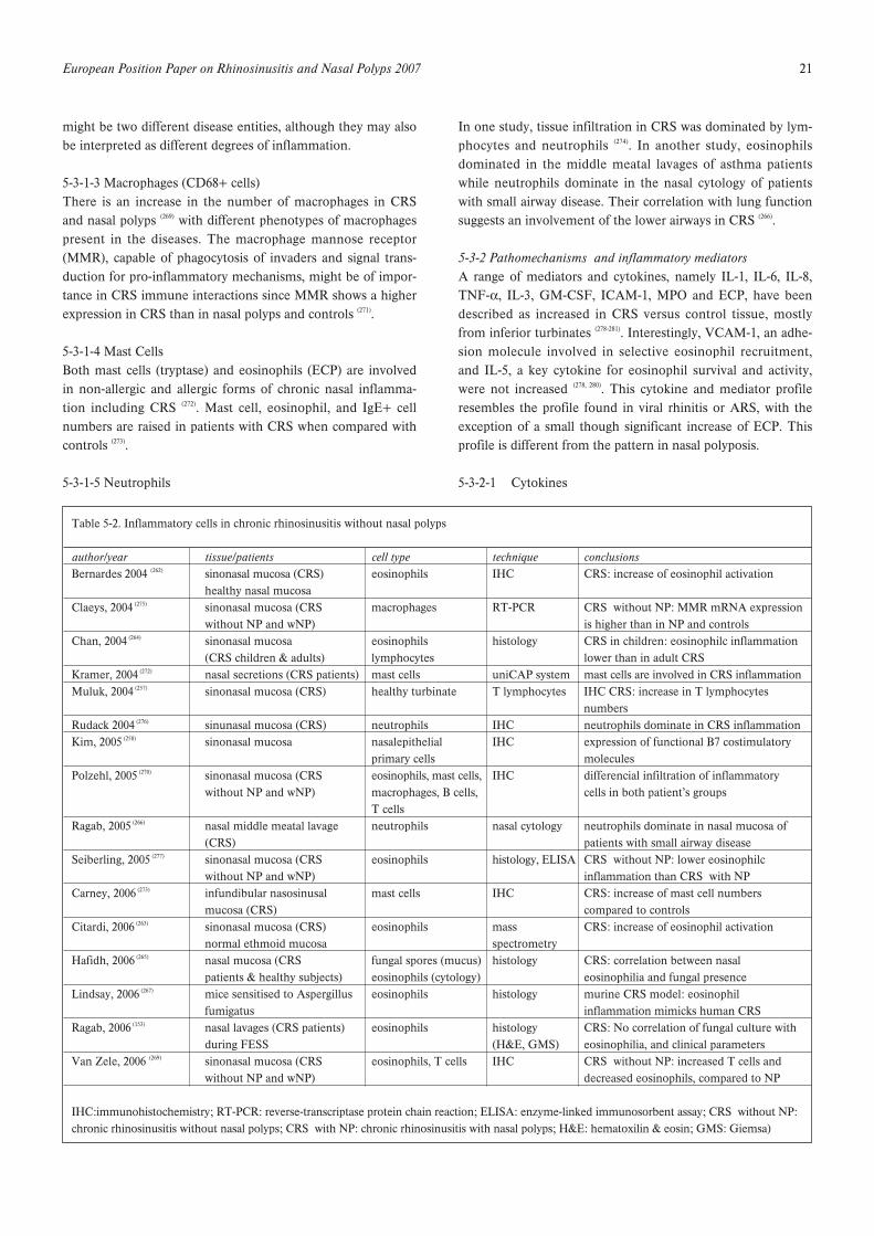
European Position Paper on Rhinosinusitis and Nasal Polyps 2007 21
might be two different disease entities, although they may alsobe interpreted as different degrees of inflammation.
5-3-1-3 Macrophages (CD68+ cells)There is an increase in the number of macrophages in CRSand nasal polyps (269) with different phenotypes of macrophagespresent in the diseases. The macrophage mannose receptor(MMR), capable of phagocytosis of invaders and signal trans-duction for pro-inflammatory mechanisms, might be of impor-tance in CRS immune interactions since MMR shows a higherexpression in CRS than in nasal polyps and controls (271).
5-3-1-4 Mast CellsBoth mast cells (tryptase) and eosinophils (ECP) are involvedin non-allergic and allergic forms of chronic nasal inflamma-tion including CRS (272). Mast cell, eosinophil, and IgE+ cellnumbers are raised in patients with CRS when compared withcontrols (273).
5-3-1-5 Neutrophils
In one study, tissue infiltration in CRS was dominated by lym-phocytes and neutrophils (274). In another study, eosinophilsdominated in the middle meatal lavages of asthma patientswhile neutrophils dominate in the nasal cytology of patientswith small airway disease. Their correlation with lung functionsuggests an involvement of the lower airways in CRS (266).
5-3-2 Pathomechanisms and inflammatory mediatorsA range of mediators and cytokines, namely IL-1, IL-6, IL-8,TNF-!, IL-3, GM-CSF, ICAM-1, MPO and ECP, have beendescribed as increased in CRS versus control tissue, mostlyfrom inferior turbinates (278-281). Interestingly, VCAM-1, an adhe-sion molecule involved in selective eosinophil recruitment,and IL-5, a key cytokine for eosinophil survival and activity,were not increased (278, 280). This cytokine and mediator profileresembles the profile found in viral rhinitis or ARS, with theexception of a small though significant increase of ECP. Thisprofile is different from the pattern in nasal polyposis.
5-3-2-1 Cytokines
Table 5-2. Inflammatory cells in chronic rhinosinusitis without nasal polyps
author/year tissue/patients cell type technique conclusionsBernardes 2004 (262) sinonasal mucosa (CRS) eosinophils IHC CRS: increase of eosinophil activation
healthy nasal mucosaClaeys, 2004 (275) sinonasal mucosa (CRS macrophages RT-PCR CRS without NP: MMR mRNA expression
without NP and wNP) is higher than in NP and controlsChan, 2004 (264) sinonasal mucosa eosinophils histology CRS in children: eosinophilc inflammation
(CRS children & adults) lymphocytes lower than in adult CRSKramer, 2004 (272) nasal secretions (CRS patients) mast cells uniCAP system mast cells are involved in CRS inflammationMuluk, 2004 (257) sinonasal mucosa (CRS) healthy turbinate T lymphocytes IHC CRS: increase in T lymphocytes
numbersRudack 2004 (276) sinunasal mucosa (CRS) neutrophils IHC neutrophils dominate in CRS inflammationKim, 2005 (258) sinonasal mucosa nasalepithelial IHC expression of functional B7 costimulatory
primary cells moleculesPolzehl, 2005 (270) sinonasal mucosa (CRS eosinophils, mast cells, IHC differencial infiltration of inflammatory
without NP and wNP) macrophages, B cells, cells in both patient’s groupsT cells
Ragab, 2005 (266) nasal middle meatal lavage neutrophils nasal cytology neutrophils dominate in nasal mucosa of(CRS) patients with small airway disease
Seiberling, 2005 (277) sinonasal mucosa (CRS eosinophils histology, ELISA CRS without NP: lower eosinophilcwithout NP and wNP) inflammation than CRS with NP
Carney, 2006 (273) infundibular nasosinusal mast cells IHC CRS: increase of mast cell numbersmucosa (CRS) compared to controls
Citardi, 2006 (263) sinonasal mucosa (CRS) eosinophils mass CRS: increase of eosinophil activationnormal ethmoid mucosa spectrometry
Hafidh, 2006 (265) nasal mucosa (CRS fungal spores (mucus) histology CRS: correlation between nasalpatients & healthy subjects) eosinophils (cytology) eosinophilia and fungal presence
Lindsay, 2006 (267) mice sensitised to Aspergillus eosinophils histology murine CRS model: eosinophilfumigatus inflammation mimicks human CRS
Ragab, 2006 (153) nasal lavages (CRS patients) eosinophils histology CRS: No correlation of fungal culture withduring FESS (H&E, GMS) eosinophilia, and clinical parameters
Van Zele, 2006 (269) sinonasal mucosa (CRS eosinophils, T cells IHC CRS without NP: increased T cells andwithout NP and wNP) decreased eosinophils, compared to NP
IHC:immunohistochemistry; RT-PCR: reverse-transcriptase protein chain reaction; ELISA: enzyme-linked immunosorbent assay; CRS without NP:chronic rhinosinusitis without nasal polyps; CRS with NP: chronic rhinosinusitis with nasal polyps; H&E: hematoxilin & eosin; GMS: Giemsa)
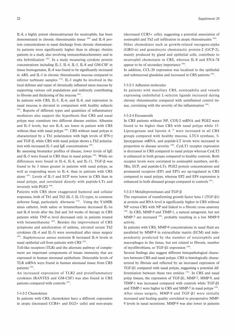
22 Supplement 20
IL-8, a highly potent chemoattractant for neutrophils, has beendemonstrated in chronic rhinosinusitis tissue (282) and IL-8 pro-tein concentrations in nasal discharge from chronic rhinosinusi-tis patients were significantly higher than in allergic rhinitispatients in a study also involving immunohistochemistry and insitu hybridization (283). In a study measuring cytokine proteinconcentrations including IL-3, IL-4, IL-5, IL-8 and GM-CSF intissue homogenates, IL-8 was found to be significantly increasedin ARS, and IL-3 in chronic rhinosinusitis mucosa compared toinferior turbinate samples (278). IL-3 might be involved in thelocal defense and repair of chronically inflamed sinus mucosa bysupporting various cell populations and indirectly contributingto fibrosis and thickening of the mucosa (284).In patients with CRS, IL-5, IL-6, and IL-8, and expression innasal mucosa is elevated in comparison with healthy subjects(285). Reports of different types and quantities of inflammatorymediators also support the hypothesis that CRS and nasalpolyps may constitute two different disease entities. Albuminand IL-5 levels, but not IL-8, are lower in patient with CRSwithout than with nasal polyps (286). CRS without nasal polyps ischaracterized by a Th1 polarization with high levels of IFN-#and TGF-", while CRS with nasal polyps shows a Th2 polariza-tion with increased IL-5 and IgE concentrations (269).By assessing biomarker profiles of disease, lower levels of IgEand IL-5 were found in CRS than in nasal polyps (248). While nodifferences were found in IL-6, IL-8, and IL-11, TGF-" wasfound to be 3 times greater in patients with nasal polyps, aswell as responding more to IL-4, than in patients with CRSalone (287). Levels of IL-5 and ECP were lower in CRS than innasal polyps, and correlated directly with peptide-LTs andinversely with PGE2 (288).Patients with CRS show exaggerated humoral and cellularresponses, both of Th1 and Th2 (IL-5, IL-13) types, to commonairborne fungi, particularly Alternaria (152). Using the YAMIKsinus catheter, both saline or betamethasone decreased IL-1!and IL-8 levels after the 2nd and 3rd weeks of therapy in CRSpatients while TNF-! level decreased only in patients treatedwith betamethasone (289). Besides the improvement of CRSsymptoms and amelioration of asthma, elevated serum Th2cytokines (IL-4 and IL-5) were normalized after sinus surgery(290). Staphylococcus aureus exotoxin B increased IL-6 levels innasal epithelial cell from patients with CRS (291).Toll-like receptors (TLR) and the alternate pathway of comple-ment are important components of innate immunity that areexpressed in human sinonasal epithelium. Detectable levels ofTLR mRNA were found in human sinonasal tissue from CRSpatients (292).An increased expression of TLR2 and proinflammatorycytokines (RANTES and GM-CSF) was also found in CRSpatients compared with controls (293).
5-3-2-2 ChemokinesIn patients with CRS, chemokines have a different expressionin atopic (increased CCR4+ and EG2+ cells) and non-atopic
(decreased CCR5+ cells), suggesting a potential association ofeosinophil and Th2 cell infiltration in atopic rhinosinusitis (294).Other chemokines such as growth-related oncogene-alpha(GRO-!) and granulocyte chemotactic protein-2 (GCP-2),mainly produced by gland and epithelial cells, contribute toneutrophil chemotaxis in CRS, whereas IL-8 and ENA-78appear to be of secondary importance (286).In addition, CCL-20 expression was localized to the epithelialand submucosal glandular and increased in CRS patients (295).
5-3-2-3 Adhesion moleculesIn patients with maxillary CRS, eosinophilia and vesselsexpressing endothelial L-selectin ligands increased duringchronic rhinosinusitis compared with uninflamed control tis-sue, correlating with the severity of the inflammation (296).
5-3-2-4 EicosanoidsIn CRS patients without NP, COX-2 mRNA and PGE2 werefound to be higher than CRS with nasal polyps while 15-Lipoxygenase and lipoxin A (4) were increased in all CRSgroups compared with healthy mucosa. LTC4 synthase, 5-lipoxygenase mRNA, and peptide-LT levels were increased inproportion to disease severity (288). CysLT1 receptor expressionis decreased in CRS compared to nasal polyps whereas CysLT2is enhanced in both groups compared to healthy controls. Bothreceptor levels were correlated to eosinophil numbers, sol-IL-5R!, ECP, and peptide-LTs. PGE2 protein concentrations andprostanoid receptors (EP1 and EP3) are up-regulated in CRScompared to nasal polyps, whereas EP2 and EP4 expression isenhanced in both diseased groups compared to controls (297).
5-3-2-5 Metalloproteinases and TGF-"The expression of transforming growth factor beta 1 (TGF-"1)at protein and RNA level is significantly higher in CRS withoutNP versus CRS with NP and linked to a fibrotic cross anatomy(298). In CRS, MMP-9 and TIMP-1, a natural antagonist, but notMMP-7 are increased (299), probably resulting in a low MMP-9activity.In patients with CRS, MMP-9 concentrations in nasal fluid areparalleled by MMP-9 in extracellular matrix (ECM) and inde-pendently predicted by the number of neutrophils andmacrophages in the tissue, but not related to fibrosis, numberof myofibroblasts, or TGF-"1 expression (300).Several findings also suggest different histopathological charac-ters between CRS and nasal polyps. CRS is histologically charac-terized by fibrosis and reflected by an increased expression ofTGF-"1 compared with nasal polyps, suggesting a potential dif-ferentiation between these two entities (301). In CRS and nasalpolyp tissues, the expression of TGF-"1, MMP-7, MMP-9, andTIMP-1 was increased compared with controls while TGF-"1and TIMP-1 were higher in CRS and MMP-7 in nasal polyps (302).After sinus surgery, MMP-9 and TGF-"1 were initiallyincreased and healing quality correlated to preoperative MMP-9 levels in nasal secretions. MMP-9 was also lower in patients
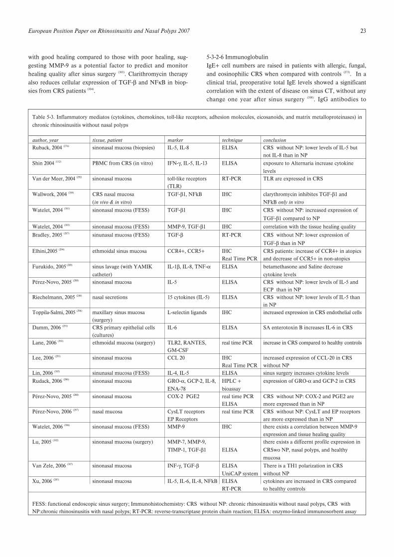
European Position Paper on Rhinosinusitis and Nasal Polyps 2007 23
with good healing compared to those with poor healing, sug-gesting MMP-9 as a potential factor to predict and monitorhealing quality after sinus surgery (303). Clarithromycin therapyalso reduces cellular expression of TGF-" and NF$B in biop-sies from CRS patients (304).
5-3-2-6 ImmunoglobulinIgE+ cell numbers are raised in patients with allergic, fungal,and eosinophilic CRS when compared with controls (273). In aclinical trial, preoperative total IgE levels showed a significantcorrelation with the extent of disease on sinus CT, without anychange one year after sinus surgery (308). IgG antibodies to
Table 5-3. Inflammatory mediatos (cytokines, chemokines, toll-like receptors, adhesion molecules, eicosanoids, and matrix metalloproteinases) inchronic rhinosinusitis without nasal polyps
author, year tissue, patient marker technique conclusionRuback, 2004 (276) sinonasal mucosa (biopsies) IL-5, IL-8 ELISA CRS without NP: lower levels of IL-5 but
not IL-8 than in NPShin 2004 (152) PBMC from CRS (in vitro) IFN-#, IL-5, IL-13 ELISA exposure to Alternaria increase cytokine
levelsVan der Meer, 2004 (292) sinonasal mucosa toll-like receptors RT-PCR TLR are expressed in CRS
(TLR)Wallwork, 2004 (304) CRS nasal mucosa TGF-"1, NFkB IHC clarythromycin inhibites TGF-"1 and
(in vivo & in vitro) NFkB only in vitroWatelet, 2004 (301) sinonasal mucosa (FESS) TGF-"1 IHC CRS without NP: increased expression of
TGF-"1 compared to NPWatelet, 2004 (303) sinonasal mucosa (FESS) MMP-9, TGF-"1 IHC correlation with the tissue healing qualityBradley, 2005 (287) sinunasal mucosa (FESS) TGF-" RT-PCR CRS without NP: lower expression of
TGF-" than in NPElhini,2005 (294) ethmoidal sinus mucosa CCR4+, CCR5+ IHC CRS patients: increase of CCR4+ in atopics
Real Time PCR and decrease of CCR5+ in non-atopicsFurukido, 2005 (289) sinus lavage (with YAMIK IL-1", IL-8, TNF-! ELISA betamethasone and Saline decrease
catheter) cytokine levelsPérez-Novo, 2005 (288) sinonasal mucosa IL-5 ELISA CRS without NP: lower levels of IL-5 and
ECP than in NPRiechelmann, 2005 (248) nasal secretions 15 cytokines (IL-5) ELISA CRS without NP: lower levels of IL-5 than
in NPToppila-Salmi, 2005 (296) maxillary sinus mucosa L-selectin ligands IHC increased expression in CRS endothelial cells
(surgery)Damm, 2006 (291) CRS primary epithelial cells IL-6 ELISA SA enterotoxin B increases IL-6 in CRS
(cultures)Lane, 2006 (293) ethmoidal mucosa (surgery) TLR2, RANTES, real time PCR increase in CRS compared to healthy controls
GM-CSFLee, 2006 (295) sinonasal mucosa CCL 20 IHC increased expression of CCL-20 in CRS
Real Time PCR without NPLin, 2006 (305) sinunasal mucosa (FESS) IL-4, IL-5 ELISA sinus surgery increases cytokine levelsRudack, 2006 (286) sinonasal mucosa GRO-!, GCP-2, IL-8, HPLC + expression of GRO-! and GCP-2 in CRS
ENA-78 bioassayPérez-Novo, 2005 (288) sinonasal mucosa COX-2 PGE2 real time PCR CRS without NP: COX-2 and PGE2 are
ELISA more expressed than in NPPérez-Novo, 2006 (297) nasal mucosa CysLT receptors real time PCR CRS without NP: CysLT and EP receptors
EP Receptors are more expressed than in NPWatelet, 2006 (306) sinonasal mucosa (FESS) MMP-9 IHC there exists a correlation between MMP-9
expression and tissue healing qualityLu, 2005 (302) sinonasal mucosa (surgery) MMP-7, MMP-9, there exists a diffeernt profile expression in
TIMP-1, TGF-"1 ELISA CRSwo NP, nasal polyps, and healthymucosa
Van Zele, 2006 (307) sinonasal mucosa INF-#, TGF-" ELISA There is a TH1 polarization in CRSUniCAP system without NP
Xu, 2006 (285) sinonasal mucosa IL-5, IL-6, IL-8, NFkB ELISA cytokines are increased in CRS comparedRT-PCR to healthy controls
FESS: functional endoscopic sinus surgery; Immunohistochemistry: CRS without NP: chronic rhinosinusitis without nasal polyps, CRS withNP:chronic rhinosinusitis with nasal polyps; RT-PCR: reverse-transcriptase protein chain reaction; ELISA: enzymo-linked immunosorbent assay

24 Supplement 20
Alternaria and Cladosporium are clearly increased in patientswith CRS compared with normal individuals while less than30% of CRS patients have specific IgE antibodies to Alternariaor Cladosporium (152). Fungal-specific IgG (IgG3) and IgA levels(to Alternaria alternata and Aspergillus fumigatus), but not IgE,are higher in CRS with eosinophilic mucus compared withhealthy volunteers, challenging the presumption of a uniquepathogenic role of fungal allergy in “allergic fungal sinusitis”(150).
5-3-2-7 Nitric Oxide (NO)CRS epthelial cells show a stronger expression of TLR-4 andiNOS than controls, iNOS being upregulated in nasal epitheli-um and correlated with TLR-4 (309). In a prospective randomizedtrial in patients with CRS who had failed initial medical thera-py with nasal corticosteroids, the rise in nNO seen on bothmedical and surgical treatments correlated with symptomscore, saccharin clearance time, endoscopic changes, and polyp
size, suggesting that nNO provides a non-invasive objectivemeasure of the response of CRS to therapy on an individualbasis (310). However, some contradictory results on the role ofnNO on nasal inflammation have been recently assessed (311).
5-3-2-8 NeuropeptidesNeurogenic inflammation may play a potential role on themanifestation of chronic rhinosinusitis (312). In addition, CGRP(trigeminal sensory) and VIP (parasympathetic) levels in salivawere significantly elevated between attacks in patients with thediagnosis of allergic CRS and migraine compared to controls ,returning to baseline after pseudoephedrine therapy but onlyin CRS patients (313).
5-3-2-9 MucinsAirway mucus is overproduced in CRS. Mucins are the majorcomponents of mucus and the macromolecules that impart rhe-ologic properties to airway mucus. MUC5AC and MUC5B are
Table 5-4. Inflammatory mediators (immunoglubulins, nitric oxide, neuropeptides, mucins, and others) in Chronic Rhinosinusitis without nasal polyps
author, year tissue, patient marker technique conclusionKim, 2004 (314) maxillary sinonasal mucosa MUC5AC, MUC5B RT-PCR increased in CRS compared to healthy
controlsLee, 2004 (317) maxillary sinonasal mucosa MUC8 RT-PCR MUC8 is upregulated in CRS compared to
controlsManiscalco, 2004 (324) allergic rhinitis patients nasal NO chemi-lumine- nNO during humming is a reliable marker
scence of sinus patencyShin, 2004 (152) PBMC from CRS (in vitro) fungal IgG UniCAP system IgG is increased in CRS compared to
healthy controlsWang, 2004 (309) sinonasal mucosa TLR4, iNOS in-situ TLR4 and iNOS are increased in CRS
hybridization compared to healthy controlsAli, 2005 (316) CRS patients (sinusal mucus) MUC5AC, MUC2 ELISA increase of MUC5AC and decrease of
MUC2 in CRSPant, 2005 (150) CRS patients (serum) fungal IgG, IgA ELISA IgG3 and IgA are increased in CRSSun, 2005 (321) CRS patients (sinus effusions, VEGF ELISA VEGF expression is higher in the sinus
nasal secretions, serum) mucosa than in nasal mucosa and serumBellamy, 2006 (313) allergic CRS (saliva) VIP, CGRP radioimmuno- neuropetides are increased in acute attacks
assayCarney, 2006 (273) infundibular sinonasal mucosa IgE+ cells immuno- IgE+ cells are increased in CRS compared
histochemistry to healthy controlsLal, 2006 (308) CRS patients (serum) total IgE ELISA correlation of IgE levels with the CRS extentMartínez, 2006 (319) sinonasal mucosa (FESS) MUC1, MUC2, MUC4 IHC
MUC5AC, MUC5B In situ there exist different MUC expressionhybridization patterns depending on the sinonasal disease
Pena, 2006 (320) children with CRS MUC5AC IHC MUC5AC is expressed in the epithelium(sinonasal mucosa) but not in submucosal glands
Ragab SM, 2006 (310) CRS patients nasal NO chemi- medial and surgical treatments increaseluminescence nNO, with correlation with nasal symptoms
Viswanathan, 2006 (315) CRS patients (nasal mucus) MUC5AC, MUC5B ELISA increased in CRS compared to healthycontrols
Hu, 2007 (322) children with CRS VEGF IHC CRS without NP: lower expression of(sinonasal mucosa) VGEF compared to NP
Lee, 2006 (323) sinonasal mucosa (FESS) surfactant protein-A RT-PCR increased expression of SP-A in CRScompared to healthy controls
FESS: functional endoscopc sinus surgery; IHC:immunohistochemistry; CRS without NP:chronic rhinosinusitis without nasal polyps; CRS withNP:chronic rhinosinusitis with nasal polyps; RT-PCR: reverse-transcriptase protein chain reaction; ELISA: enzymo-linked immunosorbent assay
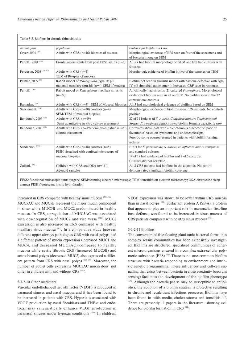
European Position Paper on Rhinosinusitis and Nasal Polyps 2007 25
increased in CRS compared with healthy sinus mucosa (314, 315).MUC5AC and MUC5B represent the major mucin componentin sinus while MUC5B and MUC2 predominated in healthymucosa. In CRS, upregulation of MUC5AC was associatedwith downregulation of MUC2 and vice versa (316). MUC8expression is also increased in CRS compared with healthymaxillary sinus mucosa (317). In a comparative study betweendifferent upper airways pathologies CRS with nasal polyps hada different pattern of mucin expression (increased MUC1 andMUC4, and decreased MUC5AC) compared to healthymucosa while cystic fibrosis CRS (increased MUC5B) andantrochoanal polyps (decreased MUC2) also expressed a differ-ent pattern from CRS with nasal polyps (318, 319). Moreover, thenumber of goblet cells expressing MUC5AC mucin does notdiffer in children with and without CRS (320).
5-3-2-10 Other mediatorsVascular endothelial-cell growth factor (VEGF) is produced inparanasal sinuses and nasal mucosa and it has been found tobe increased in patients with CRS. Hypoxia is associated withVEGF production by nasal fibroblasts and TNF-! and endo-toxin may synergistically enhance VEGF production inparanasal sinuses under hypoxic conditions (321). In children,
VEGF expression was shown to be lower within CRS mucosathan in nasal polyps (322). Surfactant protein A (SP-A), a proteinthat appears to play an important role in mammalian first-linehost defense, was found to be increased in sinus mucosa ofCRS patients compared with healthy sinus mucosa (323).
5-3-2-11 BiofilmsThe conversion of free-floating planktonic bacterial forms intocomplex sessile communities has been extensively investigat-ed. Biofilms are structured, specialised communities of adher-ent micro-organisms encased in a complex extra-cellular poly-meric substance (EPS) (325).There is no one common biofilmstructure with bacteria responding to environment and intrin-sic genetic programming. These influences and cell-cell sig-nalling that exists between bacteria in close proximity (quorumsensing) facilitates the development of the biofilm phenotype(326). Although the bacteria per se may be susceptible to antibi-otics, the adoption of a biofilm strategy is protective resultingin chronic and recalcitrant infectious processes. Biofilms havebeen found in otitis media, cholesteatoma and tonsillitis (327).There are presently 11 papers in the literature showing evi-dence for biofilm formation in CRS (328).
Table 5-5. Biofilms in chronic rhinosinusitis
author, year population evidence for biofilms in CRSCryer, 2004 (329) Adults with CRS (n=16) Biopsies of mucosa Morphological evidence of EPS seen on four of the specimens and
of bacteria in one on SEMPerloff, 2004 (330) Frontal recess stents from post FESS adults (n=6) All six had biofilm morphology on SEM and five had cultures with
S.aureusFerguson, 2005 (331, 467) Adults with CRS (n=4) Morphologic evidence of biofilm in two of the samples on TEM
TEM of Biospies of mucosaPalmer, 2005 (332) Rabbit model of P.aeruginosa (type IV pili Biofilm not seen in sinusitis model with bacteria defective with type
mutants) maxillary sinusitis (n=4) SEM of mucosa IV pili (impaired attachement). Increased CBF seen in response.Perloff, (241) Rabbit model of P.aeruginosa maxillary sinusitis All clinically had sinusitis. 21 cultured P.aeruginosa. Morphological
(n=22) evidence of biofilm seen in all on SEM No biofilm seen in the 22contraleteral controls
Ramadan, (333) Adults with CRS (n=5) SEM of Mucosal biopsies All 5 had morphological evidence of biofilms based on SEMSanclement, (334) Adults with CRS (n=30) controls (n=4) Morphological evidence of biofilms seen in 24 patients. No controls
SEM/TEM of mucosal biopsies positive.Bendouah, 2006 (335) Adults with CRS (n=19) 22 of 31 isolates of S. Aureus, Coagulase negative Staphylococcal
Semi quantitative in vitro culture assessment Species, P. aeruginosa demonstrated biofilm forming capacity in vitroBendouah, 2006 (336) Adults with CRS (n=19) Semi quantitative in vitro Correlates above data with a dichotomous outcome of ‘poor or
culture assessment favourable’ based on symptoms and endoscopic signs.Poor outcome overrepresented in patients with biofilm formingisolates
Sanderson, (337) Adults with CRS (n=18) controls (n=5) FISH for S. pneumoniae, S. aureus, H. influenza and P. aeruginosaFISH visualised with confocal microscopy of and standard culturesmucosal biopsies 14 of 18 had evidence of biofilm and 2 of 5 controls.
Cultures did not correlate.Zuliani, (338) Children with CRS and OSA (n=16 ) All 8 CRS patients had biofilms in the adenoids. No control
Adenoid samples demonstrated significant biofilm coverage.
FESS: functional endoscopic sinus surgery; SEM:scanning electron microscopy; TEM:transmission electron microscopy; OSA:obstructibe sleepapnoea FISH:fluorescent in situ hybridisation
26 Supplement 20
5-4 Chronic rhinosinusitis with nasal polyps
5-4-1 Histopathology and inflammatory cellsHistomorphological characterisation of polyp tissue reveals fre-quent epithelial damage, a thickened basement membrane,and oedematous to sometimes fibrotic stromal tissue, with areduced number of vessels and glands, but virtually no neuralstructure (339-341). The stroma of mature polyps is mainly charac-terised by its oedematous nature and consists of supportingfibroblasts and infiltrating inflammatory cells, localized around“empty” pseudocyst formations. Among the inflammatorycells, EG2 (activated) eosinophils are a prominent and charac-teristic feature in about 80% of European polyps (342), whereaslymphocytes and neutrophils are the predominant cells in cys-tic fibrosis and in CRS without NP. Eosinophils are localisedaround the vessels, glands, and directly beneath the mucosalepithelium (340). However, neutrophils are also a constant find-ing in nasal polyps, and their number is increased compared tocontrols (269). Furthermore, increased numbers of activated T-cells and plasma cells characterize the typical cell composition.
In small polyps, not larger than 5 mm, growing on normallooking mucosa of the middle turbinate in patients with bilat-eral polyposis, the early processes of polyp growth have beenstudied (343). Numerous subepithelial EG2+ eosinophils werepresent in the luminal compartment of the early stage polyp,forming a cap over the central pseudocyst area. In contrast,mast cells were scarce in the polyp tissue, but were normallydistributed in the pedicle and the adjacent mucosa, which hada normal appearance. This contrasts to mature polyps, wheredegranulated mast cells and eosinophils are often diffusely dis-tributed in the polyp tissue. Fibronectin deposition wasnoticed around the eosinophils in the luminal compartment ofthe early stage polyp, was accumulated subepithelially, andformed a network-like structure in the polyp centre and withinthe pseudocysts. The presence of myofibroblasts was limited tothe central pseudocyst area. Interestingly, albumin and proba-bly other plasma proteins were deposited within the pseudo-cysts, adjacent to the eosinophil infiltration. These observa-tions suggest a central deposition of plasma proteins, regulatedby the subepithelial eosinophilic inflammation, as a patho-genetic principle of polyp formation and growth.
5-4-1-1 LymphocytesNasal polyps show a significantly increased number of T-lym-phocytes (CD3) and activated T-lymphocytes (CD25) com-pared to control patients (269). In non-allergic CRS with nasalpolyps a tendency to fewer CD4+ cells in the epithelium andmore CD8+ cells in the lamina propria was found (344). Aninverse median ratio of CD4+/CD8+ T cells as compared tothe middle turbinate of control subjects was found in onerecent study (345). Functional studies on T-cells, especially T reg-ulatory cells, are lacking so far. Interestingly, there were almostno naïve B-lymphocytes (CD20) present in the tissue, although
a significantly higher number of plasma cells (CD138) was pre-sent in NP versus controls and CRS without polyps (269, 346). Thisfact is reflected by a significant increase in immunoglobulin A,G and E synthesis (van Zele, unpublished).
S. aureus superantigens (SAgs) bind the V"-region of the T-cellreceptor (TCR) outside the peptide-binding site.Approximately 50 distinct V"-domains exist in the humanrepertoire, and distinct SAgs will bind only particular domains,generating a pattern of V"-enrichment in lymphocytes depen-dent on the binding characteristics of a given toxin. Flowcytometry was used to analyze the V"-repertoire of polyp-derived CD4+ and CD8+ lymphocytes in the light of theknown skewing associated with SAg exposure. Seven of 20subjects exhibited skewing in V"-domains with strong associa-tions to S. aureus SAgs. This study establishes evidence of S.aureus SAg-T-cell interactions in polyp lymphocytes of 35% ofCRS with NP patients (347).
5-4-1-2 EosinophilsAn increased number of eosinophils, demonstrated by HE stain-ing or EG2 IHC, is a hallmark of Caucasian NPs. Eosinophilnumbers are significantly higher in NP tissue compared to CRS(348) and other sinus disease and control mucosa, and are furtherincreased in patients with co-morbid asthma and/or aspirin sen-sitivity, but independent from atopy (192, 349). In a study evaluatingthe percentage of eosinophils (out of 1000 inflammatory cellscounted per vision field), 31 patients with untreated chronicsinusitis without nasal polyps all had less than 10% eosinophils(overall mean 2%), whereas in 123 untreated nasal polyp speci-men, 108 samples showed more than 10% eosinophils (overallmean 50%) (350). Generally, the differences in ECP measurementbetween diseases are more pronounced than the cell numbers,indicating a more intense activation of eosinophils in polyps.However, the eosinophilic inflammation in nasal polyp tissuefrom China, as measured by ECP and cytokine/chemokine lev-els (IL-5, eotaxin), was not significantly different from controltissue, and was significantly lower compared to Caucasianpolyps. The semi-quantitative scores for EG2+ eosinophils were0,45+1.15 for the Chinese polyp patients and 1,95 ± 2,85 for theCaucasian polyp patients, being significantly different (346).Furthermore, eosinophil numbers are not different from con-trols and cystic fibrosis polyps (269).
5-4-1-3 Macrophages and dendritic cellsMacrophage numbers seem to be slightly increased in nasalpolyps, and these cells express increased amounts ofmacrophage mannose receptors (MMR), an innate pattern rec-ognizing receptor, capable of phagocytosis of invaders and sig-nal transduction for proinflammatory mechanisms (275). Therealso is a higher number of macrophages in patients with CFthan in patients with CRS or in controls (351). Our knowledge ondendritic cells is very limited; they are present in nasal polyps,and express the high affinity IgE receptor (344, 352).
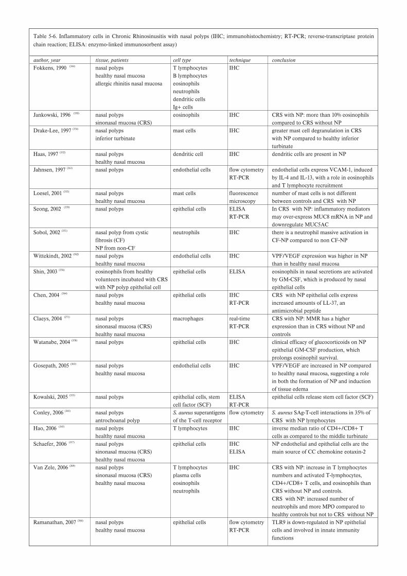
Table 5-6. Inflammatory cells in Chronic Rhinosinusitis with nasal polyps (IHC; immunohistochemistry; RT-PCR; reverse-transcriptase proteinchain reaction; ELISA: enzymo-linked immunosorbent assay)
author, year tissue, patients cell type technique conclusionFokkens, 1990 (344) nasal polyps T lymphocytes IHC
healthy nasal mucosa B lymphocytesallergic rhinitis nasal mucosa eosinophils
neutrophilsdendritic cellsIg+ cells
Jankowski, 1996 (350) nasal polyps eosinophils IHC CRS with NP: more than 10% eosinophilssinonasal mucosa (CRS) compared to CRS without NP
Drake-Lee, 1997 (354) nasal polyps mast cells IHC greater mast cell degranulation in CRSinferior turbinate with NP compared to healthy inferior
turbinateHaas, 1997 (352) nasal polyps dendritic cell IHC dendritic cells are present in NP
healthy nasal mucosaJahnsen, 1997 (361) nasal polyps endothelial cells flow cytometry endothelial cells express VCAM-1, induced
RT-PCR by IL-4 and IL-13, with a role in eosinophilsand T lymphocyte recruitment
Loesel, 2001 (353) nasal polyps mast cells fluorescence number of mast cells is not differenthealthy nasal mucosa microscopy between controls and CRS with NP
Seong, 2002 (359) nasal polyps epithelial cells ELISA In CRS with NP: inflammatory mediatorsRT-PCR may over-express MUC8 mRNA in NP and
downregulate MUC5ACSobol, 2002 (351) nasal polyp from cystic neutrophils IHC there is a neutrophil massive activation in
fibrosis (CF) CF-NP compared to non CF-NPNP from non-CF
Wittekindt, 2002 (362) nasal polyps endothelial cells IHC VPF/VEGF expression was higher in NPhealthy nasal mucosa than in healthy nasal mucosa
Shin, 2003 (356) eosinophils from healthy epithelial cells ELISA eosinophils in nasal secretions are activatedvolunteers incubated with CRS by GM-CSF, which is produced by nasalwith NP polyp epithelial cell epithelial cells
Chen, 2004 (364) nasal polyps epithelial cells IHC CRS with NP epithelial cells expresshealthy nasal mucosa RT-PCR increased amounts of LL-37, an
antimicrobial peptideClaeys, 2004 (271) nasal polyps macrophages real-time CRS with NP: MMR has a higher
sinonasal mucosa (CRS) RT-PCR expression than in CRS without NP andhealthy nasal mucosa controls
Watanabe, 2004 (358) nasal polyps epithelial cells IHC clinical efficacy of glucocorticoids on NPepithelial GM-CSF production, whichprolongs eosinophil survival.
Gosepath, 2005 (363) nasal polyps endothelial cells IHC VPF/VEGF are increased in NP comparedhealthy nasal mucosa to healthy nasal mucosa, suggesting a role
in both the formation of NP and inductionof tissue edema
Kowalski, 2005 (355) nasal polyps epithelial cells, stem ELISA epithelial cells release stem cell factor (SCF)cell factor (SCF) RT-PCR
Conley, 2006 (365) nasal polyps S. aureus superantigens flow cytometry S. aureus SAg-T-cell interactions in 35% ofantrochoanal polyp of the T-cell receptor CRS with NP lymphocytes
Hao, 2006 (345) nasal polyps T lymphocytes IHC inverse median ratio of CD4+/CD8+ Thealthy nasal mucosa cells as compared to the middle turbinate
Schaefer, 2006 (357) nasal polyps epithelial cells IHC NP endothelial and epithelial cells are thesinonasal mucosa (CRS) ELISA main source of CC chemokine eotaxin-2healthy nasal mucosa
Van Zele, 2006 (269) nasal polyps T lymphocytes IHC CRS with NP: increase in T lymphocytessinonasal mucosa (CRS) plasma cells numbers and activated T-lymphocytes,healthy nasal mucosa eosinophils CD4+/CD8+ T cells, and eosinophils than
neutrophils CRS without NP and controls.CRS with NP: increased number ofneutrophils and more MPO compared tohealthy controls but not to CRS without NP
Ramanathan, 2007 (366) nasal polyps epithelial cells flow cytometry TLR9 is down-regulated in NP epithelialhealthy nasal mucosa RT-PCR cells and involved in innate immunity
functions
28 Supplement 20
5-4-1-4 Mast CellsThe number of mast cells is not different between controls andnasal polyps, these cells are however more often IgE-positive,especially in asthmatics, independent of atopy (353). There wasgreater mast cell degranulation in the nasal polyp compared toinferior turbinate (354). The density of mast cells in the epithelialand stromal layers of nasal polyps correlated with the expres-sion of SCF mRNA and protein in the supernatants of NPepithelial cells (355).
5-4-1-5 NeutrophilsThere is an increased number of neutrophils and higher con-centrations of MPO protein in nasal polyps vs. controls, butnot compared to CRS without polyps, and both parameters areeven higher in CF NPs (269, 351) indicating a massive activation inCF- NPs compared to non-CF-NPs. The role of neutrophilscurrently is not understood in NPs.
5-4-1-6 Epithelial cellsHuman nasal epithelial cells contain and secrete IL-8, GM-CSF, eotaxin, eotaxin-2 and RANTES, and thus may provideenough growth factors to attract eosinophils (356, 357), with GM-CSF being important for the survival of those cells (358).Epithelial cells also release stem cell factor (SCF), a cytokinewith chemotactic and survival activity for mast cells, with theexpression of SCF mRNA correlating to SCF protein, and withthe density of mast cells in epithelial and stromal layers ofnasal polyps (355).Inflammatory mediators may lead to over-expression of MUC8mRNA in nasal polyps and downregulation of MUC5ACmRNA expression, and influence the composition of mucus inpolyp disease (359). Nasal polyp epithelial cells also expressincreased amounts of LL-37, an antimicrobial peptide (360), butdown-regulate the level of TLR9 expression (347), and are thusdirectly involved in innate immunity functions.
5-4-1-7 Endothelial cells (See also adhesion molecules)Endothelial cells express VCAM-1, induced by IL-4 and IL-13,which plays an important role for the preferential recruitmentof eosinophils and T lymphocytes (361). However, a key phe-nomenon in nasal polyps is the remarkable oedema, whichawaits explanation. Vascular permeability/vascular endothelialgrowth factor (VPF/VEGF) plays an important role in induc-ing angiogenesis and modulating capillary permeability. In fact,the expression of VPF/VEGF in specimens of nasal polypswas significantly stronger than in specimens of healthy nasalmucosa of controls (362). VPF/VEGF in polypous tissue wasmainly localized in vascular endothelial cells, in basal mem-branes and perivascular spaces, and in epithelial cells. Themarkedly increased expression in nasal polyps as opposed tohealthy nasal mucosa suggests that VPF/VEGF may play a sig-nificant role in both the formation of nasal polyps and in theinduction of heavy tissue oedema (363).
5-4-2 Pathomechanisms and Inflammatory Mediators
5-4-2-1 CytokinesA large body of studies has focused on eosinophilic mediatorsin nasal polyp tissue, and demonstrated that different cell typesgenerate these mediators. Early studies by Denburg et al (367)
demonstrated that conditioned medium, derived from culturednasal polyp epithelial cells, contained potent eosinophilcolony-stimulating activities, as well as an interleukin-3-likeactivity. The authors suggested that accumulation ofeosinophils in polyps may partly be a result of differentiationof progenitor cells stimulated by soluble haemopoietic factorsderived from mucosal cell populations. An increased synthesisof GM-CSF by epithelial cells, fibroblasts, monocytes, andeosinophils was suggested later (99, 368-370). According to Hamiloset al (30), polyp tissue samples from patients with or withoutallergy contained different cytokine profiles. Other studiesinvolving protein measurements in tissue homogenates couldnot support these findings (31, 278).In contrast, IL-5 was found to be significantly increased innasal polyps, compared to healthy controls, and the concentra-tion of IL-5 was independent of the atopic status of the patient.Indeed, the highest concentrations of IL-5 were found in sub-jects with non-allergic asthma and aspirin sensitivity.Furthermore, eosinophils were positively stained for IL-5, sug-gesting a possible autocrine role for this cytokine in the activa-tion of eosinophils, and a strong correlation between concen-trations of IL-5 protein and eosinophilic cationic protein (ECP)was demonstrated later (192). The key role of IL-5 was supportedby the finding that treatment of eosinophil-infiltrated polyp tis-sue with neutralizing anti-IL-5 monoclonal antibody (mAB),but not anti-IL-3 or anti-GM-CSF mAbs in vitro, resulted ineosinophil apoptosis and decreased tissue eosinophilia (371).Collectively, these studies suggest that increased production ofIL-5 is likely to influence the predominance and activation ofeosinophils in nasal polyps independent of atopy. The lack ofdifference in the amounts of cytokines detected in polyps fromallergic or non-allergic patients was meanwhile supported byseveral other studies (372, 373). Furthermore, Wagenmann et al (374)
demonstrated that both Th1 and Th2 type cytokines wereupregulated in eosinophilic NP, irrespective of allergen skintest results.Recently, the regulation of the IL-5 receptor, which exists inthe soluble and transmembrane isoform, has been investigated(375). Whereas the probably antagonistic soluble isoform isupregulated, the signal transducing transmembrane isoform isdown-regulated in nasal polyps, especially if associated withasthma.
5-4-2-2 ChemokinesRecent studies have also shown that nasal polyps also expresshigh levels of RANTES and eotaxin, the predominant recog-nised eosinophil chemoattractants. Bartels and colleagues (376)
demonstrated that expression of eotaxin- and RANTES

European Position Paper on Rhinosinusitis and Nasal Polyps 2007 29
mRNA, but not MCP-3 mRNA, was elevated in non-atopicand atopic nasal polyps, when compared to normal nasalmucosa. Similarly, several publications demonstrated anincreased mRNA expression for eotaxin, eotaxin-2, eotaxin-3,and MCP-4 (357, 377-379). The expression of eotaxin-2, a CCR3-spe-cific chemokine, was found to be the most prominent of thethree chemokines investigated. According to other data (31, 192, 343),it appears that eotaxin, rather than RANTES, in cooperationwith IL-5, plays a key role in chemo-attraction and activationof eosinophils in NP tissue. This is in accordance with the find-ings of a recent extensive study of about 950 non-allergic andallergic polyp patients, which has also suggested that nasalpolyp eosinophilic infiltration and activation may correlatemainly with increased eotaxin gene expression, rather thanwith RANTES expression (380). The excessive production ofeotaxin in nasal polyps was recently confirmed in comparisonto controls and CRS patients (269).
5-4-2-3 Adhesion moleculesStudies of cell adhesion molecules are relatively few. Earlystudies by Symon and colleagues (381) demonstrated that ICAM-1, E-selectin and P-selectin were well expressed by nasal polypendothelium, whereas VCAM-1 expression was weak orabsent. An elegant study by Jahnsen et al (382), employing three-colour immunofluorescence staining, has however demonstrat-ed that both the number of eosinophils and the proportion ofvessels positive for VCAM-1 were significantly increased innasal polyps compared with the turbinate mucosa of the samepatients. Moreover, treatment with topical glucocorticosteroidsdecreases the density of eosinophils and the expression ofVCAM-1 in polyps (383). The interaction between VLA-4 oneosinophils and VCAM-1 on endothelial cells may not only beof particular importance for transendothelial migration ofeosinophils, but may also modify their activation and effectorfunctions (384).
5-4-2-4 EicosanoidsLevels of leukotrienes and their receptors have been shown tobe up-regulated in nasal polyp tissue (385, 386), more pronouncedin patients with intolerance to aspirin (288, 387, 388). A recent studysuggested that high levels of LTC4 synthase are directly linkedto the manifestation of aspirin intolerance (389), and polymor-phisms linked to this enzyme have been suggested to cause thesyndrome (390). However, up-regulation of cysteinyl-leucotrienes(CysLTs) does occur in chronic rhinosinusitis even in theabsence of clinical aspirin sensitivity and appears to be closelyrelated to the severity of eosinophilic inflammation (IL-5 andECP), a hallmark of nasal polyp disease (288). Increased expres-sion of the CysLT1 and CysLT2 receptors have been demon-strated on inflammatory cells in nasal polyp tissue (297, 391, 392), butalso inflammatory cells in nasal lavage from patients with aller-gic rhinitis (393). Thus, the regulation of CysLTs does not seemto be a pathognomonic feature of aspirin intolerance.The regulation of the cyclo-oxygenases and their products, the
prostaglandins, in nasal polyps is characterized by a deficit inthe production of PGE2 compared to CysLT levels (297, 393-395).This phenomenon was also observed in nasal polyp tissue andperipheral blood from aspirin-intolerant subjects (396), and cyclo-oxygenase (COX)-2 mRNA expression and NF-kB activitywere markedly lower in nasal polyp tissue from aspirin intoler-ant subjects compared to tolerant patients (397, 398). However, ourrecent data indicate that levels of PGE2 inversely correlatewith the degree of eosinophilic inflammation in nasal tissuefrom aspirin intolerant, but also tolerant patients. These find-ings support previous studies (395, 399), and suggest that this down-regulation may again represent a bystander phenomenonlinked to the severity of the inflammatory process. Of interest,research on prostanoid E (EP) receptor regulation has recentlybeen performed, with controversial results. A down-regulationof EP1 and EP3 and an up–regulation of EP2 and EP4 recep-tors in nasal polyp tissue compared to normal nasal mucosawas shown (297). It is known that EP2 and EP4 are highlyexpressed on eosinophils and a deficiency of PGE2 productionmay up-regulate the expression of these receptors. However,the expression of EP2 and EP4 receptors did not correlate witheosinophil numbers or eosinophil activation markers, indicat-ing that the regulation of these receptors may involve othercellular sources. This was recently confirmed by Ying et al.who showed the expression of EP receptors in a variety ofinflammatory cells in the nasal mucosa (400). These authorsfound a down-regulation of the EP2 receptor in aspirin intoler-ant patients and speculated on the effects on the production ofinflammatory mediators. However, PGD2 may also mediateeosinophil chemotaxis and activation and the production ofcytokines such as interleukins IL-4, IL-5, and IL-13 by humanTh2 inflammatory cells via different receptors, e.g. via theCRTH2 receptor (401, 402). The relative contributions of EP andother PG receptors is far from being unravelled, and so far, lit-tle points to a specific change in PG regulation in aspirin-intol-erant subjects.Finally, lipoxins are generally associated with anti-inflammato-ry effects and have been reported to reduce leukocyte infiltra-tion (403). However, certain di- hydroxyeicosatetraenoic acids(HETEs), which are precursors of these molecules, may havealso pro-inflammatory effects, specifically neutrophilic andeosinophilic chemotaxis (404). Nasal polyp tissue has been shownto have a high capacity to produce LXA4 after incubation withexogenous LTA4 in the presence of polymorphonuclear granu-locytes (405). In addition, severity of asthma has been associatedwith increased expression and activation of the 15-LO (lipoxy-genase) enzyme, collagen deposition and eosinophil accumula-tion (406). Interestingly, the capacity to produce lipoxins isdecreased in epithelial cells from patients with aspirin intoler-ance (407). These results are in line with those showing that basallevels of this enzyme are increased in nasal polyp tissue ofaspirin-tolerant, but down- regulated in aspirin intolerant sub-jects. 15-LO and LXA4 levels were increased in all chronicsinus disease groups, but significantly down-regulated in
30 Supplement 20
patients with aspirin intolerance, suggesting a specific regula-tion in this subgroup (297).Summarizing, eicosanoid changes in paranasal sinus diseasesare generally characterized by an up- regulation of CysLTs,LXA4 and PGD2 and a down- regulation of COX-2 and PGE2.Eosinophilic markers such as ECP and IL-5 correlate directlywith LTC4/D4/E4 and inversely with PGE2 concentrations,demonstrating the close relationship to severity of inflamma-tion. In the sinus mucosa of aspirin intolerant subjects, thesechanges might be extreme, as the degree of inflammation ismaximal, and the clinically apparent aspirin intolerance triadmay be dependent on severe inflammation in the airways. Incontrast, specific changes such as a relative down-regulation oflipoxin LXA4 in those patients is less obvious, as they possiblyonly unfold under the pre-condition of severe inflammation.
5-4-2-5 Metalloproteinases and TGF-"The expression of TGF-"1 and TGF-"2, predominantly byeosinophils, and their putative effects on fibroblast activity andpathogenesis of nasal polyps have been suggested in severalstudies (222-224). These studies compared protein levels in tissuehomogenates from patients with nasal polyps who were eitheruntreated or treated with oral corticosteroid, and control sub-jects. Patients with untreated polyp samples and controlsshowed significantly higher concentrations of IL-5, eotaxin,ECP and albumin, and significantly lower concentrations ofTGF-"1. In contrast, corticosteroid treatment significantlyreduced IL-5, ECP and albumin concentrations, whereas TGF-"1 was increased (205).These observations suggest that IL-5 and TGF-"1 representcytokines with counteracting activities, with a low TGF-"1 pro-tein concentration in IL-5 driven nasal polyps. Furthermore,they support the deposition of albumin and other plasma pro-teins as a possible pathogenic principle of polyp formation,caused by the lack of upregulation/production of TGF-"1. Thelack of TGF also may prevent the upregulation of TIMPs, thusfailing to prevent ECM breakdown by metallo-proteinases. Therelative down-regulation of ECM is especially apparent in com-parison to CRS, demonstrating a significantly increased TGFversus controls (269).TGF-"1 is a potent fibrogenic cytokine that stimulates extracellu-lar matrix formation, acts as a chemoattractant for fibroblasts,but inhibits the synthesis of IL-5 and abrogates the survival-pro-longing effect of haematopoietins (IL-5 and GM-CSF) oneosinophils (225). Staphylococcal enterotoxins may induce a furtherdown-regulation of TGF in specific populations of patients (346).Oedema and pseudocyst formation characterize NP, with a fewareas of fibrosis. An imbalance of metallo-proteinases with anupregulation of MMP-7 and MMP-9, but not TIMP-1, in nasalpolyps has been recently demonstrated (408). This results in theenhancement of MMP-9 in NP, which may account for oede-ma formation with albumin retention. The therapeutic effectof macrolide antibiotics may partially be related to the suppres-sion of MMPs in the airways (409).
5-4-2-6 Nitric Oxide (NO)Inducible nitric oxide synthase (iNOS) expression is upregulat-ed in nasal polyp epithelium, especially in patients with asthmaand aspirin–exacerbated respiratory disease (410). The role of NOin nasal polyp formation and the possible diagnostic use arecurrently evaluated.
5-4-3 Impact of Staphylococcus aureus enterotoxins (SAEs)Early studies have shown that tissue IgE concentrations andthe number of IgE positive cells may be raised in nasal polyps,suggesting the possibility of local IgE production (411). The localproduction of IgE is a characteristic feature of nasal polyposis,with a more than tenfold increase of IgE producing plasmacells in NP versus controls. Analysis of specific IgE revealed amulticlonal IgE response in nasal polyp tissue and IgE antibod-ies to Staphylococcus aureus enterotoxins (SAEs) in about 30-50%of the patients and in about 60-80% of nasal polyp subjectswith asthma (192, 343, 348, 412-414). A recent prospective study revealedthat colonization of the middle meatus with Staphylococcus
aureus is significantly more frequent in NP (63.6%) compared toCRS (27.3%, p < 0.05), and is related to the prevalence of IgEantibodies to classical enterotoxins (27.8 vs 5.9%) (415). If aspirinsensitivity, including asthma, accompanied nasal polyp disease,the S. aureus colonization rate was as high as 87.5%, and IgEantibodies to enterotoxins were found in 80% of cases.Total and specific IgE as well as ECP in polyp homogenatesare only partially reflected in the serum of the patients, howev-er, the likelyhood is clearly increased in patients with nasalpolyps and asthma. Staining of NP tissue revealed follicularstructures characterised by B- and T-cells, and lymphoidagglomerates with diffuse plasma cell infiltration, demonstrat-ing the organization of secondary lymphoid tissue with consec-utive local IgE production in NP (416).The classical SAEs, especially TSST-1 and Staphylococcus pro-tein A (SPA), are excellent candidates to induce multiclonalIgE synthesis by increasing the release of IL-4 as well as theexpression of CD40 ligand on T-cells and B7.2 on B-cells cellscells (417, 418). SPA furthermore interacts with the VH3-family ofimmunoglobulin heavy chain variable gene products and thuspreferentially selects plasma cells presenting suchimmunoglobulins on their surface, which leads to a VH3 bias(419). In fact, follicle-like aggregates can be found in nasal polyps,expressing CD20+ B-cells, CD3+ T-cells and IgE plasma cells,but largely lacking CD1a+ dendritic antigen presenting cells,supporting the concept of a superantigen stimulation (416). SAEsfurthermore stimulate T-cells by binding to the variable beta-chain of the T-cell receptor, which induces cytokine produc-tion of IL-4 and IL-5, directly activate eosinophils and prolongtheir survival and also may directly activate epithelial cells torelease chemokines (420). SAEs also activate antigen-presentingcells to increase antigen uptake.In vivo animal models support the pivotal role of SAEs in air-way disease (421), with SAEs inducing eosinophilic inflamma-tory responses in sensitized mice in both, the upper and the
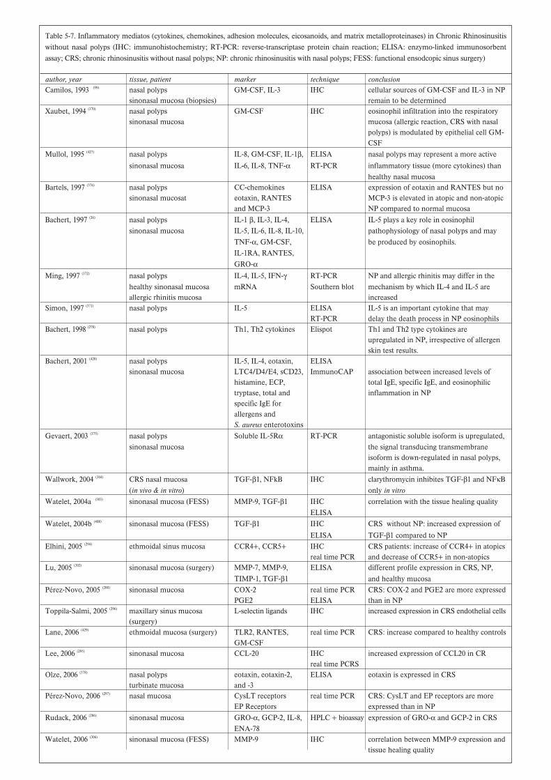
Table 5-7. Inflammatory mediatos (cytokines, chemokines, adhesion molecules, eicosanoids, and matrix metalloproteinases) in Chronic Rhinosinusitiswithout nasal polyps (IHC: immunohistochemistry; RT-PCR: reverse-transcriptase protein chain reaction; ELISA: enzymo-linked immunosorbentassay; CRS; chronic rhinosinusitis without nasal polyps; NP: chronic rhinosinusitis with nasal polyps; FESS: functional ensodcopic sinus surgery)
author, year tissue, patient marker technique conclusionCamilos, 1993 (99) nasal polyps GM-CSF, IL-3 IHC cellular sources of GM-CSF and IL-3 in NP
sinonasal mucosa (biopsies) remain to be determinedXaubet, 1994 (370) nasal polyps GM-CSF IHC eosinophil infiltration into the respiratory
sinonasal mucosa mucosa (allergic reaction, CRS with nasalpolyps) is modulated by epithelial cell GM-CSF
Mullol, 1995 (427) nasal polyps IL-8, GM-CSF, IL-1", ELISA nasal polyps may represent a more activesinonasal mucosa IL-6, IL-8, TNF-! RT-PCR inflammatory tissue (more cytokines) than
healthy nasal mucosaBartels, 1997 (376) nasal polyps CC-chemokines ELISA expression of eotaxin and RANTES but no
sinonasal mucosat eotaxin, RANTES MCP-3 is elevated in atopic and non-atopicand MCP-3 NP compared to normal mucosa
Bachert, 1997 (26) nasal polyps IL-1 ", IL-3, IL-4, ELISA IL-5 plays a key role in eosinophilsinonasal mucosa IL-5, IL-6, IL-8, IL-10, pathophysiology of nasal polyps and may
TNF-!, GM-CSF, be produced by eosinophils.IL-1RA, RANTES,GRO-!
Ming, 1997 (372) nasal polyps IL-4, IL-5, IFN-# RT-PCR NP and allergic rhinitis may differ in thehealthy sinonasal mucosa mRNA Southern blot mechanism by which IL-4 and IL-5 areallergic rhinitis mucosa increased
Simon, 1997 (371) nasal polyps IL-5 ELISA IL-5 is an important cytokine that mayRT-PCR delay the death process in NP eosinophils
Bachert, 1998 (278) nasal polyps Th1, Th2 cytokines Elispot Th1 and Th2 type cytokines areupregulated in NP, irrespective of allergenskin test results.
Bachert, 2001 (428) nasal polyps IL-5, IL-4, eotaxin, ELISAsinonasal mucosa LTC4/D4/E4, sCD23, ImmunoCAP association between increased levels of
histamine, ECP, total IgE, specific IgE, and eosinophilictryptase, total and inflammation in NPspecific IgE forallergens andS. aureus enterotoxins
Gevaert, 2003 (375) nasal polyps Soluble IL-5R! RT-PCR antagonistic soluble isoform is upregulated,sinonasal mucosa the signal transducing transmembrane
isoform is down-regulated in nasal polyps,mainly in asthma.
Wallwork, 2004 (304) CRS nasal mucosa TGF-"1, NFkB IHC clarythromycin inhibites TGF-"1 and NF$B(in vivo & in vitro) only in vitro
Watelet, 2004a (303) sinonasal mucosa (FESS) MMP-9, TGF-"1 IHC correlation with the tissue healing qualityELISA
Watelet, 2004b (408) sinonasal mucosa (FESS) TGF-"1 IHC CRS without NP: increased expression ofELISA TGF-"1 compared to NP
Elhini, 2005 (294) ethmoidal sinus mucosa CCR4+, CCR5+ IHC CRS patients: increase of CCR4+ in atopicsreal time PCR and decrease of CCR5+ in non-atopics
Lu, 2005 (302) sinonasal mucosa (surgery) MMP-7, MMP-9, ELISA different profile expression in CRS, NP,TIMP-1, TGF-"1 and healthy mucosa
Pérez-Novo, 2005 (288) sinonasal mucosa COX-2 real time PCR CRS: COX-2 and PGE2 are more expressedPGE2 ELISA than in NP
Toppila-Salmi, 2005 (296) maxillary sinus mucosa L-selectin ligands IHC increased expression in CRS endothelial cells(surgery)
Lane, 2006 (429) ethmoidal mucosa (surgery) TLR2, RANTES, real time PCR CRS: increase compared to healthy controlsGM-CSF
Lee, 2006 (295) sinonasal mucosa CCL-20 IHC increased expression of CCL20 in CRreal time PCRS
Olze, 2006 (378) nasal polyps eotaxin, eotaxin-2, ELISA eotaxin is expressed in CRSturbinate mucosa and -3
Pérez-Novo, 2006 (297) nasal mucosa CysLT receptors real time PCR CRS: CysLT and EP receptors are moreEP Receptors expressed than in NP
Rudack, 2006 (286) sinonasal mucosa GRO-!, GCP-2, IL-8, HPLC + bioassay expression of GRO-! and GCP-2 in CRSENA-78
Watelet, 2006 (306) sinonasal mucosa (FESS) MMP-9 IHC correlation between MMP-9 expression andtissue healing quality

32 Supplement 18
lower airways, when applicated in either of those locations (422).In fact, when comparing SAE-IgE positive nasal polyps toSAE-IgE negative, the number of IgE positive cells andeosinophils is significantly increased. The more severe inflam-mation is also reflected by significantly increased levels of IL-5,ECP and total IgE. Furthermore, a possible link to aspirin sen-sitivity has been proposed, with SAEs creating a severe inflam-mation possibly serving as a basis for the specific changes seenin aspirin sensitivity (412, 414). A review on the current knowledgeon the impact of SAEs on nasal polyp disease and lower airwaydisease was recently published (206). IgE antibody formation toSAE can be seen in nasal polyp tissue, but rarely in CRS with-out polyps.In conclusion, SAEs are able to induce a more severeeosinophilic inflammation as well as the synthesis of a multi-clonal IgE response with high total IgE concentrations in thetissue, which would suggest that SAEs are at least modifiers ofdisease in CRS with nasal polyps (206, 420). Interestingly, similarfindings have recently been reported in asthma, which isknown to be associated with NP (349), and in COPD (423), thusproviding a link between upper and lower airways.
5-4-4 Nasal polyps in cystic fibrosisNPs and CF-NPs share the remodelling pattern, i.e. they canbe discriminated from CRS without polyps and controls by theoedema formation. Different to NPs, CF-NP display a veryprominent neutrophilic inflammation, with abnormallyincreased concentrations of IL-1", IL-8 and MPO (269) and anincreased activity of NF$B (424), whereas macrophages, but noteosinophils, are significantly increased over control mucosa.Furthermore, there seems to be a reactive imbalance in innateimmunity markers, with mRNA expression of HBD 2 and ofTLR2 being significantly higher in CF-NP compared to non-CF-NP (425).The calcium-activated chloride channel hCLCA1 is thought toregulate the expression of soluble gel-forming mucins, and isupregulated by IL-9. Increased expression of IL-9 and IL-9R,as well as upregulation of hCLCA1, in mucus-overproducingepithelium of patients with cystic fibrosis supports the hypoth-esis that IL-9 contributes to mucus overproduction in cysticfibrosis (426).
5-4-5 CRS with or without nasal polypsWhen searching through the literature, it is apparent that defi-nitions do matter; especially in older literature, the term CRSwas confused by “hyperplastic CRS” or “hyperplastic CRS withpolyp formation”, which would probably be equivalent to NPsrather than CRS. However, because of a lack of clinical dataprovided in those papers, the distinction between these dis-eases can often not be made, and the interpretation from thosestudies has to be done with caution. For the purpose of thischapter, we have used clearly defined patient populations.Whereas NPs are characterized by oedema formation, with anincreased VPF/VEGF, an upregulation of MMPs, but not
TIMP, and a low level or decrease in TGF-"1, CRS shows afibrotic remodelling pattern with a balanced MMP system anda significantly increased TGF-" protein content (269). This fun-damental difference has been linked to the predominant typeof inflammation: eosinophilic or neutrophilic; however, thisnotion has to be challenged in the light of 1) the fact that NPsand NPs in CF both show oedema formation, but are charac-terized by either abundant eosinophils or neutrophils and theirproducts; and 2) the fact that NPs from different parts of theworld do not show the same degree of eosinophilic inflamma-tory pattern (346). It is important to note that also in NPs, thenumber of neutrophils is increased versus controls, and thefrequent impression of a predominantly “eosinophilic” inflam-mation neglects those cells.Both chronic sinus diseases, CRS and NPs, show increasednumbers and activation of T-cells, while only NPs display asignificant increase in plasma cells and consecutiveimmunoglobulin synthesis (269). The role of this observation isunclear, and may reflect a constant microbial trigger.NP have significantly higher levels of eosinophilic markers(eosinophils, eotaxin and ECP) compared to CRS and controlsin Caucasians, in whom CRS is characterized by proinflamma-tory cytokines (IL-1" and TNF-!), a Th1 polarisation with highlevels of IFN-#, and a significant increase in profibrotic TGF-",while NPs show a Th2 polarisation with high IL-5 and IgE con-centrations and a decrease in TGF-" (269). Further studies willhave to investigate TH1 and TH2-specific transduction signals,as well as the role of T regulatory cells, to fully understand thestability of these patterns; studies in non-caucasian populationsmay further be helpful to differentiate primary from secondaryphenomena.
5-5 Aspirin sensitivity – Inflammatory mechanisms in acute andchronic rhinosinusitis
5-5-1 IntroductionThe presence of aspirin-intolerance in a patient with rhinosi-nusitis with or without nasal polyposis is associated with a par-ticularly persistent and treatment-resistant form of disease,coexisting usually with severe asthma and referred to as the“aspirin triad” (430). The prevalence of nasal polyposis in aspirin-sensitive asthmatics may be as high as 60-70%, as compared toless than 10% in the population of aspirin-tolerant asthmatics(34) .The unusual severity of the upper airway disease in thesepatients is reflected by high recurrence of nasal polyps, and fre-quent need for endoscopic sinus surgery (25, 431, 432). Rhinosinusitisin aspirin hypersensitive patients with nasal polyposis is charac-terized by involvement of all sinuses and nasal passages andhigher thickness of hypertrophic mucosa as has been docu-mented with computer tomography (433).
5-5-2 Mechanisms of acute ASA-induced reactionsIn ASA-sensitive patients acute nasal symptoms (sneezing, rhi-norrhea and congestion) may be induced by challenge with

European Position Paper on Rhinosinusitis and Nasal Polyps 2007 33
oral or intranasal aspirin but also with other cross-reactingnonsteroidal anti-inflammatory drugs (NSAIDs). The mecha-nism of these acute adverse reactions has been attributed toinhibition by NSAIDs of an enzyme cyclooxygenase-1, withsubsequent inflammatory cell activation and release of bothlipid and non-lipid mediators (434, 435). The ASA-induced nasalreaction is accompanied by an increase in both glandular(lactoferrin, lysozyme) and plasma (albumin) proteins in nasalsecretions indicating a mixed response, involving both glandu-lar and vascular sources (388). Concomitant release of both mastcell (tryptase, histamine) and eosinophil (ECP) specific media-tors into nasal washes clearly indicate activation of both typesof cells (436-438). Increased concentration of cysteinyl leukotrienesin nasal secretion was also observed within minutes after ASA-challenge although the cellular source of leukotrienes has notbeen determined (439). In parallel with inflammatory mediatorrelease an influx of leucocytes into nasal secretions occurredwith significant enrichment in eosinophils (436).
5-5-3 Chronic rhinosinusitis with nasal polypsAlthough the pathogenesis of chronic eosinophilic inflamma-tion of the airway mucosa and nasal polyps in ASA-sensitivepatients, does not seem to be related to intake of aspirin orother NSAIDs it has been speculated that the pathomech-anism underlying rhinosinusitis with nasal polyps in aspirin-sensitive patients may be different from that in aspirin tolerantpatients (440).
5-5-3-1 Cells and cytokine profileA high degree of marked tissue eosinophilia is a prominentfeature of rhinosinusits with nasal polypos in ASA-hypersensi-tive patients and accordingly significantly more ECP wasreleased from non-stimulated or stimulated nasal polyp dis-persed cells from ASA-sensitive patients (350, 441). An increasednumber of eosinophils in the tissue has been linked to a dis-tinctive profile of cytokine expression with upregulation of sev-eral cytokines related to eosinophil activation and survival (eg.IL-5, GM-CSF, RANTES, eotaxin) (442-444). It has been suggestedthat overproduction of IL-5 might be a major factor responsi-ble for an increased survival of eosinophils in the nasal polypsresulting in increased intensity of the eosinophilic inflamma-tion particularly in aspirin-sensitive patients. In fact decreasedapoptosis was documented in polyps from aspirin-sensitivepatients, and increased infiltration with eosinophils was associ-ated with prominent expression of CD45RO+ activated/mem-ory cells and this cellular pattern was related to clinical featuresof rhinosinusitis (445). Bachert at al (192) demonstrated that IgE-antibodies to Staphylococcal enterotoxins (SAEs) were presentin nasal polyp tissue and their concentration correlated withthe levels of ECP, eotaxin and IL-5. These relations seemed tobe particularly evident in ASA-sensitive patients suggestingthat an increased expression of IL-5 and ECP in polyp tissuefrom ASA-sensitive patients may be related to the presence ofSAE that can exert direct effects on eosinophil proliferation
and survival or may act as a superantigen to trigger a T-cellmediated inflammatory reaction (412).
Not only activated eosinophils but also mast cells are abundantin the nasal polyps tissue from ASA-sensitive patients (355, 446).The density of mast cells was correlated with the number ofpolypectomies, implicating an important role for these cells inthe pathogenesis of CRS with nasal polyps. Stem cell factor(SCF) also called c-kit ligand is a multi-potent cytokine gener-ated by nasal polyp epithelial cells and critical for differentia-tion, survival, chemotaxis and activation and of human mastcells but also involved in eosinophil activation and degranula-tion. SCF expression in nasal polyp epithelial cells in culturecorrelated closely with the density of mast cells in nasal polyptissue and was significantly higher in asthmatic patients withaspirin hypersensitivity as compared to aspirin tolerant patients(355).
5-5-3-2 Arachidonic acid metabolitesSince Szczeklik et al (447) reported an increased susceptibility ofnasal polyps cells from ASA-sensitive patients to the inhibitoryaction of aspirin, arachidonic aid metabolism abnormalitieshave been considered a distinctive feature of nasal polyps inthis subpopulation of patients. A significantly lower generationof PGE2 by nasal polyps and, nasal polyp epithelial cells aswell as a decreased expression of COX-2 in nasal polyps ofthese patients were reported (398, 448). Low expression of COX-2mRNA in nasal polyps from ASA-sensitive patients was in turnlinked to a downregulation of NF-$B activity and to abnormalregulation of COX-2 expression mechanisms at the transcrip-tional level (449, 450). Since PGE2 has significant anti-inflammatoryactivity, including inhibitory effect on eosinophil chemotaxisand activation, it has been speculated that an intrinsic defect inlocal generation of PGE2 could contribute to development ofmore severe eosinophilic inflammation in aspirin-sensitivepatients. Although a significant deficit of PGE2 was demon-strated in polyp tissue of ASA-sensitive as compared to ASA-tolerant patients, decreased expression of COX-2mRNA andPGE production seem to be a feature of CRS with nasal polypsalso in patients without ASA-sensitivity representing moregeneral mechanism involved in the growth of nasal polyps. Onthe other hand the percentages of neutrophils, mast cells,eosinophils, and T cells expressing prostaglandin EP2, but notEP1, EP3, or inflammation in ASA-sensitive patients and somestudies demonstrated an increased production of cysteinylleukotrienes in nasal polyps of ASA-sensitive asthmatics ascompared to aspirin tolerant patients in vitro (400, 451) but theseobservations could not be reproduced in vivo when nasalwashes were analysed (388, 452). Similarly when nasal polyp dis-persed cells were cultured basal and stimulated release ofLTC4 was found to be similar in nasal polyp cells from fromASA-sensitive and ASA-tolerant patients (355). Whole bloodcells from aspirin sensitive and tolerant patients did not differin their ability to generate cyclooygenase and lipoxygenase

34 Supplement 20
products (453). More recently an increased expression ofenzymes involved in production of leukotrienes (5-LOX andLTC4 synthase) and an increased generation of LTC4/D4/E4in nasal polyp tissue from ASA-sensitive patients were found(288, 439, 454). Cysteinyl leukotriene production correlated with tissueECP concentration both in ASA-sensitive and ASA-tolerantpolyps suggesting that these mediators may be linked to tissueeosinophilia rather that to aspirin-sensitivity. On the otherhand an increased expression of leukotriene LT1 receptors wasfound in the nasal mucosa of ASA-sensitive patients, suggest-ing local hyper-responsiveness to leukotrienes in this subpopu-lation of patients (389, 392). More recently other arachidonic acidmetabolites generated on 15-LOX pathway have been associat-ed with CRS with nasal polyps in ASA-sensitive patients. Innasal polyp epithelial cells from ASA-sensitive but not ASA-
tolerant patients aspirin triggers 15-HETE generation, suggest-ing the presence of a specific abnormality of 15-LO pathway inthese patients (448). Upregulation of 15-lipoxygenase anddecreased production of the anti-inflammatory 15-LO metabo-lite lipoxin A4 found in nasal polyp tissue from ASA-sensitivepatients further points to a distinctive but not yet understoodrole for 15-LO metabolites in nasal polyps (288).
5-6 Conclusion
Although far from being completely understood, pathomech-anisms in ARS, CRS and NP are better understood today andbegin to allow us to differentiate these diseases via theircytokine profile, their pattern of inflammation as well asremodeling processes.
European Position Paper on Rhinosinusitis and Nasal Polyps 2007 35
6-1 Assessment of rhinosinusitis symptoms
6-1-1 Symptoms of rhinosinusitisSubjective assessment of rhinosinusitis is based on symptoms.
• nasal blockage, congestion or stuffiness;• nasal discharge or postnasal drip, often mucopurulent;• facial pain or pressure, headache, and• reduction/loss of smell.
Besides these local symptoms, there are distant and generalsymptoms. Distant symptoms are pharyngeal, laryngeal andtracheal irritation causing sore throat, dysphonia and cough,whereas general symptoms include drowsiness, malaise andfever. Individual variations of these general symptom patternsare many (24, 455-459). It is interesting to note that only a small pro-portion of patients with purulent rhinosinusitis, without coex-isting chest disease, complain of cough (460).
The symptoms are principally the same in acute and chronicrhinosinusitis as well as in CRS with nasal polyps, but thesymptom pattern and intensity may vary. Acute forms of infec-tions, both acute and acute exacerbations of chronic rhinosi-nusitis, have usually more distinct and often more severesymptoms.
Simple nasal polyps may cause constant non-periodic nasalblockage , which can have a valve-like sensation allowing bet-ter airflow in only one direction. Nasal polyps may cause nasalcongestion, which can be a feeling of pressure and fullness inthe nose and paranasal cavities. This is typical for ethmoidalpolyposis, which in severe cases can cause widening of thenasal and paranasal cavities demonstrated radiologically and inextreme cases, hyperteliorism. Disorders of smell are moreprevalent in patients with nasal polyps than in other chronicrhinosinusitis patients (25).
6-1-2 Subjective assessment of the symptomsSubjective assessment of the symptoms should consider thestrength or degree of the symptoms, the duration of the symp-tom. During the last decade more attention has been paid notonly to symptoms but also to their effect on the patient’s quali-ty of life (QoL) (461, 462).The assessment of subjective symptoms is done using ques-tionnaires or in clinical studies recorded in logbooks.Evaluation frequency depends on the aims of the study, usual-ly once or twice daily. Continuous recording devices are alsoavailable.
The degree or strength of the symptoms can be estimatedusing many different grading tools.• recorded as such: severe, moderate, slight and no symptom;
• recorded as numbers: from 0 to 4 or as many degrees asneeded;
• recorded as VAS score on a line giving a measurable con-tinuum (0 – 10 cm).
Terms such as mild, moderate or severe may include bothsymptom severity estimation, but also an estimate of durationi.e. “moderate symptom severity” can mean an intense symp-tom but only for a short time in the recorded period or lesssevere symptom but lasting for most of the recording period.
A recent study has considered the relationship between subjec-tive assessment instruments in chronic rhinosinusitis and hasshown that ‘mild’ equates to a visual analogue score of 3 orless, ‘moderate’ to > 3-7 and ‘severe’ to > 7-10 (14).
The duration of the symptoms is evaluated as symptomatic orsymptom-free moments in given time periods, i.e. as hoursduring the recording period or as day per week.
“No symptom” can be regarded as a consistent finding in moststudies. It provides the possibility to record time periods (eg.days) without symptoms, which can be reliably comparedbetween testees (inter-patient) and from study to study.
These criteria are inconsistent and not always comparablewhen considering rhinosinusitis. (459), where the symptoms mayfluctuate from time to time. Nevertheless in many randomised,controlled and prospective rhinosinusitis intervention studies,both allergic and infective, these methods of recording symp-toms have given statistically significant results.
In a study correlating nasosinal symptoms with topographicdistribution of chronic rhinosinuitis as demonstrated by CTscanning, the symptoms of nasal obstruction, anterior andposterior nasal discharge, sneezing and facial congestionfailed to discriminate site of disease. By contrast loss of smelland flavour were correlated with what the authors refer to as‘diffuse rhinosinusitis’ ie mainly CRS with nasal polyps,whereas cacosmia and facial pain correlated with ‘localised oranterior sinusitis’ ie mainly sinusitis of dental or foreign bodyorigin. (84)
6-1-3 Validation of subjective symptoms assessmentValidation of the rhinosinusitis symptoms to show the rele-vance in distinguishing disease modalities and repeatabiltybetween ratings of the same patient (intrapatient, longitudinalvalidity) and between different patients (interpatient, cross-sec-tional validity) have been done. Lately, more specific and vali-dated subjective symptom scoring tools have become available
6. Diagnosis
36 Supplement 20
with the development of quality of life (QoL) evaluations.These are either assess general health evaluating (463, 464) or aredisease specific (461, 462, 465).
6-1-3-1 Nasal obstructionValidation of subjective assessment of nasal obstruction orstuffiness has been done by studying the relationship betweensubjective and objective evaluation methods for functionalnasal obstruction. However, the patient’s interpretation ofnasal blockage varies from true mechanical obstruction of air-flow to the sensation of fullness in the midface.
Generally the subjective sensation of nasal obstruction and rhi-nomanometric or nasal peak flow evaluations show a goodintra-individual correlation in a number of studies consideringnormal controls, patients with structural abnormalities, hyper-reactivity or infective rhinitis (466-470). However, there are alsosome studies where this correlation is not seen (471) or the corre-lation was poor (472, 473).
The interpatient variation in subjective scoring suggests thatevery nose is ”individually calibrated”, which makes interpa-tient comparisons less reliable but still significant (466, 468).
Subjective nasal obstruction correlates better with objectivefunctional measurements of nasal airflow resistance (rino-manometry, peak flow) than with measurements of nasal cavi-ty width, such as acoustic rhinometry (470, 474).Nasal obstruction can also be assessed objectively by testsusing personal nasal peak flow instruments, inspiratory or expi-ratory, which patients can take home or to their work place anddo measurements at any desired time intervals.Subjective assessment of nasal obstruction is a well validatedcriterion.
6-1-3-2 Nasal dischargeTechniques for objective assessment of nasal discharge are notas good as for nasal obstruction: Counting the nose blowingsin a diary card or using a new handkerchief from a countedreservoir for each blow and possibly collecting the used hand-kerchieves in plastic bags for weighing have been used in acuteinfective rhinitis (475) and in “autonomic (previously termedvasomotor) rhinitis” (476).
Validating correlation studies between “objective” dischargemeasures (collecting and measuring amount or weight of nasalsecretion as drops, by suction, or using hygroscopic paperstrips etc) and subjective scoring of nasal discharge or post-nasal drip has not been done.
6-1-3-3 Smell abnormalitiesFluctuations in the sense of smell are associated with chronicrhinosinusitis. This may be due to mucosal obstruction of theolfactory niche (conductive loss) and/or degenerative alter-
ations in the olfactory mucosa due to the disease or its treat-ment eg. repeated nasal surgery.
Subjective scoring of olfaction is a commonly used assessmentmethod. In validating clinical settings, subjective scores havebeen found to correlate significantly to objective olfactorythreshold and qualitative tests in normal population, rhinosi-nusitis and other disease conditions (477-480) as well as numerousclinical studies concerning other diseases than rhinosinusitis(Evidence level Ib).
6-1-3-4 Facial pain and pressureFacial or dental pain, especially unilateral, have been found tobe predictors of acute maxillary sinusitis with fluid retention inpatients with a suspicion of infection, when validated by maxil-lary antral aspiration (455) or paranasal sinus radiographs (481). Theimportance of facial pain as a cardinal sign of chronic rhinosi-nusitis has also been called into question (482) where the symp-toms are more diffuse and fluctuate rendering the clinical cor-relation of facial pain and pressure scorings against objectiveassessments unconvincing. Poor correlation between facialpain localisation and the affected paranasal sinus CT pathologyin patients with supposed infection, both acute and chronic,has been reported (483). However, rhinosinusitis disease specificquality of life studies also include facial pain-related parame-ters, which have been validated (465).
6-1-3-5 Overall rating of rhinosinusitis severityOverall rating of rhinosinusitis severity can be obtained as suchor by total symptoms scores, which are summed scores of theindividual symptoms scores. These are both commonly used,but according to an old validation study for measuring theseverity of rhinitis, scores indicating the course of individualsymptoms should not be combined into a summed score,rather the patient's overall rating of the condition should beused (484). QoL methods have produced validated questionnaireswhich measure the impact of overall rhinosinusitis symptomson everyday life (461).
6-1-3-6 Chronic Sinusitis Survey (CSS)This is a 6-item duration based monitor of sinusitis specificoutcomes which has both systemic and medication-based sec-tions (485). In common with other questionnaires, it is rather bet-ter at determining the relative impact of chronic rhinosinusitiscompared to other diseases than as a measure of improvementfollowing therapeutic intervention but can be a useful tool (462,
486) [Evidence Level IIb]. Mean scores one year after endoscopicfrontal sinus surgery showed a significant improvement insymptoms of pain, congestion, and drainage and medicationuse was also significantly reduced (487).
6-1-3-7 The Chronic Rhinosinusitis Type Specific QuestionnaireThis test contains three forms. Form 1 collects data on nasaland sinus symptoms prior to treatment, Form 2 collects data

European Position Paper on Rhinosinusitis and Nasal Polyps 2007 37
on the clinical classification of sinus disease and Form 3 dataon nasal and sinus symptoms after sinus surgery. Hoffman etal have used this in combination with an SF-36 to look atpatient outcomes after surgical management of chronic rhinos-inusitis though it is somewhat time consuming to complete (488).
6-2 Examination
6-2-1 Anterior rhinoscopyAnterior rhinoscopy alone is inadequate, but remains the firststep in examining a patient with these diseases.
6-2-2 EndoscopyThis may be performed without and with decongestion andsemi-quantitative scores (457) for polyps, oedema, discharge,crusting and scarring (post-operatively) can be obtained (Table6-1) at baseline and at regular intervals following therapeutic
interventions eg at 3, 6, 9 and 12 months. This has a high inter-rater concordance (489). A number of staging systems for polypshave been proposed (490-492). Johansson showed good correlationbetween a 0 - 3 scoring system and their own system in whichthey estimated the percentage projection of polyps from thelateral wall and the percentage of the nasal cavity volume occu-pied by polyps. However, they did not find a correlationbetween size of polyps and symptoms. (Level III).
6-2-3 Nasal cytology, biopsy and bacteriologyGenerally cytology has not proved a useful tool in diagnosis ofrhinosinusitis although a biopsy may be indicated to excludemore sinister and severe conditions such as neoplasia and thevasculitides.
Several microbiology studies (494-498) [Evidence Level IIb] haveshown a reasonable correlation between specimens taken fromthe middle meatus under endoscopic control and proof punc-ture leading to the possibility of microbiological confirmationof both the pathogen and its response to therapy (Table 6-2). Ameta-analysis showed an accuracy of 87% with a lower endconfidence level of 81.3% for the endoscopically directed mid-dle meatal culture when compared with maxillary sinus taps inacute maxillary sinus infection (499).
6-2-4 ImagingPlain sinus x-rays are insensitive and of limited usefulness forthe diagnosis of rhinosinusitis due to the number of false posi-tive and negative results (501-503). Nevertheless it can be useful toprove ARS in studies.
Transillumination was advocated in the 1970’s as an inexpen-sive and efficacious screening modality for sinus pathology (504).The insensitivity and unspecificity makes it unreliable for thediagnosis of rhinosinusitis (505).
Sinus ultrasound is also insensitive and of limited usefulnessfor the diagnosis of rhinosinusitis due to the number of falsepositive and negative results. However, the results in welltrained hands are comparable to X-ray in the diagnostics ofARS (41, 506, 507).
Table 6-1. Endoscopic appearances scores
characteristic Baseline & Follow-uppolyp left (0,1,2,3)polyp, right (0,1,2,3)oedema, left (0,1,2,)oedema, right (0,1,2,)discharge, left (0,1,2)discharge, right (0,1,2)postoperative scores to be used foroutcome assessment onlyscarring, left (0.1,2)scarring, right (0.1,2)crusting, left (0,1,2)crusting, right (0,1,2)total points
0-Absence of polyps;1-polyps in middle meatus only;2-polyps beyond middle meatus but not blocking the nose completely;3-polyps completely obstructing the nose.Oedema: 0-absent; 1-mild; 2-severe.Discharge: 0-no discharge; 1-clear, thin discharge; 2-thick, purulentdischarge.Scarring: 0-absent; 1-mild; 2-severe.Crusting: 0-absent; 1-mild; 2-severe.
Table 6-2. Bacteriology of Rhinosinusitis; Correlation of middle meatus versus maxillary sinus
MM: middle meatus; ESS: endoscopic sinus surgery
author no of samples type of rhinosinusitis technique concordance
Gold & Tami, 1997 (495) 21 chronic endoscopic tap (MM) v maxillary aspiration during ESS 85.7%
Klossek et al, 1998 (494) 65 chronic endoscoic swab (MM) v maxillary aspiration during ESS 73.8%
Vogan et al, 2000 (496) 16 acute endoscoic swab (MM) v maxillary sinus tap 93%
Casiano et al, 2001 (497) 29 acute (intensive care) endoscopic tissue culture (MM) v maxillary sinus tap 60%
Talbot et al, 2001 (500) 46 acute endoscopic swab (MM) v maxillary sinus tap 90.6%
Joniau et al 2005 (498) 26 acute endoscopic swab (MM) v maxillary sinus tap
88.5%
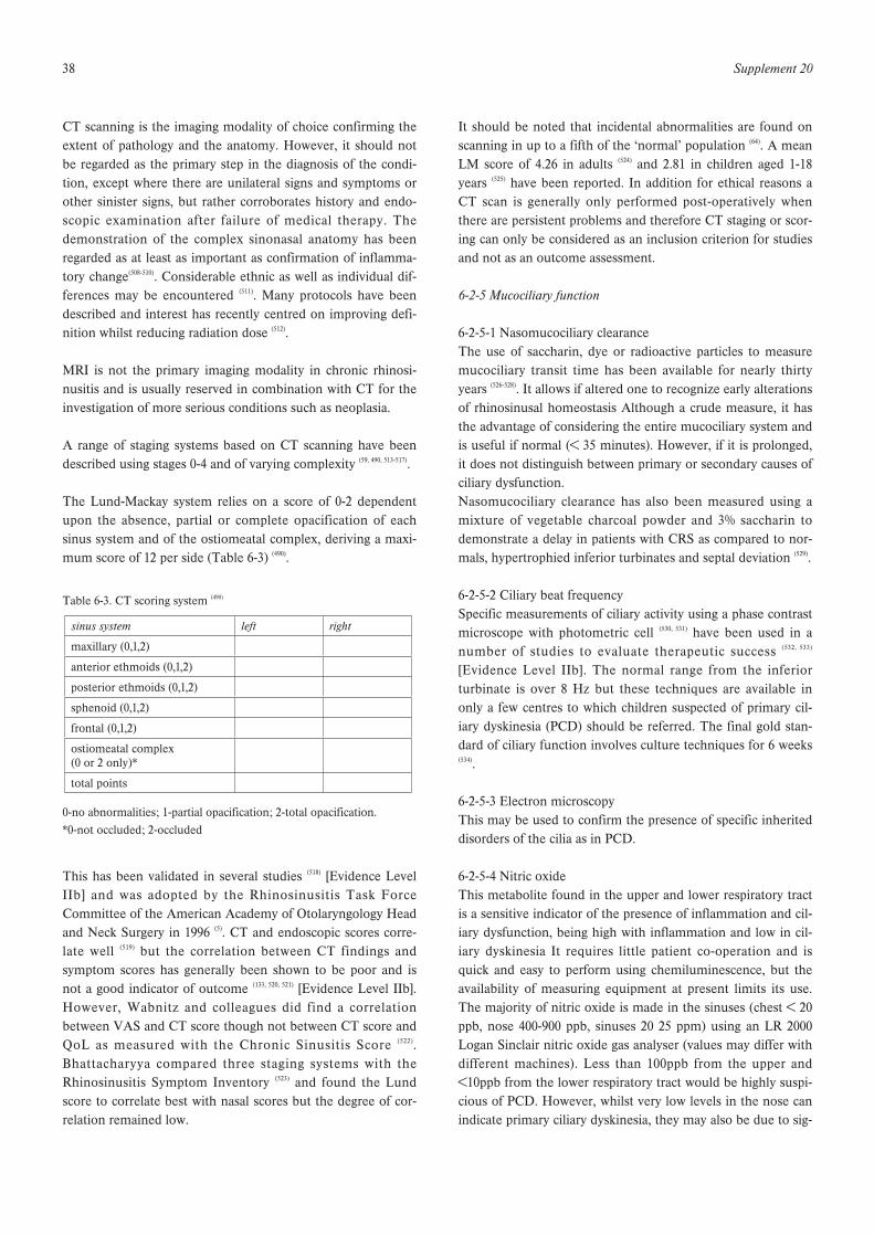
38 Supplement 20
CT scanning is the imaging modality of choice confirming theextent of pathology and the anatomy. However, it should notbe regarded as the primary step in the diagnosis of the condi-tion, except where there are unilateral signs and symptoms orother sinister signs, but rather corroborates history and endo-scopic examination after failure of medical therapy. Thedemonstration of the complex sinonasal anatomy has beenregarded as at least as important as confirmation of inflamma-tory change(508-510). Considerable ethnic as well as individual dif-ferences may be encountered (511). Many protocols have beendescribed and interest has recently centred on improving defi-nition whilst reducing radiation dose (512).
MRI is not the primary imaging modality in chronic rhinosi-nusitis and is usually reserved in combination with CT for theinvestigation of more serious conditions such as neoplasia.
A range of staging systems based on CT scanning have beendescribed using stages 0-4 and of varying complexity (59, 490, 513-517).
The Lund-Mackay system relies on a score of 0-2 dependentupon the absence, partial or complete opacification of eachsinus system and of the ostiomeatal complex, deriving a maxi-mum score of 12 per side (Table 6-3) (490).
This has been validated in several studies (518) [Evidence LevelIIb] and was adopted by the Rhinosinusitis Task ForceCommittee of the American Academy of Otolaryngology Headand Neck Surgery in 1996 (5). CT and endoscopic scores corre-late well (519) but the correlation between CT findings andsymptom scores has generally been shown to be poor and isnot a good indicator of outcome (133, 520, 521) [Evidence Level IIb].However, Wabnitz and colleagues did find a correlationbetween VAS and CT score though not between CT score andQoL as measured with the Chronic Sinusitis Score (522).Bhattacharyya compared three staging systems with theRhinosinusitis Symptom Inventory (523) and found the Lundscore to correlate best with nasal scores but the degree of cor-relation remained low.
It should be noted that incidental abnormalities are found onscanning in up to a fifth of the ‘normal’ population (64). A meanLM score of 4.26 in adults (524) and 2.81 in children aged 1-18years (525) have been reported. In addition for ethical reasons aCT scan is generally only performed post-operatively whenthere are persistent problems and therefore CT staging or scor-ing can only be considered as an inclusion criterion for studiesand not as an outcome assessment.
6-2-5 Mucociliary function
6-2-5-1 Nasomucociliary clearanceThe use of saccharin, dye or radioactive particles to measuremucociliary transit time has been available for nearly thirtyyears (526-528). It allows if altered one to recognize early alterationsof rhinosinusal homeostasis Although a crude measure, it hasthe advantage of considering the entire mucociliary system andis useful if normal (< 35 minutes). However, if it is prolonged,it does not distinguish between primary or secondary causes ofciliary dysfunction.Nasomucociliary clearance has also been measured using amixture of vegetable charcoal powder and 3% saccharin todemonstrate a delay in patients with CRS as compared to nor-mals, hypertrophied inferior turbinates and septal deviation (529).
6-2-5-2 Ciliary beat frequencySpecific measurements of ciliary activity using a phase contrastmicroscope with photometric cell (530, 531) have been used in anumber of studies to evaluate therapeutic success (532, 533)
[Evidence Level IIb]. The normal range from the inferiorturbinate is over 8 Hz but these techniques are available inonly a few centres to which children suspected of primary cil-iary dyskinesia (PCD) should be referred. The final gold stan-dard of ciliary function involves culture techniques for 6 weeks(534).
6-2-5-3 Electron microscopyThis may be used to confirm the presence of specific inheriteddisorders of the cilia as in PCD.
6-2-5-4 Nitric oxideThis metabolite found in the upper and lower respiratory tractis a sensitive indicator of the presence of inflammation and cil-iary dysfunction, being high with inflammation and low in cil-iary dyskinesia It requires little patient co-operation and isquick and easy to perform using chemiluminescence, but theavailability of measuring equipment at present limits its use.The majority of nitric oxide is made in the sinuses (chest < 20ppb, nose 400-900 ppb, sinuses 20 25 ppm) using an LR 2000Logan Sinclair nitric oxide gas analyser (values may differ withdifferent machines). Less than 100ppb from the upper and<10ppb from the lower respiratory tract would be highly suspi-cious of PCD. However, whilst very low levels in the nose canindicate primary ciliary dyskinesia, they may also be due to sig-
Table 6-3. CT scoring system (490)
0-no abnormalities; 1-partial opacification; 2-total opacification.*0-not occluded; 2-occluded
sinus system left right
maxillary (0,1,2)
anterior ethmoids (0,1,2)
posterior ethmoids (0,1,2)
sphenoid (0,1,2)
frontal (0,1,2)
ostiomeatal complex(0 or 2 only)*
total points
European Position Paper on Rhinosinusitis and Nasal Polyps 2007 39
nificant sinus obstruction eg severe CRS with nasal polyps.Conversely elevated levels suggest nasal inflammation butostiomeatal patency (535) [Evidence Level IIb]. It can potentiallybe used, as an outcome measure after therapy (536) [EvidenceLevel IIa] However, some contradictory results on the role ofnNO on nasal inflammation have been recently assessed (311).
6-2-6 Nasal airway assessment
6-2-6-1 Nasal inspiratory peak flowThis inexpensive, quick and easy test is a useful estimate ofairflow which can be performed at home as well as in the hos-pital setting. However, it measures both sides together and haslittle direct role in the assessment of chronic rhinosinusitis. Itcould be used to assess gross reduction in nasal polyps andcompares well with rhinomanometry (537, 538) [Evidence LevelIIb]. Normative data is now available in an adult Caucasianpopulation (539). Expiratory peak flow is less often used asmucus is expelled into the mask and the technique may beassociated with eustachian dysfunction.Furthemore in non-allergic, non-infectitious perennial rhinitisversus controls, repeated PNIF resulted in a brief but statisti-cally significant increase in nasal airway resistance in therhinitic patients suggesting a neuronal mechanism (540)
6-2-6-2 Rhinomanometry (active anterior and posterior).The measurement of nasal airway resistance by assessing nasalflow at a constant pressure is again of limited usefulness inchronic rhinosinusitis and with and without nasal polyps but canbe useful in confirming that improvement in nasal congestion isthe result of reduction in inflammation in the middle meatusrather than mechanical obstruction (532) [Evidence Level IIb].
6-2-6-3 Acoustic rhinometryThe distortion of a sound wave by nasal topography allowsquantification of area at fixed points in the nose from whichvolume may be derived. It can be used to demonstrate subtlechanges, both as a result of medical and surgical intervention(536, 538, 541, 542) [Evidence Level IIa].
6-2-6-4 RhinostereometryThis also measures subtle changes in mucosal swelling, largelyin the inferior turbinates (543, 544) [Level IIb] and is therefore notdirectly applicable to assessment of chronic rhinosinusitis.
6-2-7 Olfaction
6-2-7-1 Threshold TestingThe estimation of olfactory thresholds by the presentation ofserial dilutions of pure odourants such as pm carbinol have beenused in a number of studies (533, 541, 545-547) [Evidence Level IIb].
6-2-7-2 Other quantitative olfactory testingScratch and sniff test using patches impregnated with micro-
encapsulated odorants are available (548) and have been utilisedin studies of both chronic rhinosinusitis with and without nasalpolyps (538). A cruder screening test, the Zurich Smell Diskettetest may also be used and has the advantage of pictorial repre-sentation of the items (549, 550). Also on a national footing, theBarcelona Smell Test has been developed, comprising 24 odor-ants and has been compared with the Zurich Smell DisketteTest (480). More complex tests exist (551) e.g. ‘Sniff ‘n’ sticks’which limit their application to the research setting. A com-bined supra-threshold detection and identification test hasbeen devised as a cross-cultural tool in the European popula-tion, the results of which are presented in the appendix (552)
[Evidence Level III].
Sources of some commercially available and validated olfacto-ry tests are also mentioned in the appendix.
6-2-8 Aspirin and other challengesObjective experiments to differentiate patient groups accordingto rhinosinusitis severity or aetiology have been done usingnasal provocation with histamine or metacholine (553, 554) whichtest mucosal hyper-reactivity. The tests can differentiate sub-populations with statistical significance, but because of consid-erable overlap of results, these tests have not achieved theequivalent position in rhinitis severity evaluations as the corre-sponding bronchial tests i.e in asthma diagnosis.
Establishing a diagnosis of aspirin hypersensitivity is importantsince it provides the patient with a long list of common drugsthat must not be taken in view of the risk of a severe reaction. Itdiagnoses a particular type of asthma and sinonasal disease andallows choice of a specific therapy, i.e. aspirin desensitization.The oral aspirin challenge test was introduced to clinical prac-tice in the early 1970’s (555). Over the following years it was vali-dated and more frequently used (556-558). An inhalation test wasintroduced in 1977 by S Bianco. This challenge is safer andfaster to perform than the oral one, although less sensitive (559-
561). Contrary to the oral challenge it does not produce systemicreactions. The nasal provocation test was employed in the late1980’s (562, 563). It is recommended especially for patients withpredominantly nasal symptoms and those in whom oral orinhaled tests are contraindicated because of the asthma severi-ty. A negative nasal challenge should be followed by oral chal-lenge. Lysine aspirin, the only truly soluble form of aspirinmust be used for both respiratory routes. Test procedures haverecently been reviewed in detail (564). The sensitivity and speci-ficity of the tests are shown in Table 6-4.
Table 6-4. Diagnosis of aspirin sensitivity
history ± challenge sensitivity (%) specificity (%)
oral 77 93
bronchial 77 93
nasal 73 94
s
40 Supplement 20
6-2-9 Laboratory assesments – C-reactive protein (CRP)Known since 1930, C-reactive protein is part of the acute phaseresponse proteins. Its principal properties are short half-life (6-8 h), rapid response (within 6 hours) and high levels (x500 nor-mal) after injury. It activates the classical complement pathway,leading to bacterial opsonization. Studies have shown that theCRP value is useful in the diagnosis of bacterial infections (565).However, among patients suspected of an infectious disease,CRP levels up to 100 mg/l are compatible with all types ofinfections (bacterial, viral, fungal, and protozoal) (566).
Sequential CRP measurements will have greater diagnosticvalue than a single measurement and changes of the CRP val-ues often reflect the clinical course. When used in generalpractice the diagnostic value of CRP is found to be high inadults with pneumonia, sinusitis and tonsillitis. Measurementof CRP is an important diagnostic test but the analysis shouldnot stand alone but be evaluated together with the patient'shistory and clinical examination (567).CRP is most reliably used for exclusion of bacterial infection:two values less than 10 mg/l and 8 12 hours apart can be takento exclude bacterial infection. (566).
6-3 Quality of Life
During the last decade more attention has been paid to notonly symptoms but also to patient’s quality of life (QoL) (462) ormore accurately health-related quality of life (HRQoL). TheQoL questionnaires can provide either general (generic) or dis-ease specific health assessment. However, it is of interest thatthe severity of nasal symptoms or findings do not always corre-late with QoL scales (522, 568). [Evidence Level IIb].
6-3-1 General (generic) health status instrumentsGeneral (generic) measurements enable the comparison ofpatients suffering from chronic rhinosinusitis with otherpatient groups. Of these the Medical Outcomes Study ShortForm 36 (SF36) (463) is by far the most widely used and well vali-dated and this has been used both pre- and post-operatively inchronic rhinosinusitis. (536, 569) [Evidence Level IIa,IIb]. Itincludes eight domains: physical functioning, role functioningphysical, bodily pain, general health, vitality, social function-ing, role-functioning emotional and mental health. Many othergeneric measurements are also available (464). For exampleEuroQOL, Short Form-12 and Quality of Well-Being Scalehave been used in sinusitis studies (570).The Glasgow Benefit Inventory has also been applied tochronic rhinosinusitis and its treatment (571) as has the EuroQoland McGill pain questionnaires (572, 573).
6-3-2 Disease specific health status instruments (see also appendix)Several disease specific questionnaires for evaluation of qualityof life in chronic rhinosinusitis have been published. In thesequestionnaires specific symptoms for rhinosinusitis are includ-
ed. Such areas include headache, facial pain or pressure, nasaldischarge or postnasal drip, and nasal congestion. Disease spe-cific questionnaires are usually used to measure effects withinthe disease in response to interventions. They are usually moresensitive than general health status instruments.
6-3-2-1 Rhinosinusitis outcome measure (RSOM) andSinonasal Outcome Test-20RSOM contains 31 items classified into 7 domains and takesapproximately 20 minutes to complete (574). RSOM has been well-validated and allows measurement of symptom severity andimportance to the patient. The severity and importance scales,however, make it somewhat difficult for the patient to fill thequestionnaire (575) RSOM-31 has been used in medical studies (576).
6-3-2-2 Sinonasal Outcome Test 20A modified instrument referred to as the Sinonasal OutcomeTest 20 (SNOT 20) is validated and easy to use (465). This has beenused in a number of studies both medical and surgical (536, 577)
[Evidence Levels Ib, IIb]. However, the lack of the SNOT-20 isthat it does no contain questions on nasal obstruction and loss ofsmell and taste. These questions are included in the SNOT-22questionnaire which however is not validated. This test was theprimary outcome in the largest audit to date of surgery for chron-ic rhinosinusitis with or without nasal polyps (520).
6-3-2-3 Sinonasal Outcome Test 16The Sinonasal Outcome Test 16 (SNOT 16) is also a rhinosi-nusitis specific quality of life health related instrument (578).
6-3-2-4 Rhinosinusitis Disablity Index (RSDI)In this 30 item validated questionnaire, the patient is asked torelate nasal and sinus symptoms to specific limitations on dailyfunctioning (461, 579). It is similar to the RSOM 31 in the types ofquestions it contains. It can be completed easily and quicklybut does not allow the patient to indicate their most importantsymptoms. However, it does have some general questionssimilar to the SF-36.
6-3-2-5 Rhinoconjunctivitis quality of life questionnaire (RQLQ)This is a well-validated questionnaire but specifically focuseson allergy and is not validated in acute and CRS with or with-out NP (580).A newer standardized version RQLQ(S) is also available and ithas been used for example in a nasal lavage study in chronicrinosinusitis (581).
6-3-2-6 RhinoQOLThe RhinoQol is a sinusitis specific instrument which mea-sures symptom frequency, bothersomeness and impact. It canbe used for acute and chronic sinusitis (582).
6-3-2-7 Rhinitis Symptom Utility Index (RSUI)This consists of ten questions on the severity and frequency of
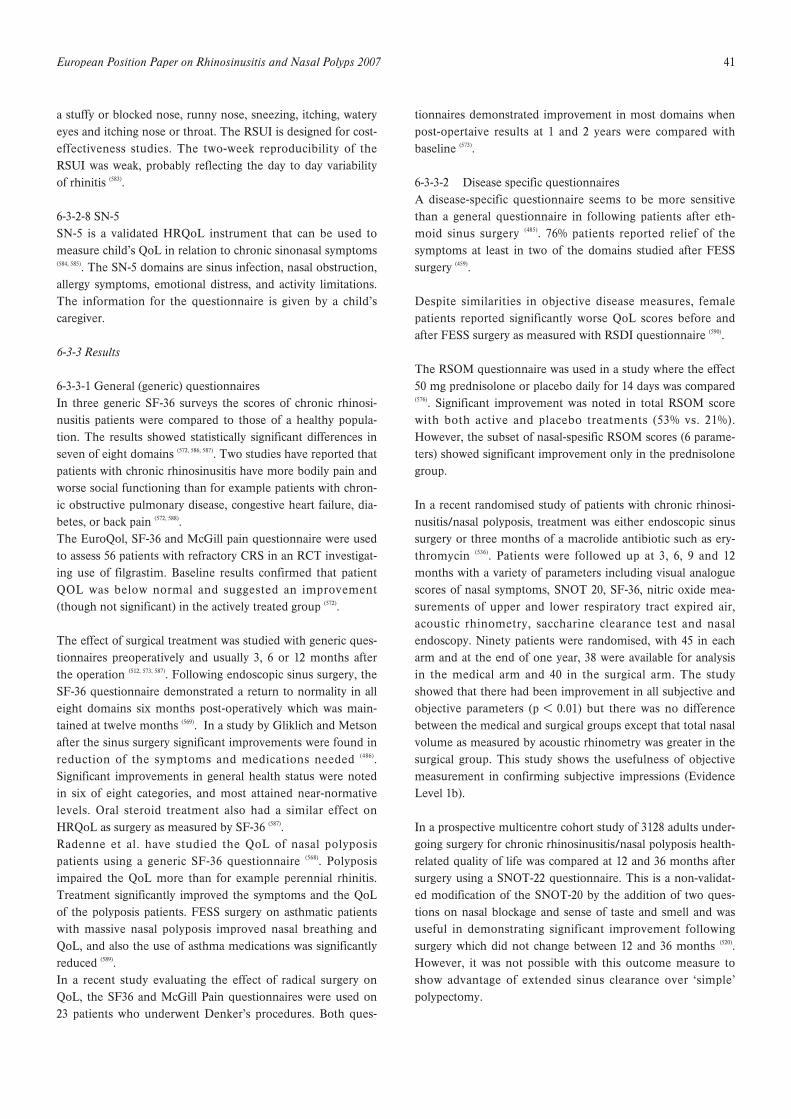
European Position Paper on Rhinosinusitis and Nasal Polyps 2007 41
a stuffy or blocked nose, runny nose, sneezing, itching, wateryeyes and itching nose or throat. The RSUI is designed for cost-effectiveness studies. The two-week reproducibility of theRSUI was weak, probably reflecting the day to day variabilityof rhinitis (583).
6-3-2-8 SN-5SN-5 is a validated HRQoL instrument that can be used tomeasure child’s QoL in relation to chronic sinonasal symptoms(584, 585). The SN-5 domains are sinus infection, nasal obstruction,allergy symptoms, emotional distress, and activity limitations.The information for the questionnaire is given by a child’scaregiver.
6-3-3 Results
6-3-3-1 General (generic) questionnairesIn three generic SF-36 surveys the scores of chronic rhinosi-nusitis patients were compared to those of a healthy popula-tion. The results showed statistically significant differences inseven of eight domains (572, 586, 587). Two studies have reported thatpatients with chronic rhinosinusitis have more bodily pain andworse social functioning than for example patients with chron-ic obstructive pulmonary disease, congestive heart failure, dia-betes, or back pain (572, 588).The EuroQol, SF-36 and McGill pain questionnaire were usedto assess 56 patients with refractory CRS in an RCT investigat-ing use of filgrastim. Baseline results confirmed that patientQOL was below normal and suggested an improvement(though not significant) in the actively treated group (572).
The effect of surgical treatment was studied with generic ques-tionnaires preoperatively and usually 3, 6 or 12 months afterthe operation (512, 573, 587). Following endoscopic sinus surgery, theSF-36 questionnaire demonstrated a return to normality in alleight domains six months post-operatively which was main-tained at twelve months (569). In a study by Gliklich and Metsonafter the sinus surgery significant improvements were found inreduction of the symptoms and medications needed (486).Significant improvements in general health status were notedin six of eight categories, and most attained near-normativelevels. Oral steroid treatment also had a similar effect onHRQoL as surgery as measured by SF-36 (587).Radenne et al. have studied the QoL of nasal polyposispatients using a generic SF-36 questionnaire (568). Polyposisimpaired the QoL more than for example perennial rhinitis.Treatment significantly improved the symptoms and the QoLof the polyposis patients. FESS surgery on asthmatic patientswith massive nasal polyposis improved nasal breathing andQoL, and also the use of asthma medications was significantlyreduced (589).In a recent study evaluating the effect of radical surgery onQoL, the SF36 and McGill Pain questionnaires were used on23 patients who underwent Denker’s procedures. Both ques-
tionnaires demonstrated improvement in most domains whenpost-opertaive results at 1 and 2 years were compared withbaseline (573).
6-3-3-2 Disease specific questionnairesA disease-specific questionnaire seems to be more sensitivethan a general questionnaire in following patients after eth-moid sinus surgery (485). 76% patients reported relief of thesymptoms at least in two of the domains studied after FESSsurgery (459).
Despite similarities in objective disease measures, femalepatients reported significantly worse QoL scores before andafter FESS surgery as measured with RSDI questionnaire (590).
The RSOM questionnaire was used in a study where the effect50 mg prednisolone or placebo daily for 14 days was compared(576). Significant improvement was noted in total RSOM scorewith both active and placebo treatments (53% vs. 21%).However, the subset of nasal-spesific RSOM scores (6 parame-ters) showed significant improvement only in the prednisolonegroup.
In a recent randomised study of patients with chronic rhinosi-nusitis/nasal polyposis, treatment was either endoscopic sinussurgery or three months of a macrolide antibiotic such as ery-thromycin (536). Patients were followed up at 3, 6, 9 and 12months with a variety of parameters including visual analoguescores of nasal symptoms, SNOT 20, SF-36, nitric oxide mea-surements of upper and lower respiratory tract expired air,acoustic rhinometry, saccharine clearance test and nasalendoscopy. Ninety patients were randomised, with 45 in eacharm and at the end of one year, 38 were available for analysisin the medical arm and 40 in the surgical arm. The studyshowed that there had been improvement in all subjective andobjective parameters (p < 0.01) but there was no differencebetween the medical and surgical groups except that total nasalvolume as measured by acoustic rhinometry was greater in thesurgical group. This study shows the usefulness of objectivemeasurement in confirming subjective impressions (EvidenceLevel 1b).
In a prospective multicentre cohort study of 3128 adults under-going surgery for chronic rhinosinusitis/nasal polyposis health-related quality of life was compared at 12 and 36 months aftersurgery using a SNOT-22 questionnaire. This is a non-validat-ed modification of the SNOT-20 by the addition of two ques-tions on nasal blockage and sense of taste and smell and wasuseful in demonstrating significant improvement followingsurgery which did not change between 12 and 36 months (520).However, it was not possible with this outcome measure toshow advantage of extended sinus clearance over ‘simple’polypectomy.

42 Supplement 20
Nowadays there are many generic and disease specific HRQoLquestionnaires available to rhinosinusitis studies. However,most of the questionnaires are not yet validated. QoL measure-ment is quite a new tool evaluating the impact of disease andthe efficacy of treatment. In rhinosinusitis studies, when theeffect of medical treatment or surgery has been evaluated, QoLhas been considered to be an important outcome measure-ment as distinct from classic rhinosinusitis symptom parame-ters. In a number of studies, chronic rhinosinusitis has beenshown to significantly impair QoL [Level Ib] (465, 572, 584, 591, 592) andthis has also been shown to improve significantly with treat-ment [Level IIb] (459, 486, 585, 587, 593, 594).
European Position Paper on Rhinosinusitis and Nasal Polyps 2007 43
7-1 Treatment of rhinosinusitis with corticosteroids
The introduction of topically administered glucocorticoids hasimproved the treatment of upper (rhinitis, nasal polyps) andlower (asthma) airway inflammatory disease. The clinical effi-cacy of glucocorticoids may depend in part on their ability toreduce airway eosinophil infiltration by preventing theirincreased viability and activation. Both topical and systemicglucocorticoids may affect the eosinophil function by bothdirectly reducing eosinophil viability and activation (370, 427, 595, 596)
or indirectly reducing the secretion of chemotactic cytokinesby nasal mucosa and polyp epithelial cells (368, 597-599). The potencyof these effects is lower in nasal polyps than in nasal mucosasuggesting an induced inflammatory resistance to steroid treat-ment in chronic rhinosinusitis / nasal polyposis (596, 597).
The biological action of glucocorticoids is mediated throughactivation of intracellular glucocorticoid receptors (GR) (600),expressed in many tissues and cells (601) Two human isoforms ofGR have been identified, GR! and GR", which originate fromthe same gene by alternative splicing of the GR primary tran-script (602). Upon hormone binding, GR! enhances anti-inflam-matory or represses proinflammatory gene transcription, andexerts most of the anti-inflammatory effects of glucocorticoidsthrough protein-protein interactions between GR and tran-scription factors, such as AP-1 and NF-$B. The GR" isoformdoes not bind steroids but may interfere with the GR* func-tion. There may be several mechanisms accounting for theresistance to the antiinflammatory effects of glucocorticoids,including an overexpression of GR" or a downexpression ofGR! . Increased expression of GR" has been reported inpatients with nasal polyps (603, 604) while downregulation of GR*levels after treatment with glucocorticoids (605, 606) has also beenpostulated to be one of the possible explanations for the sec-ondary glucocorticoid resistance phenomenon.
The anti-inflammatory effect of corticosteroids could, theoreti-cally, be expected as well in non-allergic (i.e. infectious) as inallergic rhinosinusitis. Tissue eosinophilia is thus also seen inCRS (259).
Indications for corticosteroids in rhinosinusitis:• Acute rhinosinusitis;• Prophylactic treatment of acute recurrent rhinosinusitis;• Chronic rhinosinusitis without NP;• Chronic rhinosinusitis with NP;• Postoperative treatment of chronic rhinosinusitis with or
without NP.
7-1-1 Acute rhinosinusitis
Most studies on corticosteroids in ARS determine the effect oftopical corticosteroids as adjunct therapy to antibiotics. Veryrecently a study is published in which topical corticosteroidtreatment as monotherapy is compared to antibiotics
7-1-1-1 Topical corticosteroid as monotherapy in acute rhinosi-nusitisRecently mometasone furoate (MF) has been used and com-pared to amoxicillin and placebo in ARS (607). MF 200 µg twicedaily was significantly superior to placebo and amoxicillin atimproving symptom score. Used once daily MF was also supe-rior to placebo but not to amoxicillin. This randomized studywas done double-blind, double-dummy on 981 subjects. Thisis the first study to show topical steroids when used twice dailyare effective in ARS as monotherapy and more effective thanamoxicillin when used twice daily.
7-1-1-2 Topical corticosteroid as adjunct therapy in acute rhi-nosinusitisQvarnberg et al (608) measured the clinical effect of budesonide(BUD)/placebo as a complement to erythromycin and sinuswashout in a randomized, double-blind study on patientsreferred for sinus surgery due to chronic or recurrent acutemaxillary sinusitis. Three months treatment was given to 20subjects in 2 groups, all without NP. Treatment with BUDresulted in a significant improvement of nasal symptoms, facialpain and sensitivity. No significant improvement was seen inmucosal thickening on x-ray. The final clinical outcome didnot differ between the groups. No side effects of treatmentwere noted. It is not possible in this study to distinguish chron-ic from ARS but all cases were reported to have intermittent“episodes of sinusitis for the last two years”.
In a multicentre study Meltzer et al (609) used flunisolide as adjunc-tive therapy to amoxicillin clavulanate potassium in patients withARS or CRS for three weeks and an additional four weeks ononly flunisolide. The overall score for global assessment of effica-cy was greater in patients treated with flunisolide than placebo(p=0.007) after 3 weeks and after 4 additional weeks p=0.08. Nodifference was seen on x-ray but inflammatory cells were signifi-cantly reduced in flunisolide group compared to placebo.
Barlan et al (610) used BUD as adjunctive therapy to amoxillinclavulanate potassium for three weeks in a randomized, place-bo controlled study in children with ARS. Improvement incough and nasal secretion were seen at the end of the secondweek of treatment in the BUD group, p < 0.05 for both symp-toms compared to placebo. At the end of week three therewere no differences between the groups.Melzer et al (611) gave mometasone furoate (MF) 400 µg to 200
7. Management
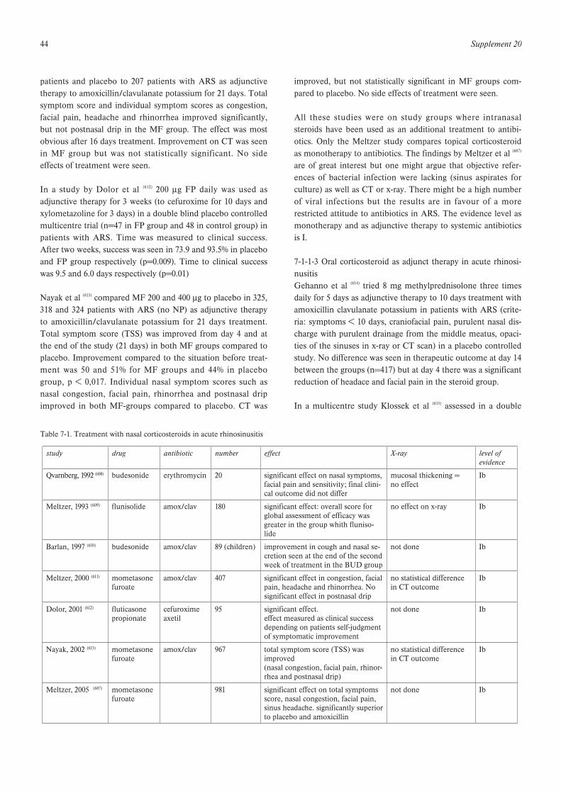
44 Supplement 20
patients and placebo to 207 patients with ARS as adjunctivetherapy to amoxicillin/clavulanate potassium for 21 days. Totalsymptom score and individual symptom scores as congestion,facial pain, headache and rhinorrhea improved significantly,but not postnasal drip in the MF group. The effect was mostobvious after 16 days treatment. Improvement on CT was seenin MF group but was not statistically significant. No sideeffects of treatment were seen.
In a study by Dolor et al (612) 200 µg FP daily was used asadjunctive therapy for 3 weeks (to cefuroxime for 10 days andxylometazoline for 3 days) in a double blind placebo controlledmulticentre trial (n=47 in FP group and 48 in control group) inpatients with ARS. Time was measured to clinical success.After two weeks, success was seen in 73.9 and 93.5% in placeboand FP group respectively (p=0.009). Time to clinical successwas 9.5 and 6.0 days respectively (p=0.01)
Nayak et al (613) compared MF 200 and 400 µg to placebo in 325,318 and 324 patients with ARS (no NP) as adjunctive therapyto amoxicillin/clavulanate potassium for 21 days treatment.Total symptom score (TSS) was improved from day 4 and atthe end of the study (21 days) in both MF groups compared toplacebo. Improvement compared to the situation before treat-ment was 50 and 51% for MF groups and 44% in placebogroup, p < 0,017. Individual nasal symptom scores such asnasal congestion, facial pain, rhinorrhea and postnasal dripimproved in both MF-groups compared to placebo. CT was
improved, but not statistically significant in MF groups com-pared to placebo. No side effects of treatment were seen.
All these studies were on study groups where intranasalsteroids have been used as an additional treatment to antibi-otics. Only the Meltzer study compares topical corticosteroidas monotherapy to antibiotics. The findings by Meltzer et al (607)
are of great interest but one might argue that objective refer-ences of bacterial infection were lacking (sinus aspirates forculture) as well as CT or x-ray. There might be a high numberof viral infections but the results are in favour of a morerestricted attitude to antibiotics in ARS. The evidence level asmonotherapy and as adjunctive therapy to systemic antibioticsis I.
7-1-1-3 Oral corticosteroid as adjunct therapy in acute rhinosi-nusitisGehanno et al (614) tried 8 mg methylprednisolone three timesdaily for 5 days as adjunctive therapy to 10 days treatment withamoxicillin clavulanate potassium in patients with ARS (crite-ria: symptoms < 10 days, craniofacial pain, purulent nasal dis-charge with purulent drainage from the middle meatus, opaci-ties of the sinuses in x-ray or CT scan) in a placebo controlledstudy. No difference was seen in therapeutic outcome at day 14between the groups (n=417) but at day 4 there was a significantreduction of headace and facial pain in the steroid group.
In a multicentre study Klossek et al (615) assessed in a double
Table 7-1. Treatment with nasal corticosteroids in acute rhinosinusitis
study drug antibiotic number effect X-ray level of evidence
Qvarnberg, 1992 (608) budesonide erythromycin 20 significant effect on nasal symptoms, facial pain and sensitivity; final clini-cal outcome did not differ
mucosal thickening = no effect
Ib
Meltzer, 1993 (609) flunisolide amox/clav 180 significant effect: overall score for global assessment of efficacy wasgreater in the group whith fluniso-lide
no effect on x-ray Ib
Barlan, 1997 (610) budesonide amox/clav 89 (children) improvement in cough and nasal se-cretion seen at the end of the secondweek of treatment in the BUD group
not done Ib
Meltzer, 2000 (611) mometasonefuroate
amox/clav 407 significant effect in congestion, facial pain, headache and rhinorrhea. No significant effect in postnasal drip
no statistical differencein CT outcome
Ib
Dolor, 2001 (612) fluticasonepropionate
cefuroximeaxetil
95 significant effect. effect measured as clinical successdepending on patients self-judgmentof symptomatic improvement
not done Ib
Nayak, 2002 (613) mometasonefuroate
amox/clav 967 total symptom score (TSS) was improved(nasal congestion, facial pain, rhinor-rhea and postnasal drip)
no statistical differencein CT outcome
Ib
Meltzer, 2005 (607) mometasonefuroate
981 significant effect on total symptoms score, nasal congestion, facial pain, sinus headache. significantly superior to placebo and amoxicillin
not done Ib

European Position Paper on Rhinosinusitis and Nasal Polyps 2007 45
blind, randomised study in parallel groups the efficacy and toler-ance to prednisone administered for 3 days in addition to cefpo-doxime in adult patients presenting with an acute bacterial rhi-nosinusitis (proven by culture) with severe pain. The assess-ments made during the first 3 days of treatment showed a statis-tically significant difference in favour of the prednisone groupregarding pain, nasal obstruction and consumption of paraceta-mol. There was no difference between the two groups after theend of the antibiotic treatment. The tolerance measuredthroughout the study was comparable between the two groups.
Pain is significantly relieved during treatment with prednisonebut after 10 days on antibiotics there was no differencebetween the two groups. Evidence level for steroids as painreliever: I but there is no evidence for a more positive longterm outcome compared to placebo.
7-1-2 Prophylactic treatment of recurrent episodes of acute rhinos-inusitisIn a study by Puhakka et al (616) FP (200 µg four times daily) orplacebo were used for 6 days in 199 subjects with an acutecommon cold, 24-48 hours after onset of symptoms to studythe preventive effects of FP on risk for development of ARS.Frequency of sinusitis at day 7 in subjects positive for rhi-novirus, based on x-ray, was 18.4% and 34.9% in FP and place-bo group respectively (p = 0.07) thus indicating a non-signifi-cant effect of FP.
Cook et al. randomized, as a continuation of an acute episodeof rhinosinusitis, patients with at least 2 episodes of rhinosi-nusitis in the previous 6 months or at least 3 episodes in thelast 12 months for a double blind, placebo-controlled studywith FP, 200 mcg QD. 227 subjects were included. Additionallycefuroxime axetil 250 mg BID was used for the first 20 days.39% had a recurrence in the placebo group and 25% in the FPgroup (p = 0.016) during the seven week follow-up period.
Mean number of days to first recurrence was 97.5 and 116.6respectively (p = 0.011) (617).
There is very low evidence for a prophylactic effect of nasalcorticosteroids to prevent recurrence of ARS episodes.
7-1-3 Chronic rhinosinusitis without nasal polyps
7-1-3-1 Topical corticosteroid chronic rhinosinusitis withoutnasal polypsParikh et al (618) performed a randomized, double blind, place-bo-controlled trial on patients with chronic RS on two groupswith respectively 9 and 13 subjects (2 subjects in each groupwith nasal polyps) to test fluticasone propionate for 16 weeks.No significant improvement was seen, as measured by symp-tom scores, diary card, acoustic rhinometry or endoscopy. Noside effects were seen in either group.
In another double blind placebo controlled study on patientswith CRS (without NP) with allergy to house dust mite andwho had recently been operated on but still had signs ofchronic RS, 256 ug budesonide (BUD) or placebo was instilledinto the maxillary sinus once a day through a sinus catheter forthree weeks (619). A regression of more than 50% of total nasalsymptom scores was seen in 11/13 in the BUD group and 4/13in placebo group. The effect was more long term in BUDgroup, i.e. 2-12 months compared with less than 2 months inthe placebo group (who had experienced an effect during thecatheter period). A significant decrease was also seen in BUDgroup after three weeks treatment for CD-3, eosinophils andcells expressing IL-4 and IL-5.
In a study by Cuenant et al (620) tixocortol pivalate was given asendonasal irrigation in combination with neomycin for 11 daysin a double blind placebo controlled in patients with chronicRS. Maxillary ostial patency and nasal obstruction was signifi-
Table 7-2. Treatment with oral corticosteroids in acute rhinosinusitis
study drug antibiotic number effect effect at end of treatment level of evidence
Gehanno, 2000 (614) 8 mg metylpred-nisolone TDS
amoxicillinclavulanate
417 significant reduction of headace and facial pain
no statistical differenceat 14 days
Ib
Klossek, 2004 (615) oral prednisone cefpodoxime 289 improvement in pain, nasal obstruc-tion and consumption of paracetamolduring first 3 days
no statistical differenceat end of study
Ib
Table 7-3. Treatment with nasal corticosteroids in prophylaxis of acute rhinosinusitis
study drug number time (weeks) effect comments level of evidence
Puhakka, 1998 (616) FP 199 1 N.S. common cold Ib
Cook, 2002 (617) FP 227 7 increased time to first recurrence. decreased frequency of ARS
Ib
46 Supplement 20
cantly improved in the tixocortol group compared to placebo.Patients with CRS without allergy responded better to topicalsteroids than those with allergy.
Sykes et al (621) looked on 50 patients with mucopurulent CRSand allocated them to 3 groups for local treatment with sprayswith either dexamethasone + tramazoline + neomycin/dexam-ethasone + tramazoline/placebo 4 times daily for 4 weeks andevaluation was performed double blinded. Treatment in bothactive groups was more effective than placebo (discharge,blockage and facial pain and x-ray) but no difference was seenwith the addition of neomycin to dexamethasone.
A recent multicentre double-blinded placebo-controlled ran-domised trial of 134 patients with CRS without nasal polypstreated with topical budesonide for 20 weeks showed signifi-cant improvement in a number of parameters including symp-tom score and nasal inspiratory peak flow (622). Quality of lifeassessments did not change however.
There is some evidence for an effect of topical intranasalsteroids in CRS, particularly with intramaxillary instillation ofsteroids. No side effects were seen, including no increasedsigns of infection with intranasal corticosteroid treatment.
7-1-3-2 Oral corticosteroid chronic rhinosinusitis without nasalpolypsThere are no data showing efficacy of oral corticosteroids inchronic rhinosinusitis without nasal polyps.
7-1-4 Chronic rhinosinusitis with NPIn studies on the treatment of NP, it is of value to look sepa-rately at the effect on rhinitis symptoms associated with poly-posis and the effect on the size of nasal polyps per se. Onlyplacebo controlled studies will be referred to.
7-1-4-1 Topical corticosteroid chronic rhinosinusitis with
nasal polypsMygind et al (623) showed that beclomethasone dipropionate(BDP) 400 µg daily for three weeks reduced nasal symptoms in19 patients with NP compared to a control group of 16 patientstreated with placebo aerosol. Reduction of polyp size did notdiffer in this short treatment study.
In another study with BDP 400 µg daily for four weeks (doubleblind, cross over with 9 and 11 subjects in each group),Deuschl and Drettner (624) found a significant improvement innasal symptoms of blockage and nasal patency as measuredwith rhinomanometry. Difference in size of polyps was, how-ever, not seen.
Holopainen et al (625) showed in a randomized, double blind,paralell, placebo controlled study with 400 µg budesonide(n=19) for 4 months that total mean score and nasal peak flowwere in favour for budesonide. Polyps also decreased in size inthe budesonide group.
Tos et al (626) also showed that budesonide in spray (128 µg) andpowder (140 µg) were both significantly more effective thanplacebo (multicentre) concerning reduction of polyp size,improvement of sense of smell, reduction of symptom scoreand overall assessment compared to placebo.
Vendelo Johansen (627) tested BUD 400 µg daily compared toplacebo for three months in a multicentre, randomized, doubleblind study in patients with small and medium-sizedeosinophilic nasal polyps (grade 1-2). Polyps decreased in theBUD group while an increase was seen in the placebo group.The difference in polyp score between the groups was signifi-cant (p<0.01). Both nasal symptoms (blockage, runny nose,sneezing) and peak nasal inspiratory flow (PNIF) improved sig-nificantly in BUD group.
Lildholt et al (491) compared BUD 400 or 800 µg daily with place-
Table 7-4. Treatment with nasal corticosteroids in chronic rhinosinusitis without nasal polyposis
study drug number time symptoms other effects level of evidence
Cuenant, 1986 (620) tixocortolirrigation
60 11 days nasal obstruction significantly improved
maxillary ostial patencysignificantly improved
Ib
Sykes, 1986 (621) dexamethasone+ tramazoline
50 4 wks discharge, obstruction and fa-cial pain significantly improved
plain x-ray and nasal airwayresistance and mucocili-ary clearance significantimproved
IIa
Parikh, 2001 (618) fluticasonepropionate
22 16 wks not significant acoustic rhinometry not significant.
Ib
Lavigne, 2002 (619) intrasinusbudesonide
26 3 wks total symptom score significant improved
T-cells,eosinophilsmRNA for IL-4, and IL-5 significantly improved
Ib
Lund et al 2004 (622) budesonide 134 20 wks significant symptom impro-vement
significant improvement inairway using PNIF
Ib
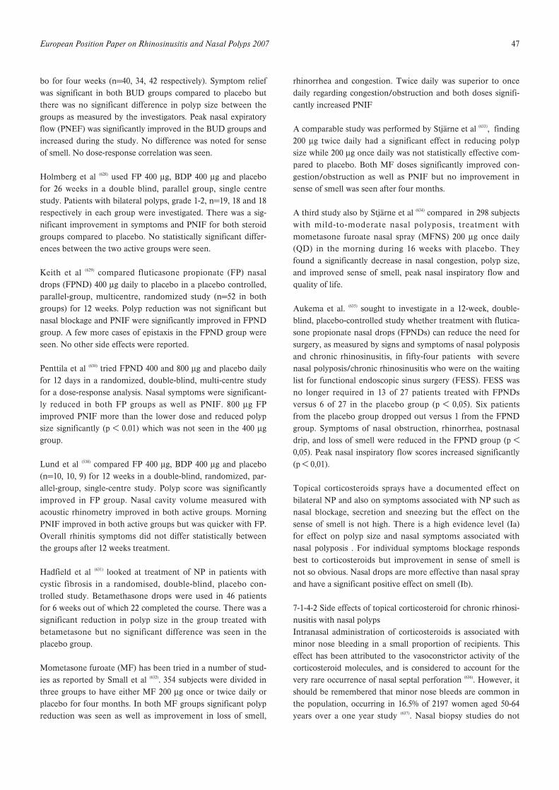
European Position Paper on Rhinosinusitis and Nasal Polyps 2007 47
bo for four weeks (n=40, 34, 42 respectively). Symptom reliefwas significant in both BUD groups compared to placebo butthere was no significant difference in polyp size between thegroups as measured by the investigators. Peak nasal expiratoryflow (PNEF) was significantly improved in the BUD groups andincreased during the study. No difference was noted for senseof smell. No dose-response correlation was seen.
Holmberg et al (628) used FP 400 µg, BDP 400 µg and placebofor 26 weeks in a double blind, parallel group, single centrestudy. Patients with bilateral polyps, grade 1-2, n=19, 18 and 18respectively in each group were investigated. There was a sig-nificant improvement in symptoms and PNIF for both steroidgroups compared to placebo. No statistically significant differ-ences between the two active groups were seen.
Keith et al (629) compared fluticasone propionate (FP) nasaldrops (FPND) 400 µg daily to placebo in a placebo controlled,parallel-group, multicentre, randomized study (n=52 in bothgroups) for 12 weeks. Polyp reduction was not significant butnasal blockage and PNIF were significantly improved in FPNDgroup. A few more cases of epistaxis in the FPND group wereseen. No other side effects were reported.
Penttila et al (630) tried FPND 400 and 800 µg and placebo dailyfor 12 days in a randomized, double-blind, multi-centre studyfor a dose-response analysis. Nasal symptoms were significant-ly reduced in both FP groups as well as PNIF. 800 µg FPimproved PNIF more than the lower dose and reduced polypsize significantly (p < 0.01) which was not seen in the 400 µggroup.
Lund et al (538) compared FP 400 µg, BDP 400 µg and placebo(n=10, 10, 9) for 12 weeks in a double-blind, randomized, par-allel-group, single-centre study. Polyp score was significantlyimproved in FP group. Nasal cavity volume measured withacoustic rhinometry improved in both active groups. MorningPNIF improved in both active groups but was quicker with FP.Overall rhinitis symptoms did not differ statistically betweenthe groups after 12 weeks treatment.
Hadfield et al (631) looked at treatment of NP in patients withcystic fibrosis in a randomised, double-blind, placebo con-trolled study. Betamethasone drops were used in 46 patientsfor 6 weeks out of which 22 completed the course. There was asignificant reduction in polyp size in the group treated withbetametasone but no significant difference was seen in theplacebo group.
Mometasone furoate (MF) has been tried in a number of stud-ies as reported by Small et al (632). 354 subjects were divided inthree groups to have either MF 200 µg once or twice daily orplacebo for four months. In both MF groups significant polypreduction was seen as well as improvement in loss of smell,
rhinorrhea and congestion. Twice daily was superior to oncedaily regarding congestion/obstruction and both doses signifi-cantly increased PNIF
A comparable study was performed by Stjärne et al (633), finding200 µg twice daily had a significant effect in reducing polypsize while 200 µg once daily was not statistically effective com-pared to placebo. Both MF doses significantly improved con-gestion/obstruction as well as PNIF but no improvement insense of smell was seen after four months.
A third study also by Stjärne et al (634) compared in 298 subjectswith mild-to-moderate nasal polyposis, treatment withmometasone furoate nasal spray (MFNS) 200 µg once daily(QD) in the morning during 16 weeks with placebo. Theyfound a significantly decrease in nasal congestion, polyp size,and improved sense of smell, peak nasal inspiratory flow andquality of life.
Aukema et al. (635) sought to investigate in a 12-week, double-blind, placebo-controlled study whether treatment with flutica-sone propionate nasal drops (FPNDs) can reduce the need forsurgery, as measured by signs and symptoms of nasal polyposisand chronic rhinosinusitis, in fifty-four patients with severenasal polyposis/chronic rhinosinusitis who were on the waitinglist for functional endoscopic sinus surgery (FESS). FESS wasno longer required in 13 of 27 patients treated with FPNDsversus 6 of 27 in the placebo group (p < 0,05). Six patientsfrom the placebo group dropped out versus 1 from the FPNDgroup. Symptoms of nasal obstruction, rhinorrhea, postnasaldrip, and loss of smell were reduced in the FPND group (p <0,05). Peak nasal inspiratory flow scores increased significantly(p < 0,01).
Topical corticosteroids sprays have a documented effect onbilateral NP and also on symptoms associated with NP such asnasal blockage, secretion and sneezing but the effect on thesense of smell is not high. There is a high evidence level (Ia)for effect on polyp size and nasal symptoms associated withnasal polyposis . For individual symptoms blockage respondsbest to corticosteroids but improvement in sense of smell isnot so obvious. Nasal drops are more effective than nasal sprayand have a significant positive effect on smell (Ib).
7-1-4-2 Side effects of topical corticosteroid for chronic rhinosi-nusitis with nasal polypsIntranasal administration of corticosteroids is associated withminor nose bleeding in a small proportion of recipients. Thiseffect has been attributed to the vasoconstrictor activity of thecorticosteroid molecules, and is considered to account for thevery rare occurrence of nasal septal perforation (636). However, itshould be remembered that minor nose bleeds are common inthe population, occurring in 16.5% of 2197 women aged 50-64years over a one year study (637). Nasal biopsy studies do not
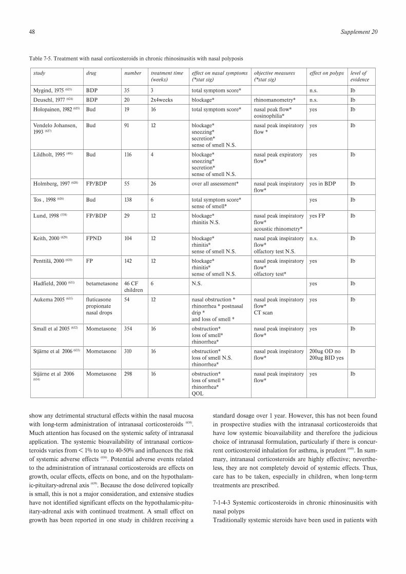
48 Supplement 20
show any detrimental structural effects within the nasal mucosawith long-term administration of intranasal corticosteroids (638).Much attention has focused on the systemic safety of intranasalapplication. The systemic bioavailability of intranasal corticos-teroids varies from < 1% to up to 40-50% and influences the riskof systemic adverse effects (636). Potential adverse events relatedto the administration of intranasal corticosteroids are effects ongrowth, ocular effects, effects on bone, and on the hypothalam-ic-pituitary-adrenal axis (639). Because the dose delivered topicallyis small, this is not a major consideration, and extensive studieshave not identified significant effects on the hypothalamic-pitu-itary-adrenal axis with continued treatment. A small effect ongrowth has been reported in one study in children receiving a
standard dosage over 1 year. However, this has not been foundin prospective studies with the intranasal corticosteroids thathave low systemic bioavailability and therefore the judiciouschoice of intranasal formulation, particularly if there is concur-rent corticosteroid inhalation for asthma, is prudent (640). In sum-mary, intranasal corticosteroids are highly effective; neverthe-less, they are not completely devoid of systemic effects. Thus,care has to be taken, especially in children, when long-termtreatments are prescribed.
7-1-4-3 Systemic corticosteroids in chronic rhinosinusitis withnasal polypsTraditionally systemic steroids have been used in patients with
Table 7-5. Treatment with nasal corticosteroids in chronic rhinosinusitis with nasal polyposis
study drug number treatment time (weeks)
effect on nasal symptoms(*stat sig)
objective measures(*stat sig)
effect on polyps level of evidence
Mygind, 1975 (623) BDP 35 3 total symptom score* n.s. Ib
Deuschl, 1977 (624) BDP 20 2x4weeks blockage* rhinomanometry* n.s. Ib
Holopainen, 1982 (625) Bud 19 16 total symptom score* nasal peak flow*eosinophilia*
yes Ib
Vendelo Johansen, 1993 (627)
Bud 91 12 blockage*sneezing*secretion*sense of smell N.S.
nasal peak inspiratoryflow *
yes Ib
Lildholt, 1995 (491) Bud 116 4 blockage*sneezing*secretion*sense of smell N.S.
nasal peak expiratoryflow*
yes Ib
Holmberg, 1997 (628) FP/BDP 55 26 over all assessment* nasal peak inspiratory flow*
yes in BDP Ib
Tos , 1998 (626) Bud 138 6 total symptom score*sense of smell*
yes Ib
Lund, 1998 (538) FP/BDP 29 12 blockage*rhinitis N.S.
nasal peak inspiratoryflow*acoustic rhinometry*
yes FP Ib
Keith, 2000 (629) FPND 104 12 blockage*rhinitis*sense of smell N.S.
nasal peak inspiratoryflow*olfactory test N.S.
n.s. Ib
Penttilä, 2000 (630) FP 142 12 blockage*rhinitis*sense of smell N.S.
nasal peak inspiratoryflow*olfactory test*
yes Ib
Hadfield, 2000 (631) betametasone 46 CF children
6 N.S. yes Ib
Aukema 2005 (635) fluticasonepropionatenasal drops
54 12 nasal obstruction * rhinorrhea * postnasaldrip *and loss of smell *
nasal peak inspiratoryflow*CT scan
yes Ib
Small et al 2005 (632) Mometasone 354 16 obstruction*loss of smell*rhinorrhea*
nasal peak inspiratoryflow*
yes Ib
Stjärne et al 2006 (633) Mometasone 310 16 obstruction*loss of smell N.S.rhinorrhea*
nasal peak inspiratoryflow*
200ug OD no200ug BID yes
Ib
Stjärne et al 2006(634)
Mometasone 298 16 obstruction*loss of smell *rhinorrhea*QOL
nasal peak inspiratoryflow*
yes Ib

European Position Paper on Rhinosinusitis and Nasal Polyps 2007 49
NP although no placebo-controlled studies or dose-effect stud-ies have supported the concept. The clinical acceptance thatsystemic steroids have a significant effect on NP are supportedby open studies where a single injection of 14 mg betameta-sone have been compared with snare polypectomy surgery (493,
641). In these studies effects are seen on nasal polyp size, nasalsymptom score and nasal expiratory peak flow but it is difficultto differentiate the effect of systemic steroids from that of topi-cal treatment since both treatments were used at the sametime. The control groups underwent surgery during the studyperiod.
In another open study oral prednisolone was given in doses of60 mg to 25 patients with severe polyposis for four days and foreach of the following 12 days the dose was reduced by 5 mgdaily. Antibiotics and antacids were also given. 72% experi-enced a clear improvement due to involution of polyps (642) andin 52% a clear improvement was seen on CT. In particularnasal obstruction and the sense of smell were reported toimprove. Out of 22 subjects treated, 10 were polyp free basedon anterior rhinoscopy 2 weeks – 2 months after therapy.
Damm et al. (643) showed a good effect with combined treat-ment using topical steroids (budesonide, unknown doses) andoral treatment with fluocortolone 560 mg or 715 mg in 2 differ-ent groups of patients with 20 severe cases of CRS with NP.This study was not controlled. A large improvement of symp-toms was seen (80%) and improvement on MRI (> 30% reduc-tion of MRT-pathology) was observed in 50%.
Recently, however, two well designed studies have showneffect of systemic steroids in NP. Benitez et al (644) performed arandomized placebo controlled study with prednisone for twoweeks (30 mg 4 days followed by a 2-day reduction of 5 mg).After two weeks on prednisone or placebo, the prednisonegroup continued for ten weeks on intranasal BUD. After twoweeks treatment a significant polyp reduction was seen, severalsymptoms improved and anterior rhinomanometry improvedcompared to the placebo group. After 12 weeks a significantreduction of CT-changes were seen in the steroid treatedgroup.
In a double-blind randomized, placebo controlled study,Hissaria et al compared 50 mg prednisolone daily for 14 dayswith placebo (576). A significant improvement was found in nasalsymptoms (obstruction, secretion, sneezing, sense of smell),endoscopic findings of polyp size and MRI scores supportingthe effect of systemic steroids on NP.
There is no study available on depot injection of corticos-teroids or local injection into polyps or the inferior turbinate.These types of treatment are actually obsolete, because of therisk of fat necrosis at the site of the injection or blindness fol-lowing endonasal injection.
Studies on systemic steroids in NP has recently been publishedgiving support to the clinical impression that they are effectiveafter two weeks use in doses acceptable for a majority ofpatients. As well as symptom relief, an effect on polyp size andMRI changes are seen. Evidence level:Ib.
7-1-4-4 Side effects of systemic corticosteroids in chronic rhi-nosinusitis with nasal polypsThe anti-inflammatory effects of corticosteroids cannot be sep-arated from their metabolic effects as all cells use the sameglucocorticoid receptor; therefore when corticosteroids are pre-scribed measures should be taken to minimize their sideeffects. Clearly, the chance of significant side effects increaseswith the dose and duration of treatment and so the minimumdose necessary to control the disease should be given.
As a guide for oral treatment, the approximate equivalentdoses of the main corticosteroids in terms of their glucocorti-coid (or anti-inflammatory) properties are listed below (645).
7-1-5 Postoperative treatment with topical corticosteriods forchronic rhinosinusitis with NP to prevent recurrence of polypsThere are a couple of studies on nasal steroids used after surgi-cal resection of polyps.Drettner et al (646) used flunisolide 200 µg daily for 3 months ina double-blind, placebo controlled study with 11 subjects inboth groups. A statistically significant effect was seen on nasalsymptoms but not on polyp score.
Table 7-6. Treatment with systemic corticosteroids in chronic rhinosinusitis with NP
study drug number dose/time effect symptoms effect polyps level of evidence
Lildholt, 1988 (641) betametamethasone/BDP 53 ?/52 weeks yes yes III
Lildholt, 1997 (493) betametamethasone/budesonide 16 14mg/52 weeks yes yes III
van Camp, 1994 (642) prednisolone 60 mg 25 2 weeks 72% yes (10/22) III
Lildholt, 1997 (493) betametamethasone/budesonide 16 14mg/52 weeks yes yes III
Damm, 1999 (643) budesonide + fluocortolone 20 ? yes ? III
Benitez , 2006 (644) prednisone + Budesonide 84 2 weeks/10 weeks yes yes Ib
Hissaria, 2006 (576) prednisolone 41 50mg/2 weeks yes yes Ib
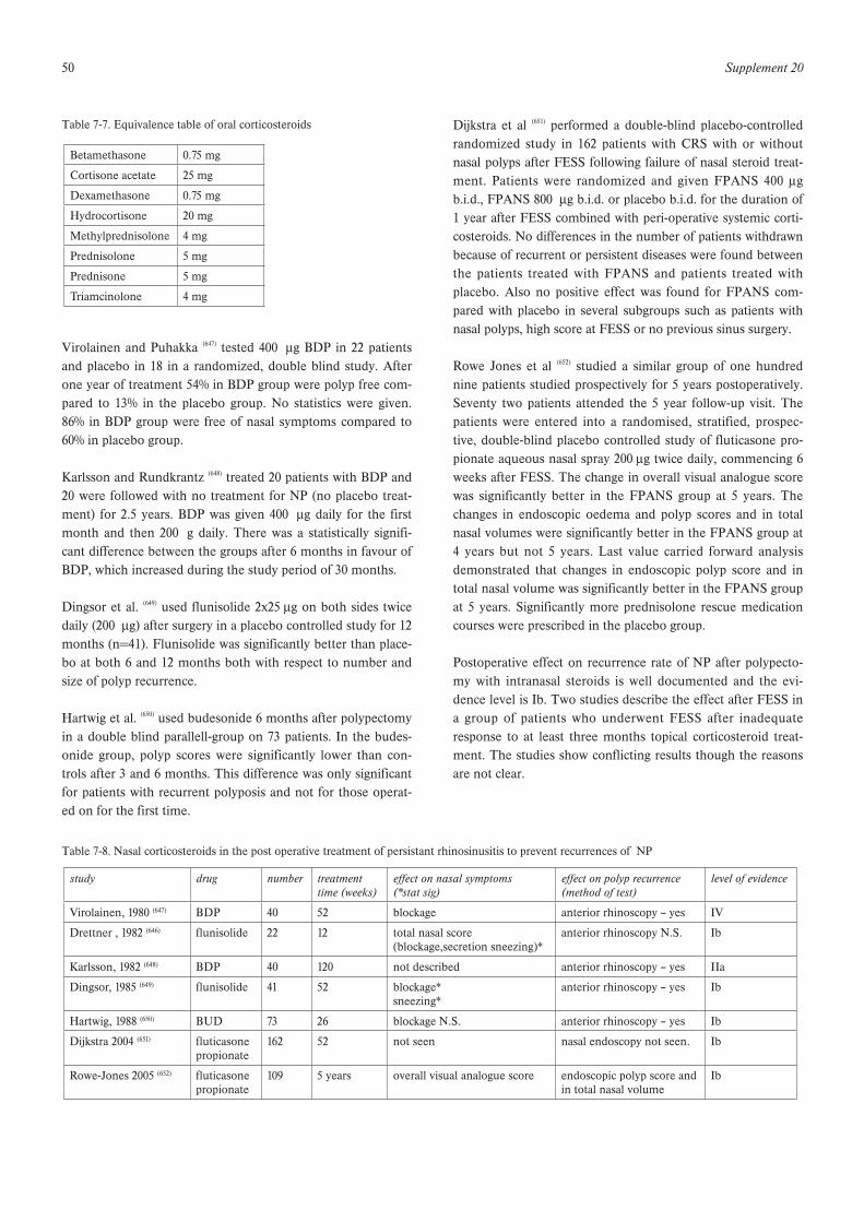
50 Supplement 20
Virolainen and Puhakka (647) tested 400 µg BDP in 22 patientsand placebo in 18 in a randomized, double blind study. Afterone year of treatment 54% in BDP group were polyp free com-pared to 13% in the placebo group. No statistics were given.86% in BDP group were free of nasal symptoms compared to60% in placebo group.
Karlsson and Rundkrantz (648) treated 20 patients with BDP and20 were followed with no treatment for NP (no placebo treat-ment) for 2.5 years. BDP was given 400 µg daily for the firstmonth and then 200 g daily. There was a statistically signifi-cant difference between the groups after 6 months in favour ofBDP, which increased during the study period of 30 months.
Dingsor et al. (649) used flunisolide 2x25 µg on both sides twicedaily (200 µg) after surgery in a placebo controlled study for 12months (n=41). Flunisolide was significantly better than place-bo at both 6 and 12 months both with respect to number andsize of polyp recurrence.
Hartwig et al. (650) used budesonide 6 months after polypectomyin a double blind parallell-group on 73 patients. In the budes-onide group, polyp scores were significantly lower than con-trols after 3 and 6 months. This difference was only significantfor patients with recurrent polyposis and not for those operat-ed on for the first time.
Dijkstra et al (651) performed a double-blind placebo-controlledrandomized study in 162 patients with CRS with or withoutnasal polyps after FESS following failure of nasal steroid treat-ment. Patients were randomized and given FPANS 400 µgb.i.d., FPANS 800 µg b.i.d. or placebo b.i.d. for the duration of1 year after FESS combined with peri-operative systemic corti-costeroids. No differences in the number of patients withdrawnbecause of recurrent or persistent diseases were found betweenthe patients treated with FPANS and patients treated withplacebo. Also no positive effect was found for FPANS com-pared with placebo in several subgroups such as patients withnasal polyps, high score at FESS or no previous sinus surgery.
Rowe Jones et al (652) studied a similar group of one hundrednine patients studied prospectively for 5 years postoperatively.Seventy two patients attended the 5 year follow-up visit. Thepatients were entered into a randomised, stratified, prospec-tive, double-blind placebo controlled study of fluticasone pro-pionate aqueous nasal spray 200 µg twice daily, commencing 6weeks after FESS. The change in overall visual analogue scorewas significantly better in the FPANS group at 5 years. Thechanges in endoscopic oedema and polyp scores and in totalnasal volumes were significantly better in the FPANS group at4 years but not 5 years. Last value carried forward analysisdemonstrated that changes in endoscopic polyp score and intotal nasal volume was significantly better in the FPANS groupat 5 years. Significantly more prednisolone rescue medicationcourses were prescribed in the placebo group.
Postoperative effect on recurrence rate of NP after polypecto-my with intranasal steroids is well documented and the evi-dence level is Ib. Two studies describe the effect after FESS ina group of patients who underwent FESS after inadequateresponse to at least three months topical corticosteroid treat-ment. The studies show conflicting results though the reasonsare not clear.
Table 7-7. Equivalence table of oral corticosteroids
Betamethasone 0.75 mg
Cortisone acetate 25 mg
Dexamethasone 0.75 mg
Hydrocortisone 20 mg
Methylprednisolone 4 mg
Prednisolone 5 mg
Prednisone 5 mg
Triamcinolone 4 mg
s
Table 7-8. Nasal corticosteroids in the post operative treatment of persistant rhinosinusitis to prevent recurrences of NP
study drug number treatment time (weeks)
effect on nasal symptoms(*stat sig)
effect on polyp recurrence(method of test)
level of evidence
Virolainen, 1980 (647) BDP 40 52 blockage anterior rhinoscopy – yes IV
Drettner , 1982 (646) flunisolide 22 12 total nasal score (blockage,secretion sneezing)*
anterior rhinoscopy N.S. Ib
Karlsson, 1982 (648) BDP 40 120 not described anterior rhinoscopy – yes IIa
Dingsor, 1985 (649) flunisolide 41 52 blockage* sneezing*
anterior rhinoscopy – yes Ib
Hartwig, 1988 (650) BUD 73 26 blockage N.S. anterior rhinoscopy – yes Ib
Dijkstra 2004 (651) fluticasonepropionate
162 52 not seen nasal endoscopy not seen. Ib
Rowe-Jones 2005 (652) fluticasonepropionate
109 5 years overall visual analogue score endoscopic polyp score and in total nasal volume
Ib

European Position Paper on Rhinosinusitis and Nasal Polyps 2007 51
7-1-6 Side-effects of corticosteroidsThe safety of nasal and oral corticosteroids has been the subjectof concern in medical literature since many patients with chronicsinus disease are prescribed these drugs due to their good effica-cy. Suppression of the hypothalamic-pituary-adrenal axis, osteo-porosis or changes in bone mineral density, growth retardation inchildren, cataracts and glaucoma have been reported to be themain adverse effects of corticosteroid treatment (653). In relation toadverse effects of corticosteroids, it is obvious that a clear distinc-tion needs to be made between nasal and oral corticosteroids.Nasal corticosteroid treatment represents one of the long-termtreatment modalities in patients with chronic sinus disease. Itis well established that absorption into the systemic circulationtakes place after nasal administration of corticosteroids.However, several factors influence the systemic absorption,like the molecular characteristics of the corticosteroid, the pre-scribed dose, the mode of delivery and the severity of theunderlying disease (653). There is insufficient evidence from theliterature to relate the use of nasal corticosteroids at licenseddoses to changes in bone mineral biology, cataract and glauco-ma. Adrenal suppression may occur with some nasal corticos-teroids at licensed doses, but the clinical relevance remainsuncertain. Overuse of nasal corticosteroids may be responsiblefor adrenal insufficiency and decrease in bone mineral density(654). Of note, inhaled corticosteroids are the mainstay of treat-ment for children and adults with asthma and are more oftenassociated with systemic side effects than the nasal route oftreatment for rhinosinusitis (655).Nasal corticosteroid-induced septal perforation is rarelydescribed in literature (656). Whether septal perforation relates torepeated traumas of the nasal mucosa and septal cartilage bythe nasal device, to the underlying nasal disorder for whichcorticosteroids were prescribed or to a direct adverse effect ofthe steroid used, remains unclear.Short treatment with oral corticosteroids is effective in chronicrhinosinusitis with nasal polyps. It is obvious that repeated orprolonged use of oral corticosteroids is associated with a signif-icantly enhanced risk of the above mentioned side effects (657).
7-2 Treatment of rhinosinusitis with antibiotics
7-2-1 Acute community acquired rhinosinusitisAlthough more than 2000 studies on the antibiotic treatmentof ARS have already been published, only 49, involving 13660participants, meet the Cochrane Board criteria for placebo con-trol, statistical analysis, sufficient sample sizes, and the descrip-tion of clinical improvements or success rates (40).Primary outcomes were:a. clinical cure;b. clinical cure or improvement.Secondary outcomes were:a. radiographic improvement;b. relapse rates;c. dropouts due to adverse effects.
Major comparisons were antibiotic versus control (n=3) (658-660);newer, non-penicillin antibiotic versus penicillin class (n=10);and amoxicillin-clavulanate versus other extended spectrumantibiotics (n=17), where n is the number of trials. Most trialswere conducted in otolaryngology settings. Only 8 trialsdescribed adequate allocation and concealment procedures; 20were double-blinded.
Compared to control, penicillin improved clinical cures [rela-tive risk (RR) 1.72; 95% confidence interval (CI) 1.00 to 2.96].For the outcome of cure or improvement, 77.2% of penicillin-treated participants and 61.5% of control participants wereresponders. Individuals treated with penicillin were more likelyto be cured [RR 1.72; 95% CI 1.00 to 2.96] or cured/improved[RR 1.24; 95% CI 1.00 to 1.53]. Rates for cure or improvementwere 82.3% for amoxicillin and 68.6% for placebo. Participantstreated with amoxicillin were not more likely to be cured thanwith placebo [RR 2.06; 95% CI 0.65 to 6.53] or cured/improved[RR 1.26; 95% CI 0.91 to 7.94] but there was significant variabil-ity between studies. Radiographic outcomes were improved byantibiotic treatment. (40).
Comparisons between newer non-penicillins (cephalosporins,macrolides, minocycline), versus penicillins (amoxicillin, peni-cillin V) showed no significant differences [RR for cure 1.07;95% CI 0.99 to 1.17]; Rates for cure or improvement were 84%for both antibiotic classes. Drop-outs due to adverse eventswere infrequent, and. these rates were not significantly differ-ent [RR 0.61; 95% CI 0.33 to 1.11]. Cumulative meta-analysis ofstudies ordered by year of publication (a proxy for prevalenceof beta-lactamase-producing organisms) did not show a trendtowards reduced efficacy of amoxicillin compared to newernon-penicillin antibiotics.
Because macrolides are bacteriostatic and cephalosporins bac-tericidal, subgroup analyses were performed to determine ifone of these two classes were superior to penicillins. In thesubgroup analyses, cephalosporins and macrolides showedsimilar response rates compared to penicillins.
Sixteen trials, involving 4818 participants, compared a newernon-penicillin antibiotic (macrolide or cephalosporin) to amox-icillin-clavulantate. Three studies were double-blind. Rates forcure or improvement were 72.7 % and 72.9 % for newer non-penicillins and amoxicillin-clavulanate respectively. Neithercure rates (RR 1.03; 95% CI 0.96 to 1.11) nor cured/improve-ment rates (RR 0.98; 95% CI 0.95 to 1.01), differed between thegroups. Compared to amoxicillin-clavulanate, dropouts due toadverse effects were significantly lower for cephalosporinantibiotics (RR 0.47; 95% CI 0.30 to 0.73). Relapse rates withinone month of successful therapy were 7.7% and did not differbetween the groups.
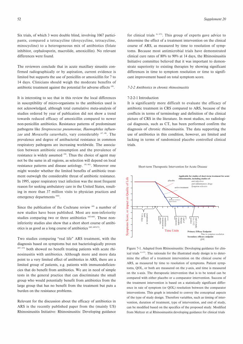
52 Supplement 20
Six trials, of which 3 were double blind, involving 1067 partici-pants, compared a tetracycline (doxycycline, tetracycline,minocycline) to a heterogeneous mix of antibiotics (folateinhibitor, cephalosporin, macrolide, amoxicillin). No relevantdifferences were found.
The reviewers conclude that in acute maxillary sinusitis con-firmed radiographically or by aspiration, current evidence islimited but supports the use of penicillin or amoxicillin for 7 to14 days. Clinicians should weigh the moderate benefits ofantibiotic treatment against the potential for adverse effects (40).
It is interesting to see that in this review the local differencesin susceptibility of micro-organisms to the antibiotics used isnot acknowledged, although total cumulative meta-analysis ofstudies ordered by year of publication did not show a trendtowards reduced efficacy of amoxicillin compared to newernon-penicillin antibiotics. Resistance patterns of predominantpathogens like Streptococcus pneumoniae, Haemophilus influen-zae and Moraxella catarrhalis, vary considerably (47, 48). Theprevalence and degree of antibacterial resistance in commonrespiratory pathogens are increasing worldwide. The associa-tion between antibiotic consumption and the prevalence ofresistance is widely assumed (50). Thus the choice of agent maynot be the same in all regions, as selection will depend on localresistance patterns and disease aetiology. (50, 661). Moreover onemight wonder whether the limited benefits of antibiotic treat-ment outweigh the considerable threat of antibiotic resistance.In 1995, upper respiratory tract infection was the most frequentreason for seeking ambulatory care in the United States, result-ing in more than 37 million visits to physician practices andemergency departments (662).
Since the publication of the Cochrane review (40) a number ofnew studies have been published. Most are non-inferioritystudies comparing two or three antibiotics (663-668). These non-inferiority studies also show that a short short course of antibi-otics is as good as a long course of antibiotics (665, 669-671).
Two studies comparing “real life” ARS treatment, with thediagnosis based on symptoms but not bacteriologically proven(607, 672) both showed no benefit treating patients with acute rhi-nosinusitis with antibiotics. Although more and more datapoint to a very limited effect of antibiotics in ARS, there are alimited group of patients, e.g. patients with immunodeficien-cies that do benefit from antibiotics. We are in need of simpletests in the general practice that can discriminate the smallgroup who would potentially benefit from antibiotics from thelarge group that has no benefit from the treatment but puts aburden on the resistance problems.
Relevant for the discussion about the efficacy of antibiotics inARS is the recently published paper from the (mainly US)Rhinosinusitis Initiative: Rhinosinusitis: Developing guidance
for clinical trials (6, 673). This group of experts gave advice todetermine the effect of a treatment intervention on the clinicalcourse of ARS, as measured by time to resolution of symp-toms. Because most antimicrobial trials have demonstratedclinical cure rates of 80% to 90% at 14 days, the RhinosinusitisInitiative committee believed that it was important to demon-strate superiority to existing therapies by showing significantdifferences in time to symptom resolution or time to signifi-cant improvement based on total symptom score.
7-2-2 Antibiotics in chronic rhinosinusitis
7-2-2-1 IntroductionIt is significantly more difficult to evaluate the efficacy ofantibiotic treatment in CRS compared to ARS, because of theconflicts in terms of terminology and definition of the clinicalpicture of CRS in the literature. In most studies, no radiologi-cal diagnosis, such as CT, has been performed confirm thediagnosis of chronic rhinosinusitis. The data supporting theuse of antibiotics in this condition, however, are limited andlacking in terms of randomized placebo controlled clinicaltrials.
Short-term Therapeutic Intervention for Acute Disease
Applicable for studies of short-term treatment for acuterhinosinusitis, including studies of: anti-infectives anti-inflammatory drugs Symptom relievers
placebo
active treatment
difference in time toimprovement
Primary Efficay Endpoint Time to sympton resolutionSecondary efficary endpoint QOL
Sym
ptom
sSc
ore,
orim
pact
onQ
ualit
yof
Lif
e
symptom
sat
least7-10
days
randomize
treatementor
placebo
endof
therapy(variableduration)
endofstudy
(i.e.3-12w
eeks)
Time Units
Figure 7-1. Adapted from Rhinosinusitis: Developing guidance for clin-ical trials (6, 673). The rationale for the illustrated study design is to deter-mine the effect of a treatment intervention on the clinical course ofARS, as measured by time to resolution of symptoms. Patient symp-toms, QOL, or both are measured on the y-axis, and time is measuredon the x-axis. The therapeutic intervention that is to be tested can becompared with either placebo or a comparator intervention. Success ofthe treatment intervention is based on a statistically significant differ-ence in rate of symptom (or QOL) resolution between the comparatorinterventions. This graph is intended to convey the conceptual aspectsof the type of study design. Therefore variables, such as timing of inter-vention, duration of treatment, type of intervention, and end of study,can be modified based on the specifics of the proposed study. Modifiedfrom Meltzer et al Rhinosinusitis:developing guidance for clinical trials
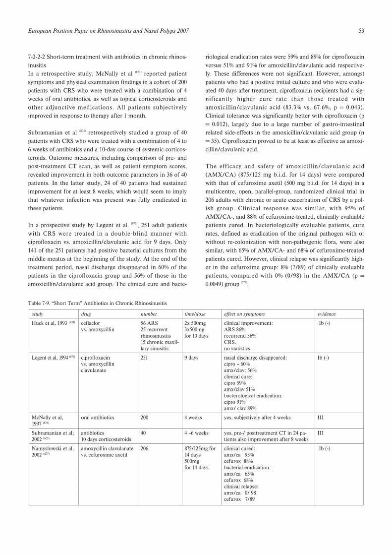
European Position Paper on Rhinosinusitis and Nasal Polyps 2007 53
7-2-2-2 Short-term treatment with antibiotics in chronic rhinos-inusitisIn a retrospective study, McNally et al (674) reported patientsymptoms and physical examination findings in a cohort of 200patients with CRS who were treated with a combination of 4weeks of oral antibiotics, as well as topical corticosteroids andother adjunctive medications. All patients subjectivelyimproved in response to therapy after 1 month.
Subramanian et al (675) retrospectively studied a group of 40patients with CRS who were treated with a combination of 4 to6 weeks of antibiotics and a 10-day course of systemic corticos-teroids. Outcome measures, including comparison of pre- andpost-treatment CT scan, as well as patient symptom scores,revealed improvement in both outcome parameters in 36 of 40patients. In the latter study, 24 of 40 patients had sustainedimprovement for at least 8 weeks, which would seem to implythat whatever infection was present was fully eradicated inthese patients.
In a prospective study by Legent et al. (676), 251 adult patientswith CRS were treated in a double-blind manner withciprofloxacin vs. amoxicillin/clavulanic acid for 9 days. Only141 of the 251 patients had positive bacterial cultures from themiddle meatus at the beginning of the study. At the end of thetreatment period, nasal discharge disappeared in 60% of thepatients in the ciprofloxacin group and 56% of those in theamoxicillin/clavulanic acid group. The clinical cure and bacte-
riological eradication rates were 59% and 89% for ciprofloxacinversus 51% and 91% for amoxicillin/clavulanic acid respective-ly. These differences were not significant. However, amongstpatients who had a positive initial culture and who were evalu-ated 40 days after treatment, ciprofloxacin recipients had a sig-nificantly higher cure rate than those treated withamoxicillin/clavulanic acid (83.3% vs. 67.6%, p = 0.043).Clinical tolerance was significantly better with ciprofloxacin (p= 0.012), largely due to a large number of gastro-intestinalrelated side-effects in the amoxicillin/clavulanic acid group (n= 35). Ciprofloxacin proved to be at least as effective as amoxi-cillin/clavulanic acid.
The efficacy and safety of amoxicillin/clavulanic acid(AMX/CA) (875/125 mg b.i.d. for 14 days) were comparedwith that of cefuroxime axetil (500 mg b.i.d. for 14 days) in amulticentre, open, parallel-group, randomized clinical trial in206 adults with chronic or acute exacerbation of CRS by a pol-ish group. Clinical response was similar, with 95% ofAMX/CA-, and 88% of cefuroxime-treated, clinically evaluablepatients cured. In bacteriologically evaluable patients, curerates, defined as eradication of the original pathogen with orwithout re-colonization with non-pathogenic flora, were alsosimilar, with 65% of AMX/CA- and 68% of cefuroxime-treatedpatients cured. However, clinical relapse was significantly high-er in the cefuroxime group: 8% (7/89) of clinically evaluablepatients, compared with 0% (0/98) in the AMX/CA (p =0.0049) group (677).
Table 7-9. “Short Term” Antibiotics in Chronic Rhinosinusitis
study drug number time/dose effect on symptoms evidence
Huck et al, 1993 (678) ceflaclorvs. amoxycillin
56 ARS 25 recurrentrhinosinusitis15 chronic maxil-lary sinusitis
2x 500mg3x500mgfor 10 days
clinical improvement:ARS 86% recurrend 56%CRS. no statistics
Ib (-)
Legent et al, 1994 (676) ciprofloxacinvs. amoxycillin clavulanate
251 9 days nasal discharge disappeared: cipro – 60%amx/clav: 56%clinical cure:cipro 59%amx/clav 51%bacterological eradication:cipro 91%amx/ clav 89%
Ib (-)
McNally et al, 1997 (674)
oral antibiotics 200 4 weeks yes, subjectively after 4 weeks III
Subramanian et al;2002 (675)
antibiotics10 days corticosteroids
40 4 –6 weeks yes, pre-/ posttreatment CT in 24 pa-tients also improvement after 8 weeks
III
Namyslowski et al, 2002 (677)
amoxycillin clavulanatevs. cefuroxime axetil
206 875/125mg for 14 days500mgfor 14 days
clinical cured:amx/ca 95%cefurox 88%bacterial eradication:amx/ca 65%cefurox 68%clinical relapse:amx/ca 0/ 98cefurox 7/89
Ib (-)
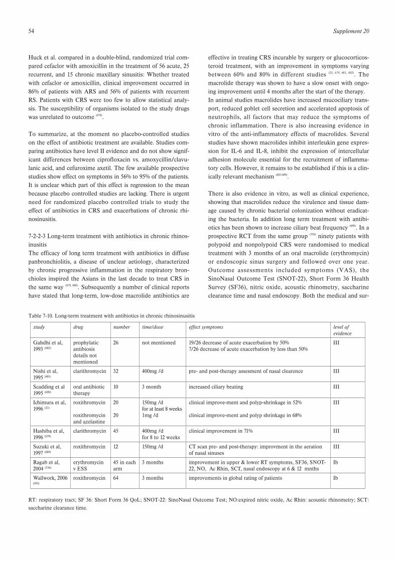
54 Supplement 20
Huck et al. compared in a double-blind, randomized trial com-pared cefaclor with amoxicillin in the treatment of 56 acute, 25recurrent, and 15 chronic maxillary sinusitis: Whether treatedwith cefaclor or amoxicillin, clinical improvement occurred in86% of patients with ARS and 56% of patients with recurrentRS. Patients with CRS were too few to allow statistical analy-sis. The susceptibility of organisms isolated to the study drugswas unrelated to outcome (678).
To summarize, at the moment no placebo-controlled studieson the effect of antibiotic treatment are available. Studies com-paring antibiotics have level II evidence and do not show signif-icant differences between ciprofloxacin vs. amoxycillin/clavu-lanic acid, and cefuroxime axetil. The few available prospectivestudies show effect on symptoms in 56% to 95% of the patients.It is unclear which part of this effect is regression to the meanbecause placebo controlled studies are lacking. There is urgentneed for randomized placebo controlled trials to study theeffect of antibiotics in CRS and exacerbations of chronic rhi-nosinusitis.
7-2-2-3 Long-term treatment with antibiotics in chronic rhinos-inusitisThe efficacy of long term treatment with antibiotics in diffusepanbronchiolitis, a disease of unclear aetiology, characterizedby chronic progressive inflammation in the respiratory bron-chioles inspired the Asians in the last decade to treat CRS inthe same way (679, 680). Subsequently a number of clinical reportshave stated that long-term, low-dose macrolide antibiotics are
effective in treating CRS incurable by surgery or glucocorticos-teroid treatment, with an improvement in symptoms varyingbetween 60% and 80% in different studies (23, 679, 681, 682). Themacrolide therapy was shown to have a slow onset with ongo-ing improvement until 4 months after the start of the therapy.In animal studies macrolides have increased mucociliary trans-port, reduced goblet cell secretion and accelerated apoptosis ofneutrophils, all factors that may reduce the symptoms ofchronic inflammation. There is also increasing evidence invitro of the anti-inflammatory effects of macrolides. Severalstudies have shown macrolides inhibit interleukin gene expres-sion for IL-6 and IL-8, inhibit the expression of intercellularadhesion molecule essential for the recruitment of inflamma-tory cells. However, it remains to be established if this is a clin-ically relevant mechanism (683-689).
There is also evidence in vitro, as well as clinical experience,showing that macrolides reduce the virulence and tissue dam-age caused by chronic bacterial colonization without eradicat-ing the bacteria. In addition long term treatment with antibi-otics has been shown to increase ciliary beat frequency (690). In aprospective RCT from the same group (536) ninety patients withpolypoid and nonpolypoid CRS were randomised to medicaltreatment with 3 months of an oral macrolide (erythromycin)or endoscopic sinus surgery and followed over one year.Outcome assessments included symptoms (VAS), theSinoNasal Outcome Test (SNOT-22), Short Form 36 HealthSurvey (SF36), nitric oxide, acoustic rhinometry, saccharineclearance time and nasal endoscopy. Both the medical and sur-
Table 7-10. Long-term treatment with antibiotics in chronic rhinosinusitis
RT: respiratory tract; SF 36: Short Form 36 QoL; SNOT-22: SinoNasal Outcome Test; NO:expired nitric oxide, Ac Rhin: acoustic rhinometry; SCT:saccharine clearance time.
study drug number time/dose effect symptoms level of evidence
Gahdhi et al, 1993 (682)
prophylaticantibiosisdetails notmentioned
26 not mentioned 19/26 decrease of acute exacerbation by 50%7/26 decrease of acute exacerbation by less than 50%
III
Nishi et al, 1995 (681)
clarithromycin 32 400mg /d pre- and post-therapy assesment of nasal clearence III
Scadding et al1995 (690)
oral antibiotictherapy
10 3 month increased ciliary beating III
Ichimura et al, 1996 (23)
roxithromycin
roxithromycinand azelastine
20
20
150mg /dfor at least 8 weeks1mg /d
clinical improve-ment and polyp-shrinkage in 52%
clinical improve-ment and polyp shrinkage in 68%
III
Hashiba et al, 1996 (679)
clarithromycin 45 400mg /dfor 8 to 12 weeks
clinical improvement in 71% III
Suzuki et al, 1997 (680)
roxithromycin 12 150mg /d CT scan pre- and post-therapy: improvment in the aeration of nasal sinuses
III
Ragab et al,2004 (536)
erythromycinv ESS
45 in each arm
3 months improvement in upper & lower RT symptoms, SF36, SNOT-22, NO, Ac Rhin, SCT, nasal endoscopy at 6 & 12 mnths
Ib
Wallwork, 2006(691)
roxithromycin 64 3 months improvements in global rating of patients Ib
European Position Paper on Rhinosinusitis and Nasal Polyps 2007 55
gical treatment of CRS significantly improved almost all sub-jective and objective parameters, with no significant differencebetween the two groups nor between polypoid and nonpoly-poid CRS except for total nasal volume which was greater aftersurgery and in the polypoid patients.
Wallwork et al (691) conducted a double-blind, randomized,placebo-controlled clinical trial on 64 patients with CRS.Subjects received either 150 mg roxithromycin daily for 3months or placebo. The description of the patient populationsis limited, but patients with NP were excluded (personal com-munication by author). They showed a significant improve-ment in global patient rating compared to placebo. The othercomparisons were made between pre- and post-treatment situ-ations. In this comparison a statistically significant improve-ment was found in SNOT-20 score, nasal endoscopy, saccha-rine transit time, and IL-8 levels in lavage fluid (p < 0,05) inthe macrolide group. A correlation was noted betweenimproved outcome measures and low IgE levels.
The benefit of long-term, low-dose macrolide treatment seemsto be that it is, in selected cases, effective when topical steroidsand short courses of antibiotics fail. The exact mechanism ofaction is not known, but it probably involves downregulationof the local host immune response as well as a downgrading ofthe virulence of the colonizing bacteria. Placebo-controlledstudies should be performed to establish the efficacy ofmacrolides if this treatment is to be accepted as evidence-based medicine.
7-2-3 Exacerbations of chronic rhinosinusitis
7-2-3-1 Short-term treatment with oral antibiotics in acuteexacerbations of chronic rhinosinusitis chronic rhinosinusitisIn open trials, oral antibiotics have an effect on the symptoma-tology of acute exacerbations of CRS (677, 692). In some of thesestudies patients with ARS or CRS are combined with patientswith acute exacerbations of CRS (693, 694). No studies have shownefficacy of antibiotics in acute exacerbations of CRS in a dou-ble blind placebo controlled manner.
In conclusion data on the treatment of acute exacerbation ofCRS are mostly level IV evidence and include oral and localantibiotics. Double-blind data show a positive effect of theaddition of topical corticosteroid treatment to oral antibioticsin the treatment of acute exacerbation of CRS.
7-2-3-2 Short-term treatment with local antibiotics in acuteexacerbations of chronic rhinosinusitisSome studies have compared the effects of local antibiotics inCRS and acute exacerbation of CRS (620, 695-697).
Desrosiers studied in a randomized, double-blind trial oftobramycin-saline solution versus saline-only solution adminis-
tered thrice daily to the nasal passages by means of a large-par-ticle nebulizer apparatus for 4 weeks in twenty patients withCRS refractory to medical and surgical therapy. He found nosignificant difference between the groups and concluded thatlarge-particle nebulized aerosol therapy may offer a safe andeffective management alternative for patients with refractoryrhinosinusitis irrespective of the addition of gentamicin (698).
Sykes found no additional effect with the addition of neomycinto a spray containing dexamethasone and tramazoline fourtimes daily to both nostrils for 2 weeks (621).
However, Mosges and Leonard did find differences betweenlocal antibiotics and placebo (695, 697). Mosges showed a positiveeffect for fusafungine nasal spray as early as the first 24h oftreatment which was not seen in the placebo group. Theantimicrobiological effect of this preparation is unclear.
Schienberg et al. studied the effectiveness of aerosol deliveryof antibiotics to the sinuses via a nebulizer in 41 patients whohad chronic, recurrent rhinosinusitis that had persisted despiteendoscopic sinus surgery (ESS) and that had not responded tomultiple courses of oral antibiotics. Following 3 to 6 weeks oftreatment, 34 patients (82.9%) experienced either an excellentor good response to treatment. Side effects were infrequent,mild, and transient. They concluded that nebulized antibioticsshould be considered for all patients with CRS who haveundergone ESS and who have failed to respond to oral antibi-otics or who do not tolerate them (699).
Further studies with better characterized patient populationsare needed.
7-2-4 Side-effects of antibioticsCommon side effects of antibiotics include sickness, diarrhoea,and, in women, vaginal yeast infections. Some side effects aremore severe and, depending on the antibiotic, may influencekidney and liver function. Rarely the bone-marrow or otherorgans are affected. Blood tests are used to monitor suchadverse reactions.Some people who receive antibiotics develop colitis, an inflam-mation of the large intestine. The colitis results from a toxinproduced by the bacterium, Clostridium difficile, which growsunchecked when other antibacteria are killed by the antibi-otics.Antibiotics can cause allergic reactions. Most are mild allergicreactions and consist of an itchy rash or slight wheezing.Patients claiming allergic reactions to antibiotics actually meanside-effects. This is important to realize because on one handsevere allergic reactions (anaphylaxis) can be life threatening,on the other hand some patients claim to be allergic to manydifferent classes of antibiotics making treatment difficult. Mostadverse events related to antimicrobials are reversible rapidlyon cessation of the medication. Irreversible toxicities include
56 Supplement 20
aminoglycoside-induced ototoxicity, Stevens-Johnson syn-drome, and toxicity secondary to nitrofurantoin.Another important consequence of the use of antibiotics is thedevelopment of resistance. Resistance to antibiotics is a majorpublic-health problem and antibiotic use is being increasinglyrecognised as the main selective pressure driving this resis-tance. Prescription of antibiotics in Europe varies greatly: thehighest rate was in France and the lowest was in theNetherlands (700). A shift from the old narrow-spectrum antibi-otics to the new broad-spectrum antibiotics is being seen.Higher rates of antibiotic resistance are found in high consum-ing countries, probably related to this higher consumption.
7-3 Other medical management for rhinosinusitis
Standard conservative treatment for ARS and CRS is based onshort or long-term antibiotics and topical steroids with theaddition of decongestants - mostly in a short term regimen andfor the acute attack itself. Many other types of preparationshave been investigated, but substantial evidence for their bene-fit is poor. These medications include antral washings, isoton-ic/hypertonic saline as nasal douche, antihistamines, antimy-cotics, mucolytic agents/phytomedical preparations,immunomodulators/immunostimulants and bacterial lysatepreparations. For selected patients with CRS and gastroe-sophageal reflux, the impact of antireflux treatment on sinussymptom scores has been studied. Topical nasal application offurosemide and capsaicin have also been considered in thetreatment of nasal polyposis and prevention of recurrence.
7-3-1 Decongestants
7-3-1-1 Acute rhinosinusitisNasal decongestants are applied in the treatment of ARS inorder to decrease congestion and in the hope of improving bet-ter sinus ventilation and drainage and symptomatic relief ofnasal congestion. Experimental trials on the effect of topicaldecongestants by CT (701) and MRI scans (702) on ostial andostiomeatal complex patency have confirmed marked effect oncongestion of inferior and middle turbinates and infundibularmucosa, but no effect on ethmoidal and maxillary sinusmucosa. Experimental studies suggested beneficial anti-inflam-matory effect of xylometazoline and oxymetazoline bydecreasing nitric oxide synthetase (703) and anti-oxidant action(704). In contrast to previous in vitro trials on the effect of decon-gestants on mucociliary transport, a controlled clinical trial (II)by Inanli et al. suggested improvement in mucociliary clear-ance in vivo, after 2 weeks of oxymetazoline application inacute bacterial rhinosinusitis, compared to fluticasone, hyper-tonic saline and saline, but it did not show significant improve-ment compared to the group where no topical nasal treatmentwas given, and the clinical course of the disease between thegroups was not significantly different.(705). This is in concor-dance with previous randomized controlled trial in adult acute
maxillary sinusitis (Ib), which did not prove significant impactof decongestant when added to antibiotic treatment in terms ofdaily symptoms scores on headache and obstruction and sinusx-ray scores, although decongestant and placebo were appliedthrough a bellow, which should have enabled better dispersionof the solution in the nasal cavity (706). Decongestant treatmentdid not prove superior to saline, when added to antibiotic andantihistamine treatment in a randomized double-blind place-bo-controlled trial for acute paediatric rhinosinusitis (Ib) (707).However, a double blind, randomized, placebo controlled trialdemonstrated a significant protective effect of 14-day nasaldecongestant (combined with topical budesonide after 7 days)in the prevention of the development of nosocomial maxillarysinusitis in mechanically ventilated patients in the intensivecare unit (708). Radiologically confirmed maxillary sinusitis wasobserved in 54% of patients in the active treatment group andin the 82% of the controls, respectively, while infective maxil-lary sinusitis was obseved in 8% and 20%, respectively (708).Clinical experience, however, supports the use of topical appli-cation of decongestants to the middle meatus in ARS but notby spray or drops (evidence level IV).
7-3-1-2 Chronic rhinosinusitis without nasal polypsThe use of decongestants for adult CRS has not been evaluatedin a randomized controlled trial. Decongestants and sinusdrainge did not prove to be superior to saline in chronic paedi-atric maxillary sinusitis in terms of subjective or x-ray scores (709).
7-3-1-3 Chronic rhinosinusitis with nasal polypsCT studies before and after decongestant application inpatients with nasal polyposis did not show any densitometricchanges in the sinuses or polyps, only decongestion of theinferior turbinates (710). A randomized double blind placebocontrolled trial did not show any difference between placebo,epinephrine and naphazoline on polyp size at endoscopy andlateral imaging (711).
7-3-1-4 Side effects of decongestantsThe most frequent adverse event related to topical nasaldecongestants is rebound nasal congestion in patients withprolonged treatment or overuse of vasoconstrictive topicalmedications. The effect occurs due to tachyphylaxis after 5 to 7days of medication use. Shorter duration of decongestion andrebound effect results in increased daily dose and may lead torhinitis medicamentosa (712). Significantly greater nasal reactivi-ty, compared to placebo, was demonstrated after few weeks ofnasal decongestant.Major adverse effects are more related to systemic deconges-tants, ranging in severity from tremor and headache to individ-ual reports of stroke, myocardial infarction, chest pain,seizures, insomnia, nausea and vomiting, fatigue, and dizzi-ness. There are even some case reports reporting on similarside effects of topical decongestants, especially in patients withincreased cardiovascular risks (713-716).
European Position Paper on Rhinosinusitis and Nasal Polyps 2007 57
7-3-2 Mucolytics
7-3-2-1 Acute rhinosinusitisMucolytics were used as adjuncts to antibiotic treatment anddecongestant treatment in ARS in order to reduce the viscosityof sinus secretion. The benefit of such treatment has not beenevaluated in many trials. In paediatric rhinosinusitis, a RCT(Ib) did not prove bromhexine superior to saline in inhalationfor children with CRS (717). A second RCT (Ib) suggestedbromhexine was superior to placebo (718).
7-3-2-2 Chronic rhinosinusitisA cohort study in a mixed group of 45 ARS and CRS patientssuggested beneficial effect of adding mucolytic to standard rhi-nosinusitis treatment in terms of reducing treatment duration(719)(evidence level III).
7-3-2-3 Nasal polypsNo clinical trials have tested the effect of mucolytics in nasalpolyp treatment.
7-3-3 Antihistamines, cromones
7-3-3-1 Acute rhinosinusitisThe beneficial effect of loratadine in terms of symptom reduc-tion for the treatment of ARS in patients with allergic rhinitiswas confirmed in a multicentre randomized double-blind, place-bo controlled trial (Ib)(720). Patients receiving loratadine as anadjunct to antibiotic treatment suffered significantly less sneez-ing and obstruction on daily VAS scores, and overall improve-ment was confirmed by their physicians. Cromolyn did notprove better than saline in a RCT (Ib) for treatment of acutehyperreactive sinusitis measured by subjective scores and ultra-sound scans, leading to 50% improvement in both groups (721). ARCT (Ib) for acute paediatric rhinosinusitis did not confirm anybenefit of oral antihistamine-nasal decongestant drops (707).
7-3-3-2 Chronic rhinosinusitisAlthough generally not recommended as rhinosinusitis treat-ment, an evaluation study of CRS treatment in the USArevealed antihistamines as rather often presribed medication inpatients with CRS (an average of 2.7 antibiotic courses; nasalsteroids and prescription antihistamines 18.3 and 16.3 weeks,respectively, in a 12-month period) (722). However, no evidenceof beneficial effects of antihistamine treatment for CRS isfound, as there are no controlled trials evaluating such treat-ment.
7-3-3-3 Nasal polypsCetirizine in a dose of 20 mg/day for three months, significant-ly reduced sneezing, rhinorrhoea and obstruction compared toplacebo in the postoperative treatment of recurrent polyposisbut with no effect on polyp size (Ib) (723).
7-3-3-4 AntihistaminesUnlike first generation antihistamines, where central nervoussystem and peripheric muscarinic side effects are significant,frequency of side effects in newer second generation antihista-mines is low. The most commonly reported events duringtreatment with second generation antihistamines were upperrespiratory tract infection, wheezing, vulvitis, cough, headache,migraine, drowsiness, sedation and injury, most of themreported in 1 to 5% of the treated population, however, notnecessarily related to medication. Although caution with car-diotoxicity and potential for interaction with drugs metabolisedby the hepatic cytochrome P450 system, applied to older non-sedating antihistamines (like terfenadine or astemizole), thisrisk seems to be absent in newer compounds (desloratadine,levocetirizine, fexofenadine), at least in recommended treat-ment regimens.
7-3-4 AntimycoticsAntimycotics are used as topical and systemic treatment, as anadjunct to sinus surgery, in allergic fungal, and invasive fungalrhinosinusitis, especially in immunocompromized patients (724).Surgery is considered the first line treatment for allergic fungal(725) and invasive fungal rhinosinusitis (726). Although the use ofantimycotics in the treatment of allergic fungal rhinosinusitishas not been tested in controlled trials, high dose postopera-tive itraconazole, combined with oral and topical steroids in acohort of 139 patiens with AFS reduced the need for revisionsurgery rate to 20.5% (727). The state-of-art treatment for invasivefungal sinusitis is based on small series of patients and casereports, which do not meet the criteria for meta-analysis andmay be considered as level IV evidence.
7-3-4-1 Acute rhinosinusitisNo controlled trials for antimycotic treatment for ARS wasfound on the Medline search.
7-3-4-2 Chronic rhinosinusitisThe fungal hypothesis, based of the premise of an altered localimmune (non-allergic) response to fungal presence innasal/sinus secretions resulting in the generation of chroniceosinophilic rhinosinusitis and nasal polyposis (148), has led toidea of treating CRS with and without NPs with a topicalantimycotic. Although the presence of fungus in sinus secre-tions was detected in a high proportion (< 90%) of patientswith CRS, as well as in a control disease-free population in afew study centres (148, 149), it cannot be taken as proof of aetiolo-gy. Until recently a few case studies (level IV) had been con-ducted (728, 729). Ponikau et al, in a group of 51 patients with CRS,including polyposis patients, treated patients with topicalamphotericin B as nasal/sinus washing, without placebo orother control treatment. The treatment resulted in 75% subjec-tive improvement and 74% endoscopic improvement (728). Asthe authors stated, antifungal treatment should be evaluated ina controlled trial to be justified.
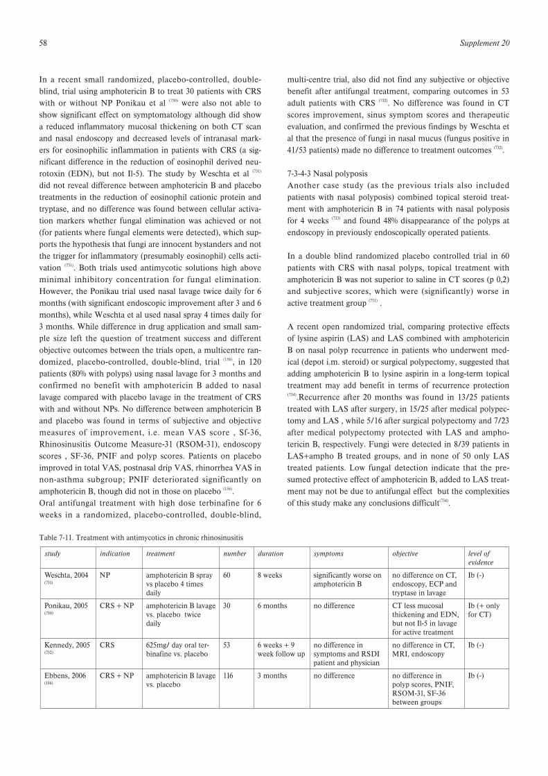
58 Supplement 20
In a recent small randomized, placebo-controlled, double-blind, trial using amphotericin B to treat 30 patients with CRSwith or without NP Ponikau et al (730) were also not able toshow significant effect on symptomatology although did showa reduced inflammatory mucosal thickening on both CT scanand nasal endoscopy and decreased levels of intranasal mark-ers for eosinophilic inflammation in patients with CRS (a sig-nificant difference in the reduction of eosinophil derived neu-rotoxin (EDN), but not Il-5). The study by Weschta et al (731)
did not reveal difference between amphotericin B and placebotreatments in the reduction of eosinophil cationic protein andtryptase, and no difference was found between cellular activa-tion markers whether fungal elimination was achieved or not(for patients where fungal elements were detected), which sup-ports the hypothesis that fungi are innocent bystanders and notthe trigger for inflammatory (presumably eosinophil) cells acti-vation (731). Both trials used antimycotic solutions high aboveminimal inhibitory concentration for fungal elimination.However, the Ponikau trial used nasal lavage twice daily for 6months (with significant endoscopic improvement after 3 and 6months), while Weschta et al used nasal spray 4 times daily for3 months. While difference in drug application and small sam-ple size left the question of treatment success and differentobjective outcomes between the trials open, a multicentre ran-domized, placebo-controlled, double-blind, trial (156), in 120patients (80% with polyps) using nasal lavage for 3 months andconfirmed no benefit with amphotericin B added to nasallavage compared with placebo lavage in the treatment of CRSwith and without NPs. No difference between amphotericin Band placebo was found in terms of subjective and objectivemeasures of improvement, i.e. mean VAS score , Sf-36,Rhinosinusitis Outcome Measure-31 (RSOM-31), endoscopyscores , SF-36, PNIF and polyp scores. Patients on placeboimproved in total VAS, postnasal drip VAS, rhinorrhea VAS innon-asthma subgroup; PNIF deteriorated significantly onamphotericin B, though did not in those on placebo (156).Oral antifungal treatment with high dose terbinafine for 6weeks in a randomized, placebo-controlled, double-blind,
multi-centre trial, also did not find any subjective or objectivebenefit after antifungal treatment, comparing outcomes in 53adult patients with CRS (732). No difference was found in CTscores improvement, sinus symptom scores and therapeuticevaluation, and confirmed the previous findings by Weschta etal that the presence of fungi in nasal mucus (fungus positive in41/53 patients) made no difference to treatment outcomes (732).
7-3-4-3 Nasal polyposisAnother case study (as the previous trials also includedpatients with nasal polyposis) combined topical steroid treat-ment with amphotericin B in 74 patients with nasal polyposisfor 4 weeks (733) and found 48% disappearance of the polyps atendoscopy in previously endoscopically operated patients.
In a double blind randomized placebo controlled trial in 60patients with CRS with nasal polyps, topical treatment withamphotericin B was not superior to saline in CT scores (p 0,2)and subjective scores, which were (significantly) worse inactive treatment group (731) .
A recent open randomized trial, comparing protective effectsof lysine aspirin (LAS) and LAS combined with amphotericinB on nasal polyp recurrence in patients who underwent med-ical (depot i.m. steroid) or surgical polypectomy, suggested thatadding amphotericin B to lysine aspirin in a long-term topicaltreatment may add benefit in terms of recurrence protection(734).Recurrence after 20 months was found in 13/25 patientstreated with LAS after surgery, in 15/25 after medical polypec-tomy and LAS , while 5/16 after surgical polypectomy and 7/23after medical polypectomy protected with LAS and ampho-tericin B, respectively. Fungi were detected in 8/39 patients inLAS+ampho B treated groups, and in none of 50 only LAStreated patients. Low fungal detection indicate that the pre-sumed protective effect of amphotericin B, added to LAS treat-ment may not be due to antifungal effect but the complexitiesof this study make any conclusions difficult(734).
Table 7-11. Treatment with antimycotics in chronic rhinosinusitis
study indication treatment number duration symptoms objective level of evidence
Weschta, 2004 (731)
NP amphotericin B spray vs placebo 4 timesdaily
60 8 weeks significantly worse on amphotericin B
no difference on CT, endoscopy, ECP and tryptase in lavage
Ib (-)
Ponikau, 2005 (730)
CRS + NP amphotericin B lavage vs. placebo twice daily
30 6 months no difference CT less mucosal thickening and EDN, but not Il-5 in lavagefor active treatment
Ib (+ only for CT)
Kennedy, 2005 (732)
CRS 625mg/ day oral ter-binafine vs. placebo
53 6 weeks + 9 week follow up
no difference insymptoms and RSDI patient and physician
no difference in CT, MRI, endoscopy
Ib (-)
Ebbens, 2006 (156)
CRS + NP amphotericin B lavage vs. placebo
116 3 months no difference no difference in polyp scores, PNIF, RSOM-31, SF-36 between groups
Ib (-)
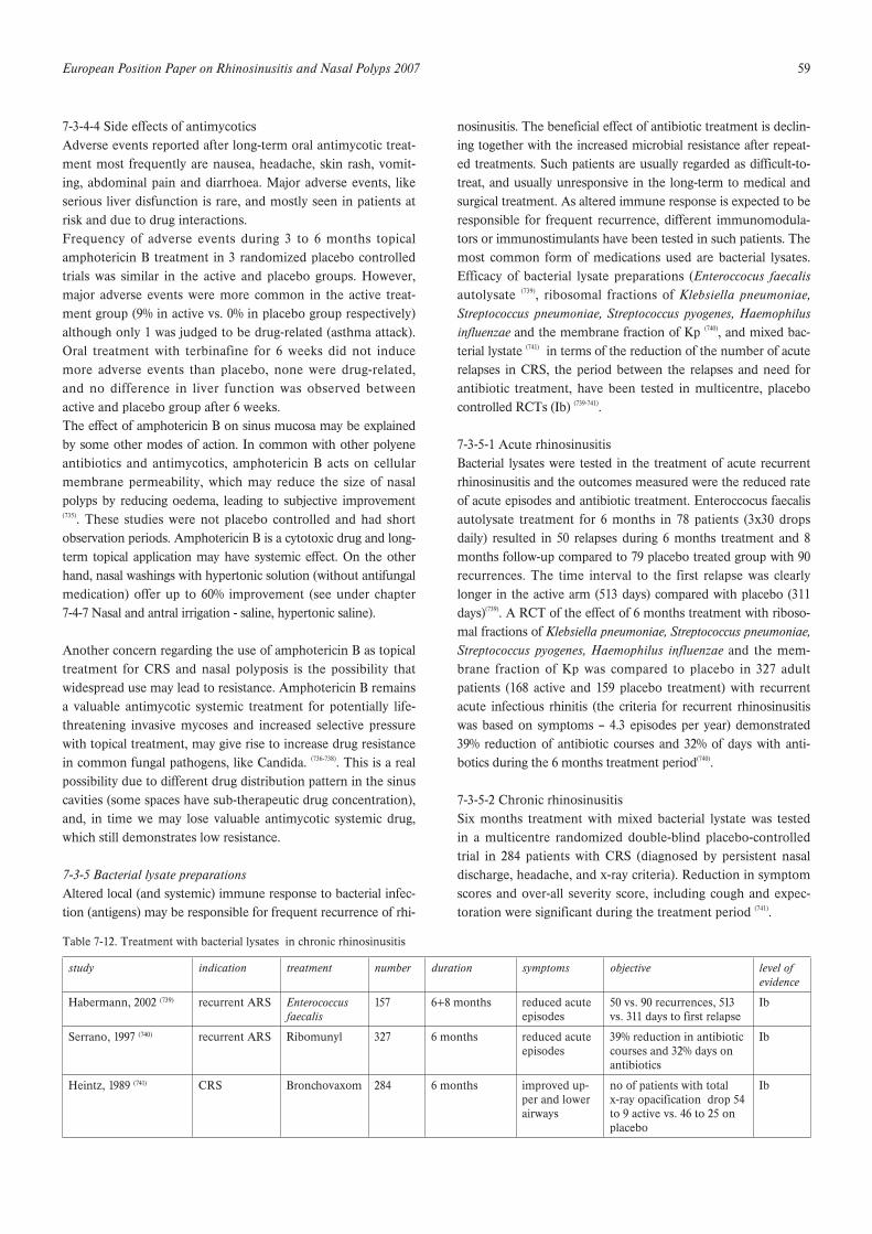
European Position Paper on Rhinosinusitis and Nasal Polyps 2007 59
7-3-4-4 Side effects of antimycoticsAdverse events reported after long-term oral antimycotic treat-ment most frequently are nausea, headache, skin rash, vomit-ing, abdominal pain and diarrhoea. Major adverse events, likeserious liver disfunction is rare, and mostly seen in patients atrisk and due to drug interactions.Frequency of adverse events during 3 to 6 months topicalamphotericin B treatment in 3 randomized placebo controlledtrials was similar in the active and placebo groups. However,major adverse events were more common in the active treat-ment group (9% in active vs. 0% in placebo group respectively)although only 1 was judged to be drug-related (asthma attack).Oral treatment with terbinafine for 6 weeks did not inducemore adverse events than placebo, none were drug-related,and no difference in liver function was observed betweenactive and placebo group after 6 weeks.The effect of amphotericin B on sinus mucosa may be explainedby some other modes of action. In common with other polyeneantibiotics and antimycotics, amphotericin B acts on cellularmembrane permeability, which may reduce the size of nasalpolyps by reducing oedema, leading to subjective improvement(735). These studies were not placebo controlled and had shortobservation periods. Amphotericin B is a cytotoxic drug and long-term topical application may have systemic effect. On the otherhand, nasal washings with hypertonic solution (without antifungalmedication) offer up to 60% improvement (see under chapter7-4-7 Nasal and antral irrigation - saline, hypertonic saline).
Another concern regarding the use of amphotericin B as topicaltreatment for CRS and nasal polyposis is the possibility thatwidespread use may lead to resistance. Amphotericin B remainsa valuable antimycotic systemic treatment for potentially life-threatening invasive mycoses and increased selective pressurewith topical treatment, may give rise to increase drug resistancein common fungal pathogens, like Candida. (736-738). This is a realpossibility due to different drug distribution pattern in the sinuscavities (some spaces have sub-therapeutic drug concentration),and, in time we may lose valuable antimycotic systemic drug,which still demonstrates low resistance.
7-3-5 Bacterial lysate preparationsAltered local (and systemic) immune response to bacterial infec-tion (antigens) may be responsible for frequent recurrence of rhi-
nosinusitis. The beneficial effect of antibiotic treatment is declin-ing together with the increased microbial resistance after repeat-ed treatments. Such patients are usually regarded as difficult-to-treat, and usually unresponsive in the long-term to medical andsurgical treatment. As altered immune response is expected to beresponsible for frequent recurrence, different immunomodula-tors or immunostimulants have been tested in such patients. Themost common form of medications used are bacterial lysates.Efficacy of bacterial lysate preparations (Enteroccocus faecalisautolysate (739), ribosomal fractions of Klebsiella pneumoniae,Streptococcus pneumoniae, Streptococcus pyogenes, Haemophilusinfluenzae and the membrane fraction of Kp (740), and mixed bac-terial lystate (741) in terms of the reduction of the number of acuterelapses in CRS, the period between the relapses and need forantibiotic treatment, have been tested in multicentre, placebocontrolled RCTs (Ib) (739-741).
7-3-5-1 Acute rhinosinusitisBacterial lysates were tested in the treatment of acute recurrentrhinosinusitis and the outcomes measured were the reduced rateof acute episodes and antibiotic treatment. Enteroccocus faecalisautolysate treatment for 6 months in 78 patients (3x30 dropsdaily) resulted in 50 relapses during 6 months treatment and 8months follow-up compared to 79 placebo treated group with 90recurrences. The time interval to the first relapse was clearlylonger in the active arm (513 days) compared with placebo (311days)(739). A RCT of the effect of 6 months treatment with riboso-mal fractions of Klebsiella pneumoniae, Streptococcus pneumoniae,Streptococcus pyogenes, Haemophilus influenzae and the mem-brane fraction of Kp was compared to placebo in 327 adultpatients (168 active and 159 placebo treatment) with recurrentacute infectious rhinitis (the criteria for recurrent rhinosinusitiswas based on symptoms – 4.3 episodes per year) demonstrated39% reduction of antibiotic courses and 32% of days with anti-botics during the 6 months treatment period(740).
7-3-5-2 Chronic rhinosinusitisSix months treatment with mixed bacterial lystate was testedin a multicentre randomized double-blind placebo-controlledtrial in 284 patients with CRS (diagnosed by persistent nasaldischarge, headache, and x-ray criteria). Reduction in symptomscores and over-all severity score, including cough and expec-toration were significant during the treatment period (741).
study indication treatment number duration symptoms objective level of evidence
Habermann, 2002 (739) recurrent ARS Enterococcusfaecalis
157 6+8 months reduced acute episodes
50 vs. 90 recurrences, 513 vs. 311 days to first relapse
Ib
Serrano, 1997 (740) recurrent ARS Ribomunyl 327 6 months reduced acute episodes
39% reduction in antibioticcourses and 32% days onantibiotics
Ib
Heintz, 1989 (741) CRS Bronchovaxom 284 6 months improved up-per and lowerairways
no of patients with totalx-ray opacification drop 54to 9 active vs. 46 to 25 on placebo
Ib
Table 7-12. Treatment with bacterial lysates in chronic rhinosinusitis

60 Supplement 20
7-3-5-3 Nasal polyposisNo data could be found on treatment with bacterial lysates innasal polyposis.
7-3-6 Immunomodulators/immunostimulantsTreatment with filgrastim, recombinant human granulocytecolony stimulating factor, was tested in a RCT (Ib) in a groupof CRS patients refractory to conventional treatment, whichdid not confirm significantly improved outcomes after suchexpensive treatment (572). A pilot study (III) with interferongamma suggested this treatment may be beneficial in treatingresistent CRS, but the number of patients was not adequate toprovide evidence to justify such treatment (742). Certain groupsof antibiotics may be regarded as immunomodulators, likequinolones (743) and macrolides (744).
7-3-7 Nasal and antral irrigation (saline, hypertonic saline)A number of randomized controlled trials have tested nasaland antral irrigation with isotonic or hypertonic saline in thetreatment of acute and chronic rhinosinusitis. Although salineis considered as a control treatment itself, patients in these ran-domized trials were assigned to different modalities of applica-tion of saline or hypertonic saline, or hypertonic compared toisotonic saline. The results between the groups were com-pared. Most of them offer evidence that nasal washouts or irri-gations with isotonic or hypertonic saline are beneficial interms of alleviation of symptoms, endoscopic findings andHRQL improvement in patients with CRS. Hypertonic saline ispreferred to isotonic treatment for rhinosinusitis by someauthors in the USA, mostly based on a paper indicating it sig-nifcantly improves nasal mucociliary clearance measured bysaccharine test, in healty volunteers (745).
7-3-7-1 Acute rhinosinusitisA randomized trial (Ib) by Adam et al (746) with two controls,compared hypertonic nasal saline to isotonic saline and notreatment in 119 patients with common cold and ARS (whichwere the majority). Outcome measures were subjective nasalsymptoms scores (congestion, secretion, headache) at day-3,day-8-10 and the day of symptom resolution. Rhinosinusitispatients (98%) were also treated with antibiotics. There was nodifference between the groups and only 44% of the patientswould use the hypertonic saline spray again. Thirty-two percentnoted burning, compared with 13% of the normal saline group.
Antral irrigation (Ib) did not offer significant benefit when addedto standard 10-day antibiotic treatment in (4 antibiotics+ decon-gestants vs. antral washouts; 50 patients per group) ARS, demon-strating approximately 5% better cure rate in each group forwashouts than for decongestants, which was not significant (747).
7-3-7-2 Chronic rhinosinusitisA randomised controlled trial (RCT) by Bachmann (Ib), com-paring isotonic saline and EMS solution (balneotherapeutic
water) in the treatment of CRS in a double-blind fashionrevealed improvement in both groups, with no differencebetween them (748). In the 7-days follow-up, nasal air flow wasnot improved significantly. Subjective complaints, endonasalendoscopy, and radiology results revealed a significant improve-ment in both groups (p = 0.0001). A similar RCT by Taccarielloet al (Ib),with a longer follow-up confirmed that nasal washingwith sea water and alkaline nasal douche produced benefit overstandard treatments. Douching per se improved endoscopicappearances (p = 0,009), and quality of life scores (p = 0,008)(749). These measures did not change in a control group (n = 22)who received standard treatment for CRS, but no douche.There were significant differences between the two douchingpreparations - the alkaline nasal douche improved endoscopicappearances but did not enhance quality of life, whereas theopposite was true for the spray. Rabago et al. (Ib) tested benefitfrom daily hypertonic saline washings compared to standardCRS treatment (control) for 6 months in a RCT using subjec-tive scores instruments: Medical Outcomes Survey Short Form(SF-12), the Rhinosinusitis Disability Index (RSDI), and aSingle-Item Sinus-Symptom Severity Assessment (SIA).Experimental subjects reported fewer 2-week periods withsinus-related symptoms (p < 0,05), used less antibiotics (p <0,05), and used less nasal spray (p = 0,06) (750). On the exit ques-tionnaire 93% of study subjects reported overall improvementof sinus-related quality of life, and none reported worsening (p< 0,001); on average, experimental subjects reported 57 -/+4.5% improvement measured by Medical Outcomes SurveyShort Form (SF-12), the Rhinosinusitis Disability Index(RSDI), and a Single-Item Sinus-Symptom SeverityAssessment (SIA). A double blind RCT (Ib) compared theeffect of nasal wash with hypertonic saline (3.5%) versus normalsaline (NS) (0.9%) for the 4 weeks in treatment of paediatricCRS using cough and nasal secretions/postnasal drip as subjec-tive and a radiology score as objective outcome measures (751).Hypertonic saline demonstrated significant improvement for allthe scores (13/15 for cough, 13/15 postnasal drip, 14/15 x-rayscores), while saline improved only postnasal drip.A recent double blind randomized controlled trial in 57 patientswith CRS , with minimum persistence of symptoms for 1 yearand previously unsuccesufully treated with conventional med-ical treatment, demonstrated significantly better improvementafter nasal lavage for 60 days with hypertonic Dead sea salt(DSS) solution compared to conventional hypertonic saline, interms of rhinosinusitis symptoms scores and rhinoconjunctivitisquality-of-life scores which was attributed to the presence ofmagnesium and other oligominerals known to have an effect innskin conditions (581). Similar results were found in a recent casetrial in 31 patients with resistant CRS. (752).A randomized controlled clinical trial of nasal washing withnormal saline, hypertonic buffered saline and no treatment in60 patients after endoscopic sinus surgery did not prove any ofthe treatment superior. Hypertonic saline induced greater dis-charge during the first 5 postoperative days and increased pain
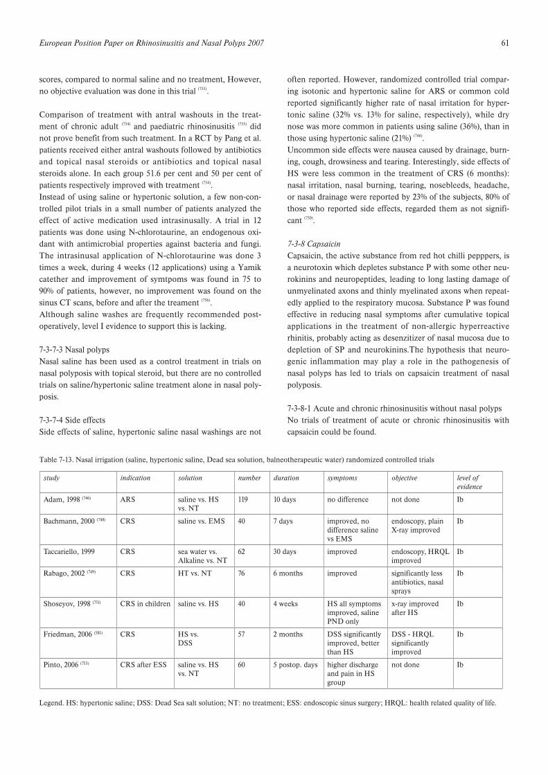
European Position Paper on Rhinosinusitis and Nasal Polyps 2007 61
scores, compared to normal saline and no treatment, However,no objective evaluation was done in this trial (753).
Comparison of treatment with antral washouts in the treat-ment of chronic adult (754) and paediatric rhinosinusitis (755) didnot prove benefit from such treatment. In a RCT by Pang et al.patients received either antral washouts followed by antibioticsand topical nasal steroids or antibiotics and topical nasalsteroids alone. In each group 51.6 per cent and 50 per cent ofpatients respectively improved with treatment (754).Instead of using saline or hypertonic solution, a few non-con-trolled pilot trials in a small number of patients analyzed theeffect of active medication used intrasinusally. A trial in 12patients was done using N-chlorotaurine, an endogenous oxi-dant with antimicrobial properties against bacteria and fungi.The intrasinusal application of N-chlorotaurine was done 3times a week, during 4 weeks (12 applications) using a Yamikcatether and improvement of symtpoms was found in 75 to90% of patients, however, no improvement was found on thesinus CT scans, before and after the treament (756).Although saline washes are frequently recommended post-operatively, level I evidence to support this is lacking.
7-3-7-3 Nasal polypsNasal saline has been used as a control treatment in trials onnasal polyposis with topical steroid, but there are no controlledtrials on saline/hypertonic saline treatment alone in nasal poly-posis.
7-3-7-4 Side effectsSide effects of saline, hypertonic saline nasal washings are not
often reported. However, randomized controlled trial compar-ing isotonic and hypertonic saline for ARS or common coldreported significantly higher rate of nasal irritation for hyper-tonic saline (32% vs. 13% for saline, respectively), while drynose was more common in patients using saline (36%), than inthose using hypertonic saline (21%) (746).Uncommon side effects were nausea caused by drainage, burn-ing, cough, drowsiness and tearing. Interestingly, side effects ofHS were less common in the treatment of CRS (6 months):nasal irritation, nasal burning, tearing, nosebleeds, headache,or nasal drainage were reported by 23% of the subjects, 80% ofthose who reported side effects, regarded them as not signifi-cant (750).
7-3-8 CapsaicinCapsaicin, the active substance from red hot chilli pepppers, isa neurotoxin which depletes substance P with some other neu-rokinins and neuropeptides, leading to long lasting damage ofunmyelinated axons and thinly myelinated axons when repeat-edly applied to the respiratory mucosa. Substance P was foundeffective in reducing nasal symptoms after cumulative topicalapplications in the treatment of non-allergic hyperreactiverhinitis, probably acting as desenzitizer of nasal mucosa due todepletion of SP and neurokinins.The hypothesis that neuro-genic inflammation may play a role in the pathogenesis ofnasal polyps has led to trials on capsaicin treatment of nasalpolyposis.
7-3-8-1 Acute and chronic rhinosinusitis without nasal polypsNo trials of treatment of acute or chronic rhinosinusitis withcapsaicin could be found.
Table 7-13. Nasal irrigation (saline, hypertonic saline, Dead sea solution, balneotherapeutic water) randomized controlled trials
Legend. HS: hypertonic saline; DSS: Dead Sea salt solution; NT: no treatment; ESS: endoscopic sinus surgery; HRQL: health related quality of life.
study indication solution number duration symptoms objective level of evidence
Adam, 1998 (746) ARS saline vs. HS vs. NT
119 10 days no difference not done Ib
Bachmann, 2000 (748) CRS saline vs. EMS 40 7 days improved, no difference salinevs EMS
endoscopy, plain X-ray improved
Ib
Taccariello, 1999 CRS sea water vs. Alkaline vs. NT
62 30 days improved endoscopy, HRQL improved
Ib
Rabago, 2002 (749) CRS HT vs. NT 76 6 months improved significantly less antibiotics, nasal sprays
Ib
Shoseyov, 1998 (751) CRS in children saline vs. HS 40 4 weeks HS all symptoms improved, saline PND only
x-ray improvedafter HS
Ib
Friedman, 2006 (581) CRS HS vs.DSS
57 2 months DSS significantly improved, better than HS
DSS - HRQL significantlyimproved
Ib
Pinto, 2006 (753) CRS after ESS saline vs. HS vs. NT
60 5 postop. days higher discharge and pain in HS group
not done Ib
a

62 Supplement 20
7-3-8-2 Nasal polypsA case study (III) by Filiaci et al. has demonstrated significantreduction in the size of nasal polyps after five (weekly) topicalapplications of capsaicin (30 mmol/L) solution in patients withnasal polyposis (757). The authors noted increased nasaleosinophilia after the treatment, which was not correlated tothe polyp size. A case study by Baudoin et al. has demonstrat-ed significant reduction of sinonasal polyposis after 5 consecu-tive days treatment with increasing doses (30-100 mmol/L) oftopical capsaicin in massive polyposis measured by CT scans atentry and after 4 weeks (III) (758). ECP in nasal lavage was notinfluenced by the treatment. Protection of polyp recurrencefollowing endonasal surgery by 5 topical applications of cap-saicin in 51 patient after surgery with a 9 months follow-up hasconfirmed significant recurrence protection and significantlybetter nasal patency in the active group in a randomized, dou-ble blind, placebo controlled trial (Ib) by Zheng et al (759). Theauthors used 70% ethanol 3x10⁻⁶ ml capsaicin solution, whichmay explain the high rate of recurrence in the control groupafter ESS, which received only 70% ethanol. They noted 40%polyp stage 0 (Malm) and 45% stage 1 in the active treatmentgroup, while controls demonstrated 45% stage 2 and 40% stage3 polyposis following treatment at 9 months observation. Thelow cost of capsaicin treatment was noted as a certain advan-tage compared to other postoperative treatments. As capsaicinis NF$B antagonist in vitro, some other modes of action maybe proposed (760).
7-3-8-3 Side effectsThe most common side effect following nasal capsaicin appli-cation, if not previously topically anaesthetized, is severe burn-ing sensation in the nose and lips, and lacrimation. However,previous topical nasal anaesthaesia with 10% xylocaine spray inplacebo-controlled trial completely blinded the active treat-ment (761). In the trials of nasal capsaicin treatment for idiopath-ic rhinitis, some other side effects were reported: dyspnoea,headache, cough, epistaxis, dryness of nasal mucosa and exan-thema (762, 763).
7-3-9 FurosemideThe protection of the hyper-reactive response to different chal-lenges (propranolol) (764); metabisulphite (765); and exercise (766) inasthmatics was demonstrated after inhalation of furosemide,suggesting bronchoprotective effects, similar to the effect ofcromones.
7-3-9-1 Acute and chronic rhinosinusitis without nasal polypsNo trials of treatment of acute or chronic rhinosinusitis withfurosemide have been found.
7-3-9-2 Nasal polypsProtection against nasal polyp recurrence following surgerywith 1-9 years follow-up, comparable to the effect of the topicalsteroid, was demonstrated after topical application of
furosemide in 97 patients postoperatively versus mometasonefuroate in 33 patients, in a prospective non-randomized con-trolled trial (IIa) by Passali et al (767), which had been previouslyreported by the same group in a case study. Relapses wererecorded in 17.5% in the furosemide, 24.2% in the mometasoneand 30% in the no treatment group, suggesting thatfurosemide, as a much cheaper medication than steroids,might be considered as a prophylaxis to polyp recurrence.A recent randomized controlled trial compared the effect ofshort term pre-operative treatment with oral methylpred-nisolone (1mg/kg) versus inhalation of 10 ml of 6.6 mmolfurosemide solution in 40 patients with nasal polyposis. Bothwere effective but no difference was found between the twotreatment modalities after 7-day treatment, in terms of polypsize reduction on endoscopy, nasal symptom scores (except forolfaction, which was better in steroid group) and intraoperativebleeding. Histological analysis of the polyps at surgery suggest-ed a strong anti-inflammatory effect of oral steroid (in terms ofeosinophil count reduction), while furosemide treatmentdemonstrated only an effect on oedema (768).Randomized placebo-controlled trials, especially long termtreatment, are however lacking.
7-3-10 Proton pump inhibitorsNumerous trials during the past decade indicated an associa-tion between extraoesophageal reflux and airway disease, andbeneficial effect of PPI treatment on upper airway symptoms,including some symptoms of CRS, was suggested., Postnasaldrip, a relevant CRS symptom, was established as one of thesymptoms responding to PPI treatment. However, sensation ofpostnasal drip (PND)was confirmed in groups of patients withidiopathic rhinitis, without evidence of rhinosinusitis, and inpatients without rhinitis and sinusitis.(769), and greater exposureto gastric acid was demonstrated in patients with PND than inthe controls. As PPI treatment reduces acidity, it is a dilemmaif PPI acts on rhinitis, rhinosinusitis or sensation of PND.Most reviews on current evidence on the association betweenreflux and sinus disease advocate higher quality of controlledtrials on both etiology and treatment, in pediatric and adultpopulation. As PPI treatment of extraoesophageal reflux dis-ease (like laryngitis) is based on long-term high-dose regimen,potential side effects should be considered.
7-3-10-1 Acute rhinosinusitisThere are no trials with proton pump inhibitors for ARS.
7-3-10-2 Chronic rhinosinusitisThere is no evidence for benefits in the general population suf-fering from rhinosinusitis following treatment with protonpump-inhibitors, although subjective improvement was notedin patients with laryngopharyngeal reflux (proved by pH-metry) and rhinosinusitis. Grade C evidence for a positiveassociation between gastroesophageal reflux and rhinosinusitiswas found in a meta analysis of the literature for this co-mor-
European Position Paper on Rhinosinusitis and Nasal Polyps 2007 63
bidity (57 articles screened, 14 articles included) (770, 771)). A num-ber of case trials of rhinosinusitis, especially paediatric (770), havetested the efficacy of anti-reflux treatment with proton pumpinhibitors on the clinical course and symptoms of rhinosinusi-tis. Increased rates of reflux were detected in CRS in adultsunresponsive to standard treatment (772, 773). Further research isexpected in this field, and such treatment should be justifiedby randomized controlled trials.Non-controlled trials, especially in children, indicate the effecton some symptoms of rhinosinusitis, presumably postnasaldrip and cough. However, a recent meta-analysis of random-ized controlled trials of outcomes of the treatment of non-spe-cific cough with proton pump inhibitors confirmed that it isinsufficient evidence to definitely conclude that reflux treat-ment with PPI is universally beneficial for cough associatedwith reflux in adults. The beneficial effect was only seen insub-analysis and its effect was small (774).
7-3-10-3 Nasal polypsThere are no data on proton-pump inhibitors in nasal polyposis.
7-3-11 AntileukotrienesLeukotrienes are upregulated in asthma and nasal polyposis,especially in aspirin- sensitive disease.
7-3-11-1 Acute rhinosinusitisNo trials were done on the antileukotrienes treatment in ARS.
7-3-11-2 Chronic rhinosinusitis and nasal polypsOpen studies suggest that anti- leukotrienes might be of bene-fit in nasal polyposis (775-777).Antileukotriene treatment in 36 patients with CRS with orwithout NP, added to standard treatment, resulted in statisti-cally significant improvement in scores for headache, facialpain and pressure, ear discomfort, dental pain, purulent nasaldischarge, postnasal drip, nasal congestion and obstruction,olfaction, and fever. Overall improvement was noted by 72% ofthe patients and side-effects occurred in 11% of the patients (778).In a selected group of 15 ASA triad patients, addition ofantileukotriene treatment resulted in 9/15 with sinusitis experi-encing improvement and over-all benefit in 12/15 patients,which was confirmed by endoscopy (779). In a group of patientswith nasal polyposis, significant subjective improvement innasal symptoms occurred in 64% aspirin tolerant patients and50% aspirin sensitive patients Significant improvement in peakflow occurred only in aspirin tolerant patients, while acousticrhinometry, nasal inspiratory peak flow and nitric oxide levelsdid not change (777). A prospective double blind comparativestudy on 40 patients compared the effects of the leukotrienereceptor antagonist, montelukast and beclomethasone nasalspray on the post-operative course of patients with sinonasalpolyps. No significant differences were found between thesetwo post-operative treatments in the year after surgery (780).
Results of these studies indicate that there is a need for (larger)controlled trials of antileukotriene treatment in CRS with orwithout NP.
7-3-12 Aspirin desensitization
7-3-12-1 Acute and chronic rhinosinusitis without nasal polypsNo controlled trials of aspirin desensitization treatment foracute and chronic rhinosinusitis were found.
7-3-12-2 Chronic rhinosinusitis and nasal polyps with aspirinintoleranceSystemic aspirin desensitisation or topical lysine-aspirin treat-ment (the only truly soluble form of aspirin) may be implicat-ed in protection against chronic rhinosinusitis with nasal poly-posis recurrence.Sixty-five aspirin-sensitive patients with aspirin sensitive asth-ma underwent aspirin challenge, followed by aspirin desensiti-zation and daily treatment with aspirin over 1 to 6 years (mean,3.1 years). There were significant reductions in numbers ofsinus infections per year and an improvement in olfaction.Numbers of sinus and polyp operations per year were signifi-cantly reduced and doses of nasal corticosteroids were signifi-cantly reduced. There were reductions in hospitalizations fortreatment of asthma per year and reduction in use of systemat-ic corticosteroids (781-783).
Nucera et al. have followed three groups of patients with nasalpolyposis (about 50% aspirin sensitive), the first with 76 con-secutive nasal polypectomy patients who had a topical lysine-acetylsalicylate-therapy afterwards, the second 49 patients with40 mg triamcinolone retard (“medical polypectomy”) and alsofurther lysine-acetylsalicylate-therapy and the third with 191control patients who underwent only polypectomy butreceived no placebo. The group treated with lysine-acetylsali-cylate postoperatively had a recurrence rate of 6.9% after l yearand 65% after six years postoperatively, while controls experi-enced recurrence in 51.3% at l year and 93.5% at six years afterthe operation, indicating a significant protection against recur-rence from the lysine-acetylsalicylate treatment. Systemic corti-coid therapy and nasal lysine-acetylsalicylate-therapy resultedin 33% with unchanged polyp size after three years comparedto 15% in the operated-not treated group, but this was not sta-tistically significant (784).
A case controlled trial of treament with lysine aspirin to onenostril and placebo to the other in 13 patients with bilateralnasal polyposis resulted in delayed polyp recurrence and 8remained symptom free at 15 months observation period,which was significantly better than results of the patients previ-ously treated with steroid for recurrence protection. Endoscopyand acoustic rhinometry indicated minor polyp size on theaspirin treated side (785).
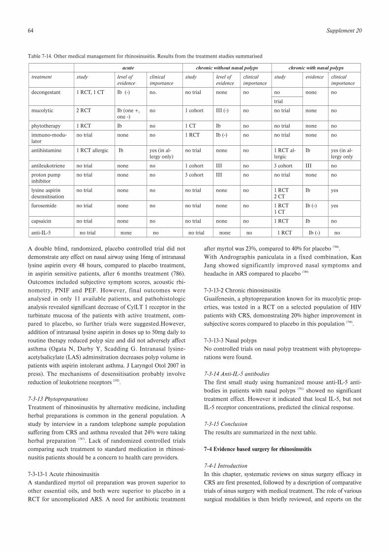
64 Supplement 20
A double blind, randomized, placebo controlled trial did notdemonstrate any effect on nasal airway using 16mg of intranasallysine aspirin every 48 hours, compared to placebo treatment,in aspirin sensitive patients, after 6 months treatment (786).Outcomes included subjective symptom scores, acoustic rhi-nometry, PNIF and PEF. However, final outcomes wereanalysed in only 11 available patients, and pathohistologicanalysis revealed significant decrease of CylLT 1 receptor in theturbinate mucosa of the patients with active treatment, com-pared to placebo, so further trials were suggested.However,addition of intranasal lysine aspirin in doses up to 50mg daily toroutine therapy reduced polyp size and did not adversely affectasthma (Ogata N, Darby Y, Scadding G. Intranasal lysine-acetylsalicylate (LAS) adminsitration decreases polyp volume inpatients with aspirin intolerant asthma. J Laryngol Otol 2007 inpress). The mechanisms of desensitisation probably involvereduction of leukotriene receptors (392).
7-3-13 PhytopreparationsTreatment of rhinosinusitis by alternative medicine, includingherbal preparations is common in the general population. Astudy by interview in a random telephone sample populationsuffering from CRS and asthma revealed that 24% were takingherbal preparation (787). Lack of randomized controlled trialscomparing such treatment to standard medication in rhinosi-nusitis patients should be a concern to health care providers.
7-3-13-1 Acute rhinosinusitisA standardized myrtol oil preparation was proven superior toother essential oils, and both were superior to placebo in aRCT for uncomplicated ARS. A need for antibiotic treatment
after myrtol was 23%, compared to 40% for placebo (788).With Andrographis paniculata in a fixed combination, KanJang showed significantly improved nasal symptoms andheadache in ARS compared to placebo (789)
7-3-13-2 Chronic rhinosinusitisGuaifenesin, a phytopreparation known for its mucolytic prop-erties, was tested in a RCT on a selected population of HIVpatients with CRS, demonstrating 20% higher improvement insubjective scores compared to placebo in this population (790).
7-3-13-3 Nasal polypsNo controlled trials on nasal polyp treatment with phytoprepa-rations were found.
7-3-14 Anti-IL-5 antibodiesThe first small study using humanized mouse anti-IL-5 anti-bodies in patients with nasal polyps (791) showed no significanttreatment effect. However it indicated that local IL-5, but notIL-5 receptor concentrations, predicted the clinical response.
7-3-15 ConclusionThe results are summarized in the next table.
7-4 Evidence based surgery for rhinosinusitis
7-4-1 IntroductionIn this chapter, systematic reviews on sinus surgery efficacy inCRS are first presented, followed by a description of comparativetrials of sinus surgery with medical treatment. The role of varioussurgical modalities is then briefly reviewed, and reports on the
Table 7-14. Other medical management for rhinosinusitis. Results from the treatment studies summarised
acute chronic without nasal polyps chronic with nasal polyps
treatment study level of evidence
clinicalimportance
study level of evidence
clinicalimportance
study evidence clinical importance
decongestant 1 RCT, 1 CT Ib (-) no. no trial none no no none no
trial
mucolytic 2 RCT Ib (one +, one -)
no 1 cohort III (-) no no trial none no
phytotherapy 1 RCT Ib no 1 CT Ib no no trial none no
immuno-modu-lator
no trial none no 1 RCT Ib (-) no no trial none no
antihistamine 1 RCT allergic Ib yes (in al-lergy only)
no trial none no 1 RCT al-lergic
Ib yes (in al-lergy only
antileukotriene no trial none no 1 cohort III no 3 cohort III no
proton pumpinhibitor
no trial none no 3 cohort III no no trial none no
lysine aspirindesensitisation
no trial none no no trial none no 1 RCT2 CT
Ib yes
furosemide no trial none no no trial none no 1 RCT 1 CT
Ib (-) yes
capsaicin no trial none no no trial none no 1 RCT Ib no
anti-IL-5 no trial none no no trial none no 1 RCT Ib (-) no

European Position Paper on Rhinosinusitis and Nasal Polyps 2007 65
effects of concomitant diseases on sinus surgery outcomes aredetailed. There is evidence that CRS with and without polyps aredistinct subgroups of chronic inflammatory diseases of the upperairway mucosa (see chapters 2 to 4). Approximately 20% ofpatients with CRS develop nasal polyps (792), which may predis-pose to less favourable results of sinus surgery (793, 794).Accordingly, reviewed articles are grouped into CRS withoutpolyps and CRS with polyps, when the authors differentiatedbetween these two subgroups. Except for a few reports on limit-ed sinus surgery in recurrent ARS (795), sinus surgery is generallyrestricted to chronic rhinosinusitis (CRS). Therefore, currentlyavailable data do not suffice to judge the role of surgery in acuteor acute recurrent rhinosinusitis. For surgical interventions incomplications of ARS see chapter 8, for paediatric sinus surgerysee chapter 9, for a detailed description of complications of sinussurgery refer to chapter 7, and for postoperative medical treat-ment please refer to chapter section 7-1-5.
It is difficult to generalise about sinus surgery studies becausesurgery is indicated in selected patients who are not sufficient-ly responsive to medical treatment. Moreover, only a few pub-lications on sinus surgery qualify for evidence based evaluation(796) and frequently studies included in systematic reviews areassigned low evidence levels (797-799) This is in part due to specificproblems in conducting surgical trials. In general, surgery isdifficult to estimate or standardize, particularly in multi-centretrials, and the type of treatment is difficult to conceal (blind-ing). Randomization may pose ethical problems unless narrowinclusion criteria are set (514). Low budget investigator initiatedtrials not monitored by professional clinical research organisa-tions are the rule.
In addition, a variety of confounders make it difficult to obtainhomogenous patient groups with comparable therapeutic pro-cedures for unbiased evaluation of sinus surgery outcomes.Possible relevant surgical factors include whether an externalor endonasal approach is chosen, whether a functional or con-ventional surgical procedure is selected, if the extent of thesurgical intervention is limited, extended or radical, and whatkind of instruments are employed. Patient dependent factorsinclude age, extent and duration of disease, previous surgery,presence of polyps, concomitant diseases such as ASA-intoler-ance, asthma, or cystic fibrosis, and particular aetiologiesincluding dental, autoimmune, immune, and fungal disease (800-
803). Moreover, mode and duration of pre- and post-operativedrug therapy may alter the outcome.
7-4-2 Effectiveness of sinus surgery and comparison with medicaltreatment
7-4-2-1 Systematic reviews and outcomes research on sinussurgery effectivenessSeveral reviews did not differentiate between CRS with andwithout polyps such as Terris and Davidson who analysed 10
case series. Patients’ judgement of outcome using an unbal-anced three item verbal rating scale was employed. (804). A ‘verygood’ result was defined as complete resolution of symptoms orless than 2 rhinosinusitis episodes per year, a ‘good’ result wasimprovement but without complete resolution of symptoms or2-5 sinusitis episodes per year, and a ‘poor’ result was noimprovement or deterioration. Articles reporting a total of 1,713patients were evaluated. Subjectively, 63% of patients reported avery good result, 28% a good result, and 9% an unsatisfactoryresult. Twelve percent of patients required revision surgery andcomplications occurred in 1.6% of patients. With 1.5%, bleedingwas most the most common complication (level IV).In a systematic review, 12 case series of endoscopic sinussurgery were evaluated and compared with 6 case series ofconventional techniques (797). In patients with CRS with or with-out polyps, overall success rates ranging from approximately70% to 90% after sinus surgery were reported. Revision surgerywas reported in 7% to 10%. If reported, complications werebelow 1% (level IV).In a recent meta-analysis, the impact of endoscopic sinussurgery on sinus symptoms and quality of life was evaluated inadults after failed medical treatment (803). Articles dealing withboth or not differentiating between CRS with and withoutpolyps were included. Forty-five of 886 screened articles wereincluded for full review. The reasons for exclusion were notdetailed. Of the included articles, 1 article qualified for level IIevidence, 42 for level IV evidence and 2 for level V evidence.The authors conclude that there is substantial level IV evi-dence with supporting level II evidence that endoscopic sinussurgery is effective in improving symptoms and QoL in adultpatients with CRS.In a recent Cochrane review, 3 randomized controlled sinussurgery studies were included(796). The authors conclude thatFESS, as practiced in the included trials, is not superior tomedical treatment with or without sinus irrigation in patientswith CRS.
7-4-2-1-1 CRS without polypsIn 2000 the Clinical Effectiveness Unit of the Royal College ofSurgeons of England conducted a National Comparative Auditof the Surgery for Nasal Polyposis and Chronic Rhinosinusitiscovering the work of 298 consultants working in 87 hospitalsites in England and Wales (521). Patients undergoing sinussurgery were prospectively enrolled and followed up in thisobservational study at 3, 12 and 36 months post-operativelyusing the SNOT-22 as the main outcome measure. One third(952) of the 3128 patients participating in this study had CRSwithout nasal polyps and 2176 suffered from CRS with polyps.Outcomes are reported separately for the two rhinosinusitissubgroups. CRS-patients without polyps less frequently suf-fered from concomitant asthma and ASA-intolerance, had lessprevious sinonasal surgery, their mean CT score was lower andtheir mean SNOT-22 symptom score was slightly higher thanthat of CRS patients with polyps. All forms of sinus surgery
66 Supplement 20
were considered though the majority were performed endo-scopically. Overall there was a high level of satisfaction withthe surgery and clinically significant improvement in theSNOT-22 scores were demonstrated at 3, 12 and 36 months.Revision surgery was indicated in 4.1% at 12 months and 10.4%at 36 months (Level IIc).In addition to this outcomes research study, 2 recent caseseries are also presented to supplement outcome data on CRSwithout polyps. In a retrospective analysis, 123 patients withCRS without nasal polyps who underwent primary FESS witha minimum 1-year follow-up period were evaluated (793).Outcome parameters included the Sino-Nasal Outcome Test(SNOT-20) questionnaire, the Lund-Mackay CT-scoring sys-tem, and the need of revision surgery. SNOT-20 scores were26.5 preoperatively with significant improvement to 5.1 at 6months and 5.0 at 12 months postoperatively (85% improve-ment) (level IV). In a case series of 77 patients with CRS with-out polyps, symptom and endoscopy scores were followedbetween 3 and 9 years after FESS (805). Saline douches and nasalsteroids were postoperatively administered as required. Afterat least 3 years, more than 90% of the patients reported symp-tom improvement of 80% or more. Revision surgery was per-formed in 15%. At the end of the follow up period, 5 patients(7%) received nasal steroids.
7-4-2-1-2 CRS with polypsWithin the framework of the NHS R&D Health TechnologyAssessment Programme, the clinical effectiveness of functionalendoscopic sinus surgery to treat CRS with polyps wasreviewed. The authors screened 444 articles and evaluated 33articles published between 1978 and 2001 (806). Major reasons forexclusion were the narrative character of the publication orless than 50 patients with polyps. The authors reviewed 3 RCTcomparing functional sinus surgery with Caldwell Luc or con-ventional endonasal procedures (n=240), 3 non-randomizedstudies also comparing different surgical modalities (n=2699)and 27 case series (n=8208). Consistently, patients judged theirsymptom ‘improved’ or ‘greatly improved’ in 75 to 95 percent(level IV). The percentage of overall complications was 1.4%for FESS compared to 0.8% for conventional procedures.Two thirds (2176) of the 3128 patients participating in theNational Comparative Audit had CRS with nasal polyps (521).CRS patients with polyps had no longer duration of disease, nohigher previous steroid treatment, nor ratings of their generalhealth before surgery than CRS patients without polyps.Irrespective of extent of surgery, clinically significant improve-ment in the SNOT-22 scores were demonstrated at 3, 12 and36 months. Polyp patients benefited more from surgery thanthe chronic rhinosinusitics without polyps. Revision surgerywas indicated in 3.6% at 12 months and 11.8% at 36 months.Major complications were rare (Level IIc).In this context, a case series study of CRS patients with partic-ularly extensive polyposis is worth mentioning (807). Of the 118patients reviewed, 59 (50%) had asthma, and 93 (79%) had doc-
umented allergy. All patients received extensive bilateral nasalpolypectomy, complete anterior and posterior ethmoidectomy,and maxillary sinusotomy. One hundred (85%) also had frontalor sphenoid sinusotomy. Follow-up ranged from 12 to 168(median 40) months. Despite pre- and postoperative nasal andsystemic steroid treatment in the majority of patients, 71 (60%)developed recurrent polyposis, 55 (47%) were advised to under-go revision surgery, and 32 (27%) underwent revision surgery.History of previous sinus surgery or asthma predicted higherrecurrence and revision surgery rates. History of allergy alsopredicted recurrence and need for revision.
Conclusion: One major outcomes research study (level II) andmore than a hundred reviewed case series (level IV) with high-ly consistent results suggest that patients with CRS with andwithout polyps benefit from sinus surgery. Major complica-tions occur in less than 1%, and revision surgery is performedin approximately 10% within 3 years.
7-4-2-2 Combined surgical and medical treatment vs. sole med-ical treatmentCRS may be cured with medical treatment alone. Moreover,sinus surgery is almost always preceded and/or followed byvarious forms of medical treatment including nasal douches,nasal steroids, systemic steroids, and systemic antibiotics. Fewstudies compared sinus surgery, which was always combinedwith medical treatment, with medical treatment alone. In twostudies, CRS patients with and without polyps were not differ-entiated.In a prospective trial, 160 CRS patients with or without polypswere enrolled and treated with either medical therapy alone ormedical therapy plus endonasal sinus surgery (486). Group alloca-tion was not randomized and the non-surgical cohort had lessmales, less concomitant diseases, less polyps, and less severedisease. Outcome parameters included the SF-36 and ChronicRhinosinusitis Survey (CSS) questionnaire. At follow up after3 months, the surgically treated group improved more than thenon-surgically treated group, however, improvement was notadjusted for pre-treatment scores (level IV).In a prospective observational study conducted by theCooperative Outcomes Group for ENT of the AmericanAcademy of Otolaryngology – Head and Neck Surgery, 31 oto-laryngologists enrolled 276 CRS patients with or withoutpolyps (808). Follow up was 207, 164 and 117 patients after 3, 6and 12 months, respectively. Success was defined as 40% ormore improvement in a subset of the CSS. Based on judgmentof the participating physicians, 83 patients received functionalendoscopic sinus surgery and 118 patients received medicaltreatment only. Surgically treated patients had a 3 times higherchance for success than patients treated only medically (p <0.01), however, this disproportion disappeared when correctedfor baseline CSS-scores in a logistic regression approach (levelIV).
European Position Paper on Rhinosinusitis and Nasal Polyps 2007 67
7-4-2-2-1 CRS without polypsIn a prospective, randomized, controlled trial, Ragab andcoworkers enrolled 90 patients with CRS (536). Of the 90 includ-ed patients, 55 suffered from CRS without polyps and arereported separately. During a run in phase, all patients receiveda 6-week regimen of dexamethasone-21-isonicotinate and tra-mazoline hydrochloride (DRS) spray and an alkaline nasaldouche. Patients who remained symptomatic after this treat-ment were then randomized to a medically or a surgicallytreated arm. In the medically randomized group, all patientsreceived a 12-week course of erythromycin, alkaline nasaldouche, and intranasal corticosteroid preparations. In the sur-gically randomized group, FESS was performed tailored to theextent of disease. After endoscopic sinus surgery, all patientswere prescribed a 2-week course of erythromycin, DRS spray,and alkaline nasal douche, followed by a 3-month course offluticasone propionate intranasal spray. After that, topical corti-costeroid spray was given as needed. Outcome parametersincluded the SNOT-20, SF-36, nasal NO and acoustic rhinome-try measurements. At follow up visits after 6 months and at 1year, significantly improved outcome parameters wereobserved in both treatment arms, with no significant differencebeing found between the medical and surgical groups (p > 0,05), except for the rhinometrically assessed total nasal volume,in which the surgical treatment demonstrated greater improve-ments (Level Ib).
7-4-2-2-2 CRS with polypsIn an open, randomized trial, Lildholdt and co-workers includ-ed 53 patients with nasal polyps (641). All patients received nasalsteroid spray during the 12 month study period. Snare polypec-tomy was additionally performed in 26 patients, whereas 27patients received an intramuscular depot betamethasone injec-tion. After 1 year, no relevant difference in the main outcomeparameters including sense of smell, PNIF, and disease recur-rence were observed (level Ib).In a second open RCT, Lildholt and co-workers included 34patients with nasal polyps who did not improve after participa-tion in a preceding placebo controlled trial comparing twodoses of intranasal budesonide(493). Sixteen patients received asingle depot injection of 14 mg betamethasone and 18 patientsunderwent intranasal snare polypectomy. Outcomes wereassessed after 11 months additional nasal steroid treatment andagain after 12 months without any treatment. Snare polypecto-my and systemic betamethasone outcome 12 months aftertreatment were remarkably similar as measured by mean nasalimprovement score, polyp score or mean score of sense ofsmell (p > 0,05). Within 1 year without nasal steroid treatment,50% of the patients experienced further nasal polyps, howeverthe authors did not differentiate polyp recurrence by medicalor surgical treatment (level Ib).In a study by Blomqvist and coauthors, 32 CRS patients withpolyps were pretreated with systemic steroids (prednisolone forfourteen days) and budesonide for 4 weeks (809). Thereafter,
FESS was performed on one side while the other sideremained untouched employing a randomized prospectivematched samples design. Following surgery, intranasal steroidswere given for an additional 12 months on both sides. Thesense of smell was tested for each nostril separately. Itimproved after treatment with systemic and topical steroidswithout additional improvement on the operated side. Surgeryhad an additional beneficial effect on nasal obstruction andsecretion that persisted over the study period. However, 25%percent of the patients required surgery also on the yet unoper-ated side. The authors conclude that surgical treatment is indi-cated after steroid treatment, if nasal obstruction persists butnot if hyposmia is the primary symptom (Level Ib).In the prospective, randomized, controlled trial by Ragab andco-authors already described (536), 35 CRS patients with polypsremained symptomatic after a 6-week intensive medical regi-men and were randomized into the study. At follow up visitsafter 6 months and at 1 year, both treatment arms reported sig-nificantly improved outcome parameters, with no significantdifference being found between the medical and surgicalgroups (p > 0,05), except for the total nasal volume, in whichthe surgical treatment demonstrated greater improvements(Level Ib).In a recent RCT, 109 patients with CRS with extensive nasalpolyps were included (587). Fifty-three patients were randomlyallocated to receive oral prednisone for 2 weeks (30 mg/day for4 days followed by a two days reduction of 5 mg) and 56 toundergo endoscopic sinus surgery. All patients additionallyreceived intranasal budesonide for 12 months. Patients wereevaluated for nasal symptoms, polyp size, and QoL employingthe SF-36 questionnaire. At 6 and 12 months, a significantimprovement in all SF-36 domains, nasal symptoms and polypsize was observed after both medical and surgical treatment. Asignificant advantage for the surgically treated group wasobserved for nasal obstruction, loss of smell and polyp size 6months after randomization and for polyp size also 12 monthsafter randomization (level Ib).
Conclusion: In the majority of CRS patients, appropriate med-ical treatment is as effective as surgical treatment. Sinussurgery should be reserved for patients who do not satisfactori-ly respond to medical treatment.
7-4-3 Surgical modalities
7-4-3-1 Endonasal versus external approachEndonasal approaches include surgical procedures performedthrough the nostril, irrespective of the extent of surgery andthe kind of visualization of the surgical field. Today, endonasalprocedures are predominantly performed employing endo-scopes. The most commonly performed external surgical pro-cedures include the sublabial transfacial Caldwell Lucapproach with or without transantral ethmoidectomy and sphe-noidectomy, and transfacial frontoethmoidectomy. In a few
68 Supplement 20
studies, outcomes of external and transnasal procedures werecompared though not differentiating between CRS patientswith and without polyps.Penttila and co-workers randomized 150 CRS patients afterfailed antimicrobial therapy and antral irrigations for chronicmaxillary sinusitis to either endonasal endoscopic sinussurgery (n=75) or an external Caldwell Luc approach (n=75).The percent changes of symptom scores before and one yearfollowing surgery were evaluated (Level Ib). Functional endo-scopic sinus surgery revealed significant advantages in therelief of nasal obstruction, hyposmia and rhinorrhea, but notfacial pain. Patients overall judgement and rate of complicationalso significantly favoured the endonasal approach (810, 811). Thestudy population was re-evaluated 5 to 9 years later with 85%of the former study participants responding to a questionnaire.In both surgical groups, approximately 80% were asymptomaticor distinctly improved without relevant intergroup differences(812). However, postoperative cheek pain and paraesthesia werenoted in 23% of Caldwell Luc group, which is a frequent com-plication of this approach (813). The histopathology of maxillarysinus specimens obtained before and 1 year after surgery frompatients of the two treatment arms of the Penttila-studies wereevaluated by Forsgren et al., indicating a greater reduction ininflammatory parameters in the mucosa of the maxillary sinusafter the Caldwell-Luc approach (814).In a retrospective evaluation, Unlu and coauthors randomlyselected 37 Caldwell-Luc-operated and 40 endonasally operat-ed patients. Outcome was assessed with nasal endoscopy andCT-scans (815). CT was normal in 12% of Caldwell-Luc-operatedpatients in comparison to 75% of endosopically operatedpatients. Endoscopy revealed a patency rate of the antral win-dow in 48% Caldwell-Luc-operated and 86.7% in endonasallyoperated patients. The authors conclude superiority of theendonasal approach. (Level IV).Videler and co-authors treated 23 CRS patients refractory torepeated endonasal procedures, Caldwell Luc and intensivemedical treatment via an Caldwell Luc approach with removalof the medial maxilarry wall (573). Clinical improvement wasobserved the majority of patients (level IV).
7-4-3-1-1 CRS without polypsNo studies comparing endonasal surgery with externalfontoethmoidectomy or Caldwell-Luc approach in patientswith CRS without polyps were found.
7-4-3-1-2 CRS with polypsNo studies comparing endonasal surgery with externalfontoethmoidectomy were identified. In several studies,endonasal sinus surgery was compared with the Caldwell-Lucapproach in CRS patients with polyps. In the NHS R&DHealth Technology Assessment Programme evaluation, anoverall symptomatic improvement was reported in approxi-mately 80% after endoscopic sinus surgery and in 43 to 84%after conventional surgery including Caldwell Luc. Disease
recurrence was 8% for FESS compared to 14% for Caldwell-Luc approach (806).McFadden and co-workers evaluated the long term outcome(up to 11 years) in 25 patients with extensive nasal polyps andASA-intolerance. Sixteen patients were operated via an extend-ed endonasal approach and 9 via a Caldwell Luc approach withradical sphenoethmoidectomy. Of the endonasally operatedpatients, 6 underwent a revision surgery via a Caldwell Lucapproach whereas none of the patients who had received aCaldwell Luc approach initially was reoperated (432).
Conclusion: Long term symptom relief in chronic sinusitis canbe obtained by the endonasal endoscopic and the Caldwell-Luc approach, however, results favour the endonasal approach.The Caldwell-Luc approach carries a higher risk of early post-operative facial swelling and infraorbital nerve irritation.Comparative studies of endonasal versus external frontoeth-moid approaches are currently not available.
7-4-3-2 Conventional endonasal surgery versus functionalendonasal sinus surgeryConventional sinus surgery is a collective term for surgicaltechniques already used before the devlopment of functionalsinus surgery. They include external approaches, maxillarysinus irrigation, simple (snare) polypectomy, inferior meatalantrostomy, and radical transnasal spheno-ethmoidectomywith or without middle turbinate resection. Unlike functionaltechniques, conventional sinus procedures do not proceedalong the natural pathways of sinus ventilation and mucociliarytransport revealed by the fundamental work of Messerklinger(816). Restoring ventilation and mucociliary transport by func-tional surgery along the natural ostia allows recovery of thediseased sinus mucosa, which is not resected (817, 818).Concurrently with the development of the functionalapproach, rigid endoscopes became available, which improvedvisualization during endonasal surgery. The evolving conceptof functional endoscopic sinus surgery (FESS) spread world-wide by the efforts of Stammberger and Kennedy (819, 820). In twostudies, a conventional approach was compared with functionalsinus surgery in CRS patients with or without polyps.In a prospective controlled trial, Arnes and coauthors per-formed an inferior meatal antrostomy on one side and a mid-dle meatal antrostomy in the opposite nasal cavity in 38patients with recurrent acute or chronic maxillary sinusitis (821).The laterality was randomized. After an observation periodranging between 1 and 5 years, no significant side differencesin symptom scores or radiological findings were observed(level Ib).In a randomized controlled trial, 25 patients after functionalendoscopic sinus surgery were compared with 25 after conven-tional surgery. Conventional surgery included antral puncture,intranasal ethmoidectomy, and Caldwell-Luc procedures.Follow up ranged from 15-33 months with a mean of 19months, at the end of which 76% of the functional group had
European Position Paper on Rhinosinusitis and Nasal Polyps 2007 69
complete relief of symptoms, 16% partial relief and 8% norelief as compared to 60%, 16%, 24% in the conventionallytreated group (822). However, this study is flawed by an elusivemethod of randomization, lack of information on homogeneityof patients groups, and high variability of procedures per-formed. (no level applied).
7-4-3-2-1 CRS without polypsEighty-nine patients with chronic sinusitis confined to themaxillary sinus without nasal polyps were enrolled in aprospective randomized controlled trial (823). After antibiotictherapy for at least 4 weeks prior to inclusion, 45 patientsreceived sinus irrigation only and 44 patient sinus irrigationfollowed by FESS. Patients were followed in regular intervalsup to one year. The per protocol analysis included 36 ‘irriga-tion only’ and 41 ‘irrigation and FESS’ patients. In 13 ‘irriga-tion only’ patients and 2 ‘irrigation and FESS’ patients, secondsurgery was performed due to lack of efficacy (p < 0.001).Moreover, outcomes for purulent discharge and loss of smellshowed significant improvement in ‘irrigation and FESS’ groupas compared with those obtained by sinus irrigation alone afterone year’s observation. Scores for other sinusitis symptoms didnot differ significantly between the groups (Level Ib).
7-4-3-2-2 CRS with polypsIn the NHS R&D Health Technology Assessment Programmeevaluation (806), polyp recurrence was 28% following function-al endoscopic ethmoidectomy compared to 35% followingintranasal polypectomy. The percentage of overall complica-tions was 1.4% for FESS compared to 0.8% for conventionalprocedures.Hopkins and co-workers analyzed 1848 patients with nasalpolyps, a subgroup of 3128 patients who participated in theNational Comparative Audit of Surgery for Nasal Polyposisand Rhinosinusitis (824). The authors compared the SNOT-20supplemented with two additional items (SNOT-22) after sim-ple polypectomy and after more extensive surgery both largelyperformed endoscopically in addition to medical treatment.The SNOT-scores did not differ significantly between the twotreatment arms after 12 and 36 months, if adjusted for relevantconfounders. Revision surgery was carried out more frequentlyin the polypectomy only group in the first 12 months aftersurgery (p = 0.04), but this difference was not significant at 36months. Complication rates did not differ significantly.
Conclusion: Functional endoscopic surgery is superior to mini-mal conventional procedures including polypectomy and antralirrigations, but superiority to inferior meatal antrostomy orconventional sphenoethmoidectomy is not yet proven.
7-4-3-3 Extent of surgeryExtent of surgery may vary from mere uncinectomy to radicalsphenoethmoidectomy with middle turbinate resection. In sev-eral studies, the extent of sinus surgery on various outcome
parameters was investigated in CRS patients, not differentiat-ing between CRS with and without polyps. In a prospectivetrial, 65 CRS patients with and without polyps were random-ized to undergo limited endonasal functional surgery(infundibulectomy) and a more extensive functional procedureincluding sphenoethmoidectomy and wide opening of thefrontal recess. Disease extent was similar in both treatmentarms. Outcome parameters included symptom scores,rhinoscopy scores and nasal saccharin transport time (825). Recallrates were below 60%. Outcome parameters revealed no rele-vant differences after 3, 6 and 12 months (level Ib).Based on the concept that diseased sinus prechambers, notsmall sinus ostia, are the cause for chronic sinus inflammation,minimal invasive sinus surgery (MIST) is advocated by someauthors (826). Basically, sinus ostia are exposed during MIST, butnot enlarged. In a prospective, uncontrolled trial, Catalano andRoffman followed 85 patients with CRS for a mean 24 months.Changes in the CSS score served as outcome parameter andrevealed significant improvements similar to FESS studies(level IV).Some authors advocate partial resection of the middleturbinate to expand the surgical approach (827), while othersmodify it only in case of abnormalities and leave as much aspossible of the middle turbinate intact as a landmark in caserevision surgery is needed (817). In a retrospective evaluationincluding 100 FESS patients, Giacchi and coauthors preservedthe middle turbinate on one side and partially resected it onthe other side (828). The authors observed no side differences inthe outcome parameters studied (level Ib).In a randomized trial, 1106 matched CRS patients with andwithout polyps, who underwent similar functional endonasalsinus surgery with (509 patients) or without (597 patients) par-tial middle turbinate resection (829). Partial middle turbinateresection was associated with less synechia formation (p <0.05) and less revision surgeries (p < 0.05) than middleturbinate preservation. Complications particularly caused bypartial middle turbinate resection were not observed (level Ib).In a prospective, randomized trial, uncinectomy was performedin 295 patients with chronic maxillary sinusitis. In 140 patients,a large middle meatal window (diameter > 16 mm) was eitherunilaterally or bilaterally created, whereas in 140 patients smallmiddle meatal antral windows (diameter < 6 mm) were pro-duced. In 170 patients, no preoperative CT was available.Follow up visits were attended by 133 (45%) patients 12 to 38months after surgery. Outcome parameters included patientsjudgment of symptom change (absent, improved, unchanged,worsened) and various endoscopic findings. Symptom relief,endoscopy findings and antral window size did not depend onthe surgically created antral window diameter (830).
7-4-3-3-1 CRS without polypsNo reports comparing less or more extensive surgical proceduresexplicitly in CRS patients without polyps could be identified.
70 Supplement 20
7-4-3-3-2 CRS with polypsThe patency rate after large middle meatal antrostomy andundisturbed maxillary ostium in endoscopic sinus surgery fornasal Polyposis was compared in 60 patients with bilateralnasal polyps and chronic maxillary sinusitis (831). A large middlemeatal antrostomy was performed on one side, whereas on theother side an uncinectomy preserving the natural maxillaryostium was done. The sides were chosen randomly. The paten-cy rates of a large middle meatal antrostomy were significantlyhigher 3 months after surgery when compared with undis-turbed maxillary ostium. This difference became insignificantafter 12 months (level Ib).Jankowski and co-authors retrospectively compared a caseseries of 37 CRS patients with extensive nasal polyps treatedwith FESS with a historical group of 36 patients with similardisease extent treated with radical sphenoethmoidectomy andmiddle turbinate resection (832). Outcome parameters assessed 5years following surgery included a mailed questionnaire onnasal symptoms, the number of patients with revision surgery,and nasal endoscopy scores at a follow up visit. Recall wasbelow 80% and differed significantly between the two groups.The radical surgical procedure yielded better symptom scores,less recurrences, and better endoscopy scores at the follow upvisit (level IV).
Conclusion: In CRS patients not previously operated, extend-ed surgery does not yield better results than limited surgicalprocedures. Although not evidence based, the extent ofsurgery is frequently tailored to the extent of disease, whichappears to be a reasonable approach. In primary paranasalsurgery, surgical conservatism is recommended.
7-4-3-4 Revision surgeryApproximately 10% operated patients respond insufficiently tosinus surgery with concomitant medical therapy and eventuallyrequire a secondary surgical procedure (833). Middle turbinatelateralisation, synechiae and scar formation in the middle mea-tus, an incompletely resected uncinate process, and retainedethmoid cells are frequent findings in patients undergoing revi-sion surgery (834-836). Previous revision surgery, extensive polyps,bronchial asthma, ASA-intolerance and cystic fibrosis are pre-dictors for revision surgery (793, 801, 837-840). Inflammatory involve-ment of underlying bone may also be of significance (159).Technical issues of sinus revision surgery have recently bereported by Cohen and Kennedy (841). A more extensive surgicalprocedure and also external approaches may be indicated (573, 832,
842). Success rates of revision endoscopic sinus surgery has beenreported to range between 50 and 70% (514, 838) (level IV).Complication rates of revision surgery are higher when com-pared with initial surgery and approximate 1%, but may be ashigh as 7% (833, 840).
7-4-3-4-1 CRS without polypsIn a case series of CRS patients without polyps, 15% were sur-
gical revisions (805). These patients had higher CT scores beforetheir initial surgery and also before the revision surgery thanpatients undergoing primary surgery (level IV). McMains andKountakis reported a series of 125 patients with a follow up ofat least 2 years after revision endonasal sinus surgery (839).Outcome parameters included the SNOT-20 and an endoscopyscore. Of these patients, 66 suffered from CRS without nasalpolyps. These patients experienced a significant improvementof their outcome parameters comparable with the results afterprimary surgery reported in other trials (level IV).
7-4-3-4-2 CRS with polypsMcMains and Kountakis also reported the results of 59 CRSpatients with nasal polyps after revision surgery (839). Consistentwith the results of the National Comparative Audit (824) and thecomparative study by Deal and co-workers (793), CRS patientswith polyps had lower SNOT scores preoperatively (less severesymptoms), more previous surgeries, and a higher CT scorepreoperatively than CRS patients without polyps. However, theimprovement of outcome parameters after revision surgery wassignificant and comparable with the improvement in CRSpatients without polyps.
Conclusion: Revision endonasal sinus surgery is only indicat-ed, if medical treatment is not sufficiently effective. Substantialsymptomatic improvement is generally observed in both, CRSwith and without polyps, though the improvement is some-what less than after primary surgery. Complication rates andparticularly the risk of disease recurrence are higher than afterprimary surgery. Some patients still suffer from CRS symp-toms after several extensive surgical procedures. CT scans fre-quently show mucosal alterations adjacent to hyperscleroticbony margins in an extensively operated sinus system. As arule, revision surgery is not indicated in these patients.
7-4-3-5 InstrumentsIn recent years, numerous instruments have been developedfor sinus surgery. Cutting forceps may improve controlledmucosal resection and help to avoid mucosal tearing. Poweredinstruments may facilitate controlled resection, particularly oflarge nasal polyps. Continuous suction/irrigation of microde-briders improve visualisation of the surgical field. Moreover,lasers have been employed for mucosal excisions and boneablation. Given the number of devices available, only a fewcomparative studies have been published. In these reports,CRS patients with and without polyps were not differentiated.
7-4-3-6 Cutting instrumentsIn a prospective double blind trial, 100 consecutive patientswere followed after endoscopic sinus surgery (843). Cutting for-ceps had been randomly used on one side and non-cutting for-ceps on the other side. Lateralised symptoms (headache, max-illary pressure, nasal obstruction and secretions) and endoscop-ic findings (secretion, pus, blood, crusts, oedema, polyps and
European Position Paper on Rhinosinusitis and Nasal Polyps 2007 71
adhesions) were evaluated on both sides 1 year postoperative-ly. Both types of instruments gave satisfactory healing situa-tions. No significant difference in the global symptom andendoscopic score between the 2 types of instruments wasfound (level Ib).
7-4-3-7 Powered instrumentsMicrodebriders were initially developed for arthroscopicsurgery. They consist of a suction based powered instrumentwith a blunt end and guarded inner 90° oscillating or rotatingblade, frequently supplemented with a device for irrigation.Cutting and removing only tissue suctioned into the instru-ment opening while blood and tissue debris are removed bysuction/irrigation, they provide excellent control and precisionof soft tissue resection (844).In a retrospective case series study, a group of 250 patientsundergoing surgery with a microdebrider was compared with agroup of 225 patients undergoing endoscopic sinus surgerywith conventional instruments (845). The assignment of patientsto each group was arbitrary and non-random, with the majorityof the standard technique patients being treated earlier in thestudy. The use of the microdebrider demonstrated faster heal-ing with less crusting than standard techniques, as well asdecreased bleeding, synechia formation, lateralization of themiddle turbinate, and ostial reocclusion (level IV).In a prospective randomized trial, 24 CRS patients were treatedwith microdebriders on one side and with conventional instru-ments on the other side (846).The authors were unable todemonstrate an advantage of mechanical debriders over con-ventional instrumentation (level Ib).Hackman and Ferguson reviewed both, positive and negativetissue effects secondary to powered instrumentation (844). Theauthors conclude that microdebriders will continue to advancethe field of endoscopic surgery, providing clearer operativefields and causing less tissue trauma in experienced hands.However, their literature review also illustrates the potentialseverity of complications, when orbital and cerebral contentsare rapidly aspirated by the powered instrument (no levelapplied).
7-4-3-8 LaserIn a randomized controlled trial, outcomes for holmium-YAGassisted sinus surgery were evaluated in 32 patients with CRSundergoing ESS (847). Following a randomization plan, one sidewas operated with conventional instruments and the contralat-eral side with laser. The use of holmium-YAG laser in ESSresulted in significantly lower blood loss during surgery andless post-operative crust formation than conventional ESS, butlong term subjective outcomes did not show significant differ-ences between the methods (level Ib).In a similar experimental setup, the application of KTP lasersin endoscopic sinus surgery was investigated in 24 patients (848).Laser-assisted FESS was performed on one side and FESSwith conventional instruments on the other side. Patient symp-
toms were recorded using a self-administered questionnairepreoperatively, and postoperatively on weeks 1, 4, 12 and 24.Of the parameters assessed in the course of healing, oedemaprevailed on the laser-assisted side, while crusting was charac-teristic in the traditional operation site. Overall, KTP-laser-assisted FESS was as effective as endonasal sinus surgery withconventional instruments. Disadvantages of the laser-assistedprocedure included the investment for the instrument and theadditional time needed for laser surgery (level Ib).
Conclusion: Laser assisted endonasal surgery, powered instru-ments or sharp forceps offer some advantages over conven-tional instruments but may be associated with some particularrisks. Currently, there is no evidence that they improve sinussurgery outcomes.
7-5 Influence of age concomitant diseases on sinus surgery outcome
7-5-1 Sinus surgery in the elderlyPrevious survey data ranks rhinosinusitis the sixth most com-mon chronic condition of elderly persons, occurring more fre-quently than cataracts, diabetes and general visual impairment(849). In a case series study, 56 CRS patients between 61 and 80years old were followed after functional endoscopic sinussurgery with nasal endoscopy and the SNOT-20 (849). Outcomeswere comparable to reports from younger patient populations,no severe complication occurred (level IV). In a retrospectivecase control study, functional endonasal sinus surgery out-come in 46 CRS patients > 65 years were compared with 522CRS patients who were 18-64 years old (850). In the elder patientgroup, complications occurred significantly more frequentlythan in the younger patients group. In particular orbital com-plication were frequently observed in the elder patient group(level III). Jiang and Su retrospectively compared complicationrates of 171 CRS patients elder than 65 years with 837 adultpatients and 104 patients younger than 16 years. They foundthat the geriatric group experienced a disproportionately largershare of operative complications. Outcomes were similar in allthree groups (851).
Conclusion: CRS is a common condition in the elderly.Reported sinus surgery outcomes do not differ from a youngerpatient population. However, higher surgical complicationrates were found in 2 reports. Moreover, general anaesthesiabears higher risks and the capacity to recover from a severesurgical complication such as a CSF leak may be impaired.
7-5-1-1 AsthmaBronchial asthma is frequently associated with CRS with andwithout polyps and may have influence on sinus surgery out-comes. A trend for more severe sinus disease in CRS patientswith concomitant asthma without aspirin intolerance has beenreported by Kennedy (514). Clinically, CRS patients with polypsand asthma have higher CT-scores, more severe nasal obstruc-
72 Supplement 20
tion and hyposmia, and more severe asthma, while CRSpatients without polyps and asthma experience more severeheadache and postnasal discharge (852). Investigations of con-comitant asthma on sinus surgery outcomes in CRS patientswith or without nasal polyps yielded inconsistent results (853).Concomitant asthma was associated with worse postoperativeendoscopy findings in two retrospective analyses (801, 803), but hadno independent influence on other outcome parameters (levelIV). Consistently, symptom scores improved significantly inboth asthmatics and non-asthmatics postoperatively, but asth-matics exhibited significantly worse postoperative endoscopicoutcomes in 21 asthmatic compared with 77 non-asthmatic. Nodifference was found in other outcome parameters betweenthe two groups (854) (level IV)In 3 other studies on various predictors of treatment success ofsinus surgery, asthma had no independent influence on out-come parameters (514, 802, 855).
7-5-1-1-1 CRS without polypsConcomitant asthma is frequent in patients with sinusitis with-out polyps. In a retrospective evaluation, 13 of 73 CRS patientswithout polyps had also asthma. However, concomitant asth-ma did not influence sinus surgery outcomes in this study (805).In a case series studies, Dunlop and coworkers followed thecourse of 50 CRS patients with bronchial asthma after sinussurgery (852). In this group, 16 CRS patients without polyps hadconcomitant asthma. Their sinusitis symptoms improved sig-nificantly following sinus surgery (level IV).
7-5-1-1-2 CRS with polypsIn a prospective outcome analysis, 79 patients underwentendoscopic sinus surgery for CRS with polyps (853). In a sub-group of 22 CRS patients with concomitant asthma, morerecurrences and less symptom score improvement wasobserved (level IV). In the study by Dunlop and coworkersdescribed above (852), 34 CRS patients with polyps and concomi-tant asthma revealed improved sinus symptoms 1 year aftersinus surgery (level IV).
Conclusion: Currently there is no evidence that CRS patientswith asthma benefit less from sinus surgery than patients with-out asthma concerning their CRS symptoms.
7-5-2 Effect of sinus surgery on bronchial asthmaThe incidence of self reported rhinosinusitis in asthma patientswas recently evaluated employing the data of two major asth-ma trials (856). Self reported rhinosinusitis was associated withbronchial asthma in 70% of the 2500 study participants.Asthma patients with concomitant rhinosinusitis had moreasthma exacerbations, worse asthma symptoms, worse cough,and worse sleep quality. The question, how sinus surgery andmedical CRS treatment may alter the course of bronchial asth-ma, was reviewed by Lund (857) and Scadding (858). The authorsdescribe the somewhat intricate base of evidence and conclude
that the weight of evidence suggests a beneficial effect. Studiespublished thereafter support this view. However, once again,authors did not differentiate between CRS with and withoutpolyps.In a follow up analysis of long term results of functionalendonasal sinus surgery, 72 of 120 patients answered a ques-tionnaire. A subgroup of 30 patients with CRS and asthmawere analysed (859). On average 6.5 years post surgery, asthmasymptom improvement, less asthma attacks, less inhaler andless oral steroid use were reported by the majority of patients(level IV).Park and co-authors retrospectively evaluated the data of asubgroup of 79 of 134 sinus surgery patients with asthma (860)with a questionnaire. Improved asthma was noted by 80% ofthe patients (level IV)In a controlled study, 15 patients with CRS and asthma under-went endoscopic sinus surgery and 6 patients, who rejectedsurgery, were treated with nasal steroids only (861). The authorscompared peak expiatory flow (PEF) and oral steroid con-sumption 6 months before and after treatment. In the surgical-ly treated patients, mean PEF improved 98±45 l/min (p<0.005)whereas no change was observed in the medically treatedgroup. Oral steroid consumption was reduced in 7 surgicallytreated patients, remained unchanged in 2 and increased in 2and was not nedded before and after surgery in 4 patients(level IV).In a retrospective medical record analysis 13 patients withchronic bronchial asthma who underwent FESS and receivedcomprehensive asthma care before and after FESS (mean, 19.3and 33.1 months, respectively) were included. Outcomes com-prised pre- and post-FESS individual and group mean asthmasymptom scores, medication use scores, pulmonary functiontest results, and emergency department visits or hospitaladmissions for asthma (862). Following FESS, there was no sta-tistically significant change in group mean asthma symptomscores, asthma medication use scores, pulmonary function testresults, and the number of emergency department visits orhospital admissions (level IV).In a case series study, 50 CRS patients with concomitant asth-ma were included (852). Twelve months after endoscopic sinussurgery, 40% noted asthma improvement, 54% stated that therewas no difference and 6% thought their asthma control wasworse. Inhalant steroids could be reduced by 20%, and weretaken at the same dose as preoperatively by 64%. However,oral steroid consumption was reduced significantly (p<0.001)and hospital admissions for asthma were less frequent in theyear after surgery than in the year before (p < 0.025, level IV).Dhong and co-authors followed the clinical course of 19patients with CRS and asthma who underwent ESS for rhinosi-nusitis (863). They observed a significant improvement in diurnaland nocturnal asthma symptoms and asthma medicationscores. Pulmonary function tests did not change (level IV).Ragab and co-workers report a prospective evaluation of a sub-group of 43 asthma patients (864) joining a randomised trial com-
European Position Paper on Rhinosinusitis and Nasal Polyps 2007 73
paring the effects of sinus surgery and medical treatment inCRS patients with and without polyps (865). Outcome parametersincluded asthma symptoms, control, forced expiratory volumein one second (FEV1), peak flow, exhaled nitric oxide, medica-tion use and hospitalisation at 6 and 12 months from the startof the study. Overall asthma control improved significantly fol-lowing both treatment modalities, but was better maintainedafter medical therapy, where improvement could also bedemonstrated in the subgroup with nasal polyps. Medical treat-ment was superior to surgery with respect to a decrease inexhaled nitric oxide and increase in FEV1 in the polyppatients. Two patients noted worsening of asthma post-opera-tively. Treatment of chronic rhinosinusitis, medical or surgical,benefits concomitant asthma; that associated with nasal poly-posis benefits more from medical therapy (level Ib).
Although asthma may accompany chronic rhinosinusitis withand without polyps, several studies particularly addressedlower airway effects of sinus surgery in CRS patients withpolyps. In a prospective outcome analysis, 79 patients under-went endoscopic sinus surgery for CRS (853). Twenty-eightpatients with asthma symptoms were assessed before and aftersurgery, using peak flow (liter/second) and medication scoresPatients showed improvement in terms of their asthma symp-toms, peak flow and medication score. (level IV).Palmer and co-workers retrospectively reviewed the charts of asubgroup of 15 CRS patients with steroid dependent asthmaselected from a group of 75 consecutive CRS patients with asth-ma who underwent endoscopic sinus surgery (866). Outcome para-meters included the number of days and total dose of oral pred-nisone and antibiotics in the year before and after sinus surgery.Fourteen of the 15 patients meeting study criteria decreased theirpostoperative prednisone requirement by total number of days(preoperative 84 versus postoperative 63 days (p < 0.0001).Postoperatively, patients required an average of 1300 mg less oralprednisone (p < 0.033). Antibiotic use also decreased (p < 0.045),with an average use of antibiotic nine weeks preoperativelyversus seven weeks postoperatively (level IV).In a prospective trial, Lamblin and co-workers included 46CRS patients with polyps and concomitant bronchial asthma(n=16) or non-symptomatic bronchial hyperresponsiveness(n=30). Outcome parameters measured at baseline (T0), after 1year (T1) and after 4 years (T2) included nasal symptomscores, various spirometry values and a bronchial carbacholchallenge (867). All patients were treated first with nasal steroidsfor 6 weeks (beclomethasone 600 µg/d). Eighteen patientswere successfully treated with nasal steroids (nasal steroidsresponders) and medical CRS treatment was continued with-out sinus surgery. In 28 patients who did not improve withnasal steroids (nasal steroids non-responders), intranasal she-noethmoidectomy was performed in addition to continuednasal steroid treatment. At baseline, clinical characteristics ofnasal steroid responders and non-responders -including thefrequency of Samter’s triad- did not reveal significant differ-
ences. Despite combined surgical and medical CRS-treatment,nasal steroid non-responders demonstrated a significantdecrease of their spirometry values at T1 (p < 0.05) and at T2(p < 0.0005), whereas no significant change was observed innasal steroid responders. BHR did not significantly changeover the 4-yr follow-up period in the two groups. No change inpulmonary symptoms and/or asthma severity occurred.
Conclusion: Apparently, various confounders not yet suffi-ciently defined influence the effects of surgical CRS treatmenton concomitant asthma. In studies published in recent years,predominantly positive effects of surgical CRS treatment onconcomitant asthma severity were reported However, the levelof evidence is low.
7-5-2-1 ASA intoleranceIntolerance to acetylsalicylic acid derived compounds such asaspirin or other acid NSAIDs frequently manifests as Samter’striad characterized by bronchial asthma, aspirin sensitivity, andCRS with polyps. The majority of CRS patients with aspirinintolerance have diffuse, extensive rhinosinusitis (514). In anearly report, poorer outcome in 11 patients with ASA-intoler-ance out of 120 prospectively followed patients treated withsinus surgery was observed (514). However, when stratified forextent of disease, ASA-intolerance did not adversely affect out-come (Level IV). In more recent trials, ASA-intolerance wasrather consistently found to adversely affect sinus surgery out-comes.In a case series study, 80 patients with ASA-intolerance andmostly extensive polyps were followed after sinus surgery (868).Sinus symptoms and asthma severity improved in more than80% of the patients. Before surgery, more than 30% weresteroid dependent due to asthma severity and less than 10%after surgery. However, a significant incidence of revisionsurgery was observed in this patient group (level IV).A higher number of repeat operations was also observed in aretrospective case control trial (869) including 18 patients withand 22 patients without ASA-intolerance (level IV).In a retrospective chart review, 17 patients who underwent ESSwith nasal polyps and steroid-dependent asthma with or with-out aspirin sensitivity and a minimum of 1 year postoperativefollow-up were evaluated (870). Nine patients were ASA sensi-tive, and eight patients were ASA tolerant. The postoperativeLund-Mackay scores (p < 0,001), the forced expiratory volumeat 1 second (FEV1, p < 0,05), and systemic steroid consump-tion (p < 0,05) improved significantly in the 17 patients. UnlikeASA tolerant patients, the 9 ASA sensitive patients did nothave a significant improvement in postoperative FEV1 andsinonasal symptoms (level IV).In a multivariate analysis of various outcome predictors,119 adult patients with CRS were prospectively followed-up for1.4 +/- 0.35 years after sinus surgery. ASA intolerance was theonly concomitant condition significantly worsening the out-come (519).
74 Supplement 20
Conclusion: CRS patients with ASA-intolerance tend to sufferfrom more extensive sinus disease. They benefit from sinussurgery, but to a lesser extent than patients without ASA-intol-erance. They are more prone to disease recurrence and morefrequently undergo revision surgery than ASA-tolerant CRSpatients.
7-5-2-2 Allergy and atopyIn most studies, the diagnosis of allergy was based solely onthe presence of a positive skin prick test and/or serum specificIgE determinations. This indicates atopy, but may not sufficeto diagnose allergic rhinitis (AR), particularly persistent AR (871).Walker and co-authors matched a cohort of 19186 individualswithout ENT disease registered in 1988 in the U.S: NavyAviation Medical Data retrieval System with 678 persons withAR as the only ENT disease (872). During the period from 1990to 1995, physicals were identified of 465 AR cases and 12,628controls. The incidence of chronic sinusitis in the AR groupwas 5/465 compared to 30/12628 in the control group (riskratio = 4.5, 95% CI 1.7 to 11.6). Consistently, the reported inci-dence of atopy in CRS patients ranges between 50 and 80%which is higher than in the general population. CRS in atopicpatients appears to be more severe (60, 873-878). Atopy was equallyfrequently associated with CRS with and without polyps (879).Conversely, in patients with positive skin prick tests to housedust mites, pathologic CT findings were significantly more fre-quent than in prick test negative controls (880).
In several trials not differentiating between CRS with andwithout polyps, atopy interfered with sinus surgery outcome. Itwas associated with less symptom improvement in one retro-spective evaluation of several sinus surgery outcome predictors(801), but had no relevant influence on pre- or postoperative CTor endoscopy findings nor on QoL scores in an other evalua-tion (803).However, antiallergic treatment appears to compensate for thepossible shortcomings of sinus surgery in allergic patients.Nishioka and co-authors compared postoperative middlemeatal antrostomy patency, middle meatal synechia formationand polyp recurrence in 211 non-allergic CRS patients and 72CRS patients considered allergic based on clinical history, skinprick tests and serum specific IgE (879). Of the 72 allergicpatients, 66 received an allergen specific immunotherapy eitherbefore or after surgery. Allergic patients had a significantlyhigher incidence of recurrent sinusitis, which could be reducedby allergen specific immunotherapy. The authors concludethat allergic patients treated with surgery and concomitantimmunotherapy do as well as non-allergic patients, whereasallergic patients treated with surgery alone do substantiallyworse (level IV).Similarly, immunotherapy and medical allergy treatmentbefore surgery improved the surgical success rate at a 1 yearfollow up in children with CRS from 64% to 84% (p = 0,022)(881). The latter figure was identical to the success rate in chil-
dren without allergy (level IV).A consistent finding was reported earlier by Schlenter andMann, who surgically treated 31 allergic and 34 non-allergicCRS patients (882). Of the 31 allergic patients, 15 underwent con-comitant allergen specific immunotherapy. Surgical outcomewas comparable in non-allergic and allergic patients after con-comitant immunotherapy, but significantly worse in allergicpatients without immunotherapy, despite antiallergic medicaltreatment (level IV).
7-5-2-2-1 CRS without polypsIn a prospective study, 24 CRS patients without polyps allergicto perennial allergens and 82 patients with CRS without polypswithout allergy underwent endoscopic endonasal ethmoidecto-my after medical pre-treatment (883). Comparing both groups,symptom scores did not differ significantly before and 6 to 18months after surgery.Consistently, in a case series of 77 CRS patients withoutpolyps, concomitant allergy had no influence on surgical out-come (805).In a double-blind placebo-controlled trial, 26 CRS-patientswithout polyps with positive skin prick tests to house dustmiteand persistent symptoms after sinus surgery received 256microg budesonide daily or placebo through an intubationdevice into one of the maxillary sinuses for 3 weeks beforeclinical assessment and a second biopsy (619). The authors foundan improvement in the symptom scores in 11 of the 13 patientswho received budesonide and a decrease in CD3 positive cells(p = 0,02) and eosinophils (p = 0,002), and a decrease in thedensity of cells expressing interleukin 4 (p = 0,0001) and inter-leukin 5 messenger RNA (p = 0,006) after treatment.
7-5-2-2-2 CRS with polypsIn patients with extensive polyposis (807), allergy diagnosis pre-dicted a worse outcome and increased recurrence rate (levelIV)
Conclusion: Allergic rhinitis may predispose to and aggravateCRS. In several studies, positive skin prick tests and/or serumspecific IgE to inhalant allergens were associated with poorersinus surgery outcome, particularly in CRS with polyps. Thisshortcoming can be compensated for with antiallergic treat-ment. After confirmation of allergy by allergic history or appro-priate clinical tests, allergen specific immunotherapy apparent-ly improves sinus surgery results in atopic or allergic CRSpatients.
7-5-2-3 Cystic fibrosisThis autosomal recessive genetic disease is characterized byepithelial secretory dysfunction and frequently associated withCRS. In cystic fibrosis, CRS with and without nasal polyps isobserved (884). Immunologically, CRS in cystic fibrosis (CF)patients differs from CRS in patients without CF (425, 884).Persistent colonisation with Pseudomonas aeruginosa is a
European Position Paper on Rhinosinusitis and Nasal Polyps 2007 75
common finding. Vitamin K deficiency related coagulopathiesare also common (885). Reports concentrating on CRS in CFpatients have mainly concerned the paediatric population,which is in part due to reduced life expectancy.Due to underlying medical issues such as acquired coagu-lopathies and advanced pulmonary disease, perioperative mor-bidity is assumed to be higher in this group (886). In 41 patientswith CF undergoing 52 sinus surgeries performed by a singlesurgeon over a 34-month period, complication occurred in11.5%, including 2 cases of epistaxis, 1 case of periorbitalecchymosis, and 1 case of pulmonary hemorrhage. Delayedcomplications included 1 case of epistaxis and 1 case ofintranasal scarring (level IV). No increased perioperative riskwas found by others (887) (level IV).The paranasal sinuses may act as a reservoir from where bacte-ria spread to the lower respiratory tract. After lung transplanta-tion, sinus derived graft infection with Pseudomonas aerugi-nosa may induce a frequently lethal bronchiolitis obliteranssyndrome. In 37 patients with cystic fibrosis after lung trans-plantation, sinus surgery was performed and repeated sinusaspirates and bronchoalveolar lavages were obtained for micro-biological examniations. Sinus surgery was successful (three orless Pseudomonas aeruginosa positive aspirates) in 54% andpartially successful (4 or 5 positive aspirates) in 27% of patients(888). A significant correlation of bacterial growth in sinus aspi-rates and bronchoalveolar lavages was observed (p < 0,0001).Successful sinus management led to a lower incidence of tra-cheobronchitis and pneumonia (p = 0,009) and a trend towarda lower incidence (p = 0,23) of bronchiolitis obliterans syn-drome (Level IV).Functional endoscopic sinus surgery with subsequent monthlyantimicrobial antral lavages (n=32) were compared with a his-toric control group receiving conventional sinus surgery with-out postoperative lavages (n=19). CF-patients with CRS withand without nasal polyps were included. For repeated antrallavages, modified 19 gauge butterfly intravenous catheterswere fixed in the maxillary sinus. Conventionally operatedpatients underwent one or more of the following procedures:polypectomy, ethmoidectomy, antrostomy, or Caldwell-Lucoperation (889). The group treated with functional sinus surgeryand antral lavages had fewer operations per patient, and adecrease in repeated surgery at 1 year (10% vs 47%) and 2 yearfollow up (22% vs 72%) (level IV).
7-5-2-3-1 CRS with polypsSeveral reports explicitly describe CRS with polyps in CFpatients. From a cohort of 650 patients undergoing endoscopicsinus surgery for CRS, 28 patients suffered from cystic fibrosis(800). Overall subjective improvement rate in the cohort as awhole was 91% improved, whereas 54% of the cystic fibrosispatients derived significant benefit at six month follow-up.(Level IV).In a retrospective report, 8 CRS patients with polyps out of 16adults with cystic fibrosis underwent sinus surgery (890). The
mean number of previous surgery in the surgically treatedgroup was 2.7. The authors report improved pulmonary func-tion, sinus symptoms, and exercise tolerance 3 months postsurgery, however, polyps recurred in all patients within 18months (Level IV).Rowe-Jones and Mackay performed endoscopic sinus surgeryon 46 cystic fibrosis patients with chronic, polypoid rhinosi-nusitis (891). Their mean age at first surgery was 23 ± 7.5 years.Follow-up ranged from 1 month to 6 years (mean, 28.2months). Overall, 50% of the patients suffered either recur-rence of preoperative severity or had to undergo second endo-scopic sinus procedure (level IV).
Conclusion: CF patients frequently suffer from severe CRS, inparticular with diffuse polyps refractory to medical treatment.Due to a tendency to recur, repeated sinus surgery is oftenneeded to achieve symptomatic relief. In CF patients, theparanasal sinuses may serve as a source for Pseudomonasaeruginosa induced lung infections. Consequent local antibiot-ic lavages help to prevent recurrent CRS and lung infection.
7-5-2-4 Sinus surgery in the immune compromised patientImmune deficiency states are frequently associated with CRSinclude HIV-infection, bone marrow transplantation andhumoral immondeficiencies.
7-5-2-4-1 HIVRhinosinusitis in the HIV-infected is an increasingly commonproblem. The gradual depression of humoral and cellularimmunity, delayed mucociliary transport, nasopharyngeal lym-phoid tissue hyperplasia and a tendency towards increased IgElevels may contribute to sinusitis development. Particularly atCD4-counts below 50 cells per mm3, Pseudomonas aeruginosais a common pathogen (892). Cytomegalovirus may cause sinusi-tis in HIV-infected patients and they have an increased risk todevelop invasive fungal sinusitis. Thus CT scans, and sinuslavages with special stains, cytologies and cultures are requiredin refractory sinusitis or patients with low CD4 counts (893). Thefirst line treatment of sinusitis in HIV-positive patients is med-ical, in refractory cases targeted to the identified organisms.Surgical treatment is reserved for patients who do not respondto targeted medical treatment.Sabini and co-authors retrospectively reviewed their experi-ence with performing endoscopic sinus surgery in 16 acquiredimmune deficiency (AIDS) patients (117). At an average follow-up time of 16 months, 14 of the endoscopic sinus surgerypatients reported improvement from their preoperative condi-tion (level IV).In a retrospective case series study, 106 HIV+ patients whounderwent sinus surgery between 1987 and 1998 were evaluat-ed (894). Between 1987 and 1991, 36 patients were treated withminimal invasive sinus surgery just addressing the involvedsinus with only 20% clinical improvement. Since 1992, theauthors treated their HIV+ patients with more extensive
76 Supplement 20
surgery including sphenoethmoidectomy, middle meatalantrostomy and drainage of the frontal recess, which resultedin a clinical improvement rate of 75%, irrespective of the CD4counts (level IV).In two case series, Murphy and co-workers observed the clini-cal outcome of 30 HIV-positive CRS patients refractory tomedical treatment (895). Outcome parameters included olfactorytests, symptom scores, and a quality of well-being assessment.Symptom and well-being scores improved significantly follow-ing endoscopic sinus surgery, whereas olfactory thresholds didnot improve significantly (level IV).Patients with AIDS may develop acute invasive fungal sinusi-tis. If detected early, combined surgical and antifungal treat-ment may be beneficial (896, 897).
7-5-2-4-2 Bone marrow transplantBone marrow transplantation (BMT) is a frequent cause ofacquired immune deficiency. Both, cellular and humoralimmunity are impaired. Particularly allogeneic BMT requiresintense immunosuppression to allow initial engraftment and toprevent graft versus host disease. Allogeneic BMT is associatedwith acute and chronic CRS in approximately 40% (898). Sinusmicobiology was investigated in 18 BMT patients who devel-oped sinusitis evaluating 41 microbiologcal specimensobtained by antral puncture and nasal swabs from the middlemeatus (899). Agents most commonly isolated were gram-nega-tive bacteria including Pseudomonas aeruginosa and Searratiamarescens. Gram positive bacteria were isolated in 27%.Various fungi were isolated in 16% of the specimens.Micorbiological results of antral punctures and nasal swabswere consistent in 5 of 41 specimens.Kennedy and co-workers report on 29 bone marrow transplantrecipients with documented invasive fungal infections of thesinuses and paranasal tissues (1.7% of 1692 bone marrow trans-plants performed). All patients received medical management,such as amphotericin, rifampin, and colony-stimulating factors,in addition to surgical intervention (900). Surgical managementranged from minimally invasive procedures to extensive resec-tions including medial maxillectomies. The mortality rate fromthe initial fungal infection was 62%. Twenty-seven percentresolved the initial infections but subsequently died of othercauses. Prognosis was poor when cranial and orbital involve-ment and/or bony erosion occurred. Extensive surgery was notsuperior to endoscopic functional surgery (level IV).Sinus surgery was performed in 28 of 311 bone marrow trans-plant patients retrospectively evaluated (901). No fungal sinusitiswas observed. An aggressive surgical approach yielded a highmortality rate whereas limited surgical approaches with inten-sive postoperative care proved appropriate (level IV).
7-5-2-4-3 Non-acquired immunodeficienciesPatients with humoral immunodeficiencies including commonvariable immunodeficiency, ataxia telangiextasia, or X-linkedagammaglobulinaemia are at increased risk to develop CRS (902-
905). In patients with CRS refractory to medical and surgicaltreatment, non-acquired immune deficiencies may affecthumoral, cellular, and frequently both immune response path-ways. Chee and co-workers selected 79 out of 316 patients withCRS with and without polyps, who suffered from severe CRSrefractory to medical treatment (114). Fifty-seven patients hadundergone one or more previous sinus surgeries.Approximately 30% of the 79 included patients suffered fromdecreased T-cell function and approximately 20% had someform of immunoglobulin deficiency. Common variable immun-odeficiency was diagnosed in 10%. Accordingly, in a high num-ber of patients with long lasting rhinosinusitis, humoral defi-ciencies were identified, particularly of the IgG3-subclass (906, 907).However, in unselected patients with sinus fungus ball, CRSwith and without polyps, humoral deficiencies were not morefrequent than in the general population (908). Recently, the rele-vance of isolated immunoglobulin or IgG subclass deficiencieshas been challenged and vaccine response to protein and cap-sular polysaccharides has been suggested superior to assesshumoral immune function in CRS patients (909-912). One publica-tion reporting on sinus surgery results in 11 patients withhumoral deficiencies was identified (913), and resolution of sinussymptoms was observed in 5 of 9 evaluable patients under con-comitant IV immunoglobulin therapy (level IV).
Conclusion: IN HIV-positive patients, three case series suggestbeneficial effects of sinus surgery refractory to medical treat-ment. In patients sinusitis before or after bone marrow trans-plantation and in non-acquired immunodeficiency syndromes,current data to judge the role of sinus surgery are insufficient.
7-6 Complications of surgical treatment
7-6-1 IntroductionAfter the introduction of endoscopic sinus surgery, the indica-tion for operations in this region expanded, the number ofoperators increased together with an increase in the numbersof operations, but also increasing the absolute number of iatro-genic complications. As a consequence, for a period of time inthe United States, paranasal sinus surgery was the most fre-quent source of medicolegal claims (914).
7-6-2 Complications of sinus surgeryFactors responsible for complications are the variability of theanatomy of this region, the proximity of the brain and orbitaand last but not least the ability of the operator to maintainorientation especially in revision surgery.
The typical complications are listed in Table 7-15.
7-6-3 Epidemiology of complications of sinus surgery using non-endoscopic techniquesThe following table presents the number of complications inseveral studies using non-endoscopic sinus surgery.
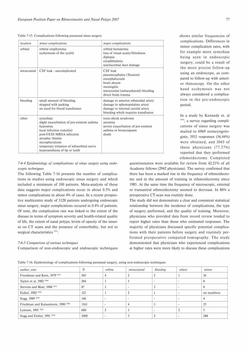
European Position Paper on Rhinosinusitis and Nasal Polyps 2007 77
7-6-4 Epidemiology of complications of sinus surgery using endo-scopic techniquesThe following Table 7-16 presents the number of complica-tions in studies using endoscopic sinus surgery and whichincluded a minimum of 100 patients. Meta-analysis of thesedata suggests major complications occur in about 0.5% andminor complications in about 4% of cases. In a recent prospec-tive multicentre study of 3128 patients undergoing endoscopicsinus surgery, major complications occured in 0.4% of patients.Of note, the complication rate was linked to the extent of thedisease in terms of symptom severity and health-related qualityof life, the extent of nasal polyps, levels of opacity of the sinus-es on CT scans and the presence of comorbidity, but not tosurgical characteristics (521).
7-6-5 Comparison of various techniquesComparison of non-endoscopic and endoscopic techniques
shows similar frequencies ofcomplications. Differences inminor complication rates, withfor example more synechiaebeing seen in endoscopicsurgery, could be a result ofthe more precise follow-upusing an endoscope, as com-pared to follow-up with anteri-or rhinoscopy. On the otherhand ecchymosis was notalways considered a complica-tion in the pre-endoscopicperiod.
In a study by Kennedy et. al(944), a survey regarding compli-cations of sinus surgery wasmailed to 6969 otolaryngolo-gists; 3933 responses (56.44%)were obtained, and 3043 ofthese physicians (77.37%)reported that they performedethmoidectomy. Completed
questionnaires were available for review from 42.21% of allAcademy fellows (2942 physicians). The survey confirmed thatthere has been a marked rise in the frequency of ethmoidecto-my and in the amount of training in ethmoidectomy since1985. At the same time the frequency of microscopic, externalor transantral ethmoidectomy seemed to decrease. In 86% apreoperative CT-scan was routinly done.The study did not demonstrate a clear and consistent statisticalrelationship between the incidence of complications, the typeof surgery performed, and the quality of training. Moreover,physicians who provided data from record review tended toreport higher rates than those who estimated responses. Themajority of physicians discussed specific potential complica-tions with their patients before surgery and routinely per-formed preoperative computed tomography. The studydemonstrated that physicians who experienced complicationsat higher rates were more likely to discuss these complications
Table 7-15. Complications following paranasal sinus surgery
location minor complications major complications
orbital orbital emphysemaecchymosis of the eyelid
orbital hematomaloss of visual acuity/blindnessdiplopiaenophthalmianasolacrimal duct damage
intracranial CSF leak - uncomplicated CSF leakpneumcephalus (Tension)encephalocoelebrain abcessmeningitisintracranial (subarachnoid) bleedingdirect brain trauma
bleeding small amount of bleedingstopped with packingno need for blood transfusion
damage to anterior ethmoidal arterydamage to sphenopalatine arterydamage to internal carotid arterybleeding which requires transfusion
other synechiaeslight exacerbation of pre-existent asthmahyposmialocal infection (osteitis)post-FESS MRSA infectionatrophic rhinitismyospherulosistemporary irritation of infraorbital nervehyperaesthesia of lip or teeth
toxic-shock syndromeanosmiasevere exacerbation of pre-exsitentasthma or broncospasmdeath
Table 7-16. Epidemiology of complications following paranasal surgery, using non-endoscopic techniques
author, year N orbita intracranial bleeding others minor
Freedman and Kern, 1979 (915) 565 4 2 2 1 16
Taylor et al, 1982 (916) 284 1 3 - - 8
Stevens and Blair, 1988 (917) 87 3 - 3 - 8
Eichel, 1982 (918) 123 1 2 1 - no numbers
Sogg, 1989 (919) 146 - - - - 4
Friedman and Katsantonis, 1990 (920) 1163 - 4 3 - 25
Lawson, 1991 (921) 600 2 3 - 2 5
Sogg and Eichel, 1991 (922) 3000 - 5 2 - 288
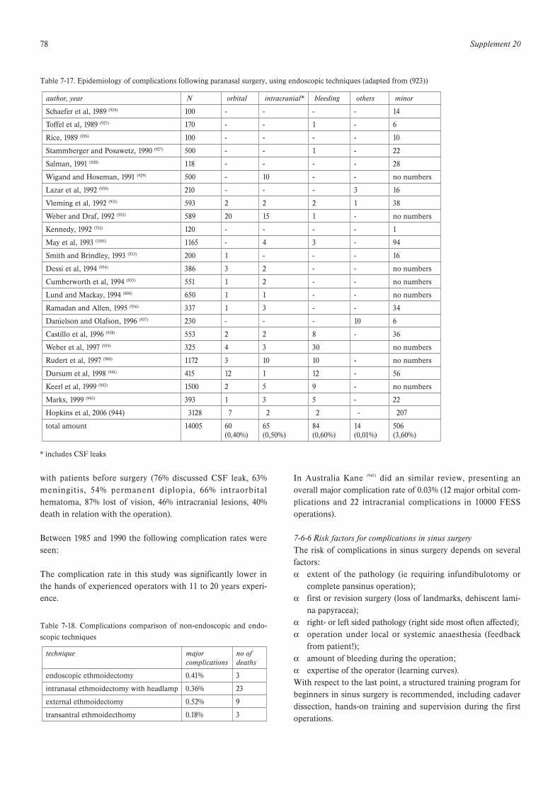
78 Supplement 20
with patients before surgery (76% discussed CSF leak, 63%meningitis, 54% permanent diplopia, 66% intraorbitalhematoma, 87% lost of vision, 46% intracranial lesions, 40%death in relation with the operation).
Between 1985 and 1990 the following complication rates wereseen:
The complication rate in this study was significantly lower inthe hands of experienced operators with 11 to 20 years experi-ence.
In Australia Kane (945) did an similar review, presenting anoverall major complication rate of 0.03% (12 major orbital com-plications and 22 intracranial complications in 10000 FESSoperations).
7-6-6 Risk factors for complications in sinus surgeryThe risk of complications in sinus surgery depends on severalfactors:! extent of the pathology (ie requiring infundibulotomy or
complete pansinus operation);! first or revision surgery (loss of landmarks, dehiscent lami-
na papyracea);! right- or left sided pathology (right side most often affected);! operation under local or systemic anaesthesia (feedback
from patient!);! amount of bleeding during the operation;! expertise of the operator (learning curves).With respect to the last point, a structured training program forbeginners in sinus surgery is recommended, including cadaverdissection, hands-on training and supervision during the firstoperations.
Table 7-17. Epidemiology of complications following paranasal surgery, using endoscopic techniques (adapted from (923))
* includes CSF leaks
author, year N orbital intracranial* bleeding others minor
Schaefer et al, 1989 (924) 100 - - - - 14
Toffel et al, 1989 (925) 170 - - 1 - 6
Rice, 1989 (926) 100 - - - - 10
Stammberger and Posawetz, 1990 (927) 500 - - 1 - 22
Salman, 1991 (928) 118 - - - - 28
Wigand and Hoseman, 1991 (929) 500 - 10 - - no numbers
Lazar et al, 1992 (930) 210 - - - 3 16
Vleming et al, 1992 (931) 593 2 2 2 1 38
Weber and Draf, 1992 (932) 589 20 15 1 - no numbers
Kennedy, 1992 (514) 120 - - - - 1
May et al, 1993 (1105) 1165 - 4 3 - 94
Smith and Brindley, 1993 (933) 200 1 - - - 16
Dessi et al, 1994 (934) 386 3 2 - - no numbers
Cumberworth et al, 1994 (935) 551 1 2 - - no numbers
Lund and Mackay, 1994 (800) 650 1 1 - - no numbers
Ramadan and Allen, 1995 (936) 337 1 3 - - 34
Danielson and Olafson, 1996 (937) 230 - - - 10 6
Castillo et al, 1996 (938) 553 2 2 8 - 36
Weber et al, 1997 (939) 325 4 3 30 no numbers
Rudert et al, 1997 (940) 1172 3 10 10 - no numbers
Dursum et al, 1998 (941) 415 12 1 12 - 56
Keerl et al, 1999 (942) 1500 2 5 9 - no numbers
Marks, 1999 (943) 393 1 3 5 - 22
Hopkins et al, 2006 (944) 3128 7 2 2 - 207
total amount 14005 60(0,40%)
65(0,50%)
84(0,60%)
14 (0,01%)
506(3,60%)
Table 7-18. Complications comparison of non-endoscopic and endo-scopic techniques
technique major complications
no ofdeaths
endoscopic ethmoidectomy 0.41% 3
intranasal ethmoidectomy with headlamp 0.36% 23
external ethmoidectomy 0.52% 9
transantral ethmoidecthomy 0.18% 3
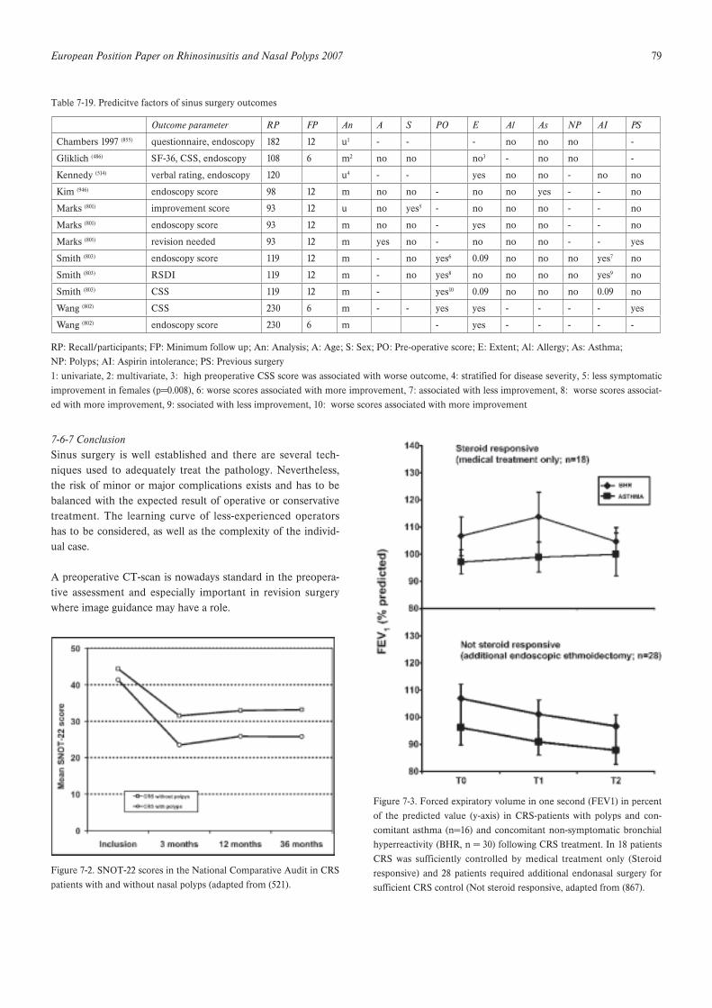
European Position Paper on Rhinosinusitis and Nasal Polyps 2007 79
7-6-7 ConclusionSinus surgery is well established and there are several tech-niques used to adequately treat the pathology. Nevertheless,the risk of minor or major complications exists and has to bebalanced with the expected result of operative or conservativetreatment. The learning curve of less-experienced operatorshas to be considered, as well as the complexity of the individ-ual case.
A preoperative CT-scan is nowadays standard in the preopera-tive assessment and especially important in revision surgerywhere image guidance may have a role.
Table 7-19. Predicitve factors of sinus surgery outcomes
RP: Recall/participants; FP: Minimum follow up; An: Analysis; A: Age; S: Sex; PO: Pre-operative score; E: Extent; Al: Allergy; As: Asthma;NP: Polyps; AI: Aspirin intolerance; PS: Previous surgery1: univariate, 2: multivariate, 3: high preoperative CSS score was associated with worse outcome, 4: stratified for disease severity, 5: less symptomaticimprovement in females (p=0.008), 6: worse scores associated with more improvement, 7: associated with less improvement, 8: worse scores associat-ed with more improvement, 9: ssociated with less improvement, 10: worse scores associated with more improvement
Outcome parameter RP FP An A S PO E Al As NP AI PS
Chambers 1997 (855) questionnaire, endoscopy 182 12 u1 - - - no no no -
Gliklich (486) SF-36, CSS, endoscopy 108 6 m2 no no no3 - no no -
Kennedy (514) verbal rating, endoscopy 120 u4 - - yes no no - no no
Kim (946) endoscopy score 98 12 m no no - no no yes - - no
Marks (801) improvement score 93 12 u no yes5 - no no no - - no
Marks (801) endoscopy score 93 12 m no no - yes no no - - no
Marks (801) revision needed 93 12 m yes no - no no no - - yes
Smith (803) endoscopy score 119 12 m - no yes6 0.09 no no no yes7 no
Smith (803) RSDI 119 12 m - no yes8 no no no no yes9 no
Smith (803) CSS 119 12 m - yes10 0.09 no no no 0.09 no
Wang (802) CSS 230 6 m - - yes yes - - - - yes
Wang (802) endoscopy score 230 6 m - yes - - - - -
a
Figure 7-2. SNOT-22 scores in the National Comparative Audit in CRSpatients with and without nasal polyps (adapted from (521).
Figure 7-3. Forced expiratory volume in one second (FEV1) in percentof the predicted value (y-axis) in CRS-patients with polyps and con-comitant asthma (n=16) and concomitant non-symptomatic bronchialhyperreactivity (BHR, n = 30) following CRS treatment. In 18 patientsCRS was sufficiently controlled by medical treatment only (Steroidresponsive) and 28 patients required additional endonasal surgery forsufficient CRS control (Not steroid responsive, adapted from (867).
80 Supplement 20
8-1 Introduction
In the pre-antibiotic era, complications of rhinosinusitis repre-sented extremely common and dangerous clinical events.Today, thanks to more reliable diagnostic methods (CT, MRI)and to the wide range of available antibiotics, their incidenceand related mortality have dramatically decreased. In somecases however, if sinus infection is untreated or inadequatelytreated, complications can still develop (947). In patients affectedby acute bacterial rhinosinusitis with intracranial spreaddespite antibiotic therapy, there still is a high incidence of mor-bidity and mortality rate, estimated at between 5% and 10% (948).
Complications of rhinosinusitis are classically defined asorbital, osseous and endocranial (948) though rarely some unusu-al complications can develop (Table 8-1) (949-953).
An extremely useful test, although not specific, is the whitecell count which, if elevated in ARS unresponsive to treat-ment, is highly suggestive of a complication.
8-2 Epidemiology of complications
Epidemiological data concerning the complications of rhinosi-nusitis vary widely and there is no consensus on the exactprevalence of the different types of complications. Moreover,the relationship between acute or chronic rhinosinusitis andthe various complications is not clearly defined in the litera-ture. This is probably related to the different number andmethods of sampling patients in the various studies and noaccount is taken of local demographics. For these reasons, asTable 8-1 clearly shows, an attempt to make a comparison ofthe different epidemiological data available is difficult.
For example, whilst the percentage is similar in two studiesthat compared two different groups of selected patients affect-ed with pansinusitis (72.4% and 75% respectively) (472,473), the per-centage in another (955) is smaller (37%); this is probably due tothe fact that in this sample, both acute and chronic diseasewere studied, whereas the other two authors focused theirattention on acute cases.
In another mixed (acute and chronic) sample, Clayman high-lighted the frequency of intracranial complications in patientwith complicated rhinosinusitis as about 3.7 %, but no data con-cerning the global prevalence of complications were given. (959).
8-3 Orbital complications
8-3-1 SystemicIf there is a complication in rhinosinusitis, the eye is ofteninvolved (956, 1111) espeacialy in ethmoiditis, whereas this is rare insphenoidal infection (961). The spread of infection directly viathe thin and often dehiscent lamina papyracea (961); or by veins(962) occurs with relative ease.
According to Chandler’s classification orbital complicationsmay progress in the following steps (963):
periorbital cellulitis (preseptal edema),orbital cellulitis,subperiosteal abscess,orbital abscess or phlegmon andcavernous sinus thrombosis (947, 964).
Moreover orbital complications especially in children, oftenoccur without pain (965, 1106). Orbital involvement is manifested by
8. Complications of rhinosinusitis and nasal polyps
Table 8-1. Epidemiological data of complications in rhinosinusitis
author country age pathology pts total % of complications
orbital intracranial osseous soft tissue
Mortimore, 1999 (954)
South Africa adults acute pansinusitis 87 72.4% (63/87)
Ogunleye, 2001 (955)
Nigeria adults acute/chronic pansinusitis
90 37% (33/90) 41% 5% 32% 18%
Eufinger, 2001 (956)
Germany adults/ children
acute pansinusitis 36 75% (27/36) 58% (20+1/36) 11% (3+1/36) 8.4% (3/36)
Kuranov, 2001 (957)
Russia adults rhinosinusitis 0.8% 0.01%
Gallagher, 1998 (958)
USA adults rhinosinusitis 176 8.5% (15/176)
Clayman, 1991 (959)
USA adults acute/chronic rhinosinusitis
649 3.7% (24/649)
Lerner, 1995 (960) USA children rhinosinusitis 443 3%(14/443)

European Position Paper on Rhinosinusitis and Nasal Polyps 2007 81
swelling, exophthalmos, and impaired extra-ocular eye move-ments (966). Periorbital or orbital cellulitis may result from director vascular spread of the sinus infection. As the spread of sinusinfection through the orbit follows a well-described pattern,the initial manifestations are oedema and erythema of themedial aspects of the eyelid. Spread of infection from the max-illary or frontal sinus produces swelling of the lower or uppereyelid, respectively (964).
8-3-2 Periorbital cellulitisPeriorbital cellulitis (inflammation of the eyelid and conjuncti-va) (953) involves the tissue anterior to the orbital septum and isreadily seen on CT scan as soft tissue swelling. It is the mostcommon complication of rhinosinusitis in children (967) and itmanifests itself as orbital pain, blepharal oedema and highfever (968). Periorbital cellulitis usually responds to an oralantibiotic appropriate to common sinus organisms but if notaggressively treated, may spread beyond the orbital septum (967).
8-3-3 Orbital cellulitisAs the inflammatory changes spread beyond the orbital sep-tum, proptosis develops together with some limitation of ocu-lar motion, indicating orbital cellulitis. Further signs are con-junctival oedema (chemosis), a protruding eyeball (proptosis),ocular pain and tenderness, and decreased movement of theextraocular muscles (953, 969).
This complication requires aggressive treatment with intra-venous antibiotics.
Any children with rhinosinusitis and proptosis, ophthalmople-gia, or decreased visual acuity should have a CT scan of thesinuses with orbital detail to distinguish between an orbitaland periorbital (subperiosteal) abscess. Both conditions causeproptosis and limited ocular movement. Evidence of an abcesson the CT scan or progressive orbital findings after initial i.v.antibiotic therapy are indications for orbital exploration anddrainage. Repeated ophthalmologic examination of visual acu-ity should take place and i.v. antibiotic therapy may be con-verted into oral when the patient has been afebrile for 48 hoursif the ophthalmological symptoms and signs are resolving (967).
8-3-4 Subperiosteal or orbital abscessThe clinical features of a subperiosteal abscess are oedema,erythema, chemosis and proptosis of the eyelid with limitationof ocular motility and as a consequence of extra-ocular muscleparalysis, the globe becomes fixed (ophthalmoplegia) and visu-al acuity diminishes.
An orbital abcess generally results from diagnostic delay or toimmunosuppression of the patient (968) with a frequency of 9%and 8.3% (351, 970) in paediatric studies.
A CT scan of the sinuses with orbital sequences to distinguishbetween orbital and periorbital (subperiosteal) abscess shouldbe performed. Evidence of an abscess on the CT scan orabsence of clinical improvement after 24-48 hours of i.v. antibi-otics are indications for orbital exploration and drainage.Anophthalmologist should check visual acuity from the earlystages of the illness and i.v. therapy should cover aerobic andanaerobic pathogens. It can be converted to an oral preparationwhen the patient has been afebrile for 48 hours (967).
Blindness may result from central retinal artery occlusion,optic neuritis, corneal ulceration, or pan-ophthalmitis. In sucha case the CT usually reveals oedema of the medial rectusmuscle, lateralization of the periorbita, and displacement ofthe globe downward and laterally. When the CT scan showsobliteration of the detail of the extraocular muscle and theoptic nerve by a confluent mass, the orbital cellulitis has pro-gressed to an abscess, in which there is sometimes air due toanaerobic bacteria. Sepsis not infrequently can spread intracra-nially as well as anteriorly into the orbit (971).
8-4 Endocranial complications
These include epidural or subdural abscesses, brain abscess,meningitis (most commonly), cerebritis, and cavernous sinusthrombosis (967, 972, 973).
The clinical presentation of all these complication is non-spe-cific, being characterized by high fever, frontal or retro-orbitalmigraine, generic signs of meningeal irritation and by variousdegrees of altered mental state(958) while intracranial abscessesare often heralded by signs of increased intracranial pressure,meningeal irritation, and focal neurologic deficits (966). Althoughan intracranial abscess is relatively asymptomatic, subtle affec-tive and behavioral changes often occur showing altered neu-rologic function, altered consciousness, gait instability, andsevere, progressive headache (953, 967).
Endocranial complications are most often associated with eth-moidal or frontal rhinosinusitis. Infections can proceed fromthe paranasal cavities to the endocranial structures by two dif-ferent routes: pathogens, starting from the frontal sinus mostcommonly or ethmoid sinus, can pass through the diploicveins to reach the brain; alternatively, they can reach theintracranial structures by eroding the sinus bones (958).
All endocranial complications start as cerebritis, but as necrosisand liquefaction of brain tissue progresses, a capsule developsresulting in brain abscess. Studies show a high incidence ofanaerobic organisms or mixed aerobic-anaerobic in patientswith CNS complications.
A CT scan is essential for diagnosis as it allows an extremelyaccurate definition of bone involvement, whereas MRI is
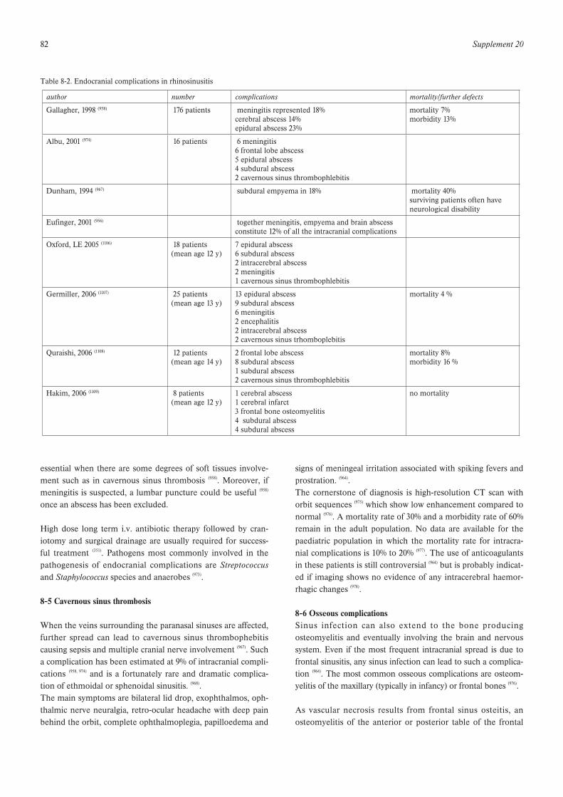
82 Supplement 20
essential when there are some degrees of soft tissues involve-ment such as in cavernous sinus thrombosis (958). Moreover, ifmeningitis is suspected, a lumbar puncture could be useful (958)
once an abscess has been excluded.
High dose long term i.v. antibiotic therapy followed by cran-iotomy and surgical drainage are usually required for success-ful treatment (351). Pathogens most commonly involved in thepathogenesis of endocranial complications are Streptococcusand Staphylococcus species and anaerobes (973).
8-5 Cavernous sinus thrombosis
When the veins surrounding the paranasal sinuses are affected,further spread can lead to cavernous sinus thrombophebitiscausing sepsis and multiple cranial nerve involvement (967). Sucha complication has been estimated at 9% of intracranial compli-cations (958, 974) and is a fortunately rare and dramatic complica-tion of ethmoidal or sphenoidal sinusitis. (968).The main symptoms are bilateral lid drop, exophthalmos, oph-thalmic nerve neuralgia, retro-ocular headache with deep painbehind the orbit, complete ophthalmoplegia, papilloedema and
signs of meningeal irritation associated with spiking fevers andprostration. (964).The cornerstone of diagnosis is high-resolution CT scan withorbit sequences (975) which show low enhancement compared tonormal (976). A mortality rate of 30% and a morbidity rate of 60%remain in the adult population. No data are available for thepaediatric population in which the mortality rate for intracra-nial complications is 10% to 20% (977). The use of anticoagulantsin these patients is still controversial (964) but is probably indicat-ed if imaging shows no evidence of any intracerebral haemor-rhagic changes (978).
8-6 Osseous complicationsSinus infection can also extend to the bone producingosteomyelitis and eventually involving the brain and nervoussystem. Even if the most frequent intracranial spread is due tofrontal sinusitis, any sinus infection can lead to such a complica-tion (964). The most common osseous complications are osteom-yelitis of the maxillary (typically in infancy) or frontal bones (976).
As vascular necrosis results from frontal sinus osteitis, anosteomyelitis of the anterior or posterior table of the frontal
Table 8-2. Endocranial complications in rhinosinusitis
author number complications mortality/further defects
Gallagher, 1998 (958) 176 patients meningitis represented 18%cerebral abscess 14%epidural abscess 23%
mortality 7%morbidity 13%
Albu, 2001 (974) 16 patients 6 meningitis6 frontal lobe abscess5 epidural abscess4 subdural abscess2 cavernous sinus thrombophlebitis
Dunham, 1994 (967) subdural empyema in 18% mortality 40% surviving patients often haveneurological disability
Eufinger, 2001 (956) together meningitis, empyema and brain abscess constitute 12% of all the intracranial complications
Oxford, LE 2005 (1106) 18 patients(mean age 12 y)
7 epidural abscess6 subdural abscess2 intracerebral abscess2 meningitis1 cavernous sinus thrombophlebitis
Germiller, 2006 (1107) 25 patients(mean age 13 y)
13 epidural abscess9 subdural abscess6 meningitis2 encephalitis2 intracerebral abscess2 cavernous sinus trhomboplebitis
mortality 4 %
Quraishi, 2006 (1108) 12 patients(mean age 14 y)
2 frontal lobe abscess8 subdural abscess1 subdural abscess2 cavernous sinus thrombophlebitis
mortality 8%morbidity 16 %
Hakim, 2006 (1109) 8 patients(mean age 12 y)
1 cerebral abscess1 cerebral infarct3 frontal bone osteomyelitis4 subdural abscess4 subdural abscess
no mortality
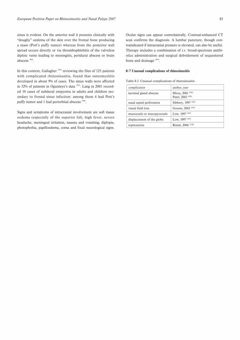
European Position Paper on Rhinosinusitis and Nasal Polyps 2007 83
sinus is evident. On the anterior wall it presents clinically with“doughy” oedema of the skin over the frontal bone producinga mass (Pott’s puffy tumor) whereas from the posterior wallspread occurs directly or via thrombophlebitis of the valvelessdiploic veins leading to meningitis, peridural abscess or brainabscess (964).
In this context, Gallagher (958) reviewing the files of 125 patientswith complicated rhinosinusitis, found that osteomyelitisdeveloped in about 9% of cases. The sinus walls were affectedin 32% of patients in Ogunleye’s data (955). Lang in 2001 record-ed 10 cases of subdural empyema in adults and children sec-ondary to frontal sinus infection: among them 4 had Pott’spuffy tumor and 1 had periorbital abscess (948).
Signs and symptoms of intracranial involvement are soft tissueoedema (especially of the superior lid), high fever, severeheadache, meningeal irritation, nausea and vomiting, diplopia,photophobia, papilloedema, coma and focal neurological signs.
Ocular signs can appear controlaterally. Contrast-enhanced CTscan confirms the diagnosis. A lumbar puncture, though con-traindicated if intracranial pressure is elevated, can also be useful.Therapy includes a combination of i.v. broad-spectrum antibi-otics administration and surgical debridement of sequesteredbone and drainage (964).
8-7 Unusual complications of rhinosinusitis
Table 8-3. Unusual complications of rhinosinusitis
complication author, year
lacrimal gland abscess Mirza, 2001 (949)
Patel, 2003 (950)
nasal septal perforation Sibbery, 1997 (979)
visual field loss Gouws, 2003 (952)
mucocoele or mucopyocoele Low, 1997 (972)
displacement of the globe Low, 1997 (972)
septicaemia Rimal, 2006 (1110)

84 Supplement 20
9-1 Introduction
Rhinosinusitis is a common problem in children that is oftenoverlooked. It is a multifactorial disease in which the impor-tance of several predisposing factors changes with age and isdifferent from the adult form of the disease in many respects(Table 9-1). The management of rhinosinusitis in children is acontroversial and rapidly evolving issue.
9-2 Anatomy
In the newborn, the maxillary sinus extends to a depth ofabout 7 mm, is 3 mm wide and 7 mm high (980). In the newborn,two to three ethmoid cells are found bilaterally, and by the ageof four the ethmoid labyrinth has formed. The sphenoid sinus-es are also present in the neonate. Each sphenoid sinus is 4mm wide and 2 mm high. At birth the frontal sinuses are not
present, but they gradually develop from the anterior ethmoidcells into the cranium. When the upper edge of the aircell(cupola) reaches the same level as the roof of the orbit, it canbe termed a frontal sinus, a situation that appears around theage of five. When a child reaches the age of 7-8 years the floorof the maxillary sinus already occupies the same level as thenasal floor.
9-3 Epidemiology and pathophysiology
Since the introduction of CT-scanning, it has become clearthat a runny nose in a child is not only due to limited rhinitisor adenoid hypertrophy, but that in the majority of the casesthe sinuses are involved as well. Van der Veken (220) in a CTscan study showed that in children with a history of chronicpurulent rhinorrhea and nasal obstruction, 64 % showedinvolvement of the sinuses. In a MRI study of a non-ENT pae-diatric population (981) it was shown that the overall prevalenceof sinusitis signs in children is 45 %. This prevalence increasesin the presence of a history of nasal obstruction to 50 %, to 80% when bilateral mucosal swelling is present on rhinoscopy, to81 % after a recent upper respiratory tract infection (URTI),and to 100 % in the presence of purulent secretions. AlsoKristo et al (982) found a similar overall percentage (50 %) ofabnormalities on MRI in 24 school children. They included,however, a follow-up after 6 to 7 months, and found that abouthalf of the abnormal sinuses on MRI findings had resolved orimproved without any intervention.Epidemiologic studies on rhinosinusitis in children are limitedbut reveal the following information on the pathophysiologyand clinically relevant factors influencing the prevalence of rhi-nosinusitis in children:1. There is a clear-cut decrease in the prevalence of rhinosi-nusitis after 6 to 8 years of age. This is the natural history ofthe disease in children and is probably related to an immatureimmune system in the younger child (222, 223)
2. In temperate climates there is a definite increase in theoccurrence of CRS in children during the autumn and in thewintertime, so that the season seems to be another importantfactor (222).3. Younger children staying in day care centres show a dramat-ic increase in the prevalence of chronic or recurrent rhinosi-nusitis compared to children staying at home. See also section1-1.
Although viruses are uncommonly recovered from sinus aspi-rates (983), most authors agree (984, 985) that viral infections are thetrigger to rhinosinusitis. Although CT scan abnormalites canbe seen up to several weeks after the onset of a URTI, one canassume that only 5 to 10 % of the URTIs in early childhood are
9. Special considerations: Rhinosinusitis in children special
Table 9-1. Differences between paediatric and adult chronic rhinosinusitis
young children adults
Commensal microflora
Coagulase negative staphylococci
30% 35%
Staphylococcus aureus 20% 8%
Haemophilus influenzae 40% 0%
Moraxella catarrhalis 24% 0%
Streptococcus pneumoniae 50% 26%
Corynebacterium species 52% 23%
Streptococcus viridans 30% 4%
Immunity immature: defective responseto polysaccharide antigens(IgG2, IgA)
mature, exceptin a subset
History self-limited in time (improves after the age of 6-8years)
no history ofspontaneousimprovementafter certain age
Histology mainly neutrophi-lic disease, lessbasement memb-rane thickening and mucus glandhyperplasia, more mast cells (Sobo,2003)
mainlyeosinophils
Endoscopy polyps are rare, except in CF
polyps frequentlypresent
CT-scan younger child more diffusesinusitis, involving all sinus
sphenoid andposterior sinusless ofteninvolved

European Position Paper on Rhinosinusitis and Nasal Polyps 2007 85
complicated by ARS (986). The time course (i.e. clinical symp-toms) of viral to bacterial rhinosinusitis is the same as inadults.
The most common bacterial species isolated from the maxil-lary sinuses of patients with ARS are Streptococcus pneumoniae,Haemophilus influenzae, and Moraxella catarrhalis, the latterbeing more common in children (45, 46).
Antral punctures are now rarely performed in children but it isinteresting to know from studies in the past there is a good cor-relation of bacteriology between the maxillary sinus and themiddle meatal specimen (83 %), and a poor correlation betweenthose of the nasopharynx and the maxillary sinus (45 %) (987).
9-4 Symptoms and signs
Wald stresses that 3 common clinical developments shouldalert a clinician to the possibility of rhinosinusitis (990):1. signs and symptoms of a cold that are persisting beyond 10
days (any nasal discharge, daytime cough worsening at night)2. a cold that seems more severe then usual (high fever, copi-
ous purulent discharge, peri-orbital oedema and pain)3. a cold that after several days of improvement worsen (with
or without fever) .
The neutrophil content of nasal brushings if > or = 5% pre-dicts maxillary sinusitis as judged from x-rays with a sensitivi-ty of 91% and a predictive value of 84%, but only in non-aller-gic children (991).
9-5 Clinical examination
Physical examination of a child's nose is often difficult, andonly limited rhinoscopy is tolerated. The examination may besimply accomplished by lifting the tip of the nose upwards(young children have wide noses with round nostrils, allowingeasy examination of the condition of the inferior turbinates).Another convenient method is the use of an otoscope (992, 993).Usually the nasal and pharyngeal mucosa appears erythema-tous with yellow to greenish purulent rhinorrhoea of varyingviscosity. A post nasal drip was seen in 60%, pus in the middle
meatus in 50% in one study (989). Turbinate swelling was presentin 29% in another (988). Lymphoid hyperplasia of the tonsils,adenoids and parapharyngeal wall may also be observed. Thecervical lymph nodes may be moderately enlarged and slightlytender (992, 994).
Anterior rhinoscopy remains the first step but is inadequate byitself.Endoscopy with a 2.7 mm rigid endoscope in the younger child(when possible a 4 mm in the older child) is more useful thenflexible nasendoscopy, not only for the diagnosis but also forthe exclusion of other conditions such as: presence of polyps,foreign bodies, tumours and septal deviations. In the youngerchild total anaesthesia is necessary to perform a thorough nasalendoscopy. Moreover it allows direct sampling of middle mea-tus flora.
9-6 Investigations
9-6-1 MicrobiologyMicrobiological assessment is usually not necessary in childrenwith uncomplicated acute or chronic rhinosinusitis.Indication for microbiology are: (995)
1. severe illness or toxic child;2. acute illness in a child not improving with medical therapy
within 48-72 hours;3. an immuocompromised host;4. the presence of suppurative (intra-orbital, intracranial)
complications (orbital cellulitis excepted).Quantification of bacterial growth can also help in distinguish-ing contamination from real infection, and isolates should beconsidered positive when a type of bacteria is present in aquantity of at least 10000 colonies/ml (990).
9-6-2 ImagingImaging is not necessary to confirm the diagnosis of rhinosi-nusitis in children. The increase in thickness of both the softtissue and the bony vault of the palate in children under 10years of age limits the usefulness of transillumination andultrasonography in the younger age group (505).Plain x-rays are insensitive with limited usefulness for diagno-sis or to guide surgery and correlate very poorly with CT scans.The marginal benefits are insufficient to justify the exposure toradiation (220).CT scanning remains the imaging methodology of choice,because of its ability to resolve both bone and soft tissue, withgood visualisation of the ostiomeatal complex. The indicationsfor CT scanning in a child are the same as those given previ-ously for a microbiology specimen with one extra indicationwhich is if surgery is being considered after failure of medicaltherapy. The high incidence of asymptomatic children with CTscan abnormalities (996) must be remembered plus the fact thatsuch children do not require treatment (997).
Table 9-2. Presenting symptoms of rhinosinusitis in children. (223, 988, 989).
rhinorrhoea (71 to 80 %) all forms
cough (50 to 80 %) all forms
fever (50 to 60 %) acute
pain (29 to 33 %) acute
nasal obstruction (70 to 100 %) chronic
mouth breathing (70 to 100 %) chronic
ear complaints (recurrent purulent otitis media or OME in 40 to 68 %)
chronic

86 Supplement 20
A number of studies suggest that the growth of the maxillarysinus is not impaired by extensive or chronic disease, unlikethe temporal bone and it seems that the presence of ahypoplastic maxillary sinus per se is not an indication forsurgery (998)
9-6-3 Additional investigationsIn the presence of recalcitrant rhinosinusitis, underlying condi-tions must be considered, preferably before undertaking anysurgical procedure.
9-6-3-1 AllergyThe role of atopy in chronic rhinosinusitis is unclear. Manyauthors attribute a great deal of importance to allergy (101, 988, 993)
although others (61, 220, 999) did not find an increased prevalence ofrhinosinusitis in allergic children.In a CT scan study Iwens et al. (1994) found signs of mucosalinflammation in 61 % of atopic children (61). Ramadan (1999)showed that allergic patients had a higher CT scan score thannon-allergic patients (875). Allergic children have more URTproblems and more time off school than their non-allergicpeers (1000). Therefore in children with CRS and with a sugges-tive history (asthma, eczema), and/or physical examinationfindings (allergic salute, watery rhinorrhea, nasal blockage,sneezing, boggy turbinate), allergic assessment (skin prick,RAST) should be performed.
9-6-3-2 Immune deficiencyAll young children have a physiologic primary immune deficien-cy (993, 1001). Defence against polysaccharide encapsulated bacteriavia immunoglobulin G subclasses 2 and 4 may not reach adultlevels until the age of 10 years (1002). IgG subclass deficiency canlead to protracted or chronic rhinosinusitis (1003-1005). According toPolmar (1004) recurrent and chronic rhinosinusitis is the mostcommon clinical presentation of common variable immunodefi-ciencies. Although not all patients who lack secretory IgA anti-bodies have an increased number of more severe respiratoryinfections, the subject who has IgA deficiency and chronic rhi-nosinusitis is a difficult management problem, especially if anIgG subclass deficiency is also present. Replacement therapycannot be provided (1005). Patients with primary or acquiredimmune deficiencies (e.g. treatment for malignancies, organtransplants, maternally transmitted AIDS or blood-transmittedAIDS in haemophilics, drug induced conditions) are at risk fordeveloping a difficult-to-treat rhinosinusitis with resistant oruncommon micro-organisms and fungi. Also the initial signsand symptoms may be non-specific, such as thin rhinorrhea,mild congestion, and chronic cough (993).
9-6-3-3 Cystic fibrosisCystic fibrosis is caused by a mutation of the gene FES1encoding the cystic fibrosis transmembrane conductance regu-lator (CFTR). This gene contains 27 exons encompassingapproximately 252 kb of DNA on chromosone 7q31.2. The
most common mutation, deletion of phenylalanine at position508 (F508) accounts for nearly 70 % of mutuations inEuropean-derived Caucasian population (1006).
In children with cystic fibrosis, sinusitis is a common problem.Although the prevalence of nasal disease was previously esti-mated to be between 6 and 20% (1007), Yung et al (1008) found it tobe over 50 % and Brihaye et al. (1009) reported that performingrigid endoscopy in 84 patients with cystic fibrosis, revealedinflammatory polyps in 45 % (mean age 15 years) and medialbulging of the lateral nasal wall in 12 % (mean age 5 years). Inpatients with cystic fibrosis and chronic rhinosinusitis, CTshowed in 100 % (1009) opacification of the anterior complex(anterior ethmoid, maxillary and -if developed- frontal sinus)and 57 % showed clouding of the posterior complex (posteriorethmoid and sphenoid). In all children with a medial displace-ment of the lateral nasal wall, there was a soft tissue mass in themaxillary antrum (large quantity of secretions surrounded bypolyposal mucosa, representing a mucopurulent rhinosinusi-tus). In 80 % of these children the displacement was so extremethat the lateral nasal wall touched the septum, resulting in totalnasal blockage. In a study by Brihaye et al (1009) massive polyposiswas never found before the age of 5 years. Mucopurulence inthe maxillary sinus occurs at a younger age (3 months to 8years) and the maxillary sinus seems to be the first sinus affect-ed by the disease. Recent data suggest that CF heterozygotesare over-represented in the paediatric CRS population (1010).
9-6-3-4 Primary ciliary dyslcinesiaPrimary ciliary dyskinesia (PCD) (1011), an autosomal recessive dis-order involving dysfunction of cilia is present in 1 of 15000 of thepopulation and should always be considered in any neonate withrespiratory or ENT problems of unknown origin. At least half ofthe PCD patients have symptoms when first born and especiallyin a term baby with no risk factor for congenital infection show-ing signs of rhinitis at birth, PCD should be excluded. The sameapplies to an infant or older child with atypical asthma, unre-sponsive to treatment, chronic wet cough and sputum produc-tion, very severe gastro-oesophageal reflux, bronchiectasis, rhi-nosinusitis (rarely with polyposis), chronic and severe secretoryotitis media, particularly with continuous, long lasting and diffusedischarge from the ears after grommet insertion. Roughly halfthe children with PCD have situs inversus and bronchiectasis inaddition to rhinosinusitis (Kartagener’s syndrome).
There are two ways to screen for PCD: saccharine test andnasal nitric oxide. The saccharine test is a cheap and easy pro-cedure to screen older children and adults, but is relativelyunreliable Nasal nitric oxide (nNO) measurements can bemade in children over 5years old. Recent data suggests thatPCD epithelia have a common defect – a lack of induciblenitric oxide synthase. In PCD values of nNO are usually lessthan 100; values exceeding 250ppb have a sensitivity of 95 %for excluding the diagnosis of PCD (1012). Since very low nNO

European Position Paper on Rhinosinusitis and Nasal Polyps 2007 87
values can occur with severe nasal congestion the procedureshould be repeated after decongestion or a brief course of oralplus topical corticosteroids.If the child is too young for the tests, if there is any doubtabout the validity of the saccharine test, or the results are posi-tive (transport time longer than 60 minutes, nNO less than250ppb) or there exists a strong clinical suspicion, the ciliarybeat frequency should be tested from a nasal epithelial biopsy.
If direct inspection of the ciliary beat frequency is abnormal(less than 11-16 Hz) an ultrastructure study of cilia is needed.The most common ciliary abnormalities in PCD are: dyneinarm defects (absence or reduced number of inner, outer orboth dynein arms), tubular defects (transposition and extramicrotubules), radial spokes defects or absence, ciliary dysori-entation (suspected if mean standard deviation of angle is larg-er than 20°), abnormal basal apparatus, ciliary aplasia, abnor-mally long cilia (1013). Many of these abnormalities on TEM(transmission electron microscopy), however, can be transientor occur secondarily after infection. Secondary ciliary dyskine-sia, the acquired form (infections, inflammatory or toxic) ismostly correlated with other anomalies, such as microtubularabnormalities and composed cilia. However, there exists agreat overlap of ultrastructural abnormalities between the two(534). Therefore the study of cilia after sequential monolayer-sus-pension culture technique excludes the acquired form (1014).
9-6-3-5 Gastro-oesophageal refluxThe parallel existence of upper airway inflammation with ensu-ing problems of intractable rhinosinusitis, otitis, and gastro-oesophageal reflux (GER) has been observed and suggests acausal relationship. Barbero found in a group of patients withupper airway disease and GER, that anti-reflux measures maypermit a greater well-being and that GER maybe among thevariables leading to refractory chronic upper airway disease (1015).The otolaryngologist should be suspicious of GER in childrencomplaining of chronic nasal discharge and obstruction com-bined with chronic cough, hoarseness and stridulous respira-tion. The endoscopic appearance of the laryngeal and trachealareas are of considerable importance in conjunction withoesophageal examination, in determining the potential rela-tionship between GER and otolaryngologic abnormalities. Thediagnosis needs to be confirmed by oesophageal 24 hours pHmonitoring: in 30 children with chronic sinus disease 63 % hadoesophageal reflux and 32 % had nasopharyngeal reflux (772).
9-7 Management
9-7-1 Management of acute rhinosinusitis in childrenJust as in adults acute rhinosinusitis in children usually onlyneeds symptomatic treatment.
9-7-1-1 Antibiotics in ARS in childrenA Cochrane meta-analysis (1016) of antibiotics for persistent nasal
discharge concluded that antibiotics given for 10 days reducedthe probability of persistence in the short to medium term.The benefits were modest and for 8 children treated one addi-tional child would be cured (NNT 8, 95% CI 5 to 29). No longterm benefits were documented. This meta-analysis is a combi-nation of studies in rhinosinusitis in children with symptomsfor as little as 10 days (1017) to more than 3 months (1018). Twomore recent RCT comparing antibiotics to placebo or anothertherapy do not alter the conclusions of the meta – analysis (1019,
1020). According to the members of the consensus meeting inBrussels, 1996: Management of rhinosinusitis in children: (1021)
antibiotics should be reserved largely for severe disease e.g.:1. a severe illness or toxic condition in a child with suspected
or proven suppurative complication. Intravenous adminis-tration of an appropriate agent is recommended. Theantibiotic selected should be effective against the peni-cillin-resistant Streptococcus pneumonia, beta-lactamaseproducing H. influenzae and Moraxella catarrhalis
2. severe acute rhinosinusitis: in ambulatory patients forwhom oral therapy is appropriate, an agent should beselected that is resistant to the action of beta-lactamaseenzymes (amoxicillin-potassium clavunate or a second gen-eration cephalosporin such as cefuroxime axetil)
3. non-severe acute rhinosinusitis: only in a child with pro-tracted symptoms to whom antibiotics can be given on anindividual basis (presence of asthma, chronic bronchitis,acute otitis media, etc.)
In those children for whom antibiotic therapy is preferred,amoxycillin (45 mg/kg/day, doubled if under 2 or with risk fac-tors for resistance) is appropriate. If the patient's condition hasnot improved within 72 hours, a change of antibiotic to anagent effective against the resistant organism prevalent in thecommunity should be considered.
Patients with a penicillin allergy should receive a suitable alter-native antibiotic such as azithromycin or clarithromycin asfirst-line therapy.
9-7-1-2 Topical corticosteroids in ARSTopical corticosteroids may be a useful ancillary treatment toantibiotics in childhood rhinosinusitis, effective in reducing thecough and nasal discharge earlier in the course of ARS (610 , 1022).There are a large number of studies showing that local corti-costeroids are effective and safe in children with rhinitis (1023-1027).
9-7-1-3 Topical or oral decongestantsMost authors prefer topical !2 agonists (xylo- and oxymetazo-line) in appropriate concentrations. Careful dosage is impor-tant when treating infants and young children, to prevent toxicmanifestations.A double blind, randomised controlled trial (RCT) by Michel (1028)
(III, no power), compared isotonic EMS solution (balneothera-peutic water) with xylometazoline 0.05% solution in the treat-

88 Supplement 20
ment of acute rhinosinusitis with middle ear involvement during14 days in 66 children, aged 2-6 years, and revealed no differencein improvement in both groups, in terms of mucosal inflamma-tion, nasal patency, middle ear function and general state ofhealth, as outcome measures (1028). A double blind, randomisedcontrolled trial (RCT) by McCormick (707) showed no additiveeffect of adding decongestant-antihistamine (nasal oxymeta-zolone and oral syrup containing brompheniramine and phenyl-propanolamine) to amoxicillin (III, no power).
9-7-1-4 Nasal douchingSaline nose drops or sprays are popular with paediatricians (993,
994, 1005). As long as the saline is isotonic and at body tempera-ture, it can help in eliminating nasal secretions and it candecrease nasal oedema.
9-7-2 Management of chronic rhinosinusitis in childrenChronic rhinosinusitis in the young child does not have to betreated, as spontaneous resolution is the norm (1029). Van Buchemet al. followed 169 children with a runny nose for 6 months,treating them only with decongestants or saline nose drops.They did not find a single child who developed a clinically seri-ous disease with general symptoms such as marked pain, pres-sure on sinuses, local swelling, or empyema, showing that com-plications of rhinosinusitis in a child are uncommon (223).
9-7-2-1 Treatment of chronic rhinosinusitisThe data on specific treatment of CRS in children very are lim-ited. A quality of life tool for children SN-5 is now available (584).
9-7-2-2 AntibioticsAs described under 9-7-1-1 Antibiotic in ARS in children aCochrane meta-analysis (1016) of antibiotics for persistent nasaldischarge concluded that antibiotics given for 10 days reducedthe probability of persistence in the short to medium term.The benefits were modest and for 8 children treated one addi-tional child would be cured (NNT 8, 95% CI 5 to 29). No longterm benefits were documented. The only study really treatingCRS (1018) was negative.
9-7-2-3 Topical corticosteroidsThere are no data describing the efficacy of topical corticos-teroids in paediatric CRS. There are a large number of studiesshowing that local corticosteroids are effective and safe in chil-dren with rhinitis (1023-1027) and one may assume that the same istrue for CRS (level IV).
9-7-2-4 Nasal douchingIn one double-blind RCT twenty children aged 3 to 14 yearswith a history of bronchial asthma complicated by chronicsinusitis were studied in a double-blind study. Patientsreceived, at random, over a period of 2 weeks, either 2 mlsaline or 2 ml bromhexine (2 mg/ml) t.i.d. by means of a homenebulizer. Both types of nebulization were equally efficient in
reducing the symptom score (level III, no power) (717). A secondrandomized study in thirty children with CRS aged 3 to 16years compared the effect of 4 weeks of douching with hyper-tonic saline (HS) (3.5%) versus normal saline (NS) (0.9%). Bothtreatments were effective although the HS seemed to be a littlemore effective than the NS (751). No data on side effects weregiven (level III).
9-7-2-5 GER therapyIn children with chronic rhinosinusitis and gastro-oesophagealreflux (GER) proven by 24 hours of pH monitoring Phipps et al.(772) found that most children showed improvement of sinus dis-ease. Bothwell et al. (1030) suggested that in 89 % of the children (25out of 28) surgery could be avoided. These studies indicate thatGER should be evaluated and treated in children with chronicsinus disease before sinus surgical intervention (level III).
9-7-2-6 Effect on asthmaIn a study of eighteen children with sinusitis and asthma, med-ical treatment of sinusitis with topical corticosteroids, antibi-otics and 2 days of oral steroid improved asthma and increasedthe interferon-# / IL4 ratio in nasal lavage (1031). Tsao noted thatnasal douching improved bronchial hyperreactivity in asthmat-ic children (1032) level III.
9-7-2-7 Surgical treatment of rhinosinusitisIn chronic rhinosinusitis surgery should follow thorough inves-tigation of underlying factors and a prolonged trial of medicaltherapy.
The following procedures are ineffective and therefore not rec-ommended: antral lavage (992, 1033), inferior meatal antrostomy (992,
1034), except possibly in PCD. The Caldwell – Luc operation iscontra-indicated because it can damage unerupted teeth (993, 1005).
Most of the controversies seem to centre on the indications forfunctional endoscopic sinus surgery in children. (FESS or pae-diatric FESS=PESS). The "functional" in FESS stands for therestoration of the function of the ostiomeatal complex i.e. ven-tilation and drainage. In 1998 an international consensus wasreached concerning the indications of FESS in children (1021):a. absolute indications:
1. complete nasal obstruction in cystic fibrosis due to mas-sive polyposis or due to medialization of the lateralnasal wall;
2. orbital abscess;3. intracranial complications;4. antrochoanal polyp;5. mucocoeles or mucopyocoeles;6. fungal rhinosinusitis;
b. possible indications:in chronic rhinosinusitis with frequent exacerbations thatpersist despite optimal medical management and afterexclusion of any systemic disease, endoscopic sinus surgery

European Position Paper on Rhinosinusitis and Nasal Polyps 2007 89
is a reasonable alternative to continuous medical treat-ment. Optimal management includes a 2-6 weeks of ade-quate antibiotics (IV or oral) with treatment of concomi-tant disease.
Surgery for chronic rhinosinusitis with frequent exacerbation ismostly limited to a partial ethmoidectomy: removal of theuncinate process, with or without a maxillary antrostomy inthe middle meatus, and opening of the bulla is often sufficient.In other cases such as in cystic fibrosis with massive polyposis,extensive ethmosphenoidectomy may be necessary.
Most results are judged on symptomatic relief and do notinclude endoscopic examination or CT scan.
A meta-analysis performed by Hebert et al. (1035) showed in 8published articles (832 patients) positive outcome rates goingfrom 88 to 92 %. The average combined follow-up was 3.7years. They concluded that FESS is a safe and effective treat-ment for chronic rhinosinusitis that is refractory to medicaltreatment. Further, similar results have been published (1036-1038).Lieu and Piccirrillo (1039) retrospectively analysed the results ofESS in 133 children unresponsive to medical therapy using a 4stage classification and suggested that operation was particular-ly effective for those in the intermediate stages.
Chan (1999) reported on 14 children with post FESS refractoryrhinosinusitis and noted that 10 of them who were operatedwhen under 4.8 years needed a disproportionately higher rateof further surgical intervention compared to the remainingclinical population. Ostiomeatal scarring was the most difficultcomplication (1040). They recommended judicious use of FESSin the very young. ESS is unlikely to be successful in underthree year olds (1041) and its efficacy is reduced if the child isexposed to tobacco smoke. (1042). Disease duration prior tosurgery does not affect outcome (1043). Intravenous dexametha-sone per-operatively reduced swelling and scarring and wasparticularly useful in children with asthma, lower CT grades,no tobacco smoke exposure and in those over 6 years (1044).
Similar results were published by Jiang et al. (1036) and Fakhri etal. (1037) showing a postoperative improvement in 84 % of theFESS patients (n=121). For this indication Bothwell et al. (1038)
found no statistically significant difference in the outcome offacial growth between a retrospective age-matched cohort out-come study between 46 children who underwent FESS surgeryand 21 children who did not, using qualitative antropomorphicanalysis of 12 standard facial measurements after a 13.2 yearsfollow-up.
Duplechain (1045) reported for the first time the results of thiskind of surgery in cystic fibrosis children followed by manyother authors (891, 1008, 1046, 1047). Co-ordinated care by paediatricians,pulmonologists anaesthetists, surgeons and physiotherapists is
required, complication rates are around 11% in a recent study(886). The results are less good than in non-CF children witharound 50% of children reporting improvement at 2 years.However, sinus surgery post-lung transplantation is associatedwith a lower incidence of tracheobronchitis and pneumonia (888).
The effectiveness of adenoidectomy in the management ofpaediatric rhinosinusitis is still a controversial issue. It is diffi-cult to differentiate between the symptoms typical for chronicrhinosinusitis and those of adenoid hypertrophy. Hibert (1048)
showed that nasal obstruction, snoring and speech defectsoccur more frequently in children with adenoid hypertrophywhile symptoms of rhinorrhoea, cough, headache, signs ofmouth breathing, and abnormalities on anterior rhinoscopyoccur as frequently in children with chronic rhinosinusitis as inchildren with adenoid hypertrophy.Antibiotic-resistant bacteria were found on adenoid tissue cul-ture in 56% of children undergoing adenoidectomy for hyper-trophy plus OME and CRS compared to 22% undergoing ade-noidectomy for hypertrophy without those complications (1049).Wang et al. e.g. found no significant correlation between thesize of the adenoid and the presence of purulent secretions inthe middle meatus on fibreoptic examination in 420 childrenbetween the age of 1 and 7 years, while there was a very signif-icant correlation between the size of the adenoid and the com-plaints of mouth breathing (p < 0.001) and snoring (p < 0.001)(1050). The size of the adenoid and associated diseases seem to befactors for consideration.Adenoidectomy was included in the stepwise protocol for thetreatment of paediatric rhinosinusitis proposed by Don et al.(1051). Recently, Ungkanont et al. proved adenoidectomy to beeffective in the management of paediatric rhinosinusitis. Theysuggest performing an adenoidectomy as a surgical optionbefore endoscopic sinus surgery (ESS), especially in youngerchildren with obstructive symptoms (1052).Ramadan (1999) has undertaken a prospective non- random-ized study comparing ESS to adenoidectomy in the treatmentof rhinosinusitis in 66 children with improvement in 77% of 31children in the ESS group compared to 47% of 30 in the ade-noidectomy group, (OR 3.9, p = 0.01) (1053). Multivariate analysisdemonstrated that ESS was significantly better after age, sex,allergy, asthma, day care and CT stage were adjusted for (OR5.2, p = 0.03). Asthma was an independent predictor of success(OR 4.3, p = 0.03).
90 Supplement 20
10-1 Introduction
Due to its strategic position at the entry of the airway, the noseplays a crucial role in airway homeostasis. By warming up,humidifying and filtering incoming air, the nose is essential inthe protection and homeostasis of lower airways (1054) The noseand bronchi are linked anatomically, are both lined with apseudo-stratified respiratory epithelium and equipped with anarsenal of innate and acquired immune defense mechanisms.It is not hard to imagine that nasal conditions causing nasalobstruction may become a trigger for lower airway pathologyin susceptible individuals. In chronic sinus disease with nasalpolyps (1055) total blockage of nasal breathing may occur, hencebypassing nasal functions that may be relevant in preventinglower airway disease. It is, however, evident that the naso-bronchial interaction is not restricted to bronchial repercus-sions of hampered nasal air conditioning. Nose and bronchiseem to communicate via mechanisms such as neural reflexesand systemic pathways. Bronchoconstriction following expo-sure of the nose to cold air suggests that neural reflexes con-nect nose and lung (1056). Recently, the systemic nature of theinteraction between nose and bronchi has been proposed.Indeed, many inflammatory diseases of the upper airwaysshow a systemic immunologic component involving the bloodstream and bone marrow (1057). In addition, genetic factors mayalso play a role in the manifestation of nasal and/or bronchialdisease (1058). In spite of the fact that aspiration of nasal contentsmay take place in neurologically impaired individuals, it is notclear whether micro-aspiration of nasal contents plays a role inthe development or severity of bronchial disease (1059).
10-2 Asthma and Chronic Rhinosinusitis without NP
Bronchial asthma is considered a comorbid condition of CRS.In some centres, around 50% of patients with CRS have clini-cal asthma (1060, 1061) Interestingly, most patients with CRS whodo not report having asthma show bronchial hyperreactivitywhen given a metacholine challenge test (1061). Others reportthat 60 % of patients with CRS have lower airway involvement,assessed by history, pulmonary function and histamine provo-cation tests (865). Alternatively, sinonasal symptoms are fre-quently reported in asthmatic patients, ranging up to 80 % insome studies. Radiologic imaging of the sinuses has demon-strated mucosal thickening of the sinus mucosa in up to 84 %of patients with severe asthma (1062). However, these epidemio-logic and radiologic data should be interpreted with caution asthey may reflect a large referral bias.
CRS is currently thought to have a multifactorial etiology, in
which host factors like anatomical, local defense and immuno-logic factors, act in synergy with microbial and environmentalfactors in the development and chronicity of the disorder.Histopathologic features of CRS and asthma largely overlap.Heterogeneous eosinophilic inflammation and features of air-way remodeling like epithelial shedding and basement mem-brane thickening are found in the mucosa of CRS and asthmapatients (1061). Cytokine patterns in sinus tissue of CRS highlyresemble those of bronchial tissue in asthma (30), explaining thepresence of eosinophils in both conditions. Therefore,eosinophil degranulation proteins may cause damage to thesurrounding structures and induce symptoms at their locationin the airway. Finally, lavages from CRS patients show thateosinophils were the dominant cell type in both nasal andbroncho-alveolar lavages in the subgroup of patients with CRSwith asthma (266). Beside the similarities in pathophysiology,sinusitis has been aetiologically linked to bronchial asthma,and vice versa. As is the case in allergic airway inflammation,sinusitis and asthma can affect and amplify each other via thesystemic route, involving interleukin (IL)-5 and the bone mar-row. In both CRS and allergic asthma, similar pro-inflammato-ry markers are found in the blood. Recently, nasal applicationof Staphylococcus aureus enterotoxin B has been shown toaggravate the allergen-induced bronchial eosinophilia in amouse model (422). Here, mucosal contact with enterotoxin Binduced the systemic release of the typical T helper 2 cytokinesIL-4, IL-5 and IL-13, leading to aggravation of experimentalasthma. However, the interaction between both rhinosinusitisand asthma in not always clinically present, as Ragab et al. (266)
found no correlation between rhinosinusitis and asthma severi-ty. However, patients with asthma showed more CT scanabnormalities than non-asthmatic patients (1063), and CT scanabnormalities in severe asthmatic patients correlated with spu-tum eosinophilia and pulmonary function (1062).
Endoscopic sinus surgery (ESS) for CRS aims at alleviatingsinonasal symptoms but also improves bronchial symptomsand reduces medication use for bronchial asthma (861, 863, 1064, 1065).After a mean follow-up period of 6.5 years, 90% of asthmaticpatients reported their asthma was better than it had beenbefore the ESS, with a reduction of the number of asthmaattacks and medication use for asthma (1060). Also in childrenwith chronic rhinosinusitis and asthma, sinus surgery improvesthe clinical course of asthma, reflected by a reduced number ofasthma hospitalizations and schooldays missed (1066). Lung func-tion in asthma patients with CRS was reported to benefit fromESS by some authors (853, 861), but denied by others (863, 1064, 1066). Ofnote, not all studies show beneficial effects of ESS on asthma(862). The reason for the inconsistency in study results between
10. Chronic rhinosinusitis with or without nasal polyps inrelation to the lower airways
European Position Paper on Rhinosinusitis and Nasal Polyps 2007 91
studies relates to the heterogeneity and small number ofpatients included in these studies, and difference in outcomeparameters studied. Interestingly, the presence of lower airwaydisease may have a negative impact on the outcome after ESS.Outcomes after ESS were significantly worse in the asthmacompared to the non-asthma group (853, 1065). Poor outcomes afterESS have also been reported in patients with aspirin-intolerantasthma (869, 1067, 1068). On the other hand, other authors report thatasthma does not represent a predictor of poor symptomaticoutcome after primary (855, 1069) or revision ESS (1063). In a series of120 patients undergoing ESS, Kennedy (514) reports that asthmadid not affect the outcome after ESS when comparing patientswith equally severe sinus disease, except for the worst patients,in which asthma did adversely affect the outcome.
Until recently, no well-conducted clinical trials have been per-formed showing beneficial effects of medical therapy for CRSon bronchial asthma. Ragab et al. (864) published the first ran-domized prospective study of surgical compared to medicaltherapy of 43 patients with CRS with/without NP and asthma.Medical therapy consisted of a 12 weeks course of ery-thromycin, alkaline nasal douches and intranasal corticosteroidpreparation, followed by intranasal corticosteroid preparationtailored to the patients' clinical course. The surgical treatmentgroup underwent ESS followed by a 2 week course of ery-thromycin, alkaline nasal douches and intranasal corticosteroidpreparation, 3 months of alkaline nasal douches and intranasalcorticosteroid, followed by intranasal corticosteroid prepara-tion tailored to the patients' clinical course. Both medical aswell as surgical treatment regimens for CRS were associatedwith subjective and objective improvements in asthma state.Interestingly, improvement in upper airway symptoms correlat-ed with improvement in asthma symptoms and control.
10-3 Asthma and Chronic Rhinosinusitis with NP
Seven percent of asthma patients have nasal polyps (174) and innon-atopic asthma and late onset asthma, polyps are diagnosedmore frequently (10-15%). Aspirin-induced asthma is a distinct
clinical syndrome characterized by the triad aspirin sensitivity,asthma and nasal polyposis and has an estimated prevalence of1% in the general population and 10% among asthmatics (556).
Increased nasal colonization by Staphylococcus aureus and pres-ence of specific IgE directed against Staphylococcus aureusenterotoxins was found in NP patients (415). Interestingly, rates ofcolonization and IgE presence in NP tissue were increased insubjects with NP and co-morbid asthma or aspirin sensitivity. Bytheir superantigenic activity, enterotoxins may activate inflam-matory cells in an antigen-non-specific way. Indeed, nasal appli-cation of Staphylococcus aureus enterotoxin B is capable ofaggravating experimental allergic asthma (422). Besides bacterialenterotoxins, Ponikau et al. report on the potentially importantrole of fungi, especially Alternaria, in the generation of chronicsinus disease with NP (1070). By their capacity to induce eosinophildegranulation (1071), Alternaria may contribute to the inflammato-ry spectrum of CRS with/without NP and asthma.
No well-conducted trials on the effects of medical therapy forNP on asthma have been conducted so far. Therefore, well-designed trials on nasal corticosteroids, oral antibiotics, vacci-nation therapy or anti-leukotriene treatment in patients withNP and asthma are warranted. After ESS for NP in patientswith concomitant asthma, a significant improvement in lungfunction and a reduction of systemic steroid use was noted,whereas this was not the case in aspirin intolerant asthmapatients (1068). In a small series of patients with NP, endoscopicsinus surgery did not affect the asthma state (589). However,nasal breathing and quality of life improved in most patients.
10-4 COPD and rhinosinusitis
Up to 88 % of patients with COPD presenting at an academicunit of respiratory disease may experience nasal symptoms,most commonly rhinorrhoea (1072). Nasal symptoms in COPDpatients correspond well with an overall impairment of thequality of life (1072). So far, no further information is available onthe nasobronchial interaction in COPD patients.
92 Supplement 20
11-1 Direct Costs
Chronic rhinosinusitis, can be debilitating for patients andimposes a major economic cost on society in terms of bothdirect costs as well as decreased productivity. To better evalu-ate the socioeconomic impact of chronic rhinosinusitis, thecurrent English literature has been reviewed. Data from out-side the USA are very limited. In a 1999 publication, Ray et al(3) estimated the total direct (medical and surgical) costs ofsinusitis to be a staggering $5.78 billion in the US. This figurewas extrapolated from governmental surveys such as thenational health care survey and medical expenditure data. Thecost of physician visits resulting in a primary diagnosis ofsinusitis was $3.39 billion, which does not reflect the completecost of radiographic studies, medication, or productivity losses.
Acknowledging that other airway disorders are closely tied torhinosinusitis, Ray et al (3) used the Delphi method to quantifyhow often rhinosinusitis is a secondary diagnosis contributing tothe primary diagnosis assigned by physicians. An expert panelexamined the co-incidence of rhinosinusitis in diseases such asasthma, otitis media, and allergic rhinitis, and determined that10-15% of the cost of these other diseases was attributable to rhi-nosinusitis, increasing the economic burden of rhinosinusitis tothe often quoted $5.78 billion sum. Ray’s paper relied on datacollected by the National Centre for Health Statistics and didnot attempt to distinguish ARS from the chronic form of thisdisease. Addressing the cost analysis in the diagnosis of chronicrhinosinusitis, Stankiewicz and Chow concluded that, at the pre-sent moment, subjective diagnostic paradigm for chronic rhinos-inusitis is most cost-effective, although less accurate (1073).Franzese and Stringer made an economic analysis comparing anormal CT scan to limited coronal CT scan (1074). In this studylimited CT scan was found to be less cost-effective than the fullCT scan, costing $217,13 more per correct diagnosis
In 2002, Murphy et al (1075) examined a single health mainte-nance organization to evaluate the cost of CRS. The authorscompared the costs of healthcare for members with a diagnosisof CRS to the cost of those without the diagnosis during 1994and were able to determine the direct medical costs of the dis-ease based on reimbursements paid rather than charges sub-mitted. According to Murphy's study, patients with a diagnosisof CRS made 43% more outpatient and 25% more urgent carevisits than the general population (p = 0.001). CRS patientsfiled 43% more prescriptions, yet had fewer hospital stays thanthe general HMO adult population. In total, the cost of treatingpatients with CRS was $2609 per year, 6% more than the aver-age adult in the HMO. Because patients received all healthcareservices in one integrated system, this figure includes the costs
of radiography, hospitalization, and medication. CRS carespecifically cost $206 per patient per year, thus contributing toa calculated nationwide direct cost of $4.3 billion annuallybased on the 1994 statistic of 20.9 million individuals seekingcare for CRS. Using the more recent value of 32 million affect-ed, (80) the overall cost would increase to $6.39 billion annually.
Addressing the cost of pharmacologic management of CRS,Gliklich and Metson's (1076) 1998 study reported an annualexpenditure of $1220. This figure is the sum of OTC medica-tions ($198), nasal sprays ($250), and antibiotics ($772). In anARS pharmacoeconomic review article on antibacterial use,Wasserfallen et al suggest that, of the different treatmentstrategies, symptomatic treatment (patients being treated withantibacterials only if they fail to improve after 7 days) was themost cost-effective approach, compared with treating patientson the basis of specific clinical criteria, empirical treatmentwith antibiotics, or radiology-guided treatment (1077).
Only one study in Europe has been found which considers thecosts of CRS. This study was done in patients with severechronic rhinosinusitis visiting a university hospital in theNetherlands (1078). The direct cost of the CRS of these severepatients was $ 1861/year.No data are available distinguishing costs of nasal polyps fromCRS.
In conclusion we can deduce from these limited data that theaverage direct costs of CRS per patient per year is between$ 200,- and $ 2000,- depending on the severity of the disease.
11-2 Indirect Costs
The studies of direct medical costs demonstrate the social eco-nomic burden of the disorder. However, the total costs of CRSare greater. With 85% of patients with CRS of working age(between 18-65 years old) indirect costs such as missed work-days and decreased productivity at work significantly add tothe economic burden of disease (80).
Goetzel et al (2) attempted to quantify the indirect costs of rhi-nosinusitis. Their 2003 study resulted in rhinosinusitis beingnamed one of the top ten most costly health conditions to USemployers. A large multi-employer database was used to trackinsurance claims through employee health insurance, absenteedays, and short-term disability claims. Episodes of illness werelinked to missed workdays and disability claims, accurately cor-relating absenteeism to a given disease.
In a large sample size (375000), total healthcare payments per
11. Socio-economic cost of chronic rhinosinusitis and nasal polyps
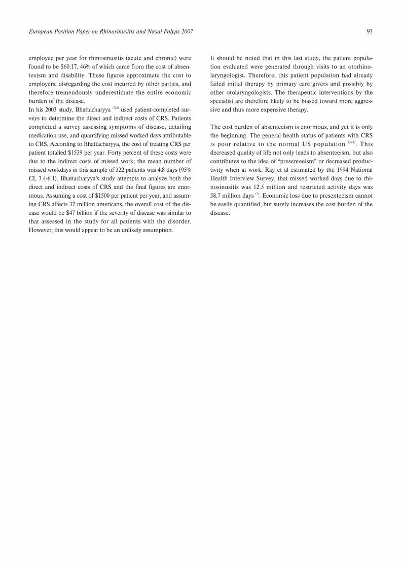
European Position Paper on Rhinosinusitis and Nasal Polyps 2007 93
employee per year for rhinosinusitis (acute and chronic) werefound to be $60.17, 46% of which came from the cost of absen-teeism and disability. These figures approximate the cost toemployers, disregarding the cost incurred by other parties, andtherefore tremendously underestimate the entire economicburden of the disease.In his 2003 study, Bhattacharyya (722) used patient-completed sur-veys to determine the direct and indirect costs of CRS. Patientscompleted a survey assessing symptoms of disease, detailingmedication use, and quantifying missed worked days attributableto CRS. According to Bhattacharyya, the cost of treating CRS perpatient totalled $1539 per year. Forty percent of these costs weredue to the indirect costs of missed work; the mean number ofmissed workdays in this sample of 322 patients was 4.8 days (95%CI, 3.4-6.1). Bhattacharyya’s study attempts to analyze both thedirect and indirect costs of CRS and the final figures are enor-mous. Assuming a cost of $1500 per patient per year, and assum-ing CRS affects 32 million americans, the overall cost of the dis-ease would be $47 billion if the severity of disease was similar tothat assessed in the study for all patients with the disorder.However, this would appear to be an unlikely assumption.
It should be noted that in this last study, the patient popula-tion evaluated were generated through visits to an otorhino-laryngologist. Therefore, this patient population had alreadyfailed initial therapy by primary care givers and possibly byother otolaryngologists. The therapeutic interventions by thespecialist are therefore likely to be biased toward more aggres-sive and thus more expensive therapy.
The cost burden of absenteeism is enormous, and yet it is onlythe beginning. The general health status of patients with CRSis poor relative to the normal US population (588). Thisdecreased quality of life not only leads to absenteeism, but alsocontributes to the idea of “presenteeism” or decreased produc-tivity when at work. Ray et al estimated by the 1994 NationalHealth Interview Survey, that missed worked days due to rhi-nosinusitis was 12.5 million and restricted activity days was58.7 million days (3). Economic loss due to presenteeism cannotbe easily quantified, but surely increases the cost burden of thedisease.
94 Supplement 20
Trial design has largely focussed on medical therapy. The FDArecommends three components (1079)
1. the objective of the study must be clearly stated, coupledwith a summary of the methods used for analysis of theresults
2. the design must permit quantitative assessment of drug (ietherapeutic) effectiveness by a valid comparison with acontrol group
3. the study protocol should accurately define the design andduration of the study, sample size issues and whether treat-ments are parallel or sequential.
A single primary outcome measure is preferred to minimisethe possibility of a Type I error (incorrectly assuming that adrug is effective). However, the FDA recognises that 2 may beappropriate.
Trials may be conducted in acute and chronic rhinosinusitiswith or without polyps and may consider single, short-term orlonger term interventions. Studies may be conducted in prima-ry or secondary care and the criteria for diagnosis, inclusionand exclusion together with outcome measures will dependupon the setting.
Table 12-1 Criteria for studies conducted in primary and secondary care
criteria primary care secondary care
symptom profile & severity usingVAS
+ +
endoscopy (scoring eg 0-3) - +
imagingplain x-rayCT (scoring eg Lund-Mackay 0-24)
+-
-+
medication use + +
co-morbidity (eg allergy, asthma, aspirin sensitivity)
+ +
smoking history + +
additional tests (eg micro-biology, smell, mediators, cytology, mucociliary function, haematology, airway
± ±
T
Table 12-2. Data for all studies to include:
Title
rationale for study
objectives
design
study population: inclusion & exclusion criteria
outcomes:
primary & secondary
subjective & objective
safety assessments
statistical methodology/power analysis
ethics approval
drop-out analysis
12. Outcomes measurements in research

European Position Paper on Rhinosinusitis and Nasal Polyps 2007 95
13-1 Introduction
The following schemes for diagnosis and treatment are theresult a critical evaluation of the available evidence. The tables
give the level of evidence for studies with a positive outcomeand well powered studies with negative outcoume. Ib (-) in thistables means a well designed (Ib) study with a negative out-come. The grade of recommendation for the available therapyis given. Under relevance it is indicated whether the group ofauthors think this treatment to be of relevance in the indicateddisease.
13. Evidence based schemes for diagnostic and treatment
Table 13-1. Treatment evidence and recommendations for adults withacute rhinosinusitis
# : Ib (-) study with a negative outcome
therapy level grade of recommen-dation
relevance
oral antibiotic Ia A yes: after 5 days,or in severe cases
topical corticosteroid Ib A yes
topical steroid and oralantibiotic combined
Ib A yes
oral corticosteroid Ib A yes reduces pain in severe disease
oral antihistamine Ib B yes, only in allergic patients
nasal douche Ib (-)# D no
decongestant Ib (-)# D yes, as symtomatic relief
mucolytics none no no
phytotherapy Ib D no
Table 13-2. Treatment evidence and recommendations for childrenwith acute rhinosinusitis
therapy level grade of recom-mendation
relevance
oral antibiotic Ia A yes: after 5 days,or in severe cases
topical corticosteroid IV D yes
topical steroid on topof oral antibiotic
Ib A yes
topical decongestant III (-) C no
saline douching IV D yes
t
Table 13-3 Treatment evidence and recommendations for adults with chronic rhinosinusitis without nasal polyps *
* Some of these studies also included patients with CRS with nasal polyps* Acute exacerbations of CRS should be treated like acute rhinosinusitis# : Ib (-) study with a negative outcome
therapy level grade of recommendation relevance
oral antibiotic therapy short term < 2 weeks Ib (-) C no
oral antibiotic therapy long term > 12 weeks Ib (-) A yes
Antibiotics – topical III D no
steroid – topical Ib (-) A yes
steroid – oral no data D no
nasal saline douche Ib (-) A yes
decongestant oral / topical no data D no
mucolytics III C no
antimycotics – systemic Ib (-)# D no
antimycotics – topical Ib (-)# D no
oral antihistamine in allergic patients no data D no
proton pump inhibitors no data D no
bacterial lysates Ib (-) A no
immunomodulators Ib (-)# D no
phytotherapy Ib (-)# D no
anti-leukotrienes III C no
t

96 Supplement 20
Table 13-4 Treatment evidence and recommendations postoperativecare in adults with chronic rhinosinusitis without NP*
* Some of these studies also included patients with CRS with nasal polyps
therapy level grade of recommen-dation
relevance
oral antibiotic shortterm < 2 weeks
no data D yes, immediately postoperative,if pus was seenduring operation
oral antibioticlong term ~ 12 weeks
no data D yes
topical antibiotics no data D no
topical steroid Ib (one +, one -)
B yes
oral steroid no data D short term yeslong term no
nasal douche no data available
D yes
Table 13-5. Treatment evidence and recommendations for adults with chronic rhinosinusitis with nasal polyps *
* Some of these studies also included patients with CRS without nasal polyps# : Ib (-) study with a negative outcome
therapy level grade of recommendation relevance
oral antibiotics short term < 2 weeks no data D no
oral antibiotic long term > 12 weeks Ib yes, for late relapse
topical antibiotics no data D no
topical steroids Ib A yes
oral steroids Ib A yes
nasal douche Ib no data in single use A yes for symptomatic relief
decongestant topical / oral no data in single use D no
mucolytics no data D no
antimycotics – systemic Ib (-)# D no
antimycotics – topical Ib (-)# A no
oral antihistamine in allergic patients Ib (1)# A no, in allergy
capsaicin II B no
proton pump inhibitors II C no
immunomodulators no data D no
phytotherapy no data D no
anti leukotrienes III C no
A
Table 13-6. Treatment evidence and recommendations postoperative treatment in adults with chronic rhinosinusitis with nasal polyps*
* Some of these studies also included patients with CRS without nasal polyps .
Therapy Level Grade of recommendation Relevance
oral antibioticsshort term < 2 weeks
no data D immediately post-operative,if pus was seen during operation
oral antibioticslong term > 12 weeks
Ib A yes
topical steroids after FESS Ib (2 studies one + one -) B yes
topical steroids after polypectomy Ib A yes
oral steroids no data D yes
nasal douche no data D yes
T
Table 13-7 Treatment evidence and recommendations for childrenwith chronic rhinosinusitis
Therapy level grade of recom-mendation
relevance
oral antibiotic Ia A yes, small effect
topical corticosteroid IV D yes
saline douching III C yes
Therapy for gastro-oesophageal reflux
III C yes
European Position Paper on Rhinosinusitis and Nasal Polyps 2007 97
13-2 Introduction
Since the preparation of the first EP3OS document an increas-ing amount of evidence on the pathophysiology, diagnosis andtreatment has been published (Figure1-1).However, in compiling the tables on the various forms of ther-apy, it may be that despite well powered level Ib trials, no sig-nificant benefit has been demonstrated. Equally results may beequivocal or apparently positive results are undermined by thesmall number of trials conducted and/or the small number ofparticipants in the trial(s). In these cases, after detailed discus-sion, the EPOS group decided in most cases, that there was noevidence at present to recommend use of the treatment inquestion. Alternatively for some treatments no trials have beenconducted, even though the treatment is commonly used inwhich case a pragmatic approach has been adopted in the rec-ommendations.
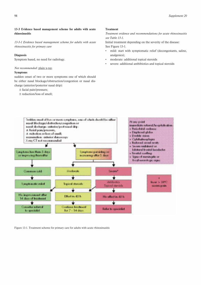
98 Supplement 20
13-3 Evidence based management scheme for adults with acuterhinosinusitis
13-3-1 Evidence based management scheme for adults with acuterhinosinusitis for primary care
DiagnosisSymptom based, no need for radiology.
Not recommended: plain x-ray.Symptomssudden onset of two or more symptoms one of which shouldbe either nasal blockage/obstruction/congestion or nasal dis-charge (anterior/posterior nasal drip):
± facial pain/pressure;± reduction/loss of smell;
TreatmentTreatment evidence and recommendations for acute rhinosinusitissee Table 13-1.Initial treatment depending on the severity of the disease:See Figure 13-1.• mild: start with symptomatic relief (decongestants, saline,
analgesics);• moderate: additional topical steroids• severe: additional antbibiotics and topical steroids
Figure 13-1. Treatment scheme for primary care for adults with acute rhinosinusitis

European Position Paper on Rhinosinusitis and Nasal Polyps 2007 99
13-3-2 Evidence based management scheme for adults with acuterhinosinusitis for ENT specialists
Diagnosis
Symptomssudden onset of two or more symptoms one of which shouldbe either nasal blockage/obstruction/congestion or nasal dis-charge (anterior/posterior nasal drip):
± facial pain/pressure;± reduction/loss of smell;
Signs• nasal examination (swelling, redness, pus);• oral examination: posterior discharge;• exclude dental infection.ENT-examination including nasal endoscopy.
Not recommended: plain x-ray.
CT-Scan is also not recommended unless additional problemssuch as:• very severe diseases,• immunocompromised patients;• signs of complications.
Figure 13-2. Treatment scheme for ENT specialists for adults with acute rhinosinusitis

100 Supplement 20
13-4 Evidence based management scheme for adults with chronicrhinosinusitis without nasal polyps
13-4-1 Evidence based management scheme for adults with CRSwith or without NP for primary care and non-ENT specialists
Diagnosis
Symptoms present longer than 12 weekstwo or more symptoms one of which should be either nasalblockage/obstruction/congestion or nasal discharge(anterior/posterior nasal drip):
± facial pain/pressure,± reduction or loss of smell;
Additional diagnostic information• questions on allergy should be added and, if positive, aller-
gy testing should be performed.
Not recommended: plain x-ray or CT-scan
Acute exacerbations of CRS should be treated like acute rhi-nosinusitis (1080).
Figure 13-3 Treatment scheme for Chronic Rhinosinusitis with or without nasal polyps for primary care and non-ENT specialists
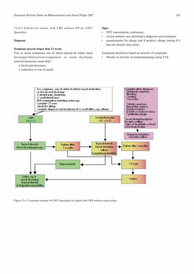
European Position Paper on Rhinosinusitis and Nasal Polyps 2007 101
13-4-2 Scheme for adults with CRS without NP for ENT-Specialists
Diagnosis
Symptoms present longer than 12 weeksTwo or more symptoms one of which should be either nasalblockage/obstruction/congestion or nasal discharge(anterior/posterior nasal drip):
± facial pain/pressure,± reduction or loss of smell;
Signs• ENT examination, endoscopy;• review primary care physician’s diagnosis and treatment;• questionnaire for allergy and if positive, allergy testing if it
has not already been done.
Treatment should be based on severity of symptoms• Decide on severity of symptomatology using VAS
Figure 13-4. Treatment scheme for ENT-Specialists for adults with CRS without nasal polyps
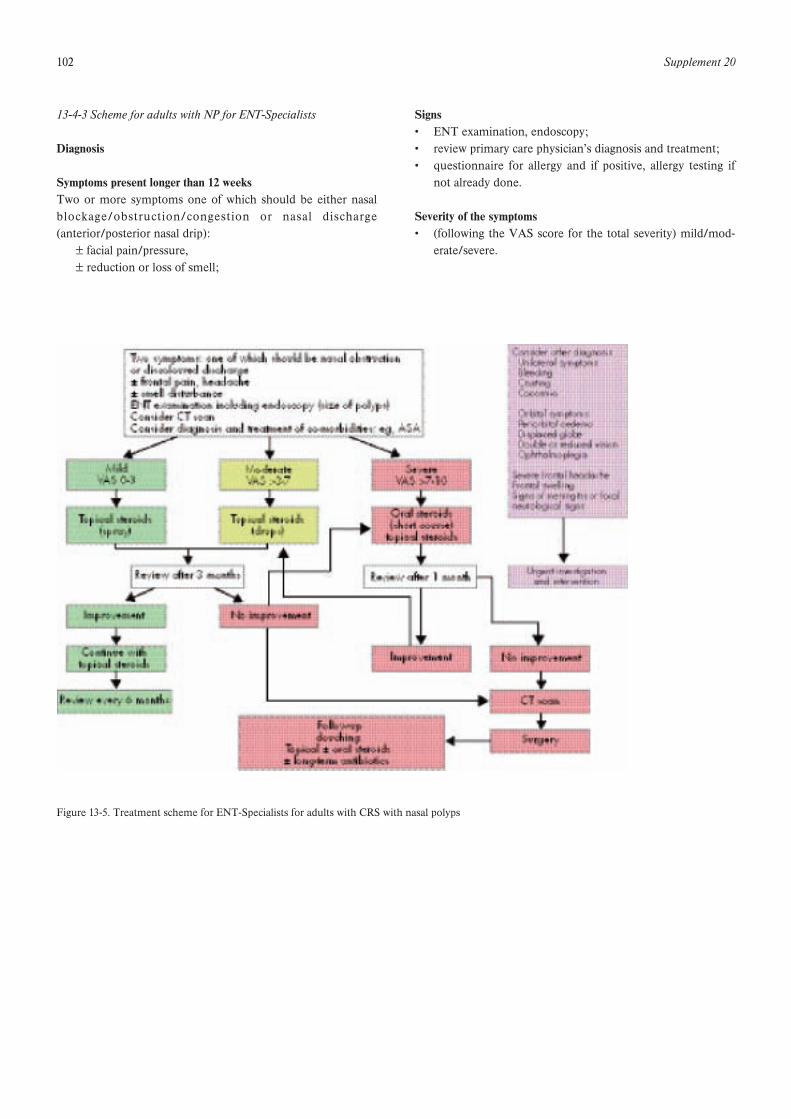
102 Supplement 20
13-4-3 Scheme for adults with NP for ENT-Specialists
Diagnosis
Symptoms present longer than 12 weeksTwo or more symptoms one of which should be either nasalblockage/obstruction/congestion or nasal discharge(anterior/posterior nasal drip):
± facial pain/pressure,± reduction or loss of smell;
Signs• ENT examination, endoscopy;• review primary care physician’s diagnosis and treatment;• questionnaire for allergy and if positive, allergy testing if
not already done.
Severity of the symptoms• (following the VAS score for the total severity) mild/mod-
erate/severe.
Figure 13-5. Treatment scheme for ENT-Specialists for adults with CRS with nasal polyps

European Position Paper on Rhinosinusitis and Nasal Polyps 2007 103
13-5 Evidence based schemes for therapy in children
The following scheme should help different disciplines in thetreatment of rhinosinusitis in children. The recommendationsare based on the available evidence, but the choices need to bemade depending on the circumstances of the individual case.
13-5-1 Evidence based management scheme for children withacute rhinosinusitis
Diagnosis
Symptomssudden onset of two or more symptoms one of which shouldbe either nasal blockage/obstruction/congestion or nasal dis-charge (anterior/posterior nasal drip):
± facial pain/pressure;± reduction/loss of smell;
Signs (if applicable)• nasal examination (swelling, redness, pus);• oral examination: posterior discharge;• exclude dental infection.ENT-examination including nasal endoscopy .
Not recommended: plain x-ray.
CT-Scan is also not recommended unless additional problemssuch as:• very severe diseases,• immunocompromised patients;• signs of complications.
TreatmentTreatment evidence and recommendations for acute rhinosinusitissee Table 13-2.Initial treatment depending on the severity of the disease:See Figure 13-6.
Sudden onset of two or more symptoms one of which should be eithernasal blockage/obstruction/congestion ornasal discharge: anterior/post nasal drip;± facial pain/pressure,± reduction or loss of smell;examination: anterior rhinoscopyX-ray/CT not recommended
Symptoms less than 5 daysor improving thereafter
At any pointImmediate referral/hospitalisation• Periorbital oedema• Displaced globe• Double vision• Ophthalmoplegia• Reduced visual acuity• Severe unilateral or
bilateral frontal headache• Frontal swelling• Signs of meningitis or
focal neurologic signs
Common cold
Symptoms persistentor increasing after 5 days
Symptomatic relief
No
Symptomatic relief
Moderate
Asthmachronic bronchitis
Yes
Oral amoxicillincan be considered
Severe*
Non-toxic
Oral antibiotics
Toxic, severely ill
HospitalisationIV antibiotics
HospitalisationNasal endoscopy
CultureImaging
IV antibioticsand/or surgery
No effect in 48 h
Hospitalisation
*fever > 38°Csevere pain
Figure 13-6. Treatment scheme for children with acute rhinosinusitis
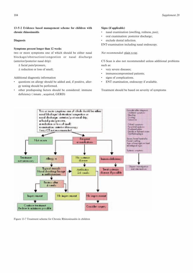
104 Supplement 20
13-5-2 Evidence based management scheme for children withchronic rhinosinusitis
Diagnosis
Symptoms present longer than 12 weekstwo or more symptoms one of which should be either nasalblockage/obstruction/congestion or nasal discharge(anterior/posterior nasal drip):
± facial pain/pressure,± reduction or loss of smell;
Additional diagnostic information• questions on allergy should be added and, if positive, aller-
gy testing should be performed.• other predisposing fsctors should be considered: immune
deficiency ( innate , acquired, GERD)
Signs (if applicable)• nasal examination (swelling, redness, pus);• oral examination: posterior discharge;• exclude dental infection.ENT-examination including nasal endoscopy.
Not recommended: plain x-ray.
CT-Scan is also not recommended unless additional problemssuch as:• very severe diseases;• immunocompromised patients;• signs of complications.• ENT examination, endoscopy if available.
Treatment should be based on severity of symptoms
Figure 13-7 Treatment scheme for Chronic Rhinosinusitis in children

European Position Paper on Rhinosinusitis and Nasal Polyps 2007 105
While our understanding of CRS has increased considerably, thisonly serves to outline areas that will require further explorationand clinical trials for validation of observations and hypotheses.
14-1 Epidemiology: Identifying the risk factors for development ofCRS and NPOur understanding of factors predisposing to CRS and NPremain embryonic with few studies to date. Epidemiologic stud-ies aimed at identification of personal risk factors and environ-mental modifiers are required to increase our understanding ofthe disease process, select appropriate populations for clinical tri-als and interpret information from genetic studies.
Addressing this need will require detailed assessment and long-term follow up of a well-characterised patient populations toidentify risk factors associated with development of CRS. Aprospective population study of age- and sex-matched controlledatopic and non-atopic individuals might allow better characteri-zation of the incidence of all upper respiratory tract symptomsincluding acute and chronic rhinosinusitis over a 5-year period.Similarly, a long-term follow-up of a cohort of patients withnasal polyposis would allow study of the natural history of thecondition.
Identification of environmental modifiers will require prospec-tive assessment of a very large cohort of patients to monitor fordevelopment of disease. While probably impractical for CRSalone, the trend to development of databases of medical andgenetic information on large populations such as the UKBioBank initiative may eventually offer populations for thisresearch.
14-2 Beyond infection: New roles for bacteriaIt is increasingly recognised that bacteria may play a role in thechronic inflammation of CRS. Among others, Staphylococcusaureus has been specifically implicated, with possible persistencefavoured by bacterial biofilms. In the light of this evidence, therole of bacteria in CRS needs to further explored in at least threeareas.1. Host factors favouring persistence of bacterial colonisation
need to be better characterised.2. The relative importance of biofilms and intracellular S. aureus
in the development and persistence of CRS must be assessed3. The role of S. aureus in development or persistence of CRS
via postulated staphylococcal enterotoxins stimulating T-cellsdirectly via a superantigenic mechanism needs to be validat-ed.
14-3 Host responseFurther studies into the mechanisms leading to the developmentof CTRS need to be identified. Events occurring at the level ofthe epithelium including non-specific defences such as innateimmunity need better description and offer potential targets fortherapy.
14-4 GeneticsFinally, pathogenesis of CRS may be better explored withresearch techniques taken from the growing field of genetics.Population association studies may allow detection of polymor-phisms in genes in individuals suffering from CRS. Studies ofcandidate genes in currently known pathways may allow identifi-cation of specific genetic polymorphisms in the different steps ofthe pathways, while whole genome scans probing the entiregenome offer the hope of identification of novel genes not sus-pected of being implicated.
Studies of gene expression in biopsy samples will help identifydifferential gene activation in different disease states and follow-ing different courses of therapy. Both of these offer the hope fordevelopment of tests allowing better differentiation of diseasestates and targeted therapy, with the tantalizing promise of identi-fication of new drug targets not presently exploited.
These studies will require a cohort of investigators trained inthese new techniques and development of multidisciplinarygroups to collect and exploit the large populations required forthese studies. Multinational collaborative initiatives will have tobe initiated to collect the large sample sizes of affected individu-als required for this work.
14-5 Clinical trialsAlthough much work has been recently been done on chronic rhi-nosinusitis and nasal polyps this work needs to be validated interms of its clinical impact. Our understanding needs to translateinto therapy for disease and experimental hypotheses need to veri-fy by appropriate clinical trials. The following suggestions shouldhighlight some areas of interest for further investigation.
1. Areas that have been identified need to be targeted with spe-cific therapies. In particular, means of targeting biofilms, reduc-ing S. aureus colonisation and of modulating response to S.aureus enterotoxins need to be developed and assessed bymeans of well-designed clinical trials.2. Emerging therapies need to be assessed critically in order todetermine which ones are effective and in which settings. Thereis an urgent need for randomized placebo controlled trials tostudy the effect of antibiotics in acute rhinosinusitis, chronic rhi-nosinusitis and exacerbations of chronic rhinosinusitis. These
14. Research needs and priorities
106 Supplement 20
should be compared with nasal steroids as a single modality oftreatment for these conditions.
3. A well-powered prospective study of the effectiveness ofmacrolides in CRS and NP would allow validation of the posi-tive effects suggested by some studies. The role of topicalantibiotic therapy in exacerbations of CRS needs to be per-formed with well-characterized patients to identify optimal sit-uations for use.
4. In the same fashion, novel therapies introduced over thecoming years should be scrutinised closely prior to widespreadadoption. Specifically, it is hoped that in the future clinicianswill remain vigilant to claims made without the support ofprospective, adequately powered clinical trials.
5. Surgical management for CRS and NP will probably continueto play a role in the management of CRS. In the future, ratherthan attempting to demonstrate superiority of one therapy overanother, studies should target selected patient populations or sit-uations so as to guide the clinician to a rational use of medicaland/or surgical therapy as part of a comprehensive treatmentplan individualised to stage of disease and patient needs.
6. Most QoL and symptom-specific questionnaires have beendesigned for a North American population and need to be vali-dated for European patients.
7. The relationship between the upper and lower respiratory tractneeds further investigation and will offer further insight intopathophysiology of inflammation and therapeutic possibilities.
European Position Paper on Rhinosinusitis and Nasal Polyps 2007 107
Acute non-viral rhinosinusitis (ARS): an episode of sudden onsetwith an increase of symptoms after 5 days or persistent symp-toms after 10 days with less than 12 weeks duration.
Acute viral rhinosinusitis: an episode of sudden onset with dura-tion of symptoms for less than 10 days
Common cold: Acute viral rhinosinusitis
Chronic rhinosinusitis (CRS): a condition lasting for >12 weeks,comprising two or more symptoms one of which should beeither nasal blockage/obstruction/congestion or nasal discharge(anterior/posterior nasal drip):
± facial pain/pressure;± reduction or loss of smell;
May occur with or without polyps
Cacosmia: awareness of an unpleasant smell, often rotten orfaeculent
Cobblestoned: an irregular bumpy mucosal surface usually post-surgery for polypsConventional surgery: a range of operations which predated endo-scopic sinus surgery eg polypectomy, inferior meatal antrostomy,Caldwell-Luc operation, Denker’s procedure, external fronto-eth-moidectomy
Endonasal surgery: any surgery performed through the nose
Functional surgery: an operation which aims to restore functioneg restitution of mucocilary clearance, improvement in olfaction
Iatrogenic: a condition induced unintentionally by a physician,usually by a therapeutic action
Local corticosteroid: topical intranasal instillation of a corticos-teroid preparation
Middle meatus: that area of the lateral wall of the nose lying later-al to the middle turbinate
Middle meatal antrostomy: an opening into the maxillary sinusthourgh the lateral wall of the middle meatus
Mucopurulent secretion: a mixture of opaque and discolouredmucus which is not frank pus
Orbital complications:Ecchymosis: an area of discoloration beneath the skin secondaryto bleedingEnophthalmos: abnormal retraction of the eyeball into the socketMyospherulosis: granulomatous foreign body reaction in soft tis-sues due to extravasation of paraffin or oilOrbital emphysema: the presence of air within the soft tissues ofthe eyePeriorbital cellulitis: inflammation of the eyelid and conjunctiva
Ostiomeatal complex: that area of the middle meatus into whichthe maxillary, anterior ethmoid and frontal sinuses drain
Pansinustis: involvement of all paranasal sinuses, usually demon-strated radiologically
Pathogen: any organism capable of producing disease
Rhinitis medicamentosa: a condition associated with use ofintranasal decongestants in which the nasal mucosa undergoesatrophy
Rhinorrhoea: any discharge from the nose. May run from thefront of the nose (anterior) or into the back (posterior or post-nasal discharge)
Rhinosinusitis: inflammation of the nose and paranasal sinuses
Simple polypectomy: surgical removal of polyps from the nasalcavity without additional surgery on the paranasal sinuses
Treatment:short term treatment: usually 2 weeks or lesslong-term treatment: usually 12 weeks or longer
15. Glosarry terms
108 Supplement 20
16-1 General health status instruments:
- SF-36: www.sf-36.org- Euro-QOL: www.euroqol.org- SF-12: derived from the SF-36: www.sf-36.org- Quality of Well-Being Scale: [email protected] Glasgow Benefit Inventory:
www.ihr.mrc.ac.uk/scottish/products/ghsq.php- Mc-Gill pain questionnaire: Ronald Melzack: Department
of Psychology, McGill University, 1205 Dr. PenfieldAvenue, Montreal, Que. H3A 1B1, , Canada
16-2 Disease specific health status instruments:
- RSOM-31: Jay Piccirillo: [email protected] SNOT-20: derived from the RSOM-31: Jay Piccirillo: pic-
[email protected] SNOT-16: derived from the SNOT-20: Eric Anderson,
Department of Otolaryngology-Head and Neck Surgery,University of Washington School of Medicine, Box 356515,Seattle, WA 98195-6515, USA
- RSDI: Michael Benninger, Department of Otolaryngology-Head and Neck Surgery, Henry Ford Hospital, 2799 WGrand Blvd, Detroit, MI 48202, USA
- RQLQ: www.qoltech.co.uk- RSUI: D.A. Revicki: [email protected] SN-5: David Kay: [email protected] RhinoQol: Steven Atlas: [email protected]
16. Information on QOL instruments:

European Position Paper on Rhinosinusitis and Nasal Polyps 2007 109
17. Survey of published olfactory tests
Author(s) Year Test name Test-Time Country Sample size Test retest Subject differences Method
Cain (1081-1083) 198319881989
CCCRC 35 min USA >700 Age, gender, diseases, olfactory disorders.
1/ Threshold. N-butanol.2AFC 4-correct-in-a-row method. Separate nostrils. Odours in squeeze bottles2/Identification. 10 odours (score on &+1). Forced choice from 20 (or 16) descriptors. Odours in jars. Sepa-rate nostrils. Feedback.
Doty et al (1084) 1984(a,b)1985
UPSIT 15 min USA >3000 r=0.981 Age, gender, culture, smoker, disease, olfactory disorder, malingering.
Identification of 40encapsuated odours. 4AFC. Scratch-and-sniff- technique
Wright (1085) 1987 OdourantConfusion
15 min USA 480 Disease. Identification of 10 odours each presented once (100 stimuli or 121 if a blank is added). Forced choice from list of 10 names. Pattern of odorant identification and misidentification.
Kurtz et al (1086) 2001 Matrix(OCM)
Hendriks (1087) 1988 GITU Netherlands 221 Age, gender, olfac-tory disorders.
Identification of 18 or 36 odours. Forced choice either from 4 alternatives or from alist of 24 for 18 odours to identify.“Everyday life” odours. Odours in jars.
Corwin (1088) 19891992
YN-OIT USA Age, disease Based on 20 UPSIT odours. Yes or no matching of a descrip-tor to a proposedodour.
Takagi (1089) 1989 T&TOlfacto-meter
Japan >1000 Olfactory disorders. Thresholds of detec-tion and recognition for 5 odorants. Odours on slips of filter papers. Separate nostrils.
Anderson et al 1992 SDOIT USA Young children
Age. Identification of 10 odours. Forced choice using an array of 20 visual stimuli. Odours in jars.
Eloit and Trotier (1090)
1994 France 84 Olfactory disorder, disease.
Odours in bottles.1/Threshold to 5 odorants.2/Identification of 6 odorants.Odours in bottles.
Doty et al (1091, 1092) 19951996
CC-SITMOD-SIT
5 min USAEuropeAsia
>3000 r=0.71 Age, gender, olfac-tory disorders.
Identification of 12 encapsulated odours. 4AFC. Scratch and sniff technique.
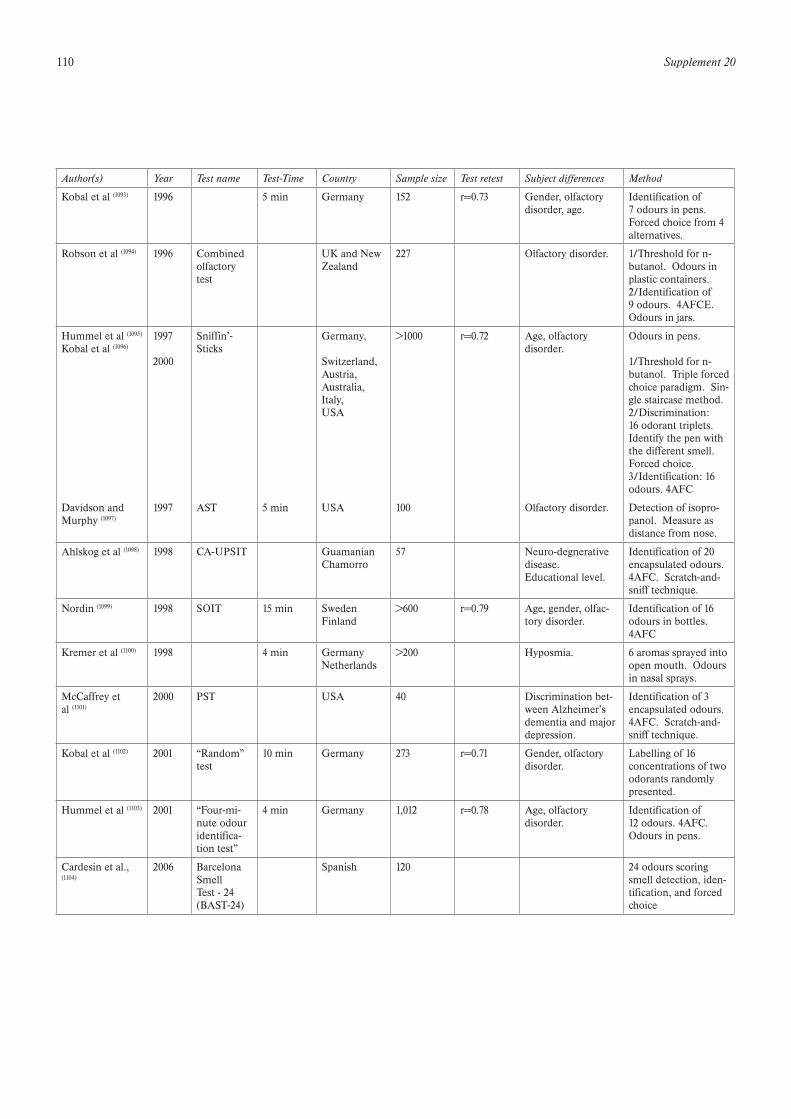
110 Supplement 20
Author(s) Year Test name Test-Time Country Sample size Test retest Subject differences Method
Kobal et al (1093) 1996 5 min Germany 152 r=0.73 Gender, olfactory disorder, age.
Identification of7 odours in pens. Forced choice from 4 alternatives.
Robson et al (1094) 1996 Combined olfactorytest
UK and New Zealand
227 Olfactory disorder. 1/Threshold for n-butanol. Odours in plastic containers.2/Identification of 9 odours. 4AFCE. Odours in jars.
Hummel et al (1095) Kobal et al (1096)
1997 2000
Sniffin’-Sticks
Germany, Switzerland,Austria,Australia,Italy,USA
>1000 r=0.72 Age, olfactory disorder.
Odours in pens. 1/Threshold for n-butanol. Triple forced choice paradigm. Sin-gle staircase method.2/Discrimination: 16 odorant triplets. Identify the pen with the different smell. Forced choice.3/Identification: 16 odours. 4AFC
Davidson andMurphy (1097)
1997 AST 5 min USA 100 Olfactory disorder. Detection of isopro-panol. Measure as distance from nose.
Ahlskog et al (1098) 1998 CA-UPSIT GuamanianChamorro
57 Neuro-degnerative disease.Educational level.
Identification of 20encapsulated odours. 4AFC. Scratch-and-sniff technique.
Nordin (1099) 1998 SOIT 15 min SwedenFinland
>600 r=0.79 Age, gender, olfac-tory disorder.
Identification of 16 odours in bottles. 4AFC
Kremer et al (1100) 1998 4 min GermanyNetherlands
>200 Hyposmia. 6 aromas sprayed into open mouth. Odours in nasal sprays.
McCaffrey etal (1101)
2000 PST USA 40 Discrimination bet-ween Alzheimer’sdementia and majordepression.
Identification of 3encapsulated odours. 4AFC. Scratch-and-sniff technique.
Kobal et al (1102) 2001 “Random” test
10 min Germany 273 r=0.71 Gender, olfactory disorder.
Labelling of 16 concentrations of two odorants randomlypresented.
Hummel et al (1103) 2001 “Four-mi-nute odouridentifica-tion test”
4 min Germany 1,012 r=0.78 Age, olfactory disorder.
Identification of12 odours. 4AFC. Odours in pens.
Cardesin et al., (1104)
2006 Barcelona SmellTest - 24 (BAST-24)
Spanish 120 24 odours scoring smell detection, iden-tification, and forced choice
European Position Paper on Rhinosinusitis and Nasal Polyps 2007 111
1. Durr DG, Desrosiers MY, Dassa C. Impact of rhinosinusitis inhealth care delivery: the Quebec experience. J Otolaryngol.2001;30(2):93-7.
2. Goetzel RZ, Hawkins K, Ozminkowski RJ, Wang S. The healthand productivity cost burden of the “top 10” physical and mentalhealth conditions affecting six large U.S. employers in 1999. JOccup Environ Med. 2003;45(1):5-14.
3. Ray NF, Baraniuk JN, Thamer M, Rinehart CS, Gergen PJ,Kaliner M, et al. Healthcare expenditures for sinusitis in 1996:contributions of asthma, rhinitis, and other airway disorders. JAllergy Clin Immunol. 1999;103(3 Pt 1):408-14.
4. Anon JB, Jacobs MR, Poole MD, Ambrose PG, Benninger MS,Hadley JA, et al. Antimicrobial treatment guidelines for acutebacterial rhinosinusitis. Otolaryngol Head Neck Surg. 2004Jan;130(1 Suppl):1-45.
5. Report of the Rhinosinusitis Task Force Committee Meeting.Alexandria, Virginia, August 17, 1996. Otolaryngol Head NeckSurg. 1997 Sep;117(3 Pt 2):S1-68.
6. Meltzer EO, Hamilos DL, Hadley JA, Lanza DC, Marple BF,Nicklas RA, et al. Rhinosinusitis: Developing guidance for clini-cal trials. J Allergy Clin Immunol. 2006 Nov;118(5 Suppl):S17-61.
7. Meltzer EO, Hamilos DL, Hadley JA, Lanza DC, Marple BF,Nicklas RA, et al. Rhinosinusitis: Establishing definitions for clin-ical research and patient care. Otolaryngology - Head & NeckSurgery. 2004;131(6 SUPPL.):S1-S62.
8. Fokkens W, Lund V, al. e. European position paper on rhinosi-nusitis and nasal polyps. Rhinol Suppl. 2005(18):1-87.
9. Fokkens W, Lund V, Bachert C, Clement P, Helllings P,Holmstrom M, et al. EAACI position paper on rhinosinusitis andnasal polyps executive summary. Allergy. 2005;60(5):583-601.
10. Sackett DL, Rosenberg WM, Gray JA, Haynes RB, RichardsonWS. Evidence based medicine: what it is and what it isn’t. Bmj.1996 Jan 13;312(7023):71-2.
11. Shekelle PG, Woolf SH, Eccles M, Grimshaw J. Clinical guide-lines: developing guidelines. Bmj. 1999 Feb 27;318(7183):593-6.
12. New guidelines for sinusitis target prescribing practices. DisManag Advis. 2004 Mar;10(3):27-30.
13. Bachert C, Hormann K, Mosges R, Rasp G, Riechelmann H,Muller R, et al. An update on the diagnosis and treatment ofsinusitis and nasal polyposis. Allergy. 2003;58(3):176-91.
14. Lim M, Lew-Gor S, Darby Y, Brookes N, Scadding GK, VJ L.The relationship between subjective assessment instruments inchronic rhinosinusitis. . Rhinology. 2007;in press.
15. Winstead W. Rhinosinusitis. Prim Care. 2003;30(1):137-54.16. Slavin RG. Nasal polyps and sinusitis. Jama. 1997;278(22):1849-54.17. Sturgess JM, Chao J, Wong J, Aspin N, Turner JA. Cilia with
defective radial spokes: a cause of human respiratory disease. NEngl J Med. 1979;300(2):53-6.
18. Bhattacharyya Nl. The role of infection in chronic rhinosinusitis.Curr Allergy Asthma Rep. 2002;2(6):500-6.
19. Zacharek MA, Krouse JH. The role of allergy in chronic rhinosi-nusitis. Curr Opin Otolaryngol Head Neck Surg. 2003;11(3):196-200.
20. Jones NS. CT of the paranasal sinuses: a review of the correlationwith clinical, surgical and histopathological findings. ClinOtolaryngol. 2002;27(1):11-7.
21. Jones NS, Strobl A, Holland I. A study of the CT findings in 100patients with rhinosinusitis and 100 controls. Clin Otolaryngol.1997;22(1):47-51.
22. Steinke JW, Bradley D, Arango P, Crouse CD, Frierson H,Kountakis SE, et al. Cysteinyl leukotriene expression in chronichyperplastic sinusitis-nasal polyposis: importance to eosinophiliaand asthma. J Allergy Clin Immunol. 2003;111(2):342-9.
23. Ichimura K, Shimazaki Y, Ishibashi T, Higo R. Effect of newmacrolide roxithromycin upon nasal polyps associated withchronic sinusitis. Auris Nasus Larynx. 1996;23:48-56.
24. Dykewicz MS. 7. Rhinitis and sinusitis. J Allergy Clin Immunol.2003;111(2 Suppl):S520-9.
25. Vento SI, Ertama LO, Hytonen ML, Wolff CH, Malmberg CH.Nasal polyposis: clinical course during 20 years. Ann AllergyAsthma Immunol. 2000;85(3):209-14.
26. Bachert C, Van Cauwenberge PBl. Inflammatory mechanisms inchronic sinusitis. Acta Otorhinolaryngol Belg. 1997;51(4):209-17.
27. Berger G, Kattan A, Bernheim J, Ophir Dl. Polypoid mucosawith eosinophilia and glandular hyperplasia in chronic sinusitis: ahistopathological and immunohistochemical study.Laryngoscope. 2002;112(4):738-45.
28. Rudack C, Stoll W, Bachert C. Cytokines in nasal polyposis,acute and chronic sinusitis. Am J Rhinol. 1998;12(6):383-8.
29. Hamilos DL, Leung DY, Wood R, Bean DK, Song YL,Schotman E, et al. Eosinophil infiltration in nonallergic chronichyperplastic sinusitis with nasal polyposis (CHS/NP) is associatedwith endothelial VCAM-1 upregulation and expression of TNF-alpha. Am J Respir Cell Mol Biol. 1996;15(4):443-50.
30. Hamilos DL, Leung DY, Wood R, Cunningham L, Bean DK,Yasruel Z, et al. Evidence for distinct cytokine expression inallergic versus nonallergic chronic sinusitis. J Allergy ClinImmunol. 1995;96(4):537-44.
31. Bachert C, Wagenmann M, Hauser U, Rudack Cl. IL-5 synthesisis upregulated in human nasal polyp tissue. J Allergy ClinImmunol. 1997;99(6 Pt 1):837-42.
32. Hedman J, Kaprio J, Poussa T, Nieminen MM. Prevalence ofasthma, aspirin intolerance, nasal polyposis and chronic obstruc-tive pulmonary disease in a population-based study. Int JEpidemiol. 1999;28(4):717-22.
33. Larsen K. The clinical relationship of nasal polyps to asthma.Allergy Asthma Proc. 1996;17(5):243-9.
34. Settipane GA, Chafee FH. Nasal polyps in asthma and rhinitis. Areview of 6,037 patients. J Allergy Clin Immunol. 1977;59(1):17-21.
35. Caplin I, Haynes JT, Spahn J. Are nasal polyps an allergic phe-nomenon? Ann Allergy. 1971;29(12):631-4.
36. van Gageldonk-Lafeber AB, Heijnen ML, Bartelds AI, PetersMF, van der Plas SM, Wilbrink B. A case-control study of acuterespiratory tract infection in general practice patients in TheNetherlands. Clin Infect Dis. 2005 Aug 15;41(4):490-7.
37. Gwaltney JM, Jr., Jones JG, Kennedy DW. Medical managementof sinusitis: educational goals and management guidelines. TheInternational Conference on sinus Disease. Ann Otol RhinolLaryngol Suppl. 1995;167:22-30.
38. Leggett JE. Acute sinusitis. When--and when not--to prescribeantibiotics. Postgrad Med. 2004 Jan;115(1):13-9.
39. Lindbaek M. Acute sinusitis: guide to selection of antibacterialtherapy. Drugs. 2004;64(8):805-19.
40. Williams Jr JW, Aguilar C, Cornell J, Chiquette E. Dolor RJ,Makela M, Holleman DR, et al. Antibiotics for acute maxillarysinusitis (Cochrane Review). Cochrane Database Syst Rev. 2003(4).
41. Varonen H, Savolainen S, Kunnamo I, Heikkinen R, Revonta M.Acute rhinosinusitis in primary care: a comparison of symptoms,signs, ultrasound, and radiography. Rhinology. 2003Mar;41(1):37-43.
42. Engels EA, Terrin N, Barza M, Lau J. Meta-analysis of diagnostictests for acute sinusitis. J Clin Epidemiol. 2000 Aug;53(8):852-62.
43. Giesbers HrNo-IVTV, Nationale Atlas Volksgezondheid.Bilthoven: RIVM, < Gezondheid\Ziekten enaandoeningen\Ziekten van de ademhalingswegen, 27 september2002. Neusbijholte ontsteking 1995-1999. 2002 [cited; Availablefrom: http://www.zorgatlas.nl
44. Gijsen R, Poos M. Nationaal Kompas. 2003 [cited; Available from:45. Gwaltney JM, Jr. Acute community-acquired sinusitis. Clin
Infect Dis. 1996;23(6):1209-23; quiz 24-5.
18. References
112 Supplement 20
46. Berg O, Carenfelt C, Kronvall G. Bacteriology of maxillary sinusi-tis in relation to character of inflammation and prior treatment.Scand J Infect Dis. 1988;20(5):511-6.
47. Felmingham D, Feldman C, Hryniewicz W, Klugman K, KohnoS, Low DE, et al. Surveillance of resistance in bacteria causingcommunity-acquired respiratory tract infections. Clin MicrobiolInfect. 2002;8(Suppl 2):12-42.
48. Hoban D, Felmingham D. The PROTEKT surveillance study:antimicrobial susceptibility of Haemophilus influenzae andMoraxella catarrhalis from community-acquired respiratory tractinfections. J Antimicrob Chemother. 2002;50(Suppl S1):49-59.
49. Klossek JM, Chidiac C, Serrano E. Current position of the man-agement of community-acquired acute maxillary sinusitis or rhi-nosinusitis in France and literature review. Rhinology.2005;43(SUPPL. 19):1-33.
50. Cars O, Molstad S, Melander A. Variation in antibiotic use in theEuropean Union. Lancet. 2001;357(9271):1851-3.
51. Benninger M, Brook I, Farrell DJ. Disease severity in acute bacte-rial rhinosinusitis is greater in patients infected with Streptococcuspneumoniae than in those infected with Haemophilus influenzae.Otolaryngol Head Neck Surg. 2006 Oct;135(4):523-8.
52. Pedersen M, Sakakura Y, Winther B, Brofeldt S, Mygind N.Nasal mucociliary transport, number of ciliated cells, and beatingpattern in naturally acquired common colds. Eur J Respir DisSuppl. 1983;128 (Pt 1):355-65.
53. Hinni ML, McCaffrey TV, Kasperbauer JL. Early mucosalchanges in experimental sinusitis. Otolaryngol Head Neck Surg.1992 Oct;107(4):537-48.
54. Kaliner M. Treatment of sinusitis in the next millennium.Allergy Asthma Proc. 1998;19(4):181-4.
55. Lanza DC, Kennedy DW. Adult rhinosinusitis defined.Otolaryngol Head Neck Surg. 1997;117(3 Pt 2):S1-7.
56. Savolainen S. Allergy in patients with acute maxillary sinusitis.Allergy. 1989;44(2):116-22.
57. Alho OP, Karttunen TJ, Karttunen R, Tuokko H, Koskela M,Suramo I, et al. Subjects with allergic rhinitis show signs of moreseverely impaired paranasal sinus functioning during viral coldsthan nonallergic subjects. Allergy. 2003 Aug;58(8):767-71.
58. Hinriksdottir I, Melen I. Allergic rhinitis and upper respiratorytract infections. Acta Otolaryngol Suppl. 1994;515:30-2.
59. Newman LJ, Platts-Mills TA, Phillips CD, Hazen KC, Gross CW.Chronic sinusitis. Relationship of computed tomographic find-ings to allergy, asthma, and eosinophilia. Jama. 1994;271(5):363-7.
60. Emanuel IA, Shah SB. Chronic rhinosinusitis: allergy and sinuscomputed tomography relationships. Otolaryngol Head NeckSurg. 2000;123(6):687-91.
61. Iwens P, Clement PA. Sinusitis in allergic patients. Rhinology.1994;32(2):65-7.
62. Naclerio RM, deTineo ML, Baroody FM. Ragweed allergic rhini-tis and the paranasal sinuses. A computed tomographic study.Arch Otolaryngol Head Neck Surg. 1997;123(2):193-6.
63. Moser FG, Panush D, Rubin JS, Honigsberg RM, Sprayregen S,Eisig SB. Incidental paranasal sinus abnormalities on MRI of thebrain. Clin Radiol. 1991 Apr;43(4):252-4.
64. Lloyd GA. CT of the paranasal sinuses: study of a control seriesin relation to endoscopic sinus surgery. J Laryngol Otol.1990;104(6):477-81.
65. Havas TE, Motbey JA, Gullane PJ. Prevalence of incidentalabnormalities on computed tomographic scans of the paranasalsinuses. Arch Otolaryngol Head Neck Surg. 1988;114(8):856-9.
66. Patel K, Chavda SV, Violaris N, Pahor AL. Incidental paranasalsinus inflammatory changes in a British population. J LaryngolOtol. 1996 Jul;110(7):649-51.
67. Zinreich SJ, Kennedy DW, Kumar AJ, Rosenbaum AE,Arrington JA, Johns ME. MR imaging of normal nasal cycle:comparison with sinus pathology. J Comput Assist Tomogr. 1988Nov-Dec;12(6):1014-9.
68. Holzmann D, Willi U, Nadal D. Allergic rhinitis as a risk factorfor orbital complication of acute rhinosinusitis in children. Am JRhinol. 2001;15(6):387-90.
69. Chen CF, Wu KG, Hsu MC, Tang RB. Prevalence and relation-ship between allergic diseases and infectious diseases. JMicrobiol Immunol Infect. 2001 Mar;34(1):57-62.
70. Karlsson G, Holmberg K. Does allergic rhinitis predispose tosinusitis? Acta Otolaryngol Suppl. 1994;515:26-8; discussion 9.
71. Wise SK, Wise JC, DelGaudio JM. Association of nasopharyn-geal and laryngopharyngeal reflux with postnasal drip symptoma-tology in patients with and without rhinosinusitis. AmericanJournal of Rhinology. 2006;20(3):283-9.
72. Dinis PB, Martins ML, Subtil J. Does Helicobacter pylori play arole in upper respiratory tract inflammation? A case report. EarNose Throat J. 2005 Apr;84(4):238-40.
73. Skoulas IG, Helidonis E, Kountakis SE. Evaluation of sinusitis inthe intensive care unit patient. Otolaryngol Head Neck Surg. 2003Apr;128(4):503-9.
74. Rouby JJ, Poete P, Martin de Lassale E, Nicolas MH, Bodin L,Jarlier V, et al. Prevention of gram negative nosocomial bron-chopneumonia by intratracheal colistin in critically ill patients.Histologic and bacteriologic study. Intensive Care Med.1994;20(3):187-92.
75. Holzapfel L. Nasal vs oral intubation. Minerva Anestesiol. 2003May;69(5):348-52.
76. van Zanten AR, Dixon JM, Nipshagen MD, de Bree R, GirbesAR, Polderman KH. Hospital-acquired sinusitis is a commoncause of fever of unknown origin in orotracheally intubated criti-cally ill patients. Crit Care. 2005 Oct 5;9(5):R583-90.
77. Le Moal G, Lemerre D, Grollier G, Desmont C, Klossek JM,Robert R. Nosocomial sinusitis with isolation of anaerobic bacte-ria in ICU patients. Intensive Care Med. 1999 Oct;25(10):1066-71.
78. Degano B, Genestal M, Serrano E, Rami J, Arnal JF. Effect oftreatment on maxillary sinus and nasal nitric oxide concentra-tions in patients with nosocomial maxillary sinusitis. Chest. 2005Sep;128(3):1699-705.
79. Collins JG. Prevalence of selected chronic conditions: UnitedStates, 1990-1992. Vital Health Stat 10. 1997(194):1-89.
80. Blackwell DCJ, Coles R. Summary health statistics for US adults:National Health Interview Survey 1997. National Center forHealth Statistics. Vital Health Stat. 2002;10(205):15.
81. Shashy RG, Moore EJ, Weaver A. Prevalence of the chronicsinusitis diagnosis in Olmsted County, Minnesota. ArchOtolaryngol Head Neck Surg. 2004 Mar;130(3):320-3.
82. Hughes RG, Jones NS. The role of nasal endoscopy in outpatientmanagement. Clin Otolaryngol. 1998;23(3):224-6.
83. Bhattacharyya N. Clinical and symptom criteria for the accuratediagnosis of chronic rhinosinusitis. Laryngoscope. 2006 Jul;116(7Pt 2 Suppl 110):1-22.
84. Bonfils P, Nores JM, Halimi P, Avan P, Le Bihan C, Landais P.Correlation between nasosinusal symptoms and topographicdiagnosis in chronic rhinosinusitis. Annals of Otology, Rhinology& Laryngology. 2005;114(1 I):74-83.
85. Chen Y, Dales R, Lin M. The epidemiology of chronic rhinosi-nusitis in Canadians. Laryngoscope. 2003;113(7):1199-205.
86. Greisner WA, 3rd, Settipane GA. Hereditary factor for nasalpolyps. Allergy Asthma Proc. 1996;17(5):283-6.
87. Gordts F, Clement, P.A.R., Buisseret, T. Prevalence of sinusitissigns in a non-ENT population. Otorhinolaryngology.1996;58:315-9.
88. Ahsan SF, Jumans S, Nunez DA. Chronic rhinosinusitis: a com-parative study of disease occurrence in North of Scotland andSouthern Caribbean otolaryngology outpatient clinics over a twomonth period. Scott Med J. 2004 Nov;49(4):130-3.
89. Al-Rawi MM, Edelstein DR, Erlandson RAl. Changes in nasalepithelium in patients with severe chronic sinusitis: a clinico-pathologic and electron microscopic study. Laryngoscope.1998;108(12):1816-23.
90. Hadfield PJ, Rowe-Jones JM, Mackay IS. The prevalence of nasalpolyps in adults with cystic fibrosis. Clin Otolaryngol.2000;25(1):19-22.
91. Rowe-Jones JM, Shembekar M, Trendell-Smith N, Mackay IS.Polypoidal rhinosinusitis in cystic fibrosis: a clinical andhistopathological study. Clin Otolaryngol. 1997;22(2):167-71.
European Position Paper on Rhinosinusitis and Nasal Polyps 2007 113
92. Rowe-Jones JM, Trendell-Smith N, Shembekar M, Mackay IS.Polypoid rhinosinusitis in patients with host defence deficiencies:cellular infiltration and disease severity. Rhinology.1997;35(3):113-7.
93. Wojtczak HA, Kerby GS, Wagener JS, Copenhaver SC, GotlinRW, Riches DW, et al. Beclomethasone diproprionate reducedairway inflammation without adrenal suppression in young chil-dren with cystic fibrosis: a pilot study. Pediatr Pulmonol. 2001Oct;32(4):293-302.
94. Krause HF. Allergy and chronic rhinosinusitis. Otolaryngol HeadNeck Surg. 2003;128(1):14-6.
95. Jones NS, Carney AS, Davis A. The prevalence of allergic rhinos-inusitis: a review. J Laryngol Otol. 1998 Nov;112(11):1019-30.
96. Bailey B. The impact of pollution on the upper alimentary andrespiratory tracts. Otolaryngol Head Neck Surg. 1992;106:736-40.
97. Stammberger H. Functional endoscopic sinus surgery.Philadelphia: B.C. Decker; 1991.
98. Slavin RG. Sinusitis in adults and its relation to allergic rhinitis,asthma, and nasal polyps. J Allergy Clin Immunol. 1988;82(5 Pt2):950-6.
99. Hamilos DL, Leung DY, Wood R, Meyers A, Stephens JK,Barkans J, et al. Chronic hyperplastic sinusitis: association of tis-sue eosinophilia with mRNA expression of granulocyte-macrophage colony-stimulating factor and interleukin-3. J AllergyClin Immunol. 1993;92(1 Pt 1):39-48.
100. Rachelefsky GS, Goldberg M, Katz RM, Boris G, Gyepes MT,Shapiro MJ, et al. Sinus disease in children with respiratory aller-gy. J Allergy Clin Immunol. 1978;61(5):310-4.
101. Shapiro GG. Role of allergy in sinusitis. Pediatr Infect Dis.1985;4(6 Suppl):S55-9.
102. Shapiro GG, Virant FS, Furukawa CT, Pierson WE, BiermanCW. Immunologic defects in patients with refractory sinusitis.Pediatrics. 1991;87(3):311-6.
103. Beninger M. Rhinitis, sinusitis and their relationship to allergies.Am J Rhinol. 1992;6:37-43.
104. Grove R, Farrior, J. Chronic hyperplastic sinusitis in allergicpatients: a bacteriologic study of 200 operative cases. J AllergyClin Immunol. 1990;11:271-6.
105. Friedman WH. Surgery for chronic hyperplastic rhinosinusitis.Laryngoscope. 1975;85(12 pt 1):1999-2011.
106. Lane AP, Pine HS, Pillsbury HC, 3rd. Allergy testing andimmunotherapy in an academic otolaryngology practice: a 20-year review. Otolaryngol Head Neck Surg. 2001 Jan;124(1):9-15.
107. Bousquet J, Van Cauwenberge P, Khaltaev N. Allergic rhinitisand its impact on asthma. J Allergy Clin Immunol. 2001Nov;108(5 Suppl):S147-334.
108. Slavin RG. Relationship of nasal disease and sinusitis tobronchial asthma. Ann Allergy. 1982;49(2):76-9.
109. Juntunen K, Tarkkanen J, Makinen J. Caldwell-Luc operation inthe treatment of childhood bronchial asthma. Laryngoscope.1984;94(2 Pt 1):249-51.
110. Nisioka GJ, Cook, P.R., Davis, W.E., McKinsey, J.P. Functionalendoscopic sinus surgery in patients with chronic sinusitis andasthma. Otolaryngol Head Neck Surg. 1994;110(6):494-500.
111. Salvin RG, Cannon RE, Friedman WH, Palitang E, Sundaram M.Sinusitis and bronchial asthma. J Allergy Clin Immunol. 1980Sep;66(3):250-7.
112. Schwartz HJ, Thompson JS, Sher TH, Ross RJ. Occult sinusabnormalities in the asthmatic patient. Arch Intern Med.1987;147(12):2194-6.
113. Bresciani M, Paradis L, Des Roches A, Vernhet H, Vachier I,Godard P, et al. Rhinosinusitis in severe asthma. J Allergy ClinImmunol. 2001;107(1):73-80.
114. Chee L, Graham SM, Carothers DG, Ballas ZK. Immune dys-function in refractory sinusitis in a tertiary care setting.Laryngoscope. 2001;111(2):233-5.
115. Porter JP, Patel AA, Dewey CM, Stewart MG. Prevalence ofsinonasal symptoms in patients with HIV infection. Am J Rhinol.1999 May-Jun;13(3):203-8.
116. Garcia-Rodriguez JF, Corominas M, Fernandez-Viladrich P,Monfort JL, Dicenta M. Rhinosinusitis and atopy in patients
infected with HIV. Laryngoscope. 1999 Jun;109(6):939-44.117. Sabini P, Josephson GD, Reisacher WR, Pincus R. The role of
endoscopic sinus surgery in patients with acquired immune defi-ciency syndrome. Am J Otolaryngol. 1998;19(6):351-6.
118. Riordan JR, Rommens JM, Kerem B, Alon N, Rozmahel R,Grzelczak Z, et al. Identification of the cystic fibrosis gene:cloning and characterization of complementary DNA. Science.1989 Sep 8;245(4922):1066-73.
119. Kerem B, Rommens JM, Buchanan JA, Markiewicz D, Cox TK,Chakravarti A, et al. Identification of the cystic fibrosis gene:genetic analysis. Science. 1989 Sep 8;245(4922):1073-80.
120. Cuppens H, Marynen P, De Boeck C, Cassiman JJ. Detection of98.5% of the mutations in 200 Belgian cystic fibrosis alleles byreverse dot-blot and sequencing of the complete coding regionand exon/intron junctions of the CFTR gene. Genomics. 1993Dec;18(3):693-7.
121. De Gaudemar I, Contencin P, Van den Abbeele T, Munck A,Navarro J, Narcy P. Is nasal polyposis in cystic fibrosis a directmanifestation of genetic mutation or a complication of chronicinfection? Rhinology. 1996;34(4):194-7.
122. Kerrebijn JD, Poublon RM, Overbeek SE. Nasal and paranasaldisease in adult cystic fibrosis patients. Eur Respir J.1992;5(10):1239-42.
123. Stern RC, Boat TF, Wood RE, Matthews LW, Doershuk CF.Treatment and prognosis of nasal polyps in cystic fibrosis. Am JDis Child. 1982;136(12):1067-70.
124. Davidson TM, Stearns G. Extended indications for endoscopicsinus surgery. Ear Nose Throat J. 1994 Jul;73(7):467-8, 73-4.
125. Jorissen MB, De Boeck K, Cuppens H. Genotype-phenotype cor-relations for the paranasal sinuses in cystic fibrosis. Am J RespirCrit Care Med. 1999;159(5 Pt 1):1412-6.
126. Wang X, Moylan B, Leopold DA, Kim J, Rubenstein RC, TogiasA, et al. Mutation in the gene responsible for cystic fibrosis andpredisposition to chronic rhinosinusitis in the general population.Jama. 2000;284(14):1814-9.
127. Ellegard EK. The etiology and management of pregnancy rhini-tis. Am J Respir Med. 2003;2(6):469-75.
128. Sobol SE, Frenkiel S, Nachtigal D, Wiener D, Teblum C. Clinicalmanifestations of sinonasal pathology during pregnancy. JOtolaryngol. 2001 Feb;30(1):24-8.
129. Sorri M, Hartikainen-Sorri AL, Karja J. Rhinitis during pregnan-cy. Rhinology. 1980 Jun;18(2):83-6.
130. Zinreich SJ, Mattox, D.E., Kennedy, D.W., Chisholm, H.L.,Diffley, D.M., Rosenbaum, A.E. Concha bullosa: CT evaluation.J Comput Assist Tomogr. 1988;12:788-4.
131. Caughey RJ, Jameson MJ, Gross CW, Han JK. Anatomic riskfactors for sinus disease: fact or fiction? Am J Rhinol. 2005 Jul-Aug;19(4):334-9.
132. Bolger WE, Butzin CA, Parsons DS. Paranasal sinus bonyanatomic variations and mucosal abnormalities: CT analysis forendoscopic sinus surgery. Laryngoscope. 1991;101(1 Pt 1):56-64.
133. Holbrook EH, Brown CL, Lyden ER, Leopold DA. Lack of sig-nificant correlation between rhinosinusitis symptoms and specificregions of sinus computer tomography scans. American Journalof Rhinology. 2005;19(4):382-7.
134. Wagenmann M, Naclerio RM. Complications of sinusitis. JAllergy Clin Immunol. 1992;90(3 Pt 2):552-4.
135. Willner A, Choi SS, Vezina LG, Lazar RH. Intranasal anatomicvariations in pediatric sinusitis. Am J Rhinol. 1997;11(5):355-60.
136. Min YG, Jung HW, Kim HS, Park SK, Yoo KY. Prevalence andrisk factors of chronic sinusitis in Korea: results of a nationwidesurvey. Eur Arch Otorhinolaryngol. 1996;253(7):435-9.
137. Yasan H, Dogru H, Baykal B, Douner F, Tuz M. What is therelationship between chronic sinus disease and isolated nasal sep-tal deviation? Otolaryngology - Head & Neck Surgery.2005;133(2):190-3.
138. Calhoun KH, Waggenspack GA, Simpson CB, Hokanson JA,Bailey BJ. CT evaluation of the paranasal sinuses in symptomaticand asymptomatic populations. Otolaryngol Head Neck Surg.1991;104(4):480-3.
114 Supplement 20
139. Kayalioglu G, Oyar O, Govsa F. Nasal cavity and paranasal sinusbony variations: a computed tomographic study. Rhinology.2000;38(3):108-13.
140. Perez P, Sabate J, Carmona A, Catalina-Herrera CJ, Jimenez-Castellanos J. Anatomical variations in the human paranasalsinus region studied by CT. J Anat. 2000;197(Pt 2):221-7.
141. Bhattacharyya N. Bacterial infection in chronic rhinosinusitis: Acontrolled paired analysis. American Journal of Rhinology.2005;19(6):544-8.
142. Araujo E, Palombini BC, Cantarelli V, Pereira A, Mariante Al.Microbiology of middle meatus in chronic rhinosinusitis. Am JRhinol. 2003;17(1):9-15.
143. Brook I. Microbiology and management of sinusitis. JOtolaryngol. 1996;25(4):249-56.
144. Brook I. Bacteriology of chronic maxillary sinusitis in adults. AnnOtol Rhinol Laryngol. 1989;98(6):426-8.
145. Plouin-Gaudon I, Clement S, Huggler E, Chaponnier C, FrancoisP, Lew D, et al. Intracellular residency is frequently associatedwith recurrent Staphylococcus aureus rhinosinusitis. Rhinology.2006 Dec;44(4):249-54.
146. Schubert MS. Fungal rhinosinusitis: diagnosis and therapy. CurrAllergy Asthma Rep. 2001;1(3):268-76.
147. Schubert MS. Allergic fungal sinusitis. Otolaryngol Clin NorthAm. 2004 Apr;37(2):301-26.
148. Ponikau JU, Sherris DA, Kern EB, Homburger HA, Frigas E,Gaffey TA, et al. The diagnosis and incidence of allergic fungalsinusitis. Mayo Clin Proc. 1999;74(9):877-84.
149. Braun H, Buzina W, Freudenschuss K, Beham A, StammbergerH. ‘Eosinophilic fungal rhinosinusitis’: a common disorder inEurope? Laryngoscope. 2003;113(2):264-9.
150. Pant H, Kette FE, Smith WB, Wormald PJ, Macardle PJ. Fungal-specific humoral response in eosinophilic mucus chronic rhinosi-nusitis. Laryngoscope. 2005;115(4):601-6.
151. Sasama J, Sherris DA, Shin SH, Kephart GM, Kern EB, PonikauJU. New paradigm for the roles of fungi and eosinophils inchronic rhinosinusitis. Current Opinion in Otolaryngology &Head & Neck Surgery. 2005;13(1):2-8.
152. Shin SH, Ponikau JU, Sherris DA, Congdon D, Frigas E,Homburger HA, et al. Chronic rhinosinusitis: An enhancedimmune response to ubiquitous airborne fungi. Journal ofAllergy & Clinical Immunology. 2004;114(6):1369-75.
153. Ragab A, Clement P, Vincken W, Nolard N, Simones F. Fungalcultures of different parts of the upper and lower airways inchronic rhinosinusitis. Rhinology. 2006 Mar;44(1):19-25.
154. Murr AH, Goldberg AN, Vesper S. Fungal speciation usingquantitative polymerase chain reaction (QPCR) in patients withand without chronic rhinosinusitis. Laryngoscope.2006;116(8):1342-8.
155. Weschta M, Rimek D, Formanek M, Podbielski A, RiechelmannH. Effect of nasal antifungal therapy on nasal cell activationmarkers in chronic rhinosinusitis. Archives of Otolaryngology --Head & Neck Surgery. 2006;132(7):743-7.
156. Ebbens FA, Scadding GK, Badia L, Hellings PW, Jorissen M,Mullol J, et al. Amphotericin B nasal lavages: not a solution forpatients with chronic rhinosinusitis. J Allergy Clin Immunol.2006 Nov;118(5):1149-56.
157. Lee JT, Kennedy DW, Palmer JN, Feldman M, Chiu AG. Theincidence of concurrent osteitis in patients with chronic rhinosi-nusitis: A clinicopathological study. American Journal ofRhinology. 2006;20(3):278-82.
158. Kennedy DW, Senior BA, Gannon FH, Montone KT, Hwang P,Lanza DC. Histology and histomorphometry of ethmoid bone inchronic rhinosinusitis. Laryngoscope. 1998;108(4 Pt 1):502-7.
159. Khalid AN, Hunt J, Perloff JR, Kennedy DW. The role of bonein chronic rhinosinusitis. Laryngoscope. 2002;112(11):1951-7.
160. Perloff JR, Gannon FH, Bolger WE, Montone KT, Orlandi R,Kennedy DW. Bone involvement in sinusitis: an apparent path-way for the spread of disease. Laryngoscope. 2000;110(12):2095-9.
161. Raynal M, Peynegre R, Beautru R, Coste A. [Sinus mucocelesand surgery in iatrogenic diseases]. Ann Otolaryngol ChirCervicofac. 1999 May;116(2):85-91.
162. Gutman M, Houser S. Iatrogenic maxillary sinus recirculationand beyond. Ear Nose Throat J. 2003 Jan;82(1):61-3.
163. Morinaka S, Ichimiya M, Nakamura H. Detection of Helicobacterpylori in nasal and maxillary sinus specimens from patients withchronic sinusitis. Laryngoscope. 2003;113(9):1557-63.
164. Ozdek A, Cirak MY, Samim E, Bayiz U, Safak MA, Turet S. Apossible role of Helicobacter pylori in chronic rhinosinusitis: apreliminary report. Laryngoscope. 2003;113(4):679-82.
165. Johansson L, Akerlund A, Holmberg K, Melen I, Bende M.Prevalence of nasal polyps in adults: the Skovde population-based study. Ann Otol Rhinol Laryngol. 2003;112(7):625-9.
166. el Hasnaoui A, Jankowski R, Serrano E, Pribil C, Neukirch F,Klossek JM. Evaluation of a diagnostic questionnaire for nasalpolyposis: an observational, cross-sectional study. Rhinology.2004 Mar;42(1):1-7.
167. Klossek JM, Neukirch F, Pribil C, Jankowski R, Serrano E,Chanal I, et al. Prevalence of nasal polyposis in France: A cross-sectional, case-control study. Allergy. 2005;60(2):233-7.
168. Johansson L, Bramerson A, Holmberg K, Melen I, Akerlund A,Bende M. Clinical relevance of nasal polyps in individuals recruit-ed from a general population-based study. Acta Otolaryngol. 2004Jan;124(1):77-81.
169. Larsen PL, Tos M. Origin of nasal polyps. Laryngoscope.1991;101(3):305-12.
170. Larsen PL, Tos M. Site of origin of nasal polyps. Transcraniallyremoved naso-ethmoidal blocks as a screening method for nasalpolyps in autopsy material. Rhinology. 1995;33(4):185-8.
171. Larsen P, Tos M. Anatomic site of origin of nasal polyps: endo-scopic nasal and paranasal sinus surgery as a screening methodfor nasal polyps in autopsy material. Am J Rhinol. 1996;10:211=6.
172. Drake-Lee A. Nasal polyps. In: Mygind N, Naclerio RM, editor.Allergic and non-allergic rhinitis. Copenhagen: Munksgaard; 1993.
173. Larsen K, Tos M. The estimated incidence of symptomatic nasalpolyps. Acta Otolaryngol. 2002;122(2):179-82.
174. Settipane GA, Chafee, F.H. Nasal polyps in asthma and rhinitis. JAllergy Clin Immunol. 1977;59:17-21.
175. Larsen K. The clinical relationship of nasal polyps to asthma. In:Settipane G, Lund VJ, Bernstein JM, Tos M, editor. Nasalpolyps: epidemiology, pathogenesis and treatment. Rhode Island:Oceanside Publications; 1997. p. 97-104.
176. Larsen K, Tos M. A long-term follow-up study of nasal polyppatients after simple polypectomies. Eur Arch Otorhinolaryngol.1997;254(Suppl 1):S85-8.
177. Rugina M, Serrano E, Klossek JM, Crampette L, Stoll D, BebearJP, et al. Epidemiological and clinical aspects of nasal polyposis inFrance; the ORLI group experience. Rhinology. 2002;40(2):75-9.
178. Collins MM, Pang YT, Loughran S, Wilson JA. Environmentalrisk factors and gender in nasal polyposis. Clin Otolaryngol.2002;27(5):314-7.
179. Drake-Lee AB, Lowe D, Swanston A, Grace A. Clinical profileand recurrence of nasal polyps. J Laryngol Otol. 1984;98(8):783-93.
180. Moloney JR. Nasal polyps, nasal polypectomy, asthma, andaspirin sensitivity. Their association in 445 cases of nasal polyps. JLaryngol Otol. 1977;91(10):837-46.
181. Larsen K, Tos M. Clinical course of patients with primary nasalpolyps. Acta Otolaryngol. 1994;114(5):556-9.
182. Settipane G. Epidemiology of nasal polyps. In: Settipane G,Lund VJ, Bernstein JM, Tos M., editor. Nasal polyps: epidemiol-ogy, pathogenesis and treatment. Rhode Island: OceansidePublications; 1997. p. 17-24.
183. Hosemann W, Gode U, Wagner W. Epidemiology, pathophysiol-ogy of nasal polyposis, and spectrum of endonasal sinus surgery.Am J Otolaryngol. 1994;15(2):85-98.
184. Bunnag C, Pacharee P, Vipulakom P, Siriyananda C. A study ofallergic factor in nasal polyp patients. Ann Allergy.1983;50(2):126-32.
185. Kern R, Schenck H-P. Allergy: a constant factor in the etiology ofso-called mucous nasal polyps. J Allergy. 1993;4:483.
186. Delaney JC. Aspirin idiosyncrasy in patients admitted for nasalpolypectomy. Clin Otolaryngol. 1976;1(1):27-30.

European Position Paper on Rhinosinusitis and Nasal Polyps 2007 115
187. Blumstein GI, Tuft Ll. Allergy treatment in recurrent nasal polypo-sis: its importance and value. Am J Med Sci. 1957;234(3):269-80.
188. English G. Nasal polyposis. In: GM E, editor. Otolaryngology.Philadelphia: Harper and Row; 1985. p. 1-30.
189. Pepys J, Duveen GE. Negative skin tests in allergic rhinitis andnasal polyposis. Int Arch Allergy Immunol. 1951;2:147-60.
190. Drake-Lee AB. Histamine and its release from nasal polyps: pre-liminary communication. J R Soc Med. 1984;77(2):120-4.
191. Liu CM, Shun CT, Hsu MM. Lymphocyte subsets and antigen-spe-cific IgE antibody in nasal polyps. Ann Allergy. 1994;72(1):19-24.
192. Bachert C, Gevaert P, Holtappels G, Johansson SG, vanCauwenberge Pl. Total and specific IgE in nasal polyps is relatedto local eosinophilic inflammation. J Allergy Clin Immunol.2001;107(4):607-14.
193. Collins MM, Loughran S, Davidson P, Wilson JA. Nasal polypo-sis: prevalence of positive food and inhalant skin tests.Otolaryngol Head Neck Surg. 2006 Nov;135(5):680-3.
194. Pang YT, Eskici O, Wilson JA. Nasal polyposis: role of subclini-cal delayed food hypersensitivity. Otolaryngol Head Neck Surg.2000;122(2):298-301.
195. Downing E, Braman S, Settipane GA. Bronchial reactivity inpatients with nasal polyposis before and after polypectomy. JAllergy Clin Immunol. 1982;69(2):102.
196. Szczeklik A, Nizankowska E, Duplaga M. Natural history ofaspirin-induced asthma. AIANE Investigators. EuropeanNetwork on Aspirin-Induced Asthma. Eur Respir J.2000;16(3):432-6.
197. Settipane GA, Chafee FH, Klein DE. Aspirin intolerance. II. Aprospective study in an atopic and normal population. J AllergyClin Immunol. 1974;53(4):200-4.
198. Chafee F, Settipane GA. Aspirin intolerance. I. Frequency in anallergic population. J Allergy Clin Immunol. 1974;53:193-9.
199. Weber RW, Hoffman M, Raine DA, Jr., Nelson HS. Incidence ofbronchoconstriction due to aspirin, azo dyes, non-azo dyes, andpreservatives in a population of perennial asthmatics. J AllergyClin Immunol. 1979;64(1):32-7.
200. Szczeklik A, Gryglewski RJ, Czerniawska-Mysik G. Clinical pat-terns of hypersensitivity to nonsteroidal anti-inflammatory drugsand their pathogenesis. J Allergy Clin Immunol. 1977;60(5):276-84.
201. Spector SL, Wangaard CH, Farr RS. Aspirin and concomitantidiosyncrasies in adult asthmatic patients. J Allergy ClinImmunol. 1979;64(6 Pt 1):500-6.
202. Ogino S, Harada T, Okawachi I, Irifune M, Matsunaga T, NaganoT. Aspirin-induced asthma and nasal polyps. Acta OtolaryngolSuppl. 1986;430:21-7.
203. Szczeklik A, Stevenson DD. Aspirin-induced asthma: advances inpathogenesis and management. J Allergy Clin Immunol.1999;104(1):5-13.
204. May A, Wagner D, Langenbeck U, Weber A. [Family study ofpatients with aspirin intolerance and rhinosinusitis]. Hno.2000;48(9):650-4.
205. Moloney JR, Oliver RT. HLA antigens, nasal polyps and asthma.Clin Otolaryngol. 1980;5(3):183-9.
206. Zhang N, Gevaert P, van Zele T, Perez-Novo C, Patou J,Holtappels G, et al. An update on the impact of Staphylococcusaureus enterotoxins in chronic sinusitis with nasal polyposis.Rhinology. 2005;43(3):162-8.
207. Lockey RF, Rucknagel DL, Vanselow NA. Familial occurrence ofasthma, nasal polyps and aspirin intolerance. Ann Intern Med.1973;78(1):57-63.
208. Settipane GA. Benefit/risk ratio of aspirin. NES AllergyProceedings. 1981;2:96-102.
209. Drake-Lee A. Nasal polyps in identical twins. J Laryngol Otol.1992;106(12):1084-5.
210. Karjalainen J, Joki-Erkkila VP, Hulkkonen J, Pessi T, NieminenMM, Aromaa A, et al. The IL1A genotype is associated with nasalpolyposis in asthmatic adults. Allergy. 2003 May;58(5):393-6.
211. Yea SS, Yang YI, Park SK, Jang WH, Lee SS, Seog DH, et al.Interleukin-4 C-590T polymorphism is associated with protectionagainst nasal polyps in a Korean population. Am J Rhinol. 2006Sep-Oct;20(5):550-3.
212. Luxenberger W, Posch U, Berghold A, Hofmann T, Lang-LoidoltD. HLA patterns in patients with nasal polyposis. Eur ArchOtorhinolaryngol. 2000;257(3):137-9.
213. Molnar-Gabor E, Endreffy E, Rozsasi A. HLA-DRB1, -DQA1,and -DQB1 genotypes in patients with nasal polyposis.Laryngoscope. 2000;110(3 Pt 1):422-5.
214. Fajardo-Dolci G, Solorio-Abreu J, Romero-Alvarez JC, Zavaleta-Villa B, Cerezo-Camacho O, Jimenez-Lucio R, et al. DQA1 andDQB1 association and nasal polyposis. Otolaryngology - Head &Neck Surgery. 2006;135(2):243-7.
215. Ramirez-Anguiano J, Yamamoto-Furusho JK, Barquera R,Beltran O, Granados J. Association of HLA-DR3 and HLA-DR4with sinonasal polyposis in Mexican Mestizos. Otolaryngology -Head & Neck Surgery. 2006;135(1):90-3.
216. Liu Z, Kim J, Sypek JP, Wang IM, Horton H, Oppenheim FG, etal. Gene expression profiles in human nasal polyp tissues studiedby means of DNA microarray. Journal of Allergy & ClinicalImmunology. 2004;114(4):783-90.
217. Wang X, Dong Z, Zhu DD, Guan B. Expression profile ofimmune-associated genes in nasal polyps. Annals of Otology,Rhinology & Laryngology. 2006;115(6):450-6.
218. Kim J, Hanley JA. The role of woodstoves in the etiology ofnasal polyposis. Arch Otolaryngol Head Neck Surg.2002;128(6):682-6.
219. Maresh MM Washburn A. Paranasal sinuses from birth to lateadolescence. Clinical and roentgengraphic evidence of infection.Am J Dis Child. 1940(60):841-61.
220. van der Veken PJ, Clement PA, Buisseret T, Desprechins B,Kaufman L, Derde MP. CT-scan study of the incidence of sinusinvolvement and nasal anatomic variations in 196 children.Rhinology. 1990;28(3):177-84.
221. Kristo A, Uhari M, Luotonen J, Koivunen P, Ilkko E, TapiainenT, et al. Paranasal sinus findings in children during respiratoryinfection evaluated with magnetic resonance imaging. Pediatrics.2003 May;111(5 Pt 1):e586-9.
222. Bagatsch K DK, Parthenheimer F, Ritter B. Morbidates analyseder unspezifisch-infektbedingten acute Erkrankungen derRespirationtraktes und der Mittelohrräume des Kindesalterns ineinem Ballungsgebiet mit modernen Wohnbedingungen. HNOPraxis. 1980(5):1-8.
223. Van Buchem FL, Peeters MF, Knottnerus JA. Maxillary sinusitisin children. Clin Otolaryngol. 1992;17(1):49-53.
224. Kakish KS, Mahafza T, Batieha A, Ekteish F, Daoud A. Clinicalsinusitis in children attending primary care centers. Pediatr InfectDis J. 2000 Nov;19(11):1071-4.
225. Celedon JC, Litonjua AA, Weiss ST, Gold DR. Day care atten-dance in the first year of life and illnesses of the upper and lowerrespiratory tract in children with a familial history of atopy.Pediatrics. 1999 Sep;104(3 Pt 1):495-500.
226. Chen Y, Li WX, Yu SZ, Qian WH. Chang-Ning epidemiologicalstudy of children’s health: I: Passive smoking and children’s res-piratory diseases. Int J Epidemiol. 1988;17(2):348-55.
227. Samet JM. Adverse effects of smoke exposure on the upper air-way. Tob Control. 2004 Mar;13 Suppl 1:i57-60.
228. Baier G, Stopper H, Kopp C, Winkler U, Zwirner-Baier Il.[Respiratory diseases and genotoxicity in tobacco smoke exposedchildren]. Laryngorhinootologie. 2002;81(3):217-25.
229. Incaudo GA. Diagnosis and treatment of allergic rhinitis andsinusitis during pregnancy and lactation. Clinical Reviews inAllergy & Immunology. 2004;27(2):159-77.
230. Jain SK, Tunkel DE, Bishai WR. Management of acute rhinosi-nusitis, bronchitis syndromes, and acute otitis media. AdvancedStudies in Medicine. 2005;5(7):344-50.
231. Sih T. Correlation between respiratory alterations and respiratorydiseases due to urban pollution. Int J Pediatr Otorhinolaryngol.1999;49(Suppl 1):S261-7.
232. Kvaerner KJ, Tambs K, Harris JR, Mair IW, Magnus P. Otitismedia: relationship to tonsillitis, sinusitis and atopic diseases. IntJ Pediatr Otorhinolaryngol. 1996 Apr;35(2):127-41.
233. Krzeski A, Kapiszewska-Dzedzej D, Gorski NP, Jakubczyk I. Cysticfibrosis in rhinologic practice. Am J Rhinol. 2002;16(3):155-60.
116 Supplement 20
234. Raman V, Clary R, Siegrist KL, Zehnbauer B, Chatila TA.Increased prevalence of mutations in the cystic fibrosis trans-membrane conductance regulator in children with chronic rhi-nosinusitis. Pediatrics. 2002;109(1):E13.
235. Sivasli E, Sirikci A, Bayazyt YA, Gumusburun E, Erbagci H,Bayram M, et al. Anatomic variations of the paranasal sinus areain pediatric patients with chronic sinusitis. Surg Radiol Anat.2003;24(6):400-5.
236. Yu X, Sperling A, Blair C, Thompson K, Naclerio R. Antigenstimulation of TH2 cells augments acute bacterial sinusitis inmice. J Allergy Clin Immunol. 2004 Aug;114(2):328-34.
237. Ramadan HH, Meek RB, Dawson GS, Spirou GA, Cuff CF,Berrebi AS. Histologic and immunologic observations of viral-induced rhinosinusitis in the mouse. Am J Rhinol. 2002;16(1):61-7.
238. Khoury P, Baroody FM, Klemens JJ, Thompson K, Naclerio RM.Effect of montelukast on bacterial sinusitis in allergic mice. AnnAllergy Asthma Immunol. 2006 Sep;97(3):329-35.
239. Passariello C, Schippa S, Conti C, Russo P, Poggiali F, Garaci E,et al. Rhinoviruses promote internalisation of Staphylococcusaureus into non-fully permissive cultured pneumocytes.Microbes Infect. 2006 Mar;8(3):758-66.
240. Rudack C, Hauser U, Wagenmann M, Bachert C, Ganzer U.[Cytokine pattern in various forms of sinusitis].Laryngorhinootologie. 1998;77(1):34-7.
241. Perloff JR, Palmer JN. Evidence of bacterial biofilms in a rabbitmodel of sinusitis. Am J Rhinol. 2005 Jan-Feb;19(1):1-6.
242. Min YG, Lee KS. The role of cytokines in rhinosinusitis. JKorean Med Sci. 2000;15(3):255-9.
243. Whiteman SC, Bianco A, Knight RA, Spiteri MA. Human rhi-novirus selectively modulates membranous and soluble forms ofits intercellular adhesion molecule-1 (ICAM-1) receptor to pro-mote epithelial cell infectivity. J Biol Chem. 2003 Apr4;278(14):11954-61.
244. Engquist S, Lundberg C, Venge P. Granulocyte proteases inhuman maxillary sinus secretions. Scand J Infect Dis.1983;15(1):119-23.
245. Westrin KM, Stierna P, Soderlund K. Microorganisms and leuko-cytes in purulent sinusitis: a symbiotic relationship in metabo-lism. Acta Otolaryngol Suppl. 1994;515:18-21.
246. Repka-Ramirez S, Naranch K, Park YJ, Clauw D, Baraniuk JN.Cytokines in nasal lavage fluids from acute sinusitis, allergicrhinitis, and chronic fatigue syndrome subjects. Allergy AsthmaProc. 2002;23(3):185-90.
247. Bachert C, Wagenmann M, Hauser U. Proinflammatorycytokines: measurement in nasal secretion and induction ofadhesion receptor expression. Int Arch Allergy Immunol. 1995May-Jun;107(1-3):106-8.
248. Riechelmann H, Deutschle T, Rozsasi A, Keck T, Polzehl D,Burner H. Nasal biomarker profiles in acute and chronic rhinosi-nusitis. Clinical & Experimental Allergy. 2005;35(9):1186-91.
249. Roseler S, Holtappels G, Wagenmann M, Bachert C. Elevatedlevels of interleukins IL-1 beta, IL-6 and IL-8 in naturallyacquired viral rhinitis. Eur Arch Otorhinolaryngol. 1995;252Suppl 1:S61-3.
250. Greve JM, Davis G, Meyer AM, Forte CP, Yost SC, Marlor CW,et al. The major human rhinovirus receptor is ICAM-1. Cell. 1989Mar 10;56(5):839-47.
251. Papi A, Johnston SL. Respiratory epithelial cell expression of vas-cular cell adhesion molecule-1 and its up-regulation by rhinovirusinfection via NF-kappaB and GATA transcription factors. J BiolChem. 1999 Oct 15;274(42):30041-51.
252. Sarin S, Undem B, Sanico A, Togias A. The role of the nervoussystem in rhinitis. J Allergy Clin Immunol. 2006 Nov;118(5):999-1016.
253. Tai CF, Baraniuk JN. Upper airway neurogenic mechanisms.Curr Opin Allergy Clin Immunol. 2002;2(1):11-9.
254. Stierna P, Carlsoo B. Histopathological observations in chronicmaxillary sinusitis. Acta Otolaryngol. 1990;110(5-6):450-8.
255. Georgitis JW, Matthews BL, Stone B. Chronic sinusitis: charac-terization of cellular influx and inflammatory mediators in sinuslavage fluid. Int Arch Allergy Immunol. 1995;106(4):416-21.
256. Jankowski R, Bouchoua F, Coffinet L, Vignaud JM. Clinical fac-tors influencing the eosinophil infiltration of nasal polyps.Rhinology. 2002;40(4):173-8.
257. Muluk NB, Koc C, Atasoy P. Localization of T cells and subtypesin the paranasal sinus and turbinate mucosa in patients withchronic sinusitis. Journal of Otolaryngology. 2004;33(4):235-42.
258. Kim J, Myers AC, Chen L, Pardoll DM, Truong-Tran QA, LaneAP, et al. Constitutive and inducible expression of b7 family ofligands by human airway epithelial cells. Am J Respir Cell MolBiol. 2005 Sep;33(3):280-9.
259. Bhattacharyya N, Vyas DK, Fechner FP, Gliklich RE, Metson Rl.Tissue eosinophilia in chronic sinusitis: quantification tech-niques. Arch Otolaryngol Head Neck Surg. 2001;127(9):1102-5.
260. Szucs E, Ravandi S, Goossens A, Beel M, Clement PA.Eosinophilia in the ethmoid mucosa and its relationship to theseverity of inflammation in chronic rhinosinusitis. Am J Rhinol.2002;16(3):131-4.
261. Zadeh MH, Banthia V, Anand VK, Huang C. Significance ofeosinophilia in chronic rhinosinusitis. Am J Rhinol.2002;16(6):313-7.
262. Bernardes JF, Shan J, Tewfik M, Hamid Q, Frenkiel S, EidelmanDH. Protein nitration in chronic sinusitis and nasal polyposis:Role of eosinophils. Otolaryngology - Head & Neck Surgery.2004;131(5):696-703.
263. Citardi MJ, Song W, Batra PS, Lanza DC, Hazen SL.Characterization of oxidative pathways in chronic rhinosinusitisand sinonasal polyposis. American Journal of Rhinology.2006;20(3):353-9.
264. Chan KH, Abzug MJ, Coffinet L, Simoes EAF, Cool C, Liu AH.Chronic rhinosinusitis in young children differs from adults: Ahistopathology study. Journal of Pediatrics. 2004;144(2):206-12.
265. Hafidh M, Harney M, Kane R, Donnelly M, Landers R, SmythD. The role of fungi in the etiology of chronic rhinosinusitis: Aprospective study. Auris Nasus Larynx. 2006 Oct 26.
266. Ragab A, Clement P, Vincken W. Correlation between the cytol-ogy of the nasal middle meatus and BAL in chronic rhinosinusi-tis. Rhinology. 2005;43(1):11-7.
267. Lindsay R, Slaughter T, Britton-Webb J, Mog SR, Conran R,Tadros M, et al. Development of a murine model of chronic rhi-nosinusitis. Otolaryngology - Head & Neck Surgery.2006;134(5):724-30.
268. Seiberling KA, Conley DB, Tripathi A, Grammer LC, Shuh L,Haines IG, et al. Superantigens and chronic rhinosinusitis:Detection of staphylococcal exotoxins in nasal polyps.Laryngoscope. 2005;115(9):1580-5.
269. Van Zele T, Claeys S, Gevaert P, Van Maele G, Holtappels G,Van Cauwenberge P, et al. Differentiation of chronic sinus dis-eases by measurement of inflammatory mediators. Allergy. 2006Nov;61(11):1280-9.
270. Polzehl D, Weschta M, Podbielski A, Riechelmann H, Rimek D.Fungus culture and PCR in nasal lavage samples of patients withchronic rhinosinusitis. Journal of Medical Microbiology.2005;54(1):31-7.
271. Claeys S, De Belder T, Holtappels G, Gevaert P, Verhasselt B,Van Cauwenberge P, et al. Macrophage mannose receptor inchronic sinus disease. Allergy. 2004 Jun;59(6):606-12.
272. Kramer MF, Burow G, Pfrogner E, Rasp G. In vitro diagnosis ofchronic nasal inflammation. Clin Exp Allergy. 2004Jul;34(7):1086-92.
273. Carney AS, Tan LW, Adams D, Varelias A, Ooi EH, WormaldPJ. Th2 immunological inflammation in allergic fungal sinusitis,nonallergic eosinophilic fungal sinusitis, and chronic rhinosinusi-tis. American Journal of Rhinology. 2006;20(2):145-9.
274. Rudack C, Sachse F, Alberty J. Chronic rhinosinusitis--need forfurther classification? Inflamm Res. 2004 Mar;53(3):111-7.
275. Claeys S, Be Belder T, Holtappels G, Gevaert P, Verhasselt B,Van Cauwenberge P, et al. Macrophage mannose receptor inchronic sinus disease. Allergy. 2004;59(6):606-12.
276. Rudack C, Sachse F, Alberty J. Chronic rhinosinusitis - Need forfurther classification? Inflammation Research. 2004;53(3):111-7.
European Position Paper on Rhinosinusitis and Nasal Polyps 2007 117
277. Seiberling KA, Grammer L, Kern RC. Chronic rhinosinusitis andsuperantigens. Otolaryngologic Clinics of North America.2005;38(6):1215-36.
278. Bachert C, Wagenmann M, Rudack C, Hopken K, Hillebrandt M,Wang D, et al. The role of cytokines in infectious sinusitis andnasal polyposis. Allergy. 1998;53(1):2-13.
279. Rhyoo C, Sanders SP, Leopold DA, Proud D. Sinus mucosal IL-8gene expression in chronic rhinosinusitis. J Allergy ClinImmunol. 1999;103(3 Pt 1):395-400.
280. Nonoyama T, Harada T, Shinogi J, Yoshimura E, Sakakura Y.Immunohistochemical localization of cytokines and cell adhesionmolecules in maxillary sinus mucosa in chronic sinusitis. AurisNasus Larynx. 2000;27(1):51-8.
281. Demoly P, Crampette L, Mondain M, Enander I, Jones I,Bousquet J. Myeloperoxidase and interleukin-8 levels in chronicsinusitis. Clin Exp Allergy. 1997;27(6):672-5.
282. Takeuchi K, Yuta A, Sakakura Y. Interleukin-8 gene expressionin chronic sinusitis. Am J Otolaryngol. 1995;16(2):98-102.
283. Suzuki H, Takahashi Y, Wataya H, Ikeda K, Nakabayashi S,Shimomura A, et al. Mechanism of neutrophil recruitmentinduced by IL-8 in chronic sinusitis. J Allergy Clin Immunol.1996;98(3):659-70.
284. Persson CGA EJ, Andersson M, et al. Epithelium, microcircula-tion and eosinophils - new aspects of the allergic airway in vivo.Allergy. 1997;52:241.
285. Xu R, Xu G, Shi J, Wen W. A correlative study of NF-kappaBactivity and cytokines expression in human chronic nasal sinusi-tis. J Laryngol Otol. 2006 Oct 11:1-6.
286. Rudack C, Sachse F, Alberty J. Primary role of growth-relatedoncogene-a and granulocyte chemotactic protein-2 as neutrophilchemoattractants in chronic rhinosinusitis. Clinical &Experimental Allergy. 2006;36(6):748-59.
287. Bradley DT, Kountakis SE. Role of interleukins and transforminggrowth factor-beta in chronic rhinosinusitis and nasal polyposis.Laryngoscope. 2005;115(4):684-6.
288. Perez-Novo CA, Watelet JB, Claeys C, Van Cauwenberge P,Bachert C. Prostaglandin, leukotriene, and lipoxin balance inchronic rhinosinusitis with and without nasal polyposis. Journalof Allergy & Clinical Immunology. 2005;115(6):1189-96.
289. Furukido K, Takeno S, Ueda T, Yajin K. Cytokine profile inparanasal effusions in patients with chronic sinusitis using theYAMIK sinus catheter with and without betamethasone.European Archives of Oto-Rhino-Laryngology. 2005;262(1):50-4.
290. Liu T, Wang BQ, Yang PC. A possible link between sinusitis andlower airway hypersensitivity: the role of Staphylococcal entero-toxin B. Clin Mol Allergy. 2006;4:7.
291. Damm M, Quante G, Rosenbohm J, Rieckmann R.Proinflammatory effects of Staphylococcus aureus exotoxin B onnasal epithelial cells. Otolaryngol Head Neck Surg. 2006Feb;134(2):245-9.
292. Vandermeer J, Sha Q, Lane AP, Schleimer RP. Innate immunityof the sinonasal cavity: expression of messenger RNA for com-plement cascade components and toll-like receptors. ArchOtolaryngol Head Neck Surg. 2004 Dec;130(12):1374-80.
293. Lane AP, Truong-Tran QA, Schleimer RP. Altered expression ofgenes associated with innate immunity and inflammation inrecalcitrant rhinosinusitis with polyps. American Journal ofRhinology. 2006;20(2):138-44.
294. Elhini A, Abdelwahab S, Ikeda K. Th1 and Th2 cell population inchronic ethmoidal rhinosinusitis: A chemokine receptor assay.Laryngoscope. 2005;115(7):1272-7.
295. Lee JH, Kang HJ, Woo JS, Chae SW, Lee SH, Hwang SJ, et al.Up-regulation of chemokine ligand 20 in chronic rhinosinusitis.Arch Otolaryngol Head Neck Surg. 2006 May;132(5):537-41.
296. Toppila-Salmi SK, Myller JP, Torkkeli TVM, Muhonen JV,Renkonen JA, Rautiainen ME, et al. Endothelial L-selectin lig-ands in sinus mucosa during chronic maxillary rhinosinusitis.American Journal of Respiratory & Critical Care Medicine.2005;171(12):1350-7.
297. Perez-Novo CA, Claeys C, Van Cauwenberge P, Bachert C.Expression of eicosanoid receptors subtypes and eosinophilicinflammation: implication on chronic rhinosinusitis. Respir Res.2006;7:75.
298. Watelet JB CC, Perez-Novo C, Gevaert P, van Cauwenberge P,Bachert C. TGF-beta 1 in remodeling of nasal tissue: differencesbetween chronic rhinosinusitis and nasal polyposis. Am J Rhinol.2003;in press.
299. Watelet JB BC, Claeys C, van Cauwenberge P. MatrixMetalloproteinases MMP-7, MMP-9 and their tissue inhibitorTIM-1: expression in chronic sinusitis versus nasal polyposis.Allergy. 2003;in press.
300. Watelet JB, Demetter P, Claeys C, Van Cauwenberge P, CuvelierC, Bachert C. Neutrophil-derived metalloproteinase-9 predictshealing quality after sinus surgery. Laryngoscope. 2005;115(1I):56-61.
301. Watelet JB, Claeys C, Perez-Novo C, Gevaert P, VanCauwenberge P, Bachert C. Transforming growth factor beta1 innasal remodeling: Differences between chronic rhinosinusitis andnasal polyposis. American Journal of Rhinology. 2004;18(5):267-72.
302. Lu X, Liu Z, Cui Y. [The protein expression difference of trans-forming growth factor beta1, matrix metalloproteinases 1,7,9 andtissue inhibitors of matrix metalloproteinases-1 between chronicrhinosinusitis, nasal polyps and normal mucosa tissues]. LinChuang Er Bi Yan Hou Ke Za Zhi. 2005 Jul;19(14):633-5.
303. Watelet JB, Claeys C, Van Cauwenberge P, Bachert C. Predictiveand monitoring value of matrix metalloproteinase-9 for healingquality after sinus surgery. Wound Repair Regen. 2004 Jul-Aug;12(4):412-8.
304. Wallwork B, Coman W, Mackay-Sim A, Cervin A. Effect of clar-ithromycin on nuclear factor-kappa B and transforming growthfactor-beta in chronic rhinosinusitis. Laryngoscope. 2004Feb;114(2):286-90.
305. Lin A, Busaba NY. Staphylococcus aureus and endoscopic sinussurgery. Current Opinion in Otolaryngology & Head & NeckSurgery. 2006;14(1):19-22.
306. Watelet JB, Demetter P, Claeys C, Van Cauwenberge P, CuvelierC, Bachert C. Wound healing after paranasal sinus surgery:Neutrophilic inflammation influences the outcome.Histopathology. 2006;48(2):174-81.
307. Van Zele T, Gevaert P, Watelet JB, Claeys G, Holtappels G,Claeys C, et al. Staphylococcus aureus colonization and IgE anti-body formation to enterotoxins is increased in nasal polyposis. JAllergy Clin Immunol. 2004 Oct;114(4):981-3.
308. Lal D, Baroody FM, Weitzel EK, DeTineo M, Naclerio RM.Total IgE levels do not change 1 year after endoscopic sinussurgery in patients with chronic rhinosinusitis. InternationalArchives of Allergy & Immunology. 2006;139(2):146-8.
309. Wang C, Dong Z, Guan G, Yang Z. [Expression of induciblenitric oxide synthase mRNA in epithelial cell of nasal mucosa isupregulated through Toll-like receptor-4]. Lin Chuang Er Bi YanHou Ke Za Zhi. 2004 May;18(5):268-9.
310. Ragab SM, Lund VJ, Saleh HA, Scadding G. Nasal nitric oxide inobjective evaluation of chronic rhinosinusitis therapy. Allergy.2006;61(6):717-24.
311. Struben VM, Wieringa MH, Feenstra L, de Jongste JC. Nasalnitric oxide and nasal allergy. Allergy. 2006 Jun;61(6):665-70.
312. Baraniuk JNl. Neurogenic mechanisms in rhinosinusitis. CurrAllergy Asthma Rep. 2001;1(3):252-61.
313. Bellamy JL, Cady RK, Durham PL. Salivary levels of CGRP andVIP in rhinosinusitis and migraine patients. Headache.2006;46(1):24-33.
314. Kim DH, Chu HS, Lee JY, Hwang SJ, Lee SH, Lee HM. Up-regu-lation of MUC5AC and MUC5B mucin genes in chronic rhinosi-nusitis. Arch Otolaryngol Head Neck Surg. 2004 Jun;130(6):747-52.
315. Viswanathan H, Brownlee IA, Pearson JP, Carrie S. MUC5Bsecretion is up-regulated in sinusitis compared with controls. AmJ Rhinol. 2006 Sep-Oct;20(5):554-7.
316. Ali MS, Hutton DA, Wilson JA, Pearson JP. Major secretorymucin expression in chronic sinusitis. Otolaryngology - Head &Neck Surgery. 2005;133(3):423-8.
118 Supplement 20
317. Lee HM, Kim DH, Kim JM, Lee SH, Hwang SJ. MUC8 mucingene up-regulation in chronic rhinosinusitis. Ann Otol RhinolLaryngol. 2004 Aug;113(8):662-6.
318. Martinez-Anton A, Debolos C, Garrido M, Roca-Ferrer J,Barranco C, Alobid I, et al. Mucin genes have different expres-sion patterns in healthy and diseased upper airway mucosa. ClinExp Allergy. 2006 Apr;36(4):448-57.
319. Martinez-Anton A, Roca-Ferrer J, Mullol J. Mucin gene expres-sion in rhinitis syndromes. Current Allergy & Asthma Reports.2006;6(3):189-97.
320. Pena A. [A medical history of Bernardo O’Higgins (1778-1842)].Rev Med Chil. 1999;127(7):862-8.
321. Sun D, Matsune S, Ohori J, Fukuiwa T, Ushikai M, Kurono Y.TNF-a and endotoxin increase hypoxia-induced VEGF produc-tion by cultured human nasal fibroblasts in synergistic fashion.Auris, Nasus, Larynx. 2005;32(3):243-9.
322. Hu KH, Lee FP, Cheng YJ, Huang HM. Vascular endothelialgrowth factor and children featuring nasal polyps. Int J PediatrOtorhinolaryngol. 2007 Jan;71(1):23-8.
323. Lee HM, Kang HJ, Woo JS, Chae SW, Lee SH, Hwang SJ.Upregulation of surfactant protein A in chronic rhinosinusitis.Laryngoscope. 2006 Feb;116(2):328-30.
324. Maniscalco M, Sofia M, Weitzberg E, De Laurentiis G, StanziolaA, Rossillo V, et al. Humming-induced release of nasal nitricoxide for assessment of sinus obstruction in allergic rhinitis: pilotstudy. Eur J Clin Invest. 2004 Aug;34(8):555-60.
325. Hall-Stoodley L, Stoodley P. Biofilm formation and dispersal andthe transmission of human pathogens. Trends Microbiol. 2005Jan;13(1):7-10.
326. Stoodley P, Sauer K, Davies DG, Costerton JW. Biofilms as com-plex differentiated communities. Annu Rev Microbiol.2002;56:187-209.
327. Post JC, Stoodley P, Hall-Stoodley L, Ehrlich GD. The role ofbiofilms in otolaryngologic infections. Curr Opin OtolaryngolHead Neck Surg. 2004 Jun;12(3):185-90.
328. Harvey R, Lund V. Biofilms and chronic rhinosinusitis: systemat-ic review of evidence, current concepts and directions forresearch. Rhinology. 45(1):3-13 2007.
329. Cryer J, Schipor I, Perloff JR, Palmer JN. Evidence of bacterialbiofilms in human chronic sinusitis. ORL J OtorhinolaryngolRelat Spec. 2004;66(3):155-8.
330. Perloff JR, Palmer JN. Evidence of bacterial biofilms on frontalrecess stents in patients with chronic rhinosinusitis. AmericanJournal of Rhinology. 2004;18(6):377-80.
331. Ferguson BJ, Stolz DB. Demonstration of biofilm in human bac-terial chronic rhinosinusitis. American Journal of Rhinology.2005;19(5):452-7.
332. Palmer JN. Bacterial biofilms: Do they play a role in chronicsinusitis? Otolaryngologic Clinics of North America.2005;38(6):1193-201.
333. Ramadan HH, Sanclement JA, Thomas JG. Chronic rhinosinusi-tis and biofilms. Otolaryngology - Head & Neck Surgery.2005;132(3):414-7.
334. Sanclement JA, Webster P, Thomas J, Ramadan HH. Bacterialbiofilms in surgical specimens of patients with chronic rhinosi-nusitis. Laryngoscope. 2005;115(4):578-82.
335. Bendouah Z, Barbeau J, Hamad WA, Desrosiers M. Biofilm for-mation by Staphylococcus aureus and Pseudomonas aeruginosais associated with an unfavorable evolution after surgery forchronic sinusitis and nasal polyposis. Otolaryngology - Head &Neck Surgery. 2006;134(6):991-6.
336. Bendouah Z, Barbeau J, Hamad WA, Desrosiers M. Use of an invitro assay for determination of biofilm-forming capacity of bacteriain chronic rhinosinusitis. Am J Rhinol. 2006 Sep-Oct;20(5):434-8.
337. Sanderson AR, Leid JG, Hunsaker D. Bacterial biofilms on thesinus mucosa of human subjects with chronic rhinosinusitis.Laryngoscope. 2006;116(7):1121-6.
338. Zuliani G, Carron M, Gurrola J, Coleman C, Haupert M, Berk R,et al. Identification of adenoid biofilms in chronic rhinosinusitis.International Journal of Pediatric Otorhinolaryngology.2006;70(9):1613-7.
339. Taylor M. Histochemical studies on nasal polypi. J LaryngolOtol. 1963;77:326-41.
340. Kakoi H, Hiraide F. A histological study of formation and growthof nasal polyps. Acta Otolaryngol. 1987;103(1-2):137-44.
341. Mygind N LT. Nasal Polyposis: an inflammatory disease and itstreatment. Copenhagen: Munksgaard; 1997.
342. Stoop AE, van der Heijden HA, Biewenga J, van der Baan S.Eosinophils in nasal polyps and nasal mucosa: an immunohisto-chemical study. J Allergy Clin Immunol. 1993;91(2):616-22.
343. Bachert C, Gevaert P, Holtappels G, Cuvelier C, vanCauwenberge Pl. Nasal polyposis: from cytokines to growth. AmJ Rhinol. 2000;14(5):279-90.
344. Fokkens WJ, Holm AF, Rijntjes E, Mulder PG, Vroom TM.Characterization and quantification of cellular infiltrates in nasalmucosa of patients with grass pollen allergy, non-allergic patientswith nasal polyps and controls. Int Arch Allergy Appl Immunol.1990;93(1):66-72.
345. Hao J, Pang YT, Wang DY. Diffuse mucosal inflammation innasal polyps and adjacent middle turbinate. Otolaryngology -Head & Neck Surgery. 2006;134(2):267-75.
346. Zhang N, Holtappels G, Claeys C, Huang G, van CauwenbergeP, Bachert C. Pattern of inflammation and impact ofStaphylococcus aureus enterotoxins in nasal polyps from south-ern China. Am J Rhinol. 2006 Jul-Aug;20(4):445-50.
347. Conley DB, Tripathi A, Seiberling KA, Schleimer RP, Suh LA,Harris K, et al. Superantigens and chronic rhinosinusitis: skewingof T-cell receptor V beta-distributions in polyp-derived CD4+and CD8+ T cells. Am J Rhinol. 2006 Sep-Oct;20(5):534-9.
348. Conley DB, Tripathi A, Ditto AM, Reid K, Grammer LC, KernRC. Chronic sinusitis with nasal polyps: Staphylococcal exotoxinimmunoglobulin E and cellular inflammation. American Journalof Rhinology. 2004;18(5):273-8.
349. Bachert C, Gevaert P, Howarth P, Holtappels G, vanCauwenberge P, Johansson SG. IgE to Staphylococcus aureusenterotoxins in serum is related to severity of asthma. J AllergyClin Immunol. 2003;111(5):1131-2.
350. Jankowski R. Eosinophils in the pathophysiology of nasal polypo-sis. Acta Otolaryngol. 1996;116(2):160-3.
351. Sobol SE, Christodoulopoulos P, Manoukian JJ, Hauber HP,Frenkiel S, Desrosiers M, et al. Cytokine profile of chronicsinusitis in patients with cystic fibrosis. Arch Otolaryngol HeadNeck Surg. 2002;128(11):1295-8.
352. Haas N, Hamann K, Grabbe J, Niehus J, Kunkel G, Kolde G, etal. Demonstration of the high-affinity IgE receptor (Fc epsilonRI) on Langerhans’ cells of diseased nasal mucosa. Allergy.1997;52(4):436-9.
353. Loesel LS. Immunopathologic study of chronic sinusitis: a pro-posal for atopic and non-atopic IgE-activated mast cell allergicinflammation. Ann Otol Rhinol Laryngol. 2001;110(5 Pt 1):447-52.
354. Drake-Lee A, Price J. Mast cell ultrastructure in the inferiorturbinate and stroma of nasal polyps. J Laryngol Otol.1997;111(4):340-5.
355. Kowalski ML, Lewandowska-Polak A, Wozniak J, Ptasinska A,Jankowski A, Wagrowska-Danilewicz M, et al. Association ofstem cell factor expression in nasal polyp epithelial cells withaspirin sensitivity and asthma. Allergy. 2005;60(5):631-7.
356. Shin SH, Lee SH, Jeong HS, Kita H. The effect of nasal polypepithelial cells on eosinophil activation. Laryngoscope.2003;113(8):1374-7.
357. Schaefer D, Meyer JE, Pods R, Pethe W, Hedderich J, SchmidtC, et al. Endothelial and epithelial expression of eotaxin-2(CCL24) in nasal polyps. International Archives of Allergy &Immunology. 2006;140(3):205-14.
358. Watanabe K, Shirasaki H, Kanaizumi E, Himi T. Effects of gluco-corticoids on infiltrating cells and epithelial cells of nasal polyps.Ann Otol Rhinol Laryngol. 2004 Jun;113(6):465-73.
359. Seong JK, Koo JS, Lee WJ, Kim HN, Park JY, Song KS, et al.Upregulation of MUC8 and downregulation of MUC5AC byinflammatory mediators in human nasal polyps and culturednasal epithelium. Acta Otolaryngol. 2002;122(4):401-7.
European Position Paper on Rhinosinusitis and Nasal Polyps 2007 119
360. Chen PH, Fang SY. The expression of human antimicrobial pep-tide LL-37 in the human nasal mucosa. Am J Rhinol. 2004 Nov-Dec;18(6):381-5.
361. Jahnsen FL, Brandtzaeg P, Haye R, Haraldsen G. Expression offunctional VCAM-1 by cultured nasal polyp-derived microvascu-lar endothelium. Am J Pathol. 1997;150(6):2113-23.
362. Wittekindt C, Hess A, Bloch W, Sultanie S, Michel O.Immunohistochemical expression of VEGF and VEGF receptorsin nasal polyps as compared to normal turbinate mucosa. EurArch Otorhinolaryngol. 2002;259(6):294-8.
363. Gosepath J, Brieger J, Lehr HA, Mann WJ. Expression, localiza-tion, and significance of vascular permeability/vascular endothe-lial growth factor in nasal polyps. American Journal ofRhinology. 2005;19(1):7-13.
364. Chen PH, Fang SY. Expression of human beta-defensin 2 inhuman nasal mucosa. Eur Arch Otorhinolaryngol. 2004May;261(5):238-41.
365. Conley DB, Tripathi A, Seiberling KA, Suh LA, Harris KE,Paniagua MC, et al. Superantigens and chronic rhinosinusitis II:analysis of T-cell receptor V beta domains in nasal polyps. Am JRhinol. 2006 Jul-Aug;20(4):451-5.
366. Ramanathan M, Jr., Lee WK, Dubin MG, Lin S, Spannhake EW,Lane AP. Sinonasal epithelial cell expression of toll-like receptor9 is decreased in chronic rhinosinusitis with polyps. Am J Rhinol.2007 Jan-Feb;21(1):110-6.
367. Denburg JA, Otsuka H, Ohnisi M, Ruhno J, Bienenstock J,Dolovich J. Contribution of basophil/mast cell and eosinophilgrowth and differentiation to the allergic tissue inflammatoryresponse. Int Arch Allergy Appl Immunol. 1987;82(3-4):321-6.
368. Mullol J, Xaubet A, Gaya A, Roca-Ferrer J, Lopez E, FernandezJC, et al. Cytokine gene expression and release from epithelialcells. A comparison study between healthy nasal mucosa andnasal polyps. Clin Exp Allergy. 1995;25(7):607-15.
369. Ohno I, Lea R, Finotto S, Marshall J, Denburg J, Dolovich J, etal. Granulocyte/macrophage colony-stimulating factor (GM-CSF)gene expression by eosinophils in nasal polyposis. Am J RespirCell Mol Biol. 1991;5(6):505-10.
370. Xaubet A, Mullol J, Lopez E, Roca-Ferrer J, Rozman M, CarrionT, et al. Comparison of the role of nasal polyp and normal nasalmucosal epithelial cells on in vitro eosinophil survival. Mediationby GM-CSF and inhibition by dexamethasone. Clin Exp Allergy.1994;24(4):307-17.
371. Simon HU, Yousefi S, Schranz C, Schapowal A, Bachert C, BlaserK. Direct demonstration of delayed eosinophil apoptosis as a mech-anism causing tissue eosinophilia. J Immunol. 1997;158(8):3902-8.
372. Ming YG LC, Rhee CS, et al. Inflammatory cytokine expressionon nasal polyps developed in allergic and infectious rhinitis. ActaOtolaryngol Suppl. 1997;117:302.
373. Lee CH, Rhee CS, Min YG. Cytokine gene expression in nasalpolyps. Ann Otol Rhinol Laryngol. 1998;107(8):665-70.
374. Wagenmann M G-AM, Helmig P. Increased production of type-2and type-1 cytokines in nasal polyps. J Allergy Clin Immunol.2000;105(Supplement):S210.
375. Gevaert P, Bachert C, Holtappels G, Novo CP, Van Der HeydenJ, Fransen L, et al. Enhanced soluble interleukin-5 receptor alphaexpression in nasal polyposis. Allergy. 2003;58(5):371-9.
376. Bartels J, Maune S, Meyer JE, Kulke R, Schluter C, Rowert J, etal. Increased eotaxin-mRNA expression in non-atopic and atopicnasal polyps: comparison to RANTES and MCP-3 expression.Rhinology. 1997;35(4):171-4.
377. Jahnsen FL, Haye R, Gran E, Brandtzaeg P, Johansen FE.Glucocorticosteroids inhibit mRNA expression for eotaxin,eotaxin-2, and monocyte-chemotactic protein-4 in human airwayinflammation with eosinophilia. J Immunol. 1999;163(3):1545-51.
378. Olze H, Forster U, Zuberbier T, Morawietz L, Luger EO.Eosinophilic nasal polyps are a rich source of eotaxin, eotaxin-2and eotaxin-3. Rhinology. 2006;44(2):145-50.
379. Mullol J, Roca-Ferrer J, Alobid I, Pujols L, Valero A, Xaubet A,et al. Effect of desloratadine on epithelial cell granulocyte-macrophage colony-stimulating factor secretion and eosinophilsurvival. Clin Exp Allergy. 2006 Jan;36(1):52-8.
380. Shin SH, Park JY, Jeon CH, Choi JK, Lee SH. Quantitativeanalysis of eotaxin and RANTES messenger RNA in nasalpolyps: association of tissue and nasal eosinophils. Laryngoscope.2000;110(8):1353-7.
381. Symon FA, Walsh GM, Watson SR, Wardlaw AJ. Eosinophiladhesion to nasal polyp endothelium is P-selectin-dependent. JExp Med. 1994;180(1):371-6.
382. Jahnsen FL, Haraldsen G, Aanesen JP, Haye R, Brandtzaeg P.Eosinophil infiltration is related to increased expression of vascu-lar cell adhesion molecule-1 in nasal polyps. Am J Respir CellMol Biol. 1995;12(6):624-32.
383. Tingsgaard PK, Bock T, Larsen PL, Tos M. Topical budesonidetreatment reduces endothelial expression of intercellular adhe-sion molecules (vascular cell adhesion molecule-1 and P-selectin)and eosinophil infiltration in nasal polyps. Acta Otolaryngol.1999;119(3):362-8.
384. Palframan RT, Collins PD, Severs NJ, Rothery S, Williams TJ,Rankin SM. Mechanisms of acute eosinophil mobilization fromthe bone marrow stimulated by interleukin 5: the role of specificadhesion molecules and phosphatidylinositol 3-kinase. J ExpMed. 1998;188(9):1621-32.
385. Baenkler HW, Schafer D, Hosemann Wl. Eicosanoids from biop-sy of normal and polypous nasal mucosa. Rhinology.1996;34(3):166-70.
386. Klapan I, Culo F, Culig J, Bukovec Z, Simovic S, Viseslav C, etal. Arachidonic acid metabolites and sinonasal polyposis. I.Possible prognostic value. Am J Otolaryngol. 1995;16(6):396-402.
387. Szczeklik A, Sladek K, Dworski R, Nizankowska E, Soja J,Sheller J, et al. Bronchial aspirin challenge causes specificeicosanoid response in aspirin-sensitive asthmatics. Am J RespirCrit Care Med. 1996;154(6 Pt 1):1608-14.
388. Kowalski ML, Sliwinska-Kowalska M, Igarashi Y, White MV,Wojciechowska B, Brayton P, et al. Nasal secretions in responseto acetylsalicylic acid. J Allergy Clin Immunol. 1993;91(2):580-98.
389. Adamjee J, Suh HJ, Park HS, Choi JH, Penrose JF, Lam BK, etal. Expression of 5-lipoxygenase and cyclooxygenase pathwayenzymes in nasal polyps of patients with aspirin-intolerant asth-ma. Journal of Pathology. 2006;209(3):392-9.
390. Kedda MA, Shi J, Duffy D, Phelps S, Yang I, O’Hara K, et al.Characterization of two polymorphisms in the leukotriene C4synthase gene in an Australian population of subjects with mild,moderate, and severe asthma. J Allergy Clin Immunol. 2004May;113(5):889-95.
391. Chao SS, Graham SM, Brown CL, Kline JN, Hussain I. Cysteinylleukotriene 1 receptor expression in nasal polyps. Annals ofOtology, Rhinology & Laryngology. 2006;115(5):394-7.
392. Sousa AR, Parikh A, Scadding G, Corrigan CJ, Lee TH.Leukotriene-receptor expression on nasal mucosal inflammatorycells in aspirin-sensitive rhinosinusitis. N Engl J Med.2002;347(19):1493-9.
393. Figueroa DJ, Borish L, Baramki D, Philip G, Austin CP, EvansJF. Expression of cysteinyl leukotriene synthetic and signallingproteins in inflammatory cells in active seasonal allergic rhinitis.Clin Exp Allergy. 2003 Oct;33(10):1380-8.
394. Maclouf JA, Murphy RC. Transcellular metabolism of neutrophil-derived leukotriene A4 by human platelets. A potential cellularsource of leukotriene C4. J Biol Chem. 1988 Jan 5;263(1):174-81.
395. Mullol J, Fernandez-Morata JC, Roca-Ferrer J, Pujols L, XaubetA, Benitez P, et al. Cyclooxygenase 1 and cyclooxygenase 2expression is abnormally regulated in human nasal polyps. JAllergy Clin Immunol. 2002;109(5):824-30.
396. Gosepath J, Brieger J, Gletsou E, Mann WJ. Expression andlocalization of cyclooxigenases (Cox-1 and Cox-2) in nasal respi-ratory mucosa. Does Cox-2 play a key role in the immunology ofnasal polyps? J Investig Allergol Clin Immunol. 2004;14(2):114-8.
397. Smith WL, Dewitt DL. Prostaglandin endoperoxide H synthases-1 and -2. Adv Immunol. 1996;62:167-215.
398. Picado C, Fernandez-Morata JC, Juan M, Roca-Ferrer J, FuentesM, Xaubet A, et al. Cyclooxygenase-2 mRNA is downexpressedin nasal polyps from aspirin-sensitive asthmatics. Am J RespirCrit Care Med. 1999;160(1):291-6.

120 Supplement 20
399. Pinto S, Gallo O, Polli G, Boccuzzi S, Paniccia R, Brunelli T, etal. Cyclooxygenase and lipoxygenase metabolite generation innasal polyps. Prostaglandins Leukot Essent Fatty Acids.1997;57(6):533-7.
400. Ying S, Meng Q, Scadding G, Parikh A, Corrigan CJ, Lee TH.Aspirin-sensitive rhinosinusitis is associated with reduced E-prostanoid 2 receptor expression on nasal mucosal inflammatorycells. Journal of Allergy & Clinical Immunology. 2006;117(2):312-8.
401. Xue L, Gyles SL, Wettey FR, Gazi L, Townsend E, Hunter MG,et al. Prostaglandin D2 causes preferential induction of proin-flammatory Th2 cytokine production through an action onchemoattractant receptor-like molecule expressed on Th2 cells. JImmunol. 2005 Nov 15;175(10):6531-6.
402. Nantel F, Fong C, Lamontagne S, Wright DH, Giaid A, DesrosiersM, et al. Expression of prostaglandin D synthase and theprostaglandin D2 receptors DP and CRTH2 in human nasalmucosa. Prostaglandins Other Lipid Mediat. 2004 Jan;73(1-2):87-101.
403. McMahon B, Godson C. Lipoxins: endogenous regulators ofinflammation. Am J Physiol Renal Physiol. 2004Feb;286(2):F189-201.
404. Maddox JF, Serhan CN. Lipoxin A4 and B4 are potent stimulifor human monocyte migration and adhesion: selective inactiva-tion by dehydrogenation and reduction. J Exp Med. 1996 Jan1;183(1):137-46.
405. Edenius C, Kumlin M, Bjork T, Anggard A, Lindgren JA.Lipoxin formation in human nasal polyps and bronchial tissue.FEBS Lett. 1990;272(1-2):25-8.
406. Chu HW, Balzar S, Westcott JY, Trudeau JB, Sun Y, Conrad DJ,et al. Expression and activation of 15-lipoxygenase pathway insevere asthma: relationship to eosinophilic phenotype and colla-gen deposition. Clin Exp Allergy. 2002 Nov;32(11):1558-65.
407. Sanak M, Levy BD, Clish CB, Chiang N, Gronert K, Mastalerz L,et al. Aspirin-tolerant asthmatics generate more lipoxins thanaspirin-intolerant asthmatics. Eur Respir J. 2000 Jul;16(1):44-9.
408. Watelet JB, Bachert C, Claeys C, Van Cauwenberge P. Matrixmetalloproteinases MMP-7, MMP-9 and their tissue inhibitorTIMP-1: expression in chronic sinusitis vs nasal polyposis.Allergy. 2004 Jan;59(1):54-60.
409. Kanai K, Asano K, Hisamitsu T, Suzaki H. Suppression of matrixmetalloproteinase production from nasal fibroblasts by macrolideantibiotics in vitro. Eur Respir J. 2004 May;23(5):671-8.
410. Parikh A, Scadding GK, Gray P, Belvisi MG, Mitchell JA. Highlevels of nitric oxide synthase activity are associated with nasalpolyp tissue from aspirin-sensitive asthmatics. Acta Otolaryngol.2002;122(3):302-5.
411. Donovan R, Johansson SG, Bennich H, Soothill JF.Immunoglobulins in nasal polyp fluid. Int Arch Allergy ApplImmunol. 1970;37(2):154-66.
412. Perez-Novo CA, Kowalski ML, Kuna P, Ptasinska A, Holtappels G,van Cauwenberge P, et al. Aspirin sensitivity and IgE antibodies toStaphylococcus aureus enterotoxins in nasal polyposis: studies onthe relationship. Int Arch Allergy Immunol. 2004 Mar;133(3):255-60.
413. Tripathi A, Conley DB, Grammer LC, Ditto AM, Lowery MM,Seiberling KA, et al. Immunoglobulin E to Staphylococcal andStreptococcal Toxins in Patients with Chronic Sinusitis/NasalPolyposis. Laryngoscope. 2004 Oct;114(10):1822-6.
414. Suh YJ, Yoon SH, Sampson AP, Kim HJ, Kim SH, Nahm DH, etal. Specific immunoglobulin E for staphylococcal enterotoxins innasal polyps from patients with aspirin-intolerant asthma. ClinExp Allergy. 2004 Aug;34(8):1270-5.
415. Van Zele T, Gevaert P, Watelet JB, Claeys G, Holtappels G,Claeys C, et al. Staphylococcus aureus colonization and IgE anti-body formation to enterotoxins is increased in nasal polyposis [6].Journal of Allergy & Clinical Immunology. 2004;114(4):981-3.
416. Gevaert P, Holtappels G, Johansson SGO, Cuvelier C, VanCauwenberge P, Bachert C. Organization of secondary lymphoidtissue and local IgE formation to Staphylococcus aureus entero-toxins in nasal polyp tissue. Allergy. 2005;60(1):71-9.
417. Hofer MF, Harbeck RJ, Schlievert PM, Leung DY. Staphylococcaltoxins augment specific IgE responses by atopic patients exposedto allergen. J Invest Dermatol. 1999;112(2):171-6.
418. Jabara HH, Geha RS. The superantigen toxic shock syndrometoxin-1 induces CD40 ligand expression and modulates IgE iso-type switching. Int Immunol. 1996;8(10):1503-10.
419. Roben PW, Salem AN, Silverman GJ. VH3 family antibodiesbind domain D of staphylococcal protein A. J Immunol.1995;154(12):6437-45.
420. Bachert C, Gevaert P, van Cauwenberge P. Staphylococcusaureus enterotoxins: a key in airway disease? Allergy.2002;57(6):480-7.
421. Herz U, Ruckert R, Wollenhaupt K, Tschernig T, Neuhaus-Steinmetz U, Pabst R, et al. Airway exposure to bacterial super-antigen (SEB) induces lymphocyte-dependent airway inflamma-tion associated with increased airway responsiveness--a model fornon-allergic asthma. Eur J Immunol. 1999 Mar;29(3):1021-31.
422. Hellings PW, Hens G, Meyts I, Bullens D, Vanoirbeek J, GevaertP, et al. Aggravation of bronchial eosinophilia in mice by nasaland bronchial exposure to Staphylococcus aureus enterotoxin B.Clin Exp Allergy. 2006 Aug;36(8):1063-71.
423. Rohde G, Gevaert P, Holtappels G, Borg I, Wiethege A, ArinirU, et al. Increased IgE-antibodies to Staphylococcus aureusenterotoxins in patients with COPD. Respir Med. 2004Sep;98(9):858-64.
424. Carrabino S, Carpani D, Livraghi A, Di Cicco M, Costantini D,Copreni E, et al. Dysregulated interleukin-8 secretion and NF-kappaB activity in human cystic fibrosis nasal epithelial cells.Journal of Cystic Fibrosis. 2006;5(2):113-9.
425. Claeys S, Van Hoecke H, Holtappels G, Gevaert P, De Belder T,Verhasselt B, et al. Nasal polyps in patients with and without cys-tic fibrosis: A differentiation by innate markers and inflammatorymediators. Clinical & Experimental Allergy. 2005;35(4):467-72.
426. Hauber HP, Manoukian JJ, Nguyen LH, Sobol SE, Levitt RC,Holroyd KJ, et al. Increased expression of interleukin-9, inter-leukin-9 receptor, and the calcium-activated chloride channelhCLCA1 in the upper airways of patients with cystic fibrosis.Laryngoscope. 2003;113(6):1037-42.
427. Mullol J, Xaubet A, Lopez E, Roca-Ferrer J, Picado C.Comparative study of the effects of different glucocorticosteroidson eosinophil survival primed by cultured epithelial cell super-natants obtained from nasal mucosa and nasal polyps. Thorax.1995;50(3):270-4.
428. Bachert C, Gevaert P, Holtappels G, Johansson SG, vanCauwenberge P. Total and specific IgE in nasal polyps is relatedto local eosinophilic inflammation. J Allergy Clin Immunol. 2001Apr;107(4):607-14.
429. Lane AP, Truong-Tran QA, Myers A, Bickel C, Schleimer RP.Serum amyloid A, properdin, complement 3, and toll-like recep-tors are expressed locally in human sinonasal tissue. AmericanJournal of Rhinology. 2006;20(1):117-23.
430. Samter M, Beers RF, Jr. Intolerance to aspirin. Clinical studiesand consideration of its pathogenesis. Ann Intern Med.1968;68(5):975-83.
431. Jantti-Alanko S, Holopainen E, Malmberg H. Recurrence ofnasal polyps after surgical treatment. Rhinol Suppl. 1989;8:59-64.
432. McFadden EA, Kany RJ, Fink JN, Toohill RJ. Surgery for sinusi-tis and aspirin triad. Laryngoscope. 1990;100(10 Pt 1):1043-6.
433. Kowalski ML. Rhinosinusitis and nasal polyposis in aspirin sensi-tive and aspirin tolerant patients: are they different? Thorax.2000;55(Suppl 2):S84-6.
434. Stevenson DD, Szczeklik A. Clinical and pathologic perspectiveson aspirin sensitivity and asthma. J Allergy Clin Immunol. 2006Oct;118(4):773-86; quiz 87-8.
435. Szczeklik A, Gryglewski RJ, Czerniawska-Mysik G. Relationshipof inhibition of prostaglandin biosynthesis by analgesics to asth-ma attacks in aspirin-sensitive patients. Br Med J.1975;1(5949):67-9.
436. Kowalski ML, Grzegorczyk J, Wojciechowska B, Poniatowska M.Intranasal challenge with aspirin induces cell influx and activa-tion of eosinophils and mast cells in nasal secretions of ASA-sen-sitive patients. Clin Exp Allergy. 1996;26(7):807-14.
European Position Paper on Rhinosinusitis and Nasal Polyps 2007 121
437. Nasser S, Christie PE, Pfister R, Sousa AR, Walls A, Schmitz-Schumann M, et al. Effect of endobronchial aspirin challenge oninflammatory cells in bronchial biopsy samples from aspirin-sen-sitive asthmatic subjects. Thorax. 1996 Jan;51(1):64-70.
438. Fischer AR, Rosenberg MA, Lilly CM, Callery JC, Rubin P,Cohn J, et al. Direct evidence for a role of the mast cell in thenasal response to aspirin in aspirin-sensitive asthma. J AllergyClin Immunol. 1994 Dec;94(6 Pt 1):1046-56.
439. Picado C, Ramis I, Rosello J, Prat J, Bulbena O, Plaza V, et al.Release of peptide leukotriene into nasal secretions after localinstillation of aspirin in aspirin-sensitive asthmatic patients. AmRev Respir Dis. 1992;145(1):65-9.
440. Pawliczak R, Lewandowska-Polak A, Kowalski ML. Pathogenesisof nasal polyps: An update. Current Allergy & Asthma Reports.2005;5(6):463-71.
441. Kowalski ML, Lewandowska A, Wozniak J, Makowska J,Jankowski A, DuBuske L. Inhibition of nasal polyp mast cell andeosinophil activation by desloratadine. Allergy. 2005;60(1):80-5.
442. Hamilos DL, Leung DY, Huston DP, Kamil A, Wood R, HamidQ. GM-CSF, IL-5 and RANTES immunoreactivity and mRNAexpression in chronic hyperplastic sinusitis with nasal polyposis(NP). Clin Exp Allergy. 1998;28(9):1145-52.
443. Varga EM, Jacobson MR, Masuyama K, Rak S, Till SJ, Darby Y,et al. Inflammatory cell populations and cytokine mRNA expres-sion in the nasal mucosa in aspirin-sensitive rhinitis. Eur RespirJ. 1999;14(3):610-5.
444. Pods R, Ross D, van Hulst S, Rudack C, Maune S. RANTES,eotaxin and eotaxin-2 expression and production in patients withaspirin triad. Allergy. 2003 Nov;58(11):1165-70.
445. Kowalski ML, Grzegorczyk J, Pawliczak R, Kornatowski T,Wagrowska-Danilewicz M, Danilewicz M. Decreased apoptosisand distinct profile of infiltrating cells in the nasal polyps ofpatients with aspirin hypersensitivity. Allergy. 2002;57(6):493-500.
446. Pawankar R. Nasal polyposis: an update: editorial review. CurrOpin Allergy Clin Immunol. 2003;3(1):1-6.
447. Szczeklik A, Gryglewski RJ, Olszewski E, Dembinska-Kiec A,Czerniawska-Mysik G. Aspirin-sensitive asthma: the effect ofaspirin on the release of prostaglandins from nasal polyps.Pharmacol Res Commun. 1977;9(5):415-25.
448. Kowalski ML, Pawliczak R, Wozniak J, Siuda K, Poniatowska M,Iwaszkiewicz J, et al. Differential metabolism of arachidonic acidin nasal polyp epithelial cells cultured from aspirin-sensitive andaspirin-tolerant patients. Am J Respir Crit Care Med. 2000;161(2Pt 1):391-8.
449. Pujols L, Mullol J, Alobid I, Roca-Ferrer J, Xaubet A, Picado C.Dynamics of COX-2 in nasal mucosa and nasal polyps fromaspirin-tolerant and aspirin-intolerant patients with asthma. JAllergy Clin Immunol. 2004 10;114(4):814-9.
450. Picado C, Bioque G, Roca-Ferrer J, Pujols L, Mullol J, Benitez P,et al. Nuclear factor-kappaB activity is down-regulated in nasalpolyps from aspirin-sensitive asthmatics. Allergy. 2003;58(2):122-6.
451. Yamashita T, Tsuji H, Maeda N, Tomoda K, Kumazawa T.Etiology of nasal polyps associated with aspirin-sensitive asthma.Rhinol Suppl. 1989;8:15-24.
452. Jung TT, Juhn SK, Hwang D, Stewart R. Prostaglandins,leukotrienes, and other arachidonic acid metabolites in nasalpolyps and nasal mucosa. Laryngoscope. 1987;97(2):184-9.
453. Gray PA, Warner TD, Vojnovic I, Del Soldato P, Parikh A,Scadding GK, et al. Effects of non-steroidal anti-inflammatorydrugs on cyclo-oxygenase and lipoxygenase activity in wholeblood from aspirin-sensitive asthmatics vs healthy donors. Br JPharmacol. 2002 Dec;137(7):1031-8.
454. Owens JM, Shroyer KR, Kingdom TT. Expression of cyclooxyge-nase and lipoxygenase enzymes in nasal polyps of aspirin-sensi-tive and aspirin-tolerant patients. Archives of Otolaryngology --Head & Neck Surgery. 2006;132(6):579-87.
455. Berg O, Carenfelt C. Analysis of symptoms and clinical signs inthe maxillary sinus empyema. Acta Otolaryngol. 1988 Mar-Apr;105(3-4):343-9.
456. Williams JW, Jr., Simel DL, Roberts L, Samsa GP. Clinical eval-uation for sinusitis. Making the diagnosis by history and physicalexamination. Ann Intern Med. 1992 Nov 1;117(9):705-10.
457. Lund VJ, Kennedy DW. Quantification for staging sinusitis. TheStaging and Therapy Group. Ann Otol Rhinol Laryngol Suppl.1995;167:17-21.
458. Spector SL, Bernstein IL, Li JT, Berger WE, Kaliner MA, SchullerDE, et al. Parameters for the diagnosis and management of sinusi-tis. J Allergy Clin Immunol. 1998 Dec;102(6 Pt 2):S107-44.
459. Damm M, Quante G, Jungehuelsing M, Stennert E. Impact offunctional endoscopic sinus surgery on symptoms and quality oflife in chronic rhinosinusitis. Laryngoscope. 2002;112(2):310-5.
460. O’Hara J, Jones NS. “Post-nasal drip syndrome”: most patientswith purulent nasal secretions do not complain of chronic cough.Rhinology. 2006 Dec;44(4):270-3.
461. Benninger MS, Senior BA. The development of theRhinosinusitis Disability Index. Arch Otolaryngol Head NeckSurg. 1997;123(11):1175-9.
462. Metson RB, Gliklich RE. Clinical outcomes in patients withchronic sinusitis. Laryngoscope. 2000;110(3 Pt 3):24-8.
463. Ware JE, Jr., Sherbourne CD. The MOS 36-item short-formhealth survey (SF-36). I. Conceptual framework and item selec-tion. Med Care. 1992;30(6):473-83.
464. Anderson RT, Aaronson NK, Wilkin D. Critical review of theinternational assessments of health-related quality of life. QualLife Res. 1993;2(6):369-95.
465. Piccirillo JF, Merritt MG, Jr., Richards ML. Psychometric andclinimetric validity of the 20-Item Sino-Nasal Outcome Test(SNOT-20). Otolaryngol Head Neck Surg. 2002;126(1):41-7.
466. Fairley JW, Durham LH, Ell SR. Correlation of subjective sensa-tion of nasal patency with nasal inspiratory peak flow rate. ClinOtolaryngol. 1993;18(1):19-22.
467. Sipila J, Suonpaa J, Laippala P. Sensation of nasal obstructioncompared to rhinomanometric results in patients referred for sep-toplasty. Rhinology. 1994;32(3):141-4.
468. Simola M, Malmberg H. Sensation of nasal airflow comparedwith nasal airway resistance in patients with rhinitis. ClinOtolaryngol. 1997;22(3):260-2.
469. Hirschberg A, Rezek O. Correlation between objective and sub-jective assessments of nasal patency. ORL J OtorhinolaryngolRelat Spec. 1998;60(4):206-11.
470. Numminen J, Ahtinen M, Huhtala H, Rautiainen M.Comparison of rhinometric measurements methods in intranasalpathology. Rhinology. 2003;41(2):65-8.
471. Jones AS, Willatt DJ, Durham LM. Nasal airflow: resistance andsensation. J Laryngol Otol. 1989;103(10):909-11.
472. Eccles R, Jones AS. The effect of menthol on nasal resistance toair flow. J Laryngol Otol. 1983 Aug;97(8):705-9.
473. Roithmann R, Cole P, Chapnik J, Barreto SM, Szalai JP, ZamelN. Acoustic rhinometry, rhinomanometry, and the sensation ofnasal patency: a correlative study. J Otolaryngol. 1994Dec;23(6):454-8.
474. Szucs E, Clement PA. Acoustic rhinometry and rhinomanometryin the evaluation of nasal patency of patients with nasal septaldeviation. Am J Rhinol. 1998;12(5):345-52.
475. Ostberg B, Winther B, Borum P, Mygind N. Common cold andhigh-dose ipratropium bromide: use of anticholinergic medica-tion as an indicator of reflex-mediated hypersecretion. Rhinology.1997 Jun;35(2):58-62.
476. Malmberg H, Grahne B, Holopainen E, Binder E. Ipratropium(Atrovent) in the treatment of vasomotor rhinitis of elderlypatients. Clin Otolaryngol. 1983 Aug;8(4):273-6.
477. Amoore JE, Ollman BG. Practical test kits for quantitatively eval-uating the sense of smell. Rhinology. 1983;21(1):49-54.
478. Cain WS. Testing olfaction in a clinical setting. Ear Nose ThroatJ. 1989;68(4):316, 22-8.
479. Simola M. Allergic and non-allergic rhinitis: a long-term clinicalfollow-up study. Helsinki: Helsinki University; 2001.

122 Supplement 20
480. Cardesin A, Alobid I, Benitez P, Sierra E, de Haro J, Bernal-Sprekelsen M, et al. Barcelona Smell Test - 24 (BAST-24): valida-tion and smell characteristics in the healthy Spanish population.Rhinology. 2006 Mar;44(1):83-9.
481. Williams JW, Jr., Roberts L, Jr., Distell B, Simel DL. Diagnosingsinusitis by X-ray: is a single Waters view adequate? J Gen InternMed. 1992 Sep-Oct;7(5):481-5.
482. Jones NS, Cooney TR. Facial pain and sinonasal surgery.Rhinology. 2003 Dec;41(4):193-200.
483. Mudgil SP, Wise SW, Hopper KD, Kasales CJ, Mauger D,Fornadley JA. Correlation between presumed sinusitis-inducedpain and paranasal sinus computed tomographic findings. AnnAllergy Asthma Immunol. 2002 Feb;88(2):223-6.
484. Linder A. Symptom scores as measures of the severity of rhinitis.Clin Allergy. 1988 Jan;18(1):29-37.
485. Gliklich RE, Metson R. Techniques for outcomes research inchronic sinusitis. Laryngoscope. 1995;105(4 Pt 1):387-90.
486. Gliklich RE, Metson R. Effect of sinus surgery on quality of life.Otolaryngol Head Neck Surg. 1997;117(1):12-7.
487. Metson R, Gliklich RE. Clinical outcome of endoscopic surgeryfor frontal sinusitis. Arch Otolaryngol Head Neck Surg.1998;124(10):1090-6.
488. Hoffman SR, Mahoney MC, Chmiel JF, Stinziano GD, HoffmanKN. Symptom relief after endoscopic sinus surgery: an out-comes-based study. Ear Nose Throat J. 1993;72(6):413-4, 9-20.
489. Annamalai S, Kumar NA, Madkour MB, Sivakumar S, Kubba H.An association between acquired epiphora and the signs andsymptoms of chronic rhinosinusitis: A prospective case-controlstudy. American Journal of Rhinology. 2003;17(2):111-4.
490. Lund VJ, Mackay IS. Staging in rhinosinusitus. Rhinology.1993;31(4):183-4.
491. Lildholdt T, Rundcrantz H, Lindqvist N. Efficacy of topical corti-costeroid powder for nasal polyps: a double-blind, placebo-con-trolled study of budesonide. Clin Otolaryngol. 1995;20(1):26-30.
492. Johansson L, Akerlund A, Holmberg K, Melen I, Stierna P,Bende M. Evaluation of methods for endoscopic staging of nasalpolyposis. Acta Otolaryngol. 2000;120(1):72-6.
493. Lildholdt T, Rundcrantz H, Bende M, Larsen K. Glucocorticoidtreatment for nasal polyps. The use of topical budesonide pow-der, intramuscular betamethasone, and surgical treatment. ArchOtolaryngol Head Neck Surg. 1997;123(6):595-600.
494. Klossek JM, Dubreuil L, Richet H, Richet B, Sedallian A, BeutterP. Bacteriology of the adult middle meatus. J Laryngol Otol.1996;110(9):847-9.
495. Gold SM, Tami TA. Role of middle meatus aspiration culture inthe diagnosis of chronic sinusitis. Laryngoscope. 1997;107(12 Pt1):1586-9.
496. Vogan JC, Bolger WE, Keyes AS. Endoscopically guided sinonasalcultures: a direct comparison with maxillary sinus aspirate cultures.Otolaryngol Head Neck Surg. 2000 Mar;122(3):370-3.
497. Casiano RR, Cohn S, Villasuso E, 3rd, Brown M, Memari F,Barquist E, et al. Comparison of antral tap with endoscopicallydirected nasal culture. Laryngoscope. 2001;111(8):1333-7.
498. Joniau S, Vlaminck S, Van Landuyt H, Kuhweide R, Dick C.Microbiology of sinus puncture versus middle meatal aspirationin acute bacterial maxillary sinusitis. American Journal ofRhinology. 2005;19(2):135-40.
499. Benninger MS, Payne SC, Ferguson BJ, Hadley JA, Ahmad N.Endoscopically directed middle meatal cultures versus maxillarysinus taps in acute bacterial maxillary rhinosinusitis: A meta-analysis. Otolaryngology - Head & Neck Surgery. 2006;134(1):3-9.
500. Talbot GH, Kennedy DW, Scheld WM, Granito K. Rigid nasalendoscopy versus sinus puncture and aspiration for microbiologicdocumentation of acute bacterial maxillary sinusitis. Clin InfectDis. 2001 Nov 15;33(10):1668-75.
501. Jonas I, Mann W. [Misleading x-ray diagnosis due to maxillarysinus asymmetries (author’s transl)]. Laryngol Rhinol Otol(Stuttg). 1976;55(11):905-13.
502. McAlister WH, Lusk R, Muntz HR. Comparison of plain radi-ographs and coronal CT scans in infants and children with recur-rent sinusitis. AJR Am J Roentgenol. 1989;153(6):1259-64.
503. Iinuma T, Hirota Y, Kase Y. Radio-opacity of the paranasal sinus-es. Conventional views and CT. Rhinology. 1994;32(3):134-6.
504. Landman MD. Ultrasound screening for sinus disease.Otolaryngol Head Neck Surg. 1986;94(2):157-64.
505. Otten FW, Grote JJ. The diagnostic value of transilluminationfor maxillary sinusitis in children. Int J Pediatr Otorhinolaryngol.1989;18(1):9-11.
506. Vento SI, Ertama LO, Hytonen ML, Malmberg CH. A-modeultrasound in the diagnosis of chronic polypous sinusitis. ActaOtolaryngol. 1999;119(8):916-20.
507. Laine K, Maatta T, Varonen H, Makela M. Diagnosing acutemaxillary sinusitis in primary care: a comparison of ultrasound,clinical examination and radiography. Rhinology. 1998Mar;36(1):2-6.
508. Kazkayasi M, Karadeniz Y, Arikan OK. Anatomic variations ofthe sphenoid sinus on computed tomography. Rhinology.2005;43(2):109-14.
509. Erdem G, Erdem T, Miman MC, Ozturan O. A radiologicalanatomic study of the cribriform plate compared with constantstructures. Rhinology. 2004 Dec;42(4):225-9.
510. Arikan OK, Unal B, Kazkayasi M, Koc C. The analysis of anteriorskull base from two different perspectives: coronal and recon-structed sagittal computed tomography. Rhinology. 2005Jun;43(2):115-20.
511. Badia L, Lund VJ, Wei W, Ho WK. Ethnic variation in sinonasalanatomy on CT-scanning. Rhinology. 2005 Sep;43(3):210-4.
512. Baumann I, Blumenstock G. Impact of gender on general health-related quality of life in patients with chronic sinusitis. AmericanJournal of Rhinology. 2005;19(3):282-7.
513. Friedman WH, Katsantonis GP, Sivore M, Kay S. Computedtomography staging of the paranasal sinuses in chronic hyperplas-tic rhinosinusitis. Laryngoscope. 1990;100(11):1161-5.
514. Kennedy DW. Prognostic factors, outcomes and staging in eth-moid sinus surgery. Laryngoscope. 1992;102(12 Pt 2 Suppl 57):1-18.
515. Glicklich R, Metson R. A comparison of sinus computed tomog-raphy (CT) staging system for outcomes research. Am J Rhinol.1994;8:291-7.
516. Jorgensen RA. Endoscopic and computed tomographic findingsin ostiomeatal sinus disease. Arch Otolaryngol Head Neck Surg.1991;117(3):279-87.
517. Gaskins RE. A surgical staging system for chronic sinusitis. Am JRhinol. 1992;6:5-12.
518. Oluwole M, Russell N, Tan L, Gardiner Q, White P. A compari-son of computerized tomographic staging systems in chronicsinusitis. Clin Otolaryngol. 1996;21(1):91-5.
519. Smith TL, Batra PS, Seiden AM, Hanley M. Evidence supportingendoscopic sinus surgery in the management of adult chronic rhi-nosinusitis: A systematic review. American Journal of Rhinology.2005;19(6):537-43.
520. Browne JP, Hopkins C, Slack R, Topham J, Reeves B, Lund V, etal. Health-related quality of life after polypectomy with and with-out additional surgery. Laryngoscope. 2006 Feb;116(2):297-302.
521. Hopkins C, Browne JP, Slack R, Lund VJ, Topham J, Reeves BC,et al. Complications of surgery for nasal polyposis and chronicrhinosinusitis: The results of a national audit in England andWales. Laryngoscope. 2006;116(8):1494-9.
522. Wabnitz DAM, Nair S, Wormald PJ. Correlation between preop-erative symptom scores, quality-of-life questionnaires, and stag-ing with computed tomography in patients with chronic rhinosi-nusitis. American Journal of Rhinology. 2005;19(1):91-6.
523. Bhattacharyya N. A comparison of symptom scores and radi-ographic staging systems in chronic rhinosinusitis. AmericanJournal of Rhinology. 2005;19(2):175-9.
524. Ashraf N, Bhattacharyya Nl. Determination of the “incidental”Lund score for the staging of chronic rhinosinusitis. OtolaryngolHead Neck Surg. 2001;125(5):483-6.
525. Hill M, Bhattacharyya N, Hall TR, Lufkin R, Shapiro NL.Incidental paranasal sinus imaging abnormalities and the normalLund score in children. Otolaryngol Head Neck Surg. 2004Feb;130(2):171-5.
European Position Paper on Rhinosinusitis and Nasal Polyps 2007 123
526. Andersen I, Camner P, Jensen PL, Philipson K, Proctor DF.Nasal clearance in monozygotic twins. Am Rev Respir Dis.1974;110(3):301-5.
527. Puchelle E, Aug F, Pham QT, Bertrand A. Comparison of threemethods for measuring nasal mucociliary clearance in man. ActaOtolaryngol. 1981 Mar-Apr;91(3-4):297-303.
528. Passali D, Bellussi L, Bianchini Ciampoli M, De Seta E.Experiences in the determination of nasal mucociliary transporttime. Acta Otolaryngol. 1984 Mar-Apr;97(3-4):319-23.
529. Passali D, Ferri R, Becchini G, Passali GC, Bellussi L. Alterationsof nasal mucociliary transport in patients with hypertrophy of theinferior turbinates, deviations of the nasal septum and chronicsinusitis. Eur Arch Otorhinolaryngol. 1999;256(7):335-7.
530. Rutland J, Dewar A, Cox T, Cole P. Nasal brushing for the studyof ciliary ultrastructure. J Clin Pathol. 1982;35(3):357-9.
531. Rautiainen M, Matsune S, Shima S, Sakamoto K, Hanamure Y,Ohyama M. Ciliary beat of cultured human respiratory cells stud-ied with differential interference microscope and high speedvideo system. Acta Otolaryngol. 1992 Sep;112(5):845-51.
532. Lund VJ, Scadding GK. Objective assessment of endoscopicsinus surgery in the management of chronic rhinosinusitis: anupdate. J Laryngol Otol. 1994;108(9):749-53.
533. Abdel-Hak B, Gunkel A, Kanonier G, Schrott-Fischer A, Ulmer H,Thumfart Wl. Ciliary beat frequency, olfaction and endoscopic sinussurgery. ORL J Otorhinolaryngol Relat Spec. 1998;60(4):202-5.
534. Jorissen M, Van der Schueren B, Van den Berghe H, CassimanJJ. Contribution of in vitro culture methods for respiratoryepithelial cells to the study of the physiology of the respiratorytract. Eur Respir J. 1991 Feb;4(2):210-7.
535. Colantonio D, Brouillette L, Parikh A, Scadding GK. Paradoxicallow nasal nitric oxide in nasal polyposis. Clin Exp Allergy.2002;32(5):698-701.
536. Ragab SM, Lund VJ, Scadding G. Evaluation of the medical andsurgical treatment of chronic rhinosinusitis: a prospective, ran-domised, controlled trial. Laryngoscope. 2004 May;114(5):923-30.
537. Holmstrom M, Scadding GK, Lund VJ, Darby YC. Assessmentof nasal obstruction. A comparison between rhinomanometryand nasal inspiratory peak flow. Rhinology. 1990;28(3):191-6.
538. Lund VJ, Flood J, Sykes AP, Richards DH. Effect of fluticasonein severe polyposis. Arch Otolaryngol Head Neck Surg.1998;124(5):513-8.
539. Ottaviano G, Scadding GK, Coles S, Lund VJ. Peak nasal inspira-tory flow; normal range in adult population. Rhinology. 2006Mar;44(1):32-5.
540. Braat JP, Fokkens WJ, Mulder PG, Kianmaneshrad N, Rijntjes E,Gerth van Wijk R. Forced expiration through the nose is a stimu-lus for NANIPER but not for controls. Rhinology. 2000Dec;38(4):172-6.
541. Lund VJ, Holmstrom M, Scadding GK. Functional endoscopicsinus surgery in the management of chronic rhinosinusitis. Anobjective assessment. J Laryngol Otol. 1991;105(10):832-5.
542. Numminen J, Dastidar P, Heinonen T, Karhuketo T, RautiainenM. Reliability of acoustic rhinometry. Respir Med.2003;97(4):421-7.
543. Juto JE, Lundberg C. An optical method for determiningchanges in mucosal congestion in the nose in man. ActaOtolaryngol. 1982;94(1-2):149-56.
544. Grudemo H, Juto JE. Intranasal histamine challenge in normalsubjects and allergic rhinitis before and after intranasal budes-onide studied with rhinostereometry and micromanipulator-guid-ed laser Doppler flowmetry. ORL J Otorhinolaryngol Relat Spec.2000 Jan-Feb;62(1):33-8.
545. Amoore JE. Odor standards in squeeze bottlekits for matchingquality and intensity. Wat Sci Tech. 1992;25:1-9.
546. Rowe-Jones JM, Mackay IS. A prospective study of olfaction fol-lowing endoscopic sinus surgery with adjuvant medical treat-ment. Clin Otolaryngol. 1997;22(4):377-81.
547. Delank KW, Stoll W. Olfactory function after functional endo-scopic sinus surgery for chronic sinusitis. Rhinology.1998;36(1):15-9.
548. Doty RL, Shaman P, Dann M. Development of the University ofPennsylvania Smell Identification Test: a standardized microen-capsulated test of olfactory function. Physiol Behav.1984;32(3):489-502.
549. Simmen D. Screeningtest des Geruchssinnes mit Riechdisketten.Laryngorhinootologie. 1998;77:1-6.
550. Briner HR, Simmen D, Jones N. Impaired sense of smell inpatients with nasal surgery. Clin Otolaryngol. 2003 Oct;28(5):417-9.
551. Kobal G, Hummel T, Sekinger B, Barz S, Roscher S, Wolf S.“Sniffin’ sticks”: screening of olfactory performance. Rhinology.1996;34(4):222-6.
552. Thomas-Danguin T, Rouby C, Sicard G, Vigouroux M, Farget V,Johanson A, et al. Development of the ETOC: a European test ofolfactory capabilities. Rhinology. 2003 Sep;41(3):142-51.
553. Gerth van Wijk R, Dieges PH. Nasal hyper-responsiveness to his-tamine, methacholine and phentolamine in patients with perenni-al non-allergic rhinitis and in patients with infectious rhinitis.Clin Otolaryngol. 1991 Apr;16(2):133-7.
554. Simola M, Malmberg H. Sense of smell in allergic and nonaller-gic rhinitis. Allergy. 1998;53(2):190-4.
555. Stevenson DD. Diagnosis, prevention, and treatment of adversereactions to aspirin and nonsteroidal anti-inflammatory drugs. JAllergy Clin Immunol. 1984;74(4 Pt 2):617-22.
556. Szczeklik A, Stevenson DD. Aspirin-induced asthma: advances inpathogenesis, diagnosis, and management. J Allergy ClinImmunol. 2003 May;111(5):913-21; quiz 22.
557. Nizankowska E, Bestynska-Krypel A, Cmiel A, Szczeklik A. Oraland bronchial provocation tests with aspirin for diagnosis ofaspirin-induced asthma. Eur Respir J. 2000 May;15(5):863-9.
558. Dahlen B, Zetterstrom O. Comparison of bronchial and per oralprovocation with aspirin in aspirin-sensitive asthmatics. EurRespir J. 1990 May;3(5):527-34.
559. Schmitz-Schumann M, Schaub E, Virchow C. [Inhalation provo-cation test with lysine-acetylsalicylic acid in patients with analget-ics-induced asthma (author’s transl)]. Prax Klin Pneumol. 1982Jan;36(1):17-21.
560. Phillips GD, Foord R, Holgate ST. Inhaled lysine-aspirin as abronchoprovocation procedure in aspirin-sensitive asthma: itsrepeatability, absence of a late-phase reaction, and the role of his-tamine. J Allergy Clin Immunol. 1989 Aug;84(2):232-41.
561. Melillo G, Balzano G, Bianco S, Dahlen B, Godard P, KowalskyML, et al. Report of the INTERASMA Working Group onStandardization of Inhalation Provocation Tests in Aspirin-inducedAsthma. Oral and inhalation provocation tests for the diagnosis ofaspirin-induced asthma. Allergy. 2001 Sep;56(9):899-911.
562. Ortolani C, Mirone C, Fontana A, Folco GC, Miadonna A,Montalbetti N, et al. Study of mediators of anaphylaxis in nasalwash fluids after aspirin and sodium metabisulfite nasal provoca-tion in intolerant rhinitic patients. Ann Allergy. 1987 Nov;59(5 Pt 2):106-12.
563. Patriarca G, Nucera E, DiRienzo V, Schiavino D, Pellegrino S,Fais G. Nasal provocation test with lysine acetylsalicylate inaspirin-sensitive patients. Ann Allergy. 1991 Jul;67(1):60-2.
564. Nizankowska-Mogilnicka E, Bochenek G, Mastalerz L, Dahlén B,Dahlén S-E, Picado C, et al. EAACI/GA2LEN guideline: aspirinprovocation tests for diagnosis of aspirin hypersensitivity. . JAllergy. Allergy 2007; june in press.
565. Cambau E. [C-reactive protein: general review and role in thestudy of infections]. Pathol Biol (Paris). 1988 Dec;36(10):1232-6.
566. Ahlers AA, Schonheyder HC. [C-reactive protein in patients withinfection]. Ugeskr Laeger. 1990 Dec 31;153(1):13-6.
567. Hansen JG, Dahler-Eriksen BS. [C-reactive protein and infectionsin general practice]. Ugeskr Laeger. 2000 Apr 24;162(17):2457-60.
568. Radenne F, Lamblin C, Vandezande LM, Tillie-Leblond I,Darras J, Tonnel AB, et al. Quality of life in nasal polyposis. JAllergy Clin Immunol. 1999;104(1):79-84.
569. Winstead W, Barnett SN. Impact of endoscopic sinus surgery onglobal health perception: an outcomes study. Otolaryngol HeadNeck Surg. 1998;119(5):486-91.
570. Linder JA, Singer DE, Ancker M, Atlas SJ. Measures of health-
124 Supplement 20
related quality of life for adults with acute sinusitis. J Gen InternMed. 2003;18(5):390-401.
571. Salhab M, Matai V, Salam MA. The impact of functional endo-scopic sinus surgery on health status. Rhinology. 2004Jun;42(2):98-102.
572. van Agthoven M, Fokkens WJ, van de Merwe JP, Marijke vanBolhuis E, Uyl-de Groot CA, Busschbach JJ. Quality of life ofpatients with refractory chronic rhinosinusitis: effects of filgras-tim treatment. Am J Rhinol. 2001;15(4):231-7.
573. Videler WJM, Wreesmann VB, Van Der Meulen FW, Knegt PP,Fokkens WJ. Repetitive endoscopic sinus surgery failure: A rolefor radical surgery? Otolaryngology - Head & Neck Surgery.2006;134(4):586-91.
574. Piccirillo JF ED, Haiduk A et al. Psychometric and clinimetricvalidity of the 3-item rhinosinusitis outcome measure (RSOM-31). Am J Rhinol. 1995;9:297-306.
575. Morley AD, Sharp HR. A review of sinonasal outcome scoring sys-tems - Which is best? Clinical Otolaryngology. 2006;31(2):103-9.
576. Hissaria P, Smith W, Wormald PJ, Taylor J, Vadas M, Gillis D,et al. Short course of systemic corticosteroids in sinonasal poly-posis: A double-blind, randomized, placebo-controlled trial withevaluation of outcome measures. Journal of Allergy & ClinicalImmunology. 2006;118(1):128-33.
577. Browne J, Hopkins J, Hopkins C, Slack R, van der Meulen J,Lund V, et al. The National Comparative Audit of Surgery forNasal Polyposis and Chronic Rhinosinusitis. Royal College ofSurgeons of England 2003. 2003.
578. Anderson ER, Murphy MP, Weymuller EA, Jr. Clinimetric eval-uation of the Sinonasal Outcome Test-16. Student ResearchAward 1998. Otolaryngol Head Neck Surg. 1999;121(6):702-7.
579. Senior BA, Glaze C, Benninger MS. Use of the RhinosinusitisDisability Index (RSDI) in rhinologic disease. Am J Rhinol.2001;15(1):15-20.
580. Juniper EF, Guyatt GH. Development and testing of a new mea-sure of health status for clinical trials in rhinoconjunctivitis. ClinExp Allergy. 1991;21(1):77-83.
581. Friedman M, Vidyasagar R, Joseph N. A randomized, prospec-tive, double-blind study on the efficacy of Dead Sea salt nasalirrigations. Laryngoscope. 2006;116(6):878-82.
582. Atlas SJ, Gallagher PM, Wu YA, Singer DE, Gliklich RE,Metson RB, et al. Development and validation of a new health-related quality of life instrument for patients with sinusitis.Quality of Life Research. 2005;14(5):1375-86.
583. Revicki DA, Leidy NK, Brennan-Diemer F, Thompson C, TogiasA. Development and preliminary validation of the multiattributeRhinitis Symptom Utility Index. Qual Life Res. 1998;7(8):693-702.
584. Kay DJ, Rosenfeld RM. Quality of life for children with persis-tent sinonasal symptoms. Otolaryngol Head Neck Surg.2003;128(1):17-26.
585. Rudnick EF, Mitchell RB. Improvements in quality of life in chil-dren after surgical therapy for sinonasal disease. Otolaryngology -Head & Neck Surgery. 2006;134(5):737-40.
586. Durr DG, Desrosiers MY, Dassa C. Quality of life in patientswith rhinosinusitis. J Otolaryngol. 1999;28(2):108-11.
587. Alobid I, Benitez P, Bernal-Sprekelsen M, Roca J, Alonso J,Picado C, et al. Nasal polyposis and its impact on quality of life:Comparison between the effects of medical and surgical treat-ments. Allergy. 2005;60(4):452-8.
588. Gliklich RE, Metson R. The health impact of chronic sinusitis inpatients seeking otolaryngologic care. Otolaryngol Head NeckSurg. 1995;113(1):104-9.
589. Uri N, Cohen-Kerem R, Barzilai G, Greenberg E, Doweck I,Weiler-Ravell D. Functional endoscopic sinus surgery in thetreatment of massive polyposis in asthmatic patients. J LaryngolOtol. 2002;116(3):185-9.
590. Mendolia-Loffredo S, Laud PW, Sparapani R, Loehrl TA, SmithTL. Sex differences in outcomes of sinus surgery. Laryngoscope.2006;116(7):1199-203.
591. Gliklich RE, Hilinski JM. Longitudinal sensitivity of generic and
specific health measures in chronic sinusitis. Qual Life Res.1995;4(1):27-32.
592. Wang PC, Tai CJ, Lin MS, Chu CC, Liang SC. Quality of life inTaiwanese adults with chronic rhino-sinusitis. Qual Life Res.2003;12(4):443-8.
593. Aukema AA, Fokkens WJ. Chronic rhinosinusitis: managementfor optimal outcomes. Treat Respir Med. 2004;3(2):97-105.
594. Khalid AN, Quraishi SA, Kennedy DW. Long-term quality of lifemeasures after functional endoscopic sinus surgery. Am J Rhinol.2004 May-Jun;18(3):131-6.
595. Mullol J, Xaubet A, Lopez E, Roca-Ferrer J, Carrion T, Rosello-Catafau J, et al. [Eosinophil activation by epithelial cells of therespiratory mucosa. Comparative study of normal mucosa andinflammatory mucosa]. Med Clin (Barc). 1997;109(1):6-11.
596. Mullol J, Lopez E, Roca-Ferrer J, Xaubet A, Pujols L,Fernandez-Morata JC, et al. Effects of topical anti-inflammatorydrugs on eosinophil survival primed by epithelial cells. Additiveeffect of glucocorticoids and nedocromil sodium. Clin ExpAllergy. 1997;27(12):1432-41.
597. Mullol J, Roca-Ferrer J, Xaubet A, Raserra J, Picado C.Inhibition of GM-CSF secretion by topical corticosteroids andnedocromil sodium. A comparison study using nasal polypepithelial cells. Respir Med. 2000;94(5):428-31.
598. Roca-Ferrer J, Mullol J, Lopez E, Xaubet A, Pujols L, FernandezJC, et al. Effect of topical anti-inflammatory drugs on epithelialcell-induced eosinophil survival and GM-CSF secretion. EurRespir J. 1997;10(7):1489-95.
599. Xaubet A, Mullol J, Roca-Ferrer J, Pujols L, Fuentes M, PerezM, et al. Effect of budesonide and nedocromil sodium on IL-6and IL-8 release from human nasal mucosa and polyp epithelialcells. Respir Med. 2001;95(5):408-14.
600. Leung DY, Bloom JW. Update on glucocorticoid action andresistance. J Allergy Clin Immunol. 2003;111(1):3-22; quiz 3.
601. Pujols L, Mullol J, Roca-Ferrer J, Torrego A, Xaubet A,Cidlowski JA, et al. Expression of glucocorticoid receptor alpha-and beta-isoforms in human cells and tissues. Am J Physiol CellPhysiol. 2002;283(4):C1324-31.
602. Oakley RH, Sar M, Cidlowski JA. The human glucocorticoidreceptor beta isoform. Expression, biochemical properties, andputative function. J Biol Chem. 1996;271(16):9550-9.
603. Pujols L, Mullol J, Benitez P, Torrego A, Xaubet A, de Haro J, etal. Expression of the glucocorticoid receptor alpha and beta iso-forms in human nasal mucosa and polyp epithelial cells. RespirMed. 2003;97(1):90-6.
604. Hamilos DL, Leung DY, Muro S, Kahn AM, Hamilos SS,Thawley SE, et al. GRbeta expression in nasal polyp inflammato-ry cells and its relationship to the anti-inflammatory effects ofintranasal fluticasone. J Allergy Clin Immunol. 2001;108(1):59-68.
605. Knutsson PU, Bronnegard M, Marcus C, Stierna P. Regulation ofglucocorticoid receptor mRNA in nasal mucosa by local adminis-tration of fluticasone and budesonide. J Allergy Clin Immunol.1996;97(2):655-61.
606. Pujols L, Mullol J, Perez M, Roca-Ferrer J, Juan M, Xaubet A, etal. Expression of the human glucocorticoid receptor alpha andbeta isoforms in human respiratory epithelial cells and their regu-lation by dexamethasone. Am J Respir Cell Mol Biol.2001;24(1):49-57.
607. Meltzer EO, Bachert C, Staudinger H. Treating acute rhinosinusi-tis: Comparing efficacy and safety of mometasone furoate nasalspray, amoxicillin, and placebo. Journal of Allergy & ClinicalImmunology. 2005;116(6):1289-95.
608. Qvarnberg Y, Kantola O, Salo J, Toivanen M, Valtonen H, VuoriE. Influence of topical steroid treatment on maxillary sinusitis.Rhinology. 1992;30(2):103-12.
609. Meltzer EO, Orgel HA, Backhaus JW, Busse WW, Druce HM,Metzger WJ, et al. Intranasal flunisolide spray as an adjunct tooral antibiotic therapy for sinusitis. J Allergy Clin Immunol.1993;92(6):812-23.
610. Barlan IB, Erkan E, Bakir M, Berrak S, Basaran MM. Intranasal
European Position Paper on Rhinosinusitis and Nasal Polyps 2007 125
budesonide spray as an adjunct to oral antibiotic therapy foracute sinusitis in children. Ann Allergy Asthma Immunol.1997;78(6):598-601.
611. Meltzer EO, Charous BL, Busse WW, Zinreich SJ, Lorber RR,Danzig MR. Added relief in the treatment of acute recurrentsinusitis with adjunctive mometasone furoate nasal spray. TheNasonex Sinusitis Group. J Allergy Clin Immunol.2000;106(4):630-7.
612. Dolor RJ, Witsell DL, Hellkamp AS, Williams JW, Jr., CaliffRM, Simel DL. Comparison of cefuroxime with or withoutintranasal fluticasone for the treatment of rhinosinusitis. TheCAFFS Trial: a randomized controlled trial. Jama.2001;286(24):3097-105.
613. Nayak AS, Settipane GA, Pedinoff A, Charous BL, Meltzer EO,Busse WW, et al. Effective dose range of mometasone furoatenasal spray in the treatment of acute rhinosinusitis. Ann AllergyAsthma Immunol. 2002;89(3):271-8.
614. Gehanno P, Beauvillain C, Bobin S, Chobaut JC, Desaulty A,Dubreuil C, et al. Short therapy with amoxicillin-clavulanate andcorticosteroids in acute sinusitis: results of a multicentre study inadults. Scand J Infect Dis. 2000;32(6):679-84.
615. Klossek JM, Desmonts-Gohler C, Deslandes B, Coriat F,Bordure P, Dubreuil C, et al. [Treatment of functional signs ofacute maxillary rhinosinusitis in adults. Efficacy and tolerance ofadministration of oral prednisone for 3 days]. Presse Med. 2004Mar 13;33(5):303-9.
616. Puhakka T, Makela MJ, Malmstrom K, Uhari M, Savolainen J,Terho EO, et al. The common cold: effects of intranasal fluticas-one propionate treatment. J Allergy Clin Immunol. 1998Jun;101(6 Pt 1):726-31.
617. Cook PR. J Allergy Clin Immunol 2002 Feb(1):39-56.618. Parikh A, Scadding GK, Darby Y, Baker RC. Topical corticos-
teroids in chronic rhinosinusitis: a randomized, double-blind,placebo-controlled trial using fluticasone propionate aqueousnasal spray. Rhinology. 2001;39(2):75-9.
619. Lavigne F, Cameron L, Renzi PM, Planet JF,Christodoulopoulos P, Lamkioued B, et al. Intrasinus administra-tion of topical budesonide to allergic patients with chronic rhi-nosinusitis following surgery. Laryngoscope. 2002;112(5):858-64.
620. Cuenant G, Stipon JP, Plante-Longchamp G, Baudoin C,Guerrier Y. Efficacy of endonasal neomycin-tixocortol pivalateirrigation in the treatment of chronic allergic and bacterial sinusi-tis. ORL J Otorhinolaryngol Relat Spec. 1986;48(4):226-32.
621. Sykes DA, Wilson R, Chan KL, Mackay IS, Cole PJ. Relativeimportance of antibiotic and improved clearance in topical treat-ment of chronic mucopurulent rhinosinusitis. A controlled study.Lancet. 1986;2(8503):359-60.
622. Lund VJ, Black JH, Szabo LZ, Schrewelius C, Akerlund A.Efficacy and tolerability of budesonide aqueous nasal spray inchronic rhinosinusitis patients. Rhinology. 2004 Jun;42(2):57-62.
623. Mygind N, Pedersen CB, Prytz S, Sorensen H. Treatment ofnasal polyps with intranasal beclomethasone dipropionateaerosol. Clin Allergy. 1975;5(2):159-64.
624. Deuschl H, Drettner B. Nasal polyps treated by beclomethasonenasal aerosol. Rhinology. 1977;15(1):17-23.
625. Holopainen E, Grahne B, Malmberg H, Makinien J, Lindqvist N.Budesonide in the treatment of nasal polyposis. Eur J Respir DisSuppl. 1982;122:221-8.
626. Tos M, Svendstrup F, Arndal H, Orntoft S, Jakobsen J, Borum P,et al. Efficacy of an aqueous and a powder formulation of nasalbudesonide compared in patients with nasal polyps. Am J Rhinol.1998;12(3):183-9.
627. Vendelo Johansen L, Illum P, Kristensen S, Winther L, VangPetersen S, Synnerstad B. The effect of budesonide (Rhinocort)in the treatment of small and medium-sized nasal polyps. ClinOtolaryngol. 1993;18(6):524-7.
628. Holmberg K, Juliusson S, Balder B, Smith DL, Richards DH,Karlsson G. Fluticasone propionate aqueous nasal spray in thetreatment of nasal polyposis. Ann Allergy Asthma Immunol.1997;78(3):270-6.
629. Keith P, Nieminen J, Hollingworth K, Dolovich J. Efficacy and
tolerability of fluticasone propionate nasal drops 400 microgramonce daily compared with placebo for the treatment of bilateralpolyposis in adults. Clin Exp Allergy. 2000;30(10):1460-8.
630. Penttila M, Poulsen P, Hollingworth K, Holmstrom M. Dose-related efficacy and tolerability of fluticasone propionate nasaldrops 400 microg once daily and twice daily in the treatment ofbilateral nasal polyposis: a placebo-controlled randomized studyin adult patients. Clin Exp Allergy. 2000;30(1):94-102.
631. Hadfield PJ, Rowe-Jones JM, Mackay IS. A prospective treat-ment trial of nasal polyps in adults with cystic fibrosis. Rhinology.2000;38(2):63-5.
632. Small CB, Hernandez J, Reyes A, Schenkel E, Damiano A,Stryszak P, et al. Efficacy and safety of mometasone furoate nasalspray in nasal polyposis. Journal of Allergy & ClinicalImmunology. 2005;116(6):1275-81.
633. Stjarne P, Mosges R, Jorissen M, Passali D, Bellussi L,Staudinger H, et al. A randomized controlled trial of mometa-sone furoate nasal spray for the treatment of nasal polyposis.Archives of Otolaryngology -- Head & Neck Surgery.2006;132(2):179-85.
634. Stjarne P, Blomgren K, Caye-Thomasen P, Salo S, Soderstrom T.The efficacy and safety of once-daily mometasone furoate nasalspray in nasal polyposis: A randomized, double-blind, placebo-controlled study. Acta Oto-Laryngologica. 2006;126(6):606-12.
635. Aukema AAC, Mulder PGH, Fokkens WJ. Treatment of nasalpolyposis and chronic rhinosinusitis with fluticasone propionatenasal drops reduces need for sinus surgery. Journal of Allergy &Clinical Immunology. 2005;115(5):1017-23.
636. Salib RJ, Howarth PH. Safety and tolerability profiles ofintranasal antihistamines and intranasal corticosteroids in thetreatment of allergic rhinitis. Drug Saf. 2003;26(12):863-93.
637. Lund VJ, Preziosi P, Hercberg S, Hamoir M, Dubreuil C, PesseyJJ, et al. Yearly incidence of rhinitis, nasal bleeding, and othernasal symptoms in mature women. Rhinology. 2006Mar;44(1):26-31.
638. Holm AF, Fokkens WJ, Godthelp T, Mulder PG, Vroom TM,Rijntjes E. A 1-year placebo-controlled study of intranasal flutica-sone propionate aqueous nasal spray in patients with perennialallergic rhinitis: a safety and biopsy study. Clin Otolaryngol.1998;23(1):69-73.
639. Bielory L, Blaiss M, Fineman SM, Ledford DK, Lieberman P,Simons FE, et al. Concerns about intranasal corticosteroids forover-the-counter use: position statement of the Joint Task Forcefor the American Academy of Allergy, Asthma and Immunologyand the American College of Allergy, Asthma and Immunology.Ann Allergy Asthma Immunol. 2006 Apr;96(4):514-25.
640. Skoner D. Update of growth effects of inhaled and intranasal cor-ticosteroids. Curr Opin Allergy Clin Immunol. 2002 Feb;2(1):7-10.
641. Lildholdt T, Fogstrup J, Gammelgaard N, Kortholm B, Ulsoe C.Surgical versus medical treatment of nasal polyps. ActaOtolaryngol. 1988;105(1-2):140-3.
642. van Camp C, Clement PA. Results of oral steroid treatment innasal polyposis. Rhinology. 1994;32(1):5-9.
643. Damm M, Jungehulsing M, Eckel HE, Schmidt M, Theissen P.Effects of systemic steroid treatment in chronic polypoid rhinosi-nusitis evaluated with magnetic resonance imaging. OtolaryngolHead Neck Surg. 1999;120(4):517-23.
644. Benitez P, Alobid I, De Haro J, Berenguer J, Bernal-SprekelsenM, Pujols L, et al. A short course of oral prednisone followed byintranasal budesonide is an effective treatment of severe nasalpolyps. Laryngoscope. 2006;116(5):770-5.
645. Sean C. Sweetman, Paul S. Blake, Julie M. McGlashan, C. G.Martindale: the complete Drug reference. London:Pharmaceutical Press.
646. Drettner B, Ebbesen A, Nilsson M. Prophylactive treatment withflunisolide after polypectomy. Rhinology. 1982;20(3):149-58.
647. Virolainen E, Puhakka H. The effect of intranasal beclometha-sone dipropionate on the recurrence of nasal polyps after eth-moidectomy. Rhinology. 1980;18(1):9-18.
648. Karlsson G, Rundcrantz H. A randomized trial of intranasal
126 Supplement 20
beclomethasone dipropionate after polypectomy. Rhinology.1982;20(3):144-8.
649. Dingsor G, Kramer J, Olsholt R, Soderstrom T. Flunisolide nasalspray 0.025% in the prophylactic treatment of nasal polyposisafter polypectomy. A randomized, double blind, parallel, placebocontrolled study. Rhinology. 1985;23(1):49-58.
650. Hartwig S, Linden M, Laurent C, Vargo AK, Lindqvist N.Budesonide nasal spray as prophylactic treatment after polypecto-my (a double blind clinical trial). J Laryngol Otol.1988;102(2):148-51.
651. Dijkstra MD, Ebbens FA, Poublon RM, Fokkens WJ.Fluticasone propionate aqueous nasal spray does not influencethe recurrence rate of chronic rhinosinusitis and nasal polyps 1year after functional endoscopic sinus surgery. Clin Exp Allergy.2004 Sep;34(9):1395-400.
652. Rowe-Jones JM, Medcalf M, Durham SR, Richards DH, MackayIS. Functional endoscopic sinus surgery: 5 year follow up andresults of a prospective, randomised, stratified, double-blind,placebo controlled study of postoperative fluticasone propionateaqueous nasal spray. Rhinology. 2005;43(1):2-10.
653. Cave A, Arlett P, Lee E. Inhaled and nasal corticosteroids: fac-tors affecting the risks of systemic adverse effects. PharmacolTher. 1999 Sep;83(3):153-79.
654. Licata AA. Systemic effects of fluticasone nasal spray: report of 2cases. Endocr Pract. 2005 May-Jun;11(3):194-6.
655. Allen DB. Effects of inhaled steroids on growth, bone metabo-lism, and adrenal function. Adv Pediatr. 2006;53:101-10.
656. Cervin A, Andersson M. Intranasal steroids and septum perfora-tion--an overlooked complication? A description of the course ofevents and a discussion of the causes. Rhinology. 1998Sep;36(3):128-32.
657. Walsh LJ, Lewis SA, Wong CA, Cooper S, Oborne J, Cawte SA,et al. The impact of oral corticosteroid use on bone mineral den-sity and vertebral fracture. Am J Respir Crit Care Med. 2002 Sep1;166(5):691-5.
658. Lindbaek M, Hjortdahl P, Johnsen UL. Randomised, doubleblind, placebo controlled trial of penicillin V and amoxycillin intreatment of acute sinus infections in adults. Bmj.1996;313(7053):325-9.
659. van Buchem FL, Knottnerus JA, Schrijnemaekers VJ, PeetersMF. Primary-care-based randomised placebo-controlled trial ofantibiotic treatment in acute maxillary sinusitis. Lancet.1997;349(9053):683-7.
660. Axelsson A, Chidekel N, Grebelius N, Jensen C. Treatment ofacute maxillary sinusitis. A comparison of four different meth-ods. Acta Otolaryngol. 1970;70(1):71-6.
661. hCars O. The hidden impact of antibacterial resistance in respira-tory tract infection. Steering an appropriate course: principles toguide antibiotic choice. Respir Med. 2001;95(Suppl A):S20-5; dis-cussion S6-7.
662. McCaig LF, Hughes JM. Trends in antimicrobial drug prescrib-ing among office-based physicians in the United States. Jama.1995;273(3):214-9.
663. Murray JJ, Emparanza P, Lesinskas E, Tawadrous M, Breen JD.Efficacy and safety of a novel, single-dose azithromycin micros-phere formulation versus 10 days of levofloxacin for the treat-ment of acute bacterial sinusitis in adults. Otolaryngology - Head& Neck Surgery. 2005;133(2):194-200.
664. Polonovski JM, El Mellah M. Treatment of acute maxillary sinusi-tis in adults: Comparison of cefpodoxime-proxetil and amoxi-cillin-clavulanic acid. [French]. Presse Medicale. 2006;35(1 I):33-8.
665. Poole M, Anon J, Paglia M, Xiang J, Khashab M, Kahn J. A trialof high-dose, short-course levofloxacin for the treatment of acutebacterial sinusitis. Otolaryngology - Head & Neck Surgery.2006;134(1):10-7.
666. Gehanno P, Berche P, Hercot O, D’Arras L, Cabrillac-Rives S,Derobert E, et al. Efficiency of a four-day course of pristinamycincompared to a five-day course of cefuroxime axetil for acute bac-terial maxillary sinusitis in adult outpatients. [French]. Medecineet Maladies Infectieuses. 2004;34(7):293-302.
667. Henry DC, Kapral D, Busman TA, Paris MM. Cefdinir versus
levofloxacin in patients with acute rhinosinusitis of presumedbacterial etiology: A multicenter, randomized, double-blindstudy. Clinical Therapeutics. 2004;26(12):2026-33.
668. Riffer E, Spiller J, Palmer R, Shortridge V, Busman TA, Valdes J.Once daily clarithromycin extended-release vs twice-daily amoxi-cillin/clavulanate in patients with acute bacterial sinusitis: A ran-domized, investigator-blinded study. Current Medical Research& Opinion. 2005;21(1):61-70.
669. Keating KN, Friedman HS, Perfetto EM. Moxifloxacin versuslevofloxacin for treatment of acute rhinosinusitis: A retrospectivedatabase analysis of treatment duration, outcomes, and charges.Current Medical Research & Opinion. 2006;22(2):327-33.
670. Jareoncharsri P, Bunnag C, Fooanant S, Tunsuriyawong P,Voraprayoon S, Srifuengfung S, et al. An open label, randomizedcomparative study of levofloxacin and amoxicillin/clavulanic acidin the treatment of purulent sinusitis in adult Thai patients.Rhinology. 2004 Mar;42(1):23-9.
671. Murray JJ, Emparanza P, Lesinskas E, Tawdrous M, Breenz JD,Marple BF. Editorial commentary: Dilemma in trial design: Docurrent study designs adequately evaluate effectiveness antibioticin ABRS? Otolaryngology - Head & Neck Surgery.2005;133(2):200-1.
672. Merenstein D, Whittaker C, Chadwell T, Wegner B, D’Amico F.Are antibiotics beneficial for patients with sinusitis complaints? Arandomized double-blind clinical trial. Journal of FamilyPractice. 2005;54(2):144-51.
673. Meltzer EO, Hamilos DL, Hadley JA, Lanza DC, Marple BF,Nicklas RA, et al. Rhinosinusitis: Developing guidance for clini-cal trials. Otolaryngol Head Neck Surg. 2006 Nov;135(5Suppl):S31-80.
674. McNally PA, White MV, Kaliner MA. Sinusitis in an allergist’soffice: analysis of 200 consecutive cases. Allergy Asthma Proc.1997;18(3):169-75.
675. Subramanian HN, Schechtman KB, Hamilos DL. A retrospectiveanalysis of treatment outcomes and time to relapse after inten-sive medical treatment for chronic sinusitis. Am J Rhinol.2002;16(6):303-12.
676. Legent F, Bordure P, Beauvillain C, Berche P. A double-blind com-parison of ciprofloxacin and amoxycillin/clavulanic acid in the treat-ment of chronic sinusitis. Chemotherapy. 1994;40(Suppl 1):8-15.
677. Namyslowski G, Misiolek M, Czecior E, Malafiej E, Orecka B,Namyslowski P, et al. Comparison of the efficacy and tolerabilityof amoxycillin/clavulanic acid 875 mg b.i.d. with cefuroxime 500mg b.i.d. in the treatment of chronic and acute exacerbation ofchronic sinusitis in adults. J Chemother. 2002;14(5):508-17.
678. Huck W, Reed BD, Nielsen RW, Ferguson RT, Gray DW, LundGK, et al. Cefaclor vs amoxicillin in the treatment of acute, recur-rent, and chronic sinusitis. Arch Fam Med. 1993;2(5):497-503.
679. Hashiba M, Baba S. Efficacy of long-term administration of clar-ithromycin in the treatment of intractable chronic sinusitis. ActaOtolaryngol Suppl. 1996;525:73-8.
680. Suzuki H, Shimomura A, Ikeda K, Oshima T, Takasaka T.Effects of long-term low-dose macrolide administration on neu-trophil recruitment and IL-8 in the nasal discharge of chronicsinusitis patients. Tohoku J Exp Med. 1997;182(2):115-24.
681. Nishi K, Mizuguchi M, Tachibana H, Ooka T, Amemiya T,Myou S, et al. [Effect of clarithromycin on symptoms andmucociliary transport in patients with sino-bronchial syndrome].Nihon Kyobu Shikkan Gakkai Zasshi. 1995;33(12):1392-400.
682. Gandhi A, Brodsky L, Ballow M. Benefits of antibiotic prophy-laxis in children with chronic sinusitis: assessment of outcomepredictors. Allergy Proc. 1993;14(1):37-43.
683. Kohyama T, Takizawa H, Kawasaki S, Akiyama N, Sato M, Ito K.Fourteen-member macrolides inhibit interleukin-8 release byhuman eosinophils from atopic donors. Antimicrob AgentsChemother. 1999;43(4):907-11.
684. Khair OA, Andrews JM, Honeybourne D, Jevons G, Vacheron F,Wise R, et al. Lung concentrations of telithromycin after oraldosing Bacterial-induced release of inflammatory mediators bybronchial epithelial cells
Effect of erythromycin on Haemophilus influenzae endotoxin-induced

European Position Paper on Rhinosinusitis and Nasal Polyps 2007 127
release of IL-6, IL-8 and sICAM-1 by cultured human bronchialepithelial cells. J Antimicrob Chemother. 2001;47(6):837-40.
685. Khair OA, Devalia JL, Abdelaziz MM, Sapsford RJ, Davies RJ.Effect of erythromycin on Haemophilus influenzae endotoxin-induced release of IL-6, IL-8 and sICAM-1 by cultured humanbronchial epithelial cells. Eur Respir J. 1995 Sep;8(9):1451-7.
686. Suzuki H, Shimomura A, Ikeda K, Furukawa M, Oshima T,Takasaka T. Inhibitory effect of macrolides on interleukin-8secretion from cultured human nasal epithelial cells.Laryngoscope. 1997;107(12 Pt 1):1661-6.
687. Miyanohara T, Ushikai M, Matsune S, Ueno K, Katahira S,Kurono Y. Effects of clarithromycin on cultured human nasalepithelial cells and fibroblasts. Laryngoscope. 2000;110(1):126-31.
688. Nonaka M, Pawankar R, Saji F, Yagi T. Effect of roxithromycinon IL-8 synthesis and proliferation of nasal polyp fibroblasts.Acta Otolaryngol Suppl. 1998;539:71-5.
689. Cervin A. The anti-inflammatory effect of erythromycin and itsderivatives, with special reference to nasal polyposis and chronicsinusitis. Acta Otolaryngol. 2001;121(1):83-92.
690. Scadding GK, Lund VJ, Darby YC. The effect of long-termantibiotic therapy upon ciliary beat frequency in chronic rhinosi-nusitis. J Laryngol Otol. 1995;109(1):24-6.
691. Wallwork B, Coman W, Mackay-Sim A, Greiff L, Cervin A. Adouble-blind, randomized, placebo-controlled trial of macrolidein the treatment of chronic rhinosinusitis. Laryngoscope. 2006Feb;116(2):189-93.
692. Fombeur JP, Barrault S, Koubbi G, Laurier JN, Ebbo D,Lecomte F, et al. Study of the efficacy and safety of ciprofloxacinin the treatment of chronic sinusitis. Chemotherapy.1994;40(Suppl 1):24-8.
693. Matthews BL, Kohut RI, Edelstein DR, Rybak LP, Rapp M,McCaffrey TV, et al. Evaluation of cefixime in the treatment ofbacterial maxillary sinusitis. South Med J. 1993;86(3):329-33.
694. Pakes GE, Graham JA, Rauch AM, Collins JJ. Cefuroxime axetilin the treatment of sinusitis. A review. Arch Fam Med.1994;3(2):165-75.
695. Mosges R, Spaeth J, Berger K, Dubois F. Topical treatment ofrhinosinusitis with fusafungine nasal spray. A double-blind,placebo-controlled, parallel-group study in 20 patients.Arzneimittelforschung. 2002;52(12):877-83.
696. Wahl KJ, Otsuji A. New medical management techniques foracute exacerbations of chronic rhinosinusitis. Curr OpinOtolaryngol Head Neck Surg. 2003;11(1):27-32.
697. Leonard DW, Bolger WE. Topical antibiotic therapy for recalci-trant sinusitis. Laryngoscope. 1999;109(4):668-70.
698. Desrosiers MY, Salas-Prato M. Treatment of chronic rhinosinusi-tis refractory to other treatments with topical antibiotic therapydelivered by means of a large-particle nebulizer: results of a con-trolled trial. Otolaryngol Head Neck Surg. 2001;125(3):265-9.
699. Scheinberg PA, Otsuji A. Nebulized antibiotics for the treatmentof acute exacerbations of chronic rhinosinusitis. Ear Nose ThroatJ. 2002;81(9):648-52.
700. Goossens H, Ferech M, Vander Stichele R, Elseviers M.Outpatient antibiotic use in Europe and association with resis-tance: a cross-national database study. Lancet. 2005 Feb 12-18;365(9459):579-87.
701. Stringer SP, Mancuso AA, Avino AJ. Effect of a topical vasocon-strictor on computed tomography of paranasal sinus disease.Laryngoscope. 1993;103(1 Pt 1):6-9.
702. Benammar-Englmaier M, Hallermeier JK, Englmaier B. [Alpha-mimetic effects on nasal mucosa in magnetic resonance tomogra-phy]. Digitale Bilddiagn. 1990;10(2):46-50.
703. Westerveld GJ, Voss HP, van der Hee RM, de Haan-KoelewijnGJ, den Hartog GJ, Scheeren RA, et al. Inhibition of nitric oxidesynthase by nasal decongestants. Eur Respir J. 2000;16(3):437-44.
704. Westerveld GJ, Scheeren RA, Dekker I, Griffioen DH, Voss HP,Bast A. Anti-oxidant actions of oxymethazoline and xylometha-zoline. Eur J Pharmacol. 1995;291(1):27-31.
705. Inanli S, Ozturk O, Korkmaz M, Tutkun A, Batman C. The
effects of topical agents of fluticasone propionate, oxymetazoline,and 3% and 0.9% sodium chloride solutions on mucociliary clear-ance in the therapy of acute bacterial rhinosinusitis in vivo.Laryngoscope. 2002;112(2):320-5.
706. Wiklund L, Stierna P, Berglund R, Westrin KM, Tonnesson M.The efficacy of oxymetazoline administered with a nasal bellowscontainer and combined with oral phenoxymethyl-penicillin inthe treatment of acute maxillary sinusitis. Acta OtolaryngolSuppl. 1994;515:57-64.
707. McCormick DP, John SD, Swischuk LE, Uchida T. A double-blind, placebo-controlled trial of decongestant-antihistamine forthe treatment of sinusitis in children. Clin Pediatr (Phila).1996;35(9):457-60.
708. Pneumatikos I, Konstantonis D, Dragoumanis C, Danielides V,Bouros D. Preventing nosocomial sinusitis in the ICU: Reply tovan Zanten et al. [12]. Intensive Care Medicine. 2006;32(9):1452-3.
709. Otten FW. Conservative treatment of chronic maxillary sinusitisin children. Long-term follow-up. Acta Otorhinolaryngol Belg.1997;51(3):173-5.
710. Elbez M, De Pressigny M, Halimi P, Aidan D, Bonfils P, TrotouxJ. [Does the use of nasal vasoconstrictor agents change tomoden-sitometric images of nasosinusal polyposis?]. Ann OtolaryngolChir Cervicofac. 1993;110(5):277-80.
711. Johansson L, Oberg D, Melen I, Bende M. Do topical nasaldecongestants affect polyps? Acta Oto-Laryngologica.2006;126(3):288-90.
712. Graf P, Enerdal J, Hallen H. Ten days’ use of oxymetazolinenasal spray with or without benzalkonium chloride in patientswith vasomotor rhinitis. Arch Otolaryngol Head Neck Surg. 1999Oct;125(10):1128-32.
713. Glazener F, Blake K, Gradman M. Bradycardia, hypotension, andnear-syncope associated with Afrin (oxymetazoline) nasal spray.N Engl J Med. 1983 Sep 22;309(12):731.
714. Montalban J, Ibanez L, Rodriguez C, Lopez M, Sumalla J,Codina A. Cerebral infarction after excessive use of nasal decon-gestants. J Neurol Neurosurg Psychiatry. 1989 Apr;52(4):541-3.
715. Loewen AH, Hudon ME, Hill MD. Thunderclap headache andreversible segmental cerebral vasoconstriction associated with useof oxymetazoline nasal spray. Cmaj. 2004 Sep 14;171(6):593-4.
716. Zavala JA, Pereira ER, Zetola VH, Teive HA, Novak EM,Werneck LC. Hemorrhagic stroke after naphazoline exposition:case report. Arq Neuropsiquiatr. 2004 Sep;62(3B):889-91.
717. Van Bever HP, Bosmans J, Stevens WJ. Nebulization treatmentwith saline compared to bromhexine in treating chronic sinusitisin asthmatic children. Allergy. 1987;42(1):33-6.
718. Tarantino V, Stura M, Marenco G, Leproux GB, Cremonesi G.[Advantages of treatment with bromhexine in acute sinus inflam-mation in children. Randomized double-blind study versus place-bo]. Minerva Pediatr. 1988;40(11):649-52.
719. Szmeja Z, Golusinski W, Mielcarek-Kuchta D, Laczkowska-Przybylska J. [Use of mucolytic preparations (Mucosolvan) inselected diseases of the upper respiratory tract. Part II].Otolaryngol Pol. 1997;51(5):480-6.
720. Braun JJ, Alabert JP, Michel FB, Quiniou M, Rat C, Cougnard J,et al. Adjunct effect of loratadine in the treatment of acute sinusi-tis in patients with allergic rhinitis. Allergy. 1997;52(6):650-5.
721. Sederberg-Olsen JF, Sederberg-Olsen AE. Intranasal sodium cro-moglycate in post-catarrhal hyperreactive rhinosinusitis: a dou-ble-blind placebo controlled trial. Rhinology. 1989;27(4):251-5.
722. Bhattacharyya Nl. The economic burden and symptom manifes-tations of chronic rhinosinusitis. Am J Rhinol. 2003;17(1):27-32.
723. Haye R, Aanesen JP, Burtin B, Donnelly F, Duby C. The effectof cetirizine on symptoms and signs of nasal polyposis. JLaryngol Otol. 1998;112(11):1042-6.
724. Kuhn FA, Javer AR. Allergic fungal rhinosinusitis: perioperativemanagement, prevention of recurrence, and role of steroids andantifungal agents. Otolaryngol Clin North Am. 2000;33(2):419-33.
725. Schubert MS. Medical treatment of allergic fungal sinusitis. AnnAllergy Asthma Immunol. 2000;85(2):90-7; quiz 7-101.
726. Rizk SS, Kraus DH, Gerresheim G, Mudan S. Aggressive combi-

128 Supplement 20
nation treatment for invasive fungal sinusitis in immunocompro-mised patients. Ear Nose Throat J. 2000 Apr;79(4):278-80, 82, 84-5.
727. Rains BM, 3rd, Mineck CW. Treatment of allergic fungal sinusi-tis with high-dose itraconazole. Am J Rhinol. 2003;17(1):1-8.
728. Ponikau JU, Sherris DA, Kita H, Kern EB. Intranasal antifungaltreatment in 51 patients with chronic rhinosinusitis. J AllergyClin Immunol. 2002;110(6):862-6.
729. Jen A, Kacker A, Huang C, Anand V. Fluconazole nasal spray inthe treatment of allergic fungal sinusitis: A pilot study. Ear, Nose,& Throat Journal. 2004;83(10):692-5.
730. Ponikau JU, Sherris DA, Weaver A, Kita H. Treatment of chron-ic rhinosinusitis with intranasal amphotericin B: A randomized,placebo-controlled, double-blind pilot trial. Journal of Allergy &Clinical Immunology. 2005;115(1):125-31.
731. Weschta M, Rimek D, Formanek M, Polzehl D, Podbielski A,Riechelmann H. Topical antifungal treatment of chronic rhinosi-nusitis with nasal polyps: a randomized, double-blind clinicaltrial. J Allergy Clin Immunol. 2004 Jun;113(6):1122-8.
732. Kennedy DW, Kuhn FA, Hamilos DL, Zinreich SJ, Butler D,Warsi G, et al. Treatment of chronic rhinosinusitis with high-dose oral terbinafine: A double blind, placebo-controlled study.Laryngoscope. 2005;115(10 I):1793-9.
733. Ricchetti A, Landis BN, Maffioli A, Giger R, Zeng C, Lacroix JS.Effect of anti-fungal nasal lavage with amphotericin B on nasalpolyposis. J Laryngol Otol. 2002;116(4):261-3.
734. Corradini C, Del Ninno M, Buonomo A, Nucera E, Paludetti G,Alonzi C, et al. Amphotericin B and lysine acetylsalicylate in thecombined treatment of nasal polyposis associated with mycoticinfection. Journal of Investigational Allergology & ClinicalImmunology. 2006;16(3):188-93.
735. Hartsel SC, Benz SK, Ayenew W, Bolard J. Na+, K+ and Cl-selectivity of the permeability pathways induced through sterol-containing membrane vesicles by amphotericin B and other poly-ene antibiotics. Eur Biophys J. 1994;23(2):125-32.
736. Yang YL, Li SY, Cheng HH, Lo HJ. The trend of susceptibilitiesto amphotericin B and fluconazole of Candida species from 1999to 2002 in Taiwan. BMC Infect Dis. 2005;5:99.
737. Barker KS, Crisp S, Wiederhold N, Lewis RE, Bareither B,Eckstein J, et al. Genome-wide expression profiling reveals genesassociated with amphotericin B and fluconazole resistance inexperimentally induced antifungal resistant isolates of Candidaalbicans. J Antimicrob Chemother. 2004 Aug;54(2):376-85.
738. Rogers TR. Antifungal drug resistance: limited data, dramaticimpact? Int J Antimicrob Agents. 2006 Jun;27 Suppl 1:7-11.
739. Habermann W, Zimmermann K, Skarabis H, Kunze R, Rusch V.[Reduction of acute recurrence in patients with chronic recurrenthypertrophic sinusitis by treatment with a bacterial immunostim-ulant (Enterococcus faecalis Bacteriae of human origin].Arzneimittelforschung. 2002;52(8):622-7.
740. Serrano E, Demanez JP, Morgon A, Chastang C, VanCauwenberge P. Effectiveness of ribosomal fractions ofKlebsiella pneumoniae, Streptococcus pneumoniae,Streptococcus pyogenes, Haemophilus influenzae and the mem-brane fraction of Kp (Ribomunyl) in the prevention of clinicalrecurrences of infectious rhinitis. Results of a multicenter dou-ble-blind placebo-controlled study. Eur Arch Otorhinolaryngol.1997;254(8):372-5.
741. Heintz B, Schlenter WW, Kirsten R, Nelson K. Clinical efficacyof Broncho-Vaxom in adult patients with chronic purulent sinusi-tis--a multi-centric, placebo-controlled, double-blind study. Int JClin Pharmacol Ther Toxicol. 1989;27(11):530-4.
742. Jyonouchi H, Sun S, Kelly A, Rimell FL. Effects of exogenousinterferon gamma on patients with treatment-resistant chronicrhinosinusitis and dysregulated interferon gamma production: apilot study. Arch Otolaryngol Head Neck Surg. 2003;129(5):563-9.
743. Dalhoff A, Shalit I. Immunomodulatory effects of quinolones.Lancet Infect Dis. 2003;3(6):359-71.
744. Davidson R, Peloquin L. Anti-inflammatory effects of themacrolides. J Otolaryngol. 2002;31(Suppl 1):S38-40.
745. Talbot AR, Herr TM, Parsons DS. Mucociliary clearance and
buffered hypertonic saline solution. Laryngoscope.1997;107(4):500-3.
746. Adam P, Stiffman M, Blake RL, Jr. A clinical trial of hypertonicsaline nasal spray in subjects with the common cold or rhinosi-nusitis. Arch Fam Med. 1998;7(1):39-43.
747. Axelsson A, Grebelius N, Jensen C, Melin O, Singer F.Treatment of acute maxillary sinusitis. IV. Ampicillin, cephradineand erythromycinestolate with and without irrigation. ActaOtolaryngol. 1975;79(5-6):466-72.
748. Bachmann G, Hommel G, Michel Ol. Effect of irrigation of thenose with isotonic salt solution on adult patients with chronicparanasal sinus disease. Eur Arch Otorhinolaryngol.2000;257(10):537-41.
749. Taccariello M, Parikh A, Darby Y, Scadding G. Nasal douchingas a valuable adjunct in the management of chronic rhinosinusi-tis. Rhinology. 1999;37(1):29-32.
750. Rabago D, Zgierska A, Mundt M, Barrett B, Bobula J, MaberryR. Efficacy of daily hypertonic saline nasal irrigation amongpatients with sinusitis: a randomized controlled trial. J Fam Pract.2002;51(12):1049-55.
751. Shoseyov D, Bibi H, Shai P, Shoseyov N, Shazberg G, Hurvitz H.Treatment with hypertonic saline versus normal saline nasalwash of pediatric chronic sinusitis. J Allergy Clin Immunol.1998;101(5):602-5.
752. Levine HL, Cordray S, Miner LA. Use of Dead Sea salt solutionfor chronic rhinitis and rhinosinusitis. Operative Techniques inOtolaryngology-Head & Neck Surgery. 2006;17(2):147-50.
753. Pinto JM, Elwany S, Baroody FM, Naclerio RM. Effects of salinesprays on symptoms after endoscopic sinus surgery. AmericanJournal of Rhinology. 2006;20(2):191-6.
754. Pang YT, Willatt DJ. Do antral washouts have a place in the cur-rent management of chronic sinusitis? J Laryngol Otol.1996;110(10):926-8.
755. Maes JJ, Clement PA. The usefulness of irrigation of the maxil-lary sinus in children with maxillary sinusitis on the basis of theWater’s X-ray. Rhinology. 1987;25(4):259-64.
756. Neher A, Fischer H, Appenroth E, Lass-Florl C, Mayr A,Gschwendtner A, et al. Tolerability of N-chlorotaurine in chronicrhinosinusitis applied via yamik catheter. Auris, Nasus, Larynx.2005;32(4):359-64.
757. Filiaci F, Zambetti G, Luce M, Ciofalo A. Local treatment ofnasal polyposis with capsaicin: preliminary findings. AllergolImmunopathol (Madr). 1996;24(1):13-8.
758. Baudoin T, Kalogjera L, Hat Jl. Capsaicin significantly reducessinonasal polyps. Acta Otolaryngol. 2000;120(2):307-11.
759. Zheng C, Wang Z, Lacroix JS. Effect of intranasal treatment withcapsaicin on the recurrence of polyps after polypectomy and eth-moidectomy. Acta Otolaryngol. 2000;120(1):62-6.
760. Sancho R, Lucena C, Macho A, Calzado MA, Blanco-Molina M,Minassi A, et al. Immunosuppressive activity of capsaicinoids:capsiate derived from sweet peppers inhibits NF-kappaB activa-tion and is a potent antiinflammatory compound in vivo. Eur JImmunol. 2002;32(6):1753-63.
761. Van Rijswijk JB, Boeke EL, Keizer JM, Mulder PG, Blom HM,Fokkens WJ. Intranasal capsaicin reduces nasal hyperreactivity inidiopathic rhinitis: a double-blind randomized application regi-men study. Allergy. 2003 Aug;58(8):754-61.
762. Lacroix JS, Buvelot JM, Polla BS, Lundberg JM. Improvement ofsymptoms of non-allergic chronic rhinitis by local treatment withcapsaicin. Clin Exp Allergy. 1991 Sep;21(5):595-600.
763. Riechelmann H, Davris S, Bader D. [Treatment of perennial non-allergic rhinopathy with capsaicin]. Hno. 1993 Oct;41(10):475-9.
764. Myers JD, Higham MA, Shakur BH, Wickremasinghe M, IndPW. Attenuation of propranolol-induced bronchoconstriction byfrusemide. Thorax. 1997 Oct;52(10):861-5.
765. Yates DH, O’Connor BJ, Yilmaz G, Aikman S, Worsdell M,Barnes PJ, et al. Effect of acute and chronic inhaled furosemideon bronchial hyperresponsiveness in mild asthma. Am J RespirCrit Care Med. 1995 Dec;152(6 Pt 1):2173-5.
766. Munyard P, Chung KF, Bush A. Inhaled frusemide and exercise-

European Position Paper on Rhinosinusitis and Nasal Polyps 2007 129
induced bronchoconstriction in children with asthma. Thorax.1995 Jun;50(6):677-9.
767. Passali D, Bernstein JM, Passali FM, Damiani V, Passali GC,Bellussi L. Treatment of recurrent chronic hyperplastic sinusitiswith nasal polyposis. Arch Otolaryngol Head Neck Surg.2003;129(6):656-9.
768. Kroflic B, Coer A, Baudoin T, Kalogjera L. Topical furosemideversus oral steroid in preoperative management of nasal polypo-sis. European Archives of Oto-Rhino-Laryngology.2006;263(8):767-71.
769. Loehrl TA, Smith TL, Merati A, Torrico-Brusky L, Hoffman RG,Toohill RJ. Pharyngeal pH probe findings in patients with post-nasal drainage. Am J Rhinol. 2005 Jul-Aug;19(4):340-3.
770. Weaver EM. Association between gastroesophageal reflux andsinusitis, otitis media, and laryngeal malignancy: a systematicreview of the evidence. Am J Med. 2003;115(Suppl 3A):81S-9S.
771. Yuengsrigul A, Chin TW, Nussbaum E. Immunosuppressive andcytotoxic effects of furosemide on human peripheral bloodmononuclear cells. Ann Allergy Asthma Immunol. 1999;83(6 Pt1):559-66.
772. Phipps CD, Wood WE, Gibson WS, Cochran WJ.Gastroesophageal reflux contributing to chronic sinus disease inchildren: a prospective analysis. Arch Otolaryngol Head NeckSurg. 2000;126(7):831-6.
773. Ulualp SO, Toohill RJ, Hoffmann R, Shaker R. Possible relation-ship of gastroesophagopharyngeal acid reflux with pathogenesisof chronic sinusitis. Am J Rhinol. 1999;13(3):197-202.
774. Chang AB, Lasserson TJ, Gaffney J, Connor FL, Garske LA.Gastro-oesophageal reflux treatment for prolonged non-specificcough in children and adults. Cochrane Database Syst Rev.2006(4):CD004823.
775. Dahlen B, Nizankowska E, Szczeklik A, Zetterstrom O,Bochenek G, Kumlin M, et al. Benefits from adding the 5-lipoxy-genase inhibitor zileuton to conventional therapy in aspirin-intol-erant asthmatics. Am J Respir Crit Care Med. 1998 Apr;157(4 Pt1):1187-94.
776. Kieff DA, Busaba NY. Efficacy of montelukast in the treatmentof nasal polyposis. Annals of Otology, Rhinology & Laryngology.2005;114(12 I):941-5.
777. Ragab S, Parikh A, Darby YC, Scadding GK. An open audit ofmontelukast, a leukotriene receptor antagonist, in nasal polyposisassociated with asthma. Clin Exp Allergy. 2001;31(9):1385-91.
778. Parnes SM, Chuma AV. Acute effects of antileukotrienes onsinonasal polyposis and sinusitis. Ear Nose Throat J.2000;79(1):18-20, 4-5.
779. Ulualp SO, Sterman BM, Toohill RJ. Antileukotriene therapy forthe relief of sinus symptoms in aspirin triad disease. Ear NoseThroat J. 1999;78(8):604-6, 8, 13, passim.
780. Mostafa BE, Abdel HH, Mohammed HE, Yamani M. Role ofleukotriene inhibitors in the postoperative management of nasalpolyps. ORL J OtorhinolaryngolRelat Spec. 2005;67(3):148.
781. Stevenson DD, Hankammer MA, Mathison DA, ChristiansenSC, Simon RA. Aspirin desensitization treatment of aspirin-sen-sitive patients with rhinosinusitis-asthma: long-term outcomes. JAllergy Clin Immunol. 1996 Oct;98(4):751-8.
782. Stevenson DD. Aspirin desensitization in patients with AERD.Clin Rev Allergy Immunol. 2003;24(2):159-68.
783. Mardiney M, Borish L. Aspirin desensitization for chronic hyper-plastic sinusitis, nasal polyposis, and asthma triad. ArchOtolaryngol Head Neck Surg. 2001;127(10):1287.
784. Nucera E, Schiavino D, Milani A, Del Ninno M, Misuraca C,Buonomo A, et al. Effects of lysine-acetylsalicylate (LAS) treat-ment in nasal polyposis: two controlled long term prospective fol-low up studies. Thorax. 2000;55(Suppl 2):S75-8.
785. Scadding GK, Hassab M, Darby YC, Lund VJ, Freedman A.Intranasal lysine aspirin in recurrent nasal polyposis. ClinOtolaryngol. 1995;20(6):561-3.
786. Parikh AA, Scadding GK. Intranasal lysine-aspirin in aspirin-sen-sitive nasal polyposis: A controlled trial. Laryngoscope.2005;115(8):1385-90.
787. Blanc PD, Trupin L, Earnest G, Katz PP, Yelin EH, Eisner MDl.
Alternative therapies among adults with a reported diagnosis ofasthma or rhinosinusitis : data from a population-based survey.Chest. 2001;120(5):1461-7.
788. Federspil P, Wulkow R, Zimmermann T. [Effects of standardizedMyrtol in therapy of acute sinusitis--results of a double-blind,randomized multicenter study compared with placebo].Laryngorhinootologie. 1997;76(1):23-7.
789. Gabrielian ES, Shukarian AK, Goukasova GI, Chandanian GL,Panossian AG, Wikman G, et al. A double blind, placebo-con-trolled study of Andrographis paniculata fixed combination KanJang in the treatment of acute upper respiratory tract infectionsincluding sinusitis. Phytomedicine. 2002;9(7):589-97.
790. Wawrose SF, Tami TA, Amoils CP. The role of guaifenesin inthe treatment of sinonasal disease in patients infected with thehuman immunodeficiency virus (HIV). Laryngoscope.1992;102(11):1225-8.
791. Gevaert P, Lang-Loidolt D, Lackner A, Stammberger H,Staudinger H, Van Zele T, et al. Nasal IL-5 levels determine theresponse to anti-IL-5 treatment in patients with nasal polyps. JAllergy Clin Immunol. 2006 Nov;118(5):1133-41.
792. Settipane GA. Epidemiology of nasal polyps. Allergy AsthmaProc. 1996;17(5):231-6.
793. Deal RT, Kountakis SE. Significance of nasal polyps in chronicrhinosinusitis: Symptoms and surgical outcomes. Laryngoscope.2004;114(11 I):1932-5.
794. Eichel BS. A proposal for a staging system for hyperplastic rhi-nosinusitis based on the presence or absence of intranasal poly-posis. Ear Nose Throat J. 1999;78(4):262-5, 8.
795. Cook PR, Nishioka GJ, Davis WE, McKinsey JP. Functionalendoscopic sinus surgery in patients with normal computedtomography scans. OtolaryngolHead Neck Surg. 1994;110(6):505.
796. Khalil HS, Nunez DA. Functional endoscopic sinus surgeryfor chronic rhinosinusitis. CochraneDatabaseSystRev.2006;3:CD004458.
797. Lund VJ. Evidence-based surgery in chronic rhinosinusitis. ActaOtolaryngol. 2001;121(1):5-9.
798. Wasserman JM, Wynn R, Bash TS, Rosenfeld RM. Levels of evi-dence in otolaryngology journals. Otolaryngol Head Neck Surg.2006 May;134(5):717-23.
799. McLeod RS. Issues in surgical randomized controlled trials.World J Surg. 1999 Dec;23(12):1210-4.
800. Lund VJ, MacKay IS. Outcome assessment of endoscopic sinussurgery. J R Soc Med. 1994;87(2):70-2.
801. Marks SC, Shamsa F. Evaluation of prognostic factors in endo-scopic sinus surgery. Am J Rhinol. 1997;11(3):187-91.
802. Wang PC, Chu CC, Liang SC, Tai CJ. Outcome predictors forendoscopic sinus surgery. Otolaryngol Head Neck Surg.2002;126(2):154-9.
803. Smith TL, Mendolia-Loffredo S, Loehrl TA, Sparapani R, LaudPW, Nattinger AB. Predictive factors and outcomes in endoscop-ic sinus surgery for chronic rhinosinusitis. Laryngoscope.2005;115(12):2199.
804. Terris MH, Davidson TM. Review of published results for endo-scopic sinus surgery. Ear Nose Throat J. 1994;73(8):574-80.
805. Giger R, Dulguerov P, Quinodoz D, Leuba D, Landis BN,Lacroix JS, et al. Chronic panrhinosinusitis without nasal polyps:Long-term outcome after functional endoscopic sinus surgery.Otolaryngol Head Neck Surg. 2004 Oct;131(4):534-41.
806. Dalziel K, Stein K, Round A, Garside R, Royle P. Systematicreview of endoscopic sinus surgery for nasal polyps. HealthTechnol Assess. 2003;7(17):iii, 1-159.
807. Wynn R, Har-El G. Recurrence rates after endoscopic sinussurgery for massive sinus polyposis. Laryngoscope. 2004May;114(5):811-3.
808. Witsell DL, Stewart MG, Monsell EM, Hadley JA, Terrell JE,Hannley MT, et al. The Cooperative Outcomes Group for ENT:A multicenter prospective cohort study on the effectiveness ofmedical and surgical treatment for patients with chronic rhinosi-nusitis. Otolaryngology - Head & Neck Surgery. 2005;132(2):171-9.
809. Blomqvist EH, Lundblad L, Anggard A, Haraldsson PO, Stjarne
130 Supplement 20
Pl. A randomized controlled study evaluating medical treatmentversus surgical treatment in addition to medical treatment ofnasal polyposis. J Allergy Clin Immunol. 2001;107(2):224-8.
810. Penttil„ MA. Endoscopic findings after functional and radicalsinus surgery: A prospective randomized study. Am J Rhinology.1994;8:71.
811. Penttila MA, Rautiainen ME, Pukander JS, Karma PH.Endoscopic versus Caldwell-Luc approach in chronic maxillarysinusitis: comparison of symptoms at one-year follow-up.Rhinology. 1994;32(4):161-5.
812. Penttila M, Rautiainen M, Pukander J, Kataja M. Functional vs.radical maxillary surgery. Failures after functional endoscopicsinus surgery. Acta Otolaryngol Suppl. 1997;529:173-6.
813. DeFreitas J, Lucente FE. The Caldwell-Luc procedure: institu-tional review of 670 cases: 1975-1985. Laryngoscope.1988;98(12):1297-300.
814. Forsgren K, Fukami M, Penttila M, Kumlien J, Stierna P.Endoscopic and Caldwell-Luc approaches in chronic maxillarysinusitis: a comparative histopathologic study on preoperativeand postoperative mucosal morphology. Ann Otol RhinolLaryngol. 1995;104(5):350-7.
815. Unlu HH, Akyar S, Caylan R, Nalca Y. Concha bullosa. JOtolaryngol. 1994;23(1):23-7.
816. Messerklinger W. [On the drainage of the human paranasal sinus-es under normal and pathological conditions. 1]. MonatsschrOhrenheilkd Laryngorhinol. 1966;100(1-2):56-68.
817. Stammberger H. An endoscopic study of tubal function and thediseased ethmoid sinus. Arch Otorhinolaryngol. 1986;243(4):254-9.
818. Guevara N, Hofman V, Hofman P, Santini J, Castillo L. A com-parison between functional and radical sinus surgery in an experi-mental model of maxillary sinusitis. Rhinology. 2006Dec;44(4):255-8.
819. Stammberger H. Endoscopic endonasal surgery--concepts intreatment of recurring rhinosinusitis. Part II. Surgical technique.Otolaryngol Head Neck Surg. 1986;94(2):147-56.
820. Kennedy DW, Zinreich SJ, Shaalan H, Kuhn F, Naclerio R, LochE. Endoscopic middle meatal antrostomy: theory, technique, andpatency. Laryngoscope. 1987;97(8 Pt 3 Suppl 43):1-9.
821. Arnes E, Anke IM, Mair IWl. A comparison between middle andinferior meatal antrostomy in the treatment of chronic maxillarysinus infection. Rhinology. 1985;23(1):65-9.
822. Venkatachalam VP, Jain A. Comparative evaluation of functionalendoscopic sinus surgery and conventional surgery in the man-agement of chronic sinusitis. J Indian Med Assoc. 2002;100(2):78-9, 82-3.
823. Hartog B, van Benthem PP, Prins LC, Hordijk GJ. Efficacy ofsinus irrigation versus sinus irrigation followed by functionalendoscopic sinus surgery. Ann Otol Rhinol Laryngol.1997;106(9):759-66.
824. Hopkins C, Browne JP, Slack R, Lund V, Topham J, Reeves B, etal. The national comparative audit of surgery for nasal polyposisand chronic rhinosinusitis. Clin Otolaryngol. 2006;31(5):390.
825. Kuehnemund M, Lopatin A, Amedee RG, Mann WJ. Endonasalsinus surgery: extended versus limited approach. Am J Rhinol.2002;16(4):187-92.
826. Catalano PJ, Roffman EJ. Evaluation of middle meatal stentingafter minimally invasive sinus techniques (MIST). OtolaryngolHead Neck Surg. 2003;128(6):875-81.
827. Toffel PH. Secure endoscopic sinus surgery with partial middleturbinate modification: a 16-year long-term outcome report andliterature review. Curr Opin Otolaryngol Head Neck Surg.2003;11(1):13-8.
828. Giacchi RJ, Lebowitz RA, Jacobs JB. Middle turbinate resection:issues and controversies. Am J Rhinol. 2000;14(3):193-7.
829. Havas TE, Lowinger DS. Comparison of functional endonasalsinus surgery with and without partial middle turbinate resection.Ann Otol Rhinol Laryngol. 2000;109(7):634-40.
830. Albu S, Tomescu E. Small and large middle meatus antrostomiesin the treatment of chronic maxillary sinusitis. Otolaryngol HeadNeck Surg. 2004 Oct;131(4):542-7.
831. Wadwongtham W, Aeumjaturapat S. Large middle meatal
antrostomy vs undisturbed maxillary ostium in the endoscopicsinus surgery of nasal polyposis. J Med Assoc Thai.2003;86(Suppl 2):S373-8.
832. Jankowski R, Pigret D, Decroocq F, Blum A, Gillet P.Comparison of radical (nasalisation) and functional ethmoidecto-my in patients with severe sinonasal polyposis. A retrospectivestudy. Rev Laryngol Otol Rhinol (Bord). 2006;127(3):131-40.
833. Bhattacharyya N. Clinical outcomes after revision endoscopicsinus surgery. Arch Otolaryngol Head Neck Surg. 2004Aug;130(8):975-8.
834. Musy PY, Kountakis SE. Anatomic findings in patients undergo-ing revision endoscopic sinus surgery. American Journal ofOtolaryngology - Head & Neck Medicine & Surgery.2004;25(6):418-22.
835. Ramadan HH. Surgical causes of failure in endoscopic sinussurgery. Laryngoscope. 1999;109(1):27.
836. Richtsmeier WJ. Top 10 reasons for endoscopic maxillary sinussurgery failure. Laryngoscope. 2001;111(11 Pt 1):1952-6.
837. Marks SC, Kissner D. Endoscopic sinus surgery in patients withcystic fibrosis. Otolaryngol Head Neck Surg. 1996 Jun;114(6):840-1.
838. King JM, Caldarelli DD, Pigato JB. A review of revision function-al endoscopic sinus surgery. Laryngoscope. 1994 Apr;104(4):404-8.
839. McMains KC, Kountakis SE. Revision functional endoscopicsinus surgery: Objective and subjective surgical outcomes.American Journal of Rhinology. 2005;19(4):344-7.
840. Chu CT, Lebowitz RA, Jacobs JB. An analysis of sites of diseasein revision endoscopic sinus surgery. Am J Rhinol. 1997 Jul-Aug;11(4):287-91.
841. Cohen NA, Kennedy DW. Revision Endoscopic Sinus Surgery.Otolaryngologic Clinics of North America. 2006;39(3):417-35.
842. Cutler JL, Duncavage JA, Matheny K, Cross JL, Miman MC, OhCK. Results of Caldwell-Luc after failed endoscopic middle mea-tus antrostomy in patients with chronic sinusitis. Laryngoscope.2003;113(12):2148.
843. Vauterin T, Vander Poorten V, Jorissen M. Long term effects ofcutting forceps in endoscopic sinus surgery. Rhinology. 2006Jun;44(2):123-7.
844. Hackman TG, Ferguson BJ. Powered instrumentation and tissueeffects in the nose and paranasal sinuses. Curr Opin OtolaryngolHead Neck Surg. 2005 Feb;13(1):22-6.
845. Krouse JH, Christmas DA, Jr. Powered instrumentation in func-tional endoscopic sinus surgery. II: A comparative study. EarNose Throat J. 1996;75(1):42.
846. Selivanova O, Kuehnemund M, Mann WJ, Amedee RG.Comparison of conventional instruments and mechanical debrid-ers for surgery of patients with chronic sinusitis. Am J Rhinol.2003;17(4):197-202.
847. Metson R. Holmium:YAG laser endoscopic sinus surgery: a ran-domized, controlled study. Laryngoscope. 1996;106(1 Pt 2 Suppl77):1-18.
848. Gerlinger I, Lujber L, Jarai T, Pytel J. KTP-532 laser-assisted endo-scopic nasal sinus surgery. Clin Otolaryngol. 2003;28(2):67-71.
849. Colclasure JC, Gross CW, Kountakis SE. Endoscopic sinussurgery in patients older than sixty. Otolaryngology - Head &Neck Surgery. 2004;131(6):946-9.
850. Ramadan HH, VanMetre R. Endoscopic sinus surgery in geriatricpopulation. Am J Rhinol. 2004 Mar-Apr;18(2):125-7.
851. Jiang RS, Hsu CY. Endoscopic sinus surgery for the treatment ofchronic sinusitis in geriatric patients. Ear Nose Throat J.2001;80(4):230-2.
852. Dunlop G, Scadding GK, Lund VJ. The effect of endoscopicsinus surgery on asthma: management of patients with chronicrhinosinusitis, nasal polyposis, and asthma. Am J Rhinol.1999;13(4):261-5.
853. Dejima K, Hama T, Miyazaki M, Yasuda S, Fukushima K,Oshima A, et al. A clinical study of endoscopic sinus surgery forsinusitis in patients with bronchial asthma. International Archivesof Allergy & Immunology. 2005;138(2):97-104.
854. Kim HY, Dhong HJ, Chung SK, Chung YJ, Kim MG. Clinical
European Position Paper on Rhinosinusitis and Nasal Polyps 2007 131
characteristics of chronic rhinosinusitis with asthma. Auris NasusLarynx. 2006;33(4):403.
855. Chambers DW, Davis WE, Cook PR, Nishioka GJ, Rudman DT.Long-term outcome analysis of functional endoscopic sinussurgery: correlation of symptoms with endoscopic examinationfindings and potential prognostic variables. Laryngoscope.1997;107(4):504-10.
856. Dixon AE, Kaminsky DA, Holbrook JT, Wise RA, Shade DM,Irvin CG. Allergic rhinitis and sinusitis in asthma differential:Effects on symptoms and pulmonary function. Chest.2006;130(2):429-35.
857. Lund VJ. The effect of sinonasal surgery on asthma. Allergy.1999;54(Suppl 57):141-5.
858. Scadding G. The effect of medical treatment of sinusitis uponconcomitant asthma. Allergy. 1999;54(Suppl 57):136-40.
859. Senior BA, Kennedy DW. Management of sinusitis in the asth-matic patient. Ann Allergy Asthma Immunol. 1996;77(1):6-15;quiz -9.
860. Park AH, Lau J, Stankiewicz J, Chow J. The role of functionalendoscopic sinus surgery in asthmatic patients. J Otolaryngol.1998;27(5):275.
861. Ikeda K, Tanno N, Tamura G, Suzuki H, Oshima T, ShimomuraA, et al. Endoscopic sinus surgery improves pulmonary functionin patients with asthma associated with chronic sinusitis. AnnOtol Rhinol Laryngol. 1999;108(4):355-9.
862. Goldstein MF, Grundfast SK, Dunsky EH, Dvorin DJ, Lesser R.Effect of functional endoscopic sinus surgery on bronchial asth-ma outcomes. Arch Otolaryngol Head Neck Surg.1999;125(3):314-9.
863. Dhong HJ, Jung YS, Chung SK, Choi DC. Effect of endoscopicsinus surgery on asthmatic patients with chronic rhinosinusitis.Otolaryngol Head Neck Surg. 2001;124(1):99-104.
864. Ragab S, Scadding GK, Lund VJ, Saleh H. Treatment of chronicrhinosinusitis and its effects on asthma. Eur Respir J. 2006Jul;28(1):68-74.
865. Ragab A, Clement P, Vincken W. Objective assessment of lowerairway involvement in chronic rhinosinusitis. Am J Rhinol. 2004Jan-Feb;18(1):15-21.
866. Palmer JN, Conley DB, Dong RG, Ditto AM, Yarnold PR, KernRC. Efficacy of endoscopic sinus surgery in the management ofpatients with asthma and chronic sinusitis. Am J Rhinol.2001;15(1):49-53.
867. Lamblin C, Brichet A, Perez T, Darras J, Tonnel AB, Wallaert B.Long-term follow-up of pulmonary function in patients with nasalpolyposis. Am J Respir Crit Care Med. 2000;161(2 Pt 1):406-13.
868. McFadden EA, Woodson BT, Fink JN, Toohill RJ. Surgical treat-ment of aspirin triad sinusitis. Am J Rhinol. 1997;11(4):263-70.
869. Amar YG, Frenkiel S, Sobol SEl. Outcome analysis of endoscop-ic sinus surgery for chronic sinusitis in patients having Samter’striad. J Otolaryngol. 2000;29(1):7-12.
870. Batra PS, Kern RC, Tripathi A, Conley DB, Ditto AM, HainesGK, III, et al. Outcome analysis of endoscopic sinus surgery inpatients with nasal polyps and asthma. Laryngoscope.2003;113(10):1703.
871. Riechelmann H, Mewes T, Weschta M, Gropper G. Nasal aller-gen provocation with Dermatophagoides pteronyssinus inpatients with chronic rhinitis referred to a rhinologic surgical cen-ter. Ann Allergy Asthma Immunol. 2002;88(6):624.
872. Walker C, Williams H, Phelan J. Allergic rhinitis history as a pre-dictor of other future disqualifying otorhinolaryngologicaldefects. Aviat Space Environ Med. 1998;69(10):952-6.
873. Ferguson BJ, Johnson JT. Allergic rhinitis and rhinosinusitis. Isthere a connection between allergy and infection? PostgradMed.1999;105(4):55.
874. Pinto JM, Baroody FM. Chronic sinusitis and allergic rhinitis: atthe nexus of sinonasal inflammatory disease. J Otolaryngol.2002;31(Suppl 1):S10-7.
875. Ramadan HH, Fornelli R, Ortiz AO, Rodman S. Correlation ofallergy and severity of sinus disease. Am J Rhinol.1999;13(5):345-7.
876. Smart BA. The impact of allergic and nonallergic rhinitis on pedi-
atric sinusitis. Current Allergy & Asthma Reports. 2006;6(3):221-7.877. Gutman M, Torres A, Keen KJ, Houser SM. Prevalence of aller-
gy in patients with chronic rhinosinusitis. Otolaryngol Head NeckSurg. 2004 May;130(5):545-52.
878. Suzuki M, Watanabe T, Suko T, Mogi G. Comparison of sinusi-tis with and without allergic rhinitis: characteristics of paranasalsinus effusion and mucosa. Am J Otolaryngol. 1999;20(3):143-50.
879. Nishioka GJ, Cook PR, Davis WE, McKinsey JP.Immunotherapy in patients undergoing functional endoscopicsinus surgery. Otolaryngol Head Neck Surg. 1994;110(4):406-12.
880. Berrettini S, Carabelli A, Sellari-Franceschini S, Bruschini L,Abruzzese A, Quartieri F, et al. Perennial allergic rhinitis andchronic sinusitis: correlation with rhinologic risk factors. Allergy.1999;54(3):242.
881. Ramadan HH, Hinerman RA. Outcome of endoscopic sinussurgery in children with allergic rhinitis. Am J Rhinol.2006;20(4):438.
882. Schlenter WW, Mann WJ. [Operative therapy in chronic sinusitis- results in allergic and nonallergic patients]. Laryngol RhinolOtol (Stuttg). 1983;62(6):284-8.
883. Bertrand B, Eloy P, Rombeaux Pl. Allergy and sinusitis. ActaOtorhinolaryngol Belg. 1997;51(4):227-37.
884. Bergoin C, Gosset P, Lamblin C, Bolard F, Turck D, Tonnel AB,et al. Cell and cytokine profile in nasal secretions in cystic fibro-sis. J CystFibros. 2002;1(3):110.
885. Conway SP. Vitamin K in cystic fibrosis. J R Soc Med. 2004;97Suppl 44:48.
886. Albritton FD, Kingdom TTl. Endoscopic sinus surgery inpatients with cystic fibrosis: an analysis of complications. Am JRhinol. 2000;14(6):379-85.
887. Schulte DL, Kasperbauer JL. Safety of paranasal sinus surgery inpatients with cystic fibrosis. Laryngoscope. 1998;108(12):1813-5.
888. Holzmann D, Speich R, Kaufmann T, Laube I, Russi EW,Simmen D, et al. Effects of sinus surgery in patients with cysticfibrosis after lung transplantation: a 10-year experience.Transplantation. 2004 Jan 15;77(1):134-6.
889. Moss RB, King VV. Management of sinusitis in cystic fibrosis byendoscopic surgery and serial antimicrobial lavage. Reduction inrecurrence requiring surgery. Arch Otolaryngol Head Neck Surg.1995;121(5):566-72.
890. Halvorson DJ, Dupree JR, Porubsky ES. Management of chronicsinusitis in the adult cystic fibrosis patient. Ann Otol RhinolLaryngol. 1998;107(11 Pt 1):946-52.
891. Rowe-Jones JM, Mackay IS. Endoscopic sinus surgery in thetreatment of cystic fibrosis with nasal polyposis. Laryngoscope.1996;106(12 Pt 1):1540-4.
892. Shah AR, Hairston JA, Tami TA. Sinusitis in HIV: Microbiologyand therapy. Current Allergy & Asthma Reports. 2005;5(6):495-9.
893. Belafsky P, Kissinger P, Davidowitz SB, Amedee RG. HIVsinusitis: rationale for a treatment algorithm. J La State Med Soc.1999 Jan;151(1):11-8.
894. Friedman M, Landsberg R, Tanyeri H, Schults RA, Kelanic S,Caldarelli DD. Endoscopic sinus surgery in patients infected withHIV. Laryngoscope. 2000;110(10 Pt 1):1613.
895. Murphy C, Davidson TM, Jellison W, Austin S, Mathews WC,Ellison DW, et al. Sinonasal disease and olfactory impairment inHIV disease: endoscopic sinus surgery and outcome measures.Laryngoscope. 2000;110(10 Pt 1):1707-10.
896. Hunt SM, Miyamoto RC, Cornelius RS, Tami TA. Invasive fun-gal sinusitis in the acquired immunodeficiency syndrome.OtolaryngolClin North Am. 2000;33(2):335.
897. Anselmo-Lima WT, Lopes RP, Valera FCP, Demarco RC.Invasive fungal rhinosinusitis in immunocompromised patients.Rhinology. 2004;42(3):141-4.
898. Savage DG, Taylor P, Blackwell J, Chen F, Szydlo RM, Rule SA,et al. Paranasal sinusitis following allogeneic bone marrow trans-plant. Bone Marrow Transplant. 1997;19(1):55-9.
899. Imamura R, Voegels R, Sperandio F, Sennes LU, Silva R, ButuganO, et al. Microbiology of sinusitis in patients undergoing bone mar-row transplantation. OtolaryngolHead Neck Surg. 1999;120(2):279.
900. Kennedy CA, Adams GL, Neglia JP, Giebink GS. Impact of surgi-
132 Supplement 20
cal treatment on paranasal fungal infections in bone marrow trans-plant patients. Otolaryngol Head Neck Surg. 1997;116(6 Pt 1):610-6.
901. Sterman BM. Sinus surgery in bone marrow transplantationpatients. Am J Rhinol. 1999;13(4):315.
902. Buehring I, Friedrich B, Schaaf J, Schmidt H, Ahrens P, Zielen S.Chronic sinusitis refractory to standard management in patientswith humoral immunodeficiencies. Clin Exp Immunol.1997;109(3):468-72.
903. Rose MA, Schubert R, Schmitt-Grohe S, Reichenbach J, Zielen S.Immunoglobulins and inflammatory cytokines in nasal secretionsin humoral immunodeficiencies. Laryngoscope. 2006;116(2):239.
904. Cunningham-Rundles C, Bodian C. Common variable immunod-eficiency: clinical and immunological features of 248 patients.Clin Immunol. 1999;92(1):34.
905. Sneller MC. Common variable immunodeficiency. Am J MedSci. 2001;321(1):42.
906. Scadding GK, Lund VJ, Darby YC, Navas-Romero J, SeymourN, Turner MW. IgG subclass levels in chronic rhinosinusitis.Rhinology. 1994;32(1):15-9.
907. Sethi DS, Winkelstein JA, Lederman H, Loury MC.Immunologic defects in patients with chronic recurrent sinusitis:diagnosis and management. Otolaryngol Head Neck Surg.1995;112(2):242-7.
908. Jiang RS, Hsu CY. Serum immunoglobulins and IgG subclasslevels in sinus mycetoma. Otolaryngol Head Neck Surg. 2004May;130(5):563-6.
909. Buckley RH. Immunoglobulin G subclass deficiency: fact orfancy? Curr Allergy Asthma Rep. 2002;2(5):356.
910. Maguire GA, Kumararatne DS, Joyce HJ. Are there any clinicalindications for measuring IgG subclasses? Ann Clin Biochem.2002;39(Pt 4):374.
911. Seppanen M, Suvilehto J, Lokki ML, Notkola IL, Jarvinen A,Jarva H, et al. Immunoglobulins and complement factor C4 inadult rhinosinusitis. Clinical & Experimental Immunology.2006;145(2):219-27.
912. Tahkokallio O, Seppala IJ, Sarvas H, Kayhty H, Mattila PS.Concentrations of serum immunoglobulins and antibodies to pneu-mococcal capsular polysaccharides in patients with recurrent orchronic sinusitis. Ann Otol Rhinol Laryngol. 2001;110(7 Pt 1):675-81.
913. Lusk RP, Polmar SH, Muntz HR. Endoscopic ethmoidectomyand maxillary antrostomy in immunodeficient patients. ArchOtolaryngol Head Neck Surg. 1991;117(1):60-3.
914. Rice DH. Endoscopic sinus surgery. Otolaryngol Clin North Am.1993 Aug;26(4):613-8.
915. Freedman HM, Kern EB. Complications of intranasal ethmoidec-tomy: a review of 1,000 consecutive operations. Laryngoscope.1979;89(3):421-34.
916. Taylor JS, Crocker PV, Keebler JS. Intranasal ethmoidectomyand concurrent procedures. Laryngoscope. 1982;92(7 Pt 1):739-43.
917. Stevens HE, Blair NJ. Intranasal sphenoethmoidectomy: 10-yearexperience and literature review. J Otolaryngol. 1988;17(5):254-9.
918. Eichel BS. The intranasal ethmoidectomy: a 12-year perspective.Otolaryngol Head Neck Surg. 1982;90(5):540-3.
919. Sogg A. Long-term results of ethmoid surgery. Ann Otol RhinolLaryngol. 1989 Sep;98(9):699-701.
920. Friedman WH, Katsantonis GP. Intranasal and transantral eth-moidectomy: a 20-year experience. Laryngoscope.1990;100(4):343-8.
921. Lawson W. The intranasal ethmoidectomy: an experience with1,077 procedures. Laryngoscope. 1991 Apr;101(4 Pt 1):367-71.
922. Sogg A, Eichel B. Ethmoid surgery complications and theiravoidance. Ann Otol Rhinol Laryngol. 1991 Sep;100(9 Pt 1):722-4.
923. Brenkman CJ, Vries de N e. Neusbijholte chirurgie, goedaardigeaandoeningen. Den Haag: Kugler; 2002.
924. Schaefer SD, Manning S, Close LG. Endoscopic paranasal sinussurgery: indications and considerations. Laryngoscope. 1989Jan;99(1):1-5.
925. Toffel PH, Aroesty DJ, Weinmann RHt. Secure endoscopic sinussurgery as an adjunct to functional nasal surgery. ArchOtolaryngol Head Neck Surg. 1989;115(7):822-5.
926. Rice DH. Endoscopic sinus surgery: results at 2-year followup.
Otolaryngol Head Neck Surg. 1989 Oct;101(4):476-9.927. Stammberger H, Posawetz W. Functional endoscopic sinus
surgery. Concept, indications and results of the Messerklingertechnique. Eur Arch Otorhinolaryngol. 1990;247(2):63-76.
928. Salman SD. Complications of endoscopic sinus surgery. Am JOtolaryngol. 1991 Nov-Dec;12(6):326-8.
929. Wigand ME, Hosemann WG. Results of endoscopic surgery ofthe paranasal sinuses and anterior skull base. J Otolaryngol.1991;20(6):385-90.
930. Lazar RH, Younis RT, Gross CW. Pediatric functional endonasalsinus surgery: review of 210 cases. Head Neck. 1992;14(2):92-8.
931. Vleming M, Middelweerd RJ, de Vries N. Complications ofendoscopic sinus surgery. Arch Otolaryngol Head Neck Surg.1992 Jun;118(6):617-23.
932. Weber R, Draf W. [Complications of endonasal micro-endoscop-ic ethmoid bone operation]. Hno. 1992;40(5):170-5.
933. Smith LF, Brindley PC. Indications, evaluation, complications,and results of functional endoscopic sinus surgery in 200 patients.Otolaryngol Head Neck Surg. 1993 Jun;108(6):688-96.
934. Dessi P, Castro F, Triglia JM, Zanaret M, Cannoni M. Majorcomplications of sinus surgery: a review of 1192 procedures. JLaryngol Otol. 1994;108(3):212-5.
935. Cumberworth VL, Sudderick RM, Mackay IS. Major complica-tions of functional endoscopic sinus surgery. Clin Otolaryngol.1994;19(3):248-53.
936. Ramadan HH, Allen GC. Complications of endoscopic sinussurgery in a residency training program. Laryngoscope.1995;105(4 Pt 1):376-9.
937. Danielsen A, Olofsson J. Endoscopic endonasal sinus surgery. Along-term follow-up study. Acta Otolaryngol. 1996;116(4):611-9.
938. Castillo L, Verschuur HP, Poissonnet G, Vaille G, Santini J.Complications of endoscopically guided sinus surgery.Rhinology. 1996 Dec;34(4):215-8.
939. Weber R, Draf W, Keerl R, Schick B, Saha A. Endonasalmicroendoscopic pansinusoperation in chronic sinusitis. II.Results and complications. Am J Otolaryngol. 1997;18(4):247-53.
940. Rudert H, Maune S, Mahnke CG. [Complications of endonasalsurgery of the paranasal sinuses. Incidence and strategies for pre-vention]. Laryngorhinootologie. 1997;76(4):200-15.
941. Dursun E, Bayiz U, Korkmaz H, Akmansu H, Uygur K. Follow-up results of 415 patients after endoscopic sinus surgery. EurArch Otorhinolaryngol. 1998;255(10):504-10.
942. Keerl R, Stankiewicz J, Weber R, Hosemann W, Draf W.Surgical experience and complications during endonasal sinussurgery. Laryngoscope. 1999 Apr;109(4):546-50.
943. Marks SC. Learning curve in endoscopic sinus surgery.Otolaryngol Head Neck Surg. 1999 Feb;120(2):215-8.
944. Kennedy DW, Shaman P, Han W, Selman H, Deems DA, LanzaDC. Complications of ethmoidectomy: a survey of fellows of theAmerican Academy of Otolaryngology-Head and Neck Surgery.Otolaryngol Head Neck Surg. 1994 Nov;111(5):589-99.
945. Kane K. Australian experience with functional endoscopic sinussurgery and its complications. Ann Otol Rhinol Laryngol. 1993Aug;102(8 Pt 1):613-5.
946. Kim HY, Dhong HJ, Chung SK, Chung YJ, Min JY. Prognosticfactors of pediatric endoscopic sinus surgery. Int J PediatrOtorhinolaryngol. 2005;69(11):1535.
947. Healy GB. Chandler et al.: “The pathogenesis of orbital compli-cations in acute sinusitis.” (Laryngoscope 1970;80:1414-1428).Laryngoscope. 1997;107(4):441-6.
948. Lang EE, Curran AJ, Patil N, Walsh RM, Rawluk D, Walsh MA.Intracranial complications of acute frontal sinusitis. ClinOtolaryngol. 2001;26(6):452-7.
949. Mirza S, Lobo CJ, Counter P, Farrington WT. Lacrimal glandabscess: an unusual complication of rhinosinusitis. ORL JOtorhinolaryngol Relat Spec. 2001;63(6):379-81.
950. Patel N, Khalil HM, Amirfeyz R, Kaddour HS. Lacrimal glandabscess complicating acute sinusitis. Int J PediatrOtorhinolaryngol. 2003;67(8):917-9.
951. Kuo WT, Lee TJ, Chen YL, Huang CC. Nasal septal perforation
European Position Paper on Rhinosinusitis and Nasal Polyps 2007 133
caused by invasive fungal sinusitis. Chang Gung Med J.2002;25(11):769-73.
952. Gouws P. Visual-field loss caused by sinusitis: a case report. EarNose Throat J. 2003;82(1):42-5.
953. Gungor A, Corey JP. Pediatric sinusitis: a literature review withemphasis on the role of allergy. Otolaryngol Head Neck Surg.1997;116(1):4-15.
954. Mortimore S, Wormald PJ. Management of acute complicatedsinusitis: a 5-year review. Otolaryngol Head Neck Surg.1999;121(5):639-42.
955. Ogunleye AO, Nwaorgu OG, Lasisi AO. Complications of sinusi-tis in Ibadan, Nigeria. West Afr J Med. 2001;20(2):98-101.
956. Eufinger H, Machtens E. Purulent pansinusitis, orbital cellulitisand rhinogenic intracranial complications. J CraniomaxillofacSurg. 2001;29(2):111-7.
957. Kuranov NI. [Orbital and intracranial complications of rhinosi-nusitis]. Vestn Otorinolaringol. 2001(4):46-7.
958. Gallagher RM, Gross CW, Phillips CD. Suppurative intracranialcomplications of sinusitis. Laryngoscope. 1998;108(11 Pt 1):1635-42.
959. Clayman GL, Adams GL, Paugh DR, Koopmann CF, Jr.Intracranial complications of paranasal sinusitis: a combinedinstitutional review. Laryngoscope. 1991;101(3):234-9.
960. Lerner DN, Choi SS, Zalzal GH, Johnson DL. Intracranial com-plications of sinusitis in childhood. Ann Otol Rhinol Laryngol.1995;104(4 Pt 1):288-93.
961. Rumelt S, Rubin PA. Potential sources for orbital cellulitis. IntOphthalmol Clin. 1996;36(3):207-21.
962. Eustis HS, Mafee MF, Walton C, Mondonca J. MR imaging andCT of orbital infections and complications in acute rhinosinusitis.Radiol Clin North Am. 1998;36(6):1165-83, xi.
963. Chandler JR, Langenbrunner DJ, Stevens ER. The pathogenesisof orbital complications in acute sinusitis. Laryngoscope.1970;80(9):1414-28.
964. Josephson JS, Rosenberg SI. Sinusitis. Clin Symp. 1994;46(2):1-32.965. Gordts F, Herzeel R. Orbital involvement in sinus pathology:
often without ocular pain. Bull Soc Belge Ophtalmol.2002(285):9-14.
966. Wald ER. Sinusitis in children. N Engl J Med. 1992;326(5):319-23.967. Dunham ME. New light on sinusitis. Contemp Pediatr.
1994;11(10):102-6, 8, 10 passim.968. Berenholz L, Kessler A, Shlomkovitz N, Sarfati S, Segal S. Superior
ophthalmic vein thrombosis: complication of ethmoidal rhinosi-nusitis. Arch Otolaryngol Head Neck Surg. 1998;124(1):95-7.
969. Bergin DJ, Wright JE. Orbital cellulitis. Br J Ophthalmol.1986;70(3):174-8.
970. Mitchell R, Kelly J, Wagner J. Bilateral orbital complications ofpediatric rhinosinusitis. Arch Otolaryngol Head Neck Surg.2002;128(8):971-4.
971. Jones NS, Walker JL, Bassi S, Jones T, Punt J. The intracranialcomplications of rhinosinusitis: can they be prevented?Laryngoscope. 2002;112(1):59-63.
972. Low DE, Desrosiers M, McSherry J, Garber G, Williams JW, Jr.,Remy H, et al. A practical guide for the diagnosis and treatmentof acute sinusitis. Cmaj. 1997;156(Suppl 6):S1-14.
973. Giannoni CM, Stewart MG, Alford EL. Intracranial complica-tions of sinusitis. Laryngoscope. 1997;107(7):863-7.
974. Albu S, Tomescu E, Bassam S, Merca Z. Intracranial complica-tions of sinusitis. Acta Otorhinolaryngol Belg. 2001;55(4):265-72.
975. Gungor A, Adusumilli V, Corey JP. Fungal sinusitis: progressionof disease in immunosuppression--a case report. Ear Nose ThroatJ. 1998;77(3):207-10, 15.
976. Skouteris CA, Velegrakis G, Christodoulou P, Helidonis E.Infantile osteomyelitis of the maxilla with concomitant subpe-riosteal orbital abscess: a case report. J Oral Maxillofac Surg.1995;53(1):67-70.
977. Bromberg Tea. Devastating complications of acute pediatric bac-terial sinusitis. Otolaryngol Head Neck Surg. 1999;120:575-9.
978. Bhatia K, Jones NS. Septic cavernous sinus thrombosis sec-ondary to sinusitis: are anticoagulants indicated? A review of theliterature. J Laryngol Otol. 2002 Sep;116(9):667-76.
979. Siberry GK, Costarangos C, Cohen BA. Destruction of the nasal
septum by aspergillus infection after autologous bone marrowtransplantation. N Engl J Med. 1997;337(4):275-6.
980. Birrel J. Pediatric Otolaryngology. The nose and sinuses. Chapter9 Embryology and developmental anatomy. . Bristol: ohn Wrightand Sons Ltd; 1978.
981. Gordts F, Clement PA, Destryker A, Desprechins B, Kaufman L.Prevalence of sinusitis signs on MRI in a non-ENT paediatricpopulation. Rhinology. 1997 Dec;35(4):154-7.
982. Kristo A, Alho OP, Luotonen J, Koivunen P, Tervonen O, UhariM. Cross-sectional survey of paranasal sinus magnetic resonanceimaging findings in schoolchildren. Acta Paediatr. 2003;92(1):34-6.
983. Wald ER, Milmoe GJ, Bowen A, Ledesma-Medina J, SalamonN, Bluestone CD. Acute maxillary sinusitis in children. N Engl JMed. 1981 Mar 26;304(13):749-54.
984. Gwaltney JM, Jr., Phillips CD, Miller RD, Riker DK. Computedtomographic study of the common cold. N Engl J Med.1994;330(1):25-30.
985. Anon JB. Acute bacterial rhinosinusitis in pediatric medicine:current issues in diagnosis and management. Paediatr Drugs.2003;5 Suppl 1:25-33.
986. Aitken M, Taylor JA. Prevalence of clinical sinusitis in youngchildren followed up by primary care pediatricians. Arch PediatrAdolesc Med. 1998 Mar;152(3):244-8.
987. Orobello PW, Jr., Park RI, Belcher LJ, Eggleston P, LedermanHM, Banks JR, et al. Microbiology of chronic sinusitis in chil-dren. Arch Otolaryngol Head Neck Surg. 1991;117(9):980-3.
988. Riding KH, Irvine R. Sinusitis in children. J Otolaryngol. 1987Aug;16(4):239-43.
989. Clement PA, Bijloos J, Kaufman L, Lauwers L, Maes JJ, Van derVeken P, et al. Incidence and etiology of rhinosinusitis in chil-dren. Acta Otorhinolaryngol Belg. 1989;43(5):523-43.
990. Wald ER. Beginning antibiotics for acute rhinosinusitis andchoosing the right treatment. Clin Rev Allergy Immunol. 2006Jun;30(3):143-52.
991. Sacco O, Tarantino V, Lantero S, Silvestri M, Spallarossa D,Barretta MA, et al. Nasal brushing: a clinically useful procedurein pediatric patients with rhinosinusitis? Int J PediatrOtorhinolaryngol. 1999;50(1):23-30.
992. Lusk RP, Lazar RH, Muntz HR. The diagnosis and treatment ofrecurrent and chronic sinusitis in children. Pediatr Clin NorthAm. 1989;36(6):1411-21.
993. Manning SC. Pediatric sinusitis. Otolaryngol Clin North Am.1993;26(4):623-38.
994. Willner A, Lazar RH, Younis RT, Beckford NS. Sinusitis in chil-dren: current management. Ear Nose Throat J. 1994;73(7):485-91.
995. Clement PA, Bluestone CD, Gordts F, Lusk RP, Otten FW,Goossens H, et al. Management of rhinosinusitis in children. IntJ Pediatr Otorhinolaryngol. 1999 Oct 5;49 Suppl 1:S95-100.
996. Glasier CM, Ascher DP, Williams KD. Incidental paranasal sinusabnormalities on CT of children: clinical correlation. AJNR Am JNeuroradiol. 1986 Sep-Oct;7(5):861-4.
997. Lesserson JA, Kieserman SP, Finn DG. The radiographic inci-dence of chronic sinus disease in the pediatric population.Laryngoscope. 1994;104(2):159-66.
998. April MM, Zinreich SJ, Baroody FM, Naclerio RMl. Coronal CTscan abnormalities in children with chronic sinusitis.Laryngoscope. 1993;103(9):985-90.
999. Iwens P, Clement PA. [Sinusitis in atopic children]. ActaOtorhinolaryngol Belg. 1994;48(4):383-6.
1000. Ciprandi G, Tosca MA, Fasce L. Allergic children have morenumerous and severe respiratory infections than non-allergic chil-dren. Pediatr Allergy Immunol. 2006 Aug;17(5):389-91.
1001. Albegger Kl. [ENT aspects of rhino-sinusitis in children (author’stransl)]. Hno. 1980;28(10):321-8.
1002. Oxelius VA. IgG subclass levels in infancy and childhood. ActaPaediatr Scand. 1979 Jan;68(1):23-7.
1003. Milczuk HA, Dalley RW, Wessbacher FW, Richardson MA.Nasal and paranasal sinus anomalies in children with chronicsinusitis. Laryngoscope. 1993;103(3):247-52.
1004. Polmar SH. The role of the immunologist in sinus disease. J
134 Supplement 20
Allergy Clin Immunol. 1992;90(3 Pt 2):511-4; discussion 4-5.1005. Rachelefsky GS, Katz RM, Siegel SC. Chronic sinusitis in the
allergic child. Pediatr Clin North Am. 1988;35(5):1091-101.1006. Mickle JE, Cutting GR. Genotype-phenotype relationships in
cystic fibrosis. Med Clin North Am. 2000 May;84(3):597-607.1007. Neely JG, Harrison GM, Jerger JF, Greenberg SD, Presberg H.
The otolaryngologic aspects of cystic fibrosis. Trans Am AcadOphthalmol Otolaryngol. 1972;76(2):313-24.
1008. Yung MW, Gould J, Upton GJ. Nasal polyposis in children withcystic fibrosis: a long-term follow-up study. Ann Otol RhinolLaryngol. 2002;111(12 Pt 1):1081-6.
1009. Brihaye P, Clement PA, Dab I, Desprechin B. Pathological changesof the lateral nasal wall in patients with cystic fibrosis (mucoviscido-sis). Int J Pediatr Otorhinolaryngol. 1994;28(2-3):141-7.
1010. Raman V, Clary R, Siegrist KL, Zehnbauer B, Chatila TA.Increased prevalence of mutations in the cystic fibrosis trans-membrane conductance regulator in children with chronic rhi-nosinusitis. Pediatrics. 2002 Jan;109(1):E13.
1011. Sleigh MA. Primary ciliary dyskinesia. Lancet. 1981 Aug29;2(8244):476.
1012. Narang I, Ersu R, Wilson NM, Bush A. Nitric oxide in chronicairway inflammation in children: diagnostic use and pathophysio-logical significance. Thorax. 2002 Jul;57(7):586-9.
1013. Jorissen M. Differential diagnosis of local defense mechanismdiseases in ENT. Acta Otorhinolaryngol Belg. 2000;54(3):413-5.
1014. Jorissen M, Willems T. Success rates of respiratory epithelial cellculture techniques with ciliogenesis for diagnosing primary ciliarydyskinesia. Acta Otorhinolaryngol Belg. 2000;54(3):357-65.
1015. Barbero GJl. Gastroesophageal reflux and upper airway disease.Otolaryngol Clin North Am. 1996;29(1):27-38.
1016. Morris P, Leach A. Antibiotics for persistent nasal discharge (rhi-nosinusitis) in children (Cochrane Review). Cochrane DatabaseSyst Rev. 2002(4):CD001094.
1017.Finkelstein JA, Davis RL, Dowell SF, Metlay JP, Soumerai SB,Rifas-Shiman SL, et al. Reducing antibiotic use in children: a ran-domized trial in 12 practicesA randomized, placebo-controlled trial of antimicrobial treatmentfor children with clinically diagnosed acute sinusitisA randomized controlled trial of azithromycin andamoxycillin/clavulanate in the management of subacute child-hood rhinosinusitisIntranasal budesonide spray as an adjunct to oral antibiotic thera-py for acute sinusitis in childrenConservative treatment of chronic maxillary sinusitis in children.Long-term follow-upReattendance and complications in a randomised trial of pre-scribing strategies for sore throat: the medicalising effect of pre-scribing antibioticsIntranasal budesonide spray as an adjunct to oral antibiotic thera-py for acute sinusitis in childrenIs antibiotic treatment of chronic sinusitis effective in children?Acute rhinosinusitis : a pharmacoeconomic review of antibacterialuseTreatment of pediatric sinusitisAntibiotics for persistent nasal discharge (rhinosinusitis) in childrenAntimicrobial guidelines for the treatment of acute bacterial rhinos-inusitis in immunocompetent childrenTechnical report: evidence for the diagnosis and treatment of acuteuncomplicated sinusitis in children: a systematic overviewDisorders of ciliary motilityDiagnosis and treatment of uncomplicated acute sinusitis in chil-drenMedical management of pediatric chronic sinusitisJudicious use of antibiotics for common pediatric respiratory infec-tionsShort course antibiotic therapy for respiratory infections: a reviewof the evidenceAntibiotics for persistent nasal discharge (rhinosinusitis) in childrenMedical management of acute bacterial sinusitis.Recommendations of a clinical advisory committee on pediatricand adult sinusitis
Acute and chronic sinusitis in childrenCurrent concepts in the management of paediatric rhinosinusitisPediatric rhinosinusitisInfectious intracranial complications of sinusitis, other thanmeningitis, in children: 12-year review. Pediatrics. 2001;108(1):1-7.
1018. Otten FW, Grote JJ. Treatment of chronic maxillary sinusitis inchildren. Int J Pediatr Otorhinolaryngol. 1988;15(3):269-78.
1019. Kristo A, Uhari M, Luotonen J, Ilkko E, Koivunen P, Alho OP.Cefuroxime axetil versus placebo for children with acute respira-tory infection and imaging evidence of sinusitis: A randomized,controlled trial. Acta Paediatrica. 2005;94(9):1208-13.
1020. Balatsouras DG, Korres S, Rallis E, Eliopoulos P, Ferekidis E.Twice-daily dosing of loracarbef 15 mg/kg versus 30 mg/kg in thetreatment of children with acute sinusitis. Drugs Exp Clin Res.2005;31 Suppl:1-5.
1021. Clement PA, Bluestone CD, Gordts F, Lusk RP, Otten FW,Goossens H, et al. Management of rhinosinusitis in children:consensus meeting, Brussels, Belgium, September 13, 1996. ArchOtolaryngol Head Neck Surg. 1998 Jan;124(1):31-4.
1022. Yilmaz G, Varan B, Yilmaz T, Gurakan B. Intranasal budesonidespray as an adjunct to oral antibiotic therapy for acute sinusitis inchildren. Eur Arch Otorhinolaryngol. 2000;257(5):256-9.
1023. Passalacqua G, Albano M, Canonica GW, Bachert C, VanCauwenberge P, Davies RJ, et al. Inhaled and nasal corticos-teroids: safety aspects. Allergy. 2000 Jan;55(1):16-33.
1024. Scadding GK. Corticosteroids in the treatment of pediatric allergicrhinitis. J Allergy Clin Immunol. 2001 Jul;108(1 Suppl):S59-64.
1025. Fokkens WJ, Cserhati E, dos Santos JM, Praca F, van Zanten M,Schade A, et al. Budesonide aqueous nasal spray is an effectivetreatment in children with perennial allergic rhinitis, with anonset of action within 12 hours. Ann Allergy Asthma Immunol.2002 Sep;89(3):279-84.
1026. Fokkens WJ, Scadding GK. Perennial rhinitis in the under 4s: adifficult problem to treat safely and effectively? A comparison ofintranasal fluticasone propionate and ketotifen in the treatmentof 2-4-year-old children with perennial rhinitis. Pediatr AllergyImmunol. 2004 Jun;15(3):261-6.
1027. Baena-Cagnani CE. Safety and tolerability of treatments for aller-gic rhinitis in children. Drug Saf. 2004;27(12):883-98.
1028. Michel O, Essers S, Heppt WJ, Johannssen V, Reuter W, HommelG. The value of Ems Mineral Salts in the treatment of rhinosinusi-tis in children: Prospective study on the efficacy of mineral saltsversus xylometazoline in the topical nasal treatment of children.International Journal of Pediatric Otorhinolaryngology.2005;69(10):1359-65.
1029. Poole MD. Pediatric endoscopic sinus surgery: the conservativeview. Ear Nose Throat J. 1994 Apr;73(4):221-7.
1030. Bothwell MR, Parsons DS, Talbot A, Barbero GJ, Wilder Bl.Outcome of reflux therapy on pediatric chronic sinusitis.Otolaryngol Head Neck Surg. 1999;121(3):255-62.
1031. Tosca MA, Cosentino C, Pallestrini E, Caligo G, Milanese M,Ciprandi G. Improvement of clinical and immunopathologicparameters in asthmatic children treated for concomitant chronicrhinosinusitis. Ann Allergy Asthma Immunol. 2003;91(1):71-8.
1032. Tsao CH, Chen LC, Yeh KW, Huang JL. Concomitant chronicsinusitis treatment in children with mild asthma: the effect onbronchial hyperresponsiveness. Chest. 2003;123(3):757-64.
1033. Maes JJ, Clement PA. [The value of maxillary sinus irrigation inchildren with maxillary sinusitis using the Waters film]. ActaOtorhinolaryngol Belg. 1986;40(4):570-81.
1034. Lund VJ. Inferior meatal antrostomy. Fundamental considera-tions of design and function. J Laryngol Otol Suppl. 1988;15:1-18.
1035. Hebert RL, 2nd, Bent JP, 3rd. Meta-analysis of outcomes of pedi-atric functional endoscopic sinus surgery. Laryngoscope.1998;108(6):796-9.
1036. Jiang RS, Hsu CY. Functional endoscopic sinus surgery in childrenand adults. Ann Otol Rhinol Laryngol. 2000;109(12 Pt 1):1113-6.
1037. Fakhri S, Manoukian JJ, Souaid JP. Functional endoscopic sinussurgery in the paediatric population: outcome of a conservativeapproach to postoperative care. J Otolaryngol. 2001;30(1):15-8.
1038. Bothwell MR, Piccirillo JF, Lusk RP, Ridenour BDl. Long-term
European Position Paper on Rhinosinusitis and Nasal Polyps 2007 135
outcome of facial growth after functional endoscopic sinussurgery. Otolaryngol Head Neck Surg. 2002;126(6):628-34.
1039. Lieu JE, Piccirillo JF, Lusk RP. Prognostic staging system andtherapeutic effectiveness for recurrent or chronic sinusitis in chil-dren. Otolaryngol Head Neck Surg. 2003;129(3):222-32.
1040. Chan KH, Winslow CP, Abzug MJ. Persistent rhinosinusitis inchildren after endoscopic sinus surgery. Otolaryngol Head NeckSurg. 1999;121(5):577-80.
1041. Ramadan HH. Relation of age to outcome after endoscopic sinussurgery in children. Arch Otolaryngol Head Neck Surg.2003;129(2):175-7.
1042. Ramadan HH, Hinerman RA. Smoke exposure and outcome ofendoscopic sinus surgery in children. Otolaryngol Head NeckSurg. 2002;127(6):546-8.
1043. Ramadan HH. Timing of endoscopic sinus surgery in children: isthere an impact on outcome? Laryngoscope. 2001;111(10):1709-11.
1044. Ramadan HH. Corticosteroid therapy during endoscopic sinussurgery in children: is there a need for a second look? ArchOtolaryngol Head Neck Surg. 2001;127(2):188-92.
1045. Duplechain JK, White JA, Miller RH. Pediatric sinusitis. Therole of endoscopic sinus surgery in cystic fibrosis and other formsof sinonasal disease. Arch Otolaryngol Head Neck Surg.1991;117(4):422-6.
1046. Jones JW, Parsons DS, Cuyler JP. The results of functional endo-scopic sinus (FES) surgery on the symptoms of patients with cys-tic fibrosis. Int J Pediatr Otorhinolaryngol. 1993;28(1):25-32.
1047. Rosbe KW, Jones DT, Rahbar R, Lahiri T, Auerbach AD.Endoscopic sinus surgery in cystic fibrosis: do patients benefitfrom surgery? Int J Pediatr Otorhinolaryngol. 2001 Nov1;61(2):113-9.
1048. Hibbert J. The occurrence of adenoidal signs and symptoms innormal children. Clin Otolaryngol. 1981 Apr;6(2):97-100.
1049. McClay JE. Resistant bacteria in the adenoids: a preliminaryreport. Arch Otolaryngol Head Neck Surg. 2000;126(5):625-9.
1050. Wang D, Clement P, Kaufman L, Derde MP. Fiberoptic evalua-tion of the nasal and nasopharyngeal anatomy in children withsnoring. J Otolaryngol. 1994 Feb;23(1):57-60.
1051. Don DM, Yellon RF, Casselbrant ML, Bluestone CD. Efficacy ofa stepwise protocol that includes intravenous antibiotic therapyfor the management of chronic sinusitis in children and adoles-cents. Arch Otolaryngol Head Neck Surg. 2001;127(9):1093-8.
1052. Ungkanont K, Damrongsak S. Effect of adenoidectomy in chil-dren with complex problems of rhinosinusitis and associated dis-eases. Int J Pediatr Otorhinolaryngol. 2004 Apr;68(4):447-51.
1053. Ramadan HH. Adenoidectomy vs endoscopic sinus surgery forthe treatment of pediatric sinusitis. Arch Otolaryngol Head NeckSurg. 1999;125(11):1208-11.
1054. Hens G, Hellings PW. The nose: gatekeeper and trigger ofbronchial disease. Rhinology. 2006 Sep;44(3):179-87.
1055. Carayol N, Crampette L, Mainprice B, Ben-Soussen P,Verrecchia M, Bousquet J, et al. Inhibition of mediator andcytokine release from dispersed nasal polyp cells by mizolastine.Allergy. 2002;57(11):1067-70.
1056. Johansson A, Bende M, Millqvist E, Bake B. Nasobronchial rela-tionship after cold air provocation. Respir Med. 2000Nov;94(11):1119-22.
1057. Denburg JA, Keith PK. Systemic aspects of chronic rhinosinusi-tis. Immunol Allergy Clin North Am. 2004 Feb;24(1):87-102.
1058. Barnes KC. Genetic epidemiology of health disparities in allergyand clinical immunology. J Allergy Clin Immunol. 2006Feb;117(2):243-54; quiz 55-6.
1059. Braunstahl GJ, Hellings PW. Nasobronchial interaction mecha-nisms in allergic airways disease. Curr Opin Otolaryngol HeadNeck Surg. 2006 Jun;14(3):176-82.
1060. Senior BA, Kennedy DW, Tanabodee J, Kroger H, Hassab M,Lanza DC. Long-term impact of functional endoscopic sinussurgery on asthma. Otolaryngol Head Neck Surg. 1999;121(1):66-8.
1061. Ponikau JU, Sherris DA, Kephart GM, Kern EB, Gaffey TA,
Tarara JE, et al. Features of airway remodeling and eosinophilicinflammation in chronic rhinosinusitis: Is the histopathology sim-ilar to asthma? Journal of Allergy & Clinical Immunology.2003;112(5):877-82.
1062. ten Brinke A, Grootendorst DC, Schmidt JT, De Bruine FT, vanBuchem MA, Sterk PJ, et al. Chronic sinusitis in severe asthma isrelated to sputum eosinophilia. J Allergy Clin Immunol.2002;109(4):621-6.
1063. Kountakis SE, Bradley DT. Effect of asthma on sinus computedtomography grade and symptom scores in patients undergoingrevision functional endoscopic sinus surgery. American Journalof Rhinology. 2003;17(4):215-9.
1064. Nishioka GJ, Cook PR, Davis WE, McKinsey JP. Functionalendoscopic sinus surgery in patients with chronic sinusitis andasthma. Otolaryngol Head Neck Surg. 1994;110(6):494-500.
1065. Dinis PB, Gomes A. Sinusitis and asthma: how do they interre-late in sinus surgery? Am J Rhinol. 1997;11(6):421-8.
1066. Manning SC, Wasserman RL, Silver R, Phillips DL. Results ofendoscopic sinus surgery in pediatric patients with chronic sinusi-tis and asthma. Arch Otolaryngol Head Neck Surg.1994;120(10):1142-5.
1067. Schaitkin B, May M, Shapiro A, Fucci M, Mester SJ. Endoscopicsinus surgery: 4-year follow-up on the first 100 patients.Laryngoscope. 1993 Oct;103(10):1117-20.
1068. Batra PS, Kern RC, Tripathi A, Conley DB, Ditto AM, HainesGK, 3rd, et al. Outcome analysis of endoscopic sinus surgery inpatients with nasal polyps and asthma. Laryngoscope.2003;113(10):1703-6.
1069. Mehanna H, Mills J, Kelly B, McGarry GW. Benefit from endo-scopic sinus surgery. Clin Otolaryngol. 2002;27(6):464-71.
1070. Ponikau JU, Sherris DA, Kephart GM, Adolphson C, Kita H.The role of ubiquitous airborne fungi in chronic rhinosinusitis.Current Allergy & Asthma Reports. 2005;5(6):472-6.
1071. Inoue Y, Matsuwaki Y, Shin SH, Ponikau JU, Kita H.Nonpathogenic, environmental fungi induce activation anddegranulation of human eosinophils. J Immunol. 2005 Oct15;175(8):5439-47.
1072. Hurst JR, Wilkinson TM, Donaldson GC, Wedzicha JA. Upperairway symptoms and quality of life in chronic obstructive pul-monary disease (COPD). Respir Med. 2004 Aug;98(8):767-70.
1073. Stankiewicz JA, Chow JM. Cost analysis in the diagnosis ofchronic rhinosinusitis. Am J Rhinol. 2003;17(3):139-42.
1074. Franzese CB, Stringer SP. Economic analysis of the use of limit-ed coronal computed tomography scans in the management ofsinusitis. American Journal of Rhinology. 2004;18(5):329-34.
1075. Murphy MP, Fishman P, Short SO, Sullivan SD, Yueh B,Weymuller EA, Jr. Health care utilization and cost among adultswith chronic rhinosinusitis enrolled in a health maintenanceorganization. Otolaryngol Head Neck Surg. 2002;127(5):367-76.
1076. Gliklich RE, Metson R. Economic implications of chronic sinusi-tis. Otolaryngol Head Neck Surg. 1998;118(3 Pt 1):344-9.
1077. Wasserfallen JB, Livio F, Zanetti G. Acute rhinosinusitis : a phar-macoeconomic review of antibacterial use. Pharmacoeconomics.2004;22(13):829-37.
1078. van Agthoven M, Uyl-de Groot CA, Fokkens WJ, van de MerweJP, Busschbach JJ. Cost analysis of regular and filgrastim treat-ment in patients with refractory chronic rhinosinusitis.Rhinology. 2002;40(2):69-74.
1079. Katz R. FDA: evidentiary standards for drug development andapproval. NeuroRx. 2004 Jul;1(3):307-16.
1080. Pfaller MA, Ehrhardt AF, Jones RN. Frequency of pathogen occur-rence and antimicrobial susceptibility among community-acquiredrespiratory tract infections in the respiratory surveillance programstudy: microbiology from the medical office practice environment.Am J Med. 2001;111(Suppl 9A):4S-12S; discussion 36S-8S.
1081. Cain WS. Testing olfaction in a clinical setting. Ear Nose ThroatJ. 1989;68(4):316, 22-8.
1082. Cain WS, Gent J, Catalanotto FA, Goodspeed RB. Clinical eval-uation of olfaction. Am J Otolaryngol. 1983 Jul-Aug;4(4):252-6.
1083. Cain WS, Gent JF, Goodspeed RB, Leonard G. Evaluation of
136 Supplement 20
olfactory dysfunction in the Connecticut Chemosensory ClinicalResearch Center. Laryngoscope. 1988 Jan;98(1):83-8.
1084. Doty RL, Shaman P, Dann M. Development of the University ofPennsylvania Smell Identification Test: a standardized microen-capsulated test of olfactory function. Physiol Behav.1984;32(3):489-502.
1085. Wright HN. Characterization of olfactory dysfunction. ArchOtolaryngol Head Neck Surg. 1987;113(2):163-8.
1086. Kurtz DB, White TL, Sheehe PR, Hornung DE, Kent PF.Odorant confusion matrix: the influence of patient history onpatterns of odorant identification and misidentification in hypos-mia. Physiol Behav. 2001 Mar;72(4):595-602.
1087. Hendriks AP. Olfactory dysfunction. Rhinology. 1988Dec;26(4):229-51.
1088. Corwin J. Olfactory identification in hemodialysis: acute andchronic effects on discrimination and response bias.Neuropsychologia. 1989;27(4):513-22.
1089. Takagi SF. A standardized olfactometer in Japan. A review overten years. Ann N Y Acad Sci. 1987;510:113-8.
1090. Eloit C, Trotier D. A new clinical olfactory test to quantify olfac-tory deficiencies. Rhinology. 1994 Jun;32(2):57-61.
1091. Doty RL, Marcus A, Lee WW. Development of the 12-itemCross-Cultural Smell Identification Test (CC-SIT).Laryngoscope. 1996 Mar;106(3 Pt 1):353-6.
1092. Doty RL, McKeown DA, Lee WW, Shaman P. A study of thetest-retest reliability of ten olfactory tests. Chem Senses. 1995Dec;20(6):645-56.
1093. Kobal G, Hummel T, Sekinger B, Barz S, Roscher S, Wolf S.“Sniffin’ sticks”: screening of olfactory performance. Rhinology.1996;34(4):222-6.
1094. Robson AK, Woollons AC, Ryan J, Horrocks C, Williams S,Dawes PJ. Validation of the combined olfactory test. ClinOtolaryngol. 1996 Dec;21(6):512-8.
1095. Hummel T, Sekinger B, Wolf SR, Pauli E, Kobal G. ‘Sniffin’sticks’: olfactory performance assessed by the combined testingof odor identification, odor discrimination and olfactory thresh-old. Chem Senses. 1997 Feb;22(1):39-52.
1096. Kobal G, Klimek L, Wolfensberger M, Gudziol H, Temmel A,Owen CM, et al. Multicenter investigation of 1,036 subjects using astandardized method for the assessment of olfactory function com-bining tests of odor identification, odor discrimination, and olfac-tory thresholds. Eur Arch Otorhinolaryngol. 2000;257(4):205-11.
1097. Davidson TM, Murphy C. Rapid clinical evaluation of anosmia.The alcohol sniff test. Arch Otolaryngol Head Neck Surg. 1997Jun;123(6):591-4.
1098. Ahlskog JE, Waring SC, Petersen RC, Esteban-Santillan C, Craig
UK, O’Brien PC, et al. Olfactory dysfunction in Guamanian ALS,parkinsonism, and dementia. Neurology. 1998 Dec;51(6):1672-7.
1099. Nordin S, Bramerson A, Liden E, Bende M. The ScandinavianOdor-Identification Test: development, reliability, validity andnormative data. Acta Otolaryngol. 1998 Mar;118(2):226-34.
1100. Kremer B, Klimek L, Mosges R. Clinical validation of a newolfactory test. Eur Arch Otorhinolaryngol. 1998;255(7):355-8.
1101. McCaffrey RJ, Duff K, Solomon GS. Olfactory dysfunction dis-criminates probable Alzheimer’s dementia from major depres-sion: a cross-validation and extension. J Neuropsychiatry ClinNeurosci. 2000 Winter;12(1):29-33.
1102. Kobal G, Palisch K, Wolf SR, Meyer ED, Huttenbrink KB,Roscher S, et al. A threshold-like measure for the assessment ofolfactory sensitivity: the “random” procedure. Eur ArchOtorhinolaryngol. 2001 May;258(4):168-72.
1103. Hummel T, Konnerth CG, Rosenheim K, Kobal G. Screening ofolfactory function with a four-minute odor identification test: reli-ability, normative data, and investigations in patients with olfacto-ry loss. Ann Otol Rhinol Laryngol. 2001 Oct;110(10):976-81.
1104. Cardesin A, Alobid I, Benitez P, Sierra E, de Haro J, Bernal-Sprekelsen M, et al. Barcelona Smell Test - 24 (BAST-24): valida-tion and smell characteristics in the healthy Spanish population.Rhinology. 2006 Mar;44(1):83-9.
1105. May M LH, Schaitkin B, et al. Results of surgery. In: Levine HMM, editor. Rhinology and sinusology. New York: ThiemeMedical Publishers, Inc.; 1993.
1106. Oxford LE, McClay J. Complications of acute sinusitis in chil-dren. Otolaryngology - Head & Neck Surgery. 2005;133(1):32-7.
1107. Germiller JA, Monin DL, Sparano AM, Tom LW. Intracranialcomplications of sinusitis in children and adolescents and theiroutcomes. Arch Otolaryngol Head Neck Surg. 2006Sep;132(9):969-76.
1108. Quraishi H, Zevallos JP. Subdural empyema as a complication ofsinusitis in the pediatric population. Int J PediatrOtorhinolaryngol. 2006 Sep;70(9):1581-6.
1109. Hakim HE, Malik AC, Aronyk K, Ledi E, Bhargava R. The preva-lence of intracranial complications in pediatric frontal sinusitis.Int J Pediatr Otorhinolaryngol. 2006 Aug;70(8):1383-7.
1110. Rimal D, Hashmi SM, Prinsley PR. An unusual presentation ofsphenoid sinusitis with septicaemia in a healthy young adult.Emerg Med J. 2006 Jun;23(6):e36.
1111. Hytonen M, Atula T, Pitkaranta A. Complications of acutesinusitis in children. Acta Otolaryngol Suppl. 2000;543:154-7.
1112. Lim M, Lew-Gor S, Darby Y, Brookes N, Scadding G, Lund V.The relationship between subjective assessment instruments inchronic rhinosinusitis. . Rhinology. 2007;45(2):144-7.