Gene Expression for Carbonic Anhydrase Isoenzymes in Human Nasal Mucosa
Upload
khangminh22Category
view
4download
0
MALIGNANT CATARRHAL FEVER VIRUS IN NASALSECRETIONS OF WILDEBEEST: A PROBABLEMECHANISM FOR VIRUS TRANSMISSION 1
Authors: RWEYEMAMU, M. M., KARSTAD, L., MUSHI, E. Z., OTEMA,J. C., JESSETT, D. M., et al.
Source: Journal of Wildlife Diseases, 10(4) : 478-487
Published By: Wildlife Disease Association
URL: https://doi.org/10.7589/0090-3558-10.4.478
BioOne Complete (complete.BioOne.org) is a full-text database of 200 subscribed and open-access titlesin the biological, ecological, and environmental sciences published by nonprofit societies, associations,museums, institutions, and presses.
Your use of this PDF, the BioOne Complete website, and all posted and associated content indicates youracceptance of BioOne’s Terms of Use, available at www.bioone.org/terms-of-use.
Usage of BioOne Complete content is strictly limited to personal, educational, and non - commercial use.Commercial inquiries or rights and permissions requests should be directed to the individual publisher ascopyright holder.
BioOne sees sustainable scholarly publishing as an inherently collaborative enterprise connecting authors, nonprofitpublishers, academic institutions, research libraries, and research funders in the common goal of maximizing access tocritical research.
Downloaded From: https://bioone.org/journals/Journal-of-Wildlife-Diseases on 24 Jul 2022Terms of Use: https://bioone.org/terms-of-use

478 Journal of Wildlife Diseases Vol. 10, October, 1974
MALIGNANT CATARRHAL FEVER VIRUS IN NASALSECRETIONS OF WILDEBEEST: A PROBABLE MECHANISM
FOR VIRUS TRANSMISSIONrn
M. M, RWEYEMAMU,� L. KARSTAD,� E. Z. MUSHI,�J J. C. OTEMA, � D. M. JESSETT, �
L, ROWE, :� �. DREvEMO,� J. G. GROOTENHUIS�J
Abstract.’ The virus of malignant catarrhal fever (MCFV) was isolated from thenasal secretions of 6 of 66 recently captured blue wildebeest (Connochaetes taurinus).
Ten MCFV isolates were made from 131 nasal swab specimens but only one isolatewas obtained from 168 blood samples. All MCFV isolates from nasal secretionswere from wildebeest in captivity, under the stresses of confinement, changes innutrition, or after injections of a corticosteroid drug, betamethasone. One isolateof MCFV was made from the tonsils of a pregnant wildebeest. It is postulatedthat nasal shedding of MCFV may be a mechanism for transmission of virus amongwildebeest and from wildebeest to cattle.
INTRODUCTION
Infection of blue wildebeest with avirus which causes bovine malignantcatarrhal fever (MCFV) was demonstra-ted by Plowright and his associates.1#{176}Viremia in wildebeest was detected bycattle inoculation and also by isolationof the virus in bovine thyroid (BTh) cellcultures.5’ In the blood of both wilde-beest and cattle, virus was found to becell-associated in the buffy-coat fractionof the blood. MCFV was characterizedas a herpesvirus.’7 Virus transmission oc-curred among wildebeest calves in cap-tivity, also from wildebeest to domesticbovine calves in pen contact.6 The modeof transmission was not detected. Cattle-to cattle transmission did not occur.
In order to get more informationabout the carrier state in wildebeest andpossible means of virus transmission,nasal secretions and body tissues ofwildebeest were examined for MCFV.
MATERIALS AND METHODS
Wildebeest
Wildebeest were captured by drug im-mobilization or trapping in several loca-tions on the Kajiado Plains of Kenya’sMasailand. They were held for variablelengths of time at the capture sites inportable wooden pens, one or two ani-mals per 4m x 4m pen. After a periodof 2-3 weeks in field quarantine, some of
w A Co-operative research project between the East African Veterinary Research Organization(EAVRO) and the Government of Kenya, supported by the United Nations DevelopmentProgram with the Food and Agriculture Organization (FAO) of the United Nations asthe executing agency. Published with the approval of the Acting Director. EAVRO. theDirector of Veterinary Services, Kenya and the Director General, FAO, Rome, Italy,
2� Dr. Rweyemamu was Head of the Virology Division, EAVRO. His present address is theWellcome Foundation Limited. Wellcome FMDV Laboratory, Pirbright. Woking, Surrey,GU 24 ONQ. England,
[3 Ors, Karstad, Drevemo and Grootenhuis were Wildlife Veterinarians, (FAO). Wildlife DiseasesSection, Veterinary Research Laboratory, P.O. Kabete. Kenya. L. Karstad’s presentaddress is Dept. of Pathology, University of Guelph, Guelph, Ontario NIG 2W1, Canada.
S, Drevemo’s present address is the Royal Veterinary College S-l0405 Stockholm. Sweden.
4 E, Z, Mushi was Veterinary Research Officer (Trainee-Virology) EAVRO; L. Rowe. C.Otema and D. Jessett. technicians, EAVRO. Mr. Rowe and Mr. Jessett were employedby the British Overseas Development Administration on Research Schemes R 2396 andR 2843.
Downloaded From: https://bioone.org/journals/Journal-of-Wildlife-Diseases on 24 Jul 2022Terms of Use: https://bioone.org/terms-of-use

Journal of Wildlife Diseases Vol. 10, October, 1974 479
the wildebeest were moved by truck toa paddock at EAVRO.
Drug immobilization was accomp-lished with a mixture of etorphine (0.008-0.015 mg/kg), acepromazine (0.03-0.06mg/kg�5i and xylazine (0.2-0.3 mg/kg)I8�administered via projectile syringes. Theeffects of etorphine were reversed withdiprenorphine.[tl Xylazine alone (0.4mg/kg) was given to penned wildebeestto facilitate handling and transporta-tion. A few wildebeest were killed byshooting for collection of tissue speci-mens.
Specimen Collection
For virus isolation 14 ml of bloodwere collected from the jugular vein into7 ml of 1.5% EDTA (ethybene-diamine-tetra-acetic acid) in 0.7% NaCI. Nasalsecretions were obtained by gently swab-bing the nasal mucosa as deeply as pos-sible with a 20 cm cotton-tipped woodenapplicator stick. Vaginal secretions wereswabbed in a few wildebeest, after abor-tion or parturition or when lesions ofany kind were present in the vagina.Tissues for virus isolation were takenfrom animals which were shot or whichdied in captivity. All specimens weretransported to the laboratory on ice andwere processed within 24 hours of col-lection.
Cell cultures
Trypsin dispersed BTh monolayerswere prepared according to the methodsof Plowright and Ferris9 except thatHSLS medium of Johnson and Smith3was used supplemented with 10%serum for cell growth and only 5% formaintenance. Secondary monolayerswere prepared by dispersal of primarycultures with a mixture of 0.2% ver-sene and 0.25% trypsin (VT) and al-lowed to form either in tubes or flasks.These monolayers were used routinely
for virus isolation attempts from wilde-beest and cattle specimens. Occasionallycell cultures were prepared from wilde-beest kidney or thyroids using the sametechnique in an attempt to “unmask”latent virus in wildebeest tissues.
Virus Isolation
The method used for isolating MCFVfrom whole blood and from tissue speci-mens of wildebeest and cattle was asdescribed by Pbowright et al.1’ After col-lection of nasal and vaginal secretions,the swabs were immediately broken intouniversal bottles containing 3 ml Earle’ssaline solution supplemented with 10%normal calf serum and antibiotics (peni-cillin 200 i.u./ml; streptomycin 200 mg/ml; and nystatin 50 units/mI). Fluid intowhich the swabs had been immersedwas inoculated onto confluent BThmonolayers. In early attempts, the fluidswere inoculated directly into culturetubes without any clarification. Later,nasal swab fluids were clarified eitherby bight centrifugation or centrifugationfollowed by filtration of the supernatethrough a 450 nm membrane filter. Thesupernate or filtrate was inoculated ontoBTh monolayers, 0.2 ml for each of fivetubes per sample. The sediments wereresuspended in 1.2 ml of medium andinoculated onto BTh cells.
Inoculated BTh monolayers and un-inoculated controls were examined forcytopathic effect (CPE) on the 4th daypost inoculation (p.i.) and thereafterevery 2nd day. Medium was changedevery 4 days. Cultures were scored nega-tive if after 21 days observation theyhad shown no CPE.
Cell cultures showing some CPE weredispersed using VT and the resultant cellsuspension from a set of five tubes waspooled into I ml of HSLS medium andinoculated onto two confluent BTh se-condary monobayers in ‘Falcon’LE bottles.
5� Etorphine combined with acepromazine, marketed as Immobilon, Reckitt and Colman, Hull.England.
� Xylazine as Rompun, Bayer, Leverkusen, Germany.
� Diprenorphine as Revivon, Reckitt and Colman.
� Falcon Plastics Ltd., Oxnard California 93030, U.S.A.
Downloaded From: https://bioone.org/journals/Journal-of-Wildlife-Diseases on 24 Jul 2022Terms of Use: https://bioone.org/terms-of-use

480 Journal of Wildlife Diseases Vol. 10, October, 1974
The monolayers were examined every 2days. On the 7th day p.i., if no CPE hadbeen observed, one ‘Falcon’ bottle mono-layer was passaged into two bottles toform new subcultures and the remainingbottle was examined for 21 days. Thesubcultures were passaged every 7 daysup to 3 times. At each passage level onlyhalf the number of bottles were sub-cultured, the rest being left for extendedexamination. Cultures showing CPEwere subcultured until reproducibleCPE was maximum within 5 days.
A further method of virus isolationwas to prepare monolayer cell culturesfrom thyroids and kidneys of freshlyaborted wildebeest foetuses or just-deadwildebeest. Such monolayers were sub-cultured up to 10 times, after which theywere discarded as negative if CPE hadnot been demonstrated.
A viral agent was identified as MCFVby its CPE,6” infectivity for cattle,producing a characteristic MCF syn-drome,2 and re-isolation of the virusfrom the blood or tissue of inoculatedcattle. MCFV CPE appeared as foci ofsyncytia which eventually detached with-out spreading to involve the whole mono-layer. In coversbip culture preparationsfixed with Bouin’s solution and stainedwith haematoxylin and eosin, the syncy-tia were observed to contain nuclei withlarge, somewhat basophilic inclusions.
MCFV isolates were stored at -70 Cin the form of infected cells suspended
in HSLS medium with 20% bovine se-
rum and 10% glycerol.
Antibody Assay
Serum neutralization tests were car-ried out as described by Pbowright7 withminor modifications. The high passagecell-free WC1 1 strain of MCFV’2 wasdiluted to give approximately 100 TCIDScper 0.1 ml and sera were titrated bydoubling dilution series. Neutralizationwas effected by incubating mixtures ofequal volumes of serum dilutions andvirus at 4 C overnight, before inoculatingfive replicate roller tube secondary BTh
cultures with each serum-virus mixture.
RESULTS
Viremia, Antibody and Excretion of Virus
by Captive Wildebeest
Nine pregnant wildebeest cows werecaptured on the Kajiado plains of Kenyaon 30th January, 1973. They were heldunder quarantine for 4 weeks in por-table holding pens at the capture site.At the end of the quarantine period theywere transported under sedation to thelaboratory where they were released intoa 2 ha paddock which was doubly fencedwith inner chain-link and outer barbedwire fencing. They were allowed to grazefreely with five steers in the paddock(Fig. 1).
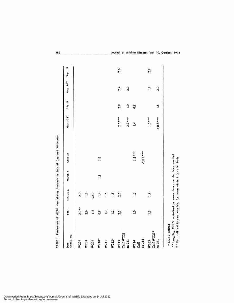
During transportation three of thenine wildebeest reacted badly to the xyla-zinc, acepromazine, and etorphine com-bination which had been used success-fully on these animals at the time ofcapture. These three animals (W207,W208, W212) died. They were all inadvanced pregnancy. Of the remainingsix wildebeest cows, one (W209) diedsuddenly 2 days after arrival at thelaboratory; another (W21 1 ) aborted 3days after arrival, became recumbentand died 2 days later. A third wildebeest(W2l0) also aborted 5 days after ar-rival at the laboratory but did not die.There remained three pregnant wilde-beest plus W210 for extended observa-tions. The three pregnant wildebeest allcalved normally 8-1 1 weeks after arrivalat the laboratory. The calf from W214died following an accident the day afterbirth. Wildebeest cow W210 was alsoinjured and was killed, in extremis on1 lth May. The nasal secretions and bloodof the wildebeest were sampled at thetime of transportation to the laboratorypaddock, and then weekly for the first 8weeks. Specimens were taken also on theday of calving, at which time the calveswere also tested for viremia and nasalvirus excretion. At the time intervalsshown in Table I, serum samples werecollected for antibody assay. All wilde-beest cows had high levels of neutralizingantibody (Table I). Contact cattle werebled weekly for viremia testing.
No virus was isolated from any of theblood samples, vaginal swabs, tissues of
Downloaded From: https://bioone.org/journals/Journal-of-Wildlife-Diseases on 24 Jul 2022Terms of Use: https://bioone.org/terms-of-use

-� I
Journal of Wildlife Diseases Vol. 10, October, 1974 481
FIGURE 1. Captive wildebeest in close association with cattle.
wildebeest which died, nor monolayersfrom aborted foetuses; nor from the calfof cow W2l4. However, monobayers pre-pared from the thyroids of wildebeestcow W2b0, which was killed after it hadaccidentally injured its neck, yielded acytopathogenic agent with a CPE typicalof MCFV. Syncytial foci were observedat the second blind passage at 6 days( Fig. 2 ) . Syncytia were easily discern-ible on further subculture of the mono-layer. The agent thus isolated producedtypical MCFV CPE in bovine thyroidmonolayers, lamb testis and lamb kidneymonolayers. The CPE was characterizedby formation of smooth syncytial mas-ses. Two steers which were inoculatedsubcutaneously with the 4th subcultureof wildebeest thyroid cells and fluids de-veloped the fatal ‘head and eye’ form ofMCF2 after an incubation period of 20days. The illness lasted 6 and 8 days, re-spectively. Viremia was first detected 14days p.i. and lasted to death. The grosspost-mortem lesions also were charac-teristic of MCF2.
All but one of the wildebeest nasalswab samples in the first collection yield-ed negative results. The only isolationof MCFV was made from wildebeestcow W2l2, from unclarified nasal swabfluid collected on the day of transpor-tation from the quarantine pens to thelaboratory paddock. This wildebeest un-fortunately died through an adverse reac-tion to the immobilizing drugs. Nasalswab fluids inoculated onto BTh rollertube monolayers produced typical MCFVCPE by the 10th day p.i. The agent thusisolated was passaged successfully inBTh cultures and at the 6th passagelevel it was inoculated into two steersin which it caused typical MCF. MCFVwas isolated from nasal secretions fromone of three wildebeest (W30) capturedand introduced into the paddock on 18thJuly, 1973. This animal was tested forMCFV in blood and nasal secretions 8times at approximately weekly intervals.MCFV was isolated only from the lastsample of nasal secretions taken on 20thSeptember, 1973.
S -�
,,, .,,.�. ..�. -:- 4, .-.j , . ., -
t ,4:�. ‘�i�L!
Downloaded From: https://bioone.org/journals/Journal-of-Wildlife-Diseases on 24 Jul 2022Terms of Use: https://bioone.org/terms-of-use
482 Journal of Wildlife Diseases Vol. 10, October, 1974
: “C 00
� r-4 c-i
V
1,)
N
. �. � 00 �
� r� e� ‘.�
00 � 00 006.-i -� -
N_;. * * * *
0 * * * *- * * * *
>, �f2 N ‘1� s�
Ca c-� e�- -�
V
6)a)
-aa)
�0
6) * *
� � : : �0
0. = sa� ci �
0 0. - - � ,J.�
.4- � V0�0
� �Ca�,) 00 �0
C � V
.- � .0
Ca �4�4
-0 � 000 0�E
-2�< N
�‘1 �0
� C. sC� C� � ‘�. 0’�..� , c-i ‘- c-i - - - c-I - -
A
U.D6) .0 Vz
>
b � C C�#{149}00 c-i c-i �U. c-i c-i �‘ C - - c-I - -
o 0.0
6)
o
CCC 0*
- - ZCaa. c-i c-i uz U U U. 2.0
w � N00�©�c-rCC�’�t � �-� � C C C - - - - 5- - ‘4-’ �.0 � lu� .,0 c-i c-i c-i c-i c-i c-i c-i� c-i� c-i * * *
Downloaded From: https://bioone.org/journals/Journal-of-Wildlife-Diseases on 24 Jul 2022Terms of Use: https://bioone.org/terms-of-use

#{149}1�.
#{149}.
/ 0�& �‘ #{149}
Journal of Wildlife Diseases Vol. 10, October, 1974 483
MCFV was isolated also from nasalsecretions of an orphaned wildebeestcalf which was captured by motor ye-hide chase when it was about 2 weeksold. An attempt was made to rear it bybottle feeding but it died on the 18thday of captivity, having become weakand lethargic. MCFV was isolated froma nasal swab specimen taken when thecalf was moribund but no virus wasfound in a blood specimen taken at thesame time. In this case the nasal swabfluid had been clarified by centrifugation.MCFV was isolated from both the su-pernate and deposit.
Effect of a Corticosteroid on Virus excretion
Two experiments were carried out inorder to assess the effect of a cortico-steroid drug on MCFV excretion. Inthe first experiment, the three survivingwildebeest from the first group in thewildebeest-cattle contact experiment(W2l3, W214, W263) and the twowildebeest calves (WC2I, WC2S) were
transferred from the paddock to holdingpens: each dam was housed togetherwith her calf. These animals had notshown viremia nor nasal virus excretion
during an observation period of 5#{189}months. The wildebeest cows were eachgiven a course of 40 mg betamethasonedaily by intramuscular injection for 7days. Each cow was also given a dailyintramuscular injection of 2 x l0� i.u.penicillin and 2 g streptomycin. Hand-ling was facilitated by xylazine sedation.All of the wildebeest cows and calves
were each tested daily for viremia andnasal virus excretion for 9 days, atwhich time they were returned to the
paddock where they continued to grazewith contact steers and three other fresh-
ly introduced wildebeest. Sampling wascontinued on days 14, 21 and 40 afterthe first betamethasone injection.
Viremia was not demonstrated in any
of the animals but MCVF was isolatedfrom the nasal swab sample collectedfrom cow W263 on the 14th day after
FIGURE 2. Syncytium produced in bovine thyroid cell monolayer by a strain of malignant
catarrhal fever virus iso’ated from wildebeest nasal secretions. Note prominent inclusions in
some of the nuclei. Haematoxylin-eosin x 800.
Downloaded From: https://bioone.org/journals/Journal-of-Wildlife-Diseases on 24 Jul 2022Terms of Use: https://bioone.org/terms-of-use

484 Journal of Wildlife Diseases Vol. 10, October, 1974
the initial betamethasone injection (i.e.7 days from the last injection). Viruswas also isolated from her calf, WC2S,persistently from the 14th up to the56th day, when it died. Altogether therewere six nasal MCFV isolations madefrom this calf by weekly samplings.Before it died calf WC2S had been thinand unthrifty, presumably because ofearly cessation of lactation by its dam,caused probably by the betamethasoneinjections. It died after immobilizationand handling. Necropsy revealed no be-sions suggestive of MCF. No virus wasisolated from spleen and lymph nodesbut MCFV was isolated from the buffycoat of the terminal blood sample.
In the second experiment, eight wilde-beest cows were immobilized in the fieldand held in pens at the capture site.They were given a daily course of beta-methasone by intramuscular injection for7 days and blood and nasal secretionswere tested as in the first experiment,except that vaginal swabs also weretaken. From one wildebeest cow MCFvirus was isolated from nasal secretionscollected on the 13th day after the begin-ning of the 7 day course of betametha-sone injections.
All eight of these wildebeest hadMCFV neutralizing antibody at the be-ginning of the experiment, with titresranging from 1.8-2.2 log,0 SN�. Serataken I 1 to 16 days later, after the beta-methasone injections but tested at thesame time as the pre-injection samples,had titres reduced in six of seven of theanimals, the mean reduction being 0.4log,0 SN�. One of the post-injection serawas lost. The one wildebeest which didnot show a reduction in antibody titrefollowing betamethasone treatment wasthe one from which MCFV was isolatedfrom nasal secretions. This wildebeestshowed a titre of 1.8 log,0 SN,o in boththe pre-and post-injection serum samples.
From the 7th to the 9th days, all eightwildebeest in this experiment developedpustular vulvo-vaginitis. Infectious bo-vine rhinotracheitis (IBR) virus was iso-bated from vaginal swab specimens fromseven of the eight wildebeest. This dis-ease outbreak is described in anotherpublication.4
Wildebeest-Cattle Contact
Altogether 17 steers were used in thecontact experiment in the paddock atMuguga. Initially three groups of fouranimals each were serially introducedinto the paddock. The first group wasremoved from the paddock at the timeof the first wildebeest abortion. Thesecond group was introduced then andremoved on the day of the second abor-tion and the third group which was in-troduced at that time was removed whenthe first wildebeest calved normally, atwhich time a fourth group of five steerswas introduced and remained in contactwith the wildebeest for 190 days. Thefirst 12 cattle have remained healthy todate (12 months). The last group of fivecattle was housed after having been incontact with wildebeest for 190 dayswithout any clinical response. They werethen challenged by subcutaneous inocu-lation with the nasal virus isolate fromW30 to determine their susceptibility to
MCF. Before challenge they were bloodsampled daily for 14 days for evidenceof viremia. Only three cattle werechallenged and the other two werehoused with them in one loose box. Rec-tal temperatures were recorded daily andwhole blood samples were taken at 2 dayintervals until the first animal showeda clinical response, after which bloodsamples were collected daily up to thetime of death. On the 6th day p.i. one ofthe inoculated cattle and one uninocu-bated control animal were viraemic. Thiscontrol animal became febrile on day12 and the pyrexia together with vira-emia continued until death on the 21stday p.i. The inoculated cattle becamefebrile on days 16, 19, and 20, respec-tively, and died on days 26, 27 and 30p.i. Viraemia was demonstrated in allanimals. The CPE in cell cultures inocu-lated with blood samples and the clinicaland pathological signs in the three inocu-bated and the control were all typical ofMCFV.
Isolation of MCFV from Wildebeest Tonsil
Blood and nasal swab specimens weretested for MCFV form 43 free-rangingwildebeest comprising 36 adult females,
Downloaded From: https://bioone.org/journals/Journal-of-Wildlife-Diseases on 24 Jul 2022Terms of Use: https://bioone.org/terms-of-use

Journal of Wildlife Diseases Vol. 10, October, 1974 485
two adult males and five calves. No virusisolations were made. MCFV was isola-ted, however, from the tonsil of a preg-nant wildebeest shot on 20th November1973, about 4 months before the normalcalving period.
Other numbers of wildebeest tissuesprocessed but found negative for MCFVwere the following: lymph node 9, thy-roid 6, foetus 4, trachea 3, pharynx 3,nasal mucosa 2, brain I, vagina 1, andplacenta I.
The wildebeest tonsil MCFV isolatewas inoculated subcutaneously into four3 year old steers, as 10 ml each of in-fected tissue culture medium and cellsfrom the third passage in BTh cells. Allfour steers developed typical MCF.Viraemia was first demonstrated on days12, 14, 14 and 24 p.i., respectively, andfever commenced on p.i. days 20, 18, 20and 29, i.e. 4 to 8 days after the begin-ning of viraemia. Viraemia was detectedintermittently until the animals died, ondays 27, 28, 30 and 58 p.i. respectively.Nasal secretions were swabbed andinoculated onto BTh cells at 2 day in-tervals until the first animal becamefebrile, then daily until death. MCFVwas not isolated from any of the nasalswab samples.
Summary of MCFV Isolations
from Wildebeest
Blood and nasal swab specimens andvarious body tissues were tested for
MCFV from 66 wildebeest, comprising55 adult females, three adult males, andeight calves. Only one isolate of MCFVwas made from 43 free-ranging wilde-beest when specimens were taken at thetime they were shot or captured; thisisolate was from tonsil tissues. In con-trast, isolates of MCFV were made fromnasal secretions of six of 23 captive wil-debeest, four from 19 adult females andtwo from three wildebeest calves. MCFVwas isolated repeatedly from nasal se-
cretions of one of the captive calves. Oneisolation of MCFV was also made fromthyroid tissues of a captive wildebeestcow.
DISCUSSION
In the studies reported here, MCFVwas demonstrated in the nasal secre-tions of six of the 66 wildebeest ( i.e.9% ) and altogether, virus was demon-strated in 8% of I 3 1 nasal swabs tested.In contrast only one of the 168 wilde-beest blood specimens yielded MCFV, inspite of the fact that a much greateramount of cellular material was inocula-ted from buffy coats than from nasalswabs. Hence, the quantity of virus innasal secretions seems to have beenhigher than in blood. Also this nasalvirus excretion in the absence of a de-tectable viraemia seems to suggest thesource of such virus to have been locatedeither in the upper respiratory or thepharyngeal-tonsilar regions. Our isola-tion of MCFV from tonsilar tissue lendsweight to this argument. Besides thekidney and thyroids, MCFV may alsopersist in the tonsilar or pharyngeal tis-sues of carrier wildebeest. Further stud-ies are required to determine whetherpersistence of MCFV in the tonsil occursonly in wildebeest, which could explainwhy this species transmits MCFV readilyto cattle, and horizontally within thewildebeest population.
Our results indicate that shedding ofMCFV in nasal secretions is probablythe principal mechanism for transmissionof infection among wildebeest and fromwildebeest to cattle, at least during closecontact, as shown previously by Plow-right.5’6 We have not been able so far todemonstrate unequivocally whether wil-debeest excrete virus nasally in a stableinfectious cell-free state. All nasal virusisolates except one were made from un-clarified fluid. In one instance in whichwe isolated virus after attempts to sepa-rate out cell-associated virus by lightcentrifugation, virus was isolated fromboth the supernate and precipitate. How-ever, until further evidence is obtained,these results do not constitute sufficientgrounds for claiming that wildebeest cx-crete MCFV nasally in a cell-free stableinfectious state. Our isolate of virus fromthe supernate might be a reflection ofpoor cell packing by centrifugation. Theprogeny of all the isolates in our studieswere cell-associated. In this context, it
Downloaded From: https://bioone.org/journals/Journal-of-Wildlife-Diseases on 24 Jul 2022Terms of Use: https://bioone.org/terms-of-use

486 Journal of Wildlife Diseases Vol. 10, October, 1974
should be observed that infectious cell-free virus has been demonstrated in thefeather follicle epithelium in Marek’sdisease, another typically cell-associatedherpesvirus infection.’
Pbowright detected higher rates ofviraemia in wildebeest calves than inadults, and among adults, viraemia wasmost common during late pregnancy.”4Epizootiologically, MCF in cattle isusually associated with calving in thecontact wildebeest population. The stres-ses of late pregnancy and calving prob-ably lead to a reactivation of latent virusin wildebeest, to transplacental infectionand wildebeest-to-wildebeest transmis-sion, in addition to dissemination of thevirus by neonatal calves, as demonstratedby Plowright.’8 In our studies, we isola-ted MCFV from nasal secretions fromanimals which had been held in confine-ment in pens for a period, under thestress of artificial feeding, or after beta-methasone administration. Although thenumbers studied are small, our resultsindicate that betamethasone reactivatedlatent MCF virus infection in wildebeest,in the same manner as Sheffy and Da-vies’2 demonstrated reactivation of IBRvirus in cattle. Incidentally, we also ob-tamed evidence of reactivation of IBR inthe wildebeest.4
Grazing of wildebeest in a paddockdoes not seem to have constituted suf-ficient stress to the wildebeest to resultin carrier breakdown. That the majorityof the wildebeest in the paddock prob-
Acknowledgements
ably carried MCF virus is supported bythe isolation of MCFV from W263 afterbetamethasone injections and in cell cub-tures derived from the thyroid of W210.The virus isolated from the nasal secre-tion of wildebeest cow W30 may wellhave originated from W263 or calfWC2S, since these two animals had beenexcreting virus for at least 7 weeks whilstin contact with W30 and before viruswas detected in W30. The apparent spon-taneous development of MCF in one ofthe five cattle is most likely explicable onthe steer having acquired infection fromcontact with wildebeest in the paddock.These cattle were probably challengedwhen some of them were incubating theinfection. It is unlikely that the controlsteer which developed apparently spon-taneous MCF could have acquired infec-tion from the inoculated pen-mates, as itdeveloped viraemia and disease beforethe inoculated animals. Also, retrospec-tively, we have realized that this animaldeveloped MCF viraemia 61 days afterour last isolation of MCFV from nasalsecretions of wildebeest W30 in the pad-dock. Such a period is within the limitsof the incubation period of MCF in cat-tle.2’#{176}
These experiments have provided ad-ditional evidence on the carrier status ofMCFV in wildebeest and have demon-strated that nasal excretion may be theprincipal route of transmission of MCFVfrom wildebeest to cattle and within thewildebeest population.
We are grateful to the Kenya Wildlife Management Project for provision of some of the
e,s’wrimental wildebeest and to the Chief Game Warden, Kenya, for permission to capture wilde-
heest for this research. The District Veterinary Officer for Kajiado, Dr. R. Eugster, has given
continuing support and encouragement. The careful technical assistance of Mrs. E. Finnerup and
Messrs. J. Mugo and Kariuki Wachira is also acknowledged.
LITERATURE CITED
I. CALNEK, B. W., HANS K. ADLDINGER and DONALD E. KAHN. 1970.Feather follicle epithelium: a source of enveloped and infectious cell-freeherpesvirus from Marek’s disease. Avian Dis. 14: 219-233.
2. DAUBNEY, R. and J. R. HUDSON. 1936. Transmission experiments withbovine malignant catarrh. J. comp. Path. 49: 63-89.
3. JOHNSON, R. H. and V. W. SMITH. 1962. The production and use of tissueculture rinderpest vaccine in Nigeria. Bull. epizoot. Dis. Afr. 10: 417-422.
Downloaded From: https://bioone.org/journals/Journal-of-Wildlife-Diseases on 24 Jul 2022Terms of Use: https://bioone.org/terms-of-use
Journal of Wildlife Diseases Vol. 10, October, 1974 487
4. KARSTAD, L., S. DREVEMO, J. C. OTEMA and D. M. JESSETT. 1973. Vul-vovaginitis in wildebeest caused by the virus of infectious bovine rhino-tracheitis. J. WildI. Dis. 10: 392-396.
5. PLOWRIGHT, W. 1965. Malignant catarrhal fever in East Africa. I. Behaviourof the virus in free-living populations of blue wildebeest (Gorgon taurinustaurinus, Burchell) Res. vet. Sci. 6: 56-68.
6. PLOWRIGHT, W. 1965. Malignant catarrhal fever in East Africa. II. Observa-tions on wildebeest calves at the laboratory and contact transmission ofthe infection to cattle. Res. vet. Sci. 6: 69-83.
7. PLOWRIGHT, W. 1967. Malignant catarrhal fever in East Africa. III. Neutra-lizing antibody in free living wildebeest. Res. vet. Sci. 8: 129-136.
8. PLOWRIGHT, W. 1968. Malignant catarrhab fever. J. Am. vet. med. Ass. 152:795-804.
9. PLOWRIGHT, W. and R. D. FERRIS. 1961. The preparation of bovine thy-roid monolayers for use in virobogical investigations. Res. vet. Sci. 2: 149-152.
10. PLOWRIGHT, W., R. D. FERRIS and G. R. SCOTT. 1960. Blue wildebeestand the virus of malignant catarrhal fever. Nature (Lond.) 188: lb 67-1169.
I I . PLOWRIGHT, W., M. KALUNDA, D. M. JESSETF and K. A. J. HERNI-MAN. 1972. Congenital infection of cattle with the herpesvirus causingmalignant catarrhal fever. Res. vet. Sci. 13: 37-45.
12. PLOWRIGHT, W., R. F. MACADAM and J. A. ARMSTRONG. 1965. Growthand characterization of the virus of bovine malignant catarrhal fever inEast Africa. J. gen. Microbiol. 39: 253-266.
13. SHEFFY, BEN E. and D. HUGH DAVIES. 1972. Reactivation of bovineherpesvirus after corticosteroid treatment. Proc. Soc. exp. Biol. Med. 140:974-976.
Received for publication 29 March 1974
Downloaded From: https://bioone.org/journals/Journal-of-Wildlife-Diseases on 24 Jul 2022Terms of Use: https://bioone.org/terms-of-use
Copyright © 2022 FDOKUMEN