Evidence of a role for B cell–activating factor of the TNF family in the pathogenesis of chronic...
18
Evidence of a role for B cell–activating factor of the TNF family in the pathogenesis of chronic rhinosinusitis with nasal polyps Atsushi Kato, PhD a , Anju Peters, MD a , Lydia Suh, BSc a , Roderick Carter, BSc a , Kathleen E. Harris, BSc a , Rakesh Chandra, MD b , David Conley, MD b , Leslie C. Grammer, MD a , Robert Kern, MD b , and Robert P. Schleimer, PhD a a Division of Allergy and Immunology, Department of Medicine, Chicago, Ill b Department of Otolaryngology, Northwestern University Feinberg School of Medicine, Chicago, Ill Abstract Background—The polypoid form of chronic rhinosinusitis (chronic rhinosinusitis with nasal polyps [CRSwNP]) is a highly prevalent disease that often requires surgical intervention for treatment. Nasal polyps contain large quantities of B lymphocytes and immunoglobulin as well as eosinophils. Objectives—The objective of this study was to investigate the expression of B cell–activating factor of the TNF family (BAFF), an important regulator of class-switch recombination and immunoglobulin production, in patients with chronic rhinosinusitis (CRS). Methods—We collected nasal tissue and nasal lavage fluid from patients with CRS and control subjects. We assayed mRNA for BAFF and B-lymphocyte markers, CD20 and transmembrane activator and calcium-modulator and cyclophilin ligand interactor, by using real-time PCR, and assayed BAFF protein by using ELISA and immunohistochemistry. Results—BAFF mRNA was significantly increased in nasal polyps from patients with CRSwNP (P < .001) compared with inferior turbinate tissue from patients with CRS or healthy subjects. BAFF protein was also elevated in polypoid tissue and nasal lavage from patients with CRSwNP. Immunohistochemistry showed considerable BAFF staining in mucosal epithelial cells in nasal polyps along with unidentified cells in the lamina propria. Expression of mRNA for BAFF in sinonasal tissue was significantly correlated with CD20 and transmembrane activator and CAML interactor in sinus tissue. IgA, an immunoglobulin isotype known to activate eosinophils, was also significantly elevated in the polypoid tissue. Conclusion—Overproduction of BAFF in nasal polyps may contribute to the pathogenesis of CRSwNP via the local induction of IgA and activation of eosinophils. Keywords Chronic rhinosinusitis; nasal polyps; BAFF; IgA; B cells; eosinophils; epithelial cells © 2008 American Academy of Allergy, Asthma & Immunology Reprint requests: Robert P. Schleimer, PhD, Division of Allergy and Immunology, Northwestern University Feinberg School of Medicine, 240 E Huron, Chicago, IL 60611. [email protected]. Disclosure of potential conflict of interest: A. Peters has served as a member of the American Academy of Allergy, Asthma & Immunology. D. Conley has received research support from Acclarent. L. C. Grammer has served as an expert witness on reaction during hemodialysis litigation. R. Kern has served as a member of the American Rhinologic Society. The rest of the authors have declared that they have no conflict of interest. Clinical implications: Expression or function of BAFF in nasal polyps may have a pathogenic role in CRSwNP. NIH Public Access Author Manuscript J Allergy Clin Immunol. Author manuscript; available in PMC 2010 January 5. Published in final edited form as: J Allergy Clin Immunol. 2008 June ; 121(6): 1385–1392.e2. doi:10.1016/j.jaci.2008.03.002. NIH-PA Author Manuscript NIH-PA Author Manuscript NIH-PA Author Manuscript
-
Upload
independent -
Category
Documents
-
view
1 -
download
0
Transcript of Evidence of a role for B cell–activating factor of the TNF family in the pathogenesis of chronic...

Evidence of a role for B cell–activating factor of the TNF family inthe pathogenesis of chronic rhinosinusitis with nasal polyps
Atsushi Kato, PhDa, Anju Peters, MDa, Lydia Suh, BSca, Roderick Carter, BSca, Kathleen E.Harris, BSca, Rakesh Chandra, MDb, David Conley, MDb, Leslie C. Grammer, MDa, RobertKern, MDb, and Robert P. Schleimer, PhDaaDivision of Allergy and Immunology, Department of Medicine, Chicago, IllbDepartment of Otolaryngology, Northwestern University Feinberg School of Medicine, Chicago, Ill
AbstractBackground—The polypoid form of chronic rhinosinusitis (chronic rhinosinusitis with nasalpolyps [CRSwNP]) is a highly prevalent disease that often requires surgical intervention fortreatment. Nasal polyps contain large quantities of B lymphocytes and immunoglobulin as well aseosinophils.
Objectives—The objective of this study was to investigate the expression of B cell–activating factorof the TNF family (BAFF), an important regulator of class-switch recombination andimmunoglobulin production, in patients with chronic rhinosinusitis (CRS).
Methods—We collected nasal tissue and nasal lavage fluid from patients with CRS and controlsubjects. We assayed mRNA for BAFF and B-lymphocyte markers, CD20 and transmembraneactivator and calcium-modulator and cyclophilin ligand interactor, by using real-time PCR, andassayed BAFF protein by using ELISA and immunohistochemistry.
Results—BAFF mRNA was significantly increased in nasal polyps from patients with CRSwNP(P < .001) compared with inferior turbinate tissue from patients with CRS or healthy subjects. BAFFprotein was also elevated in polypoid tissue and nasal lavage from patients with CRSwNP.
Immunohistochemistry showed considerable BAFF staining in mucosal epithelial cells in nasalpolyps along with unidentified cells in the lamina propria. Expression of mRNA for BAFF insinonasal tissue was significantly correlated with CD20 and transmembrane activator and CAMLinteractor in sinus tissue. IgA, an immunoglobulin isotype known to activate eosinophils, was alsosignificantly elevated in the polypoid tissue.
Conclusion—Overproduction of BAFF in nasal polyps may contribute to the pathogenesis ofCRSwNP via the local induction of IgA and activation of eosinophils.
KeywordsChronic rhinosinusitis; nasal polyps; BAFF; IgA; B cells; eosinophils; epithelial cells
© 2008 American Academy of Allergy, Asthma & ImmunologyReprint requests: Robert P. Schleimer, PhD, Division of Allergy and Immunology, Northwestern University Feinberg School of Medicine,240 E Huron, Chicago, IL 60611. [email protected] of potential conflict of interest: A. Peters has served as a member of the American Academy of Allergy, Asthma &Immunology. D. Conley has received research support from Acclarent. L. C. Grammer has served as an expert witness on reaction duringhemodialysis litigation. R. Kern has served as a member of the American Rhinologic Society. The rest of the authors have declared thatthey have no conflict of interest.Clinical implications: Expression or function of BAFF in nasal polyps may have a pathogenic role in CRSwNP.
NIH Public AccessAuthor ManuscriptJ Allergy Clin Immunol. Author manuscript; available in PMC 2010 January 5.
Published in final edited form as:J Allergy Clin Immunol. 2008 June ; 121(6): 1385–1392.e2. doi:10.1016/j.jaci.2008.03.002.
NIH
-PA Author Manuscript
NIH
-PA Author Manuscript
NIH
-PA Author Manuscript

Chronic rhinosinusitis (CRS) is the most common chronic disease in adults in the United Statesand is estimated to affect 5% to 15% of the urban population.1,2 Primarily on the basis ofphysical examination, histology, and clinical course, CRS is frequently divided into 2 types:CRS with nasal polyps (CRSwNP) and CRS without nasal polyps (CRSsNP). The etiologyand pathogenesis of CRS remain controversial, but recent studies have implicated Alternariafungi or toxin-secreting staphylococci as key pathogens initiating the symptomatic mucosalinflammation.3,4 Histologic studies have demonstrated significant tissue eosinophilia in a highproportion of CRS cases, most prominently in CRSwNP.5 The ultimate factors inducing thismucosal eosinophilia remain uncertain, but several studies have reported that IL-5 (aneosinophil survival and differentiation factor), eotaxins (eosinophil chemoattractants) andeosinophil cationic protein (an indicator of the presence of eosinophil) are significantlyincreased in polyp tissue compared with sinonasal tissue from patients with CRSsNP or fromhealthy subjects.6–8 Taken together, these results point to a prominent role for eosinophils inthe pathophysiology of CRSwNP and further suggest that factors triggering eosinophildegranulation may also be associated with polyp formation.
In the case of several diseases of the airways, there are compelling reasons to believe that localproliferation and activation of B cells is of central pathogenic importance.9–14 Local B-cellclass-switch recombination and synthesis of IgE and IgA can mediate activation of airway mastcells and eosinophils, respectively, in response to antigen exposure. In the case of CRS, a largeproportion of patients with nasal polyps demonstrate the presence of local IgE againstaeroallergens without evidence of circulating IgE against the same antigens.12,13 Recentstudies have indicated that plasma cell number and antigen-specific IgE concentration areincreased in the polypoid sinonasal mucosal tissue from patients with CRSwNP.8,15,16 Incontrast with IgE, which is believed to activate mast cells in atopic patients with CRS, the roleof IgA in CRS is poorly understood. Interestingly, IgA can serve as a trigger for eosinophildegranulation by binding to surface receptors present on these cells. Although it has becomeclear that B-cell accumulation and immunoglobulin production at local mucosal sites in theairway are of great importance to airway inflammatory diseases, the mechanism of localimmunoglobulin class switching and production is not fully understood.
B cell–activating factor of the TNF family (BAFF; also known as BLyS, TNFSF13B, TALL-1,and THANK) and a proliferation-inducing ligand (APRIL) are recently identified members ofthe TNF superfamily that play important roles in B-cell survival, proliferation, and maturation.17–19 Although class-switch recombination is generally thought to be highly dependent onligation of CD40 (on B cells) and CD40 ligand (on activated T cells), it has been reported thatBAFF and APRIL also promote T cell–dependent immunoglobulin production as well asCD40-independent, T cell–independent immunoglobulin class switching and production.20–22 BAFF binds to 3 receptors that are selectively expressed on B cells and plasma cells,including BAFF receptor (BAFF-R), transmembrane activator and CAML interactor (TACI),and B-cell maturation antigen. APRIL also binds to TACI and B-cell maturation antigen, butnot BAFF-R. BAFF-R is a potent regulator of mature B-cell survival and IgE production byBAFF.23 In contrast, TACI has been considered to suppress B-cell proliferation and survivalbut is critical for the class-switch recombination and production of IgA in human beings.19,24 Although BAFF has been recognized to be mainly a product of myeloid cells such asmonocytes, macrophages, dendritic cells, and neutrophils, nonlymphoid cell types also produceBAFF, including salivary gland epithelial cells and astrocytes.19 Recently we havedemonstrated that BAFF is produced by bronchial epithelial cells after stimulation with ligandfor Toll-like receptor (TLR)–3, IFNs, and TNF in quantities of the same order of magnitudeas produced by myeloid cells.25 In the current study, we investigated whether BAFF and APRILmight be involved in the pathogenesis of CRS. We discovered that resected polyp tissue frompatients with CRSwNP had elevated levels of BAFF protein as well as markers of B cells and
Kato et al. Page 2
J Allergy Clin Immunol. Author manuscript; available in PMC 2010 January 5.
NIH
-PA Author Manuscript
NIH
-PA Author Manuscript
NIH
-PA Author Manuscript

IgA. These findings imply that upregulation of BAFF inCRSwNP may amplify eosinophilicinflammation via induction of class-switch recombination and production of IgA by B cells innasal polyps.
METHODSPatients and biopsies
Patients with CRS were recruited from the allergy clinic and the otolaryngology clinic atNorthwestern University and the Northwestern Sinus Center. Sinonasal and polyp tissues wereobtained from routine functional endoscopic sinus surgery in the patients with CRS. Allsubjects met the criteria for CRS as defined by the Sinus and Allergy Health Partnership.1 Thepresence of sinusitis or bilateral nasal polyps was confirmed by office endoscopy and computedtomography imaging. All patients scheduled for surgery had previously failed to respond toadequate trials of conservative medical therapy (prolonged antibiotic regimens, nasal steroidsprays, oral steroids, saline irrigations, and decongestants) for control of symptoms. Patientswith an isolated antrochoanal polyp, cystic fibrosis, or unilateral nasal polyps were excludedfrom the study. Details of subjects’ characteristics are included in Table I. Disease-free controlsubjects undergoing procedures to correct anatomical defects without history of CRS or asthmawere recruited from the otolaryngology clinic at Northwestern University. All subjects signedinformed consent, and the protocol and consent forms governing procedures for this study havebeen approved by the Institutional Review Board of Northwestern University Feinberg Schoolof Medicine. A portion of each sample for isolation of RNA was transferred in RNAlater(Ambion, Austin, Tex) and stored at −20°C.
Cell cultureThe methods for primary nasal epithelial cell (PNEC) culture are described in this article’sOnline Repository at www.jacionline.org and in Kim et al.26
Real-time PCRTotal RNA from sinus tissue was extracted using QIAzol (Qiagen, Valencia, Calif) and wascleaned and treated with DNase I using RNeasy (Qiagen) according to the manufacturer’sinstructions. Quality of total RNA from sinus tissue was assessed with a 2100 Bioanalyzer(Agilent Technologies, Santa Clara, Calif) using a RNA 6000 Nano LabChip (AgilentTechnologies). Single-strand cDNA was synthesized with SuperScript II reverse transcriptase(Invitrogen, Carlsbad, Calif) and random primers. Semiquantitative real-time RT-PCR wasperformed with a TaqMan method using an Applied Biosystems 7500 Sequence DetectionSystem (Applied Biosystems, Foster City, Calif) as described previously.25 Primer and probesets for 5 genes, BAFF,25 APRIL,25 CD20 (sense, 5′-CACTCTTCAGGAGGATGTCTTCACT-3′; anti-sense, 5′-TCTGGACAGCCCCCAAAGT-3′; minor groove binder-probe, 5′-TCTTCATGAGGGAATCTA-3′), TACI (sense, 5′-AACTCGGGAAGGTACCAAGG-3′;antisense, 5′-GCTGTAGACCAGGGCCACC-3′; 6-carboxyfluorescein/6-carboxytetramethylrhodamine-probe, 5′-CCAGAAGCAAGTC CAGCTCTCCCGG-3′), andβ-actin (ACTB; sense, 5′-CTGGCCGGGACCTGACT-3′; antisense, 5′-GCAGCCGTGGCCATCTC-3′;MGB-probe, 5′-CACCACCACGGCCGA-3′) weresynthesized by Applied Biosystems or Integrated DNA Technologies (Coralville, Iowa). Aprimer and probe set for β-glucuronidase (human β-glucuronidase endogenous control, partnumber 4326320) was purchased from Applied Biosystems. To determine the exact copynumber of the target genes, quantified aliquots of purified PCR fragments of the target geneswere serially diluted and used as standards in each experiment. Aliquots of cDNA equivalentto 10 ng of total RNA were used for real-time PCR. The mRNA expression levels were
Kato et al. Page 3
J Allergy Clin Immunol. Author manuscript; available in PMC 2010 January 5.
NIH
-PA Author Manuscript
NIH
-PA Author Manuscript
NIH
-PA Author Manuscript

normalized to the median expression of housekeeping genes, ACTB for PNEC and β-glucuronidase for nasal tissue.
Measurement of BAFF and IgA in tissue homogenates and nasal lavage fluidsFreshly obtained tissue specimens were weighed, and 1 mL PBS supplemented with 0.05%Tween 20 (Sigma-Aldrich, St Louis, Mo) and 1% protease inhibitor cocktail (PN; P8340,Sigma-Aldrich) was added per every 100 mg tissue. The tissue was then homogenized with anUltra-Turrax T8 homogenizer (IKA, Wilmington, NC) at setting 3 for 1 minutes on ice. Afterhomogenization, the suspension was centrifuged at 4000 rpm for 20 minutes at 4°C, and thesupernatants were stored at −80°C until analyzed.
Nasal lavage was performed by instilling 5mL sterile PBS into each nostril, holding for 5seconds and then expelling into a sterile container. Samples were concentrated 2-fold by usingCentriplus YM-10 (Millipore, Bedford, Mass) and stored at −80°C until analyzed.
The concentrations of BAFF (R&D Systems, Minneapolis, Minn) and IgA (Bethyl Laboratory,Montgomery, Tex) in cell-free supernatants were determined by specific ELISA kits. Theminimal detection limits for these kits are 15 pg/mL and 7.8 ng/mL, respectively. Theconcentration of total protein was measured with a Bradford method–based Bio-Rad proteinassay kit (Bio-Rad, Hercules, Calif). Concentrations of BAFF and IgA in the tissue homogenateand nasal lavage were normalized to the concentration of total protein.
ImmunohistochemistryNasal tissue was dehydrated, infiltrated, and embedded with paraffin, and tissue was sectionedat 3 µm by using a Leica RM2245 Cryostat (Leica Microsystems Inc, Bannockburn, Ill).Sections were rehydrated and blocked for endogenous peroxidase activity with 3% H2O2/methanol. After rinsing, tissue sections were blocked for nonspecific binding with 1% goatserum/0.3% Tween-20/PBS. Tissue sections were then incubated with 0.125 µg/mL ratantihuman BAFF mAb (clone: Buffy 2, IgM; Abcam, Cambridge, Mass) or 0.125 µg/mL ratIgM antibody (clone: RTK2118; Abcam) for 1 hour at room temperature. Sections were rinsedand then incubated in biotinylated secondary goat antirat antibody (Jackson ImmunoResearchLaboratories, West Grove, Pa) at a 1:500 dilution for 1 hour at room temperature. After anotherrinse, sections were incubated in ABC reagent (avidin–biotin–horseradish peroxidasecomplex; Vector Laboratories, Burlingame, Calif) for 1 hour at room temperature. Sectionswere rinsed again and incubated in diaminobenzidine reagent (Invitrogen) for 10 minutes atroom temperature. They were then rinsed in deionized H2O, counterstained with hematoxylin,dehydrated, cleared, mounted, and coverslipped by using Cytoseal 60 (Richard-AllanScientific, Kalamazoo, Mich) in preparation for microscopic analysis.
The number of BAFF positive cells in epithelium, glands, and submucosae was counted byusing a magnification of × 400. Each section was randomly selected, and diagnosis wasunknown to the observer. The tissue area counted was determined using the National Institutesof Health–issued Image J software.
StatisticsAll data are reported as the means ± SEMs unless otherwise noted. Differences between groupswere analyzed by using the Wilcoxon signed-rank test or the Mann-Whitney U test.Correlations were assessed by using the Spearman rank correlation. A P value less than .05was considered significant.
Kato et al. Page 4
J Allergy Clin Immunol. Author manuscript; available in PMC 2010 January 5.
NIH
-PA Author Manuscript
NIH
-PA Author Manuscript
NIH
-PA Author Manuscript

RESULTSBAFF expression in CRS
To determine the relevance of BAFF and APRIL expression in chronic rhinosinusitis, sinonasaland polyp tissues were collected from 39 subjects with CRSsNP, 60 subjects with CRSwNP,and 30 control subjects. Subject characteristics are shown in Table I. Subjects in the variousgroups were of similar age and sex. None of the patients had Churg-Strauss syndrome orallergic fungal sinusitis, and 1 patient with CRSwNP had aspirin sensitivity (but not asthma)in this study. Historically, approximately 80% of nasal polyps have eosinophilia (ie, greaterthan 5 eosinophils per high power field) in our clinic.
It has been reported that mRNA expression of housekeeping genes, including ACTB, iselevated in the nasal mucosa of patients with CRS compared with control subjects.27 Wetherefore first screened the expression of housekeeping genes in the inferior turbinate (IT) frompatients with CRS and in nasal polyp tissue by using the TaqMan Human Endogenous ControlPlate (Applied Biosystems), which included 11 housekeeping genes and an internal positivecontrol. We found that the level of mRNA expression of 3 housekeeping genes, 18S rRNA,acidic ribosomal protein, and β-glucuronidase, was not significantly different between IT andnasal polyp tissue and that the level of mRNA expression of the 8 other genes, including ACTB,was elevated in the nasal polyps (n = 4; data not shown). We thus selected β-glucuronidase asthe housekeeping gene for normalization in the current report.
We assessed the expression of BAFF and APRIL in IT tissue from patients with CRSsNP andCRSwNP and controls, as well as in polyp tissue from patients with CRSwNP. Expression ofβ-glucuronidase was not significantly different between the 4 groups (data not shown). BAFFmRNA was significantly increased in polyp tissue from patients with CRSwNP (P <.001) incomparison with IT tissue from either patients with CRS or control subjects (Fig 1, A). Incontrast, expression of mRNA for APRIL was not different in the same groups (data notshown). To confirm this observation at the protein level, we made detergent extracts fromhomogenates of IT and nasal polyp tissues and then measured the concentration of BAFF byusing ELISA. BAFF protein was significantly increased in polyp tissue (P <.05) comparedwith IT tissue from either patients with CRS or control subjects (Fig 1, B). BAFF was detectedin 11 of 23 nasal polyp extracts from patients with CRSwNP and in 2 of 17 IT extracts frompatients with CRSsNP, but it was not detected in IT extracts from either patients with CRSwNP(n = 8) or control subjects (n = 13; Fig 1, B). We also determined BAFF protein expression innasal lavage fluids collected from separate groups of subjects. BAFF protein was detected in5 of 12 nasal lavage fluids from patients with CRSwNP (P < .05) but was not detected in nasallavage from patients with CRSsNP (n = 7) or from control subjects (n = 7; Fig 1, C). Thesedata suggest that BAFF is locally produced in the nasal mucosa of 40% to 50% of patients withCRSwNP.
To examine the BAFF-producing cells in nasal mucosa, we used immunohistochemistry todetect BAFF in uncinate tissue and nasal polyp tissue. As shown in Fig 2, BAFF staining wasobserved in the mucosal and the glandular epithelium as well as in submucosal cells. Elevatedsubmucosal staining was found in tissue from patients with CRSwNP. Similar data wereobtained by using a different clone of antihuman BAFF mAb (a kind gift from Drs FabienneMackay and Charles Mackay, Garvan Institute of Medical Research, Darlinghurst, Australia).We also counted the number of BAFF-positive cells by using a semiquantitative method.Perhaps because BAFF was highly expressed by epithelial cells in all subject groups, we didnot observe a significant difference in the number of BAFF-positive cells in the epithelium andglands among the 3 groups of subjects (Table II). However, BAFF-positive cells weresignificantly elevated in the submucosal region of uncinate and nasal polyp tissues from thepatients with CRSwNP (Fig 2, D– F; Table II).
Kato et al. Page 5
J Allergy Clin Immunol. Author manuscript; available in PMC 2010 January 5.
NIH
-PA Author Manuscript
NIH
-PA Author Manuscript
NIH
-PA Author Manuscript

Nasal polyps from patients with CRSwNP have long been known to be characterized byeosinophilic inflammation.1,5 Immunohistochemical staining showed that some of the BAFF-positive cells in polyp tissue may be eosinophils (Fig 2, F, arrow).We therefore tested whethereosinophils were able to produce BAFF. Purified human eosinophils from peripheral bloodwere stimulated with IL-5, GM-CSF, plate-bound secretory IgA, IFN-β, and IFN-γ. However,we could not find detectable amounts of BAFF in supernatants of eosinophils stimulated aslong as 72 hours (data not shown).
BAFF and APRIL expression in nasal epithelial cellsAlthough we have reported that bronchial epithelial cells are capable of producing BAFF,25 itis not known whether nasal epithelial cells also produce BAFF. To test the possible role of theexpression of BAFF and APRIL in nasal epithelial cells, PNECs were treated for 6 hours withvarious stimuli including TLR ligands and cytokines. In resting PNECs, expression of BAFFand APRIL was minimal. However, mRNA for BAFF was significantly upregulated bystimulation with double-stranded RNA (dsRNA) (TLR-3 ligand; 213-fold: n = 7, P < .05),IFN-β (22-fold; n = 8; P < .05), IFN-γ (6-fold; n = 9; P < .05), and IFN-λ1 (6-fold; n = 5; P <.05), and was not affected by peptidoglycan (TLR-2 ligand), LPS (TLR-4 ligand), flagellin(TLR-5 ligand), TNF, IL-1β, IL-4, IL-6, IL-10, IL-13, IL-17, oncostatin M, and TGF-β inPNEC (see this article’s Fig E1, A, in the Online Repository at www.jacionline.org; data notshown). In contrast, mRNA for APRIL was significantly but weakly upregulated by stimulationwith dsRNA (2-fold; n = 7; P < .05), IL-4 (2-fold; n = 7; P < .05), IFN-β (2-fold; n = 8; P < .05) and IFN-γ (2-fold; n = 9; P <.05; Fig E1, A). To examine further the details of the inductionof mRNA for BAFF, we determined the time dependence of the responses in PNECs.Expression of mRNA for BAFF induced by dsRNA peaked at 24 hours, and elevated expressionof BAFF persisted as long as 72 hours (Fig E1, B). In contrast, expression of mRNA for BAFFinduced by IFN-β peaked at 6 hours, then returned to baseline at 72 hours (Fig E1, B). Thesedata imply that the mechanism of dsRNA-dependent BAFF expression in PNECs may be thesame as in bronchial epithelial cells, in which we identified an autocrine/paracrine loopinvolving IFN-β. 25 To confirm these findings at the protein level, we measured the productionof BAFF by using ELISA. Significant levels of BAFF were detected in the supernatant afterstimulation with dsRNA and IFN-β for 72 hours (control, undetectable; dsRNA-treated, 64 ±10 pg/mL, n = 7; IFN-β–treated, 20 ± 5 pg/mL, n = 4; Fig E1, C).
CD20 and IgA expression in nasal polypsBecause BAFF is known to be an important factor in the generation, maintenance, andactivation of B lymphocytes, we next assessed the level of expression of mRNA for the B-cellmarker CD20 and for TACI (a receptor for BAFF expressed on B cells and plasma cells) in ITtissue from patients with CRS and control subjects, and in polyp tissue from patients withCRSwNP. The expression of CD20 (and TACI) was highly elevated in nasal polyps frompatients with CRSwNP compared with IT tissue from patients with CRS and control subjects(Fig 3, A; data not shown; P < .05). The levels of expression of CD20 and TACI weresignificantly correlated with the expression of BAFF (CD20, r = 0.672, P < .001; TACI, r =0.673, P < .001; Fig 3, B), suggesting that locally produced BAFF may be involved in theelevation of B cells. As a monitor of local B-cell responses, we examined the concentration ofIgA and IgE in tissue extracts. The level of IgA was significantly increased in polyp tissuefrom patients with CRSwNP (25.7 ± 3.6 µg/mg; n = 10; P < .001) compared with IT tissuefrom patients with CRSsNP (7.4 ± 1.9 µg/mg; n = 11), patients with CRSwNP (6.6 ± 1.3 µg/mg; n = 9), or healthy subjects (5.2 ± 1.1 µg/mg; n = 9; Fig 4). In contrast, we did not observea significant difference in the levels of total IgE in the tissue homogenates from these groupsof subjects (data not shown).
Kato et al. Page 6
J Allergy Clin Immunol. Author manuscript; available in PMC 2010 January 5.
NIH
-PA Author Manuscript
NIH
-PA Author Manuscript
NIH
-PA Author Manuscript

DISCUSSIONB cell–activating factor of the TNF family is an important regulator of immunoglobulin class-switch recombination and production in B lymphocytes, independent of T-cell help. BAFFalso enhances T cell–dependent immunoglobulin class switching and production. This studyprovides the first demonstration that BAFF is significantly increased in polypoid tissue frompatients with CRSwNP (Fig 1 and Fig 2). Significant levels of BAFF protein were found inboth polypoid tissue and nasal lavage taken from patients with CRSwNP (Fig 1). BAFF-positive cells were elevated in the submucosal tissue of uncinate and nasal polyps from patientswith CRSwNP (Fig 2; Table II). We also demonstrate that IgA was significantly elevated inpolyp tissue from patients with CRSwNP (Fig 4).
It has been reported that B cells, plasma cells, and antigen-specific IgE accumulate in the nasalmucosa of patients with CRSwNP, although the mechanism is not fully understood.8,12,13,15,16 In the current study, we demonstrate that mRNA encoding BAFF was significantly increasedin polyp tissue (Fig 1, A). BAFF protein was detected in 40% to 50% of polyp tissue extractsand was also detected in nasal lavage from a distinct cohort of patients with CRSwNP (Fig 1,B and C), reinforcing this finding. Although BAFF protein was found to be elevated in onlyhalf of the CRSwNP extracts and nasal lavage samples, elevated BAFF mRNA was detectedin nearly all polyp tissues. This difference may reflect the detection limit of the ELISA and therelatively dilute extracts that were used. Importantly, expression of BAFF was correlated withexpression of CD20 in nasal tissue and with levels of TACI, a receptor for BAFF and APRILfound on B cells and plasma cells (Fig 3, B). These data suggest that increased levels of BAFFmay play a role in local proliferation and activation of B cells and plasma cells in nasal polyptissue. It is possible that other B-cell activators such as IL-6 and/or B-cell chemokines may beinvolved in B-cell accumulation and activation in nasal polyps.
Our in vitro data showed that nasal epithelial cells were able to make BAFF (Fig E1), promptingus to search for BAFF-producing cells in nasal mucosa by using immunohistochemistry. Inthis experiment, we selected uncinate tissue instead of IT because uncinate tissue is in proximityto the region where nasal polyps generally form. We found that mucosal epithelial cells andglandular epithelial cells were both a major source of BAFF in the nasal mucosa (Fig 2).Surprisingly, BAFF was constitutively expressed in the epithelium of all subjects, and the levelof expression was not significantly different in uncinate tissue from patients with CRS andcontrol subjects or in nasal polyps despite the fact that BAFF mRNA was elevated only in nasalpolyp tissue (Fig 1 and Fig 2; Table II). There are several possible explanations for why resultsfrom experiments evaluating epithelial BAFF staining and experiments assessing levels ofBAFF protein may not agree. We previously showed that airway epithelial cells produce BAFFprotein on both the apical and basolateral sides of the monolayer after stimulation.25 The currentstudy showed that BAFF protein was detected only in the nasal lavage of patients withCRSwNP (Fig 1, C). These data suggest that BAFF protein production may be elevated in theepithelium of patients with CRSwNP, and activated epithelial cells may constitutively secreteBAFF into the lumen and submucosa. BAFF, like other members of the TNF superfamily,must be cleaved from the cell surface by furin convertase enzymes 17,28 to be liberated intothe nasal lavage and, presumably, the tissue extracts. Although the total number of epithelialcells expressing BAFF on their surface may not differ between patients and controls, theprocess by which it is liberated may occur only in patients with CRSwNP. Alternatively, it ispossible that the BAFF, that we have detected in nasal lavage fluid from patients with CRSwNP,might have been produced by cells in the lamina propria of the nasal polyp tissue. Data fromimmunohistochemistry clearly showed that BAFF-positive cells were elevated in thesubmucosal tissue of patients with CRSwNP (Fig 2; Table II). Interestingly, BAFF-positivecells were elevated in the submucosal region of both uncinate tissue and nasal polyps (TableII). This result suggests that elevated BAFF production may not occur exclusively in nasal
Kato et al. Page 7
J Allergy Clin Immunol. Author manuscript; available in PMC 2010 January 5.
NIH
-PA Author Manuscript
NIH
-PA Author Manuscript
NIH
-PA Author Manuscript

polyp tissue and that elevations of BAFF may not be sufficient to lead to the generation of anasal polyp.
Nasal polyps are characterized by eosinophilic inflammation. We therefore considered thepossibility that BAFF is made by infiltrating eosinophils in the submucosa of nasal polyps.Data from immunohistochemistry showed that some eosinophil-like cells from patients withCRSwNP stained for BAFF (Fig 2, F). However, we could not obtain evidence indicating thateither resting or primed eosinophils purified from peripheral blood make BAFF by using anin vitro culture system (data not shown). Previous studies have shown that eosinophils isolatedfrom bronchoalveolar lavage are phenotypically distinct from those primed by cytokines invitro.29,30 For example, tissue eosinophils in lung from patients with asthma expressed reducedIL-5 receptor and are relatively resistant to anti–IL-5 antibody treatment, in contrast witheosinophils in the blood.31,32 Although further experiments will be required to determine theidentity of the BAFF-producing cells in the submucosa, data from immunohistochemistrysuggest that 1 type may be lymphocytes (Fig 2). Several groups have reported that BAFF isexpressed by T cells, although the level of expression is rather weak compared with that ofmyeloid cells.19,33 BAFF expression in T cells has been suggested to be elevated inautoimmune diseases such as SLE.34 It has also been reported that malignant B cells andsalivary gland B cells in Sjögren syndrome synthesize BAFF, although normal B cells do notusually make BAFF.19,35 Although B cells have only trace levels of membrane-bound BAFF,recent studies show that blood B cells can have receptor-bound BAFF that can be detected assurface BAFF by using flow cytometry.36 In the same studies, germinal center B cells did nothave receptor-bound BAFF, possibly because germinal centers contain low levels of solubleBAFF.36 Future studies will be required to determine whether infiltrating eosinophils, T cells,B cells, or other cell types in nasal polyps synthesize BAFF or have receptor-bound BAFF.
We also examined expression of APRIL in CRS. APRIL was not elevated in nasal mucosa ofpatients with CRS, and APRIL was not strongly induced by most stimuli tested in PNECs (FigE1, A; data not shown). He et al22 recently reported that baseline expression of APRIL is muchhigher in intestinal epithelial cells than epithelial cells from the epidermis, lung, and mouth.APRIL is strongly induced by flagellin and LPS in intestinal epithelial cells,22 whereas weobserved that flagellin and LPS did not affect the expression of APRIL in PNEC (Fig E1, A;data not shown). It has been reported that TLR-4 and TLR-5 are highly expressed in intestinalepithelial cells, possibly because the surface of the human intestine is densely colonized by avariety of largely commensal microbial species, and intestinal epithelial cells need to detectthese bacteria to prevent invasion.22,37 In contrast, the most prominently expressed TLRs inairway epithelial cells are TLR-2 and TLR-3.38–40 The differences in BAFF and APRILexpression between intestinal and airway epithelial cells may suggest that APRIL is animportant regulator of induction of class switching and production of IgA for protection fromcommensal bacteria in the intestine, whereas airway epithelial BAFF may trigger the inductionof immunoglobulin class-switch recombination and production induced by infection withviruses and bacteria in the airways.
One hallmark feature of CRS is upper airway inflammation. The mucosal lining in patientswith CRSsNP is characterized by basement membrane thickening, goblet cell hyperplasia,glandular hyperplasia, limited subepithelial edema, mononuclear cell infiltration, and apredominantly neutrophilic inflammation, with a diminished contribution of eosinophilscompared with CRSwNP.1,2 CRSwNP reveals frequent epithelial damage and a thickenedbasement membrane, and a prominent feature is edematous and occasionally fibrotic stromaltissue.1,2 CRSwNP is usually characterized by intense eosinophilic inflammation. Importantly,EG2+ activated eosinophils are found in about 80% of nasal polyp tissue from patients withCRSwNP.5,41 In the middle and inferior turbinate, EG2+ eosinophils are also found in patientswith CRSwNP, whereas no EG2+ eosinophils are detected in control subjects, although some
Kato et al. Page 8
J Allergy Clin Immunol. Author manuscript; available in PMC 2010 January 5.
NIH
-PA Author Manuscript
NIH
-PA Author Manuscript
NIH
-PA Author Manuscript

resting eosinophils are found.5 The mechanisms of eosinophil accumulation in CRSwNP arewell studied. Overproduction of eotaxins and RANTES in nasal polyp epithelium might beimportant in promoting the local chemotaxis of eosinophils.7,42 Upregulation of the endothelialadhesion molecules vascular cell adhesion molecule 1 and P-selectin regulates eosinophilmigration into nasal polyps.42–44 Local production of IL-5 and GM-CSF contributes to thelocal survival and differentiation of eosinophils.41,45 Despite ample information on themechanisms of eosinophil accumulation in CRSwNP, the mechanism of activation ofeosinophils in nasal polyp is not fully understood, although locally produced IL-5 and GMCSFare thought to contribute to the activation of eosinophils in polyps.
The current finding, that BAFF is elevated in CRSwNP, has considerable implications for thelocal activation of immunoglobulin production and class switching, as has been proposed tooccur in allergic diseases of the upper airways.9,14,46,47 Production of IgA is of particularinterest in patients with CRSwNP because nasal polyps are characterized by intenseeosinophilic inflammation. One of the most potent stimulators of eosinophil degranulation isIgA, which is abundant on mucosal surfaces. In the current study, we showed that IgA wassignificantly elevated in nasal polyp tissue (Fig 4). This finding confirms very recent resultsfrom Van Zele et al,48 who reported that levels of IgA are much higher in nasal polyphomogenates compared with homogenates of tissue from control subjects or patients withCRSsNP. Van Zele et al48 also showed that total IgE was elevated in the nasal polyp. Themechanism of the elevation of local IgA in nasal polyps is unknown. In the current report, weshow that expression of BAFF was highly correlated with expression of both the B-cell markerCD20 and TACI, which is a critical receptor expressed on both B cells and plasma cells thatmediates class-switch recombination for IgA induced by BAFF (Fig 3). Thus, it is tempting tospeculate that local elevations of BAFF play an important role in activating B cells in nasalpolyp tissue to undergo class-switch recombination and to express IgA, and that disruption ofBAFF production or signaling might diminish local B-cell responses. Elevated local productionof IgA could in turn contribute directly to activation of eosinophils in nasal polyp tissue, anevent that would be expected to promote the impressive edema observed in CRSwNP.Interestingly, polypoid CRS in China has been reported to have a lesser eosinophilic phenotypethan in Western countries.49 These polyps still contain high levels of B cells andimmunoglobulins.49 It would be of interest to determine whether BAFF elevation exists in thisform of polypoid CRS.
In summary, we report here that nasal epithelial cells produce the B cell–activating factorBAFF, and that patients with CRSwNP have elevated levels of BAFF, B cells, and IgA inpolypoid tissue. Our findings indicate that overproduction of BAFF in nasal polyps maycontribute to the pathogenesis of CRSwNP, perhaps via expansion and activation of B cells.The associated increased in production of IgA may contribute to the local activation ofeosinophils.
Supplementary MaterialRefer to Web version on PubMed Central for supplementary material.
AcknowledgmentsWe thank Drs Fabienne Mackay and Charles Mackay (Garvan Institute of Medical Research, Darlinghurst, Australia)for the kind gift of antihuman BAFF mAb.
Supported in part by National Institutes of Health grants R01 HL068546, R01 HL078860, and 1R01 AI072570 andby a grant from the Ernest S. Bazley Trust.
Kato et al. Page 9
J Allergy Clin Immunol. Author manuscript; available in PMC 2010 January 5.
NIH
-PA Author Manuscript
NIH
-PA Author Manuscript
NIH
-PA Author Manuscript

Abbreviations
APRIL A proliferation-inducing ligand
BAFF B cell–activating factor of the TNF family
BAFF-R B cell–activating factor of the TNF family receptor
CRS Chronic rhinosinusitis
CRSsNP Chronic rhinosinusitis without nasal polyps
CRSwNP Chronic rhinosinusitis with nasal polyps
IT Inferior turbinate
PNEC Human primary nasal epithelial cell
TACI Transmembrane activator and CAML interactor
TLR Toll-like receptor
REFERENCES1. Meltzer EO, Hamilos DL, Hadley JA, Lanza DC, Marple BF, Nicklas RA, et al. Rhinosinusitis:
establishing definitions for clinical research and patient care. J Allergy Clin Immunol 2004;114:155–212. [PubMed: 15577865]
2. Hamilos DL. Chronic rhinosinusitis patterns of illness. Clin Allergy Immunol 2007;20:1–13. [PubMed:17534042]
3. Bachert C, Gevaert P, van Cauwenberge P. Staphylococcus aureus superantigens and airway disease.Curr Allergy Asthma Rep 2002;2:252–258. [PubMed: 11918868]
4. Shin SH, Ponikau JU, Sherris DA, Congdon D, Frigas E, Homburger HA, et al. Chronic rhinosinusitis:an enhanced immune response to ubiquitous airborne fungi. J Allergy Clin Immunol 2004;114:1369–1375. [PubMed: 15577837]
5. Stoop AE, van der Heijden HA, Biewenga J, van der Baan S. Eosinophils in nasal polyps and nasalmucosa: an immunohistochemical study. J Allergy Clin Immunol 1993;91:616–622. [PubMed:8436776]
6. Bachert C, Wagenmann M, Hauser U, Rudack C. IL-5 synthesis is upregulated in human nasal polyptissue. J Allergy Clin Immunol 1997;99:837–842. [PubMed: 9215253]
7. Olze H, Forster U, Zuberbier T, Morawietz L, Luger EO. Eosinophilic nasal polyps are a rich sourceof eotaxin, eotaxin-2 and eotaxin-3. Rhinology 2006;44:145–150. [PubMed: 16792175]
8. Van Zele T, Claeys S, Gevaert P, Van Maele G, Holtappels G, Van Cauwenberge P, et al. Differentiationof chronic sinus diseases by measurement of inflammatory mediators. Allergy 2006;61:1280–1289.[PubMed: 17002703]
9. Takhar P, Smurthwaite L, Coker HA, Fear DJ, Banfield GK, Carr VA, et al. Allergen drives classswitching to IgE in the nasal mucosa in allergic rhinitis. J Immunol 2005;174:5024–5032. [PubMed:15814733]
10. Nakajima S, Gillespie DN, Gleich GJ. Differences between IgA and IgE as secretory proteins. ClinExp Immunol 1975;21:306–317. [PubMed: 1183074]
11. Merrett TG, Houri M, Mayer AL, Merrett J. Measurement of specific IgE antibodies in nasal secretion:evidence for local production. Clin Allergy 1976;6:69–73. [PubMed: 1248100]
12. Small P, Barrett D, Frenkiel S, Rochon L, Cohen C, Black M. Local specific IgE production in nasalpolyps associated with negative skin tests and serum RAST. Ann Allergy 1985;55:736–739.[PubMed: 4061982]
13. Shatkin JS, Delsupehe KG, Thisted RA, Corey JP. Mucosal allergy in the absence of systemic allergyin nasal polyposis and rhinitis: a meta-analysis. Otolaryngol Head Neck Surg 1994;111:553–556.[PubMed: 7970791]
Kato et al. Page 10
J Allergy Clin Immunol. Author manuscript; available in PMC 2010 January 5.
NIH
-PA Author Manuscript
NIH
-PA Author Manuscript
NIH
-PA Author Manuscript

14. Takhar P, Corrigan CJ, Smurthwaite L, O'Connor BJ, Durham SR, Lee TH, et al. Class switchrecombination to IgE in the bronchial mucosa of atopic and nonatopic patients with asthma. J AllergyClin Immunol 2007;119:213–218. [PubMed: 17208604]
15. Van Zele T, Gevaert P, Watelet JB, Claeys G, Holtappels G, Claeys C, et al. Staphylococcus aureuscolonization and IgE antibody formation to enterotoxins is increased in nasal polyposis. J AllergyClin Immunol 2004;114:981–983. [PubMed: 15480349]
16. Polzehl D, Moeller P, Riechelmann H, Perner S. Distinct features of chronic rhino-sinusitis with andwithout nasal polyps. Allergy 2006;61:1275–1279. [PubMed: 17002702]
17. Schneider P, MacKay F, Steiner V, Hofmann K, Bodmer JL, Holler N, et al. BAFF, a novel ligandof the tumor necrosis factor family, stimulates B cell growth. J Exp Med 1999;189:1747–1756.[PubMed: 10359578]
18. Hahne M, Kataoka T, Schroter M, Hofmann K, Irmler M, Bodmer JL, et al. APRIL, a new ligand ofthe tumor necrosis factor family, stimulates tumor cell growth. J Exp Med 1998;188:1185–1190.[PubMed: 9743536]
19. Mackay F, Silveira PA, Brink R. B cells and the BAFF/APRIL axis: fast-forward on autoimmunityand signaling. Curr Opin Immunol 2007;19:327–336. [PubMed: 17433868]
20. Litinskiy MB, Nardelli B, Hilbert DM, He B, Schaffer A, Casali P, et al. DCs induce CD40-independent immunoglobulin class switching through BLyS and APRIL. Nat Immunol 2002;3:822–829. [PubMed: 12154359]
21. Xu W, He B, Chiu A, Chadburn A, Shan M, Buldys M, et al. Epithelial cells trigger frontlineimmunoglobulin class switching through a pathway regulated by the inhibitor SLPI. Nat Immunol2007;8:294–303. [PubMed: 17259987]
22. He B, Xu W, Santini PA, Polydorides AD, Chiu A, Estrella J, et al. Intestinal bacteria trigger T cell-independent immunoglobulin A(2) class switching by inducing epithelial-cell secretion of thecytokine APRIL. Immunity 2007;26:812–826. [PubMed: 17570691]
23. Castigli E, Wilson SA, Scott S, Dedeoglu F, Xu S, Lam KP, et al. TACI and BAFFR mediate isotypeswitching in B cells. J Exp Med 2005;201:35–39. [PubMed: 15630136]
24. Castigli E, Wilson SA, Garibyan L, Rachid R, Bonilla F, Schneider L, et al. TACI is mutant in commonvariable immunodeficiency and IgA deficiency. Nat Genet 2005;37:829–834. [PubMed: 16007086]
25. Kato A, Truong-Tran AQ, Scott AL, Matsumoto K, Schleimer RP. Airway epithelial cells produceB cell-activating factor of TNF family by an IFN-beta-dependent mechanism. J Immunol2006;177:7164–7172. [PubMed: 17082634]
26. Kim J, Myers AC, Chen L, Pardoll DM, Truong-Tran QA, Lane AP, et al. Constitutive and inducibleexpression of B7 family of ligands by human airway epithelial cells. Am J Respir Cell Mol Biol2005;33:280–289. [PubMed: 15961727]
27. Lane AP, Truong-Tran QA, Schleimer RP. Altered expression of genes associated with innateimmunity and inflammation in recalcitrant rhinosinusitis with polyps. Am J Rhinol 2006;20:138–144. [PubMed: 16686375]
28. Thomas G. Furin at the cutting edge: from protein traffic to embryogenesis and disease. Nat Rev MolCell Biol 2002;3:753–766. [PubMed: 12360192]
29. Sedgwick JB, Calhoun WJ, Vrtis RF, Bates ME, McAllister PK, Busse WW. Comparison of airwayand blood eosinophil function after in vivo antigen challenge. J Immunol 1992;149:3710–3718.[PubMed: 1358975]
30. Kroegel C, Liu MC, Hubbard WC, Lichtenstein LM, Bochner BS. Blood and bronchoalveolareosinophils in allergic subjects after segmental antigen challenge: surface phenotype, densityheterogeneity, and prostanoid production. J Allergy Clin Immunol 1994;93:725–734. [PubMed:8163782]
31. Leckie MJ, ten Brinke A, Khan J, Diamant Z, O'Connor BJ, Walls CM, et al. Effects of an interleukin-5blocking monoclonal antibody on eosinophils, airway hyper-responsiveness, and the late asthmaticresponse. Lancet 2000;356:2144–2148. [PubMed: 11191542]
32. Liu LY, Sedgwick JB, Bates ME, Vrtis RF, Gern JE, Kita H, et al. Decreased expression of membraneIL-5 receptor alpha on human eosinophils, II: IL-5 down-modulates its receptor via a proteinase-mediated process. J Immunol 2002;169:6459–6466. [PubMed: 12444155]
Kato et al. Page 11
J Allergy Clin Immunol. Author manuscript; available in PMC 2010 January 5.
NIH
-PA Author Manuscript
NIH
-PA Author Manuscript
NIH
-PA Author Manuscript

33. Huard B, Arlettaz L, Ambrose C, Kindler V, Mauri D, Roosnek E, et al. BAFF production by antigen-presenting cells provides T cell co-stimulation. Int Immunol 2004;16:467–475. [PubMed: 14978020]
34. Yoshimoto K, Takahashi Y, Ogasawara M, Setoyama Y, Suzuki K, Tsuzaka K, et al. Aberrantexpression of BAFF in T cells of systemic lupus erythematosus, which is recapitulated by a humanT cell line, Loucy. Int Immunol 2006;18:1189–1196. [PubMed: 16740602]
35. Daridon C, Devauchelle V, Hutin P, Le Berre R, Martins-Carvalho C, Bendaoud B, et al. Aberrantexpression of BAFF by B lymphocytes infiltrating the salivary glands of patients with primarySjogren's syndrome. Arthritis Rheum 2007;56:1134–1144. [PubMed: 17393395]
36. Darce JR, Arendt BK, Chang SK, Jelinek DF. Divergent effects of BAFF on human memory B celldifferentiation into Ig-secreting cells. J Immunol 2007;178:5612–5622. [PubMed: 17442944]
37. Gewirtz AT, Navas TA, Lyons S, Godowski PJ, Madara JL. Cutting edge: bacterial flagellin activatesbasolaterally expressed TLR5 to induce epithelial proinflammatory gene expression. J Immunol2001;167:1882–1885. [PubMed: 11489966]
38. Sha Q, Truong-Tran AQ, Plitt JR, Beck LA, Schleimer RP. Activation of airway epithelial cells bytoll-like receptor agonists. Am J Respir Cell Mol Biol 2004;31:358–364. [PubMed: 15191912]
39. Homma T, Kato A, Hashimoto N, Batchelor J, Yoshikawa M, Imai S, et al. Corticosteroid andcytokines synergistically enhance toll-like receptor 2 expression in respiratory epithelial cells. Am JRespir Cell Mol Biol 2004;31:463–469. [PubMed: 15242847]
40. Kato A, Schleimer RP. Beyond inflammation: airway epithelial cells are at the interface of innate andadaptive immunity. Curr Opin Immunol 2007;19:711–720. [PubMed: 17928212]
41. Hamilos DL, Leung DY, Wood R, Meyers A, Stephens JK, Barkans J, et al. Chronic hyperplasticsinusitis: association of tissue eosinophilia with mRNA expression of granulocyte-macrophagecolony-stimulating factor and interleukin-3. J Allergy Clin Immunol 1993;92:39–48. [PubMed:8335853]
42. Beck LA, Stellato C, Beall LD, Schall TJ, Leopold D, Bickel CA, et al. Detection of the chemokineRANTES and endothelial adhesion molecules in nasal polyps. J Allergy Clin Immunol 1996;98:766–780. [PubMed: 8876553]
43. Jahnsen FL, Haraldsen G, Aanesen JP, Haye R, Brandtzaeg P. Eosinophil infiltration is related toincreased expression of vascular cell adhesion molecule-1 in nasal polyps. Am J Respir Cell MolBiol 1995;12:624–632. [PubMed: 7539273]
44. Symon FA, Walsh GM, Watson SR, Wardlaw AJ. Eosinophil adhesion to nasal polyp endotheliumis P-selectin-dependent. J Exp Med 1994;180:371–376. [PubMed: 7516413]
45. Simon HU, Yousefi S, Schranz C, Schapowal A, Bachert C, Blaser K. Direct demonstration of delayedeosinophil apoptosis as a mechanism causing tissue eosinophilia. J Immunol 1997;158:3902–3908.[PubMed: 9103460]
46. Diaz-Sanchez D, Dotson AR, Takenaka H, Saxon A. Diesel exhaust particles induce local IgEproduction in vivo and alter the pattern of IgE messenger RNA isoforms. J Clin Invest 1994;94:1417–1425. [PubMed: 7523450]
47. Cameron L, Hamid Q, Wright E, Nakamura Y, Christodoulopoulos P, Muro S, et al. Local synthesisof epsilon germline gene transcripts, IL-4, and IL-13 in allergic nasal mucosa after ex vivo allergenexposure. J Allergy Clin Immunol 2000;106:46–52. [PubMed: 10887304]
48. Van Zele T, Gevaert P, Holtappels G, van Cauwenberge P, Bachert C. Local immunoglobulinproduction in nasal polyposis is modulated by superantigens. Clin Exp Allergy 2007;37:1840–1847.[PubMed: 17941912]
49. Zhang N, Holtappels G, Claeys C, Huang G, van Cauwenberge P, Bachert C. Pattern of inflammationand impact of Staphylococcus aureus enterotoxins in nasal polyps from southern China. Am J Rhinol2006;20:445–450. [PubMed: 16955777]
Kato et al. Page 12
J Allergy Clin Immunol. Author manuscript; available in PMC 2010 January 5.
NIH
-PA Author Manuscript
NIH
-PA Author Manuscript
NIH
-PA Author Manuscript
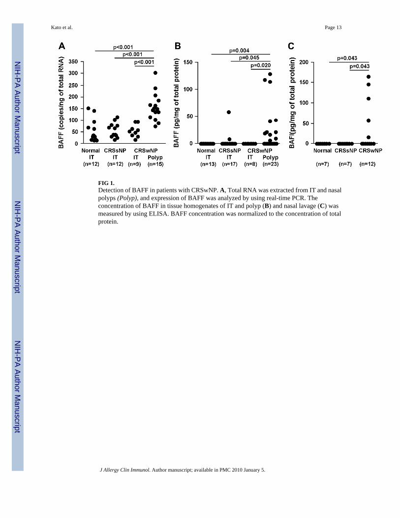
FIG 1.Detection of BAFF in patients with CRSwNP. A, Total RNA was extracted from IT and nasalpolyps (Polyp), and expression of BAFF was analyzed by using real-time PCR. Theconcentration of BAFF in tissue homogenates of IT and polyp (B) and nasal lavage (C) wasmeasured by using ELISA. BAFF concentration was normalized to the concentration of totalprotein.
Kato et al. Page 13
J Allergy Clin Immunol. Author manuscript; available in PMC 2010 January 5.
NIH
-PA Author Manuscript
NIH
-PA Author Manuscript
NIH
-PA Author Manuscript

FIG 2.Immunohistochemistry of BAFF was performed by using antihuman BAFF mAb (clone: Buffy2). A, Negative control antibody staining in uncinate tissue (UT) from a patient with CRSsNP.B–F, Representative immunostaining for BAFF in UT from a control subject (B), a patientwith CRSsNP (C), and a patient with CRSwNP (D) and in nasal polyp tissue (E and F).Arrow, Eosinophil-like cells. Magnification ×200 (A–E); ×400 (F).
Kato et al. Page 14
J Allergy Clin Immunol. Author manuscript; available in PMC 2010 January 5.
NIH
-PA Author Manuscript
NIH
-PA Author Manuscript
NIH
-PA Author Manuscript

FIG 3.B-cell accumulation in nasal polyps. A, Total RNA was extracted from IT and nasal polyps(Polyp), and expression of CD20 was analyzed by using real-time PCR. B, Relationship ofexpression of BAFF and the B-cell marker CD20 or the BAFF receptor TACI in the nasal tissuewas evaluated by using real-time PCR. Correlations were assessed by using the Spearman rankcorrelation.
Kato et al. Page 15
J Allergy Clin Immunol. Author manuscript; available in PMC 2010 January 5.
NIH
-PA Author Manuscript
NIH
-PA Author Manuscript
NIH
-PA Author Manuscript

FIG 4.Elevated expression of IgA in nasal polyps. Measurement of IgA in tissue homogenates of ITfrom control subjects, from patients with CRSsNP and CRSwNP, and in nasal polyps wasmeasured by using ELISA. IgA concentration was normalized to the concentration of totalprotein.
Kato et al. Page 16
J Allergy Clin Immunol. Author manuscript; available in PMC 2010 January 5.
NIH
-PA Author Manuscript
NIH
-PA Author Manuscript
NIH
-PA Author Manuscript

NIH
-PA Author Manuscript
NIH
-PA Author Manuscript
NIH
-PA Author Manuscript
Kato et al. Page 17
TAB
LE I
Subj
ect c
hara
cter
istic
s
Nor
mal
CR
SsN
PC
RSw
NP
CR
SwN
P po
lyp
Tota
l no.
of s
ubje
cts
n =
30 (1
8 M
/12
F)n
= 39
(18
M/2
1 F)
n =
60 (2
6 M
/34
F)—
Age
(y),
med
ian
(ran
ge)
36 (2
0–77
)41
(25–
64)
42 (2
5–77
)—
YN
UY
NU
YN
U—
Ato
py8
211
1818
331
227
—
Ast
hma
030
07
302
3227
1—
Met
hodo
logy
use
d
Tis
sue
RN
An
= 12
(10
M/2
F)
n =
12 (6
M/6
F)
n =
9 (4
M/5
F)
n =
15 (1
0 M
/5 F
)
A
ge (y
), m
edia
n (r
ange
)36
(20–
77)
34 (2
7–55
)44
(28–
69)
44 (2
8 –
74)
Tis
sue
extra
ctn
= 13
(9 M
/4 F
)n
= 17
(9 M
/8 F
)n
= 8
(2 M
/6 F
)n
= 23
(6 M
/17
F)
A
ge (y
), m
edia
n (r
ange
)36
(27–
71)
40 (2
7–61
)47
(27–
69)
41 (2
6–59
)
Nas
al la
vage
n =
7 (2
M/5
F)
n =
7 (4
M/3
F)
n =
12 (4
M/8
F)
—
A
ge (y
), m
edia
n (r
ange
)48
(23–
60)
33 (3
0–59
)50
(25–
77)
—
Im
mun
ohis
toch
emis
tryn
= 4
(3 M
/1 F
)n
= 9
(4 M
/5 F
)n
= 9
(6 M
/3 F
)n
= 7
(7 M
/0 F
)
A
ge (y
), m
edia
n (r
ange
)35
(25–
42)
43 (2
9–64
)32
(27–
74)
39 (2
7–74
)
F, F
emal
e; M
, mal
e; N
, no;
U, u
nkno
wn;
Y, y
es.
J Allergy Clin Immunol. Author manuscript; available in PMC 2010 January 5.

NIH
-PA Author Manuscript
NIH
-PA Author Manuscript
NIH
-PA Author Manuscript
Kato et al. Page 18
TABLE II
BAFF-positive cells in nasal mucosal tissue from normal subjects and patients with CRS
Normal UT CRSsNP UT CRSwNP UT CRSwNP polyp
Epithelium n = 4 n = 7 n = 6 n = 6
BAFF+ cells (102 cells/mm2)
8.6 ± 3.2 12.4 ± 6.1 (NS) 6.0 ± 1.6 (NS) 7.7 ± 3.3 (NS)
Glands n = 4 n = 7 n = 8 n = 5
BAFF+ cells (102 cells/mm2)
7.3 ± 3.5 3.7 ± 0.9 (NS) 5.7 ± 1.9 (NS) 6.0 ± 2.9 (NS)
Submucosa n = 4 n = 9 n = 9 n = 7
BAFF+ cells (102 cells/mm2)
2.8 ± 0.7 7.4 ± 2.2 (NS) 7.0 ± 0.6 (P = .003) 6.5 ± 1.0 (P = .024)
UT, Uncinate tissue; NS, not significant.
Data are presented as means ± SEMs. P value is determined compared with normal.
J Allergy Clin Immunol. Author manuscript; available in PMC 2010 January 5.