A Graded Approach to Repairing the Stenotic Nasal Vestibule
7
ORIGINAL ARTICLE A Graded Approach to Repairing the Stenotic Nasal Vestibule Steven Marc Daines, MD; Grant S. Hamilton III, MD; Steven Ross Mobley, MD Objective: To describe a graded approach to repairing vestibular stenosis that involves restoring structural sup- port to the ala. Methods: Retrospective review of 5 nostrils in 4 pa- tients who presented to the senior author with vestibu- lar stenosis. The cause was burn injury in 3 patients and congenital in 1 patient. The cornerstone is a batten graft to restore strength to the ala. A short-term thermoplas- tic stent helps the nostril assume its natural shape. When an obstructing cicatrix is present, it is excised in a sec- ond stage followed by full-thickness skin grafting. The patients were evaluated up to 16 months postopera- tively. Vestibular patency was documented using high- resolution photographs, and medical records were re- viewed for complications. Results: Two patients had their nostrils repaired in a single stage and the others required 2 stages. In all pa- tients, significant improvement in nostril diameter was maintained. The patients were satisfied with the func- tional and aesthetic results. Stenting averaged 13 days af- ter surgery and was well tolerated. No wound compli- cations occurred. Conclusions: In patients with vestibular stenosis, we use a graded approach that addresses the inherent weakness of the nasal ala to achieve long-term vestibular patency. This technique restores form and function to the ste- notic vestibule while avoiding long-term stenting. Arch Facial Plast Surg. 2010;12(5):332-338 N ASAL VESTIBULAR STENO- sis is a relatively uncom- mon deformity with sig- nificant aesthetic and functional effects on the patient. 1 Trauma, infection, iatrogenic in- sults, and congenital lesions have all been described in the literature 2-5 as causes of ves- tibular stenosis. Traumatic injury to the ala or vestibular lining is typically secondary to burns or complex lacerations. 2,4,5 Infec- tions that involve the anterior nasal mu- cosa, including syphilis, tuberculosis, smallpox, chickenpox, systemic lupus ery- thematosus, rhinoscleroma, and leprosy can lead to scarring within the vestibule and con- tracture of the ala. 2-5 Iatrogenic trauma to the nostril resulting in narrowing of the na- sal aperture can include prolonged nasal packing, chemical cauterization, and poorly executed surgical maneuvers that sacrifice the lining membrane of the vestibule. 2,3 Con- genital vestibular stenosis is less common and is typically seen in conjunction with a cleft lip or nose deformity. 4,6 Stenosis of the nasal vestibule can be a vexing deformity for the facial reconstruc- tive surgeon to successfully repair. Like other hollow orifices in the human body (eg, lacrimal ducts, urethra, and ear canals), once injured, the vestibule has a tendency to res- car and contract despite seemingly well- designed surgical maneuvers to excise the offending cicatrix and create a new vestibu- lar lining. 7 From a pathophysiologic stand- point, stenosis of the anterior nares can be caused by direct injury to the delicate lobule- ala-columella complex, loss of healthy ves- tibular lining, or both. 8,9 Direct injury to the ala, such as chemical burns, leads to tissue loss and scar contracture, which effec- tively sucks in and flattens the ala. 10 Partial or circumferential scarring within the ves- tibule contributes additional contractive forces that tend to obliterate the cephalic and lateral portions of the vestibule and distort the flaccid alar wing. 9 The medial aspect of the vestibule tends to be less affected by these distorting forces given the inherent support mechanism of the juxtaposed me- dial crura and caudal septum. 9 In addi- tion to tissue loss and scar contracture, the constant negative pressure generated by inspiratory forces on the nostril further adds to the tendency for the injured ves- tibule to contract. 11 The same factors that initiate and per- petuate vestibular stenosis also serve to frus- trate surgical attempts to correct the defor- Author Affiliations: Division of Otolaryngology–Head & Neck Surgery, University of Utah School of Medicine, Salt Lake City (Drs Daines and Mobley), and Department of Otolaryngology–Head & Neck Surgery, Division of Facial Plastic and Reconstructive Surgery, University of Iowa Hospitals and Clinics, Iowa City (Dr Hamilton). (REPRINTED) ARCH FACIAL PLAST SURG/ VOL 12 (NO. 5), SEP/OCT 2010 WWW.ARCHFACIAL.COM 332 ©2010 American Medical Association. All rights reserved. Downloaded From: https://jamanetwork.com/ on 04/17/2022
-
Upload
khangminh22 -
Category
Documents
-
view
5 -
download
0
Transcript of A Graded Approach to Repairing the Stenotic Nasal Vestibule
ORIGINAL ARTICLE
A Graded Approach to Repairing the StenoticNasal VestibuleSteven Marc Daines, MD; Grant S. Hamilton III, MD; Steven Ross Mobley, MD
Objective: To describe a graded approach to repairingvestibular stenosis that involves restoring structural sup-port to the ala.
Methods: Retrospective review of 5 nostrils in 4 pa-tients who presented to the senior author with vestibu-lar stenosis. The cause was burn injury in 3 patients andcongenital in 1 patient. The cornerstone is a batten graftto restore strength to the ala. A short-term thermoplas-tic stent helps the nostril assume its natural shape. Whenan obstructing cicatrix is present, it is excised in a sec-ond stage followed by full-thickness skin grafting. Thepatients were evaluated up to 16 months postopera-tively. Vestibular patency was documented using high-resolution photographs, and medical records were re-viewed for complications.
Results: Two patients had their nostrils repaired in asingle stage and the others required 2 stages. In all pa-tients, significant improvement in nostril diameter wasmaintained. The patients were satisfied with the func-tional and aesthetic results. Stenting averaged 13 days af-ter surgery and was well tolerated. No wound compli-cations occurred.
Conclusions: In patients with vestibular stenosis, we usea graded approach that addresses the inherent weaknessof the nasal ala to achieve long-term vestibular patency.This technique restores form and function to the ste-notic vestibule while avoiding long-term stenting.
Arch Facial Plast Surg. 2010;12(5):332-338
N ASAL VESTIBULAR STENO-sis is a relatively uncom-mon deformity with sig-nificant aesthetic andfunctional effects on the
patient.1 Trauma, infection, iatrogenic in-sults, and congenital lesions have all beendescribed in the literature2-5 as causes of ves-tibular stenosis. Traumatic injury to the alaor vestibular lining is typically secondaryto burns or complex lacerations.2,4,5 Infec-tions that involve the anterior nasal mu-cosa, including syphilis, tuberculosis,smallpox, chickenpox, systemic lupus ery-thematosus, rhinoscleroma, and leprosy canlead to scarringwithin thevestibuleandcon-tracture of the ala.2-5 Iatrogenic trauma tothe nostril resulting in narrowing of the na-sal aperture can include prolonged nasalpacking, chemical cauterization, and poorlyexecuted surgical maneuvers that sacrificethe liningmembrane of the vestibule.2,3 Con-genital vestibular stenosis is less commonand is typically seen in conjunction with acleft lip or nose deformity.4,6
Stenosis of the nasal vestibule can be avexing deformity for the facial reconstruc-tive surgeon to successfully repair. Likeother hollow orifices in the human body (eg,lacrimal ducts, urethra, and ear canals), once
injured, the vestibule has a tendency to res-car and contract despite seemingly well-designed surgical maneuvers to excise theoffending cicatrix and create a new vestibu-lar lining.7 From a pathophysiologic stand-point, stenosis of the anterior nares can becaused by direct injury to the delicate lobule-ala-columella complex, loss of healthy ves-tibular lining, or both.8,9 Direct injury to theala, such as chemical burns, leads to tissueloss and scar contracture, which effec-tively sucks in and flattens the ala.10 Partialor circumferential scarring within the ves-tibule contributes additional contractiveforces that tend to obliterate the cephalic andlateral portions of the vestibule and distortthe flaccid alar wing.9 The medial aspect ofthe vestibule tends to be less affected bythese distorting forces given the inherentsupport mechanism of the juxtaposed me-dial crura and caudal septum.9 In addi-tion to tissue loss and scar contracture, theconstant negative pressure generated byinspiratory forces on the nostril furtheradds to the tendency for the injured ves-tibule to contract.11
The same factors that initiate and per-petuate vestibular stenosis also serve to frus-trate surgical attempts to correct the defor-
Author Affiliations: Division ofOtolaryngology–Head & NeckSurgery, University of UtahSchool of Medicine, Salt LakeCity (Drs Daines and Mobley),and Department ofOtolaryngology–Head & NeckSurgery, Division of FacialPlastic and ReconstructiveSurgery, University of IowaHospitals and Clinics, Iowa City(Dr Hamilton).
(REPRINTED) ARCH FACIAL PLAST SURG/ VOL 12 (NO. 5), SEP/OCT 2010 WWW.ARCHFACIAL.COM332
©2010 American Medical Association. All rights reserved.
Downloaded From: https://jamanetwork.com/ on 04/17/2022
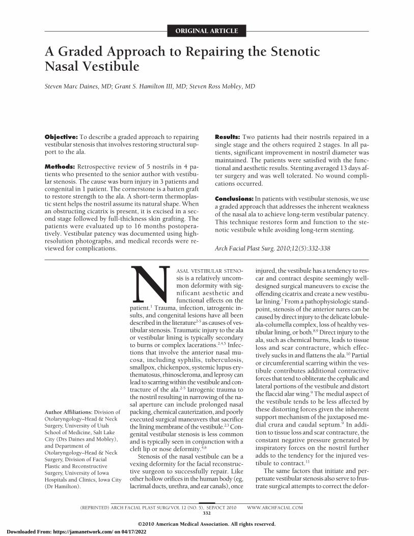
mity. A photograph of the ala in a cadaver dissectiondemonstrates that the lower lateral cartilage does not sup-port the entire ala (Figure 1). The most caudal and lat-eral portion of the ala, which overlies the vestibule, is in-stead supported by easily injured fibrofatty or fibromusculartissue. Once injured, the lack of cartilage prevents the alafrom withstanding the irresistible forces of postoperativescar contracture. Furthermore, when trying to rearrangeand/or excise the intravestibular cicatrix, the dictum “scarbegets scar” holds true. Complicated intravestibular re-pairs often lead to a vicious cycle of new scar formationand recurrent stenosis of the nostril.4,12
Many different strategies have been described in the lit-erature to address stenosis of the nasal vestibule. The com-mon theme in each approach is the need to excise the ob-structing cicatrix, replace the scar tissue with a new, healthylining, and stent the nostril postoperatively to prevent re-stenosis. The use of cartilage grafts has been advocated byseveral authors as a means to combat restenosis. Egan andKim11 describe using a composite conchal graft to stent theregion of the nasal sill, and Karen et al6 advocate the use ofan auricular composite graft to replace the intravestibularscar and to stiffen the repair. These approaches address iso-lated stenosis within the vestibule owing to scar tissue, butthey do not correct for alar collapse. Copcu13 reports usinga gingivobuccal mucosal flap in conjunction with a carti-lage graft to strengthen the weakened ala, but he does notprovide details on the exact placement of the graft or ex-plain how he obtains a natural internal nostril contour. Blan-dini et al12 describe the theoretical use of cartilage grafts inconjunction with other techniques for relining the in-jured vestibule, but they did not use cartilage grafts in theirseries.
We reviewed the literature extensively using the key-words nostril stenosis and nasal vestibular stenosis. To ourknowledge, no reports in the literature have described atechnique for reestablishing the inherent structural sup-port of the ala in patients with vestibular stenosis whileallowing for a graded surgical approach via an optionalsecond stage for excision of scar and creation of a neoves-tibular lining with a full-thickness skin graft. Further-more, the technique described in this article makesprogress in the use of postoperative stenting methods.Although other techniques have almost uniformly re-quired months of postoperative stenting to prevent sur-
gical failure, the senior author (S.R.M.) uses a custom-fashioned, limited-duration, thermoplastic stent thatspares the patient the discomfort and cosmetic draw-backs of prolonged stent use. The custom-molded stentalso contributes to establishing a more natural nostril con-tour. We share the senior author’s experience in using agraded approach for repairing congenital and acquiredstenosis of the nasal vestibule.
METHODS
All patients were evaluated by the senior author and diag-nosed as having stenosis of the nasal vestibule based on physi-cal examination findings. Routine digital photographs were takenbefore and after surgery. A graded approach was used to re-pair the deformity based on the following algorithm. In pa-tients whose anterior nasal stenosis was secondary to direct in-jury to the ala with resultant alar in-drawing and contracture,a single-stage operation was performed for placement of an alarbatten graft and fitting of a short-term thermoplastic stent. Inpatients with a significant intravestibular cicatrix, a second-stage operation was performed to excise the offending scar andcreate a neovestibular lining with a full-thickness skin graft,again followed by short-term stent placement.
SINGLE-STAGE REPAIR
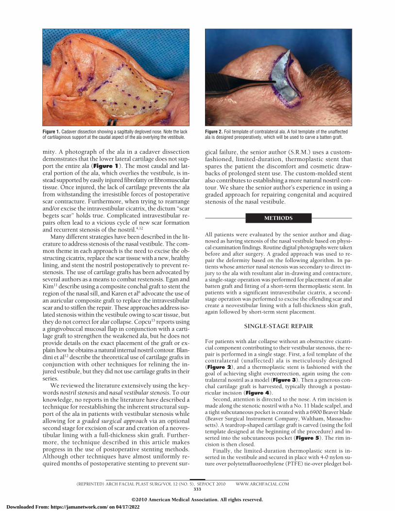
For patients with alar collapse without an obstructive cicatri-cial component contributing to their vestibular stenosis, the re-pair is performed in a single stage. First, a foil template of thecontralateral (unaffected) ala is meticulously designed(Figure 2), and a thermoplastic stent is fashioned with thegoal of achieving slight overcorrection, again using the con-tralateral nostril as a model (Figure 3). Then a generous con-chal cartilage graft is harvested, typically through a postau-ricular incision (Figure 4).
Second, attention is directed to the nose. A rim incision ismade along the stenotic nostril with a No. 11 blade scalpel, anda tight subcutaneous pocket is created with a 6900 Beaver blade(Beaver Surgical Instrument Company, Waltham, Massachu-setts). A teardrop-shaped cartilage graft is carved (using the foiltemplate designed at the beginning of the procedure) and in-serted into the subcutaneous pocket (Figure 5). The rim in-cision is then closed.
Finally, the limited-duration thermoplastic stent is in-serted in the vestibule and secured in place with 4-0 nylon su-ture over polytetrafluoroethylene (PTFE) tie-over pledget bol-
Figure 1. Cadaver dissection showing a sagittally degloved nose. Note the lackof cartilaginous support at the caudal aspect of the ala overlying the vestibule.
Figure 2. Foil template of contralateral ala. A foil template of the unaffectedala is designed preoperatively, which will be used to carve a batten graft.
(REPRINTED) ARCH FACIAL PLAST SURG/ VOL 12 (NO. 5), SEP/OCT 2010 WWW.ARCHFACIAL.COM333
©2010 American Medical Association. All rights reserved.
Downloaded From: https://jamanetwork.com/ on 04/17/2022

sters to distribute the tension of the suture and protect the nasalskin (Figure 6). The senior author has found the use of PTFEpledgets extremely useful in nasal reconstruction because theyallow a protective interface between the suture (which is help-ing to secure the intravestibular stent) and the skin. Withoutthe use of the PTFE pledgets, the suture can have a cheese wireeffect on the skin, resulting in a superficial laceration. The ther-moplastic stent is left in for approximately 2 weeks.
TWO-STAGE REPAIR
In patients whose vestibular stenosis is caused by alar collapseand intravestibular scarring, a 2-stage approach is used for re-
pair. The first stage proceeds in a similar fashion to the tech-nique described for the single-stage vestibular stenosis repairwith attention to 2 important technical details. First, the rimincision must be placed exactly at the future edge of the ves-tibular opening (Figure 7A). The second technical detail con-cerns the creation of the tight subcutaneous pocket that willreceive the auricular cartilage graft: the pocket is created witha thin layer of overlying skin so that the alar batten graft willbe in very close proximity to the external nasal skin (Figure 7B).This is an important technical detail because in stage 2, muchof the tissue between the internal nasal lining and the cartilagegraft will be excised. If the graft is not placed in close approxi-mation to the external nasal skin, a portion of it could be un-intentionally excised (along with obstructing scar tissue) in thesecond stage, compromising the structural integrity of the re-construction. After the batten graft has been inserted and rim
Figure 3. Molding of thermoplastic stent. A custom vestibular stent ismolded with the goal of achieving slight overcorrection.
Figure 4. Auricular cartilage graft. A generous-sized piece of conchalcartilage is harvested.
Figure 5. Insertion of cartilage graft. After the rim incision is made, thecartilage graft is placed in a tight subcutaneous pocket within the ala.
Figure 6. Vestibular stents in situ. The stents are secured within thevestibule using polytetrafluoroethylene tie-over pledget bolsters to distributethe tension of the suture on the skin of the ala.
A B
Figure 7. Important technical details for 2-stage repair. A, Rim incisionplaced at future edge of vestibular opening. B, Batten graft placed in closeproximity to external nasal skin.
(REPRINTED) ARCH FACIAL PLAST SURG/ VOL 12 (NO. 5), SEP/OCT 2010 WWW.ARCHFACIAL.COM334
©2010 American Medical Association. All rights reserved.
Downloaded From: https://jamanetwork.com/ on 04/17/2022
incision closed, a thermoplastic stent is sewn into the vesti-bule, as previously described.
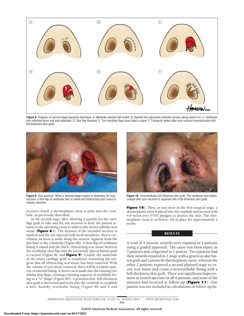
In the second stage, after allowing 2 months for the carti-lage graft to take and the rim incision to heal, the patient re-turns to the operating room to address the intravestibular scartissue (Figure 8A). The location of the intended incision ismarked and the site injected with local anesthetic; then a cur-vilinear incision is made along the stenotic segment from thealar base to the columella (Figure 8B). A thin flap of vestibularlining is raised and the thick, obstructing scar tissue betweenthe vestibular skin flap and the previously placed batten graftis excised (Figure 8C and Figure 9). Usually the undersideof the intact cartilage graft is visualized, reassuring the sur-geon that all obstructing scar tissue has been removed. Withthe volume of scar tissue removed, there will be a relative pau-city of internal lining. A down cut is made into the existing ves-tibular skin flaps, creating a missing segment of vestibular lin-ing in a “V” shape (Figure 8D). A postauricular, full-thicknessskin graft is harvested and sewn into the vestibule to establisha new, healthy vestibular lining (Figure 8E and F and
Figure 10). Then, as was done in the first surgical stage, athermoplastic stent is placed into the vestibule and secured with4-0 nylon over PTFE pledgets to protect the skin. The ther-moplastic stent is, as before, left in place for approximately 2weeks.
RESULTS
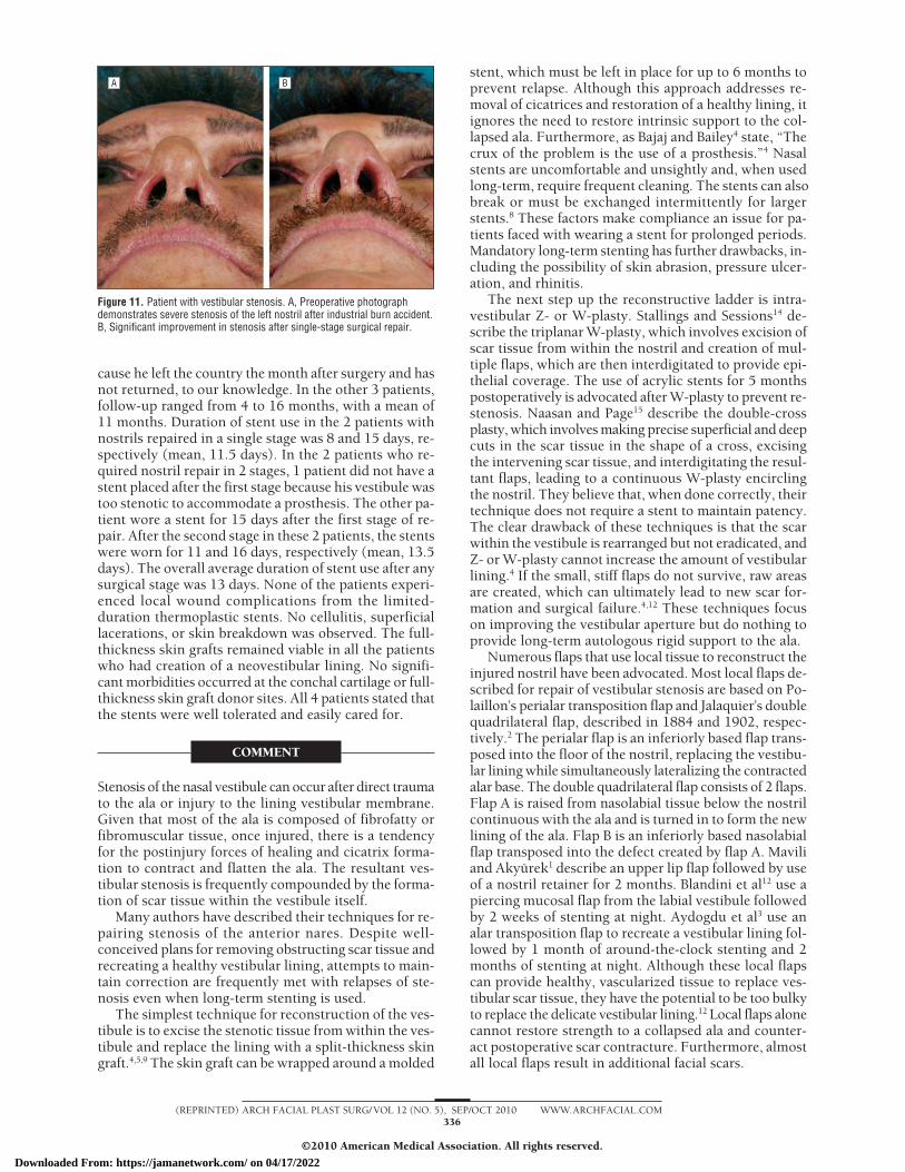
A total of 5 stenotic nostrils were repaired in 4 patientsusing a graded approach. The cause was burn injury in3 patients and congenital in 1 patient. Two patients hadtheir nostrils repaired in 1 stage with a generous alar bat-ten graft and custom-fit thermoplastic stent, whereas theother 2 patients required a second planned stage to ex-cise scar tissue and create a neovestibular lining with afull-thickness skin graft. There was significant improve-ment in nostril aperture in all 4 patients, and none of thestenoses had recurred at follow-up (Figure 11). Onepatient was not included in calculations of follow-up be-
A AB C
D AE F
Figure 8. Diagram of second-stage operative technique. A, Markedly stenotic left nostril. B, Dashed line represents planned incision along nostril rim. C, Vestibularskin reflected down and scar debulked. D, Skin flap bisected. E, Two resulting flaps sewn back in place. F, Triangular defect after scar excision reconstructed withfull-thickness skin graft.
Figure 9. Scar excision. When a second-stage surgery is necessary for scarexcision, a thin flap of vestibular skin is raised and obstructing scar tissue issharply resected.
Figure 10. Intravestibular full-thickness skin graft. The vestibular skin defectcreated after scar excision is replaced with a full-thickness skin graft.
(REPRINTED) ARCH FACIAL PLAST SURG/ VOL 12 (NO. 5), SEP/OCT 2010 WWW.ARCHFACIAL.COM335
©2010 American Medical Association. All rights reserved.
Downloaded From: https://jamanetwork.com/ on 04/17/2022
cause he left the country the month after surgery and hasnot returned, to our knowledge. In the other 3 patients,follow-up ranged from 4 to 16 months, with a mean of11 months. Duration of stent use in the 2 patients withnostrils repaired in a single stage was 8 and 15 days, re-spectively (mean, 11.5 days). In the 2 patients who re-quired nostril repair in 2 stages, 1 patient did not have astent placed after the first stage because his vestibule wastoo stenotic to accommodate a prosthesis. The other pa-tient wore a stent for 15 days after the first stage of re-pair. After the second stage in these 2 patients, the stentswere worn for 11 and 16 days, respectively (mean, 13.5days). The overall average duration of stent use after anysurgical stage was 13 days. None of the patients experi-enced local wound complications from the limited-duration thermoplastic stents. No cellulitis, superficiallacerations, or skin breakdown was observed. The full-thickness skin grafts remained viable in all the patientswho had creation of a neovestibular lining. No signifi-cant morbidities occurred at the conchal cartilage or full-thickness skin graft donor sites. All 4 patients stated thatthe stents were well tolerated and easily cared for.
COMMENT
Stenosis of the nasal vestibule can occur after direct traumato the ala or injury to the lining vestibular membrane.Given that most of the ala is composed of fibrofatty orfibromuscular tissue, once injured, there is a tendencyfor the postinjury forces of healing and cicatrix forma-tion to contract and flatten the ala. The resultant ves-tibular stenosis is frequently compounded by the forma-tion of scar tissue within the vestibule itself.
Many authors have described their techniques for re-pairing stenosis of the anterior nares. Despite well-conceived plans for removing obstructing scar tissue andrecreating a healthy vestibular lining, attempts to main-tain correction are frequently met with relapses of ste-nosis even when long-term stenting is used.
The simplest technique for reconstruction of the ves-tibule is to excise the stenotic tissue from within the ves-tibule and replace the lining with a split-thickness skingraft.4,5,9 The skin graft can be wrapped around a molded
stent, which must be left in place for up to 6 months toprevent relapse. Although this approach addresses re-moval of cicatrices and restoration of a healthy lining, itignores the need to restore intrinsic support to the col-lapsed ala. Furthermore, as Bajaj and Bailey4 state, “Thecrux of the problem is the use of a prosthesis.”4 Nasalstents are uncomfortable and unsightly and, when usedlong-term, require frequent cleaning. The stents can alsobreak or must be exchanged intermittently for largerstents.8 These factors make compliance an issue for pa-tients faced with wearing a stent for prolonged periods.Mandatory long-term stenting has further drawbacks, in-cluding the possibility of skin abrasion, pressure ulcer-ation, and rhinitis.
The next step up the reconstructive ladder is intra-vestibular Z- or W-plasty. Stallings and Sessions14 de-scribe the triplanar W-plasty, which involves excision ofscar tissue from within the nostril and creation of mul-tiple flaps, which are then interdigitated to provide epi-thelial coverage. The use of acrylic stents for 5 monthspostoperatively is advocated after W-plasty to prevent re-stenosis. Naasan and Page15 describe the double-crossplasty, which involves making precise superficial and deepcuts in the scar tissue in the shape of a cross, excisingthe intervening scar tissue, and interdigitating the resul-tant flaps, leading to a continuous W-plasty encirclingthe nostril. They believe that, when done correctly, theirtechnique does not require a stent to maintain patency.The clear drawback of these techniques is that the scarwithin the vestibule is rearranged but not eradicated, andZ- or W-plasty cannot increase the amount of vestibularlining.4 If the small, stiff flaps do not survive, raw areasare created, which can ultimately lead to new scar for-mation and surgical failure.4,12 These techniques focuson improving the vestibular aperture but do nothing toprovide long-term autologous rigid support to the ala.
Numerous flaps that use local tissue to reconstruct theinjured nostril have been advocated. Most local flaps de-scribed for repair of vestibular stenosis are based on Po-laillon’s perialar transposition flap and Jalaquier’s doublequadrilateral flap, described in 1884 and 1902, respec-tively.2 The perialar flap is an inferiorly based flap trans-posed into the floor of the nostril, replacing the vestibu-lar lining while simultaneously lateralizing the contractedalar base. The double quadrilateral flap consists of 2 flaps.Flap A is raised from nasolabial tissue below the nostrilcontinuous with the ala and is turned in to form the newlining of the ala. Flap B is an inferiorly based nasolabialflap transposed into the defect created by flap A. Maviliand Akyürek1 describe an upper lip flap followed by useof a nostril retainer for 2 months. Blandini et al12 use apiercing mucosal flap from the labial vestibule followedby 2 weeks of stenting at night. Aydogdu et al3 use analar transposition flap to recreate a vestibular lining fol-lowed by 1 month of around-the-clock stenting and 2months of stenting at night. Although these local flapscan provide healthy, vascularized tissue to replace ves-tibular scar tissue, they have the potential to be too bulkyto replace the delicate vestibular lining.12 Local flaps alonecannot restore strength to a collapsed ala and counter-act postoperative scar contracture. Furthermore, almostall local flaps result in additional facial scars.
A B
Figure 11. Patient with vestibular stenosis. A, Preoperative photographdemonstrates severe stenosis of the left nostril after industrial burn accident.B, Significant improvement in stenosis after single-stage surgical repair.
(REPRINTED) ARCH FACIAL PLAST SURG/ VOL 12 (NO. 5), SEP/OCT 2010 WWW.ARCHFACIAL.COM336
©2010 American Medical Association. All rights reserved.
Downloaded From: https://jamanetwork.com/ on 04/17/2022

A few authors6,11-13 have advocated composite or car-tilage-alone grafts in the context of repairing stenosis ofthe nasal vestibule. Blandini et al12 describe the theoreti-cal advantage of placing a small cartilage graft under thevestibular labial mucosal flap, stating that a cartilage graftmay eliminate the need for a stent. Copcu13 advocatesa gingivobuccal mucosal flap and uses a cartilage graftbetween the outer nasal skin and the inner mucosalflap for structural support. However, he does not pro-vide specific details regarding the size or placement ofthe graft. Kim16 describes using a composite auricularcartilage graft in a patient with scarring of the nasalsill and columella. A vertical incision is made in thescar tissue, and narrow tunnels are made medially andlaterally. The graft is inserted into the defect with itsflanges in the narrow tunnels. The composite graftreplaces a portion of the excised cicatrix and providesstructural support to resist the forces of scarificationand compress excess scar tissue. A rubber stent is usedat night for 7 months. Kim’s technique appears limitedto repairing stenosis involving the nasal sill, and itdoes not eliminate the need for long-term stenting.Karen et al6 describe an auricular composite graftusing a full-thickness wedge of auricle from helix toantihelix to repair stenosis of the vestibule. Mostpatients in their series were treated for stenosis of thenasal floor. The obvious drawbacks of their techniqueare a visible scar, potential notching of the helix, andthe risk of contributing to further stenosis by transferof 2 avascular elements (skin and cartilage) simulta-neously into an already scarred wound bed.
Taking into account the view that “one size fits all” rarelyworks in nasal reconstruction, we favor a graded ap-proach for repair of vestibular stenosis. In patients whosenostril stenosis is due to a scarred, contracted ala withoutan internal obstructive component, the defect can be re-paired with a single-stage operation. The centerpiece ofour reconstructive technique is a generous alar batten graft,which we prefer to place into the substance of the fleshyala in a tight subcutaneous pocket in close proximity tothe external nasal skin. Augmenting the structural sup-port of the flaccid ala counteracts the persistent forces ofhealing and cicatrix formation and eliminates the need forprolonged stenting to prevent relapse. A thermoplastic stentis fashioned and suture secured into the vestibule in theimmediate postoperative period to act as a scaffold forhealing and to aid in achieving a natural shape to the re-constructed nostrils. In patients with a significant ob-structing, intravestibular scar, a planned second-stage op-eration can be performed to excise the scar tissue and tocreate a neovestibular lining with a full-thickness skingraft. This approach for relining the vestibule transfersthin, healthy tissue to the nostril without the need forcomplex local flaps or unreliable W- or Z-plasty. Be-cause an alar batten graft has previously been placed, thereis no need for prolonged postoperative stenting to main-tain nostril patency.
With a mean long-term follow-up of 11 months, werecognize some may argue that we have not followedup our patients long enough to determine whetherrestenosis will occur. However, we believe that if thereconstructed nostril has not demonstrated alar col-
lapse or neocicatrix formation by 11 months, the like-lihood of reconstructive failure occurring after thatpoint is low.
A vast array of nostril stents and retainers have beendescribed in the literature. Some authors7,17,18 haveadvocated using custom-molded stents to maintain thecorrected contour of the nostril after surgery. Althoughthese stents may function well, most reconstructive sur-geons do not have the requisite materials readily acces-sible to make such a device. Other stent materialsmentioned in the literature include acrylic resin,5,14 sili-cone,3,12 nasopharyngeal airway tubes,11 and rubber.16
Although these materials may be easier to procure, theinherent flaw in choosing these types of stents is thatthey all tend to be round. Clearly, if 1 of the goals inrepairing vestibular stenosis is to restore the naturalcontour of the nostril, a round stent will not achievethis end point. Even digital dilation has been promotedas an effective means to prevent postoperative resteno-sis, but this method has obvious hygienic drawbacks.15
We choose to fashion a stent from thermoplastic mate-rial for 3 main reasons: the material is readily accessiblein any operating room where rhinoplasty is performed,the stent can be custom molded using the contralateralnostril as a template to reestablish the natural shape ofthe reconstructed nostril, and it is well tolerated by thepatient. Two other groups of coauthors18,19 also describeusing thermoplastic material for fabricating vestibularstents. However, 1 group19 states that the stent shouldbe molded around a cylindrical object instead of tailor-ing the stent to the nostril’s native contour, whereas theother group18 creates a custom acrylic vestibular device1 week after surgery using a hydrophilic vinyl polysi-loxane impression material. One of the drawbacks ofthe latter method is the discomfort associated withpacking the patient’s nose posterior to the valve in thefirst postoperative visit to fashion a custom cast of thevestibule.
CONCLUSIONS
Stenosis of the nasal vestibule is a challenging defor-mity to repair because of frequent relapses and diffi-culty in reestablishing a natural contour in the recon-structed nostril. We use a graded approach to repairstenosis of the anterior nares, which relies on a sturdyalar batten graft to restore intrinsic support to the col-lapsed ala. In patients with obstructing scar tissue withinthe vestibule, a second planned surgical stage is used toexcise the cicatrix and create a neovestibular lining witha full-thickness skin graft. After each surgical stage, a ther-moplastic stent is suture secured within the vestibule tocombat stubborn healing forces, which can lead to con-tracture and restenosis. The stent has an added advan-tage of being custom molded and readily available in theoperating room. In our series, 5 stenotic nostrils wererepaired in 4 patients. Satisfactory improvement of nos-tril aperture was achieved and maintained in all 5 nos-trils. Postoperative stenting averaged less than 2 weeksand was well tolerated by all 4 patients with no woundcomplications.
(REPRINTED) ARCH FACIAL PLAST SURG/ VOL 12 (NO. 5), SEP/OCT 2010 WWW.ARCHFACIAL.COM337
©2010 American Medical Association. All rights reserved.
Downloaded From: https://jamanetwork.com/ on 04/17/2022

Accepted for Publication: February 3, 2010.Correspondence: Steven Marc Daines, MD, Division ofOtolaryngology–Head and Neck Surgery, University ofUtah, 50 N Medical Dr, 3C120, Salt Lake City, UT 84132([email protected]).Author Contributions: Study concept and design: Daines,Hamilton, and Mobley. Acquisition of data: Daines andMobley. Analysis and interpretation of data: Daines, Hamil-ton, and Mobley. Drafting of the manuscript: Daines andMobley. Critical revision of the manuscript for importantintellectual content: Daines, Hamilton, and Mobley. Sta-tistical analysis: Daines and Mobley. Administrative, tech-nical, and material support: Daines, Hamilton, and Mob-ley. Study supervision: Mobley.Financial Disclosure: None reported.Previous Presentation: This study was presented as aposter at the American Academy of Facial Plastic and Re-constructive Surgery Fall Meeting; September 18, 2008;Chicago, Illinois.
REFERENCES
1. Mavili E, Akyürek M. Use of upper lip flap for correction of nostril stenosis. Oto-laryngol Head Neck Surg. 1999;121(6):840-841.
2. al-Qattan MM, Robertson GA. Acquired nostril stenosis. Ann Plast Surg. 1991;27(4):382-386.
3. Aydogdu E, Akan M, Gideroglu K, Akoz T. Alar transposition flap for stenosis ofthe nostril. Scand J Plast Reconstr Surg Hand Surg. 2006;40(5):311-314.
4. Bajaj PS, Bailey BN. Stenosis of the nostrils: a report of three cases. Br J PlastSurg. 1969;22(3):269-273.
5. Shah JS. Stenosis of the nostrils: a case report, following smallpox. Plast Re-constr Surg. 1967;39(1):57-58.
6. Karen M, Chang E, Keen MS. Auricular composite grafting to repair nasal ves-tibular stenosis. Otolaryngol Head Neck Surg. 2000;122(4):529-532.
7. Wolfe SA, Podda S, Mejia M. Correction of nostril stenosis and alteration of nos-tril shape with an orthonostric device. Plast Reconstr Surg. 2008;121(6):1974-1977.
8. Daya M. Nostril stenosis corrected by release and serial stenting. J Plast Recon-str Aesthet Surg. 2008;62(8):1012-1019.
9. O’Connor GB. An operation for the correction of atresia or stenosis of the ante-rior nares. Arch Otolaryngol. 1937;25:208-210.
10. Hafezi F, Karimi H, Nouhi A. Aesthetic septorhinoplasty in the burned nose. Burns.2005;31(2):223-229.
11. Egan KK, Kim DW. A novel intranasal stent for functional rhinoplasty and nostrilstenosis. Laryngoscope. 2005;115(5):903-909.
12. Blandini D, Tremolada C, Beretta M, Mascetti M. Iatrogenic nostril stenosis: aes-thetic correction using a vestibular labial mucosa flap. Plast Reconstr Surg. 1995;95(3):569-571.
13. Copcu E. Reconstruction of total and near-total nostril stenosis in the burnednose with gingivo-mucosal flap. Burns. 2005;31(6):802-803.
14. Stallings JO, Sessions DG. Stenosis of the nostril: repair by triplanar W-plasty.Arch Otolaryngol. 1971;93(5):525-528.
15. Naasan A, Page RE. The double cross plasty: a new technique for nasal stenosis.Br J Plast Surg. 1992;45(2):165-168.
16. Kim DW. Nostril stenosis secondary to a laser burn injury: correction with a com-posite graft. Ear Nose Throat J. 2004;83(9):614-615.
17. Costa P, Orlando A, Di Mascio D. An expansible splint for treatment of nostrilstenosis. Ann Plast Surg. 1995;34(2):197-200.
18. Menger D-J, Lohuis PJFM, Kerssemakers S, Nolst Trenite GJ. Postoperative man-agement of nasal vestibular stenosis: the custom-made vestibular device. ArchFacial Plast Surg. 2005;7(6):381-386.
19. Johnson J, Candia J, LaTrenta G, Madden MR, Goodwin CW, Finkelstein J.A nasal trumpet orthosis to maintain nares openings and respiratory functionfor patients with facial burns: a case report. J Burn Care Rehabil. 1992;13(6):677-679.
Announcement
Visit www.archfacial.com. As an individual sub-scriber, you may elect to be contacted when a specificarticle is cited by any of the hundreds of journals hostedby HighWire. You also may sign up to receive an e-mailalert when articles on particular topics are published.
(REPRINTED) ARCH FACIAL PLAST SURG/ VOL 12 (NO. 5), SEP/OCT 2010 WWW.ARCHFACIAL.COM338
©2010 American Medical Association. All rights reserved.
Downloaded From: https://jamanetwork.com/ on 04/17/2022