Systemic multicompartmental effects of the gut microbiome on mouse metabolic phenotypes
Upload
khangminh22Category
view
1download
0
The Chronic Rhinosinusitis
Microbiome
Edward J. Cleland
M.B.B.S.
Submitted for the title of Doctor of Philosophy
July 2014
Department of Otolaryngology Head and Neck Surgery
University of Adelaide, Adelaide, Australia
08 Fall

i
This thesis is dedicated to my beautiful wife Esther.

ii
Table of Contents
Thesis declaration .................................................................................................................... vi
Acknowledgements ................................................................................................................. vii
Publications arising from this thesis .................................................................................. ix
Awards arising from this thesis ............................................................................................ x
Presentations arising from this thesis ............................................................................... xi
List of abbreviations ............................................................................................................. xiii
List of tables .............................................................................................................................. xvi
List of figures .......................................................................................................................... xvii
Thesis summary ................................................................................................................... xviii
Chapter 1: Literature review ................................................................................................ 1
1.1 Chronic rhinosinusitis – the disease entity .................................................................................. 1
1.1.1 Chronic rhinosinusitis defined ............................................................................................... 1
1.1.2 Epidemiology of chronic rhinosinusitis ................................................................................. 4
1.1.3 Chronic rhinosinusitis subclassification ................................................................................ 5
1.1.4 Medical management of chronic rhinosinusitis .................................................................... 8
1.1.5 Surgical management of chronic rhinosinusitis .................................................................. 13
1.2 Aetiopathogenesis of chronic rhinosinusitis .............................................................................. 15
1.2.1 Aetiology ............................................................................................................................. 15
1.2.2 Host factors ........................................................................................................................ 16
1.2.3 Environmental factors – bacteria ....................................................................................... 20
1.2.4 Environmental factors – fungi ............................................................................................ 25

iii
1.3 Microbe detection in chronic rhinosinusitis .............................................................................. 29
1.3.1 Culture ................................................................................................................................ 29
1.3.2 Microscopy ......................................................................................................................... 30
1.3.3 Molecular ............................................................................................................................ 32
1.4 The microbiome concept ........................................................................................................... 37
1.4.1 Definition ............................................................................................................................ 37
1.4.2 The ecology of the microbiome .......................................................................................... 38
1.4.3 Specific influences of the microbiome ................................................................................ 41
1.5 Probiotics – manipulating the microbiome ............................................................................... 43
1.5.1 Probiotics defined ............................................................................................................... 43
1.5.2 Probiotics and their mechanisms of action ......................................................................... 43
1.5.3 Probiotics in medicine ......................................................................................................... 48
1.5.4 Probiotics in CRS and the nasal cavity ................................................................................ 50
Summary of the literature review ..................................................................................... 57
Studies to be performed ....................................................................................................... 60
Chapter 2: The bacteriology of chronic rhinosinusitis and the pre-‐eminence of
Staphylococcus aureus in revision patients .................................................................... 61
Statement of authorship .................................................................................................................. 63
2.1 Abstract ...................................................................................................................................... 64
2.2 Introduction ............................................................................................................................... 66
2.3 Methods ..................................................................................................................................... 68
2.4 Results ........................................................................................................................................ 71
2.5 Discussion .................................................................................................................................. 73
2.6 Conclusion .................................................................................................................................. 78

iv
Chapter 3: The bacterial microbiome in chronic rhinosinusitis: richness,
diversity, post-‐operative changes and patient outcomes .......................................... 79
Statement of authorship .................................................................................................................. 81
3.1 Abstract ...................................................................................................................................... 82
3.2 Introduction ............................................................................................................................... 84
3.3 Methods ..................................................................................................................................... 86
3.4 Results ........................................................................................................................................ 91
3.5 Discussion .................................................................................................................................. 99
3.6 Conclusion ................................................................................................................................ 105
Chapter 4: The fungal microbiome in chronic rhinosinusitis: richness,
diversity, post-‐operative changes and patient outcomes ........................................ 106
Statement of authorship ................................................................................................................ 108
4.1 Abstract .................................................................................................................................... 109
4.2 Introduction ............................................................................................................................. 111
4.3 Methods ................................................................................................................................... 113
4.4 Results ...................................................................................................................................... 118
4.5 Discussion ................................................................................................................................ 125
4.6 Conclusion ................................................................................................................................ 131
Chapter 5: Probiotic manipulation of the chronic rhinosinusitis
microbiome ............................................................................................................................. 132
Statement of authorship ................................................................................................................ 134
5.1 Abstract .................................................................................................................................... 135
5.2 Introduction ............................................................................................................................. 137
5.3 Methods ................................................................................................................................... 139

v
5.4 Results ...................................................................................................................................... 143
5.5 Discussion ................................................................................................................................ 145
5.6 Conclusion ................................................................................................................................ 150
Thesis synopsis ...................................................................................................................... 151
References ............................................................................................................................... 157

vi
Thesis declaration
This work contains no material which has been accepted for the award of any
other degree or diploma in any university or other tertiary institution to Edward
John Cleland and, to the best of my knowledge and belief, contains no material
previously published or written by another person, except where due reference
has been made in the text.
I give consent to this copy of my thesis when deposited in the University
Library, being made available for loan and photocopying, subject to the provisions
of the Copyright Act 1968.
I acknowledge that copyright of published works contained within this thesis
resides with the copyright holders of those works.
I also give permission for the digital version of my thesis to be made
available on the web, via the University’s digital research repository, the Library
catalogue, the Australasian Digital Theses Program (ADTP) and also through web
search engines, unless permission has been granted by the University to restrict
access for a period of time.
Dr Edward J. Cleland

vii
Acknowledgements
The completion of a PhD thesis is never an individual endeavor. Over the past
three years I have received an enormous amount of support, without which the
successful completion of this work would have been impossible. Here I would like
to formally acknowledge the following people.
Firstly I would like to thank Professor PJ Wormald for being such a
steadfast influence during my PhD. Prof has been a mentor to me for both my
clinical and academic endeavors. His brilliance as a clinician and surgeon
combined with a passion for academia has highlighted to me the possibility for a
rich and balanced medical career. Prof’s guidance and friendship during this
period has been invaluable and I hope for this relationship to continue in the
future.
I would also like to thank my laboratory supervisor Sarah Vreugde. Sarah
became a part of the department 6 months after the commencement of my PhD
and provided me with important assistance during a difficult period of my
candidature. Sarah’s enthusiasm for research and willingness to create a
welcoming and productive environment in the laboratory were greatly appreciated.
My fellow registrars and friends Andrew Foreman, Sam Boase, Rowan
Valentine, Josh Jervis-Bardy, Vikram Padhye, Camille Jardeleza and Thanh Ha
have all been incredibly supportive of my work. From sharing the ups and downs
to providing important expertise, these contributions have all been appreciated.
The assistance of Ahmed Bassiouni warrants special mention. Ahmed’s
energy and contribution to the department has been vital for both my projects and

viii
those of others. Amanda Drilling, Clare Cooksley, Daniel Cantero, Sukanya Rajiv,
Dijana Miljkovic, Neil Tan, Eugene Roscioli, Yuresh Naidoo, Brendan Hanna, Phil
Chen, Joseph Brunworth and the other members of the research department,
thank you all for facilitating my work and for your friendship.
To my collaborators, Dr Craig James and Debbie Dyer of Adelaide
Pathology Partners, Dr Graham van Renen, Ms Cathy Jarman and the staff at
Memorial Hospital, Dr Scot Dowd of MR DNA, Dr Alkis Psaltis and Dr Stephen
Floreani, thank you all for your important contributions.
The financial support of the Garnett Passe and Rodney Williams Memorial
Foundation is also acknowledged. This work would not have been possible without
their important assistance. Furthermore the ongoing financial contributions of this
foundation continue to support research that has arisen from this PhD thesis.
Finally I would like to thank my wife Esther for her unconditional support
and the sacrifices that she has made to support my work. Her understanding of my
frequently irregular hours, which has often come before other important family
obligations, can never be fully recognized. I hope however she has enjoyed and
shared in my successes and is aware of just how important her role has been.

ix
Publications arising from this thesis
1. Cleland EJ, Bassiouni A, Wormald PJ. The bacteriology of chronic
rhinosinusitis and the pre-eminence of Staphylococcus aureus in revision
patients. Int Forum Allergy Rhinol 2013.
2. Cleland EJ, Bassiouni A, Boase S, Dowd S, Vreugde S, Wormald PJ. The
fungal microbiome in chronic rhinosinusitis: richness, diversity, postoperative
changes and patient outcomes. Int Forum Allergy Rhinol 2014;4:259-65.
3. Cleland EJ, Bassiouni A, Vreugde S, Wormald PJ. The bacterial microbiome in
chronic rhinosinusitis: richness, diversity, post-operative changes and patient
outcomes. Submitted for publication.
4. Cleland EJ, Drilling A, Bassiouni A, James C, Vreugde S, Wormald PJ.
Probiotic manipulation of the chronic rhinosinusitis microbiome. Int Forum
Allergy Rhinol 2014;4:309-14.

x
Awards arising from this thesis
Florey Medical Research Foundation Bertha Sudholz Research Scholarship
University of Adelaide, Adelaide, Australia, February 27, 2013.
Basic Science Research Award
American Rhinologic Society, Vancouver, Canada, September 28, 2013.
Ronald Gristwood Medal – Best Presentation
ASOHNS SA Annual Registrar’s Research Presentation Meeting, Adelaide,
Australia, October 31, 2013.
The RP Jepson Medal
The Royal Australian College of Surgeons, Adelaide, Australia, November 12,
2014.
xi
Presentations arising from this thesis
Molecular techniques of bacterial detection in chronic rhinosinusitis.
The Australian Society of Otolaryngology Head & Neck Surgery (SA) – Registrar
research presentation night, Adelaide, Australia, November 2011.
The bacteriology of chronic rhinosinusitis and the pre-eminence of Staphylococcus aureus in revision patients.
58th Annual Meeting of The American Rhinologic Society, Washington D.C., USA,
September 2012.
Analysis of fungal diversity through molecular characterization of the chronic rhinosinusitis microbiome.
The Australian Society of Otolaryngology Head & Neck Surgery (SA) – Registrar
research presentation night, Adelaide, Australia, November 2012.
Analysis of fungal diversity through molecular characterization of the chronic rhinosinusitis microbiome.
The Australian Society of Otolaryngology Head & Neck Surgery ASM, Perth,
March 2013.
Probiotic manipulation of the chronic rhinosinusitis microbiome.
59th Annual Meeting of The American Rhinologic Society, Vancouver, Canada,
September 2013.
xii
Probiotic manipulation of the chronic rhinosinusitis microbiome.
The Queen Elizabeth Hospital Research Day, The Bazil Hetzel Institute, Adelaide,
Australia, October 2013.
Probiotic manipulation of the chronic rhinosinusitis microbiome.
The Australian Society of Otolaryngology Head & Neck Surgery (SA) – Registrar
research presentation night, Adelaide, Australia, November 2013.
Microbes and Chronic Rhinosinusitis
Invited speaker, St. Vincent’s Hospital FESS Course, Sydney, Australia, August
2014.

xiii
List of abbreviations
16S &18S A part of ribosomal RNA. S represents Svedberg units
ABRS Acute bacterial rhinosinusitis
AD Atopic dermatitis
AFRS Allergic fungal rhinosinusitis
AFS Allergic fungal sinusitis
ARS Acute rhinosinusitis
Atl Autolysin
CFT Canine fossa trephination
CRS Chronic rhinosinusitis
CRSsNP Chronic rhinosinusitis sans nasal polyps
CRSwNP Chronic rhinosinusitis with nasal polyps
DNA Deoxyribonucleic acid
E. Coli Escherichia coli
EMRS Eosinophilic mucous rhinosinusitis
EPOS European position statement on sinusitis
Esp A serine protease
ESS Endoscopic sinus surgery
FESS Functional endoscopic sinus surgery

xiv
H. Influenzae Haemophilus influenzae
HGT Horizontal gene transfer
IL Interleukin
INCS Intranasal corticosteroid
ITS Internal transcribed spacer
KOH A potassium hydroxide preparation
LK Lund-Kennedy
LM Lund-Mackay
M. Catarrhalis Moraxella Catarrhalis
MRA Mean relative abundance
MUC3 A mucin glycoprotein
NO Nitric oxide
OTU Operational taxonomic unit
P. aeruginosa Pseudomonas aeroginosa
PAMP Pathogen associated molecular patterns
PAR Protease-activated receptors
PBS Phosphate buffered saline
PCR Polymerase chain reaction
PRR Pattern-recognition receptors
RCT Randomized controlled trial
xv
rDNA Ribosomoal deoxyribonucleic acid
rRNA Ribosomal ribonucleic acid
S. aureus Staphylococcus aureus
SA Staphylococcus aureus
SAE S. aureus enterotoxins
SE Staphylococcus epidermidis
SNOT-20 Sinonasal outcome test 20
SNOT-22 Sinonasal outcome test 22
SspA Staphylococcus serine protease gene encoding V8
protease
T-RFLP Terminal restriction fragment length polymorphism
TEFAP Tag-encoded FLX amplicon pyrosequencing
Th T helper cell (1,2, 17 etc)
TLR Toll-like receptors
VAS Visual analogue symptom score
xvi
List of tables
Table 1-1 Categories of rhinosinusitis ..................................................................... 3
Table 1-2 Bent-Kuhn criteria for diagnosis of AFRS ............................................... 7
Table 2-1 Demographics and disease characteristics of study population ........... 69
Table 2-2 Bacterial culture results ......................................................................... 72
Table 2-3 Results of the univariate logistic regression model ............................... 74
Table 2-4 Results of the multivariate logistic regression models (Revision vs
Primary). ................................................................................................................ 74
Table 3-1 Demographics and disease characteristics of the study population ..... 91
Table 3-2 Prevalence and mean relative abundance (MRA) for the 40 most
frequently detected species (intra-operative samples) .......................................... 95
Table 4-1 Demographics and disease characteristics of the study population ... 118
Table 4-2 Prevalence and mean relative abundance (MRA) for the 40 most
frequently detected genera (intra-operative samples) ......................................... 122

xvii
List of figures
Figure 1-1 Clinical photograph of R maxillary sinus showing changes in a
previously operated sinus which do not met the definition of a nasal polyp ............ 6
Figure 1-2 CRS treatment algorithm ....................................................................... 9
Figure 1-3 Factors contributing to CRS aetiopathogenesis .................................. 16
Figure 1-4 S. aureus associations with disease severity ...................................... 24
Figure 1-5 Schematic overview of microbial detection techniques ....................... 30
Figure 1-6 General processes shaping the microbiome ....................................... 39
Figure 1-7 Serine protease Esp mechanism of action .......................................... 54
Figure 5-1 Representative slides from the 4 treatment groups ........................... 141
Figure 5-2 Goblet cell counts from the 4 treatment groups ................................. 143
xviii
Thesis summary
The body of research described within this PhD thesis investigates the presence
and role of microbial communities or the microbiome in chronic rhinosinusitis
(CRS). After a rigorous review of the literature as it relates to the
aetiopathogenesis and microbiology of this disease it is clear that much confusion
exists regarding the importance of host and environmental factors which
predispose towards the disease state. In particular, significant deficiencies exist in
how we conceptualize the role of bacteria and fungi in this ecological niche
because our understanding of these microbes has been shaped by detection
techniques that lack the sensitivity to accurately detect the microbiome
constituents. To address this, we set out to accurately define the microbiome in
CRS patients and healthy controls as a basis for further ecological studies. Using
this knowledge we then attempted to manipulate the microbiome in a murine
model of sinusitis so that the probiotic potential of certain bacteria could be
determined.
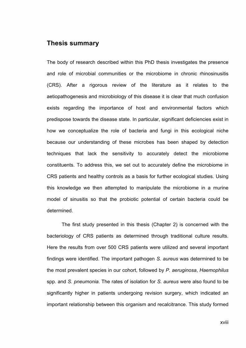
The first study presented in this thesis (Chapter 2) is concerned with the
bacteriology of CRS patients as determined through traditional culture results.
Here the results from over 500 CRS patients were utilized and several important
findings were identified. The important pathogen S. aureus was determined to be
the most prevalent species in our cohort, followed by P. aeruginosa, Haemophilus
spp. and S. pneumonia. The rates of isolation for S. aureus were also found to be
significantly higher in patients undergoing revision surgery, which indicated an
important relationship between this organism and recalcitrance. This study formed

xix
an important basis for the other microbe detection studies described in the
following chapters, which utilized sensitive molecular techniques.
Chapter 3 sees investigation of the bacterial component of CRS
microbiome using a 16S rDNA bacterial tag-encoded FLX amplicon
pyrosequencing technique. The large number of species detected in both controls
and CRS patients highlighted the sensitivity of this technique for detecting
bacteria. Unlike the previous study, other metrics of presence were also
characterized such as richness, diversity and relative abundance. The temporal
changes occurring in the CRS cohort were examined post-operatively and
correlations were made with quality of life. S. aureus was shown to be prevalent in
both CRS patients and controls, however its relative abundance in CRS patients
was significantly higher than in the control group. The species Acinetobacter
johnsonii was also of interest. Here it was found to be significantly more abundant
in the control group, which may be of functional importance for the healthy
sinonasal microbiome.
The study reported in Chapter 4 describes the equally important, but until
this time neglected, fungal component of the microbiome. In this instance 18S
rDNA fungal tag-encoded FLX amplicon pyrosequencing was performed and fungi
were found to be ubiquitous to all patients. The striking finding of this study was
the sheer diversity of fungi present across the cohort. Several genera including
Malassezia were described for the first time in the sinonasal cavity. The presence
of Malassezia was of great interest due to its previously described association with
atopic disease. In CRS Malassezia may also play an important role as a disease
modifier.

xx
Finally, after successfully defining the microbiome our next aim was to
manipulate it (Chapter 5). Here we investigated the probiotic potential of S.
epidermidis against S. aureus in a murine model of sinusitis. Intranasal
inoculations of phosphate buffered saline, S. epidermidis, S, aureus or a
combination of the two were administered to 20 mice. PAS-positive goblet cell
counts of the mouse nasal mucosa revealed that mice receiving a combination of
S. epidermidis and S. aureus had significantly lower goblet cell counts than those
receiving S. aureus alone. This finding was of great importance as it confirmed the
probiotic potential of S. epidermidis by demonstrating its ability to augment the
pathogenicity of S. aureus in the nasal cavity. The implications of this finding for
topical intranasal probiotics in CRS are far reaching and should not be
underestimated. Most importantly it highlights the viability of ecologically based
treatment paradigms in the management of CRS.

Chronic rhinosinusitis – the disease entity
1
Chapter 1: Literature review
1.1 Chronic rhinosinusitis – the disease entity
1.1.1 Chronic rhinosinusitis defined
Chronic rhinosinusitis (CRS) is part of a spectrum of inflammatory conditions
involving the nasal cavity and paranasal sinuses. Our understanding of these
conditions has evolved over time, resulting in a corresponding change in our
definition of this disease entity.
Most panels and expert guideline documents now agree on the use of the
term rhinosinusitis instead of sinusitis. This change in nomenclature is an
acknowledgement that both rhinitis and sinusitis usually coexist in these
individuals and are part of the same disease process.1
The most contemporary guidelines on rhinosinusitis are those put forth by
the European Position Paper on Rhinosinusitis and Nasal Polyps 2012 (EPOS),
which defines the entity in adults as:
• Inflammation of the nose and the paranasal sinuses characterized by
two or more symptoms, one of which should be either nasal
blockage/obstruction/congestion or nasal discharge (anterior/posterior
nasal drip)
o ± Facial pain/pressure
o ± Reduction or loss of smell
and/either
• Endoscopic signs of

Chronic rhinosinusitis – the disease entity
2
o Nasal polyps, and/or
o Mucopurulent discharge primarily from middle meatus and/or
o Oedema/mucosal obstruction primarily in middle meatus
and/or
• CT changes
o Mucosal changes within the ostiomeatal complex and/or sinuses.1
Earlier guidelines for diagnosis relied solely on the presence of major and
minor symptoms to make a clinical diagnosis.2 This method was inherently limited,
as a diagnosis of rhinosinusitis was not subjected to the additional rigour of
endoscopic examination and radiological investigations, and resulted in a revised
set of guidelines as put forth by Benninger et al. to address these concerns.3
Once a diagnosis of rhinosinusitis is made patients may be categorized on
a temporal basis into acute (<12 weeks duration) and chronic (>12 weeks
duration) rhinosinusitis categories.1 Earlier classifications of rhinosinusitis divided
these groups further to encompass subacute, recurrent acute and acute
exacerbations in the context of chronic disease (Table 1-1),2 a distinction which is
still made by many clinicians.

Chronic rhinosinusitis – the disease entity
3
Table 1-1 Categories of rhinosinusitis
Category Duration of symptoms
Acute 0 – 4 weeks
Subacute 4 – 12 weeks
Chronic ≥ 12 weeks
Recurrent, acute ≥ 4 episodes/year +
each episode lasting ≥7–10 days
Acute exacerbations of chronic Sudden worsening of chronic
rhinosinusitis, return to baseline after
treatment
Acute rhinosinusitis (ARS) can be further divided into viral, post-viral and
acute bacterial rhinosinusitis (ABRS) sub-categories. The aetiologic agent in viral
ARS is most commonly found to be rhinovirus (50%), but may also result form
influenza viruses, parainfluenza viruses, adenovirus, respiratory syncytial virus,
and enterovirus.4 ABRS is frequently preceded by the viral and post-viral variants
of ARS, a fact which can make the distinction of these subtypes difficult in the
clinical setting. The most commonly detected microorganisms in a meta-analysis
of ABRS patients included Streptococcus pneumonia (32.7%), H. influenza
(31.6%), S. aureus (10.1%), and M. catarrhalis (8.8%).5
For patients with ARS where symptoms last longer than the arbitrary 12
weeks, a diagnosis of CRS is made. This transition from ARS to CRS is an

Chronic rhinosinusitis – the disease entity
4
important one and has been noted to occur with corresponding changes in the
presence of microbes. This relationship will be discussed elsewhere (see 1.2.3).
These definitions and categories of rhinosinusitis are of considerable utility
for researchers and clinicians as they provide a well-defined and reproducible
means of categorizing patients with rhinosinusitis. Not only does this allow for
more accurate study design, but also the treatment of patients with CRS can be
tailored with greater accuracy to the specifics of the individual.
1.1.2 Epidemiology of chronic rhinosinusitis
Estimates of the prevalence of rhinosinusitis are limited by the quality of data in
the literature, which are largely derived from broad-based population studies.
These studies usually rely on self-reported symptoms by a patient rather than
formal diagnostic criteria as used by the otolaryngologist.
One publication using self-reported ‘sinus trouble’ for greater than 3 months
in the year, estimated the prevalence of CRS at 15.5% of the total population in
the United States.6 A Canadian study asking patients if the they had been
diagnosed with CRS by a health care professional found its presence in 5.7% of
females and 3.4% of males.7 Lower rates of CRS have been described in a
Korean study which found symptoms of rhinosinusitis in only 1% of the
population,8 a difference which may be reflective of geographic variability. Another
study using disease classification codes generated from medical records found
CRS in only 2% of patients.9 This result highlights the potential for exaggerated
rates of prevalence in studies using self-reported data.

Chronic rhinosinusitis – the disease entity
5
The socioeconomic impact of CRS is also significant. In the United States,
the number of office based doctor visits resulting in a diagnosis of CRS was 11.6
million in the year 2000.10 It has been shown that patients with CRS visit primary
care clinicians twice as often as those without the disorder and have five times as
many prescriptions filled.11 Approximately 500,000 surgical procedures are
performed on the paranasal sinuses annually,12 and when these figures are
extrapolated to the general population the direct cost of CRS in the United States
has been estimated at $4.3 billion.13
The health burden of CRS on patients relative to the general population is
also significantly worse for several domains such as bodily pain, general health,
vitality, and social functioning. Compared with other chronic diseases these
patients also score significantly worse for bodily pain and social functioning than
patients with congestive heart failure, angina, chronic obstructive pulmonary
disease, and back pain.14 These findings highlight the impact of CRS on the
population, the extent of which is potentially underestimated, especially compared
to other disease states.
1.1.3 Chronic rhinosinusitis subclassification
The presence of nasal polyposis is the most common clinical factor used to
differentiate between patients with CRS. To make a diagnosis of CRS with nasal
polyps (CRSwNP), endoscopic examination of the nasal cavity must reveal
bilateral polyps in the middle meatus. If no polyps are visualized in the middle
meatus patients are defined as CRS sans nasal polyps (CRSsNP). The diagnosis
of nasal polyps in patients who have previously undergone sinus surgery can be
problematic due to altered anatomy or the previous removal of polyps. In this

Chronic rhinosinusitis – the disease entity
6
instance, the presence of bilateral pedunculated lesions > 6 months after surgery
on endoscopic examination is considered sufficient grounds for a diagnosis of
CRSwNP. Other changes in mucosa that are often observed such as a
cobblestoned appearance, whilst representing inflammatory change, do not meet
the definition of a nasal polyp (see Figure 1-1).1 Although not mandatory,
histological analysis of a mature polyp may also assist diagnosis. Here the polyp
will usually appear as a large pseudocyst area containing albumin surrounded by
subepithelial eosinophilia.15
Figure 1-1 Clinical photograph of R maxillary sinus showing changes in a
previously operated sinus which do not meet the definition of a nasal polyp.
Image superimposed on schematic of human paranasal sinuses (open source
image).
Further subclassification of CRS patients can be made on the presence of
eosinophilic mucous (EM) and/or fungal hyphae.16 Using the Bent-Kuhn criteria a

Chronic rhinosinusitis – the disease entity
7
patient is diagnosed with allergic fungal rhinosinusitis (AFRS) in the presence of:
nasal polyposis, fungi on staining, eosinophilic mucous without fungal invasion into
sinus tissue, type I hypersensitivity to fungi and characteristic radiological findings
with soft tissue differential densities on CT scanning (Table 1-2).17 These criteria
must be used with caution however, as they are not specific for AFRS patients and
must also be considered in the context of more sensitive techniques for the
detection of fungi, which have highlighted its ubiquitous presence in the sinonasal
cavity.18,19
Table 1-2 Bent-Kuhn criteria for diagnosis of AFRS
1 Nasal polyposis
2 Fungi on staining
3 Eosinophilic mucous without fungal invasion into sinus tissue
4 Type 1 hypersensitivity to fungi
5 Radiological findings consistent with fungal disease
Patients with eosinophilic mucous, but an absence of fungi and its
associated hypersensitivity, are provided the alternative diagnosis of eosinophilic
mucous rhinosinusitis (EMRS).20 Once again the ubiquitous presence of fungi
makes this classification unreliable, and is more a reflection of the sensitivity of the
fungal detection technique used. Others have attempted to differentiate the
CRSwNP group further from polyp histomorphological characteristics. On this
basis they can be divided into eosinophilic, neutrophilic, and noneosinophilic

Chronic rhinosinusitis – the disease entity
8
nonneutrophilic types. This distinction has come about due to regional differences
observed in polyp histology between western and Asian countries.21
The variability of these subclassifications of CRS reflect the difficulty
researchers have in defining this heterogeneous group of patients with a
multifactorial disease aetiology. Accurate categorization of these groups is
necessary for appropriate study design and also to aid the clinician in tailoring
suitable treatment regimes. To make the distinction between patients more
accurately it is likely that a combination of clinical (phenotype), host (genotype)
and environmental (microbiome) factors will be of great import in the future.
1.1.4 Medical management of chronic rhinosinusitis
Prior to surgical intervention all patients presenting to the Otolaryngologist are first
commenced on a trial of ‘maximal medical therapy’. What this therapy is
comprised of is variable between institutions and lacking consensus, but would
usually be comprised of saline rinses, topical and oral corticosteroids and a short
course of oral antibiotics.22 Medical treatments can be of utility in both the pre and
post-operative setting (Figure 1-2). The most common medical treatments will be
discussed in the following paragraphs.

Chronic rhinosinusitis – the disease entity
9
Figure 1-2 CRS treatment algorithm. Adapted from Palmer et al.23 Medical therapy plays an important role pre and post-operatively and may be necessary in some patients long-term.
Corticosteroids are an important component of medical management for
CRS patients and can be delivered either topically or orally. These medications
effect eosinophil function by directly reducing eosinophil viability and activation24-26
or by indirectly reducing the secretion of chemotactic cytokines by nasal mucosa
and nasal polyp epithelial cells.27,28 A recent meta-analysis of topical intranasal
corticosteroids (INCS) in CRSsNP patients found significantly improved symptom
scores in the treatment group compared to those receiving placebo. Sinus delivery
methods were also more effective than nasal delivery methods, but there was no
difference in endoscopic appearance nor was there any difference seen for the
type of steroid used.1 The CRSwNP group has also been shown to benefit from

Chronic rhinosinusitis – the disease entity
10
INCS. These patients demonstrate significant improvements in symptoms, polyp
size, polyp recurrence and nasal airflow.1
Oral systemic steroids may be of benefit in CRSsNP patients although high-
level evidence is currently lacking to support their use. A recent systematic review
of 30 studies on systemic steroid use in CRSsNP patients identified that the
efficacy of single-modality oral steroid use in CRSsNP remained untested.
Furthermore, no randomized controlled trials (RCT) had been performed that
examined the efficacy of oral steroids as part of multimodal therapy in CRSsNP.29
Three trials, involving 166 patients, met the inclusion criteria of a recent Cochrane
review exploring the benefits of oral steroids in CRSwNP patients. The results
indicated a short-term benefit for a brief (2 – 4 week) course of oral steroids of
variable doses and duration when compared to placebo. Improvements were seen
for an objective reduction in polyp size and a subjective improvement of nasal
symptoms and quality of life. These trials were however, of moderate to low quality
and it was not possible to quantify the overall size of this effect.30
The side effect profile of corticosteroids needs to be balanced against any
benefits. Whilst long-term use of INCS are generally considered to be safe due
their low bioavailability (<1%),31 a number of patients still experience side effects
related to their use which may include epistaxis, itching, sneezing and dry nose.1
Oral steroids have the potential for more significant side effects particularly when
given at higher doses for longer durations. These side effects include changes in
bone mineral density, fat metabolism, proximal muscle strength, appetite and
glucose tolerance. Patients are also at risk of early cataract formation and
suppression of the pituitary-hypothalamic axis.1 A frank discussion between the

Chronic rhinosinusitis – the disease entity
11
clinician and patient regarding the risks versus benefits of steroid use should
always be undertaken prior to their prescription.
Antibiotics are used in CRS for their antibacterial and immunomodulatory
properties. Short duration antibiotics (< 4 weeks) are generally prescribed in the
setting of acute exacerbations of CRS and should be prescribed on the basis of
culture directed sensitivities.32 Antibiotics are also prescribed by some centers as
part of ‘maximal medical therapy’ even in the absence of purulent discharge.22
Macrolides are the most frequently prescribed long-term antibiotics (> 4 weeks)
and are chosen due to their ability to reduce inflammation in the airway.
Macrolides are believed to achieve this immunomodulatory effect by blocking the
production of proinflammatory cytokines, such as interleukin 8 and tumor necrosis
factor-α, combined with effects on neutrophil migration and adhesion. Additional
mechanisms may include other immunomodulatory effects, changes to mucus
secretion and synthesis, and non-bacteriostatic/cidal microbial activity.33 Wallwork
et al. performed a RCT investigating the role of macrolides in a group of CRSsNP
patients. These patients received roxithromycin for a period of 3 months and
demonstrated significant improvements in quality of life, nasal endoscopy,
saccharine transit time, and IL-8 levels in lavage fluid. Improved outcome
measures were particularly noted in patients with low serum IgE levels.34 Another
RCT using the alternate macrolide azithromycin for 3 months failed to demonstrate
any significant improvement over placebo.35 The benefit of antibiotics has also
been investigated in CRSwNP patients. Van Zele et al. performed the first double-
blind, placebo-controlled study in this group. The authors found that the use of
doxycycline reduced the size of nasal polyps and systemic markers of

Chronic rhinosinusitis – the disease entity
12
inflammation.36 Doxycycline was used in these polyp patients due to the belief that
S. aureus enterotoxins may be important disease modifiers in nasal polyposis.37
Data regarding antibiotic usage whilst accepted in the setting of acute
exacerbations of CRS, remains controversial long term. Whilst macrolides in
particular may be beneficial in some patients, further research is required to better
define these individuals.
Topical antibiotic preparations are potentially beneficial for a small
subgroup of patients. One placebo-controlled RCT evaluated the efficacy of
mupirocin rinses for the treatment of recalcitrant CRS with S aureus. Whilst this
treatment eradicated S. aureus from the nasal cavity in the majority of individuals
and resulted in improvement in endoscopic scores, there was no significant
change in the quality of life of these patients compared to those receiving
placebo.38 Given that the majority of these patients will also go on to recolonize S.
aureus,39 one must question the utility of such treatments and consider the
broader impact of such measures on other members of the sinonasal microbiome
and their important contribution to health in this niche.
Saline irrigations are an important component of the pre and post-operative
care of patients with CRS. Irrigation of the sinonasal cavity can be limited pre-
operatively by anatomical factors. In particular, the frontal recess and sphenoid
sinuses are poorly penetrated in their native state.40 Surgery to the sinuses has
been shown to improve the penetration of nasal irrigants. Here the critical sinus
ostial dimension required to allow adequate penetration in 95% of cases, has been
identified at 3.95 mm.41 Whilst not as beneficial as INCS, it is now generally
Chronic rhinosinusitis – the disease entity
13
accepted that saline irrigations are advantageous in the treatment of the
symptoms of CRS when used as a sole treatment modality or adjunct.42
Combined these treatments allow many patients to avoid surgical
intervention for the management of CRS. However, even if surgery is performed,
these medications continue to play an important role as improved penetration of
irrigants and other topical agents delivered to the sinuses post-operatively may
ultimately help to avoid the need for revision surgery.
1.1.5 Surgical management of chronic rhinosinusitis
Functional endoscopic sinus surgery (FESS) is the term used to describe surgery
to the paranasal sinuses for the management of rhinosinusitis. FESS relies on a
surgical concept aimed at restoring the health of diseased sinuses by restoring
drainage pathways impacted upon by sinus pathology through improving
ventilation and mucociliary clearance. Originally described by Messerklinger and
popularized by such proponents as Stammberger,43 the technique has evolved
over time and includes a spectrum of procedures ranging from middle meatal
antrostomies with anterior ethmoidectomies (mini FESS), to complete fronto-
spheno-ethmoidectomies (full house FESS). Some patients with extensive severe
disease go on to receive a Draf-III44/frontal drillout procedure and/or canine fossa
trephination (CFT) of the maxillary sinuses. FESS has been shown to offer
patients improvements in quality of life and other objective measures.1 The
secondary, and equally important, effect of surgery is improved delivery of topical
solutions to the nasal cavity.40 As already mentioned, the benefit of greater
penetration of topical medications cannot be underestimated and may also be of

Chronic rhinosinusitis – the disease entity
14
particular significance for the efficacy of novel topical treatments such as
probiotics described later in this thesis (see 1.1).

Aetiopathogenesis of chronic rhinosinusitis
15
1.2 Aetiopathogenesis of chronic rhinosinusitis
1.2.1 Aetiology
The definitions as set out for the diagnosis of CRS (see 1.1.1) and its
subclassification (see 1.1.3) make it clear that this disease entity most likely
encompasses a number of varying mechanisms, all of which converge on
sinonasal inflammation and a clinical picture of CRS. The historical perspective of
CRS considered it as an entity, largely driven by microbes, and this is perhaps
best seen through early writings which described the disease as an infectious
process starting in the nose and spreading through the ethmoidal prechambers to
the frontal and maxillary sinuses.43 Infection in a traditional sense however, must
also satisfy the postulates as put forth by Robert Koch, which links individual
microbes to a specific disease.45 Chronic diseases such as CRS generally lie
outside these postulates due to a variety of host and environmental factors, which
contribute to the disease process. In particular, a polymicrobial aetiology has been
implicated in the pathogenesis of several important chronic diseases including
CRS.46 Whilst a number of dominant pathogens are highlighted in the CRS
literature due to their association with disease severity, a paradigm shift is now
occurring away from a monomicrobial perspective to one that encompasses the
functional importance of a community of microorganisms (microbiome). This
change has largely been brought about by the emergence of highly sensitive
culture independent techniques, (see 1.3.3) and the application of microbiome
theory (see 1.1), which parallels many of the principles of modern ecology.47 Here
the host and environmental factors implicated in the aetiology of CRS are
discussed (Figure 1-3).

Aetiopathogenesis of chronic rhinosinusitis
16
Figure 1-3 Factors contributing to CRS aetiopathogenesis. A complex
interplay between the host and environmental factors are believed to be important
in aetiopathogenesis of CRS.
1.2.2 Host factors
The historical perspective of microbes being the predominant drivers of CRS lost
favor in the literature due to the presence of a number of important host factors.
These will be discussed in the following paragraphs.
The anatomy of the paranasal sinuses is frequently variable between
individuals. Anatomical variants such as deviation of the nasal septum, concha
bullosa and Haller cells may all contribute to obstruction of the normal sinus
drainage pathways. These variations have been studied in CRS and control
populations and generally occur with similar frequencies between the two
groups.48,49 The presence of these factors alone is therefore unlikely to be
significant in the pathogenesis of CRS. Several anatomical factors have been
Aetiopathogenesis of chronic rhinosinusitis
17
implicated in disease recalcitrance after surgery. These include middle turbinate
lateralization, middle meatal antrostomy stenosis and incomplete
ethmoidectomy.50,51 Patients with a stenosed frontal sinus ostium and residual
frontal sinus disease are also more likely to be symptomatic or have endoscopic
evidence of polyp recurrence or endoscopic evidence of persistent infection.52
Altered cilia function is believed to be involved in the pathogenesis of CRS.
Normally, ciliary activity protects the respiratory tract against inhaled particles,
including microbes, by transporting them trapped in mucous towards the
nasopharynx.53 An increased prevalence of CRS is seen in patients with genetic
ciliopathic conditions such as primary ciliary dyskinesia (Kartagener’s syndrome),1
which highlights the role of ciliary function in maintaining sinus homeostasis. Other
non-genetic factors relating to ciliary function may also be of importance in CRS.
Decreased concentrations of nitric oxide (NO), an important regulator of
mucociliary activity have also been found in the airways of patients with CRS
leading researchers to hypothesize that a lack of NO may contribute to the
pathogenesis of this disease.54 Recently the bitter taste receptor T2R38 was found
to stimulate ciliated epithelial cells to produce NO, resulting in bactericidal activity
and an increase in mucociliary clearance.55
Perturbations in the innate immune system may be significant in CRS
pathogenesis. The innate immune system deploys a limited number of receptors
with specificity for conserved microbial structures. Recognition of these structures
by the innate immune system induces costimulators, cytokines, and chemokines,
which recruit and activate antigen-specific lymphocytes and initiate adaptive
immune responses.56 To achieve this process effectively the immune system must

Aetiopathogenesis of chronic rhinosinusitis
18
be able to recognize microbial patterns and differentiate these from molecular
structures present on host cells. Specific pathogen classes express class specific
molecules; the pathogen associated molecular patterns (PAMP) and activation of
PAMP receptors or pattern-recognition receptors (PRR) (e.g. Toll-like receptors
(TLR)) induces multiple signal cascades, involving complement activation,
haemostasis, phagocytosis, inflammation, and apoptosis, in response to
pathogens.1 To date the majority of TLR research has focused on inflammatory
lower-airway disease, however the presence of TLRs 1 through 10 have been
identified in human sinonasal mucosa of CRS and control patients via real-time
PCR, a finding which has led researchers to suggest that there may be differences
in the expression of TLRs between these two groups.57 Increases in TLR2 gene
expression have been observed in CRS when mRNA levels are normalized to the
ribosomal protein 18S.58 Other groups have also reported increased or decreased
levels of TLR2 mRNA in sinusitis.59 Lane et al. also showed that CRSwNP is
associated with decreased levels of sinonasal epithelial cell TLR9 protein, a PRR
for unmethylated bacterial nucleotides and hypothesized that since TLR9
stimulation induces Th1-type cytokines, decreased TLR9 levels in CRSwNP
patients may mediate inflammation through suppression of Th1-type cytokines.58
Compared to the innate immune response the adaptive immune system is
highly specialized. Adaptive immunity is mediated by clonally distributed T and B
lymphocytes and is characterized by specificity and memory.60 CRS appears to be
mediated by CD4+ helper T cells, which can be functionally divided into a T-helper
1 (Th1) or T-helper 2 (Th2) phenotype based on differing patterns of cytokine
secretion. Th1 cells secrete interferon-γ and tumor necrosis factor α, which

Aetiopathogenesis of chronic rhinosinusitis
19
activate macrophages and cytotoxic T lymphocytes whilst assisting IgG2a
production. Th2 cells, on the other hand, secrete a number of interleukins (IL)
including IL-4, IL-5, IL-9, and IL-13.61 The proinflammatory effects of these
cytokines are controlled by regulatory T cells,62 which prevent an excessive
response. In CRS, differences in cytokine profiles have allowed for patients to be
divided on the basis of their T-helper subtype. CRSsNP is generally associated
with Th1 subtype whereas CRSwNP with the Th2 subtype. This association has
been called into question by differences in patient ethnicity and their associated
cytokine profiles. Here Caucasian patients with polyps are found to have an
eosinophil predominance reflecting a Th2 biased response whereas Asian patients
with nasal polyps Asian counterparts demonstrated a Th1 / Th17 polarization and
neutrophilia.63,64
It is clear that complex innate and adaptive immune pathways are present
at the mucosal surface both constitutively and in response to specific challenges.
Whilst functional deficiencies of these critical processes may lead to infection,
increased activity or dysregulated responses of the innate or adaptive immune
system could lead to the persistent inflammation and its consequent deleterious
effects.60 The role of these factors in the pathogenesis of CRS requires ongoing
study, but does highlight an important means by which microbes behave as
disease modifiers in the susceptible host.
In 1968 Samter and Beers described an important association between
aspirin intolerance and the presence of CRSwNP and asthma. Known as Samter’s
triad these patients displayed a more severe form of CRS, which was reflected by
worse findings radiologically and surgically recalcitrant disease.65 Administration of

Aetiopathogenesis of chronic rhinosinusitis
20
aspirin (acetylsalicylic acid) results in cyclo-oxygenase-1 inhibition that shunts
arachadonic acid metabolism down the 5-lipoxygenase pathway. This results in
increased production of leukotrienes, a family of eicosanoid inflammatory
mediators. In aspirin sensitive individuals, the arachadonic acid metabolism is
skewed towards leukotriene production at baseline and administration of aspirin
results in excessive leukotriene production well beyond normal limits.66 In
Samter’s triad the elevated baseline leukotriene production is believed to be
associated with the development of nasal polyps however, aspirin desensitization
in these individuals has subjective benefits only with some nasal symptoms and no
demonstrable benefit on polyp mass, suggesting alternative mechanisms at play.67
1.2.3 Environmental factors – bacteria
A large portion of the literature relating to CRS is concerned with the microbiology
of the sinonasal cavity. Bacteria have been investigated in their planktonic, biofilm
and intracellular forms and certain species have been associated with disease
severity and quality of life (QOL). The use of antimicrobials as part of maximal
medical therapy,22 would imply a proven causative link between bacteria and CRS.
This relationship however, remains unproven and microbes are better regarded as
disease modifiers in the predisposed host.
To make comparisons between health and disease, knowledge of the
healthy sinonasal flora is necessary. Historically, the healthy sinus has been
considered sterile.68-70 Others have found bacteria in only a minority of healthy
controls.71 These results contrast with that of sensitive molecular detection
techniques, which typically find bacteria in all healthy patients and make claims of
a sterile sinus untrue.72,73 Using the IBIS biosequencer, Boase et al. found several

Aetiopathogenesis of chronic rhinosinusitis
21
prevalent species in a control population including: Propionibacterium acnes
(85%), Staphylococcus epidermidis (67%), Staphylococcus aureus (33%),
Nocardia asteroides (17%) and Streptococcus agalactiae (17%).72 Using a
different culture independent technique (pyrosequencing), Stephenson et al. found
S. aureus in 100% of controls. Corynebacterium (78%) and Propionibacterium
(67%) were also commonly detected genera in this study.73 Others have
suggested that members of the order Lactobacillales are important in the healthy
sinus and play an important protective role against other pathogens.74 All these
studies highlight the lack of consensus in the literature regarding the bacterial
constituents of a healthy sinus, particularly when the presence of S. aureus, a
species traditionally perceived to be pathogenic is highly prevalent in controls.
Further studies using comparable molecular detection techniques and other
metrics of presence are required to address this deficiency.
The majority of studies concerned with the bacteriology of CRS have
utilized culture-based methods for bacterial detection. This technique along with
other microbial detection methods are discussed in more detail elsewhere (see
1.1). The importance of anaerobes in CRS was highlighted in one study that
reported on the microbiology in patients transitioning from ARS to CRS. Here
patients initially cultured aerobic organisms consistent with ARS (e.g. S.
pneumoniae, H. influenzae, and M. catarrhalis). However, those that had disease
persistence consistent with CRS cultured anaerobes.75 The prevalence of
anaerobic bacteria in CRS patients has most likely been underestimated by many
studies due to a failure to utilize the specific handling and culture techniques
required for their detection.76 Those studies that have used methods which allow

Aetiopathogenesis of chronic rhinosinusitis
22
for anaerobe detection, have reported their presence in large numbers.77-80 Others
have shown similar numbers of anaerobes and aerobes or even a predominance
of aerobes.81,82 Studies using molecular techniques have also highlighted a
predominance of anaerobes in CRS patients,73,83 and whilst suitable comparisons
were not made with control patients in these studies, others have shown using
alternate molecular techniques that anaerobic species are also present in 83% of
controls.72 These findings cast doubt on the pathogenic importance of anaerobes
in CRS patients. It may be that the presence of anaerobes in the sinonasal cavity
is simply a reflection of environmental pressures, which would be consistent with
all other mucosal surfaces where it is generally accepted that anaerobes
outnumber aerobic bacteria.84 Specific anaerobes may however be functionally
important due to their ability to augment the pathogenicity of other members of the
niche. Sibley et al. used a drosophila model of polymicrobial infections to
eloquently demonstrate that the pathogenicity of the principal pathogen in cystic
fibrosis, Pseudomonas aeruginosa, could be enhanced by the presence of non-
pathogenic oral anaerobes.85 The co-occurrence of certain species may have
important implications for bacteria in the sinonasal cavity and may explain why
some species behave as pathogens in CRS patients and innocuous commensals
in controls.
The biofilm phenotype has drawn considerable interest in CRS due to its
association with disease severity. Over 99% of bacteria are capable of forming
biofilms, which are defined as a “microbially derived sessile community
characterized by cells that are irreversibly attached to a substratum or interface or
to each other, are embedded in a matrix of extracellular polymeric substances that

Aetiopathogenesis of chronic rhinosinusitis
23
they have produced, and exhibit an altered phenotype with respect to growth rate
and gene transcription.”86 Biofilms have been shown in CRS to be associated with
worse pre and post-operative symptoms and mucosal inflammation after sinus
surgery.87,88 In particular, S. aureus biofilms have been associated with worse
patient outcomes, compared to other species such as Haemophilus influenzae
biofilms, which are associated with mild disease.89,90 Biofilms may also be
polymicrobial and here S. aureus biofilms have been found in association with P.
aeruginosa and fungi.91 It has been shown in an animal model that bacterial
biofilms cause sinonasal inflammation and epithelial injury, thus providing a
hospitable environment for fungal biofilm formation.92,93 The synergism occurring
between these two kingdoms of life is of great interest and once again highlights
the role of the polymicrobial community and the importance of synergistic
relationships between its members in CRS.
S. aureus is associated with disease severity in several ways (Figure 1-2)
including when residing within sinonasal epithelial cells. Here it has been
suggested to represent a reservoir of pathogenic organisms, capable of promoting
infection and disease recalcitrance. Due to the association of biofilm presence and
intracellular S. aureus, biolfilms are thought to play an important role in the
transition of bacteria from its planktonic state into the intracellular niche.94 Whilst
S. aureus predominates in this setting, microcolonies of other bacteria have also
been found in the intramucosal niche.95 To date rigorous broad-based detection
techniques have not been applied to the detection of these alternative
intramucosal and intracellular community members. This shortcoming should see
attention in future research. Intracellular bacteria in CRS do not appear to provoke

Aetiopathogenesis of chronic rhinosinusitis
24
immune detection by the host and it may therefore represent a phenotype that
actively evades host immunity.95 This relationship may also explain the
association between intracellular S. aureus and clinical and microbiological
relapse of disease following ESS,96 where dispersal of bacteria from a protected
niche may occur.
Figure 1-4 S. aureus associations with disease severity. S. aureus is associated with disease severity when present as planktonic, biofilm or intracellular bacteria. The immunomodulatory properties of S. aureus superantigens or enterotoxins (SAEs) also influence disease state.
The immunomodulatory properties of S. aureus in CRS patients are also
likely to be of importance. IgE antibodies to S. aureus enterotoxins (SAEs) have
been identified in CRS patients colonized with S. aureus.37,97,98 In particular, IgE
formation to SAEs, is strongly associated with asthma in patients with CRSwNP.37
SAEs have effects on a variety of cell types including: epithelial cells, lymphocytes,
eosinophils, fibroblasts and mast cells. These effects are aimed towards assisting
S. aureus in evading the host immune response, and result in skewing of the
inflammatory response in the Th2 direction, generation of local polyclonal IgE,

Aetiopathogenesis of chronic rhinosinusitis
25
promotion of eosinophil survival and mast cell degranulation, and alteration of
eicosanoid metabolism.1 S. aureus biofilms have also been shown to promote an
eosinophilic, Th2-polarized inflammation in CRS patients, irrespective of the
presence of polyps and independent of the superantigen pathway.99 These
observations are of interest and highlight the presence of alternate mechanisms by
which this microorganism may interact with the host.
The bacterial interactions highlighted above are a reflection of research
directed towards species of importance, which have been determined
predominantly with traditional culture techniques. It is to be expected that many
new species of interest will emerge with the advent of sensitive molecular
techniques and lead towards new paths of enquiry.
1.2.4 Environmental factors – fungi
The role of fungi in CRS remains as controversial today as it did when Ponikau et
al. found fungi in the sinuses of 96% of patients with CRS and 100% of controls
using a novel culture technique,18 a finding also replicated by others.19 On the
basis of culture results and histopathologic findings the authors reported the
presence of allergic fungal sinusitis (AFS) (see 1.1.3) in 93% of CRS patients.
However, because the presence of eosinophilia was observed with an absence of
specific IgE hypersensitivity to fungal allergens in the majority of patients, an
alternative nomenclature of eosinophilic fungal rhinosinusitis was proposed. These
findings provided the basis for the “Fungal Hypothesis of CRS,” which proposed
an excessive, non-IgE mediated host response to common airborne fungi was the
primary pathogenic trigger in most forms of CRS.1

Aetiopathogenesis of chronic rhinosinusitis
26
In an attempt to test the fungal hypothesis of CRS, five double-blinded,
randomized studies have investigated the role of topical antifungals.100-104 The
findings of these were pooled in a recent meta-analysis, which found that no
statistically significant benefit of topical antifungals over placebo existed for any
outcome. Symptom scores actually statistically favored the placebo group and
adverse event reporting was significantly higher in the antifungal group.105 A
follow-up study of CRS patients receiving topical amphotericin also showed no
significant effect on any pro-inflammatory chemokine, cytokine or growth factor in
the lavage samples of these patients.106 These findings combined led many
clinicians to abandon the fungal hypothesis, however like bacteria, there are
several other ways in which fungi may act as important disease modifying
organisms in CRS.
Several substances are secreted by nasal epithelial and immune cells,
which play an important role in the innate immune systems response to fungi.107
The cathelicidin LL-37 is an important innate defense peptide with antimicrobial
activity. In CRS, LL-37 has been found to be significantly up regulated at the
mRNA and protein level in response to fungal allergens.108 The antimicrobial
proteins lysozyme and lactoferrin are also important in the innate immunity of the
sinonasal cavity. Lysozyme displays fungicidal activity toward many fungi
commonly identified in patients with CRS.107 Reduced expression of lactoferrin in
the nasal mucosa of CRS patients has also been found,109 however its relationship
with the fungal state remains unclear.
Whilst consistent in vitro or in vivo evidence is not available to demonstrate
that fungal antigens are the primary targets of the mucosal T cell or B cell

Aetiopathogenesis of chronic rhinosinusitis
27
responses observed in CRS,1 fungi have still been shown to exhibit important
immunomodulatory properties. Alternaria for example contain intrinsic proteases
that non-specifically activate protease-activated receptors (PAR) present on the
apical surface of nasal epithelial cells, resulting in secondary effects on
eosinophils and neutrophils.110,111 The non-specific effects resulting from these
proteases are likely to be significant as PARs have been shown to be up-regulated
in CRS patients. Given the ubiquitous presence of fungi this could be of
significance in the predisposed individual with abnormalities in the protease-PAR
signaling pathway.112 Fungal cell walls contain chitin, a polysaccharide polymer,
which is also recognized by PARs on nasal epithelial cells. Chitin has been shown
in a murine model to activate innate mechanisms,113 and induce local Th2 immune
responses accompanied by the presence of eosinophils, basophils and Th2
lymphocytes.114 These responses and those occurring relating to the effects of
fungal proteases illustrate important mechanisms whereby fungi interact with the
host immune system.
To date the mycology of CRS is poorly described in the literature. Fungi are
typically difficult to culture and standard laboratory techniques do not allow it to be
readily cultured. Acknowledging this deficiency Ponikau et al. developed a novel
culture technique, which utilized a mucolytic agent to release fungal elements from
mucus and allow for direct contact with the growth media.18 This technique was
also used by groups in Europe and Asia to characterize the presence of fungi in
these populations.19,115 Using these methods Ponikau et al. were able to highlight
the ubiquitous presence of fungi in the nasal cavity of these patients and on
average identified approximately 3 species per patient being.18,19 Using the same

Aetiopathogenesis of chronic rhinosinusitis
28
technique Jiang et al. was only able to identify fungi in 49% of lavage specimens
which may have been reflective of regional differences.115 Other studies in North
America have however reported similarly low rates with a comparable culture
method.116 It is most likely that these lower rates of detection reflect deficiencies in
the detection technique. A study in Australia using intra-nasal air samplers
attempted to quantify both the numbers and genera of fungi that were inhaled
during normal outdoor activities. These authors highlighted the presence of
significant amounts of fungi entering the nasal cavity in all individuals with the
dominant inhaled fungi identified being Arthrinium, Curvularia, Epicoccum,
Pithomyces, Spegazzinia, Bipolaris and Xylariaceae species.117 Traditional culture
studies of CRS patients have typically identified several prevalent fungal genera
including Aspergillus, Alternaria, Cladosporium and Penicillium,18,19,118 but it
remains unclear which species are colonizers as opposed to environmental
contaminants. Whilst focused PCR techniques can be used for the identification of
medically important fungi,119 broad-based pyrosequencing techniques are yet to
be used for the identification of fungi in CRS patients. If the prevalence of fungi in
the sinonasal cavity parallels that seen with bacterial microbiome studies, there is
potentially a large component of the ecological framework of this niche that
remains uncharacterized, despite its potentially important functional implications.

Microbe detection in chronic rhinosinusitis
29
1.3 Microbe detection in chronic rhinosinusitis
1.3.1 Culture
An overview of the various detection techniques is shown in Figure 1-5. In the
context of modern microbial detection techniques, traditional cultures have several
important limitations that must be considered when interpreting results. Best
estimates would suggest that only 1-10% of all bacteria in a particular environment
are able to be cultured under laboratory conditions.120 Despite this, for much of
the 20th century, culture-based methods have been the mainstay of clinical
microbiology and are still used routinely for patient care. Culture techniques
typically rely on simulating the ideal environmental conditions that promote the
growth of a pathogen once taken from its native environment. Identification of a
suspected pathogen involves an initial culture followed by sub-plating onto more
selective media. The application of biochemical assays may also be required to
differentiate species when this distinction cannot be made purely on the basis of
phenotypic traits.76 A number of other important factors such as choice of growth
media, temperature and pH all influence the ultimate detection of microbes by this
technique. In general, culture techniques tend to greatly overestimate the
importance of organisms that are easily cultured. Conversely, the presence of
potentially important or fastidious organisms, which require more specialized
culture methods, are frequently underestimated.121 The lack of sensitivity for this
technique is highlighted on review of the CRS literature where aerobic cultures are
negative in between 20 and 50% of patients, whilst specific anaerobic cultures are
negative in up to 65% of patients.122,123 Given that the sinus is not sterile, these

Microbe detection in chronic rhinosinusitis
30
detection rates represent a significant limitation when compared to more sensitive
microscopy and molecular techniques.
Figure 1-5 Schematic overview of microbial detection techniques
1.3.2 Microscopy
Several microscopy techniques are used in clinical and basic science applications
relating to CRS. These methods achieve visualization of microbes with the use of
non-specific broad stains, specific stains and electron microscopy.
Nonspecific stains merely identify the presence or absence of an organism,
and give little information in regards to speciation. Commonly employed broad-
range stains include Gram, acid-fast, and live/dead stains for bacteria, and 10%
potassium hydroxide (KOH) or silver stains for fungi.76 The gram staining
procedure has the advantage of using ordinary light microscopy, and allows
storage of preparations for long periods. This technique is commonly used by
bacteriologists as it allows for the division of prokaryotes into two large groups

Microbe detection in chronic rhinosinusitis
31
based on differences in cell wall structure.124 Live/dead stains such as the
BacLight Viability Kit (Invitrogen Corp., Carlsbad, CA), a nucleic acid probing
technique have been used in studies examining biofilms in CRS.125 This technique
is the most suitable technique for quantifying biofilm biomass within the sinuses,125
and is able to differentiate between live and dead cells by fluorescing green or red
respectively. Limitations include its inability to determine the specific species
present, and also the need for prompt processing and analysis of the specimen.126
This can be problematic, particularly if access to specialized microscopes is not
readily available. For fungi, 10% KOH is often used to denature human cells and
allow for visualization of fungal hyphae, while silver stains are frequently used for
histologic analyses under a light microscope.76
Fluorescence in situ hybridization (FISH) is a detection technique that
allows for the identification of bacterial or fungal DNA using species-specific
probes.127,128. In CRS, this technique has allowed researchers to move from
identifying the presence of biofilms without speciation (BacLight) to determining
the most prevalent biofilm forming species and also the presence of polymicrobial
biofilms.91 The use of FISH as a diagnostic modality does have several limitations
of note. The need to presumptively nominate the organisms to be probed creates
a selection bias from the outset and potentially important microbes involved in
CRS pathogenesis may remain untested. A major technical constraint, relates to
the ability to test for multiple species in a single patient. Several probes may be
used concurrently, but only if they bind to different targets and fluoresce at
different wavelengths.76 The number of probes that can be applied to any
individual piece of tissue is also a significant limitation.129 FISH has also been

Microbe detection in chronic rhinosinusitis
32
used for the detection of intracellular bacteria in CRS,94 which highlights the
versatility of this technique when combined with advanced imaging such as
confocal scanning laser microscopy (CSLM) to define bacteria on mucosal
surfaces and in deeper tissue layers.
Of the various imaging modalities that can be used to image biofilms on the
sinus mucosa, confocal scanning laser microscopy (CSLM) has emerged as being
superior to both scanning electron microscopy and transmission electron
microscopy.130 Standard electron microscopy may also be performed to determine
the presence of biofilms and morphologies consistent with planktonic cells.76 For
the purposes of biofilm detection however, the limitations of scanning electron
microscopy and transmission electron microscopy, which have poor specificity and
sensitivity respectively, should be noted.130
1.3.3 Molecular
With the limitations of culture techniques in detecting fastidious microbes and the
technical difficulties of microscopy techniques, the emergence of molecular
methods for microbe detection has revolutionized the study of complex microbial
communities. A major advantage of molecular techniques is the speed in which
results can be obtained. Whereas cultures frequently take days before a result is
determined, molecular microbial studies can usually be performed within 24
hours.131 In the clinical setting this time difference can have a real impact on
decision-making and its subsequent impact on patient care.

Microbe detection in chronic rhinosinusitis
33
In CRS a number of molecular techniques have been used to date to define
the microbiome. Terminal restriction fragment length polymorphism (T-RFLP), a
fingerprinting technique relies on separating different variants of a gene (e.g. 16S
rRNA) in the community sample by electrophoresis.83 Typically the dynamic range
is limited (so only the few most abundant members of the community can be
observed), and it is difficult to relate banding patterns to changes in particular
species or lineages. This technique also makes it impossible to combine data from
different studies into a single analysis.132
Sequencing techniques have also been used to define the CRS
microbiome.73,133,134 These techniques have several advantages including
classification of taxonomy and function, greater dynamic range and ability to
compare complex samples, and also the ability to combine sequences from
different studies.135 A particular benefit of sequencing techniques is the ability to
determine which specific genes or species contribute to differences among
communities.132
Pyrosequencing is now the preferred sequencing method and is
significantly cheaper and faster than the first generation Sanger sequencing
technology.136 The pyrosequencing process first requires extraction of DNA from
an entire microbial community, and a particular target region (e.g. 16S for bacteria,
18S or ITS for fungi) flanked by conserved primers is amplified by PCR before
sequencing. This generates an amplicon dataset, in which every read stems from
a homologous region, and the sequence variation between the reads reflects the
phylogenetic diversity in the community. During pyrosequencing, each nucleotide
base is washed across a plate with hundreds of thousands of wells in which beads

Microbe detection in chronic rhinosinusitis
34
attached to multiple copies of a single DNA molecule are localized. If the first
unpaired base in a well is complimentary to the incoming base, then synthesis
occurs and through a series of chemical reactions light is emitted. Subsequent
synthesis and increased light emission will occur if a homopolymer is present. The
pattern of light intensities, or flowgram, emitted by each well can then be used to
determine the DNA sequence.137
A modification of the pyrosequencing technique is tag-encoded FLX
amplicon pyrosequencing (TEFAP). Here a unique tag is encoded within the
amplicons for each individual sample. This tag allows for the identification of any
given sequence and the corresponding sample from which it originated. This
important process allows multiple samples to be combined after labeling into a
single pyrosequencing run. Initially the cost of pyrosequencing was prohibitively
expensive with the cost of processing one sample in the order of $5,000 USD.
With the advent of TEFAP however, hundreds of individually tagged samples may
be combined into a single run. This has resulted in a corresponding fall in cost per
sample to the $110–$150 range, and includes up to 3,000 individual sequence
reads per sample.120
Like microarray (to be discussed shortly), sequencing-based approaches
require an amplification step to increase the signal to observable levels.
Amplification is usually achieved through polymerase chain reaction (PCR), a step
that is known to introduce biases.138 PCR protocols have been developed to
minimize these biases, yet they cannot be completely eliminated.139 Other
limitations include the selection of PCR primers. Primers are designed to target the
broadest range of organisms however, they have been shown to selectively enrich

Microbe detection in chronic rhinosinusitis
35
the sample for some sequences while underrepresenting, if not missing others.140
The selection of a primer must therefore be considered carefully as there is a real
potential to introduce bias into the analysis.141 Another drawback to sequencing
techniques is that they do not necessarily indicate that viability of an organism at
the time of sampling.76 This may have implications for microbial communities with
a high turn over. In the sinonasal cavity this may also have relevance where it is
impossible to determine if inhaled microbes such as fungi remain viable on
entering an altered host environment.
DNA microarrays such as the PhyloChip represent an intermediate
approach, which lies somewhere between electrophoresis techniques and
sequencing. Microarrays has been used in one CRS study,74 and utilize
photolithography chip technology to perform sequence analyses on amplified
small-subunit rRNA genes.142 This method provides a convenient way to screen
16S rRNA gene sequences and has the potential to be cheaper and provide
greater dynamic range than sequencing studies. The major limitation of this
technique is that the sequences must be known in advance so that they can be
printed on the chip.132 This limitation is particularly important where novel taxa are
concerned, as a priori knowledge of these sequences is not possible.
It is clear that the emergence of molecular sequencing methods have given
researchers a huge advantage in identifying the constituents of the microbiome.
Whilst this technology remains in its infancy, there is a lack of consensus as to the
best methods for many of the steps in this process from DNA extraction to
sequencing. The diverse range of methodologies that have been reported in the
literature evidences this, and makes direct comparisons between studies difficult.

Microbe detection in chronic rhinosinusitis
36
Compared to traditional culture techniques however, it is apparent that with
ongoing technological advancements the potential of these methods to provide
new microbial insights is staggering.

The microbiome concept
37
1.4 The microbiome concept
The dominance of culture techniques in clinical practice, both past and present,
has conditioned clinicians towards a narrowed perspective based on the presence
of readily cultureable microorganisms. The rise of culture independent molecular
methods, as discussed above (see 1.3.3), has highlighted the presence of a huge
diversity of microbes on many human body surfaces including the sinonasal cavity.
For the clinician and researcher alike to comprehend the presence of these rich
communities of bacteria and fungi, alternative theories are required to rationalize
the unique pressures that are at play in a polymicrobial community and their
impact on functional outcomes. Here the concepts relating to the pressures
influencing the constituents of the microbiome are discussed from an ecological
perspective.
1.4.1 Definition
The term microbiome was first coined by Joshua Lederberg to describe the
ecological community of commensal, symbiotic, and pathogenic microorganisms
that literally share our body space.143 A distinction can also be made between
"microbiome" and "microbiota" to describe either the collective genomes of the
microorganisms that reside in an environmental niche or the microorganisms
themselves, respectively.144-146 These terms however, are often used
interchangeably and by the original definitions may be considered to be largely
synonymous.

The microbiome concept
38
1.4.2 The ecology of the microbiome
Concepts relating to the rationalization of the microbiome have largely drawn from
principles of ecology derived from the macroscopic world. The relationships
occurring within these environments are thought to have much in common with
microbial ecology and therefore the questions being asked as they relate to the
human microbiome are new only in terms of the system to which they are being
applied.145
Several fundamental processes of community ecology shape the microbial
residents of an individual: dispersal, local diversification, environmental selection,
and ecological drift (Figure 1-6). When these processes are combined with
ecological theory an important framework can be constructed for understanding
the dynamics of a microbial community. For the researcher and clinician alike,
acknowledging these processes will ultimately have an impact on formulating
strategies for restoring and maintaining the microbiome and the important health-
associated ecosystem services that it provides.47
Dispersal refers to the movement of organisms across space, and
represents a fundamental process by which the diversity of a local community may
be enriched.147 This naturally leads one to appreciate the human body as an
“island,” or patch of habitat that is continually sampling a pool of external colonists.
Dispersal may be influenced by a number of factors including microbial traits,
transmission routes between hosts or the environment and ex-host survivability.47

The microbiome concept
39
Figure 1-6 General processes shaping the microbiome. Dispersal, local diversification, environmental selection, and ecological drift are important processes shaping the constituents of the microbiome.
Local diversification represents a second process at play in microbial
communities. This term refers to the evolution of divergent ecological
characteristics within a particular lineage.148 In the human body, large microbial
population sizes, high growth rates, and strong selective regimes may result in
rapid microbial adaptation via mutation or recombination.47 Genetic recombination
through horizontal gene transfer (HGT) may be particularly common amongst
community members. HGT refers to the acquisition of genetic material from non-
parental lineages. This process provides other community members with rapid
access to genetic innovations, allowing traits such as virulence, antibiotic

The microbiome concept
40
resistance and xenobiotic metabolism to spread through the human
microbiome.149 In CRS this could be of functional relevance, and explain the
presence of certain species as both commensals and pathogens. Other factors
driving local diversification may include bacteriophage,47 and bacterial adaptations
against protozoan predation. Selective predation of bacteria by protozoa, although
not yet researched in the sinonasal cavity may be at play in CRS. Once again
these interactions may give rise to diverse routes of bacterial defense, including
adaptive mechanisms (e.g. bacterial biofilms), and promote bacterial evolution.150
Environmental selection occurs as a result of deterministic fitness
differences between species.147 Over time this may contribute to changes in the
relative abundance of community members. The human body itself plays a role in
environmental selection by acting as a habitat filter due to the unique collection of
resources and conditions, which preferentially allow for the growth of some
species but not others.47 A simple analogy of this in medicine is the use of
standard culture techniques where certain media preferentially select for the
growth of specific microbes. An alternative view considers the human body and its
symbionts as a community of interacting cells. Here feedback mechanisms
between host and microbe assumes that each may shape the other. In humans
this is best represented by interactions between the host immune system and
microbes.151
The final process shaping the microbiome is ecological drift, which can be
defined as random changes occurring in species relative abundances.147 As a
result of this process, low abundance species are more likely to trend towards
local extinction and become lost from the microbiome. These low abundance

The microbiome concept
41
species may include recent immigrants, species affected by antibiotics, or those
occupying niches with a low carrying capacity. Species may also be rescued from
the brink of extinction if a competitive advantage is gained relative to other
community members.47 Here a member’s abundance may be replenished from
external sources through mechanisms of dispersal as mentioned above, a
mechanism likely to be of particular importance in the sinonasal cavity given its
close proximity to the external environment.
1.4.3 Specific influences of the microbiome
Human microbial communities are not stable, but rather are in a constant state of
flux depending on the ecological pressures applied to them (see 1.4.2). Two
significant factors influencing the CRS microbiome will be discussed here.
Antibiotics are widely used in the management of CRS patients and need to
be considered as major confounders in the context of microbiome studies. Whilst
antibiotics are often prescribed on the basis of traditional culture results and
sensitivities, these medications are not specific to those species, but rather cause
widespread disturbances in a large proportion of the community. This perturbation
provides remaining community members or new colonists (entering via dispersal)
the opportunity to increase their abundance in the vacated niche.47 Studies
examining the impact of antimicrobial treatments on the resilience of the
microbiome are limited. Dramatic shifts have been reported as soon as one week
after antibiotic treatment with reduced bacterial diversity. These resident
microbiota have been subsequently shown to recover to resemble the pre-
treatment state in some patients, however this may remain disturbed for up to four
years post treatment, and continue to harbour genes associated with antibiotic

The microbiome concept
42
resistance.152 Other studies have found that whilst community changes tended to
return towards their initial state after antibiotics, this return was often incomplete.
This was particularly the case in the setting of repeated disturbances caused by
recurrent courses of antibiotics.153 The functional implications of these changes
remain poorly understood to date and highlight the need for restraint in antibiotic
usage given that ultimately, these medications may be having a greater
detrimental effect on beneficial bacteria than the pathogens for which they are
prescribed.154
Corticosteroids are generally considered to exert their beneficial effects in
CRS patients by reducing sinonasal inflammation through suppression of the
systemic adaptive immune response. The innate immune system may however be
enhanced by corticosteroids and a growing body of evidence suggests that
antimicrobial factors released by epithelial cells including complement, collectins,
lysozyme, lactoferrin, secretory leukocyte protease inhibitor, and defensins are
therefore unlikely to be inhibited topical corticosteroids.155 A recent in vitro study
has shown evidence that fluticasone, mometasone, and budesonide directly
reduce S. aureus biofilm production independent of host inflammatory factors.156
The mechanisms by which corticosteroids exert both direct and indirect anti-
bacterial action are yet to be understood, however, it is likely that researchers
have underestimated the potential for these medications to significantly influence
microbiota in the nasal cavity and its impact on microbiome studies. Like
antibiotics, further research is required to define the degree to which these
medications alter the microbiome. Until this is known study design should reflect
this uncertainty.

Probiotics – manipulating the microbiome
43
1.5 Probiotics – manipulating the microbiome
1.5.1 Probiotics defined
Probiotics are best defined as: live bacteria which when administered in adequate
amounts confer a health benefit on the host.157 This distinction needs to be made
from the putative commensal organisms that reside in a particular ecological
niche. These commensal microbiota may be a source from which probiotic species
are isolated, but until characterized for content, stability, and health effects, they
are not probiotics.157 Probiotics are often added to a delivery vehicle (e.g. food) to
target an effect in the host and must be distinguished from prebiotics, which are
agents used to promote the growth of pre-existent microbiota.158 Currently no legal
definition for probiotics exists in countries such as Australia and the United States.
This has led to the emergence of many commercial products, which are labeled as
“probiotics” that do not meet the fundamental criteria stipulated in the scientific
definition. Clinicians must be aware of this discrepancy and only recommend
probiotics for clinical purposes when there is a validated health benefit observed in
human studies.157
1.5.2 Probiotics and their mechanisms of action
Probiotics are believed to exert their beneficial effects through three modes of
action:
1. Modulation of the host’s defenses including the innate and acquired
immune system.
2. A direct effect on other microorganisms (commensal and/or pathogens).

Probiotics – manipulating the microbiome
44
3. Actions affecting microbial products (e.g. toxins) and other host products
(e.g. bile salts).159
Much of our understating of relating to the mechanisms of probiotics is
derived from studies involving the gastrointestinal tract. The immunomodulatory
properties of probiotic bacteria in this setting are believed to be triggered by
products such as metabolites, cell wall components and DNA after contact with a
suitable recognition receptor on the host cell. Adhesion itself with host epithelial
cells or the release of soluble factors by bacteria may also trigger a signaling
cascade resulting in immunomodulation.159 One ex vitro study using human
monocyte-derived dendritic cells matured in the presence of Lactobacillus
rhamnosus used these cells to instruct naive CD4+ T cells. These T cells showed
decreased proliferation and cytokine production, especially of IL-2, IL-4, and IL-10,
whilst maintaining normal responsiveness of T cells to IL-2.160 The direct adhesion
of bacteria to epithelial cells has not been demonstrated in the gut. Bacteria have
however, been observed to reach the antigen-presenting dendritic cells after
transcytosis by microfold cells. These microfold cells exist in follicle-associated
epithelium of the Peyer's patch and collect at a low level, luminal gut antigens and
pass them down to dendritic cells, which are localized below the epithelial cells.
The dendritic cells then selectively induce IgA, which helps protect against
mucosal penetration by bacteria. This response is believed to be restricted to the
mucosal immune compartment and thus avoids the potentially deleterious effects
of a systemic immune response.161 Dendritic cells have also been identified in the
sinonasal epithelium,162 but how they respond to certain bacteria in this ecological
niche remains poorly understood. Likewise the potentially beneficial effects of

Probiotics – manipulating the microbiome
45
probiotic species at distant mucosal sites such as the gut on other host areas such
as the sinonasal mucosa are yet to be elucidated.
Just as the host must combat infection from prokaryotes such as bacteria,
these species must also be able to combat the anti-microbial properties of one
another. Bacteria have a direct effect on other microorganisms resident in the
niche through a number of mechanisms including: the production of antimicrobial
substances, competing for limited resources and a variety of anti-adhesive, anti-
invasive and antitoxin effects.159
Bacteriocins stand out among a wide variety of antimicrobial ribosomal
peptides synthesized by bacteria. Lactic acid and hydrogen peroxide are two
commonly described bacteriocins.163 Species, which produce low-molecular-
weight bacteriocins, may also secrete antibiotics such as reuterin (3-
hydroxypropionaldehyde) as seen by Lactobacillus reuteri. Reuterin is a broad-
spectrum antibiotic active not only against Gram-positive and Gram-negative
bacteria but also against fungi, protozoa and viruses.164 Probiotic bacteria are also
known to produce deconjugated bile acids (a derivative of bile salts). These
deconjugated bile acids show strong antimicrobial properties and are another
example of how probiotics may influence neighbouring microbiota.159
The limited quantity of iron in the host available for utilization by bacteria
provides an important example of how members of the microbiome need to
compete for this limited resource. Iron is an essential element for almost all
bacteria with the exception of lactobacilli, which do not require its presence in their
natural habitat.165 This provides lactobacilli with an important competitive
advantage compared to other iron-dependent microorganisms. Some Lactobacilli

Probiotics – manipulating the microbiome
46
have an additional survival advantage due to their ability to bind ferric hydroxide at
their cell surface and thus make it unavailable for utilization by other pathogenic
microorganisms.166 An alternative approach to iron utilization is seen by the
probiotic strain Escherichia coli Nissle. Whilst dependent on the presence of iron
like many pathogenic bacteria, this strain utilizes it more efficiently by taking
advantage of at least seven different iron uptake systems.167,168
The adherence of probiotic bacteria to epithelial cells has been observed in
in vitro studies. Here it is believed that a beneficial effect may be achieved when
probiotic species block the adherence of pathogens competing for the same
binding sites.159 In the gut, an anti-adhesive effect may also be achieved when
probiotic species induce increased mucin production. Lactobacillus strains have
been shown to increase the extracellular secretion of MUC3 (a large mucin
glycoprotein expressed by the human intestine), which consequently reduced the
adherence of the enteropathogen E. coli.169 Goblet cells are also commonly found
in the sinonasal mucosa,170 and in CRS patients nasal discharge is a frequent
complaint. Using probiotic species that promote healthy levels of mucous
production may therefore have therapeutic advantages. Other anti-adhesive
mechanisms of probiotics, which warrant further investigation include degradation
of carbohydrate receptors by secreted proteins, biofilm formation, production of
receptor analogues and the induction of biosurfactants.159
The invasion of epithelial cells is an important component of pathogenicity
for many pathogenic bacteria.171 Several probiotic species have been shown to
specifically interfere with host cell invasion. E. Coli Nelli strain has been shown to
inhibit invasion of gut epithelial cells by variety of bacteria including: Salmonella

Probiotics – manipulating the microbiome
47
typhimurium, Yersinia enterocolitica, Shigella flexneri, Legionella pneumophila and
Listeria monocytogenes. In this case the secretion of an unidentified factor is
believed to result in the inhibitory effect.172 Other species have also been shown to
be capable of inhibiting epithelial cell invasion, however in vivo studies are
required to validate this further. Invasion of epithelial cells by bacteria has also
been reported in patients with CRS. In particular, Staphylococcus aureus has
shown itself capable of invading epithelial cells in these patients and has been
associated with the presence of biofilms,94 and microbiological relapse of disease
following ESS.96 Probiotic species, which inhibit the invasion of S. aureus into
nasal epithelial cells, may therefore merit further research due to their potential
therapeutic importance.
A major virulence factor for many pathogenic bacteria is their ability to
produce toxins. As an example, S. aureus a prevalent CRS pathogen82 is known
to produce a variety of toxins, depending on its environment, including pyrogenic
toxin superantigens, exfoliative toxins, leukocidins, and others.173 S. aureus
superantigen toxins are also known to exist in CRS patients, and are implicated in
the inflammatory process.97 Unfortunately, probiotics capable of targeting S.
aureus toxins are yet to be identified. Other probiotic bacteria do however have
antitoxin effects such as Bifidobacterium breve (the species present in the
probiotic drink marketed by Yakult) and Bifidobacterium pseudocatenulatum.
These species inhibit Shiga toxin expression (a cause of dysentery) in E. coli
strains in vitro and in mice, through the production of high levels of acetic acid.174

Probiotics – manipulating the microbiome
48
1.5.3 Probiotics in medicine
The use of probiotic treatments in medicine is not new. In 1907, Nobel laureate
Elie Metchnikov showed that consumption of Bulgarian yoghurt (which contains
lactic acid bacteria) was good for health.175 Since this time, our ability to detect the
constituents of microbial communities and determine their functional importance
has increased dramatically. These insights have given researchers in many fields
of medicine the ability to offer alternative ecologically based treatments either
based of a specific mechanism of action, or associations with health.
Atopic dermatitis (AD) has emerged as a target for probiotic treatments.
Like CRS, the disease is caused by a combination of genetic and environmental
factors, and in severe form is often associated with food allergy. The majority of
atopic dermatitis patients also have elevated serum IgE levels and peripheral
eosinophilia. Whilst most children with AD are symptom free by adulthood, it can
be the starting point for the progression of several allergic disorders such as
asthma and allergic rhinitis.176 Several randomized controlled trials have explored
the preventative effect of orally ingested probiotics in developing atopic dermatitis.
These trials using either Lactobacillus or Bifidobacterium species which were
given to infants with a high risk of developing allergy, starting immediately after
birth, have had mixed results. Wickens et al. showed that Lactobacillus
rhamnosus, but not Bifidobacterium animalis, significantly reduced the incidence of
AD by almost 50% compared to placebo by 2 years of age.177 Others have also
shown a reduction of the incidence of AD in those receiving probiotics.178
Contrasting with these results, Abrahamsson et al. found no reduction for the
incidence of AD, although subgroup analysis revealed a significant reduction in
Probiotics – manipulating the microbiome
49
IgE-associated AD.179 Two other prevention studies also showed no change in
incidence of AD.180,181 A review of probiotics directed towards the treatment of AD,
rather than prevention, has also yielded mixed results. Four studies have shown a
positive outcome.182-185 Another 3 have shown no effect on AD in general, but did
show improvement in the severity of IgE-associated AD as measured by a
validated clinical scoring system.186-188 These findings would suggest that possibly
only children who are a young age and already have evidence of IgE sensitization
(positive skin prick test and/or elevated total or specific IgE levels) might benefit
from probiotics for the treatment of AD.176
A recent systematic review of patients receiving probiotics for irritable bowel
syndrome, clinical entity characterized by abdominal pain, mucous in the stools,
and alternating diarrhea and constipation, identified 10 randomized controlled trials
involving 918 patients. The probiotics used included Lactobacillus,
Bifidobacterium, Streptococcus or a combination of probiotic species and showed
a significant reduction in irritable bowel syndrome symptoms.189 Another study
investigating the effect of probiotics on bowel disease was that of Van Nood et al.
These authors eloquently demonstrated the utility of an ecologically based
treatment by taking a probiotic approach to the management of a dysregulated
microbiome in the context of Clostridium difficile infection. The authors treated
patients with an infusion of a solution containing donor feces through a
nasoduodenal tube. An impressive 81% of patients had resolution of C. difficile–
associated diarrhea after the first infusion. Those who did not initially respond
were given a second infusion with feces from a different donor resulting in
resolution for 2 of these 3 patients. After donor-feces infusion, patients showed

Probiotics – manipulating the microbiome
50
increased fecal bacterial diversity, similar to that in healthy donors.190 The
perspective of this study in relation to probiotics is interesting. Here the beneficial
properties of this treatment are not necessarily related to the presence of a single
probiotic species, but rather the replacement of a dysregulated unhealthy bowel
microbiome with another healthy community of microbes.
1.5.4 Probiotics in CRS and the nasal cavity
A Cochrane review examining probiotics for preventing acute upper respiratory
tract infections identified 10 randomized controlled trials involving 3451
participants. The study found that oral probiotics were better than placebo when
measuring the number of participants experiencing episodes of acute URTI, which
included patients with ARS. Limited information from 3 of these trials also showed
that patients receiving probiotics received fewer prescriptions for antibiotics. The
most commonly used probiotics were Lactobacillus and Bifidobacterium species,
but also included Propionibacterium freudenreichii and Streptococcus
thermophilus. In general, the side effects for the probiotics received were minor
with gastrointestinal symptoms being the most common.191
Orally ingested probiotics have also been used in one CRS study. Here a
randomized, double-blinded, placebo-controlled trial of 77 patients comparing the
usefulness of an oral probiotic (Lactobacillus rhamnosus) versus placebo was
investigated. The authors hypothesized that an alteration in the systemic
inflammatory response in CRS patients, such as a skewed response to the helper
Th2 lineage could be augmented by the supplementation of probiotics which may
alter intestinal microflora and promote Th1 responses.192 Patients went on to
receive twice daily, oral supplements of either the probiotic or the placebo for a

Probiotics – manipulating the microbiome
51
period of four weeks. The primary outcome measure was a score of patient
symptom severity as measured with a 20-item Sinonasal Outcome Test (SNOT-
20).193 Overall improvement in symptom severity was seen in both study groups at
4 weeks but no improvement was identified in the probiotic group at 8 weeks.
Importantly, no significant differences were found between the probiotic and the
placebo group at either time point.192 The authors of this study felt that a lack of
improvement in nasal symptoms in the probiotic group at 2 months may have been
partly related to the short duration of treatment, a proposition that needs to be
investigated in future studies.
A Japanese study recently investigated the role of normal nasal flora in
preventing Staphylococcus aureus colonization. First, a surveillance study was
performed on 156 healthy volunteers, with swabs taken from the anterior nares.
Staphylococcus epidermidis was the most prevalent species (100%), followed by
members of the genus Corynebacterium (52.5%) and S. aureus (25%). The
authors found that carriers of Corynebacteria had lower rates of co-colonization
with S. aureus (8.5%), compared to of non-carriers of Cornebacteria (44·5%). A
subgroup of heavily colonized Corynebacteria carriers was also identified in this
group. Here their colonization was found to be stable throughout the 3 years of the
study, and these participants never acquired S. aureus.194 The authors
hypothesized that the low incidence of S. aureus colonization in the carriers with
Corynebacteria, compared to non-carriers, indicated the possibility of competition
for survival between S. aureus and Corynebacteria, and went on to test this by
artificially implanting a strain of Corynebacterium sp. into the nares of 17 S. aureus
carriers. S. aureus was completely eradicated in 71% of carriers by up to 15

Probiotics – manipulating the microbiome
52
inoculations. Another 10 volunteers received, similar doses of 0·9% NaCl or S.
epidermidis into the nares but this did not eradicate S. aureus. The authors of this
study were unable to find any bacteriocin-like activity against S. aureus from the
Corynebacterium sp. used, but did show that this species had a greater ability to
bind nasal mucous than both S. aureus and S. epidermidis.194 This affinity to bind
nasal mucous may be a possible explanation for this probiotic species’ mechanism
of action, which allows it to displace other species from the niche. The implication
of this finding is relevant to the management of CRS, where S. aureus is a
dominant pathogen, and resistant to our current treatments. This study also
distinguishes itself from others due to the topical route of delivery for the probiotic
and shows a shift from utilizing immunomodulatory mechanisms, to actions that
directly influence other microbes in the niche.
Staphylococcus epidermidis has also emerged as a potential probiotic
candidate due to its ability to produce the serine protease Esp.195 Iwase et al
showed through an epidemiological study that the presence of Esp-producing
strains of S. epidermidis, in the nasal cavity of human volunteers, correlated with
the absence of S. aureus.196 These authors subsequently showed that an Esp-
producing strain of S. epidermidis was capable of inhibiting biofilm formation and
nasal colonization by S. aureus. An in vitro study using purified Esp showed both
inhibition of biofilm formation and destruction of pre-existing S. aureus biofilms.
Analysis by microscopy, confirmed that Esp changed S. aureus from the sessile to
the planktonic form with disruption of the biofilm. Esp was also shown to potentially
modulate the host immune system after human beta-defensin 2, an antimicrobial
peptide component of the innate immune system secreted by keratinocytes, was

Probiotics – manipulating the microbiome
53
shown to effectively kill S. aureus biofilms in the presence of Esp. Esp alone
demonstrated no bactericidal activity whilst human beta-defensin 2 alone
demonstrated only low bactericidal activity towards S. aureus in biofilms. Finally an
in vivo study was performed and showed that an Esp-producing strain of S.
epidermidis was able to eliminate S. aureus from the nasal cavity of volunteers
who were known S. aureus carriers. Here purified Esp alone was also able to clear
S. aureus, but an isogenic Esp mutant and other wild strains of S. epidermidis
which were non-Esp producing did not.196
These exciting findings prompted further studies investigating the anti-
staphylococcal properties of S. epidermidis. Park et al. described the use of a
murine model to show that mice nasally pre-colonized with S. epidermidis became
more resistant to colonization with methicillin-resistant S. aureus. The strain of S.
epidermidis used in this study was only a weak producer of Esp, which led the
authors to conclude that other mechanisms of bacterial interference may also have
been at play.197
Sugimoto et al. aimed to elucidate the substrate specificity and target
proteins of Esp and thereby determine the mechanism by which Esp disassembles
S. aureus biofilms. This was achieved by using a mutant Esp protein with defective
proteolytic activity, which did not disassemble the biofilm formed by a clinically
isolated methicillin-resistant S. aureus strain, thereby indicating that the
proteolytic activity of Esp is essential for biofilm disassembly. Esp was also shown
to degrade specific proteins in the biofilm-matrix and cell wall fractions. Proteomic
and immunological analyses showed that Esp degrades at least 75 proteins,
including 11 biofilm formation and colonization-associated proteins, such as the

Probiotics – manipulating the microbiome
54
extracellular adherence protein, the extracellular matrix protein-binding protein,
fibronectin-binding protein A, and protein A (Figure 1-7). In addition, Esp
selectively degraded several human receptor proteins of S. aureus (e.g.,
fibronectin, fibrinogen, and vitronectin) that are involved in its colonization or
infection. These results suggest that Esp inhibits S. aureus colonization and
biofilm formation by degrading specific proteins that are crucial for biofilm
construction and host-pathogen interaction.198 Esp has also been shown to cleave
autolysin (Atl)-derived murein hydrolases, which prevents staphylococcal release
of DNA, an important component of the extracellular matrix in biofilms. This was
revealed when x-ray crystallography showed that the three-dimensional structure
of serine protease Esp was highly homologous to that of S. aureus V8 (SspA) a
protease which also cleaves Atl-derived murein hydrolases.199
Figure 1-7 Serine protease Esp mechanism of action. Esp is secreted by S. epidermidis and degrades proteins associated with biofilm formation and colonization for S. aureus.
Currently no studies exist where topically delivered probiotics have been
utilized for the management of CRS in humans. Abreu et al, after performing a
microbiome study found that Corynebacterium tuberculostearicum was

Probiotics – manipulating the microbiome
55
significantly more abundant in CRS patients compared to controls and associated
with worse symptoms. Contrastingly, species belonging to the order
Lactobacillales were found to be present in high abundance in healthy mucosal
samples, yet depleted in CRS patients. Using a murine model of sinusitis the
authors went on to demonstrate that coinstallation of Corynebacterium
tuberculostearicum and Lactobacillus sakei into the nasal cavity resulted in
histological findings (i.e. goblet cell numbers) comparable to that of animals
receiving Lactobacillus sakei alone, but significantly lower than those receiving C.
tuberculostearicum alone.74 These findings would suggest L. sakei protects the
sinus epithelium against C. tuberculostearicum infection, a finding that would
recommend this species as a potential probiotic in the management of CRS.
Further studies are required to determine its specific mechanism of action, survival
characteristics and performance in the nasal cavity, in the context of other
common pathogens such as S. aureus and P. aeruginosa.
It is clear from reviewing the literature that there is no consensus as to the
best probiotic species to be used in conditions such as CRS. This is likely due to
many factors including:
• Deficiencies in our understanding of the pathophysiology of CRS and
the role microbes play in the disease state
• A lack of agreement on the constituents of a healthy sinonasal
microbiome versus an unhealthy one
• A poor understanding of probiotic mechanisms of action

Probiotics – manipulating the microbiome
56
• Uncertainty regarding best mode of probiotic delivery (i.e. oral vs.
topical)
• A paucity of data relating to the efficacy of single species probiotic
treatments versus community transfer.
• A lack of understanding relating to symbiotic relationships present in
the microbiome, the presence of which are required to promote the
expression of probiotic properties.
Overcoming these deficiencies in our knowledge will ultimately lead to the
identification and utilization of probiotic species directed towards correcting
specific functional deficits in an individuals microbiome.

57
Summary of the literature review
CRS is a disease characterized by nasal obstruction, nasal discharge and clinical
findings consistent with sinonasal inflammation. The disease burden of CRS is
significant and potentially underestimated in gross terms but also compared to
other chronic diseases. It is apparent that our ability to classify this heterogenous
group of patients is lacking. Whilst dividing patients into those with and without
polyps is readily determined by the clinician, there are potentially more accurate
ways in which these patients can be classified by taking into account a
combination of clinical (phenotype), host (genotype) and environmental
(microbiome) factors. After a diagnosis of CRS these patients are typically treated
with maximal medical therapy, which is comprised of saline irrigations, INCS and
oral steroids, and antibiotics. Patients with persistent symptoms progress to
surgery, but despite all these measures a number of patients continue to
experience disease recalcitrance. Clearly there is a need for new treatments
utilizing novel mechanisms aimed at improving the disease state.
The discordant views regarding the host and environmental factors
implicated in the aetiopathogenesis of CRS are likely to be more in agreement
than previously suspected. Host factors such as deficiencies in cilia function and
the innate and acquired immune systems highlight the likelihood of a
predisposition towards the disease state in some individuals. In their own right
these factors may be sufficient to cause CRS, but it is also likely that
environmental factors such as bacteria and fungi, which exert direct and indirect
effects may play an important role in the aetiopathogenesis of this disease by
acting as important disease modifiers.

58
Our understanding of the microbial constituents of the sinonasal cavity, or
the microbiome has previously been underrepresented by the widespread use of
limited detection techniques in the literature. The emergence of techniques such
as pyrosequencing have allowed for the detection of potentially all bacteria and
fungi present in a sample, and has finally provided researchers with the means to
describe the microbiome with a degree of fidelity which was previously impossible
to achieve. The current paucity of literature using these techniques highlights a
need for microbiome studies in CRS aimed towards defining the members of this
niche and the temporal changes that are occurring.
Microbiome studies in other regions of the body such as the gut, have
highlighted the important role that the microbiome plays in health and disease. Not
only have these studies identified new organisms of interest, but the importance of
interactions within a polymicrobial community is also highlighted. The functional
consequences of a dysregulated microbiome in these regions is likely to translate
to the sinonasal cavity and whilst the exact perturbations in CRS remain poorly
defined, further studies based on findings from microbiome studies will assist in
identifying the complex microbe-microbe and host-microbe interactions at play.
Probiotics represent a novel treatment strategy for CRS, which
acknowledges the functional implications of a dysregulated microbiome and the
need to correct it. By drawing on ecological principles these treatments have the
potential to improve the disease state by utilizing antimicrobial mechanisms
directed at specific pathogens and harnessing other indirect immunomodulatory
effects.

59
Currently our understanding of the microbiome and the complex
interactions occurring within it is in its infancy. The studies presented in the
following section attempt to address some of these deficiencies.

60
Studies to be performed
1. Determine the bacteriology of a large cohort of CRS patients using a standard
culture technique. Define the prevalence of key pathogens and their
association with disease severity. The data generated from this study will also
provide a baseline from which to compare bacterial detection using sensitive
molecular techniques.
2. Use a highly sensitive 16S rDNA bacterial tag-encoded FLX amplicon
pyrosequencing technique to characterize the bacterial component of the
microbiome in both controls and CRS patients. Temporal changes occurring in
the CRS cohort will also be examined post-operatively and the results will be
correlated with subjective and objective outcome measures.
3. To define the fungal component of the CRS microbiome through 18S fungal
tag-encoded FLX amplicon pyrosequencing. In so doing the characterization of
the microbiome for both kingdoms of life (bacteria and fungi) will be completed.
Once again temporal changes occurring in the CRS cohort will be examined
and correlations with subjective and objective outcome measures will be made
for both the CRS and control groups.
4. To investigate to investigate the probiotic potential of S. epidermidis against S.
aureus in a mouse model by taking an ecological approach to managing
pathogenic components of the microbiome.

The bacteriology of chronic rhinosinusitis and the pre-eminence of Staphylococcus aureus in revision patients Cleland, E.J., Bassiouni, A., Wormald, P.J.
61
Chapter 2: The bacteriology of chronic rhinosinusitis and
the pre-eminence of Staphylococcus aureus in revision
patients
Conducted in the Department of Otolaryngology Head and Neck Surgery,
University of Adelaide, Adelaide, Australia
Financial assistance provided by the Garnett Passe and Rodney Williams
Memorial Foundation
The bacteriology of chronic rhinosinusitis and the pre-eminence of Staphylococcus aureus in revision patients Cleland, E.J., Bassiouni, A., Wormald, P.J.
63
Statement of authorship
The bacteriology of chronic rhinosinusitis and the pre-eminence of Staphylococcus
aureus in revision patients. Int Forum Allergy Rhinol 2013.
By signing the Statement of Authorship, each author certifies that their stated contribution to the
publication is accurate and that permission is granted for the publication to be included in the
candidate’s thesis.
Cleland, E.J. Project design, data collection, manuscript preparation
Bassiouni, A. Project design, data collection, manuscript editing
Wormald, P.J. Project supervision, manuscript editing

The bacteriology of chronic rhinosinusitis and the pre-eminence of Staphylococcus aureus in revision patients Cleland, E.J., Bassiouni, A., Wormald, P.J.
64
2.1 Abstract
Background
The role of bacteria in the aetiopathogenesis of chronic rhinosinusitis (CRS)
remains an area of interest. The impact of surgery and factors such as the
presence of polyps, asthma and aspirin sensitivity on the bacterial state are poorly
understood. To determine the effect of these factors this study examines the
culture results from a large cohort of CRS patients.
Methods
This retrospective study utilised the culture results from 513 CRS patients, which
were analysed for species growth and compared to factors such as previous
surgery, presence of polyps, aspirin sensitivity and asthma. Univariate and
multivariate logistic regression models were utilised for statistical analysis.
Results
83% of patients had a positive culture result. The average number of isolates
detected per patient was 0.95. S. aureus was the most frequently cultured
organism (35%), followed by P. aeruginosa (9%), Haemophilus spp. (7%) and S.
pneumonia (5%). Revision patients were more likely to grow S. aureus (P =
0.001), P. aeruginosa (P = 0.044) and have a positive culture (P = 0.001). Asthma
was correlated with a positive culture (P = 0.039). No difference was determined
between polyp and non-polyp patients for any of the bacterial outcomes.
Conclusion
This study highlights important factors in the bacteriology of CRS patients. S.
aureus was the most prevalent species identified in our cohort followed by P.

The bacteriology of chronic rhinosinusitis and the pre-eminence of Staphylococcus aureus in revision patients Cleland, E.J., Bassiouni, A., Wormald, P.J.
65
aeruginosa. S. aureus rates of isolation were also significantly higher in patients
undergoing revision surgery. No association was found between the presence of
nasal polyposis and culture rates.

The bacteriology of chronic rhinosinusitis and the pre-eminence of Staphylococcus aureus in revision patients Cleland, E.J., Bassiouni, A., Wormald, P.J.
66
2.2 Introduction
The role of bacteria as an environmental factor in the aetiopathogenesis of CRS is
an area of ongoing interest. A definitive representation of the various organisms
involved, and their importance, continues to be disputed. Previous studies have
suggested that a number of specific organisms are particularly linked to disease
recalcitrance. The species most commonly implicated are Staphylococcus aureus
and Pseudomonas aeruginosa.89,90,200-202 Surprisingly, whilst P. aeruginosa has
been shown to be more frequently cultured in patients who progress to revision
surgery,122,203,204 this relationship has not been conclusively demonstrated for S.
aureus.
The detection of bacteria through culture independent techniques such as
16S rRNA gene sequencing, is increasingly being utilised to investigate the
microbiome of CRS patients.73,83 These detection techniques provide a sensitive
means by which bacteria resident in a CRS patient can be identified. This is best
highlighted by rates of species detection using a molecular technique which
demonstrated an increased diversity of bacterial species by a magnitude of 10,73
when compared to standard cultures, and is also consistent with other reports
where it is proposed that only 1-10% of bacteria encountered in the environment
are able to be cultured in the laboratory.120 Whilst 16S rRNA gene sequencing
methods have the ability to reveal a greater biodiversity in samples, traditional
cultures have been shown to correlate well with species abundance.133 The cost
benefit of traditional cultures versus molecular detection techniques, which
demand specific equipment and laboratory skill sets must be considered.76 As a
The bacteriology of chronic rhinosinusitis and the pre-eminence of Staphylococcus aureus in revision patients Cleland, E.J., Bassiouni, A., Wormald, P.J.
67
result, traditional cultures continue to play an important role in bacterial detection
for both the clinical and research settings.
The objective of this research is to study, through standard cultures, the
bacteriology of CRS patients undergoing primary and revision surgery. Through
the power of a large cohort, and by controlling for clinical factors, we aim to
investigate the prevalence and clinical associations of bacteria in CRS.

The bacteriology of chronic rhinosinusitis and the pre-eminence of Staphylococcus aureus in revision patients Cleland, E.J., Bassiouni, A., Wormald, P.J.
68
2.3 Methods
Study design
This retrospective study utilises prospectively collected data, from the tertiary
referral practice of the senior author (PJW). The local Human Research Ethics
Committee gave approval for the study (2012052). All patients who underwent
endoscopic sinus surgery (ESS) in the senior author’s (PJW) practice had cultures
obtained at the time of surgery. Patients who underwent ESS for CRS between
2003 and 2011 as defined by the Rhinosinusitis Task Force3 were identified
through the practice database. Patients were excluded if less than 18 years of
age, immunocompromised, or diagnosed with cystic fibrosis or Kartagener’s
syndrome. Patients without microbiological investigations were not included.
The extent of surgery varied from middle meatal antrostomies with anterior
ethmoidectomies (mini FESS), to complete fronto-spheno-ethmoidectomies (full
house FESS). A number of revision patients with extensive severe disease
received a Draf-III/frontal drillout procedure and/or canine fossa trephination (CFT)
of the maxillary sinus. Before proceeding to surgery, all patients underwent a
standard trial of medical therapy. This included a 3-week course of systemic
prednisolone and a 2 month course of topical nasal steroid and saline washes.
Data collection
Samples for microbiological determination were harvested intraoperatively under
endoscopic guidance by two means: a swab from the sinus cavity and/or direct
retrieval of mucous (if present) and mucosal tissue from within the sinus cavity.
Intraoperative samples were processed with standard techniques as set out by the
Clinical and Laboratory Standards Institute.205

The bacteriology of chronic rhinosinusitis and the pre-eminence of Staphylococcus aureus in revision patients Cleland, E.J., Bassiouni, A., Wormald, P.J.
69
A database was created to collate demographics and microbiological
results. These results were recorded for growth, and by species as indicated by
the microbiology laboratory report. For several organisms identification did not
extend beyond the genus level. In these cases this has been indicated as
appropriate by convention. Isolates were recorded per person irrespective of the
number of samples harvested.
Table 2-1 Demographics and disease characteristics of study population
Characteristics Primary surgical
patients n=162
Revision surgical
patients n=351
Total
n=513
Mean age (SD) 51 (16) 51 (14) 51 (15)
Male/female 96/66 184/167 280/233
Nasal polyps (%) 53 (33) 192 (55) 245 (48)
Asthma (%) 47 (29) 160 (46) 207 (40)
Aspirin sensitivity
(%)
3 (2) 35 (10) 38 (7)
Smoking (%) 10 (6) 23 (7) 33 (6)
Mean CT scores* 13 15 14
*Total Lund MacKay score
Data analysis
Statistical analysis utilized a univariate logistic regression model. This was applied
to the two patient groups (primary and revision) that were compared on each
bacteria outcome for the 5 most commonly cultured pathogenic bacteria and also
for a positive culture result. Where a significant result was determined, a
multivariate logistic regression model was then applied to control for the potential

The bacteriology of chronic rhinosinusitis and the pre-eminence of Staphylococcus aureus in revision patients Cleland, E.J., Bassiouni, A., Wormald, P.J.
70
confounders: age, sex, presence of nasal polyps, asthma, aspirin sensitivity and
smoking. All calculations were performed using SAS Version 9.3 (SAS Institute
Inc., Cary, NC, USA) and a P-value < 0.05 was considered significant.

The bacteriology of chronic rhinosinusitis and the pre-eminence of Staphylococcus aureus in revision patients Cleland, E.J., Bassiouni, A., Wormald, P.J.
71
2.4 Results
515 patients satisfied the selection criteria. 2 patients were excluded due to
incomplete demographic data, reflecting the high standard of prospectively
collected data. The demographic data for these patients is represented in Table
2-1. The total culture rate was 83% with the predominant organisms being S.
aureus, P. aeruginosa, Haemophilus spp., S. pneumonia and E. coli. A full list of
organisms cultured including floras is shown in Table 2-2. The univariate logistic
regression model yielded significant results between primary and revision patients
for S. aureus (P = 0.001), P. aeruginosa (P = 0.024) and positive culture (P <
0.001) (Table 2-3). The results for Haemophilus spp., S. pneumonia and E. coli did
not achieve significance. After controlling for confounders with the multivariate
logistic regression model (Table 2-4), the results for S. aureus, P. aeruginosa and
positive culture remained significant (P = 0.001, P = 0.044 and P = 0.001
respectively).

The bacteriology of chronic rhinosinusitis and the pre-eminence of Staphylococcus aureus in revision patients Cleland, E.J., Bassiouni, A., Wormald, P.J.
72
Table 2-2 Bacterial culture results
Primary n=162
Revision n=351
Total n=513
Total positive cultures 119 306 425
Average number of species cultured per patient 0.84 1.01 0.95
Bacteria
Aerobic and facultative anaerobic bacteria Gram positive
Bacillus sp. Corynebacterium sp. Staphylococcus aureus Streptococcus intermedius Streptococcus pneumonia Streptococcus Group G Microaerophilic streptococci
0 1 40 1 9 0 1
1 0 138 4 17 1 0
1 1 178 5 26 1 1
Gram negative Acinetobacter sp. Citobacter spp. Coliform sp. Enterobacter spp. Escherichia coli Haemophilus spp. Klebsiella spp. Moraxella catarrhalis Proteus mirabilis Pseudomonas aeruginosa Serratia marcescens
0 2 0 4 11 7 2 4 8 7 1
2 1 1 7 12 28 12 6 11 37 2
2 3 1 11 23 35 14 10 19 44 3
Flora Oral, skin and respiratory
36
68
104
Anaerobic bacteria Mixed anaerobe sp.
0
1
1
Other Miscellaneous organisms not otherwise identified
2
4
6

The bacteriology of chronic rhinosinusitis and the pre-eminence of Staphylococcus aureus in revision patients Cleland, E.J., Bassiouni, A., Wormald, P.J.
73
2.5 Discussion
This study is a retrospective review of the bacteriology results of CRS patients
presenting for primary or revision endoscopic sinus surgery. The key findings are
the significantly higher rates of S. aureus (39% versus 25%) and P. aeruginosa
cultures (11% versus 4%) in the revision group compared to primaries. A
significant relationship was also shown in the revision group when considering all
bacteria, where positive cultures (87% versus 73%) were more frequently
observed.
For any discussion related to the rates of bacterial isolation it is important to
be mindful of what is represented by the culture rate for a specific species. Nadel
et al. highlighted that culture rates can be described in a variety of ways.122 For
example, some authors report species rates per total number of isolates. This can
be misleading, particularly when multiple specimens from the same patients are
obtained. To maintain clinical relevance in this study, species culture rates are
expressed as the number of patients who cultured a particular species per total
number of patients.
The total positive culture rate of 83% was comparable to Klossek et al.206
and Finegold.207 Another study by Desrosiers et al. had a considerably lower
positive culture rate at 46%.208 The higher rate of positive culture in revision versus
primary patients (87% vs 73%) was an interesting finding in our study and may be
indicative of an increased bacterial load in those patients with recalcitrant disease.
This finding remained significant after controlling for confounders which were
subsequently tested separately to explore any further relationships. Of these, only
asthma was found to be significantly associated with a positive culture in the

The bacteriology of chronic rhinosinusitis and the pre-eminence of Staphylococcus aureus in revision patients Cleland, E.J., Bassiouni, A., Wormald, P.J.
74
univariate and multivariate models (in this instance revision surgery was also
controlled). 87% of asthmatics had a positive culture versus 79% of non-
asthmatics (P = 0.039).
Table 2-3 Results of the univariate logistic regression model (Revision vs Primary) Outcome variable Odds ratio
(Revision vs. Primary)
95% Confidence interval
Chi-Square
P-value
S. aureus 1.98 1.30 - 3.00 10.28 0.001 P. aeruginosa 2.61 1.14 - 5.99 5.12 0.024 Haemophilus spp. 1.92 0.82 - 4.49 2.26 0.133 S. pneumonia 0.87 0.38 - 1.98 0.12 0.733 E. coli 0.49 0.21 - 1.13 2.83 0.092 Positive culture 2.46 1.54 - 3.93 14.14 < 0.001
Table 2-4 Results of the multivariate logistic regression models (Revision vs Primary). Controlling for age, sex, presence of nasal polyps, asthma, aspirin sensitivity and smoking Outcome variable
Odds ratio (Revision vs. Primary
95% Confidence interval
Chi-Square
P-value
S. aureus 2.09 1.35 - 3.22 11.09 0.001 P. aeruginosa 2.39 1.03 - 5.59 4.07 0.044 Positive culture 2.41 1.47 - 3.94 12.19 0.001
Asthma itself has been identified as a predictor of poor outcome post
endoscopic sinus surgery,209 and in our cohort, 46% of revision patients were
asthmatics. Nadel et al. reported no significant differences in sinus cultures
between asthmatics and non-asthmatics.122 Others have shown higher rates of S.
aureus colonisation in a subgroup of polyp patients with asthma and also aspirin
sensitivity.37 The role of S. aureus in this setting and its ability to provoke a
proinflammatory response through superantigens has already been described.210
The systemic effect of this may be the exacerbation of asthma. Locally the
The bacteriology of chronic rhinosinusitis and the pre-eminence of Staphylococcus aureus in revision patients Cleland, E.J., Bassiouni, A., Wormald, P.J.
75
heightened inflammatory state due to S. aureus superantigen acitivity, may be
providing a hospitable environment for bacteria and thus be a driving force for the
higher positive culture rates in these patients.
S. aureus, was the most commonly detected species in our patients (35%).
This correlated with the results of Nadel et al. who reported rates of S. aureus
detection of 31% in a cohort of 265 patients.122 Gittelman et al. reported similar
rates of S. aureus detection in an outpatient setting.211 Our detection rates of S.
aureus contrasted with the results of Niederfuhr et al. who identified it in only 19%
of CRS patients with biopsy and lavage samples.82 Higher rates of S. aureus
detection (50%) have been seen in a recent molecular study utilising 16S rRNA
gene sequencing. Interestingly though, in this paper, the rate of S. aureus isolation
by standard culture remained in the order of 18%.73
P. aeruginosa was another commonly isolated organism (9%). The rates of
prevalence reported in the literature vary widely, with culture rates between 0%82
and 17%.122 This is in stark contrast to the detection rate reported using a
molecular technique. These authors demonstrated P. aeruginosa in a remarkable
93% of polyp, 92% of mucus and 90% of turbinate samples.83
A large proportion of the study population were revision surgery candidates
(68%) reflecting the nature of referrals seen in this tertiary rhinology practice.
Significantly higher rates of P. aeruginosa were found in revision patients as
previously described by Brook and Frazier.203 Other studies have also confirmed
this relationship.122,204 Interestingly though, whilst S. aureus has been shown to be
a predominant species in post surgical infections,200,201,212-214 no study to our
knowledge has identified a significantly higher rate of S. aureus detection in

The bacteriology of chronic rhinosinusitis and the pre-eminence of Staphylococcus aureus in revision patients Cleland, E.J., Bassiouni, A., Wormald, P.J.
76
revision patients. It is likely the power of our large cohort has enabled us to
elucidate this finding. If we consider revision surgery as a marker of disease
recalcitrance the higher rates of S. aureus and P. aeruginosa seen in this group
are of great interest, and parallel the findings related to S. aureus biofilms and
disease severity.89,215
Comparisons between the data presented here and that found in control
patients in other studies is limited due to methodological differences. The study
already referenced by Niederfuhr et al included 21 controls. This group showed no
significant differences in the bacteriologic features between those with and without
polyps, and control patients.82 The major limitations of this study were the
alternative inferior turbinate site from which these patients were sampled and the
selection criteria for a normal control. In this case, patients being treated for nasal
structural abnormalities were selected as controls, yet they had not been cleared
of sinus disease radiographically. Klossek et al. also described the middle meatus
bacteriology of a large cohort of controls (n=139). They found the isolation of
Streptococcus spp, H. influenzae, S. pneumoniae, Prevotella spp, Fusobacterium
spp and S. aureus to be significantly associated with CRS.206 Another study using
a rigorous inclusion criteria and collection technique in a group of 14 patients
undergoing Lefort I osteotomies revealed a sinus positive culture rate of only 18%.
The organisms cultured in their cohort were coagulase negative staphylococci,
Citrobactor fundii and polymorphic floras.71 Key pathogens such as S. aureus and
P. aeruginosa were notably absent.
When considering floras, we could not reproduce the findings of Schlosser
et al., who found that coagulase negative staphylococcus was found with
The bacteriology of chronic rhinosinusitis and the pre-eminence of Staphylococcus aureus in revision patients Cleland, E.J., Bassiouni, A., Wormald, P.J.
77
increased frequency in patients with previously operated on frontal sinuses.216 Our
results were not sinus specific which may have limited this comparison.
Furthermore, no significant difference was found between polyp and non-polyp
patients for all of the bacterial outcomes, which was comparable with other
studies.82,217 In particular though, S. aureus was not seen more frequently in polyp
patients which contrasted with the finding of Van Zele et al.37
Other considerations relating to the microbiology that have not been
measured in this study include the effect of recurrent courses of antimicrobial
agents. Patients with chronic rhinosinusitis frequently receive multiple courses of
antibiotics. The impact of these antibiotics in selecting for organisms outside their
spectrum of cover to create superinfections in CRS is not yet fully understood and
it may, for example, contribute to higher rates of P. aeruginosa detection in some
patients. The effect of topical intranasal corticosteroids is also of interest. Their
influence on culture rates is potentially widespread given their use in pre-operative
and post-operative patients, and is supported by one study which has shown that
intranasal corticosteroids used pre-operatively are associated with lower rates of
bacterial recovery in revision patients.208

The bacteriology of chronic rhinosinusitis and the pre-eminence of Staphylococcus aureus in revision patients Cleland, E.J., Bassiouni, A., Wormald, P.J.
78
2.6 Conclusion
Several important relationships in regards to the microbiology of CRS patients
have been recognised by this study. Revision patients are more likely to have a
positive culture. S. aureus and P. aeruginosa are predominant pathogens in CRS
patients, and for the first time, significantly higher rates of S. aureus have been
demonstrated in a revision population. Its relationship with recalcitrance should
therefore see ongoing attention in future research.

The bacterial microbiome in chronic rhinosinusitis: richness, diversity, post-operative changes and patient outcomes Cleland, E.J., Bassiouni, A., Vreugde, S., Wormald, P.J.
79
Chapter 3: The bacterial microbiome in chronic
rhinosinusitis: richness, diversity, post-operative
changes and patient outcomes
Conducted in the Department of Otolaryngology Head and Neck Surgery,
University of Adelaide, Adelaide, Australia
Financial assistance provided by the Garnett Passe and Rodney Williams
Memorial Foundation

The bacterial microbiome in chronic rhinosinusitis: richness, diversity, post-operative changes and patient outcomes Cleland, E.J., Bassiouni, A., Vreugde, S., Wormald, P.J.
80
Manuscript prepared for submission
Under review.

The bacterial microbiome in chronic rhinosinusitis: richness, diversity, post-operative changes and patient outcomes Cleland, E.J., Bassiouni, A., Vreugde, S., Wormald, P.J.
81
Statement of authorship
The bacterial microbiome in chronic rhinosinusitis: richness, diversity, post-
operative changes and patient outcomes.
By signing the Statement of Authorship, each author certifies that their stated contribution to the
publication is accurate and that permission is granted for the publication to be included in the
candidate’s thesis.
Cleland, E.J. Project design, data collection, DNA extraction, bioinformatics, manuscript
preparation
Bassiouni, A. Bioinformatics, manuscript editing
Vreugde, S. Project supervision, manuscript editing
Wormald, P.J. Project supervision, surgical procedures, sample collection, manuscript editing

The bacterial microbiome in chronic rhinosinusitis: richness, diversity, post-operative changes and patient outcomes Cleland, E.J., Bassiouni, A., Vreugde, S., Wormald, P.J.
82
3.1 Abstract
Introduction
The bacterial microbiome in chronic rhinosinusitis (CRS) patients remains poorly
understood with little consensus in regards to its constituents and the importance
of particular species. This study examines the bacterial component of the CRS
microbiome using a pyrosequencing technique in an effort to determine the
diversity, richness, prevalence and abundance of bacterial species in these
patients. Importantly the temporal changes in the microbiome and correlations with
patient outcomes are explored.
Methods
Swabs were collected from 23 CRS patients and 11 controls intra-operatively and
again at an early and late post-operative time point. Bacterial DNA was extracted
from the swabs then sequenced using 16S rDNA bacterial tag-encoded FLX
amplicon pyrosequencing.
Results
456 unique bacterial species were detected. No difference was seen for richness
or diversity between the study groups (P > 0.05). Diversity declined post-
operatively in the CRS group (P = 0.01). Propionibacterium acnes and
Staphylococcus epidermidis were the most prevalent species. Several significant
differences were determined for prevalence and mean relative abundance (MRA)
between the study groups. In particular, Acinetobacter johnsonii was more
prevalent and had a higher MRA in controls. Furthermore the MRA of this species
increased post-operatively and was associated with improved quality of life (QOL).
The bacterial microbiome in chronic rhinosinusitis: richness, diversity, post-operative changes and patient outcomes Cleland, E.J., Bassiouni, A., Vreugde, S., Wormald, P.J.
83
Conclusions
Species richness and diversity were similar between controls and CRS patients.
Species diversity declined post-operatively in CRS patients. S. aureus MRA was
higher in the CRS group compared to controls, but was not associated with worse
disease severity. Acinetobacter johnsonii was prevalent and abundant in controls
and associated with improved QOL.
The bacterial microbiome in chronic rhinosinusitis: richness, diversity, post-operative changes and patient outcomes Cleland, E.J., Bassiouni, A., Vreugde, S., Wormald, P.J.
84
3.2 Introduction
Chronic rhinosinusitis (CRS) is a multifactorial disease which has a significant
impact on quality of life.14 In an effort to better understand the etiopathogenesis of
this disease researchers have given much attention to the microbial constituents of
the sinonasal cavity and the interactions occurring amongst these members and
the host.
Until recently, many studies examining the bacteriology of CRS patients
have relied on standard culture techniques.82,122,218 Others have looked towards
microscopy methods such as fluorescence in situ hybridization (FISH), particularly
those studying biofilms.91,127 These techniques have provided researchers with
important insights into the microbial residents of the sinonasal cavity.
Unfortunately these techniques also have important limitations and inherently yield
results that may misrepresent the true diversity of organisms present in this niche.
This is due to a dependence on a microorganism’s growth characteristics during
culture; or in the case of FISH, an inability to probe for multiple species
simultaneously.
The emergence of culture independent techniques such as 16S rDNA
sequencing have given researchers the ability to study the microbiome with a high
level of sensitivity whilst bypassing the limitations imposed by standard culture. In
CRS, several studies have already attempted to define the microbiome as it
relates to bacteria,72,73,83,133 and fungi.219 Despite methodological differences,
these studies have highlighted a huge diversity of microorganisms, which have
previously gone undetected with traditional techniques.

The bacterial microbiome in chronic rhinosinusitis: richness, diversity, post-operative changes and patient outcomes Cleland, E.J., Bassiouni, A., Vreugde, S., Wormald, P.J.
85
The challenge beyond detection is to understand how perturbations of the
microbiome in CRS correlate with the clinical state and to determine if pathogens
implicated in the pathogenesis of this disease continue to be supported from a
polymicrobial perspective of disease where the functional consequences of the
community is potentially as relevant as its individual members.
The aim of this study was to build on early efforts aimed at characterizing
the microbiome of both the healthy sinus and that of patients with CRS by
determining the richness, diversity, prevalence and relative abundance of the
bacteria species detected. Importantly this study distinguishes itself from others by
seeking to examine the poorly understood temporal changes occurring in the
microbiome after surgery and how these changes impact on subjective and
objective patient outcomes.

The bacterial microbiome in chronic rhinosinusitis: richness, diversity, post-operative changes and patient outcomes Cleland, E.J., Bassiouni, A., Vreugde, S., Wormald, P.J.
86
3.3 Methods
Study design and population
Approval for the study was obtained from this institution’s Human Ethics
Committee. Consecutive patients were prospectively recruited and included those
attending the tertiary referral practice of the senior author (P.J.W.), and The
Queen Elizabeth Hospital, Adelaide, Australia.
The disease group included patients with a diagnosis of CRS as set out by
the Rhinosinusitis Task Force,3 who were undergoing primary or revision
endoscopic sinus surgery (ESS). These patients were included if the disease
severity was sufficient to merit middle meatal antrostomies, fronto-
ethmoidectomies and sphenoidectomies (i.e. a “full-house” ESS). A number of
revision patients with extensive, severe disease also received a Draf-III/frontal
drillout procedure and/or canine fossa trephination of the maxillary sinus. Patients
were excluded from the study if they were < 18 years, immunocompromised or
had a primary mucociliary impairment. The use of antibiotics, antifungals and oral
or intranasal corticosteroids in the 2 weeks prior to surgery were also grounds for
exclusion.
The control group included patients undergoing endoscopic transphenoidal
resections of pituitary adenomas and 1 patient who underwent endoscopic sinus
surgery for the removal of an inverting papilloma isolated to one sinus with the
control swab taken from the contralateral normal side. All patients in the control
group were required to have an absence of previous sinonasal procedures. These
patients were also required to demonstrate a disease free state through a lack of
sino-nasal symptoms and normal endoscopic examination findings. CT or MRI

The bacterial microbiome in chronic rhinosinusitis: richness, diversity, post-operative changes and patient outcomes Cleland, E.J., Bassiouni, A., Vreugde, S., Wormald, P.J.
87
scans were also evaluated to confirm disease absence. Finally the control group
was also subject to the exclusion criteria outlined for the CRS group.
Demographic data was compiled for age, sex, presence of polyps, previous
sinus surgery, asthma and aspirin sensitivity.
Patient assessment
Two subjective measurement tools were utilized in this study to correlate
associations between bacteria and quality of life. These were a modified Sino-
Nasal Outcome Test 22 (SNOT-22) quality of life index (22 items, each scored
from 0 – 3; total score range, 0 – 66)220 and a sinus visual analogue symptom
(VAS) scoring pro forma (summation of six individual symptom scores and an
overall symptomatology score; total score range, 0 – 70).221
The endoscopic picture was objectively graded with the Lund-Kennedy (LK)
scoring system.221 Scores were obtained from the operative surgeon or a member
of the research team. Both were blinded to the microbiological outcomes of the
study.
CT scans were scored per the Lund-Mackay (LM) scoring system.221
Sample collection
Swabs were collected from the control and CRS groups intra-operatively. Post-
operative swabs were also collected from the CRS group at approximately 6 (early
post-operative) and 12 weeks (late post-operative). An endoscopically guided
flocked swab (Copan Italia S.p.A., Brescia, Italy) was directed to the middle
meatus / anterior ethmoid region. Swabs were taken from this region irrespective
of the presence of purulence. All swabs were taken from the same site and side of

The bacterial microbiome in chronic rhinosinusitis: richness, diversity, post-operative changes and patient outcomes Cleland, E.J., Bassiouni, A., Vreugde, S., Wormald, P.J.
88
each patient at all time points and any swabs that were inadvertently contaminated
through contact with a non-target region (eg. nasal vestibule) were discarded.
Once retrieved, the swab head was immediately separated from the swab shaft
and placed into a sterile container. This process ensured that any DNA from other
patient surfaces, which may have contaminated the proximal swab shaft during
handling by the surgeon, was isolated from the experimental specimen. Samples
were immediately placed on ice and then transported to the laboratory for storage
at -80o C.
DNA extraction
Swab heads were thawed, then cut into small pieces and placed in 180 µl of
enzymatic lysis buffer overnight at room temperature. A 5mm steel bead was then
added to each sample and agitated for 20 seconds at 15 Hz. The steel bead was
then removed and the process was repeated with 0.1mm glass beads for 5
minutes at 30 Hz. The enzymatic lysis buffer and remainder of the extraction
protocol were prepared and performed as per the Qiagen DNeasy Blood & Tissue
Kit instructions (Qiagen, CA, USA). Extracted DNA was stored at -80o C until
pyrosequencing.
PCR amplification of 16S and pyrosequencing
Tag-encoded FLX-Titanium amplicon pyrosequencing for bacterial organisms was
performed as previously described.222 Briefly, a selective panbacterial 27Fmod
(AGRGTTTGATCMTGGCTCAG) and 519Rmodbio
(GWATTACCGCGGCKGCTG) primer set was applied against the 16S gene for
PCR amplification. PCR and pyrosequencing was performed by MR DNA

The bacterial microbiome in chronic rhinosinusitis: richness, diversity, post-operative changes and patient outcomes Cleland, E.J., Bassiouni, A., Vreugde, S., Wormald, P.J.
89
(Shallowater, TX). Pyrosequencing was performed on the Roche Titanium 454
sequencer (F. Hoffmann-La Roche Ltd, Basel, Switzerland).
Bioinformatics pipeline
Pyrosequencing data was processed using a proprietary analysis pipeline (MR
DNA, Shallowater, TX). Sequences were trimmed of barcodes and primers. Short
sequences (< 200bp), sequences with ambiguous base calls and sequences with
homopolymer runs exceeding 6bp were removed. Sequences were then denoised
and chimeras removed. Operational taxonomic units were defined after removal of
singleton sequences and clustering at 3% divergence (97% similarity). OTUs were
then taxonomically classified using BLASTn against a curated database (MR DNA,
Shallowater, TX) derived from public sources including NCBI, Greengenes, and
RDP. The resulting OTU table was used to calculate the Shannon index of
diversity using the alpha_rarefaction.py workflow script from QIIME223 using a
maximum rarefaction depth of 300.
Statistical analysis
All tests were performed at the taxonomic rank of species.
First, comparisons were made between the control and CRS group at the
intra-operative time point. These comparisons included:
a) Richness (number of unique genera per sample): compared using a T-test
b) Diversity (a measure of richness and evenness): Shannon index compared
using a T-test.
c) Prevalence (absence vs. presence of a particular species as a binary
outcome): compared using Fisher's exact test.

The bacterial microbiome in chronic rhinosinusitis: richness, diversity, post-operative changes and patient outcomes Cleland, E.J., Bassiouni, A., Vreugde, S., Wormald, P.J.
90
d) Mean relative abundance (MRA) (proportional presence of a species compared
to others in the same sample): compared using Metastats,224 a validated
metagenomics tool for detecting differentially abundant taxa between two
groups.
Second, for the CRS cohort, temporal changes were examined at the intra-
operative, early and late post-operative time points. The microbiological outcomes
were as described above (richness, diversity, prevalence, MRA) and were
assessed through ANOVA (richness, diversity and relative abundance) and logistic
regression (prevalence).
Third, we explored whether the microbiological outcomes impacted on
quality of life (QOL) in the CRS group. Intra-operative SNOT-22 and VAS scores
were tested against richness, relative abundance (Pearson correlation) and
prevalence (logistic regression). These tests were repeated for the LK and LM
scores to assess for any impact on objective disease severity. To test for the effect
of richness and relative abundance on post-operative QOL scores we used an
analysis of covariance (ANCOVA) model. This model was utilized to control for
improvements in QOL related to the confounding effect of surgery.
All statistics were performed using the R statistical software (R Foundation
for Statistical Computing, Vienna, Austria) and packages from the Scientific
Python stack (scipy, numpy, rpy2, pandas, ipython).
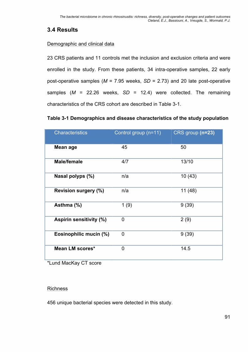
The bacterial microbiome in chronic rhinosinusitis: richness, diversity, post-operative changes and patient outcomes Cleland, E.J., Bassiouni, A., Vreugde, S., Wormald, P.J.
91
3.4 Results
Demographic and clinical data
23 CRS patients and 11 controls met the inclusion and exclusion criteria and were
enrolled in the study. From these patients, 34 intra-operative samples, 22 early
post-operative samples (M = 7.95 weeks, SD = 2.73) and 20 late post-operative
samples (M = 22.26 weeks, SD = 12.4) were collected. The remaining
characteristics of the CRS cohort are described in Table 3-1.
Table 3-1 Demographics and disease characteristics of the study population
Characteristics Control group (n=11) CRS group (n=23)
Mean age 45 50
Male/female 4/7 13/10
Nasal polyps (%) n/a 10 (43)
Revision surgery (%) n/a 11 (48)
Asthma (%) 1 (9) 9 (39)
Aspirin sensitivity (%) 0 2 (9)
Eosinophilic mucin (%) 0 9 (39)
Mean LM scores* 0 14.5
*Lund MacKay CT score
Richness
456 unique bacterial species were detected in this study.

The bacterial microbiome in chronic rhinosinusitis: richness, diversity, post-operative changes and patient outcomes Cleland, E.J., Bassiouni, A., Vreugde, S., Wormald, P.J.
92
The mean sample richness was 27.18 and 21.26 in the control and CRS groups
respectively and was not significantly different (P = 0.279). Post-operatively
sample richness increased substantially to 50.95 at the early post-operative time
point before declining to a 19.7 at the late post-operative time point (P = 0.986).
Diversity
The Shannon index was 2.502 and 2.246 in the control and CRS groups
respectively and was not significantly different (P = 0.516). This index was also
applied to observe for any significant post-operative changes in diversity in the
CRS group. Here a significant decline in diversity was seen with a fall from 2.246
intra-operatively to 1.789 and 1.193 at the early and late post-operative time points
respectively (P = 0.01).
Prevalence
The 40 most prevalent species detected intra-operatively are shown in Table 2.
Propionibacterium acnes and Staphylococcus epidermidis were the most prevalent
species detected (both 88%), followed by Corynebacterium segmentosum (71%),
Staphylococcus aureus (71%) and Corynebacterium tuberculostearicum (65%).
Several species were significantly more prevalent in the control patients compared
to CRS patients: Acinetobacter johnsonii (82% vs 26%, P = 0.003),
Corynebacterium confusum (73% vs 26%, P = 0.023), Corynebacterium
fastidiosum (64% vs 22%, P = 0.026) and Tissierella praeacuta (27% vs 0%, P =
0.028). Prevalence was also evaluated controlling for the presence of species at
<1% relative abundance. When this was performed only Acinetobacter johnsonii
and Corynebacterium confusum remained significant (P = 0.005 and P = 0.016
respectively). No species were significantly more prevalent in the CRS group.

The bacterial microbiome in chronic rhinosinusitis: richness, diversity, post-operative changes and patient outcomes Cleland, E.J., Bassiouni, A., Vreugde, S., Wormald, P.J.
93
The temporal changes in prevalence observed in the CRS group were
examined through logistic regression. 15 species were identified with significant
temporal changes. However, when the presence of a species at < 1% abundance
was controlled only 7 species were identified as having a significant temporal
change. Comparing the intra-operative, early and late post-operative prevalence
with this abundance threshold, both Acinetobacter johnsonii (13%, 86%, 85%, P <
0.001) and Acinetobacter schindleri (9%, 9%, 65%, P < 0.001) increased, whilst
Acidovorax facilis (39%, 23%, 0%, P = 0.007), Anaerococcus hydrogenalis (30%,
23%, 0%, P = 0.022), Corynebacterium segmentosum (43%, 9% and 0%, P =
0.004), Peptoniphilus asaccharolyticus (30%, 5% and 5%, P = 0.03) and
Staphylococcus epidermidis (78%, 64%, 35%, P = 0.008) all fell.
Abundance
103 species were determined to have a significant difference in MRA when intra-
operative samples were compared between the control and CRS group.
Surprisingly 102 of these species were rare constituents of the microbiome (MRA
<1%). Staphylococcus aureus was the only species found to have a significantly
higher MRA in the CRS group compared to controls (20% vs. 3% respectively, P =
0.034). S. aureus was also the most abundant species in CRS patients followed by
S. epidermidis (10%), Propionibacterium acnes (8%), Anaerococcus hydrogenalis
(5%) and Pseudomonas aeruginosa (4%). In controls, the MRA of Staphylococcus
epidermidis (9%) and Propionibacterium acnes (11%) was also high but not
significantly different to the CRS group. The most abundant species in the control
group was Acinetobacter johnsonii with an MRA of 22%, compared to 4% in the
CRS group (P = 0.058). Other abundant species in controls included

The bacterial microbiome in chronic rhinosinusitis: richness, diversity, post-operative changes and patient outcomes Cleland, E.J., Bassiouni, A., Vreugde, S., Wormald, P.J.
94
Marinilactibacillus psychrotolerans (18%) and Peptoniphilus asaccharolyticus
(6%). Several species from the genus Corynebacterium were also abundant in
both controls and CRS patients respectively: Corynebacterium confusum (5% vs
4%), Corynebacterium propinquum (5% vs 1%) and Corynebacterium
segmentosum (3% vs 4%). These differences were not significant (P > 0.05).
Corynebacterium tuberculostearicum whilst prevalent in our patients (65%), was a
rare constituent of the samples tested with an MRA of <1% in both controls and
CRS patients and not significantly different (P > 0.05). The MRA for the 40 most
prevalent species are presented in Table 3-2.
Analysis of temporal changes in MRA for the CRS cohort showed a
significant increase in Acinetobacter johnsonii from 4% intra-operatively to 37% at
the late post-operative time point (P < 0.001) and Acinetobacter schindleri, which
increased from <1% intra-operatively to 5% at the late post-operative time point (P
< 0.001). Anaerococcus hydrogenalis, Anaerococcus octavius, Corynebacterium
segmentosum, Peptoniphilus assacharolyticus, Propionibacterium acnes and
Staphylococcus epidermidis all declined during the post-operative period (P <
0.05). Significant changes were identified in a further 12 species, however these
were of extremely low MRA (<1%) at all time points. Staphylococcus aureus MRA
did not change significantly during the post-operative period, but did increase to
38% before declining to 33% at the early and late post-operative time points
respectively (P > 0.05).

The bacterial microbiome in chronic rhinosinusitis: richness, diversity, post-operative changes and patient outcomes Cleland, E.J., Bassiouni, A., Vreugde, S., Wormald, P.J.
95
Table 3-2 Prevalence and mean relative abundance (MRA) for the 40 most
frequently detected species (intra-operative samples)
All patients (n=33)
Control (n=11) CRS (n=23)
Species Prev*
(%)
Prev
(%)
MRA
(%)
Prev
(%)
MRA
(%)
Propionibacterium acnes 88.24 100 10.67 82.61 7.55
Staphylococcus epidermidis
88.24 90.91 8.88 86.96 10.25
Corynebacterium segmentosum
70.59 90.91 2.89 60.87 3.72
Staphylococcus aureus 70.59 81.82 2.61 65.22 20.45
Corynebacterium tuberculostearicum
64.71 63.64 0.72 65.22 0.89
Acidovorax facilis 58.82 45.45 0.48 65.22 2.28
Peptoniphilus asaccharolyticus
58.82 81.82 6.14 47.83 1.84
Anaerococcus hydrogenalis
55.88 45.45 1.44 60.87 4.52
Acinetobacter schindleri 50 72.73 1.52 39.13 0.83
Anaerococcus octavius 50 72.73 2.88 39.13 4.01
Acinetobacter johnsonii 44.12 81.82 22.36 26.09 3.75
Propionibacterium granulosum
44.12 63.64 0.76 34.78 0.37
Corynebacterium confusum
41.18 72.73 5.45 26.09 3.96
Finegoldia magna 38.24 45.45 0.79 34.78 0.65
Corynebacterium fastidiosum
35.29 63.64 0.16 21.74 0.27
Corynebacterium macginleyi
32.35 36.36 0.15 30.43 0.20
Gordonia rubripertinctus 32.35 45.45 0.69 26.09 0.86
Gordonia terrae 32.35 45.45 0.49 26.09 0.52
Marinilactibacillus psychrotolerans
32.35 54.55 18.34 21.74 1.22
Stenotrophomonas maltophilia
29.41 18.18 0.02 34.78 0.63

The bacterial microbiome in chronic rhinosinusitis: richness, diversity, post-operative changes and patient outcomes Cleland, E.J., Bassiouni, A., Vreugde, S., Wormald, P.J.
96
Sphingobium yanoikuyae 26.47 36.36 0.05 21.74 0.22
Streptococcus pseudopneumoniae
26.47 18.18 0.17 30.43 0.79
Corynebacterium propinquum
23.53 45.45 5.28 13.04 0.58
Escherichia fergusonii 23.53 9.09 < 0.01
30.43 1.8
Fusobacterium nucleatum 23.53 27.27 0.41 21.74 3.28
Neisseria flavescens 23.53 27.27 0.04 21.74 0.96
Staphylococcus lugdunensis
23.53 18.18 0.46 26.09 1.05
Campylobacter bacteroides ureolyticus
20.59 36.36 0.1 13.04 0.06
Corynebacterium accolens 17.65 27.27 0.02 13.04 0.02
Pseudomonas aeruginosa 17.65 9.09 < 0.01
21.74 4.37
Streptococcus oralis 17.65 36.36 0.12 8.7 0.19
Afipia broomeae 14.71 27.27 0.01 8.7 0.01
Alicyclobacillus ferripilum 14.71 27.27 0.37 8.7 0.03
Gemella sanguinis 14.71 0 0 21.74 0.04
Haemophilus parainfluenzae
14.71 18.18 0.02 13.04 0.07
Klebsiella oxytoca 14.71 9.09 0.01 17.39 0.14
Novosphingobium spp. 14.71 18.18 0.07 13.04 0.12
Propionibacterium avidum 14.71 9.09 0.04 17.39 0.07
Acinetobacter calcoaceticus
11.76 18.18 0.09 8.7 0.08
Aggregatibacter segnis 11.76 0 0 17.39 0.08
*Prevalence
Quality of life
Intra-operative comparisons of QOL scores showed no correlation between
richness and VAS and SNOT 22 scores (P > 0.05).
Logistic regression of prevalence revealed a significant correlation between
Acinetobacter johnsonii and QOL for both the VAS (coefficient = -0.051, P = 0.005)

The bacterial microbiome in chronic rhinosinusitis: richness, diversity, post-operative changes and patient outcomes Cleland, E.J., Bassiouni, A., Vreugde, S., Wormald, P.J.
97
and SNOT 22 (coefficient = -0.055, P = 0.004) scores. Similarly Streptococcus
pseudopneumoniae was also significantly associated with QOL for VAS
(coefficient = 0.101, P = 0.035) and SNOT 22 (coefficient = 0.089, P = 0.035).
Pearson correlation of relative abundance and intraoperative QOL scores
revealed a significant relationship between 8 species and VAS. 6 were negatively
correlated: Anaerococcus octavius (r = -0.495, P = 0.023), Corynebacterium
accolens (r = -0.460, P = 0.036), Corynebacterium macginleyi (r = -0.464, P =
0.034), Gordonia terrae (r = -0.441, P = 0.045), Neisseria mucosa (r = -0.472, P =
0.031), Propionibacterium granulosum (r = -0.551, P = 0.01), and 2 species were
positively correlated: Neisseria bacilliformis (r = 0.48, P = 0.028) and
Streptococcus pseudopneumoniae (r = 0.503, P = 0.02).
5 species had a correlation between relative abundance and SNOT 22
scores. Of these 3 were negatively correlated: Chryseobacterium isbiliense (r = -
0.461, P = 0.031), Corynebacterium accolens (r = -0.578, P = 0.005),
Corynebacterium macginleyi (r = -0.621, P = 0.002), and 2 were positively
correlated: Corynebacterium striatum (r = 0.44, P = 0.04) and Granulicatella
adiacens (r = 0.475, P = 0.026).
ANCOVA analysis revealed that richness had no significant effect on QOL
scores for either VAS or SNOT 22. The effect of relative abundance was also
tested with this model. Here the relative abundance of 33 species was significantly
correlated with changes in VAS (P < 0.05) and 22 species were significantly
correlated with changes in SNOT 22 scores (P < 0.05). The majority of these
species were present at extremely low MRA (<1%) and are not reported in full
here. Of particular interest was the species Acinetobacter johnsonii, which had a

The bacterial microbiome in chronic rhinosinusitis: richness, diversity, post-operative changes and patient outcomes Cleland, E.J., Bassiouni, A., Vreugde, S., Wormald, P.J.
98
significant reductive effect on QOL scores (improvement) for both the VAS (F =
5.863, P = 0.019) and SNOT 22 (F = 8.207, P = 0.006) scoring systems.
Pseudomonas aeruginosa was also noted to have a significant effect on SNOT 22
(F = 4.201, P = 0.045). In this case the effect was additive indicating an
association with poorer QOL.
Mucosal appearance and CT scores
No correlation was found for LK and LM scores for richness (P > 0.05) in the CRS
group. Likewise, logistic regression analysis of bacterial prevalence and both LK
and LM scores did not predict for any bacterial outcomes (P > 0.05).
Correlations were sought between bacteria relative abundance and LM
scores of which 3 were found to have significant negative correlations.
Alicyclobacillus ferripilum (r = -0.457, P = 0.028), Burkholderia cepacia (r = -0.466,
P = 0.025) and Erythrobacter citreus (r = -0.466, P = 0.025). All of these species
were however present at extremely low MRA (<1%).
7 significant correlations were also found between relative abundance and
LK scores. Klebsiella oxytoca showed a negative correlation with LK scores (r -
0.455, P = 0.029), whilst the remainder: Capnocytophaga gingivalis (r = 0.416, P
= 0.048), Neisseria bacilliformis (r = 0.419, P = 0.046), Neisseria cinerea (r =
0.415, P = 0.049), Polaribacter Spp. (r = 0.514, P = 0.012), Streptococcus
agalactiae (r = 0.499, P = 0.015), Streptococcus sanguinis (r = 0.487 P = 0.018)
were positively correlated. These species were all once again present at extremely
low MRA (≤1%).

The bacterial microbiome in chronic rhinosinusitis: richness, diversity, post-operative changes and patient outcomes Cleland, E.J., Bassiouni, A., Vreugde, S., Wormald, P.J.
99
3.5 Discussion
This study investigated the bacterial microbiome in the sinuses of CRS patients
and controls using 16S rDNA pyrosequencing. We have determined the richness,
diversity, prevalence, MRA and post-operative changes occurring in these
patients. Significant differences were found for diversity, prevalence and MRA
between controls and CRS patients. These findings were accompanied by
temporal changes, which highlighted the importance of several species including
Acinetobacter johnsonii. ANCOVA analysis also showed an important correlation
between Acinetobacter johnsonii and improved QOL. Mucosal appearance and CT
scores were examined and whilst several correlations were found for various
species these were all of extremely low MRA (<1%) and of uncertain significance.
Of the bacterial species identified with significant relationships between
CRS patients and controls, Acinetobacter johnsonii stood out as a microorganism
of interest. Acinetobacter johnsonii was significantly more prevalent in controls
compared to CRS patients (82% vs 26%, P = 0.003), and this relationship
remained when the presence of this species at <1% relative abundance was
excluded. Acinetobacter johnsonii was also the most abundant species in the
control group with an MRA of 22%, compared to 4% in the CRS group (P = 0.058).
The post-operative increases in prevalence were mirrored by an increase in the
MRA of Acinetobacter johnsonii. Here a significant increase from 4% intra-
operatively to 37% at the late post-operative time point was observed (P < 0.001).
The changes seen in Acinetobacter johnsonii prevalence and MRA were paralleled
by significant QOL correlations for both VAS (P = 0.005) and SNOT 22 (P = 0.004)
scores. Importantly, even when controlling for the beneficial effects of surgery on

The bacterial microbiome in chronic rhinosinusitis: richness, diversity, post-operative changes and patient outcomes Cleland, E.J., Bassiouni, A., Vreugde, S., Wormald, P.J.
100
QOL scores Acinetobacter johnsonii continued to be associated with significantly
improved QOL scores for both the VAS and SNOT 22 (both P < 0.05).
The changes seen for Acinetobacter johnsonii highlight a potentially
important relationship between this species and health. Acinetobacter belongs to
the class of Gram-negative bacteria known as Gammaproteobacteria. Interestingly
members of this class of bacteria were identified by Hanski et al. to be significantly
depleted on the skin of atopic individuals.225 Furthermore the functional role of the
Gammaproteobacteria was supported in that study by in vitro measurements of IL-
10 expression, an important anti-inflammatory cytokine involved in immunologic
tolerance. Here IL-10 expression was positively correlated with the abundance of
the Gammaproteobacterial genus Acinetobacter on the skin of healthy, but not
atopic individuals.225 Just as S. aureus enterotoxins have been associated with a
decrease in the production of T cell regulating cytokines such as IL-10 and TGF-
β1 in CRS,226 factors released by Acinetobacter johnsonsii in CRS patients may
play a functionally important role by promoting the production IL-10 and its
beneficial anti-inflammatory effects. The immunomodulatory properties of this
species require further investigation to establish these relationships and whether
this species has potential as a probiotic in CRS.
Previous culture-based studies investigating the prevalence of S. aureus
have found it to be higher in both CRS patients compared to controls, and higher
in patients with more severe disease such as those progressing to revision
surgery.206,227 This study identified a similarly high prevalence of S. aureus in both
controls and CRS patients (82% vs 65% respectively, P > 0.05). When the MRA of
S. aureus was also examined in these two groups an important difference was

The bacterial microbiome in chronic rhinosinusitis: richness, diversity, post-operative changes and patient outcomes Cleland, E.J., Bassiouni, A., Vreugde, S., Wormald, P.J.
101
identified. Here S. aureus MRA was significantly higher in the CRS group
compared to controls (20% vs 3%, P = 0.034). These findings draw attention to the
sensitivity of molecular techniques in detecting species present at low abundance,
which may otherwise be missed by culture techniques and support the fact that
MRA is potentially a more meaningful measure of an microorganism’s presence in
the sinonasal cavity. Interestingly despite the increase in S. aureus abundance in
the CRS population, it was not associated with a poorer quality of life or worse
objective disease severity such as LM and LK scores in this study. Larger samples
sizes may be required to identify these relationships if present.
Corynebacterium tuberculostearicum has recently been implicated as a
potential CRS pathogen in another microbiome study. Here the abundance of this
species was shown to be increased in CRS patients with a corresponding
depletion of lactic acid producing bacteria, which were thought to play a protective
role.74 Several Corynebacterium species were identified in this study, which were
prevalent including Corynebacterium tuberculostearicum in both controls and CRS
patients (64 vs 65% respectively). The MRA of this species was however
extremely low (<1%) in both patient groups and was not associated with any
significant changes post-operatively or correlated with any of the subjective or
objective measures of disease severity. Whilst this study does not support the role
of this species as a pathogen, future studies should investigate this association
further.
The mean sample richness was higher in controls compared to the CRS
group, but did not achieve significance (27.18 vs 21.26 respectively, P > 0.05).
This finding was consistent with one other study, which also found no

The bacterial microbiome in chronic rhinosinusitis: richness, diversity, post-operative changes and patient outcomes Cleland, E.J., Bassiouni, A., Vreugde, S., Wormald, P.J.
102
difference,133 but contrasted with another where bacterial richness was
significantly reduced in CRS patients compared to controls.74 The reason for
changes in richness in CRS remains unclear. Several factors may be involved,
which can be described within the framework of ecological theory. This concept
provides a useful method for rationalizing changes occurring in the microbiome
caused by specific influences on the niche.47 In the sinus, competition between
microbes for finite resources required for survival is likely to influence the overall
species richness. The interplay between host immunity and its impact on the
composition of the microbiome, which in itself demonstrates immunomodulatory
properties, may also play an important role in shaping these changes.151 Extrinsic
forces such as long-term pre-operative antibiotics may also be implicated and
have been shown to reduce genus level richness.133 The effect of antibiotics on
richness whilst poorly defined, may represent a potentially significant confounder
when examining microbiome data.
Post-operatively sample richness increased substantially to 50.95 before
declining to a mean of 19.7 at the early and late post-operative time points
respectively (P > 0.05). Whilst this finding did not achieve significance, the
changes occurring in the early post-operative period are still of interest, and could
be indicative of environmental changes which are hospitable to increased species
richness. These may include such factors as the presence of blood products from
surgery, inflammatory changes and altered cilia function. An expansion of bacterial
richness post-operatively may also impact on other kingdoms of life. In this same
cohort reduced fungal richness was identified post-operatively.219 It is plausible
that the fungal changes seen in these patients may have been in response to
increased competition from the bacterial component of the microbiome and

The bacterial microbiome in chronic rhinosinusitis: richness, diversity, post-operative changes and patient outcomes Cleland, E.J., Bassiouni, A., Vreugde, S., Wormald, P.J.
103
reflective of important cross-kingdom interactions. Further studies examining these
two kingdoms of life concurrently are required.
Species diversity was examined in this study with the Shannon index.
Whilst diversity was higher in the control group compared to the CRS group intra-
operatively this difference was not significant (P > 0.05). Post-operatively a
significant decline in diversity was identified on ANOVA (P = 0.01). The reason for
this post-operative drop in diversity was unclear, but may indicate disruption in the
local environment brought about by surgery, as well as the institution of post-
operative medical therapies such as antibiotics. Similar to richness, other
environmental pressures could also be driving these changes and further research
is required to define these relationships.
Many species found to be present at significantly different abundance levels
between the control and CRS groups were present at very low abundance levels
in this study. Whilst most of these species are unlikely to be of functional
significance, the ‘keystone-pathogen hypothesis’ suggests that certain low
abundance species may play a disproportionately important role in shaping the
microbiome and its functional contribution to the disease state.228 In periodontitis,
a polymicrobial disease analogous to CRS, Porphyromonas gingivalis has been
identified to act as a keystone pathogen. In this case P. gingivalis at very low
abundance levels was shown to promote pathogenic host-polymicrobial
interactions through the normally benign oral microbiota, which ultimately resulted
in the disruption of host homeostasis.229 An alternative perspective to that of the
keystone pathogen was put forth by Sibley et al. who used a Drosophila model of
polymicrobial infection to investigate interactions between the important cystic

The bacterial microbiome in chronic rhinosinusitis: richness, diversity, post-operative changes and patient outcomes Cleland, E.J., Bassiouni, A., Vreugde, S., Wormald, P.J.
104
fibrosis pathogen P. aeruginosa and 40 oropharyngeal commensals. Here a
number of oropharyngeal flora isolates that were avirulent or beneficial to the fly
were shown to have the capacity to synergistically enhance pathogenicity in the
presence of P. aeruginosa.85 These studies highlight the complex interactions
occurring amongst microbes and their ability to promote the disease state. In CRS
further studies investigating these interactions are urgently required to further
explore the importance of species of interest detected in microbiome studies.

The bacterial microbiome in chronic rhinosinusitis: richness, diversity, post-operative changes and patient outcomes Cleland, E.J., Bassiouni, A., Vreugde, S., Wormald, P.J.
105
3.6 Conclusion
This study has brought attention to several important findings relating to the CRS
microbiome and is the first to report on post-operative changes. Species richness
and diversity appeared to be similar between controls and CRS patients. The post-
operative state in CRS patients was characterized by significantly lower species
diversity. S. aureus MRA had significantly higher abundance in the CRS group
compared to controls. Acinetobacter johnsonii was more abundant in controls
compared to CRS, and our results suggest that it is associated with improvements
in QOL post-operatively. As such its potential as a probiotic in CRS should be
investigated. Future studies should also investigate the sinonasal microbiome in
greater depth and explore host-microbiome as well as microbe-microbe
interactions.

The fungal microbiome in chronic rhinosinusitis: richness, diversity, post-operative changes and patient outcomes Cleland, E.J., Bassiouni, A., Boase, S., Dowd, S., Vreugde, S., Wormald, P.J.
106
Chapter 4: The fungal microbiome in chronic
rhinosinusitis: richness, diversity, post-operative
changes and patient outcomes
Conducted in the Department of Otolaryngology Head and Neck Surgery,
University of Adelaide, Adelaide, Australia
Financial assistance provided by the Garnett Passe and Rodney Williams
Memorial Foundation

The fungal microbiome in chronic rhinosinusitis: richness, diversity, post-operative changes and patient outcomes Cleland, E.J., Bassiouni, A., Boase, S., Dowd, S., Vreugde, S., Wormald, P.J.
108
Statement of authorship
The fungal microbiome in chronic rhinosinusitis: richness, diversity, postoperative
changes and patient outcomes. Int Forum Allergy Rhinol 2014;4:259-65.
By signing the Statement of Authorship, each author certifies that their stated contribution to the
publication is accurate and that permission is granted for the publication to be included in the
candidate’s thesis.
Cleland, E.J. Project design, data collection, DNA
extraction, bioinformatics, manuscript
preparation
Bassiouni, A. Bioinformatics, manuscript editing
Boase, S. Endoscopic scoring, manuscript
editing
Dowd, S. Pyrosequencing, bioinformatics
Vreugde, S. Project supervision, manuscript
editing
Wormald, P.J. Project supervision, surgical
procedures, sample collection,
manuscript editing

The fungal microbiome in chronic rhinosinusitis: richness, diversity, post-operative changes and patient outcomes Cleland, E.J., Bassiouni, A., Boase, S., Dowd, S., Vreugde, S., Wormald, P.J.
109
4.1 Abstract
Introduction
Our understanding of fungi in chronic rhinosinusitis (CRS) has been limited by
previously employed detection techniques. This study examines the fungal
component of the microbiome in CRS patients and controls using a highly
sensitive culture-independent molecular technique. The aims of this study include
the characterization of fungal richness, prevalence, abundance, temporal changes
and their relationship with patient outcomes.
Methods
Swabs were collected from the sinuses of 23 CRS patients and 11 controls.
Collection occurred intra-operatively, and at 6 and 12 weeks post-operatively. DNA
was extracted from the swabs and fungal outcomes were determined through 18S
rDNA fungal tag-encoded FLX amplicon pyrosequencing.
Results
Fungi were ubiquitous to all patients. A total of 207 fungal genera were detected
with a mean sample richness of 8.18 and 12.14 in the control and CRS groups
respectively. Malassezia was detected in all patients at surgery and was also the
most abundant. Post-operatively, fungal richness decreased (P < 0.05) and was
associated with declines in the prevalence of Fusarium and Neocosmospora (P <
0.05). Neocosmospora was also less abundant post-operatively (P < 0.05). No
correlations were found with quality of life.

The fungal microbiome in chronic rhinosinusitis: richness, diversity, post-operative changes and patient outcomes Cleland, E.J., Bassiouni, A., Boase, S., Dowd, S., Vreugde, S., Wormald, P.J.
110
Conclusions
This is the first study to use a highly sensitive pyrosequencing technique to reveal
the true diversity of fungi in the sinuses of CRS patients and post-operative
changes in richness. The presence of Malassezia, a genus not previously
described in the sinuses is of great interest, and its potential as a disease modifier
should see further investigation given its association with atopic disease.

The fungal microbiome in chronic rhinosinusitis: richness, diversity, post-operative changes and patient outcomes Cleland, E.J., Bassiouni, A., Boase, S., Dowd, S., Vreugde, S., Wormald, P.J.
111
4.2 Introduction
Our understanding of the interactions between pathogens in complex microbial
communities remains in its infancy. Animal models of sinusitis have highlighted the
impact of cross-kingdom synergy between bacteria and fungus.92 Studies of
chronic rhinosinusitis (CRS) patients have also shown a close association
between bacterial and fungal biofilms.91,128 These findings illustrate the need to
consider the presence of specific pathogens in the context of a greater community
of microorganisms, and how the interactions between these members may be
contributing to health and disease.
Studies by Ponikau et al.18 and Braun et al.19 highlighted the ubiquitous
nature of fungi in CRS patients. These studies both utilized modified traditional
culture techniques to detect the presence of fungi. Culture based methods are
inherently limited however, by the selective pressures that nutrient media place on
organisms. This potential confounder is further emphasized by estimates that
suggest that only 1% to 10% of known microogranisms are cultureable under
laboratory conditions120 and highlights the need to consider alternative methods for
their detection.
Recently, culture-independent molecular techniques have given
researchers the ability to detect potentially all organisms in an ecological niche.
Using these methods several studies have attempted to describe the CRS
microbiome as it relates to bacteria.72,73,83,133 The fungal microbiome however,
remains poorly understood and this is reflected by a paucity of literature as it
relates to both CRS and other ecological niches of the human body.

The fungal microbiome in chronic rhinosinusitis: richness, diversity, post-operative changes and patient outcomes Cleland, E.J., Bassiouni, A., Boase, S., Dowd, S., Vreugde, S., Wormald, P.J.
112
The aim of this study was to characterize the fungal microbiome of the
sinuses in CRS patients and healthy controls by determining the richness,
prevalence, abundance and temporal changes after surgery. Patient outcomes
were also examined through objective and subjective means and compared
against the fungal state.

The fungal microbiome in chronic rhinosinusitis: richness, diversity, post-operative changes and patient outcomes Cleland, E.J., Bassiouni, A., Boase, S., Dowd, S., Vreugde, S., Wormald, P.J.
113
4.3 Methods
Study design and population
This prospective study recruited consecutive patients attending the tertiary referral
practice of the senior author (P.J.W.), and The Queen Elizabeth Hospital,
Adelaide, South Australia, Australia. Approval for the study was obtained from this
institution’s Human Ethics Committee.
The disease (CRS) group included patients undergoing endoscopic sinus
surgery in the form of middle meatal antrostomies, fronto-ethmoidectomies and
sphenoidectomies. A number of revision patients with extensive, severe disease
also received a Draf-III/frontal drillout procedure and/or canine fossa trephination
of the maxillary sinus. All patients were required to satisfy a diagnosis of CRS as
set out by the Rhinosinusitis Task Force,3 and were excluded from the study if
they were < 18 years, immunocompromised or had an impairment of mucociliary
function. The use of antibiotics, antifungals and oral or intranasal corticosteroids in
the 2 weeks prior to surgery were also grounds for exclusion.
The control group consisted of patients undergoing endoscopic
transphenoidal resections of pituitary adenomas and 1 patient undergoing
endoscopic sinus surgery for the removal of an inverting papilloma isolated to one
sinus. In this case swabs were collected from the endoscopically and
radiographically normal contralateral side. Exclusion criteria for the control group
included those set out for the CRS group plus the presence of any sino-nasal
symptoms, a history of previous sino-nasal surgery, sinus disease on CT or MRI
and the presence of abnormal intra-operative findings on endoscopy.

The fungal microbiome in chronic rhinosinusitis: richness, diversity, post-operative changes and patient outcomes Cleland, E.J., Bassiouni, A., Boase, S., Dowd, S., Vreugde, S., Wormald, P.J.
114
Demographic data was compiled for age, sex, presence of polyps, previous
sinus surgery, asthma and aspirin sensitivity.
Patient assessment
To correlate any associations between fungi and quality of life, two subjective
measurement tools were utilized in this study: a modified Sino-Nasal Outcome
Test 22 (SNOT-22) quality of life index (22 items, each scored from 0 – 3; total
score range, 0 – 66)220 and a sinus visual analogue symptom (VAS) scoring pro
forma (summation of six individual symptom scores and an overall
symptomatology score; total score range, 0 – 70).221
Lund-Kennedy (LK) scores were recorded for objective assessment of the
sinus mucosa.221 Scores were obtained from the operative surgeon or a member
of the research team (SB). Both were blinded to the microbiological outcomes of
the study.
CT scans were scored per the Lund-Mackay (LM) scoring system.221
Sample collection
Swabs were collected from the control and CRS groups intra-operatively. Post-
operative swabs were also collected from the CRS group at 6 (early post-
operative) and 12 weeks (late post-operative). On all occasions a flocked swab
(Copan Italia S.p.A., Brescia, Italy) was used under endoscopic guidance and
directed to the middle meatus / anterior ethmoid region irrespective of the
presence of purulence. Particular care was taken to sample the same site and side
of each patient at all time points. Any swabs that were inadvertently contaminated
through contact with with a non-target region (eg. nasal vestibule) were discarded.
After the sample had been retrieved the swab head was immediately separated

The fungal microbiome in chronic rhinosinusitis: richness, diversity, post-operative changes and patient outcomes Cleland, E.J., Bassiouni, A., Boase, S., Dowd, S., Vreugde, S., Wormald, P.J.
115
from the swab shaft and placed into a sterile container. This process ensured that
any DNA from other patient surfaces, which may have contaminated the proximal
swab shaft (eg. surgeon’s gloves), was not stored with the specimen. Samples
were immediately placed on ice and then transported to the laboratory for storage
at -80 degrees Celsius.
DNA extraction
Swab heads were thawed, then cut into small pieces and placed in 180 µl of
enzymatic lysis buffer overnight at room temperature. This was followed by a bead
beating process with 5mm steel beads for 20 seconds at 15 Hz and then again
with 0.1mm glass beads for 5 mins at 30 Hz. The enzymatic lysis buffer and
remainder of the extraction protocol were prepared and performed as per the
Qiagen DNeasy Blood & Tissue Kit instructions (Qiagen, Valencia, CA). Extracted
DNA was stored at -80 degress Celsius until pyrosequencing.
PCR amplification of 18S and pyrosequencing
Tag-encoded FLX-Titanium amplicon pyrosequencing for fungal organisms was
performed as previously described.222 Briefly, a selective panfungal forward
funSSUF-TGGAGGGCAAGTCTGGTG SSUFungiR
TCGGCATAGTTTATGGTTAAG primer set was applied against the 18S gene for
PCR amplification. PCR and pyrosequencing was performed by MR DNA
(Shallowater, TX). Pyrosequencing was performed on the Roche Titanium 454
sequencer (F. Hoffmann-La Roche Ltd, Basel, Switzerland).
Bioinformatics pipeline
Pyrosequencing data was processed using a proprietary analysis pipeline.
Sequences were depleted of barcodes and primers. Short sequences (< 200bp),

The fungal microbiome in chronic rhinosinusitis: richness, diversity, post-operative changes and patient outcomes Cleland, E.J., Bassiouni, A., Boase, S., Dowd, S., Vreugde, S., Wormald, P.J.
116
sequences with ambiguous base calls and sequences with homopolymer runs
exceeding 6bp were removed. Sequences were then denoised and chimeras
removed. Operational taxonomic units (OTUs) were defined after removal of
singleton sequences and clustering at 1% divergence (99% similarity). OTUs were
then taxonomically classified using BLASTn against a curated database derived
from public sources including NCBI, Greengenes, and RDP (MR DNA version
02012013) database.
Statistical analysis
All tests were performed at the taxonomic rank of genus.
First, comparisons were made between the control and CRS group at the
intra-operative time point. These comparisons included:
e) Richness (number of unique genera per sample): compared using a T-test
f) Prevalence (absence vs. presence of a particular genus as a binary outcome):
compared using Fisher's exact test.
g) Mean relative abundance (MRA) (proportional presence of a genus compared
to others in the same sample): compared using Metastats,224 a validated
metagenomics tool for detecting differentially abundant taxa between two
groups.
Second, for the CRS cohort, temporal changes were examined at the intra-
operative, early and late post-operative time points. The microbiological outcomes
were as described above (richness, prevalence, MRA) and were assessed through
ANOVA (richness and relative abundance) and logistic regression (prevalence).

The fungal microbiome in chronic rhinosinusitis: richness, diversity, post-operative changes and patient outcomes Cleland, E.J., Bassiouni, A., Boase, S., Dowd, S., Vreugde, S., Wormald, P.J.
117
Third, we explored whether the microbiological outcomes impacted on
quality of life (QOL) in the intra-operative CRS group. SNOT-22 and VAS scores
were tested against richness, relative abundance (Pearson correlation) and
prevalence (logistic regression). These tests were repeated for the LK and LM
scores to assess for any fungal impact on objective disease severity.
To test for the effect of richness and relative abundance on post-operative
QOL scores we used an analysis of covariance (ANCOVA) model. This model was
utilized to control for improvements in QOL related to the confounding effect of
surgery.
All statistics were performed using the R statistical software (R Foundation
for Statistical Computing, Vienna, Austria) and packages from the Scientific
Python stack (scipy, numpy, rpy2, pandas, ipython).

The fungal microbiome in chronic rhinosinusitis: richness, diversity, post-operative changes and patient outcomes Cleland, E.J., Bassiouni, A., Boase, S., Dowd, S., Vreugde, S., Wormald, P.J.
118
4.4 Results
Demographic and clinical data
23 CRS patients and 11 controls met the inclusion and exclusion criteria and were
enrolled in the study. From these patients, 34 intra-operative samples, 22 early
post-operative samples (M = 7.95 weeks, SD = 2.73) and 20 late post-operative
samples were collected (M = 22.26 weeks, SD = 12.4). Of these 76 samples, 4
failed to amplify and were excluded on the basis of being below detection.
Importantly, fungi were detected in other samples processed for these patients.
One patient was lost to follow up at 6 weeks and another two at 12 weeks. Further
characteristics of the CRS cohort are described in Table 4-1.
Table 4-1 Demographics and disease characteristics of the study population
Characteristics Control group (n=11) CRS group (n=23)
Mean age 45 50
Male/female 4/7 13/10
Nasal polyps (%) n/a 10 (43)
Revision surgery (%) n/a 11 (48)
Asthma (%) 1 (9) 9 (39)
Aspirin sensitivity (%) 0 2 (9)
Eosinophilic mucin (%) 0 9 (39)
Mean LM scores* 0 14.5
*Lund MacKay CT score

The fungal microbiome in chronic rhinosinusitis: richness, diversity, post-operative changes and patient outcomes Cleland, E.J., Bassiouni, A., Boase, S., Dowd, S., Vreugde, S., Wormald, P.J.
119
Richness
A total of 207 unique fungal genera were detected.
The mean sample richness was 8.18 and 12.14 in the control and CRS
groups respectively and was not significantly different (P = 0.103). Sample
richness was however significantly decreased post-operatively in CRS patients
with falls from 12.14 intra-operatively to 6.9 and 6.74 at 6 and 12 weeks
respectively (P = 0.007).
Prevalence
The 40 most prevalent genera are shown in

The fungal microbiome in chronic rhinosinusitis: richness, diversity, post-operative changes and patient outcomes Cleland, E.J., Bassiouni, A., Boase, S., Dowd, S., Vreugde, S., Wormald, P.J.
120
Table 4-2. Of these, Malassezia was the most prevalent genus and detected in all
patients. Other prevalent genera included Calicium, Neocosmospora, Fusarium,
Saccharomyces and Aspergillus. Scutellospora was the only genus found to be
significantly different between controls and CRS patients (36% vs. 5%
respectively, P = 0.033). No other significant differences in prevalence between
the other genera were detected. Prevalence was also evaluated with an
abundance threshold as it was felt that extremely low abundance genera (<1%)
could be skewing prevalence results. When prevalence was compared on this
basis no significant difference was determined for all genera, including
Scutellospora.
Temporal changes in prevalence were also examined for the CRS group.
Significant changes were detected for Dendryphiella, Fusarium, Malassezia,
Mortierella, Neocosmospora and Pleistophora (P < 0.05). However when the
presence of extremely low abundance genera (<1%) were excluded, only
Fusarium and Neocosmospora remained significant, with falls in prevalence of
both.
Abundance
Malassezia was the most abundant genus in both control and CRS patients with
MRA scores of 57.5% and 50.09% respectively. Neocosmospora was also
abundant in controls (11.42%) and CRS patients (6.22%). Whilst Calicium and
Fusarium were both prevalent species, the MRA of these genera was low in both
controls and CRS patients (0.47% vs 0.29% and 1.08% vs 2.18% respectively).
The MRA for the 40 most prevalent genera are presented in Table 4-2. Notably the

The fungal microbiome in chronic rhinosinusitis: richness, diversity, post-operative changes and patient outcomes Cleland, E.J., Bassiouni, A., Boase, S., Dowd, S., Vreugde, S., Wormald, P.J.
121
MRA of genera traditionally associated with disease in CRS such as Alternaria and
Aspergillus was low.
When the MRA was compared between controls and CRS patients a
significant difference was detected for 33 genera (P < 0.05). However, all of these
genera were poorly represented in control samples (mean relative abundance
<1%) and only 3 genera represented at levels of >1% were found in the CRS
group (Chaeotomium – 2.58%, Mortierella – 2.37% and Trichocladium – 1.92%).
The post-operative changes in MRA for CRS patients showed a significant
decline for Basidiobolus, and increase for both Cyberlindera and Dendryphiella (P
< 0.05). However, the mean relative abundance at all time points was low for
these genera (<1%, <2%, <1% respectively). The genus Neocosmospora was also
shown to decrease post operatively and fell from an MRA of 6.22% intra-
operatively to 1.41% and 1.59% in the early and late post-operative periods
respectively (P < 0.05).

The fungal microbiome in chronic rhinosinusitis: richness, diversity, post-operative changes and patient outcomes Cleland, E.J., Bassiouni, A., Boase, S., Dowd, S., Vreugde, S., Wormald, P.J.
122
Table 4-2 Prevalence and mean relative abundance (MRA) for the 40 most
frequently detected genera (intra-operative samples)
All patients
(n=33)
Control (n=11) CRS (n=22)*
Genus Prevalence
(%)
Prevalence
(%)
MRA
(%)
Prevalence
(%)
MRA
(%)
Malassezia 100 100 57.5 100 50.09
Calicium 54.55 45.45 0.47 59.09 0.29
Neocosmospora 48.48 27.27 11.42 59.09 6.22
Fusarium 39.39 27.27 1.08 45.45 2.18
Saccharomyces 36.36 18.18 0.12 45.45 1.11
Aspergillus 33.33 27.27 0.85 36.36 1.56
Cladosporium 33.33 36.36 3.23 31.82 3.28
Basidiobolus 27.27 18.18 7.57 31.82 0.57
Mortierella 24.24 9.09 0.02 31.82 2.37
Penicillium 24.24 9.09 0.01 31.82 0.98
Ceratostomella 21.21 9.09 0.03 27.27 0.13
Juncigena 21.21 27.27 0.25 18.18 0.07
Powellomyces 21.21 27.27 0.34 18.18 0.07
Glomales 18.18 18.18 2.62 18.18 0.55
Hypomyces 18.18 18.18 0.02 18.18 0.09
Rhizophydium 18.18 18.18 0.39 18.18 0.91
Endogone 15.15 0 0 22.73 0.35
Glomeromycota 15.15 0 0 22.73 1.43
Alternaria 15.15 18.18 0.3 13.64 2.42
Eutypa 15.15 18.18 0.47 13.64 0.10
Galactomyces 15.15 18.18 9.52 13.64 0.01
Glomus 15.15 18.18 0.27 13.64 1.76
Pleospora 15.15 18.18 0.13 13.64 3.96
Scutellospora 15.15 36.36 0.22 4.55 0.03

The fungal microbiome in chronic rhinosinusitis: richness, diversity, post-operative changes and patient outcomes Cleland, E.J., Bassiouni, A., Boase, S., Dowd, S., Vreugde, S., Wormald, P.J.
123
Thermoascus 12.12 0 0 18.18 0.12
Ambispora 12.12 9.09 0.01 13.64 1.35
Candida 12.12 9.09 0.01 13.64 3.72
Davidiella 12.12 9.09 < 0.01 13.64 0.06
Lecanora 12.12 9.09 0.01 13.64 0.2
Lepraria 9.09 0 0 13.64 0.02
Sclerotium 9.09 0 0 13.64 < 0.01
Chytridium 9.09 9.09 0.79 9.09 0.18
Funneliformis 9.09 9.09 0.00 9.09 0.15
Paraglomus 9.09 9.09 0.11 9.09 0.03
Rhodotorula 9.09 9.09 0.00 9.09 0.08
Phacellium 9.09 18.18 0.06 4.55 0.01
Capnobotryella 6.06 0 0 9.09 0.02
Clathrospora 6.06 0 0 9.09 0.04
Cordyceps 6.06 0 0 9.09 0.02
Cosmospora 6.06 0 0 9.09 0.04
*Note: 1 sample failed to amplify in the intra-operative CRS group
Quality of life
Intra-operatively, no correlation was found for richness or prevalence when
compared against VAS and SNOT 22 scores (P > 0.05). Relative abundance was
also tested. Sclerotium was found to positively correlate with VAS (r = 0.468, P <
0.05) and Ceratostomella was positively correlated with SNOT 22 (r = 0.439, P <
0.05). Hypomyces was negatively correlated for both VAS (r = - 0.455, P < 0.05)
and SNOT 22 (r = - 0.467, P < 0.05).
ANCOVA analysis indicated that genera richness had no significant effect
on QOL scores (P > 0.05). The ANCOVA model also showed that the relative
abundance of 14 genera had a significant effect on VAS and/or SNOT 22 scores

The fungal microbiome in chronic rhinosinusitis: richness, diversity, post-operative changes and patient outcomes Cleland, E.J., Bassiouni, A., Boase, S., Dowd, S., Vreugde, S., Wormald, P.J.
124
(P < 0.05, data not shown) independent of surgery. The majority of these fungal
genera were however, present at extremely low MRA (<1%). Cladosporium and
Pleistophora were the only two fungal genera present at higher levels that had a
statistically significant reductive effect on QOL scores. Cladosporium VAS (F =
5.06, P = 0.028) and Pleistophora SNOT 22 (F = 5.37, P = 0.024).
Mucosal appearance and CT scores
Fungal richness did not correlate with LK scores (P > 0.05) in CRS patients. A
Logistic regression analysis of fungal prevalence and LK scores did not predict for
any fungal outcomes (P > 0.05). A moderate negative correlation (r = -0.485)
between LK scores and MRA was noted for Neocosmospora (P = 0.022). No
correlation between LK scores and MRA was found for the remaining genera.
A moderate negative correlation (r = -0.449) was found between the relative
abundance of Davidiella and LM scores (P = 0.036). However, the MRA of this
species was extremely low (<1%). No other significant correlation was found for
richness or prevalence for LM scores.

The fungal microbiome in chronic rhinosinusitis: richness, diversity, post-operative changes and patient outcomes Cleland, E.J., Bassiouni, A., Boase, S., Dowd, S., Vreugde, S., Wormald, P.J.
125
4.5 Discussion
This prospective study determined the fungal richness, prevalence, abundance
and post-operative changes of the fungal microbiome in the paranasal sinuses for
a group of controls and CRS patients. Using a highly sensitive culture-independent
technique, no major differences were detected in the fungal microbiome between
controls and CRS patients intra-operatively. Temporal changes in the microbiome
were also studied in the CRS group and here post-operative declines in richness,
and the presence of the genera Fusarium and Neocosmospora were noted.
The genera Malassezia was both a prevalent and abundant genus and is of
great interest in this study. Malassezia was detected in 100% of controls and CRS
patients with an MRA of 57% and 50% respectively. On this basis malassezia was
the most prevalent and abundant fungal genera detected in this study. In contrast,
culture based studies described by others were unable to detect Malassezia in any
patients.18,19 Malassezia is well known for being difficult to culture and requires the
use of special media due to its lipid dependent nature. These specialized media
are not routinely employed in clinical mycology laboratories,230 and may explain its
presence going undetected. In line with the growth requirements of this genus,
Malassezia is typically found in sebum-rich areas of the body such as the trunk
and head and is considered part of the normal cutaneous flora.230 The presence of
this organism in the sinonasal cavity is less clear given its growth needs and could
represent ongoing seeding from the nasal vestibule rather than permanent niche
colonization.
Malassezia has been described in human cutaneous and systemic
diseases including dermatitis, pityriasis versicolor, seborrhoeic dermatitis,

The fungal microbiome in chronic rhinosinusitis: richness, diversity, post-operative changes and patient outcomes Cleland, E.J., Bassiouni, A., Boase, S., Dowd, S., Vreugde, S., Wormald, P.J.
126
folliculitis, atopic eczema/dermatitis syndrome (AEDS), catheter-related
fungaemia, peritonitis and meningitis.231 AEDS if of particular interest as it
represents a chronic inflammatory disorder, which parallels CRS in many ways.
The mechanisms promoting the release of Th2 cytokines in eosinophilic CRS are
not completely understood. S. aureus superantigens have been implicated.232
Others have shown a Th2 response in the presence of S. aureus biofilms,
independent of superantigens.99 Similar to CRS, AEDS patients also demonstrate
a Malassezia-specific Th2 response,233 and it is possible that the presence of
Malassezia in some CRS patients may skew the cytokine profile in a similar way.
AEDS patients show increased type I hypersensitivity to Malassezia and increased
Malassezia-specific IgE.230 The presence of Malassezia-specific IgE in CRS
populations is as yet untested and should be investigated. LL-37 (a cathelicidin)
expression is implicated in the host response to Malassezia. Excessive levels are
seen in severe AEDS and are believed to play a role in sustained inflammation
and other detrimental effects.230 This antimicrobial peptide has been studied in
CRS and was shown to be elevated in nasal tissue and eosinophilic mucus.234 LL-
37 mRNA has also shown to be elevated to Aspergillus and Alternaria allergens in
a nasal explant model.108 Malassezia driven expression of LL-37 in CRS may be a
significant factor contributing to inflammation in these patients.
A total of 207 unique fungal genera were identified in this study. Our 18S
ribosomal DNA (rDNA) fungal tag-encoded FLX amplicon pyrosequencing
technique allowed us to demonstrate a huge diversity of fungi in both CRS patients
and controls. This technique is clearly superior when compared to traditional
culture based techniques of detection such as that used by Ponikau et al. and
Braun et al. where only 40 and 33 unique fungal genera were identified

The fungal microbiome in chronic rhinosinusitis: richness, diversity, post-operative changes and patient outcomes Cleland, E.J., Bassiouni, A., Boase, S., Dowd, S., Vreugde, S., Wormald, P.J.
127
respectively.18,19 An alternative molecular technique, the Ibis T5000, coupling
nucleic acid amplification to high-performance electrospray ionization mass
spectrometry and base-composition analysis,235 has also been used in our
department. This technique whilst promising for detecting fungal organisms, had a
surprisingly low yield with only 4 of 38 CRS patients testing positive for fungi from
2 genera.72 The presence of fungi in its biofilm phenotype has also been studied in
CRS, using fluorescent in situ hybridization techniques. These studies confirmed
the presence of fungal biofilms in some patients, but due to the use of pan-fungal
probes, were unable to define them on a species level.91,128 Species-specific
probes can be used with this technique however this has not been performed due
to the need for a prior knowledge of fungi of interest. The large number of unique
genera detected in this study highlights the importance of gene sequencing
techniques to identify fungi including uncultivable genera of interest, which have
not previously been the target of focused detection techniques.
The mean sample richness determined in this study was less in the controls
compared to CRS patients, although this was not statistically significant (8.14 vs
12.14 respectively, P > 0.05). The precise mechanisms, which shape fungal
richness in the sinonasal cavity are not clear, but may be influenced by host and
environmental factors. Inhalation of airborne fungi is likely to play an important role
in shaping the fungal microbiome in the sinonasal cavity. The use of intra-nasal air
samplers for the detection of fungi has confirmed that fungal spores from a wide
range of genera are continually inhaled from the surrounding environment into the
nasal cavity. Factors such as respiration type, the distribution of fungi in the local
microenvironment and the type of activity undertaken by the subject all influence
the degree of fungal exposure.117 This variability in fungal exposure whilst difficult

The fungal microbiome in chronic rhinosinusitis: richness, diversity, post-operative changes and patient outcomes Cleland, E.J., Bassiouni, A., Boase, S., Dowd, S., Vreugde, S., Wormald, P.J.
128
to quantify, may influence fungal richness independent of any disease process. In
this study, post-operative fungal richness was decreased at both the early and late
post-operative time points (6.9 and 6.73 respectively, P = 0.007). This decline in
richness represents a durable post-operative change in these patients and is likely
to have occurred independently of environmental influences – a variable which
remained constant in the majority of patients. The impact of surgery on fungal
richness may also account for the post-operative changes seen in this study.
Initially surgery may disrupt mucociliary function, but this has generally normalized
by 6 to 12 weeks.236,237 The timing of post-operative sampling in this study
correlates with the period when improved ciliary function is expected. More
efficient clearance of inhaled fungi by cilia, combined with a decreased mucosal
disease burden, may contribute towards reduced richness post-operatively. In a
similar fashion, saline douches, a routine component of post-operative
management may also influence fungal richness. Here the irrigating action may
serve to assist clearance of fungi from the sinonasal cavity.
Traditional culture studies of CRS patients have typically identified several
prevalent fungal genera including Aspergillus, Alternaria, Cladosporium and
Penicillium.18,19,118 These genera were also well represented in samples collected
in this study (Table 2.). In addition to Malassezia, many other prevalent species
were identified in our CRS patients including: Calicium (59%), Neocosmospora
(59%), Fusarium (45%) and Saccharomyces (45%). Compared to the control
group, Scutellospora was the only genus found to be significantly different in
controls compared to CRS patients (36% vs. 5% respectively, P = 0.033). The
relevance of this finding is uncertain given that this genus had an extremely low
MRA (<1%). Given the large number of unique genera detected in this study, it

The fungal microbiome in chronic rhinosinusitis: richness, diversity, post-operative changes and patient outcomes Cleland, E.J., Bassiouni, A., Boase, S., Dowd, S., Vreugde, S., Wormald, P.J.
129
was surprising that more differences were not noted between the two groups. This
sameness in prevalence may be a reflection of similar environmental exposures to
airborne fungi in these patients, but also needs to be considered in the context of
MRA.
Temporal changes in prevalence were also examined. Fusarium and
Neocosmospora were the only genera significantly decreased post-operatively in
the CRS group, a finding which was present even when controlling for their
presence at extremely low abundance (<1%). Fusarium fell from 45% in CRS
patients intra-operatively to 19% and 0% in the early and late post-operative
periods respectively (P = 0.03). In humans, Fusarium is associated with skin
infections, keratitis, onychomycosis and may cause disseminated disease in
immunocompromised patients.238 The prevalence of Fusarium in CRS patients
detected with standard cultures ranges from 2% to 16%,18,19 which is likely to be
underrepresented when compared to our molecular technique. Fusarium may
potentially be an important pathogen in CRS. One recent study examined the
beneficial effect of targeted oral antifungal agents in a subgroup of Fusarium-
positive CRS patients and showed improvements in endoscopic appearance and
symptoms.239 Fusarium is recognized as one of the most drug-resistant fungal
genera,238 and this resistance to treatment may have been reflected in other CRS
studies in which a response to oral antifungal agents or a reduction in
inflammatory markers was lacking.100,102,104,106 The prevalence of Neocosmospora
fell from 59% in CRS patients intra-operatively to 10% and 11% at the early and
late post-operative periods respectively (P = 0.013). The declines in prevalence of
this genus are of interest and parallel significant declines also seen in MRA.
Neocosmospora has been described in human infections such as keratitis, skin

The fungal microbiome in chronic rhinosinusitis: richness, diversity, post-operative changes and patient outcomes Cleland, E.J., Bassiouni, A., Boase, S., Dowd, S., Vreugde, S., Wormald, P.J.
130
granulomas and arthritis, although these cases are rare.240 A conflicting negative
correlation was observed between the presence of Neocosmospora and LK
scores, casting doubt on its role as a pathogen in this ecological niche. Further
studies are required to more accurately characterize this genus in CRS.
Correlations between fungal richness or prevalence and QOL were not
found for the intra-operative CRS cohort in this study. Significant relationships
between relative abundance and QOL were found for Sclerotium, Ceratostomella
and Hypomyces (P < 0.05). These genera were minor constituents of the
microbiome with an MRA <1% in all cases. A growing body of evidence suggests
that some low abundance members of the microbiome may be disproportionately
physiologically active compared to more abundant members.241 This makes
conclusions regarding rare constituents of the microbiome and their functional
importance difficult. Similarly the genera identified as significant by the ANCOVA
model, except for Cladosporium and Pleistophora, were also of low MRA (<1%).
These results should be interpreted with caution due to the limitations of the
ANCOVA model in this context because the covariates (fungal relative abundance
and surgery) are not completely independent. Surgery affects the microbiome
itself, in addition to the QOL outcome. These factors highlight the complex
dynamics at play in the microbiome and the need for further research to better
understand these complex relationships and their impact on the CRS disease
state.
The fungal microbiome in chronic rhinosinusitis: richness, diversity, post-operative changes and patient outcomes Cleland, E.J., Bassiouni, A., Boase, S., Dowd, S., Vreugde, S., Wormald, P.J.
131
4.6 Conclusion
This study uses a highly sensitive pyrosequencing technique to determine the
diversity of fungi in controls and CRS patients. Although no major differences were
found between the microbiome of these two groups intra-operatively, post-
operative declines in richness and the presence of the genera Fusarium and
Neocosmospora were noted. The detection of fungi not previously described in
this ecological niche such as Malassezia highlights the utility of our detection
technique. The association between Malassezia and atopic disease, marks this
genus as a potential disease-modifying organism in CRS. Further investigation is
required to better define the ecological pressures influencing the fungal
microbiome and how Malassezia, and other taxa of interest contribute towards
health or disease.
Probiotic manipulation of the chronic rhinosinusitis microbiome Cleland, E.J., Drilling A., Bassiouni, A., James, C., Vreugde, S., Wormald, P.J.
132
Chapter 5: Probiotic manipulation of the chronic
rhinosinusitis microbiome
Conducted in the Department of Otolaryngology Head and Neck Surgery,
University of Adelaide, Adelaide, Australia
Financial assistance provided by the Garnett Passe and Rodney Williams
Memorial Foundation

Probiotic manipulation of the chronic rhinosinusitis microbiome Cleland, E.J., Drilling A., Bassiouni, A., James, C., Vreugde, S., Wormald, P.J.
134
Statement of authorship
Probiotic manipulation of the chronic rhinosinusitis microbiome. Int Forum Allergy
Rhinol 2014;4:309-14.
By signing the Statement of Authorship, each author certifies that their stated contribution to the
publication is accurate and that permission is granted for the publication to be included in the
candidate’s thesis.
Cleland, E.J. Project design, animal handling, data
collection and analysis, manuscript
preparation
Drilling, A. Bacterial inoculant preparation,
manuscript editing
Bassiouni, A. Statistical analysis, manuscript
editing
James, C. Histological preparations
Vreugde, S. Project supervision, manuscript
editing
Wormald, P.J. Project supervision, manuscript
editing

Probiotic manipulation of the chronic rhinosinusitis microbiome Cleland, E.J., Drilling A., Bassiouni, A., James, C., Vreugde, S., Wormald, P.J.
135
5.1 Abstract
Background
Staphylococcus aureus (SA) is a key pathogenic component of the chronic
rhinosinusitis (CRS) microbiome and is associated with increased disease severity
and poor post-operative outcomes. Probiotic treatments potentially offer a novel
approach to the management of pathogenic bacteria in these recalcitrant patients
through supporting a healthy community of commensal species. This study aims to
investigate the probiotic properties of Staphylococcus epidermidis (SE) against SA
in a mouse model of sinusitis.
Methods
20 C57/BL6 mice received intranasal inoculations of phosphate buffered saline
(PBS), SE, SA or a combination of SE and SA (SE+SA) for 3 days. Following
sacrifice, the mouse snouts were harvested and prepared for histological analysis.
Counts of PAS-positive goblet cells were the primary outcome measure.
Results
Goblet cell counts were significantly higher in both the SA and SE+SA groups
compared to those receiving PBS or SE alone (P < 0.05). However, the SE+SA
group demonstrated significantly lower goblet cell counts compared to the SA
group (P < 0.05). Mice receiving SE alone did not show a significant difference to
those receiving PBS (P > 0.05). The presence of SA post-inoculation was
confirmed by culture in both the SA and SE+SA groups.

Probiotic manipulation of the chronic rhinosinusitis microbiome Cleland, E.J., Drilling A., Bassiouni, A., James, C., Vreugde, S., Wormald, P.J.
136
Conclusion
This study confirms the probiotic potential of SE against SA in a mouse model of
sinusitis. Whilst the interactions that occur between many probiotic species and
pathogens are yet to be fully understood, studies such as this support further
exploration of ecologically based treatment paradigms for the management of
CRS.

Probiotic manipulation of the chronic rhinosinusitis microbiome Cleland, E.J., Drilling A., Bassiouni, A., James, C., Vreugde, S., Wormald, P.J.
137
5.2 Introduction
The ability to detect and identify prokaryotes in the human body has been greatly
aided by the emergence of 16S rRNA gene sequencing techniques.242 Studies
such as those from The Human Microbiome Project Consortium have been able to
demonstrate a huge diversity of microorganisms in several regions of the human
body.243 The challenge beyond the detection and identification of organisms
however, is to understand how these diverse communities of bacteria interact with
the host and one another to promote health or disease.
Many studies have attempted to define the bacterial communities present in
CRS patients. This has been achieved through a variety of standard culture,
microscopy and molecular techniques.76 Despite the limitations of some of these
detection modalities, Staphylococcus aureus (SA) has continued to demonstrate
itself as a key pathogenic component of the CRS microbiome. This association is
particularly apparent in recalcitrant patients and has been confirmed when present
as planktonic bacteria or biofilms.89,90,133,202,227
The relationship between SA and disease severity makes it an ideal target
for probiotic therapies. Probiotics are defined as live bacteria which when
administered in adequate amounts confer a health benefit on the host.157 The use
of these treatments in CRS represent a novel approach aimed at restoring the
balance between beneficial commensals and pathogenic species. Whilst the
benefit of an oral probiotic has been tested in one CRS study,192 the role of
topically delivered probiotic treatments to the sinonasal cavity remains unclear.
Topicalization of probiotic preparations provides an exciting mechanism by which

Probiotic manipulation of the chronic rhinosinusitis microbiome Cleland, E.J., Drilling A., Bassiouni, A., James, C., Vreugde, S., Wormald, P.J.
138
clinicians can potentially promote and maintain a healthy sinus ecosystem by
directly manipulating the microbiome.
The murine model of sinusitis used in this study represents an important
tool where the interactions between pathogens and potential probiotics can be
assessed whilst maintaining a controlled environment. This model is anatomically
well described,244 and has been validated by others examining the effects of
bacterial interactions.74
In this study we explore the probiotic properties of Staphylococcus
epidermidis (SE). SE has been shown to exert a probiotic effect through the
secretion of serine protease Esp, which inhibits both biofilm formation and nasal
colonization by SA.196 This study aims to utilise these properties to demonstrate
the probiotic role of an Esp-producing strain of SE against SA in a murine model of
sinusitis by determining goblet hyperplasia and associated mucous
hypersecretion, an important marker of airway inflammation.245

Probiotic manipulation of the chronic rhinosinusitis microbiome Cleland, E.J., Drilling A., Bassiouni, A., James, C., Vreugde, S., Wormald, P.J.
139
5.3 Methods
Study conditions
Ethics approval was obtained from the local Animal Ethics Committee. 20 C57BL/6
mice (Animal Laboratory Services, Adelaide, South Australia) aged 6-8 weeks
were obtained. All mice were certified as specific pathogen free and housed per
treatment group in individually ventilated micro-isolator cages supplied by an H14
HEPA filtration system (Techniplast, Italy). All procedures including inoculations
were performed in a laminar flow change station (Tecniplast, Italy) and mice were
only fed sterilized pellets and water, thus preventing microbial contamination.
Inoculant preparation and dosing
To prepare nasal inoculants, stock cultures of SA (ATCC 25923) and SE (Esp-
producing strain) were grown overnight on 1% nutrient agar plates made up of
nutrient broth powder (Oxoid, Thebarton, South Australia) and 1% bacteragar
powder (BD, North Ryde, New South Wales). A single colony was inoculated into
nutrient broth and subsequently grown for 16 hrs at 37°C with shaking. Cultures
were centrifuged using a Beckman TJ-6 centrifuge (Beckman Coulter, Lane Cove,
New South Wales) at 4000rpm for 10 minutes. The pellets were resuspended into
phosphate buffered saline (PBS) to a final concentration of 1x10¹¹ and 2x10¹¹
CFU/mL for SA and SE respectively
Each of the 4 treatment arms contained 5 mice, which received a daily
inoculant dose of either 30ul of either PBS (PBS group), SE at a concentration of
6.7x10¹⁰ CFU/mL suspended in PBS (SE group), SA at a concentration of
3.4x10¹⁰ CFU/mL suspended in PBS (SA group) or a combination of SE and SA at
the concentrations 6.7x10¹⁰ and 3.4x10¹⁰ respectively, suspended in PBS (SE+SA

Probiotic manipulation of the chronic rhinosinusitis microbiome Cleland, E.J., Drilling A., Bassiouni, A., James, C., Vreugde, S., Wormald, P.J.
140
group) for 3 days. Prior to inoculation all mice were sedated with intraperitoneal
injections of Ketamine/Xylazine. Intranasal inoculations were administered once
daily and divided equally between both sides of the nasal cavity. The inoculant
droplet was placed onto the anterior nares with a pipette. This was then drawn into
the nasal cavity as the mouse inspired.
Histologic preparation and culture
At the end of the treatment cycle the mice were sacrificed by cervical dislocation.
Snouts were harvested and processed with a similar technique to that described
previously by Jacob et al.244 Briefly, a coronal slice was taken 1mm posterior to the
eyes after which the overlying skin and mandible was removed. Snouts were fixed
whole in 10% neutral buffered formalin for a minimum of 24 hours and were then
decalcified in Surgipath Decalcifier II (Leica Microsystems, Germany) for 6 hours.
The decalcifying solution was then drained and snouts were rinsed with water.
Snouts were sliced at 3, 4 and 5mm intervals from the nasal tip with additional
slices taken as required to reveal the target zone (superior turbinelle). Slides were
then placed in a cassette and processed to paraffin wax using an 8-hour xylene-
free (using isopropyl alcohol as the solvent substitute for xylene) processing cycle
on a Leica Peloris (Leica Biosystems, Germany) automated tissue processor.
Tissue slices were then embedded on the optimal cut face and sectioned at 5 µm
prior to staining with a standard Periodic Acid Schiff (PAS) technique (Figure
5-1).246
To confirm the presence of SA in the SE+SA and SA groups, swabs were
taken from the posterior choanae immediately after coronal section of the snout
using a fine flocked swab (Copan Italia S.p.A., Brescia, Italy), and stored in a liquid

Probiotic manipulation of the chronic rhinosinusitis microbiome Cleland, E.J., Drilling A., Bassiouni, A., James, C., Vreugde, S., Wormald, P.J.
141
amies medium. Cultures were performed and processed with standard techniques
as set out by the Clinical and Laboratory Standards Institute.205
Figure 5-1 Representative slides from the 4 treatment groups. Arrows
represent PAS-positive goblet cells. A. PBS group, B. SE group, C. SE+SA
group, D. SA group.
Statistical analysis
Slides were deidentified and PAS-positive goblet cell counts were determined at
40x magnification by agreement between two blinded observers. Goblet cell
counts were measured from the same histological section of the left and right
superior turbinelle for each mouse. A mean score was derived for each mouse
from these counts which was then used for the remainder of the statistical
analysis. The counts were expressed per 100µm segment of mucosa. A significant

Probiotic manipulation of the chronic rhinosinusitis microbiome Cleland, E.J., Drilling A., Bassiouni, A., James, C., Vreugde, S., Wormald, P.J.
142
difference between the means of the treatment groups was first examined with
one-way ANOVA. Following this, a pairwise comparison was performed
with Tukey's honestly significant difference (HSD) test. A P value < 0.05 was
considered significant. Confidence intervals (CI) were described at 95%. All
calculations were performed using the R statistical software (R Foundation for
Statistical Computing, Vienna, Austria) through the IPython notebook
(http://ipython.org).

Probiotic manipulation of the chronic rhinosinusitis microbiome Cleland, E.J., Drilling A., Bassiouni, A., James, C., Vreugde, S., Wormald, P.J.
143
5.4 Results
Of the 20 mice included in the study 19 survived the duration of treatment. 1
mouse in the SA group died at approximately 48 hours after the first inoculation
and was excluded from further analysis.
SA was detected by traditional culture in 4 of 5 mice in the SE+SA group
and 4 of 4 mice in the SA group, confirming the presence of the pathogenic
inoculant in these treatment arms.
Figure 5-2 Goblet cell counts from the 4 treatment groups. Values represent
mean and standard deviation. * P < 0.05.
Goblet cell counts were enumerated from the histological slides and the
mean (M) and standard deviation (SD) values were determined for the treatment

Probiotic manipulation of the chronic rhinosinusitis microbiome Cleland, E.J., Drilling A., Bassiouni, A., James, C., Vreugde, S., Wormald, P.J.
144
groups: PBS (M = 0.3, SD = 0.45), SE (M = 0.9, SD = 0.65), SE+SA (M = 4.5, SD
= 0.94) and SA (M = 7.38, SD = 1.31) (Figure 5-2). ANOVA (P < 0.001) confirmed
a statistically significant difference between the treatment groups. Pairwise
comparison (Tukey's HSD) between both the SA and SE+SA groups
demonstrated significantly higher mean goblet cell counts per 100µm, compared
to: (a) those receiving PBS: 7.38 vs 0.3 (difference in means: 7.08, CI: 5.41 to
8.74) and 4.5 vs 0.3 (difference in means: 4.2, CI: 2.63 to 5.77) respectively (both
P < 0.001); and (b) those receiving SE: 7.38 vs 0.9, (difference in means: 6.48, CI:
4.81 to 8.14); and 4.5 vs 0.9 (difference in means: 3.6, CI: 2.03 to 5.17)
respectively (both P < 0.001). Importantly, the SE+SA group also demonstrated
significantly lower goblet cell counts per 100µm, compared to the SA group: 4.5 vs
7.38 (difference in means: -2.88, CI: -4.54 to -1.21, P = < 0.001). Mice receiving
SE alone did not show a significant difference to those receiving PBS (P > 0.05).

Probiotic manipulation of the chronic rhinosinusitis microbiome Cleland, E.J., Drilling A., Bassiouni, A., James, C., Vreugde, S., Wormald, P.J.
145
5.5 Discussion
This study demonstrates the probiotic potential of SE in a mouse model of sinusitis
by showing reduced PAS positive goblet cell counts in a group of mice co-
inoculated with SE+SA compared to those receiving SA alone. This finding has
implications for the treatment of SA, which is an important pathogen in the
paranasal sinuses of recalcitrant CRS patients.
Recent studies sampling the CRS microbiome using culture-independent,
molecular methods have highlighted a huge diversity of microbiota in this
ecological niche.72,73,83,133 Stephenson et al. highlighted the presence of
anaerobes which were previously underrepresented by studies using standard
cultures.73 Other studies have explored the changes in relative abundance
(proportional presence of a species versus other species in a sample) of SA and
have found that it is higher in CRS patients compared to controls.72,133 The metric
of relative abundance in this setting adds much weight to the argument that SA is
an important pathogen and confirms that comparisons of prevalence alone can be
misleading when judging the shifts in microbial presence between patient groups.
The major benefit of these studies is that our understanding of the microbial
residents in this ecological niche is increasing rapidly, including the functional role
of these species in the microbiome and their role as pathogens, commensals or
probiotics.
The concept of using ‘good’ bacteria to support homeostasis is not new. In
1907 Nobel laureate Elie Metchnikoff demonstrated a link between the
consumption of yoghurt containing lactic acid bacteria and health.175 More recently
van Nood et al. demonstrated the less palatable but efficacious effect of donor

Probiotic manipulation of the chronic rhinosinusitis microbiome Cleland, E.J., Drilling A., Bassiouni, A., James, C., Vreugde, S., Wormald, P.J.
146
feces infused into the duodenum for the treatment of recurrent C. difficile infections
by transplanting a healthy bowel microbiome into these patients,190 highlighting the
ability of probiotics to promote health in the setting of a dysregulated microbiome.
The mechanisms by which probiotics exert their beneficial effects are not
fully understood. Proposed mechanisms include: improved colonization resistance
to pathogens by reinforcing the mucosal barrier, competition with pathogens for
binding sites and available substrates, production of antimicrobial components and
activation and modulation of the immune system.175 The route of delivery of
probiotic regimens is also likely to be an important factor. Mukerji et al. aimed to
alter the intestinal microflora with Lactobacillus rhamnosis in an attempt to
decrease the systemic immune response to inflammation in CRS patients. Using
twice daily oral supplements of L. rhamnosus for 4 weeks the authors were unable
to show improvements in the quality of life of CRS patients compared to
placebo.192 Another study using an orally ingested probiotic cocktail including
Lactobacillus GG showed reduced nasal colonization of potentially pathogenic
bacteria including SA in a group of healthy patients.247
Intranasal inoculations of probiotic candidates have been studied in both
human and animal studies. Abreu et al. showed the potential for topical treatments
of Lactobacillus sakeii to protect against Corynebacterium tuberculostearicum
infection in a murine model of sinusitis after finding that lactic acid bacteria species
were depleted in the sinuses of a group of CRS patients, whilst C.
tuberculostearicum was increased.74 Corynebacteria may however play an
important role in health due to their ability to compete with S. aureus. Uehara et al
identified a low incidence of S. aureus colonization in the nasal vestibule of

Probiotic manipulation of the chronic rhinosinusitis microbiome Cleland, E.J., Drilling A., Bassiouni, A., James, C., Vreugde, S., Wormald, P.J.
147
carriers of Corynebacteria. A subsequent bacterial interference study by these
authors using an isolated Corynebacterium sp., administered to the nares of
humans, was able to eradicate the presence of S. aureus in 71% of these
patients.194 Further studies are required to define if C. tuberculostearicum also has
anti–S. aureus properties and what its function is in the context of the CRS
microbiome.
The capacity for a species to be either probiotic or pathogenic may also be
strain-dependent and the expression of these traits are also likely to be influenced
by the prevailing ecological conditions of the niche. This relationship is seen for SE
where bacterial interference properties depend on the expression of certain
inhibitory factors by specific strains,196 and highlights the need for caution when
attempting to define the role of closely related constituents of the microbiome,
where similar functional outcomes cannot be assumed.
SE plays a dualistic role in humans as both a pathogen and an innocuous
commensal. SE is often described in foreign body infections such as those
involving indwelling catheters and other medical implants,248 and is a frequent
cause of nosocomial infections.249 These infections are not usually severe
however, and this is emphasized by a number of traits which promote the
persistence in the body of this organism such as immune evasion molecules,
biofilm adhesion and aggregation properties, production of protective exopolymers
and the ability to sense antimicrobial peptides.249 These survival traits are also
vital properties in a probiotic species where persistence in the host is required
whilst maintaining a competitive advantage over other more virulent pathogenic
species.

Probiotic manipulation of the chronic rhinosinusitis microbiome Cleland, E.J., Drilling A., Bassiouni, A., James, C., Vreugde, S., Wormald, P.J.
148
In the sinonasal cavity, SE has generally been considered as a commensal
species. Using a 16S rRNA gene sequencing technique, SE was found in 91% of
controls in our own department (unpublished data), a finding comparable to
studies using traditional culture techniques.82 The use of SE as a potential
probiotic in CRS is supported by its natural occurrence in this ecological niche in
health and the evidence that coagulase-negative Staphylococci biofilms when
present, are not associated with poor evolution in CRS patients post-
operatively.200 When tested alone in this study, the SE group showed no difference
in PAS-positive goblet cell counts compared to the PBS group further highlighting
the commensal nature of this species in the nasal cavity.
Iwase et al. confirmed the role of SE as an important nasal commensal by
demonstrating its ability to inhibit SA biofilm formation and nasal colonization. The
inhibitory factor produced by SE was found to be serine protease Esp, and its
properties against SA were confirmed after introduction of an Esp-producing strain
of SE into the nasal cavities of healthy volunteers who were SA carriers.196 Further
studies have since shown that serine protease Esp degrades at least 75 proteins,
including those associated with biofilm formation and colonization-associated
proteins (e.g. extracellular adherence protein, extracellular matrix protein-binding
protein, fibronectin-binding protein A, and protein A). Esp was also shown to
selectively degrade several human receptor proteins of SA (e.g., fibronectin,
fibrinogen, and vitronectin) that are involved in colonization or infection.198
The interactions occurring between SE and SA in the sinuses are of great
interest. Whilst only some strains of SE produce serine protease Esp, it is possible
that alternative mechanisms exist whereby SE may decrease the virulence of SA

Probiotic manipulation of the chronic rhinosinusitis microbiome Cleland, E.J., Drilling A., Bassiouni, A., James, C., Vreugde, S., Wormald, P.J.
149
infections. Several other species may also inhibit the virulence of SA and provide
the same functional outcome in the absence of SE. This functional redundancy,241
brought about by other species in this ecological niche is of great importance as it
could provide an important framework for tailored probiotic therapies where certain
species that exhibit a survival advantage are preferentially selected to suit an
individual’s dysfunctional microbiome.
The post-operative period represents a time of instability in the CRS
microbiome. The removal of diseased mucosa during surgery and the resultant
impact on ciliary function, which does not return for 6 to 12 weeks,236,237 creates a
state where sinus homeostasis is temporarily impaired. Antibiotics are routinely
prescribed post-operatively in an attempt to reduce the risk of infection with
pathogenic species. However, these non-selective medications disrupt the entire
microbial community, which is unlikely to return to its initial state.153 The
disturbances created by these medical and surgical treatments provide an
opportunity for pre-existent community members or new colonists to increase their
abundance in the vacated niche.47 Timely administration of probiotic supplements
during these periods is therefore likely to give clinicians the best opportunity to
recolonize the sinuses with desirable species which promote the maintenance of
health.

Probiotic manipulation of the chronic rhinosinusitis microbiome Cleland, E.J., Drilling A., Bassiouni, A., James, C., Vreugde, S., Wormald, P.J.
150
5.6 Conclusion
This study confirms the probiotic potential of SE against SA in a mouse model of
sinusitis by demonstrating decreased PAS positive goblet cell counts compared to
those receiving SA alone. Further research is required to define the competitive
relationship between these two species and the niche conditions that promote the
beneficial properties of SE. The use of ecologically based treatment paradigms for
the management of CRS represent a novel means of managing the host
bioburden and its consequent impact on the disease state.

Thesis synopsis
The extensive literature review described in Chapter 1 summarizes the disease
entity chronic rhinosinusitis (CRS). CRS is characterized by a number of
symptoms including nasal obstruction and discharge, which are manifested
clinically as sinonasal inflammation. The importance of this disease is highlighted
through its impact on quality of life, which is significant yet frequently
underestimated. Our ability to classify patients beyond the presence or absence of
certain phenotypic traits acknowledges that this disease is multifactorial and likely
includes perturbations of both host factors and those related to the microbiome.
The persistence of disease in some patients despite maximal medical and surgical
treatment highlights important limitations in both our understanding of the
aetiopathogenesis of CRS and also our current treatment paradigms. The work
contained in this PhD thesis recognizes these deficiencies in our knowledge on
both levels and sets out to improve them by investigating the CRS microbiome and
the therapeutic implications of manipulating it.
The first step towards defining the CRS microbiome was to determine the
microbial constituents of the sinonasal cavity as detected with a traditional culture
technique. The literature review highlighted a large number of studies that had
used culture-based methods. Unfortunately, many of these studies were subject to
significant limitations. Most important of these were the absence of meticulous
collection techniques and the presentation of data derived from small cohorts. The
study presented in Chapter 2 remedied these deficiencies by examining the
culture data from 513 CRS patients undergoing primary and revision surgery. This
study, now the largest study of its type in the literature, further distinguished itself
from others with the mandatory use of endoscopically guided cultures in all

Thesis synopsis
152
patients – a technique that minimized contamination from non-target sites. The
power of a large cohort and the meticulous collection techniques enabled us to
identify a number of important associations between disease severity and the
microbial state such as higher rates of positive culture and an increased
prevalence of S. aureus and P. aeruginosa in revision patients, a group with
inherently more severe disease. These results were important as culture continues
to be widely utilized in clinical practice, despite the emergence of more sensitive
techniques for microbe detection. Furthermore, these results provided a reference
for comparisons with future studies using novel culture-independent molecular
techniques.
The literature review highlighted a paucity of studies using broad based
molecular techniques to examine the CRS microbiome. The studies that were
identified demonstrated a lack of uniformity in regards to patient selection, sample
collection and the molecular techniques employed to ultimately detect the
microbial constituents within. To improve upon these deficiencies careful
consideration was given to the design of the microbiome studies presented in
Chapter 3 and 4. In particular rigorous exclusion criteria were used for both
controls and CRS patients. These included confirmation of the absence of disease
both endoscopically and radiologically in the control group and the exclusion of
patients that had been recently exposed to intranasal steroids or antibiotics pre-
operatively. These criteria among others enabled the collection of high quality data
whilst minimizing bias.
Another key deficit in our understanding of the CRS microbiome related to
the temporal changes occurring in these patients. To address this, intra-operative,

Thesis synopsis
153
early-postoperative and late post-operative samples were collected from patients
in an attempt to understand the impact of surgery on the microbiome. These
changes were given further perspective by a number of other objective and
subjective measures of disease severity, which were concurrently determined
such as grading of mucosal appearance, CT findings and measures of quality of
life. By examining these important indicators of disease severity, correlations could
be made between several microorganisms and health and disease.
Our investigation of the bacterial component of the microbiome highlighted
that species richness and diversity were similar between controls and CRS
patients intra-operatively. Post-operatively however, species diversity was noted to
significantly decline in CRS patients. The reason and implication of this change
was unclear but could have been the result of environmental influences in the
post-operative sinus. S. aureus mean relative abundance (MRA) was significantly
higher in the CRS group compared to controls, but despite previously being
identified as a significant pathogen was not associated with worse objective or
subjective disease severity. The species of greatest interest identified by this study
was the Gram-negative bacteria Acinetobacter johnsonii. This species was highly
prevalent and abundant in the control group and significantly associated with
improved QOL scores post-operatively in CRS patients. This finding is potentially
of great significance in the search for novel probiotic species however further work
is required to confirm the functional importance of this microorganism in the
sinonasal cavity.
The study of the fungal microbiome was the first to be described in the
literature using a broad based molecular method to detect this kingdom of life in

Thesis synopsis
154
the sinonasal cavity. Important insights have now been gained into the presence of
fungi in the sinonasal cavity despite it being frequently neglected. Firstly, we were
able to highlight the sensitivity of an 18S rDNA pyrosequencing technique
compared to modified culture techniques. Using this method we identified a
staggering 207 unique fungal genera in this study. This was considerably higher
than other studies exploring the presence of fungi in this niche and highlighted the
huge diversity of fungi in both CRS patients and controls. Interestingly whilst no
significant differences were noted between the presence of fungi in CRS and
controls intra-operatively, the genus Malassezia was identified in the sinonasal
cavity for the first time. This genus was particularly significant due to its prevalence
(100%) and high MRA in both controls and CRS patients (57% and 50%
respectively). Malassezia has been associated with atopic dermatitis, a disease
that parallels the pathogenesis of CRS in many ways such as skewing of the Th2
response. Further investigations exploring the immunomodulatory properties of
Malassezia in CRS are required to confirm the functional importance of this genus.
The findings from our microbiome studies highlighted the presence of
perturbations in the communities of bacteria and fungi in the sinonasal cavity
between CRS patients and controls. The clinical implication of these findings was
the need to explore measures aimed at correcting a dysregulated microbiome.
Probiotics represent a novel treatment strategy, which provide a potential
alternative to current treatments by utilizing ecological principles to control
microbial populations. The high MRA of S. aureus identified in our CRS patients
and the body of literature linking this species with disease severity and
recalcitrance highlighted it as an important target for therapeutic intervention.

Thesis synopsis
155
Consequently a murine model of sinusitis was employed to explore the
interactions between various bacteria and the host, and an Esp-secreting strain of
S. epidermidis was selected as the candidate probiotic. Esp has been described
as a serine protease that inhibits both biofilm formation and nasal colonization by
S. aureus. By measuring goblet cell hyperplasia, an important measure of airway
inflammation, our study confirmed the beneficial properties of S. epidermidis by
showing decreased PAS positive goblet cell counts in mice receiving intranasal
inoculants of S. aureus combined with S. epidermidis compared to those receiving
S. aureus alone. These findings have important implications for the viability of
ecologically based treatment paradigms which may prove effective for the control
of specific pathogenic components of the microbiome in CRS and its consequent
impact on patient health.
Despite the knowledge gained from the studies contained in this PhD
thesis, our understanding of the CRS and the healthy human sinonasal
microbiome remains in its infancy. New technologies utilizing molecular techniques
continue to emerge and the cost of utilizing them is improving. The bioinformatics
systems that are now being employed to analyze the huge volume of data created
by these studies also continues to improve and researchers are beginning to
explore microbial relationships with a degree of finesse that was previously
impossible. These improvements will undoubtedly see use in future studies and
make meaningful contributions to this rapidly growing body of literature.
The changes occurring in the microbiome due to our interventions also
requires further investigation. In particular, the impact of antibiotics and their
potentially deleterious effects must be quantified. Commonly used medications
Thesis synopsis
156
such as steroids may also be implicated in these adverse changes and a better
understanding of their impact on the microbiome may result in further
rationalization of our treatment paradigms.
Perturbations in the microbiome caused by the disease state and our
various interventions should also be explored from a metagenomics perspective.
Here community genomic information can be utilized to understand such factors
as community metabolism and the synergistic relationships that exist between
certain species. Studies exploring these relationships will enhance or
understanding of the functional importance of various species. In particular, this
will improve our ability to determine new probiotic species and the best way to
employ them.
References
157
References
1. Fokkens WJ, Lund VJ, Mullol J, et al. European Position Paper on
Rhinosinusitis and Nasal Polyps 2012. Rhinol Suppl 2012:3 p preceding table of
contents, 1-298.
2. Lanza DC, Kennedy DW. Adult rhinosinusitis defined. Otolaryngology--head
and neck surgery : official journal of American Academy of Otolaryngology-Head
and Neck Surgery 1997;117:S1-7.
3. Benninger MS, Ferguson BJ, Hadley JA, et al. Adult chronic rhinosinusitis:
definitions, diagnosis, epidemiology, and pathophysiology. Otolaryngology--head
and neck surgery : official journal of American Academy of Otolaryngology-Head
and Neck Surgery 2003;129:S1-32.
4. Heikkinen T, Jarvinen A. The common cold. Lancet 2003;361:51-9.
5. Payne SC, Benninger MS. Staphylococcus aureus is a major pathogen in
acute bacterial rhinosinusitis: a meta-analysis. Clin Infect Dis 2007;45:e121-7.
6. Collins JG. Prevalence of selected chronic conditions: United States, 1990-
1992. Vital Health Stat 10 1997:1-89.
7. Chen Y, Dales R, Lin M. The epidemiology of chronic rhinosinusitis in
Canadians. Laryngoscope 2003;113:1199-205.
8. Min YG, Jung HW, Kim HS, Park SK, Yoo KY. Prevalence and risk factors
of chronic sinusitis in Korea: results of a nationwide survey. Eur Arch
Otorhinolaryngol 1996;253:435-9.

References
158
9. Shashy RG, Moore EJ, Weaver A. Prevalence of the chronic sinusitis
diagnosis in Olmsted County, Minnesota. Archives of otolaryngology--head & neck
surgery 2004;130:320-3.
10. Cherry DK, Woodwell DA. National Ambulatory Medical Care Survey: 2000
summary. Advance data 2002:1-32.
11. Ray NF, Baraniuk JN, Thamer M, et al. Healthcare expenditures for
sinusitis in 1996: contributions of asthma, rhinitis, and other airway disorders. The
Journal of allergy and clinical immunology 1999;103:408-14.
12. Owings MF, Kozak LJ. Ambulatory and inpatient procedures in the United
States, 1996. Vital and health statistics Series 13, Data from the National Health
Survey 1998:1-119.
13. Anand VK. Epidemiology and economic impact of rhinosinusitis. The Annals
of otology, rhinology & laryngology Supplement 2004;193:3-5.
14. Gliklich RE, Metson R. The health impact of chronic sinusitis in patients
seeking otolaryngologic care. Otolaryngology--head and neck surgery : official
journal of American Academy of Otolaryngology-Head and Neck Surgery
1995;113:104-9.
15. Bachert C, Gevaert P, Holtappels G, Cuvelier C, van Cauwenberge P.
Nasal polyposis: from cytokines to growth. Am J Rhinol 2000;14:279-90.
16. Meltzer EO, Hamilos DL. Rhinosinusitis diagnosis and management for the
clinician: a synopsis of recent consensus guidelines. Mayo Clin Proc 2011;86:427-
43.

References
159
17. Bent JP, 3rd, Kuhn FA. Diagnosis of allergic fungal sinusitis. Otolaryngol
Head Neck Surg 1994;111:580-8.
18. Ponikau JU, Sherris DA, Kern EB, et al. The diagnosis and incidence of
allergic fungal sinusitis. Mayo Clin Proc 1999;74:877-84.
19. Braun H, Buzina W, Freudenschuss K, Beham A, Stammberger H.
'Eosinophilic fungal rhinosinusitis': a common disorder in Europe? Laryngoscope
2003;113:264-9.
20. Ferguson BJ. Eosinophilic mucin rhinosinusitis: a distinct clinicopathological
entity. Laryngoscope 2000;110:799-813.
21. Ikeda K, Shiozawa A, Ono N, et al. Subclassification of chronic
rhinosinusitis with nasal polyp based on eosinophil and neutrophil. Laryngoscope
2013;123:E1-9.
22. Young LC, Stow NW, Zhou L, Douglas RG. Efficacy of medical therapy in
treatment of chronic rhinosinusitis. Allergy Rhinol (Providence) 2012;3:e8-e12.
23. Palmer JN, Kennedy DW. Medical management in functional endoscopic
sinus surgery failures. Curr Opin Otolaryngol Head Neck Surg 2003;11:6-12.
24. Xaubet A, Mullol J, Lopez E, et al. Comparison of the role of nasal polyp
and normal nasal mucosal epithelial cells on in vitro eosinophil survival. Mediation
by GM-CSF and inhibition by dexamethasone. Clin Exp Allergy 1994;24:307-17.
25. Mullol J, Xaubet A, Lopez E, Roca-Ferrer J, Picado C. Comparative study
of the effects of different glucocorticosteroids on eosinophil survival primed by
cultured epithelial cell supernatants obtained from nasal mucosa and nasal polyps.
Thorax 1995;50:270-4.

References
160
26. Mullol J, Lopez E, Roca-Ferrer J, et al. Effects of topical anti-inflammatory
drugs on eosinophil survival primed by epithelial cells. Additive effect of
glucocorticoids and nedocromil sodium. Clin Exp Allergy 1997;27:1432-41.
27. Mullol J, Xaubet A, Gaya A, et al. Cytokine gene expression and release
from epithelial cells. A comparison study between healthy nasal mucosa and nasal
polyps. Clin Exp Allergy 1995;25:607-15.
28. Xaubet A, Mullol J, Roca-Ferrer J, et al. Effect of budesonide and
nedocromil sodium on IL-6 and IL-8 release from human nasal mucosa and polyp
epithelial cells. Respiratory medicine 2001;95:408-14.
29. Lal D, Hwang PH. Oral corticosteroid therapy in chronic rhinosinusitis
without polyposis: a systematic review. Int Forum Allergy Rhinol 2011;1:136-43.
30. Martinez-Devesa P, Patiar S. Oral steroids for nasal polyps. The Cochrane
database of systematic reviews 2011:CD005232.
31. Salib RJ, Howarth PH. Safety and tolerability profiles of intranasal
antihistamines and intranasal corticosteroids in the treatment of allergic rhinitis.
Drug Saf 2003;26:863-93.
32. Nagi MM, Desrosiers MY. Algorithms for management of chronic
rhinosinusitis. Otolaryngol Clin North Am 2005;38:1137-41, vii.
33. Harvey RJ, Wallwork BD, Lund VJ. Anti-Inflammatory Effects of Macrolides:
Applications in Chronic Rhinosinusitis. Immunology and Allergy Clinics of North
America 2009;29:689-703.

References
161
34. Wallwork B, Coman W, Mackay-Sim A, Greiff L, Cervin A. A double-blind,
randomized, placebo-controlled trial of macrolide in the treatment of chronic
rhinosinusitis. Laryngoscope 2006;116:189-93.
35. Videler WJ, Badia L, Harvey RJ, et al. Lack of efficacy of long-term, low-
dose azithromycin in chronic rhinosinusitis: a randomized controlled trial. Allergy
2011;66:1457-68.
36. Van Zele T, Gevaert P, Holtappels G, et al. Oral steroids and doxycycline:
two different approaches to treat nasal polyps. J Allergy Clin Immunol
2010;125:1069-76 e4.
37. Van Zele T, Gevaert P, Watelet JB, et al. Staphylococcus aureus
colonization and IgE antibody formation to enterotoxins is increased in nasal
polyposis. The Journal of allergy and clinical immunology 2004;114:981-3.
38. Jervis-Bardy J, Boase S, Psaltis A, Foreman A, Wormald PJ. A randomized
trial of mupirocin sinonasal rinses versus saline in surgically recalcitrant
staphylococcal chronic rhinosinusitis. Laryngoscope 2012;122:2148-53.
39. Jervis-Bardy J, Wormald PJ. Microbiological outcomes following mupirocin
nasal washes for symptomatic, Staphylococcus aureus-positive chronic
rhinosinusitis following endoscopic sinus surgery. Int Forum Allergy Rhinol
2012;2:111-5.
40. Harvey R, Goddard J, Wise S, Schlosser R. Effects of endoscopic sinus
surgery and delivery device on cadaver sinus irrigation. Otolaryngology - Head
and Neck Surgery 2008;139:137-42.

References
162
41. Grobler A, Weitzel EK, Buele A, et al. Pre- and postoperative sinus
penetration of nasal irrigation. Laryngoscope 2008;118:2078-81.
42. Harvey R, Hannan SA, Badia L, Scadding G. Nasal saline irrigations for the
symptoms of chronic rhinosinusitis. The Cochrane database of systematic reviews
2007:CD006394.
43. Stammberger H, Posawetz W. Functional endoscopic sinus surgery.
Concept, indications and results of the Messerklinger technique. Eur Arch
Otorhinolaryngol 1990;247:63-76.
44. Draf W. Endonasal micro-endoscopic frontal sinus surgery: The fulda
concept. Operative Techniques in Otolaryngology-Head and Neck Surgery
1991;2:234-40.
45. Koch R. Investigations into bacteria: V. The etiology of anthrax, based on
the ontogenesis of Bacillus anthracis Cohns Beitrage zur Biologie der Pflanzen
1876;2:277-310.
46. Nelson A, De Soyza A, Perry JD, Sutcliffe IC, Cummings SP. Polymicrobial
challenges to Koch's postulates: Ecological lessons from the bacterial vaginosis
and cystic fibrosis microbiomes. Innate Immun 2012.
47. Costello EK, Stagaman K, Dethlefsen L, Bohannan BJ, Relman DA. The
application of ecological theory toward an understanding of the human
microbiome. Science 2012;336:1255-62.
48. Bolger WE, Butzin CA, Parsons DS. Paranasal sinus bony anatomic
variations and mucosal abnormalities: CT analysis for endoscopic sinus surgery.
Laryngoscope 1991;101:56-64.

References
163
49. Jones NS, Strobl A, Holland I. A study of the CT findings in 100 patients
with rhinosinusitis and 100 controls. Clin Otolaryngol Allied Sci 1997;22:47-51.
50. Musy PY, Kountakis SE. Anatomic findings in patients undergoing revision
endoscopic sinus surgery. Am J Otolaryngol 2004;25:418-22.
51. Okushi T, Mori E, Nakayama T, et al. Impact of residual ethmoid cells on
postoperative course after endoscopic sinus surgery for chronic rhinosinusitis.
Auris Nasus Larynx 2012;39:484-9.
52. Naidoo Y, Wen D, Bassiouni A, Keen M, Wormald PJ. Long-term results
after primary frontal sinus surgery. Int Forum Allergy Rhinol 2012;2:185-90.
53. Wilson R, Roberts D, Cole P. Effect of bacterial products on human ciliary
function in vitro. Thorax 1985;40:125-31.
54. Lindberg S, Cervin A, Runer T. Nitric oxide (NO) production in the upper
airways is decreased in chronic sinusitis. Acta oto-laryngologica 1997;117:113-7.
55. Adappa ND, Zhang Z, Palmer JN, et al. The bitter taste receptor T2R38 is
an independent risk factor for chronic rhinosinusitis requiring sinus surgery. Int
Forum Allergy Rhinol 2014;4:3-7.
56. Medzhitov R, Janeway C, Jr. Innate immunity. N Engl J Med 2000;343:338-
44.
57. Vandermeer J, Sha Q, Lane AP, Schleimer RP. Innate immunity of the
sinonasal cavity: expression of messenger RNA for complement cascade
components and toll-like receptors. Arch Otolaryngol Head Neck Surg
2004;130:1374-80.

References
164
58. Lane AP, Truong-Tran QA, Schleimer RP. Altered expression of genes
associated with innate immunity and inflammation in recalcitrant rhinosinusitis with
polyps. Am J Rhinol 2006;20:138-44.
59. Dong Z, Yang Z, Wang C. Expression of TLR2 and TLR4 messenger RNA
in the epithelial cells of the nasal airway. Am J Rhinol 2005;19:236-9.
60. Ramanathan M, Jr., Lane AP. Innate immunity of the sinonasal cavity and
its role in chronic rhinosinusitis. Otolaryngol Head Neck Surg 2007;136:348-56.
61. Eliashar R, Levi-Schaffer F. The role of the eosinophil in nasal diseases.
Curr Opin Otolaryngol Head Neck Surg 2005;13:171-5.
62. Romagnani S. Regulatory T cells: which role in the pathogenesis and
treatment of allergic disorders? Allergy 2006;61:3-14.
63. Zhang N, Van Zele T, Perez-Novo C, et al. Different types of T-effector cells
orchestrate mucosal inflammation in chronic sinus disease. J Allergy Clin Immunol
2008;122:961-8.
64. Van Zele T, Claeys S, Gevaert P, et al. Differentiation of chronic sinus
diseases by measurement of inflammatory mediators. Allergy 2006;61:1280-9.
65. Amar YG, Frenkiel S, Sobol SE. Outcome analysis of endoscopic sinus
surgery for chronic sinusitis in patients having Samter's triad. J Otolaryngol
2000;29:7-12.
66. Christie PE, Tagari P, Ford-Hutchinson AW, et al. Urinary leukotriene E4
concentrations increase after aspirin challenge in aspirin-sensitive asthmatic
subjects. The American review of respiratory disease 1991;143:1025-9.

References
165
67. Forer B, Kivity S, Sade J, Landsberg R. Aspirin desensitization for ASA
triad patients--prospective study of the rhinologist`s perspective. Rhinology
2011;49:95-9.
68. Bjorkwall T. Bacteriological examinations in maxillary sinusitis. Acta oto-
laryngologica Supplementum 1950;83:9-58.
69. Sobin J, Engquist S, Nord CE. Bacteriology of the maxillary sinus in healthy
volunteers. Scand J Infect Dis 1992;24:633-5.
70. Cook HE, Haber J. Bacteriology of the maxillary sinus. Journal of oral and
maxillofacial surgery : official journal of the American Association of Oral and
Maxillofacial Surgeons 1987;45:1011-4.
71. Abou-Hamad W, Matar N, Elias M, et al. Bacterial flora in normal adult
maxillary sinuses. American Journal of Rhinology & Allergy 2009;23:261-3.
72. Boase S, Foreman A, Cleland E, et al. The microbiome of chronic
rhinosinusitis: culture, molecular diagnostics and biofilm detection. BMC infectious
diseases 2013;13:210.
73. Stephenson MF, Mfuna L, Dowd SE, et al. Molecular characterization of the
polymicrobial flora in chronic rhinosinusitis. J Otolaryngol Head Neck Surg
2010;39:182-7.
74. Abreu NA, Nagalingam NA, Song Y, et al. Sinus microbiome diversity
depletion and Corynebacterium tuberculostearicum enrichment mediates
rhinosinusitis. Science translational medicine 2012;4:151ra24.
75. Brook I, Frazier EH, Foote PA. Microbiology of the transition from acute to
chronic maxillary sinusitis. Journal of medical microbiology 1996;45:372-5.

References
166
76. Feazel LM, Frank DN, Ramakrishnan VR. Update on bacterial detection
methods in chronic rhinosinusitis: implications for clinicians and research
scientists. Int Forum Allergy Rhinol 2011;1:451-9.
77. Frederick J, Braude AI. Anaerobic infection of the paranasal sinuses. N
Engl J Med 1974;290:135-7.
78. Karma P, Jokipii L, Sipila P, Luotonen J, Jokipii AM. Bacteria in chronic
maxillary sinusitis. Arch Otolaryngol 1979;105:386-90.
79. Erkan M, Aslan T, Ozcan M, Koc N. Bacteriology of antrum in adults with
chronic maxillary sinusitis. Laryngoscope 1994;104:321-4.
80. Brook I. Bacteriology of chronic maxillary sinusitis in adults. Ann Otol Rhinol
Laryngol 1989;98:426-8.
81. Ramadan HH. What is the bacteriology of chronic sinusitis in adults?
American Journal of Otolaryngology 1995;16:303-6.
82. Niederfuhr A, Kirsche H, Riechelmann H, Wellinghausen N. The
bacteriology of chronic rhinosinusitis with and without nasal polyps. Archives of
otolaryngology--head & neck surgery 2009;135:131-6.
83. Stressmann FA, Rogers GB, Chan SW, et al. Characterization of bacterial
community diversity in chronic rhinosinusitis infections using novel culture-
independent techniques. American Journal of Rhinology & Allergy 2011;25:e133-
40.
84. Brook I. The role of bacteria in chronic rhinosinusitis. Otolaryngologic clinics
of North America 2005;38:1171-92.

References
167
85. Sibley CD, Duan K, Fischer C, et al. Discerning the complexity of
community interactions using a Drosophila model of polymicrobial infections. PLoS
Pathog 2008;4:e1000184.
86. Donlan RM, Costerton JW. Biofilms: survival mechanisms of clinically
relevant microorganisms. Clin Microbiol Rev 2002;15:167-93.
87. Psaltis AJ, Weitzel EK, Ha KR, Wormald PJ. The effect of bacterial biofilms
on post-sinus surgical outcomes. American Journal of Rhinology 2008;22:1-6.
88. Singhal D, Psaltis AJ, Foreman A, Wormald PJ. The impact of biofilms on
outcomes after endoscopic sinus surgery. American Journal of Rhinology &
Allergy 2010;24:169-74.
89. Foreman A, Wormald PJ. Different biofilms, different disease? A clinical
outcomes study. The Laryngoscope 2010;120:1701-6.
90. Singhal D, Foreman A, Bardy JJ, Wormald PJ. Staphylococcus aureus
biofilms: Nemesis of endoscopic sinus surgery. Laryngoscope 2011;121:1578-83.
91. Foreman A, Psaltis AJ, Tan LW, Wormald PJ. Characterization of bacterial
and fungal biofilms in chronic rhinosinusitis. Am J Rhinol Allergy 2009;23:556-61.
92. Boase S, Valentine R, Singhal D, Tan LW, Wormald PJ. A sheep model to
investigate the role of fungal biofilms in sinusitis: fungal and bacterial synergy. Int
Forum Allergy Rhinol 2011;1:340-7.
93. Boase S, Jervis-Bardy J, Cleland E, Pant H, Tan L, Wormald PJ. Bacterial-
induced epithelial damage promotes fungal biofilm formation in a sheep model of
sinusitis. Int Forum Allergy Rhinol 2013.

References
168
94. Tan NC, Foreman A, Jardeleza C, Douglas R, Tran H, Wormald PJ. The
multiplicity of Staphylococcus aureus in chronic rhinosinusitis: correlating surface
biofilm and intracellular residence. Laryngoscope 2012;122:1655-60.
95. Wood AJ, Fraser JD, Swift S, Patterson-Emanuelson EA, Amirapu S,
Douglas RG. Intramucosal bacterial microcolonies exist in chronic rhinosinusitis
without inducing a local immune response. Am J Rhinol Allergy 2012;26:265-70.
96. Tan NC, Foreman A, Jardeleza C, Douglas R, Vreugde S, Wormald PJ.
Intracellular Staphylococcus aureus: the Trojan horse of recalcitrant chronic
rhinosinusitis? Int Forum Allergy Rhinol 2013;3:261-6.
97. Seiberling KA, Conley DB, Tripathi A, et al. Superantigens and chronic
rhinosinusitis: detection of staphylococcal exotoxins in nasal polyps. The
Laryngoscope 2005;115:1580-5.
98. Bachert C, Zhang N, Holtappels G, et al. Presence of IL-5 protein and IgE
antibodies to staphylococcal enterotoxins in nasal polyps is associated with
comorbid asthma. J Allergy Clin Immunol 2010;126:962-8, 8 e1-6.
99. Foreman A, Holtappels G, Psaltis AJ, et al. Adaptive immune responses in
Staphylococcus aureus biofilm-associated chronic rhinosinusitis. Allergy
2011;66:1449-56.
100. Ebbens FA, Scadding GK, Badia L, et al. Amphotericin B nasal lavages: not
a solution for patients with chronic rhinosinusitis. J Allergy Clin Immunol
2006;118:1149-56.

References
169
101. Gerlinger I, Fittler A. In reference to The Effect of Topical Amphotericin B on
Inflammatory Markers in Patients with Chronic Rhinosinusitis: A Multicenter
Randomized Controlled Study. Laryngoscope 2010;120:210-2; author reply 3-4.
102. Liang K-L, Su M-C, Shiao J-Y, et al. Amphotericin B irrigation for the
treatment of chronic rhinosinusitis without nasal polyps: A randomized, placebo-
controlled, double-blind study. American Journal of Rhinology 2008;22:52-8.
103. Ponikau JU, Sherris DA, Weaver A, Kita H. Treatment of chronic
rhinosinusitis with intranasal amphotericin B: a randomized, placebo-controlled,
double-blind pilot trial. J Allergy Clin Immunol 2005;115:125-31.
104. Weschta M, Rimek D, Formanek M, Polzehl D, Podbielski A, Riechelmann
H. Topical antifungal treatment of chronic rhinosinusitis with nasal polyps: a
randomized, double-blind clinical trial. J Allergy Clin Immunol 2004;113:1122-8.
105. Sacks PL, Harvey RJ, Rimmer J, Gallagher RM, Sacks R. Topical and
systemic antifungal therapy for the symptomatic treatment of chronic rhinosinusitis.
The Cochrane database of systematic reviews 2011:CD008263.
106. Ebbens FA, Georgalas C, Luiten S, et al. The effect of topical amphotericin
B on inflammatory markers in patients with chronic rhinosinusitis: a multicenter
randomized controlled study. Laryngoscope 2009;119:401-8.
107. Woods CM, Hooper DN, Ooi EH, Tan LW, Carney AS. Human lysozyme
has fungicidal activity against nasal fungi. Am J Rhinol Allergy 2011;25:236-40.
108. Ooi EH, Wormald PJ, Carney AS, James CL, Tan LW. Fungal allergens
induce cathelicidin LL-37 expression in chronic rhinosinusitis patients in a nasal
explant model. Am J Rhinol 2007;21:367-72.
References
170
109. Psaltis AJ, Bruhn MA, Ooi EH, Tan LW, Wormald PJ. Nasal mucosa
expression of lactoferrin in patients with chronic rhinosinusitis. Laryngoscope
2007;117:2030-5.
110. Shin SH, Lee SH, Jeong HS, Kita H. The effect of nasal polyp epithelial
cells on eosinophil activation. Laryngoscope 2003;113:1374-7.
111. Shin SH, Lee YH, Jeon CH. Protease-dependent activation of nasal polyp
epithelial cells by airborne fungi leads to migration of eosinophils and neutrophils.
Acta Otolaryngol 2006;126:1286-94.
112. Ebbens FA, Georgalas C, Fokkens WJ. The mold conundrum in chronic
hyperplastic sinusitis. Curr Allergy Asthma Rep 2009;9:114-20.
113. Lee CG, Da Silva CA, Dela Cruz CS, et al. Role of chitin and
chitinase/chitinase-like proteins in inflammation, tissue remodeling, and injury.
Annual review of physiology 2011;73:479-501.
114. Reese TA, Liang HE, Tager AM, et al. Chitin induces accumulation in tissue
of innate immune cells associated with allergy. Nature 2007;447:92-6.
115. Jiang RS, Su MC, Lin JF. Nasal mycology of chronic rhinosinusitis. Am J
Rhinol 2005;19:131-3.
116. Lebowitz RA, Waltzman MN, Jacobs JB, Pearlman A, Tierno PM. Isolation
of fungi by standard laboratory methods in patients with chronic rhinosinusitis.
Laryngoscope 2002;112:2189-91.
117. Green BJ, O'Meara T, Sercombe J, Tovey E. Measurement of personal
exposure to outdoor aeromycota in northern New South Wales, Australia. Annals
of agricultural and environmental medicine : AAEM 2006;13:225-34.

References
171
118. Collins MM, Nair SB, Der-Haroutian V, et al. Effect of Using Multiple Culture
Media for the Diagnosis of Noninvasive Fungal Sinusitis. American Journal of
Rhinology 2005;19:41-5.
119. Karakousis A, Tan L, Ellis D, Alexiou H, Wormald PJ. An assessment of the
efficiency of fungal DNA extraction methods for maximizing the detection of
medically important fungi using PCR. J Microbiol Methods 2006;65:38-48.
120. Sun Y, Wolcott RD, Dowd SE. Tag-encoded FLX amplicon pyrosequencing
for the elucidation of microbial and functional gene diversity in any environment.
Methods in molecular biology 2011;733:129-41.
121. Cartwright CP, Stock F, Gill VJ. Improved enrichment broth for cultivation of
fastidious organisms. J Clin Microbiol 1994;32:1825-6.
122. Nadel DM, Lanza DC, Kennedy DW. Endoscopically guided cultures in
chronic sinusitis. American Journal of Rhinology 1998;12:233-41.
123. Mantovani K, Bisanha AA, Demarco RC, Tamashiro E, Martinez R,
Anselmo-Lima WT. Maxillary sinuses microbiology from patients with chronic
rhinosinusitis. Braz J Otorhinolaryngol 2010;76:548-51.
124. Barile MF. Gram staining technique. Methods Mycoplasmol 1983;1:39-41.
125. Singhal D, Boase S, Field J, Jardeleza C, Foreman A, Wormald PJ.
Quantitative analysis of in vivo mucosal bacterial biofilms. Int Forum Allergy Rhinol
2012;2:57-62.
126. Foreman A, Singhal D, Psaltis AJ, Wormald PJ. Targeted imaging modality
selection for bacterial biofilms in chronic rhinosinusitis. The Laryngoscope
2010;120:427-31.

References
172
127. Sanderson AR, Leid JG, Hunsaker D. Bacterial biofilms on the sinus
mucosa of human subjects with chronic rhinosinusitis. The Laryngoscope
2006;116:1121-6.
128. Healy DY, Leid JG, Sanderson AR, Hunsaker DH. Biofilms with fungi in
chronic rhinosinusitis. Otolaryngol Head Neck Surg 2008;138:641-7.
129. Foreman A, Boase S, Psaltis A, Wormald PJ. Role of bacterial and fungal
biofilms in chronic rhinosinusitis. Current allergy and asthma reports 2012;12:127-
35.
130. Ha KR, Psaltis AJ, Tan L, Wormald PJ. A sheep model for the study of
biofilms in rhinosinusitis. American Journal of Rhinology 2007;21:339-45.
131. Dowd SE, Wolcott RD, Sun Y, McKeehan T, Smith E, Rhoads D.
Polymicrobial nature of chronic diabetic foot ulcer biofilm infections determined
using bacterial tag encoded FLX amplicon pyrosequencing (bTEFAP). PLoS One
2008;3:e3326.
132. Hamady M, Knight R. Microbial community profiling for human microbiome
projects: Tools, techniques, and challenges. Genome Res 2009;19:1141-52.
133. Feazel LM, Robertson CE, Ramakrishnan VR, Frank DN. Microbiome
complexity and Staphylococcus aureus in chronic rhinosinusitis. The
Laryngoscope 2012;122:467-72.
134. Aurora R, Chatterjee D, Hentzleman J, Prasad G, Sindwani R, Sanford T.
Contrasting the Microbiomes From Healthy Volunteers and Patients With Chronic
Rhinosinusitis. JAMA otolaryngology-- head & neck surgery 2013.

References
173
135. Lozupone CA, Knight R. Global patterns in bacterial diversity. Proc Natl
Acad Sci U S A 2007;104:11436-40.
136. Margulies M, Egholm M, Altman WE, et al. Genome sequencing in
microfabricated high-density picolitre reactors. Nature 2005;437:376-80.
137. Quince C, Lanzen A, Curtis TP, et al. Accurate determination of microbial
diversity from 454 pyrosequencing data. Nature methods 2009;6:639-41.
138. Sipos R, Szekely A, Revesz S, Marialigeti K. Addressing PCR biases in
environmental microbiology studies. Methods Mol Biol 2010;599:37-58.
139. Hori M, Fukano H, Suzuki Y. Uniform amplification of multiple DNAs by
emulsion PCR. Biochemical and biophysical research communications
2007;352:323-8.
140. Hong S, Bunge J, Leslin C, Jeon S, Epstein SS. Polymerase chain reaction
primers miss half of rRNA microbial diversity. ISME J 2009;3:1365-73.
141. Hazen TC, Rocha AM, Techtmann SM. Advances in monitoring
environmental microbes. Current opinion in biotechnology 2013;24:526-33.
142. Wilson KH, Wilson WJ, Radosevich JL, et al. High-density microarray of
small-subunit ribosomal DNA probes. Appl Environ Microbiol 2002;68:2535-41.
143. Lederberg J, McCray A. The Scientist : 'Ome Sweet 'Omics-- A
Genealogical Treasury of Words. The Scientist 2001;17.
144. Backhed F, Ley RE, Sonnenburg JL, Peterson DA, Gordon JI. Host-
bacterial mutualism in the human intestine. Science 2005;307:1915-20.

References
174
145. Turnbaugh PJ, Ley RE, Hamady M, Fraser-Liggett CM, Knight R, Gordon
JI. The human microbiome project. Nature 2007;449:804-10.
146. Ley RE, Peterson DA, Gordon JI. Ecological and evolutionary forces
shaping microbial diversity in the human intestine. Cell 2006;124:837-48.
147. Vellend M. Conceptual synthesis in community ecology. The Quarterly
review of biology 2010;85:183-206.
148. Schluter D. The ecology of adaptive radiation: Oxford University Press;
2000.
149. Smillie CS, Smith MB, Friedman J, Cordero OX, David LA, Alm EJ. Ecology
drives a global network of gene exchange connecting the human microbiome.
Nature 2011;480:241-4.
150. Matz C, Kjelleberg S. Off the hook--how bacteria survive protozoan grazing.
Trends Microbiol 2005;13:302-7.
151. Hooper LV, Littman DR, Macpherson AJ. Interactions between the
microbiota and the immune system. Science 2012;336:1268-73.
152. Jakobsson HE, Jernberg C, Andersson AF, Sjolund-Karlsson M, Jansson
JK, Engstrand L. Short-term antibiotic treatment has differing long-term impacts on
the human throat and gut microbiome. PLoS One 2010;5:e9836.
153. Dethlefsen L, Relman DA. Incomplete recovery and individualized
responses of the human distal gut microbiota to repeated antibiotic perturbation.
Proc Natl Acad Sci U S A 2011;108 Suppl 1:4554-61.
154. Blaser M. Antibiotic overuse: Stop the killing of beneficial bacteria. Nature
2011;476:393-4.

References
175
155. Schleimer RP. Glucocorticoids suppress inflammation but spare innate
immune responses in airway epithelium. Proceedings of the American Thoracic
Society 2004;1:222-30.
156. Goggin R, Jardeleza C, Wormald PJ, Vreugde S. Corticosteroids directly
reduce Staphylococcus aureus biofilm growth: An in vitro study. Laryngoscope
2013.
157. Sanders ME. Probiotics: definition, sources, selection, and uses. Clin Infect
Dis 2008;46 Suppl 2:S58-61; discussion S144-51.
158. Ulbrich T, Plogsted S, Geraghty ME, Reber KM, Valentine CJ. Probiotics
and Prebiotics: Why Are They “Bugging” Us in the Pharmacy? The Journal of
Pediatric Pharmacology and Therapeutics 2009;14:17-24.
159. Oelschlaeger TA. Mechanisms of probiotic actions - A review. International
journal of medical microbiology : IJMM 2010;300:57-62.
160. Braat H, van den Brande J, van Tol E, Hommes D, Peppelenbosch M, van
Deventer S. Lactobacillus rhamnosus induces peripheral hyporesponsiveness in
stimulated CD4+ T cells via modulation of dendritic cell function. The American
journal of clinical nutrition 2004;80:1618-25.
161. Macpherson AJ, Uhr T. Induction of protective IgA by intestinal dendritic
cells carrying commensal bacteria. Science 2004;303:1662-5.
162. Rampey AM, Lathers DM, Woodworth BA, Schlosser RJ.
Immunolocalization of dendritic cells and pattern recognition receptors in chronic
rhinosinusitis. Am J Rhinol 2007;21:117-21.

References
176
163. Maqueda M, Sanchez-Hidalgo M, Fernandez M, Montalban-Lopez M,
Valdivia E, Martinez-Bueno M. Genetic features of circular bacteriocins produced
by Gram-positive bacteria. FEMS microbiology reviews 2008;32:2-22.
164. Cleusix V, Lacroix C, Vollenweider S, Le Blay G. Glycerol induces reuterin
production and decreases Escherichia coli population in an in vitro model of
colonic fermentation with immobilized human feces. FEMS Microbiol Ecol
2008;63:56-64.
165. Weinberg ED. The Lactobacillus anomaly: total iron abstinence.
Perspectives in biology and medicine 1997;40:578-83.
166. Elli M, Zink R, Rytz A, Reniero R, Morelli L. Iron requirement of
Lactobacillus spp. in completely chemically defined growth media. J Appl Microbiol
2000;88:695-703.
167. Grozdanov L, Raasch C, Schulze J, et al. Analysis of the genome structure
of the nonpathogenic probiotic Escherichia coli strain Nissle 1917. J Bacteriol
2004;186:5432-41.
168. Grosse C, Scherer J, Koch D, Otto M, Taudte N, Grass G. A new ferrous
iron-uptake transporter, EfeU (YcdN), from Escherichia coli. Molecular
microbiology 2006;62:120-31.
169. Mack DR, Ahrne S, Hyde L, Wei S, Hollingsworth MA. Extracellular MUC3
mucin secretion follows adherence of Lactobacillus strains to intestinal epithelial
cells in vitro. Gut 2003;52:827-33.

References
177
170. Halama AR, Decreton S, Bijloos JM, Clement PA. Density of epithelial cells
in the normal human nose and the paranasal sinus mucosa. A scanning electron
microscopic study. Rhinology 1990;28:25-32.
171. Ernst RK, Guina T, Miller SI. How Intracellular Bacteria Survive: Surface
Modifications That Promote Resistance to Host Innate Immune Responses.
Journal of Infectious Diseases 1999;179:S326-S30.
172. Altenhoefer A, Oswald S, Sonnenborn U, et al. The probiotic Escherichia
coli strain Nissle 1917 interferes with invasion of human intestinal epithelial cells
by different enteroinvasive bacterial pathogens. FEMS Immunol Med Microbiol
2004;40:223-9.
173. Verkaik NJ, Dauwalder O, Antri K, et al. Immunogenicity of toxins during
Staphylococcus aureus infection. Clin Infect Dis 2010;50:61-8.
174. Asahara T, Shimizu K, Nomoto K, Hamabata T, Ozawa A, Takeda Y.
Probiotic bifidobacteria protect mice from lethal infection with Shiga toxin-
producing Escherichia coli O157:H7. Infect Immun 2004;72:2240-7.
175. Meurman JH. Probiotics: do they have a role in oral medicine and dentistry?
European Journal of Oral Sciences 2005;113:188-96.
176. van der Aa LB, Heymans HS, van Aalderen WM, Sprikkelman AB.
Probiotics and prebiotics in atopic dermatitis: review of the theoretical background
and clinical evidence. Pediatric allergy and immunology : official publication of the
European Society of Pediatric Allergy and Immunology 2010;21:e355-67.

References
178
177. Wickens K, Black PN, Stanley TV, et al. A differential effect of 2 probiotics
in the prevention of eczema and atopy: a double-blind, randomized, placebo-
controlled trial. J Allergy Clin Immunol 2008;122:788-94.
178. Kalliomaki M, Salminen S, Arvilommi H, Kero P, Koskinen P, Isolauri E.
Probiotics in primary prevention of atopic disease: a randomised placebo-
controlled trial. Lancet 2001;357:1076-9.
179. Abrahamsson TR, Jakobsson T, Bottcher MF, et al. Probiotics in prevention
of IgE-associated eczema: a double-blind, randomized, placebo-controlled trial. J
Allergy Clin Immunol 2007;119:1174-80.
180. Taylor AL, Dunstan JA, Prescott SL. Probiotic supplementation for the first
6 months of life fails to reduce the risk of atopic dermatitis and increases the risk of
allergen sensitization in high-risk children: a randomized controlled trial. J Allergy
Clin Immunol 2007;119:184-91.
181. Kopp MV, Hennemuth I, Heinzmann A, Urbanek R. Randomized, double-
blind, placebo-controlled trial of probiotics for primary prevention: no clinical effects
of Lactobacillus GG supplementation. Pediatrics 2008;121:e850-6.
182. Majamaa H, Isolauri E. Probiotics: a novel approach in the management of
food allergy. J Allergy Clin Immunol 1997;99:179-85.
183. Isolauri E, Arvola T, Sutas Y, Moilanen E, Salminen S. Probiotics in the
management of atopic eczema. Clin Exp Allergy 2000;30:1604-10.
184. Kirjavainen PV, Salminen SJ, Isolauri E. Probiotic bacteria in the
management of atopic disease: underscoring the importance of viability. Journal of
pediatric gastroenterology and nutrition 2003;36:223-7.

References
179
185. Weston S, Halbert A, Richmond P, Prescott SL. Effects of probiotics on
atopic dermatitis: a randomised controlled trial. Archives of disease in childhood
2005;90:892-7.
186. Rosenfeldt V, Benfeldt E, Nielsen SD, et al. Effect of probiotic Lactobacillus
strains in children with atopic dermatitis. J Allergy Clin Immunol 2003;111:389-95.
187. Viljanen M, Savilahti E, Haahtela T, et al. Probiotics in the treatment of
atopic eczema/dermatitis syndrome in infants: a double-blind placebo-controlled
trial. Allergy 2005;60:494-500.
188. Sistek D, Kelly R, Wickens K, Stanley T, Fitzharris P, Crane J. Is the effect
of probiotics on atopic dermatitis confined to food sensitized children? Clin Exp
Allergy 2006;36:629-33.
189. Moayyedi P, Ford AC, Talley NJ, et al. The efficacy of probiotics in the
treatment of irritable bowel syndrome: a systematic review. Gut 2010;59:325-32.
190. van Nood E, Vrieze A, Nieuwdorp M, et al. Duodenal infusion of donor
feces for recurrent Clostridium difficile. N Engl J Med 2013;368:407-15.
191. Hao Q, Lu Z, Dong BR, Huang CQ, Wu T. Probiotics for preventing acute
upper respiratory tract infections. The Cochrane database of systematic reviews
2011:CD006895.
192. Mukerji SS, Pynnonen MA, Kim HM, Singer A, Tabor M, Terrell JE.
Probiotics as adjunctive treatment for chronic rhinosinusitis: a randomized
controlled trial. Otolaryngol Head Neck Surg 2009;140:202-8.
193. Piccirillo JF, Merritt MG, Jr., Richards ML. Psychometric and clinimetric
validity of the 20-Item Sino-Nasal Outcome Test (SNOT-20). Otolaryngology--head

References
180
and neck surgery : official journal of American Academy of Otolaryngology-Head
and Neck Surgery 2002;126:41-7.
194. Uehara Y, Nakama H, Agematsu K, et al. Bacterial interference among
nasal inhabitants: eradication of Staphylococcus aureus from nasal cavities by
artificial implantation of Corynebacterium sp. The Journal of hospital infection
2000;44:127-33.
195. Moon JL, Banbula A, Oleksy A, Mayo JA, Travis J. Isolation and
characterization of a highly specific serine endopeptidase from an oral strain of
Staphylococcus epidermidis. Biological chemistry 2001;382:1095-9.
196. Iwase T, Uehara Y, Shinji H, et al. Staphylococcus epidermidis Esp inhibits
Staphylococcus aureus biofilm formation and nasal colonization. Nature
2010;465:346-9.
197. Park B, Iwase T, Liu GY. Intranasal application of S. epidermidis prevents
colonization by methicillin-resistant Staphylococcus aureus in mice. PLoS One
2011;6:e25880.
198. Sugimoto S, Iwamoto T, Takada K, et al. Staphylococcus epidermidis Esp
Degrades Specific Proteins Associated with Staphylococcus aureus Biofilm
Formation and Host-Pathogen Interaction. J Bacteriol 2013.
199. Chen C, Krishnan V, Macon K, Manne K, Narayana SV, Schneewind O.
Secreted Proteases Control Autolysin-mediated Biofilm Growth of Staphylococcus
aureus. The Journal of biological chemistry 2013;288:29440-52.
200. Bendouah Z, Barbeau J, Hamad W, Desrosiers M. Biofilm formation by
Staphylococcus aureus and Pseudomonas aeruginosa is associated with an

References
181
unfavorable evolution after surgery for chronic sinusitis and nasal polyposis.
Otolaryngology - Head and Neck Surgery 2006;134:991-6.
201. Coffey CS, Sonnenburg RE, Melroy CT, Dubin MG, Senior BA.
Endoscopically guided aerobic cultures in postsurgical patients with chronic
rhinosinusitis. American Journal of Rhinology 2006;20:72-6.
202. Jervis-Bardy J, Foreman A, Field J, Wormald PJ. Impaired mucosal healing
and infection associated with Staphylococcus aureus after endoscopic sinus
surgery. American Journal of Rhinology & Allergy 2009;23:549-52.
203. Brook I, Frazier EH. Correlation between microbiology and previous sinus
surgery in patients with chronic maxillary sinusitis. Ann Otol Rhinol Laryngol
2001;110:148-51.
204. Rickert SM, Rachakonda T, Hiltzik DH, Kacker A. Microbiology and
antibiotic resistance of chronic rhinosinusitis in patients undergoing primary vs.
revision endoscopic sinus surgery. Laryngoscope 2010;120 Suppl 4:S243.
205. Clinical and Laboratory Standards Institute (CLSI). Abbreviated
Identification of Bacteria and Yeast. Approved Guideline. Second ed. Wayne,
Pennsylvania 19087-1898 USA2008.
206. Klossek JM, Dubreuil L, Richet H, Richet B, Beutter P. Bacteriology of
chronic purulent secretions in chronic rhinosinusitis. J Laryngol Otol
1998;112:1162-6.
207. Finegold SM, Flynn MJ, Rose FV, et al. Bacteriologic findings associated
with chronic bacterial maxillary sinusitis in adults. Clinical infectious diseases : an
official publication of the Infectious Diseases Society of America 2002;35:428-33.

References
182
208. Desrosiers M, Hussain A, Frenkiel S, et al. Intranasal corticosteroid use is
associated with lower rates of bacterial recovery in chronic rhinosinusitis.
Otolaryngology--head and neck surgery : official journal of American Academy of
Otolaryngology-Head and Neck Surgery 2007;136:605-9.
209. Baudoin T, Cupic H, Geber G, Vagic D, Grgic M, Kalogjera L.
Histopathologic parameters as predictors of response to endoscopic sinus surgery
in nonallergic patients with chronic rhinosinusitis. Otolaryngology--head and neck
surgery : official journal of American Academy of Otolaryngology-Head and Neck
Surgery 2006;134:761-6.
210. DeLeo FR, Diep BA, Otto M. Host defense and pathogenesis in
Staphylococcus aureus infections. Infect Dis Clin North Am 2009;23:17-34.
211. Gittelman PD, Jacobs JB, Lebowitz AS, Tierno PM, Jr. Staphylococcus
aureus nasal carriage in patients with rhinosinusitis. The Laryngoscope
1991;101:733-7.
212. Bhattacharyya N, Kepnes LJ. The microbiology of recurrent rhinosinusitis
after endoscopic sinus surgery. Archives of otolaryngology--head & neck surgery
1999;125:1117-20.
213. Bhattacharyya N, Gopal HV, Lee KH. Bacterial infection after endoscopic
sinus surgery: a controlled prospective study. Laryngoscope 2004;114:765-7.
214. Desrosiers M. Refractory chronic rhinosinusitis: pathophysiology and
management of chronic rhinosinusitis persisting after endoscopic sinus surgery.
Current allergy and asthma reports 2004;4:200-7.

References
183
215. Singhal D, Foreman A, Jervis-Bardy J, Wormald PJ. Staphylococcus
aureus biofilms: Nemesis of Endoscopic Sinus Surgery. Laryngoscope 2010;in
press.
216. Schlosser RJ, London SD, Gwaltney JM, Jr., Gross CW. Microbiology of
chronic frontal sinusitis. The Laryngoscope 2001;111:1330-2.
217. Brook I, Frazier EH. Bacteriology of chronic maxillary sinusitis associated
with nasal polyposis. Journal of medical microbiology 2005;54:595-7.
218. Brook I. Acute and chronic bacterial sinusitis. Infect Dis Clin North Am
2007;21:427-48, vii.
219. Cleland EJ, Bassioni A, Boase S, Dowd S, Vreugde S, Wormald PJ. The
fungal microbiome in chronic rhinosinusitis: richness, diversity, postoperative
changes and patient outcomes. Int Forum Allergy Rhinol 2014;4:259-65.
220. Hopkins C, Gillett S, Slack R, Lund VJ, Browne JP. Psychometric validity of
the 22-item Sinonasal Outcome Test. Clinical otolaryngology : official journal of
ENT-UK ; official journal of Netherlands Society for Oto-Rhino-Laryngology &
Cervico-Facial Surgery 2009;34:447-54.
221. Lund VJ, Kennedy DW. Quantification for staging sinusitis. The Staging and
Therapy Group. Ann Otol Rhinol Laryngol Suppl 1995;167:17-21.
222. Handl S, Dowd SE, Garcia-Mazcorro JF, Steiner JM, Suchodolski JS.
Massive parallel 16S rRNA gene pyrosequencing reveals highly diverse fecal
bacterial and fungal communities in healthy dogs and cats. FEMS Microbiol Ecol
2011;76:301-10.
References
184
223. Caporaso JG, Kuczynski J, Stombaugh J, et al. QIIME allows analysis of
high-throughput community sequencing data. Nature methods 2010;7:335-6.
224. White JR, Nagarajan N, Pop M. Statistical methods for detecting
differentially abundant features in clinical metagenomic samples. PLoS
computational biology 2009;5:e1000352.
225. Hanski I, von Hertzen L, Fyhrquist N, et al. Environmental biodiversity,
human microbiota, and allergy are interrelated. Proc Natl Acad Sci U S A
2012;109:8334-9.
226. Patou J, Gevaert P, Van Zele T, Holtappels G, van Cauwenberge P,
Bachert C. Staphylococcus aureus enterotoxin B, protein A, and lipoteichoic acid
stimulations in nasal polyps. J Allergy Clin Immunol 2008;121:110-5.
227. Cleland EJ, Bassiouni A, Wormald PJ. The bacteriology of chronic
rhinosinusitis and the pre-eminence of Staphylococcus aureus in revision patients.
Int Forum Allergy Rhinol 2013.
228. Hajishengallis G, Darveau RP, Curtis MA. The keystone-pathogen
hypothesis. Nat Rev Microbiol 2012;10:717-25.
229. Hajishengallis G, Liang S, Payne MA, et al. Low-abundance biofilm species
orchestrates inflammatory periodontal disease through the commensal microbiota
and complement. Cell host & microbe 2011;10:497-506.
230. Ashbee HR. Update on the genus Malassezia. Medical mycology : official
publication of the International Society for Human and Animal Mycology
2007;45:287-303.

References
185
231. Ashbee HR, Evans EG. Immunology of diseases associated with
Malassezia species. Clin Microbiol Rev 2002;15:21-57.
232. Bachert C, Claeys SE, Tomassen P, van Zele T, Zhang N. Rhinosinusitis
and asthma: a link for asthma severity. Curr Allergy Asthma Rep 2010;10:194-201.
233. Linder MT, Johansson C, Zargari A, et al. Detection of Pityrosporum
orbiculare reactive T cells from skin and blood in atopic dermatitis and
characterization of their cytokine profiles. Clinical & Experimental Allergy
1996;26:1286-97.
234. Ooi EH, Wormald PJ, Carney AS, James CL, Tan LW. Human cathelicidin
antimicrobial peptide is up-regulated in the eosinophilic mucus subgroup of chronic
rhinosinusitis patients. Am J Rhinol 2007;21:395-401.
235. Ecker DJ, Sampath R, Massire C, et al. Ibis T5000: a universal biosensor
approach for microbiology. Nat Rev Microbiol 2008;6:553-8.
236. Forsgren K, Stierna P, Kumlien J, Carlsoo B. Regeneration of maxillary
sinus mucosa following surgical removal. Experimental study in rabbits. Ann Otol
Rhinol Laryngol 1993;102:459-66.
237. Czaja JM, McCaffrey TV. The Correlation Between Ciliary Ultrastructure
and Ciliary Beat Frequency in Experimental Chronic Sinusitis. American Journal of
Rhinology 1998;12:317-23.
238. Dignani MC, Anaissie E. Human fusariosis. Clin Microbiol Infect 2004;10
Suppl 1:67-75.
References
186
239. Lee AS, Przybyszewski B, Montone K, Lanza DC. Oral antifungal therapy
for Fusarium-associated chronic rhinosinusitis. Int Forum Allergy Rhinol
2012;2:45-50.
240. Manikandan P, Vismer HF, Kredics L, et al. Corneal ulcer due to
Neocosmospora vasinfecta in an immunocompetent patient. Medical Mycology
2008;46:279-84.
241. Shade A, Handelsman J. Beyond the Venn diagram: the hunt for a core
microbiome. Environmental microbiology 2012;14:4-12.
242. Fox GE, Stackebrandt E, Hespell RB, et al. The phylogeny of prokaryotes.
Science 1980;209:457-63.
243. Structure, function and diversity of the healthy human microbiome. Nature
2012;486:207-14.
244. Jacob A, Faddis BT, Chole RA. Chronic bacterial rhinosinusitis: description
of a mouse model. Archives of otolaryngology--head & neck surgery
2001;127:657-64.
245. Rogers DF. The airway goblet cell. The international journal of biochemistry
& cell biology 2003;35:1-6.
246. Bancroft JD, Stevens A. Theory and practice of histological techniques. 4th
Edition ed. Edinburgh: Churchill Livingstone; 1996.
247. Gluck U, Gebbers JO. Ingested probiotics reduce nasal colonization with
pathogenic bacteria (Staphylococcus aureus, Streptococcus pneumoniae, and
beta-hemolytic streptococci). The American journal of clinical nutrition
2003;77:517-20.

References
187
248. Vuong C, Otto M. Staphylococcus epidermidis infections. Microbes and
infection / Institut Pasteur 2002;4:481-9.
249. Otto M. Staphylococcus epidermidis--the 'accidental' pathogen. Nat Rev
Microbiol 2009;7:555-67.
Copyright © 2022 FDOKUMEN