Dynamics and Stabilization of the Human Gut Microbiome during the First Year of Life
Upload
khangminh22Category
view
1download
0
RESEARCH ARTICLE Open Access
The influence of blood on the human gutmicrobiomeThierry Chénard, Mandy Malick, Jean Dubé and Eric Massé*
Abstract
Background: Colorectal cancer (CRC) is one of the prevailing causes of cancer mortality in the world. A commonscreening test for CRC is based on the human hemoglobin immunochemical based fecal occult blood test (iFOBT),which consists in the detection of blood in the patient’s stool. In addition to iFOBT, recent studies support the useof the gut microbiome as a biomarker for CRC prediction. However, these studies did not take into account theeffect of blood itself on the microbiome composition, independently of CRC. Therefore, we investigated themicrobiome of patients undergoing the iFOBT screening in order to determine the effect of blood alone. Ourcohort consisted of patients who had no blood in their stools (n = 265) or did have blood but no underlyingprecancerous or cancerous lesions (n = 235). We also identified bacterial taxa specifically associated with thepresence of blood in stools.
Results: We observed significant differences in the intestinal bacterial composition that could be solely caused bythe presence of blood in stools. More precisely, we identified 12 bacterial species showing significant differences inabundance between both our study groups. These species, Bacteroides uniformis, Collinsella aerofaciens, Eggerthellalenta and Clostridium symbiosum demonstrated increased abundance in the presence of blood. In contrast, thespecies Prevotella copri, Coprococcus eutactus and catus, Faecalibacterium prausnitzii, Roseburia faecis, Blautia obeum,Gemmiger formicilis and Clostridium celatum showed decreased abundance in patients with blood in their stools.Notably, we found multiple taxa that were reported in previous studies linking microbiome composition anddiseases.
Conclusions: We show that, in the absence of disease, blood in the stools has a major influence on thecomposition of the microbiome. Our data suggest that blood itself should be taken into consideration wheninvestigating the microbiome signatures of intestinal diseases.
Keywords: Gut microbiota, Colorectal cancer, Blood, 16S rRNA gene amplicon sequencing, Bacteria, Taxonomicbiomarkers, LefSe, iFOBT testing
BackgroundThe development of various diseases such as type 2 dia-betes, colorectal cancer (CRC), inflammatory bowel dis-eases and even depression, which is seemingly unrelatedto the gut, has been discussed as being linked to thecomposition of the intestinal microbiome [1–9]. For ex-ample, Fusobacterium nucleatum has been identified asplaying multiple roles in the development of CRC or itstreatment [10], such as augmenting CRC cell prolifera-tion [11] or increasing resistance to chemotherapy [12].Moreover, enterotoxigenic Bacteroides fragilis [13] and
polyketide synthase-producing Escherichia coli [14] havealso been reported to promote colorectal cancer by pro-ducing DNA damaging molecules such as colobactin[15]. Remarkably, even though the microbiome and hu-man health are tightly intertwined, the microbiome com-position remains underused as a biomarker of diseaseprogression or in the diagnosis process.CRC is one of the three most common forms of cancer
in both males and females [16]. The development ofCRC starts with the formation of precancerous, mostlyhyperplasic, polyps in the colon that eventually accumu-late mutations until they form invasive malignant tumors[17]. It is critical to diagnose the development of theprecancerous polyps early since relative survival rate
© The Author(s). 2020 Open Access This article is distributed under the terms of the Creative Commons Attribution 4.0International License (http://creativecommons.org/licenses/by/4.0/), which permits unrestricted use, distribution, andreproduction in any medium, provided you give appropriate credit to the original author(s) and the source, provide a link tothe Creative Commons license, and indicate if changes were made. The Creative Commons Public Domain Dedication waiver(http://creativecommons.org/publicdomain/zero/1.0/) applies to the data made available in this article, unless otherwise stated.
* Correspondence: [email protected] of Biochemistry, Université de Sherbrooke, 3201 Jean-MignaultStreet, Sherbrooke, Sherbrooke, QC J1E 4K8, Canada
Chénard et al. BMC Microbiology (2020) 20:44 https://doi.org/10.1186/s12866-020-01724-8
drops from 90 to 65% after 5 years if the diagnosis isestablished once the tumors have become invasive [16].To help detect early presence of CRC, the province of
Québec (Canada) instated a province-wide CRC screen-ing for patients over 50 years of age, a population withhigher risks of developing CRC. This screening specific-ally uses the immunochemical fecal occult blood test(iFOBT) [18] to identify patients at high risks of havingeither precancerous polyps or colon cancer due to thepresence of blood in their stools [19]. However, typicalresults from iFOBT screening exhibit a false-positive ratearound 40% [20], which represents more than ten-thousand patients yearly, only in Québec, that arerequested to undergo colonoscopy. In the group of false-positive patients, the presence of blood could be due toother minor problems such as inflammation or hemor-rhoids. This high level of false-positive results haveprompted many groups to investigate alternatives toiFOBT testing, such as specific intestinal microbiomesignatures found in conjunction with colonic lesions[21–23].Even though many reports have linked dysbiosis of the
gut microbiome to the development of colon cancer andother intestinal diseases, none of these reports have, toour knowledge, investigated the effect of intestinal bloodon the composition of the microbiome. Most of thesestudies compared healthy individuals to those that haddeveloped intestinal lesions with no mention of the pos-sible effect of the presence of blood in stool [22–25]. Wehypothesized that the presence of blood in stools with-out any underlying intestinal lesions or polyps will affectthe composition of the microbiome.To address whether blood alone might influence
microbiome analyses pertaining to bowel diseases, wesequenced the 16S rRNA gene from stool samplesremaining after the iFOBT test of patients participat-ing in Quebec’s province-wide CRC screening. Noneof the participants was suffering from CRC or anyother important bowel diseases. The sequencing datawas used to identify bacterial populations that hadsignificant differences in abundance related to thepresence or absence of blood in stools. Our analysisindicated that presence of blood in stools of patientswithout lesions (false-positive) has a major impact onthe microbiome composition.
ResultsPatient populationOur cohort is composed of 500 patients separated intwo groups: 265 patients who had negative iFOBT re-sults (patients negative for blood) and 235 who had posi-tive iFOBT results (patients positive for blood) butwhose colonoscopy did not reveal any trace of precan-cerous polyps or cancerous lesions (false positive). This
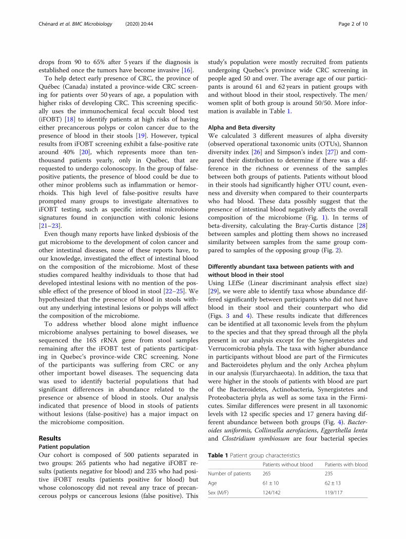
study’s population were mostly recruited from patientsundergoing Quebec’s province wide CRC screening inpeople aged 50 and over. The average age of our partici-pants is around 61 and 62 years in patient groups withand without blood in their stool, respectively. The men/women split of both group is around 50/50. More infor-mation is available in Table 1.
Alpha and Beta diversityWe calculated 3 different measures of alpha diversity(observed operational taxonomic units (OTUs), Shannondiversity index [26] and Simpson’s index [27]) and com-pared their distribution to determine if there was a dif-ference in the richness or evenness of the samplesbetween both groups of patients. Patients without bloodin their stools had significantly higher OTU count, even-ness and diversity when compared to their counterpartswho had blood. These data possibly suggest that thepresence of intestinal blood negatively affects the overallcomposition of the microbiome (Fig. 1). In terms ofbeta-diversity, calculating the Bray-Curtis distance [28]between samples and plotting them shows no increasedsimilarity between samples from the same group com-pared to samples of the opposing group (Fig. 2).
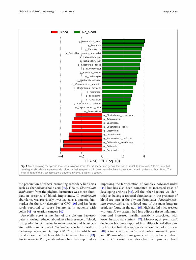
Differently abundant taxa between patients with andwithout blood in their stoolUsing LEfSe (Linear discriminant analysis effect size)[29], we were able to identify taxa whose abundance dif-fered significantly between participants who did not haveblood in their stool and their counterpart who did(Figs. 3 and 4). These results indicate that differencescan be identified at all taxonomic levels from the phylumto the species and that they spread through all the phylapresent in our analysis except for the Synergistetes andVerrucomicrobia phyla. The taxa with higher abundancein participants without blood are part of the Firmicutesand Bacteroidetes phylum and the only Archea phylumin our analysis (Euryarchaeota). In addition, the taxa thatwere higher in the stools of patients with blood are partof the Bacteroidetes, Actinobacteria, Synergistetes andProteobacteria phyla as well as some taxa in the Firmi-cutes. Similar differences were present in all taxonomiclevels with 12 specific species and 17 genera having dif-ferent abundance between both groups (Fig. 4). Bacter-oides uniformis, Collinsella aerofaciens, Eggerthella lentaand Clostridium symbiosum are four bacterial species
Table 1 Patient group characteristics
Patients without blood Patients with blood
Number of patients 265 235
Age 61 ± 10 62 ± 13
Sex (M/F) 124/142 119/117
Chénard et al. BMC Microbiology (2020) 20:44 Page 2 of 10

Fig. 1 Various alpha-diversity measures (Observed OTUs, Shannon diversity index and the Simpson’s index) in all participants separated betweenboth groups. In red, patients who had blood in their stool and in green patients who did not. *p-value< 0.05, **p-value< 0.005, ***p-value< 0.0005
Fig. 2 Visualization of the Bray-Curtis distance between samples in our study with ellipses representing the 95% confidence interval of eachgroup. In red, patients who had blood in their stool and in green, patients who did not
Chénard et al. BMC Microbiology (2020) 20:44 Page 3 of 10
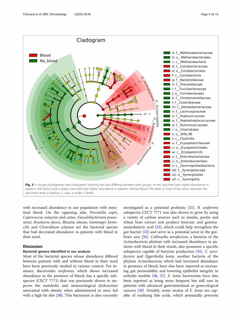
with increased abundance in our population with intes-tinal blood. On the opposing side, Prevotella copri,Coprococcus eutactus and catus, Faecalibacterium praus-nitzii, Roseburia faecis, Blautia obeum, Gemmiger formi-cilis and Clostridium celatum are the bacterial speciesthat had decreased abundance in patients with blood intheir stool.
DiscussionBacterial genera identified in our analysisMost of the bacterial species whose abundance differedbetween patients with and without blood in their stoolhave been previously studied in various context. For in-stance, Bacteroides uniformis, which shows increasedabundance in the presence of blood, has a specific sub-species (CECT 7771) that was previously shown to im-prove the metabolic and immunological dysfunctionassociated with obesity when administered to mice fedwith a high-fat diet [30]. This bacterium is also currently
investigated as a potential probiotic [31]. B. uniformissubspecies CECT 7771 was also shown to grow by usinga variety of carbon sources such as insulin, pectin andwheat bran extract and produce butyrate and gamma-aminobutyric acid [32], which could help strengthen thegut barrier [33] and serve as a potential actor in the gut-brain axis [34]. Collinsella aerofaciens, a bacteria of theActinobacteria phylum with increased abundance in pa-tients with blood in their stools, also possesses a specificsubspecies capable of butyrate production [35]. C. aero-faciens and Eggerthella lenta, another bacteria of thephylum Actinobacteria which had increased abundancein presence of blood, have also been reported as increas-ing gut permeability and lowering epithelial integrity inarthritis models [36, 37]. E. lenta bacteremia have alsobeen reported as being more frequent but still rare inpatients with advanced gastrointestinal or gynecologicalcancers [38]. Notably, some strains of E. lenta are cap-able of oxidizing bile acids, which potentially prevents
Fig. 3 A circular phylogenetic tree (cladogram) showing the taxa differing between both groups. In red, taxa that have higher abundance inpatients with blood and in green, taxa that have higher abundance in patients without blood. The letter in front of the taxon represent thetaxonomy level. p: phylum, c: class, o: order, f: family
Chénard et al. BMC Microbiology (2020) 20:44 Page 4 of 10
the production of cancer-promoting secondary bile acidssuch as chenodeoxycholic acid [39]. Finally, Clostridiumsymbiosum from the phylum Firmicutes was more abun-dant in presence of blood. Importantly, C. symbiosumabundance was previously investigated as a potential bio-marker for the early detection of CRC [40] and has beenrarely reported to cause bacteremia in patients withcolon [41] or ovarian cancers [42].Prevotella copri, a member of the phylum Bacteroi-
detes, showing reduced abundance in presence of blood,is a predominant species in many people and is associ-ated with a reduction of Bacteroides species as well asLachnospiraceae and Group XIV Clostridia, which areusually described as favorable to intestinal health [43].An increase in P. copri abundance has been reported as
improving the fermentation of complex polysaccharides[44] but has also been correlated to increased risks ofdeveloping arthritis [45]. All the other bacteria we iden-tified as having a reduced abundance in the presence ofblood are part of the phylum Firmicutes. Faecalibacter-ium prausnitzii is considered one of the main butyrateproducer found in the gut [46]. High-fat fed mice treatedwith oral F. prausnitzii had less adipose tissue inflamma-tion and increased insulin sensitivity associated withlower hepatic fat content [47]. Moreover, F. prausnitziidepletion has been reported in multiple bowel disorderssuch as Crohn’s disease, colitis as well as colon cancer[48]. Coprococcus eutactus and catus, Roseburia faecisand Blautia obeum are genera with little known aboutthem. C. catus was described to produce both
Fig. 4 Graph showing the specific linear discrimination scores for the species and genera that had an absolute score over 2. In red, taxa thathave higher abundance in patients with blood in their samples and in green, taxa that have higher abundance in patients without blood. Theletter in front of the taxon represent the taxonomy level. g: genus, s: species
Chénard et al. BMC Microbiology (2020) 20:44 Page 5 of 10
propionate and butyrate, two metabolites beneficial forhuman health [49] and C. eutactus abundance was re-ported as reduced in patients with irritable bowel syn-drome [50] and Parkinson’s disease [51]. R. faecis alsoproduces butyrate and is able to produce an inhibitorysubstance against Bacillus subtilis [52]. B. obeum is cap-able of hydrolyzing bile salts [53] and produces a lanti-biotic peptide effective against multiple Clostridiumspecies [54, 55]. Finally, Clostridium celatum andGemmiger formicilis have no known association to non-infectious human diseases nor are they known to pro-duce metabolites of interest.
Potential effect of blood on the microbiome beingattributed to diseases in previous studiesTo show the possible importance of considering thepresence of blood in stools of patients, we compared thetaxa whose abundance vary between our study and fourpublished studies comparing the microbiome of patientssuffering from CRC, Crohn’s disease, multiple sclerosis,ulcerative colitis and rheumatoid arthritis [22–25](Table 2). All the reported studies have precision thatmaximally reached the genus level and some of themhad different OTUs for the same genus with variationsin different directions making it harder to compare pre-cisely. For example, Zackular et al. reported OTUs asso-ciated with the Clostridium genus both increased anddecreased in patients with adenomas while our analysisshowed increased levels of Clotridium and more pre-cisely C. symbiosum but a decreased level of C. celatum,which could reflect their finding but with higher reso-lution. Additionally, except for the study by Gevers et al.[25], all the previously published reports used around100 patients separated between their specific conditions.Finally, both analyses pertaining to CRC had the mostdifferences in the variation of common bacteria with ouranalysis. This possibly indicates that cancerous lesionsare associated with more distinct variations of themicrobiome than blood itself when comparing it toother diseases such as Crohn’s or colitis.
ConclusionIn conclusion, we have shown using a cohort of 500 pa-tients that the presence of blood in stools has a major ef-fect on the composition of the intestinal microbiomewith more than 10 bacterial species having significantvariations in their abundance. Furthermore, we showedthat multiple taxa previously associated with the pres-ence of diseases in other studies could be associated withthe presence of intestinal blood. The results of our studyindicate that the presence of blood should be consideredas an important parameter when studying intestinal dis-eases and their related microbiomes.
MethodsPatient recruitment and sample collectionWe obtained patient stool samples remaining after theiFOBT performed at the CIUSSS de l’Estrie - CHUS.Those samples were collected using the OC-Auto FITcollection kit (Polymedco) and kept at − 80 °C until use.For this experiment, we used samples with negativeiFOBT results and those from subjects with positiveiFOBT results but for whom the subsequent colonos-copy revealed no trace of neither precancerous polypsnor cancerous lesions or other severe bowel diseases. Apositive iFOBT result is currently defined by an amountof blood in the stool over 175 ng/ml (defined by theMinistère de la Santé et des Services Sociaux-MSSS).Since we only had access to the patients’ medical file,and not the patients themselves, we were able to obtainpatients sex and age. However, additional commonlyused personal information such as BMI, ethnicity, bloodglucose levels or smoking status were not available formost of the participants and could not be used in ouranalysis. We excluded, from our study, patients whosuffered from other severe diseases such as bacterial in-fections or any other cancers. This study was approvedby the research ethics committee of the CIUSSS de l’Es-trie - CHUS (#2016–1197) and the consent of all partici-pants was obtained via a reverse consent form.
DNA extraction and 16S rRNA gene sequencingMicrobial genomic DNA was extracted from the stoolsamples using the QIAamp Fast DNA stool mini-kit(QIAGEN) and kept at − 80 °C until use. The V4 regionof the 16S rRNA gene in each sample was amplifiedusing the 515F (5′-GTGCCAGCMGCCGCGGTAA-3′)and 806R (5′-GGACTACHVGGGTWTCTAAT-3′) pri-mer pair initially created by Caporaso et al. [56] andthen sequenced using the Illumina MiSeq Personal Se-quencing platform following the protocol described byKozich et al. [57].
Data analysisTaxonomic profilingUsing QIIME 2 [58] (version 2019.1.0), the raw sequen-cing data was demultiplexed and sequence quality wascontrolled using DADA2 [59] to truncate the low qualityregions of the sequences. Afterward, chimeric sequenceswere removed using the vsearch uchime-denovo func-tion with default parameters [60]. The taxonomic assig-nation was performed using Naive Bayes classifiertrained using the Greengenes taxonomy [61] fromaugust 2013 where the sequenced were trimmed to theV4 region bound by the 515F/806R primer pair andclustered at 99% sequence identity. This taxonomic clas-sifier is available on the QIIME 2 website (https://docs.qiime2.org/2018.11/data-resources/).
Chénard et al. BMC Microbiology (2020) 20:44 Page 6 of 10
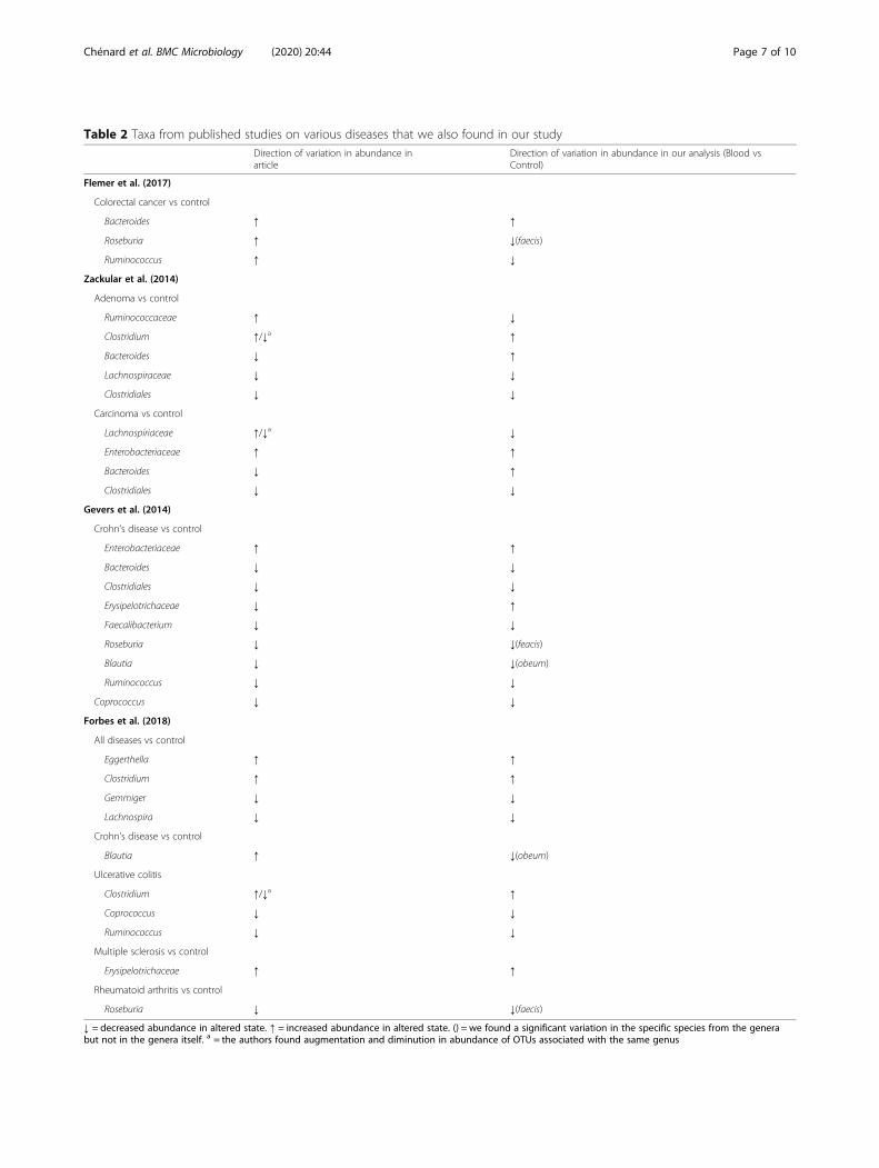
Table 2 Taxa from published studies on various diseases that we also found in our studyDirection of variation in abundance inarticle
Direction of variation in abundance in our analysis (Blood vsControl)
Flemer et al. (2017)
Colorectal cancer vs control
Bacteroides ↑ ↑
Roseburia ↑ ↓(faecis)
Ruminococcus ↑ ↓
Zackular et al. (2014)
Adenoma vs control
Ruminococcaceae ↑ ↓
Clostridium ↑/↓a ↑
Bacteroides ↓ ↑
Lachnospiraceae ↓ ↓
Clostridiales ↓ ↓
Carcinoma vs control
Lachnospiriaceae ↑/↓a ↓
Enterobacteriaceae ↑ ↑
Bacteroides ↓ ↑
Clostridiales ↓ ↓
Gevers et al. (2014)
Crohn’s disease vs control
Enterobacteriaceae ↑ ↑
Bacteroides ↓ ↓
Clostridiales ↓ ↓
Erysipelotrichaceae ↓ ↑
Faecalibacterium ↓ ↓
Roseburia ↓ ↓(feacis)
Blautia ↓ ↓(obeum)
Ruminococcus ↓ ↓
Coprococcus ↓ ↓
Forbes et al. (2018)
All diseases vs control
Eggerthella ↑ ↑
Clostridium ↑ ↑
Gemmiger ↓ ↓
Lachnospira ↓ ↓
Crohn’s disease vs control
Blautia ↑ ↓(obeum)
Ulcerative colitis
Clostridium ↑/↓a ↑
Coprococcus ↓ ↓
Ruminococcus ↓ ↓
Multiple sclerosis vs control
Erysipelotrichaceae ↑ ↑
Rheumatoid arthritis vs control
Roseburia ↓ ↓(faecis)
↓ = decreased abundance in altered state. ↑ = increased abundance in altered state. () = we found a significant variation in the specific species from the generabut not in the genera itself. a = the authors found augmentation and diminution in abundance of OTUs associated with the same genus
Chénard et al. BMC Microbiology (2020) 20:44 Page 7 of 10
Analysis of alpha and beta-diversityWe studied the alpha-diversity within our cohort usingthe vegan [62] and phyloseq [63] package developed forthe R environment for statistical computing [64]. Wecalculated 3 different alpha diversity measures (observedOTUs, Shannon diversity index [26] and the Simpson’sindex [27]) to evaluate microbiome diversity for each pa-tients. Observed OTUs is the number of different OTUsobserved within an individual sample. Shannon diversityindex quantifies the uncertainty in the prediction of theidentity of an element taken at random from a samplealso known as the evenness of a sample. As for theSimpson’s index, it represents the probability of two ran-domly picked elements of a sample being part of thesame OTU. We used Student t-tests to compare alphadiversity measures between patients with and withoutblood in their stools. For beta-diversity, we calculatedthe Bray-Curtis distance [28] between all samples andplotted those distances using a Non-metric Multidimen-sional Scaling [65] (nMDS) ordination. We used theBray-Curtis dissimilarity to quantify the dissimilarity be-tween two samples using not only the presence of anelement but also its abundance within the sample.
Differentially abundant taxa using LEfSeFor these analyses, we removed all OTUs present in lessthan 10% of our study population, merged OTUs at thespecies level and converted the read count to relativeabundances. To identify differentially abundant taxa be-tween patients with positive and negative iFOBT resultswe used the biomarker discovery algorithm LEfSe [29].LEfSe identifies features with are statistically differentbetween conditions by first using a non-parametricKruskal Wallis sum-rank test [66] followed by a pairwiseWilcoxon rank-sum test [67] on the features that weresignificant in the previous step. Finally, a linear discrim-inant analysis [68] (LDA) model is built using featuresthat remain significant after the two previous statisticaltests to estimate the effect size.
AbbreviationsCRC: Colorectal cancer; IFOBT: Immunochemical based fecal occult bloodtest; LDA: Linear discriminant analysis; LEfSe: Linear discriminant analysisEffect Size; NMDS: Non-metric Multidimensional Scaling; OTU: Operationaltaxonomic units
AcknowledgementsWe thank Jude Beaudouin, Karine Prévost and Mélina Arguin for excellenttechnical assistance.
Authors’ contributionsEM and JD designed the experiment. MM participated in the analysis. TCperformed the bioinformatic and statistical analysis and wrote themanuscript. All authors read and approved the final manuscript.
FundingWork in the Massé Lab has been supported by grants from the CanadianInstitutes of Health Research (CIHR) MOP69005, MERCK and the Centre deRecherche du Centre Hospitalier Universitaire de Sherbrooke (CRCHUS).
Availability of data and materialsThe dataset generated and analysed in the current study is publicly availablein NCBI’s Sequence Read Archive (SRA) repository under the BioProject IDPRJNA577051 (http://www.ncbi.nlm.nih.gov/bioproject/577051).
Ethics approval and consent to participateThis study was approved by the research ethics committee of the CIUSSS del’Estrie - CHUS (#2016–1197) and the consent of all participants was obtainedvia a reverse consent form. Each potential participant was sent a letterdetailing the experiments and asked to return the form if they wished toopt-out of the microbiome studies. Absence of response was considered asconsent to participate and this consent can be revoked at any time duringthe study.
Consent for publicationNot applicable.
Competing interestsThe authors declare that they have no competing interests.
Received: 28 October 2019 Accepted: 10 February 2020
References1. Irrazábal T, Belcheva A, Girardin SE, Martin A, Philpott DJ. The multifaceted
role of the intestinal microbiota in colon cancer. Mol Cell. 2014;54:309–20.2. El Mouzan MI, Winter HS, Assiri AA, Korolev KS, Al Sarkhy AA, Dowd SE, et al.
Microbiota profile in new-onset pediatric Crohn’s disease: data from a non-Western population. Gut Pathog. 2018;10:49.
3. Lv LX, Fang DQ, Shi D, Chen DY, Yan R, Zhu YX, et al. Alterations andcorrelations of the gut microbiome, metabolism and immunity in patientswith primary biliary cirrhosis. Environ Microbiol. 2016;18:2272–86.
4. Jayasudha R, Kalyana Chakravarthy S, Sai Prashanthi G, Sharma S, Garg P,Murthy SI, et al. Alterations in gut bacterial and fungal microbiomes areassociated with bacterial keratitis, an inflammatory disease of the humaneye. J Biosci. 2018;43:835–56.
5. Gorkiewicz G, Moschen A. Gut microbiome: a new player in gastrointestinaldisease. Virchows Arch. 2018;472:159–72.
6. Rajagopala SV, Vashee S, Oldfield LM, Suzuki Y, Venter JC, Telenti A, et al.The human microbiome and cancer. Cancer Prev Res. 2017;10:226–34.
7. Marchesi JR, Dutilh BE, Hall N, Peters WHM, Roelofs R, Boleij A, et al.Towards the human colorectal cancer microbiome. PLoS One. 2011;6:e20447.
8. Jiang H, Ling Z, Zhang Y, Mao H, Ma Z, Yin Y, et al. Altered fecal microbiotacomposition in patients with major depressive disorder. Brain Behav Immun.2015;48:186–94.
9. Sabatino A, Regolisti G, Cosola C, Gesualdo L, Fiaccadori E. Intestinalmicrobiota in type 2 diabetes and chronic kidney disease. Current DiabetesReports. 2017;17:16.
10. Garrett WS. The gut microbiota and colon cancer. Science. 2019;364:1133–5.11. Bullman S, Pedamallu CS, Sicinska E, Clancy TE, Zhang X, Cai D, et al.
Analysis of Fusobacterium persistence and antibiotic response in colorectalcancer. Science. 2017;358:1443–8.
12. Brennan CA, Garrett WS. Fusobacterium nucleatum — symbiont, opportunistand oncobacterium. Nat Rev Microbiol. 2019;17:156–66.
13. Dejea CM, Fathi P, Craig JM, Boleij A, Taddese R, Geis AL, et al. Patients withfamilial adenomatous polyposis harbor colonic biofilms containingtumorigenic bacteria. Science. 2018;359:592–7.
14. Arthur JC, Perez-Chanona E, Mühlbauer M, Tomkovich S, Uronis JM, Fan TJ,et al. Intestinal inflammation targets cancer-inducing activity of themicrobiota. Science. 2012;338:120–3.
15. Thakur BK, Malaisé Y, Martin A. Unveiling the mutational mechanism of thebacterial Genotoxin Colibactin in colorectal Cancer. Mol Cell. 2019;74:227–9.
16. Miller KD, Siegel RL, Lin CC, Mariotto AB, Kramer JL, Rowland JH, et al.Cancer treatment and survivorship statistics, 2016. CA Cancer J Clin. 2016;66:271–89.
17. Fearon ER. Molecular genetics of colorectal Cancer. Annu Rev Pathol MechDis. 2011;6:479–507.
18. Saito H. An immunological fecal occult blood test for mass screening ofcolorectal cancer by reversed passive hemagglutination (RPHA). Japanese JGastroenterol. 1984;81:2831.
Chénard et al. BMC Microbiology (2020) 20:44 Page 8 of 10
19. Gouvernement du Québec. Immunochemical Fecal Occult Blood Test(iFOBT) | Gouvernement du Québec. https://www.quebec.ca/en/health/advice-and-prevention/screening-and-carrier-testing-offer/colorectal-cancer-screening/immunochemical-fecal-occult-blood-test-ifobt/. .
20. Fraser CG, Mathew CM, Mowat NAG, Wilson JA, Carey FA, Steele RJC.Evaluation of a card collection-based faecal immunochemical test inscreening for colorectal cancer using a two-tier reflex approach. Gut. 2007;56:1415–8.
21. Sun J, Kato I. Gut microbiota, inflammation and colorectal cancer. Genesand Diseases. 2016;3:130–43.
22. Flemer B, Lynch DB, Brown JMR, Jeffery IB, Ryan FJ, Claesson MJ, et al.Tumour-associated and non-tumour-associated microbiota in colorectalcancer. Gut. 2017;66:633–43.
23. Zackular JP, Rogers MAM, Ruffin MT, Schloss PD. The human gutmicrobiome as a screening tool for colorectal Cancer. Cancer Prev Res.2014;7:1112–21.
24. Forbes JD, Chen CY, Knox NC, Marrie RA, El-Gabalawy H, De Kievit T, et al. Acomparative study of the gut microbiota in immune-mediatedinflammatory diseases - does a common dysbiosis exist? Microbiome. 2018;6:221.
25. Gevers D, Kugathasan S, Denson LA, Vázquez-Baeza Y, Van Treuren W, RenB, et al. The treatment-naive microbiome in new-onset Crohn’s disease. CellHost Microbe. 2014;15:382–92.
26. Shannon CE. A mathematical theory of communication. Bell Syst Tech J.1948;27:379–423.
27. Simpson EH. Measurement of diversity. Nature. 1949;163:688.28. Bray JR, Curtis JT. An ordination of the upland Forest communities of
southern Wisconsin. Source Ecol Monogr. 1957;27:325–49.29. Segata N, Izard J, Waldron L, Gevers D, Miropolsky L, Garrett WS, et al.
Metagenomic biomarker discovery and explanation. Genome Biol. 2011;12:R60.
30. Gauffin Cano P, Santacruz A, Moya Á, Sanz Y. Bacteroides uniformis CECT7771 ameliorates metabolic and immunological dysfunction in mice withhigh-fat-diet induced obesity. PLoS One. 2012;7:e41079.
31. Fernández-Murga ML, Sanz Y. Safety assessment of Bacteroides uniformisCECT 7771 isolated from stools of healthy breast-fed infants. PLoS One.2016;11:e0145503.
32. Benítez-Páez A, Gómez del Pulgar EM, Sanz Y. The Glycolytic Versatility ofBacteroides uniformis CECT 7771 and Its Genome Response to Oligo andPolysaccharides. Front Cell Infect Microbiol. 2017;7:383.
33. Baxter NT, Schmidt AW, Venkataraman A, Kim KS, Waldron C, Schmidt TM.Dynamics of human gut microbiota and short-chain fatty acids in responseto dietary interventions with three fermentable fibers. MBio. 2019;10:e02566–18.
34. Mittal R, Debs LH, Patel AP, Nguyen D, Patel K, O’Connor G, et al.Neurotransmitters: the critical modulators regulating gut-brain Axis. J CellPhysiol. 2017;232:2359–72.
35. Qin P, Zou Y, Dai Y, Luo G, Zhang X, Xiao L. Characterization a novel butyricacid-producing bacterium Collinsella aerofaciens Subsp. Shenzhenensis SubspNov. Microorganisms. 2019;7:E78.
36. Kalinkovich A, Livshits G. A cross talk between dysbiosis and gut-associatedimmune system governs the development of inflammatory arthropathies.Semin Arthritis Rheum. 2019;49:474–84.
37. Balakrishnan B, Luckey D, Taneja V. Autoimmunity-associated gutcommensals modulate gut permeability and immunity in humanized mice.Mil Med. 2019;184:529–36.
38. Woerther PL, Antoun S, Chachaty E, Merad M. Eggerthella lenta bacteremiain solid tumor cancer patients: pathogen or witness of frailty? Anaerobe.2017;47:70–2.
39. Harris SC, Devendran S, Méndez-García C, Mythen SM, Wright CL, Fields CJ,et al. Bile acid oxidation by Eggerthella lenta strains C592 and DSM 2243 T.Gut Microbes. 2018;9:523–39.
40. Xie YH, Gao QY, Cai GX, Sun XM, Zou TH, Chen HM, et al. FecalClostridium symbiosum for noninvasive detection of early and advancedcolorectal Cancer: test and validation studies. EBioMedicine.2017;25:32–40.
41. Elsayed S, Zhang K. Bacteraemia caused by Clostridium symbiosum. J ClinMicrobiol. 2004;42:4390–2.
42. Toprak N, Özcan E, Pekin T, Yumuk P, Soyletir G. Bacteraemia caused byClostridium symbiosum: case report and review of the literature. Indian JMed Microbiol. 2014;32:92–4.
43. Franke T, Deppenmeier U. Physiology and central carbon metabolism of thegut bacterium Prevotella copri. Mol Microbiol. 2018;109:528–40.
44. Kovatcheva-Datchary P, Nilsson A, Akrami R, Lee YS, De Vadder F, AroraT, et al. Dietary fiber-induced improvement in glucose metabolism isassociated with increased abundance of Prevotella. Cell Metab.2015;22:971–82.
45. Scher JU, Sczesnak A, Longman RS, Segata N, Ubeda C, Bielski C, et al.Expansion of intestinal Prevotella copri correlates with enhancedsusceptibility to arthritis. Elife. 2013;2:e01202.
46. Duncan SH, Hold GL, Harmsen HJM, Stewart CS, Flint HJ. Growthrequirements and fermentation products of Fusobacterium prausnitzii, and aproposal to reclassify it as Faecalibacterium prausnitzii gen. Nov., comb. nov.Int J Syst Evol Microbiol. 2002;52:2141–6.
47. Munukka E, Rintala A, Toivonen R, Nylund M, Yang B, Takanen A, et al.Faecalibacterium prausnitzii treatment improves hepatic health and reducesadipose tissue inflammation in high-fat fed mice. ISME J. 2017;11:1667–79.
48. Lopez-Siles M, Duncan SH, Garcia-Gil LJ, Martinez-Medina M.Faecalibacterium prausnitzii: from microbiology to diagnostics andprognostics. ISME J. 2017;11:841–52.
49. Reichardt N, Duncan SH, Young P, Belenguer A, McWilliam Leitch C, ScottKP, et al. Phylogenetic distribution of three pathways for propionateproduction within the human gut microbiota. ISME J. 2014;8:1323–35.
50. Malinen E, Krogius-Kurikka L, Lyra A, Nikkilä J, Jääskeläinen A, Rinttilä T, et al.Association of symptoms with gastrointestinal microbiota in irritable bowelsyndrome. World J Gastroenterol. 2010;16:4532–40.
51. Petrov VA, Saltykova IV, Zhukova IA, Alifirova VM, Zhukova NG, DorofeevaYB, et al. Analysis of gut microbiota in patients with Parkinson’s disease. BullExp Biol Med. 2017;162:734–7.
52. Hatziioanou D, Mayer MJ, Duncan SH, Flint HJ, Narbad A. A representativeof the dominant human colonic Firmicutes, Roseburia faecis M72/1, forms anovel bacteriocin-like substance. Anaerobe. 2013;23:5–8.
53. Mullish BH, McDonald JAK, Pechlivanis A, Allegretti JR, Kao D, Barker GF,et al. Microbial bile salt hydrolases mediate the efficacy of faecal microbiotatransplant in the treatment of recurrent Clostridioides difficile infection. Gut.2019;68:1791–800.
54. Hatziioanou D, Gherghisan-Filip C, Saalbach G, Horn N, Wegmann U,Duncan SH, et al. Discovery of a novel lantibiotic nisin O from Blautiaobeum A2–162, isolated from the human gastrointestinal tract. Microbiol(United Kingdom). 2017;163:1292–305.
55. Gherghisan-Filip C, Saalbach G, Hatziioanou D, Narbad A, Mayer MJ.Processing and structure of the lantibiotic peptide Nso from the human gutbacterium Blautia obeum A2-162 analysed by mass spectrometry. Sci Rep.2018;8:10077.
56. Caporaso JG, Lauber CL, Walters WA, Berg-Lyons D, Huntley J, Fierer N, et al.Ultra-high-throughput microbial community analysis on the Illumina HiSeqand MiSeq platforms. ISME J. 2012;6:1621–4.
57. Kozich JJ, Westcott SL, Baxter NT, Highlander SK, Schloss PD. Developmentof a dual-index sequencing strategy and curation pipeline for analyzingamplicon sequence data on the MiSeq Illumina sequencing platform. ApplEnviron Microbiol. 2013;79:5112–20.
58. Bolyen E, Rideout JR, Dillon MR, Bokulich NA, Abnet C, Al-Ghalith GA, et al.Reproducible, interactive, scalable and extensible microbiome data scienceusing QIIME 2. Nat Biotech. 2019;37:852–7.
59. Callahan BJ, McMurdie PJ, Rosen MJ, Han AW, Johnson AJA, Holmes SP.DADA2: high-resolution sample inference from Illumina amplicon data. NatMethods. 2016;13:581–3.
60. Rognes T, Flouri T, Nichols B, Quince C, Mahé F. VSEARCH: a versatile opensource tool for metagenomics. PeerJ. 2016;4:e2584.
61. McDonald D, Price MN, Goodrich J, Nawrocki EP, Desantis TZ, Probst A, et al.An improved Greengenes taxonomy with explicit ranks for ecological andevolutionary analyses of bacteria and archaea. ISME J. 2012;6:610–8.
62. Oksanen J, Blanchet G, Friendly M, Kindt R, Legendre P, McGlinn D, et al.vegan: Community Ecology Package. 2018. https://cran.r-project.org/package=vegan.
63. McMurdie PJ, Holmes S. Phyloseq: an R package for reproducible interactiveanalysis and graphics of microbiome census data. PLoS One. 2013;8:e61217.
64. The R Core Team. R: A language and environment for statistical computing.R foundation for Statistical Computing, Vienne, Austria. 2018. https://www.R-project.org/.
65. Kruskal JB. Nonmetric multidimensional scaling: a numerical method.Psychometrika. 1964;29:115–29.
Chénard et al. BMC Microbiology (2020) 20:44 Page 9 of 10

66. Kruskal WH, Wallis WA. Use of ranks in one-criterion variance analysis. J AmStat Assoc. 1952;47:583–621.
67. Wilcoxon F. Individual comparisons by ranking methods. Biom Bull.1945;1:80.
68. Xanthopoulos P, Pardalos PM, Trafalis TB. Linear Discriminant Analysis. In: InRobust Data Mining. New York: Springer; 2013. p. 27–33.
Publisher’s NoteSpringer Nature remains neutral with regard to jurisdictional claims inpublished maps and institutional affiliations.
Chénard et al. BMC Microbiology (2020) 20:44 Page 10 of 10
Copyright © 2022 FDOKUMEN