A Journey on the Skin Microbiome: Pitfalls and Opportunities
25
International Journal of Molecular Sciences Review A Journey on the Skin Microbiome: Pitfalls and Opportunities Dario Pistone 1,2, * , Gabriele Meroni 3 , Simona Panelli 1 , Enza D’Auria 4 , Miriam Acunzo 4 , Ajay Ratan Pasala 1 , Gian Vincenzo Zuccotti 1,4 , Claudio Bandi 5 and Lorenzo Drago 2 Citation: Pistone, D.; Meroni, G.; Panelli, S.; D’Auria, E.; Acunzo, M.; Pasala, A.R.; Zuccotti, G.V.; Bandi, C.; Drago, L. A Journey on the Skin Microbiome: Pitfalls and Opportunities. Int. J. Mol. Sci. 2021, 22, 9846. https://doi.org/10.3390/ ijms22189846 Academic Editor: Amedeo Amedei Received: 9 August 2021 Accepted: 8 September 2021 Published: 12 September 2021 Publisher’s Note: MDPI stays neutral with regard to jurisdictional claims in published maps and institutional affil- iations. Copyright: © 2021 by the authors. Licensee MDPI, Basel, Switzerland. This article is an open access article distributed under the terms and conditions of the Creative Commons Attribution (CC BY) license (https:// creativecommons.org/licenses/by/ 4.0/). 1 Pediatric Clinical Research Center “Invernizzi”, Department of Biomedical and Clinical Sciences “L. Sacco”, University of Milan, 20157 Milan, Italy; [email protected] (S.P.); [email protected] (A.R.P.); [email protected] (G.V.Z.) 2 Department of Biomedical Sciences for Health, University of Milan, 20133 Milan, Italy; [email protected] 3 Department of Biomedical Surgical and Dental Sciences-One Health Unit, University of Milan, 20133 Milan, Italy; [email protected] 4 Department of Pediatrics, Children’s Hospital Vittore Buzzi, University of Milan, 20154 Milan, Italy; [email protected] (E.D.); [email protected] (M.A.) 5 Pediatric Clinical Research Center “Invernizzi”, Department of Biosciences, University of Milan, 20133 Milan, Italy; [email protected] * Correspondence: [email protected] Abstract: The human skin microbiota is essential for maintaining homeostasis and ensuring barrier functions. Over the years, the characterization of its composition and taxonomic diversity has reached outstanding goals, with more than 10 million bacterial genes collected and cataloged. Nevertheless, the study of the skin microbiota presents specific challenges that need to be addressed in study design. Benchmarking procedures and reproducible and robust analysis workflows for increasing comparability among studies are required. For various reasons and because of specific technical problems, these issues have been investigated in gut microbiota studies, but they have been largely overlooked for skin microbiota. After a short description of the skin microbiota, the review tackles methodological aspects and their pitfalls, covering NGS approaches and high throughput culture- based techniques. Recent insights into the “core” and “transient” types of skin microbiota and how the manipulation of these communities can prevent or combat skin diseases are also covered. Finally, this review includes an overview of the main dermatological diseases, the changes in the microbiota composition associated with them, and the recommended skin sampling procedures. The last section focuses on topical and oral probiotics to improve and maintain skin health, considering their possible applications for skin diseases. Keywords: skin microbiota; skin sampling techniques; NGS; culturomics 1. Introduction Our body is home to a complex community of microorganisms that help us maintain homeostasis and prevent colonization from pathogens [1,2]. This residential community is known as the microbiota (often incorrectly used as a synonym for microbiome), referring to all the microorganisms, including archaea, bacteria, eukaryotes (fungi and yeasts, protists), viruses, and bacteriophages that colonize and inhabit a specific niche of our body. Instead, the microbiome describes the entire set of genomes and microbial genes found in a specific microbiota [1–3]. Site-specific microbial communities colonize different anatomical niches of the human body (e.g., the skin, gut, oral cavity, nasal cavities, and urogenital tract). In 2007, the U.S. National Institutes of Health (NIH) started the “Human Microbiome Project” (HMP), a two-phase project to (a) produce the reference genome sequences for at least 900 bacteria, (b) catalog microbial genome sequences, and (c) help researchers in metagenomic data management [4]. The initial stage of the project, called the Human Microbiome Project 1 (HMP1), which was established in 2008 and completed in 2013, aimed Int. J. Mol. Sci. 2021, 22, 9846. https://doi.org/10.3390/ijms22189846 https://www.mdpi.com/journal/ijms
-
Upload
khangminh22 -
Category
Documents
-
view
1 -
download
0
Transcript of A Journey on the Skin Microbiome: Pitfalls and Opportunities
International Journal of
Molecular Sciences
Review
A Journey on the Skin Microbiome: Pitfalls and Opportunities
Dario Pistone 1,2,* , Gabriele Meroni 3 , Simona Panelli 1 , Enza D’Auria 4 , Miriam Acunzo 4,Ajay Ratan Pasala 1, Gian Vincenzo Zuccotti 1,4 , Claudio Bandi 5 and Lorenzo Drago 2
�����������������
Citation: Pistone, D.; Meroni, G.;
Panelli, S.; D’Auria, E.; Acunzo, M.;
Pasala, A.R.; Zuccotti, G.V.; Bandi, C.;
Drago, L. A Journey on the Skin
Microbiome: Pitfalls and
Opportunities. Int. J. Mol. Sci. 2021,
22, 9846. https://doi.org/10.3390/
ijms22189846
Academic Editor: Amedeo Amedei
Received: 9 August 2021
Accepted: 8 September 2021
Published: 12 September 2021
Publisher’s Note: MDPI stays neutral
with regard to jurisdictional claims in
published maps and institutional affil-
iations.
Copyright: © 2021 by the authors.
Licensee MDPI, Basel, Switzerland.
This article is an open access article
distributed under the terms and
conditions of the Creative Commons
Attribution (CC BY) license (https://
creativecommons.org/licenses/by/
4.0/).
1 Pediatric Clinical Research Center “Invernizzi”, Department of Biomedical and Clinical Sciences “L. Sacco”,University of Milan, 20157 Milan, Italy; [email protected] (S.P.); [email protected] (A.R.P.);[email protected] (G.V.Z.)
2 Department of Biomedical Sciences for Health, University of Milan, 20133 Milan, Italy;[email protected]
3 Department of Biomedical Surgical and Dental Sciences-One Health Unit, University of Milan, 20133 Milan,Italy; [email protected]
4 Department of Pediatrics, Children’s Hospital Vittore Buzzi, University of Milan, 20154 Milan, Italy;[email protected] (E.D.); [email protected] (M.A.)
5 Pediatric Clinical Research Center “Invernizzi”, Department of Biosciences, University of Milan, 20133 Milan,Italy; [email protected]
* Correspondence: [email protected]
Abstract: The human skin microbiota is essential for maintaining homeostasis and ensuring barrierfunctions. Over the years, the characterization of its composition and taxonomic diversity has reachedoutstanding goals, with more than 10 million bacterial genes collected and cataloged. Nevertheless,the study of the skin microbiota presents specific challenges that need to be addressed in studydesign. Benchmarking procedures and reproducible and robust analysis workflows for increasingcomparability among studies are required. For various reasons and because of specific technicalproblems, these issues have been investigated in gut microbiota studies, but they have been largelyoverlooked for skin microbiota. After a short description of the skin microbiota, the review tacklesmethodological aspects and their pitfalls, covering NGS approaches and high throughput culture-based techniques. Recent insights into the “core” and “transient” types of skin microbiota and howthe manipulation of these communities can prevent or combat skin diseases are also covered. Finally,this review includes an overview of the main dermatological diseases, the changes in the microbiotacomposition associated with them, and the recommended skin sampling procedures. The last sectionfocuses on topical and oral probiotics to improve and maintain skin health, considering their possibleapplications for skin diseases.
Keywords: skin microbiota; skin sampling techniques; NGS; culturomics
1. Introduction
Our body is home to a complex community of microorganisms that help us maintainhomeostasis and prevent colonization from pathogens [1,2]. This residential community isknown as the microbiota (often incorrectly used as a synonym for microbiome), referring toall the microorganisms, including archaea, bacteria, eukaryotes (fungi and yeasts, protists),viruses, and bacteriophages that colonize and inhabit a specific niche of our body. Instead,the microbiome describes the entire set of genomes and microbial genes found in a specificmicrobiota [1–3]. Site-specific microbial communities colonize different anatomical nichesof the human body (e.g., the skin, gut, oral cavity, nasal cavities, and urogenital tract).In 2007, the U.S. National Institutes of Health (NIH) started the “Human MicrobiomeProject” (HMP), a two-phase project to (a) produce the reference genome sequences forat least 900 bacteria, (b) catalog microbial genome sequences, and (c) help researchers inmetagenomic data management [4]. The initial stage of the project, called the HumanMicrobiome Project 1 (HMP1), which was established in 2008 and completed in 2013, aimed
Int. J. Mol. Sci. 2021, 22, 9846. https://doi.org/10.3390/ijms22189846 https://www.mdpi.com/journal/ijms
Int. J. Mol. Sci. 2021, 22, 9846 2 of 25
to characterize the microbial communities from 300 healthy patients across five differentanatomical sites (i.e., the gastrointestinal tract, urogenital tract, skin, nasal cavities, andoral cavity) [4]. The second stage, called the Integrative Human Microbiome Project (iHMPor HMP2), was designed to characterize in more detail the host-microbiome interactions,focusing on three conditions: pregnancy and pre-term birth, the onset of inflammatorybowel diseases (IBD), and the onset of type II diabetes [5]. Specifically, concerning the skinmicrobiome, the characterization of its diversity has reached outstanding goals over theyears. For example, consortia such as the integrated Human Skin Microbial Gene Catalog(iHSMGC) collected and cataloged more than 10 million genes [6], an impressive task tohave accomplished, and included a high number of individuals in the study, applying NextGeneration Sequencing (NGS) technology and huge computing power. Nevertheless, thestudy of the skin microbiota presents specific challenges to consider when designing a study.The aim of this review was to discuss such challenges and recent insights into the “core” and“transient” kinds of skin microbiota and how the manipulation of these communities canprevent or combat skin diseases. After a short description of important and less charismaticmembers of the skin microbiota, we tackle methodological issues related to the availablesequencing approaches and high throughput culture-based techniques, such as culturomics.We then discuss the necessity of benchmarking procedures and establishing reproducibleand robust analysis workflows to increase comparability. This issue has also been addressedin the context of gut microbiota studies [7–9], but it has been largely overlooked for skinmicrobiota for various reasons and because of specific technical problems that are discussed.This review includes an overview of the main dermatological diseases, the changes in themicrobiota composition that have been associated with them so far, and the recommendedsampling procedures. Finally, Section 6 is dedicated to state-of-the-art topical and oralprobiotics for improving and maintaining skin health, and possible future applications forskin diseases are presented.
2. References Analyzed and Methodologies of Study
Over the last decade, we have witnessed a growing interest in studying the micro-bial community of the skin [10,11]. Obtaining better knowledge of the composition anddiversity of the microbes inhabiting different sites and layers of the human skin remainsfundamental to gaining insight into the relationship between microbiota dysbiosis andthe development of pathological conditions. In this review, we selected published articleson skin microbiota, focusing particularly on the last decade (2010–2021), for three mainpurposes: (1) to investigate the link between microbiota alterations and the developmentof skin diseases; (2) to delineate guidelines for more standardized sampling methods anddiagnostic procedures; (3) to discuss the development and optimization of remediationstrategies based on topical and oral probiotics.
It is worth noting that the study of skin microbiota can present more challengescompared to other human microbial communities, mainly for the following reasons: (1) ithas an uneven distribution, and some areas are scarcely colonized; (2) the microbes residingin the deeper layers of the skin are difficult to sample; (3) the level of human DNAcontamination can be relatively high, especially where invasive sampling procedures areapplied; (4) external environmental conditions and cleaning practices can deeply affectthe skin microbiota; (5) the distinction among stable and transient microorganisms is notalways easy to establish, especially for low-abundance taxa.
We followed the history of the study of the skin microbiota through its main milestones,and it has often been coupled with the development of sequencing technology [12–16].Indeed, the research that has applied culture-independent approaches and NGS technologyhas produced the main advancements in our knowledge of the composition and function ofthis microbial community. Therefore, we mainly focused on such recent literature [17–22].
Int. J. Mol. Sci. 2021, 22, 9846 3 of 25
3. The Skin Microbiota
As the most exposed organ of our body, with an estimated surface area of about1.8 m2 (even larger when considering follicular structures and other appendages), the skinis inhabited by more than one million bacteria/cm2 co-participating in maintaining thephysical barrier function to the external environment and preventing the penetration andinvasion of pathogens [2,3,23,24]. Besides its protective function, the skin plays an essentialrole in thermoregulation processes and vitamin D synthesis [25]. However, rather than abarricade that separates us from the bacterial community living on us, the skin should beregarded as a wide and dynamic interface on which the microbiota cooperates with theimmune system in a modulatory activity that is crucial for our health [26–28].
The human skin can roughly be grouped into three main physiological types: (1) oily/sebaceous areas (i.e., forehead, upper back, and nose); (2) dry areas (i.e., forearm and lowerback); (3) moist areas (i.e., armpits, backs of knees, nostrils, and groin). Specific bacterialtaxa inhabit these different cutaneous micro-environments: Cutibacterium and Propionibac-terium have a clear preference for sebaceous niches, whereas the genera Staphylococcus andCorynebacterium tend to colonize moist zones, and Proteobacteria and Flavobacteriales thrivein dry sites [12]. The same area can also be deeply affected by lipid and water levels; forexample, sebum concentration of the cheek, but not the forehead, is significantly correlatedwith microbial composition and diversity and, on the contrary, the hydration level of theforehead was found to be a good predictor of nature and diversity of this site-specificmicrobiota [29].
Many of the skin commensals generally found on healthy human skin belong to fourphyla: Actinobacteria (Corynebacterium, Propionibacterium, Cutibacterium, Micrococcus, Acti-nomyces, Brevibacterium), Firmicutes (Staphylococcus, Streptococcus, Finegoldia), Proteobacteria(Paracoccus, Haematobacter), and Bacteroidetes (Prevotella, Porphyromonas, Chryseobacterium).
Coagulase-negative staphylococci (CoNS) such as Staphylococcus epidermidis andStaphylococcus hominis are among the most common Gram-positive species inhabitingthe human skin [30]. These bacteria, previously considered innocuous, have an activerole in contrasting the colonization of Staphylococcus aureus and other pathogens but arenow regarded as important opportunistic pathogens as well [31,32]. Indeed, S. epidermidisand other CoNS can frequently cause nosocomial and neonatal infections and colonizeprosthetics and other medical devices [33,34]. Moreover, CoNS can cause debilitatingand difficult-to-eradicate infections that are causing medical concern due to their biofilmformation propensity and their role as reservoirs of antibiotic-resistant genes [35]. Anotherubiquitous inhabitant of the human skin is Cutibacterium acnes (formerly Propionibacteriumacnes), a Gram-positive facultative anaerobe with a lipophilic attitude that tends to colonizethe pilosebaceous unit [36]. Different strains of C. acnes have been previously implicated ina plethora of diseases, including acne (see below). Its role as an opportunistic pathogen,especially in post-surgery wound infections, is becoming more prominent as well [37–39].
Archaea enclose up to 4% of the entire microbial diversity of the skin [40,41]. TheThaumarchaeota phylum is one of the most abundant, and its members could be implicatedin ammonia oxidation processes [41]. In the complex human skin ecosystem, even thepresence of mites, especially Demodex spp. as ubiquitous ectoparasites of the pilosebaceousunit, is common, and their possible pathogenic role is highly debated [42]. Indeed, evenif the causal relationship between the abundance of Demodex spp. and a specific skin-associated disease has not been clarified yet, the high density of these parasites has beententatively associated with some inflammatory conditions such as rosacea, blepharitis,perioral, and seborrheic dermatitis or chalazion [43–45]. Healthy human skin also harborsresident and transient viruses such as cutaneous beta and gamma human papillomaviruses,commonly found in many individuals [46,47]. Although low in biomass and probably oneof the most understudied communities on the skin, bacteriophages can deeply influencethe microbiota diversity and its physiological activity [48].
The amount of skin bacteria on humans is relatively high, and they are constantlyshed in the surrounding environment together with dead cells of the stratum corneum of
Int. J. Mol. Sci. 2021, 22, 9846 4 of 25
the skin, so the fact that they are often the major microbial component in the air, soil, andother surfaces in a crowded urban area is not surprising [49,50].
The constant crosstalk between the skin immune system and the cutaneous microbiotaacts as a powerful pathogen control system [1,3]. However, under some circumstances, forexample, when the defensive skin barrier is compromised or a disequilibrium betweencommensal bacteria and pathogens occurs, skin diseases can arise [2,3].
Aging is another key factor causing critical changes in the skin microbiota. Some au-thors have reported increased species diversity in the skin microbial community of elderlypeople [51–53], although conflicting results have also been published [54]. Interestingly, ithas been shown that data from skin microbiota can be used to predict age more accuratelythan gut or oral microbiota [55].
Skin aging is a process that induces alterations in skin structure and physiology, witha decrease in hydration levels, the appearance of hyperpigmented spots and wrinkles, andmodified sebaceous gland activity [56]. In particular, the reduction in sebum productionmay reduce nutrients for commensal bacteria and favors the colonization of opportunisticspecies. Several studies reported a reduction in the dominant genus Cutibacterium and aparallel increase in the relative abundance of Corynebacterium and some Proteobacteria ondifferent skin sites in older groups [51,52]. The fungal diversity of the genus Malassezia isalso known to experience age-related changes, with older individuals showing Malasseziasympodialis as the predominant species [57]. The prevalence of Demodex increases with ageas well. In this respect, it has been reported that Corynebacterium kroppenstedtii-like OTUstend to replace other Corynebacterium OTUs in older adults [58]. The relationship betweenthese skin-inhabiting mites and their main symbionts might deserve further attention (seebelow).
The use of cosmetics can also boost bacterial diversity on the skin, with an increase ingenera such as Ralstonia spp., which have been tentatively linked to the ability to metabolizexenobiotics [59,60].
Historically, microbiology has always explored microbial skin communities usingculture-dependent approaches and studying individual species as isolated units [3,61].The fundamental limit in this targeted approach is the selection and isolation of only theculturable fraction of the microbiota (focusing on bacteria), which represents a small partof the entire skin microbial diversity [62]. Indeed, the vast majority of skin commensalbacteria remains unculturable or difficult to cultivate (see below), a sign of a highly di-versified bacterial community with specific growing requirements [1,15,24,63,64]. Thegreat opportunities offered by culture-independent methods were already clear severaldecades ago, at the beginning of the PCR era, but technology started a real revolution inthe field with the advent of NGS platforms only in the most recent years. From the firststudy by Woese (1977), in which they used the 16S rRNA gene to investigate the phylogenyof prokaryotes [65], the advancements of molecular biology-based techniques from theSanger method up to the latest high throughput NGS technologies have rapidly providednew possibilities to increase the level of microbiome characterization. These studies com-monly use two main NGS approaches—amplicon sequencing and whole-genome shotgunsequencing [2] (see Section 4). Despite the detailed picture of the microbial communitycomposition obtained with these methods, elucidating the role of the skin microbiotain different diseases will also require the application of other techniques (derived frommetatranscriptomics, metaproteomics, and metabolomics), to investigate gene expressionprofiles and metabolic byproducts [22].
3.1. Resident and Transient Skin Microbiota
The human skin microbiota is characterized by a rather high degree of temporalvariability, especially when compared to more stable microbial communities, such as thoselocalized in the gut or the mouth [66]. High-resolution time-series studies, following thecomposition of the individual skin microbiota over time, have determined the absence of ahighly abundant core microbiota, although some taxa can be rather persistent [67,68]. All
Int. J. Mol. Sci. 2021, 22, 9846 5 of 25
areas protected or less exposed to the external environment, such as the external auditorycanal (inside the ear), the nare (inside the nostril), and the inguinal crease, were shownto be more consistent over time [12]. As a general rule, sites harboring a greater diversityof microorganisms tend to be less stable over time in terms of community members andbacterial composition; these sites include the volar forearm (forearm), the popliteal fossa,the antecubital fossa, the plantar heel (the bottom of the heel of the foot) and the interdigitalweb space (between the fingers) [12].
Skin microorganisms can be classified into resident and transient, even if defining aclear edge is difficult. For facilitating classification into these two categories, one criterioncould be that transient microbes can be permanently removed by applying detergent anddisinfectant agents, or soap and water [69,70]. Another classification approach, practicaland straightforward, identifies the commensal and symbiotic microbiota with the ‘normal’and resident component, whereas the pathogens, supposedly derived from the environ-ment, would constitute the transient part [24]. According to the circumstances, severalskin microorganisms can act either as commensal or pathogenic, adding a further level ofcomplexity. The fact that the skin, particularly the epidermis, is directly exposed to theexternal environment can also be considered a confounding factor in the resident/transientdistinction.
Resident skin bacteria play a series of essential functions, such as the inhibition andcontrol of pathogens (and other transient bacteria) through the production of antimicro-bial metabolites and the modulation and training of the immune system. Indeed, thedynamic equilibrium between commensal, opportunistic, and pathogenic species remainsa fundamental factor in skin homeostasis and health.
Transient bacteria, deriving from other body sites, direct skin contact, or indirect shar-ing of objects and tools, can temporarily colonize the skin since they might not reproducedue to the inhibiting action of permanent microbiota. Persistent contamination from envi-ronmental bacteria can be a serious issue to resolve when trying to characterize the residentskin microbiota, especially for low-abundance taxa, but longitudinal and comparativestudies can solve this problem.
A healthy skin microbial community tends to be relatively stable over long periodsof time, and it is also known to return rapidly to its original state after environmentalperturbations. However, simple routine actions, such as applying perfumes or cosmetics orswimming in seawater, can deeply affect and change the composition of the bacterial skincommunity for hours or even days [71,72].
NGS technology and molecular analyses allow for investigating such temporal andspatial variability under the circumstances previously mentioned—that is, changing abioticfactors, disruption of the residential microbial community, and temporal colonization oftransient environmental microbes [73].
3.2. Insight into the Core Microbiota of Derma and Adnexal Structures
The skin is a complex structure, and so far, the focus of this paper has been on theepidermis. Under the most superficial layer, the derma constitutes a connective tissue-richstratum laying on the subcutaneous tissues. The derma is more heterogeneous than theepidermis and rich in secondary structures, such as hair follicles, sweat glands, sebaceousglands (oil glands), apocrine glands, lymphatic vessels, nerves, and blood vessels.
The dermis (the skin tissue underlying the epidermis) microbiota presents distinctivefeatures compared to the superficial microbial community. The microbiota living in thedeeper layer of the skin is reported to be quantitatively and qualitatively limited, witha tendency towards stability and, in general, more conserved among different individu-als [10]. Generally, it primarily consists of a subset of the entire bacterial diversity reportedin the superficial layers of the skin [10]. Microbes inhabiting the dermis might also have anessential role in replenishing the bacterial surface population as skin flakes off or in theprocess of skin recolonization after environmental shocks. Nevertheless, part of the typical
Int. J. Mol. Sci. 2021, 22, 9846 6 of 25
microbiota inhabiting the epidermis, for example, staphylococcal species, might induceinflammation once penetrating the dermis or lower layers of the skin [74].
For a long time, the derma was considered sterile and not inhabited by bacteria orother microorganisms and regarded as a hostile environment. However, on the contrary,this body niche can offer several resource-rich environments for fungi and bacteria, such asthe hair follicles and the eccrine and apocrine glands [10].
The pilosebaceous unit, particularly the sebaceous follicle, is a lipid-rich niche inhab-ited mainly by Cutibacterium acnes (>90%), some species belonging to the genera Corynebac-terium spp. and Staphylococcus spp. (circa 5%), and Malassezia spp. fungi. The follicularopening and the upper part of the hair follicle present more variability, either in species orabundance, compared to the lower layers.
Scalp hair follicles, which are rich in sebum produced by sebaceous glands, are alsocolonized by Cutibacterium spp. (mainly C. acnes) and staphylococci (mainly S. epidermidis),which can alone make up more than 90% of the gene sequencing in scalp microbiota stud-ies [75]. Malassezia spp. and Corynebacterium spp. represent other important componentsof the scalp microbiota that benefit from lipids of the sebum. The remaining part of themicrobiota consists of less numerous species belonging to Streptococcus spp., Acinetobacterspp., and Prevotella spp. [76,77].
The microbiota of the hair and fingernails, highly keratinized structures, is highly vari-able among human beings, but the presence of unique individual signatures might haveapplications in forensic science [78]. Moreover, fingernails can be easily colonized by a rangeof microbes, including pathogens that can represent a possible source of infection [79–81].
3.3. Dysbiosis of the Skin Microbiota in Specific Diseases
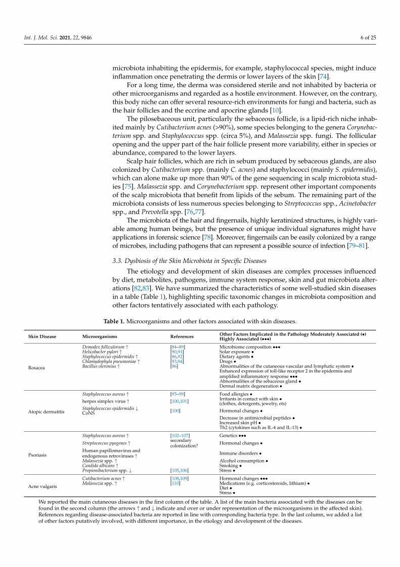
The etiology and development of skin diseases are complex processes influencedby diet, metabolites, pathogens, immune system response, skin and gut microbiota alter-ations [82,83]. We have summarized the characteristics of some well-studied skin diseasesin a table (Table 1), highlighting specific taxonomic changes in microbiota composition andother factors tentatively associated with each pathology.
Table 1. Microorganisms and other factors associated with skin diseases.
Skin Disease Microorganisms References Other Factors Implicated in the Pathology Moderately Associated (•)Highly Associated (•••)
Rosacea
Demodex folliculorum ↑ [84–89] Microbiome composition •••Helicobacter pylori ↑ [90,91] Solar exposure •Staphylococcus epidermidis ↑ [86,92] Dietary agents •Chlamydophyla pneumoniae ↑ [93,94] Drugs •Bacillus oleronius ↑ [86] Abnormalities of the cutaneous vascular and lymphatic system •
Enhanced expression of toll-like receptor 2 in the epidermis andamplified inflammatory response •••Abnormalities of the sebaceous gland •Dermal matrix degeneration •
Atopic dermatitis
Staphylococcus aureus ↑ [95–99] Food allergies •herpes simplex virus ↑ [100,101] Irritants in contact with skin •
(clothes, detergents, jewelry, ets)Staphylococcus epidermidis ↓CoNS [100] Hormonal changes •
Decrease in antimicrobial peptides •Increased skin pH •Th2 (cytokines such as IL-4 and IL-13) •
Psoriasis
Staphylococcus aureus ↑ [102–107] Genetics •••Streptococcus pyogenes ↑ secondary
colonization? Hormonal changes •Human papillomavirus andendogenous retroviruses ↑ Immune disorders •Malassezia spp. ↑ Alcohol consumption •Candida albicans ↑ Smoking •Propionibacterium spp. ↓ [105,106] Stress •
Acne vulgaris
Cutibacterium acnes ↑ [108,109] Hormonal changes •••Malassezia spp. ↑ [110] Medications (e.g. corticosteroids, lithium) •
Diet •Stress •
We reported the main cutaneous diseases in the first column of the table. A list of the main bacteria associated with the diseases can befound in the second column (the arrows ↑ and ↓ indicate and over or under representation of the microorganisms in the affected skin).References regarding disease-associated bacteria are reported in line with corresponding bacteria type. In the last column, we added a listof other factors putatively involved, with different importance, in the etiology and development of the diseases.
Int. J. Mol. Sci. 2021, 22, 9846 7 of 25
3.3.1. Acne Vulgaris
Acne vulgaris is a skin lesion affecting more than 9% of the human population, withhigher prevalence (up to 80–90%) in adolescents and young adults [111–113]. This skincondition is caused by the obstruction and inflammation of the pilosebaceous units, whichcan result in the formation of comedones, papules, pustules, nodules, and cysts [114].The pathophysiology of acne vulgaris is somewhat complex, and multiple studies sug-gested that strains of C. acnes and Malassezia sp. are implicated in its development [115].The biosynthesis of vitamin B12 by C. acnes has been hypothesized to have a role in thepathogenesis of acne; an increase in vitamin B12 might cause a concomitant productionof porphyrins promoting inflammation [116]. Other biological factors might be triggeringacne vulgaris, and thus, the roles of the host immune response and other members of themicrobiota remain to be adequately defined [117]. When comparing the microbiota of theskin surface and follicles, the main bacterial and fungal species are present in both niches,and commonly detected with different sampling and sequencing methods; higher diversityand site specificity are reported for viruses on the skin surface.
3.3.2. Atopic Dermatitis
Atopic dermatitis (AD) is a chronic skin alteration that can manifest in different bodysites and is characterized by dry, itchy skin patches and relapsing eczema [118]. Thiscondition affects 15–20% of children, with a lower prevalence (3%) in adults [119,120]. Lowbacterial diversity is frequently reported in AD, and cutaneous microbiota dysbiosis mightbe a driving factor in eczema pathogenesis. In particular, changes include depletion ofMalassezia spp., high non-Malassezia fungal diversity associated with the relative abundanceof S. aureus (strongly associated with AD) and S. epidermidis, and the reduction of othergenera, such as Propionibacterium [118,121–123]. Opportunistic viral, bacterial, and fungalinfections are often reported in patients with AD; dry skin and compromised microbiotamight also be more susceptible to pathogen colonization and successful establishment. Inaddition to cutaneous dysbiosis, gut dysbiosis has also been observed in AD [124,125],especially in infants, [126], although taxa specifically under- or over-represented variedamong the studies, with conflicting results [125].
The establishment of altered gut microbiota with AD seems to occur in the early stagesof development, as demonstrated by studies showing that atopic infants vs. non-atopicinfants at 1 year of age had different gut compositions at 3 weeks of age [127]. The majorityof the evidence derived from molecular data suggests that gut colonization occurs throughcontamination shortly after delivery [128,129]. At about 2.5 years of age, the composition,diversity, and functions of the infant microbiota resemble those of the microbiota of adultpeople [130]. This has caused researchers to become interested in shaping gut microbiota inthe early stages to prevent the development of allergic diseases later in life with strategiesbased on the use of probiotics (see below).
3.3.3. Psoriasis
Psoriasis is an idiopathic skin disorder affecting approximately 1–2% of the humanpopulation [131,132]. The disease is characterized by a chronic inflammatory conditionthat can manifest into hyperkeratosis, hyperproliferation of keratinocytes, infiltration ofthe skin by immune cells, and angiogenesis [133]. Skin lesions from these patients hadhigher bacterial diversity compared to healthy individuals, with increased Streptococcusand significantly less C. acnes [134]. S. aureus has long been regarded as associated withpsoriasis, but the psoriasis microbiome has not been clearly defined yet, and the roles ofother components of the microbiota remain to be elucidated.
3.3.4. Rosacea
Rosacea is an inflammatory dermatosis of the facial skin affecting roughly 10% ofthe population (estimates can be rather variable, between 5% and 46%), and character-ized by flushing, redness, papules, and pustules, for which pathogenesis remains largely
Int. J. Mol. Sci. 2021, 22, 9846 8 of 25
unknown [135–137]. The majority of researchers distinguish between rosacea-like demodi-cosis and papulo pustular rosacea, with the first one presenting a higher density of Demodexmites. However, in a recent study, no clear differences were found between the two condi-tions, and the authors suggested considering the two entities as different phenotypes of thesame disease [85]. Several bacteria, such as S. epidermidis, Helicobacter pylori, Chlamydophilapneumoniae, and Bacillus oleronius, were also considered associated with the disease [138].Colonization with Demodex folliculorum and Demodex-associated bacteria (based on 16SrRNA gene sequencing) also positively correlates with disease severity [139]. As statedabove, Demodex spp. are the most complex members of the human skin microbiome; theyare mostly commensals, although a pathophysiological role in inflammatory dermatosesis recognized. Recently, there has been an interest in screening them for the presence ofendosymbionts, which are rather common in different species of mites (and, in general,arthropods), where they play key roles in biological and physiological processes and canbe the target of potential medical and therapeutical applications [140,141]). In Demodexspp., different species of the genus Bacillus (Bacillus oleronius, Bacillus simplex, Bacillus cereus,and Bacillus pumilus) have been considered presumed symbionts [142]. However, currently,Corynebacterium kroppenstedtii subsp. demodicis is the only bacterium for which a role asthe primary endosymbiont of Demodex folliculorum is well supported by the followingevidence: (a) the bacteria are vertically transmitted; (b) they are present in all individualsof the species and located in a well-defined structure in the mite opisthosoma; (c) theysupposedly participate in lipid metabolisms by providing lipid-digesting enzymes [143].
Since endosymbiont removal is known to have negative effects on the host fitness, abetter understanding of this relationship could have important implications for Demodexspp. density and the treatment of rosacea and other skin conditions.
4. Methodological Considerations on NGS Techniques
As previously mentioned, NGS warranted technological advances that facilitatedculture-independent approaches and presented a key requisite to understanding skinmicrobiota from a broader perspective [17]. In addition, it allowed for achieving a morecomprehensive view and deeper insight into the complex microbial community inhab-iting the human skin in terms of site specificity, temporal dynamics, and interpersonalvariation [12].
To these aims, a very popular approach has been (and still is) amplicon sequencing(also referred to as marker gene sequencing, MGS, or meta-barcoding) to taxonomicallydefine the skin microbiome based on specific genomic regions that guarantee a rapididentification of prokaryotes and eukaryotes at the genus and, sometimes, at the specieslevel [14,144,145]. For example, bacteria and Archaea are generally identified by targetingthe ribosomal gene 16S rRNA, while other microorganisms, such as fungi, require theamplification and sequencing of genomic regions such as the ribosomal subunit 18S rRNA,the ribosomal internal transcribed spacer (ITS), or the D1/D2 region of the 26S rRNAgene [146–148].
The classical pipeline for 16S amplicon analyses is based on the use of universalprimers targeting one or more variable (V) regions (frequently, V1–V3, V3–V4, as a rule;longer fragments positively correlate with more taxonomic precision), followed by se-quence clustering into bins called Operational Taxonomic Units (OTUs), often using a97% similarity criterion that, for bacteria, conventionally defines the taxonomic level ofgenus. This approach reduces diversity and simplifies the computational analyses, butit is not free from criticism [149]. For example, either the choice of the regions or thechoice of the primers is known to affect the outcome deeply, introducing biases in thetaxonomic picture, with artifactual under- or over-representation of some taxa [150,151].Remarkably, it has also been demonstrated that few base pair mismatches can result in pooramplification of taxa otherwise abundant on the skin, such as Propionibacterium, especiallywhen considering the V4 region [20].
Int. J. Mol. Sci. 2021, 22, 9846 9 of 25
These considerations are even more important when attempting to include an unbi-ased picture of Archaea or bacterial candidate phyla radiations in the taxonomic analyses,which are often poorly amplified by classical 16S primers [152].
The definition of the best variable region/regions is currently controversial, but it isexpected that each option is more suitable for some bacterial species and less useful forothers.
While the NGS amplicon sequencing approach remains limitative, even consideringprimer optimization and defining the best target genes [153], more accurate and completeinformation on the microbial genomes for functional analyses and for distinguishing amongdifferent strains is often needed. This is achieved by metagenomic sequencing, in whichwhole genomes from virtually every member of the bacterial community are sequenced ina “shotgun” manner.
Among its advantages, metagenomic sequencing allows for outlining a rather pre-cise representation of the entire genetic diversity and to perform functional studies whileconstructing a catalog of genes, predicting gene functions and metabolic capabilities, andevaluating the presence of antibiotic resistance or virulence factors [154]. Furthermore,with the possibility of reconstructing the complete genome of most bacteria in the sam-ple, different strains can be distinguished and tentatively associated with specific skinconditions. At the same time, even partially assembled or low-quality genomes of poorlyrepresented species can still provide important insight into their functional profile. This isparticularly relevant to the issues treated in the present review.
A recent technological breakthrough in NGS platforms, that is, the development ofsingle-molecule or third-generation sequencing, with companies such as Oxford NanoporeTechnologies (ONT) and Pacific Biosciences currently leading the market, was a result ofthe “1000 Genomes” project. This was launched in 2010 by NIH to set up revolutionarysequencing technologies that would enable a mammalian-sized genome to be sequencedfrom 1000 individuals (Spencer, 2010).
The main advantages of nanopore sequencing technology with machines such asMinION™ reside in the production of longer reads that encompass nearly the whole 16SrRNA gene, giving a better picture of the relative abundance of taxa and a better resolutionat the species level [155].
The main limitation of single-molecule sequencing has been a higher error ratethan massive parallel sequencing, but technological improvements are rapidly fillingthe gap [156–158]. This is the case of PacBio (Pacific Biosciences LA) sequencing technology,which currently has a limited utility for metagenomics [159]. However, an assembly proce-dure combining Illumina short reads with PacBio long reads is very efficient, especially forincreasing genome accuracy, by overcoming the limits of short reads in highly repetitive orpoorly amplified genomic areas [160,161]. Similarly, a combined Illumina-ONT approachcan be useful in improving quality assembly as well [162].
Largely relegated to a second stage of the charismatic NGS technology, the studyof human microbiota through cell culture still presents an important tool in functionalmicrobiota research. “Culturomics” was developed to culture and identify unknownbacteria belonging to the human microbiota as part of the renaissance of culture techniquesin microbiology. The culturomic approach, consisting of multiple culture techniques, non-sequence-based identification methods, and ad hoc media development (e.g., axenic mediafor intracellular bacteria), has enabled the on-plate growth and isolation of unknownmicroorganisms associated with humans. Genomics was indispensable for acquiringmore details on metabolic needs and/or specific nutrients [163,164]. Culturomics has acomplementary role in providing information on the viability of microorganisms and theirphysiological states that is difficult to grasp solely with genomics or by elucidating therelationship between microbiota and human health and developing experimental modelsand new therapies. Finally, a combined approach using microbial culturomics and 16rRNAmetagenomics presents a powerful tool to correctly assign OTUs within more definedspecies boundaries [165].
Int. J. Mol. Sci. 2021, 22, 9846 10 of 25
The development of advanced 3D-culture methods, with well-differentiated cell types(e.g., keratinocytes, fibroblasts, and immune cells) representing an artificial replicate ofmulti-layered human skin, has been particularly relevant for the topics treated in thiswork, as it allowed for the study of interactions among commensal, opportunistic, andpathogenic bacteria, and the investigation of critical processes such as wound healing andbiofilm formation [166,167].
Before skin metagenomic sequencing can be considered a leading guideline for stan-dard diagnostic applications, a better understanding of the microbiota composition inhealth and disease will need to be coupled with robust and standardized sampling meth-ods to reduce human DNA by unbiased whole-genome amplification.
5. Skin Sampling Procedures: Standardization and Reproducibility among Studies
One critical point towards reaching a consensus about best practices in skin metage-nomics is skin sampling—a critical process that can deeply alter and introduce bias inthe outcomes. Indeed, collecting microorganisms that inhabit a specific skin niche whilelimiting environmental bacteria and host cells is a challenging procedure.
Contamination is one of the most common issues in such studies and is due to theco-occurrence of several causes, in primis, because the microbial skin biomass is oftenlocation- and layer-specific and even quite reduced in some areas. Another aspect thatmust not be overlooked is the selection and standardization of sampling methods; minimalchanges in microbial collection procedures can deeply affect the results and hinder studycomparisons.
Several methodologies are available to carry out skin microbiome sampling, andthe most common are pre-moisten swabs, skin surface scrapes, tape strips, and skinbiopsies [16,48,66,168–171]. All these methods show advantages and disadvantages, andthe most appropriate methodology is mainly dependent on the scientific questions thathave driven the study design. Sampling practices can have profound implications on theamount of total DNA, which is pivotal in a situation where not insignificant contaminationwith the host DNA is likely present. Another critical issue is the inclusion of those bacteriainhabiting deeper skin layers (derma). Alongside these considerations, the invasive actionand discomfort caused to patients, often in delicate situations with patients with a historyof altered skin conditions or prone to skin infections, is another issue to be taken intoaccount.
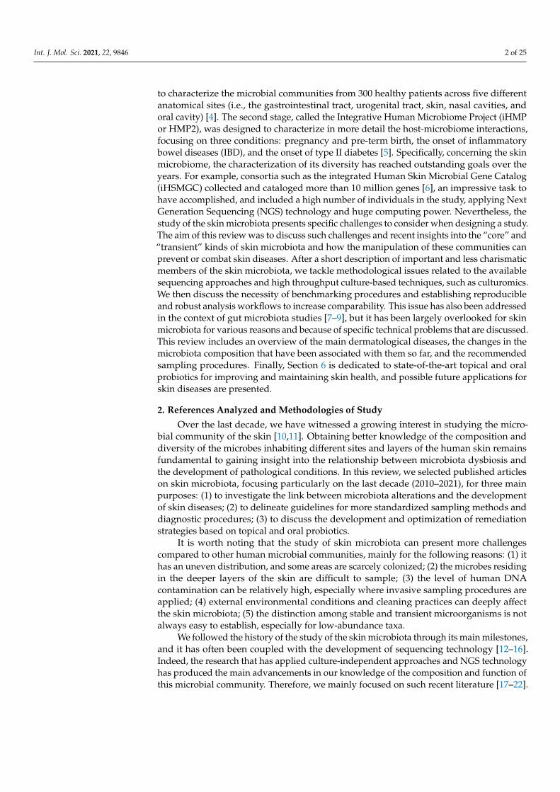
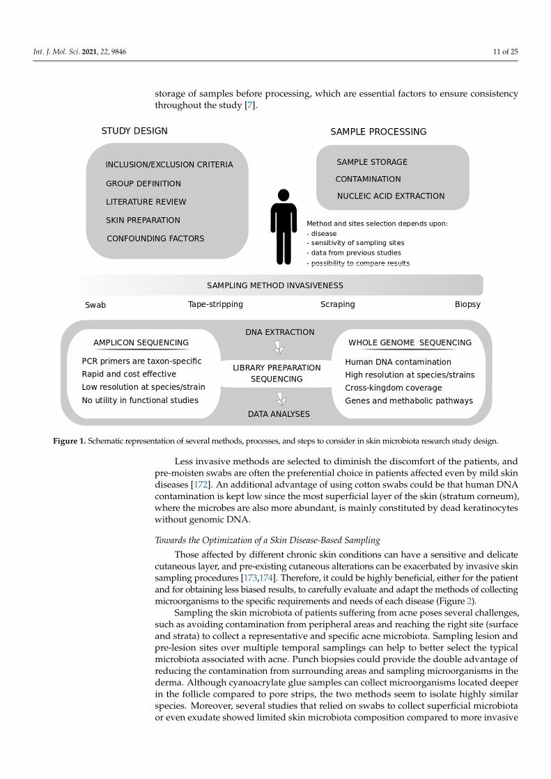
The method of choice should, thus, be chosen based on the specific requirements of theresearch activity, after having clearly defined the study design, by addressing factors suchas the number of individuals to be enrolled, their health conditions, and sampling locations.In addition, the experimental design should consider which methods are best suited for theaims of the project and the possibility of obtaining data comparable to previously publishedstudies, available databases, and genomic resources (Figure 1). This aspect is essential forapplicative purposes, such as guiding clinical diagnosis and treating skin diseases to avoidartifacts and bias. Recently, considerable efforts have been devoted to defining consensusguidelines to harmonize methods and increase the comparability between experimentsaddressing the gut microbiota [9]. These efforts should soon be extended also to studies onthe skin microbiota, which present specific and significant challenges.
The skin biopsy (punch biopsy and similar variants, such as shave biopsy and exci-sional biopsy) potentially allows for obtaining the most representative skin microbiotasample, even if relatively high similarities have been reported for cotton swabs and skin-scraping [13]. Nevertheless, punch biopsy remains the only method that can guarantee tosample microorganisms localized in the derma. On the other hand, this is also the mostinvasive sampling procedure, and it might not be indicated for sites where the skin is toothin (forehead, nose, ears) or too sensitive (groin, axilla). Furthermore, punch skin biopsycould also be problematic for groups of individuals living in remote areas or scatteredover a large territory, conditions that might also negatively affect the transport and proper
Int. J. Mol. Sci. 2021, 22, 9846 11 of 25
storage of samples before processing, which are essential factors to ensure consistencythroughout the study [7].
Int. J. Mol. Sci. 2021, 22, x FOR PEER REVIEW 11 of 26
total DNA, which is pivotal in a situation where not insignificant contamination with the host DNA is likely present. Another critical issue is the inclusion of those bacteria inhab-iting deeper skin layers (derma). Alongside these considerations, the invasive action and discomfort caused to patients, often in delicate situations with patients with a history of altered skin conditions or prone to skin infections, is another issue to be taken into ac-count.
The method of choice should, thus, be chosen based on the specific requirements of the research activity, after having clearly defined the study design, by addressing factors such as the number of individuals to be enrolled, their health conditions, and sampling locations. In addition, the experimental design should consider which methods are best suited for the aims of the project and the possibility of obtaining data comparable to pre-viously published studies, available databases, and genomic resources (Figure 1). This as-pect is essential for applicative purposes, such as guiding clinical diagnosis and treating skin diseases to avoid artifacts and bias. Recently, considerable efforts have been devoted to defining consensus guidelines to harmonize methods and increase the comparability between experiments addressing the gut microbiota [9]. These efforts should soon be ex-tended also to studies on the skin microbiota, which present specific and significant chal-lenges.
Figure 1. Schematic representation of several methods, processes, and steps to consider in skin microbiota research study design.
The skin biopsy (punch biopsy and similar variants, such as shave biopsy and exci-sional biopsy) potentially allows for obtaining the most representative skin microbiota sample, even if relatively high similarities have been reported for cotton swabs and skin-scraping [13]. Nevertheless, punch biopsy remains the only method that can guarantee to
Figure 1. Schematic representation of several methods, processes, and steps to consider in skin microbiota research study design.
Less invasive methods are selected to diminish the discomfort of the patients, andpre-moisten swabs are often the preferential choice in patients affected even by mild skindiseases [172]. An additional advantage of using cotton swabs could be that human DNAcontamination is kept low since the most superficial layer of the skin (stratum corneum),where the microbes are also more abundant, is mainly constituted by dead keratinocyteswithout genomic DNA.
Towards the Optimization of a Skin Disease-Based Sampling
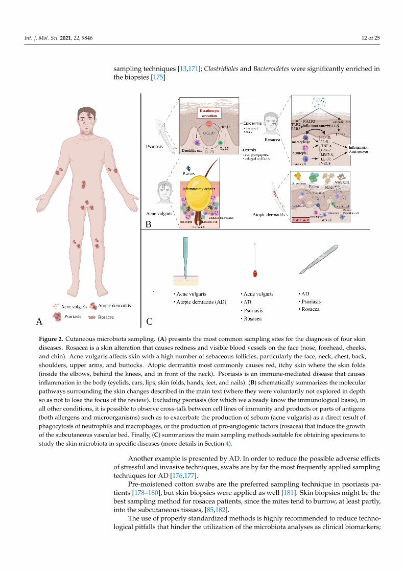
Those affected by different chronic skin conditions can have a sensitive and delicatecutaneous layer, and pre-existing cutaneous alterations can be exacerbated by invasive skinsampling procedures [173,174]. Therefore, it could be highly beneficial, either for the patientand for obtaining less biased results, to carefully evaluate and adapt the methods of collectingmicroorganisms to the specific requirements and needs of each disease (Figure 2).
Sampling the skin microbiota of patients suffering from acne poses several challenges,such as avoiding contamination from peripheral areas and reaching the right site (surfaceand strata) to collect a representative and specific acne microbiota. Sampling lesion andpre-lesion sites over multiple temporal samplings can help to better select the typicalmicrobiota associated with acne. Punch biopsies could provide the double advantage ofreducing the contamination from surrounding areas and sampling microorganisms in thederma. Although cyanoacrylate glue samples can collect microorganisms located deeperin the follicle compared to pore strips, the two methods seem to isolate highly similarspecies. Moreover, several studies that relied on swabs to collect superficial microbiotaor even exudate showed limited skin microbiota composition compared to more invasive
Int. J. Mol. Sci. 2021, 22, 9846 12 of 25
sampling techniques [13,171]; Clostridiales and Bacteroidetes were significantly enriched inthe biopsies [175].
Int. J. Mol. Sci. 2021, 22, x FOR PEER REVIEW 12 of 26
sample microorganisms localized in the derma. On the other hand, this is also the most invasive sampling procedure, and it might not be indicated for sites where the skin is too thin (forehead, nose, ears) or too sensitive (groin, axilla). Furthermore, punch skin biopsy could also be problematic for groups of individuals living in remote areas or scattered over a large territory, conditions that might also negatively affect the transport and proper storage of samples before processing, which are essential factors to ensure consistency throughout the study [7].
Less invasive methods are selected to diminish the discomfort of the patients, and pre-moisten swabs are often the preferential choice in patients affected even by mild skin diseases [172]. An additional advantage of using cotton swabs could be that human DNA contamination is kept low since the most superficial layer of the skin (stratum corneum), where the microbes are also more abundant, is mainly constituted by dead keratinocytes without genomic DNA.
Towards the Optimization of a Skin Disease-Based Sampling Those affected by different chronic skin conditions can have a sensitive and delicate
cutaneous layer, and pre-existing cutaneous alterations can be exacerbated by invasive skin sampling procedures [173,174]. Therefore, it could be highly beneficial, either for the patient and for obtaining less biased results, to carefully evaluate and adapt the methods of collecting microorganisms to the specific requirements and needs of each disease (Fig-ure 2).
Figure 2. Cutaneous microbiota sampling. Figure 2A presents the most common sampling sites for the diagnosis of four skin diseases. Rosacea is a skin alteration that causes redness and visible blood vessels on the face (nose, forehead, cheeks, and chin). Acne vulgaris affects skin with a high number of sebaceous follicles, particularly the face, neck, chest, back,
Figure 2. Cutaneous microbiota sampling. (A) presents the most common sampling sites for the diagnosis of four skindiseases. Rosacea is a skin alteration that causes redness and visible blood vessels on the face (nose, forehead, cheeks,and chin). Acne vulgaris affects skin with a high number of sebaceous follicles, particularly the face, neck, chest, back,shoulders, upper arms, and buttocks. Atopic dermatitis most commonly causes red, itchy skin where the skin folds(inside the elbows, behind the knees, and in front of the neck). Psoriasis is an immune-mediated disease that causesinflammation in the body (eyelids, ears, lips, skin folds, hands, feet, and nails). (B) schematically summarizes the molecularpathways surrounding the skin changes described in the main text (where they were voluntarily not explored in depthso as not to lose the focus of the review). Excluding psoriasis (for which we already know the immunological basis), inall other conditions, it is possible to observe cross-talk between cell lines of immunity and products or parts of antigens(both allergens and microorganisms) such as to exacerbate the production of sebum (acne vulgaris) as a direct result ofphagocytosis of neutrophils and macrophages, or the production of pro-angiogenic factors (rosacea) that induce the growthof the subcutaneous vascular bed. Finally, (C) summarizes the main sampling methods suitable for obtaining specimens tostudy the skin microbiota in specific diseases (more details in Section 4).
Another example is presented by AD. In order to reduce the possible adverse effectsof stressful and invasive techniques, swabs are by far the most frequently applied samplingtechniques for AD [176,177].
Pre-moistened cotton swabs are the preferred sampling technique in psoriasis pa-tients [178–180], but skin biopsies were applied as well [181]. Skin biopsies might be thebest sampling method for rosacea patients, since the mites tend to burrow, at least partly,into the subcutaneous tissues, [85,182].
The use of properly standardized methods is highly recommended to reduce techno-logical pitfalls that hinder the utilization of the microbiota analyses as clinical biomarkers;
Int. J. Mol. Sci. 2021, 22, 9846 13 of 25
sampling storage and transport, DNA extraction, sequencing, and computational analysesremain other critical passages [7,16].
6. Topical and Oral Probiotics in Skin Health and Diseases: State of the Art
The pioneering work on a human infection model for Haemophilus ducreyi, a pathogencausing sexually transmitted genital ulcers and chronic cutaneous ulcer, is a clear exam-ple of microbiome involvement in disease progression and resolution [183]. This studyshowed that pustule-forming sites had a greater abundance of Proteobacteria, Bacteroidetes,Micrococcus spp., Corynebacterium spp., Paracoccus spp., and Staphylococcus spp., whereasresolved sites showed a higher amount of Actinobacteria and bacteria of the genus Propi-onibacterium. Other key factors could also be detected in early differences in microbiomecomposition between resolvers and pustule formers, or even in the immune response, witha macrophage (M) polarization shift from M1 to M2 in resolvers.
Therefore, the experimental work on H. ducreyi highlights the complexity of thecombined responses of the innate and humoral immune system and the skin microbiotatowards a colonizing pathogen. Such intricacies could be theoretically considered foreach skin pathogen, and this myriad of interactions can help us to understand why thedevelopment of microbiome-based therapies for skincare is still in an early phase.
On the other hand, the already vast literature encompassing the beneficial role ofsome skin commensals as potential microbial invaders is continuously growing [97,184].Competitive displacement by niche occupation is an important phenomenon exhibited byskin resident bacteria that impede the colonization of pathogens; high species diversity isgenerally positively correlated with resistance to invaders [185]. However, the protectiveroles of some skin microbes go far beyond spatial competition. Frequently, the levelof manipulation showed by skin commensal bacteria extends into the modification ofbiochemical and metabolic pathways, the production of compounds with anti-microbialproperties, or even the alteration of gene expression in other bacteria [186,187]. The bulkof basic research on skin microbiota and pathogen interaction presents a solid groundfor developing probiotics to maintain healthy skin [109]. Probiotics can improve skinconditions either delivered directly to the skin (topical administration) or indirectly, suchas oral probiotics [188–190]. A rather small number of studies in healthy subjects showed aremarkable positive effect of oral probiotics on skin health. For example, women receivingLactobacillus lactis H61 daily for eight weeks reported improved skin elasticity and bodyfeatures (e.g., skin appeared more hydrated and hair follicles had improved) [191]. Oralingestion of Lactobacillus plantarum HY7714 (recently renamed Lactiplantibacillus plantarum)in a group of subjects (41–59 years old) reported increased skin hydration, reduced existingwrinkles, and improved overall skin elasticity and health [192].
Probiotic lactic acid bacteria (LAB) are among the most popular microbes with broadapplications to ameliorate gastrointestinal symptoms caused by different disorders orinterventions, from functional dyspepsia to anticancer therapy [193]. Human clinicaltrials indicated that LAB topically or orally applied directly to the skin can confer benefitsincluding the reinforcement of barrier function, the modulation of the immune system,and the preservation of homeostasis [194]. In addition, LAB probiotics might amelioratesymptoms of AD [195].
AD deserves further discussion, as many studies are available on the use of probioticsupplements in both pediatric and adult patient cohorts. As discussed above, recently,there has been growing interest of researchers for shaping gut microbiota in early life toprevent the development of allergic diseases, and AD, in primis. The work by Kalliomäkiet al. (2001), who administered Lactobacillus GG to both mothers in the third trimesterof pregnancy and infants in the first six months of life, aiming to study their effect onAD development, presents the pioneering approach of shaping gut microbiota in order toprevent allergic diseases [127]. This study has been followed by an impressive numberof studies evaluating different probiotics strains with different dosages and differentintervention times. Meta-analyses and systematic reviews also draw different conclusions

Int. J. Mol. Sci. 2021, 22, 9846 14 of 25
due to the high heterogeneity of the studies [196–199]. The timing of probiotics for favoringimmune tolerance appears to be critical [200].
Overall, a combination of prenatal and postnatal probiotics supplementation forallergy prevention (e.g., AD, urticaria) has shown the most consistent benefits, althoughtheir routine use cannot be recommended. In most studies, single or multiple strains oflactobacilli and bifidobacteria have been used to treat and prevent allergic diseases; thedemonstrated effect of one probiotic strain cannot be extrapolated to another strain [201].
Similar to AD prevention, the use of oral probiotics in AD treatment also led tocontrasting and not conclusive results, as outlined by recent meta-analyses [202,203].
In contrast to the bulk of studies focusing on AD, few studies have investigated thepotential role of oral probiotics in other skin conditions, such as psoriasis [204,205].
Of note, several studies have shown that dead bacteria and bacterial molecular com-ponents exert probiotic properties [206,207]. Currently, the term “postbiotic” refers tosoluble components with biological activity that could be a safer alternative to the use ofwhole bacteria [208]. Very few studies have investigated the role of postbiotics in AD inadults [209] and in children with promising results [210,211], but research in this field isstill in its infancy. New confirmation about the role of probiotic (prebiotics and postbiotics)therapy is needed, and it is important to better define other factors, such as which diseasecan benefit most, the most efficient bacterial combination, the optimal dosage, and theduration of the treatment.
Regarding topical probiotics, a cream containing Nitrosomonas eutropha for topical useis currently marketed in the United States. The microorganism N. eutropha is an ammonia-oxidizing bacteria (AOB), which have been detected in human microbiomes and modernhygienic lifestyles appear to be involved in their depletion [212]. Two recent clinical studieshave shown its effectiveness on keratosis pilaris, facial wrinkles, and acne [213–215]. AOBwere shown to inhibit, in vitro, the polarization to M2 via the anti-inflammatory cytokineIL-10, also hypothesizing a potential role in AD [216].
The cutaneous microbiota, as it interacts with the immune system, can also acceleratewound healing processes, as shown in the case of the commensal S. epidermidis, which caninduce re-epithelization of the skin after injury, mediating CD8+ T cell response [217]. C.acnes was shown to ferment glycerol into short-chain fatty acids, suppressing the growthof virulent methicillin-resistant S. aureus USA300. Another skin dweller, Corynebacteriumstriatum, can modify, on a large scale, the transcriptional program of co-cultured S. aureusso that it can suppress virulence-related genes and overexpress genes associated withnon-pathogenic phenotypes [218].
Bacteriotherapy, in the form of topically applied bacterial lysate, probiotics, or bacterialskin transplant, has shown promising results in animal and human trials for different skinconditions associated with an altered skin microbiota [219]. Recently, clinical studies haveinvestigated if topical therapy could ameliorate the microflora of AD patients, inducinga positive skin microbiota balance by eliminating pathogenic bacteria and enhancingbeneficial bacteria [220,221].
Commensal bacteria, such as S. epidermidis and S. hominis, have been shown to secreteantimicrobial peptides that interfere with the growth of S. aureus, and the transplantationof these species onto the skin of patients with AD led to decreased colonization by thepathogens [97]. Thus, if this mechanism is exploited, it could be a potential therapy for ADpatients.
In addition, S. epidermidis secretes phenol-soluble γ- and δ-modulins with antibioticeffects on S. aureus [186]. Antimicrobials from human skin commensal bacteria protectagainst S. aureus and are deficient in AD patients; more specifically, antimicrobial peptidesproduced by CoNS collected from the healthy skin were able to selectively kill S. aureus in amouse model and decrease S. aureus colonization in AD patients [97]. Moreover, the topicalaction of S. hominis A9 (ShA9) in AD patients colonized by S. aureus was reported as safeand caused either S. aureus killing or the inhibition of toxin expression [222]. The possibleutility of gut commensals or environmental bacteria as topical probiotics has also been
Int. J. Mol. Sci. 2021, 22, 9846 15 of 25
explored. For example, topical administration of Streptococcus thermophilus in patients withAD led to a significant improvement in erythema, scaling, and pruritus [223]. In addition,a topical cream containing lysate of Vitreoscilla filiformis (a Gram-negative bacterium foundin thermal waters) resulted in clinical improvements in patients with AD [224].
Roseomonas mucosa, a member of the human microbiota, topically used in a smallsample of adult patients with AD, led to a reduction in dermatitis severity, reduced use ofcorticosteroids, and colonization by S. aureus [225]. It is worth noting that the exclusioncriteria of the study included diagnosed immunodeficiency, heart valve disease, and/or anindwelling catheter, as case reports of endocarditis and bacteremia have been reported inimmunocompromised patients [226–228]. Thus, this issue should be taken into account byclinicians when considering topical therapy in patients.
Other skin conditions, such as acne, are associated with an overgrowth of pathogenicbacteria, and the mainstays of therapy are often antibiotics. For this reason, it may behypothesized that topical probiotics could restore a more balanced microflora to decreaseacne lesions. Investigated probiotics include L. plantarum, S. epidermidis, and other health-promoting bacterial strains; a reduction in lesion concentration, erythema, and pathogenicbacteria load with an improvement in the skin barrier function were reported [229–231].One cutting-edge therapy is based on bacteriophages, or viruses that infect bacteria. Brownet al. (2016) isolated bacteriophages capable of lysing C. acnes from the human skinmicrobiota and tested their therapeutic potential [232]. However, to date, no studies havebeen conducted on bacteriophage therapy for C. acnes in humans.
Similar to AD, all the studies investigating topical therapy for acne evaluated differentprobiotics with various endpoints, making it difficult to compare the various studies [220].Although dysbiosis has been demonstrated in other skin conditions (e.g., psoriasis androsacea), topical therapy for these diseases has not been investigated yet.
The relationship between topical and oral probiotics and skin health has been underfocus for decades; consensus has been reached on the beneficial effects of probiotics onskin health, but the precise mechanisms of action, negative interactions with specificskin conditions or individual microbiotas, and possible contraindications, still requirefurther elucidation [233]. Even though the great potential of skin bacteria exhibiting amarked immunomodulatory and antimicrobial activity has been shown in vitro and in vivoexperiments, microbial therapies for the skin, based on such microorganisms, remain hardto develop and bring to market due to the still vast knowledge gap. In the years to come, itcan be expected that controlled alterations of the microbial skin communities by meansof specific bacterial strains colonization will be better investigated as possible therapeuticstrategies.
A final consideration is in regard to the fact that the development of probiotics is adaunting task involving several steps, which can be tentatively sketched as follows: (1)bacteria isolation and characterization, (2) NGS profiling, (3) phylogenetic characteriza-tion of bacterial taxa (i.e., subspecies or strains) to determine the spectrum of metaboliccapabilities and predict LGT, (4) metabolic characterization (e.g., carbon sources, aminoacids, sterols, and lipids, as well as products derived from cosmetics), (5) the presence ofresistance genes, and (6) trials with a focus on safety and side effects [19,234].
7. Conclusions
The microbial community inhabiting the skin has a pivotal role in maintaining thehealthy status of an individual, comparable in importance with the microbiota ensuringthe homeostasis of the gut or the urogenital tract [235,236]. The skin is home to a wide,variable, and site-specific microbiota, mainly consisting of commensal microorganismsthat derive nutrition from dead skin cells and secretions such as sweat and sebum. Thenormal skin microbiota tends to inhibit transient microbe colonization by producingantimicrobial substances and outcompeting other microbes that land on the skin surface,protecting the skin from pathogen infections. The skin is a dynamic environment withfinely regulated interactions among the microbial communities [57]. Each niche offers
Int. J. Mol. Sci. 2021, 22, 9846 16 of 25
specific and rather stable conditions that allow only some microbial entities to thrive. Thegreat challenge in this fluctuating microbial diversity is determining which microbes andmetabolic patterns are of key importance to maintain the stability and functionality ofthe ecological niches. Indeed, marked alterations of the skin commensal microbiota areassociated with the onset and progression of several dermatological diseases. As a matterof fact, with the increasing evidence of skin microbial contribution in health and disease,there is an urgent need to better understand such microbiome diversity and its importancein crucial metabolic processes and interactions so that it will be possible to develop targetedstrategies and effective measures in skin health care. In addition, the gut microbiotacontinuously interacts with distant organ systems, including the skin along the gut–skinaxis, and such a fundamental relationship still requires further investigation [237,238].
Basic research and clinical studies on the skin microbiome can greatly benefit from amore standardized approach at each planning and technical step, including study design,group definition, sampling procedures and methods, nucleic acid storage and extraction,and, finally, NGS sequencing and data analyses.
Standardization can increase study comparison, then NGS and other “omic” technolo-gies will be able to characterize the skin microbiome with such precision that it will even bepossible to identify the individual based on microbial signatures. The fine characterizationand temporal variation of individual skin microbiota will have important implications forskin disease management, as well as broader applications for forensic purposes or to provepersonal object ownership as well [239,240].
Clinical trials on the effectiveness of different microbial strains as topical or oralprobiotics for skin health are producing outstanding results, emphasizing safety and a lackof undesired side effects [241]. Furthermore, the knowledge gap is reducing rapidly, and thedynamics governing the stability, temporal changes, and a return to a “homeostatic” stateof the skin microbiota are starting to be better characterized. Thus, a greater understandingof the network of interactions among skin microbes and their human hosts, and how we cantherapeutically manipulate and control those interactions, could present a key pre-requisiteand a powerful tool for skin health maintenance.
Author Contributions: Conceptualization, D.P., G.M. and L.D.; methodology, investigation, re-sources G.M., A.R.P., M.A. and S.P.; writing—original draft preparation, D.P.; writing—review andediting, S.P., G.M., E.D. and C.B.; visualization, D.P. and G.M.; supervision, C.B. and G.V.Z.; fundingacquisition, G.V.Z. All authors have read and agreed to the published version of the manuscript.
Funding: This research received no external funding.
Institutional Review Board Statement: Not applicable.
Informed Consent Statement: Not applicable.
Data Availability Statement: Not applicable.
Acknowledgments: We thank the “Romeo and Enrica Invernizzi” Foundation.
Conflicts of Interest: The authors declare no conflict of interest.
References1. Belkaid, Y.; Harrison, O.J. Homeostatic Immunity and the Microbiota. Immunity 2017, 46, 562–576. [CrossRef]2. Byrd, A.L.; Belkaid, Y.; Segre, J.A. The Human Skin Microbiome. Nat. Rev. Microbiol. 2018, 16, 143–155. [CrossRef]3. Balato, A.; Cacciapuoti, S.; Di Caprio, R.; Marasca, C.; Masarà, A.; Raimondo, A.; Fabbrocini, G. Human Microbiome: Composition
and Role in Inflammatory Skin Diseases. Arch. Immunol. Ther. Exp. 2019, 67, 1–18. [CrossRef]4. Nelson, K.; Weinstock, G.; Highlander, S.; Worley, K.; Creasy, H.; Wortman, J.; Rusch, D.; Mitreva, M.; Sodergren, E.; Chinwalla,
A.; et al. A Catalog of Reference Genomes from the Human Microbiome. Science 2010, 328, 994–999. [CrossRef] [PubMed]5. Proctor, L.; Creasy, H.; Fettweis, J.; Lloyd-Price, J.; Mahurkar, A.; Zhou, W.; Buck, G.A.; Snyder, M.P.; Strauss, J.; Weinstock, G.M.;
et al. The Integrative Human Microbiome Project. Nature 2019, 569, 641–648. [CrossRef]6. Li, Z.; Xia, J.; Jiang, L.; Tan, Y.; An, Y.; Zhu, X.; Ruan, J.; Chen, Z.; Zhen, H.; Ma, Y.; et al. Characterization of the Human Skin
Resistome and Identification of Two Microbiota Cutotypes. Microbiome 2021, 9, 47. [CrossRef] [PubMed]
Int. J. Mol. Sci. 2021, 22, 9846 17 of 25
7. Drago, L.; Panelli, S.; Bandi, C.; Zuccotti, G.; Perini, M.; D’Auria, E. What Pediatricians Should Know Before Studying GutMicrobiota. J. Clin. Med. 2019, 8, 1206. [CrossRef] [PubMed]
8. Panelli, S.; Epis, S.; Cococcioni, L.; Perini, M.; Paroni, M.; Bandi, C.; Drago, L.; Zuccotti, G.V. Inflammatory Bowel Diseases, theHygiene Hypothesis and the Other Side of the Microbiota: Parasites and Fungi. Pharmacol. Res. 2020, 159, 104962. [CrossRef]
9. Poussin, C.; Sierro, N.; Boué, S.; Battey, J.; Scotti, E.; Belcastro, V.; Peitsch, M.C.; Ivanov, N.V.; Hoeng, J. Interrogating theMicrobiome: Experimental and Computational Considerations in Support of Study Reproducibility. Drug Discov. Today 2018, 23,1644–1657. [CrossRef]
10. Bay, L.; Barnes, C.J.; Fritz, B.G.; Thorsen, J.; Restrup, M.E.M.; Rasmussen, L.; Sørensen, J.K.; Hesselvig, A.B.; Odgaard, A.; Hansen,A.J.; et al. Universal Dermal Microbiome in Human Skin. mBio 2020, 11, e02945-19. [CrossRef]
11. Oh, J.; Byrd, A.L.; Deming, C.; Conlan, S.; Kong, H.H.; Segre, J.A. Biogeography and Individuality Shape Function in the HumanSkin Metagenome. Nature 2014, 514, 59–64. [CrossRef]
12. Grice, E.A.; Kong, H.H.; Conlan, S.; Deming, C.B.; Davis, J.; Young, A.C.; Bouffard, G.G.; Blakesley, R.W.; Murray, P.R.; Green,E.D.; et al. Topographical and Temporal Diversity of the Human Skin Microbiome. Science 2009, 324, 1190–1192. [CrossRef]
13. Grice, E.A.; Kong, H.H.; Renaud, G.; Young, A.C.; Bouffard, G.G.; Blakesley, R.W.; Wolfsberg, T.G.; Turner, M.L.; Segre, J.A. ADiversity Profile of the Human Skin Microbiota. Genome Res. 2008, 18, 1043–1050. [CrossRef] [PubMed]
14. Grogan, M.D.; Bartow-McKenney, C.; Flowers, L.; Knight, S.A.B.; Uberoi, A.; Grice, E.A. Research Techniques Made Simple:Profiling the Skin Microbiota. J. Investig. Dermatol. 2019, 139, 747–752.e1. [CrossRef]
15. Kong, H.H. Skin Microbiome: Genomics-Based Insights into the Diversity and Role of Skin Microbes. Trends Mol. Med. 2011, 17,320–328. [CrossRef] [PubMed]
16. Kong, H.H.; Andersson, B.; Clavel, T.; Common, J.E.; Jackson, S.A.; Olson, N.D.; Segre, J.A.; Traidl-Hoffmann, C. Performing SkinMicrobiome Research: A Method to the Madness. J. Investig. Dermatol. 2017, 137, 561–568. [CrossRef] [PubMed]
17. Ederveen, T.H.A.; Smits, J.P.H.; Boekhorst, J.; Schalkwijk, J.; van den Bogaard, E.H.; Zeeuwen, P.L.J.M. Skin Microbiota in Healthand Disease: From Sequencing to Biology. J. Dermatol. 2020, 47, 1110–1118. [CrossRef] [PubMed]
18. Godlewska, U.; Brzoza, P.; Kwiecien, K.; Kwitniewski, M.; Cichy, J. Metagenomic Studies in Inflammatory Skin Diseases. Curr.Microbiol. 2020, 77, 3201–3212. [CrossRef] [PubMed]
19. Khayyira, A.S.; Rosdina, A.E.; Irianti, M.I.; Malik, A. Simultaneous Profiling and Cultivation of the Skin Microbiome of HealthyYoung Adult Skin for the Development of Therapeutic Agents. Heliyon 2020, 6, e03700. [CrossRef]
20. Meisel, J.S.; Hannigan, G.D.; Tyldsley, A.S.; SanMiguel, A.J.; Hodkinson, B.P.; Zheng, Q.; Grice, E.A. Skin Microbiome SurveysAre Strongly Influenced by Experimental Design. J. Investig. Dermatol. 2016, 136, 947–956. [CrossRef]
21. Reiger, M.; Traidl-Hoffmann, C.; Neumann, A.U. The Skin Microbiome as a Clinical Biomarker in Atopic Eczema: Promises,Navigation, and Pitfalls. J. Allergy Clin. Immunol. 2020, 145, 93–96. [CrossRef] [PubMed]
22. Sandhu, S.S.; Pourang, A.; Sivamani, R.K. A Review of next Generation Sequencing Technologies Used in the Evaluation of theSkin Microbiome: What a Time to Be Alive. Dermatol. Online J. 2019, 25, 9.
23. Gallo, R.L. Human Skin is the Largest Epithelial Surface for Interaction with Microbes. J. Investig. Dermatol. 2017, 137, 1213–1214.[CrossRef] [PubMed]
24. Grice, E.A.; Segre, J.A. The Skin Microbiome. Nat. Rev. Microbiol. 2011, 9, 244–253. [CrossRef]25. Bikle, D.D. Vitamin D Metabolism and Function in the Skin. Mol. Cell. Endocrinol. 2011, 347, 80–89. [CrossRef]26. Chen, Y.E.; Fischbach, M.A.; Belkaid, Y. Skin Microbiota–Host Interactions. Nature 2018, 553, 427–436. [CrossRef]27. Logan, A.C.; Jacka, F.N.; Prescott, S.L. Immune-Microbiota Interactions: Dysbiosis as a Global Health Issue. Curr. Allergy Asthma
Rep. 2016, 16, 13. [CrossRef]28. Prescott, S.L.; Larcombe, D.-L.; Logan, A.C.; West, C.; Burks, W.; Caraballo, L.; Levin, M.; Etten, E.V.; Horwitz, P.; Kozyrskyj,
A.; et al. The Skin Microbiome: Impact of Modern Environments on Skin Ecology, Barrier Integrity, and Systemic ImmuneProgramming. World Allergy Organ. J. 2017, 10, 29. [CrossRef]
29. Mukherjee, S.; Mitra, R.; Maitra, A.; Gupta, S.; Kumaran, S.; Chakrabortty, A.; Majumder, P.P. Sebum and Hydration Levels inSpecific Regions of Human Face Significantly Predict the Nature and Diversity of Facial Skin Microbiome. Sci. Rep. 2016, 6, 36062.[CrossRef]
30. Dréno, B.; Araviiskaia, E.; Berardesca, E.; Gontijo, G.; Sanchez Viera, M.; Xiang, L.F.; Martin, R.; Bieber, T. Microbiome in HealthySkin, Update for Dermatologists. J. Eur. Acad. Dermatol. Venereol. 2016, 30, 2038–2047. [CrossRef]
31. Méric, G.; Mageiros, L.; Pensar, J.; Laabei, M.; Yahara, K.; Pascoe, B.; Kittiwan, N.; Tadee, P.; Post, V.; Lamble, S.; et al. Disease-Associated Genotypes of the Commensal Skin Bacterium Staphylococcus Epidermidis. Nat. Commun. 2018, 9, 5034. [CrossRef][PubMed]
32. Otto, M. Staphylococcus Epidermidis–the “Accidental” Pathogen. Nat. Rev. Microbiol. 2009, 7, 555–567. [CrossRef]33. Clark, J.J.C.; Abildgaard, J.T.; Backes, J.; Hawkins, R.J. Preventing Infection in Shoulder Surgery. J. Shoulder Elbow Surg. 2018, 27,
1333–1341. [CrossRef] [PubMed]34. Dong, Y.; Speer, C.P. The Role of Staphylococcus Epidermidis in Neonatal Sepsis: Guarding Angel or Pathogenic Devil? Int. J.
Med. Microbiol. 2014, 304, 513–520. [CrossRef]35. Szczuka, E.; Telega, K.; Kaznowski, A. Biofilm Formation by Staphylococcus Hominis Strains Isolated from Human Clinical
Specimens. Folia Microbiol. 2015, 60, 1–5. [CrossRef]
Int. J. Mol. Sci. 2021, 22, 9846 18 of 25
36. Jeon, J.; Park, S.C.; Her, J.; Lee, J.W.; Han, J.-K.; Kim, Y.-K.; Kim, K.P.; Ban, C. Comparative Lipidomic Profiling of the HumanCommensal Bacterium Propionibacterium Acnes and Its Extracellular Vesicles. RSC Adv. 2018, 8, 15241–15247. [CrossRef]
37. Boisrenoult, P. Cutibacterium Acnes Prosthetic Joint Infection: Diagnosis and Treatment. Orthop. Traumatol. Surg. Res. 2018, 104,S19–S24. [CrossRef] [PubMed]
38. Elston, M.J.; Dupaix, J.P.; Opanova, M.I.; Atkinson, R.E. Cutibacterium Acnes (Formerly Proprionibacterium Acnes) and ShoulderSurgery. Hawaii J. Health Soc. Welf. 2019, 78, 3–5.
39. Freedman, M.; Aflatooni, J.O.; Foster, R.; Haggerty, P.G.; Derber, C.J. Persistent Cutibacterium (Formerly Propionibacterium)Acnes Bacteremia and Refractory Endocarditis in a Patient with Retained Implantable Pacemaker Leads. Case Rep. Infect. Dis.2020, 2020, e8883907. [CrossRef]
40. Moissl-Eichinger, C.; Probst, A.J.; Birarda, G.; Auerbach, A.; Koskinen, K.; Wolf, P.; Holman, H.-Y.N. Human Age and SkinPhysiology Shape Diversity and Abundance of Archaea on Skin. Sci. Rep. 2017, 7, 4039. [CrossRef] [PubMed]
41. Probst, A.J.; Auerbach, A.K.; Moissl-Eichinger, C. Archaea on Human Skin. PLoS ONE 2013, 8, e65388. [CrossRef] [PubMed]42. Litwin, D.; Chen, W.; Dzika, E.; Korycinska, J. Human Permanent Ectoparasites; Recent Advances on Biology and Clinical
Significance of Demodex Mites: Narrative Review Article. Iran J. Parasitol. 2017, 12, 12–21. [PubMed]43. Czepita, D.; Kuzna-Grygiel, W.; Czepita, M.; Grobelny, A. Demodex Folliculorum and Demodex Brevis as a Cause of Chronic
Marginal Blepharitis. Ann. Acad. Med. Stetin. 2007, 53, 63–67, Discussion 67. [PubMed]44. Liang, L.; Ding, X.; Tseng, S.C.G. High Prevalence of Demodex Brevis Infestation in Chalazia. Am. J. Ophthalmol. 2014, 157,
342–348.e1. [CrossRef]45. Moravvej, H.; Dehghan-Mangabadi, M.; Abbasian, M.-R.; Meshkat-Razavi, G. Association of Rosacea with Demodicosis. Arch.
Iran Med. 2007, 10, 199–203.46. Foulongne, V.; Sauvage, V.; Hebert, C.; Dereure, O.; Cheval, J.; Gouilh, M.A.; Pariente, K.; Segondy, M.; Burguière, A.; Manuguerra,
J.-C.; et al. Human Skin Microbiota: High Diversity of DNA Viruses Identified on the Human Skin by High ThroughputSequencing. PLoS ONE 2012, 7, e38499. [CrossRef]
47. Ma, Y.; Madupu, R.; Karaoz, U.; Nossa, C.W.; Yang, L.; Yooseph, S.; Yachimski, P.S.; Brodie, E.L.; Nelson, K.E.; Pei, Z. HumanPapillomavirus Community in Healthy Persons, Defined by Metagenomics Analysis of Human Microbiome Project ShotgunSequencing Data Sets. J. Virol. 2014, 88, 4786–4797. [CrossRef]
48. Verbanic, S.; Kim, C.Y.; Deacon, J.M.; Chen, I.A. Improved Single-Swab Sample Preparation for Recovering Bacterial and PhageDNA from Human Skin and Wound Microbiomes. BMC Microbiol. 2019, 19, 214. [CrossRef]
49. Leung, M.H.Y.; Tong, X.; Bøifot, K.O.; Bezdan, D.; Butler, D.J.; Danko, D.C.; Gohli, J.; Green, D.C.; Hernandez, M.T.; Kelly, F.J.;et al. Characterization of the Public Transit Air Microbiome and Resistome Reveals Geographical Specificity. Microbiome 2021, 9,112. [CrossRef]
50. Prussin, A.J.; Marr, L.C. Sources of Airborne Microorganisms in the Built Environment. Microbiome 2015, 3, 78. [CrossRef]51. Shibagaki, N.; Suda, W.; Clavaud, C.; Bastien, P.; Takayasu, L.; Iioka, E.; Kurokawa, R.; Yamashita, N.; Hattori, Y.; Shindo, C.; et al.
Aging-Related Changes in the Diversity of Women’s Skin Microbiomes Associated with Oral Bacteria. Sci. Rep. 2017, 7, 10567.[CrossRef] [PubMed]
52. Jugé, R.; Rouaud-Tinguely, P.; Breugnot, J.; Servaes, K.; Grimaldi, C.; Roth, M.-P.; Coppin, H.; Closs, B. Shift in Skin Microbiota ofWestern European Women across Aging. J. Appl. Microbiol. 2018, 125, 907–916. [CrossRef] [PubMed]
53. Li, Z.; Bai, X.; Peng, T.; Yi, X.; Luo, L.; Yang, J.; Liu, J.; Wang, Y.; He, T.; Wang, X.; et al. New Insights Into the Skin MicrobialCommunities and Skin Aging. Front. Microbiol. 2020, 11, 565549. [CrossRef]
54. Kim, H.-J.; Kim, J.J.; Myeong, N.R.; Kim, T.; Kim, D.; An, S.; Kim, H.; Park, T.; Jang, S.I.; Yeon, J.H.; et al. Segregation ofAge-Related Skin Microbiome Characteristics by Functionality. Sci. Rep. 2019, 9, 16748. [CrossRef]
55. Huang, S.; Haiminen, N.; Carrieri, A.-P.; Hu, R.; Jiang, L.; Parida, L.; Russell, B.; Allaband, C.; Zarrinpar, A.; Vázquez-Baeza, Y.;et al. Human Skin, Oral, and Gut Microbiomes Predict Chronological Age. Msystems 2020, 5, e00630-19. [CrossRef] [PubMed]
56. Abadías-Granado, I.; Sánchez-Bernal, J.; Gilaberte, Y. The Microbiome and Aging. Plast. Aesthetic Res. 2021, 8, 27. [CrossRef]57. Boxberger, M.; Cenizo, V.; Cassir, N.; La Scola, B. Challenges in Exploring and Manipulating the Human Skin Microbiome.
Microbiome 2021, 9, 125. [CrossRef] [PubMed]58. Dimitriu, P.A.; Iker, B.; Malik, K.; Leung, H.; Mohn, W.W.; Hillebrand, G.G. New Insights into the Intrinsic and Extrinsic Factors
That Shape the Human Skin Microbiome. mBio 2019, 10, e00839-19. [CrossRef]59. Staudinger, T.; Pipal, A.; Redl, B. Molecular Analysis of the Prevalent Microbiota of Human Male and Female Forehead Skin
Compared to Forearm Skin and the Influence of Make-Up. J. Appl. Microbiol. 2011, 110, 1381–1389. [CrossRef]60. Lee, H.J.; Jeong, S.E.; Lee, S.; Kim, S.; Han, H.; Jeon, C.O. Effects of Cosmetics on the Skin Microbiome of Facial Cheeks with
Different Hydration Levels. Microbiologyopen 2018, 7, e00557. [CrossRef] [PubMed]61. Fredricks, D.N. Microbial Ecology of Human Skin in Health and Disease. J. Investig. Dermatol. Symp. Proc. 2001, 6, 167–169.
[CrossRef]62. Lee, Y.B.; Byun, E.J.; Kim, H.S. Potential Role of the Microbiome in Acne: A Comprehensive Review. J. Clin. Med. 2019, 8, 987.
[CrossRef]63. Kong, H.H.; Segre, J.A. Skin Microbiome: Looking Back to Move Forward. J. Investig. Dermatol. 2012, 132, 933–939. [CrossRef]
[PubMed]
Int. J. Mol. Sci. 2021, 22, 9846 19 of 25
64. Schommer, N.N.; Gallo, R.L. Structure and Function of the Human Skin Microbiome. Trends Microbiol. 2013, 21, 660–668.[CrossRef] [PubMed]
65. Woese, C.R.; Fox, G.E. Phylogenetic Structure of the Prokaryotic Domain: The Primary Kingdoms. Proc. Natl. Acad. Sci. USA1977, 74, 5088–5090. [CrossRef] [PubMed]
66. Costello, E.K.; Lauber, C.L.; Hamady, M.; Fierer, N.; Gordon, J.I.; Knight, R. Bacterial Community Variation in Human BodyHabitats Across Space and Time. Science 2009, 326, 1694–1697. [CrossRef] [PubMed]
67. Caporaso, J.G.; Lauber, C.L.; Costello, E.K.; Berg-Lyons, D.; Gonzalez, A.; Stombaugh, J.; Knights, D.; Gajer, P.; Ravel, J.; Fierer, N.;et al. Moving Pictures of the Human Microbiome. Genome Biol. 2011, 12, R50. [CrossRef] [PubMed]
68. Oh, J.; Byrd, A.L.; Park, M.; Kong, H.H.; Segre, J.A. Temporal Stability of the Human Skin Microbiome. Cell 2016, 165, 854–866.[CrossRef]
69. Lilly, H.A.; Lowbury, E.J. Transient Skin Flora: Their Removal by Cleansing or Disinfection in Relation to their Mode of Deposition.J. Clin. Pathol. 1978, 31, 919–922. [CrossRef]
70. Mutters, R.; Warnes, S.L. The Method Used to Dry Washed Hands Affects the Number and Type of Transient and ResidentialBacteria Remaining on the Skin. J. Hosp. Infect. 2019, 101, 408–413. [CrossRef]
71. Nielsen, M.C.; Jiang, S.C. Alterations of the Human Skin Microbiome after Ocean Water Exposure. Mar. Pollut. Bull. 2019, 145,595–603. [CrossRef]
72. Wallen-Russell, C. The Role of Every-Day Cosmetics in Altering the Skin Microbiome: A Study Using Biodiversity. Cosmetics2019, 6, 2. [CrossRef]
73. Gibbons, T.F.; Noe, J.C.; Patterson, A.T.; Lenz, B.L.; Beachkofsky, T.M. Temporal Shifts in the Collective Dermatologic Microbiomeof Military Trainees. Clin. Cosmet. Investig. Dermatol. 2019, 12, 625–637. [CrossRef]
74. Krishna, S.; Miller, L.S. Host-Pathogen Interactions between the Skin and Staphylococcus Aureus. Curr. Opin. Microbiol. 2012, 15,28–35. [CrossRef]
75. Polak-Witka, K.; Rudnicka, L.; Blume-Peytavi, U.; Vogt, A. The Role of the Microbiome in Scalp Hair Follicle Biology and Disease.Exp. Dermatol. 2020, 29, 286–294. [CrossRef] [PubMed]
76. Clavaud, C.; Jourdain, R.; Bar-Hen, A.; Tichit, M.; Bouchier, C.; Pouradier, F.; El Rawadi, C.; Guillot, J.; Ménard-Szczebara, F.;Breton, L.; et al. Dandruff Is Associated with Disequilibrium in the Proportion of the Major Bacterial and Fungal PopulationsColonizing the Scalp. PLoS ONE 2013, 8, e58203. [CrossRef]
77. Xu, Z.; Wang, Z.; Yuan, C.; Liu, X.; Yang, F.; Wang, T.; Wang, J.; Manabe, K.; Qin, O.; Wang, X.; et al. Dandruff Is Associated withthe Conjoined Interactions between Host and Microorganisms. Sci. Rep. 2016, 6, 24877. [CrossRef] [PubMed]
78. Brinkac, L.; Clarke, T.H.; Singh, H.; Greco, C.; Gomez, A.; Torralba, M.G.; Frank, B.; Nelson, K.E. Spatial and EnvironmentalVariation of the Human Hair Microbiota. Sci. Rep. 2018, 8, 9017. [CrossRef]
79. Mengist, A.; Aschale, Y.; Reta, A. Bacterial and Parasitic Assessment from Fingernails in Debre Markos, Northwest Ethiopia.Available online: https://www.hindawi.com/journals/cjidmm/2018/6532014/ (accessed on 30 June 2021).
80. Micheal, A.O.; Elisabeth, P.T.; Adenike, K.K.; Olajumoke, A.E.; Adebiyi, A.; Olutope, O.S.; Ademola, A.D. Isolation and MolecularCharacterization of Mycobiota and Other Microbiota from Fingernails. Int. J. Clin. Dev. Anat. 2021, 7, 1. [CrossRef]
81. Wałaszek, M.Z.; Kołpa, M.; Rózanska, A.; Jagiencarz-Starzec, B.; Wolak, Z.; Wójkowska-Mach, J. Nail Microbial ColonizationFollowing Hand Disinfection: A Qualitative Pilot Study. J. Hosp. Infect. 2018, 100, 207–210. [CrossRef] [PubMed]
82. De Pessemier, B.; Grine, L.; Debaere, M.; Maes, A.; Paetzold, B.; Callewaert, C. Gut-Skin Axis: Current Knowledge of theInterrelationship between Microbial Dysbiosis and Skin Conditions. Microorganisms 2021, 9, 353. [CrossRef]
83. Egert, M.; Simmering, R.; Riedel, C.U. The Association of the Skin Microbiota with Health, Immunity, and Disease. Clin. Pharmacol.Ther. 2017, 102, 62–69. [CrossRef]
84. Forton, F. Demodex-Associated Folliculitis. Am. J. Dermatopathol. 1998, 20, 536–537. [CrossRef]85. Forton, F.M.N.; De Maertelaer, V. Papulopustular Rosacea and Rosacea-like Demodicosis: Two Phenotypes of the Same Disease?
J. Eur. Acad. Dermatol. Venereol. 2018, 32, 1011–1016. [CrossRef]86. Jarmuda, S.; O’Reilly, N.; Zaba, R.; Jakubowicz, O.; Szkaradkiewicz, A.; Kavanagh, K. 2012 Potential Role of Demodex Mites and
Bacteria in the Induction of Rosacea. J. Med. Microbiol. 2012, 61, 1504–1510. [CrossRef]87. Forton, F.; Germaux, M.-A.; Brasseur, T.; De Liever, A.; Laporte, M.; Mathys, C.; Sass, U.; Stene, J.-J.; Thibaut, S.; Tytgat, M.; et al.
Demodicosis and Rosacea: Epidemiology and Significance in Daily Dermatologic Practice. J. Am. Acad. Dermatol. 2005, 52, 74–87.[CrossRef]
88. Lacey, N.; Russell-Hallinan, A.; Zouboulis, C.C.; Powell, F.C. Demodex Mites Modulate Sebocyte Immune Reaction: Possible Rolein the Pathogenesis of Rosacea. Br. J. Dermatol. 2018, 179, 420–430. [CrossRef] [PubMed]
89. Chang, Y.-S.; Huang, Y.-C. Role of Demodex Mite Infestation in Rosacea: A Systematic Review and Meta-Analysis. J. Am. Acad.Dermatol. 2017, 77, 441–447.e6. [CrossRef] [PubMed]
90. Yang, X. Relationship between Helicobacter Pylori and Rosacea: Review and Discussion. BMC Infect. Dis. 2018, 18, 318. [CrossRef]91. Lazaridou, E.; Korfitis, C.; Kemanetzi, C.; Sotiriou, E.; Apalla, Z.; Vakirlis, E.; Fotiadou, C.; Lallas, A.; Ioannides, D. Rosacea and
Helicobacter Pylori: Links and Risks. Clin. Cosmet. Investig. Dermatol. 2017, 10, 305–310. [CrossRef] [PubMed]92. Whitfeld, M.; Gunasingam, N.; Leow, L.J.; Shirato, K.; Preda, V. Staphylococcus Epidermidis: A Possible Role in the Pustules of
Rosacea. J. Am. Acad. Dermatol. 2011, 64, 49–52. [CrossRef]
Int. J. Mol. Sci. 2021, 22, 9846 20 of 25
93. Fernandez-Obregon, A.; Patton, D.L. The Role of Chlamydia Pneumoniae in the Etiology of Acne Rosacea: Response to the Useof Oral Azithromycin. Cutis 2007, 79, 163–167. [CrossRef] [PubMed]
94. Fernandez-Obregon, A. Commentary on “The Role of Chlamydia Pneumoniae in the Etiology of Acne Rosacea: Response to theUse of Oral Azithromycin”. J. Infect. 2019, 2, 11–14. [CrossRef]
95. Geoghegan, J.A.; Irvine, A.D.; Foster, T.J. Staphylococcus Aureus and Atopic Dermatitis: A Complex and Evolving Relationship.Trends Microbiol. 2018, 26, 484–497. [CrossRef]
96. Iwamoto, K.; Moriwaki, M.; Miyake, R.; Hide, M. Staphylococcus Aureus in Atopic Dermatitis: Strain-Specific Cell Wall Proteinsand Skin Immunity. Allergol. Int. 2019, 68, 309–315. [CrossRef]
97. Nakatsuji, T.; Chen, T.H.; Narala, S.; Chun, K.A.; Two, A.M.; Yun, T.; Shafiq, F.; Kotol, P.F.; Bouslimani, A.; Melnik, A.V.; et al.Antimicrobials from Human Skin Commensal Bacteria Protect against Staphylococcus Aureus and are Deficient in AtopicDermatitis. Sci. Transl. Med. 2017, 9, eaah4680. [CrossRef]
98. Hwang, J.; Jaros, J.; Shi, V.Y. Staphylococcus Aureus in Atopic Dermatitis: Past, Present, and Future. Dermatitis 2020, 31, 247–258.[CrossRef]
99. Hepburn, L.; Hijnen, D.J.; Sellman, B.R.; Mustelin, T.; Sleeman, M.A.; May, R.D.; Strickland, I. The Complex Biology andContribution of Staphylococcus Aureus in Atopic Dermatitis, Current and Future Therapies. Br. J. Dermatol. 2017, 177, 63–71.[CrossRef] [PubMed]
100. Ong, P.Y.; Leung, D.Y.M. Bacterial and Viral Infections in Atopic Dermatitis: A Comprehensive Review. Clin. Rev. Allergy Immunol.2016, 51, 329–337. [CrossRef]
101. Saintive, S.; Abad, E.; de Ferreira, C.D.; Stambovsky, M.; Cavalcante, F.S.; Gonçalves, L.S.; Vidal, F.; Dos Santos, K.R. What is theRole of Staphylococcus Aureus and Herpes Virus Infections in the Pathogenesis of Atopic Dermatitis? Future Microbiol. 2017, 12,1327–1334. [CrossRef]
102. Rademaker, M.; Agnew, K.; Anagnostou, N.; Andrews, M.; Armour, K.; Baker, C.; Foley, P.; Gebauer, K.; Gupta, M.; Marshman,G.; et al. Psoriasis and Infection. A Clinical Practice Narrative. Australas J. Dermatol. 2019, 60, 91–98. [CrossRef]
103. Ng, C.Y.; Huang, Y.H.; Chu, C.F.; Wu, T.C.; Liu, S.H. Risks for Staphylococcus Aureus Colonization in Patients with Psoriasis: ASystematic Review and Meta-Analysis. Br. J. Dermatol. 2017, 177, 967–977. [CrossRef]
104. Queirós, N.; Torres, T. HIV-Associated Psoriasis. Actas Dermosifiliogr. (Engl. Ed.) 2018, 109, 303–311. [CrossRef]105. Thio, H.B. The Microbiome in Psoriasis and Psoriatic Arthritis: The Skin Perspective. J. Rheumatol. Suppl. 2018, 94, 30–31.
[CrossRef] [PubMed]106. Yan, D.; Issa, N.; Afifi, L.; Jeon, C.; Chang, H.W.; Liao, W. The Role of the Skin and Gut Microbiome in Psoriatic Disease. Curr.
Dermatol. Rep. 2017, 6, 94–103. [CrossRef]107. de Jesús-Gil, C.; Sans-de San Nicolàs, L.; Ruiz-Romeu, E.; Ferran, M.; Soria-Martínez, L.; García-Jiménez, I.; Chiriac, A.; Casanova-
Seuma, J.M.; Fernández-Armenteros, J.M.; Owens, S.; et al. Interplay between Humoral and CLA+ T Cell Response againstCandida Albicans in Psoriasis. Int. J. Mol. Sci. 2021, 22, 1519. [CrossRef]
108. Platsidaki, E.; Dessinioti, C. Recent Advances in Understanding Propionibacterium Acnes (Cutibacterium acnes) in Acne. F1000Res2018, 7. [CrossRef]
109. Goodarzi, A.; Mozafarpoor, S.; Bodaghabadi, M.; Mohamadi, M. The Potential of Probiotics for Treating Acne Vulgaris: A Reviewof Literature on Acne and Microbiota. Dermatol. Ther. 2020, 33, e13279. [CrossRef] [PubMed]
110. Xu, H.; Li, H. Acne, the Skin Microbiome, and Antibiotic Treatment. Am. J. Clin. Dermatol. 2019, 20, 335–344. [CrossRef] [PubMed]111. Law, M.P.M.; Chuh, A.A.T.; Lee, A.; Molinari, N. Acne Prevalence and beyond: Acne Disability and Its Predictive Factors among
Chinese Late Adolescents in Hong Kong. Clin. Exp. Dermatol. 2010, 35, 16–21. [CrossRef] [PubMed]112. Lynn, D.D.; Umari, T.; Dunnick, C.A.; Dellavalle, R.P. The Epidemiology of Acne Vulgaris in Late Adolescence. Adolesc. Health
Med. Ther. 2016, 7, 13–25. [CrossRef] [PubMed]113. Tayel, K.; Attia, M.; Agamia, N.; Fadl, N. Acne Vulgaris: Prevalence, Severity, and Impact on Quality of Life and Self-Esteem
among Egyptian Adolescents. J. Egypt. Public Health Assoc. 2020, 95, 30. [CrossRef] [PubMed]114. Heng, A.H.S.; Chew, F.T. Systematic Review of the Epidemiology of Acne Vulgaris. Sci. Rep. 2020, 10, 5754. [CrossRef] [PubMed]115. Fitz-Gibbon, S.; Tomida, S.; Chiu, B.-H.; Nguyen, L.; Du, C.; Liu, M.; Elashoff, D.; Erfe, M.C.; Loncaric, A.; Kim, J.; et al.
Propionibacterium Acnes Strain Populations in the Human Skin Microbiome Associated with Acne. J. Investig. Dermatol. 2013,133, 2152–2160. [CrossRef] [PubMed]
116. Kang, D.; Shi, B.; Erfe, M.C.; Craft, N.; Li, H. Vitamin B12 Modulates the Transcriptome of the Skin Microbiota in AcnePathogenesis. Sci. Transl. Med. 2015, 7, 293ra103. [CrossRef] [PubMed]
117. Ramasamy, S.; Barnard, E.; Dawson, T.L.; Li, H. The Role of the Skin Microbiota in Acne Pathophysiology. Br. J. Dermatol. 2019,181, 691–699. [CrossRef]
118. Bjerre, R.D.; Bandier, J.; Skov, L.; Engstrand, L.; Johansen, J.D. The Role of the Skin Microbiome in Atopic Dermatitis: A SystematicReview. Br. J. Dermatol. 2017, 177, 1272–1278. [CrossRef]
119. Seite, S.; Bieber, T. Barrier Function and Microbiotic Dysbiosis in Atopic Dermatitis. Clin. Cosmet. Investig. Dermatol. 2015, 8,479–483. [CrossRef]
120. Weidinger, S.; Novak, N. Atopic Dermatitis. Lancet 2016, 387, 1109–1122. [CrossRef]121. Di Domenico, E.G.; Cavallo, I.; Capitanio, B.; Ascenzioni, F.; Pimpinelli, F.; Morrone, A.; Ensoli, F. Staphylococcus Aureus and the
Cutaneous Microbiota Biofilms in the Pathogenesis of Atopic Dermatitis. Microorganisms 2019, 7, 301. [CrossRef]
Int. J. Mol. Sci. 2021, 22, 9846 21 of 25
122. Williams, M.R.; Gallo, R.L. Evidence That Human Skin Microbiome Dysbiosis Promotes Atopic Dermatitis. J. Investig. Dermatol.2017, 137, 2460–2461. [CrossRef]
123. Edslev, S.M.; Agner, T.; Andersen, P.S. Skin Microbiome in Atopic Dermatitis. Acta Derm. Venereol. 2020, 100, adv00164. [CrossRef][PubMed]
124. Lee, S.-Y.; Lee, E.; Park, Y.M.; Hong, S.-J. Microbiome in the Gut-Skin Axis in Atopic Dermatitis. Allergy Asthma Immunol. Res.2018, 10, 354–362. [CrossRef] [PubMed]
125. Reddel, S.; Del Chierico, F.; Quagliariello, A.; Giancristoforo, S.; Vernocchi, P.; Russo, A.; Fiocchi, A.; Rossi, P.; Putignani, L.;El Hachem, M. Gut Microbiota Profile in Children Affected by Atopic Dermatitis and Evaluation of Intestinal Persistence of aProbiotic Mixture. Sci. Rep. 2019, 9, 4996. [CrossRef]
126. Zheng, H.; Liang, H.; Wang, Y.; Miao, M.; Shi, T.; Yang, F.; Liu, E.; Yuan, W.; Ji, Z.-S.; Li, D.-K. Altered Gut Microbiota CompositionAssociated with Eczema in Infants. PLoS ONE 2016, 11, e0166026. [CrossRef] [PubMed]
127. Kalliomäki, M.; Kirjavainen, P.; Eerola, E.; Kero, P.; Salminen, S.; Isolauri, E. Distinct Patterns of Neonatal Gut Microflora inInfants in Whom Atopy Was and Was Not Developing. J. Allergy Clin. Immunol. 2001, 107, 129–134. [CrossRef]
128. Panelli, S.; Schneider, L.; Comandatore, F.; Bandi, C.; Zuccotti, G.V.; D’Auria, E. Is There Life in the Meconium? A Challenging,Burning Question. Pharmacol. Res. 2018, 137, 148–149. [CrossRef]
129. Perez-Muñoz, M.E.; Arrieta, M.-C.; Ramer-Tait, A.E.; Walter, J. A Critical Assessment of the “Sterile Womb” and “in UteroColonization” Hypotheses: Implications for Research on the Pioneer Infant Microbiome. Microbiome 2017, 5, 48. [CrossRef][PubMed]
130. Grice, E.A.; Segre, J.A. The Human Microbiome: Our Second Genome. Annu. Rev. Genom. Hum. Genet. 2012, 13, 151–170.[CrossRef]
131. Langley, R.G.B.; Krueger, G.G.; Griffiths, C.E.M. Psoriasis: Epidemiology, Clinical Features, and Quality of Life. Ann. Rheum. Dis.2005, 64, ii18–ii23. [CrossRef]
132. Parisi, R.; Iskandar, I.Y.K.; Kontopantelis, E.; Augustin, M.; Griffiths, C.E.M.; Ashcroft, D.M. National, Regional, and WorldwideEpidemiology of Psoriasis: Systematic Analysis and Modelling Study. BMJ 2020, 369, m1590. [CrossRef]
133. Schön, M.P.; Boehncke, W.-H. Psoriasis. Available online: https://www.nejm.org/doi/10.1056/NEJMra041320 (accessed on 10February 2021).
134. Gao, Z.; Tseng, C.; Strober, B.E.; Pei, Z.; Blaser, M.J. Substantial Alterations of the Cutaneous Bacterial Biota in Psoriatic Lesions.PLoS ONE 2008, 3, e2719. [CrossRef]
135. Gether, L.; Overgaard, L.K.; Egeberg, A.; Thyssen, J.P. Incidence and Prevalence of Rosacea: A Systematic Review and Meta-Analysis. Br. J. Dermatol. 2018, 179, 282–289. [CrossRef]
136. Rainer, B.M.; Kang, S.; Chien, A.L. Rosacea: Epidemiology, Pathogenesis, and Treatment. Dermato-Endocrinology 2017, 9, e1361574.[CrossRef] [PubMed]
137. Daou, H.; Paradiso, M.; Hennessy, K.; Seminario-Vidal, L. Rosacea and the Microbiome: A Systematic Review. Dermatol. Ther.(Heidelb.) 2021, 11, 1–12. [CrossRef]
138. Kim, H.S. Microbiota in Rosacea|SpringerLink. Available online: https://link.springer.com/article/10.1007/s40257-020-00546-8(accessed on 11 February 2021).
139. Murillo, N.; Aubert, J.; Raoult, D. Microbiota of Demodex Mites from Rosacea Patients and Controls. Microb. Pathog. 2014, 71–72,37–40. [CrossRef] [PubMed]
140. Ogawa, M.; Takahashi, M.; Matsutani, M.; Takada, N.; Noda, S.; Saijo, M. Obligate Intracellular Bacteria Diversity in UnfedLeptotrombidium scutellare Larvae Highlights Novel Bacterial Endosymbionts of Mites. Microbiol. Immunol. 2020, 64, 1–9. [CrossRef][PubMed]
141. Bonnet, S.I.; Binetruy, F.; Hernández-Jarguín, A.M.; Duron, O. The Tick Microbiome: Why Non-Pathogenic MicroorganismsMatter in Tick Biology and Pathogen Transmission. Front. Cell. Infect. Microbiol. 2017, 7, 236. [CrossRef] [PubMed]
142. Kubiak, K.; Sielawa, H.; Chen, W.; Dzika, E. Endosymbiosis and Its Significance in Dermatology. J. Eur. Acad. Dermatol. Venereol.2018, 32, 347–354. [CrossRef] [PubMed]
143. Clanner-Engelshofen, B.M.; French, L.E.; Reinholz, M. Corynebacterium kroppenstedtii Subsp. demodicis is the Endobacterium ofDemodex folliculorum. J. Eur. Acad. Dermatol. Venereol. 2020, 34, 1043–1049. [CrossRef]
144. Ogai, K.; Shibata, K.; Takahashi, N.; Ogura, K.; Okamoto, S.; Sugama, J. Amplicon-Based Skin Microbiome Profiles Collected byTape Stripping with Different Adhesive Film Dressings: A Comparative Study. BMC Microbiol. 2021, 21, 54. [CrossRef]
145. Schoch, C.L.; Seifert, K.A.; Huhndorf, S.; Robert, V.; Spouge, J.L.; Levesque, C.A.; Chen, W.; Bolchacova, E.; Voigt, K.; Crous, P.W.;et al. Nuclear Ribosomal Internal Transcribed Spacer (ITS) Region as a Universal DNA Barcode Marker for Fungi. Proc. Natl.Acad. Sci. USA 2012, 109, 6241–6246. [CrossRef]
146. De Filippis, F.; Laiola, M.; Blaiotta, G.; Ercolini, D. Different Amplicon Targets for Sequencing-Based Studies of Fungal Diversity.Appl. Environ. Microbiol. 2017, 83, e00905-17. [CrossRef]
147. Oh, J.; Freeman, A.F.; NISC Comparative Sequencing Program; Park, M.; Sokolic, R.; Candotti, F.; Holland, S.M.; Segre, J.A.; Kong,H.H. The Altered Landscape of the Human Skin Microbiome in Patients with Primary Immunodeficiencies. Genome Res. 2013, 23,2103–2114. [CrossRef] [PubMed]
148. Zhang, E.; Tanaka, T.; Tajima, M.; Tsuboi, R.; Nishikawa, A.; Sugita, T. Characterization of the Skin Fungal Microbiota in Patientswith Atopic Dermatitis and in Healthy Subjects. Microbiol. Immunol. 2011, 55, 625–632. [CrossRef] [PubMed]

Int. J. Mol. Sci. 2021, 22, 9846 22 of 25
149. Nguyen, N.-P.; Warnow, T.; Pop, M.; White, B. A Perspective on 16S RRNA Operational Taxonomic Unit Clustering UsingSequence Similarity. NPJ Biofilms Microbiomes 2016, 2, 1–8. [CrossRef] [PubMed]
150. Castelino, M.; Eyre, S.; Moat, J.; Fox, G.; Martin, P.; Ho, P.; Upton, M.; Barton, A. Optimisation of Methods for Bacterial SkinMicrobiome Investigation: Primer Selection and Comparison of the 454 versus MiSeq Platform. BMC Microbiol. 2017, 17, 23.[CrossRef] [PubMed]
151. Palkova, L.; Tomova, A.; Repiska, G.; Babinska, K.; Bokor, B.; Mikula, I.; Minarik, G.; Ostatnikova, D.; Soltys, K. Evaluation of 16SRRNA Primer Sets for Characterisation of Microbiota in Paediatric Patients with Autism Spectrum Disorder. Sci. Rep. 2021, 11,6781. [CrossRef]
152. Pausan, M.R.; Csorba, C.; Singer, G.; Till, H.; Schöpf, V.; Santigli, E.; Klug, B.; Högenauer, C.; Blohs, M.; Moissl-Eichinger, C.Exploring the Archaeome: Detection of Archaeal Signatures in the Human Body. Front. Microbiol. 2019, 10, 2796. [CrossRef]
153. Cococcioni, L.; Panelli, S.; Varotto-Boccazzi, I.; Carlo, D.D.; Pistone, D.; Leccese, G.; Zuccotti, G.V.; Comandatore, F. IBDs and thePediatric Age: Their Peculiarities and the Involvement of the Microbiota. Dig. Liver Dis. 2021, 53, 17–25. [CrossRef]
154. de Abreu, V.A.C.; Perdigão, J.; Almeida, S. Metagenomic Approaches to Analyze Antimicrobial Resistance: An Overview. Front.Genet. 2021, 11, 575592. [CrossRef]
155. Cuscó, A.; Catozzi, C.; Viñes, J.; Sanchez, A.; Francino, O. Microbiota Profiling with Long Amplicons Using Nanopore Sequencing:Full-Length 16S RRNA Gene and the 16S-ITS-23S of the Rrn Operon. F1000Research 2019, 7, 1755. [CrossRef] [PubMed]
156. Ciuffreda, L.; Rodríguez-Pérez, H.; Flores, C. Nanopore Sequencing and Its Application to the Study of Microbial Communities.Comput. Struct. Biotechnol. J. 2021, 19, 1497–1511. [CrossRef] [PubMed]
157. Sevim, V.; Lee, J.; Egan, R.; Clum, A.; Hundley, H.; Lee, J.; Everroad, R.C.; Detweiler, A.M.; Bebout, B.M.; Pett-Ridge, J.; et al.Shotgun Metagenome Data of a Defined Mock Community Using Oxford Nanopore, PacBio and Illumina Technologies. Sci. Data2019, 6, 285. [CrossRef]
158. Toma, I.; Siegel, M.O.; Keiser, J.; Yakovleva, A.; Kim, A.; Davenport, L.; Devaney, J.; Hoffman, E.P.; Alsubail, R.; Crandall, K.A.;et al. Single-Molecule Long-Read 16S Sequencing to Characterize the Lung Microbiome from Mechanically Ventilated Patientswith Suspected Pneumonia. J. Clin. Microbiol. 2014, 52, 3913–3921. [CrossRef] [PubMed]
159. Frank, J.A.; Pan, Y.; Tooming-Klunderud, A.; Eijsink, V.G.H.; McHardy, A.C.; Nederbragt, A.J.; Pope, P.B. Improved MetagenomeAssemblies and Taxonomic Binning Using Long-Read Circular Consensus Sequence Data. Sci. Rep. 2016, 6, 25373. [CrossRef]
160. Brede, M.; Orton, T.; Pinior, B.; Roch, F.-F.; Dzieciol, M.; Zwirzitz, B.; Wagner, M.; Breves, G.; Wetzels, S.U. PacBio and IlluminaMiSeq Amplicon Sequencing Confirm Full Recovery of the Bacterial Community After Subacute Ruminal Acidosis Challenge inthe RUSITEC System. Front. Microbiol. 2020, 11, 1813. [CrossRef]
161. Mahmoud, M.; Zywicki, M.; Twardowski, T.; Karlowski, W.M. Efficiency of PacBio Long Read Correction by 2nd GenerationIllumina Sequencing. Genomics 2019, 111, 43–49. [CrossRef] [PubMed]
162. Dohm, J.C.; Peters, P.; Stralis-Pavese, N.; Himmelbauer, H. Benchmarking of Long-Read Correction Methods. NAR Genom.Bioinform. 2020, 2, lqaa037. [CrossRef] [PubMed]
163. Lagier, J.-C.; Dubourg, G.; Million, M.; Cadoret, F.; Bilen, M.; Fenollar, F.; Levasseur, A.; Rolain, J.-M.; Fournier, P.-E.; Raoult, D.Culturing the Human Microbiota and Culturomics. Nat. Rev. Microbiol. 2018, 16, 540–550. [CrossRef]
164. Lagier, J.-C.; Edouard, S.; Pagnier, I.; Mediannikov, O.; Drancourt, M.; Raoult, D. Current and Past Strategies for Bacterial Culturein Clinical Microbiology. Clin. Microbiol. Rev. 2015, 28, 208–236. [CrossRef]
165. Lagier, J.-C.; Khelaifia, S.; Alou, M.T.; Ndongo, S.; Dione, N.; Hugon, P.; Caputo, A.; Cadoret, F.; Traore, S.I.; Seck, E.H.; et al.Culture of Previously Uncultured Members of the Human Gut Microbiota by Culturomics. Nat. Microbiol. 2016, 1, 1–8. [CrossRef][PubMed]
166. Szymanski, Ł.; Jederka, K.; Cios, A.; Ciepelak, M.; Lewicka, A.; Stankiewicz, W.; Lewicki, S. A Simple Method for the Productionof Human Skin Equivalent in 3D, Multi-Cell Culture. Int. J. Mol. Sci. 2020, 21, 4644. [CrossRef]
167. Smandri, A.; Nordin, A.; Hwei, N.M.; Chin, K.-Y.; Abd Aziz, I.; Fauzi, M.B. Natural 3D-Printed Bioinks for Skin Regeneration andWound Healing: A Systematic Review. Polymers 2020, 12, 1782. [CrossRef]
168. Bjerre, R.D.; Hugerth, L.W.; Boulund, F.; Seifert, M.; Johansen, J.D.; Engstrand, L. Effects of Sampling Strategy and DNA Extractionon Human Skin Microbiome Investigations. Sci. Rep. 2019, 9, 17287. [CrossRef]
169. Boulesnane, Y.; Leloup, J.; Lerch, T.Z.; Roynette, A.; Pensé-Lhéritier, A.-M.; Mielcarek, C.; Changey, F. Impact of Sampling andDNA Extraction Methods on Skin Microbiota Assessment. J. Microbiol. Methods 2020, 171, 105880. [CrossRef]
170. Manus, M.B.; Kuthyar, S.; Perroni-Marañón, A.G.; la Mora, A.N.; Amato, K.R. Comparing Different Sample Collection andStorage Methods for Field-Based Skin Microbiome Research. Am. J. Hum. Biol. 2021, e23584. [CrossRef]
171. Ogai, K.; Nagase, S.; Mukai, K.; Iuchi, T.; Mori, Y.; Matsue, M.; Sugitani, K.; Sugama, J.; Okamoto, S. A Comparison of Techniquesfor Collecting Skin Microbiome Samples: Swabbing Versus Tape-Stripping. Front. Microbiol. 2018, 9, 2362. [CrossRef] [PubMed]
172. Dréno, B.; Martin, R.; Moyal, D.; Henley, J.B.; Khammari, A.; Seité, S. Skin Microbiome and Acne Vulgaris: Staphylococcus, a NewActor in Acne. Exp. Dermatol. 2017, 26, 798–803. [CrossRef]
173. Churiso, G.; van Henten, S.; Cnops, L.; Pollmann, J.; Melkamu, R.; Lemma, M.; Kiflie, A.; Fikre, H.; van Griensven, J.; Adriaensen,W. Minimally Invasive Microbiopsies as an Improved Sampling Method for the Diagnosis of Cutaneous Leishmaniasis. OpenForum Infect. Dis. 2020, 7, ofaa364. [CrossRef] [PubMed]
174. Wang, C.Y.; Maibach, H.I. Why Minimally Invasive Skin Sampling Techniques? A Bright Scientific Future. Cutan Ocul. Toxicol.2011, 30, 1–6. [CrossRef]

Int. J. Mol. Sci. 2021, 22, 9846 23 of 25
175. Prast-Nielsen, S.; Tobin, A.-M.; Adamzik, K.; Powles, A.; Hugerth, L.W.; Sweeney, C.; Kirby, B.; Engstrand, L.; Fry, L. Investigationof the Skin Microbiome: Swabs vs. Biopsies. Br. J. Dermatol. 2019, 181, 572–579. [CrossRef] [PubMed]
176. Kennedy, E.A.; Connolly, J.; Hourihane, J.O.; Fallon, P.G.; McLean, W.H.I.; Murray, D.; Jo, J.-H.; Segre, J.A.; Kong, H.H.; Irvine,A.D. Skin Microbiome before Development of Atopic Dermatitis: Early Colonization with Commensal Staphylococci at 2 MonthsIs Associated with a Lower Risk of Atopic Dermatitis at 1 Year. J. Allergy Clin. Immunol. 2017, 139, 166–172. [CrossRef]
177. Shi, B.; Bangayan, N.J.; Curd, E.; Taylor, P.A.; Gallo, R.L.; Leung, D.Y.M.; Li, H. The Skin Microbiome is different in Pediatricversus Adult Atopic Dermatitis. J. Allergy Clin. Immunol. 2016, 138, 1233–1236. [CrossRef]
178. Chang, H.-W.; Yan, D.; Singh, R.; Liu, J.; Lu, X.; Ucmak, D.; Lee, K.; Afifi, L.; Fadrosh, D.; Leech, J.; et al. Alteration of theCutaneous Microbiome in Psoriasis and Potential Role in Th17 Polarization. Microbiome 2018, 6, 154. [CrossRef]
179. Drago, L.; De Grandi, R.; Altomare, G.; Pigatto, P.; Rossi, O.; Toscano, M. Skin Microbiota of First Cousins Affected by Psoriasisand Atopic Dermatitis. Clin. Mol. Allergy 2016, 14, 2. [CrossRef] [PubMed]
180. Quan, C.; Chen, X.-Y.; Li, X.; Xue, F.; Chen, L.-H.; Liu, N.; Wang, B.; Wang, L.-Q.; Wang, X.-P.; Yang, H.; et al. Psoriatic Lesions AreCharacterized by Higher Bacterial Load and Imbalance between Cutibacterium and Corynebacterium. J. Am. Acad. Dermatol.2020, 82, 955–961. [CrossRef] [PubMed]
181. Fahlén, A.; Engstrand, L.; Baker, B.S.; Powles, A.; Fry, L. Comparison of Bacterial Microbiota in Skin Biopsies from Normal andPsoriatic Skin. Arch. Dermatol. Res. 2012, 304, 15–22. [CrossRef] [PubMed]
182. Forton, F.; Seys, B. Density of Demodex Folliculorum in Rosacea: A Case-Control Study Using Standardized Skin-Surface Biopsy.Br. J. Dermatol. 1993, 128, 650–659. [CrossRef]
183. van Rensburg, J.J.; Lin, H.; Gao, X.; Toh, E.; Fortney, K.R.; Ellinger, S.; Zwickl, B.; Janowicz, D.M.; Katz, B.P.; Nelson, D.E.; et al.The Human Skin Microbiome Associates with the Outcome of and Is Influenced by Bacterial Infection. mBio 2015, 6, e01315-15.[CrossRef] [PubMed]
184. Naik, S.; Bouladoux, N.; Linehan, J.L.; Han, S.-J.; Harrison, O.J.; Wilhelm, C.; Conlan, S.; Himmelfarb, S.; Byrd, A.L.; Deming, C.;et al. Commensal–Dendritic-Cell Interaction Specifies a Unique Protective Skin Immune Signature. Nature 2015, 520, 104–108.[CrossRef]
185. Nakamizo, S.; Egawa, G.; Honda, T.; Nakajima, S.; Belkaid, Y.; Kabashima, K. Commensal Bacteria and Cutaneous Immunity.Semin. Immunopathol. 2015, 37, 73–80. [CrossRef] [PubMed]
186. Cogen, A.L.; Yamasaki, K.; Sanchez, K.M.; Dorschner, R.A.; Lai, Y.; MacLeod, D.T.; Torpey, J.W.; Otto, M.; Nizet, V.; Kim, J.E.; et al.Selective Antimicrobial Action Is Provided by Phenol-Soluble Modulins Derived from Staphylococcus Epidermidis, a NormalResident of the Skin. J. Investig. Dermatol. 2010, 130, 192–200. [CrossRef] [PubMed]
187. Wanke, I.; Steffen, H.; Christ, C.; Krismer, B.; Götz, F.; Peschel, A.; Schaller, M.; Schittek, B. Skin Commensals Amplify the InnateImmune Response to Pathogens by Activation of Distinct Signaling Pathways. J. Investig. Dermatol. 2011, 131, 382–390. [CrossRef][PubMed]
188. Callewaert, C.; Knödlseder, N.; Karoglan, A.; Güell, M.; Paetzold, B. Skin Microbiome Transplantation and Manipulation: CurrentState of the Art. Comput. Struct. Biotechnol. J. 2021, 19, 624–631. [CrossRef]
189. Kober, M.-M.; Bowe, W.P. The Effect of Probiotics on Immune Regulation, Acne, and Photoaging. Int. J. Womens Dermatol. 2015, 1,85–89. [CrossRef]
190. Lolou, V.; Panayiotidis, M.I. Functional Role of Probiotics and Prebiotics on Skin Health and Disease. Fermentation 2019, 5, 41.[CrossRef]
191. Kimoto-Nira, H.; Aoki, R.; Sasaki, K.; Suzuki, C.; Mizumachi, K. Oral Intake of Heat-Killed Cells of Lactococcus lactis Strain H61Promotes Skin Health in Women. J. Nutr. Sci. 2012, 1, e18. [CrossRef]
192. Lee, D.E.; Huh, C.S.; Ra, J.; Choi, I.D.; Jeong, J.W.; Kim, S.H.; Ryu, J.H.; Seo, Y.K.; Koh, J.S.; Lee, J.H.; et al. Clinical Evidence ofEffects of Lactobacillus Plantarum HY7714 on Skin Aging: A Randomized, Double Blind, Placebo-Controlled Study. J. Microbiol.Biotechnol. 2015, 25, 2160–2168. [CrossRef]
193. Drago, L.; Meroni, G.; Pistone, D.; Pasquale, L.; Milazzo, G.; Monica, F.; Aragona, S.; Ficano, L.; Vassallo, R.; GastrobiotaGroup. Evaluation of Main Functional Dyspepsia Symptoms after Probiotic Administration in Patients Receiving ConventionalPharmacological Therapies. J. Int. Med. Res. 2021, 49. [CrossRef]
194. Sfriso, R.; Egert, M.; Gempeler, M.; Voegeli, R.; Campiche, R. Revealing the Secret Life of Skin-with the Microbiome You NeverWalk Alone. Int. J. Cosmet. Sci. 2020, 42, 116–126. [CrossRef]
195. Rather, I.A.; Bajpai, V.K.; Kumar, S.; Lim, J.; Paek, W.K.; Park, Y.-H. Probiotics and Atopic Dermatitis: An Overview. Front.Microbiol. 2016, 7, 507. [CrossRef]
196. Betsi, G.I.; Papadavid, E.; Falagas, M.E. Probiotics for the Treatment or Prevention of Atopic Dermatitis: A Review of the Evidencefrom Randomized Controlled Trials. Am. J. Clin. Dermatol. 2008, 9, 93–103. [CrossRef] [PubMed]
197. Foisy, M.; Boyle, R.J.; Chalmers, J.R.; Simpson, E.L.; Williams, H.C. Overview of Reviews The Prevention of Eczema in Infantsand Children: An Overview of Cochrane and Non-Cochrane Reviews. Evid. Based Child. Health 2011, 6, 1322–1339. [CrossRef]
198. Kim, S.-O.; Ah, Y.-M.; Yu, Y.M.; Choi, K.H.; Shin, W.-G.; Lee, J.-Y. Effects of Probiotics for the Treatment of Atopic Dermatitis: AMeta-Analysis of Randomized Controlled Trials. Ann. Allergy Asthma Immunol. 2014, 113, 217–226. [CrossRef]
199. Lee, J.; Seto, D.; Bielory, L. Meta-Analysis of Clinical Trials of Probiotics for Prevention and Treatment of Pediatric AtopicDermatitis. J. Allergy Clin. Immunol. 2008, 121, 116–121.e11. [CrossRef] [PubMed]
Int. J. Mol. Sci. 2021, 22, 9846 24 of 25
200. West, C.E.; Jenmalm, M.C.; Prescott, S.L. The Gut Microbiota and Its Role in the Development of Allergic Disease: A WiderPerspective. Clin. Exp. Allergy 2015, 45, 43–53. [CrossRef] [PubMed]
201. Sestito, S.; D’Auria, E.; Baldassarre, M.E.; Salvatore, S.; Tallarico, V.; Stefanelli, E.; Tarsitano, F.; Concolino, D.; Pensabene, L. TheRole of Prebiotics and Probiotics in Prevention of Allergic Diseases in Infants. Front. Pediatr. 2020, 8, 583946. [CrossRef]
202. Chang, Y.-S.; Trivedi, M.K.; Jha, A.; Lin, Y.-F.; Dimaano, L.; García-Romero, M.T. Synbiotics for Prevention and Treatment ofAtopic Dermatitis: A Meta-Analysis of Randomized Clinical Trials. JAMA Pediatr. 2016, 170, 236–242. [CrossRef]
203. Makrgeorgou, A.; Leonardi-Bee, J.; Bath-Hextall, F.J.; Murrell, D.F.; Tang, M.L.; Roberts, A.; Boyle, R.J. Probiotics for TreatingEczema. Cochrane Database Syst. Rev 2018, 11, CD006135. [CrossRef]
204. Groeger, D.; O’Mahony, L.; Murphy, E.F.; Bourke, J.F.; Dinan, T.G.; Kiely, B.; Shanahan, F.; Quigley, E.M.M. BifidobacteriumInfantis 35624 Modulates Host Inflammatory Processes beyond the Gut. Gut Microbes 2013, 4, 325–339. [CrossRef]
205. Navarro-López, V.; Martínez-Andrés, A.; Ramírez-Boscá, A.; Ruzafa-Costas, B.; Núñez-Delegido, E.; Carrión-Gutiérrez, M.A.;Prieto-Merino, D.; Codoñer-Cortés, F.; Ramón-Vidal, D.; Genovés-Martínez, S.; et al. Efficacy and Safety of Oral Administrationof a Mixture of Probiotic Strains in Patients with Psoriasis: A Randomized Controlled Clinical Trial. Acta Derm. Venereol. 2019, 99,1078–1084. [CrossRef] [PubMed]
206. Plaza-Díaz, J.; Ruiz-Ojeda, F.J.; Gil-Campos, M.; Gil, A. Immune-Mediated Mechanisms of Action of Probiotics and Synbiotics inTreating Pediatric Intestinal Diseases. Nutrients 2018, 10, 42. [CrossRef] [PubMed]
207. Plaza-Díaz, J.; Robles-Sánchez, C.; Abadía-Molina, F.; Sáez-Lara, M.J.; Vilchez-Padial, L.M.; Gil, Á.; Gómez-Llorente, C.; Fontana,L. Gene Expression Profiling in the Intestinal Mucosa of Obese Rats Administered Probiotic Bacteria. Sci. Data 2017, 4, 170186.[CrossRef] [PubMed]
208. Tsilingiri, K.; Barbosa, T.; Penna, G.; Caprioli, F.; Sonzogni, A.; Viale, G.; Rescigno, M. Probiotic and Postbiotic Activity in Healthand Disease: Comparison on a Novel Polarised Ex-Vivo Organ Culture Model. Gut 2012, 61, 1007–1015. [CrossRef]
209. Moroi, M.; Uchi, S.; Nakamura, K.; Sato, S.; Shimizu, N.; Fujii, M.; Kumagai, T.; Saito, M.; Uchiyama, K.; Watanabe, T.; et al.Beneficial Effect of a Diet Containing Heat-Killed Lactobacillus Paracasei K71 on Adult Type Atopic Dermatitis. J. Dermatol. 2011,38, 131–139. [CrossRef]
210. D’Auria, E.; Panelli, S.; Lunardon, L.; Pajoro, M.; Paradiso, L.; Beretta, S.; Loretelli, C.; Tosi, D.; Perini, M.; Bedogni, G.; et al. RiceFlour Fermented with Lactobacillus Paracasei CBA L74 in the Treatment of Atopic Dermatitis in Infants: A Randomized, Double-Blind, Placebo- Controlled Trial. Pharmacol. Res. 2021, 163, 105284. [CrossRef] [PubMed]
211. Yan, D.-C.; Hung, C.-H.; Sy, L.B.; Lue, K.-H.; Shih, I.-H.; Yang, C.-Y.; Chen, L.-C.; Sun, H.-L.; Lee, M.-S.; Chambard, J.; et al. ARandomized, Double-Blind, Placebo-Controlled Trial Assessing the Oral Administration of a Heat-Treated Lactobacillus ParacaseiSupplement in Infants with Atopic Dermatitis Receiving Topical Corticosteroid Therapy. Skin Pharmacol. Physiol. 2019, 32,201–211. [CrossRef] [PubMed]
212. Whitlock, D.R.; Feelisch, M. Soil bacteria, nitrite and the skin. In The Hygiene Hypothesis and Darwinian Medicine; Rook, G.A.W.,Ed.; Progress in Inflammation Research; Birkhäuser: Basel, Switzerland, 2009; pp. 103–115. ISBN 978-3-7643-8903-1.
213. Lee, N.Y.; Ibrahim, O.; Khetarpal, S.; Gaber, M.; Jamas, S.; Gryllos, I.; Dover, J.S. Dermal Microflora Restoration With Ammonia-Oxidizing Bacteria Nitrosomonas Eutropha in the Treatment of Keratosis Pilaris: A Randomized Clinical Trial. J. Drugs Dermatol.2018, 17, 285–288.
214. Navarro-López, V.; Núñez-Delegido, E.; Ruzafa-Costas, B.; Sánchez-Pellicer, P.; Agüera-Santos, J.; Navarro-Moratalla, L. Probioticsin the Therapeutic Arsenal of Dermatologists. Microorganisms 2021, 9, 1513. [CrossRef]
215. Notay, M.; Saric-Bosanac, S.; Vaughn, A.R.; Dhaliwal, S.; Trivedi, M.; Reiter, P.N.; Rybak, I.; Li, C.C.; Weiss, L.B.; Ambrogio, L.;et al. The Use of Topical Nitrosomonas Eutropha for Cosmetic Improvement of Facial Wrinkles. J. Cosmet. Dermatol. 2020, 19,689–693. [CrossRef]
216. Maura, D.; Elmekki, N.; Goddard, C.A. The Ammonia Oxidizing Bacterium Nitrosomonas Eutropha Blocks T Helper 2 CellPolarization via the Anti-Inflammatory Cytokine IL-10. Sci. Rep. 2021, 11, 14162. [CrossRef] [PubMed]
217. Leonel, C.; Sena, I.F.G.; Silva, W.N.; Prazeres, P.H.D.M.; Fernandes, G.R.; Agresti, P.M.; Drumond, M.M.; Mintz, A.; Azevedo,V.A.C.; Birbrair, A. Staphylococcus Epidermidis Role in the Skin Microenvironment. J. Cell. Mol. Med. 2019, 23, 5949–5955.[CrossRef]
218. Ramsey, M.M.; Freire, M.O.; Gabrilska, R.A.; Rumbaugh, K.P.; Lemon, K.P. Staphylococcus Aureus Shifts toward Commensalismin Response to Corynebacterium Species. Front. Microbiol. 2016, 7, 1230. [CrossRef]
219. Hendricks, A.J.; Mills, B.W.; Shi, V.Y. Skin Bacterial Transplant in Atopic Dermatitis: Knowns, Unknowns and Emerging Trends. J.Dermatol. Sci. 2019, 95, 56–61. [CrossRef] [PubMed]
220. Knackstedt, R.; Knackstedt, T.; Gatherwright, J. The Role of Topical Probiotics in Skin Conditions: A Systematic Review of Animaland Human Studies and Implications for Future Therapies. Exp. Dermatol. 2020, 29, 15–21. [CrossRef]
221. Wan, P.; Chen, J. A Calm, Dispassionate Look at Skin Microbiota in Atopic Dermatitis: An Integrative Literature Review. Dermatol.Ther. (Heidelb.) 2020, 10, 53–61. [CrossRef] [PubMed]
222. Nakatsuji, T.; Hata, T.R.; Tong, Y.; Cheng, J.Y.; Shafiq, F.; Butcher, A.M.; Salem, S.S.; Brinton, S.L.; Spergel, A.K.R.; Johnson, K.; et al.Development of a Human Skin Commensal Microbe for Bacteriotherapy of Atopic Dermatitis and Use in a Phase 1 RandomizedClinical Trial. Nat. Med. 2021, 27, 700–709. [CrossRef]
Int. J. Mol. Sci. 2021, 22, 9846 25 of 25
223. Di Marzio, L.; Centi, C.; Cinque, B.; Masci, S.; Giuliani, M.; Arcieri, A.; Zicari, L.; De Simone, C.; Cifone, M.G. Effect of theLactic Acid Bacterium Streptococcus Thermophilus on Stratum Corneum Ceramide Levels and Signs and Symptoms of AtopicDermatitis Patients. Exp. Dermatol. 2003, 12, 615–620. [CrossRef]
224. Gueniche, A.; Knaudt, B.; Schuck, E.; Volz, T.; Bastien, P.; Martin, R.; Röcken, M.; Breton, L.; Biedermann, T. Effects ofNonpathogenic Gram-Negative Bacterium Vitreoscilla Filiformis Lysate on Atopic Dermatitis: A Prospective, Randomized,Double-Blind, Placebo-Controlled Clinical Study. Br. J. Dermatol. 2008, 159, 1357–1363. [CrossRef] [PubMed]
225. Myles, I.A.; Earland, N.J.; Anderson, E.D.; Moore, I.N.; Kieh, M.D.; Williams, K.W.; Saleem, A.; Fontecilla, N.M.; Welch, P.A.;Darnell, D.A.; et al. First-in-Human Topical Microbiome Transplantation with Roseomonas Mucosa for Atopic Dermatitis. JCIInsight 2018, 3, 120608. [CrossRef]
226. Kimura, K.; Hagiya, H.; Nishi, I.; Yoshida, H.; Tomono, K. Roseomonas Mucosa Bacteremia in a Neutropenic Child: A CaseReport and Literature Review. IDCases 2018, 14, e00469. [CrossRef] [PubMed]
227. Okamoto, K.; Ayibieke, A.; Saito, R.; Ogura, K.; Magara, Y.; Ueda, R.; Ogawa, H.; Hatakeyama, S. A Nosocomial Cluster ofRoseomonas Mucosa Bacteremia Possibly Linked to Contaminated Hospital Environment. J. Infect. Chemother. 2020, 26, 802–806.[CrossRef] [PubMed]
228. Shao, S.; Guo, X.; Guo, P.; Cui, Y.; Chen, Y. Roseomonas Mucosa Infective Endocarditis in Patient with Systemic LupusErythematosus: Case Report and Review of Literature. BMC Infect. Dis. 2019, 19, 140. [CrossRef] [PubMed]
229. Baldwin, H.; Blanco, D.; McKeever, C.; Paz, N.; Vasquez, Y.N.; Quiring, J.; Enloe, C.; De León, E.; Stasko, N. Results of a Phase 2Efficacy and Safety Study with SB204, an Investigational Topical Nitric Oxide-Releasing Drug for the Treatment of Acne Vulgaris.J. Clin. Aesthet. Dermatol. 2016, 9, 12–18.
230. Kim, M.-J.; Kim, K.-P.; Choi, E.; Yim, J.-H.; Choi, C.; Yun, H.-S.; Ahn, H.-Y.; Oh, J.-Y.; Cho, Y. Effects of Lactobacillus PlantarumCJLP55 on Clinical Improvement, Skin Condition and Urine Bacterial Extracellular Vesicles in Patients with Acne Vulgaris: ARandomized, Double-Blind, Placebo-Controlled Study. Nutrients 2021, 13, 1368. [CrossRef] [PubMed]
231. Wang, Y.; Kuo, S.; Shu, M.; Yu, J.; Huang, S.; Dai, A.; Two, A.; Gallo, R.L.; Huang, C.-M. Staphylococcus Epidermidis in theHuman Skin Microbiome Mediates Fermentation to Inhibit the Growth of Propionibacterium Acnes: Implications of Probiotics inAcne Vulgaris. Appl. Microbiol. Biotechnol. 2014, 98, 411–424. [CrossRef]
232. Brown, T.L.; Petrovski, S.; Dyson, Z.A.; Seviour, R.; Tucci, J. The Formulation of Bacteriophage in a Semi Solid Preparation forControl of Propionibacterium Acnes Growth. PLoS ONE 2016, 11, e0151184. [CrossRef]
233. Lee, G.R.; Maarouf, M.; Hendricks, A.J.; Lee, D.E.; Shi, V.Y. Topical Probiotics: The Unknowns behind Their Rising Popularity.Dermatol. Online J. 2019, 25. [CrossRef]
234. Timm, C.M.; Loomis, K.; Stone, W.; Mehoke, T.; Brensinger, B.; Pellicore, M.; Staniczenko, P.P.A.; Charles, C.; Nayak, S.; Karig,D.K. Isolation and Characterization of Diverse Microbial Representatives from the Human Skin Microbiome. Microbiome 2020, 8,58. [CrossRef]
235. Uberoi, A.; Bartow-McKenney, C.; Zheng, Q.; Flowers, L.; Campbell, A.; Knight, S.A.B.; Chan, N.; Wei, M.; Lovins, V.; Bugayev, J.;et al. Commensal Microbiota Regulates Skin Barrier Function and Repair via Signaling through the Aryl Hydrocarbon Receptor.Cell Host Microbe 2021, 29, 1235–1248.e8. [CrossRef]
236. Coates, M.; Lee, M.J.; Norton, D.; MacLeod, A.S. The Skin and Intestinal Microbiota and Their Specific Innate Immune Systems.Front. Immunol. 2019, 10, 2950. [CrossRef]
237. Salem, I.; Ramser, A.; Isham, N.; Ghannoum, M.A. The Gut Microbiome as a Major Regulator of the Gut-Skin Axis. Front.Microbiol. 2018, 9, 1459. [CrossRef]
238. O’Neill, C.A.; Monteleone, G.; McLaughlin, J.T.; Paus, R. The Gut-Skin Axis in Health and Disease: A Paradigm with TherapeuticImplications. BioEssays 2016, 38, 1167–1176. [CrossRef]
239. Fierer, N.; Lauber, C.L.; Zhou, N.; McDonald, D.; Costello, E.K.; Knight, R. Forensic Identification Using Skin Bacterial Communi-ties. Proc. Natl. Acad. Sci. USA 2010, 107, 6477–6481. [CrossRef] [PubMed]
240. Meadow, J.F.; Altrichter, A.E.; Green, J.L. Mobile Phones Carry the Personal Microbiome of Their Owners. PeerJ 2014, 2, e447.[CrossRef] [PubMed]
241. Szántó, M.; Dózsa, A.; Antal, D.; Szabó, K.; Kemény, L.; Bai, P. Targeting the Gut-Skin Axis—Probiotics as New Tools for SkinDisorder Management? Exp. Dermatol. 2019, 28, 1210–1218. [CrossRef] [PubMed]