The Red and Orange Complex Subgingival Microbiome of ...
9
Citation: Tadjoedin, F.M.; Masulili, S.L.C.; Rizal, M.I.; Kusdhany, L.S.; Turana, Y.; Ismail, R.I.; Bachtiar, B.M. The Red and Orange Complex Subgingival Microbiome of Cognitive Impairment and Cognitively Normal Elderly with Periodontitis. Geriatrics 2022, 7, 12. https://doi.org/ 10.3390/geriatrics7010012 Academic Editor: Ralf Lobmann Received: 22 November 2021 Accepted: 31 December 2021 Published: 4 January 2022 Publisher’s Note: MDPI stays neutral with regard to jurisdictional claims in published maps and institutional affil- iations. Copyright: © 2022 by the authors. Licensee MDPI, Basel, Switzerland. This article is an open access article distributed under the terms and conditions of the Creative Commons Attribution (CC BY) license (https:// creativecommons.org/licenses/by/ 4.0/). geriatrics Article The Red and Orange Complex Subgingival Microbiome of Cognitive Impairment and Cognitively Normal Elderly with Periodontitis Fatimah Maria Tadjoedin 1,2 , Sri Lelyati C. Masulili 2, *, Muhammad Ihsan Rizal 1,3 , Lindawati S. Kusdhany 4 , Yuda Turana 5 , Raden Irawati Ismail 6 and Boy M. Bachtiar 7 1 Doctoral Program, Faculty of Dentistry, Universitas Indonesia, Jakarta 10430, Indonesia; [email protected] (F.M.T.); [email protected] (M.I.R.) 2 Department of Periodontics, Faculty of Dentistry, Universitas Indonesia, Jakarta 10430, Indonesia 3 Department of Oral Biology, Faculty of Dentistry, Trisakti University, Jakarta 11440, Indonesia 4 Department of Prosthodontics, Faculty of Dentistry, Universitas Indonesia, Jakarta 10430, Indonesia; [email protected] 5 Department of Neurology, School of Medicine and Health Sciences, Atma Jaya Catholic University of Indonesia, Jakarta 14440, Indonesia; [email protected] 6 Department of Psychiatry, Faculty of Medicine Universitas Indonesia—Cipto Mangunkusumo General Hospital, Jakarta 10430, Indonesia; [email protected] 7 Department of Oral Biology, Faculty of Dentistry, Universitas Indonesia, Jakarta 10430, Indonesia; [email protected] * Correspondence: [email protected] Abstract: Increasing evidence has shown an association between periodontitis and cognitive im- pairment. Subgingival microbiota play a great role in periodontitis pathogenesis. However, the correlation between the subgingival microbiome and cognitive impairment remains unclear. This study aimed to evaluate the red and orange complex subgingival microbiome of cognitively impaired and cognitively normal elderly Indonesian subjects with periodontitis. Twenty-eight elderly subjects diagnosed with periodontitis underwent two cognitive examinations using the Hopkins Verbal Learn- ing Test and the Mini-Mental State Examination. Gingival crevicular fluid taken from the periodontal pocket, at a depth between 5 and 7 mm, using a paper point was used as the subgingival samples. The subgingival microbiome in the cognitive impairment group (n = 14) and cognitively normal group (n = 14) was compared using the 16S rRNA Metagenomic iSeq™ 100 Sequencing System. There was β-diversity in the subgingival microbiota between the cognitively impaired and cognitively normal subjects. The metagenomic analysis showed a higher abundance of Porphyromonas and Treponema bac- teria in the cognitive impairment group than in the normal cognitive group (p < 0.05). The abundance of Porphyromonas gingivalis and Treponema denticola was higher in the cognitively impaired elderly subjects. The role of P. gingivalis and T. denticola in the pathogenesis of cognitive impairment needs further investigation. Keywords: subgingival microbiome; red complex; orange complex; periodontitis; cognitive impair- ment; elderly 1. Introduction The global population aged 60 years and over continues to rise, and human life expectancy continues to increase. It is estimated that the elderly population will reach nearly 2.1 billion by 2050 [1]. Indonesia will also face an aging population, with the number of people aged 60 years and over being projected to reach 15.8 percent of the total population in 2035, impacting the increase in age-related diseases and conditions [2,3]. Periodontitis and cognitive impairment are problems that often occur in the elderly. Studies have shown a link between periodontitis and cognitive impairment [4–8]. Sung Geriatrics 2022, 7, 12. https://doi.org/10.3390/geriatrics7010012 https://www.mdpi.com/journal/geriatrics
-
Upload
khangminh22 -
Category
Documents
-
view
5 -
download
0
Transcript of The Red and Orange Complex Subgingival Microbiome of ...
�����������������
Citation: Tadjoedin, F.M.; Masulili,
S.L.C.; Rizal, M.I.; Kusdhany, L.S.;
Turana, Y.; Ismail, R.I.; Bachtiar, B.M.
The Red and Orange Complex
Subgingival Microbiome of Cognitive
Impairment and Cognitively Normal
Elderly with Periodontitis. Geriatrics
2022, 7, 12. https://doi.org/
10.3390/geriatrics7010012
Academic Editor: Ralf Lobmann
Received: 22 November 2021
Accepted: 31 December 2021
Published: 4 January 2022
Publisher’s Note: MDPI stays neutral
with regard to jurisdictional claims in
published maps and institutional affil-
iations.
Copyright: © 2022 by the authors.
Licensee MDPI, Basel, Switzerland.
This article is an open access article
distributed under the terms and
conditions of the Creative Commons
Attribution (CC BY) license (https://
creativecommons.org/licenses/by/
4.0/).
geriatrics
Article
The Red and Orange Complex Subgingival Microbiome ofCognitive Impairment and Cognitively Normal Elderlywith PeriodontitisFatimah Maria Tadjoedin 1,2 , Sri Lelyati C. Masulili 2,*, Muhammad Ihsan Rizal 1,3 , Lindawati S. Kusdhany 4,Yuda Turana 5 , Raden Irawati Ismail 6 and Boy M. Bachtiar 7
1 Doctoral Program, Faculty of Dentistry, Universitas Indonesia, Jakarta 10430, Indonesia;[email protected] (F.M.T.); [email protected] (M.I.R.)
2 Department of Periodontics, Faculty of Dentistry, Universitas Indonesia, Jakarta 10430, Indonesia3 Department of Oral Biology, Faculty of Dentistry, Trisakti University, Jakarta 11440, Indonesia4 Department of Prosthodontics, Faculty of Dentistry, Universitas Indonesia, Jakarta 10430, Indonesia;
[email protected] Department of Neurology, School of Medicine and Health Sciences,
Atma Jaya Catholic University of Indonesia, Jakarta 14440, Indonesia; [email protected] Department of Psychiatry, Faculty of Medicine Universitas Indonesia—Cipto Mangunkusumo General
Hospital, Jakarta 10430, Indonesia; [email protected] Department of Oral Biology, Faculty of Dentistry, Universitas Indonesia, Jakarta 10430, Indonesia;
[email protected]* Correspondence: [email protected]
Abstract: Increasing evidence has shown an association between periodontitis and cognitive im-pairment. Subgingival microbiota play a great role in periodontitis pathogenesis. However, thecorrelation between the subgingival microbiome and cognitive impairment remains unclear. Thisstudy aimed to evaluate the red and orange complex subgingival microbiome of cognitively impairedand cognitively normal elderly Indonesian subjects with periodontitis. Twenty-eight elderly subjectsdiagnosed with periodontitis underwent two cognitive examinations using the Hopkins Verbal Learn-ing Test and the Mini-Mental State Examination. Gingival crevicular fluid taken from the periodontalpocket, at a depth between 5 and 7 mm, using a paper point was used as the subgingival samples. Thesubgingival microbiome in the cognitive impairment group (n = 14) and cognitively normal group(n = 14) was compared using the 16S rRNA Metagenomic iSeq™ 100 Sequencing System. There wasβ-diversity in the subgingival microbiota between the cognitively impaired and cognitively normalsubjects. The metagenomic analysis showed a higher abundance of Porphyromonas and Treponema bac-teria in the cognitive impairment group than in the normal cognitive group (p < 0.05). The abundanceof Porphyromonas gingivalis and Treponema denticola was higher in the cognitively impaired elderlysubjects. The role of P. gingivalis and T. denticola in the pathogenesis of cognitive impairment needsfurther investigation.
Keywords: subgingival microbiome; red complex; orange complex; periodontitis; cognitive impair-ment; elderly
1. Introduction
The global population aged 60 years and over continues to rise, and human lifeexpectancy continues to increase. It is estimated that the elderly population will reachnearly 2.1 billion by 2050 [1]. Indonesia will also face an aging population, with thenumber of people aged 60 years and over being projected to reach 15.8 percent of the totalpopulation in 2035, impacting the increase in age-related diseases and conditions [2,3].
Periodontitis and cognitive impairment are problems that often occur in the elderly.Studies have shown a link between periodontitis and cognitive impairment [4–8]. Sung
Geriatrics 2022, 7, 12. https://doi.org/10.3390/geriatrics7010012 https://www.mdpi.com/journal/geriatrics
Geriatrics 2022, 7, 12 2 of 9
et al. found that periodontitis was significantly correlated with impaired cognitive domains.Their study used the National Health and Nutrition Examination Survey (NHANES)-IIIdatabase on 4663 participants [9]. A recent meta-analysis conducted by Guo et al. showeda strong relationship between periodontitis and cognitive impairment [10]. However, therole of periodontitis as a risk factor for cognitive impairment remains unclear. Periodontitisis a chronic inflammatory disease caused by polymicrobial dysbiosis in susceptible hosts,and destroys tooth-supporting tissue [11,12]. Increased prevalence and severity of peri-odontitis in the elderly were hypothesized due to differences in the subgingival microbiotaprofile [13].
The complexity of the subgingival microbiota has been known for a long time. Por-phyromonas gingivalis, Tannerella forsythia, and Treponema denticola are the red complex andmajor periodontitis pathogens. The orange complex pathogens, Fusobacterium, Prevotella,and Campylobacter species, are also often associated with periodontitis [14]. Periodontalpathogens can contribute to systemic inflammation by releasing toxins or microbial leakageproducts into the bloodstream [15]. An experimental study by Ding et al. showed thatPorphyromonas gingivalis periodontal infection may induce cognitive impairment [16]. Thisstudy aimed to evaluate the red and orange complex subgingival microbiome of cognitivelyimpaired and cognitively normal elderly Indonesian subjects with periodontitis.
2. Materials and Methods2.1. Subjects
This study was carried out between October 2019 and March 2020. Twenty-eightsubjects were recruited from elderly people living in a nursing home in Jakarta, Indonesia.The inclusion criteria in this study for the cognitive impairment group (case) and cognitivelynormal group (control) were elderly people aged 60 years or more, males and females,diagnosed with periodontitis. The exclusion criteria were individuals who had receivedperiodontal treatment in the last 6 months, taken antibiotics within the last 3 months, wereedentulous, used removable or fixed dentures, smoked, or had dementia, diabetes mellitus,a history of stroke or stroke symptoms, or a hearing disorder.
The diagnosis of periodontitis was performed by a periodontist, based on a periodontalexamination that showed clinical attachment loss (CAL), periodontal pockets, and gingivalbleeding due to inflammation. The diagnostic criteria of periodontitis were interdental CALdetectable in ≥2 non-adjacent teeth, or buccal or oral CAL ≥3 mm with pocketing ≥3 mmdetectable in ≥2 teeth, but the observed CAL cannot be ascribed to non-periodontitis-related causes [11].
All the subjects underwent two cognitive examinations using the Hopkins VerbalLearning Test (HVLT) and the Mini-Mental State Examination (MMSE) modified andvalidated in Indonesia. The HVLT is a brief verbal memory test performed by reading12 words, and the subject recalls them in any order. This test was conducted three times us-ing the same words, and its score was calculated by adding up the words that were recalledcorrectly [17,18]. The MMSE is a widely used cognitive test, consisting of 30 questionscovering orientation, attention, memory, language, and visual-spatial skills [17,19]. In thisstudy, the subjects who were included in the cognitive impairment group were those withHVLT score ≤14 and MMSE score ≤24, while the subjects in the cognitively normal groupwere those with HVLT scores 15–36 and MMSE 25–30 [17]. This study was approved bythe Committee on Ethics of Dental Research (KEPKG) Faculty of Dentistry, UniversitasIndonesia. All subjects gave written informed consent.
2.2. Sampling and DNA Extraction
Gingival crevicular fluid (GCF) was taken from the periodontal pocket, at a depthbetween 5 and 7 mm, using a paper point. After removing supragingival plaque, the sitesto be sampled were isolated with cotton rolls, and sterile paper points were gently insertedinto the periodontal pocket for 30 s. Three paper points were then pooled into microtubes
Geriatrics 2022, 7, 12 3 of 9
that contained TE buffer. Then, DNA extraction was performed on samples, following theprotocol from the InstaGene™ Matrix (Bio-Rad, Hercules, CA, USA).
2.3. 16S Metagenomic Workflow Using iSeq™ 100 Sequencing System
Extracted gingival crevicular fluid DNA was stored at −20 ◦C until sequencing wasready to be performed. Firstly, DNA concentration was calculated using the Qubit®
3.0 fluorometer (Invitrogen, Carlsbad, CA, USA). Then, the library preparations wereimplemented by following the 16S metagenomic library preparation protocol for the MiSeqIllumina System. The first stage was performed by capturing the V3–V4 region of the 16SrRNA amplicon gene using a thermal cycler (Applied Biosystem, Waltham, MA, USA).The amplicon 16S V3–V4 region was then visualized with 1% agarose electrophoresis. Theamplicon fragment obtained was 460 bp. The next step was to purify the PCR product usingthe Agencourt AmpureXP (Beckman Coulter, Brea, CA, USA) kit following the protocolsfrom the Illumina protocol for 16S metagenomic studies, then cleaned up with 80% ethanol.Then, the PCR product was added to the sequencing adapter from the Nextera UD IndexSet A (Illumina, San Diego, CA, USA).
The barcoded amplicons were then cleaned up again with Agencourt AmpureXP,following the 16S metagenomic library preparation instructions for the MiSeq systemprotocol. The concentrations were measured using the Qubit® 3.0 fluorometer. The samplelibrary concentrations were normalized to 4 nM and then pooled. The pooled sampleswere then processed according to the iSeq Denature and Dilute protocol. Sequencing wasperformed using iSeq 300 cycle reagents for 2 × 151 cycles (Illumina, San Diego, CA, USA).The 19 h sequencing duration and secondary analysis to acquire FASTQ file results wereperformed using the Local Run Manager software integrated in the instrument. The FASTQresults were then analyzed using the following cloud-based analysis: BaseSpace SequenceHub with the 16S Metagenomic application (Illumina, San Diego, CA, USA). Database fortaxonomies was used based on RefSeq RDP 16S v3 (May 2018). All parameters were notmodified, and the results were generated in the BaseSpace Sequence Hub. Report pdf fileswere extracted from the BaseSpace Sequence Hub for further taxonomic analysis.
2.4. Data Processing and Statistical Analysis
The data were analyzed on the USEARCH pipelines (https://www.drive5.com/usearch/) using default parameters. The primer sequences were truncated, and the readswere filtered based on the expected error value. Only reads with an expected error valueless than 1.0 were used in this analysis. The unique reads and their abundance value weregenerated using the fastx_uniques command from USEARCH packages. OTU clusteringand chimera removal were performed using the UPARSE algorithm to produce OTU with>97% similarity. The taxonomic affiliation of each OTU was predicted with USEARCHagainst the Ribosomal Database Project training set v16. Alpha-diversity (richness, Shan-non) and beta-diversity (unweighted UniFrac) were performed in USEARCH using an OTUtable normalized to 10,000 reads. All data visualizations were performed using r packages.Top 50 OTUs were blasted against Greengenes v13.5 database to obtain Greengenes IDbefore running the functional analysis with PICRUSt. Potential changes in the microbiomeat the functional level were determined using the software PICRUSt, with default settings,and the Kyoto Encyclopedia of Genes and Genomes (KEGG) database release 70.0, andthey were visualized using STAMP. Differences in the abundance of red and orange com-plex subgingival microbiome were analyzed with DESeq2. The Benjamini–Hochberg falsediscovery rate technique was applied to adjust p-values [20]. The differential abundancemeasurements were statistically significant if the adjusted p-value was < 0.05.
3. Results
Twenty-eight elderly subjects fitted the inclusion and exclusion criteria. There was acognitive impairment group and a cognitively normal group, each consisting of 14 subjectsaccording to previous examinations, MMSE and HVLT. The mean age of the cognitive
Geriatrics 2022, 7, 12 4 of 9
impairment subjects was higher than the cognitively normal subjects. In addition, thenumber of female subjects was more than that of male subjects in both groups (Table 1).
Table 1. Demographic data of all subjects.
Cognitive Impairment(n = 14)
Cognitively Normal(n = 14)
Age (mean ± SD) 71.36 ± 6.95 67.43 ± 6.30Gender (M/F) 4/10 5/9
SD: standard deviation; M: male; F: female.
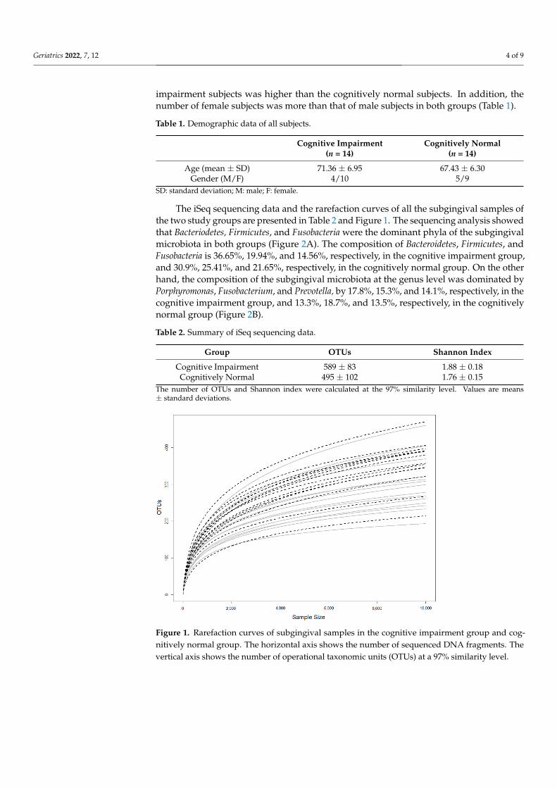
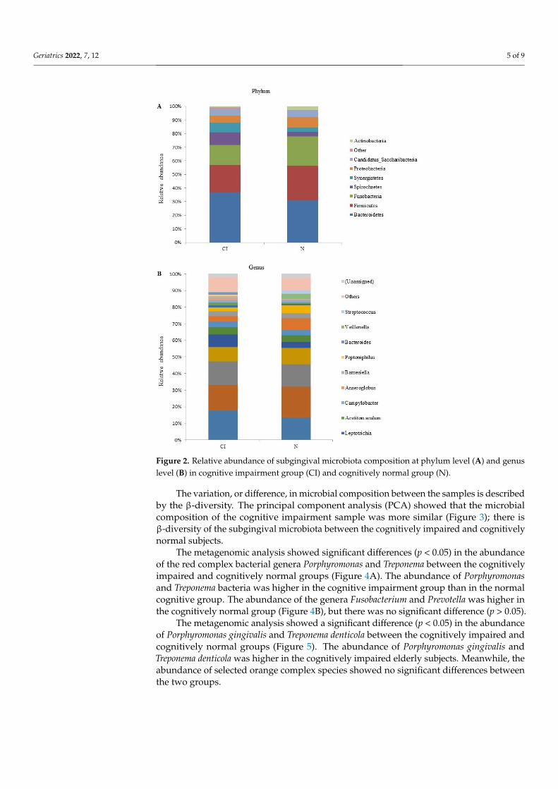
The iSeq sequencing data and the rarefaction curves of all the subgingival samples ofthe two study groups are presented in Table 2 and Figure 1. The sequencing analysis showedthat Bacteriodetes, Firmicutes, and Fusobacteria were the dominant phyla of the subgingivalmicrobiota in both groups (Figure 2A). The composition of Bacteroidetes, Firmicutes, andFusobacteria is 36.65%, 19.94%, and 14.56%, respectively, in the cognitive impairment group,and 30.9%, 25.41%, and 21.65%, respectively, in the cognitively normal group. On the otherhand, the composition of the subgingival microbiota at the genus level was dominated byPorphyromonas, Fusobacterium, and Prevotella, by 17.8%, 15.3%, and 14.1%, respectively, in thecognitive impairment group, and 13.3%, 18.7%, and 13.5%, respectively, in the cognitivelynormal group (Figure 2B).
Table 2. Summary of iSeq sequencing data.
Group OTUs Shannon Index
Cognitive Impairment 589 ± 83 1.88 ± 0.18Cognitively Normal 495 ± 102 1.76 ± 0.15
The number of OTUs and Shannon index were calculated at the 97% similarity level. Values are means± standard deviations.
Geriatrics 2022, 7, x FOR PEER REVIEW 4 of 10
3. Results Twenty-eight elderly subjects fitted the inclusion and exclusion criteria. There was a
cognitive impairment group and a cognitively normal group, each consisting of 14 sub-jects according to previous examinations, MMSE and HVLT. The mean age of the cogni-tive impairment subjects was higher than the cognitively normal subjects. In addition, the number of female subjects was more than that of male subjects in both groups (Table 1).
Table 1. Demographic data of all subjects.
Cognitive Impairment (n = 14)
Cognitively Normal (n = 14)
Age (mean ± SD) 71.36 ± 6.95 67.43 ± 6.30 Gender (M/F) 4/10 5/9
SD: standard deviation; M: male; F: female.
The iSeq sequencing data and the rarefaction curves of all the subgingival samples of the two study groups are presented in Table 2 and Figure 1. The sequencing analysis showed that Bacteriodetes, Firmicutes, and Fusobacteria were the dominant phyla of the sub-gingival microbiota in both groups (Figure 2A). The composition of Bacteroidetes, Firmicu-tes, and Fusobacteria is 36.65%, 19.94%, and 14.56%, respectively, in the cognitive impair-ment group, and 30.9%, 25.41%, and 21.65%, respectively, in the cognitively normal group. On the other hand, the composition of the subgingival microbiota at the genus level was dominated by Porphyromonas, Fusobacterium, and Prevotella, by 17.8%, 15.3%, and 14.1%, respectively, in the cognitive impairment group, and 13.3%, 18.7%, and 13.5%, re-spectively, in the cognitively normal group (Figure 2B).
Table 2. Summary of iSeq sequencing data.
Group OTUs Shannon Index Cognitive Impairment 589 ± 83 1.88 ± 0.18 Cognitively Normal 495 ± 102 1.76 ± 0.15
The number of OTUs and Shannon index were calculated at the 97% similarity level. Values are means ± standard deviations.
Figure 1. Rarefaction curves of subgingival samples in the cognitive impairment group and cogni-tively normal group. The horizontal axis shows the number of sequenced DNA fragments. The ver-tical axis shows the number of operational taxonomic units (OTUs) at a 97% similarity level.
Figure 1. Rarefaction curves of subgingival samples in the cognitive impairment group and cog-nitively normal group. The horizontal axis shows the number of sequenced DNA fragments. Thevertical axis shows the number of operational taxonomic units (OTUs) at a 97% similarity level.
Geriatrics 2022, 7, 12 5 of 9
Geriatrics 2022, 7, x FOR PEER REVIEW 5 of 10
Figure 2. Relative abundance of subgingival microbiota composition at phylum level (A) and genus level (B) in cognitive impairment group (CI) and cognitively normal group (N).
The variation, or difference, in microbial composition between the samples is de-scribed by the β-diversity. The principal component analysis (PCA) showed that the mi-crobial composition of the cognitive impairment sample was more similar (Figure 3); there is β-diversity of the subgingival microbiota between the cognitively impaired and cogni-tively normal subjects.
The metagenomic analysis showed significant differences (p < 0.05) in the abundance of the red complex bacterial genera Porphyromonas and Treponema between the cognitively impaired and cognitively normal groups (Figure 4A). The abundance of Porphyromonas and Treponema bacteria was higher in the cognitive impairment group than in the normal cognitive group. The abundance of the genera Fusobacterium and Prevotella was higher in the cognitively normal group (Figure 4B), but there was no significant difference (p > 0.05).
The metagenomic analysis showed a significant difference (p < 0.05) in the abundance of Porphyromonas gingivalis and Treponema denticola between the cognitively impaired and cognitively normal groups (Figure 5). The abundance of Porphyromonas gingivalis and Treponema denticola was higher in the cognitively impaired elderly subjects. Meanwhile, the abundance of selected orange complex species showed no significant differences be-tween the two groups.
Figure 2. Relative abundance of subgingival microbiota composition at phylum level (A) and genuslevel (B) in cognitive impairment group (CI) and cognitively normal group (N).
The variation, or difference, in microbial composition between the samples is describedby the β-diversity. The principal component analysis (PCA) showed that the microbialcomposition of the cognitive impairment sample was more similar (Figure 3); there isβ-diversity of the subgingival microbiota between the cognitively impaired and cognitivelynormal subjects.
The metagenomic analysis showed significant differences (p < 0.05) in the abundanceof the red complex bacterial genera Porphyromonas and Treponema between the cognitivelyimpaired and cognitively normal groups (Figure 4A). The abundance of Porphyromonasand Treponema bacteria was higher in the cognitive impairment group than in the normalcognitive group. The abundance of the genera Fusobacterium and Prevotella was higher inthe cognitively normal group (Figure 4B), but there was no significant difference (p > 0.05).
The metagenomic analysis showed a significant difference (p < 0.05) in the abundanceof Porphyromonas gingivalis and Treponema denticola between the cognitively impaired andcognitively normal groups (Figure 5). The abundance of Porphyromonas gingivalis andTreponema denticola was higher in the cognitively impaired elderly subjects. Meanwhile, theabundance of selected orange complex species showed no significant differences betweenthe two groups.

Geriatrics 2022, 7, 12 6 of 9Geriatrics 2022, 7, x FOR PEER REVIEW 6 of 10
Figure 3. Principal component analysis (PCA) of subgingival microbiota in cognitive impairment group (red) and cognitively normal group (blue).
Figure 4. Box and whisker plot on percentage abundance of red complex (A) and orange complex (B) genera in cognitive impairment group (CI) and cognitively normal group (N). * Significant p < 0.05.
Figure 3. Principal component analysis (PCA) of subgingival microbiota in cognitive impairmentgroup (red) and cognitively normal group (blue).
Geriatrics 2022, 7, x FOR PEER REVIEW 6 of 10
Figure 3. Principal component analysis (PCA) of subgingival microbiota in cognitive impairment group (red) and cognitively normal group (blue).
Figure 4. Box and whisker plot on percentage abundance of red complex (A) and orange complex (B) genera in cognitive impairment group (CI) and cognitively normal group (N). * Significant p < 0.05.
Figure 4. Box and whisker plot on percentage abundance of red complex (A) and orange complex(B) genera in cognitive impairment group (CI) and cognitively normal group (N). * Significant p < 0.05.

Geriatrics 2022, 7, 12 7 of 9
Geriatrics 2022, 7, x FOR PEER REVIEW 7 of 10
Figure 5. Box and whisker plot on percentage abundance of red complex (A) and orange complex (B) species in cognitive impairment group (CI) and cognitively normal group (N). * Significant p < 0.05.
4. Discussion In this study, we evaluated the subgingival microbiota composition in cognitively
impaired and cognitively normal elderly subjects with periodontitis, before analyzing the abundance of red and orange complex periodontal pathogens; 16S rRNA sequencing was used to determine the subgingival microbiota composition. β-diversity of the subgingival microbiome was found between the cognitively impaired and cognitively normal subjects. Research linking the subgingival microbiome to cognitive impairment is limited. This study is the first to evaluate the composition of the subgingival microbiota in periodontitis subjects with and without cognitive impairment in the Indonesian elderly to the best of the authors’ knowledge. A recent study by Holmer et al. (2021) demonstrated differences in the subgingival microbiota between cognitive dysfunction individuals and cognitively healthy individuals. Slackia exigua and Lachnospiraceae bacteria were more abundant in Alzheimer’s subjects than in controls [21]. Yang et al. identified Pasteurellacae and Lautro-pia mirabilis as having different abundancies between cognitively impaired and normal subjects [22].
The metagenomic analysis of periodontal pathogens showed a significant difference in P. gingivalis and T. denticola abundance, which was higher in the cognitive impairment subjects. Porphyromonas gingivalis is a periodontal pathogen that has been widely studied and associated with cognitive impairment. The Gram-negative bacterium P. gingivalis is a key pathogen that modulates the dysbiosis of its companion bacterial species [23]. More-over, P. gingivalis and T. denticola are two members of the triad of anaerobic red complex bacteria, which can be predictors of periodontitis progression [24].
Studies using other approaches to detect microbes showed that the periodontal bac-terium P. gingivalis is associated with impaired cognitive function. The administration of LPS P. gingivalis can cause cognitive impairment in C57BL/6 mice [25]. Ishida et al. also conducted a study on mice; they concluded that periodontitis induced by P. gingivalis
Figure 5. Box and whisker plot on percentage abundance of red complex (A) and orange complex(B) species in cognitive impairment group (CI) and cognitively normal group (N). * Significant p < 0.05.
4. Discussion
In this study, we evaluated the subgingival microbiota composition in cognitivelyimpaired and cognitively normal elderly subjects with periodontitis, before analyzing theabundance of red and orange complex periodontal pathogens; 16S rRNA sequencing wasused to determine the subgingival microbiota composition. β-diversity of the subgingivalmicrobiome was found between the cognitively impaired and cognitively normal subjects.Research linking the subgingival microbiome to cognitive impairment is limited. Thisstudy is the first to evaluate the composition of the subgingival microbiota in periodontitissubjects with and without cognitive impairment in the Indonesian elderly to the best of theauthors’ knowledge. A recent study by Holmer et al. (2021) demonstrated differences in thesubgingival microbiota between cognitive dysfunction individuals and cognitively healthyindividuals. Slackia exigua and Lachnospiraceae bacteria were more abundant in Alzheimer’ssubjects than in controls [21]. Yang et al. identified Pasteurellacae and Lautropia mirabilis ashaving different abundancies between cognitively impaired and normal subjects [22].
The metagenomic analysis of periodontal pathogens showed a significant differencein P. gingivalis and T. denticola abundance, which was higher in the cognitive impairmentsubjects. Porphyromonas gingivalis is a periodontal pathogen that has been widely studiedand associated with cognitive impairment. The Gram-negative bacterium P. gingivalisis a key pathogen that modulates the dysbiosis of its companion bacterial species [23].Moreover, P. gingivalis and T. denticola are two members of the triad of anaerobic redcomplex bacteria, which can be predictors of periodontitis progression [24].
Studies using other approaches to detect microbes showed that the periodontal bac-terium P. gingivalis is associated with impaired cognitive function. The administration ofLPS P. gingivalis can cause cognitive impairment in C57BL/6 mice [25]. Ishida et al. alsoconducted a study on mice; they concluded that periodontitis induced by P. gingivalis couldexacerbate brain Aβ deposition, leading to cognitive impairment through mechanisms thatinduce brain inflammation [26]. Research by Leblhuber et al. demonstrated that P. gingivaliswas associated with lower MMSE scores [27]. The role of T. denticola in cognitive functionhas not been studied as much as P. gingivalis. Su et al. (2021) demonstrated that T. denticolacould enter the brain, act directly on nerve cells, and result in intra- and extracellular Aβ1-40and Aβ1-42 accumulation in the hippocampus of C57BL/6 mice [28].
Geriatrics 2022, 7, 12 8 of 9
The orange complex pathogen is known for its ability to adhere to several oral bacteria.These bacteria can bind to other bacteria and are considered as “linking” organisms thatbridge commensal colonies, which are generally periodontal pathogens [29]. The presenceof this orange complex bacteria is very important; without it, the aggressiveness of the redcomplex would not survive in the oral cavity [30]. Nevertheless, we found no significantdifferences in the abundance of orange complex bacteria between the cognitively impairedand cognitively normal groups.
This study contributes to the development of the knowledge on the relationshipbetween periodontal pathogens and cognitive impairment, by finding a higher abundanceof P. gingivalis and T. denticola in the cognitively impaired group than in the cognitivelynormal group. The strength of this study is that all the subjects were diagnosed withperiodontitis and the subgingival samples in both groups were taken from the samepocket depth. In addition, the subjects are elderly individuals who live in the samenursing home, so they may have the same dietary habits. Nevertheless, periodontitis andcognitive impairment have various risk factors that were not included in this study, suchas educational background and depression. Another limitation is that this study did notdetermine a causal relationship between cognitive impairment and periodontitis. However,it is important to detect cognitive impairment early. Attention and awareness of periodontalhealth in the elderly are also needed.
5. Conclusions
In this study, we found that the subgingival microbiome in the cognitively impairedand cognitively normal groups was distinct. The abundance of P. gingivalis and T. denti-cola is potentially affected by cognitive impairment conditions. The role of P. gingivalisand T. denticola in the pathogenesis of cognitive impairment needs further investigation.Since the population of the present study is periodontitis subjects, the interpretation andgeneralization of the findings should be carried out among periodontitis subjects as well.
Author Contributions: Conceptualization: F.M.T., S.L.C.M. and B.M.B.; methodology: S.L.C.M.,L.S.K., B.M.B., Y.T. and R.I.I.; statistical analysis: F.M.T., M.I.R. and B.M.B.; writing—original draftpreparation: F.M.T.; writing—review and editing: F.M.T., S.L.C.M., M.I.R., L.S.K., Y.T., R.I.I. andB.M.B.; supervision and project administration: S.L.C.M. All authors have read and agreed to thepublished version of the manuscript.
Funding: This study was financially supported by a grant from Universitas Indonesia (Hibah RisetUI 2021), grant number NKB-495/UN2.RST/HKP.05.00/2021.
Institutional Review Board Statement: The study was conducted according to the guidelines ofthe Declaration of Helsinki and approved by the Committee on Ethics of Dental Research (KEPKG)Faculty of Dentistry, Universitas Indonesia (protocol code 070210219).
Informed Consent Statement: Informed consent was obtained from all subjects involved in the study.
Data Availability Statement: All data will be made available upon reasonable request.
Conflicts of Interest: The authors declare no conflict of interest. The funders had no role in the designof the study; in the collection, analyses, or interpretation of data; in the writing of the manuscript, orin the decision to publish the results.
References1. United Nations. Department of Economic and Social Affairs, Population Division. In World Population Prospects: The 2017
Revision-Key Findings and Advance Tables; Working Paper No. ESA/P/WP/248; United Nations: New York, NY, USA, 2017.2. Adioetomo, S.M.; Mujahid, G. Indonesia on the Threshold of Population Ageing; Monograph Series: No.1; UNFPA: Indonesia,
Jakarta, 2014.3. Patterson, C. World Alzheimer Report 2018-The State of the Art of Dementia Research: New Frontiers; Alzheimer’s Disease International
(ADI): London, UK, 2018.4. Gil-Montoya, J.A.; Sanchez-Lara, I.; Carnero-Pardo, C.; Fornieles, F.; Montes, J.; Vilchez, R.; Burgos, J.S.; Gonzales-Moles, M.A.;
Barrios, R.; Bravo, M. Is periodontitis a risk factor for cognitive impairment and dementia? A case-control study. J. Periodontol.2015, 86, 244–253. [CrossRef]
Geriatrics 2022, 7, 12 9 of 9
5. Iwasaki, M.; Kimura, Y.; Ogawa, H.; Yamaga, T.; Ansai, T.; Wada, T.; Sakamoto, R.; Ishimoto, Y.; Fujisawa, M.; Okumiya, K.;et al. Periodontitis, periodontal inflammation, and mild cognitive impairment: A 5-year cohort study. J. Periodontal Res. 2019, 54,233–240. [CrossRef]
6. Tadjoedin, F.M.; Kusdhany, L.S.; Turana, Y.; Bachtiar, B.M.; Masulili, S.L.C. Periodontal parameters in Indonesian elderly and itsassociation with cognitive impairment. J. Int. Dent. Med. Res. 2020, 13, 1009–1012.
7. Kim, D.H.; Han, G.S. The relationship between periodontal disease and cognitive impairment in older adults of Korea.Spec. Care Dent. 2021. [CrossRef]
8. Iwasaki, M.; Yoshihara, A.; Kimura, Y.; Sato, M.; Wada, T.; Sakamoto, R.; Ishimoto, Y.; Fukutomi, E.; Chen, W.; Imai, H.; et al.Longitudinal relationship of severe periodontitis with cognitive decline in older Japanese. J. Periodont. Res. 2016, 51, 681–688.[CrossRef]
9. Sung, C.E.; Huang, R.Y.; Cheng, W.C.; Kao, T.W.; Chen, W.L. Association between periodontitis and cognitive impairment:Analysis of national health and nutrition examination survey (NHANES) III. J. Clin. Periodontol. 2019, 46, 790–798. [CrossRef][PubMed]
10. Guo, H.; Chang, S.; Pi, X.; Hua, F.; Jiang, H.; Liu, C.; Minquan, D. The effect of periodontitis on dementia and cognitive impairment:A meta-analysis. Int. J. Environ. Res. Public Health. 2021, 18, 6823. [CrossRef] [PubMed]
11. Papapanou, P.N.; Sanz, M.; Buduneli, N.; Dietrich, T.; Feres, M.; Fine, D.H.; Flemmig, T.F.; Garcia, R.; Giannobile, W.V.; Graziani,F.; et al. Periodontitis: Consensus report of workgroup 2 of the 2017 World Workshop on the Classification of Periodontal andPeri-Implant Diseases and Conditions. J. Clin. Periodontol. 2018, 45 (Suppl. S20), S162–S170. [CrossRef]
12. Hajishengallis, G. Immunomicrobial pathogenesis of periodontitis: Keystones, pathobionts, and host response. Trends Immunol.2014, 35, 3–11. [CrossRef] [PubMed]
13. Feres, M.; Teles, F.; Teles, R.; Figueiredo, L.C.; Faveri, M. The subgingival periodontal microbiota of the aging mouth.Periodontology 2000 2016, 72, 30–53. [CrossRef] [PubMed]
14. Socransky, S.S.; Haffajee, A.D.; Cugini, M.A.; Smith, C.; Kent, R.L. Microbial complexes in subgingival plaque. J. Clin. Periodontol.1998, 25, 134–144. [CrossRef] [PubMed]
15. Bui, F.Q.; Almeida-da-silva, C.L.C.; Huynh, B.; Trinh, A.; Liu, J.; Woodward, J.; Asadi, H.; Ojcius, D.M. Association betweenperiodontal pathogens and systemic disease. Biomed. J. 2019, 42, 27–35. [CrossRef]
16. Ding, Y.; Ren, J.; Yu, H.; Yu, W.; Zhou, Y. Porphyromonas gingivalis, a periodontitis causing bacterium, induces memoryimpairment and age-dependent neuroinflammation in mice. Immun. Ageing. 2018, 15, 6. [CrossRef] [PubMed]
17. Hogervorst, E.; Mursjid, F.; Ismail, R.I.; Prasetyo, S.; Nasrun, M.; Mochtar; Ninuk, T.; Bandelow, S.; Subarkah; Kusdhany, L.; et al.Validation of two short dementia screening tests in Indonesia. In Vascular Dementia: Risk Factors, Diagnosis and Treatment; Jacobsen,S.R., Ed.; Nova Science: New York, NY, USA, 2011; pp. 235–256.
18. Brandt, J. The hopkins verbal learning test: Development of a new memory test with six equivalent forms. Clin. Neuropsychol.1991, 5, 125–142. [CrossRef]
19. Folstein, M.F.; Folstein, S.E.; McHugh, P.R. “Mini-mental state.” A practical method for grading cognitive state of patients forclinician. J. Psychiatr. Res. 1975, 12, 189–198. [CrossRef]
20. Benjamini, Y.; Hochberg, Y. Controlling the false discovery rate: A practical and powerful approach to multiple testing.J. R. Stat. Soc. 1995, 57, 289–300. [CrossRef]
21. Holmer, J.; Aho, V.; Eriksdotter, M.; Paulin, L.; Pietiäinen, M.; Auvinen, P.; Schultzberg, M.; Pussinen, P.J.; Buhlin, K. Subgingivalmicrobiota in a population with and without cognitive dysfunction. J. Oral Microbiol. 2021, 13, 1854552. [CrossRef]
22. Yang, I.; Arthur, R.A.; Zhao, L.; Clark, J.; Hu, Y.; Corwin, E.J.; Lah, J. The oral microbiome and inflammation in mild cognitiveimpairment. Exp. Gerontol. 2021, 147, 111273. [CrossRef]
23. Hajishengallis, G.; Lamont, R.J. Breaking bad: Manipulation of the host response by Porphyromonas gingivalis. Eur. J. Immunol.2014, 44, 328–338. [CrossRef] [PubMed]
24. Byrne, S.J.; Dashper, S.G.; Darby, I.B.; Adams, G.G.; Hoffmann, B.; Reynolds, E.C. Progression of chronic periodontitis can bepredicted by the levels of Porphyromonas gingivalis and Treponema denticola in subgingival plaque. Oral Microbiol. Immunol.2009, 24, 469–477. [CrossRef]
25. Zhang, J.; Yu, C.; Zhang, X.; Chen, H.; Dong, J.; Lu, W.; Song, Z.; Zhou, W. Porphyromonas gingivalis lipopolysaccharide inducescognitive dysfunction, mediated by neuronal inflammation via activation of the TLR4 signaling pathway in C57BL/6 mice.J. Neuroinflamm. 2018, 15, 37. [CrossRef] [PubMed]
26. Ishida, N.; Ishihara, Y.; Ishida, K.; Tada, H.; Funaki-Kato, Y.; Hagiwara, M.; Ferdous, T.; Abdullah, M.; Mitani, A.; Michikawa,M.; et al. Periodontitis induced by bacterial infection exacerbates features of Alzheimer’s disease in transgenic mice.NPJ Aging Mech Dis. 2017, 3, 15. [CrossRef] [PubMed]
27. Leblhuber, F.; Huemer, J.; Steiner, K.; Gostner, J.M.; Fuchs, D. Knock-on effect of periodontitis to the pathogenesis of Alzheimer’sdisease? Wien Klin. Wochenschr. 2020, 132, 493–498. [CrossRef]
28. Su, X.; Tang, Z.; Lu, Z.; Liu, Y.; He, W.; Jiang, J.; Zhang, Y.; Wu, H. Oral Treponema denticola Infection Induces Aβ1–40 andAβ1–42 Accumulation in the Hippocampus of C57BL/6 Mice. J. Mol. Neurosci. 2021, 71, 1506–1514. [CrossRef] [PubMed]
29. Hajishengallis, G.; Lamont, R.J. Beyond the Red Complex and Into More Complexity: The Polymicrobial Synergy and Dysbiosis(PSD) Model of Periodontal Disease Etiology. Mol. Oral Microbiol. 2012, 27, 409–419. [CrossRef] [PubMed]
30. Genco, R.J.; Borgnakke, W.S. Risk Factors for Periodontal Disease. Periodontol. 2000 2013, 62, 59–94. [CrossRef]