The Microbiome of Leonardo da Vinci's Drawings: A Bio ...
22
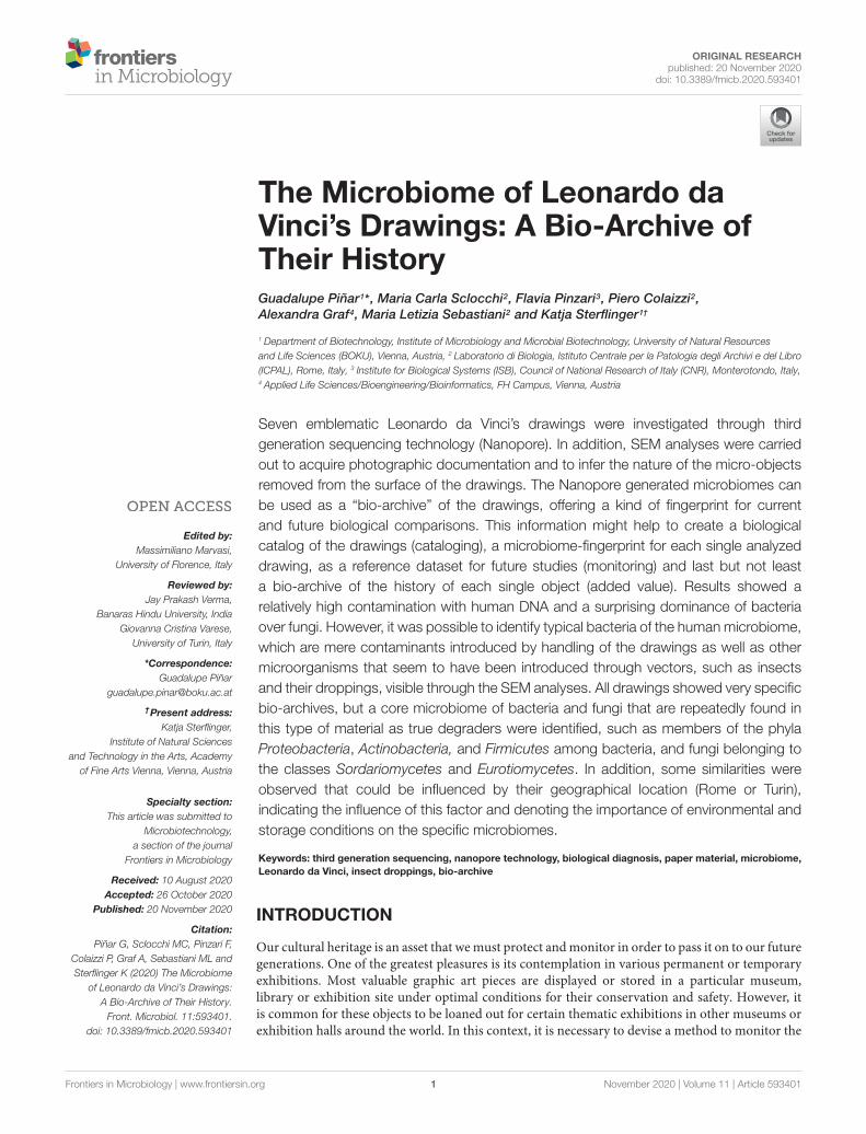
ORIGINAL RESEARCH published: 20 November 2020 doi: 10.3389/fmicb.2020.593401 Edited by: Massimiliano Marvasi, University of Florence, Italy Reviewed by: Jay Prakash Verma, Banaras Hindu University, India Giovanna Cristina Varese, University of Turin, Italy *Correspondence: Guadalupe Piñar [email protected] † Present address: Katja Sterflinger, Institute of Natural Sciences and Technology in the Arts, Academy of Fine Arts Vienna, Vienna, Austria Specialty section: This article was submitted to Microbiotechnology, a section of the journal Frontiers in Microbiology Received: 10 August 2020 Accepted: 26 October 2020 Published: 20 November 2020 Citation: Piñar G, Sclocchi MC, Pinzari F, Colaizzi P, Graf A, Sebastiani ML and Sterflinger K (2020) The Microbiome of Leonardo da Vinci’s Drawings: A Bio-Archive of Their History. Front. Microbiol. 11:593401. doi: 10.3389/fmicb.2020.593401 The Microbiome of Leonardo da Vinci’s Drawings: A Bio-Archive of Their History Guadalupe Piñar 1 * , Maria Carla Sclocchi 2 , Flavia Pinzari 3 , Piero Colaizzi 2 , Alexandra Graf 4 , Maria Letizia Sebastiani 2 and Katja Sterflinger 1† 1 Department of Biotechnology, Institute of Microbiology and Microbial Biotechnology, University of Natural Resources and Life Sciences (BOKU), Vienna, Austria, 2 Laboratorio di Biologia, Istituto Centrale per la Patologia degli Archivi e del Libro (ICPAL), Rome, Italy, 3 Institute for Biological Systems (ISB), Council of National Research of Italy (CNR), Monterotondo, Italy, 4 Applied Life Sciences/Bioengineering/Bioinformatics, FH Campus, Vienna, Austria Seven emblematic Leonardo da Vinci’s drawings were investigated through third generation sequencing technology (Nanopore). In addition, SEM analyses were carried out to acquire photographic documentation and to infer the nature of the micro-objects removed from the surface of the drawings. The Nanopore generated microbiomes can be used as a “bio-archive” of the drawings, offering a kind of fingerprint for current and future biological comparisons. This information might help to create a biological catalog of the drawings (cataloging), a microbiome-fingerprint for each single analyzed drawing, as a reference dataset for future studies (monitoring) and last but not least a bio-archive of the history of each single object (added value). Results showed a relatively high contamination with human DNA and a surprising dominance of bacteria over fungi. However, it was possible to identify typical bacteria of the human microbiome, which are mere contaminants introduced by handling of the drawings as well as other microorganisms that seem to have been introduced through vectors, such as insects and their droppings, visible through the SEM analyses. All drawings showed very specific bio-archives, but a core microbiome of bacteria and fungi that are repeatedly found in this type of material as true degraders were identified, such as members of the phyla Proteobacteria, Actinobacteria, and Firmicutes among bacteria, and fungi belonging to the classes Sordariomycetes and Eurotiomycetes. In addition, some similarities were observed that could be influenced by their geographical location (Rome or Turin), indicating the influence of this factor and denoting the importance of environmental and storage conditions on the specific microbiomes. Keywords: third generation sequencing, nanopore technology, biological diagnosis, paper material, microbiome, Leonardo da Vinci, insect droppings, bio-archive INTRODUCTION Our cultural heritage is an asset that we must protect and monitor in order to pass it on to our future generations. One of the greatest pleasures is its contemplation in various permanent or temporary exhibitions. Most valuable graphic art pieces are displayed or stored in a particular museum, library or exhibition site under optimal conditions for their conservation and safety. However, it is common for these objects to be loaned out for certain thematic exhibitions in other museums or exhibition halls around the world. In this context, it is necessary to devise a method to monitor the Frontiers in Microbiology | www.frontiersin.org 1 November 2020 | Volume 11 | Article 593401
-
Upload
khangminh22 -
Category
Documents
-
view
0 -
download
0
Transcript of The Microbiome of Leonardo da Vinci's Drawings: A Bio ...
fmicb-11-593401 November 16, 2020 Time: 16:59 # 1
ORIGINAL RESEARCHpublished: 20 November 2020
doi: 10.3389/fmicb.2020.593401
Edited by:Massimiliano Marvasi,
University of Florence, Italy
Reviewed by:Jay Prakash Verma,
Banaras Hindu University, IndiaGiovanna Cristina Varese,
University of Turin, Italy
*Correspondence:Guadalupe Piñar
†Present address:Katja Sterflinger,
Institute of Natural Sciencesand Technology in the Arts, Academy
of Fine Arts Vienna, Vienna, Austria
Specialty section:This article was submitted to
Microbiotechnology,a section of the journal
Frontiers in Microbiology
Received: 10 August 2020Accepted: 26 October 2020
Published: 20 November 2020
Citation:Piñar G, Sclocchi MC, Pinzari F,
Colaizzi P, Graf A, Sebastiani ML andSterflinger K (2020) The Microbiome
of Leonardo da Vinci’s Drawings:A Bio-Archive of Their History.
Front. Microbiol. 11:593401.doi: 10.3389/fmicb.2020.593401
The Microbiome of Leonardo daVinci’s Drawings: A Bio-Archive ofTheir HistoryGuadalupe Piñar1* , Maria Carla Sclocchi2, Flavia Pinzari3, Piero Colaizzi2,Alexandra Graf4, Maria Letizia Sebastiani2 and Katja Sterflinger1†
1 Department of Biotechnology, Institute of Microbiology and Microbial Biotechnology, University of Natural Resourcesand Life Sciences (BOKU), Vienna, Austria, 2 Laboratorio di Biologia, Istituto Centrale per la Patologia degli Archivi e del Libro(ICPAL), Rome, Italy, 3 Institute for Biological Systems (ISB), Council of National Research of Italy (CNR), Monterotondo, Italy,4 Applied Life Sciences/Bioengineering/Bioinformatics, FH Campus, Vienna, Austria
Seven emblematic Leonardo da Vinci’s drawings were investigated through thirdgeneration sequencing technology (Nanopore). In addition, SEM analyses were carriedout to acquire photographic documentation and to infer the nature of the micro-objectsremoved from the surface of the drawings. The Nanopore generated microbiomes canbe used as a “bio-archive” of the drawings, offering a kind of fingerprint for currentand future biological comparisons. This information might help to create a biologicalcatalog of the drawings (cataloging), a microbiome-fingerprint for each single analyzeddrawing, as a reference dataset for future studies (monitoring) and last but not leasta bio-archive of the history of each single object (added value). Results showed arelatively high contamination with human DNA and a surprising dominance of bacteriaover fungi. However, it was possible to identify typical bacteria of the human microbiome,which are mere contaminants introduced by handling of the drawings as well as othermicroorganisms that seem to have been introduced through vectors, such as insectsand their droppings, visible through the SEM analyses. All drawings showed very specificbio-archives, but a core microbiome of bacteria and fungi that are repeatedly found inthis type of material as true degraders were identified, such as members of the phylaProteobacteria, Actinobacteria, and Firmicutes among bacteria, and fungi belonging tothe classes Sordariomycetes and Eurotiomycetes. In addition, some similarities wereobserved that could be influenced by their geographical location (Rome or Turin),indicating the influence of this factor and denoting the importance of environmental andstorage conditions on the specific microbiomes.
Keywords: third generation sequencing, nanopore technology, biological diagnosis, paper material, microbiome,Leonardo da Vinci, insect droppings, bio-archive
INTRODUCTION
Our cultural heritage is an asset that we must protect and monitor in order to pass it on to our futuregenerations. One of the greatest pleasures is its contemplation in various permanent or temporaryexhibitions. Most valuable graphic art pieces are displayed or stored in a particular museum,library or exhibition site under optimal conditions for their conservation and safety. However, itis common for these objects to be loaned out for certain thematic exhibitions in other museums orexhibition halls around the world. In this context, it is necessary to devise a method to monitor the
Frontiers in Microbiology | www.frontiersin.org 1 November 2020 | Volume 11 | Article 593401
fmicb-11-593401 November 16, 2020 Time: 16:59 # 2
Piñar et al. Leonardo da Vinci’s Microbiomes
risks of microbial contamination that may occur duringpackaging, transport and/or exhibition at the other sites and toallow a comparison before and after these trips. This monitoringis carried out by restorers and experts from the variousinstitutions that provide and receive the object in question,but also by experts from renowned third-party institutions,which apply conventional technologies such as visual inspection,microscopy, but also introducing molecular technologies inorder to achieve a detailed assessment of the risk of biologicalcontamination posed by this type of transfer.
Conservators have long since started using new technologiessuch as miniaturized environmental condition recorders thatkeep track of how an object is stored during a move or anexhibition, away from the usual storage environment (Valentiniet al., 2018). In addition to temperature and humidity monitoringsystems, modern methods of classification and identificationhave also been developed, such as smart labels and barcodestechnologies, which make it possible to quickly catalog objectsand are already widely used in libraries. For example, theRadio-Frequency Identification systems (Steinberg et al., 2014)leverage radio waves to transmit data from chips to the readers(Alwadi et al., 2017). These systems help to cover traditionalwork processes such as check-in, check-out, anti-theft controland management of inventories. Even more innovative arethe labels based on synthetic DNA (Demidov, 2020). Theseare patented systems that make it possible to make the artpieces not forgeable. Artists can authenticate their work bylabeling it with a univocal synthetic bioengineered DNA sequencethat provides an encrypted link between the artwork and asecure database containing the definitive information about theartwork. DNA data can be retrieved by sequencing or usingother technologies like fluorescent probes (Banerjee et al., 2016).However, any object already contains DNA, from the peoplewho have handled the object since its creation and from all theorganisms and microorganisms that have come into contact withit, also through the deposition of dust and dirt. This natural bio-archive changes with time and can give certain information onsome particular events, such as the presence on the materials ofdeteriorating microorganisms harmful for conservation, but alsothe permanence of the object in particular geographical locations,as indicated by the molecular traces left by the pollen of endemicplants (Piñar et al., 2019).
Molecular techniques, including DNA-based fingerprintingtechniques in addition to cloning and Sanger sequencing,have been largely applied to investigate and monitor thebiological colonization on art objects since more than twodecades (Schabereiter-Gurtner et al., 2001). This molecularstrategy, together with imaging and chemical analyses, hasdelivered relevant information about the microbial communitiesassociated with different materials and the microbiologicalrisk they represented on their surface (Sterflinger and Piñar,2013). Nevertheless, the use of first-generation sequencingtechnology delivers limited data and is time consuming aswell as not free of bias. This strategy has become obsoleteand has been replaced by new high-throughput moleculartechnologies in which massive DNA sequencing is possible,offering a much broader view of the real communities that
colonize our cultural heritage. Next Generation Sequencing(NGS) technologies have evolved incorporating revolutionaryimprovements to address the complexities of genomes andmetagenomes at an unprecedented speed (Goodwin et al.,2016), being now well established in the field of culturalheritage. These analyses can be performed either by the so-called "shotgun metagenomic approach," by sequencing the entireDNA library representing all the molecular components of agiven sample (Teasdale et al., 2017; Piñar et al., 2020b), orby focusing on specific conserved sequences such as ribosomalRNA genes (Marvasi et al., 2019). The latter approach called“target amplification approach" has some advantages, such asthe reduction of data complexity, the possibility to assign moresequences to specific organisms and finally, facilitates some semi-quantitative analysis. This approach is extremely useful in thefield of cultural heritage, as DNA with a poor quality or lowconcentration can be amplified by using degenerate primers andPCR. Many of the studies using the latter strategy have beencollected and summarized in the review of Marvasi et al. (2019).These methods are powerful, but as in any PCR-based method,primers and/or exponential amplification introduce bias. Inaddition, the simultaneous quantitative assessment of all threedomains of life is impossible. Furthermore, the amplificationof target sequences (mainly ribosomal sequences) do not allowus to draw conclusions on any functional genes and thereforethe microbial functional traits remain uncertain. Nevertheless,the further technological development of molecular methods isovercoming many technical biases. Third generation sequencingtechnologies are emerging and offering some advantages over thelimitations suffered by NGS platforms, such as the short-readlength that needs to be assembled with the help of bioinformatictools into the original length template, and the PCR biasintroduced by clonal amplification for the detection of baseincorporation signal.
Third generation sequencing technologies offer an intelligentsolution to the limitations mentioned above (Schadt et al.,2010). Instead of sequencing a clonally amplified template, asingle DNA template is sequenced, leading to a reduced use ofreagents and chemicals, simplified protocols, and miniaturizationof the entire technical process and equipment. One exampleis the Nanopore sequencing technology, which involves theuse of protein nanopores embedded in a polymer membrane.The sequencing does not necessarily require any interveningPCR amplification or chemical labeling step. Furthermore, thistechnology offers a pocket-sized sensing device, the MinION,which offers the advantages of a portable device. To date, theNanopore sequencing technology has been scarcely applied inthe field of cultural heritage, and only four studies have reportedon the advantages of applying this state-of-the-art technologyon valuable art objects. Three of these studies (Šoltys et al.,2019; Grottoli et al., 2020; Kisová et al., 2020) reported on theadvantages of sequencing long DNA fragments when targetingribosomal regions. However, all three studies were based onthe target amplification approach that avoided inferring therelative proportion of the different phylogenetic groups. Thefourth study (Piñar et al., 2020a) applied for the first time theNanopore sequencing technology together with a whole genome
Frontiers in Microbiology | www.frontiersin.org 2 November 2020 | Volume 11 | Article 593401

fmicb-11-593401 November 16, 2020 Time: 16:59 # 3
Piñar et al. Leonardo da Vinci’s Microbiomes
amplification (WGA) protocol to perform a rapid diagnosis of thebiological infection on objects of art, unifying all the advantagesthat this new technology can offer for metagenomic analysesstarting from very low DNA concentrations and reflecting the realproportions of all domains of life.
Although molecular techniques are not completely free frombias (Sterflinger et al., 2018a), the establishment of next-and third-generation sequencing technologies in the field ofcultural heritage is providing a powerful DNA-based approachto monitor the preservation status as well as to foresee therisk of deterioration of valuable objects (Piñar et al., 2020a).The data generated can provide interesting information thatcan contribute to surprising insights into the objects beinginvestigated, such as the selection of materials at the timeof manufacture and the conditions of their storage or aboutpossible displacements and geographical origins, but also uncoverimportant information about the object’s history of use (Teasdaleet al., 2017; Piñar et al., 2019, 2020b). This information mayhelp to understand many open questions in a variety of fields,as archeology, history, restoration, philology, criminology butalso, they can contribute to giving an historical benefit to theinvestigated objects in form of a bio-archive (Piñar et al., 2019,2020b).
In the present study we apply the Nanopore sequencingtechnology in combination with a whole genome amplification(WGA) protocol to survey some of Leonardo da Vinci’s mostemblematic drawings. This strategy was applied as the onlydiagnostic method or in combination with SEM analyses, inorder to explore the nature of contaminants deposited onthe surfaces of the drawings. The sampling strategy, basedon a very delicate micro-aspiration, was aimed primarily atcollecting dust particles, microbial cells and other debris fromsmall areas of surfaces. The aim was to create a bio-archiveof the dust and other collectable material deposited on thedrawings, in order to monitor their conservation status andthus be able to use them for possible comparisons in thefuture. The sampling was not used to assess the percentageof living propagules, however, the molecular strategy adoptedcould theoretically indicate the presence of fungal or bacterialcells with an anomalous abundance, compared to typical indoordust composition. Furthermore, the information contained inthis bio-archive is an added value for the drawings, that canin turn help to understand some issues related to the historyof storage of the investigated drawings. This study representsone more example of how the Nanopore technology, in thiscase in combination with microscopy techniques, can be apractical tool for the rapid biological diagnosis and monitoringin priceless objects.
MATERIALS AND METHODS
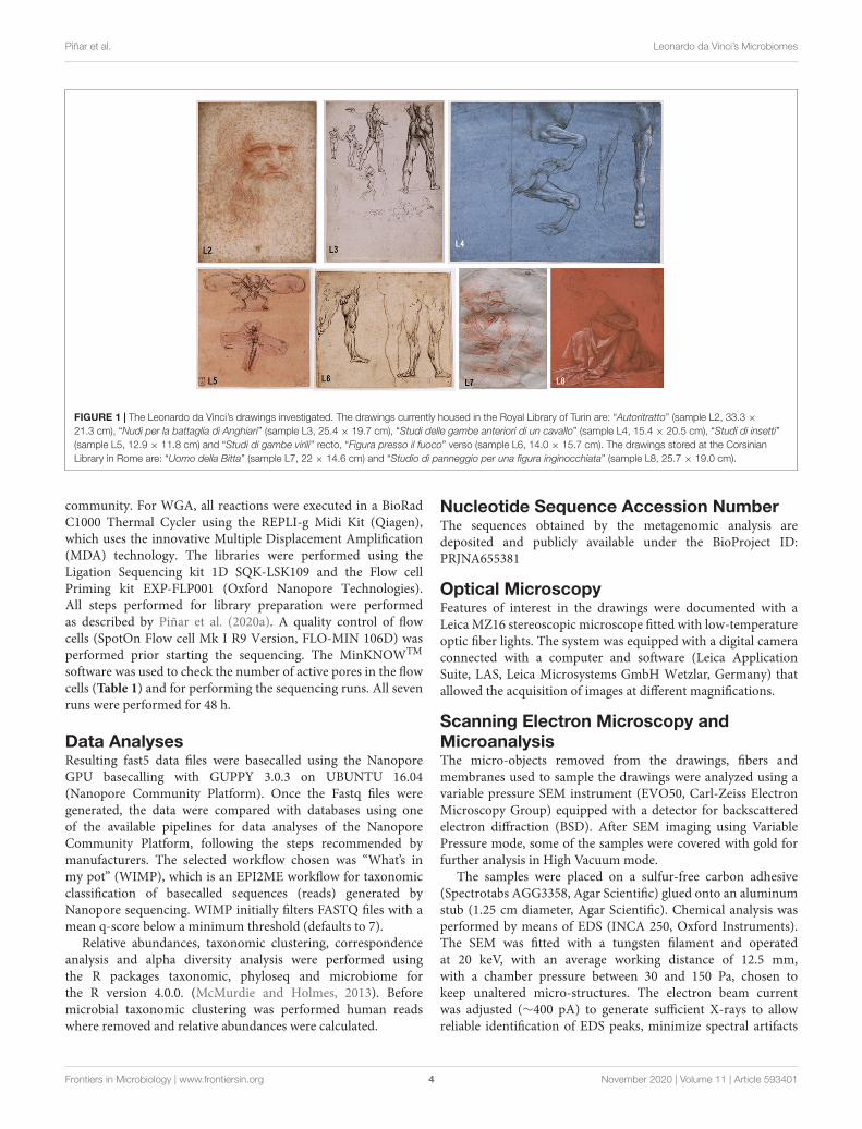
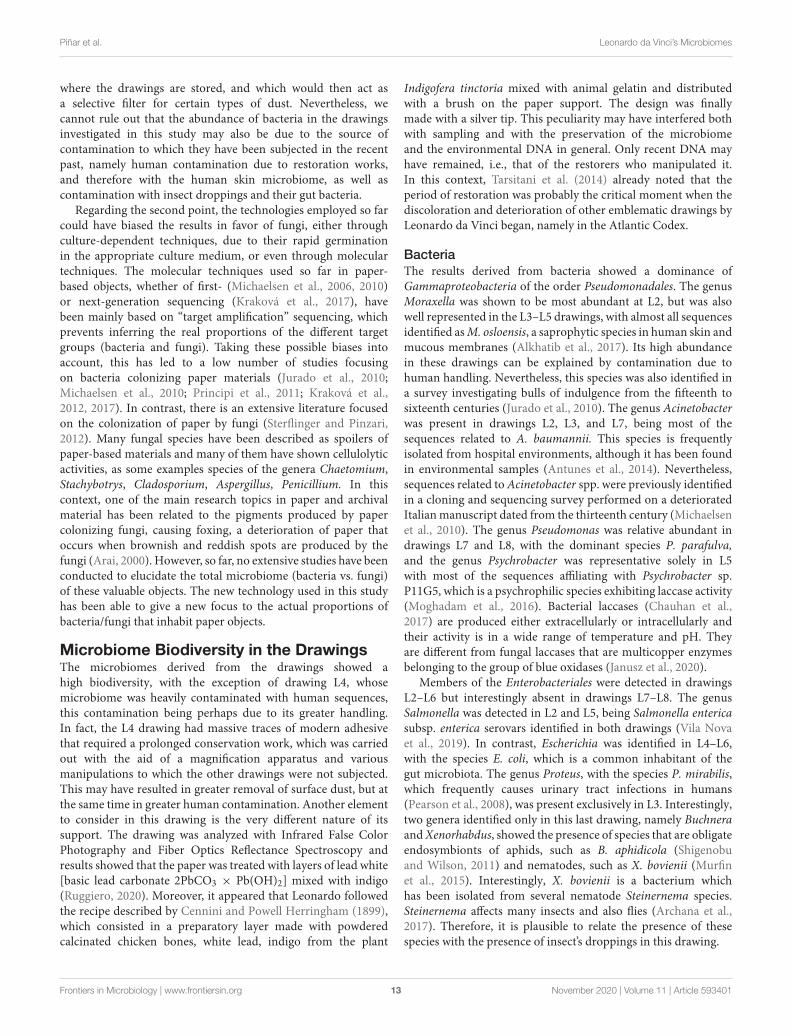
Objects and SamplingSeven emblematic drawings from Leonardo da Vinci wereselected for the analysis of their microbiomes (Figure 1). Fiveof these drawings are currently housed in the Royal Library ofTurin, located on the ground floor of the Royal Palace in Turin
(Italy): “Autoritratto” (sample L2, 33.3 × 21.3 cm), “Nudi per labattaglia di Anghiari” (sample L3, 25.4 × 19.7 cm), “Studi dellegambe anteriori di un cavallo” (sample L4, 15.4× 20.5 cm), “Studidi insetti” (sample L5, 12.9× 11.8 cm) and “Studi di gambe virili”recto, “Figura presso il fuoco” verso (sample L6, 14.0 × 15.7 cm).The last two investigated drawings are stored at the CorsinianLibrary in Rome (Italy): “Uomo della Bitta” (sample L7, 22 ×14.6 cm) and “Studio di panneggio per una figura inginocchiata”(sample L8, 25.7× 19.0 cm).
Conservators of the ICPAL (Istituto Centrale per la Patologiadegli Archivi e del Libro) collected samples from the rectoand the verso of the selected drawings. A micro-samplingmethod for fragile surfaces has been specifically developedbased on a protocol tested mainly on paintings and describedby de Carvalho et al. (2019). The system is based on amicro-aspiration aimed primarily at collecting dust particles,and microbial cells from small areas of paper or parchmentsurfaces (Supplementary Figure 1). The suction force appliedwas modulable, and the filter membranes used to trap theparticulates were sterile and made of a material suitablefor subsequent use in both molecular and SEM analysis.The micro-aspiration system was also used to sample themicroscopic objects detached from the drawings with needlesunder an optical microscope, as in the case of single fibers orparticles of a few hundred microns. The suction system wasan autoclavable polypropylene filter holder, 13 mm diameter(Swinnex filter holder, Millipore) with sterile membranes(Millipore cellulose nitrate membrane filter Ø = 13 mm, Poresize = 0.45 µm) connected to an oil-free diaphragm vacuumpump (DA14 Charles Austen MB2238. Appleton Woods Ltd.,United Kingdom).
The total number of membranes used for sampling variedbetween the drawings, ranging from 10 to 36 membranes perdrawing (depending on the size of the drawing). Each singleused membrane was immediately introduced in a single sterileEppendorf tube and appropriately labeled for the storage ofthe samples until DNA extraction analyses. The same kind ofmembranes was used for SEM observation of the particulatesampled from the drawings and its chemical analysis.
DNA ExtractionThe DNA was extracted using the FastDNA SPIN Kit for soil(MP Biomedicals, Illkrich, France) as recommended by themanufacturers. DNA extraction was performed directly from themembranes by grouping a maximum of four membranes perreaction tube. DNA extracted from each single reaction tubewas pooled per sample to obtain a single microbiome from eachdrawing. The DNA concentrations were assessed by using theQubit 2.0 fluorometer (Invitrogen Corporation), with the QubitdsDNA HS Assay Kit.
Whole Genome Amplification (WGA),Template Preparation and SequencingWhole genome amplification (WGA) and template preparationwas performed following the “Premium whole genomeamplification protocol” available in the Oxford Nanopore
Frontiers in Microbiology | www.frontiersin.org 3 November 2020 | Volume 11 | Article 593401
fmicb-11-593401 November 16, 2020 Time: 16:59 # 4
Piñar et al. Leonardo da Vinci’s Microbiomes
FIGURE 1 | The Leonardo da Vinci’s drawings investigated. The drawings currently housed in the Royal Library of Turin are: “Autoritratto” (sample L2, 33.3 ×21.3 cm), “Nudi per la battaglia di Anghiari” (sample L3, 25.4 × 19.7 cm), “Studi delle gambe anteriori di un cavallo” (sample L4, 15.4 × 20.5 cm), “Studi di insetti”(sample L5, 12.9 × 11.8 cm) and “Studi di gambe virili” recto, “Figura presso il fuoco” verso (sample L6, 14.0 × 15.7 cm). The drawings stored at the CorsinianLibrary in Rome are: “Uomo della Bitta” (sample L7, 22 × 14.6 cm) and “Studio di panneggio per una figura inginocchiata” (sample L8, 25.7 × 19.0 cm).
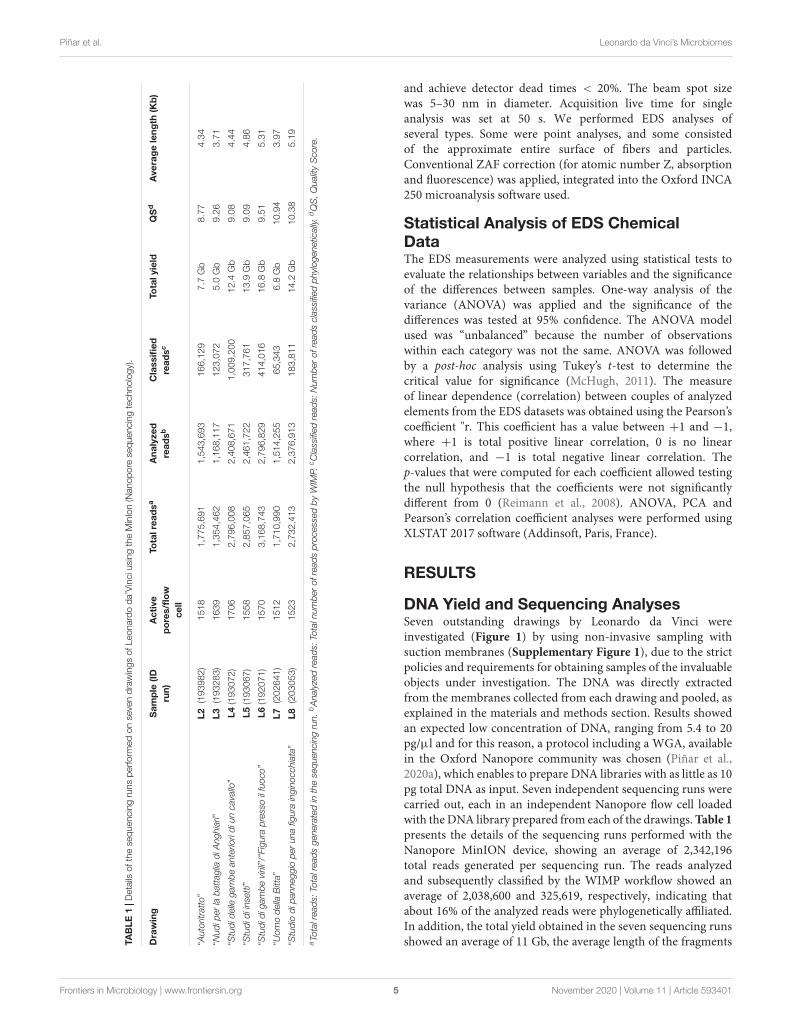
community. For WGA, all reactions were executed in a BioRadC1000 Thermal Cycler using the REPLI-g Midi Kit (Qiagen),which uses the innovative Multiple Displacement Amplification(MDA) technology. The libraries were performed using theLigation Sequencing kit 1D SQK-LSK109 and the Flow cellPriming kit EXP-FLP001 (Oxford Nanopore Technologies).All steps performed for library preparation were performedas described by Piñar et al. (2020a). A quality control of flowcells (SpotOn Flow cell Mk I R9 Version, FLO-MIN 106D) wasperformed prior starting the sequencing. The MinKNOWTM
software was used to check the number of active pores in the flowcells (Table 1) and for performing the sequencing runs. All sevenruns were performed for 48 h.
Data AnalysesResulting fast5 data files were basecalled using the NanoporeGPU basecalling with GUPPY 3.0.3 on UBUNTU 16.04(Nanopore Community Platform). Once the Fastq files weregenerated, the data were compared with databases using oneof the available pipelines for data analyses of the NanoporeCommunity Platform, following the steps recommended bymanufacturers. The selected workflow chosen was “What’s inmy pot” (WIMP), which is an EPI2ME workflow for taxonomicclassification of basecalled sequences (reads) generated byNanopore sequencing. WIMP initially filters FASTQ files with amean q-score below a minimum threshold (defaults to 7).
Relative abundances, taxonomic clustering, correspondenceanalysis and alpha diversity analysis were performed usingthe R packages taxonomic, phyloseq and microbiome forthe R version 4.0.0. (McMurdie and Holmes, 2013). Beforemicrobial taxonomic clustering was performed human readswhere removed and relative abundances were calculated.
Nucleotide Sequence Accession NumberThe sequences obtained by the metagenomic analysis aredeposited and publicly available under the BioProject ID:PRJNA655381
Optical MicroscopyFeatures of interest in the drawings were documented with aLeica MZ16 stereoscopic microscope fitted with low-temperatureoptic fiber lights. The system was equipped with a digital cameraconnected with a computer and software (Leica ApplicationSuite, LAS, Leica Microsystems GmbH Wetzlar, Germany) thatallowed the acquisition of images at different magnifications.
Scanning Electron Microscopy andMicroanalysisThe micro-objects removed from the drawings, fibers andmembranes used to sample the drawings were analyzed using avariable pressure SEM instrument (EVO50, Carl-Zeiss ElectronMicroscopy Group) equipped with a detector for backscatteredelectron diffraction (BSD). After SEM imaging using VariablePressure mode, some of the samples were covered with gold forfurther analysis in High Vacuum mode.
The samples were placed on a sulfur-free carbon adhesive(Spectrotabs AGG3358, Agar Scientific) glued onto an aluminumstub (1.25 cm diameter, Agar Scientific). Chemical analysis wasperformed by means of EDS (INCA 250, Oxford Instruments).The SEM was fitted with a tungsten filament and operatedat 20 keV, with an average working distance of 12.5 mm,with a chamber pressure between 30 and 150 Pa, chosen tokeep unaltered micro-structures. The electron beam currentwas adjusted (∼400 pA) to generate sufficient X-rays to allowreliable identification of EDS peaks, minimize spectral artifacts
Frontiers in Microbiology | www.frontiersin.org 4 November 2020 | Volume 11 | Article 593401
fmicb-11-593401 November 16, 2020 Time: 16:59 # 5
Piñar et al. Leonardo da Vinci’s Microbiomes
TAB
LE1
|Det
ails
ofth
ese
quen
cing
runs
perfo
rmed
onse
ven
draw
ings
ofLe
onar
doda
’Vin
cius
ing
the
Min
Ion
(Nan
opor
ese
quen
cing
tech
nolo
gy).
Dra
win
gS
amp
le(ID
run)
Act
ive
po
res/
flo
wce
ll
Tota
lrea
dsa
Ana
lyze
dre
adsb
Cla
ssifi
edre
adsc
Tota
lyie
ldQ
Sd
Ave
rag
ele
ngth
(Kb
)
“Aut
oritr
atto
”L2
(193
982)
1518
1,77
5,69
11,
543,
693
166,
129
7.7
Gb
8.77
4.34
“Nud
iper
laba
ttag
liadi
Ang
hiar
i”L3
(193
283)
1639
1,35
4,46
21,
168,
117
123,
072
5.0
Gb
9.26
3.71
“Stu
dide
llega
mbe
ante
riori
diun
cava
llo”
L4(1
9307
2)17
062,
796,
008
2,40
8,67
11,
009,
200
12.4
Gb
9.08
4.44
“Stu
didi
inse
tti”
L5(1
9306
7)15
582,
857,
065
2,46
1,72
231
7,76
113
.9G
b9.
094.
86
“Stu
didi
gam
bevi
rili”/
“Fig
ura
pres
soil
fuoc
o”L6
(192
071)
1570
3,16
8,74
32,
796,
829
414,
016
16.8
Gb
9.51
5.31
“Uom
ode
llaB
itta”
L7(2
0264
1)15
121,
710,
990
1,51
4,25
565
,343
6.8
Gb
10.9
43.
97
“Stu
dio
dipa
nneg
gio
per
una
figur
ain
gino
cchi
ata”
L8(2
0305
3)15
232,
732,
413
2,37
6,91
318
3,81
114
.2G
b10
.38
5.19
aTo
talr
eads
:Tot
alre
ads
gene
rate
din
the
sequ
enci
ngru
n.bA
naly
zed
read
s:To
taln
umbe
rof
read
spr
oces
sed
byW
IMP.
cC
lass
ified
read
s:N
umbe
rof
read
scl
assi
fied
phyl
ogen
etic
ally.
dQ
S,Q
ualit
yS
core
.
and achieve detector dead times < 20%. The beam spot sizewas 5–30 nm in diameter. Acquisition live time for singleanalysis was set at 50 s. We performed EDS analyses ofseveral types. Some were point analyses, and some consistedof the approximate entire surface of fibers and particles.Conventional ZAF correction (for atomic number Z, absorptionand fluorescence) was applied, integrated into the Oxford INCA250 microanalysis software used.
Statistical Analysis of EDS ChemicalDataThe EDS measurements were analyzed using statistical tests toevaluate the relationships between variables and the significanceof the differences between samples. One-way analysis of thevariance (ANOVA) was applied and the significance of thedifferences was tested at 95% confidence. The ANOVA modelused was “unbalanced” because the number of observationswithin each category was not the same. ANOVA was followedby a post-hoc analysis using Tukey’s t-test to determine thecritical value for significance (McHugh, 2011). The measureof linear dependence (correlation) between couples of analyzedelements from the EDS datasets was obtained using the Pearson’scoefficient "r. This coefficient has a value between +1 and −1,where +1 is total positive linear correlation, 0 is no linearcorrelation, and −1 is total negative linear correlation. Thep-values that were computed for each coefficient allowed testingthe null hypothesis that the coefficients were not significantlydifferent from 0 (Reimann et al., 2008). ANOVA, PCA andPearson’s correlation coefficient analyses were performed usingXLSTAT 2017 software (Addinsoft, Paris, France).
RESULTS
DNA Yield and Sequencing AnalysesSeven outstanding drawings by Leonardo da Vinci wereinvestigated (Figure 1) by using non-invasive sampling withsuction membranes (Supplementary Figure 1), due to the strictpolicies and requirements for obtaining samples of the invaluableobjects under investigation. The DNA was directly extractedfrom the membranes collected from each drawing and pooled, asexplained in the materials and methods section. Results showedan expected low concentration of DNA, ranging from 5.4 to 20pg/µl and for this reason, a protocol including a WGA, availablein the Oxford Nanopore community was chosen (Piñar et al.,2020a), which enables to prepare DNA libraries with as little as 10pg total DNA as input. Seven independent sequencing runs werecarried out, each in an independent Nanopore flow cell loadedwith the DNA library prepared from each of the drawings. Table 1presents the details of the sequencing runs performed with theNanopore MinION device, showing an average of 2,342,196total reads generated per sequencing run. The reads analyzedand subsequently classified by the WIMP workflow showed anaverage of 2,038,600 and 325,619, respectively, indicating thatabout 16% of the analyzed reads were phylogenetically affiliated.In addition, the total yield obtained in the seven sequencing runsshowed an average of 11 Gb, the average length of the fragments
Frontiers in Microbiology | www.frontiersin.org 5 November 2020 | Volume 11 | Article 593401

fmicb-11-593401 November 16, 2020 Time: 16:59 # 6
Piñar et al. Leonardo da Vinci’s Microbiomes
sequenced was 4.5 Kb, and the quality score (QS) showed anaverage of 9.6.
Microbiomes Generated From EachDrawingThe results showed that the proportion of the analyzedreads that could finally be phylogenetically assigned using theNanopore WIMP workflow was different for each drawing(Table 1). However, there was an overall high biodiversity in themicrobiome of the drawings investigated, revealing differencesbut also intriguing similarities between some of them. DrawingsL7 and L8 showed the highest biodiversity, followed by drawingsL5, L3, L6, and L2, all with similar alpha diversity, while L4showed the lowest diversity of all (Figure 2). The percentageof human reads varied between 5.35% (L2) and 74.64% (L4) ofthe classified reads with a median of 11.36%. Therefore, we canassume that the high number of human reads recovered from thesurface of drawing L4 masked the detection of reads related tomicroorganisms in this drawing. Figure 3 shows the distributionof the different superkingdoms among the investigated drawings.The relative abundance of bacteria ranged from 19% (with lowestproportion in drawing L4) to 79% (with highest proportionin drawing L6). Eukaryotes ranged from 20 to 80%, being thehighest proportion present in drawing L4, once more mainly dueto the massive presence of human DNA in this drawing. Virusesand Archaea represented less than 1% of the total microbiome inall investigated drawings.
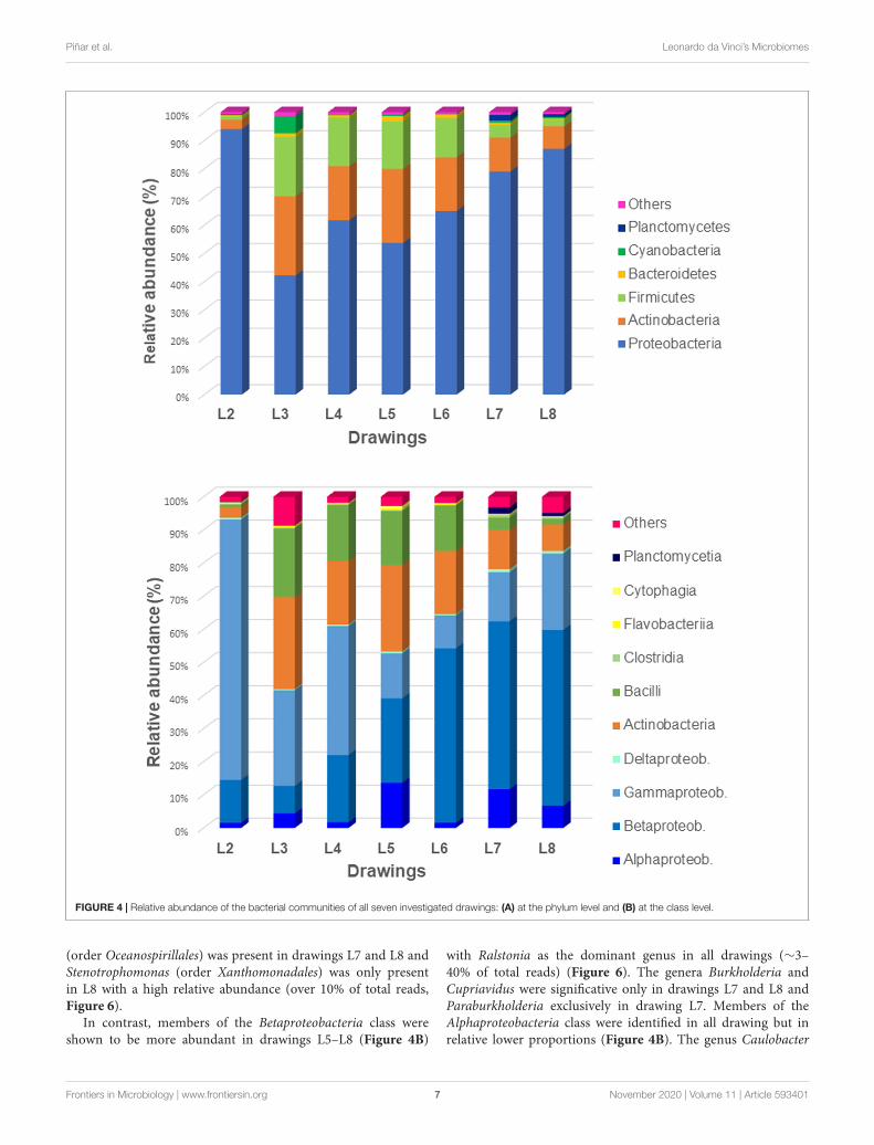
Concerning bacteria (Figure 4A), the phylum Proteobacteriadominated in all samples (42–94% of bacteria), followed bymembers of Actinobacteria (3–28% of bacteria) and Firmicutes(1–21% of bacteria). The phylum Cyanobacteria showed to be
FIGURE 2 | Alpha diversity of drawings calculated from the rarefied classifiedabundance data using the R package phyloseq.
FIGURE 3 | Relative abundance of eukaryotes, bacteria, archaea, and virusesin the microbiomes of all seven investigated drawings.
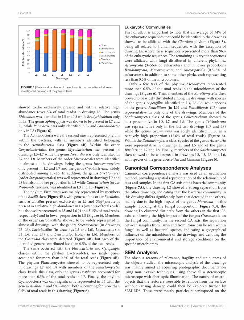
represented only in the drawing L3 (6% of bacteria) while thephylum Planctomycetes was represented only in drawings L7 andL8 (2 and 1% of bacteria, respectively). The Eukaryotes (Figure 5)were mainly represented by the phylum Ascomycota (3–56% ofeukaryotes) with the exception of the microbiome of drawingL4, which showed a massive dominance of the phylum Chordata(over 96% of the eukaryotes). This last phylum showed to bewell represented in all drawings due to the presence of humanDNA. Members of the phyla Basidiomycota, Mucoromycota, andMicrosporidia were identified in all the drawings with relativelylow proportions (0.5–5% of eukaryotes), in addition to someother phyla, each representing less than 0.5% of the microbiomes(marked as "others" in Figure 5).
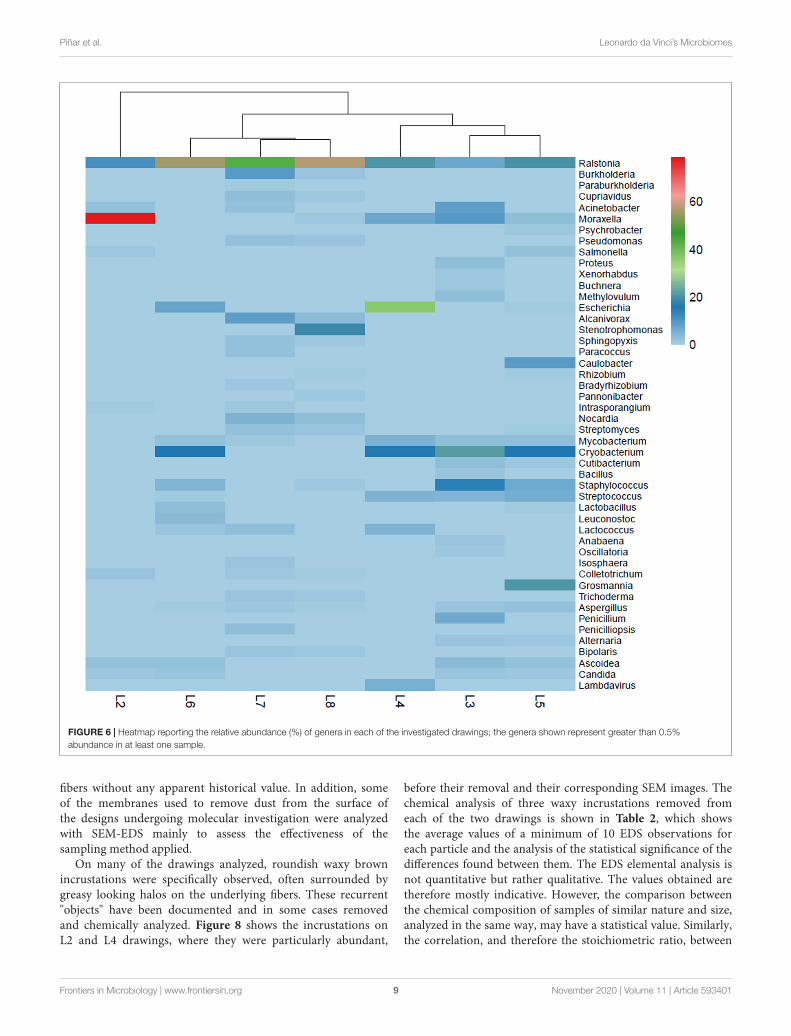
Each drawing owned a very specific microbiome, showingan independent molecular profile or “biological pedigree.”Supplementary Figure 2 shows all genera that represent morethan 0.5% of the total microbiome in each of the drawings (cutoff0.5%). These biological pedigrees are all summarized in Figure 6in form of a heatmap, showing the relative abundance (%) of eachgenus in each of the drawings.
Bacterial CommunitiesThe bacterial communities dominated over the eukaryoticcommunities in all the drawings. The most dominant phylumshowed to be the Proteobacteria with dominance of theGammaproteobacteria class in drawings 2–4 (Figure 4B). Insidethis class, members of the order Pseudomonadales were detected,such as the genus Acinetobacter, present in drawings L2, L3,and L7, the genus Moraxella, which was highly abundant inL2 (representing over 51% of total reads) but also relativeabundant in L3–L5 (1–5.25% total reads), the genus Psychrobacterbeing representative solely in L5 and the genus Pseudomonas,which was relative abundant in drawings L7 and L8 (Figure 6).Members of the Enterobacteriales were detected in drawings L2–L6 but absent in drawings L7–L8. The genus Salmonella wasdetected in L2 and L5 while Escherichia was identified in L4–L6. The genera Proteus, Xenorhabdus and Buchnera were presentexclusively in L3 (Figure 6). Interestingly, the genus Alcanivorax
Frontiers in Microbiology | www.frontiersin.org 6 November 2020 | Volume 11 | Article 593401
fmicb-11-593401 November 16, 2020 Time: 16:59 # 7
Piñar et al. Leonardo da Vinci’s Microbiomes
FIGURE 4 | Relative abundance of the bacterial communities of all seven investigated drawings: (A) at the phylum level and (B) at the class level.
(order Oceanospirillales) was present in drawings L7 and L8 andStenotrophomonas (order Xanthomonadales) was only presentin L8 with a high relative abundance (over 10% of total reads,Figure 6).
In contrast, members of the Betaproteobacteria class wereshown to be more abundant in drawings L5–L8 (Figure 4B)
with Ralstonia as the dominant genus in all drawings (∼3–40% of total reads) (Figure 6). The genera Burkholderia andCupriavidus were significative only in drawings L7 and L8 andParaburkholderia exclusively in drawing L7. Members of theAlphaproteobacteria class were identified in all drawing but inrelative lower proportions (Figure 4B). The genus Caulobacter
Frontiers in Microbiology | www.frontiersin.org 7 November 2020 | Volume 11 | Article 593401
fmicb-11-593401 November 16, 2020 Time: 16:59 # 8
Piñar et al. Leonardo da Vinci’s Microbiomes
FIGURE 5 | Relative abundance of the eukaryotic communities of all seveninvestigated drawings at the phylum level.
showed to be exclusively present and with a relative highabundance (over 5% of total reads) in drawing L5. The genusRhizobium was identified in L5 and L8 while Bradyrhizobium onlyin L8. The genus Sphingopyxis was shown to be present in L7 andL8, while Paracoccus was only identified in L7 and Pannonibacteronly in L8 (Figure 6).
The Actinobacteria were the second most represented phylumwithin the bacteria, with all members identified belongingto the Actinobacteria class (Figure 4B). Within the orderCorynebacteriales, the genus Mycobacterium was present indrawings L3–L7 while the genus Nocardia was only identified inL7 and L8. Members of the order Micrococcales were identifiedin almost all the drawings, being the genus Intrasporangiumonly present in L2 and L7 and the genus Cryobacterium widelydistributed among L3–L6. In addition, the genus Streptomyces(order Streptomycetales) was well represented in drawings L7 andL8 but also in lower proportion in L5 while Cutibacterium (orderPropionibacteriales) was identified in L3 and L5 (Figure 6).
The phylum Firmicutes was mainly represented by membersof the Bacilli class (Figure 4B), with genera of the order Bacillales,such as Bacillus present exclusively in L3 and Staphylococcus,present in a relative high abundance in L3 (over 8% of total reads)but also well represented in L5 and L6 (4 and 3.15% of total reads,respectively) and in lower proportion in L8 (Figure 6). Membersof the order Lactobacillales showed to be widely represented inalmost all drawings, with the genera Streptococcus (in drawingsL3–L6), Lactobacillus (in drawings L5 and L6), Lactococcus (inL4, L6, and L7) and Leuconostoc (solely in L6). Members ofthe Clostridia class were detected (Figure 4B), but each of theidentified genera contributed less than 0.5% of the total reads.
The same occurred with the Flavobacteria and Cytophagiaclasses within the phylum Bacteroidetes, no single genusaccounted for more than 0.5% of the total reads (Figure 4B).The phylum Planctomycetes showed to be represented onlyin drawings L7 and L8 with members of the Planctomycetiaclass. Inside this class, only the genus Isosphaera accounted formore than 0.5% of the total reads in L7. Finally, the phylumCyanobacteria was only significantly represented in L3 with thegenera Anabaena and Oscillatoria, both accounting for more than0.5% of total reads in this drawing (Figure 6).
Eukaryotic CommunitiesFirst of all, it is important to note that an average of 34% ofthe eukaryotic sequences that could be identified in the drawingsshowed to be affiliated with the Chordata phylum (Figure 5),being all related to human sequences, with the exception ofdrawing L4, where these sequences represented more than 96%of the eukaryotic sequences. The remaining eukaryotic sequenceswere affiliated with fungi distributed in different phyla, i.e.,Ascomycota (3–56% of eukaryotes) and in lower proportionsBasidiomycota, Mucoromycota and Microsporidia (0.5–5% ofeukaryotes), in addition to some other phyla, each representingless than 0.5% of the microbiomes.
Only a few taxa of the phylum Ascomycota representedmore than 0.5% of the total reads in the microbiomes of thedrawings (Figure 6). Thus, members of the Eurotiomycetes classproved to be widely distributed among the drawings, with speciesof the genus Aspergillus identified in L3, L5–L8, while speciesof the genera Penicillium (in L3) and Penicilliopsis (L7) wererepresentative in only one of the drawings. Members of theSordariomycetes class of the genus Colletotrichum showed tobe representative in L2, L7, and L8. The genus Trichodermawas representative only in the last two drawings (L7 and L8)while the genus Grosmannia was solely identified in L5 in arelatively high proportion (12.6% of total reads) (Figure 6).Within the Dothideomycetes class, species of the genus Alternariawere representative in drawings L3 and L5 and of the genusBipolaris in L7 and L8. Finally, members of the Saccharomycetesclass showed to be widespread in drawings L2, L3, L5, and L6,with species of the genera Ascoidea and Candida (Figure 6).
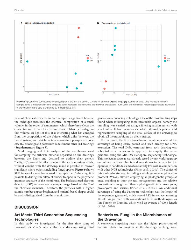
Canonical Correspondence AnalysesCanonical correspondence analysis was used as an ordinationmethod, providing a spatial representation of the relationship oftaxa and samples. In the first CA axis of the bacterial community(Figure 7A), the drawing L2 showed a strong separation fromthe other drawings, indicating that the bacterial community inthis drawing differs significantly from that detected in the others,mainly due to the high impact of the genus Moraxella on thissample. Looking at the fungal composition (Figure 7B), thedrawing L5 clustered distinctly from the others in the first CAaxis, confirming the high impact of the fungus Grosmannia onthe fungal community. In the second CA axis, the separationbetween samples from Turin and from Rome can be seen withinfungal as well as bacterial species, indicating a geographicalinfluence on the microbiome of the drawings and denoting theimportance of environmental and storage conditions on thespecific microbiomes.
SEM AnalysesFor obvious reasons of relevance, fragility and uniqueness ofthe objects studied, the microscopic analysis of the drawingswas mainly aimed at acquiring photographic documentationusing non-invasive techniques, using above all a stereoscopicmicroscope with fiber optic illumination. The nature of micro-objects that the restorers were able to remove from the surfacewithout causing damage could then be explored further bySEM-EDS. These were mostly particles superimposed on the
Frontiers in Microbiology | www.frontiersin.org 8 November 2020 | Volume 11 | Article 593401
fmicb-11-593401 November 16, 2020 Time: 16:59 # 9
Piñar et al. Leonardo da Vinci’s Microbiomes
FIGURE 6 | Heatmap reporting the relative abundance (%) of genera in each of the investigated drawings; the genera shown represent greater than 0.5%abundance in at least one sample.
fibers without any apparent historical value. In addition, someof the membranes used to remove dust from the surface ofthe designs undergoing molecular investigation were analyzedwith SEM-EDS mainly to assess the effectiveness of thesampling method applied.
On many of the drawings analyzed, roundish waxy brownincrustations were specifically observed, often surrounded bygreasy looking halos on the underlying fibers. These recurrent"objects" have been documented and in some cases removedand chemically analyzed. Figure 8 shows the incrustations onL2 and L4 drawings, where they were particularly abundant,
before their removal and their corresponding SEM images. Thechemical analysis of three waxy incrustations removed fromeach of the two drawings is shown in Table 2, which showsthe average values of a minimum of 10 EDS observations foreach particle and the analysis of the statistical significance of thedifferences found between them. The EDS elemental analysis isnot quantitative but rather qualitative. The values obtained aretherefore mostly indicative. However, the comparison betweenthe chemical composition of samples of similar nature and size,analyzed in the same way, may have a statistical value. Similarly,the correlation, and therefore the stoichiometric ratio, between
Frontiers in Microbiology | www.frontiersin.org 9 November 2020 | Volume 11 | Article 593401
fmicb-11-593401 November 16, 2020 Time: 16:59 # 10
Piñar et al. Leonardo da Vinci’s Microbiomes
FIGURE 7 | Canonical correspondence analysis plot of the first and second CA axis for bacterial (A) and fungal (B) abundance data. Dots represent samples(sample name is indicated within the dots) and colors represent the city where the drawings are located—Turin (blue) and Rom (red). Percentages indicate how muchof the variability in the data is explained by the respective axis.
pairs of chemical elements in each sample is significant becausethe technique measures the chemical composition of a smallvolume, in the order of nanometers, which therefore reflects theconcentration of the elements and their relative percentage inthat volume. In light of this, it is interesting what has emergedfrom the composition of the objects, which differ between thetwo drawings, and which contain magnesium phosphate in onecase (L2 drawing) and potassium sulfate in the other (L4 drawing)(Supplementary Figure 3).
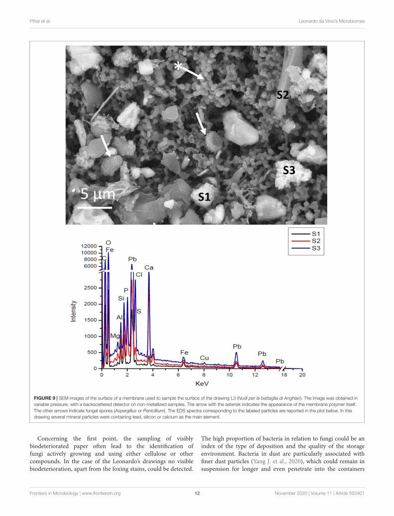
SEM imaging and EDS analysis of the membranes usedfor sampling the airborne material deposited on the drawingsbetween the fibers and destined to outline their genetic“pedigree,” showed the effectiveness of the suction system which,without contact with the drawing, made it possible to recoversignificant micro-objects including fungal spores. Figure 9 showsSEM image of a membrane used to sample the L3 drawing: it ispossible to distinguish different objects trapped in the polymericgranular structure of the membrane. The backscattered electrondetector (BSD) reconstructs a sample image based on the Z ofthe chemical elements. Therefore, the particles with a higheratomic number appear brighter, and mineral-based shapes couldbe easily distinguished from the organic ones.
DISCUSSION
Art Meets Third Generation SequencingTechnologiesIn this study we investigated for the first time some ofLeonardo da Vinci’s most emblematic drawings using third
generation sequencing technology. One of the most limiting stepsfound when investigating these invaluable objects, namely thesampling, was carried out using a filtering suction system withsmall nitrocellulose membranes, which allowed a precise andrepresentative sampling of the total surface of the drawings toobtain all the microbiome on their surfaces.
Furthermore, the tiny nitrocellulose membranes offered theadvantage of being easily pooled and used directly for DNAextraction. The total DNA extracted from each drawing wassubjected to a metagenomic approach to amplify the entiregenomes using the MinION Nanopore sequencing technology.This molecular strategy was already tested by our working groupon cultural heritage objects and was shown to be easy for theoperator to handle, fast and of a relatively low cost, in comparisonwith other NGS technologies (Piñar et al., 2020a). The choice ofthis molecular strategy, including a whole genome amplificationprotocol (WGA), allowed amplifying all phylogenetic groups atonce, enabling to infer the real metagenomes and the relativeproportions among the different phylogenetic taxa: eukaryotes,prokaryotes and viruses (Piñar et al., 2020a). An additionaladvantage of using the Nanopore technology was the length ofthe sequences generated, which were 4.5 Kb in average, meaning10-fold longer than with conventional NGS methodologies, asIon Torrent or Illumina, which yield an average of 400 b length(Kulski, 2016).
Bacteria vs. Fungi in the Microbiomes ofthe DrawingsA somewhat surprising result was the higher proportion ofbacteria relative to fungi in all the drawings, as fungi were
Frontiers in Microbiology | www.frontiersin.org 10 November 2020 | Volume 11 | Article 593401

fmicb-11-593401 November 16, 2020 Time: 16:59 # 11
Piñar et al. Leonardo da Vinci’s Microbiomes
FIGURE 8 | Insects’ droppings. Waxy brown incrustations documented on the fibers of drawing L4 (Studi delle gambe anteriori di un cavallo) and drawing L2(Autoritratto). These “objects” have been identified as insects’ droppings. They have been documented and, where possible, removed and analyzed by SEM-EDS.(a) an incrustation that was defacing the verso of drawing L4; (a1,a2) SEM images of the waxy material. The images were obtained in variable pressure, with abackscattered detector on non-metallized samples. (a1) shows the appearance of the surface of the object, covered with fungal mycelium, the detail in the squareframe corresponds to (a3). The arrows in (a2) show round-shaped crystals embedded in the amorphous matrix. (b,b1,c,c1,d,d1) Correspond to incrustations thatwere present on the drawing L2. These were made both of an amorphous matrix, as in (b1), but also by spherulites, that consist of spherical crystals made ofcalcium and magnesium phosphate. (e,e1) Show an incrustation of the L4 drawing where in the amorphous matrix were documented columnar and bipyramidalcrystals (arrow).
TABLE 2 | EDS analysis of three insects’ droppings removed and analyzed from drawing L4 (“Studi delle gambe anteriori di un cavallo”) and drawing L2 (“Autoritratto”).
C N O Na Mg P S K Ca Fe
L4.1 75.41a 4.66bc 14.10b 0.70a 0.24c 0.65b 2.45a 1.32b 0.41b 0.06b
L4.2 67.68b 8.58bc 15.89b 0.51b 0.48c 1.25b 2.50a 2.10a 0.83b 0.18ab
L4.3 56.03c 23.96a 15.48b 0.38c 0.28c 0.62b 1.69b 1.12b 0.41b 0.04b
L2.1 57.35c 0.00c 31.23a 0.08e 1.13a 3.12a 0.86c 0.13c 5.63a 0.48a
L2.2 59.29c 0.00c 30.70a 0.20d 0.90b 3.31a 0.58c 0.00c 5.02a 0.00b
L2.3 54.49c 12.63b 26.44a 0.02e 0.43c 1.05b 0.51c 0.16c 3.89a 0.38ab
Pr > F (Model) < 0.0001 < 0.0001 < 0.0001 < 0.0001 < 0.0001 < 0.0001 < 0.0001 < 0.0001 < 0.0001 0.02
Significant Yes Yes Yes Yes Yes Yes Yes Yes Yes Yes
Values are the means from a minimum of n = 15 replicates. In each column, different letter indicates statistically significant differences (P < 0.05), one-way ANOVA.
thought to be always dominant in the microbial communitiesthat colonized cultural heritage objects made or supported onpaper (Pinzari et al., 2006). Thus, our results could be explained
by (1) the sampling strategy used, which was addressed atsampling primarily the dust deposited on the surfaces, and (2)the molecular approach employed in this study.
Frontiers in Microbiology | www.frontiersin.org 11 November 2020 | Volume 11 | Article 593401
fmicb-11-593401 November 16, 2020 Time: 16:59 # 12
Piñar et al. Leonardo da Vinci’s Microbiomes
FIGURE 9 | SEM images of the surface of a membrane used to sample the surface of the drawing L3 (Nudi per la battaglia di Anghiari). The image was obtained invariable pressure, with a backscattered detector on non-metallized samples. The arrow with the asterisk indicates the appearance of the membrane polymer itself.The other arrows indicate fungal spores (Aspergillus or Penicillium). The EDS spectra corresponding to the labeled particles are reported in the plot below. In thisdrawing several mineral particles were containing lead, silicon or calcium as the main element.
Concerning the first point, the sampling of visiblybiodeteriorated paper often lead to the identification offungi actively growing and using either cellulose or othercompounds. In the case of the Leonardo’s drawings no visiblebiodeterioration, apart from the foxing stains, could be detected.
The high proportion of bacteria in relation to fungi could be anindex of the type of deposition and the quality of the storageenvironment. Bacteria in dust are particularly associated withfiner dust particles (Yang J. et al., 2020), which could remain insuspension for longer and even penetrate into the containers
Frontiers in Microbiology | www.frontiersin.org 12 November 2020 | Volume 11 | Article 593401
fmicb-11-593401 November 16, 2020 Time: 16:59 # 13
Piñar et al. Leonardo da Vinci’s Microbiomes
where the drawings are stored, and which would then act asa selective filter for certain types of dust. Nevertheless, wecannot rule out that the abundance of bacteria in the drawingsinvestigated in this study may also be due to the source ofcontamination to which they have been subjected in the recentpast, namely human contamination due to restoration works,and therefore with the human skin microbiome, as well ascontamination with insect droppings and their gut bacteria.
Regarding the second point, the technologies employed so farcould have biased the results in favor of fungi, either throughculture-dependent techniques, due to their rapid germinationin the appropriate culture medium, or even through moleculartechniques. The molecular techniques used so far in paper-based objects, whether of first- (Michaelsen et al., 2006, 2010)or next-generation sequencing (Kraková et al., 2017), havebeen mainly based on “target amplification” sequencing, whichprevents inferring the real proportions of the different targetgroups (bacteria and fungi). Taking these possible biases intoaccount, this has led to a low number of studies focusingon bacteria colonizing paper materials (Jurado et al., 2010;Michaelsen et al., 2010; Principi et al., 2011; Kraková et al.,2012, 2017). In contrast, there is an extensive literature focusedon the colonization of paper by fungi (Sterflinger and Pinzari,2012). Many fungal species have been described as spoilers ofpaper-based materials and many of them have shown cellulolyticactivities, as some examples species of the genera Chaetomium,Stachybotrys, Cladosporium, Aspergillus, Penicillium. In thiscontext, one of the main research topics in paper and archivalmaterial has been related to the pigments produced by papercolonizing fungi, causing foxing, a deterioration of paper thatoccurs when brownish and reddish spots are produced by thefungi (Arai, 2000). However, so far, no extensive studies have beenconducted to elucidate the total microbiome (bacteria vs. fungi)of these valuable objects. The new technology used in this studyhas been able to give a new focus to the actual proportions ofbacteria/fungi that inhabit paper objects.
Microbiome Biodiversity in the DrawingsThe microbiomes derived from the drawings showed ahigh biodiversity, with the exception of drawing L4, whosemicrobiome was heavily contaminated with human sequences,this contamination being perhaps due to its greater handling.In fact, the L4 drawing had massive traces of modern adhesivethat required a prolonged conservation work, which was carriedout with the aid of a magnification apparatus and variousmanipulations to which the other drawings were not subjected.This may have resulted in greater removal of surface dust, but atthe same time in greater human contamination. Another elementto consider in this drawing is the very different nature of itssupport. The drawing was analyzed with Infrared False ColorPhotography and Fiber Optics Reflectance Spectroscopy andresults showed that the paper was treated with layers of lead white[basic lead carbonate 2PbCO3 × Pb(OH)2] mixed with indigo(Ruggiero, 2020). Moreover, it appeared that Leonardo followedthe recipe described by Cennini and Powell Herringham (1899),which consisted in a preparatory layer made with powderedcalcinated chicken bones, white lead, indigo from the plant
Indigofera tinctoria mixed with animal gelatin and distributedwith a brush on the paper support. The design was finallymade with a silver tip. This peculiarity may have interfered bothwith sampling and with the preservation of the microbiomeand the environmental DNA in general. Only recent DNA mayhave remained, i.e., that of the restorers who manipulated it.In this context, Tarsitani et al. (2014) already noted that theperiod of restoration was probably the critical moment when thediscoloration and deterioration of other emblematic drawings byLeonardo da Vinci began, namely in the Atlantic Codex.
BacteriaThe results derived from bacteria showed a dominance ofGammaproteobacteria of the order Pseudomonadales. The genusMoraxella was shown to be most abundant at L2, but was alsowell represented in the L3–L5 drawings, with almost all sequencesidentified as M. osloensis, a saprophytic species in human skin andmucous membranes (Alkhatib et al., 2017). Its high abundancein these drawings can be explained by contamination due tohuman handling. Nevertheless, this species was also identified ina survey investigating bulls of indulgence from the fifteenth tosixteenth centuries (Jurado et al., 2010). The genus Acinetobacterwas present in drawings L2, L3, and L7, being most of thesequences related to A. baumannii. This species is frequentlyisolated from hospital environments, although it has been foundin environmental samples (Antunes et al., 2014). Nevertheless,sequences related to Acinetobacter spp. were previously identifiedin a cloning and sequencing survey performed on a deterioratedItalian manuscript dated from the thirteenth century (Michaelsenet al., 2010). The genus Pseudomonas was relative abundant indrawings L7 and L8, with the dominant species P. parafulva,and the genus Psychrobacter was representative solely in L5with most of the sequences affiliating with Psychrobacter sp.P11G5, which is a psychrophilic species exhibiting laccase activity(Moghadam et al., 2016). Bacterial laccases (Chauhan et al.,2017) are produced either extracellularly or intracellularly andtheir activity is in a wide range of temperature and pH. Theyare different from fungal laccases that are multicopper enzymesbelonging to the group of blue oxidases (Janusz et al., 2020).
Members of the Enterobacteriales were detected in drawingsL2–L6 but interestingly absent in drawings L7–L8. The genusSalmonella was detected in L2 and L5, being Salmonella entericasubsp. enterica serovars identified in both drawings (Vila Novaet al., 2019). In contrast, Escherichia was identified in L4–L6,with the species E. coli, which is a common inhabitant of thegut microbiota. The genus Proteus, with the species P. mirabilis,which frequently causes urinary tract infections in humans(Pearson et al., 2008), was present exclusively in L3. Interestingly,two genera identified only in this last drawing, namely Buchneraand Xenorhabdus, showed the presence of species that are obligateendosymbionts of aphids, such as B. aphidicola (Shigenobuand Wilson, 2011) and nematodes, such as X. bovienii (Murfinet al., 2015). Interestingly, X. bovienii is a bacterium whichhas been isolated from several nematode Steinernema species.Steinernema affects many insects and also flies (Archana et al.,2017). Therefore, it is plausible to relate the presence of thesespecies with the presence of insect’s droppings in this drawing.
Frontiers in Microbiology | www.frontiersin.org 13 November 2020 | Volume 11 | Article 593401
fmicb-11-593401 November 16, 2020 Time: 16:59 # 14
Piñar et al. Leonardo da Vinci’s Microbiomes
The genus Methylovulum was detected only in drawing L3 andall identified sequences affiliated with M. psychrotolerans, a cold-adapted methanotroph from terrestrial environments (Oshkinet al., 2016). Interestingly, the genus Alcanivorax was present indrawings L7 and L8, with the dominant species A. xenomutans,which has capabilities for alkane degradation and heavy-metalresistance (Fu et al., 2018). Finally, the genus Stenotrophomonas,with the species S. rhizophila and S. maltophilia, was only presentin L8 with a high relative abundance. It is important to remarkthat species of this genus have been identified as colonizers andpotential spoilers of oil paintings on canvas support (López-Miraset al., 2013), tempera paintings (Zhgun et al., 2020) as well asancient paper manuscripts (Michaelsen et al., 2010) and bookcollections (Kraková et al., 2017; Okpalanozie et al., 2018). Inaddition, Stenotrophomonas sp. has been identified in relationto the biodeterioration of other cultural heritage materials, suchas parchments (Piñar et al., 2015a) and historical stone objects(Alakomi et al., 2006; Ettenauer et al., 2011; Rosado et al., 2020).The reason for the perseverance of Stenotrophomonas in this typeof materials may be due to their extracellular enzyme activitiessuch as proteases and chitinases (Kobayashi et al., 2002) and theircapability to produce extracellular polysaccharide, enabling theiradhesion to different materials and providing biofilm formationon their surfaces (Alakomi et al., 2006).
The Betaproteobacteria were present in all drawings withthe dominant genus Ralstonia, with species affiliating toR. solanacearum, R. pickettii, R. insidiosa, and R. mannitolilytica.Ralstonia species, especially R. pickettii and R. solanacearum areprevalent colonizers of printing paper machines (Väisänen et al.,1998) and have been detected as dominant species inhabitingdamaged oil paints on canvas (Capodicasa et al., 2010; Piñaret al., 2020a). Furthermore, R. solanacearum is a phytopathogenicbacterium and may exert an active deterioration on papermaterials due to the secretion of extracellular proteins, such ascellulolytic enzymes needed for virulence in plants (Liu et al.,2005). Other Betaproteobacteria showed to be relevant only indrawings L7 and L8, as the genera Burkholderia and Cupriavidus.The species B. pseudomallei and B. multivorans were dominantin L7 and Burkholderia sp. MSMB0852 and B. mesoacidophila inL8. Species of this genus were the most frequently isolated frompaper products and printing paper machines (Väisänen et al.,1998). Cupriavidus sp. are generally heavy metal tolerant bacteriawith the ability to degrade a variety of aromatic hydrocarboncompounds (Wang et al., 2015). The species Cupriavidus sp.USMAA2-4, C. metallidurans and C. gilardii were identified inthese two drawings.
The Alphaproteobacteria class showed to be well representedin drawing L5, with a relative high abundance of species of thegenus Caulobacter, with the dominance of C. segnis. Caulobacterspecies exert a role in decomposition, including the degradationof cellulose, lignin and polyaromatic hydrocarbons and havethe capacity to grow on cellulose (Song et al., 2013). This factmay explain the presence of Caulobacter species in this paper-supported drawing and highlights the risk that this bacteriumposes to their integrity. This class showed to be relevant also indrawings L7 and L8. The genus Sphingopyxis dominated in bothdrawings. Sphingopyxis isolates have been reported to degrade
aromatic compounds in various habitats (Yang F. et al., 2020).In addition, the genus Paracoccus was only relevant in drawingL7 with the dominance of P. yeei, which has been isolated fromdiverse natural environments, including human skin (Lim et al.,2018). In contrast, the genus Pannonibacter was only relevantin L8, being all reads affiliated with P. phragmitetus, which is ahalotolerant polycyclic aromatic hydrocarbon (PAH)-degradingbacterium (Wang et al., 2016), but it has been also described as ahuman pathogen (Wang et al., 2017).
The Actinobacteria were the second most representedphylum within the bacteria. The genus Cryobacterium, withC. articum, showed relatively high abundance in drawingsL3–L6. C. articum is a psychrotolerant bacterium isolatedfrom extreme cold environments (Bajerski et al., 2011). Thisspecies was previously identified in two oil paintings on canvas(Piñar et al., 2020a). The genus Mycobacterium, with thedominant species M. haemophilum, was present in all the above-mentioned drawings in addition of L7. M. haemophilum isan emerging pathogen associated most commonly with skininfections (Tufariello et al., 2015). The genus Intrasporangiumwas present in L2 and L7 with the dominant species I. calvum,firstly isolated from the indoor environment (Kalakoutskii et al.,1967). The genus Nocardia was identified in drawings L7 andL8 with the dominant species N. farcinica. This species causeshuman infections acquired through the respiratory tract orskin (Torres et al., 2000). The genus Streptomyces was alsorepresented in drawings L7 and L8 and in lower proportion inL5. Streptomycetes are ubiquitous in nature and many speciesare potent producers of lignocellulolytic enzymes includingcellulases, hemicellulases and ligninolytic enzymes (Saini et al.,2015). The most dominant species in L7 and L8 was Streptomycessp. CNQ-509, while Streptomyces albus dominated in drawingL5. The last species is well-known for its capability to producexylanases (Saini et al., 2015), which are enzymes responsiblefor the hydrolysis of β-1,4 bonds in plant xylan, the maincomponent of hemicellulose. The detection of species of thisgenus in these drawings show a latent risk of deterioration dueto the potential cellulolytic activity of these strains. Finally, thegenus Cutibacterium (Propionibacterium) was identified in L3and L5, with the species C. acnes in both drawings. This speciesis considered as human microbial marker, however, its detectionshould be considered with caution, as it is a potential pathogen(Fitz-Gibbon et al., 2013).
The phylum Firmicutes was mainly represented by membersof the order Bacillales, with the dominance of members of thegenus Staphylococcus, present in L3, L5, L6, and L8 (Figure 6).The dominant species in all mentioned drawings was S. aureus,but other species were also identified in high proportions, suchas S. equorum, S. capitis, S. pasteuri, and S. epidermidis, amongothers. The genus Bacillus was present exclusively in L3, withthe dominant species B. mycoides and B. thuringiensis. Speciesbelonging to Bacillus and Staphylococcus have been previouslyidentified as the main bacterial species colonizing paper archivalmaterials (Michaelsen et al., 2010; Kraková et al., 2012, 2017;Karakasidou et al., 2018; Okpalanozie et al., 2018).
Members of the order Lactobacillales showed to bewidely represented in almost all drawings, with the genera
Frontiers in Microbiology | www.frontiersin.org 14 November 2020 | Volume 11 | Article 593401

fmicb-11-593401 November 16, 2020 Time: 16:59 # 15
Piñar et al. Leonardo da Vinci’s Microbiomes
Streptococcus (in drawings L3–L6), being the emerging pathogenS. pseudopneumoniae dominant in L3–L5. This species isassociated with lower-respiratory-tract infections and a highprevalence of antimicrobial resistance (Garriss et al., 2019). Incontrast, S. thermophilus showed to be the dominant speciesin L6, which is a fermentative lactic acid bacterium (Cui et al.,2016). This species shows acidifying capacity, proteolytic activity,fast growth and production of exopolysaccharide (EPS), withall these capabilities posing a risk to the integrity of the papersupport of the drawings. The genus Lactobacillus was identifiedin drawings L5 and L6, with L. salivarius and L. gallinarum asdominant species in L5 and L6, respectively, which are speciesfrequently isolated from the digestive tract of mammals (Nevilleand O’Toole, 2010) and chicken. The genus Lactococcus wasidentified in L4, L6, and L7 with L. piscium as dominant speciesin L4 and L. lactis in L6 and L7. L. lactis has been isolatedfrom a variety of environmental sources but is most commonlyassociated with fresh or fermented plant material or with milkproducts (Kelly et al., 2010). L. piscium is a psychrotrophic lacticacid bacterium and is known to be one of the predominantspecies within spoilage microbial communities in cold-storedpackaged foods (Andreevskaya et al., 2015). Finally, members ofthe Leuconostoc genus were solely identified in L6. Three speciesshowed to be dominant in this drawing, namely L. mesenteroidessubsp. dextranicum, L. kimchii, and L. citreum. All of them arecommonly used for souring vegetables, producing fermentedfoods (Dols et al., 1997). However, it is important to notethat Leuconostoc species have been detected in associationwith the fruit fly (Drosophila melanogaster), whose diet maycontain communities of fermentation microbes, including a highpercentage of Leuconostoc in the fly microbiome (Chandler et al.,2011). Also, the housefly (Musca domestica L.) lives in closeassociation with Leuconostoc (de Jonge et al., 2020), which wasdetected mainly in adult flies and associated to the buccal parts ofthe insects. It is conceivable that the discovery of this bacteriumis associated with the presence on the drawings of droppingsof insects, mainly related to synanthropic diptera (Jones et al.,1999) and dating back to the time when the drawings werenot protected and preserved in an appropriate manner (seesection “SEM-EDS”).
Regarding the members of other phyla relatively less abundantin the microbiomes of the drawings, such as Bacteroidetes,Planctomycetes or Cyanobacteria (Figure 4A), it should bementioned that most of their genera represented less than 0.5% ofthe total reads. Thus, only the genus Isosphaera, with the speciesI. pallida (Planctomycetes phylum) accounted for more than0.5% of the total reads in L7. Finally, the phylum Cyanobacteriawas only significantly represented in L3 with species of thegenera Anabaena (A. cylindrica) and Oscillatoria (O. nigro-viridis), both accounting for more than 0.5% of the total readsin this drawing. Airborne dispersal of microalgae occur at verylow concentration in the atmosphere and their occurrence is littleknown due to technical limitations in investigating microalgaefrom air samples (Tesson et al., 2016). However, recent studiesshowed that microalgae can actually be air transported andform a consistent component of airborne biological particulate,depending on the environmental conditions and the local
deposition rates (Genitsaris et al., 2011). Furthermore, both theO. nigro-viridis and the A. cylindrica are cyanobacteria whosetissue can be mineralized with calcium carbonate (Benzeraraet al., 2014). Characteristic stromatolite and travertine formationsare produced by species precipitating calcium carbonate in theirsheaths and within colonial mucilage (Komárek et al., 2003).Some fillers in paper manufacture are based on carbonates fromnatural sources, such as fossil sedimentary rocks (i.e., biomicrite)(Gerteiser and Laufmann, 1989), which can sometimes berecognized in the form of microfossils among the paper fibers(Bicchieri et al., 2019). It is therefore possible to speculate thatthe presence of cyanobacteria is due to the use of carbonate-based filler material and natural rocks, perhaps from recentsedimentation, in the manufacture -if not of the Leonardodrawing- perhaps of the cartoons supporting the drawing itself.Both in the case of airborne cyanobacterial propagules and, evenmore, of biological traces in the sizing minerals, the data confirmthe sensitivity of the molecular method used.
It is important to note that it is difficult to comparethe data generated in this study with those published inthe literature, due to the different technologies applied. Asmentioned above, there are not many studies focused onthe detection of bacteria on paper-materials and even fewerconducting culture-independent approaches. However, the datagenerated in this study through third-generation sequencinganalyses are somewhat consistent with previous studies wherefirst- and second-generation sequencing was performed. Thecore microbiome found in this study includes mainly bacteriabelonging to the phyla Proteobacteria, Actinobacteria, andFirmicutes, which correlates well with the few previously existingmolecular studies. These three phyla were previously detectedby different cloning approaches performed on photographicmaterial, including cellulose supports (Bucková et al., 2014;Puškárová et al., 2016) as well as on an ancient Italian manuscript(Michaelsen et al., 2010). Principi et al. (2011) also showed thepresence of members of the phyla Proteobacteria and Firmicutesin the pages of Leonardo da Vinci’s Atlantic Codex. Later,Kraková et al., 2017 confirmed that the core microbiome ofvarious archival materials, investigated with next generationsequencing technology (Illumina MiSeq), consisted mainly ofmembers of the three phyla mentioned above.
To summarize our data, the bacterial species detected inthis study can be roughly grouped according to their originon the paper-support of the drawings. A first group is formedby bacteria that are mainly contaminants introduced by humanmanipulation, mainly species of the genera Staphylococcus,Streptococcus, and Cutibacterium (Propionibacterium), but alsospecies of the genus Moraxella, Paracoccus, Mycobacterium,and Nocardia could have been introduced by a mishandlingof the drawings. This result is reinforced by a large amountof human DNA found in all the drawings. A second groupis formed by bacteria that may have been deposited in thedrawings through vectors, such as insects. These bacteria belongto the genera Salmonella, Escherichia, Buchnera, Xenorhabdus,and Leuconostoc. This result is supported by the finding ofinsect droppings on the drawings. The third group consists ofbacteria that are actually potential spoilers of the paper-support
Frontiers in Microbiology | www.frontiersin.org 15 November 2020 | Volume 11 | Article 593401
fmicb-11-593401 November 16, 2020 Time: 16:59 # 16
Piñar et al. Leonardo da Vinci’s Microbiomes
due to their capabilities of producing extracellular enzymes,and they carry a latent risk to the integrity of the drawings.They are members of the genera Ralstonia, Stenotrophomonas,Burkholderia, Caulobacter, Streptomyces, and Bacillus. Someof them could have been introduced into the paper duringthe production process and others have been deposited onits surface later through the air. However, it is importantto note that the bacterial community was influenced by thegeographical location of the drawings, Rome or Turin, denotingan environmental influence.
FungiAmong the detected fungi, only some members of theAscomycota phylum represented more than 0.5% of totalreads in the microbiomes of the drawings, among them,members of Sordariomycetes, Eurotiomycetes, Dothideomycetes,and Saccharomycetes. Within the Sordariomycetes, the genusColletotrichum showed to be present in L2, L7–L8. Thisgenus includes several plant pathogens (Cannon et al., 2012).The species C. higginsianum was dominant in L7–L8 whileC. orchidophilum was dominant in L2. Interestingly, the genusGrosmannia was only present at L5 in a very high proportion,with G. clavigera being the sole species identified. This speciesis the main fungal pathogen associated with the mountain pinebeetle and causes blue stain in wood (Wang et al., 2014). Thefungus is linked to American conifers. However, it is importantto note that individuals of Pseudotsuga menziesii are used as aperennial ornamental conifer species in the northern regions ofItaly. It is presumed that the presence of this fungus could bedue to a contamination event related to its presence in Rome atthe conservation institute where the drawings were analyzed andsampled. In the historical garden of the institute there are in factornamental evergreen conifer species. The fungus can also live asa saprotroph in its anamorphic state on bark and cellulosic debris(Leptographium form). A further hypothesis is that the drawingcame into contact with furniture or woody material, possibly raw,contaminated by the fungus. The DNA in G. clavigera myceliumremains stable for more than 10 years also in heat-treated woodsamples (Wong et al., 2020). Finally, the genus Trichoderma waspresent in L7 and L8, with the species T. reesei as the dominantspecies in both drawings. Trichoderma reesei is one of the mainproducers of cellulolytic and xylanolytic enzymes, being the moststudied fungus involved with lignocellulosic degradation (Bischofet al., 2016). The detection of this fungus supposes a great latentrisk of deterioration for these paper-supported drawings if theenvironmental conditions become suitable for its germination.
Members of the Eurotiomycetes were present in drawingsL3, L5–L8, being Aspergillus widespread in all these drawings,while Penicillium showed to dominate in L3 and Penicilliopsisin L7. The species A. glaucus, A. clavatus, and A. niger weredominant in all those drawings. Aspergillus species are wellknown for the production of cellulases and their main role in thebiodeterioration of paper (Das et al., 1997). The most dominantspecies found in the drawings, namely A. glaucus, is known tohave a wide environmental distribution due to its physiologicalhardiness under extreme conditions and to its ability to growon substrates with a low water activity (Hubka et al., 2013).
A. glaucus has been detected in cultural assets made of wooden(Sterflinger et al., 2018b) and canvas support (Piñar et al.,2020a), but its isolation and undistinguishable morphologicalidentification were difficult in the past (Hubka et al., 2013).Penicillium rubens showed to be the dominant species in L3.This species is a common fungus of the indoor environmentand well known for its resilience under low water activities(van Laarhoven et al., 2017). Finally, Penicilliopsis zonata wasthe dominant members of Eurotiomycetes in L7. It is importantto note that in the indoor environments, the relative ratio andabundances of fungal species belonging to Aspergillus, Eurotiumand Penicillium genera are used along with indicator species toevaluate the presence of hidden moisture damage in buildings(ISO/DIS 16000-17:2006, 2006; Baudisch et al., 2009). Mostindoor contamination studies are based on the composition ofthe dust in suspension. The fungal species in the dust depositedon surfaces are less studied and there are less significant statistics.However, the fact that the drawings, even those preservedin the same environment, presented a different mycoflora isan interesting fact as it allows to formulate hypotheses onthe conservation conditions to which the objects have beenexposed. The predominance of xerophilic fungal species suchas Penicillium rubens and Aspergillus glaucus, indicates that thedrawings have not been preserved in humid environments.
Within the Dothideomycetes only members of two generaaccounted for more than 0.5% of total reads, namely Alternariaand Bipolaris. Alternaria, with the only detected speciesA. alternata was present in L3 and L5. This fungus is a well-known paper-spoiler (Das et al., 1997; Michaelsen et al., 2006;Mesquita et al., 2009) and also considered as an allergen,imposing a severe health threat on librarians, conservators, andscholars who might be in contact with infected paper-supportedmaterials (Karakasidou et al., 2018). It must be mentioned,however, that the Alternaria is a fungus that needs water availablein the materials and that it typically develops after flooding andwetting events in building materials, including wood and paper.Therefore, the presence of hydrophilic fungal species on thedrawings, in the absence of any visible manifestation of waterdamage, is due to their common presence in the air as airbornecontaminants, also coming from the external environment, fromthe decomposing plant material typically present in parks andgardens. Bipolaris sorokiniana was the main species identified inL7 and L8. B. sorokiniana (anamorph of Cochliobolus sativus)is a phytopathogenic fungus that produce cell wall-degradingenzymes, such as cellulases, which aid invasion into host cells(Aich et al., 2017). As far as we know, B. sorokiniana has not beenconsidered as a paper degrader but, in fact, the fungus is providedwith cellulases that can degrade paper fibers and thus, this speciesposes a latent risk of deterioration for the drawings in which ithas been detected.
Members of the Saccharomycetes, with Ascoidea and Candidaspecies were detected in L2–L3 and L5–L6 but curiously,not in drawings L7–L8. A. rubescens, C. orthopsilosis, andC. dubliniensis were the dominant species detected in all fourmentioned drawings. Ascoidea species appear primarily to bedisseminated by insect vectors (Batra, 1963) and have notbeen identified in paper materials so far. In contrast, Candida
Frontiers in Microbiology | www.frontiersin.org 16 November 2020 | Volume 11 | Article 593401
fmicb-11-593401 November 16, 2020 Time: 16:59 # 17
Piñar et al. Leonardo da Vinci’s Microbiomes
spp. have been previously identified in archival documents andphotographic material, but no evidence regarding implicationin paper deterioration has been provided (Principi et al.,2011; Borrego et al., 2015; Karakasidou et al., 2018). Thedetection and prevalence of species of the genus Candidain repositories and archives may be an indication of theirubiquity in this environment, where they can be introducedfrom the outside environment or contaminated materials afterproduction. Moreover, certain species of Candida are typicalhuman microbiota, which might suggest anthropogenic pollutionby handling the collections (Borrego et al., 2015, 2017).
In summary, most of the fungi detected in this studyhave been previously identified in other studies focused onpaper-supported materials and include species of the generaTrichoderma, Aspergillus, Penicillium, Penicilliopsis, Alternaria,and Candida. On the contrary, as far as we know, species of othergenera, such as Colletotrichum and Bipolaris, which are mainlyplant pathogens, have not been described as common in papermaterials. However, they possess extracellular enzymes that maybe directly responsible for paper deterioration. Finally, the generaGrosmannia and Ascoidea include typical species associated withinsects and could have been introduced to the surface of thedrawings through these vectors. As observed with the bacterialcommunities, it was also possible to see a correlation of the fungalcommunities with their geographical location, highlighting anenvironmental and geographical influence on the microbiomesof the drawings.
Monitoring of Drawing L2 “Autoritratto”A previous study of the L2 drawing performed in 2012 showedthat both the recto and verso had been affected by a fungalattack by the species Eurotium halophilicum, a fungus capableof developing under conditions of extremely low substrate watercontent (<65%). In fact, the fungus is considered as xerophilicand needs hypertonified culture media by adding 15% NaCLfor its normal growth, which can also be replaced by otherosmoactive organic substances such as sugars (Imai, 1964). Inassociation with the fungal structures, abundant calcium oxalatecrystals, resulting from the metabolic activity of the fungus itself,were observed (Pinzari et al., 2014; Piñar et al., 2015b). In 2012the self-portrait did not show the presence of active infections bymicroorganisms but only traces of previous attacks and generaldust contamination. The membranes and swabs used to samplethe dust present on the self-portrait were used to extract the DNAof the microorganisms and to carry out both an evaluation ofthe type of contamination of the self-portrait (microbiologicaldiversity and abundance) and a comparison, using moleculartechniques, of the effectiveness of surface sampling. Details of thestudy are given in Piñar et al. (2015b), where the authors showedthat small membranes are particularly effective for non-invasivesampling from paper and for direct use in DNA extractionprotocols. Several fungal species were found to be significantlyassociated with both the recto and the verso of the drawing at thattime, such as the Phialosimplex, Penicillium, and Acremoniumspecies. From the molecular analysis, however, it was not possibleto obtain gene sequences correlated to the fungus E. halophilicum,which was instead diagnosed by SEM analysis, thanks to some
unmistakable morphological characters. A possible explanationlies in the treatments that the portrait has undergone over theyears. In 1987 Dr. Fausta Gallo, director of the biology laboratoryof the former “Istituto Centrale per la Patologia del Libro (Rome)”examined the tracks produced between 1972 and 1987 by thethermo-hydrographers placed in the drawer where the self-portrait was kept. In the spring-summer periods, the portraitwas subjected to a Relative Humidity of 70–80% with peaks of90%. The alarm was such that Dr. Fausta Gallo wrote the sameyear (1987) that the damage on the portrait was evolving andthat disinfection with ethylene oxide was necessary (Pinzari et al.,2014). Ethylene oxide is a disinfectant gas, widely used in the pastto sterilize book material, which works as an alkylating agent ofnucleic acids and proteins. The chemical action of this compoundmakes the DNA and RNA of microorganisms (and materials suchas parchment) no longer amplifiable (Michaelsen et al., 2013).There is no certainty in the documentation available at ICPALthat the self-portrait has actually undergone disinfection withethylene oxide. However, if so, any trace of DNA present on theobject and prior to treatment would be undetectable with currentanalysis techniques. In this case, the infection by the fungusE. halophilicum, evident in the images obtained with SEM butnot detected either by molecular techniques based on cloning in2012, and currently by nanopore technology, would be dated backto before 1987. The design has not been treated in any way since2012. The new series of molecular analyses, carried out by meansof Nanopore sequencing, a much more advanced techniquethan cloning, should have detected the presence of the fungus,which instead did not emerge. Other typical air contaminatingspecies were detected, despite their much lower density than thatdocumented for the E. halophilicum. Therefore, the absence ofthe fungal species among those found with Nanopore sequencingsupports the hypothesis that the design was actually treated withethylene oxide in 1987.
SEM-EDXThe incrustations analyzed so far were found to be insectdroppings (Figure 8). This evidence is due partly to the presenceof spherical particles of biogenic nature composed of magnesiumand calcium phosphate recognized as “spherulites” which arebiogenic stones that form in the gut of insects or in theirexcretion products (Gower, 2008). These spherical micro-objectsare used in archeology as markers of the presence or useof animals’ dung (Lancelotti and Madella, 2012). The otherencrustations, those without spherulites, appeared to be insteadrich in nitrogen compounds, and potassium sulfate, that are bothcatabolites of insects’ metabolism (Cochran, 1975). The smallmonoclinic and prismatic crystals characterizing these objectswere scarcely distinguishable in the matrix when visualized withBSD detector, which means that they were not presenting ahigh atomic number, and could presumably be uric salts (uricacid: C5H4N4O3). Nitrogen could make cellulose potentiallymore degradable since nitrogen-based compounds are substancesnormally absent in paper media, but which microorganismsneed to grow (Pinzari, 2011). The insects’ droppings found onthe L4 drawing were colonized by fungal mycelium. Leonardo’sdrawings have been kept for many years locked in vaults,
Frontiers in Microbiology | www.frontiersin.org 17 November 2020 | Volume 11 | Article 593401
fmicb-11-593401 November 16, 2020 Time: 16:59 # 18
Piñar et al. Leonardo da Vinci’s Microbiomes
protected by cardboard boxes and other protective systems. Toimagine that an insect can rest on a drawing to leave its droppingsis unrealistic. Therefore, the waxy incrustations removed andanalyzed could date back for many years. That is, when thedrawings were exposed to the air, so even before they werehung in glass frames, as some appear to have been aroundthe 1930s and 1950s (Misiti, 2014). The species of insects thatcause damage to books and libraries belong to different families,united by having representatives who have adapted to living inman-made environments and have spread throughout the worldthanks to commercial activities. According to their biologicalcharacteristics, they carry out their life cycle within the materialsthey feed on or in the surrounding environment (Florian, 1997).In the case of the traces found on the drawings, rather than theclassic insects harmful to paper and parchment, which typicallycause lacunas and tracks such as cockroaches, woodworms andsilverfishes, it seems that the droppings are more characteristicof species of diptera such as domestic flies and related species.House flies (Musca domestica) are synanthropic and commonlyfound on decaying matter but also feces and food. They havea complex prokaryotic microbiota in their gut, that they canrelease on materials through droppings and regurgitation offood. Furthermore, the microbiome in domestic flies can berelatively consistent across geographic locations and habitat(Park et al., 2019).
Finally, we would like to make a general discussion aboutthe conservation over the centuries of these unique andvery fragile objects, in terms of the need to remove insectexcrements, dust and the remains of fungi and bacteria, sincethey are all substances rich in nitrogen, and chemically reactivecompounds, capable of oxidizing and degrading the paperfibers. Microorganisms that are still viable also pose otherrisks, as they can proliferate and cause much more seriousdamage under certain environmental conditions. However, themanipulation of the drawings could also lead to possibleadditional contamination, as demonstrated in this study bythe presence of bacteria from the human microbiome and theabundant human DNA. However, thanks to the “dirt” foundon these heritage objects it has been possible to study theirhistory and to draw a distinctive profile that forms part ofthe drawings themselves and describes them. The informationobtained in this study comes from samples that would beeliminated in a routine conservative treatment. Incrustations,amorphous particles, pieces of glue and remains of supportingcardboard are materials that have been removed by restorersup to now. However, in light of what can be discovered fromtheir meticulous study, the process of conserving certain artisticobjects probably needs to be reconsidered because part of theirhistory is written in their "dirt".
CONCLUSION
This study shows for the first time the complete microbiomesof some of Leonardo da Vinci’s most emblematic drawings.The strategy applied for their research, namely the Nanoporesequencing technology, is one of the most advanced so far
to investigate all the phylogenetic groups included in each ofthe microbiomes. In addition, the complementation with SEManalysis allowed the visualization of insect droppings in someof the drawings, which are directly correlated with many of themicroorganisms found on their surfaces.
The results show a surprising dominance of bacteria overfungi. This fact is partly due to the sampling strategy used,aimed at sampling mainly the dust deposited on the surfaceof the drawings, but also to the source of contamination towhich they have been subjected in the recent past, resulting ina high proportion of bacteria typical of the human microbiome,introduced by intensive handling of the drawings duringrestoration works, in addition to other microorganisms that haveobviously been introduced as contaminants through vectors, suchas insects and their excrements.
It is important to clarify that, unlike other case studiesthat point to visible paper damage caused by fungi, oftenalso associated with bacteria, no clear damage apart fromfoxing was observed in this study. The sensitivity of theNanopore sequencing method, which provides information onthe abundance of the different taxa in each sample, couldhave an advantage over other methods for monitoring anongoing deterioration process, because it allows the assessmentof quantitative relationships between taxa and also the deductionof variations in these relationships due to detrimental situations.
In general, the biological information contained in theinvestigated drawings showed very specific microbiomes, whichcan be used as a bio-archive of the history the objects, providing akind of fingerprint for current and future comparisons. However,some similarities were observed that could be influenced bythe geographical location of the drawings. Some taxa were onlydetected in the drawings stored in the Royal Library of Turin,while other taxa were only detected in the drawings stored in theCorsinian Library in Rome.
The results show a high proportion of human DNA, especiallyin drawing L4, which indicates an inadequate manipulation ofthis drawing, most probably during the previous restorationprocesses. This result indicates that restoration techniques shouldbe continually reviewed based on scientific evidence and thatmonitoring studies in the field of conservation and restorationshould be encouraged.
Finally, in order to maximize the value of the samplesthat can be obtained from unique artworks, it is critical tooptimize the analytical processes through protocols that allowthe conservation and reuse of the samples in various analyses.Analytical techniques based on the study of environmentalDNA will enable the maximum exploitation of natural bio-archives represented by dust, biofilm or other traces of pastevents present in the works. In perspective, it is possibleto imagine a pipeline of measures aimed at obtaining themaximum information from samples taken from unique objects,such as Leonardo’s drawings, which allow very little sampling.Measuring the vitality and activity of individual microbial cellspresent in materials is probably the next challenge. There aretechniques that have already been consolidated in other fieldsof biology, such as cytofluorimetric measures and single-cellRNA sequencing, which examines the sequence information of
Frontiers in Microbiology | www.frontiersin.org 18 November 2020 | Volume 11 | Article 593401
fmicb-11-593401 November 16, 2020 Time: 16:59 # 19
Piñar et al. Leonardo da Vinci’s Microbiomes
individual cells with optimized NGS technologies, which wouldallow a better understanding of the function of an individualmicroorganism in the context of its microenvironment. Inthe light of the new research techniques and their resolvingpower, some considerations on the impact that cleaning andconsolidation techniques may have on the information contentof ancient materials are now really urgent. Likewise, newmethods and protocols are needed, both for the documentationand for the recovery of these natural "bio-archives" thataccompany all the unique objects of our artistic and culturalheritage, without weighing down the already arduous work ofconservators and restorers.
DATA AVAILABILITY STATEMENT
The datasets presented in this study can be found in onlinerepositories. The names of the repository/repositories andaccession number(s) can be found below: https://www.ncbi.nlm.nih.gov/, Bioproject PRJNA655381.
AUTHOR CONTRIBUTIONS
GP conceived the design of the study, conducted molecularanalyses, processed sequencing data, and wrote the manuscript.MSc and PC conducted sampling and participated in SEM
analyses. FP performed and analyzed SEM and SEM-EDS andcontributed to the writing of the manuscript. AG analyzed thesequencing data and contributed to the writing of the manuscript.MSe and KS conceived and contributed to the design of thestudy and supervised the project. All authors approved the finalversion for submission.
FUNDING
This study was funded by the “Istituto Centrale per la Patologiadegli Archivi e del Libro,” Ministero per i Beni e le AttivitàCulturali e per il Turismo, Rome, Italy.
ACKNOWLEDGMENTS
We thank the ICPAL staff and the restorers who helped tosample the drawings.
SUPPLEMENTARY MATERIAL
The Supplementary Material for this article can be foundonline at: https://www.frontiersin.org/articles/10.3389/fmicb.2020.593401/full#supplementary-material
REFERENCESAich, S., Singh, R. K., Kundu, P., Pandey, S. P., and Datta, S. (2017).
Genome-wide characterization of cellulases from the hemi-biotrophic plantpathogen, Bipolaris sorokiniana, reveals the presence of a highly stableGH7 endoglucanase. Biotechnol. Biofuels 10:135. doi: 10.1186/s13068-017-0822-0
Alakomi, H. L., Paananen, A., Suihko, M. L., Helander, I. M., and Saarela, M.(2006). Weakening effect of cell permeabilizers on Gram-negative bacteriacausing biodeterioration. Appl. Environ. Microbiol. 72, 4695–4703. doi: 10.1128/AEM.00142-06
Alkhatib, N. J., Younis, M. H., Alobaidi, A. S., and Shaath, N. M. (2017). An unusualosteomyelitis caused by Moraxella osloensis: a case report. Int. J. Surg. Case Rep.41, 146–149. doi: 10.1016/j.ijscr.2017.10.022
Alwadi, A., Kilby, J., and Gawanmeh, A. (2017). Tracking and automating a librarysystem using radio frequency identification technology. Int. J. Smart Sens. Intell.Syst. 10, 425–450. doi: 10.21307/ijssis-2017-219
Andreevskaya, M., Johansson, P., Laine, P., Smolander, O. P., Sonck, M., Rahkila,R., et al. (2015). Genome sequence and transcriptome analysis of meat-spoilage-associated lactic acid bacterium Lactococcus piscium MKFS47. Appl. Environ.Microbiol. 81, 3800–3811. doi: 10.1128/AEM.00320-15
Antunes, L. C. S., Visca, P., and Towner, K. J. (2014). Acinetobacter baumannii:evolution of a global pathogen. Pathog. Dis. 71, 292–301. doi: 10.1111/2049-632X.12125
Arai, H. (2000). Foxing caused by fungi: twenty-five years of study. Int. Biodeter.Biodegr. 46, 181–188. doi: 10.1016/S0964-8305(00)00063-9
Archana, M., D’Souza, P. E., and Patil, J. (2017). Efficacy of entomopathogenicnematodes (Rhabditida: Steinernematidae and Heterorhabditidae) ondevelopmental stages of house fly. Musca domestica. J. Parasit. Dis. 41,782–794. doi: 10.1007/s12639-017-0889-3
Bajerski, F., Ganzert, L., Mangelsdorf, K., Lipski, A., and Wagner, D. (2011).Cryobacterium arcticum sp. nov., a psychrotolerant bacterium from an Arcticsoil. Int. J. Syst. Evol. Microbiol. 61, 1849–1853. doi: 10.1099/ijs.0.027128-0
Banerjee, A., Pons, T., Lequeux, N., and Dubertret, B. (2016). Quantum dots-DNA bioconjugates: synthesis to applications. Interface Focus 6:20160064. doi:10.1098/rsfs.2016.0064
Batra, L. R. (1963). Life history of species of Ascoidea. Amer. J. Bot. 50:624.Baudisch, C., Assadian, O., and Kramer, A. (2009). Concentration of the genera
Aspergillus, Eurotium and Penicillium in 63-µm house dust fraction as a methodto predict hidden moisture damage in homes. BMC Public Health 9:247. doi:10.1186/1471-2458-9-247
Benzerara, K., Skouri-Panet, F., Li, J., Férard, C., Gugger, M., Laurent, T.,et al. (2014). Intracellular Ca-carbonate biomineralization is widespread incyanobacteria. Proc. Natl. Acad. Sci. U.S.A. 111, 10933–10938. doi: 10.1073/pnas.1403510111
Bicchieri, M., Biocca, P., Colaizzi, P., and Pinzari, F. (2019). Microscopicobservations of paper and parchment: the archaeology of small objects. Herit.Sci. 7:47. doi: 10.1186/s40494-019-0291-9
Bischof, R. H., Ramoni, J., and Seiboth, B. (2016). Cellulases and beyond: the first 70years of the enzyme producer Trichoderma reesei. Microb. Cell. Fact. 15, 1–13.doi: 10.1186/s12934-016-0507-6
Borrego, S., Molina, A., and Santana, A. (2015). Mold on stored photographs andmaps: a case study. Top. Photogr. Preserv. 16, 109–120.
Borrego, S., Molina, A., and Santana, A. (2017). Fungi in archive repositoriesenvironments and the deterioration of the graphics documents. EC Microbiol.11, 205–226.
Bucková, M., Puškárová, A., Sclocchi, M. C., Bicchieri, M., Colaizzi, P., Pinzari,F., et al. (2014). Co-occurrence of bacteria and fungi and spatial partitioningduring photographic materials biodeterioration. Polym. Degrad. Stab. 108,1–11. doi: 10.1016/j.polymdegradstab.2014.05.025
Cannon, P. F., Damm, U., Johnston, P. R., and Weir, B. S. (2012). Colletotrichum- current status and future directions. Stud. Mycol. 73, 181–213. doi: 10.3114/sim0014
Capodicasa, S., Fedi, S., Porcelli, A. M., and Zannoni, D. (2010). The microbialcommunity dwelling on a biodeteriorated 16th century painting. Int. Biodeter.Biodegr. 64, 727–733. doi: 10.1016/j.ibiod.2010.08.006
Frontiers in Microbiology | www.frontiersin.org 19 November 2020 | Volume 11 | Article 593401
fmicb-11-593401 November 16, 2020 Time: 16:59 # 20
Piñar et al. Leonardo da Vinci’s Microbiomes
Cennini, C., and Powell Herringham, C. J. (1899). The Book of the Art of CenninoCennini: A Contemporary Practical Treatise On Quattrocento Painting. London:G. Allen & Unwin, ltd.
Chandler, J. A., Lang, J. M., Bhatnagar, S., Eisen, J. A., and Kopp, A. (2011). Bacterialcommunities of diverse Drosophila species: ecological context of a host-microbe model system. PLoS Genet. 7:e1002272. doi: 10.1371/journal.pgen.1002272
Chauhan, P. S., Goradia, B., and Saxena, A. (2017). Bacterial laccase: recent updateon production, properties and industrial applications. 3 Biotech 7:323. doi:10.1007/s13205-017-0955-7
Cochran, D. G. (1975). “Excretion in Insects,” in Insect Biochemistry and Function,eds D. J. Candy and B. A. Kilby (Boston, MA: Springer).
Cui, Y., Xu, T., Qu, X., Hu, T., Jiang, X., and Zhao, C. (2016). New insights intovarious production characteristics of Streptococcus thermophilus strains. Int. J.Mol. Sci. 17:1701. doi: 10.3390/ijms17101701
Das, M. K. L., Prasad, J. S., and Ahmad, S. K. (1997). Endoglucanase production bypaper-degrading mycoflora. Letters Appl. Microbiol. 25, 313–315. doi: 10.1046/j.1472-765X.1997.00217.x
de Carvalho, H. P., Sequeira, S. O., Pinho, D., Trovão, J., Fernandes da Costa,R. M., Egas, C., et al. (2019). Combining an innovative non-invasive samplingmethod and high-throughput sequencing to characterize fungal communitieson a canvas painting. Int. Biodeter. Biodegr. 145:104816. doi: 10.1016/j.ibiod.2019.104816
de Jonge, N., Michaelsen, T. Y., Ejbye-Ernst, R., Jensen, A., Nielsen, M. E.,Bahrndorff, S., et al. (2020). Housefly (Musca domestica L.) associatedmicrobiota across different life stages. Sci. Rep. 10:7842. doi: 10.1038/s41598-020-64704-y
Demidov, V. V. (2020). “Hiding and storing messages and data in DNA,” in DNABeyond Genes, ed. V. V. Demidov. Chap. 2, (Switzerland AG: Springer, Cham.Springer Nature), 7–23. doi: 10.1007/978-3-030-36434-2
Dols, M., Chraibi, W., Remaud-Simeon, M., Lindley, N. D., and Monsan, P. F.(1997). Growth and energetics of Leuconostoc mesenteroides NRRL B-1299during metabolism of various sugars and their consequences for dextransucraseproduction. Appl. Environ. Microbiol. 63, 2159–2165. doi: 10.1128/AEM.63.6.2159-2165.1997
Ettenauer, J., Piñar, G., Sterflinger, K., Gonzalez-Muñoz, M. T., and Jroundi, F.(2011). Molecular monitoring of the microbial dynamics occurring on historicallimestone buildings during and after the in situ application of different bio-consolidation treatments. Sci. Total Environ. 409, 5337–5352. doi: 10.1016/j.scitotenv.2011.08.063
Fitz-Gibbon, S., Tomida, S., Chiu, B. H., Nguyen, L., Du, C., Liu, M., et al. (2013).Propionibacterium acnes strain populations in the human skin microbiomeassociated with acne. J. Invest. Dermatol. 133, 2152–2160. doi: 10.1038/jid.2013.21
Florian, M. L. (1997). Heritage Eaters. London: James & James, 164.Fu, X., Lai, Q., Dong, C., Wang, W., and Shao, Z. (2018). Complete genome
sequence of Alcanivorax xenomutans P40, an alkane-degrading bacteriumisolated from deep seawater. Mar. Genomics. 38, 1–4. doi: 10.1016/j.margen.2017.05.010
Garriss, G., Nannapaneni, P., Simões, A. S., Browall, S., Subramanian, K., Sá-Leão, R., et al. (2019). Genomic characterization of the emerging pathogenStreptococcus pseudopneumoniae. mBio 10:e001286-19. doi: 10.1128/mBio.01286-19
Genitsaris, S., Kormas, K. A., and Moustaka-Gouni, M. (2011). Airborne algae andcyanobacteria: occurrence and related health effects. Front. Biosci. 3:772–787.doi: 10.2741/e285
Gerteiser, N., and Laufmann, M. (1989). Effects of natural ground calciumcarbonate on wood free papermaking process and paper quality. Appita 42,295–300.
Goodwin, S., McPherson, J., and McCombie, W. (2016). Coming of age: ten yearsof next-generation sequencing technologies. Nat. Rev. Genet. 17, 333–351. doi:10.1038/nrg.2016.49
Gower, L. B. (2008). Biomimetic model systems for investigating the amorphousprecursor pathway and its role in biomineralization. Chem. Rev. 108, 4551–4627. doi: 10.1021/cr800443h
Grottoli, A., Beccaccioli, M., Zoppi, E., Fratini, R. S., Schifano, E., Santarelli, M. L.,et al. (2020). Nanopore sequencing and bioinformatics for rapidly identifying
cultural heritage spoilage microorganisms. Front. Mater. 7:14. doi: 10.3389/fmats.2020.00014
Hubka, V., Kolarík, M., Kubátová, A., and Peterson, S. W. (2013). Taxonomicrevision of Eurotium and transfer of species to Aspergillus. Mycologia 105,912–937. doi: 10.3852/12-151
Imai, M. (1964). On a Tonophilic Fungus Isolated from an Old Agar Slant.Shokubutsugaku Zasshi 77, 43–48.
ISO/DIS 16000-17:2006 (2006). Indoor air – Part 17: Detection and enumeration ofmoulds – Culture-Based Method., ICS: 13.040.20. Berlin: Beuth-Verlag.
Janusz, G., Pawlik, A., Swiderska-Burek, U., Polak, J., Sulej, J., Jarosz-Wilkołazka,A., et al. (2020). Laccase properties, physiological functions, and evolution. Int.J. Mol. Sci. 21:966. doi: 10.3390/ijms21030966
Jones, C., Isard, S., and Cortinas, M. (1999). Dispersal of synanthropic Diptera:lessons from the past and technology for the future. Ann. Entomol. Soc. Am. 92,829–839. doi: 10.1093/aesa/92.6.829
Jurado, V., Porca, E., Pastrana, M. P., Cuezva, S., Fernandez-Cortes, A., and Saiz-Jimenez, C. (2010). Microbiological study of bulls of indulgence of the 15th–16th centuries. Sci. Total Environ. 408, 3711–3715. doi: 10.1016/j.scitotenv.2010.05.008
Kalakoutskii, L. V., Kirillova, I. P., and Krassilnikov, N. A. (1967). A new genus ofthe Actinomycetales - Intrasporangium gen. nov. J. Gen. Microbiol. 48, 79–85.
Karakasidou, K., Nikolouli, K., Amoutzias, G. D., Pournou, A., Manassis,C., Tsiamis, G., et al. (2018). Microbial diversity in biodeteriorated Greekhistorical documents dating back to the 19th and 20th century: a case study.MicrobiologyOpen. 7:e00596. doi: 10.1002/mbo3.596
Kelly, W. J., Ward, L. J. H., and Leahy, S. C. (2010). Chromosomal diversity inLactococcus lactis and the origin of dairy starter cultures. ıGenome Biol. Evol. 2,729–744. doi: 10.1093/gbe/evq056
Kisová, Z., Planý, M., Pavlovic, J., Bucková, M., Puškárová, A., Kraková, L.,et al. (2020). Biodeteriogens characterization and molecular analyses of diversefuneral accessories from XVII Century. Appl. Sci. 10:5451. doi: 10.3390/app10165451
Kobayashi, D. Y., Reedy, R. M., Bick, J. A., and Oudemans, P. V. (2002).Characterization of a chitinase gene from Stenotrophomonas maltophilia strain34S1 and its involvement in biological control. Appl. Environ. Microbiol. 68,1047–1054. doi: 10.1128/AEM.68.3.1047-1054.2002
Komárek, J., Kling, H., and Komárková, J. (2003). “Filamentous cyanobacteria,” inAquatic Ecology, Freshwater Algae of North America, eds J. D. Wehr and R. G.Sheath (Cambridge, MA: Academic Press), 117–196.
Kraková, L., Chovanová, K., Selim, S. A., Šimonovicová, A., Puškarová, A., Maková,A., et al. (2012). A multiphasic approach for investigation of the microbialdiversity and its biodegradative abilities in historical paper and parchmentdocuments. Int. Biodeter. Biodegr. 70, 117–125. doi: 10.1016/j.ibiod.2012.01.011
Kraková, L., Šoltys, K., Otlewska, A., Pietrzak, K., Purkrtová, S., Savická, D., et al.(2017). Comparison of methods for identification of microbial communities inbook collections: culture-dependent (sequencing and MALDI-TOF MS) andculture-independent (Illumina MiSeq). Int. Biodeter. Biodegr. 131, 51–59. doi:10.1016/j.ibiod.2017.02.015
Kulski, J. K. (2016). “Next-generation sequencing - an overview of the history,tools, and “Omic” applications,” in Next Generation Sequencing - Advances,Applications and Challenges, ed. J. Kulski (Norderstedt: Books on Demand),doi: 10.5772/61964
Lancelotti, C., and Madella, M. (2012). The ‘invisible’ product: developing markersfor identifying dung in archaeological contexts. J. Archaeol. Sci. 39, 953–963.doi: 10.1016/j.jas.2011.11.007
Lim, J. Y., Hwang, I., Ganzorig, M., Pokhriyal, S., Singh, R., and Lee, K.(2018). Complete genome sequence of Paracoccus yeei TT13, isolated fromhuman skin. Genome Announc. 6, e1514–e1517. doi: 10.1128/genomeA.01514-17
Liu, H., Zhang, S., Schell, M. A., and Denny, T. P. (2005). Pyramiding unmarkeddeletions in Ralstonia solanacearum shows that secreted proteins in addition toplant cell-wall-degrading enzymes contribute to virulence. Mol. Plant. MicrobeInteract. 18, 1296–1305. doi: 10.1094/MPMI-18-1296
López-Miras, M. M., Martín-Sánchez, I., Yebra-Rodríguez, A., Romero-Noguera,J., Bolívar-Galiano, F., Ettenauer, J., et al. (2013). Contribution of the microbialcommunities detected on an oil painting on canvas to its biodeterioration. PLoSOne 8:e80198. doi: 10.1371/journal.pone.0080198
Frontiers in Microbiology | www.frontiersin.org 20 November 2020 | Volume 11 | Article 593401
fmicb-11-593401 November 16, 2020 Time: 16:59 # 21
Piñar et al. Leonardo da Vinci’s Microbiomes
Marvasi, M., Cavalieri, D., Mastromei, G., Casaccia, A., and Perito, B. (2019).Omics technologies for an in-depth investigation of biodeterioration of culturalheritage. Int. Biodeter. Biodegr. 144:104736. doi: 10.1016/j.ibiod.2019.104736
McHugh, M. L. (2011). Multiple comparison analysis testing in ANOVA. Biochem.Med. 21, 203–209. doi: 10.11613/bm.2011.029
McMurdie, P. J., and Holmes, S. (2013). Phyloseq: an R package for reproducibleinteractive analysis and graphics of microbiome census data. PLoS One8:e61217. doi: 10.1371/journal.pone.0061217
Mesquita, N., Portugal, A., Videira, S., Rodrıguez-Echeverri, A. A., Bandeira,A. M. L., Santos, M. J. A., et al. (2009). Fungal diversity in ancient documents:a case study on the Archive of the University of Coimbra. Int. Biodeter. Biodegr.63, 626–629. doi: 10.1016/j.ibiod.2009.03.010
Michaelsen, A., Piñar, G., and Pinzari, F. (2010). Molecular and microscopicalinvestigation of the microflora inhabiting a deteriorated Italian manuscriptdated from the thirteenth century. Microb. Ecol. 60, 69–80. doi: 10.1007/s00248-010-9667-9
Michaelsen, A., Pinzari, F., Barbabietola, N., and Piñar, G. (2013). Monitoringthe effects of different conservation treatments on paper-infecting fungi. Int.Biodeter. Biodegr. 84, 333–341. doi: 10.1016/j.ibiod.2012.08.005
Michaelsen, A., Pinzari, F., Ripka, K., Lubitz, W., and Piñar, G. (2006). Applicationof molecular techniques for the identification of fungal communities colonizingpaper material. Int. Biodeter. Biodegr. 58, 133–141. doi: 10.1016/j.ibiod.2006.06.019
Misiti, M. C. (2014). Atti del Seminario Internazionale I disegni di Leonardo.Diagnostica Conservazione Tutela, ed. M. C. Misiti (Roma: ICRCPAL-Sillabe).
Moghadam, M. S., Albersmeier, A., Winkler, A., Cimmino, L., Rise, K., Hohmann-Marriott, M. F., et al. (2016). Isolation and genome sequencing of four Arcticmarine Psychrobacter strains exhibiting multicopper oxidase activity. BMCGenomics 17:117. doi: 10.1186/s12864-016-2445-4
Murfin, K. E., Lee, M. M., Klassen, J. L., McDonald, B. R., Larget, B., Forst, S.,et al. (2015). Xenorhabdus bovienii strain diversity impacts coevolution andsymbiotic maintenance with Steinernema spp. nematode hosts. mBio 6:e00076-15. doi: 10.1128/mBio.00076-15
Neville, B. A., and O’Toole, P. W. (2010). Probiotic properties of Lactobacillussalivarius and closely related Lactobacillus species. Future Microbiol. 5, 759–774.doi: 10.2217/fmb.10.35
Okpalanozie, O. E., Adebusoye, S. A., Troiano, F., Cattò, C., Ilori, M. O., andCappitelli, F. (2018). Assessment of indoor air environment of a Nigerianmuseum library and its biodeteriorated books using culture-dependent and –independent techniques. Int. Biodeter. Biodegr. 132, 139–149. doi: 10.1016/j.ibiod.2018.03.003
Oshkin, I. Y., Belova, S. E., Danilova, O. V., Miroshnikov, K. K., Rijpstra, W. I. C.,Sinninghe Damsté, J. S., et al. (2016). Methylovulum psychrotolerans sp. nov.,a cold-adapted methanotroph from low-temperature terrestrial environments,and emended description of the genus Methylovulum. Int. J. Syst. Evol.Microbiol. 66, 2417-2423. doi: 10.1099/ijsem.0.001046
Park, R., Dzialo, M. C., Spaepen, S., Nsabimana, D., Gielens, K., Devriese, H., et al.(2019). Microbial communities of the house fly Musca domestica vary withgeographical location and habitat. Microbiome 7:147. doi: 10.1186/s40168-019-0748-9
Pearson, M. M., Sebaihia, M., Churcher, C., Quail, M. A., Seshasayee, A. S.,Luscombe, N. M., et al. (2008). Complete genome sequence of uropathogenicProteus mirabilis, a master of both adherence and motility. J. Bacteriol. 190,4027–4037. doi: 10.1128/JB.01981-07
Piñar, G., Poyntner, C., Lopandic, K., Tafer, H., and Sterflinger, K. (2020a). Rapiddiagnosis of biological colonization in cultural artefacts using the MinIONNanopore sequencing technology. Int. Biodeter. Biodegr. 148:104908. doi: 10.1016/j.ibiod.2020.104908
Piñar, G., Tafer, H., Schreiner, M., Miklas, H., and Sterflinger, K. (2020b). Decodingthe biological information contained in two ancient Slavonic parchmentcodices: an added historical value. Environ. Microbiol. 22, 3218–3233. doi: 10.1111/1462-2920.15064
Piñar, G., Poyntner, C., Tafer, H., and Sterflinger, K. (2019). A time travel story:metagenomic analyses decipher the unknown geographical shift and the storagehistory of possibly smuggled antique marble statues. Ann. Microbiol. 69, 1001–1021. doi: 10.1007/s13213-019-1446-3
Piñar, G., Sterflinger, K., and Pinzari, F. (2015a). Unmasking the measles-likeparchment discoloration: molecular and micro-analytical approach. Environ.Microbiol. 17, 427–443. doi: 10.1111/1462-2920.12471
Piñar, G., Tafer, H., Sterflinger, K., and Pinzari, F. (2015b). Amid the possible causesof a very famous foxing: molecular and microscopic insight into Leonardoda Vinci’s selfportrait. Environ. Microbiol. Rep. 7, 849–859. doi: 10.1111/1758-2229.12313
Pinzari, F. (2011). “Microbial ecology of indoor environments. The ecologicaland applied aspects of microbial contamination in archives, libraries andconservation environments,” in Sick Building Syndrome in Public Buildings andWorkplaces, Chap. 9, ed S.-A. Abdul-Wahab (Berlin: Springer).
Pinzari, F., Pasquariello, G., and De Mico, A. (2006). Biodeterioration of paper:a SEM study of fungal spoilage reproduced under controlled conditions.Macromol. Symp. 238, 57–66. doi: 10.1002/masy.200650609
Pinzari, F., Sclocchi, M. C., Colaizzi, P., and Valenti, P. (2014). “Indaginimicrobiologiche e microscopiche,” in Atti del Seminario Internazionale I disegnidi Leonardo. Diagnostica Conservazione Tutela, eds M. C. Misiti and M. C.Misiti (Roma: ICRCPAL-Sillabe), 56–65.
Principi, P., Villa, F., Sorlini, C., and Cappitelli, F. (2011). Molecular studies ofmicrobial community structure on stained pages of Leonardo da Vinci’s AtlanticCodex. Microb. Ecol. 61, 214–222. doi: 10.1007/s00248-010-9741-3
Puškárová, A., Bucková, M., Habalová, B., Kraková, L., Maková, A., and Pangallo,D. (2016). Microbial communities affecting albumen photography heritage: amethodological survey. Sci Rep. 6:20810. doi: 10.1038/srep20810
Reimann, C., Filzmoser, P., Garret, R. G., and Dutter, R. (2008). StatisticalData Analysis Explained: Applied Environmental Statistics with R. Correlation.Hoboken, NJ: John Wiley & Sons Ltd, 181–191. doi: 10.1002/9780470987605.ch11
Rosado, T., Dias, L., Lança, M., Nogueira, C., Santos, R., Martins, M. R., et al.(2020). Assessment of microbiota present on a Portuguese historical stoneconvent using high-throughput sequencing approaches. MicrobiologyOpen 9,1067–1084. doi: 10.1002/mbo3.1030
Ruggiero, D. (2020). “Spettroscopia e riflettografia sulle mediazioni grafiche neidisegni di Leonardo: la tecnologia ci racconta il passato con la voce del futuro,”in Leonardo e i suoi segreti, Publisher: Gangemi, 53–73 (in press).
Saini, A., Aggarwal, N. K., Sharma, A., and Yadav, A. (2015). Actinomycetes: asource of lignocellulolytic enzymes. Enzyme Res. 2015:279381. doi: 10.1155/2015/279381
Schabereiter-Gurtner, C., Piñar, G., Lubitz, W., and Rölleke, S. (2001). An advancedstrategy to identify bacterial communities on art objects. J. Microbiol. Methods45, 77–87. doi: 10.1016/s0167-7012(01)00227-5
Schadt, E. E., Turner, S., and Kasarskis, A. (2010). A window into third-generation sequencing. Hum. Mol. Genet. 19, 227–240. doi: 10.1093/hmg/ddq416
Shigenobu, S., and Wilson, A. C. (2011). Genomic revelations of a mutualism: thepea aphid and its obligate bacterial symbiont. Cell. Mol. Life Sci. 68, 1297-1309.doi: 10.1007/s00018-011-0645-2
Šoltys, K., Planý, M., Biocca, P., Vianello, V., Bucková, M., Puškárová, A., et al.(2019). Lead soaps formation and biodiversity in a XVIII Century wax sealcoloured with minium. Environ. Microbiol. 22, 1517–1534. doi: 10.1111/1462-2920.14735
Song, N., Cai, H. Y., Yan, Z. S., and Jiang, H. L. (2013). Cellulose degradation byone mesophilic strain Caulobacter sp. FMC1 under both aerobic and anaerobicconditions. Bioresour. Technol. 131, 281–287. doi: 10.1016/j.biortech.2013.01.003
Steinberg, M. D., Žura, I., and Murkovic Steinberg, I. (2014). Wireless smart tagwith on-board conductometric chemical sensor. Sens. Actuators B Chem. 196,208–214. doi: 10.1016/j.snb.2014.02.012
Sterflinger, K., Little, B., Piñar, G., Pinzari, F., de los Rios, A., and Gu, J. D.(2018a). Future directions and challenges in biodeterioration research onhistoric materials and cultural properties. Int. Biodeter. Biodegr. 129, 10–12.doi: 10.1016/j.ibiod.2017.12.007
Sterflinger, K., Voitl, C., Lopandic, K., Piñar, G., and Tafer, H. (2018b). Big soundand extreme fungi—xerophilic, halotolerant Aspergilli and Penicillia with lowoptimal temperature as invaders of historic pipe organs. Life 8:22. doi: 10.3390/life8020022
Frontiers in Microbiology | www.frontiersin.org 21 November 2020 | Volume 11 | Article 593401
fmicb-11-593401 November 16, 2020 Time: 16:59 # 22
Piñar et al. Leonardo da Vinci’s Microbiomes
Sterflinger, K., and Piñar, G. (2013). Microbial deterioration of cultural heritage andworks of art-tilting at windmills? Appl. Microbiol. Biotechnol. 97, 9637–9646.doi: 10.1007/s00253-013-5283-1
Sterflinger, K., and Pinzari, F. (2012). The revenge of time: fungal deteriorationof cultural heritage with particular reference to books, paper and parchment.Environ. Microbiol. 14, 559–566. doi: 10.1111/j.1462-2920.2011.02584.x
Tarsitani, G., Moroni, C., Cappitelli, F., Pasquariello, G., and Maggi, O. (2014).Microbiological analysis of surfaces of Leonardo da Vinci’s Atlantic Codex:Biodeterioration risk. Int. J. Microbiol. 2014:214364. doi: 10.1155/2014/214364
Teasdale, M. D., Fiddyment, S., Vnoucek, J., Mattiangeli, V., Speller, C., Binois, A.,et al. (2017). The York Gospels: a 1000-year biological palimpsest. R. Soc. OpenSci. 4:170988. doi: 10.1098/rsos.170988
Tesson, S., Skjøth, C. A., Šantl-Temkiv, T., and Löndahl, J. (2016). Airbornemicroalgae: insights, opportunities, and challenges. Appl. Environ. Microbiol.82, 1978–1991. doi: 10.1128/AEM.03333-15
Torres, O., Domingo, P., Pericas, R., Boiron, P., Montiel, J. A., and Vázquez, G.(2000). Infection caused by Nocardia farcinica: case report and review. Eur. J.Clin. Microbiol. Infect. Dis. EJCMID 19, 205–212. doi: 10.1007/s100960050460
Tufariello, J. M., Kerantzas, C. A., Vilchcze, C., Calder, R. B., Nordberg,E. K., Fischer, J. A., et al. (2015). The complete genome sequence of theemerging pathogen Mycobacterium haemophilum explains its unique culturerequirements. mBio 6, e01313–e1315. doi: 10.1128/mBio.01313-15
Väisänen, O. M., Weber, A., Bennasar, A., Rainey, F. A., Busse, H. J., and Salkinoja-Salonen, M. S. (1998). Microbial communities of printing paper machines.J. Appl. Microbiol. 84, 1069–1084. doi: 10.1046/j.1365-2672.1998.00447.x
Valentini, F., Calcaterra, A., Antonaroli, S., and Talamo, M. (2018). Smart portabledevices suitable for cultural heritage: a review. Sensors 18:2434. doi: 10.3390/s18082434
van Laarhoven, K. A., Peeters, L., Bekker, M., Huinink, H. P., and Adan, O. (2017).Conidia of Penicillium rubens formed at low water activities can attract morewater. MicrobiologyOpen 6:e00526. doi: 10.1002/mbo3.526
Vila Nova, M., Durimel, K., La, K., Felten, A., Bessicres, P., Mistouet, M. Y., et al.(2019). Genetic and metabolic signatures of Salmonella enterica subsp. entericaassociated with animal sources at the pangenomic scale. BMC Genomics 20:814.doi: 10.1186/s12864-019-6188-x
Wang, M., Zhang, X., Jiang, T., Hu, S., Yi, Z., Zhou, Y., et al. (2017). Liver abscesscaused by Pannonibacter phragmitetus: case report and literature review. Front.Med. 4:48. doi: 10.3389/fmed.2017.00048
Wang, X., Chen, M., Xiao, J., Hao, L., Crowley, D. E., Zhang, Z., et al. (2015).Genome sequence analysis of the naphthenic acid degrading and metal resistantbacterium Cupriavidus gilardii CR3. PLoS One 10:e0132881. doi: 10.1371/journal.pone.0132881
Wang, X., Jin, D., Zhou, L., and Zhang, Z. (2016). Draft genome sequenceof Pannonibacter phragmitetus strain CGMCC9175, a halotolerant polycyclicaromatic hydrocarbon-degrading bacterium. Genome Announc. 4, e1675–e1615. doi: 10.1128/genomeA.01675-15
Wang, Y., Lim, L., Madilao, L., Lah, L., Bohlmann, J., and Breuil, C. (2014).Gene discovery for enzymes involved in limonene modification or utilizationby the mountain pine beetle-associated pathogen Grosmannia clavigera. Appl.Environ. Microbiol. 80, 4566–4576. doi: 10.1128/AEM.00670-14
Wong, B., Leal, I., Feau, N., Dale, A., Uzunovic, A., and Hamelin, R. C. (2020).Molecular assays to detect the presence and viability of Phytophthora ramorumand Grosmannia clavigera. PLoS One 15:e0221742. doi: 10.1371/journal.pone.0221742
Yang, J., Kim, E. K., Park, H. J., McDowell, A., and Kim, Y. K. (2020). The impact ofbacteria-derived ultrafine dust particles on pulmonary diseases. Exp. Mol. Med.52, 338–347. doi: 10.1038/s12276-019-0367-3
Yang, F., Feng, H., Massey, I. Y., Huang, F., Guo, J., and Zhang, X. (2020). Genome-wide analysis reveals genetic potential for aromatic compounds biodegradationof Sphingopyxis. Biomed Res. Int. 2020:5849123. doi: 10.1155/2020/5849123
Zhgun, A., Avdanina, D., Shumikhin, K., Simonenko, N., Lyubavskaya, E., Volkov,I., et al. (2020). Detection of potential biodeterioration risks for temperapainting in 16th century exhibits from State Tretyakov Gallery. PLoS One15:e0230591. doi: 10.1371/journal.pone.0230591
Conflict of Interest: The authors declare that the research was conducted in theabsence of any commercial or financial relationships that could be construed as apotential conflict of interest.
Copyright © 2020 Piñar, Sclocchi, Pinzari, Colaizzi, Graf, Sebastiani and Sterflinger.This is an open-access article distributed under the terms of the Creative CommonsAttribution License (CC BY). The use, distribution or reproduction in other forumsis permitted, provided the original author(s) and the copyright owner(s) are creditedand that the original publication in this journal is cited, in accordance with acceptedacademic practice. No use, distribution or reproduction is permitted which does notcomply with these terms.
Frontiers in Microbiology | www.frontiersin.org 22 November 2020 | Volume 11 | Article 593401