Perspectives on the etiology of chronic rhinosinusitis: An immune barrier hypothesis
19
Perspectives on the etiology of chronic rhinosinusitis: An immune barrier hypothesis Robert C. Kern, M.D. * , David B. Conley, M.D. * , William Walsh, M.D. * , Rakesh Chandra, M.D. * , Atsushi Kato, Ph.D. # , Anju Tripathi-Peters, M.D. # , Leslie C. Grammer, M.D. # , and Robert P. Schleimer, Ph.D. # * Department of Otolaryngology–Head and Neck Surgery, Northwestern University Feinberg School of Medicine, Chicago, Illinois # Division of Allergy and Immunology, Department of Medicine, Northwestern University Feinberg School of Medicine, Chicago, Illinois Abstract Background—Chronic rhinosinusitis (CRS) has been defined as persistent symptomatic inflammation of the nasal and sinus mucosa resulting from the interaction of multiple host and environmental factors. Recent studies have implicated Alternaria fungi or toxigenic Staphylococcus aureus as critical agents in CRS pathogenesis. The emphasis on environmental agents in CRS etiology has focused interest toward elimination of those agents as the prime mechanism of therapy. This viewpoint is in marked contrast to the current perspective on some other chronic inflammatory epithelial disorders that afflict the skin, lungs, and gut, wherein host factors are believed to predispose to disease expression in the presence of ubiquitous environmental agents. Methods—The current review evaluates CRS etiology from this perspective and considers that CRS develops, in part, as an outcome of a dysfunctional host response. Specifically, evidence from our laboratory and others will be reviewed indicating that CRS is associated with a failure of the mechanical and immunologic barriers across the nasal mucosa. The hypothesis would further propose that genetic and epigenetic variation predisposes susceptible individuals to barrier failure in the presence of environmental stress leading to CRS. Results—From this unifying perspective, bacteria and fungi are seen as disease modifiers rather than primary etiologic agents. Conclusion—The goal is to place concepts of CRS pathophysiology in a framework consistent with a current understanding of chronic inflammation in general and epithelial disease in particular. Keywords Adaptive immune system; chronic rhinosinusitis; fungal hypothesis; immune barrier hypothesis; innate immune system; nasal polyps; superantigen hypothesis The sinonasal tract is the site of interface with the external environment where foreign particulates, antigens, and potential pathogens are encountered and typically cleared with minimal tissue reaction. In a significant percentage of the population, however, a chronic inflammatory infiltrate in the mucosa is apparent, resulting in the symptoms, physical findings, Copyright © 2008, OceanSide Publications, Inc., U.S.A. Address correspondence and reprint requests to Robert C. Kern, M.D., Department of Otolaryngology–Head and Neck Surgery, Northwestern University Feinberg School of Medicine, 303 East Chicago Avenue, Searle 12-561, Chicago, IL 60611 r- [email protected]. NIH Public Access Author Manuscript Am J Rhinol. Author manuscript; available in PMC 2010 January 5. Published in final edited form as: Am J Rhinol. 2008 ; 22(6): 549–559. doi:10.2500/ajr.2008.22.3228. NIH-PA Author Manuscript NIH-PA Author Manuscript NIH-PA Author Manuscript
-
Upload
independent -
Category
Documents
-
view
1 -
download
0
Transcript of Perspectives on the etiology of chronic rhinosinusitis: An immune barrier hypothesis

Perspectives on the etiology of chronic rhinosinusitis: An immunebarrier hypothesis
Robert C. Kern, M.D.*, David B. Conley, M.D.*, William Walsh, M.D.*, Rakesh Chandra, M.D.*,Atsushi Kato, Ph.D.#, Anju Tripathi-Peters, M.D.#, Leslie C. Grammer, M.D.#, and Robert P.Schleimer, Ph.D.#*Department of Otolaryngology–Head and Neck Surgery, Northwestern University Feinberg Schoolof Medicine, Chicago, Illinois#Division of Allergy and Immunology, Department of Medicine, Northwestern University FeinbergSchool of Medicine, Chicago, Illinois
AbstractBackground—Chronic rhinosinusitis (CRS) has been defined as persistent symptomaticinflammation of the nasal and sinus mucosa resulting from the interaction of multiple host andenvironmental factors. Recent studies have implicated Alternaria fungi or toxigenic Staphylococcusaureus as critical agents in CRS pathogenesis. The emphasis on environmental agents in CRS etiologyhas focused interest toward elimination of those agents as the prime mechanism of therapy. Thisviewpoint is in marked contrast to the current perspective on some other chronic inflammatoryepithelial disorders that afflict the skin, lungs, and gut, wherein host factors are believed to predisposeto disease expression in the presence of ubiquitous environmental agents.
Methods—The current review evaluates CRS etiology from this perspective and considers that CRSdevelops, in part, as an outcome of a dysfunctional host response. Specifically, evidence from ourlaboratory and others will be reviewed indicating that CRS is associated with a failure of themechanical and immunologic barriers across the nasal mucosa. The hypothesis would further proposethat genetic and epigenetic variation predisposes susceptible individuals to barrier failure in thepresence of environmental stress leading to CRS.
Results—From this unifying perspective, bacteria and fungi are seen as disease modifiers ratherthan primary etiologic agents.
Conclusion—The goal is to place concepts of CRS pathophysiology in a framework consistentwith a current understanding of chronic inflammation in general and epithelial disease in particular.
KeywordsAdaptive immune system; chronic rhinosinusitis; fungal hypothesis; immune barrier hypothesis;innate immune system; nasal polyps; superantigen hypothesis
The sinonasal tract is the site of interface with the external environment where foreignparticulates, antigens, and potential pathogens are encountered and typically cleared withminimal tissue reaction. In a significant percentage of the population, however, a chronicinflammatory infiltrate in the mucosa is apparent, resulting in the symptoms, physical findings,
Copyright © 2008, OceanSide Publications, Inc., U.S.A.Address correspondence and reprint requests to Robert C. Kern, M.D., Department of Otolaryngology–Head and Neck Surgery,Northwestern University Feinberg School of Medicine, 303 East Chicago Avenue, Searle 12-561, Chicago, IL 60611 [email protected].
NIH Public AccessAuthor ManuscriptAm J Rhinol. Author manuscript; available in PMC 2010 January 5.
Published in final edited form as:Am J Rhinol. 2008 ; 22(6): 549–559. doi:10.2500/ajr.2008.22.3228.
NIH
-PA Author Manuscript
NIH
-PA Author Manuscript
NIH
-PA Author Manuscript
and radiographic changes associated with chronic rhinosinusitis (CRS).1 The overwhelmingmajority of cases are idiopathic although a small percentage can be associated with establishedgenetic diseases such as cystic fibrosis (CF). Idiopathic CRS has been divided into CRS withnasal polyps (CRSwNP) and a tendency toward T-helper type 2 (TH2) cytokine polarizationand CRS without nasal polyps (CRSsNP) associated with TH1 cytokine polarization; it remainsunclear whether these represent a continuum of severity or distinct pathophysiological entities.2,3
Medical treatments for CRS—most commonly antimicrobials and corticosteroids—areprescribed to decrease the amount of antigenic stimulation and/or decrease the inflammatoryhost response. Endoscopic sinus surgery addresses mechanical factors that accentuate thesymptoms of CRS, typically providing significant quality-of-life improvement for patients thathave failed medical therapy.4,5 Although success rates with endoscopic sinus surgery appearto be similar in CRSwNP and CRSsNP, surgery does not directly address mucosalinflammation, resulting in symptom persistence or recurrence in a recalcitrant minority.6Progress has been hampered by a lack of understanding of the nonmechanical factors that fosterthis mucosal inflammation—essentially the etiology of CRS.
The etiology of idiopathic CRS remains a matter of vigorous debate and multiple host andenvironmental factors have been implicated.1,2 Nevertheless, interest has centered onpresumed microbial agents that set the inflammatory cascade in motion. This stems from thepreantibiotic era wherein surgical sinonasal disease most commonly consisted of interventionfor acute processes, a TH1 host response, and undeniable attribution to invasive, infectiouspathogens.1,7 In the modern era, surgical sinonasal pathology is typically characterized by achronic, noninvasive inflammation with a mixed TH1/TH2 cytokine response.3 Although therole of infectious agents in the development of CRS remains unclear, recent studies haveimplicated Alternaria fungi or toxigenic Staphylococcus aureus in CRS pathogenesis.
FUNGAL HYPOTHESISThe fungal hypothesis proposes that patients with CRS mount an eosinophilic response to fungi,with initial evidence showing some degree of fungi and eosinophilic mucin in all patients withCRS.8-11 Follow up in vitro studies exposed peripheral blood mononuclear cells (PBMCs) toAlternaria fungal extracts; cells from CRS patients generated a mixed TH1/TH2 cytokine profilewhile cells from normal patients did not respond.10 Subsequent efficacy studies showedimprovement in CRS patients using topical antifungal nasal rinses.12 A 60-kDa component ofthe Alternaria fungus was shown to trigger degranulation of eosinophils from CRS patients byacting on protease-activated receptors (PARs).13 Collectively, these data were interpreted tobe consistent with a T-cell–driven, non-IgE–mediated hypersensitivity response thatculminated in the attraction and specific targeting of eosinophils against colonized fungi in thenasal lumen of CRS patients with subsequent degranulation and mucosal damage. In thishypothesis CRSwNP and CRSsNP are viewed as differing forms of one disease resulting froma single pathogenic mechanism of variable intensity.
The mechanistic implications of the fungal hypothesis are twofold: first, Alternaria proteinsare apparently recognized by antigen-presenting cells (APCs) and presented to T cells with aTH1/TH2 cytokine response that attracts and activates eosinophils. Second, Alternaria ishypothesized to trigger the intraluminal targeting and degranulation of eosinophils by aprotease-dependent mechanism. Data thus far presented, however, fails to show any specificT-cell receptor (TCR) responses to fungal antigens that are unique in CRS.10 The obviousquestion is raised as to whether the cytokines induced in this study were the result of nonspecificprotease effects of the fungal extract on PBMCs already activated by the concurrent asthmarather than fungal antigen presentation and T-cell responses.10 Fungal extracts have
Kern et al. Page 2
Am J Rhinol. Author manuscript; available in PMC 2010 January 5.
NIH
-PA Author Manuscript
NIH
-PA Author Manuscript
NIH
-PA Author Manuscript
established non-specific protease effects distinct from any hypothetical immunologicinteractions with TCRs and the amount of extract used in these in vitro studies may be farhigher than would occur at the sinus mucosal surface in vivo.13-16
In summary, current data supporting the fungal hypothesis of CRS suggests that high levels ofAlternaria can trigger effects on PBMCs and eosinophils obtained from patients with CRS,although it is not clear that this is a disease-specific response. The clinical extrapolation ofthese findings suggests that intranasal fungi in a patient with CRS would probably exacerbatethe disease process through protease effects on nasal epithelial cells as well as activatedeosinophils and lymphocytes present in the nose. It is unclear whether Alternaria has anyrelevance to the establishment of CRS in the first place, however. Furthermore, in contrast toinitial promising results, subsequent trials using topical amphotericin failed to improve theclinical signs and symptoms in CRS patients.17 Given these issues, it is reasonable to concludethat the role of fungi in CRS etiology remains unclear.
SUPERANTIGEN (SAg) HYPOTHESISThe SAg hypothesis proposes that S. aureus, perhaps protected by biofilms or sequesteredwithin epithelial cells, secrete SAg toxins that result in a generalized stimulation of T cells,cytokine release, and a local polyclonal IgE response, all of which stimulate eosinophilrecruitment and the clinical and histopathological changes associated with CRSwNP (Fig. 1).18-20
In support of the SAg hypothesis, studies have shown an association between the presence ofstaphylococcus by nasal culture and nasal polyposis.21 Specific IgE directed against the toxinsin polyp tissue has been established in ~50% of CRSwNP patients.18 Nasal tissue from CRSsNPand normal control patients had a comparatively low level of toxin-specific IgE.20 Evidencesuggests that SAgs stimulate local immunoglobulin production in CRSwNP patients, possiblythrough direct effects on B cells in the nasal mucosa.22 Skewing of the TCR Vβ domains ofpolyp-dwelling lymphocytes has also been shown in ~50% of CRSwNP patients, which arechanges consistent with local exposure to SAg toxins.23,24 In addition, staphylococcal SAgtoxins themselves have been detected in a portion of CRSwNP patients but were absent fromcontrols.25 Most recently, in vitro studies have indicated that staphylococcal SAgs favor TH2cytokine release from nasal mucosa, a pattern that is particularly skewed in nasal polyp samples.26 These same studies also showed that another staphylococcal protein A (SpA) induces mastcell degranulation in nasal mucosa, further linking this organism with the pathogenesis of nasalpolyposis.26 In comparison, data supporting an SAg effect in CRSsNP is thus far lacking,implying that CRSwNP and CRSsNP are diseases with distinct etiologies.21,25
In summary, multiple lines of evidence indicate that perhaps one-half of CRSwNP patientsshow evidence of SAg exposure. Nevertheless, given the relatively ubiquitous nature oftoxigenic staphylococci, it remains unclear why only a fraction of exposed individuals developpolyps. Conversely, at least one-half of the CRSwNP cases have no evidence of SAg responses,despite presenting with a similar phenotypic picture. CF patients have a known susceptibilityto both staphylococcal colonization and polyp formation, but little evidence of demonstrableSAg effects and a strikingly distinct histology.27 Given the absence of a unique histological ormolecular phenotype, it is our view that SAgs are best considered to be disease modifiers inCRSwNP at this point in time. The association of staphylococcal SAgs with other epithelialdiseases such as atopic dermatitis (AD), asthma, and ulcerative colitis (UC) provides indirectsupport for this view.28-31
Kern et al. Page 3
Am J Rhinol. Author manuscript; available in PMC 2010 January 5.
NIH
-PA Author Manuscript
NIH
-PA Author Manuscript
NIH
-PA Author Manuscript

MECHANICAL AND IMMUNOLOGIC BARRIER OF THE NASAL MUCOSACRS occurs at the interface of the nasal mucosa with the external environment; it is thereforetempting to speculate that the patient manifests a response to foreign material in the nose thatresults in a persistent cellular inflammatory infiltrate triggering clinical disease. As discussedpreviously, current prevailing theories have focused interest on the identification of thepredominant, presumably microbial, agents inciting CRS rather than searching for a putativedefect(s) in the host response. Data confirming either fungi or staphylococci as the primaryantigenic/etiologic agent triggering CRS are limited, however, and clinical success with eitherantifungals or antibiotics has been unimpressive. Furthermore, these two classes of organismscan be identified in the nasal lumen of a high percentage of normal people without CRS,indicating that disease expression will manifest only in susceptible individuals. From thisperspective, CRS may be viewed as analogous to inflammatory bowel disease, wherein thetolerance mechanisms toward commensal organisms are impaired.32 In this setting, it wouldappear worthwhile to search for defects in the immune response in CRS patients, in additionto attempting to identify unique environmental agents.
The degree of bacterial colonization in the gastrointestinal tract is far greater than in theairways, but the upper respiratory tract is not sterile, and the mechanical and immunologicbarrier of the nasal mucosa is designed to expeditiously manage the constant load of foreignmaterial with minimal collateral damage. Structurally, the nasal mucosa consists of anepithelial layer of ciliated, pseudostratified, columnar cells joined by tight junctions,interspersed with goblet cells. Beneath the epithelium reside lymphocytes, plasma cells,macrophages, dendritic cells (DCs), vascular arcades, and glands. Ciliary motility and thestructural integrity of the epithelium serve as mechanical factors limiting antigenic stimulation.Allergens, fungi, and bacteria often contain proteolytic activity, which may diminish epithelialintegrity, while viruses often have the capacity to lyse epithelial cells; all of these agents exposethe underlying tissue to foreign stimulation. Despite these exposures, epithelial integrity isusually maintained and, when injury does occur, repair processes restore the mechanicalbarrier.
Thus, mechanical barriers, effective mucociliary clearance, and optimal healing limit thedegree of antigenic stimulation of immune cells residing in the mucosa. Despite this impressivebarrier function, animate and inanimate matter will stimulate the mucosal immune system,which must distinguish between commensal organisms and potential invading pathogenswithout excessive tissue damage. Two distinct but integrated immune responses to microbialentities and foreign proteins have been described: innate and acquired. The innate immunesystem refers to inborn resistance that is present before the first exposure to a pathogen. Innateresponses are initiated by membrane-bound and cytoplasmic pattern recognition receptors(PRRs) that recognize pathogen-associated molecular patterns (PAMPs) found in parasites,viruses, bacteria, yeast, and mycobacteria.33 PAMPs are conserved molecular patterns that arecommon among significant numbers of pathogens; recognition of PAMPs by PRRs serves asa “danger” signal to the host immune system.34 PRRs also identify cellular damage throughdetection of debris from necrotic cells and the combined recognition of danger and damagesignals sets in motion a response consisting of endogenous antimicrobial, antiviral, andantiproteinase products designed to aid pathogen clearance and preserve the epithelial barrier.35 In addition to the release of innate protective agents, PRR activation triggers the release ofchemokines and cytokines mediating the inflammatory response that attracts innate cellulardefenses such as neutrophils (Fig. 2). The stimulation of PRR also sets in motion and ultimatelydetermines the nature of the acquired immune response.36
The two best-characterized classes of PRRs are the toll-like receptor (TLR) family and theNOD-like receptor family.34,35 TLRs are transmembrane receptors expressed on multiple cell
Kern et al. Page 4
Am J Rhinol. Author manuscript; available in PMC 2010 January 5.
NIH
-PA Author Manuscript
NIH
-PA Author Manuscript
NIH
-PA Author Manuscript
types including respiratory epithelial cells.37-39 TLR2 plays a prominent role in responses toGram-positive bacteria (including Staphylococcus) as well as many fungal PAMPs. TLR3responds to viral replication products, TLR4 recognizes endotoxin and TLR5 responds tocomponents of flagellin.34 The NOD-like receptor family includes NOD1 and −2, which areimportant in the recognition of bacterial cell wall products including staphylococci.40
The innate immune response in the sinonasal tract includes antimicrobial factors that candirectly interact with potential pathogens.41-43 The integration of the innate and acquiredimmune responses in the sinonasal tract has not been extensively studied but likely begins withthe recognition of PAMPs and cellular damage by multiple cell types that respond by secretingimmune activating factors including cytokines that stimulate APCs and chemokines that attractthe cellular components of the immune response. Damage to the epithelium likely exposesmore PRRs to PAMPs, amplifying the immune response; if the PAMP stimulus is sufficientlystrong, an acquired immune response will result.
Tissue DCs are particularly important in generation of the acquired immune response, actingas APCs. After stimulation by PRRs through PAMP recognition, DCs become activated, ceasephagocytic activity, and acquire chemokine receptors that lead them to migrate to lymph nodeswhere they present antigen to TH cells. IL-6 has been proposed to be a key cytokine mediatingthe transition between the innate and acquired immune responses, helping to shut down manycomponents of the innate response and promoting the acquired response.44 The subsequentTH responses have classically been divided into TH1 and TH2 based on cytokine profiles.TH1 responses (IL-12 and IFN-γ) facilitate defense against intracellular pathogens. TH2responses (IL-4, IL-5, and IL-13) are of primary importance in parasitic immunity and areassociated with allergy and asthma. The type, duration, and intensity of the PAMP stimulusshape the cytokine milieu and are believed to be critical in determining the TH profile.Additional TH subsets besides TH1 and TH2 have recently been recognized, including TH17 andTreg cells.45 TH17 responses are thought to play a role in defense against extracellular bacteriaand Treg cells mediate immunosuppression and immune tolerance. Several cytokines, includingIL-6, TGF-β1, and IL-23, appear to be key factors in fostering a TH17 response. TGF-β1 alsopromotes Treg differentiation, except in the presence of high IL-6, in which case this responseis suppressed. TH1 and TH2 responses reciprocally inhibit one another and both suppressTH17 responses.45 Treg cells appear to suppress TH1, TH2, and TH17 responses, acting to limitexcessive immune responses.46 Treg responses are inactivated in situ by strong PRRstimulation, most prominently TLR2.47 These permit active protective responses to bemediated at the sites of strong PAMP stimulation while suppressing excessive or inappropriateimmune responses.
The maturation of TH subsets has been studied extensively in vitro and in mice, but theconditions necessary for in vivo polarization of the acquired effector immune responses inhealth and disease in the human nose are unknown. As mentioned earlier, however, TH1 andTH2 inflammation patterns have been associated with CRSsNP and CRSwNP, respectively.3With regard to the TH17 subset, increased IL-17+ cells have been detected in CRSwNP by insitu hybridization.48 Immunohistochemistry has also suggested increased expression of IL-17and its receptor in polyp mucosa in comparison with inferior turbinate.49 On the other hand,ELISA studies done in our laboratory on nasal tissue extracts from both CRSsNP and CRSwNPpatients have failed to establish elevated expression of IL-17A, IL-17B, or IL-17E (Carr T andSuh L, unpublished observations, 2008). With regard the Treg subset, recent evidence hasemerged suggesting reduced numbers of Treg cells in allergic rhinitis and CRSwNP.50,51
The cell types of the innate and acquired nasal immune responses, including epithelial cells,neutrophils, eosinophils, mast cells, and lymphocytes, all express PARs on their surfacemembranes.16 Although not classically considered host defense molecules, these receptors are
Kern et al. Page 5
Am J Rhinol. Author manuscript; available in PMC 2010 January 5.
NIH
-PA Author Manuscript
NIH
-PA Author Manuscript
NIH
-PA Author Manuscript
activated by environmental proteases present in bacteria, fungi, and allergens.52 PAR receptorsuse many of the same intracellular signaling pathways (e.g., NFκB) triggered by PRRstimulation.53 In consequence, at the nasal epithelial interface in vivo, PAR activation likelymodulates both the innate and the acquired immune responses to animate and inanimate foreignmaterial.16,54
In summary, the mechanical and innate immune barriers across the nasal mucosa serve toappropriately repel the constant load of exogenous stimulation and limit activation of theacquired immune response. Genetic and/or acquired defects in this complex process may atleast theoretically lead to the development of chronic inflammation seen in CRS.55-57
GENETIC FACTORS IN THE DEVELOPMENT OF CRSCF is the prototypic example of genetic CRS and dysfunction of the mechanical and innateimmune barrier presumably mediated through CFTR gene mutations has been shown.27,58,59 Genetic factors have long been suspected to influence the development of idiopathic CRSas well, based on familial patterns of disease expression. Interestingly, individuals that areheterozygous for CFTR mutations are found at a higher frequency among patients with CRSthan in normal populations.60 As discussed previously, the nasal immune response is quitecomplex and the potential genetic derangements in addition to CFTR mutations that couldtrigger CRS would appear to be numerous. Clinical experience, however, indicates that CRSpatients do not typically appear to have systemic immune defects because the chronicinflammation is restricted to the nasal mucosa or, at most, the respiratory tract mucosa, in thevast majority of cases. This suggests that narrowing the focus to genes that regulate theimmunobiology of the nasal mucosa will be of primary importance in understanding CRS.Support for this idea comes from genetic studies on other chronic inflammatory mucosaldisorders such as asthma, Crohn’s disease (CD), UC, psoriasis, and AD. In these disordersabnormalities have been identified in genes that maintain the mechanical and innate immunebarrier at the site of interface between self and nonself, as opposed to primary alterations inthe acquired immune system.56,61-63 In overview, this perspective suggests three broad areasof research into the etiology of idiopathic CRS: (1) defects in the mechanical barrier, (2) defectsin the innate immune barrier, and (3) defects in the transition from the innate to the acquiredimmune response.
Evidence for mechanical and innate barrier defects in idiopathic CRS is thus far relativelyscant.43,55 To begin to address this question, a series of experiments was undertaken by ourlaboratory from three groups of subjects: normals, CRSsNP, and CRSwNP. Real-timequantitative reverse transcription polymerase chain reaction analysis of epithelial cell scrapingsfrom these three groups of patients was performed to determine the expression of specific genesimplicated in other chronic inflammatory mucosal disorders including asthma, psoriasis, AD,chronic obstructive pulmonary disease, CD, or UC.61 Results indicate a statistically significant(5- to 10-fold) decrease in expression of the S100 family of genes in both groups of CRS patientswhen compared with healthy controls.64 These genes, part of the Epidermal DifferentiationComplex, participate in epithelial defense and repair and are regulated by the T-cell cytokineIL-22 and its receptor (IL22R).65,66 Recent studies have suggested that IL22R may be deficientin nasal polyps, suggesting that this may be one mechanism for the observed deficit in S100in CRS epithelial cells.67 In addition to S100, a significant decrease in expression was alsoobserved for the gene SPINK5 in CRSwNP epithelial cells when compared with normalpatients. Immunohistochemistry studies confirmed diminished SPINK5 protein expression innasal polyps compared with normal mucosa.64 SPINK5 is a secreted antiprotease that likelyprotects gap junctions from the attack of proteases derived from host sources as well asmicrobes and allergens.68 The predictable effect of a loss of epithelial integrity through gapjunction degradation is an increase in epithelial cell death with increased exposure of TLR
Kern et al. Page 6
Am J Rhinol. Author manuscript; available in PMC 2010 January 5.
NIH
-PA Author Manuscript
NIH
-PA Author Manuscript
NIH
-PA Author Manuscript

ligands to PAMPs and an accentuated inflammatory reaction.56 Furthermore, SPINK5 couldalso serve to protect PAR receptors, present on multiple cell types in the nasal mucosa, fromexogenous proteases.16 In support of this dual protective effect, both human and animal studieshave indicated that SPINK5 mutations are associated with chronic inflammation at epithelialsurfaces.61,68 In the case of CRSsNP patients, results showed a strong trend for lowerexpression of SPINK5 mRNA compared with normal but the difference was not statisticallysignificant. Although preliminary, this report suggests that both forms of CRS may beassociated with diminished expression of genes for epithelial repair and innate defense;CRSwNP may be associated with a deficient antiprotease activity, deficient innate immunedefense, a more fragile mechanical barrier, and a heightened proinflammatory PARresponse64 (Fig. 3).
PRRs are pivotal in the maintenance of the immune barrier and it is important to bettercharacterize their expression and function in CRS because abnormalities have already beenassociated with other chronic inflammatory diseases.61,69-71 In CF, epithelial cells mount anexcessive response to TLR2 stimulation mediated by high levels of this protein on the cellsurface.59 This suggested the hypothesis that abnormalities in PRR signaling may be criticalin the development of idiopathic CRS as well, possibly TLR2, given its importance inrecognition of both fungi and staphylococci. Initial studies using various methodologiesrevealed inconsistent results, some suggesting elevated and some reduced TLR2 expression inCRS.27,72,73 A more recent study, however, showed little TLR2 and a minimal functionalresponse to TLR2 ligands in nasal polyp epithelial cells but CRSsNP and controls were notstudied.74 More extensive studies were then undertaken by our group, thus far showing onlya trend for diminished TLR2 protein in freshly obtained CRSsNP epithelial cells whencompared with normal controls.75 More significantly, however, epithelial cultures taken fromCRS patients and normal controls indicated a decrease in some, but not all, functional responsesto TLR2 ligands as assessed by release of cytokines after in vitro challenge.75 Thesepreliminary results show that epithelial cells from CRS patients have a poor spontaneous andTLR2-induced release of neutrophil attracting chemokines such as IL-8, extending previouslyreported observations, and suggest that there is an abnormality in TLR2 signaling in the nasalepithelium of CRS patients.76 We tested CRS patients for the R753Q dysfunctional allele ofTLR2 but have not detected this rare variant in our patient samples, suggesting the possibilityof a more subtle, downstream abnormality in TLR2 signaling.71,75 In support of thishypothesis, other epithelial cytokines associated with TLR2 responses such as IL-6 werepreserved and possibly enhanced in CRS; therefore, a global decrease in nasal epithelial TLR2signaling was not observed.
A third potential mechanism for CRS development encompasses aberrant communication and/or signaling between the innate and acquired responses. IL-6 is a key cytokine mediating thetransition from innate to acquired immunity, possibly acting by dampening the innate responseand fostering the acquired response.44 One key component of IL-6 action is that this cytokinefrees helper and effector T cells from the suppressive effects of IL-10 secreted by Treg.77 Studiesof tissue extracts indicate the presence of significantly higher levels of IL-6 protein and thesoluble IL-6 receptor protein in CRSwNP when compared with CRSsNP and controls, findingsthat support and extend an earlier study.78-80 The association of elevated IL-6 and sIL-6receptor with CRSwNP suggests that derangements of this signaling pathway may besignificant in polyp formation; however, net increases in IL-6 signaling have not been indicatedin the tissue at this point. The cellular sites of IL-6 production in the nasal mucosa are notknown but a low level of secretion has been documented in epithelial cell cultures.76 Thissuggests the hypothesis that local increases in IL-6, possibly mediated by TLR2 or PARstimulation, are sufficient to inhibit local innate immune responses and may also dampen localadaptive immunosuppression mediated through Treg cells (Fig. 4). This finding also raises thepossibility that the reduced secretion of IL-8 that we have observed in CRS epithelial cells (see
Kern et al. Page 7
Am J Rhinol. Author manuscript; available in PMC 2010 January 5.
NIH
-PA Author Manuscript
NIH
-PA Author Manuscript
NIH
-PA Author Manuscript
aforementioned data) may reflect the influence of prior exposure to IL-6 in vivo. Defects ingenes that govern the pathways of production and regulation of IL-6 and its receptor in thenasal mucosa have not been identified although defects in STAT3, a signaling moleculeactivated by IL-6, have been associated with hyper-IgE syndrome. At least theoretically,however, the net effect of excess local IL-6 in the nasal mucosa would be to diminish the innateimmune response and accentuate the acquired response, a pattern consistent with the CRSphenotype.
Another pathway whereby the epithelium helps guide the acquired immune response centersaround the protein BAFF (B-cell activating factor of the TNF family), a secreted epithelialfactor instrumental in fostering local immunoglobulin responses, in particular B-cellproliferation and class switch recombination.57 In regard to CRS, BAFF protein and IgA aresignificantly elevated in CRSwNP but not CRSsNP patients.81 These results suggest thatdysfunction of the BAFF regulatory pathways may account for the excessive localimmunoglobulin production described in CRSwNP.22,82 Furthermore, IgA is a very potentstimulator of eosinophil degranulation. Although high local BAFF levels probably do notaccount for eosinophil migration, this protein may, through B-cell proliferation, class switchrecombination, and production of IgA, indirectly influence mediator release from eosinophilsand subsequent mucosal edema characteristic of nasal polyps. In summary, there is emergingevidence that CRS may be associated with changes in gene expression of proteins that regulatethe immunobiology of the nasal epithelium including the mechanical and innate immunebarriers as well as the subsequent acquired response.
GENE-ENVIRONMENT INTERACTIONS IN THE DEVELOPMENT OF CRSData recounted previously in this study suggest the hypothesis that defects in genes governingkey pathways maintaining mechanical and innate immune epithelial barriers may underlie thedevelopment of chronic inflammatory epithelial diseases such as CRS. Unlike the simpleMendelian genetics associated with CF, however, idiopathic CRS likely has a much morecomplex pattern of inheritance involving multiple genes. In fact, even in CF only ~50% ofpatients exhibit nasal polyps and the range of disease severity is broad despite identicalmutations in the CFTR gene.83 The variation of clinical phenotype indicates that even in CF,the most straight-forward case of genetic CRS, multiple factors in an individual patient stronglydetermine disease expression.84 Alterations in expression of genes other than CFTR, mediatedvia genetic variation or environmental effects, apparently combine to affect disease phenotype.Like asthma, CRS thus appears to be a disease of gene–environment interaction with compleximmunobiology involving multiple genetic loci.85,86
The relative importance of genetic versus environmental influences on disease expression inCRS is completely unknown, but in asthma the concordance rate for disease expression inidentical twins is only 50%.87 From this perspective, alterations in a few genes dispersed withincritical pathways create an inherited susceptibility to development of clinical disease that isheavily dependent on environmental exposures.86,88 Although clear epidemiologic data aredifficult to obtain, CRS is widely believed to be increasing in incidence and prevalence, similarto other chronic inflammatory diseases. In asthma and AD, the rate of increase is too rapid tobe attributed to genetic mutation and is thus attributed to environmental effects, includingchanges in microbial exposure early in life (i.e., the “hygiene hypothesis”).89,90 The effectsof changing environment on prevalence of CRS have not been directly studied but it is certainlyreasonable to hypothesize that many of the same environmental factors that influence theprevalence of atopy also influence the prevalence of CRS.91 Beyond the limited scope of atopy,chronic inflammatory disorders appear to be increasing in incidence at all of the sites ofinterface between self and nonself, including the gut (CD and UC), lungs (asthma and chronicobstructive pulmonary disease), and skin (AD and psoriasis). A mechanistic explanation of
Kern et al. Page 8
Am J Rhinol. Author manuscript; available in PMC 2010 January 5.
NIH
-PA Author Manuscript
NIH
-PA Author Manuscript
NIH
-PA Author Manuscript
precisely how the hygiene hypothesis promotes clinical disease incidence remains elusive;however, it has been proposed that environmental factors may directly alter gene expressionvia epigenetic mechanisms such as DNA methylation and histone acetylation.87,92 Theseconcepts would suggest that the absence of appropriate microbial stimulation in childhood mayresult in epigenetic variations that mediate durable changes in gene expression that latermanifest as disease on subsequent challenge. In brief, the CRS phenotype most likely resultsfrom the combined effect of genetic variation and acquired epigenetic effects across criticalpathways that control the immunobiology of the nasal mucosa. Epigenetic changes create defacto genetic changes by altering gene expression without directly altering the DNA sequence.
IMMUNE BARRIER HYPOTHESIS OF CRSThe collective analysis of the observations noted previously suggests an immune barrierhypothesis of CRS positing that defects in the mechanical and innate immune protective barrierpromote antigen passage and processing across the nasal epithelium leading to the generationof the chronic inflammatory infiltrate observed in CRS. These barrier defects may arise fromgenetic, epigenetic, or environmental (acquired) influences.
The complex interplay of multiple genetic loci and varied environmental exposures mayaccount for the range of CRS disease severity and clinical course. T-cell differentiation patternsappear to correlate somewhat with phenotype, with CRSsNP skewing toward TH1 andCRSwNP toward TH2.3 From the standpoint of clinical symptoms, CRSwNP is most closelyassociated with smell loss and obstruction and CRSsNP is associated with pain and drainage.3 Viewed in this light, CRS may best be viewed as a spectrum of disease with CRSsNP at oneend and CRSwNP at the other (Fig. 5). Individual patients may present at different points alongthis continuum with varying degrees of tissue eosinophilia and clinical symptoms dependingon particular genetic and epigenetic variations. Moreover, CRS can then be seen as a memberof a family of chronic inflammatory disorders that occur at sites of interface with the outsideworld. Skewing of the chronic inflammation in the TH1 or TH2 directions creates variabledisease phenotypes at each of the anatomic sites and it seems plausible that genetic variationsmay be shared by diseases, if those loci code for proteins that maintain more universalcomponents of the mechanical and innate immune barrier.61-63,93 The implications of thisperspective are protean as the various environmental factors thus far implicated in CRSpathogenesis can now be seen as disease modifiers rather than discrete etiologic agents.Colonization with toxigenic S. aureus increases exposure to SAgs, potentially resulting in anenhanced TH2 pattern with heightened eosinophil recruitment. Exposure to fungal proteasesthat degrade tight junctions may result in a generalized increase in antigen exposure to PRRswith more pronounced TH2 responses in a fashion analogous to the effect from allergenproteases. Acquired mucociliary defects or the presence of biofilms may increase antigenexposure and enhance TH1 responses.
Identifying key genetic polymorphisms and epigenetic variations and how they promotedisease susceptibility in the individual patient will likely be essential for the development offuture treatments for CRS. Targeting therapies to intervene in the particular defective pathway(s) present may be preferable to broad suppression of inflammation or extensive efforts toreduce microbial colonization with antibiotics. The corollary is that CRS is not one uniformdisease despite broad similarities in clinical phenotype. In short, the genetic and epigeneticvariations will be distinct between patients despite, e.g., the common presence of eosinophilicpolyps. This perspective is supported by the results of a recent trial of anti–IL-5 in CRSwNP,wherein efficacy was shown only in the fraction of patients with extremely high IL-5 levels.94
Kern et al. Page 9
Am J Rhinol. Author manuscript; available in PMC 2010 January 5.
NIH
-PA Author Manuscript
NIH
-PA Author Manuscript
NIH
-PA Author Manuscript
In summary, current studies on the etiology of most chronic inflammatory mucosal disordershave emphasized abnormalities in the expression or function of genes that maintain themechanical and innate immune barrier as it interfaces with the external environment, as wellas the environmental changes that appear to be accelerating disease expression. In the study ofCRS etiology, most interest still centers on identification of putative inciting microbial agents,likely reflecting an earlier era when sinonasal disease was primarily infectious in nature.Comparatively few studies on CRS etiology have focused on host defects, despite recentacceptance that CRS is best considered an inflammatory disease. Although the evidence forthe hypothesis that barrier function is compromised in CRS is currently very limited, it placesthe current controversies in rhinology in a framework consistent with modern concepts ofcomplex genetic disorders and chronic mucosal inflammatory disease in general. Additionalstudies on host immune dysfunction in CRS will be necessary to generate a comprehensiveunderstanding of the pathogenesis of this common disease and to make targeted therapies areality.
REFERENCES1. Benninger MS, Ferguson BJ, Hadley JA, et al. Adult chronic rhinosinusitis: Definitions, diagnosis,
epidemiology, and pathophysiology. Otolaryngol Head Neck Surg 2003;129(suppl 3):S1–S32.[PubMed: 12958561]
2. Meltzer EO, Hamilos DL, Hadley JA, et al. Rhinosinusitis: Establishing definitions for clinical researchand patient care. Otolaryngol Head Neck Surg 2004;131(suppl 6):S1–S62. [PubMed: 15577816]
3. Van Zele T, Claeys S, Gevaert P, et al. Differentiation of chronic sinus diseases by measurement ofinflammatory mediators. Allergy 2006;61:1280–1289. [PubMed: 17002703]
4. Bhattacharyya N. Symptom outcomes after endoscopic sinus surgery for chronic rhinosinusitis. ArchOtolaryngol Head Neck Surg 2004;130:329–333. [PubMed: 15023842]
5. Lund VJ. Surgical outcomes in chronic rhinosinusitis and nasal polyposis. Rhinology 2006;44:97.[PubMed: 16792165]
6. Bhattacharyya N. Influence of polyps on outcomes after endoscopic sinus surgery. Laryngoscope2007;117:1834–1838. [PubMed: 17690616]
7. Marks SC. Acute sinusitis in the rabbit model: Histologic analysis. Laryngoscope 1998;108:320–325.[PubMed: 9504601]
8. Davis LJ, Kita H. Pathogenesis of chronic rhinosinusitis: Role of airborne fungi and bacteria. ImmunolAllergy Clin North Am 2004;24:59–73. [PubMed: 15062427]
9. Ponikau JU, Sherris DA, Kern EB, et al. The diagnosis and incidence of allergic fungal sinusitis. MayoClin Proc 1999;74:877–884. [PubMed: 10488788]
10. Shin SH, Ponikau JU, Sherris DA, et al. Chronic rhinosinusitis: An enhanced immune response toubiquitous airborne fungi. J Allergy Clin Immunol 2004;114:1369–1375. [PubMed: 15577837]
11. Braun H, Buzina W, Freudenschuss K, et al. Eosinophilic fungal rhinosinusitis”: A common disorderin Europe? Laryngoscope 2003;113:264–269. [PubMed: 12567080]
12. Ponikau JU, Sherris DA, Weaver A, Kita H. Treatment of chronic rhinosinusitis with intranasalamphotericin B: A randomized, placebo-controlled, double-blind pilot trial. J Allergy Clin Immunol2005;115:125–131. [PubMed: 15637558]
13. Inoue Y, Matsuwaki Y, Shin SH, et al. Nonpathogenic, environmental fungi induce activation anddegranulation of human eosinophils. J Immunol 2005;175:5439–5447. [PubMed: 16210651]
14. Shin SH, Lee YH, Jeon CH. Protease-dependent activation of nasal polyp epithelial cells by airbornefungi leads to migration of eosinophils and neutrophils. Acta Otolaryngol 2006;126:1286–1294.[PubMed: 17101590]
15. Kauffman HF, Tomee JF, van de Riet MA, et al. Protease-dependent activation of epithelial cells byfungal allergens leads to morphologic changes and cytokine production. J Allergy Clin Immunol2000;105:1185–1193. [PubMed: 10856154]
16. Hershenson MB. Proteases and protease-activated receptors signalling: At the crossroads of acquiredand innate immunity. Clin Exp Allergy 2007;37:963–966. [PubMed: 17581188]
Kern et al. Page 10
Am J Rhinol. Author manuscript; available in PMC 2010 January 5.
NIH
-PA Author Manuscript
NIH
-PA Author Manuscript
NIH
-PA Author Manuscript
17. Ebbens FA, Scadding GK, Badia L, et al. Amphotericin B nasal lavages: Not a solution for patientswith chronic rhinosinusitis. J Allergy Clin Immunol 2006;118:1149–1156. [PubMed: 17088142]
18. Bachert C, Gevaert P, Holtappels G, et al. Total and specific IgE in nasal polyps is related to localeosinophilic inflammation. J Allergy Clin Immunol 2001;107:607–614. [PubMed: 11295647]
19. Seiberling KA, Grammer L, Kern RC. Chronic rhinosinusitis and superantigens. Otolaryngol ClinNorth Am 2005;38:1215–1236. ix. [PubMed: 16326180]
20. Zhang N, Gevaert P, van Zele T, et al. An update on the impact of Staphylococcus aureus enterotoxinsin chronic sinusitis with nasal polyposis. Rhinology 2005;43:162–168. [PubMed: 16218508]
21. Van Zele T, Gevaert P, Watelet JB, et al. Staphylococcus aureus colonization and IgE antibodyformation to enterotoxins is increased in nasal polyposis. J Allergy Clin Immunol 2004;114:981–983. [PubMed: 15480349]
22. Van Zele T, Gevaert P, Holtappels G, et al. Local immunoglobulin production in nasal polyposis ismodulated by superantigens. Clin Exp Allergy 2007;37:1840–1847. [PubMed: 17941912]
23. Bernstein J, Ballow M, Schlievert PM, et al. A superantigen hypothesis for the pathogenesis of chronichyperplastic sinusitis with massive nasal polyposis. Am J Rhinol 2003;17:321–326. [PubMed:14750606]
24. Tripathi A, Kern R, Conley DB, et al. Staphylococcal exotoxins and nasal polyposis: Analysis ofsystemic and local responses. Am J Rhinol 2005;19:327–333. [PubMed: 16171163]
25. Seiberling KA, Conley DB, Tripathi A, et al. Superantigens and chronic rhinosinusitis: Detection ofstaphylococcal exotoxins in nasal polyps. Laryngoscope 2005;115:1580–1585. [PubMed: 16148698]
26. Patou J, Gevaert P, Van Zele T, et al. Staphylococcus aureus enterotoxin B, protein A, and lipoteichoicacid stimulations in nasal polyps. J Allergy Clin Immunol 2008;121:110–115. [PubMed: 17980412]
27. Claeys S, Van Hoecke H, Holtappels G, et al. Nasal polyps in patients with and without cystic fibrosis:A differentiation by innate markers and inflammatory mediators. Clin Exp Allergy 2005;35:467–472. [PubMed: 15836755]
28. Hauk PJ, Wenzel SE, Trumble AE, et al. Increased T-cell receptor vbeta8+ T cells in bronchoalveolarlavage fluid of subjects with poorly controlled asthma: A potential role for microbial superantigens.J Allergy Clin Immunol 1999;104:37–45. [PubMed: 10400837]
29. Herz U, Ruckert R, Wollenhaupt K, et al. Airway exposure to bacterial superantigen (SEB) induceslymphocyte-dependent airway inflammation associated with increased airway responsiveness—Amodel for non-allergic asthma. Eur J Immunol 1999;29:1021–1031. [PubMed: 10092107]
30. Leung DY, Bieber T. Atopic dermatitis. Lance 2003;361:151–160.31. Shiobara N, Suzuki Y, Aoki H, et al. Bacterial superantigens and T cell receptor beta-chain-bearing
T cells in the immunopathogenesis of ulcerative colitis. Clin Exp Immunol 2007;150:13–21.[PubMed: 17614973]
32. Granucci F, Ricciardi-Castagnoli P. Interactions of bacterial pathogens with dendritic cells duringinvasion of mucosal surfaces. Curr Opin Microbiol 2003;6:72–76. [PubMed: 12615223]
33. Janeway CA Jr, Medzhitov R. Innate immune recognition. Annu Rev Immunol 2002;20:197–216.[PubMed: 11861602]
34. Akira S, Uematsu S, Takeuchi O. Pathogen recognition and innate immunity. Cell 2006;124:783–801. [PubMed: 16497588]
35. Meylan E, Tschopp J, Karin M. Intracellular pattern recognition receptors in the host response. Nature2006;442:39–44. [PubMed: 16823444]
36. Iwasaki A, Medzhitov R. Toll-like receptor control of the adaptive immune responses. Nat Immunol2004;5:987–995. [PubMed: 15454922]
37. Diamond G, Legarda D, Ryan LK. The innate immune response of the respiratory epithelium.Immunol Rev 2000;173:27–38. [PubMed: 10719665]
38. Lane AP, Truong-Tran QA, Myers A, et al. Serum amyloid A, properdin, complement 3, and toll-like receptors are expressed locally in human sinonasal tissue. Am J Rhinol 2006;20:117–123.[PubMed: 16539307]
39. Sha Q, Truong-Tran AQ, Plitt JR, et al. Activation of airway epithelial cells by toll-like receptoragonists. Am J Respir Cell Mol Biol 2004;31:358–364. [PubMed: 15191912]
Kern et al. Page 11
Am J Rhinol. Author manuscript; available in PMC 2010 January 5.
NIH
-PA Author Manuscript
NIH
-PA Author Manuscript
NIH
-PA Author Manuscript
40. Fournier B, Philpott DJ. Recognition of Staphylococcus aureus by the innate immune system. ClinMicrobiol Rev 2005;18:521–540. [PubMed: 16020688]
41. Nochi T, Kiyono H. Innate immunity in the mucosal immune system. Curr Pharm Des 2006;12:4203–4213. [PubMed: 17100623]
42. Schleimer, RP.; Lane, AP.; Kim, J. Innate and acquired immunity and epithelial cell function inchronic rhinosinusitis. In: Hamilos, D., editor. Chronic Rhinosinusitis: Patterns of Illness, Patho-physiology and Management. Marcel Dekker; London and New York: 2007. p. 51-78.
43. Ooi EH, Wormald PJ, Tan LW. Innate immunity in the paranasal sinuses: A review of nasal hostdefenses. Am J Rhinol 2008;22:13–19. [PubMed: 18284853]
44. Jones SA. Directing transition from innate to acquired immunity: Defining a role for IL-6. J Immunol2005;175:3463–3468. [PubMed: 16148087]
45. Tato CM, O’Shea JJ. Immunology: What does it mean to be just 17? Nature 2006;441:166–168.[PubMed: 16688162]
46. Romagnani S. Regulatory T cells: Which role in the pathogenesis and treatment of allergic disorders?Allergy 2006;61:3–14. [PubMed: 16364151]
47. Liu H, Komai-Koma M, Xu D, Liew FY. Toll-like receptor 2 signaling modulates the functions ofCD4+ CD25+ regulatory T cells. Proc Natl Acad Sci USA 2006;103:7048–7053. [PubMed:16632602]
48. Molet SM, Hamid QA, Hamilos DL. IL-11 and IL-17 expression in nasal polyps: Relationship tocollagen deposition and suppression by intranasal fluticasone propionate. Laryngoscope2003;113:1803–1812. [PubMed: 14520110]
49. Wang X, Dong Z, Zhu DD, Guan B. Expression profile of immune-associated genes in nasal polyps.Ann Otol Rhinol Laryngol 2006;115:450–456. [PubMed: 16805377]
50. Xu G, Mou Z, Jiang H, et al. A possible role of CD4+CD25+ T cells as well as transcription factorFoxp3 in the dysregulation of allergic rhinitis. Laryngoscope 2007;117:876–880. [PubMed:17473687]
51. Van Bruaene N, Perez-Novo C, Basinski T, et al. T cell regulation in chronic paranasal sinus disease.J Allergy Clin Immunol 2008;121:1435–1441. [PubMed: 18423831]
52. Ossovskaya VS, Bunnett NW. Protease-activated receptors: Contribution to physiology and disease.Physiol Rev 2004;84:579–621. [PubMed: 15044683]
53. Rudack C, Steinhoff M, Mooren F, et al. PAR-2 activation regulates IL-8 and GRO-alpha synthesisby NF-kappaB, but not RANTES, IL-6, eotaxin or TARC expression in nasal epithelium. Clin ExpAllergy 2007;37:1009–1022. [PubMed: 17581194]
54. Steinhoff M, Buddenkotte J, Shpacovitch V, et al. Proteinase-activated receptors: Transducers ofproteinase-mediated signaling in inflammation and immune response. Endocr Rev 2005;26:1–43.[PubMed: 15689571]
55. Ramanathan M Jr, Lane AP. Innate immunity of the sinonasal cavity and its role in chronicrhinosinusitis. Otolaryngol Head Neck Surg 2007;136:348–356. [PubMed: 17321858]
56. Holgate ST. Epithelium dysfunction in asthma. J Allergy Clin Immunol 2007;120:1233–1244. quiz1245-1236. [PubMed: 18073119]
57. Schleimer RP, Kato A, Kern R, et al. Epithelium: At the interface of innate and adaptive immuneresponses. J Allergy Clin Immunol 2007;120:1279–1284. [PubMed: 17949801]
58. Moskwa P, Lorentzen D, Excoffon KJ, et al. A novel host defense system of airways is defective incystic fibrosis. Am J Respir Crit Care Med 2007;175:174–183. [PubMed: 17082494]
59. Shuto T, Furuta T, Oba M, et al. Promoter hypomethylation of Toll-like receptor-2 gene is associatedwith increased proinflammatory response toward bacterial peptidoglycan in cystic fibrosis bronchialepithelial cells. FASEB J 2006;20:782–784. [PubMed: 16478769]
60. Cutting GR. Modifier genetics: Cystic fibrosis. Annu Rev Genom Hum Genet 2005;6:237–260.61. Cookson W. The immunogenetics of asthma and eczema: A new focus on the epithelium. Nat Rev
Immunol 2004;4:978–988. [PubMed: 15573132]62. Gaya DR, Russell RK, Nimmo ER, Satsangi J. New genes in inflammatory bowel disease: Lessons
for complex diseases? Lancet 2006;367:1271–1284. [PubMed: 16631883]
Kern et al. Page 12
Am J Rhinol. Author manuscript; available in PMC 2010 January 5.
NIH
-PA Author Manuscript
NIH
-PA Author Manuscript
NIH
-PA Author Manuscript
63. Morar N, Willis-Owen SA, Moffatt MF, Cookson WO. The genetics of atopic dermatitis. J AllergyClin Immunol 2006;118:24–34. quiz 35-26. [PubMed: 16815134]
64. Richer S, Truong-Tran A, Conley D, et al. Epithelial genes in chronic rhinosinusitis with and withoutnasal polyps. Am J Rhinol 2008;22:228–234. [PubMed: 18588753]
65. Marenholz I, Heizmann CW, Fritz G. S100 proteins in mouse and man: From evolution to functionand pathology (including an update of the nomenclature). Biochem Biophys Res Commun2004;322:1111–1122. [PubMed: 15336958]
66. Wolk K, Witte E, Wallace E, et al. IL-22 regulates the expression of genes responsible forantimicrobial defense, cellular differentiation, and mobility in keratinocytes: A potential role inpsoriasis. Eur J Immunol 2006;36:1309–1323. [PubMed: 16619290]
67. Ramanathan M Jr, Spannhake EW, Lane AP. Chronic rhinosinusitis with nasal polyps is associatedwith decreased expression of mucosal interleukin 22 receptor. Laryngoscope 2007;117:1839–1843.[PubMed: 17906500]
68. Moffatt MF. SPINK5: A gene for atopic dermatitis and asthma. Clin Exp Allergy 2004;34:325–327.[PubMed: 15005722]
69. Hanauer SB. Inflammatory bowel disease: Epidemiology, pathogenesis, and therapeuticopportunities. Inflamm Bowel Dis 2006;12(suppl 1):S3–S9. [PubMed: 16378007]
70. McGirt LY, Beck LA. Innate immune defects in atopic dermatitis. J Allergy Clin Immunol2006;118:202–208. [PubMed: 16815156]
71. Mrabet-Dahbi S, Dalpke AH, Niebuhr M, et al. The Toll-like receptor 2 R753Q mutation modifiescytokine production and Toll-like receptor expression in atopic dermatitis. J Allergy Clin Immunol2008;121:1013–1019. [PubMed: 18234309]
72. Pitzurra L, Bellocchio S, Nocentini A, et al. Antifungal immune reactivity in nasal polyposis. InfectImmun 2004;72:7275–7281. [PubMed: 15557653]
73. Lane AP, Truong-Tran QA, Schleimer RP. Altered expression of genes associated with innateimmunity and inflammation in recalcitrant rhinosinusitis with polyps. Am J Rhinol 2006;20:138–144. [PubMed: 16686375]
74. Wang J, Matsukura S, Watanabe S, et al. Involvement of Toll-like receptors in the immune responseof nasal polyp epithelial cells. Clin Immunol 2007;124:345–352. [PubMed: 17602875]
75. Grammer L, Tancowny B, Truong-Tran Q, et al. Analysis of the TLR2 signaling pathway in nasalepithelial cells from patients with chronic rhinosinusitis. J Allergy Clin Immunol 2007;119:S242.
76. Damm M, Quante G, Rosenbohm J, Rieckmann R. Proinflammatory effects of Staphylococcusaureus exotoxin B on nasal epithelial cells. Otolaryngol Head Neck Surg 2006;134:245–249.[PubMed: 16455372]
77. Pasare C, Medzhitov R. Toll pathway-dependent blockade of CD4+CD25+ T cell-mediatedsuppression by dendritic cells. Science 2003;299:1033–1036. [PubMed: 12532024]
78. Conley, D.; Tancowny, B.; Suh, L., et al. Detection of Immuno-reactive IL-6 in chronic rhinosinusitis.Presented at the 2006 combined Western/Middle section meeting; San Diego, CA. 2006;
79. Peters A, Tancowny B, Suh L, et al. Evidence for elevated IL-6 and its signaling components inchronic rhinosinusitis with nasal polyps. J Allergy Clin Immunol 2007;119:S144–S145.
80. Danielsen A, Tynning T, Brokstad KA, et al. Interleukin 5, IL6, IL12, IFN-gamma, RANTES andFractalkine in human nasal polyps, turbinate mucosa and serum. Eur Arch Otorhinolaryngol2006;263:282–289. [PubMed: 16456693]
81. Kato A, Peters A, Suh L, et al. Evidence of a role for B cell-activating factor of the TNF family(BAFF) in the pathogenesis of chronic rhinosinusitis with nasal polyps. J Allergy Clin Immunol2008;121:1385–1392. [PubMed: 18410958]
82. Sanchez-Segura A, Brieva JA, Rodriguez C. Regulation of immunoglobulin secretion by plasma cellsinfiltrating nasal polyps. Laryngoscope 2000;110:1183–1188. [PubMed: 10892693]
83. Cimmino M, Cavaliere M, Nardone M, et al. Clinical characteristics and genotype analysis of patientswith cystic fibrosis and nasal polyposis. Clin Otolaryngol Allied Sci 2003;28:125–132. [PubMed:12680831]
84. Hull J, Thomson AH. Contribution of genetic factors other than CFTR to disease severity in cysticfibrosis. Thorax 1998;53:1018–1021. [PubMed: 10195071]
Kern et al. Page 13
Am J Rhinol. Author manuscript; available in PMC 2010 January 5.
NIH
-PA Author Manuscript
NIH
-PA Author Manuscript
NIH
-PA Author Manuscript
85. Lilly CM. Diversity of asthma: Evolving concepts of patho-physiology and lessons from genetics. JAllergy Clin Immunol 2005;115(suppl 4):S526–S531. [PubMed: 15806035]
86. Thomsen SF, Ulrik CS, Kyvik KO, et al. Multivariate genetic analysis of atopy phenotypes in aselected sample of twins. Clin Exp Allergy 2006;36:1382–1390. [PubMed: 17083348]
87. Vercelli D. Genetics, epigenetics, and the environment: Switching, buffering, releasing. J AllergyClin Immunol 2004;113:381–386. quiz 387. [PubMed: 15007332]
88. Farrall M. Quantitative genetic variation: A post-modern view. Hum Mol Genet 2004;13:R1–R7.[PubMed: 14962979]
89. Kay AB. Allergy and allergic diseases. First of two parts. N Engl J Med 2001;344:30–37. [PubMed:11136958]
90. Rook GA, Stanford JL. Give us this day our daily germs. Immunol Today 1998;19:113–116. [PubMed:9540269]
91. Liu AH, Murphy JR. Hygiene hypothesis: Fact or fiction? J Allergy Clin Immunol 2003;111:471–478. [PubMed: 12642824]
92. Su RC, Becker AB, Kozyrskyj AL, Hayglass KT. Epigenetic regulation of established human type 1versus type 2 cytokine responses. J Allergy Clin Immunol 2008;121:57–63. e53. [PubMed:17980413]
93. Karin M, Lawrence T, Nizet V. Innate immunity gone awry: Linking microbial infections to chronicinflammation and cancer. Cell 2006;124:823–835. [PubMed: 16497591]
94. Gevaert P, Lang-Loidolt D, Lackner A, et al. Nasal IL-5 levels determine the response to anti-IL-5treatment in patients with nasal polyps. J Allergy Clin Immunol 2006;118:1133–1141. [PubMed:17088140]
Kern et al. Page 14
Am J Rhinol. Author manuscript; available in PMC 2010 January 5.
NIH
-PA Author Manuscript
NIH
-PA Author Manuscript
NIH
-PA Author Manuscript

Figure 1.The superantigen hypothesis proposes that Staphylococcus aureus, perhaps protected bybiofilms or sequestered within epithelial cells, secretes toxins that result in a generalizedstimulation of T cells with cytokine release as well as a local polyclonal IgE response.(Illustration by William E. Walsh, MD, CMI, 2008 William Walsh; used with permission.)
Kern et al. Page 15
Am J Rhinol. Author manuscript; available in PMC 2010 January 5.
NIH
-PA Author Manuscript
NIH
-PA Author Manuscript
NIH
-PA Author Manuscript

Figure 2.Epithelial defense in the nose first consists of mechanical barriers including mucociliary flowand tight junctions between respiratory epithelial cells that limit stimulation of patternrecognition receptors (PRRs) through diminished exposure and access. PRRs recognizepathogen-associated molecular patterns (PAMPs), which are conserved molecular patternscommon among pathogens; recognition of PAMPs by PRRs serves as a “danger” signal to thehost immune system. PRRs also identify cellular “damage” through detection of debris fromnecrotic cells and the combined recognition of danger and damage signals sets in motion aresponse consisting of endogenous antimicrobial, antiviral, and antiproteinase productsdesigned to aid pathogen clearance. PRR activation also triggers the release of chemokines andcytokines mediating the inflammatory response that attracts innate cellular defenses such asneutrophils. If sufficiently strong, PRR stimulation also sets in motion and ultimatelydetermines the nature of the acquired immune response. Although not considered host defensemolecules, PAR receptors (not depicted) are also present on many of the cell types present inthe nasal mucosa. (Illustration by William E. Walsh, MD, CMI, 2008 William Walsh; usedwith permission.)
Kern et al. Page 16
Am J Rhinol. Author manuscript; available in PMC 2010 January 5.
NIH
-PA Author Manuscript
NIH
-PA Author Manuscript
NIH
-PA Author Manuscript

Figure 3.Expression of the S100 family of genes, which participate in epithelial repair and defense, isdecreased in chronic rhinosinusitis (CRS). This suggests that CRS is associated withdiminished innate defenses and a diminished capacity for epithelial repair after damage (i.e.,viral injury). SPINK5, a secreted polyvalent antiprotease, is reduced in CRS with nasal polyps(CRSwNP) patients. SPINK5 protects epithelia from the attack of endogenous and exogenousproteases suggesting that CRSwNP may be associated with a more fragile mechanical barrier.(Illustration by William E. Walsh, MD, CMI, 2008 William Walsh; used with permission.)
Kern et al. Page 17
Am J Rhinol. Author manuscript; available in PMC 2010 January 5.
NIH
-PA Author Manuscript
NIH
-PA Author Manuscript
NIH
-PA Author Manuscript
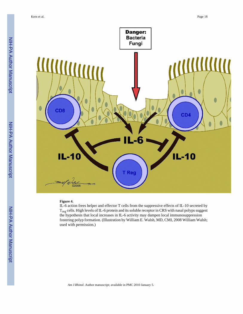
Figure 4.IL-6 action frees helper and effector T cells from the suppressive effects of IL-10 secreted byTreg cells. High levels of IL-6 protein and its soluble receptor in CRS with nasal polyps suggestthe hypothesis that local increases in IL-6 activity may dampen local immunosuppressionfostering polyp formation. (Illustration by William E. Walsh, MD, CMI, 2008 William Walsh;used with permission.)
Kern et al. Page 18
Am J Rhinol. Author manuscript; available in PMC 2010 January 5.
NIH
-PA Author Manuscript
NIH
-PA Author Manuscript
NIH
-PA Author Manuscript
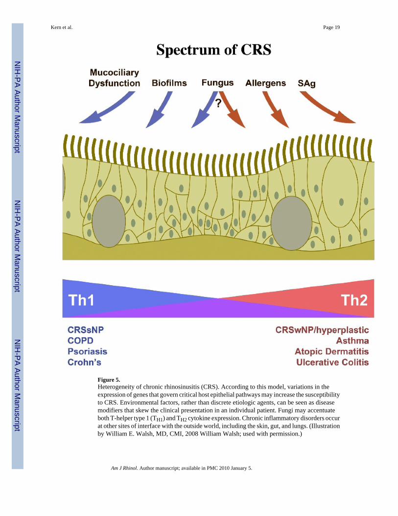
Figure 5.Heterogeneity of chronic rhinosinusitis (CRS). According to this model, variations in theexpression of genes that govern critical host epithelial pathways may increase the susceptibilityto CRS. Environmental factors, rather than discrete etiologic agents, can be seen as diseasemodifiers that skew the clinical presentation in an individual patient. Fungi may accentuateboth T-helper type 1 (TH1) and TH2 cytokine expression. Chronic inflammatory disorders occurat other sites of interface with the outside world, including the skin, gut, and lungs. (Illustrationby William E. Walsh, MD, CMI, 2008 William Walsh; used with permission.)
Kern et al. Page 19
Am J Rhinol. Author manuscript; available in PMC 2010 January 5.
NIH
-PA Author Manuscript
NIH
-PA Author Manuscript
NIH
-PA Author Manuscript