THE GENETIC ETIOLOGY OF EARLY-ONSET HEARING ...
180
THE GENETIC ETIOLOGY OF EARLY-ONSET HEARING LOSS IN NEWFOUNDLAND AND LABRADOR By © Jessica Squires A Thesis submitted to the School of Graduate Studies in partial fulfillment of the requirements for the degree of: Master of Science/Genetics/Faculty of Medicine Memorial University of Newfoundland May 2015 St. John’s, Newfoundland and Labrador
-
Upload
khangminh22 -
Category
Documents
-
view
3 -
download
0
Transcript of THE GENETIC ETIOLOGY OF EARLY-ONSET HEARING ...

THE GENETIC ETIOLOGY OF EARLY-ONSET HEARING LOSS IN NEWFOUNDLAND AND LABRADOR
By © Jessica Squires
A Thesis submitted to the School of Graduate Studies in partial fulfillment of the
requirements for the degree of:
Master of Science/Genetics/Faculty of Medicine
Memorial University of Newfoundland
May 2015
St. John’s, Newfoundland and Labrador

ii
Abstract
Hearing loss is the most common sensory disorder worldwide and > 50% of cases
can be attributed to single gene mutations. I used a targeted candidate gene approach and
Sanger sequencing to screen genomic DNA from 101 deaf probands with Newfoundland
ancestry for pathogenic mutations in deafness genes. First I screened for mutations in
WFS1, TMPRSS3, and PCDH15 that were previously identified in this population, then
for mutations in Cx26 and Cx30, and mutations in the mitochondrial genes MTRNR1 and
MTTS1. Finally, genes were targeted based on patterns of hearing loss as seen on patient
audiograms. Although several probands were “solved” by this approach, none had
mutations in WFS1, TMPRSS3 or PCDH15. One proband had digenic mutations in Cx26
and Cx30 and two probands inherited the A1555G mutation in MTRNR1. In order to
decipher several variants of unknown pathogenicity and solve more families, further
clinical recruitment and whole genome approaches are required.

iii
Acknowledgements
I thoroughly enjoyed my experience as a graduate student at Memorial University
in Dr. Terry-Lynn Young’s lab. I would like to take this opportunity to thank the
members of this lab as well as others who have assisted me along the way. First the lab
staff, Mr. Dante Galutira, Mr. Jim Houston, and Mrs. Tammy Benteau, who often went
out of their way to offer help, advice, and encouragement when I needed it the most.
Second, to the other students in the lab: Nelly Abdelfatah, Lance Doucette, and David
McComiskey who taught me many necessary lab skills, who were always there to answer
questions, and with whom I developed a relationship that will last through the years. I
would also like to thank Carol Negrijn and Ann Griffin for helping me with the family
pedigrees and patient information, as well as my committee members Roger Green and
Jane Green for helping me organize the information in my project and present it in a
manner that is easily understood. A huge thank-you to Kerri Smith for all of her help with
the submission process, and for being a wonderful 5th floor friend/coffee buddy/rant
hearer. Finally I would like to thank my supervisor, Dr. Terry-Lynn Young, who was
there to offer advice. She gave me a push when I needed it, helped me view things from
multiple perspectives, and always walked me through answering my own questions (an
important skill!). This has been an adventure I will never forget!
Other than the Young Lab members, I would like to thank my parents and my
brother for their love and support, my boyfriend Mike for his never ending love, support,
and comic relief, as well as my friends for being there to help me survive my grad school
journey.

iv
Table of Contents
ABSTRACT ................................................................................................................................ II
ACKNOWLEDGEMENTS ........................................................................................................... III
LIST OF TABLES ....................................................................................................................... VI
LIST OF FIGURES .................................................................................................................... VII
LIST OF ABBREVIATIONS ......................................................................................................... IX
LIST OF APPENDICES ................................................................................................................ X
1.INTRODUCTION .................................................................................................................... 1 1.1 PURPOSE OF STUDY ................................................................................................................... 1 1.2 OVERVIEW OF HEARING LOSS ...................................................................................................... 1
1.2.1 Nonsyndromic Autosomal Recessive Hearing Loss ...................................................... 13 1.2.2 The Founder Populations of the island of Newfoundland ............................................ 14 1.2.3 Recurrent Mutations: Founder Mutations vs. Mutation Hot Spots .............................. 15 1.2.4 Previous Hearing Loss Studies in Newfoundland ......................................................... 16 1.2.5 Mouse Models and Hereditary Hearing Loss ............................................................... 17 1.2.6 Types of Mutations that cause Hearing Loss ............................................................... 18 1.2.7 Cochlear Function ........................................................................................................ 21 1.2.8 Connexin Hearing Loss ................................................................................................. 22 1.2.8 Connexin 26 and Hearing Loss ..................................................................................... 25 1.2.9 Connexin 30 and Hearing Loss ..................................................................................... 27 1.2.10 Connexin 31 and Hearing Loss ................................................................................... 30 1.2.11 Digenic Inheritance and the Connexin Genes ............................................................. 33 1.2.12 Mitochondrial Hearing Loss ....................................................................................... 35
1.3 SUMMARY .............................................................................................................................. 41
CO-‐AUTHORSHIP STATEMENT ................................................................................................ 42
2.MATERIALS AND METHODS ................................................................................................ 43 2.1 SUBJECT RECRUITMENT ............................................................................................................ 43 2.2 OVERALL DESIGN OF CANDIDATE GENE APPROACH ....................................................................... 43
(1) Screening genes identified in hearing impaired probands from Newfoundland ............. 44 (2) Screening Genes identified in Caucasians with Hearing Loss .......................................... 44 (3) Screening Genes Causing Recognizable Audiogram Patterns (Audioprofiles) ................. 45
2.3 TECHNIQUES FOR MUTATION DETECTION .................................................................................... 45 1. Extraction of Genomic DNA .............................................................................................. 45 2. Amplification of Targeted Sequences by Polymerase Chain Reaction (PCR) ..................... 45 3. Preparation for Sanger Sequencing using ABI Cycle Sequencer ........................................ 46 4. Connexin 30 Primers and Conditions for Detecting 342kb Deletion .................................. 48

v
5. Detection of a Possible Mitochondrial Deletion ................................................................ 49 5. Verification of Pathogenicity ............................................................................................. 50 Figure 2.2. Steps of the process of AudioGene candidate gene determination and analysis (SNHL = Sensorineural Hearing Loss; Adapted from audiogene.eng.uiowa.edu) ................. 52
3. RESULTS ............................................................................................................................. 53 3.1 SCREENING FOR DEAFNESS MUTATIONS PREVIOUSLY IDENTIFIED IN NEWFOUNDLAND .......................... 53 3.2 SCREENING GENES IDENTIFIED IN CAUCASIANS WITH HEARING LOSS .................................................. 53 3.2.1 PROBANDS WITH VARIANTS IN CONNEXIN 26 AND CONNEXIN 30 .................................................. 54 3.2.2 PROBANDS WITH VARIANTS IN MTRNR1 AND MTTS1 ............................................................... 56 3.3 SCREENING GENES CAUSING RECOGNIZABLE AUDIOGRAM PATTERNS (AUDIOPROFILES) ........................ 59 3.5 SUMMARY OF RESULTS ............................................................................................................. 61
4. DISCUSSION ..................................................................................................................... 108 4.1 SCREENING PROBANDS FOR MUTATIONS PREVIOUSLY FOUND IN THE NL POPULATION ...................... 108 4.2 PROBAND FROM FAMILY 2155 WITH HEARING LOSS CAUSED BY MUTATIONS IN CONNEXIN 26 &
CONNEXIN 30 ............................................................................................................................. 109 4.3 PROBAND FROM FAMILY 2197 FOUND TO HAVE A HETEROZYGOUS 35DELG MUTATION IN CX26 ........ 112 4.4 A HETEROZYGOUS F83L VARIANT IN CX26 EXON 2 IN THE PROBAND FROM FAMILY 2091 .................. 112 4.5 VARIANTS IN THE MITOCHONDRIAL GENOME ............................................................................. 113 4.6 PROBANDS FROM FAMILIES 2112 AND 2144 HEARING LOSS CAUSED BY A MUTATION IN MTRNR1 .... 113 4.7 A G951A VARIANT IN MTRNR1 FOUND IN TWO PROBANDS FROM FAMILIES 2167 AND 2197 .......... 115 4.8 MTRNR1 COMMON VARIANTS ................................................................................................ 118 4.9 SPECIFICITY OF MITOCHONDRIAL PRIMER BINDING SITES .............................................................. 119 4.10 MATCHING MITOCHONDRIAL HAPLOTYPES .............................................................................. 119 4.11 CONNEXIN 31 VARIANTS ...................................................................................................... 120 4.12 TMC1 VARIANTS ................................................................................................................ 123 4.13 CONCLUSION ...................................................................................................................... 126
5. FUTURE DIRECTIONS ........................................................................................................ 128
6. BIBLIOGRAPHY ................................................................................................................. 130 APPENDIX 1: THE FORWARD AND REVERSE PRIMERS AND PCR CONDITIONS FOR EACH GENE. .................. 142 APPENDIX 2: NL HEARING LOSS STUDY MEDICAL QUESTIONNAIRE ...................................................... 146 APPENDIX 3: STANDARD DNA EXTRACTION PROTOCOL ..................................................................... 154 APPENDIX 4: FULL PEDIGREES FOR HL FAMILIES ............................................................................... 156 APPENDIX 5: DNA SAMPLE NUMBERS AND THEIR CORRESPONDING FAMILY NUMBERS FOR ALL PROBANDS
SCREENED ALONG WITH WHAT MUTATIONS WERE FOUND (BOTH PREVIOUS AND IN THIS STUDY) ............... 159

vi
List of Tables
Table Number
Table Page
Table Description
1 99 Prediction of the connexin 26 F83L variant effect
2 99 Polymorphisms found in MTRNR1
3 100-‐105
Mitochondrial polymorphisms found in each of the deafness probands
4 106 Hearing Loss Frequency categories 5 106 Mutations found in the five candidate genes chosen for sequencing based
on the pattern of hearing loss in the families 6 107 A summary of the gene variants found with the proband they were found
in and the hearing loss phenotype of the proband.

vii
List of Figures
Fig. # Page # Figure Description 1.1 8 The anatomy of the human ear 1.2 9 Flowchart outlining the causes of hearing loss 1.3 10 A sample audiogram 1.4 11 Sample audiograms A - Reverse Slope, B - “Cookie Bite”, C - Reverse
“Cookie Bite”, D – Ski Slope, E - Flat Loss 1.5 12 The characteristic audiogram of individuals affected by hearing loss
caused by mutations in the gene that causes Wolfram Syndrome 1.6 25 Six connexins come together to form a connexon. 1.7 26 A drawing of the inner cochlea highlighting the areas in which
connexin 26 is expressed. 1.8 30 A drawing of the inner cochlea highlighting the areas in which
connexin 30 is expressed. 1.9 33 A drawing of the inner cochlea highlighting the areas in which
connexin 31 is expressed 2.1 51 Flowchart outlining the methods followed in this project 2.2 52 Steps of the process of AudioGene candidate gene determination and
analysis 3.1 64 Flowchart outlining the methods followed with results included 3.2 65 Sequencing for mutations found in the NL population 3.3 66 An electropherogram showing heterozygous deletion of a guanine
(35delG) in connexin 26 in the proband from family 2155 3.4 67 An agarose gel showing heterozygous connexin 30 deletion in the
proband from family 2155 3.5 68 The partial pedigree for family 2155 3.6 69 The pedigree of family 2197 3.7 70 An electropherogram showing the heterozygous deletion of a guanine
(35delG) in connexin 26 in the proband from family 2197 3.8 71 Pedigree for family 2091 3.9 72 An electropherogram showing the nucleotide change of F to L at
position 83 in the proband from family 2091 3.10 73 A weblogo image pointing out the conservation of the amino acid at
position 83 3.11 74 The pedigree for family 2167 3.12 75 An electropherogram showing the homoplasmic change of guanine to
adenosine in the proband from family 2167 3.13 76 The pedigree for family 2112 3.14 77 The pedigree for family 2144 3.15 78 An electropherogram showing the homoplasmic change of adenosine
to guanine in the proband from family 2112

viii
3.16 79 An electropherogram showing the homoplasmic change of adenosine to guanine in the proband from family 2144
3.17 80 The pedigree for family 2072 3.18 81 An agarose gel showing the apparent deletion in the proband and the
proband’s mother from family 2072 3.19 82 The description of mispriming in the mitochondrial genome 3.20 83 A partial pedigree for family 2010 3.21 84 Audiograms for the proband (A) and her mother (B) from family 2072
and the audiogram for the proband (C) from family 2010. 3.22 85 The pedigree for family 2078 3.23 86 The pedigree for family 2075 3.24 87 The pedigree for family 2097 3.25 88 The pedigree for family 2156 3.26 89 The pedigree for family 2083 3.27 90 The pedigree for family 2078 showing mutations 3.28 91 The pedigree for family 2075 showing mutations 3.29 92 The pedigree for family 2097 showing mutations 3.30 93 The pedigree for family 2083 showing mutations 3.31 94 The pedigree for family 2124 3.32 95 The pedigree for family 2065 3.33 96 The pedigree for family 2146 3.34 97 The pedigree for family 2177 3.35 98 The pedigree for family 2092

ix
List of Abbreviations
Abbreviation Meaning AD Autosomal Dominant AR Autosomal Recessive ATP Adenosine Triphosphate bp Base pair bps Base pairs Cx26 Connexin 26 Cx30 Connexin 30 Cx31 Connexin 31 COCH Cochlin gene dB Decibel DFN Deafness DFNA Nonsydromic deafness autosomal dominant DFNB Nonsyndromic deafness autosomal recessive DIAPH1 Diaphanous-related Formin 1 gene DNA Deoxyribonucleic Acid GJB2 Connexin 26 GJB3 Connexin 31 GJB6 Connexin 30 HDF High-Dye Formamide Hz Hertz KCNQ4 Potassium Voltage Gated Channel gene mM Milli-molar mtDNA Mitochondrial DNA MTRNR1 Mitochondrially Encoded 12S RNA gene MTTS1 Mitochondrial Gene, Encodes the transfer RNA for Serine MYO6 Myosin VI gene NL Newfoundland and Labrador NSHL Nonsyndromic Hearing Loss PCDH15 Protocadherin related 15 gene PCR Polymerase Chain Reaction POU4F3 POU Class 4 Homeobox 3 gene SHL Syndromic Hearing Loss TECTA Tectorin Alpha gene TMC1 Transmembrane Channel-Like 1 gene TMPRSS3 Transmembrane Protease Serine 3 gene µL Micro Litre µM Micro-molar WFS1 Wolframin Syndrome 1 gene

x
List of Appendices
Appendix # Page # Title 1 142 The Forward and Reverse primers and PCR conditions for each
gene 2 146 NL Hearing Loss Study Medical Questionnaire 3 154 Standard DNA Extraction Protocol 4 156 Full Pedigrees for HL Families
5 159 DNA Sample Numbers, their Corresponding Family Numbers, Genes Screened and mutations found

1
1.Introduction
1.1 Purpose of Study
The objective of this project was to determine the underlying genetic cause of
deafness in probands (affected individuals) from Newfoundland and Labrador. These
individuals had been diagnosed with hearing loss. An attempt to identify genetic causes
was carried out using modern molecular genetic techniques.
1.2 Overview of Hearing Loss
Hearing loss is one of the most common sensory disorders worldwide, affecting
1-3 in 1000 children at birth or in early childhood, and is caused by many known
environmental and genetic factors (Retrieved from www.cdc.gov/ncbddd/hearingloss/
ehdi-data2009.html on April 24, 2014). The prevalence of pre-lingual hearing loss
increases if young children are included and all degrees of hearing loss are considered.
Even hearing loss that follows environmental affliction (for example infection, acoustic
trauma, or exposure to ototoxic drugs) is likely influenced by a genetic susceptibility (Ito
et al., 2010; Petersen et al., 2006,;Nance, 2003; Keats & Berlin, 2002). The number of
individuals affected by hearing loss increases as the population ages, with hearing
impairment affecting 25% of individuals between the age of 50 and 65, and 50% of
individuals over the age of 85 (Liu et al., 2007). Like early onset hearing loss, late onset
hearing loss can also be attributed to a variety of causes including environmental factors,
medical disorders and their treatment, and genetic susceptibility (Liu et al., 2007).

2
Hearing loss can be classified into three different types based on clinical
presentation: conductive, sensorineural, and mixed – a combination of conductive and
sensorineural. Conductive hearing loss results from abnormalities of the anatomic
components of the ear that transfer sound waves to the cochlea, such as the external ear,
the ear canal, ossicles (malleus, incus, stapes), tympanic membrane, oval window, round
window, or middle ear space (Fig.1.1). Sensorineural hearing loss results from the
dysfunction of the auditory pathway components that convert sound wave vibrations into
an electrical impulse that is transferred to the auditory cortex in the brain. Therefore this
type of hearing loss is caused by abnormalities within the cochlea, and/or auditory
(cochlear) nerve, or in rare cases auditory brainstem, or auditory structures in the brain
(Griffith, 2002) (Fig.1.1).
More than fifty percent of hearing loss cases can be attributed to an underlying
genetic cause (Ito et al., 2010; Norton 1991), and can be further categorized into two
distinct groups: syndromic (SHL) or nonsyndromic (NSHL). When an individual presents
with hearing loss along with other abnormalities, it is known as SHL. This occurs in
approximately 30% of diagnosed individuals with over 400 hearing loss syndromes
having been described, including Usher syndrome (hearing loss along with retinitis
pigmentosa – loss of peripheral vision and night blindness), Pendred syndrome (hearing
loss along with thyroid problems), and Jervell and Lange-Nielson syndrome (hearing loss
along with cardiac dysrhythmia) (Van Camp et al., 1997; Hilgert et al., 2009). When
hearing loss is not associated with any other abnormalities it is known as NSHL; 70% of
hearing loss cases fall into this category (Hilgert et al., 2009; Van Camp et al., 1997).

3
NSHL can follow every known pattern of Mendelian inheritance including
autosomal recessive (AR), autosomal dominant (AD), and X-linked. There are also
mutations in the mitochondrial genome that result in non-Mendelian maternal
inheritance, and NSHL. A set of nomenclature rules has been assigned to the different
types of genetic deafness to classify them as AD, AR or X-linked (mitochondrial hearing
loss does not have a specific nomenclature) and these types are named under the acronym
DFN (from the word “deafness”). Following DFN are the letters ‘A’ or ‘B’, ‘A’ meaning
AD inheritance (DFNA) or ‘B’ meaning AR inheritance (DFNB). When DFN is present
without an A or B, it refers to X-linked deafness (Piatto et al., 2009). AR inheritance
means the individual has to inherit two copies of a mutated gene in order to be affected,
this type of inheritance accounts for 77-88% of genetic NSHL. With AD inheritance,
which accounts for 10-20% of inherited NSHL, the individual will be affected if only one
copy of a mutated gene is inherited. The mutated gene can come from either parent, if
one parent is affected each offspring has a 50% chance of inheriting the mutated gene, if
both parents are affected the chance of inheritance increases to 75%. X-linked disorders
are passed on from parent to offspring on the X chromosome and can be dominant or
recessive, the most characteristic feature of these disorders is the absence of male-to-male
transmission. X-linked disorders also severely affect males but have variable expression
in females due to X chromosome inactivation, which occurs in early embryogenesis in
female germ line cells to compensate for the X-linked gene dosage difference between
males and females. X-linked inheritance accounts for 1-2% of NSHL. Finally,
mitochondrial hearing loss is caused by a mutation in the genome of mitochondria, an
4
organelle found within all human cells, more frequently in cells that require an abundance
of adenosine triphosphate (ATP), such as muscle cells, due to the fact that mitochondria
generate most of the cell’s supply of this form of chemical energy. Mitochondrial genetic
disorders can only be passed down from a mother to her offspring because the
mitochondria in an embryo come from the ovum. None of the mitochondria contained in
the sperm are passed on to the offspring; therefore you will only see female-to-male and
female-to-female transmission in mitochondrial genetics. Mitochondrial hearing loss is
more variable and harder to detect than other types of NSHL, and accounts for 1-20% of
NSHL depending on the population. (Nance, 2003, Petersen, 2012, Sirmaci, 2012)
(Fig.1.2). When evaluating the frequency of NSHL and SHL within the different types of
Mendelian inheritance, it can be shown that the most common type of hereditary hearing
loss is nonsyndromic autosomal recessive.
In order to categorize hearing loss into specific modes of inheritance prior to
genetic testing, two methods can be used, first a pedigree with a family history of a
defined hearing loss diagnosis is required so the hearing loss can be traced throughout the
family. Secondly, an audioprofile can be created using the person’s behavioural
audiogram and other pertinent information including age of onset and number of affected
relatives. Use of these classification methods helps to narrow down the number of genes
that are screened during the genetic testing phase, making the process more efficient.
For the first method, a pedigree is created using information from the affected
individual and family members as well as medical and census records. The pedigree will
show the relationships between individuals and their affection status. Even with a clear

5
family history, classification can be difficult, as some types of hearing loss have reduced
penetrance. Penetrance is directly linked to variable expressivity, as both influence the
effect that genetic mutations have on the population. Penetrance indicates the number of
people who have a genetic mutation and display the disorder. Expressivity refers to the
symptoms displayed by an individual that has a genetic disorder. More specifically
variable expressivity is the phenomenon where different people have the same genetic
mutation but display different symptoms or different severities of the same symptom.
For example, hearing loss can have variable expressivity, meaning a number of
individuals within a family have hearing loss but at different severities, some with a
subclinical phenotype (i.e. a mild form of hearing loss that is undetectable by standard
behavioural audiogram). In a study, ten individuals from a family are found to have a
mutation in a deafness gene, though only six of these family members display hearing
loss; this would be reduced penetrance, and it is common in AD disorders. It may make it
difficult for a researcher to confirm that the mutation is the cause of the disorder. Given
the fact that everyone has a different genetic background, a mutation can have a lowered
penetrance, so low that it may not cause a detectable phenotype. Therefore an individual
could have a mutation that causes hearing loss, but may not display it (i.e. variable
expressivity). These types of cases make it difficult to consistently identify the mode of
inheritance, especially in families with few affected individuals. Knowing the mode of
inheritance is important because it helps to narrow down the list of candidate genes, as
mutations in some genes are more likely to cause a specific type of hearing loss. AD
disorders will often show up in every generation of a pedigree (called a vertical

6
inheritance pattern) and are transmitted from mothers or fathers to both sons and
daughters. AR disorders show a horizontal pattern where they are generally seen in only
one sibship, and usually the affected siblings will have unaffected parents. Like
autosomal dominant inheritance, the number of affected females and males is nearly
equal; meaning the chance of inheriting a mutation is not affected by sex. X-linked
dominant disorders have a specific pattern where the disorder is passed to all daughters
from an affected male and affected females will pass the disease to half of their sons and
daughters. The crisscross pattern is often described as a “Knight’s Move inheritance”
after the move used in the game of chess. X-linked recessive disorders will appear in
more males than females (due to the fact that males only have one X chromosome), and
an affected male will not pass the disorder on to any of his offspring (although the
females may be carriers) (Griffiths et al., 1999). Mitochondrial disorders display a pattern
of matrilineal (maternal) inheritance because the mitochondrial genome is inherited from
the mother and not the father; therefore only children of affected females will potentially
manifest the disorder, whereas offspring of an affected male will not be affected
(Strachan et al., 1999). Understanding the inheritance pattern can help when counseling
families regarding prevention of the disorder.
The second method used to categorize hearing loss into specific modes of
inheritance is audioprofiling, where several audiograms at different ages are plotted on
the same graph. The different ages can be from a single individual or from different
members of the same family (Smith et al., 2013). A helpful online tool, called
AudioGene (audiogene.eng.uiowa.edu )(Hildebrand et al., 2008), was developed to aid in

7
audioprofiling, it “analyzes audiometric data and predicts the likely underlying genetic
cause of hearing loss based on known phenotypic parameters” (Hildebrand et al., 2008).
The user inputs the proband’s audiogram and age, and the same information for all
affected relatives, then the tool will identify a list of candidate genes/loci based on this
information. An audiogram is a graphic display of the softest sounds an individual can
hear at each specific frequency. The frequency, the pitch of a sound, is measured in Hertz
(Hz), and the intensity, the loudness of a sound, is measured in decibels (dB). The
audiogram (Fig.1.3) is produced following a hearing test by an audiologist. The pattern of
an audiogram can give an indication as to what type of hearing loss affects the individual
(Retrieved from http://www.raisingdeaf kids.org/hearingloss/testing/ audiogram/ on
August 23, 2013). On the audiogram, a reverse slope indicates low-frequency hearing
loss, mid-frequency loss is shown as a dip in the middle of the graph, also known as a
“cookie-bite”. Loss of high and low frequencies is known as a “reverse cookie-bite loss”
as the individual can hear better in the mid-frequency range creating a peak in the middle
of the graph. High frequency hearing loss is also known as sloping loss due to the sloped
configuration, and flat loss involves a similar hearing loss at all frequencies (Fig. 1.4)
(Retrieved from http://www.hearinglosshelp.com/articles/ kindsofhearinglosses.htm on
August 23, 2013). If a single pattern is present in all members of a family, it can predict
the locus of the underlying genetic cause. For example the audioprofile of individuals
with WFS1 non-syndromic hearing loss has a highly characteristic shape (Fig.1.5) (Smith
et al., 2013).

8
By performing pedigree and audioprofile analyses the list of candidate genes can
be greatly decreased. This helps the researcher narrow in on the underlying genetic cause
of hearing loss before performing mutation screening.
Figure 1.1. The anatomy of the human ear. (Created by author, adapted from http://commons.wikimedia.org/wiki/File:Ear-anatomy-text-small-en.svg)

9
Hearing Loss
Environmental (<50%)
Genetic (>50%)
Nonsyndromic (~70%)
Syndromic (~30%)
Autosomal Recessive (77-88%)
Autosomal Dominant (10-20%)
X-linked (1-2%)
Mitochondrial (1-20%)
Figure 1.2. Flowchart outlining the causes of hearing loss

10
Figure 1.3. A graph of hearing threshold – to read this graph you look at the dots to see how loud a sound has to be at a specific frequency for the individual to hear it. For example at 8000 Hz the individual can only hear sounds louder than 50 dB. Therefore the person has trouble hearing high-pitched sounds, so has high frequency hearing loss. The red line indicates the threshold for hearing loss. When an individual cannot hear sounds above 20 dBHL, they are considered to have hearing loss at that frequency. This particular individual does not have hearing loss between 125 and 1000 Hz. (Image taken from http://audiogene.eng.uiowa.edu/audioprofiles)

11
A B
C D
E
Figure 1.4. Sample audiograms. A – Reverse Slope, B – “Cookie-Bite”, C- Reverse “Cookie-Bite”, D – Sloping, E – Flat Loss (Adapted from Center for Hearing Loss Help, http://www.hearinglosshelp.com/ articles/kindsofhearinglosses.htm)

12
Figure 1.5. The characteristic audiogram of individuals affected by hearing loss often associated with mutations in the WFS1 gene that causes nonsyndromic SNHL (DFNA6/14/38) (Hildebrand, M. S. et al., 2009)

13
1.2.1 Nonsyndromic Autosomal Recessive Hearing Loss
Hearing loss can be divided into SHL and NSHL subtypes. SHL is the diagnosis
when hearing loss occurs with other clinical conditions or abnormalities. This occurs in
approximately 30% of diagnosed individuals with over 400 hearing loss syndromes
having been described, including Usher syndrome (hearing loss along with retinitis
pigmentosa – loss of peripheral vision and night blindness), Pendred syndrome (hearing
loss along with thyroid problems), and Jervell and Lange-Nielson syndrome (hearing loss
along with cardiac dysrhythmia) (Van Camp et al., 1997; Hilgert et al., 2009).
Nonsyndromic hearing loss is diagnosed when the loss of hearing is the only clinical
phenotype. Nonsyndromic autosomal recessive hearing loss is generally prelingual
(present before speech develops), almost exclusively a result of cochlear defects (thus
sensorineural), and usually more severe than all other forms. This type of hearing loss is
extremely heterogeneous; as of June 23, 2014, 80 loci have been mapped and 55 genes
have been identified (Hereditary Hearing Loss Homepage http://hereditaryhearing
loss.org). Like all autosomal recessive disorders an individual must inherit two mutated
copies of the gene, one from each parent, to present with the disease phenotype.
Mutations in genes that are located in the DFNB1 locus, connexin 26 (Cx26) and
connexin 30 (Cx30), are the cause of 30-50% of this type of hearing loss (Bhalla et al.,
2009). The remaining cases are due to mutations in numerous different genes (Hilgert et
al., 2008).
Specifically within the DFNB1 locus, mutations in Cx26, also known as GJB2,
are the most common underlying cause of AR NSHL throughout the world (Kenneson et

14
al., 2002). Another connexin gene found within this locus and 29 kb away from Cx26 is
Cx30 (or GJB6) and it is also responsible for many cases of this type of hearing loss.
Both genes are found on chromosome 13 (13q.11-13.q12) (del Castillo et al., 2002) and
will be discussed in greater detail.
1.2.2 The Founder Populations of the island of Newfoundland
The population ancestry of Newfoundland (current population of 514,536
residents; Statistics Canada http://www.statcan.gc.ca/) is greatly influenced by natural
expansion from early English and Irish settlers who came to fish the North Atlantic cod.
The majority of people from Newfoundland are direct descendants of these first
immigrants. The unique gene pool of this population is the consequence of a small
number of founders and genetic drift, especially founder effect, a type of genetic drift that
occurs when a small number of individuals break off from a larger population and form a
new colony causing a reduction in genetic variation in the new population due to
reproductive separation (Griffiths et al., 2002). Many of the immigrants in the 1600s
settled in coastal regions around Newfoundland to be in close proximity to the fishing
grounds. Individual fishing villages, locally known as “outports”, were isolated from each
other by ocean and harsh terrain. Furthermore, marriages between Catholics and
Protestants were strongly discouraged. The result was a high frequency of inbreeding
within isolated communities which remained distant from each other, increasing the
prevalence of specific genetic disorders such as hearing loss (Doucette et al., 2009;
Rahman et al., 2003; Martin et al., 2000). Other disorders, such as Familial Multiple
Endocrine Neoplasia Type I (Farid et al., 1980), hemophilia A (Xie et al., 2002),

15
hereditary non-polyposis colorectal cancer syndrome (Froggatt et al., 1999), and familial
adenomatous polyposis (Spirio et al., 1999) have been recognized to be due to founder
effects in outport communities in Newfoundland. Extended, multiplex families with
Newfoundland ancestry present an unprecedented opportunity to uncover the etiology of
genetic diseases like hearing loss.
1.2.3 Recurrent Mutations: Founder Mutations vs. Mutation Hot Spots
Common mutations may be due to either founder mutations or to recurrent
mutations at hot spots, therefore when a common mutation is discovered it is necessary to
use haplotype analysis to figure out which type of mutation you have uncovered
(Romdhane & Abdelhak, 2012). Founder mutations are mutations present in a high
frequency in a particular population, and the reason for their existence is that the
mutation was present in one ancestor or a small number of ancestors. Mutation hot spots
can produce recurrent mutations because they are DNA sequences that are highly
susceptible to be mutated (Retrieved from http://www.ncbi.nlm.nih.gov /books/
NBK5191/ on April 26, 2013). The high susceptibility of a sequence to be mutated can be
caused by instability, unequal crossing over, or a predisposition to substitutions
(Retrieved from http://www.ncbi.nlm.nih.gov/books/ NBK5191/ on April 26, 2013). To
confirm that a common mutation is due to a founder effect, a method called haplotype
analysis is carried out. A haplotype is the arrangement of marker alleles found on a single
chromosome. Marker alleles can be single nucleotide polymorphisms (SNPs) or
microsatellite markers (also known as short tandem repeats – repeating sequences of
DNA) that are passed on together through a pedigree (Zhao et al., 2003). Haplotype

16
analysis is a type of genetic test that looks closely at these linked DNA segments and
shows how the specific genetic information is passed down through pedigrees (Retrieved
from http://www.ncbi.nlm.nih.gov/books/NBK5191/ on April 26, 2013, Romdhane &
Abdelhak, 2012). The mutation is considered a founder mutation only if it is located at a
single haplotype. If the mutation is found on multiple haplotypes it is not a founder
mutation, and may be due to a mutation hot spot (Romdhane & Abdelhak, 2012).
1.2.4 Previous Hearing Loss Studies in Newfoundland
The population of the island of Newfoundland has been the focus of extensive
hearing loss research for more than fifteen years. In 2001, while studying a six-generation
Newfoundland family, Young et al. identified a missense mutation (p. 2146 G→A) in the
WFS1 gene that was causing a dominant form of inherited progressive deafness. The
mutation was found in all affected members of the family and the link between the
mutations and hearing loss was supported by haplotype and mutation analysis. This study
by Young et al. was the first to link a mutation in WFS1 with nonsyndromic hereditary
hearing loss (Young et al., 2001). In 2004, Ahmed et al. discovered two recessive
mutations in the TMPRSS3 gene in a six-generation family from the South Coast of the
island segregating an autosomal recessive form of early onset hearing loss. The role of
TMPRSS3 in hearing loss was first described in 1996 (Bonne-Tamir et al., 1996; Veske et
al., 1996). Ahmed et al. first discovered that hearing loss in the Newfoundland family
was linked to markers for DFNB8/B10. Sanger sequencing revealed two recessive
mutations, within TMPRSS3, 207delC and IVS8 + 8insT, the latter being novel.
Haplotype analysis revealed an apparent founder DFNB8/B10 associated haplotype. It

17
had previously been assumed that the founder effect of a mutation in the Newfoundland
population would be high due to the isolation of the island. However, that was not the
case in this study, which highlights the genetic heterogeneity of deafness, even in a
founder population. As well, Ahmed et al.’s findings challenged the notion that the
population of the island of Newfoundland was homogeneous.
A second (consanguineous) family, located on Newfoundland’s South Coast,
displayed profound hearing loss inherited as an apparent autosomal recessive trait. The
underlying cause of this disorder was a novel mutation in PCDH15 (a gene also
associated with Usher’s syndrome - a syndromic form of hearing loss associated with
vision impairment, Ahmed, 2001), identified by Doucette et al. in 2009. In this study, a
genome-wide scan was used to map the hearing loss trait to chromosome 10q21-22, and
genotyping identified that individuals with a hearing impairment were homozygous for a
16Mb ancestral genotype that included 44 annotated genes. Sequencing analysis was
performed and identified the novel V528D missense mutation in PCDH15. This is the
second study that found a PCDH15 mutation causing nonsyndromic hearing loss instead
of Usher’s syndrome (Doucette et al., 2009). The first study in 2003, found two missense
mutations (p.G262D and p.R134G) in PCDH15 causing nonsyndromic recessive hearing
loss (DFNB23) in two Pakistani families (Ahmed et al., 2003).
1.2.5 Mouse Models and Hereditary Hearing Loss
It is difficult to follow the process involved in the formation of the human inner
ear due to the lack of instruments available for observing the development, as well as the
lack of cell lines that could be used to replicate the events involved (Chatterjee, 2011).

18
Animal models, particularly mouse models are ideal for studying inner ear development
and also useful for researching human hereditary hearing loss (Friedman et al., 2007;
Vrijens et al., 2008), because of the similarity between inner ear structures (Avraham,
2003). There are 77 deafness genes shared by humans and mice (Retrieved from
http://hearingimpairment. jax.org/models.html on June 24, 2014). In addition to this, mice
have a short gestational period, and large litter sizes make it feasible to produce large
numbers of mutated mice (Avraham, 2003). Using mouse models, researchers can
mutate a gene of interest, and then follow the development of the structures of the inner
ear. Observing the resulting phenotype of the mutant mice provides insight into both the
normal function of the gene as well as the negative impact of the mutation on hearing
(Friedman et al., 2007). For example, early studies by Avraham et al. (1995) discovered
that the gene causing the deafness phenotype in the Snell’s waltzer mouse is Myo6,
providing a candidate gene to test in human subjects. Myo6 encodes a myosin heavy
chain protein that is expressed in the inner ear and is required for structural maintenance
(Avraham et al., 1995). Several years later, Melchionda et al. (2001) discovered the first
human mutation in the human homologue MYO6 to be associated with hereditary hearing
loss.
1.2.6 Types of Mutations that cause Hearing Loss
Variants in DNA do not always result in a disease phenotype and instead can have
a positive effect on an organism, giving them a survival advantage. When mutations do
cause a disease phenotype they are called genetic disorders (Relethford, 2012). There are
many different types of mutations that have different effects on amino acid codons, so

19
that genetic disorders caused by some mutations may be more severe than the disorder
caused by other mutations in the same gene.
One type of variant, called a point mutation, is also known as a single base
substitution because that is literally what occurs, a single base nucleotide is replaced with
a different base nucleotide. There are three types of point mutations: neutral, missense,
and nonsense. A neutral variant causes no change in the amino acid codon and therefore
is not predicted to have a negative effect on the protein. A missense mutation occurs
when the new (mutated) base nucleotide results in a new amino acid codon, for example,
a change of AUA to ACA would change the codon from isoleucine to threonine. A
nonsense mutation arises when the change of a base nucleotide results in a stop codon. If
the codon for serine, UCG, was mutated to become UAG, this would be a nonsense
mutation as UAG is a stop codon, meaning that this is the codon that signals the
premature truncation of the protein with subsequent drastic effect on the phenotype.
Another type of mutation is a frameshift mutation; these can be deletions or
insertions of one or more base pairs that can be detrimental when they occur within the
coding sequence of genes. In a frameshift mutation the number of bases deleted or
inserted is not divisible by three. For example, the deletion of five bases or insertion of
two bases cause the reading frame to shift, altering the amino acid codons that follow the
insertion or deletion and leads to premature truncation in most cases.
Other types of large multibase mutations include duplications – when a portion of
a gene, or an entire gene, is reproduced, inversions – when a segment of a chromosome is
flipped or reversed, and translocations – when segments of nonhomologous

20
chromosomes are interchanged. These can result in gene fusion where the piece of
chromosome connects genes or parts of genes that would otherwise be separate. When
these variants do not cause any noticeable effect on the organism’s phenotype, they are
said to be benign or nonpathogenic (Powar, 2007).
To determine the nature of the identified variant, first a literature search is
conducted to locate the mention of the variant in previous studies, the second step is to
check the variant in the publicly available SNP/1000 genomes database to determine the
frequency of the variant in the disorder. If the identified variant was not detected in the
SNP/1000 genome database, it is considered a novel variant. (Mitchell et al., 2005). If the
variant is considered novel, the researcher must confirm the pathogenicity of the
mutation. To determine pathogenicity of a mutation, it is helpful to have access to the
DNA of other family members, affected and unaffected. By sequencing the DNA of the
family members, a researcher can get clues as to whether or not the identified variant is
pathogenic. If the variant is found in all affected family members but not in the
unaffected members, this is one piece of evidence that the mutation may be causative. If
the mutation is found in all or many family members regardless of their affection status,
then it is likely that the mutation is benign. The next step would be to check for the
mutation in the general population, usually by screening members of the same ethnicity.
If the variant is not found in a general population screen, and is found in only affected
members of the family, it is very likely the variant is a causative mutation. If a high
proportion of the population does have the mutation, it is unlikely to be pathogenic.

21
Another piece of the puzzle is to use prediction programs to determine the impact
of a variant on the protein. There are several prediction programs available to check the
probability of a mutation being pathogenic. Such programs include SIFT (retrieved from
http://sift.jcvi.org/ on March 20, 2012 ) which predicts whether an amino acid
substitution affects protein function, WebLogo ( retrieved from
http://weblogo.berkeley.edu/ on March 20, 2012) which generates sequence logos
showing sequence conservation at specific positions, PolyPhen-2 (Retrieved from
http://genetics.bwh.harvard.edu/pph2/ on March 20, 2012) which predicts the impact of
an amino acid substitution on the structure and function of the protein, and PMut
(Retrieved from http://genetics.bwh.harvard.edu/pph2/ on March 20, 2012) which
predicts the pathogenicity of a mutation based on the protein sequence. Using these
programs can help determine the pathogenicity of a variant.
1.2.7 Cochlear Function
Mutations in the Cx26 gene, within the DFNB1 locus on chromosome 13, cause a
large proportion of NSHL; the percentage varies depending on the population (Connexin-
Deafness Homepage). In order to discuss the role of connexins in hearing loss, it is
necessary to understand the function of the cochlea. Function of the cochlear tissues and
auditory nerve depends on the ionic environment surrounding them within the inner ear,
more specifically the concentration of potassium, an ion with an important involvement
in sensory transduction (and all nerve signals throughout the human body). Three fluid
filled spaces in the cochlea, the scala media (containing endolymph), the scala tympani
and vestibule (both containing perilymph), are separated by tight junction barriers
22
(Fig.1.7). The scala media is filled with potassium rich endolymph whereas the scala
tympani and vestibuli are filled with the sodium rich perilymph, with a composition very
similar to intercellular and extracellular fluid ion concentrations, respectively. Sensory
transduction is controlled by the endocochlear potential (EP), which is approximately 80
millivolts (mV) and is generated and maintained by the stria vascularis. The distinct ion
concentrations in each of the fluid filled spaces are maintained by the tight junction
barriers between them (Wangermann, 2006).
The sensory receptors within the inner ear are called hair cells (Fig.1.4). The hair
cell stereocilia are immersed in endolymph, while the cell body is surrounded by
perilymph. When a sound travels through the outer ear, it vibrates through the ossicles
and moves the endolymph and basilar membrane upon which the hair cells sit. The
receptor potentials are generated by the movement of potassium from the endolymph into
the hair cells via the stereocilia. This begins a cycle where the potassium moves out of
the hair cell body into the perilymph and eventually returns to the endolymph via the stria
vascularis (Kikuchi et al., 2000) (Fig. 1.7).
1.2.8 Connexin Hearing Loss
Connexins are gap junction proteins that regulate ion concentrations and are
found in many different vertebrate tissues. Daniel Goodenough proposed the name
connexin in 1974. Before Goodenough isolated the subunit and called it a connexin, J.
David Robertson hinted at a hexagonal lattice of subunits in 1963 when he isolated the
subunits from Mautner cells (cells that make up the neural circuit in teleost fish that

23
mediate an escape response) of the common goldfish. He found what he called “honey-
comb-like hexagonal arrays of closely packed subunits” which Goodenough (1975) later
called connexons (the assembly of six connexin proteins) when he investigated the
structure of the gap junction of mice hepatocytes using electron microscopy,
biochemistry, and x-ray diffraction techniques. The idea that gap junctions are made up
of subunits, connexons, which further consisted of smaller units called connexins wasn’t
actually confirmed until 1977 when Makowski and colleagues used x-ray diffraction to
elucidate the composition of gap junctions (Makowski et al., 1977). They established that
the connexon was in fact a hexamer composed of molecular protein units (connexins) that
had a molecular weight between 23-28 kilo Daltons (kDa) and that the connexons in
plasma membranes of one cell connect to connexons in membranes of an adjacent cell to
form the gap junction (Fig.1.6).
The human genome contains 21 different connexin genes (Retrieved from
http://omim.org/ on August 14, 2011). The first connexins to be named were connexin 32
(GJB1 OMIM# 304040) and connexin 43 (GJA1 OMIM # 121014) (Beyer et al., 1987).
The genes were first named for the molecular mass of their protein, for example,
connexin 32 is approximately 32 kDa , connexin 43 is approximately 43 kDa, etc. This
method for nomenclature was used for a long time and is still in use today. However,
once connexin genes were separated into alpha and beta groups based on sequence
similarities and the overall predicted topological organization (Gimlich et al., 1990), a
new type of classification was established. Those belonging to the alpha group were
called “A” and those belonging to the beta group “B”. In addition to this distinction they

24
were called GJ, for gap junction, and given a number identifying the order in which they
were discovered, e.g. GJA1 is connexin 43.
Each of the 21 known connexin proteins has been mapped to different areas in the
human genome (Retrieved from http://omim.org/ on August 14, 2011). These genes are
developmentally regulated and can be co-expressed in the same cell (Kumar et al., 1992).
The function of many of the connexin genes and their products has been revealed through
the study of the effect of mutations within the genes. Mutations in many of these genes
have been associated with disease. For example mutations in the connexin 32 gene,
GJB1, underlie Charcot-Marie-Tooth-Disease, a neurological disorder involving
weakness in the foot and lower leg as well as difficulty with auditory and fine motor
skills (Bergoffen et al., 1993). Mutations in more than one connexin gene (Cx 26 and Cx
30) also cause nonsyndromic hearing loss (Pfenniger et al, 2010).
Within the cochlea both Cx26 and Cx30 are components of gap junction systems,
which are essential to the cycling of potassium. They form the channels that facilitate the
constant movement of potassium ions through the hair cells and back into the endolymph,
an ionic current flow that is undulated by vibration during the transduction of sound into
an electrical signal (Carlsson et al., 2012; Irshad et al., 2012; Ru et al., 2012).

25
1.2.8 Connexin 26 and Hearing Loss
Kelsell et al. identified Cx26 as the first autosomal NSHL susceptibility gene in
1997. The authors realized that the DFNB1 locus and Cx26 mapped to the same location
on chromosome 13. When they sequenced Cx26 in individuals with a hearing impairment
from DFNB1- linked Pakistani families, they discovered homozygous substitutions in
two of the hearing impaired offspring and found their unaffected parents to be
heterozygous carriers for these mutations. Kelsell et al. also identified mutations in the
Cx26 gene in other affected members of different Pakistani families, giving them further
Figure 1.6. Six connexins come together to form a connexon. Connexons are located in the cell membranes of cells. When cells come together connexons meet to form gap junctions. (Image taken from http://commons.wikimedia.org/wiki/File :Connexon_and_connexin_structure.svg )

26
proof that deafness in these families was caused by mutations in Cx26. The authors took
this to mean that the Cx26 protein is an essential component of the cochlea. We now
know that Cx26 is expressed in many areas within the cochlea, specifically in the regions
that separate the scala media from the scala tympani (Retrieve from
http://hereditaryhearingloss.org on November 23, 2011) (Fig.1.7). The loss of the
function of Cx26 within the inner ear is expected to disrupt the flow of potassium thus
interrupting the generation and passage of the electric signal from the inner ear to the
auditory nerve and onto the brain (Petersen 2012).
As of June 27, 2014, 111 NSHL mutations have been described within the Cx26
gene, including splice, nonsense, missense, and frameshift mutations (Retrieved from
http://davinci.crg.es/deafness/ on June 27, 2014). The most common Cx26 mutation in
the Caucasian population (Denoyelle et al., 1997), the deletion of a guanine at position 35
(c.35delG), was first identified by Zelante et al. in 1997 after they sequenced the Cx26
gene in thirty-five affected patients from the Mediterranean. This mutation causes a
frameshift that leads to premature chain termination and is found in a region of six
guanine nucleotides close to the 5′ end of the Cx26 coding region (Zelante et al., 1997).
Most of the mutations in Cx26 lead to malformed channels, which causes altered
assembly of hemichannels, disrupted targeting to the plasma membrane, or decreased
protein stability (Thönnissen et al., 2002).

27
1.2.9 Connexin 30 and Hearing Loss
Cx30 (GJB6) is another connexin gene heavily involved in NSHL and is
associated to hearing loss caused by mutations in Cx26 (retrieved from
davinci.crg.es/deafness/ on November 24, 2011). Cx30 was first described in the DFNA3
locus and found to cause autosomal dominant hearing loss (Grifa, 1999). It was
discovered when 38 families, with hearing loss linked to chromosome 13, tested negative
for Cx26 mutations (Grifa, 1999). Cx30 maps to chromosome 13q12 and is located near
(~800kb) connexin 26 (Grifa, 1999). Close to 50 percent of individuals who are
Figure 1.7. A drawing of the inner cochlea highlighting the areas in which connexin 26 is expressed. The numbers shaded orange are the areas of expression. These are the same areas of expression as connexin 30. (Image taken from Van Camp G, Smith RJH. Hereditary Hearing Loss Homepage. http://hereditaryhearingloss.org)

28
heterozygotes for Cx26 mutations also have a large deletion in the Cx30 gene causing
hearing loss (del Castillo et al., 2003; Mahdieh et al., 2010)
Many hearing impaired patients with nonsyndromic hearing loss (NSHL) have
only one mutated allele in Cx26 (del Castillo, 2002). For a long time these individuals
were considered unsolved because another Cx26 mutation was suspected, but could not
be found. Even more confounding was that some patients with hearing loss showing
linkage to DFNB1 were void of mutations in Cx26 (del Castillo, 2002). In 2002, del
Castillo et al. discovered a 342kb deletion, which they later named Δ(GJB6-D13S1830),
in the Cx30 gene - a gene known to be co-expressed with Cx26 in the inner ear (Fig.8).
This discovery was made in 33 unrelated probands affected by hearing loss with
heterozygous mutations in Cx26. Haplotype analysis was performed using markers on
13q12 to look for mutations outside of Cx26. Del Castillo et al. found that it was
impossible to amplify several markers in the vicinity of Cx30 in two of the subjects,
which led them to believe that the subjects harboured a germline deletion. Upon analysis
of DNA fragments of Cx30 they discovered that the deletion truncated the open reading
frame of the gene. The authors were able to determine the break points using sequence
tagged sites, and they created a specific Polymerase Chain Reaction (PCR) assay to
detect the Δ(GJB6-D13S1830) deletion (del Castillo, 2002). PCR is a technique that uses
DNA polymerase to synthesize a new strand of DNA that is complementary to the
template strand added to the reaction mixture. At the beginning of the reaction the
mixture is heated to separate the strands of DNA. DNA polymerase then synthesizes the
complementary strand between specific primers. The mixture is then cooled and the DNA

29
anneals. The cycles of heating and cooling are repeated, thus causing exponential
amplification of the targeted section of DNA. When the process is completed this section
of DNA will have accumulated in billions of copies. The Cx30 deletion truncates the
protein and is often found in conjunction with a heterozygous mutation in Cx26 in
hearing loss patients. The discovery of Δ(GJB6-D13S1830) solved a portion of the
hearing impaired cases that were unexplained up to this point.
Once del Castillo et al. discovered this deletion they questioned whether the
pattern of inheritance, where deafness was caused by two heterozygous mutations in two
different genes, was monogenic or digenic (del Castillo et al., 2002). If it were a
monogenic pattern (due to a single gene), there would need to be a regulatory element
upstream of both genes that controlled the expression of Cx26. They reasoned that the
deletion would suppress gene expression, resulting in hearing loss (del, 2002). A second
explanation would be that the deletion disrupts the activity of another gene that is
important for the function of Cx26. Evidence suggests that Cx30 is this other gene that is
required for Cx26 to function normally (del Castillo, 2002). Del Castillo et al. (2002)
suggested that the DFNB1 locus contains two genes, Cx26 and Cx30, and altering any
two of the four alleles will cause hearing loss. Pallares-Ruiz et al. (2002) also suspected
that this phenomenon represented a pattern of digenic inheritance and tried to determine
if this was the fact, however their results were inconclusive. It was not until 2009 that a
group provided conclusive results after testing this hypothesis. Rodriguez-Paris et al.
(2009) used allele specific analyses based on reverse transcriptase PCR (RT-PCR) using
buccal cells that express both Cx26 and Cx30 equally. In the three hearing impaired
30
probands studied, all of whom carried Δ(GJB6-D13S1830), none expressed Cx26 when
Cx30 had been altered, proving that the Cx26 protein is not produced when Δ(GJB6-
D13S1830) is present in the Cx30 allele. This finding would be expected with the loss of
a cis-regulatory element found within the deleted region thus disrupting the transcription
of Cx26 (Rodriguez-Paris, 2009).
1.2.10 Connexin 31 and Hearing Loss
Cx31, located on chromosome 1p34, encodes the human gap junction protein β-3,
also known as GJB3, and is expressed in the skin, inner ear (specifically the spiral
ligament, spiral limbus, and the auditory nerve), and the auditory nerve (Fig.1.9). Like
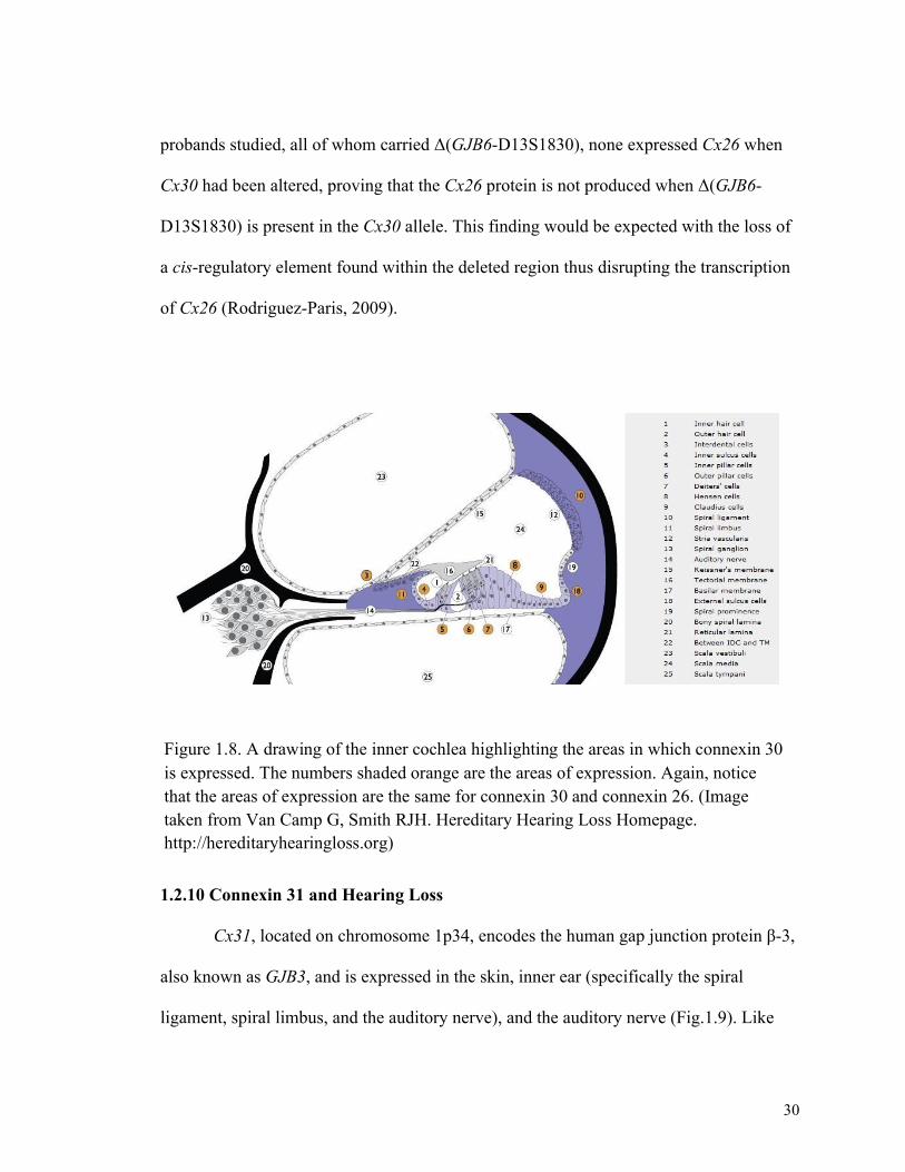
Figure 1.8. A drawing of the inner cochlea highlighting the areas in which connexin 30 is expressed. The numbers shaded orange are the areas of expression. Again, notice that the areas of expression are the same for connexin 30 and connexin 26. (Image taken from Van Camp G, Smith RJH. Hereditary Hearing Loss Homepage. http://hereditaryhearingloss.org)

31
connexin 26 and 30, connexin 31 forms channels called gap junctions in the inner ear that
allow the movement of potassium ions between the organ of Corti and the endolymph of
the scala media (Hauwe et al., 1999; Oh et al., 2012).
Linkage of Cx31 to hearing loss was first shown in 1998 when Xia et al. explored
the possibility that other connexin genes (besides Cx26 and Cx30) might be involved in
the hearing process. They used two multiplex families of Chinese ancestry to map the
third connexin gene to chromosome 1p33-p35, and used Sanger sequencing to reveal two
mutations, a missense (c.547 G>A: p.Q183K) and a nonsense (c.538 C>T: p.R180X)
mutation associated with autosomal dominant, bilateral, high-frequency loss. Using RT-
PCR they showed that Cx31 was expressed in the rat inner ear.
Mutations in Cx31 have also been shown to cause recessive hearing loss in a
compound heterozygous manner (Liu et al., 2000). Liu and his team of researchers
looked at 25 Chinese families with recessive hearing loss and found that the members of
two of the families, affected by early onset, sensorineural, bilateral hearing loss, had an
in-frame 3 base pair deletion (423-425ATT) in one allele causing the loss of an isoleucine
residue at codon 141 and a c.423A>G transversion (p.I141V) in the other allele.
Therefore, similar to Cx26, mutations in Cx31 can cause either autosomal dominant or
autosomal recessive hearing loss (Liu et al., 2000).
In 2000, Lόpez-Bigas et al. found five SNPs (c. 1227C>T, c.1610G>A, c.1700
C>T, c.1731G>A, and c.1931C>T) in Cx31 in patients affected by hearing loss. Two of
the SNPs, c.1227C>T and c. 1731G>A, cause amino acid changes p.R32W and V200I,
respectively. The nucleotide change c.1931C>T was found in 24 of the 153 hearing

32
impaired subjects screened, however it was also found in 13 percent of the control
population. In much the same way p.R32W was found in seven of the 153 subjects with
hearing loss, but also in 18 percent of the control population. The other three changes
were not found in the ethnically matched control population; however the amino acid
change V200I is in an area that is not well conserved, and in the rat and mouse the amino
acid at position 200 is an isoleucine rather than a valine. The other two SNPs (c.1227C>T
and c.1610G>A) had no obvious effect on the protein, thus Lόpez-Bigas et al. concluded
that none of the SNPs found were causative, but would be useful in segregation and
linkage disequilibrium analysis which can determine the patterns of inheritance.
Rouan et al. (2003) used HeLa cells to determine the function of the Cx31 protein
with a R32W variant. In this case they were investigating the connection between this
variant and erythrokeratodermia variabilis (EKV), a skin disease that is also caused by
mutations in Cx31. They transfected HeLa cells with mutated and wild type Cx31
expression constructs and looked at synthesis, intracellular distribution, and protein
assembly. The cells with mutated Cx31 showed no deviations in expression level,
connexon assembly, or intracellular distribution. They concluded that R32W is an
inconsequential sequence polymorphism of Cx31 (Rouan et al., 2003).

33
1.2.11 Digenic Inheritance and the Connexin Genes
It is now an accepted fact that mutations in two separate loci (genes) can cause
recognisable patterns of hereditary deafness, known as digenic inheritance. For example,
in one study it was shown that non-syndromic hearing loss can be caused by digenic
inheritance of mutations in Cx26 and Cx31 (Liu et al., 2008). This group of researchers
screened the Cx31 gene in 108 patients from China who were already shown to have
heterozygous Cx26 mutations and who did not have mutations in Cx30. They found two
mutations (N166S and A194T) in Cx31 in patients who had either 235delC or 299delAT
mutation in Cx26; none of the Cx31 mutations were found in the Chinese population
controls. Because Cx26 and Cx31 have overlapping expression patterns it is likely that
Figure 1.9. A drawing of the inner cochlea highlighting the areas in which connexin 31 is expressed. The numbers shaded orange are the areas of expression. (Image taken from Van Camp G, Smith RJH. Hereditary Hearing Loss Homepage. http://hereditaryhearingloss.org)
34
these proteins have direct physical interaction (Liu et al., 2008). They also found that
Cx26 and Cx31 form heteromeric connexons in mouse cochlea leading them to conclude
that the mutations they found in Cx26 and Cx31 were in fact the cause of hearing loss in
the patients involved (Liu et al., 2008).
Recently Oh et al. (2012) looked for a relationship between variants in Cx31 and
Cx30 and nonsyndromic hearing loss in the Korean population. Through gene sequencing
they found a total of nine variants, four of which were novel. They chose five of these
variants (three novel: Cx31: V27M and V43M, Cx30: A40V, two known: Cx31: V84I,
Cx30: A40V) to perform functional studies using a pathogenicity prediction program,
also the area of the mutation was analyzed for conservation (other variants were excluded
due to the frequency of their occurrence in unaffected individuals). Three of the variants,
V27M and V84I in Cx31, and A40V in Cx30, were predicted to be deleterious because
they were not found in unaffected individuals and the residues were highly conserved
among different species (Oh et al., 2012). As well, using biochemical-coupling tests, two
of the variants in Cx31 (V27M and V84I) were shown to affect protein function when
they were present in a heterozygous form with wild type Cx31. The biochemical-coupling
tests were performed by recording the time it took to diffuse Lucifer Yellow dye from
cell to cell using cells that contained connexins with heterozygous mutations and WT
mutations. The times were recorded as either immediate transfer: less than 30 seconds,
delayed transfer: 30 seconds to 3 minutes, and no transfer: more than 3 minutes. If the
time was delayed or there was no transfer then it was inferred that the protein was not
functioning correctly (Oh et al., 2012).

35
The studies discussed suggest that Cx31 plays a major role in the hearing process
and mutations in this gene can cause hearing loss in both an autosomal dominant and
autosomal recessive manner. It has also been suggested that this gene be included in
hearing loss screening studies along with Cx26 and Cx30 because of the connection
shown between these connexin genes and hearing loss.
1.2.12 Mitochondrial Hearing Loss
Mitochondria are membrane-bound organelles found within most eukaryotic cells
and are responsible for generating adenosine tri-phosphate (ATP), a molecule that is used
as a source of chemical energy within the body (Scheffler, 2008; Copeland, 2002). The
number of mitochondria contained within a cell depends on the organism as well as the
tissue type and can range from a couple of hundred to a few thousand. Cells within tissue
that requires a lot of energy to function, like muscle tissue and the inner ear, often contain
more mitochondria (Scheffler, 2008; Copeland, 2002).
One important distinction that separates mitochondria from other organelles
within the cell is that mitochondria have their own genome made up of several copies of a
double-stranded chromosome that takes the shape of a ring (rather than a helix, as found
in nuclear DNA) with its own genetic code (Barrell et al., 1979). The human
mitochondrial genome consists of 16,569 base pairs (Anderson et al., 1981, GenBank,)
that make up 37 genes: 13 code for the proteins that interact with nuclear proteins to carry
out oxidative phosphorylation, 22 code for the transfer RNA required for the
mitochondrial protein-synthesizing system, and 2 code for the large and small subunits of

36
ribosomal RNA (taken from www.ncbi.nlm.nih.gov/genbank/ on November 29, 2011,
Fischel-Ghodsian, N., 2002).
The mitochondrial genome is maternally inherited. While both egg and sperm
contain mitochondria, the mitochondria from the sperm are lost during early
embryogenesis (Manfredi et al., 1997). Therefore, a mother may pass on any mutations in
her mitochondrial genome to all of her children. Determining whether or not the
mutation will be passed on depends on the number of mitochondrial chromosomes or
mitochondria that contain the mutation. For example, some mitochondrial mutations are
heteroplasmic, that is, they are only present within some mitochondria out of all
mitochondria that are contained in the cell. Other mitochondrial mutations are
homoplasmic, meaning the mutation is present in all of the mitochondria (Wallace,
1992). In the case of a heteroplasmic mutation the proportion of wild type or mutant
genes that are passed on is random. It is possible that a mother will pass on only those
mitochondria that do not carry the mutation and therefore no phenotype will appear
(Fischel-Ghodsian, N., 2002). Mitochondrial mutations are difficult to pinpoint because
there are numerous DNA molecules within a single mitochondrion and there are
numerous mitochondria per cell.
In the early 1990s, it was discovered that mutations in mitochondrial genes were
linked to hereditary hearing loss (Hu et al., 1991; Jaber et al., 1992; Prezant et al., 1993).
Hu et al. (1991) analyzed 36 pedigrees with a positive history of aminoglycoside induced
hearing loss and in 22 of the pedigrees where the pattern of inheritance could be
ascertained they found that transmission of the predisposition to develop a hearing

37
impairment after aminoglycoside exposure was exclusively through females. They
considered two explanations for this type of transmission: the first was that it could be an
X-linked predisposition; the second was that it could be caused by mitochondrial
inheritance. They ruled out X-linked transmission as there was equal inheritance in
females and males and the affected males did not pass the predisposition on to their
daughters or the sons of their daughters. Because they could exclude X-linked inheritance
this left them with the conclusion that aminoglycoside induced hearing loss was a
mitochondrially inherited disorder. Hu et al. were the first group to come to this
conclusion, and at the time there were only a few rare diseases shown to be
mitochondrially inherited. Following this conclusion, the next step Prezant et al. (1993)
took was to look for mutations in the human ribosomal RNA (rRNA) genes, which are
found in mitochondrial DNA, in those families. The investigators chose this area to begin
their mutation search because they knew that the rRNA of bacteria was the target of these
antibiotics. As well, they decided to sequence the entire mitochondrial genome in a large
Arab-Israeli family. Each of the four families had a substitution of a guanine for an
adenine at position 1555, within the 12S rRNA gene. This was the first case of a
mitochondrial mutation being associated with non-syndromic hearing loss. It was
discovered that the A1555G mutation in the mitochondrial gene MTRNR1was one of the
most frequent causes of hearing loss after mutations in Cx26, and SLC26A4 (Solute
carrier family 26 member 4, the gene mutated in Pendred syndrome – deafness with
thyroid enlargement, and in DFNB4 with enlarged vestibular aqueduct; taken from
http://omim.org/ on February 13, 2012; Guo et al., 2008).

38
Since the early 90s, many families have had their hearing loss explained by
mutations in mitochondrial genes. As well, in the case of aminoglycoside-induced
hearing loss, it is possible to prevent the development of a hearing impairment by
avoiding aminoglycoside antibiotic therapy in individuals with a family history of
aminoglycoside induced hearing loss (Hu et al., 1991; Selimoglu, 2007). There are now
two mitochondrial genes associated with NSHL (Retrieved from
http://hereditaryhearingloss.org/ on November 25, 2011). Neither of these genes are
protein coding; one, MTRNR1, codes for 12S ribosomal RNA, the small subunit of
mitochondrial ribosomes that binds with the large subunit, 16S, to carry out protein
synthesis within the mitochondria. The second, MTTS1, codes for the serine (UCN)
transfer RNA, the RNA that carries serine to the polypeptide chain during protein
synthesis when it is coded for by the triplet AGN (OMIM, http://omim.org/). There are
three mutations in MTRNR1 that are associated with both aminoglycoside-induced
hearing loss and NSHL that is not triggered by aminoglycoside exposure. There are four
mutations reported to cause nonsyndromic hearing loss in MTTS1, however two of these
mutations are also associated with another disorder. The c.7445A>G mutation is
associated with palmoplantar keratoderma (PPK) and the c.7472insC mutation is
associated with neurological dysfunction, including ataxia, dysarthria and myoclonus
(Hereditary Hearing Loss Homepage http://hereditaryhearingloss.org/).
The first of these mitochondrial mutations to be associated with hearing loss was
discovered in an Israeli-Arab pedigree with hearing loss inherited through the maternal
line (Jaber et al., 1992; Prezant et al., 1993). The hearing loss was progressive and

39
usually presented in childhood. It was Prezant et al. in 1993 that sequenced the entire
mitochondrial genome of family members from the Israeli-Arab pedigree and also from
three unrelated patients with familial aminoglycoside-induced deafness. They found a
point mutation at position 1555 that changed an adenosine to a guanine in the gene that
codes for the 12S ribosomal RNA (rRNA). This mutation was shared amongst all four
families. However, Prezant et al. were not the first to suggest that familial
aminoglycoside-induced deafness was caused by a defect in the mitochondrial DNA. In
1989, Higashi looked at 28 Japanese families to determine whether males or females
passed on the trait of susceptibility of streptomycin deafness. After examining each
pedigree he concluded that it was in fact transmitted through only females. Until this
discovery, the susceptibility of the cochlea to streptomycins was assumed to be
autosomal dominant (Higashi, 1989). Higashi (1989) also suggested that because familial
hearing loss induced by streptomycin exposure could not be explained by ordinary
Mendelian inheritance, then ototoxicity caused by streptomycin intake somehow
disrupted ATP production in the mitochondria of the hair cells, and that might be caused
by changes in the mitochondrial DNA.
Once the A1555G mutation had been discovered in aminoglycoside induced
deafness it was also found to cause nonsyndromic hearing loss worldwide, in Arab-
Israeli, Japanese, Mongolian, Zairean, Spanish, Chinese, Turkish, and Balinese families
(Usami et al., 1997; Estivill et al., 1998; Kupka et al., 2002; Abreu-Silva et al., 2006;
Kokotas et al., 2009). In fact mutations in connexin 26, SLC26A4, and mtDNA A1555G
are the most prevalent causes of deafness worldwide (Guo, 2008). This means that

40
screening for the mtDNA A1555G mutation should be included when looking for the
underlying genetic cause of hearing loss in a family, especially if the inheritance appears
to be maternally transmitted or hearing loss occurred after aminoglycoside use.
The first mutation identified in MTTS1 changed an adenosine at position 7445 to a
guanine. It was found by Reid et al. in 1994 in all affected members of a family with 13
members displaying sensorineural NSHL. The second MTTS1 mutation was an insertion
of a cytosine at position 7472, found by Tiranti et al. in 1995 after sequence analysis of
the tRNA gene regions of mitochondria in affected individuals from a large Sicilian
kindred. This mutation causes nonsyndromic hearing loss as well as hearing loss
accompanied by ataxia and myoclonus (Tiranti, 1999). The third MTTS1 mutation found
to cause hearing loss was the change of a thymine to cytosine at position 7511 (7511
T>C), a mutation discovered by Sue et al. in 1999 in 36 affected members of a large
African American family when they sequenced the entire mitochondrial genome in
search of mutations because of the maternal transmission of hearing loss. This mutation,
which disrupts a highly conserved site in the mitochondrial DNA, was determined by Sue
et al. (1999) to be pathogenic. The fourth MTTS1 mutation found thus far is a change
from thymine to cytosine at position 7510 (7510 T>C), and was identified in a Caucasian
family by Hutchin et al. in 2000. Hearing loss in this family appeared to be transmitted
through the maternal line so they looked for mutations in the mitochondrial DNA. First
they screened for other known mitochondrial DNA mutations, but all were absent. They
then found the T to C transition at base pair position 7510. This novel thymine to
cytosine transition at position 7510 was found in all affected family members and was not

41
found in 141 Caucasian controls. As well the thymine at position 7510 is highly
conserved in a wide range of species (Hutchin, 2000). These mutations in the MTTS1
gene should also be included when screening for genetic hearing impairment, especially
in families where hearing loss is transmitted through the maternal line.
1.3 Summary
Hearing loss is a very debilitating disorder that causes many problems in a
developing child. If hearing loss is pre-lingual, a child will have difficulty learning to
speak, as children acquire this ability by imitating sounds they hear around them, such as
the voices of parents and siblings. The longer hearing loss remains undetected the greater
the likelihood for permanent impairments of speech and language that are necessary for
social development and the ability to take part in a standard education system (NIDCD
website). Through the study of hearing loss genetics, the underlying causes of inherited
hearing impairment can be revealed, improving the lives of those affected by the
disorder.

42
Co-authorship statement
I, Jessica Squires, hold a principal authorship status for all of the manuscript
chapters in my thesis. However, the manuscript is co-authored by my supervisor, Dr.
Terry-Lynn Young, who helped design my project and research proposal, and also
assisted in data analysis and review of my manuscript. Co-authorship is also given to
PhD students: Dr. Lance Doucette and Dr. Nelly Abdelfatah who helped with review of
my manuscript and the Young Lab Research Assistants: Mr. Dante Galutira, Mr. Jim
Houston, and Mrs. Tammy Benteau who helped with data analysis and manuscript
review.

43
2.Materials and Methods
2.1 Subject Recruitment
Probands with hearing impairment, along with their relatives, were recruited from
across Newfoundland and Labrador. This study focused on predominantly probands with
profound hearing loss as children. Probands and their blood relatives were recruited to the
study through various poster drives and through the Provincial Medical Genetics Program
of Eastern Health, St. John’s. Family history, informed consent, and permission to access
medical records and audiograms were obtained as per approved protocol #01.186
(Human Research Ethics Board, St John’s, NL, Canada). The team also collected
information on the geographical location of family founders. The patients and/or their
parents/guardians completed a hearing loss questionnaire (Appendix 2), and a blood
sample for DNA isolation was obtained from all available family members. Probands
with hearing impairment due to known environmental factors such as infection and
acoustic trauma were excluded from further study.
2.2 Overall Design of Candidate Gene Approach
A step-wise, targeted screening approach was used to look for mutations across
three categories of deafness genes: (1) genes with mutations previously identified in
hearing impaired probands with Newfoundland ancestry; (2) genes frequently mutated in
Caucasians with hearing loss, and (3) genes that cause specific, recognizable patterns of
hearing loss as seen on an audiogram. When a pathogenic mutation was identified in a

44
proband, I confirmed, where possible, which side of the family the mutation came from
by genotyping DNA from the parent(s) and other available blood relatives.
(1) Screening genes identified in hearing impaired probands from Newfoundland
Preceding this study, working as a Research Assistant in Dr. Young’s laboratory,
I screened 77 probands for mutations in genes previously found to cause hearing loss in
the Newfoundland population; namely WFS1, TMPRSS3 and PCDH15 that our lab
previously identified in deaf probands from Newfoundland (Young et al., 2001; Ahmed
et al., 2004; Doucette et al., 2009) and for common mutations causing hearing loss in
Cx26 and Cx30. The analysis of this data is part of my thesis project. In order to detect
potential recurrent mutations due to founder effects in the Newfoundland population and
potentially (and efficiently) solve one or more probands for my thesis project I also
sequenced genomic DNA of 33 newly ascertained probands for mutations in WFS1,
TMPRSS3 and PCDH15.
(2) Screening Genes identified in Caucasians with Hearing Loss
In order to detect common mutations in the Cx26 and Cx30, two different protocols were
required. To detect the common point mutations in Cx26, full, bidirectional Sanger
sequencing of Cx26 was done on DNA from the newly ascertained probands (n=33).
However, Sanger sequencing is not sufficient to detect the common mutation in Cx30, as
it is not a point mutation but a large deletion. Therefore, a PCR-based method, first
described by del Castillo et al. (2002), was used to detect the common 342 kb deletion
(del13S1830) in Cx30 in the newly ascertained probands (n=33).

45
The next step was to look for common mutations that occur in the mitochondrial genome
that can also lead to hearing loss. To do this, mitochondrial DNA from all unsolved
probands (n=101/110) was screened for mutations in the mitochondrial genes MTRNR1
and MTTS1 using full, bidirectional Sanger sequencing.
(3) Screening Genes Causing Recognizable Audiogram Patterns (Audioprofiles)
In some cases, the underlying causative gene can be suggested by patient
audioprofiles, or pattern of loss as seen on an audiogram. For the majority of probands in
this study, at least one audiogram was available and the audiograms were analyzed and
grouped into four categories (low-frequency loss, mid-frequency loss, high-frequency
loss and flat loss; Figure 2.2).
2.3 Techniques for Mutation Detection
1. Extraction of Genomic DNA
Genomic DNA was extracted from the blood of affected probands and their
relatives and archived and stored at 4°C. For the 33 new recruits, I extracted genomic
DNA from peripheral blood (leukocyte fraction) according to a standard salting out
protocol (Appendix 3).
2. Amplification of Targeted Sequences by Polymerase Chain Reaction (PCR)
PCR reactions contained 1X Kapa Taq Buffer A with 1.5 mM MgCl2 (Kapa
Biosystems, Woburn, MA), 0.9 M betaine (Sigma, Saint Louis, MO) 0.2 mM dNTPs
(Applied Biosystems, Warrington, UK), 0.4 uM of each primer (IDT, San Diego, CA),
0.4U of Kapa Taq, and 10 ng of purified DNA in a total volume of 20 µL. This master

46
mix was used for all genes with the exception of Cx26. The master mix for Cx26 did not
contain betaine. PCR was performed on a GeneAmp PCR System 9700 thermocycler
(Applied Biosystems, Foster City, CA) with cycling conditions of [94oC/ 5 min, (94oC/
30 s, 640C – 2.00C per cycle/30 s, 720C/ 30 s) x 5 cycles, (94oC/ 30 s, 54oC/ 30 s, 720C/
30 s) x 30 cycles, 720C/ 7 min]. The primers and amplification conditions for each gene
are provided in Appendix I. The PCR products were stained and run on a 1% agarose gel
(0.5g agarose/ 50mL 1X TBE + 2.5µL SYBR safe/ 50mL of 1X TBE) using gel
electrophoresis, and viewed with UV light using the Kodak Molecular Imaging system.
This allowed the product size of the PCR products to be compared to a standard DNA
ladder in order to be sure the anticipated size of DNA segment (PCR product) was
obtained.
3. Preparation for Sanger Sequencing using ABI Cycle Sequencer
Samples were prepared according to standard lab protocols: 300µL of Sephacryl
300 was added to each well of a 96 well, 0.45 µm MultiScreen filter plate and centrifuged
for 5 minutes at 1800 x g. The flow-through was then discarded. Next, the PCR products
were added to the wells of the MultiScreen plate. A 96 well PCR catch plate was placed
beneath it and the plates were centifuged at 1800 x g for 5 minutes. This time the flow-
through was retained in the PCR plate and the purified PCR product was prepared for
sequencing.
A sequencing master mix was prepared to contain the following ingredients: Sequencing
Mix (0.5µL), 5X Sequencing Buffer (2.0µL), Primer (either forward or reverse) at 1.6
mM (2.0µL), and dH2O (14.5µL). This mixture was the same for all genes and yielded

47
19.0µL volume per well of a sequencing plate. 1 µL per well of purified PCR product
was added to the master mix and then centrifuged briefly and placed on a thermocycler
for cycle sequencing with cycling conditions of [(96oC/ 1 min, 96oC/ 10 s, 500C/5 s, 60oC/4 min)
x 24 cycles, 40C hold].
The next step was ethanol precipitation of the PCR product. 5.0 µL of EDTA
(125mM) followed by 65 µL of ethanol (95%) were added to each sample in the
sequencing plate. The plate was then covered and placed at room temperature in the dark
for 30 minutes to 24 hours to precipitate the DNA. Next, the plate was centrifuged for 30
minutes at 3000 rpm. The plate was then inverted to decant the ethanol and while still
inverted was centrifuged briefly at 200rpm to further decant the ethanol. 150.0 µL of
ethanol (70%) was then placed in each well. The plate was centrifuged for 15 minutes at
3000 rpm and inverted to decant the ethanol, and then the inverted plate was briefly
centrifuged at 200 rpm to remove residual ethanol. The plate was left to dry in the dark
for 10-15 minutes to evaporate any remaining ethanol and dry the DNA pellets. The
DNA was dissolved in 15.0 µL of HDF (High-Dye Formamide), centrifuged briefly, and
placed on the thermocycler for denaturing (2 mins at 95ºC).
Automated sequencing was performed on the Applied Biosystems 3130xl Genetic
Analyzer (Applied Biosystems, Foster City, CA). The raw sequencing data was inspected
to check the quality and then analyzed with Mutation Surveyor DNA Variant Analysis
Software V4.0.6 (State College, PA), which identifies sequence variants in the sample by
comparing the sample sequence to a reference sequence.
48
4. Connexin 30 Primers and Conditions for Detecting 342kb Deletion
The Cx30 gene contains a 342kb deletion that has been associated with hereditary
hearing loss (del Castillo et al., 2002). Detection of this deletion is different from the
detection of other, mainly point mutations in this study. Rather than using Sanger
sequencing, the presence of a deletion is detected by analyzing band sizes of the PCR
products on an agarose gel. The forward and reverse strands of a segment of Cx30 were
amplified to detect the large 342kb deletion (del13S1830) (del Castillo et al., 2002). Two
primer sets were used according to the del Castillo protocol; the first primer set, called
“Connexin 30 c” (Appendix 1, Table 1), will amplify a 651 bp segment of the intact Cx30
gene (when there is no deletion). If the 342kb deletion mutation is present, the reverse
primer cannot bind, resulting in no PCR product. The second primer set “Connexin 30 d”
has a forward primer and reverse primer that anneal at opposite ends of the gene
(Appendix 1, Table 1). In a normal gene sequence, these primers lie too far apart to
produce a PCR product. However, the large deletion mutation in Cx30 removes 342kb of
sequence and by doing so, brings the forward and reverse primers in close enough
proximity to each other to produce a PCR product (405 bps). This approach provides a
positive test for the detection of the deleted allele.
In order to carry out the PCR test for the Cx30 deletion mutation, the four primers
were combined (0.2µM each) into one PCR mixture (0.4 µL each). The other reagents in
the PCR mixture include: 10x Buffer with Mg2+ (2.0µL), dNTPs (2mM; 2.0µL), Taq
Polymerase (0.08µL), betaine (4.0µL), and dH2O (9.32µL). This 19.0 µL total volume
was multiplied according to the number of samples being tested. The mixture was

49
aliquoted into wells on a PCR plate along with 1.0 µL of DNA and placed in a
thermocycler for amplification (see Appendix 1,Table 1 for cycling conditions).
The sizes of the PCR products were determined after they were separated by
electrophoresis through a 1% agarose gel (0.5g agarose/ 50mL 1X TBE + 2.5µL SYBR
safe/ 50mL of 1X TBE). The genotype of each proband at the Cx30 locus was easy to
determine with this PCR-based test. The normal sequence produces a PCR product 651
bps in size. A sequence with the deletion produces a PCR product of 405 bps (del Castillo
et al., 2002). A proband with two copies of the normal (undeleted) Cx30 gene would
yield a single 651 bp band; a proband heterozygous for the deletion would yield two
bands (one 651 bp and one 405 bp); an individual homozygous for the deletion mutation
would yield a single 405bp band.
5. Detection of a Possible Mitochondrial Deletion
Because mitochondrial traits are passed down maternally and can appear to be
transmitted in a dominant or recessive manner, at this stage of the targeted screening
protocol, it was prudent to screen all unsolved probands for potential mutations. Three
sequencing primer pairs were designed to overlap each other in order to fully cover the
MTRNR1 gene, and one primer pair was designed to cover the MTTS1 gene (Appendix I).
For example, the first primer pair for MTRNR1 covered positions 133 to 941, the second
primer pair covered positions 614 to 1463, and the third pair covered positions 1273 to
1771. These position numbers indicate the position within the circular mitochondrial
genome. The PCR products, using primers 133F-941R, were examined by gel
50
electrophoresis to check for the correct product size. For a normal MT sequence, the
expected band size is approximately 800 bps.
5. Verification of Pathogenicity
There are several steps to help verify if a variant is pathogenic, that is, causative for
deafness. Mutations were deemed pathogenic if a literature search of high quality
publications revealed strong evidence that the mutation was pathogenic. For novel
variants, they were also tested for co-segregation with hearing loss in the proband’s
extended family. The published frequency of the mutation was determined from the SNP
database, and by genotyping population controls of unaffected individuals with
Newfoundland ancestry. Furthermore, the predicted effect on the protein was estimated
using five bioinformatics programs including SIFT, Polyphen, SNPs3D, PMut, and
Weblogo. A summary of the experimental design is provided in figure 2.1.

51
Figure 2.1. Flowchart outlining the experimental design of this project.
Probands n=110
Previously screened for select NL mutations
Newly ascertained and screened for select NL mutations
Solved Solved
Unsolved
Screened for mitochondrial mutations
Solved
Unsolved
Probands with high frequency hearing loss screened for connexin 31, KCNQ4, MYO6, COCH and TMC1
Audiogram profiles
analyzed for unsolved
probands
Probands with available audiograms were analyzed
Probands with audiograms that were
too poor to interpret were excluded.

52
Figure 2.2. Steps of the process of AudioGene candidate gene determination and analysis (SNHL = Sensorineural Hearing Loss; Adapted from audiogene.eng.uiowa.edu)

53
3. Results
A candidate gene approach, using a step-wise strategy, was developed to identify
the genetic cause of hearing loss in 110 probands. The candidate gene approach began
with 33 newly recruited probands. These new recruits to the study were first screened for
deafness mutations identified in the Newfoundland population by our lab. Following this
screening, all unsolved probands were combined with the 69 unsolved probands from our
previous studies for a total of 101 unsolved probands (Fig. 3.1).
3.1 Screening for deafness mutations previously identified in Newfoundland
Direct, bidirectional Sanger sequencing of the amplicons covering pathogenic
mutations in three genes, WFS1 (8 exons), TMPRSS3 (13 exons), and PCDH15 (35
exons) all yielded high quality sequences for each of the 33 samples (Fig. 3.2). However,
none of the 33 probands were heterozygous or homozygous for the 3 known deafness
mutations. In other words, all samples yielded a normal sequence indicating that hearing
loss in the probands is not due to deafness mutations previously identified in patients with
Newfoundland ancestry.
3.2 Screening genes identified in Caucasians with hearing loss
The next step in the strategy was to carry out Sanger sequencing on exons
covering recurrent mutations in populations with similar ancestry to the Newfoundland
population. For this step, I screened the NL probands for gene mutations known to
frequently underlie hearing loss in patients of Northern European decent.

54
3.2.1 Probands with variants in connexin 26 and connexin 30
For the 33 newly ascertained probands, full gene sequencing of Cx26 was carried
out. As Cx26 has only two exons, one of which is coding, screening this entire gene was
straightforward. Screening of both exons of Cx26 across the 33 samples yielded 30
normal sequences and 3 mutated sequences. I detected 2 variants, a single base pair
deletion and common pathogenic mutation causing nonsyndromic hearing loss (Fig. 3.3).
The deletion of one guanine in a chain of six guanines in Cx26 causes a reading
frameshift in the sequence. Figure 3.3 shows the typical pattern that is seen on an
electropherogram in a person carrying one copy of the 35delG mutation.
The second variant detected in the Cx26 gene was a missense mutation,
c.250A>AG, p.F83L, determined to be nonpathogenic following bioinformatics analyses
including SIFT, Polyphen, SNPs3D, PMut, and Weblogo. A literature search also
revealed that the F83L variant was considered to be nonpathogenic in other studies.
(Retrieved from http://davinci.crg.es/deafness/index.php on October 13, 2010).
Screening for the Cx30 deletion was not done by Sanger sequencing, but rather by
examining PCR products (del Castillo et al., 2002). The deletion spans 342 kb and is
therefore too large to be detected using Sanger sequencing. Instead, the deletion can be
detected using a set of four primers designed by del Castillo et al. (2002) to amplify the
truncated construct that results from the deletion. The first set of primers will only
amplify a 651bp region in the absence of the deletion, this is because the reverse primer
is designed to anneal to the new sequence that is created in the region of the repaired

55
chromosome. The second set of primers will amplify a 405bp region when the deletion is
present; otherwise the primers lay too far apart on chromosome 13 to allow for
amplification. Both amplicons can be distinctly identified on an agarose gel (Fig. 3.4).
Although it took several attempts to get all samples to amplify, eventually all 33 samples
worked. Of the 33 samples, 1 sample was positive for the Δ(GJB6-D13S1830) deletion.
The Cx26 mutation, c.35delG, and the Δ(GJB6-D13S1830) deletion in Cx30,
were both identified in the proband from Family 2155 (Fig.3.5), a singleton family.
Together this digenic inheritance of recessive mutations cause hearing loss, that is, the
proband is heterozygous for the c.35delG mutation in Cx26 (Fig.3.3) and heterozygous
for the Δ(GJB6-D13S1830) deletion in Cx30 (Fig.3.4). According to the family history
data, the parents are unaffected. Therefore, the proband may have a recessive form of
hearing loss, or alternatively, the hearing loss is due to two de novo mutations, or one
segregating mutation and one de novo, although both of these latter possibilities are less
likely. Because there was no DNA available for the parents, I was not able to determine
which parent carried which mutation, or if either of the mutations were in fact, de novo
mutations.
The proband from family 2197 (Fig.3.6) was found to be heterozygous for the
c.35delG mutation in Cx26 (Fig.3.7). The proband had an affected father as well as two
affected uncles. The paternal grandmother was also affected, suggesting that hearing loss
was segregating as a dominant trait on the paternal side. However a second mutation was
not found in Cx26 or Cx30, and there is no further information or DNA available for

56
other members of the family. Although there are mutations in Cx26 that cause dominant
nonsyndromic hearing loss, 35delG is not one of them (Retrieved from
http://davinci.crg.es/deafness/index.php on October 9, 2014).
Family 2091 has three affected members, the proband (III-11), a paternal cousin
(III-1), and paternal grandfather (I-1), with no evidence of vertical transmission, making
the inheritance of hearing loss in the proband appear to be recessive (Fig.3.8). DNA was
only available for the proband who was identified to have a variant in Cx26, a single base
pair substitution leading to a missense mutation (c.250A>AG) causing the change of a
phenylalanine to a leucine (p.F83L) (Fig. 3.9). Using SIFT, Polyphen, SNPs3D, PMut,
and Weblogo, this polymorphism was predicted to be benign (Table 1, Fig 3.10).
3.2.2 Probands with variants in MTRNR1 and MTTS1
The A1555G mutation in mitochondrial gene MTRNR1 is one of the most
frequent causes of hearing loss after mutations in Cx26 (Guo et al., 2008), therefore it
was important to include screening of mitochondrial DNA in this study. In addition, I
decided to screen the MTTS1 gene because it is the only other mitochondrial gene in
which mutations are known to cause nonsyndromic hearing loss (Retrieved from
hereditaryhearingloss.org on September 1, 2014). Screening of mitochondrial genes
MTRNR1 and MTTS1 was carried out on 101 probands, in two rounds. In the first round,
twenty probands were screened to perfect the mitochondrial screening process. Full gene
sequencing was completed for both genes, and six polymorphisms (Guaran et al., 2013;

57
Li et al., 2004) (Table 2) were found in MTRNR1, however no mutations were identified
in MTTS1.
The G951 variant, the change from a guanine to an adenosine at position 951, was
identified in this group in one proband, family 2167 (Figs.3.11 and 3.12). This variant has
been reported in literature, however its current status is nonpathogenic (Elstner et al.,
2008).
There were 81/101 probands included in the second stage of screening (Fig.2.1),
which began when mitochondrial sequencing was perfected and included the remaining
unsolved probands. In addition to the six polymorphisms identified in the 20 probands
screened in the first round, two polymorphisms were found (G709A and T1243C) in
MTRNR1. Two probands, one from each of family 2112 and family 2144, (Figs. 3.13 and
3.14), were found to carry the homoplasmic A1555G mutation (Figs.3.15 and 3.16),
known to cause hearing loss following exposure to aminoglycosides (Guan, 2011).
3.2.3 Challenges with Mitochondrial Screening
Analysis of the first section of MTRNR1 indicated that the proband from family
2072, as well as the proband’s mother (Fig.3.17), had a lower molecular weight amplicon
than expected, as seen on the agarose gel (Fig.3.18). This could be interpreted as a
deletion; however there was no clear evidence of a deletion, i.e., a frameshift in the
sequence, upon sequencing the truncated PCR product. For reasons unknown, the quality
of the sequencing was poor up to position 580 in the mitochondrial genome. Upon
examining these results with Mutation Surveyor®, it was noticed that the sequence of the

58
MTRNR1 forward primer was showing up in the reverse sequence, however it was
349bps downstream from where this sequence should be, according to the reference
sequence. The forward sequencing primer was designed to sequence position 133 of the
mitochondrial DNA onward (Fig. 3.19).
A second attempt was made to sequence across this region in the probands’ MT
genome. In order to do this, I designed a new primer set. This approach was successful,
and after sequencing with the new primers, I discovered that there were two SNPs
(C150T and T152C) where the 3’ end of the original forward primer was expected to
anneal at position 133, which explains why the sequence was initially poor and why the
amplicon for these individuals was much smaller.
Once the SNPs, C150T and T152C, were discovered in the proband from family
2072, the agarose gel for the original MTRNR1 primer set was re-analyzed and it was
found that a second proband, from family 2010 (Fig.3.20), had the same unusual lower
molecular weight amplicon. I subsequently screened this second proband, along with five
of her relatives, for the two control region SNPs (C150T and T152C). Because mtDNA is
only passed down through the maternal line, as expected, all maternal relatives of the
proband had these SNPs (C150T and T152C), and a paternal uncle had a different
haplotype. These results help to validate the utility of my newly designed MT sequencing
primers and confirm my suspicions regarding the unusual findings.
Following the successful re-sequencing of family 2010, all probands in the study
were screened for these interesting SNPs and a table was created in order to compare the

59
mitochondrial haplotypes of all deafness probands (Table 3). From this exercise, I could
see that only Families 2072 and 2010 carried C150T and T152C in their MT genomes.
I then reasoned that if these families are related, it is also reasonable that hearing loss
might be due to the same underlying genetic etiology. Therefore, I compared the
audiograms of affected members across the two families (persons III-1, II-4 from family
2072; person XI-7 from family 2010; Fig.3.21) and submitted the audiograms to
AudioGene (Retrieved from http://audiogene.eng.uiowa.edu/ on May 8, 2011). The first
gene predicted to underlie the pattern of hearing loss was KCNQ4. I sequenced the entire
coding sequence and exon/intron boundaries of KCNQ4 in these individuals, however no
variants were found. In a similar fashion, other families with similar mitochondrial
haplotypes were considered together. Table 3 depicts probands who have matching
haplotypes. Potentially, the probands with matching haplotypes are linked on the
maternal side of the family. The blank spaces in Table 3 indicate where multiple
sequencing attempts failed to identify the correct nitrogenous base.
3.3 Screening genes causing recognizable audiogram patterns (audioprofiles)
From the unsolved probands (n=99), audiograms from 55 probands were
compared and grouped into four hearing loss categories: low frequency loss, mid
frequency loss, high frequency loss, and all frequency loss - flat line (Table 4).
Audiograms were submitted to AudioGene for a potential match to a candidate gene(s).
In the high frequency category, which included 29 families, five candidate genes (Cx31,
KCNQ4, MYO6, COCH, and TMC1) were identified as potential targeted genes by
AudioGene. In the mid frequency category, only one gene (TECTA) was predicted. For

60
the low and flat categories, three candidate genes (WFS1, POU4F3, and DIAPH1) were
indicated.
Bidirectional Sanger sequencing of the 29 probands with high frequency hearing
loss was done for Cx31, KCNQ4, MYO6, COCH, and TMC1 and revealed a total of five
variants. Two variants were identified in Cx31: c.109G>A, p.V37M and c.94C>T,
p.R32W, three were identified in TMC1: c.421C>T, p. R141W; c.545G>A, p.G182D;
and c.1763+3A>G (Table 5).
Of the two variants found in Cx31, the c.94C>T: p.R32W has a frequency of
0.015 in the SNP database (Retrieved from http://www.ncbi.nlm.nih.gov/ projects/SNP/
on July 15, 2014). This mutation was heterozygous in the proband from each of six
families: family 2155 (Fig. 3.5), family 2176 (Fig 3.11), family 2078 (Fig. 3.22), family
2075 (Fig. 3.23) and family 2097 (Fig. 3.24), family 2156 (Fig 3.25). The proband from
family 2083 carried a single copy of the c.109G>A:p.V37M variant, a novel mutation
(Fig. 3.26). The proband from family 2083 is also heterozygous for the Cx30 deletion
∆(GJB6-D13S1830). Screening of the available family members of these probands
(families 2078, Fig 3.27; 2075, Fig 3.28; 2097, Fig. 3.29; and 2083, Fig. 3.30) revealed
that the all of the mutations segregated with hearing loss.
Heterozygous mutations in TMC1 were detected in 3 probands with high
frequency hearing loss. The R141W missense mutation was found in the proband from
family 2010 (who also has mutations in the mitochondrial DNA as previously mentioned)
(Fig.3.20), the G182D in the proband from family 2124 (Fig. 3.31), and c.1763+3A>G in
the proband from family 2065 (Fig. 3.32).

61
Following the successful screening of high frequency hearing loss families, the
rest of the unsolved deafness families were also screened for these newly found
mutations, and subsequently, three more probands were identified with the heterozygous
c.421 C>T, p. R141W mutation in TMC1: Family 2146 (III-6; Fig.3.33), Family 2177
(IV-2; Fig. 3.34) and Family 2092 (III-4; Fig. 3.35). Cascade sequencing revealed 3 other
family members in 2010 (Fig. 3.20) with the heterozygous R141W mutation and 3 other
family members in 2092 (Fig. 3.35) with the heterozygous R141W mutation.
Population screening for the three TMC1 mutations produced the following
results: c.421 C>T p.R141W (dbSNP frequency of 0.001) was found in 2 out of 134 or
1.5% of alleles in ethnically matched population controls, which falls slightly above the
boundary for being considered a rare variant (<1% population frequency). The variant
c.545 G>A p.G182D was not found in dbSNP and was absent in 94 alleles of ethnically
matched population controls, leading to the belief that it could be a rare variant and
c.1763+3A>G was not present in dbSNP was absent in 102 alleles in ethnically matched
population controls, again, evidence that it could be a rare variant. All findings are
indications that the detected variants are likely pathogenic.
3.5 Summary of Results
Several significant findings came out of this research project. First of all, it was
discovered that mutations previously identified in the NL population were not found in
any of the probands included in this study. This would lead me to believe that these NL
mutations are not founder mutations. If they were, I would expect them to be identified in

62
a higher number of hearing impaired probands in the population. Instead these mutations
seem to be isolated to distinct families.
Secondly, both the 35delG mutation in Connexin 26 and the large deletion, del13S1830,
in Connexin 30 were found in two of the probands. One proband was found to be
heterozygous for both mutations (35delG and del13S1830), known as digenic inheritance,
causing hearing loss. One proband was a carrier for the 35delG mutation in connexin 26.
These mutations are the most common cause of early onset hereditary hearing loss in
Northern European populations. These findings would be expected as many families in
the current population have Northern European heritage.
In addition to this, several variants were identified following audiogram analysis and
candidate gene identification using AudioGene. Two variants were found in Connexin
31, one known variant (R32W), and one novel variant (V37M) which appeared to
segregate with hearing loss in four families. Three novel variants were found in TMC1
(R141W, G182D, and c.1763+3A>G), these variants were not found in high frequency in
ethnically matched population controls.
This study also identified two individuals who had the A1555G mutation in the MTTS1
gene, a mutation causing hearing loss or making the individual more susceptible to
developing hearing loss following the administration of aminoglycosides. The screening
of the mitochondrial genome also lead to the discovery of possible relationships among
families by comparing their mitochondrial haplotypes.

63
Overall, a total of 10 significant variants were found in 18 probands, 7 of these
variants require further discussion and research and 3 are known to be pathogenic and
cause hearing loss. A detailed summary of the gene variants found in this study is shown
in Table 6.

64
Figure 3.1. Flowchart outlining the methods followed with results included.
Probands n=110
Previously screened for select NL mutations n=77
Newly Ascertained and screened for select NL mutations n=33
Solved n=8
Solved n=1(digenic
inheritance GJB2 35delG & ∆(GJB6 del13S1830)
Unsolved n=101
Screened for mitochondrial genes MTRNR1 and MTTS1
n=101
Solved n=2
(MTRNR1 A1555G)
Unsolved n=99
Probands with high frequency hearing loss were screened for connexin 31, KCNQ4, MYO6, COCH and TMC1
Audiograms of 55 unsolved probands were grouped according to hearing loss frequency (low, mid, high frequency loss) and submitted to AudioGene, which prioritized candidate genes based on audioprofiling.
Analyzed mitochondrial SNPs in all families
Two families with the exact same SNPs in the
mitochondrial genome. Could indicate a family relationship
Sanger sequencing revealed a total of five variants in connexin 31 and
. For high frequency hearing loss (n=29), two variants (c.109G>A,p.V37M; c.94C>T,p.R32W; were identified in 1 and 6 probands, respectively) in GJB3, and three variants
(c.421C>T, p. R141W ; c.545G>A, p.G182D; and c.1763+3A>G) in 4,1 and 1 probands, respectively in TMC1

65
A
B
Figure 3.2. Samples of sequencing showing no mutations were found in the genes known to cause hearing loss in Newfoundland, A. WFS1 Exon 8, B. TMRPSS3 Exon 4, C. TMPRSS3 Exon 8, D PCDH15 Exon 13
C
D

66
Figure 3.3. An electropherogram showing an individual with a wild type Connexin 26 gene (A) and an electropherogram showing a heterozygous deletion of a guanine (35delG) (arrow) in Connexin 26 in the proband from Family 2155 (B).
Con
trol
Aff
ecte
d A
B

67
Figure 3.4. An agarose Gel showing heterozygous Connexin 30 deletion in proband from family 2155
For the corresponding family numbers for each DNA sample number see Appendix 5.
Key:
Well 1 – Ladder Well 10 – FC08-213 Well 19 – JK08 - 229 Well 2 - MT08-239 Well 11 – KS08-195 Well 20 – GH08-233 Well 3 – MR08-325 Well 12- RH08-210 Well 21 – LP09-31 Well 4 – EF08-238 Well 13-08MG1184 Well 22 – EW09-43 Well 5 - MA08-234 Well 14 – JO08-216 Well 23 - Blank Well 6 – DB08-193 Well 15 – 03MG365B Well 24 - Blank Well 7 – RP08-193 Well 16 – BM09-25 Well 25 - Blank Well 8 - CR09-48 Well 17 – AC08-223 Well 26 - Ladder Well 9 – DA08-201 Well 18 – JH08-220

68
Figure 3.5. The partial pedigree for family 2155 showing autosomal recessive transmission of hearing loss in which the proband (arrow) that carries the connexin 26 35delG mutation as well as the connexin 30 deletion. For the full pedigree see Appendix 4, Figure 1.
I
II
III
IV 1 2 3 4 5 65
7 8 9 10 11 12 13 14 15 16 17 18 19 20
1
1
1
2
2
2
3
3
3
4
4
4
5
5
5
65
65
65
7
7
8
8
9
9
10
10
11
11
12
12
13
13
14
14
15
15 16 17 18 19 20 21

69
Figu
re 3
.6.
The
pedi
gree
for f
amily
219
7 in
whi
ch th
e pr
oban
d (a
rrow
) car
ries t
he c
onne
xin
26 3
5del
G.
I
II
III
IV
V
V
1
2
3
4 5
6
7 8
9 10
11
1
2 13
14
15
1
6
1 2
4
5
6
7
9
10
1
1 12
13
15
18
20
21
2
6
2
4
2
2 17
19
2
3
2
5
14
8 3
27
28
29
30
3
1 3
2
2
1
3
4
5 6
7
8
9
10
11
12
13
1
2
3
4
1
2
3
4
5

70
Figure 3.7 An electropherogram showing heterozygous deletion of a guanine (35delG) (arrow) in Connexin 26 in the proband from Family 2197
Con
trol
Aff
ecte
d

71
Figu
re 3
.8. T
he p
edig
ree
for t
he p
roba
nd fr
om fa
mily
209
1 in
whi
ch th
e F8
3L v
aria
nt w
as d
etec
ted
in c
onne
xin
26 e
xon
2.
I
II
III
IV
1 2
3 4
5 6 5
7 8
9 10
1
1 1
2
17
16
1
5 1
4 1
3
18
1
1
1 2
2
2
3
3
3
4
4 5
6
7
8
9
10
11
12
1
3 1
4
15
1
6
17
18
24
23
22
21
20
1
9
30
2
9
28
27
26
25

72
Con
trol
Aff
ecte
d
Figure 3.9. An electropherogram showing the nucleotide change c.250A>AG (arrow) that led to the p.F83L variant in Cx26, exon 2, in the proband from family 2091.

73
Figure 3.10. A weblogo image pointing out the conservation of the amino acid at position 83. The species used were: Human, Mouse, Norway Rat, Sheep, and Rhesus Monkey.

74
Figure 3.11. A partial pedigree for family 2167 in which the proband (arrow) that carries the G951A mutation in MTRNR1. For the full pedigree see Appendix 4, Figure 2.
I
II
III
IV 1 2 3 4 5
65
7 8 9 10
1
1
1
2
2
2
3
3
3
4
4
4
5
5
65
7 8

75
Figure 3.12. An electropherogram showing the homoplasmic change of guanine to adenosine (G951A) in proband from Family 2167
Con
trol
Aff
ecte
d

76
Figure 3.13. The pedigree for family 2112 in which the proband (arrow) carries a homoplasmic A1555G mutation in MTRNR1.
III
I
II
IV
V 1 2 3 4 5
65
7
1
1
1
1
2
2
2
2
3
3
3
4
4
4
5
5
5
65
65

77
Figure 3.14. The pedigree for family 2144 in which the probands (arrow) carries homoplasmic A1555G mutation in MTRNR1.
I
II
III 1
2
3 4
1
1 2

78
Con
trol
Aff
ecte
d
Figure 3.15. An electropherogram showing the homoplasmic change of adenosine to guanine (A1555G) (arrow) in the proband from Family 2112.

79
Figure 3.16. An electropherogram showing the homoplasmic change of adenosine to guanine (A1555G) (arrow) in the proband from Family 2144
Con
trol
Aff
ecte
d
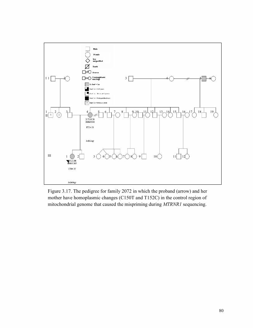
80
Figure 3.17. The pedigree for family 2072 in which the proband (arrow) and her mother have homoplasmic changes (C150T and T152C) in the control region of mitochondrial genome that caused the mispriming during MTRNR1 sequencing.
I
II
III 1 2 3 4 5 65
7 8 9 10 11 12
1
1
2
2
3
3
4
4
5
5
65
65
7 8 9 10 11 12 18 17 16 15 14 13 19

81
Daughter Mother Control Ladder
Figure 3.18. An agarose gel showing the apparent deletion in the proband and the proband’s mother from family 2072.

82
Figure 3.19. The wild type sequence of the human mitochondrial genome showing the sequence where the 3’ end of the forward primer was initially supposed to attach (pink) and the similar sequence 349 bps downstream from that point (light blue). Because it is important for primers to be specific, the SNPs in the proband’s mitochondrial genome at the binding site caused the primer to bind 349 bps away from the intentional site, therefore creating an amplicon that was approximately 349bp shorter.

83
Figure 3.20. A partial pedigree for family 2010 in which the proband (black arrow) and 4 of her family members have homoplasmic changes (C150T and T152C) in the control region of mitochondrial genome that caused the mispriming during MTRNR1 sequencing. The proband from also has the R141W mutation in TMC1.
I
II
III
IV
V
VI
VII
VIII
IX
X
XI
1
1
1
1
1
1
1
2
2
2
2
2
2
3
3
3
3
1 2
3
4
4
4
3
4
4
5
5
5
6
6
6
7
7
8
8 7
8
8
9
9
9
10
10
10
11
11
12 13
12 13
14
14
14
15
15
16
16
16
17
17
17
18 19 20
19 27 25 23
1 2 3 4 5 6 7 8 9 10 11 12 13 14 15 16
1 2 4 5 6 7 9 10 11 12 13 15 18 20 21 26 24 22
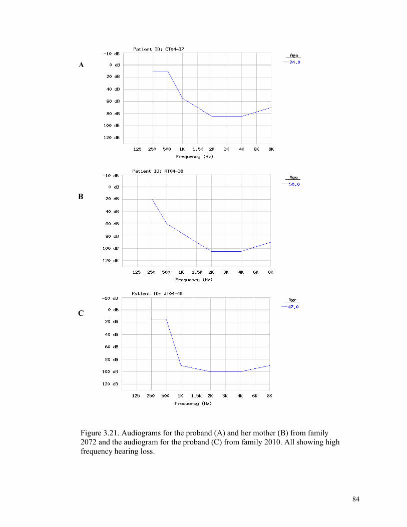
84
Figure 3.21. Audiograms for the proband (A) and her mother (B) from family 2072 and the audiogram for the proband (C) from family 2010. All showing high frequency hearing loss.
A
B
C

85
Figure 3.22. The pedigree for family 2078. The proband of this family was found to have a heterozygous c.94C>T:p.R32W mutation in connexin 31.
I
II
III 1 2
3 4 5 65
1
1
2
2
3 4
5
65

86
Figure 3.23. The pedigree for family 2075. The proband of this family was found to have a heterozygous c.94C>T:p.R32W mutation in connexin 31.
I
II
V
IV
III
1 2 3 4 5 65
7 8 9 10 11 12 13 14 15
1
1
1
1
2
2
2
2
3
3
3
4
4
4
5
5
65
65
7 8 9 10 11 12 13 14 15 16 17 18 19 20

87
Figure 3.24. A partial pedigree for family 2097. The proband of this family was found to have a heterozygous c.94C>T:p.R32W mutation in Cx31. For the full pedigree see Appendix 4, Figure 3.
I
II
III
IV
V 1 2 3 4 5 6
5 7 8 9 10
11
1
1
1
1
2
2
2
2
3
3
3
3
4
4
4
4
5
5
5
65
65
65
7
7
8
8
9
9
10
10
11

88
het c.94C>T
Figure 3.25. The pedigree for family 2156. The proband of this family was found to have a heterozygous c.94C>T:p.R32W mutation in connexin 31.

89
Figu
re 3
.26.
A p
edig
ree
for f
amily
208
3. T
he p
roba
nd o
f thi
s fam
ily w
as fo
und
to h
ave
a he
tero
zygo
us V
37M
mut
atio
n in
con
nexi
n 31
. I
II
III
IV
1 2
3 4
5 6 5
7 8
9
10
11
1 2
3 4
5 6 5
7
8
1
1
2
2
3
3
4
4
5
6 5
5
6 5
7
7
8
9 1
2
9 10
11
12
13
14
1
5 1
6
17
1
8
19
2
0
21
22
23
24
8

90
het c.94 C>T
het c.94 C>T
Figure 3.27 The pedigree for family 2078 showing the family members with the heterozygous c.94 C>T mutation.
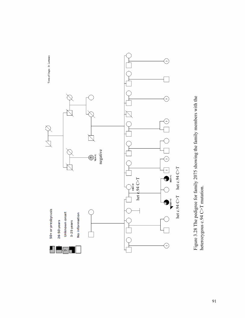
91
het c
.94
C>T
he
t c.9
4 C
>T
het c
.94
C>T
nega
tive
Figu
re 3
.28
The
pedi
gree
for f
amily
207
5 sh
owin
g th
e fa
mily
mem
bers
with
the
hete
rozy
gous
c.9
4 C
>T m
utat
ion.

92
hetc.94C
>T
het G
JB6 de
l hetc.94C
>T
het G
JB6 de
l
Figu
re 3
.29
The
pedi
gree
for f
amily
209
7 sh
owin
g th
e fa
mily
mem
bers
with
the
hete
rozy
gous
c.9
4 C
>T m
utat
ion.

93
het c.109
G>A
het c.109
G>A
het c.109
G>A
Figu
re 3
.30
The
pedi
gree
for f
amily
208
3 sh
owin
g th
e fa
mily
mem
bers
with
the
hete
rozy
gous
c.1
09 G
>A m
utat
ion.

94
Figure 3.31. The pedigree for family 2124. The proband of this family was found to have a heterozygous G182D mutation in TMC1.
I
II
III
IV
1 2 3
4
1 2 3
1 2
3 2 1

95
Figure 3.32. The pedigree for family 2065. The proband from this family has the c.1763+A>G mutation in TMC1.
I
II
III
IV
1 2 3 4 5 65
7 8 9 10 11 12 13 14
1 2 3 4 5 65
65
5 4 3 2 1
1 2 3 4 5 65
7
7
8
8
9
9
10
10
11
11
12
12
13
13
14
14
15
15
16
16
17 18

96
Figu
re 3
.33.
The
ped
igre
e fo
r fam
ily 2
146.
The
pro
band
in th
is fa
mily
has
the
R14
1W m
utat
ion
in
TMC
1
I
II
III
IV
V
1 2
3 4
5 6 5
7 8
9 10
1
1 12
13
14
15
16
17
18
19
20
21
22
1 2
1
1
1
2
2
2
3
3
3
4
4
4
5
5
6 5
6 5
7
7
8
8
9
9
10
10
11
12
13
14

97
Figure 3.34. The pedigree for family 2177. The proband from this family has the R141W mutation in TMC1.
II
I
III
IV
V 1 2 3
4 1 2 3
1 2 3
1 2 3 4
1 2 3 4

98
Figure 3.35. The pedigree for family 2092. The proband from this family has the R141W mutation in TMC1.
I
II
III
IV
1 2 3 4 5 6 5
7 8 9
1 2 3 4 5 65
7 8 9
10 11
1 2 3
1 2 3 4
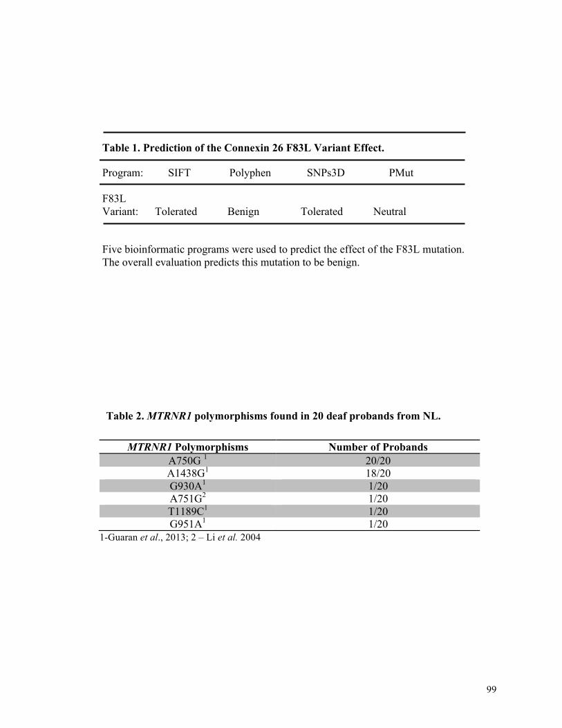
99
1-Guaran et al., 2013; 2 – Li et al. 2004
MTRNR1 Polymorphisms Number of Probands A750G 1 20/20 A1438G1 18/20 G930A1 1/20 A751G2 1/20 T1189C1 1/20 G951A1 1/20
Table 1. Prediction of the Connexin 26 F83L Variant Effect.
Program: SIFT Polyphen SNPs3D PMut
F83L Variant: Tolerated Benign Tolerated Neutral
Five bioinformatic programs were used to predict the effect of the F83L mutation. The overall evaluation predicts this mutation to be benign.
Table 2. MTRNR1 polymorphisms found in 20 deaf probands from NL.

100
Table 3*. Mitochondrial polymorphisms found in each of the deafness probands
*Tab
le 3 sh
ows the
mito
chon
drial hap
lotype
s of all prob
ands involved
. The
num
bers at the
top of th
e table show
the po
sition an
d po
ssible alleles fou
nd at tha
t position
with
in th
e mito
chon
drial gen
ome. Each nitrog
enou
s base is
colour cod
ed so
the ha
plotypes of e
ach prob
and can be
com
pared at a glance.

101
Table 3 continued. Mitochondrial polymorphisms found in each of the deafness probands continued.
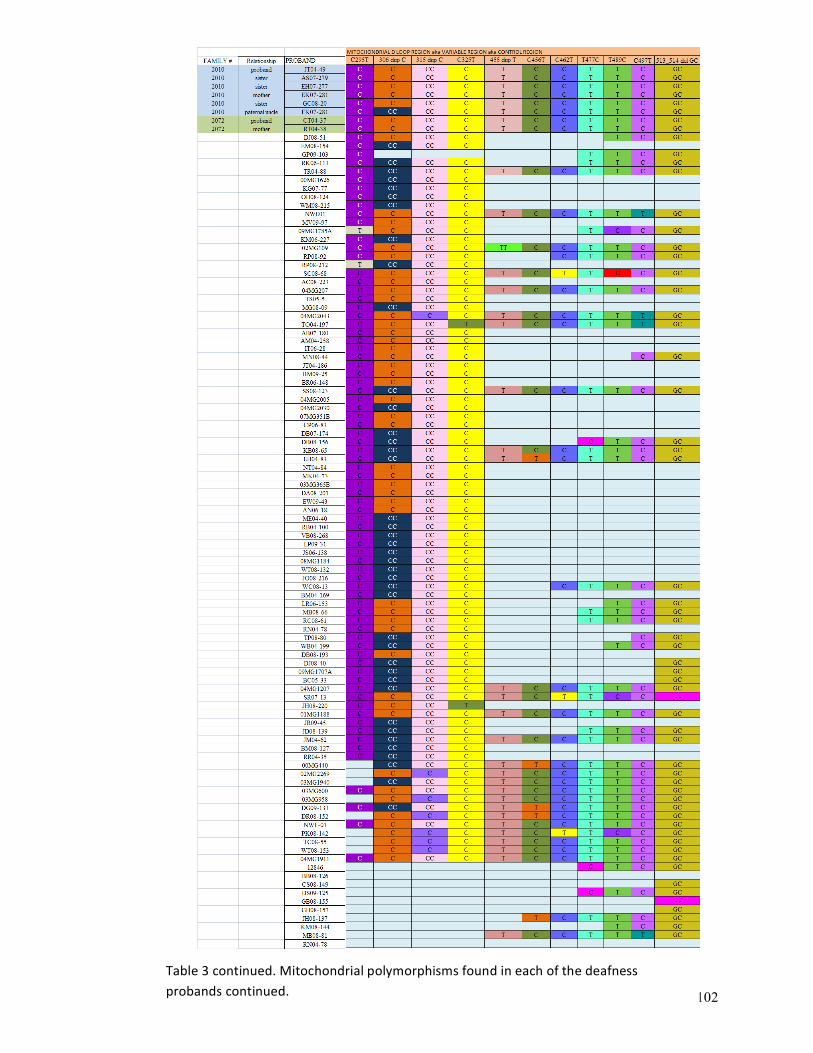
102
Table 3 continued. Mitochondrial polymorphisms found in each of the deafness probands continued.

103
Table 3 continued. Mitochondrial polymorphisms found in each of the deafness probands continued.

104 Table 3 continued. Mitochondrial polymorphisms found in each of the deafness probands continued.

105
Table 3. M
itochon
drial polym
orph
isms fou
nd in con
trols

106
Category of Hearing Loss Number of probands with the Specified Type of Hearing Loss
Low Frequency 5 Mid Frequency 2 High Frequency 29 All Frequency 19
Gene Accession number
Mutation Number of
Probands
SNP database
frequency
SIFT Polyphen
Connexin 31
NM_024009 c.109G>A, p. V37M c.94C>T, p. R32W
1 6
Novel 0.015
Deleterious Deleterious
Damaging Damaging
TMC1 NM_138691 c.421C>T, R141W c.545G>A, G182D
c.1763+3A>G
4 1 1
0.008 Novel Novel
Deleterious Tolerated
Probably Damaging Probably
Damaging
Table 4. The number of probands in each category of hearing loss frequency
Table 5. Mutations found in 2 of the candidate genes targeted by AudioGene.

107
Tabl
e 6.
A su
mm
ary
of th
e ge
ne v
aria
nts f
ound
with
the
prob
and
they
wer
e fo
und
in a
nd th
e he
arin
g lo
ss p
heno
type
of
the
prob
and.
A m
ore
deta
iled
tabl
e of
gen
es sc
reen
ed a
nd m
utat
ions
iden
tifie
d is
show
n in
App
endi
x 5.

108
4. Discussion
The purpose of this study was to determine the genetic etiology of probands
affected by nonsyndromic hereditary hearing loss. The probands in this study are all
originally from the island of Newfoundland, a founder population. An island with a
founder population is an ideal place for genetic studies. This study focused on families
with a history of hearing loss. Previous studies on hearing loss in the Newfoundland
population found causative mutations in WFS1¸ TMPRSS3, and PCDH15. These
mutations, however, were not found in the families involved in this project. Mutations
were identified in the mitochondrial gene, MTRNR1, as well as nuclear genes, Cx26,
Cx30, Cx31, and TMC1.
4.1 Screening Probands for Mutations previously found in the NL Population
The initial step of this study was to screen all probands for mutations in genes
previously found to cause hearing loss in the Newfoundland population. In 2001, A p.
2146 G>A mutation that was causing a dominant form of inherited progressive deafness
was identified in the WFS1 gene (Young et al.). Two recessive mutations in TMPRSS3
were found to underlie hearing loss in a six-generation family from the south coast of
Newfoundland (Ahmed et al., 2004), and Doucette et al. (2009) identified a novel
mutation in PCDH15 causing profound hearing loss in another south coast family. All
110 probands were screened for these mutations, however all individuals were negative.
This is interesting given that these mutations were deemed founder mutations. It would

109
be expected that the mutations would show up while screening a group of individuals
with hearing loss from the island. It is possible that the sample considered in this study
was not large enough. In future studies, more hearing loss patients should be recruited to
determine the accuracy of describing the NL mutations (in WFS1, TMRSS3, and
PCDH15) as founder mutations.
4.2 Proband from family 2155 with Hearing Loss Caused by Mutations in connexin 26 & Connexin 30
Mutations in genes that are located in the DFNB1 locus, connexin 26 (Cx26) and
connexin 30 (Cx30), are the cause of 30-50% of this type of hearing loss (Bhalla et al.,
2009). I therefore decided to screen all probands for mutations in these genes once the
NL mutations were ruled out.
The proband from family 2155 was found to carry a heterozygous c.35delG
mutation in exon 2 of Cx26 as well as a heterozygous 342 kb deletion in Cx30. These
genes encode connexin proteins. If only one of the genes has a mutation (i.e. a
heterozygous mutation), there is enough functional protein produced to allow the gap
junctions to function properly, therefore there must be a homozygous mutation in either
Cx26 or Cx30, or one mutation in each of the genes, to cause hearing loss (Smith et al.,
2014). In the case of the proband from family 2155 there is a heterozygous 35delG
mutation in connexin 26 and a heterozygous 342 kb deletion in connexin 30 together
causing hearing loss through digenic inheritance. The c.35delG mutation in connexin 26
is a known mutation first found in hearing impaired Mediterranean patients in 1997 by
Zelante et al. This particular mutation is common in Caucasian populations and is the

110
result of a worldwide founder effect (Laer et al., 2001). The deletion of a guanine at
position 35 in exon 2 of connexin 26 is a frameshift mutation that leads to premature
chain termination causing the production of a truncated, dysfunctional connexin 26
protein. The large deletion in connexin 30, given the name Δ(GJB6-D13S1830), truncates
the connexin 30 protein, in addition, the deleted portion contains a cis regulatory element,
that regulates the production of the connexin 26 protein (Rodriguez-Paris et al., 2011).
With the deletion of this regulatory element in the connexin 30 gene, the expression of
the connexin 26 protein is terminated, therefore no connexin 26 protein is produced (del
Castillo et al., 2002). In European populations this deletion is the second most frequent
mutation related to autosomal recessive hearing loss (Esteves et al., 2013). Thus, the
presence of both mutations causes the hearing loss that affects the proband from family
2155.
Digenic inheritance of the 35delG mutation in connexin 26 and Δ(GJB6-
D13S1830) in connexin 30 has been shown to cause variable hearing loss. In one study
published in 2004 (Bolz et al.) the severity of hearing loss due to this digenic inheritance
was significantly different between the two families included in the study, with one
family affected by profound hearing impairment and the other affected by
moderate/severe hearing loss. The reason for the difference in phenotype in this case is
that homozygous mutations in both Cx26 and Cx30 are found in affected members of the
families. Therefore the severity of the phenotype depends on which mutations the
individual carries. Within one family there can be different combinations of the
mutations. As mentioned in the report, one patient has a less severe hearing loss, which

111
could be caused by a modifier element that is upregulating connexin 26 or connexin 30.
The hearing loss is generally more severe if the patient has a homozygous mutation in
connexin 26 or connexin 30 (Bolz et al., 2004; Erbe et al., 2004; Marlin et al., 2005).
However, a different study found probands with del(GJB6-D13S1830)/c.35delG
presented with a more severe hearing impairment (Batissoco et al., 2009). Another study
published in 2013, by Esteves et al., states that patterns of hearing loss caused by
mutations in the connexin 26 gene have not been established, and there is no consistent
model of the phenotype caused by malfunctioning or nonfunctioning connexin 26
proteins. Therefore the phenotype caused by the digenic inheritance of the 35delG
mutation and Δ(GJB6-D13S1830) in connexin 30 is still uncertain. The severity of
hearing loss caused by mutations in Cx26 and Cx30 is highly variable, so it is difficult to
associate these mutations with a specific severity (Cama et al., 2009; Esteves et al.,
2013).
The proband from family 2155 is a 25-year-old woman who was diagnosed with
bilateral, profound, sensorineural hearing loss at the age of 18 months. It was recognized
at the age of 9 months that the proband had no speech development, and it is assumed
that she was hearing impaired since birth. As mentioned, the hearing loss phenotype
caused by Cx26 and Cx30 mutations is variable, and the hearing loss presented by this
proband is of similar clinical presentation found in previous studies (Cama et al., 2009;
Esteves et al., 2013). It is also a similar clinical presentation given for DFNB1 hearing
loss, which is congenital, non-progressive, mild-to-profound sensorineural hearing
impairment, with no other medical findings (Retrieved from www.ncbi.nlm.nih.gov

112
/sites/GeneTests/ review?db=GeneTests on July 15, 2011). As both genes are found
within the DFNB1 locus, the mutations found in this proband justify the hearing loss
profile.
4.3 Proband from family 2197 found to have a heterozygous 35delG mutation in Cx26
A heterozygous 35delG mutation was found in exon 2 of Cx26 in the proband
from family 2197 (Fig.3.6). This mutation alone is not the cause of hearing loss in this
individual, as the 35delG mutation in Cx26 has never been reported to cause hearing loss
in the heterozygous state (Kaskalan et al., 2014). I screened Cx30 in this individual,
however no mutations were detected. This proband was also included in all further gene
screening, which resulted in no additional mutation detection. This individual has a
bilateral asymmetric hearing loss that was diagnosed at age 29 and is more profound in
the right ear. There is no medical information available for this person’s family; therefore
it is difficult to determine if the single deafness allele was inherited or has arisen de novo.
4.4 A heterozygous F83L variant in Cx26 exon 2 in the proband from family 2091
The proband from family 2091 was found to have a heterozygous F83L variant in
Cx26, exon 2. Using four bioinformatics programs to predict the pathogenicity of this
variant, all predict this missense variant to be benign (Table 1). As well the Weblogo
(Fig.3.10) shows that the phenylalanine at position 83 is not well conserved. The variant
has also been reported in the literature to be a polymorphism (Ross et al., 2007; Minárik

113
et al., 2012). I concluded that this variant is probably not responsible for causing hearing
loss in the proband. There was no audiogram available for this patient so it was difficult
to choose candidate genes based on audioprofiling. Obtaining a more extensive medical
background for the individual and the family would help for future studies.
4.5 Variants in the Mitochondrial Genome
Mutations in two mitochondrial genes (MTRNR1 and MTTS1) have been
identified to cause hearing loss. The mitochondrial genome is exceptionally polymorphic
and varies greatly among individuals. Sometimes these variants are disease causing, such
as the case with A1555G in MTRNR1, and at other times there are variants commonly
found in the general population (Kazuno et al., 2006). I chose to screen for mutations in
the mitochondrial genes following the screening for Cx26 and Cx30 mutations as
mutations in the mitochondrial genome are one of the most common cause of
nonsyndromic hearing loss (Guo, 2008). I screened MTRNR1 and MTTS1 in all unsolved
probands. Seven polymorphisms were found and two probands were found to have the
A1555G mutation in MTRNR1.
4.6 Probands from families 2112 and 2144 Hearing Loss caused by a mutation in MTRNR1
The probands from families 2112 and 2144 were found to have a homoplasmic
A1555G mutation in the MTRNR1 gene. MTRNR1 is a mitochondrial gene that codes for
the 952 nucleotide long 12S ribosomal RNA (rRNA). The substitution of a guanine for an
adenosine at position 1555 in the mitochondrial genome within the MTRNR1 gene is

114
known to cause aminoglycoside induced hearing loss. Aminoglycosides are a group of
antibiotics that rid the body of harmful bacteria by binding to their 16S rRNA in the 30S
ribosomal subunit and inhibiting protein synthesis. The A1555G mutation causes the 12S
rRNA to resemble the bacterial 30S rRNA, thus when an individual with the A1555G
mutation is given aminoglycoside treatment, the aminoglycosides will inhibit protein
synthesis within their mitochondria (Guan, 2011).
The proband from family 2112 has been diagnosed with severe to profound
bilateral hearing loss. She self-described being late deafened, having lost her hearing after
an intravenous antibiotic treatment for pneumonia (which the individual reported as
penicillin, not an aminoglycoside). The proband reported that her mother was affected
with hearing loss after she was treated for tuberculosis in the 1950s. This medical
information possibly links the hearing loss to the mitochondrial A1555G mutation in the
proband, as well as the proband’s mother, obtaining DNA from the proband’s mother
would help confirm this. In the 1950s, tuberculosis was treated using streptomycin, an
aminoglycoside (Mitchison, 2005). Individuals who have the A1555G mutation often
develop hearing loss following the administration of aminoglycosides (Guan, 2011). The
proband of family 2112 has three siblings, all male; one affected by progressive bilateral
hearing loss. However, DNA was not available for these individuals.
The hearing loss in family 2112 is likely a case where both genetics and
environment come into play. The A1555G mutation is not known to cause hearing loss
on its own, but can predispose an individual to lose hearing when exposed to
aminoglycosides. Cases such as this one show that a combination of genetic and

115
environmental factors can be the cause of a condition. It is common to attribute hearing
loss to an environmental cause, such as a head injury, medication, infection, or noise
pollution. However, the appearance of hearing loss after such occurrences could be
purely coincidental, or predisposed by a genetic cause. Accepting that an environmental
occurrence is the only reason for an individual’s hearing loss and failing to further
investigate the reason a person develops the disorder can result in the omission of an
underlying genetic cause.
The proband from family 2144, with MTRNR1 A1555G, reports that her hearing
loss began during childhood after an airplane flight and an ear infection and it has been
getting progressively worse since then. She has had repeated ear infections as well as
premature graying, a minor diagnostic criterion for syndromic deafness known as
Waardenburg Syndrome (Nayak and Isaacson, 2003). As there is little information
pertaining to this probands medical history and family history it is difficult to make
connections between her hearing loss and genotype. Again, obtaining more information
from the families should be looked into as further studies would help shed more light on
this mutation and the environmental triggers of genetic hearing loss.
4.7 A G951A variant in MTRNR1 found in two probands from families 2167 and 2197
A G951A variant in MTRNR1 was found in the proband from family 2167
(Fig.3.11) and in the proband from family 2197, who also has a heterozygous 35delG
mutation in connexin 26 as previously discussed (Fig.3.6). It cannot be concluded that
this mitochondrial variant is the cause of the hearing loss expressed by these individuals.
Elstner et al. (2008) identified this variant in the 12S rRNA of 66 patients with bilateral

116
vestibulopathy (damage to the inner ear(s) causing dizziness, imbalance, and vision
disturbances) and also in 155 healthy controls. None of the individuals sequenced had
mutations in the mitochondrial genes that are known to cause hearing loss (A1555G or
C1494T). They did find four mutations with putative pathogenic effects, T669C,
C960del, C960ins, and T961G. They also found five frequent polymorphisms: G709A,
G930A, T1189C, T1243C, G1438A and seven of what they called “rare homoplasmic
changes”: T669C, T721C, G951A, C960del, C960ins, T961G, and G1007A, when they
compared the 12S rRNA sequence to the Cambridge reference sequence (Elstner et al.,
2008). G951A was not found in 155 healthy patients; however they reported that no
assumptions could be made about this variant and concluded that none of the variants
they found had proven pathogenicity (Elstner et al., 2008). The report by Elstner et al.
(2008) was the only evidence of G951A in the literature and it provided no information to
the pathogenicity of G951A. Given the limited evidence, it can only be concluded that the
G951A variant is of unknown pathogenicity. Further investigation of the genotype is
required to determine the effect of the G951A variant.
The proband from family 2167 (Fig.3.11) was diagnosed with unilateral mild to
moderately severe sensorineural hearing loss in his left ear that presents in a “cookie bite”
audiometric configuration (severe hearing loss in the mid frequencies with mild to no
hearing loss in the higher and lower frequencies). He self reports that the hearing loss is
progressing and that he is also affected by premature graying (before the age of 30) as
well as kidney problems. He also reports that he had multiple left sided ear infections
throughout his life and his hearing loss was diagnosed when he had a preschool

117
examination at age four. There are three distant relatives that have been diagnosed as
deaf. Acquiring their DNA may not help to determine the pathogenicity of the G951A
mutation because all relatives diagnosed as deaf are on the paternal side of the family,
however, if the proband’s mother and father are descendants of the same maternal
ancestor it is possible that both the mother and father have this mutation. It would be
beneficial to obtain more information on the maternal side of the family to help solve the
questions of pathogenicity that surround the G951A variant.
The proband from family 2197 has a bilateral asymmetric hearing loss that was
diagnosed at age 29 and is more profound in the right ear. There was no available
audiogram; therefore it was difficult to choose candidate genes based on audioprofiling.
The proband had an affected father as well as two affected uncles. The paternal
grandmother was also affected, suggesting that hearing loss was segregating as a
dominant trait on the paternal side This proband also has a heterozygous 35delG
mutation in connexin 26. It is possible that an interaction between connexin 26 and
mitochondrial mutations may be having an effect in this individual. However, because no
conclusion can be drawn as to whether or not the G951A mutation is pathogenic, then it
is difficult to say it is affecting the wild type connexin 26 gene. A literature search
revealed no previous studies that discussed the G951A mutation in connection with a
connexin 26 mutation. Future studies should look closely at the interaction between
MTRNR1 and Cx26, it is interesting as mutations in both cause hearing loss.

118
4.8 MTRNR1 common variants
There are many common variants in mitochondrial DNA that are not associated
with disease, including six such variants in MTRNR1 (Retrieved from http://www.mito
map.org/MITOMAP on May 31, 2011): G709A, A750G, A751G, G930A, T1189C, and
G1438A. Five of these variants are previously reported as found at significant
frequencies across several ethnic groups. Because they are at a frequency greater than 1%
in the general population, these mitochondrial variants are considered common variants
(Lu et al., 2010; Rydzanicz et al., 2010; Elstner et al., 2008; Li et al., 2004). In 2004 Li et
al., in their study of 164 Caucasian subjects under 19 years of age who were diagnosed
with nonsyndromic sensorineural deafness, reported that A751G was a novel
polymorphism in the Caucasian population. In 2009, a Polish group, Rydzanicz et al.,
found this sequence variant and submitted it to MITOMAP (reference #20071219002) as
it had not been reported on MITOMAP at that time. They called A751G a nucleotide
change, but made no other comment about its pathogenicity. No variants, pathogenic or
non-pathogenic, were found in MTTS1.
The associations made between mitochondrial variants and disease are dependent
on population genetics and because of the variability of the mitochondrial genome it is
often difficult to make conclusions about the pathogenicity of a polymorphism (Kazuno
et al., 2006). The variants found in this study have been published multiple times;
therefore it is likely that the pathogenicity assigned to each variant is correct.

119
4.9 Specificity of Mitochondrial Primer Binding Sites
The proband from family 2072 appeared to have a mitochondrial deletion,
however, following several attempts using different primer sequences a high quality
sequence was finally obtained and sequence analysis showed that two SNPs (C150T, and
T152C) were present in the mitochondrial DNA of the proband, therefore the hypothesis
that the primer is hybridizing at a point 349 bp away from the intended primer
hybridization site is most likely correct. To further support this hypothesis, specificity at
the 3’ end of a primer to the DNA sequence is extremely important as primers that have
poor specificity in this region usually produce undesirable amplicons (Dieffenbach, T, &
G, 1993). In order to have a successful PCR it is important to have the correct placement
of the 3’ end of a primer so that perfect base-pairing between the 3’ end of the primer and
the template DNA is necessary and minimal mismatch should be present (Dieffenbach et
al., 1993). Therefore, the presence of these SNPs where the 3’ end of the primer should
hybridize decreases the specificity of the primer and causes the primer to hybridize at a
position 349 base pairs away that matches the last six base pairs of the 3’ end of the
primer.
4.10 Matching Mitochondrial Haplotypes
Following the discovery that the probands of family 2010 and 2072 has the same
SNPs in their mitochondrial DNA, I thought it would be interesting to see if their
mitochondrial haplotype was identical. No other families had the same combination of
SNPs. This suggests that these families may be related through the maternal line, which is

120
likely, as they appear to have the same haplotype and they come from the same area of
the island of Newfoundland. Upon the discovery of the similarity of the SNPs found in
these two families, the audiograms were analyzed and it was noticed that the audiograms
that were available for the family members of 2010 and 2072 showed very similar
hearing loss patterns (Fig.3.21). The original SNPs found in these families (C150T and
T152C) could play a role in the hearing loss that the affected members have, or they
might just indicate that these families are related through the maternal line, and have the
same type of hearing loss caused by possible shared mutations. The audiograms and
family history of all other probands showing similar mitochondrial haplotypes should be
analyzed to see if they have similar hearing loss patterns.
4.11 Connexin 31 Variants
Mutations in connexin 31 have been shown to cause both dominant and recessive
hearing loss (Liu et al., 2000). Two mutations have been reported to cause dominant
hearing loss, R180X and E183K, and two mutations have been found to cause recessive
hearing loss, 141del_Ile, and I141V (Retrieved from http://hereditaryhearingloss.org on
May 12, 2014). Few publications regarding this connection exist in the literature
(Retrieved from davinci.crg.es/deafness/ on May 12, 2014). The recessive mutations
were found in a study of 25 Chinese families with apparent recessive hearing loss. The
study, conducted by Liu et al. (2000), found a sibling pair, with severe hearing loss at all
frequencies, and a single proband with moderate hearing loss at all frequencies, to be
compound heterozygotes for the 141delIle and I141V mutations, neither of the parents
were affected. In the case of this study, the probands screened for connexin 31 mutations

121
had high frequency hearing loss, which does not fit the phenotype for recessive connexin
31 hearing loss found in the study by Liu et al. (2000). However, the gene was suggested
as a candidate following the audioprofile investigation, and so screening went ahead.
Connexin 31 was screened in 29 probands with high frequency hearing loss and
two variants were discovered in 7 probands. The variant c.109G>A, p.V37M was found
in 1 proband; and c.94C>T, p.R32W was found in 6 probands.
The variant c.109G>A, p.V37M was found in family 2083 (Fig.3.26). The
proband from this family has postlingual hearing loss, which was reported to have
occurred between the ages of 3 and 25. Xia et al. (1998) report that autosomal dominant
deafness caused by a c.538C>T in patients who lost their hearing in late adulthood; this
onset is dissimilar to the onset in this proband. In order to reach a conclusion about the
pathogenicity of this variant, a more detailed medical history of the proband is required
as well as DNA from the probands family members.
The mutation c.94C>T, p.R32W was found in six families. The families with this
Cx31 mutation are: family 2155(Fig.3.5), family 2167 (Fig. 3.11), family 2078 (Fig.
3.22), family 2075 (Fig. 3.23), family 2097 (Fig. 3.24), and family 2156 (Fig 3.25). The
proband from family 2097 has postlingual hearing loss diagnosed between the ages of 3
and 25; this proband also has a heterozygous connexin 30 deletion Δ (GJB6-D13S1830).
While it has not yet been determined that these two mutations cause hearing loss when
inherited together, it is worth looking into for future studies. The proband from family
2155 has been previously described as this proband also has been found to have digenic

122
inheritance of the 35delG mutation in connexin 26 and Δ(GJB6-D13S1830) in connexin
30. This proband is also homozygous for R32W in connexin 31. These mutations have
not been found inherited together, and there is no published evidence of these two genes
interacting, but it is possible that having mutation in three of these connexin genes could
increase the severity of the hearing loss and is worth looking into in future studies. The
proband from family 2075 has a history of postlingual hearing loss diagnosed between
the ages of 3 and 35. The proband from family 2156 also has adult onset hearing loss,
diagnosed between 26-50 years old. The proband from family 2078 (Fig.3.22) has
postlingual hearing loss. The proband from family 2167 has been previously mentioned
as he has a variant in the mitochondrial DNA, he was diagnosed between the ages of 3
and 25, he hears well at low and high frequencies but not at middle frequencies. This is
called a “cookie bite” curve on an audiogram because the curve is high on both ends but
low in the middle and appears as if there is a bite taken out of it. All of these probands
present with different types of hearing loss, and given the variability of hearing loss
caused by mutations in connexin 31 (OMIM) you cannot rule out either of them based on
phenotype. However, the pathogenicity of R32W has been questioned in the literature. In
one study, R32W was found in hearing loss patients, but was also found in 18 percent of
the control population (Lopez-Bigas et al., 2000). Rouan et al. (2003) used HeLa cells to
test Cx31 function when the R32W variant was present, and found no deviations in
expression level, concluding that it is an inconsequential polymorphism.
It is interesting that these variants appear to segregate with hearing loss in these
families however a more detailed segregation analysis is required to confirm

123
pathogenicity (Fig.3.27, 3.28, 3.29, 3.30). Therefore in future studies segregation analysis
should be performed. In addition to segregation analysis it would be extremely beneficial
to obtain a more detailed family and medical history for each of these probands, given
that the family would be willing to divulge that information.
4.12 TMC1 Variants
Mutations in the TMC1 gene are known to cause nonsyndromic autosomal
dominant and autosomal recessive hearing loss (Hereditary Hearing Loss Homepage,
OMIM, www.omim.org). Almost all reported cases of TMC1 hearing loss report pre-
lingual severe to profound hearing loss (OMIM; Gao et al., 2013). TMC1 is a second
gene that was suggested as a candidate gene for hearing loss probands following the
audioprofiling investigation. Three mutations were found in TMC1: c.545G>A:p.G182D
in the proband from family 2124, c.1763+3A>G in the proband from family 2065 (Fig.
3.32), and R141W in the proband from families 2010 (Fig. 3.20), 2146 (Fig. 3.33), 2177
(Fig.3.34), and 2092 (Fig.3.35). All of these probands are affected by post lingual hearing
loss. There is no additional information on the proband from family 2124 (Fig. 3.31).
The proband from family 2065, with the intronic mutation, c.1763+3A>G., was
born in 1996 and has bilateral symmetrical sensorineural mild to moderate hearing loss in
the low frequencies (250-750 Hz), and severe hearing loss in high frequencies (1000-
8000Hz) that progressed rapidly in the first decade of his life. Mutations in TMC1
causing nonsyndromic hearing loss have been found more commonly in Pakistani
families (OMIM), however one study has found TMC1 hearing loss in a North American

124
Caucasian family (Kitajiri et al., 2007). The affected members from this family had
postlingual, progressive sensorineural hearing loss, which presented in the second decade
of life, starting in the high frequencies. The mutation found in this family was a missense
substitution c.G1714C, p.D572H, and falls within a large cytoplasmic loop region of the
TMC1 protein (Kitajiri et al., 2007; Labay et al., 2010 ). While this mutation was not
found in the proband from family 2065, the region of TMC1 that is mutated in the
proband lies within an intron of close proximity. Analysis of this mutation and of the
proband’s family is required to determine the pathogenicity of the c.1763+3A>G variant.
The proband from family 2010, with the R141W mutation, was born in 1965 and
has normal bilateral hearing at 250-500 Hz (low frequency), with a precipitous slope over
500 Hz where the hearing loss (bilateral and sensorineural) becomes moderately severe to
profound in high frequencies. The proband has two male relatives, who report having
hearing loss by age five, one of which had normal hearing at birth and in the other, the
age of onset is unknown. This proband also has more severe hearing loss in the higher
frequencies which is also the case with the TMC1 hearing loss in the North American
family reported by Kitajari et al. (2007). Acquiring DNA from the proband’s family
members is essential in determining the pathogenicity of this variant.
The proband from family 2092 was born in 1935. She has flat line hearing loss
(i.e. absolutely no sense of hearing) in her right ear. In her left ear she has mild sloping to
moderately severe sensorineural hearing loss in the mid to high frequencies. Audiograms
of other relatives show symmetrical bilateral loss, the same as this proband is
experiencing in her left ear. Once again, we see hearing loss affecting the high

125
frequencies, which appears to be the case in the only Caucasian family identified to have
hearing loss caused by a mutated TMC1 gene (Kitajiri et al., 2007). There is no
information as to the rate of progression of hearing loss for this individual. To determine
the effect of the mutation, DNA should be acquired from the available family members.
The proband from family 2146, born in 1958, is affected by right severe to
profound reverse slope sensorineural hearing loss. In his left ear he experiences steep
high frequency sensorineural hearing loss above 2000 Hz. He reports normal hearing
bilaterally at age 8-10 years and an onset of Meniere’s disease (a disease causing sporadic
episodes of vertigo, sensorineural hearing loss, tinnitus and ear pressure ;OMIM,
www.omim.org) symptoms in his right ear at age 37. He displays hearing loss, vertigo
attacks, tinnitus and ear fullness, all symptoms of classic Meniere’s disease with classic
progression in the right ear over 15 years with increasing hearing loss with every attack,
and partial recovery of hearing to the previous level between attacks. The high frequency
hearing loss appears to be common among all individuals found to have mutations in
TMC1. Because this type of hearing loss was displayed in the Caucausian family with
TMC1 hearing loss (Kitajiri et al., 2007) this could be an indication that it may be causing
hearing loss in these individuals. There has been no evidence of TMC1 mutations
associated with Meniere’s disease.
Lastly, the proband from family 2177 was born in 1961 and has right borderline
mild low frequency sloping to moderately severe high frequency sensorineural hearing
loss, which is reported to have started during childhood. The left ear is affected by left
mild high frequency sensorineural hearing loss at 8000 Hz. Episodes of vertigo prompted

126
a tentative diagnosis of Meniere’s disease, but it is not a classic presentation. The
proband’s aunt and brother reported having a diagnosis of Meniere’s disease with
asymmetric hearing loss. This individual also displays more severe hearing loss in the
high frequencies, which was seen in all other probands with TMC1 mutations. Both
bilateral hearing loss and asymmetrical hearing loss has been displayed in the probands,
and the TMC1 mutations causing hearing loss do not seem to specifically cause one or the
other (Kitajiri et al., 2007). This proband has also been diagnosed with Meniere’s
disease, which is interesting and worth looking into, if it were found to be the result of
the TMC1 mutation, it would be the first known case.
Like the probands with connexin 31 mutations, it will be necessary to perform
segregation analysis to verify the pathogenicity of the variants found. Also, retrieving a
more detailed family history may also present clues as to what type of hearing loss affects
these probands. It would be valuable to have an extensive family history and medical
history on each of these individuals.
4.13 Conclusion
Overall the study resolved the etiology for four hearing loss probands out of 110
that were screened. One proband has hearing loss caused by a homozygous 35delG
mutation in connexin 26, another proband has hearing loss caused by the digenic
inheritance of the 35delG mutation in connexin 26 and del13S1830 in connexin 30. Two
probands have the A1555G mutation in the MTRNR1 gene of the mitochondrial genome.

127
Other probands (n=7) have variations in connexin 31 and 6 others have variations in
TMC1, yet these cannot be considered resolved because segregation analysis has not yet
been performed to confirm the pathogenicity of the variants.
In addition, this study found that none of the probands recruited for this project
had mutations previously found to cause hearing loss in the Newfoundland population
(TMPRSS3, PCDH15, and WFS1). Inviting questions as to whether or not these are
actually founder mutations. Also of interest are the similarities between mitochondrial
haplotypes of several of the probands, especially those from family 2072 and 2010. The
affected members of this family appear to have the same type of hearing loss, however an
underlying genetic cause could not be identified. This study has brought forward
mutations in several genes (Cx31, TMC1, MTRNR1) that were not previously found in the
Newfoundland population. These findings create many opportunities to further explore
the genetic etiology of hearing loss in this province.
Determining the underlying cause of hearing loss in Newfoundland and Labrador
will improve the province’s screening strategies so that this disorder can be identified as
early as possible. Recognizing hearing loss early in life can positively impact a child and
the ability to learn communication skills and participate in a school environment.
Children who are identified as hearing impaired can be taught to communicate in a
specialized manner or can also benefit from certain types of hearing devices, such as
hearing aids or cochlear implants. In addition to this, knowing the cause of deafness can
offer relief to families who can then receive support from genetic counselors and
understand the risk of passing the disorder on to their children.

128
5. Future Directions
This study opens up many possibilities for future projects. First of all it is
necessary to perform segregation analysis on the variants of connexin 31 found in 7 of
the probands and the variants of TMC1 found in 6 of the probands, and to perform exome
sequencing (sequencing only the coding regions) on the unsolved probands with the
intent to find known and novel mutations. Secondly, it would be beneficially to recruit
more family members and collect familial DNA for those probands that were lacking
family medical history or for which there was no family DNA available (family 2197).
Having the information and resources to further studies into the cause of their hearing
loss may aid in finding a cause. A study could also be built around the mitochondrial
haplotypes that were presented in this project. These haplotypes can be used to identify
the relationships and origins of families. Linking haplotypes to phenotypes could
potentially solve several families. More specifically it would be interesting to map the
lineage of families 2072 and 2010 since this study discovered several similarities among
the members of these families affected by hearing loss. Lastly because this study dealt
with a small population of deaf probands a recruitment project could bring in more
families affected by this disorder. Screening the families would give a more in depth look
at mutations previously identified as founder mutations within the Newfoundland
population.
While this study has significantly added to the research on hearing loss genetics it
has also opened the door for many more projects to be explored. With the quick
advancement of DNA sequencing technology, as well improvements for screening

129
deafness, hopefully the studies can be carried out and conclusions can be drawn from the
information uncovered in this research project.

130
6. Bibliography Abrue-Silva, R.S., Lezirovitz, K., Braga, M.C.C., Spinelli, M., Pirana, S., Della-Rosa, V.A., Otto, P.A., et al. (2006) Prevalence of the A1555G (12S rRNA) and tRNASer(UCN) mitochondrial mutations in hearing-impaired Brazilian patients. Brazilian Journal of Medical and Biological Research 39(2) 219-226. Ahmed Z.M., Riazuddin S. Bernstein S.L., Ahmed Z., Khan S., Griffith A.J., Morell R.J., et al. (2001). Mutations of the protocadherin gene PCDH15 cause Usher syndrome type 1F. American Journal of Human Genetics 69 (1): 25–34. Ahmed, Z. M., Riazuddin, S., Ahmad, J., Bernstein, S. L., Guo, Y., Sabar, M. F., Sieving, P., Riazuddin et al. (2003) PCDH15 is expressed in the neurosensory epithelium of the eye and ear and mutant alleles are responsible for both USH1F and DFNB23. Human Molecular Genetics 12: 3215-3223 Ahmed, Z.M., Li, X.C., Powell, S.D., Riazuddin, S., Young, T.L., Ramzan, K., Ahmad, A., et al..(2004) Characterization of a new full length TMPRSS3 isoform and identification of mutant allele responsible for nonsyndromic recessive deafness in Newfoundland and Pakistan. BMC Medical Genetics 5(24) 1-8. Anderson, S., Bankier, A.T., Barrell, B.G.,de Bruijn, M.H.L., Coulson, A.R., Drouin, J., Eperon, I.C. et al. (1981) Sequence and organization of the human mitochondrial genome. Nature 290 457-465. Argus Biosciences taken from www.argusbio.com on July 9, 2010. Avraham, K.B., Hasson, T., Steel, K.P., Kingsley, D.M., Russel, L.B., Mooseker, M.S., Copeland, N.G., et al. (1995) The mouse Snell’s waltzer deafness gene encodes an unconventional myosin required for structural integrity of inner ear hair cells. Nature Genetics 11 (4), 369-375. Avraham, K.B. (2003) Mouse models for deafness: lessons for the human inner ear and hearing loss. Ear & Hearing 24, 332-341. Barrell, B.G., Bankier, A.T., and Drouin, J. (1979) A different genetic code in human mitochondria. Nature 282, 189-194. Batissoco, A. C., Abreu-Silva, R. S., Braga, M. C. C., Lezirovitz, K., Della-Rosa, V., Alfredo Jr, T., ... & Mingroni-Netto, R. C. (2009). Prevalence of GJB2 (connexin-26) and GJB6 (connexin-30) mutations in a cohort of 300 Brazilian hearing-impaired individuals: implications for diagnosis and genetic counseling. Ear and hearing, 30(1), 1-7.

131
Beyer, E.C., Paul, D.L, Goodenough, D.A. (1987) Connexin 43: a protein from rat heart homologous to a gap junction protein from the liver. The Journal of Cell Biology, 105 (6). 2621 – 2629. Bergoffen J., Scherer S.S., Wang S., Scott M.O., Bone L.J., Paul D.L., Chen K. et al. (1993). Connexin mutations in X-lined Charcot-Marie-Tooth disease. Science, 262 (5142). 2039 – 2042. Bhalla, S., Sharma, R., Khandelwal, G., Panda, N.K., & Khullar, M. (2009) Low incidence of GJB2, GJB6 and mitochondrial DNA mutations in North Indian patients with non-syndromic hearing impairment. Biochemical and Biophysical Research Communications, 385, 445-448. Bolz, H., Schade, G., Ehmer, S., Kothe, C., Hess, M., & Gal, A. (2004). Phenotypic variability of non-syndromic hearing loss in patients heterozygous for both c. 35delG of GJB2 and the 342-kb deletion involving GJB6. Hearing research, 188(1), 42-46. Bonne-Tamir B., DeStefano A.L., Briggs C.E., Adair R., Franklyn B., Weiss S., Korostishevsky M., et al. (1996) Linkage of congenital recessive deafness (gene DFNB10) to chromosome 21q22.3. American Journal of Human Genetics 58:1254-1259. Carlsson, P.I., Karltorp, E., Carlsson-Hansén, E., Åhlman, H., Mӧller, C., Vondӧbeln, U. (2012) GJB2 (connexin 26) gene mutations among hearing=impaired persons in a Swedish cohort. Acta Oto-Laryngologica 132:1301-1305 Center for Hearing Loss Help (2006). Kinds of Hearing Losses. Retrieved from http://www.hearinglosshelp.com/articles/kindsofhearinglosses.htm Centers for Disease Control and Prevention. Summary of 2009 National CDC EHDI Data: EHDI hearing screening & follow-up survey (HSFS) [CDC Web site]. 2011. Available from:www.cdc.gov/ncbddd/hearingloss/ehdi-data2009.html. Accessed May 8, 2013. Chatterjee, S. and Lufkin, T. (2011) The Sound of Silence: Mouse Models for Hearing Loss. Genetics Research International 2011 (2011): 1-9.
Copeland, W.C. (2002) Mitochondrial DNA : methods and protocols. Totowa, N.J. : Humana Press del Castillo, I., Villamar, M., Moreno-Pelayo, M.A., del Castillo, F.J., Alvarez, A., Telleria, D. et al. (2002) A deletion involving the connexin 30 gene in nonsyndromic hearing impairment. The New England Journal of Medicine. 346(5), 243-249.

132
del Castillo, I., Moreno-Pelayo, M. A., del Castillo, F. J., Brownstein, Z., Marlin, S., Adina, Q. et al. (2003). Prevalence and evolutionary origins of the del(GJB6-D13S1830) mutation in the DFNB1 locus in hearing impaired subjects: a multicenter study. American Journal of Human Genetics. 73, 1452–1458 Cama, E., Melchionda, S., Palladino, T., Carella, M., Santarelli, R., Genovese, E., ... & Arslan, E. (2009). Hearing loss features in GJB2 biallelic mutations and GJB2/GJB6 digenic inheritance in a large Italian cohort. International journal of audiology, 48(1), 12-17. Denoyelle, F., Weil D., Maw M.A., Wilcox S.A., Lench N.J., Allen-Powell D.R., Osborn A.H., et al. (1997) Prelingual deafness: high prevalence of a 30delG mutation in the connexin 26 gene. Human Molecular Genetics, 6 (12). 2173-2177.
Dieffenbach, C. W., T, M. L., & G, S. D. (1993). General concepts for PCR primer design. PCR Methods and Applications, 3(3), S30-7.
Doucette, L., Merner, N.D., Cooke, S., Ives, E., Galutira, D., Walsh, V., Walsh, T., et al. (2009) Profound, prelingual nonsyndromic deafness maps to chromosome 10q21 and is caused by a novel missense mutation in the Usher syndrome type IF gene PCDH15. European Journal of Human Genetics, 17, 554-564. Elstner, M., Schmidt, C., Zingler, V.C., Prokisch, H., Bettecken, T., Elson, J.L., Rudolph, G., et al. (2008) Mitochondrial 12S rRNA susceptibility mutations in aminoglycoside –associated and idiopathic bilateral vestibulopathy. Biochemical and Biophysical Research Communications 377: 379-383. Erbe, C. B., Harris, K. C., Runge‐Samuelson, C. L., Flanary, V. A., & Wackym, P. A. (2004). Connexin 26 and connexin 30 mutations in children with nonsyndromic hearing loss. The Laryngoscope, 114(4), 607-611. Esteves, M. C. B. N., de Lima Isaac, M., Francisco, A. M., da Silva Junior, W. A., Ferreira, C. A., & Dell’Aringa, A. H. B. (2013). Analysis of the presence of the GJB6 mutations in patients heterozygous for GJB2 mutation in Brazil.European Archives of Oto-Rhino-Laryngology, 1-5. Estivill, X., Govea, N., Barceló, E., Badenas, C., Romero, E., Moral, L., Scozzri, R., et al. (1998) Familial progressive sensorineural deafness is mainly due to the mtDNA A1555G mutation and is enhanced by treatment with aminoglycosides. American Journal of Human Genetics 62, 27-35.

133
Farid, N. R., Buehler, S., Russell, N. A., Maroun, F. B., Allerdice, P., & Smyth, H. S. (1980). Prolactinomas in familial multiple endocrine neoplasia syndrome type I: relationship to HLA and carcinoid tumors. The American Journal of Medicine, 69(6), 874-880. Fischel-Ghodsian, N. (2002) Hearing Loss and Mitochondria DNA Mutations: Clinical Implications and Biological Lessons. In Bronya J.B. Keats’, Arthur N. Popper’s, and Richard R. Fay’s (Ed.) Genetics and Auditory Disorders. pp. 228-246. New York City, NY: Springer-Verlag. Friedman, L.M., Dror, A.A., and Avraham, K.B., (2007) Mouse models to study inner ear development and hereditary hearing loss. International Journal of Developmental Biology, 51: 609-631. Froggatt, N. J., Green, J., Brassett, C., Evans, D. G. R., Bishop, D. T., Kolodner, R., & Maher, E. R. (1999). A common MSH2 mutation in English and North American HNPCC families: origin, phenotypic expression, and sex specific differences in colorectal cancer. Journal of medical genetics, 36(2), 97-102. Gao, X., Su, Y., Guan, L. P., Yuan, Y. Y., Huang, S. S., Lu, Y., ... & Dai, P. (2013). Novel Compound Heterozygous TMC1 Mutations Associated with Autosomal Recessive Hearing Loss in a Chinese Family. PloS one, 8(5), e63026. Genbank, taken from http://www.ncbi.nlm.nih. gov.qe2a-proxy.mun.ca/nuccore/NC_012 920.1 on July 2, 2010. Genetics Home Reference taken from http://ghr.nlm.nih.gov/glossary on August 30, 2010. Gimlich, R.L., Kumar, N.M., and Gilula, N.B. (1990) Differential regulation of the levels of three gap junction mRNAs in Xenopus Embryos. The Journal of Cell Biology, 110. 597-605. Goodenough, D.A. (1974). Bulk isolation of mouse hepatocyte gap junctions. Characterization of the principle protein, connexin. The Journal of Cell Biology, 61 (2), 557-563. Grifa, A., Wagner, C.A., D’Ambrosio, L., Melchionda, S., Bernardi, F., Lopez-Bigas, N., Rabionet, R., et al. (1999) Mutations in GJB6 cause nonsyndromic autosomal dominant deafness at DFNA3 locus. Nature Genetics, 23 (1), 16-17. Griffiths AJF, Gelbart WM, Miller JH, et al. Modern Genetic Analysis. New York: W. H. Freeman; 1999. Human Pedigree Analysis.Available from: http://www.ncbi.nlm.nih.gov/books/NBK21257/

134
Griffiths, A.J.F., Gelbart, W.M., Lewontin, R.C., Miller, J.H. (2002) Modern Genetic Analysis Integrating Genes and Genomes (2nd Edition) New York, NY: W.H. Freeman and Company. Guaran, V., Astolfi, L., Castiglione, A., Simoni, E., Olivetto, E., Galasso, M., ... & Martini, A. (2013). Association between idiopathic hearing loss and mitochondrial DNA mutations: A study on 169 hearing-impaired subjects. International journal of molecular medicine, 32(4), 785-794.
Guo, Y.F., Liu, X.W., Guan, J., Han, M.K., Wang, D.Y., Zhao, Y.L., Rao, S.Q., et al. (2008) GJB2, SLC26A4 and mitochondrial DNA A1555G mutations in prelingual deafness in Northern Chinese subjects. Acta Oto-Laryngologica 128, 297-303. Hereditary Hearing Loss Homepage taken from http://hereditaryhearingloss.org on July 5, 2010. Heritability Analytics Infrastructure (HAI) taken from http://www.med.mun.ca/PTRG/ What-We-Do/Infrastructure.aspx on July 16, 2013.
Hernandez V.H., Bortolozzi M., Pertegato V., Beltramello M., Giarin M., Zaccolo M., Pantano S., et al. (2007) Nature Methods 4(4). 353-358. Higashi, K. (1989) Unique inheritance of streptomycin induced deafness. Clinical Genetics 35, 433-436. Hildebrand MS, Tack D, McMordie SJ, DeLuca A, Hur IA, et al. (2008) Audioprofile-directed screening identifies novel mutations in KCNQ4 causing hearing loss at the DFNA2 locus. Genetics in Medicine. 10, 797-804. Hildebrand, M. S. et al. A contemporary review of AudioGene audioprofiling: a machine-based candidate gene prediction tool for autosomal dominant nonsyndromic hearing loss. Laryngoscope 119, 2211–2215 (2009). Hilgert, N., Smith, R.J.H, Van Camp, G. (2009). Forty-six genes causing nonsyndromic hearing impairment: Which ones should be analyzed in DNA diagnostics? Mutation Research. 681, 189-196. Hilgert, N., Smith, R.J.H., Van Camp, G. (2009) Function and expression pattern of nonsyndromic deafness genes. Current Molecular Medicine 9 (5), 546-564. Hu, D.N., Qiu, W.Q., Wu, B.T., Fang, L.Z., Zhou, F., Gu, Y.P., Zhang, Q.H., et al. (1991) Genetic aspects of antibiotic induced deafness: mitochondrial inheritance. Journal of Medical Genetics, 28, 79-83.

135
Hutchin, T.P., Parker, M.J., Young, I.D., et al. (2000) A novel mutation in the mitochondrial tRNAser(UCN) gene in a family with nonsyndromic sensorineural hearing impairment. Journal of Medical Genetics 37. 692-694. Irshad, S., Shahzad, B., Yaqoob, S. (2012) A review on autosomal recessive non-syndromic hearing impairment. Basic Sciences of Medicine, 1(3), 12-18. Ito, T., Noguchi, Y., Yashima, T., Ohno, K., Kitamura,K. (2010) Hereditary hear loss and deafness in Japan. Journal of Medical and Dental Sciences, 57, 1-10. Jaber, L., Shohat, M., Bu, X., et al. (1992) Sensorineural deafness inherited as a tissue specific mitochondrial disorder. Journal of Medical Genetics 29. 86-90. Kaskalan, E., Önalan, E. E., Kaygusuz, İ., Karlıdağ, T., Keleş, E., Akyiğit, A., & Yalçın, Ş. (2014). Analysis of GJB2 (Connexin 26) Mutation in Patients with Congenital Non-Syndromic Sensorineural Hearing Loss. Keats, B.J., & Berlin, C., (2002) Introduction and Overview: Genetics in Auditory Science and Clinical Audiology. In Keats, B.J., Popper, A.N., and Fay, R.R. (Eds), Genetics and Auditory Disorders (pp. 1-22). New York, Inc: Springer-Verlag. Kelsell, D.P., Dunlop, J., Stevens, H.P., Lench, N.J., Liang, J.N., Parry, G. et al.(1997) Connexin 26 mutations in hereditary non-syndromic sensorineural deafness. Nature 387, 80-83. Kenneson,A., Van Naarden Braun, K., Boyle, C. (2002) GJB2 (connexin 26) variants and nonsyndromic sensorineural hearing loss: a HuGE review. Genetics in Medicine, 4: 258–274. Kikuchi, T., Kimura, R.S., Paul, D.L, and Adams, J.C. (1995) Gap junctions in the rat cochlea: immunohistochemical and ultrastructural analysis. Anatomy and Embryology 191: 101-118 Kikuchi, T., Kimura, R.S., Paul, D.L., Takasaka, T., and Adams, J.C. (2000) Gap junction systems in mammalian cochlea. Brain Research Reviews, 32. 163-166 Kitajiri, S., Makishima, T., Friedman, T. B., & Griffith, A. J. (2007). A novel mutation at the DFNA36 hearing loss locus reveals a critical function and potential genotype–phenotype correlation for amino acid‐572 of TMC1. Clinical genetics, 71(2), 148-152. Kokotas, H., Grigoriadou, M., Korres, G.S., Ferekidou, E., Papadopoulou, E., Neou, P., Giannoulia-Karantana, A., et al. (2009) The A1555G mitochondrial DNA mutation in Greek patients with non-syndromic, sensorineural hearing loss. Biochemical and Biophysical Research Communications 390. 755-757

136
Kumar, N.M., and Gilula, N.B. (1992) Molecular biology and genetics of gap junction channels. Seminars in Cell Biology 3 (1). 3-16. Kupka, S., Tóth, T., Wróbel, M., Zeissler, U., Szyfter, W., Szyfter, K., Niedzielsk,a G., et al. (2002). Mutation A1555G in the 12S rRNA Gene and Its Epidemiological Importance in German, Hungarian, and Polish Patients. Human Mutation 488, 1-6. Labay, V., Weichert, R. M., Makishima, T., & Griffith, A. J. (2010). TOPOLOGY OF TRANSMEMBRANE CHANNEL-LIKE GENE 1 PROTEIN (TMC1). Biochemistry, 49(39), 8592–8598. doi:10.1021/bi1004377 Li, R., Greinwald, J.H. Jr., Yang, L., Choo, D.I., Wenstrup, R.J., Guan, M.X. (2004) Molecular analysis of the mitochondrial 12S rRNA and tRNASer(UCN) genes in paediatric subjects with nonsyndromic hearing loss. Journal of Medical Genetics, 41: 610-620 Liu, X.Z., Xia, X.J., Xu, L.R., Pandya, R., Liang, C.Y., Blanton, S.H., Brown, S.D.M., et al. (2000). Mutations in connexin31 underlie recessive as well as dominant non-syndromic hearing loss. Oxford University Press, 9 (1). 63-67 Liu, X.Z., & Yan, D. (2007) Ageing and hearing loss. Journal of Pathology 211, 188-197. Liu, W., Bostrom, M., Kinnefors, A., Rask-Andersen, H. (2009) Unique expression of connexins in the human cochlea. Hearing Research, 250 (1-2): 55-62
Mahdieh, N., Rabbani, B., Wiley, S., Akbari, M. T., & Zeinali, S. (2010). Genetic causes of nonsyndromic hearing loss in Iran in comparison with other populations. Journal of human genetics, 55(10), 639-648. Makowski, L., Caspar, D.L.D., Phillips, W.C., and Goodenough, D.A.. (1977) Gap junction structures. Analysis of x-ray diffraction data. The Journal of Cell Biology, 74. 629-645. Manfredi, G., Thyagarajan, D., Papadopoulou, L.C., Pallottim F., Schonm E.A.. (1997) The Fate of Human Sperm-Derived mtDNA in Somatic Cells. American Journal of Human Genetics 61. 953-960. Mannion, J.J. (1977) The Peopling of Newfoundland: Essays in Historic Geography. St John’s, Memorial University of Newfoundland. Marlin, S., Feldmann, D., Blons, H., Loundon, N., Rouillon, I., Albert, S., ... & Denoyelle, F. (2005). GJB2 and GJB6 mutations: genotypic and phenotypic correlations in a large cohort of hearing-impaired patients. Archives of Otolaryngology–Head & Neck Surgery, 131(6), 481-487.

137
Martin, L.J., Crawford, M.H., Koertvelyessy, T., Keeping, D., Collins, M., Huntsman, R. (2000) The population structure of ten Newfoundland outports. Human Biology, 72 (6), 997-1016. Melchionda, S., Ahituv, N., Luigi, B., Sobe, T., Glaser, F., Rabionet, R., Arbones, M.L., et al. (2001) MYO6, the human homologue of the gene responsible for deafness in Snell’s Waltzer mice, is mutated in autosomal dominant nonsyndromic hearing loss. American Journal of Human Genetics 69: 635-640.
Mitchell, A. A., Chakravarti, A., & Cutler, D. J. (2005). On the probability that a novel variant is a disease-causing mutation. Genome research, 15(7), 960-966.
Mitchison, D. A. (2005). The diagnosis and therapy of tuberculosis during the past 100 years. American journal of respiratory and critical care medicine,171(7), 699-706.
Minárik, G., Tretinárová, D., Szemes, T., & Kádasi, Ľ. (2012). Prevalence of DFNB1 mutations in Slovak patients with non-syndromic hearing loss. International Journal of Pediatric Otorhinolaryngology, 76(3), 400-403.
Mistrik, P. and Ashmore, J. (2009) The role of potassium recirculation in cochlear amplification. Current Opinion in Otolaryngology & Head and Neck surgery. 17. 394-399.
Morton, N.E. (1991) Genetic epidemiology of hearing impairment. Annals of the New York Academy of Sciences. 630: 16-31. Nance, W.E. (2003) The genetics of deafness. Mental Retardation and Developmental Disabilities Research Review. 9, 109-119 National Institute on Deafness and other Communication Disorders (NIDCD) taken from http://www.nidcd.nih.gov/ on July 15, 2010. Nickel, R., Becker, D., and Forge, A. (2006) Molecular and functional characterization of gap junctions in the avian inner ear. The Journal of Neuroscience 26 (23). 6190-6199. Nayak, C. S., & Isaacson, G. (2003). Worldwide distribution of Waardenburg syndrome. Annals of Otology Rhinology and Laryngology, 112(9), 817-820. Oh, S. K., Choi, S. Y., Yu, S. H., Lee, K. Y., Hong, J. H., Hur, S. W., ... & Kim, U. K. (2013). Evaluation of the pathogenicity of< i> GJB3</i> and< i> GJB6</i> variants associated with nonsyndromic hearing loss. Biochimica et Biophysica Acta (BBA)-Molecular Basis of Disease, 1832(1), 285-291.

138
OMIM, taken from http://omim.org/ on July 18, 2013. Pagon RA, Bird TD, Dolan CR, et al., editors. GeneReviews™ [Internet]. Seattle (WA): University of Washington, Seattle; 1993-. Illustrated Glossary. Available from: http://www.ncbi.nlm.nih.gov/books/NBK5191/ Pallares-Ruiz, N., Blanchet, P., Mondain, M., Claustres, M., and Roux, A.F. (2002). A large deletion including most of GJB6 in recessive nonsyndromic deafness: a digenic effect? European Journal of Human Genetics, 10. 72-76. Petersen, M.B., & Willems, P.J. (2006) Non-syndromic, autosomal recessive deafness. Clincal Genetics, 69, 371-392. Petersen, M.B., Grigoriadou, M., Koutroumpe, M., and Kokotas, H. (2012) The novel c.247_249delTTC (p.F83del) GJB2 mutation in a family with prelingual sensorineural deafness. International Journal of Pediatric Otorhinolaryngology, 76 (4), 549-551. Pfenniger, a. , Wohlwend, A, Kwak, B.R. (2010) Mutations in connexin genes and disease. European Journal of Clinical Investigation. 41 (1), 103-116 Phillips, L.L, Bitner-Glindzicz, M., Lench, N., Steel, K.P., Langford, C., Dawson, S.J., Davis, A., et al. (2013) The future role of genetic screening to detect newborns at risk of childhood-onset hearing loss. International Journal of Audiology. 52, 124-133. Piatto V.B., Secches L.V., Arroyo M.A.S., Lopes A.C.P., and Maniglia J.V. (2009) Nonsyndromic Deafness - Molecular Update. The Open Biology Journal. 2, 80-90 Powar, C. B. (2007). Genetics . (Vol. 1). Mumbai, IND : Global Media Prezant, T.R., Agapian, J.V., Bohlman, M.C., Bu, X., Oztas, S., Qiu, W.Q., Arnos, K.S., et al. (1993) Mitochondrial ribosomal RNA mutations associated with both antibiotic-induced and non-syndromic deafness. Nature 4. 289-294. Rahman, P., Jones, A., Curtis, J., Bartlett, S., Peddle, L., Fernandez, B.A., & Friemer, N.B. (2003) The Newfoundland population: a unique resource for genetic investigation of complex disease. Human Molecular Genetics, 12 (2), 167-172. Raising Deaf Kids (2003). Audiograms. Retrieved from http://www.raisingdeafkids.org/hearingloss/testing/audiogram/ Reid, F.M., Vernham, G.A., Jacobs, H.T.. (1994) A novel mitochondrial point mutation in a maternal pedigree with sensorineural deafness. Human Mutation 3 (3) 243-247.

139
Relethford, J.H. (2012) Foundation of Human Biology: Human Population Genetics (pp.77) Retrieved from http://site.ebrary.com.qe2a-proxy.mun.ca/lib/memorial/doc Detail.action?docID=10630499
Rodriguez-Paris, J., Schrijver, I. (2009) The digenic hypothesis unraveled: the GJB6 del(GJB6-D13S1830) mutation causes allele-specific loss of GJB2 expression in cis. Biochemical and Biophysical Research Communications, 389 (2). 354-359. Rodriguez-Paris, J., Tamayo, M. L., Gelvez, N., & Schrijver, I. (2011). Allele-specific impairment of GJB2 expression by GJB6 deletion del (GJB6-D13S1854). PloS one, 6(6), e21665. Romdhane, L., & Abdelhak, S. (2012). Genetic disorders in North African populations. In D. Kumar (Ed.),Genomics and health in the developing world (1 ed., Vol. 1, pp. 382-399). New York, NY: Oxford University Press. Ross, S. A., Novak, Z., Kumbla, R. A., Zhang, K., Fowler, K. B., & Boppana, S. (2007). GJB2 and GJB6 mutations in children with congenital cytomegalovirus infection. Pediatric research, 61(6), 687-691. Ru, B., Han, N., He, G., Brayer, K., Zhang, S., Wang, Z. (2012) Molecular cloning and evolutionary analysis of GJB6 in mammals. Biochemical Genetics 50: 213-226 Scheffler, I.E. (2008) Mitochondria. Hoboken NJ, USA: John Wiley and Sons Inc. Sirmaci A, Edwards YJK, Akay H, Tekin M (2012) Challenges in Whole Exome Sequencing: An Example from Hereditary Deafness. PLoS ONE 7(2). 1-7 Selimoglu E. (2007) Aminoglycoside-induced ototoxicity. Current Pharmaceutical Design 13 (1) 119-126.
Smith RJH, Shearer AE, Hildebrand MS, et al. Deafness and Hereditary Hearing Loss Overview. 1999 Feb 14 [Updated 2014 Jan 9]. In: Pagon RA, Adam MP, Ardinger HH, et al., editors. GeneReviews® [Internet]. Seattle (WA): University of Washington, Seattle; 1993-2014. Available from: http://www.ncbi.nlm.nih.gov/books/NBK1434/
Smith RJH, Van Camp G. Nonsyndromic Hearing Loss and Deafness, DFNB1. 1998 Sep 28 [Updated 2014 Jan 2]. In: Pagon RA, Adam MP, Ardinger HH, et al., editors. GeneReviews® [Internet]. Seattle (WA): University of Washington, Seattle; 1993-2015. Available from: http://www.ncbi.nlm.nih.gov/books/NBK1272/
Smith RJH, Van Camp G. Nonsyndromic Hearing Loss and Deafness, DFNB1. 1998 Sep 28 [Updated 2011 Jul 14]. In: Pagon RA, Adam MP, Bird TD, et al., editors.

140
GeneReviews™ [Internet]. Seattle (WA): University of Washington, Seattle; 1993-2013. Available from: http://www.ncbi.nlm.nih.gov/books/NBK1272/ Spirio, L., Green, J., Robertson, J., Robertson, M., Otterud, B., Sheldon, J., Howse, E., et al. (1999) The identical 50 splice-site acceptor mutation in five attenuatedAPC families from Newfoundland demonstrates a founder effect. Human Genetics, 105: 388–398. Stats Canada taken from http://www.statcan. gc.ca/ on January 8, 2010. Strachan T, Read AP. Human Molecular Genetics. 2nd edition. New York: Wiley-Liss; 1999. Chapter 3, Genes in pedigrees. Available from: http://www.ncbi.nlm.nih.gov/books/NBK7573/ Sue, C. M., Tanji, K., Hadjigeorgiou, G, Andreu, A. L., Nishino, I., Krishna, S., Bruno, C, et al. (1999) Maternally inherited hearing loss in a large kindred with a novel T7511C mutation in the mitochondrial DNA tRNASer(UCN) gene. Neurology 52 (9). 1905-1908. Thönnissen, E., Rabionet, R., Arbonès, M.L., Estivill, X., Willecke, K., and Ott, T. (2002). Human Genetics, 111. 190-197. Tiranti, V., Chariot, P., Carella, F., Toscano, A., Soliveri, P., Girlanda, P., Carrara, F., et al. (1995) Maternally inherited hearing loss, ataxia and myoclonus associated with a novel point mutation in mitochondrial tRNASer(UCN) gene. Human Molecular Genetics 4 (8). 1421-1427. Usami, S., Abe, S., Kasai, M., Shinkawa, H., Moeller, B., Kenyon, J.B., Kimberling, W.J.. (1997) Genetic and cinical features of sensorineural hearing loss associated with the 1555 mitochondrial mutation. Laryngoscope 107 (4) 483-490. Van Camp, V., Willems, P.J., Smith, R.J.H. (1997) Nonsyndromic hearing impairment: unparalleled heterogeneity. American Journal of Human Genetics. 60, 758-764. Van Laer, L., Coucke, P., Mueller, R. F., Caethoven, G., Flothmann, K., Prasad, S. D., ... & Van Camp, G. (2001). A common founder for the 35delG GJB2gene mutation in connexin 26 hearing impairment. Journal of medical genetics, 38(8), 515-518. Veske A., Oehlmann R., Younus F., Mohyuddin A., Muller-Myhsok B., Mehdi S.Q., Gal A. (1996) Autosomal recessive non-syndromic deafness locus (DFNB8) maps on chromosome 21q22 in a large consanguineous kindred from Pakistan. Human Molecular Genetics 5:165-168. Vrijens, K., Van Laer, L., and Van Camp, G. (2008) Human hereditary hearing impairment: mouse models can help to solve the puzzle. Human Genetics, 124: 325-348.
141
Wallace, D.C., Singh, G., Lott, M.T, Hodge, J.A., Schurr T.C, Lezza, A.M.S., Elsas II, L.J., et al. (1988) Mitochondrial DNA mutation associated with Leber's hereditary optic neuropathy. Science 242. 1427-1430. Wallace, D.C. (1992) Diseases of the mitochondrial DNA. Annual Review Biochemistry 61. 1175-1212. Willems, P.J.(Ed) (2004) Genetic Hearing Loss. New York, NY: Marcel Dekker, Inc. Wu, B.L., Kenna, M., Lip, V., Irons, M., and Platt, O. (2003) Use of a multiplex PCR/sequencing strategy to detect both connexin 30 (GJB6) 342 kb deletion and connexin 26 (GJB2) mutations in cases of childhood deafness. American Journal of Medical Genetics 121A, 102-108. Xie, Y.G., Zheng, H., Leggo, J., Scullym, M.F. and Lillicrap, D. (2002) A founder factor VIII mutation, valine 2016 to alanine, in a population with an extraordinarily high prevalence of mild hemophilia A. Journal of Thrombpsis and Haemostatsis, 87, 178–179. Young, T.L., Penney, L., Woods, M.O., Parfrey, P.S., Green, J.S., Hefferton, D. and Davidson, W.S. (1999) A fifth locus for Bardet–Biedl syndrome maps to chromosome 2q31. American Journal of Human Genetics, 64: 900–904. Young, T.L., Ives, E., Lynch, E., Person, R., Snook, S., Maclaren, L., Cator, T., et al. (2001) Non-syndromic progressive hearing loss DFNA38 is caused by heterozygous missense mutation in the Wolfram syndrome gene WFS1. Human Molecular Genetics, 10 (22), 2509-2514. Zelante L., Gasparini P., Estivill X., Melchionda S., D'Agruma L., Govea N., Milá M., et al. (1997) Connexin 26 mutations associated with the most common form of non-syndromic neurosensory autosomal recessive deafness (DFNB1) in Mediterraneans. Human Molecular Genetics, 6 (9). 1695-1609. Zhou, H., Pfeiffer, R., & Gail, M. H. (2003). Haplotype analysis in population genetics and association studies. Pharmacogenomics, 4(2), 171-178.

142
Appendix 1: The Forward and Reverse primers and PCR conditions for each gene. Ta
ble
1. T
he F
orw
ard
and
Rev
erse
prim
ers a
nd P
CR
con
ditio
ns fo
r eac
h ge
ne.
(ret
rieve
d fr
om h
ttp://
ww
w.n
cbi.n
lm.n
ih.g
ov/g
enba
nk/ o
n Fe
brua
ry 1
6, 2
011)

143
!Gen
e Fo
rwar
d Pr
imer
R
ever
se P
rimer
PC
R
Con
ditio
ns
GJB3
1.1
AG
TCTG
CC
CC
TCTG
AG
TCA
C
AA
GG
CTC
CA
ATG
AG
ATC
AG
G
TD54
, with
B
etai
ne,
5%D
MSO
GJB3
1.2
GC
TGTG
GG
TAC
AA
GG
TCTG
C
GA
GC
TGG
GC
TCA
GC
AG
TG
TD54
, with
B
etai
ne,
5%D
MSO
GJB3
1.3
CG
GC
AC
TTA
AC
AG
TTTG
CA
G
GG
GA
GG
ATT
GA
GA
GG
TAC
GG
TD
54, w
ith
Bet
aine
, 5%
DM
SO
GJB3
AltU
TR
GA
CA
GC
AC
CC
AA
GG
GTA
GA
A
TTA
GTA
GC
TGG
GC
GA
CTT
CC
TD
54, w
ith
Bet
aine
, 5%
DM
SO
GJB3
2.1
GA
AC
TCA
GA
AC
AC
TGC
CTG
GT
AG
GG
GC
ATG
TGA
CG
AA
GA
T TD
54, w
ith
Bet
aine
, 5%
DM
SO
GJB3
2.2
GG
AC
TTTG
AC
TGC
AA
CA
CC
A
GG
CA
GA
TGA
GG
TAG
CA
GA
GC
TD
54, w
ith
Bet
aine
, 5%
DM
SO
GJB3
2.3
TCTG
GC
ATG
GC
TTC
AA
TATG
TC
AC
TCA
GC
CC
CTG
TAG
GA
C
TD54
, with
B
etai
ne,
5%D
MSO
GJB3
2.4
AC
CC
AG
GC
AA
TAA
CA
AG
CTG
A
TTC
TCA
GC
CC
TCC
AG
AC
CT
TD54
, with
B
etai
ne,
5%D
MSO
GJB3
2.5
TCA
AC
CA
GG
AA
GG
GA
TCA
AC
C
CC
TTTC
TGA
GG
GC
TTA
CC
T TD
54, w
ith
Bet
aine
, 5%
DM
SO
TMC1
Exon
1
TGTA
AA
AC
GA
CG
GC
CA
GTG
GG
TGA
CA
GA
GC
AA
GA
CTC
C
CA
GG
AA
AC
AG
CTA
TGA
CC
TCTC
ATG
GA
GA
AA
GTC
TTC
GA
TT
TD54
, with
B
etai
ne,
5%D
MSO
TMC1
Exon
2
TGTA
AA
AC
GA
CG
GC
CA
GTG
TCTT
CA
CG
CA
GC
CA
GC
C
AG
GA
AA
CA
GC
TATG
AC
CG
TAC
CC
TTA
GG
TGG
ATC
AC
TTG
TD
54, w
ith
Bet
aine
, 5%
DM
SO
TMC1
Exon
3
TGTA
AA
AC
GA
CG
GC
CA
GTA
AA
CTG
TGA
CA
GG
GTC
AG
GG
C
AG
GA
AA
CA
GC
TATG
AC
CG
AA
AA
TGC
ATC
AC
TTA
CA
AA
TTA
GG
TD
54, w
ith
Bet
aine
, 5%
DM
SO

144
!
Gen
e Fo
rwar
d Pr
imer
R
ever
se P
rimer
PC
R
Con
ditio
ns
TMC
1 Ex
on 4
TG
TAA
AA
CG
AC
GG
CC
AG
TAC
TCC
TGTT
GC
ATT
CC
TTG
G
CA
GG
AA
AC
AG
CTA
TGA
CC
CC
CA
TTC
CA
TAC
AA
TCC
TGC
TD
54, w
ith
Bet
aine
, 5%
DM
SO
TMC
1 Ex
on 5
TG
TAA
AA
CG
AC
GG
CC
AG
TTG
GTG
AA
ATG
GC
AC
CA
GG
C
AG
GA
AA
CA
GC
TATG
AC
CA
AA
GG
GA
AA
GC
CC
CA
AA
AG
TD
54, w
ith
Bet
aine
, 5%
DM
SO
TMC
1 Ex
on 6
TG
TAA
AA
CG
AC
GG
CC
AG
TAA
ATC
GTG
CTC
ATT
TTG
GC
C
AG
GA
AA
CA
GC
TATG
AC
CA
AA
GTT
ATC
CA
CA
CA
CA
CTT
CTG
C
TD54
, with
B
etai
ne,
5%D
MSO
TM
C1
Exon
7
TGTA
AA
AC
GA
CG
GC
CA
GTA
TCA
CG
ATG
TGG
AG
AA
TTG
C
CA
GG
AA
AC
AG
CTA
TGA
CC
GC
ATC
ATC
AG
ATT
AA
GG
CTC
TC
TD54
, with
B
etai
ne,
5%D
MSO
TM
C1
Exon
8
TGTA
AA
AC
GA
CG
GC
CA
GTC
AG
GA
AG
GC
AA
AC
AC
TAC
TGC
C
AG
GA
AA
CA
GC
TATG
AC
CTG
CTT
CC
AA
TTTA
TAA
TCA
TTA
GC
C
TD54
, with
B
etai
ne,
5%D
MSO
TM
C1
Exon
9
TGTA
AA
AC
GA
CG
GC
CA
GTA
AG
AA
ATA
AG
AC
TGC
AG
AC
CTG
G
CA
GG
AA
AC
AG
CTA
TGA
CC
TCC
CC
TGTA
AA
TTTC
AA
TAC
CA
C
TD54
, with
B
etai
ne,
5%D
MSO
TM
C1
Exon
10
TGTA
AA
AC
GA
CG
GC
CA
GTG
CTG
CC
AG
AG
AG
AC
ATT
TCC
C
AG
GA
AA
CA
GC
TATG
AC
CTT
CC
CA
AA
AG
TCC
AG
AA
AC
TG
TD54
, with
B
etai
ne,
5%D
MSO
TM
C1
Exon
11
TGTA
AA
AC
GA
CG
GC
CA
GTA
AA
AG
GA
CC
AA
TGC
CTC
AC
C
AG
GA
AA
CA
GC
TATG
AC
CTC
CTA
TGA
CTC
TAA
GA
CG
TGA
AA
ATA
G
TD54
, with
B
etai
ne,
5%D
MSO
TM
C1
Exon
12
TGTA
AA
AC
GA
CG
GC
CA
GTG
AC
CC
CA
CA
TGG
AG
AC
CC
C
AG
GA
AA
CA
GC
TATG
AC
CA
GA
CA
TTC
AG
CC
TGA
CC
CA
G
TD54
, with
B
etai
ne,
5%D
MSO
TM
C1
Exon
13
TGTA
AA
AC
GA
CG
GC
CA
GTA
TCA
AC
ATG
GC
AG
CTG
AA
AC
C
AG
GA
AA
CA
GC
TATG
AC
CG
GC
CTG
TGTT
TATG
TTC
AG
TTC
TD
54, w
ith
Bet
aine
, 5%
DM
SO
TMC
1 Ex
on 1
4 TG
TAA
AA
CG
AC
GG
CC
AG
TTA
TTG
CTT
CTC
CA
CTT
CA
AC
AC
C
AG
GA
AA
CA
GC
TATG
AC
CTT
GG
TAG
GC
AG
AA
AC
CA
TGA
G
TD54
, with
B
etai
ne,
5%D
MSO
TM
C1
Exon
15
TGTA
AA
AC
GA
CG
GC
CA
GTC
ATT
GTC
ATT
CC
TCA
CA
TTC
TG
CA
GG
AA
AC
AG
CTA
TGA
CC
CTG
TAA
GG
GC
AG
GA
TAG
GG
G
TD54
, with
B
etai
ne,
5%D
MSO

145
!
Gen
e Fo
rwar
d Pr
imer
R
ever
se P
rimer
PC
R
Con
ditio
ns
TMC
1 Ex
on 1
6 TG
TAA
AA
CG
AC
GG
CC
AG
TGC
CTA
GC
TCA
GA
ATC
TTC
CA
AA
C
AG
GA
AA
CA
GC
TATG
AC
CC
AA
AG
GC
ATT
TCTG
GC
AA
AC
TD54
, with
B
etai
ne,
5%D
MSO
TMC
1 Ex
on 1
7 TG
TAA
AA
CG
AC
GG
CC
AG
TCC
AG
TCTC
AA
GTT
TGC
CA
GA
C
AG
GA
AA
CA
GC
TATG
AC
CTT
CA
GA
GC
CA
GC
AC
AC
AG
TC
TD54
, with
B
etai
ne,
5%D
MSO
TM
C1
Exon
18
TGTA
AA
AC
GA
CG
GC
CA
GTA
ATT
GC
AG
TCTT
CA
AG
CC
AA
T C
AG
GA
AA
CA
GC
TATG
AC
CC
AA
ATC
CC
CC
TCTG
TGA
GA
A
TD54
, with
B
etai
ne,
5%D
MSO
TM
C1
Exon
19
TGTA
AA
AC
GA
CG
GC
CA
GTC
TGC
TATT
GTT
GC
TGA
AG
GG
C
AG
GA
AA
CA
GC
TATG
AC
CG
AC
AC
CG
ATT
GTA
TTC
TCC
TCTA
AC
TD
54, w
ith
Bet
aine
, 5%
DM
SO
TMC
1 Ex
on 2
0 TG
TAA
AA
CG
AC
GG
CC
AG
TTC
TGG
TTG
AA
AG
TGG
CA
GTG
C
AG
GA
AA
CA
GC
TATG
AC
CG
GA
TCTC
ATT
TCC
AC
CA
AC
C
TD54
, with
B
etai
ne,
5%D
MSO
TM
C1
Exon
21
TGTA
AA
AC
GA
CG
GC
CA
GTT
TCC
TAC
CC
TGA
AA
GA
GG
AC
C
CA
GG
AA
AC
AG
CTA
TGA
CC
GA
AC
CC
TTA
GG
GA
GA
GTG
AG
AG
TD
54, w
ith
Bet
aine
, 5%
DM
SO
TMC
1 Ex
on 2
2-23
TG
TAA
AA
CG
AC
GG
CC
AG
TTC
ATC
CC
TATG
TCA
TCC
TGA
A
CA
GG
AA
AC
AG
CTA
TGA
CC
GTG
ATG
CC
TGA
GA
AG
GG
CTA
TD
54, w
ith
Bet
aine
, 5%
DM
SO

146
Appendix 2: NL Hearing Loss Study Medical Questionnaire

147
Appendix 2: NL Hearing Loss Study Medical Questionnaire (cont’d)

148
Appendix 2: NL Hearing Loss Study Medical Questionnaire (cont’d)

149
Appendix 2: NL Hearing Loss Study Medical Questionnaire (cont’d)

150
Appendix 2: NL Hearing Loss Study Medical Questionnaire (cont’d)
151
Appendix 2: NL Hearing Loss Study Medical Questionnaire (cont’d)

152
Appendix 2: NL Hearing Loss Study Medical Questionnaire (cont’d)

153
Appendix 2: NL Hearing Loss Study Medical Questionnaire (cont’d)

154
Appendix 3: Standard DNA Extraction Protocol Purpose: To provide a method for the extraction, precipitation, and re-suspension of DNA from (EDTA) whole blood.
Responsibilities: Lab personnel who have been trained in this procedure.
Definitions:
− hr. = hour − ppt = precipitate − WBC = white blood cell
Equipment and Supplies:
Equipment:
− centrifuge − Biological safety cabinet − 37º or 55 ºC water bath or heat block − Pipette aid
Supplies:
− 50 ml &15ml centrifuge tubes − 1 & 10ml pipettes − 2 ml micrewtube with o-ring seal screw cap − “hooked” Pasteur pipettes − 500ml plastic beaker
Reagents:
− RBC lysis solution (NH4CL/Tris) − 0.85% NaCl − Nuclei lysis buffer (10mM Tris-HCl, 400mM NaCl, 2.0 mM EDTA, pH 8.0). − 10%SDS − Protease solution (3mg/ml protease, 1% SDS, 2mM EDTA) − Saturated NaCl − Absolute ethanol − TE buffer − 70% ethanol

155
− TE buffer (10mM Tris, 1mM EDTA, pH 8.0) − Bleach (for disinfection).
Procedure:
Note: place a 1.0 litre plastic beaker containing ~100ml of bleach in the safety cabinet for waste discard/disinfection. Decant all supernatants etc. into this container.
1. Pre-warm RBC lysis solution to 37ºC. 2. In a 50ml centrifuge tube mix 5 vols. of RBC lysis solution with 1 vol. of whole
blood (to avoid aerosols add the lysis solution first then the blood). Mix by inverting and then incubate at 37ºC for 5min to 1 hr.
3. Centrifuge at 1000 x g (2500 rpm) for 5min. 4. Decant the supernatant and add 10 ml of 0.85% NaCl. Vortex (vigorously) to re-
suspend the WBC pellet then centrifuge (as previous). 5. Decant the supernatant and add 3ml of nuclei lysis buffer 6. Vortex (vigorously) to re-suspend the WBC pellet then and add 0.2 ml of 10% SDS
and 0.5ml of Protease solution. 7. Incubate at 55ºC for 2 hr. or 37ºC for 12 to 60 hr. 8. Add 1.0ml of saturated NaCl, shake/vortex vigorously for ~15sec. then centrifuge at
1000 x g (~2500 rpm) for 15min’s. 9. Carefully (so as not to disturb the pellet) decant the supernatant into a clean 15ml
centrifuge tube containing 2 volumes of absolute ethanol and gently mix by inversion until a DNA ppt. is visible. If the ppt. is not visible check the underside of the tube cap – sometimes the DNA lodges there!
10. “Hook” the DNA ppt. with a (“hooked”) Pasteur pipette, decant the waste from the tube, and then place the inverted pipette back into the tube.
11. Wash the DNA with a stream of (~ 2-5ml) of 70% ethanol (the ethanol will flow down the pipette and into the tube).
12. Break off the hooked end (containing the DNA) of the pipette into a 2 ml micrewtube with o-ring seal screw cap. Add 1.0ml of TE buffer and store at 4ºC.
13. DNA can be quantitated (UV 260/280nm) after 24 hr storage at 4ºC or >2hr. at 37ºC.
Potential Problems and Corrective Action:
Potential Problem: DNA remains as a viscous “glob” in tube.
Corrective Action: Incubate at 56 ºC for 1 hr. Or overnight at 37 ºC.
Potential Problem: DNA remains as a viscous “glob” in tube even after incubation at 56 ºC for 1 hr. Or overnight at 37 ºC.
Corrective Action: Add T.E. in 250µl increments; followed by incubation at 56 ºC for 1hr. until DNA dissolves.

156
Appendix 4: Full Pedigrees for HL Families
Figu
re 1
. The
full
pedi
gree
for f
amily
215
5. T
he p
roba
nd in
this
fam
ily h
as th
e 35
delG
mut
atio
n in
con
nexi
n 26
as
wel
l as
(del
13S1
830)
in c
onne
xin
30

157
Figu
re 2
. The
full
pedi
gree
for f
amily
216
7. T
he p
roba
nd in
this
fam
ily h
as a
G95
1A m
utat
ion
in M
TRNR
1.

158
Figu
re 3
. The
full
pedi
gree
for f
amily
209
7. T
he p
roba
nd in
this
fam
ily h
as th
e R
32W
mut
atio
n in
con
nexi
n 31

159
Appendix 5: DNA sample numbers and their corresponding Family numbers for all probands screened along with what mutations were found (both previous and in this
study) Genes previously found to cause hearing loss in NL Population

160
Genes previously found to cause hearing loss in the NL population cont’d

161
Genes previously found to cause hearing loss in the NL population cont’d

162
Mutations in Connexin 26 and Connexin 30

163
Mutations in Connexin 26 and Connexin 30 cont’d

164
Mutations in Connexin 26 and Connexin 30 cont’d

165
Mutations in the Mitochondrial genome

166
Mutations in the Mitochondrial Genome cont’d

167
Mutations in the Mitochondrial Genome cont’d

168
Mutation in Connexin 31 and TMCI

169
Mutation in Connexin 31 and TMCI cont’d

170
Mutation in Connexin 31 and TMCI cont’d