Clinical perspectives on etiology, assessment, formulation and ...
Upload
independentCategory
view
0download
0
Viral etiology of severe pneumonia among Kenyan young infantsand children
James A Berkley, FRCPCH1,2, Patrick Munywoki, MSc1, Mwanajuma Ngama, HND1, SidiKazungu, RN1, John Abwao, HND1, Anne Bett, HND1, Ria Lassauniére, BSc3, TinaKresfelder, PhD3, Patricia A Cane, PhD4, Marietjie Venter, PhD3,5, J Anthony G Scott,FRCP1,2, and D James Nokes, PhD1,6
1 Kenya Medical Research Institute (KEMRI), Centre for Geographic Medicine Research - Coast,PO Box 230, Kilifi, 80108, Kenya.2 Centre for Clinical Vaccinology & Tropical Medicine, University of Oxford, Oxford, UK.3 Respiratory and Zoonotic Virus Programme, Department of Medical Virology, Faculty of HealthSciences, University of Pretoria, Pretoria, South Africa.4 Centre for Infections, Health Protection Agency, Colindale, London, UK.5 Respiratory Virus Unit, National Institute for Communicable Diseases, Sandringham, SouthAfrica.6 Department of Biological Sciences, University of Warwick, Coventry, UK.
AbstractContext—Pneumonia is the leading cause of childhood death in sub-Saharan Africa.Comparative estimates of the contribution of causative pathogens to the burden of disease areessential for targeted vaccine development.
Objective—To determine the viral etiology of severe pneumonia among infants and children at arural Kenyan hospital using comprehensive and sensitive molecular diagnostic techniques.
Design, Setting & Participants—Prospective observational and case control study during2007 in a rural Kenyan district hospital. We recruited children age 1 day to 12 years who wereresident in a systematically enumerated catchment area: i) those admitted to Kilifi DistrictHospital meeting WHO clinical criteria for ‘severe pneumonia’ or ‘very severe pneumonia’; ii)those presenting with mild upper respiratory tract infection but not admitted and iii) well infantsand children attending for immunization.
Main Outcome Measures—The presence of respiratory viruses and the odds ratio foradmission with severe disease.
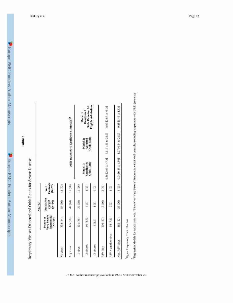
Results—759/922 (83%) eligible admissions were sampled (median age 9 months). One or morerespiratory viruses were detected in 425/759 (56%, 95% CI 52 to 60%). Respiratory syncytialvirus (RSV) was detected in 260 (34%, 95% CI 31 to 38%) and other respiratory viruses in 219(29%, 95% CI 26 to 32%), the commonest being human coronavirus 229E (n=51, 6.7%, 95% CI5.0 to 8.7%), influenza type A (n=44, 5.8%, 95% CI 4.2 to 7.7%), parainfluenza type 3 (n=29,3.8%, 95% CI 2.6 to 5.4%), adenovirus (n=29, 3.8%, 95% CI 2.6 to 5.4%) and humanmetapneumovirus (n=23, 3.0%, 95% CI 1.9 to 4.5%). Compared to well controls, detection of
Corresponding author: James A Berkley [email protected] PO Box 230, Kilifi, 80108, Kenya. Phone (mobile) +254720 867011, (office) +254 417 522535 Fax +254 417 522390.
All authors declare no conflict of interest.
Europe PMC Funders GroupAuthor ManuscriptJAMA. Author manuscript; available in PMC 2010 November 26.
Published in final edited form as:JAMA. 2010 May 26; 303(20): 2051–2057. doi:10.1001/jama.2010.675.
Europe PM
C Funders A
uthor Manuscripts
Europe PM
C Funders A
uthor Manuscripts

RSV was associated with severe disease (4% in controls, adjusted odds Ratio 6.11 [95% CI 1.65 to22.6]) whilst collectively, other respiratory viruses were not (23% in controls, adjusted odds Ratio1.27 [95% CI 0.64 to 2.52]).
Conclusions—In a sample of Kenyan infants and children admitted with severe pneumonia to arural hospital, RSV was the predominant viral pathogen.
IntroductionPneumonia is the leading cause of childhood death in sub-Saharan Africa. The main directmeans for controlling disease and death due to pneumonia are infant vaccination and casemanagement. Thus, establishing the relative contribution to severe disease of individualpathogens in infancy and vaccine efficacy in this age group are essential to reducing theburden of disease.
Observational studies including microbial culture of blood or lung aspirates, and vaccineprobe studies, have identified Streptococcus pneumoniae and Haemophilus influenzae as thecommonest bacterial causes.1, 2 Both are preventable by currently available conjugatevaccines. Consequently, other bacteria; Mycobacterium tuberculosis; Pneumocystis jiroveciand respiratory viruses are becoming of increased importance.3 Vaccines currently exist, orare in development for several respiratory viruses including respiratory syncytial virus(RSV), influenza type A and parainfluenza type 3, including combinations.4-6 In previousstudies in sub-Saharan Africa, viral etiology of pneumonia has been examined by antigendetection, serology and viral culture.7-11
The development of molecular methods has led to two advances in viral diagnosis: increasedsensitivity and the discovery of new viruses of clinical importance. However, the highsensitivity of molecular diagnostics raises questions about specificity since viral nucleic acidsequences may be detected in healthy children and may persist after illness.12 The detectionof multiple respiratory viruses by molecular methods in individual cases further underlinesthis etiological quandary.13 To date, no published studies have reported comprehensive viraletiology of pneumonia among children in sub-Saharan Africa using sensitive moleculardiagnostic methods. We aimed to determine viral etiology, incidence and clinical featuresamong infants and children meeting World Health Organisation (WHO) clinical criteria for‘severe pneumonia’ or ‘very severe pneumonia’ admitted to a rural Kenyan district hospital.
MethodsLocation
The study was conducted at Kilifi District Hospital, in a rural area on the Kenyan coast. Thehospital serves a rural agrarian population, mainly comprising the Mjikenda tribe. Humidityis high throughout the year and there are two annual rainy seasons, April-July andNovember-December.14 The area is endemic for malaria, with declining transmission overthe last 10 years. Haemophilus influenzae type b conjugate vaccine, given with Diptheria-Tetanus-Pertussis at 6, 10 and 14 weeks was introduced in 2001. Coverage of these vaccines(3rd dose) and measles vaccine at 9 months are >90%.15 Conjugate pneumococcal vaccinehad not been introduced at the time of the study.
The Collaborative Research Programme between the Kenya Medical Research Institute(KEMRI) and the Wellcome Trust runs the hospital pediatric wards and a demographicsurveillance system covering an area of ~900km2 within 50km north and south, and 30kmwest of the hospital including a population of ~240,000. The area was mapped in 2000 andevery building registered by global positioning system (GPS). The population register is
Berkley et al. Page 2
JAMA. Author manuscript; available in PMC 2010 November 26.
Europe PM
C Funders A
uthor Manuscripts
Europe PM
C Funders A
uthor Manuscripts

updated for births, deaths and migration events, which are monitored by household visitswith three re-enumeration rounds each year.16 All field based data is checked, doubleentered and verified within 48 hours of acquisition. Since 2002, patients admitted to hospitalare individually identified on the population register. In 2007, about 65% of paediatricadmissions were confirmed residents of the census area.
Participants and Clinical MethodsWe included all children age 1 day to 12 years, residing in the census area who wereadmitted to the pediatric wards between 1st January and 31st December 2007 and met WHOcriteria for the clinical syndromes of ‘severe pneumonia’ (cough or difficult breathing pluslower chest wall indrawing and no signs of ‘very severe pneumonia’) or ‘very severepneumonia’ (cough or difficult breathing plus at least one of: hypoxia, defined as an oxygensaturation <90% by fingertip pulse oximetry (Nellcor, USA), inability to drink or breastfeed, inability to sit or impaired consciousness) at admission,17 including infants aged <2months. Clinicians were trained in the recognition of the clinical signs used in the WHOsyndromic criteria for pneumonia through teaching sessions that included videos andpractical demonstrations, with additional bedside training on the wards. Clinical findingswere individually recorded for each item of history or examination on a database during theadmission assessment. Then, a standardized set of investigations were performed including:complete blood count (Beckman/Coulter, UK), blood film for malaria parasites, bloodculture (Bactec PedsPlus, Becton Dickinson, New Jersey, USA) processed by standardmethods,18 and a nasal wash sample.19 Nasal washes were conducted between 8am and10pm daily. For safety, nasal wash was not performed on infants and children with severerespiratory or cardio-vascular compromise.
HIV testing was performed according to the Kenyan national policy for all pediatric hospitaladmissions using two rapid antibody tests: Determine (Inverness Medical, Florida, USA)and Unigold (Trinity Biotech, Bray, Ireland). Children, and their families, who testedpositive were given further post-test counseling and referred to the hospital comprehensiveHIV care clinic, which provided HIV care before and after discharge according to nationalrecommendations.
For this analysis, a single sign, capillary refill time of 3 seconds or more was used as amarker of circulatory shock. Severe anemia was defined as hemoglobin <5g/dl. Severemalnutrition was defined as weight for height z score <-3 (National Centers for HealthStatistics reference standards, CDC, Atlanta, USA) or kwashiorkor.17 Treatment forpneumonia and other conditions was according to current WHO guidelines.17
In order to estimate the association of respiratory viral infection with severe disease, werecruited two further sets of children resident in the census area by convenience samplingfrom 1st May 2007 to 30th April 2008: i) mild upper respiratory tract infection (URTI): thosewith symptoms including cough, runny or blocked nose, sore throat or sneezing beingmanaged as outpatients and not meeting any criteria for pneumonia, and ii) well infants andchildren: those without any symptoms or signs of upper or lower respiratory infectionattending for routine immunization at the hospital. Following clinical history andexamination, a nasal wash specimen was taken.
Laboratory methodsNasal wash samples were stored at −80°C and analyzed as a batch at the end of the study atthe University of Pretoria, South Africa. The total viral nucleic acids were extracted from200ul of the respiratory specimens using the Magnapure LC Total Nucleic Acid IsolationKit (Roche, Manheim, Germany), according to the manufacturer's instructions. cDNA was
Berkley et al. Page 3
JAMA. Author manuscript; available in PMC 2010 November 26.
Europe PM
C Funders A
uthor Manuscripts
Europe PM
C Funders A
uthor Manuscripts

synthesized using Expand Reverse Transcriptase (Roche, Mannheim, Germany). Real-timePCR reactions were performed using the LightCycler Fast Start DNA MasterPLUSHybProbe kit (Roche, Mannheim, Germany) for Adenovirus (Adeno); Parainfluenza virus(PIV) 1,2 & 3; RSV, Influenza (Flu) types A and B, human metapneumovirus (hMPV);Human bocavirus (hBoV); human coronavirus OC43 (OC43), human coronavirus HongKong (HKU1); human coronavirus 229E (229E) and human coronavirus NL-63 (NL63).PCR amplicons for DNA sequencing were gel purified using the Wizard® SV Gel and PCRClean-Up System (Promega, Madison, WI, USA), according to the manufacturer’sinstructions. DNA sequencing was performed with specific primers using the ABI PRISMBigDye Terminator Cycle Sequencing Reaction kit (version 3.1) on an ABI PRISM 3130DNA sequencer (Applied Biosystems, Foster City, CA, USA), according to themanufacturer’s instructions.
Statistical AnalysisStatistical analysis was performed using STATA 9.1 (Stata Corp, TX, USA). Eligibility andclassification of the clinical syndromes of pneumonia were determined from the originalrecord of each item of history and examination on the database.
Incidence rates were estimated using data from all eligible admissions known to be residentin the census area on the day of admission and the mid-study (1st July 2007) censuspopulation estimates interpolated from the linear equation determined by regressingpopulation size (log 10) for all enumeration rounds to the end of 2008 against the mid-date ofeach round.
In order to describe demographic and clinical features of cases of severe and very severepneumonia, we compared those with a respiratory virus detected to those with no virusdetected. Proportions were compared using the Chi squared and Fishers exact tests asappropriate. Continuous data were compared by t-test or the Kruskall-Wallis rank sum test.
To estimate the strength of association of detection of RSV and non-RSV viruses withsevere disease, we compared admitted cases of ‘severe pneumonia’ and ‘very severepneumonia’ to controls comprising well infants and children (excluding those with signs ofURTI) using a case control design. Odds ratios were calculated by multivariable logisticregression and presented models for RSV (including those with RSV plus an additionalvirus) and respiratory viruses not including RSV that were i) unadjusted (Model 1) and ii)adjusted for age and season in monthly intervals (Model 2). To address a potential bias ofunder-representation of fatal cases, the (unadjusted) odds ratios for severe disease among alleligible admissions were modeled by imposing the proportions with RSV and other virusesdetected among fatal and non-fatal sampled cases on all eligible admissions (Model 3). Allstatistical tests were two-sided with a significance level of 5%.
The sample size for cases was determined by previous annual admissions, which wouldallow the prevalence of pathogens of 5% and 20% to be estimated with precision of +/- 2%and +/-3% respectively. We aimed to recruit 100 well controls and 100 with mild URTI inorder to describe specificities of 95% and 85% with a lower side precision of −6% and −8%,respectively. Power for the case-control analysis was confirmed for the actual numberssampled using the method described by Fliess.20
Ethical ApprovalThe study was approved by the Kenyan National Ethical Review Committee (SSC 815) andthe Oxford Tropical Ethical Review Committee (011 06). Individual written informedconsent was sought from parents or guardians of all participants.
Berkley et al. Page 4
JAMA. Author manuscript; available in PMC 2010 November 26.
Europe PM
C Funders A
uthor Manuscripts
Europe PM
C Funders A
uthor Manuscripts

ResultsDuring 1st January through 31st December 2007, 922 eligible infants and children with‘severe pneumonia’ or ‘very severe pneumonia’ were admitted and viral screening wasconducted on 759 (82%) (Figure 1). Their median age was 9.0 months (IQR 3.3 to 20), 59%were male and 52 (6.9%, 95% CI 5.2 to 8.9%) had a positive HIV rapid antibody test.Children who were not sampled were more severely ill than those sampled (eTable 1). Threequarters of deaths among non-sampled admissions occurred within 24 hours of admission.
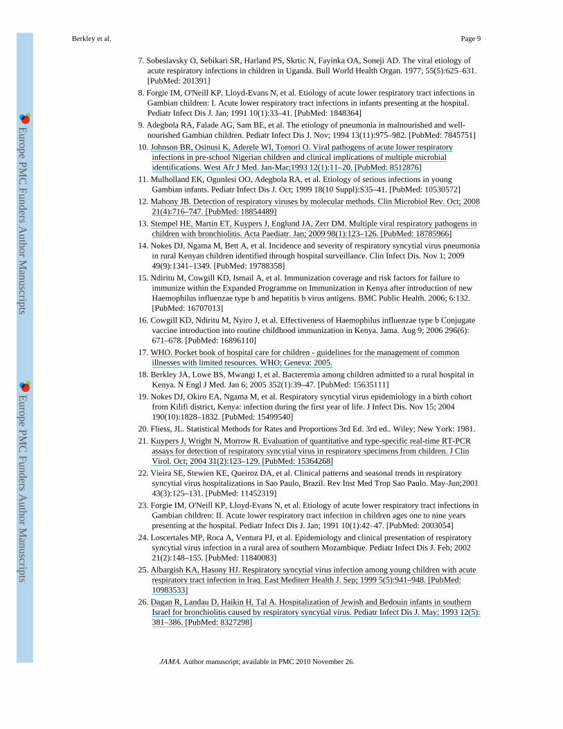
One or more respiratory viruses were detected in 425 (56%, 95% CI 52 to 60%) cases (Table1). RSV was the most commonly detected virus, present in 260 (34%, 95% CI 31 to 38%)admissions overall, and in 192/453 (42%, 95% CI 38 to 47%) infants (eTable 2). RSV wasstrongly seasonal (P<0.001), detected in more than 50% of pneumonia cases in January,February and December, but in <5% from July through September (Figure 2). Other virusesshowing clear seasonality were hMPV, PIV 3 and Flu A (eFigure 1).
Other respiratory viruses were detected in 219 (29%, 95% CI 26 to 32%)) admissions(eTable 2 & eFigure 1). These comprised: Flu A (5.8%); Flu B (0.1%); hMPV (3.0%);hBoV (2.1%); PIV 1 (2.4%); PIV 2 (1.3%); PIV 3 (3.8%); Adenovirus (3.8%); NL63(1.3%); HKU1 (0.1%); 229E (6.7%) and OC43 (1.8%).
RSV plus one or more other respiratory virus were detected in 54 (7%) cases, representing21% (95% CI 16 to 26%) of all RSV cases. These comprised 229E (14), Flu A (10),adenovirus (10), PIV 3 (6), hBoV (6), HMPV (3), PIV 1 (3), PIV 2 (3), NL63 (2), and OC43(1).
The incidence of admission with ‘severe pneumonia’ or ‘very severe pneumonia’ was 4.8%per child/year in the first year of life, 1.5% per child/year in under 5s and 0.1% amongstchildren 5 or more years old (Table 2). The incidence of admission for any respiratory virusand with RSV was highest in the first year of life (3.0% per child/year and 2.0% per child/year, respectively).
Admissions with respiratory viruses detected had a median age of 7.5 months (IQR 2.7 to18) and were younger than those with no virus detected (median age of 11 months (IQR 3.8to 24), P<0.001). However, this effect was completely accounted for by RSV (Table 3).‘Very severe pneumonia’ was less common among admissions with respiratory viruses(P<0.001). There was no association between the presence of wheeze or hypoxia and arespiratory virus being detected. Most cases with RSV did not have an admission diagnosisof bronchiolitis. Admissions with RSV were less severely ill and had fewer adverse riskfactors than admissions with no virus: they were less likely to be prematurely born, shocked,severely malnourished or to die. Of the 2 deaths among children with RSV, both hadcongenital heart disease. Admissions with respiratory viruses other than RSV were similar inall these respects to admissions with no virus detected. Among 24 deaths, 8 (33%, 95% CI16 to 55%) occurred in admissions with a virus detected.
Thirty six (4.7%, 95% CI 3.3 to 6.5%) admissions with ‘severe pneumonia’ or ‘very severepneumonia’ were bacteremic, 16 (44%, 95% CI 28 to 62%) of these had a respiratory virusdetected. Bacterial species were Streptococcus pneumonia (12), Escherichia coli (9), non-typhoidal Salmonella (3), Staphylococcus aureus (3), Acinetobacter sp. (3), Beta haemolyticStreptococci (3), Enterobacter sp. (2) and Haemophilus influenzae (1).
Among 57 well infants and children (median age 6.0 months, IQR 3.3 to 11) and 96 childrenwith symptoms of mild URTI (median age 13 months, IQR 5.6 to 25), respiratory viruseswere detected in 28% and 44% respectively, and were less frequent than among admitted
Berkley et al. Page 5
JAMA. Author manuscript; available in PMC 2010 November 26.
Europe PM
C Funders A
uthor Manuscripts
Europe PM
C Funders A
uthor Manuscripts

pneumonia cases: P<0.001 and P=0.026 respectively (Table 1). These differences were dueto RSV since overall, viruses other than RSV were as common in well infants and childrenand those with mild URTI as amongst admitted cases: P=0.85 and P=0.34 respectively(Table 1). Among those with mild URTI, viruses detected were: RSV (18%); Flu A (7%);hMPV (3%); hBoV (5%); PIV 1 (2%); Adenovirus (8%); HKU1 (2%); 229E (3%) andOC43 (2%) (eTable 3). Viruses found in well infants and children comprised: RSV (5%);hBoV (9%); OC43 (7%) and <1% for each of PIV 1, PIV 3 , Adenovirus, HKU1, and 229E.Flu A and HMPV were not detected among well infants (eTable 2).
Detection of RSV was associated with admission with severe disease compared to wellcontrols (odds ratio 9.38 (95% CI 2.99 to 47.3), 5% in controls, Model 1, Table 1) Therewas no evidence of an association between viruses other than RSV and severe disease (oddsratio 0.94 (95% CI 0.48 to 1.9), 23% in controls, Model 1, Table 1). Models adjusted for ageand calendar month (Model 2, Table 1) showed no evidence of lack of fit (Hosmer-Lemeshow chi squared 7.19, P=0.52) and did not qualitatively alter this result: RSV oddsratio 6.11 (95% CI 1.65 to 22.6) and non-RSV viruses 1.27 (95% CI 0.64 to 2.52). The thirdmodel including all eligible admissions, to address the potential bias of under-representationof fatal cases, did not differ significantly from the observed, unadjusted odds ratios (Model 3& Model 1, Table 1).
DiscussionWe found that in the catchment area of Kilifi District Hospital on the Kenyan coast, theincidence of admission with clinical syndromes of severe or very severe pneumonia rangedfrom 4.8% in the first year of life to 0.1% among those 5 years old or greater. RSV wasdetected in a third of cases overall and almost half of infant cases. The seasonality of severepneumonia was almost entirely determined by RSV. No other virus was identified in morethan 7% of admitted infants or children, and these cases were clinically similar to those inwhom no virus was detected. Viruses other than RSV were as common among well infantsand children and those with mild URTI as among those with severe disease. These findingssuggest that they make only a minor contribution to the burden of severe clinical pneumoniain this setting.
In a previous study in the same enumerated population of Kilifi 2002 through 2007, RSVwas detected by immunofluorescence in 15% of ‘severe pneumonia’ or ‘very severepneumonia’ admissions in under 5′s, rising to 27% during epidemic periods. Incidenceduring 2006/2007 was estimated at 0.99% and 0.27% per child per year in infants and under5′s respectively.14 In the present study, we found approximately twice the incidence. It isknown that real time PCR is more sensitive than traditional diagnostics. In a study byKuypers et al.21 RSV detected by both immunofluorescence and PCR had a mean viral loadof 6.1 × 10⁷ copies/ml. Whereas, RSV detected by PCR only had a lower mean viral load of4.1 × 104 copies/ml. An immunofluorescence assay detected only 19% of the RSV with viralloads <106 copies/ml.
Could our findings be due to detection of persistent viral RNA or false positive laboratoryresults? We believe that this is unlikely. Firstly, considering our well controls, if anassumption is made that all of the RSV detected are false positives due to persistence orlaboratory error, then the specificity of our test is 95% (95% CI 85 to 99%). This is similarto that reported for immunofluorescence. Such specificity would not significantly alter theinterpretation of our findings among cases. This is further supported by an odds ratiosuggesting that detection of RSV was strongly associated with severe disease. To comparethese findings with those from traditional methods, we examined published data from 8previous studies in developing countries conducted using immunofluorescence, serology or
Berkley et al. Page 6
JAMA. Author manuscript; available in PMC 2010 November 26.
Europe PM
C Funders A
uthor Manuscripts
Europe PM
C Funders A
uthor Manuscripts

viral culture.7, 8, 11, 22-27 Overall, RSV was detected in 746/3,463 (21.5%) cases and29/1,203 (2.4%) controls, giving a pooled (fixed effects) odds ratio for disease (unadjustedfor age and season) of 11.1 (95% CI 7.69 to 16.0), which is close to the unadjusted oddsratio in our study.
The high prevalence of RSV in childhood admissions with severe clinical pneumonia (34%)in contrast to the prevalence in well controls (5%), provides further support that RSVvaccination may offer considerable public health benefit. While the development of avaccine for the key target age group of early infants has focused on live virus vaccines, nonehas achieved requisite levels of both safety and immunogenicity.4 A combined PIV3/RSVlive-attenuated vaccine (Medi-534) for use in young infants (2 months) and older children (6to <24 months) is currently undergoing phase I/II clinical trials. Ideally, such trials should beaccompanied by modelling studies of the potential consequences on effectiveness of delayeddelivery to older infants28, 29 where vaccine safety and immunogenicity are demonstrablyimproved. 5, 30
Of equal interest to the high prevalence of cases with RSV detected is the low proportion ofcases with any of the other respiratory viruses. The real time PCR assay that we used haspreviously been shown to be sensitive for these viruses.31 It is possible that the use of nasalwashings targets a site within the upper respiratory tract is predilected by RSV over anyother virus. However, to our knowledge there are no definitive studies in the literature whichclearly identify selective bias in the range of respiratory viruses detectable in different partsof the upper respiratory tract using molecular diagnostics.
Influenza and parainfluenza 3 have been the most frequently detected viruses, other thanRSV, in studies employing traditional diagnostics in sub-Saharan Africa. We found these,229E and hMPV, were the most commonly detected non-RSV viruses in cases. However,these viruses appear to be common in the community causing URTI, and some are present inwell controls. In a non-epidemic period, we estimate that up to approximately 6% of severedisease is associated with influenza type A. There appears to be little potential to preventsevere disease through vaccination against PIV 3.
Strengths of our study include systematic sampling of well-characterized hospitalizedchildren from a well established, enumerated population base and detection of acomprehensive set of respiratory viruses. Our study has several limitations. In common withsimilar studies, our sampling missed most deaths because nasal washing cannot beconducted in the sickest children at admission. However, our sensitivity analysis suggeststhis in unlikely to significantly alter the main findings. We did not screen for rhinovirus andthere was a lack of x-ray data. Our data reflect a single site and findings in other ecologicalsettings may differ. We applied WHO guidelines carefully and there was good availabilityof clinical staff, oxygen and drugs. Case fatality may differ where resources are moreconstrained. Our well controls were limited in number, since we did not achieve our targetsample size due to factors outside our control, and they were not matched to exactly thesame calendar year. Small numbers resulted in wide confidence intervals in some subgroups.However, power was sufficient (>99%) for our case-control analysis of RSV.
It is likely that not all cases of severe pneumonia in the community were admitted tohospital, thus our incidence estimates are therefore the minimum. There is a demonstrabledecay with increased distance from the hospital in the incidences of all childhoodadmissions and of admissions with detectable RSV.14 A study spanning only one year andfrom a single location cannot expect to account for likely variation in the occurrence ofrespiratory viruses year-to-year or between low resource settings.
Berkley et al. Page 7
JAMA. Author manuscript; available in PMC 2010 November 26.
Europe PM
C Funders A
uthor Manuscripts
Europe PM
C Funders A
uthor Manuscripts

In summary, our study of the occurrence of respiratory viruses in children admitted withclinical syndromes of ‘severe pneumonia’ or ‘very severe pneumonia’ to a rural districthospital in coastal Kenya, has identified over 50% of cases with a detectable virus, of whichRSV was clearly predominant. We estimate that the effective prevention of RSV-associatedsevere pneumonia in this population might reduce all-cause clinical ‘severe pneumonia’ or‘very severe pneumonia’ admissions to the District hospital by a third. This contrasts withno evidence to suggest a marked effect on such admissions would arise from the preventionof any other respiratory virus, with the possible exception of influenza type A. Furthermolecular based studies of respiratory virus etiology of severe pneumonia over longerperiods and in multiple settings in sub-Saharan Africa are needed.
Supplementary MaterialRefer to Web version on PubMed Central for supplementary material.
AcknowledgmentsThe authors are grateful to the staff and patients of the pediatric wards and outpatient department of Kilifi DistrictHospital and to the clinical, ICT and laboratory staff of the KEMRI/Wellcome Trust Research Programme whocontributed to this study. We thank Dr Norbert Peshu M.P.H., Director of the Centre for Geographic MedicineResearch – coast and Dr Iqbal Khandwalla M.Med., the Medical Superintendent of Kilifi District Hospital for theiroversight and making facilities available for this research. No compensation was received for participation in thisresearch. This paper is published with permission of the director of KEMRI.
The study was conceived and designed by JAB, JAGS and DJN. JAB, PM, MN and SK collected the clinical data,nasal wash samples and provided patient care. JA, AB, RL, TK, PAC and MV developed and undertook laboratoryanalyses. Analysis was conducted by JAB, JAGS and DJN. JAB wrote the manuscript and all authors contributed tothe final version. JAB had full access to all of the data in the study and takes responsibility for the integrity of thedata and the accuracy of the data analysis.
The study was funded by a project grant (081186) from the Wellcome Trust (UK). JAB, JAGS and DJN aresupported by fellowships from the Wellcome Trust (UK). The funder had no role in the design and conduct of thestudy; collection, management, analysis, and interpretation of the data; and preparation, review, or approval of themanuscript.
References1. Cutts FT, Zaman SM, Enwere G, et al. Efficacy of nine-valent pneumococcal conjugate vaccine
against pneumonia and invasive pneumococcal disease in The Gambia: randomised, double-blind,placebo-controlled trial. Lancet. Mar 26; 2005 365(9465):1139–1146. Apr 1. [PubMed: 15794968]
2. Mulholland K, Hilton S, Adegbola R, et al. Randomised trial of Haemophilus influenzae type-btetanus protein conjugate vaccine [corrected] for prevention of pneumonia and meningitis inGambian infants. Lancet. Apr 26; 1997 349(9060):1191–1197. [PubMed: 9130939]
3. Scott JA, English M. What are the implications for childhood pneumonia of successfullyintroducing Hib and pneumococcal vaccines in developing countries? PLoS Med. Apr 22.20085(4):e86. [PubMed: 19226734]
4. Karron RA, Wright PF, Belshe RB, et al. Identification of a recombinant live attenuated respiratorysyncytial virus vaccine candidate that is highly attenuated in infants. J Infect Dis. Apr 1; 2005191(7):1093–1104. [PubMed: 15747245]
5. Wright PF, Karron RA, Belshe RB, et al. The absence of enhanced disease with wild typerespiratory syncytial virus infection occurring after receipt of live, attenuated, respiratory syncytialvirus vaccines. Vaccine. Oct 16; 2007 25(42):7372–7378. [PubMed: 17868959]
6. Schmidt AC, McAuliffe JM, Murphy BR, Collins PL. Recombinant bovine/human parainfluenzavirus type 3 (B/HPIV3) expressing the respiratory syncytial virus (RSV) G and F proteins can beused to achieve simultaneous mucosal immunization against RSV and HPIV3. J Virol. 2001;75(10):4594–4603. [PubMed: 11312329]
Berkley et al. Page 8
JAMA. Author manuscript; available in PMC 2010 November 26.
Europe PM
C Funders A
uthor Manuscripts
Europe PM
C Funders A
uthor Manuscripts
7. Sobeslavsky O, Sebikari SR, Harland PS, Skrtic N, Fayinka OA, Soneji AD. The viral etiology ofacute respiratory infections in children in Uganda. Bull World Health Organ. 1977; 55(5):625–631.[PubMed: 201391]
8. Forgie IM, O'Neill KP, Lloyd-Evans N, et al. Etiology of acute lower respiratory tract infections inGambian children: I. Acute lower respiratory tract infections in infants presenting at the hospital.Pediatr Infect Dis J. Jan; 1991 10(1):33–41. [PubMed: 1848364]
9. Adegbola RA, Falade AG, Sam BE, et al. The etiology of pneumonia in malnourished and well-nourished Gambian children. Pediatr Infect Dis J. Nov; 1994 13(11):975–982. [PubMed: 7845751]
10. Johnson BR, Osinusi K, Aderele WI, Tomori O. Viral pathogens of acute lower respiratoryinfections in pre-school Nigerian children and clinical implications of multiple microbialidentifications. West Afr J Med. Jan-Mar;1993 12(1):11–20. [PubMed: 8512876]
11. Mulholland EK, Ogunlesi OO, Adegbola RA, et al. Etiology of serious infections in youngGambian infants. Pediatr Infect Dis J. Oct; 1999 18(10 Suppl):S35–41. [PubMed: 10530572]
12. Mahony JB. Detection of respiratory viruses by molecular methods. Clin Microbiol Rev. Oct; 200821(4):716–747. [PubMed: 18854489]
13. Stempel HE, Martin ET, Kuypers J, Englund JA, Zerr DM. Multiple viral respiratory pathogens inchildren with bronchiolitis. Acta Paediatr. Jan; 2009 98(1):123–126. [PubMed: 18785966]
14. Nokes DJ, Ngama M, Bett A, et al. Incidence and severity of respiratory syncytial virus pneumoniain rural Kenyan children identified through hospital surveillance. Clin Infect Dis. Nov 1; 200949(9):1341–1349. [PubMed: 19788358]
15. Ndiritu M, Cowgill KD, Ismail A, et al. Immunization coverage and risk factors for failure toimmunize within the Expanded Programme on Immunization in Kenya after introduction of newHaemophilus influenzae type b and hepatitis b virus antigens. BMC Public Health. 2006; 6:132.[PubMed: 16707013]
16. Cowgill KD, Ndiritu M, Nyiro J, et al. Effectiveness of Haemophilus influenzae type b Conjugatevaccine introduction into routine childhood immunization in Kenya. Jama. Aug 9; 2006 296(6):671–678. [PubMed: 16896110]
17. WHO. Pocket book of hospital care for children - guidelines for the management of commonillnesses with limited resources. WHO; Geneva: 2005.
18. Berkley JA, Lowe BS, Mwangi I, et al. Bacteremia among children admitted to a rural hospital inKenya. N Engl J Med. Jan 6; 2005 352(1):39–47. [PubMed: 15635111]
19. Nokes DJ, Okiro EA, Ngama M, et al. Respiratory syncytial virus epidemiology in a birth cohortfrom Kilifi district, Kenya: infection during the first year of life. J Infect Dis. Nov 15; 2004190(10):1828–1832. [PubMed: 15499540]
20. Fliess, JL. Statistical Methods for Rates and Proportions 3rd Ed. 3rd ed.. Wiley; New York: 1981.
21. Kuypers J, Wright N, Morrow R. Evaluation of quantitative and type-specific real-time RT-PCRassays for detection of respiratory syncytial virus in respiratory specimens from children. J ClinVirol. Oct; 2004 31(2):123–129. [PubMed: 15364268]
22. Vieira SE, Stewien KE, Queiroz DA, et al. Clinical patterns and seasonal trends in respiratorysyncytial virus hospitalizations in Sao Paulo, Brazil. Rev Inst Med Trop Sao Paulo. May-Jun;200143(3):125–131. [PubMed: 11452319]
23. Forgie IM, O'Neill KP, Lloyd-Evans N, et al. Etiology of acute lower respiratory tract infections inGambian children: II. Acute lower respiratory tract infection in children ages one to nine yearspresenting at the hospital. Pediatr Infect Dis J. Jan; 1991 10(1):42–47. [PubMed: 2003054]
24. Loscertales MP, Roca A, Ventura PJ, et al. Epidemiology and clinical presentation of respiratorysyncytial virus infection in a rural area of southern Mozambique. Pediatr Infect Dis J. Feb; 200221(2):148–155. [PubMed: 11840083]
25. Albargish KA, Hasony HJ. Respiratory syncytial virus infection among young children with acuterespiratory tract infection in Iraq. East Mediterr Health J. Sep; 1999 5(5):941–948. [PubMed:10983533]
26. Dagan R, Landau D, Haikin H, Tal A. Hospitalization of Jewish and Bedouin infants in southernIsrael for bronchiolitis caused by respiratory syncytial virus. Pediatr Infect Dis J. May; 1993 12(5):381–386. [PubMed: 8327298]
Berkley et al. Page 9
JAMA. Author manuscript; available in PMC 2010 November 26.
Europe PM
C Funders A
uthor Manuscripts
Europe PM
C Funders A
uthor Manuscripts

27. Phillips PA, Lehmann D, Spooner V, et al. Viruses associated with acute lower respiratory tractinfections in children from the eastern highlands of Papua New Guinea (1983-1985). SoutheastAsian J Trop Med Public Health. Sep; 1990 21(3):373–382. [PubMed: 1963705]
28. Nokes DJ, Okiro EA, Ngama M, et al. Respiratory syncytial virus infection and disease in infantsand young children observed from birth in Kilifi District, Kenya. Clin Infect Dis. Jan 1; 200846(1):50–57. [PubMed: 18171213]
29. Nokes JD, Cane PA. New strategies for control of respiratory syncytial virus infection. Curr OpinInfect Dis. Dec; 2008 21(6):639–643. [PubMed: 18978532]
30. Wright PF, Karron RA, Belshe RB, et al. Evaluation of a live, cold-passaged, temperature-sensitive, respiratory syncytial virus vaccine candidate in infancy. J Infect Dis. Nov; 2000 182(5):1331–1342. [PubMed: 11010838]
31. Lassauniére R, Kresfelder T, Venter M. A Novel Multiplex Real-Time RT-PCR Assay with FRETHybridization Probes for the Detection and Quantification of 13 Traditional and Newly IdentifiedRespiratory Viruses. J Virological Methods. 2010 In Press.
Berkley et al. Page 10
JAMA. Author manuscript; available in PMC 2010 November 26.
Europe PM
C Funders A
uthor Manuscripts
Europe PM
C Funders A
uthor Manuscripts

Figure 1.Flow Diagram for Inclusion and Sampling of Infants and Children with ‘Severe’ or ‘VerySevere’ Pneumonia.
Berkley et al. Page 11
JAMA. Author manuscript; available in PMC 2010 November 26.
Europe PM
C Funders A
uthor Manuscripts
Europe PM
C Funders A
uthor Manuscripts

Figure 2.Seasonal Pattern of RSV and Other Viruses Detected Among Infants and Children Admittedwith ‘Severe’ or ‘Very Severe’ Pneumonia.
Berkley et al. Page 12
JAMA. Author manuscript; available in PMC 2010 November 26.
Europe PM
C Funders A
uthor Manuscripts
Europe PM
C Funders A
uthor Manuscripts
Europe PM
C Funders A
uthor Manuscripts
Europe PM
C Funders A
uthor Manuscripts
Berkley et al. Page 13
Tabl
e 1
Res
pira
tory
Vir
uses
Det
ecte
d an
d O
dds
Rat
ios
for
Seve
re D
isea
se.
No.
(%
)
Seve
re o
rV
ery
Seve
reP
neum
onia
(N=7
59)
Out
pati
ent
UR
TIa
(N=9
6)
Wel
lC
ontr
ols
(N=5
7)
No
viru
s33
4 (4
4)54
(56
)41
(72
)
Any
vir
us42
5 (5
6)42
(44
)16
(28
)O
dds
Rat
io [
95%
Con
fide
nce
Inte
rval
s]b
1
viru
s35
1 (4
6)36
(38
)15
(26
)
Mod
el 1
:U
nadj
uste
dO
dds
Rat
io
Mod
el 2
:A
djus
ted
Odd
s R
atio
Mod
el 3
:U
nadj
uste
dO
dds
Rat
io f
or A
llE
ligib
le A
dmis
sion
s
2
viru
ses
66 (
8.7)
5 (5
)1
(2)
3
viru
ses
8 (1
.1)
1 (1
)0
(0)
RSV
onl
y20
6 (2
7)15
(16
)2
(4)
9.38
[2.
99 to
47.
3]6.
11 [
1.65
to 2
2.6]
8.99
[2.
87 to
45.
2]
RSV
+ a
noth
er v
irus
54 (
7.1)
2 (2
)1
(2)
Non
-RSV
vir
us16
5 (2
2)25
(26
)13
(23
)0.
94 [
0.48
to 1
.94]
1.27
[0.
64 to
2.5
2]0.
88 [
0.45
to 1
.83]
a Upp
er R
espi
rato
ry T
ract
Inf
ectio
n
b Reg
ress
ion
Mod
els
for
Adm
issi
ons
with
‘Se
vere
’ or
‘V
ery
Seve
re’
Pneu
mon
ia v
ersu
s w
ell c
ontr
ols,
exc
ludi
ng o
utpa
tient
s w
ith U
RT
I (s
ee te
xt).
JAMA. Author manuscript; available in PMC 2010 November 26.

Europe PM
C Funders A
uthor Manuscripts
Europe PM
C Funders A
uthor Manuscripts
Berkley et al. Page 14
Tabl
e 2
Inci
denc
e of
Adm
issi
on w
ith ‘
Seve
re P
neum
onia
’ or
‘V
ery
Seve
re P
neum
onia
’ A
mon
g R
esid
ents
of
the
Kili
fi D
SS C
ensu
s A
rea.
Age
Gro
upun
der
28 d
ays
All
<1y
1 to
1.99
y2
to4.
99y
All
<5y
5 to
12.9
9yA
ll <
13y
Den
omin
ator
9,42
3 bi
rths
8,83
744
,538
104,
505
per
1,00
0 liv
e bi
rths
per
100,
000
child
ren/
yr
‘Sev
ere’
or
‘Ver
ySe
vere
’ Pn
eum
onia
6.65
4,79
81,
674
543
1,52
299
681
Any
res
pira
tory
vir
us3.
792,
993
871
213
862
3638
0
RSV
2.46
2,03
845
585
535
1523
3
229E
0.51
318
135
3210
53
46
Infl
uenz
a A
0.31
244
9732
8215
39
PIV
30.
1021
248
1157
626
Ade
novi
rus
0.10
149
7721
559
26
HM
PV0.
2013
858
1144
621
JAMA. Author manuscript; available in PMC 2010 November 26.

Europe PM
C Funders A
uthor Manuscripts
Europe PM
C Funders A
uthor Manuscripts
Berkley et al. Page 15
Tabl
e 3
Clin
ical
Fea
ture
s of
759
Chi
ldre
n A
dmitt
ed w
ith R
SV a
nd O
ther
Res
pira
tory
Vir
uses
.
Med
ian
(IQ
R)a
Med
ian
(IQ
R)a
P V
alue
b
No
viru
sN
=334
Any
vir
usN
=425
RSV
onl
yN
=206
RSV
+ a
noth
er v
irus
N=5
4N
on-R
SV v
irus
N=1
65
Age
(m
onth
s)11
.3(3
.8 to
24)
7.5
(2.7
to 1
8)<
.001
6.1
(2.5
to 1
3)<
.001
7.5
(2.7
to 1
6).0
1110
(4.5
to 2
0).3
9
Inpa
tient
Sta
y (d
ays)
4(2
to 6
)3
(2 to
5)
.17
3(2
to 5
).0
74
(3 to
6)
.80
4(2
to 6
).5
6
No.
(%
)N
o. (
%)
P V
alue
b
No
viru
sN
=33
4A
ny v
irus
N=
425
RSV
onl
yN
=20
6R
SV +
ano
ther
vir
usN
=54
Non
-RSV
vir
usN
=16
5
Ver
y Se
vere
Pne
umon
ia73
(22)
53(1
3).0
0122
(11)
.001
8(1
5).2
423
(14)
.035
Whe
eze
50(1
5)59
(14)
.67
30(1
5).9
08
(15)
.98
2(1
3).5
0
Hyp
oxia
40(1
3)40
(9)
.13
18(8
.7)
.14
8(1
5).7
014
(8.5
).1
4
Cap
illar
y re
fill
≥3 s
ecs
16(4
.8)
10(2
.4)
.066
1(0
.5)
.004
1(1
.9)
.49
8(4
.9)
.98
Seve
re A
nem
ia15
(4.6
)12
(2.9
).2
33
(1.5
).0
821
(1.9
).7
18
(4.9
).8
5
His
tory
of
Prem
atur
ity17
(7.8
)14
(4.1
).0
675
(2.9
).0
462
(4.4
).5
47
(5.7
).4
9
Con
geni
tal H
eart
Dis
ease
12(3
.6)
5(1
.2)
.05
2(1
.0)
.09
0(0
).2
33
(1.8
).4
0
HIV
26(8
.0)
26(6
.2)
.35
8(4
.0)
.07
4(7
.6)
114
(8.6
).8
2
Seve
re M
alnu
triti
on26
(7.8
)18
(4.2
).0
385
(2.4
).0
092
(3.7
).4
011
(6.7
).6
5
Bac
tere
mia
20(6
.0)
16(3
.8)
.15
5(2
.4)
.056
2(3
.7)
.75
9(5
.5)
.81
Dea
th16
(4.8
)8
(1.9
). 0
232
(1.0
).0
230
(0)
.14
6(3
.6)
.56
a IQR
= in
terq
uart
ile r
ange
b P V
alue
s in
com
pari
son
with
chi
ldre
n w
ith n
o vi
rus.
JAMA. Author manuscript; available in PMC 2010 November 26.
Copyright © 2022 FDOKUMEN