Menopause and hormone replacement therapy: effects on the immune system, arthritis and bone
Upload
khangminh22Category
view
1download
0
Variations in Menopause Etiology Affect Cognitive Outcomes: How Age, Menopause
Type, and Exogenous Ovarian Hormone Exposures Across the Lifespan Impact the
Trajectory of Brain Aging
by
Stephanie Victoria Koebele
A Dissertation Presented in Partial Fulfillment of the Requirements for the Degree
Doctor of Philosophy
Approved July 2019 by the Graduate Supervisory Committee:
Heather A. Bimonte-Nelson, Chair
Jason M. Newbern Cheryl D. Conrad Dale F. DeNardo Eric M. Reiman
ARIZONA STATE UNIVERSITY
August 2019

i
ABSTRACT
Reproductive hormones are recognized for their diverse functions beyond
reproduction itself, including a vital role in brain organization, structure, and function
throughout the lifespan. From puberty to reproductive senescence, the female is
characterized by inherent responsiveness to hormonal cyclicity. For most women, a
natural transition to menopause occurs in midlife, wherein the endogenous hormonal
milieu undergoes significant changes and marks the end of the reproductive life stage.
Although most women experience natural menopause, many women will undergo
gynecological surgery during their lifetime, which can lead to an abrupt surgical
menopause. It is of critical importance to better understand how endogenous and
exogenous reproductive hormone exposures across the lifespan influence cognitive and
brain aging, as women are at a greater risk for developing a variety of diseases after
menopause, including dementia. Using rodent models, this dissertation explores how the
etiology of reproductive senescence, that is, whether it is transitional or surgical,
influences the female phenotype to result in divergent cognitive outcomes dependent
upon a variety of factors, with an emphasis on age at the time of intervention playing a
key role in brain outcomes. Furthermore, the impact of exogenous hormone therapy on
cognition is evaluated in the context of surgical menopause. A novel rat model of
hysterectomy is also presented, with results demonstrating for the first time that the
nonpregnant uterus, which is typically considered to be a quiescent organ, may play a
unique, direct role in modulating cognitive outcomes. Neurobiological mechanisms
associated with reproductive hormones and aging are assessed to better recognize neural
correlates underlying the observed behavior changes. The overarching goal of this

ii
dissertation was to elucidate novel factors contributing to cognitive aging outcomes in
females. Collectively, the data presented herein indicate that the age at the onset of
reproductive senescence has significant implications for learning and memory outcomes,
and that variations in gynecological surgery can have unique, long-lasting effects on the
brain and cognition. Translationally, this series of experiments moves the field forward
toward the goal of improving the health and quality of life for women throughout the
lifespan.

iii
ACKNOWLEDGMENTS
I am filled with tremendous sense of gratitude for the incredible support, both
professionally and personally, I have experienced throughout my education, and
especially during the pursuit of my doctoral studies. To my exceptional mentor, Heather:
thank you for your guidance, support, generosity, and encouragement for the past eight
years. You believed in me before I believed in myself. I have grown into the scientist and
person I am today through your mentorship. Beyond the skill sets needed to succeed in
the laboratory, you have taught me to be a leader, to be independent, and that my voice
matters. You have encouraged me to think outside the box, to be brave, to be bold, to be
fierce, and to be fabulous. You instill your extraordinary passion for neuroscience,
women’s health, and education in all of your students, and teach us to be the best
scientists and mentors we can be as we move through each stage of our careers.
To my remarkable and diverse dissertation committee, Drs. Jason Newbern, Dale
DeNardo, Eric Reiman, and Cheryl Conrad: it has been an honor and a privilege to work
alongside each of you and to be mentored by you. Thank you for encouraging me to
move beyond my comfort zone, to learn many new skill sets, and to develop a broad
knowledge and deeper understanding of the complexities of the brain and endocrine
systems. To Drs. Loretta Mayer and Cheryl Dyer: a heartfelt thank you for your
tremendous generosity and encouragement in supporting my research endeavors, and for
welcoming me into your laboratory to teach me new skills. You are both wonderful role
models and I am grateful for your kindness and instrumental contributions to my success.
Tolkien said that “true education is a kind of never ending story—a matter of continual
beginnings, of habitual fresh starts, of persistent newness.” A career in science, I believe,

iv
is one that requires a drive for life-long learning, and I am privileged to know so many
talented scientists and scholars who have instilled this passion in me as well.
To the Arizona State University (ASU) Department of Psychology: thank you for
being my home for the past eleven years. When I began my undergraduate degree as a
Psychology major at ASU, I never considered that being a neuroscientist was an
accessible and attainable career option for me. It is through this department that I
discovered my passion for neuroscience, and I am incredibly grateful for the warm and
friendly community I have had the joy to work with throughout my undergraduate and
graduate degrees. A special thank you to Dr. Leona Aiken – I will always remember the
day you told me that more important than any exam was to be a strong, creative, brilliant
woman in science. Your tenacity, grit, and tremendous passion for teaching your students
—all of which you navigate with grace and style— is a source of inspiration I will carry
with me throughout my career. It is a privilege to have been your student.
Thank you to the Behavioral Neuroscience in Psychology Graduate Program, to
my outstanding graduate student peers (past and present), and to my many lab mates over
the years in the Bimonte-Nelson laboratory. I have made life-long friends throughout this
journey. Newton said “if I have seen a little further, it is by standing on the shoulders of
giants” – and it is with your love and reassurance that I am where I am today.
To the Graduate Women’s Association (GWA), for which I served as an officer
for three years of my graduate studies: thank you for the opportunity to meet so many
wonderful and diverse women across campus I never would have otherwise had the
pleasure to interact with. I learned invaluable leadership skills through this organization,
and I am grateful that I had the chance to represent and contribute to GWA’s mission.

v
Thank you to the hundreds of children I have had the privilege to interact with
through the Brain Fair for Children and Open Door initiatives at ASU. Watching these
young minds learn and discover with boundless excitement and enthusiasm motivates me
to always be the best teacher and mentor I can be, and to never stop learning.
To Mom and Dad, who have always been my cheerleaders in all that I do: thank
you for believing in the power of education and making sure that I had every opportunity
to succeed through each step of my education. My accomplishments are a reflection of
your constant love and support in every dimension. I am so grateful for your advice and
encouragement throughout this journey. I love you both, and hope that you are proud.
Finally, the research in this dissertation would not have been possible without the
incredible financial support I have been fortunate to have been awarded throughout my
doctoral studies, including the Ruth L. Kirchstein predoctoral National Research and
Service Award (F31AG056110) funded by the National Institute on Aging from 2017-
2019. I have also received numerous monetary awards for research and travel purposes,
including the Dean’s Fellowship for First-Year Graduate Students, the CLAS Graduate
Excellence Fellowship for First Generation Students, four consecutive CLAS Graduate
Excellence awards, a Society for Neuroscience Trainee Professional Development
Award, two Graduate College Travel Grants, three GPSA Individual Travel Awards, the
Arizona Alzheimer’s Disease Core Broadening Horizons Travel Award, an Outstanding
Writing in Psychology Award, the European Menopause and Andropause Society New
Investigator Award, a Graduate Student Scholarship for the Practical Workshop on
Confocal Microscopy and Quantitative Histology, and the generous funding support of
my mentor, Dr. Heather Bimonte-Nelson.

vi
TABLE OF CONTENTS CHAPTER Page
LIST OF TABLES ..................................................................................................... xviii
LIST OF FIGURES ..................................................................................................... xix
1. GENERAL INTRODUCTION ............................................................................ 1
A Brief Primer on Mammalian Sexual Differentiation .............................. 1
Brain Sexual Differentiation in Mammals ................................................ 3
On the Framework of Organizational and Activational Effects of Ovarian
Hormones on the Brain ............................................................................ 7
(Re)organizational Events Across the Lifespan ...................................... 11
Reproductive Hormones and the Brain ................................................... 14
The Female Reproductive Cycle and Menopause ................................... 15
Rodent Aging and Reproductive Senescence .......................................... 19
Ovary-Intact: A Model of the Aging Hypothalamic-Pituitary-
Gonadal Axis .............................................................................. 21
Ovariectomy: A Model of Surgical Menopause .......................... 25
4-Vinylcyclohexene Diepoxide: A Model of Transitional
Menopause ................................................................................. 27
Hysterectomy: A Novel Model of Gynecological Surgery and
Menopause in Rats ..................................................................... 31
Reproductive Hormones and Cognition .................................................. 33

vii
CHAPTER Page
Estrogen and Progesterone as the Traditional Key Ovarian
Hormone Players ........................................................................ 33
Androstenedione: Long Ignored but not Unimportant ................. 37
What About Those Gonadotropins? Cognitive Effects of LH and
FSH During Menopause and Aging ............................................ 39
Aging, Ovarian Hormones, and Altered Neural Systems ........................ 41
The Cholinergic System.............................................................. 42
The GABAergic System ............................................................. 44
MAPK/ERK1/2 Signaling Pathway ............................................ 47
Delta (Δ) FosB: A Proposed Biological Marker of Long-Term
Brain Changes ............................................................................ 49
Parameters for Understanding the Impact of Hormones on the Brain and
Cognition: Evaluating Learning and Memory in the Rodent Model ........ 52
Mazes and Memory .................................................................... 52
Summary and Dissertation Goals............................................................ 55
2. COGNITIVE CHANGES ACROSS THE MENOPAUSE TRANSITION: A
LONGITUDINAL EVALUATION OF THE IMPACT OF AGE AND
OVARIAN STATUS ON SPATIAL MEMORY ............................................... 57
ABSTRACT .......................................................................................... 58
Introduction ........................................................................................... 60
Methods ................................................................................................. 65

viii
CHAPTER Page
Animals ...................................................................................... 65
Experimental Timeline ............................................................... 65
Water Radial-Arm Maze Training .............................................. 66
VCD Injections ........................................................................... 67
Water Radial-Arm Maze Refresher and Retests .......................... 68
Water Radial-Arm Maze Flexibility Task ................................... 68
Morris Water Maze ..................................................................... 69
Visible Platform ......................................................................... 70
Sacrifices .................................................................................... 70
Statistical Analyses ..................................................................... 73
Results ................................................................................................... 76
Water Radial-Arm Maze Training .............................................. 76
Water Radial-Arm Refresher Practice ......................................... 77
Water Radial-Arm Maze Retests ................................................. 78
Water Radial-Arm Maze Flexibility Task ................................... 81
Morris Water Maze ..................................................................... 83
Visible Platform ......................................................................... 85
Serum Hormone Levels .............................................................. 85
Ovarian Follicle Counts .............................................................. 88
Uterine Horn Weights ................................................................. 91
Discussion .............................................................................................. 91
Acknowledgments ................................................................................ 105

ix
CHAPTER Page
3. EVALUATING RELATIONSHIPS AMONG NORMAL AGING,
FOLLICULAR DEPLETION, AND OVARIAN HORMONE LEVELS WITH
CHOLINE ACETYLTRANSFERASE-IMMUNOREACTIVE NEURONS IN
THE BASAL FOREBRAIN AND GLUTAMIC ACID DECARBOXYLASE
PROTEIN EXPRESSION IN THE HIPPOCAMPAL FORMATION .............. 106
ABSTRACT ........................................................................................ 107
Introduction ......................................................................................... 109
Methods ............................................................................................... 113
Subjects .................................................................................... 113
Tissue Collection ...................................................................... 114
Immunohistochemistry ............................................................. 114
Stereology ................................................................................ 116
Western Blot Protein Analysis .................................................. 116
Statistical Analyses ................................................................... 118
Results ................................................................................................. 119
GAD65 and GAD67 Western Blots .......................................... 119
Basal Forebrain ChAT-IR Stereology ....................................... 119
Correlations .............................................................................. 120
Discussion ............................................................................................ 124
Acknowledgments ................................................................................ 131
x
CHAPTER Page
4. MAZE COMPLEXITY AND COGNITIVE EXPERIENCE AFFECT MEMORY
PERFORMANCE IN ESTROGEN-TREATED RATS .................................... 132
ABSTRACT ........................................................................................ 133
Introduction ......................................................................................... 135
Methods ............................................................................................... 140
Subjects .................................................................................... 140
Ovariectomy and Hormone Replacement .................................. 140
Vaginal Cytology ..................................................................... 141
Behavior Testing ...................................................................... 142
Body Weights ........................................................................... 145
Euthanization............................................................................ 145
Statistical Analyses ................................................................... 146
Results ................................................................................................. 147
Water Radial-Arm Maze ........................................................... 147
Delayed Match-to-Sample Water Maze .................................... 149
Body Weights ........................................................................... 152
Uterine Weights ........................................................................ 152
Discussion ............................................................................................ 153
Acknowledgments ................................................................................ 162

xi
CHAPTER Page
5. EVALUATING THE COGNITIVE IMPACT OF DROSPIRENONE, A
FOURTH-GENERATION PROGESTIN, INDEPENDENTLY AND IN
COMBINATION WITH ETHINYL ESTRADIOL IN OVARIECTOMIZED
ADULT RATS ................................................................................................ 163
ABSTRACT ........................................................................................ 164
Introduction ......................................................................................... 166
Methods ............................................................................................... 170
Subjects: Study 1 ...................................................................... 170
Subjects: Study 2 ...................................................................... 171
Ovariectomy ............................................................................. 171
Hormone Treatment: Study 1 .................................................... 172
Hormone Treatment: Study 2 .................................................... 173
Vaginal Smears ........................................................................ 173
Body Weights ........................................................................... 174
Cognitive Behavioral Battery .................................................... 174
Euthanasia ................................................................................ 177
Western Blot Protein Analysis .................................................. 178
Statistical Analyses ................................................................... 179
Results ................................................................................................. 181
Study 1: Water Radial-Arm Maze ............................................. 181
Study 1: Morris Water Maze ..................................................... 182

xii
CHAPTER Page
Study 1: Open Field Task ......................................................... 182
Study 1: Peripheral Markers of Hormone Stimulation ............... 183
Study 1: Western Blot Protein Analysis .................................... 183
Study 2: Water Radial-Arm Maze ............................................. 184
Study 2: Morris Water Maze ..................................................... 185
Study 2: Open Field Task ......................................................... 186
Study 2: Peripheral Markers of Hormone Stimulation ............... 187
Discussion ............................................................................................ 189
Acknowledgments ................................................................................ 195
6. HYSTERECTOMY UNIQUELY IMPACTS SPATIAL MEMORY IN A RAT
MODEL: A ROLE FOR THE NON-PREGNANT UTERUS IN COGNITIVE
PROCESSES ................................................................................................... 196
ABSTRACT ........................................................................................ 197
Introduction ......................................................................................... 199
Methods ............................................................................................... 205
Subjects .................................................................................... 205
Surgical Procedures .................................................................. 206
Weights and Vaginal Cytology ................................................. 207
Behavioral Testing.................................................................... 207
Euthanasia ................................................................................ 211
Serum Hormone Assays............................................................ 212

xiii
CHAPTER Page
Ovarian Follicle Counts ............................................................ 213
Statistical Analyses ................................................................... 214
Results ................................................................................................. 215
Vaginal Cytology ..................................................................... 215
Water Radial-Arm Maze ........................................................... 216
Reference Memory Tasks ......................................................... 220
Visible Platform ....................................................................... 221
Body Weights ........................................................................... 221
Uterine Weights ........................................................................ 222
Ovarian Follicle Counts ............................................................ 222
Serum Hormone Levels ............................................................ 223
Discussion ............................................................................................ 225
Acknowledgments ................................................................................ 234
7. INVESTIGATING LONG-TERM COGNITIVE EFFECTS OF VARIATIONS
IN GYNECOLOGICAL SURGERY DURING ADULTHOOD IN A RAT
MODEL .......................................................................................................... 235
ABSTRACT ........................................................................................ 236
Introduction ......................................................................................... 238
Methods ............................................................................................... 242
Subjects .................................................................................... 242
Surgical Procedures .................................................................. 243

xiv
CHAPTER Page
Weekly Weights and Vaginal Cytology Monitoring .................. 244
Behavioral Tasks ...................................................................... 245
Euthanasia ................................................................................ 249
Serum Hormone Assays............................................................ 250
Ovarian Follicle Counts ............................................................ 251
Statistical Analyses ................................................................... 252
Results ................................................................................................. 253
Adult (6 Week) Cohort WRAM ................................................ 253
Adult (6 Week) Cohort Morris Water Maze .............................. 255
Adult (6 Week) Cohort Visible Platform ................................... 255
Adult (6 Week) Cohort Open Field Task................................... 256
Adult (6 Week) Cohort Vaginal Smears .................................... 256
Adult (6 Week) Cohort Body Weights ...................................... 257
Adult (6 Week) Cohort Uterine Weights ................................... 257
Adult (6 Week) Cohort Ovary Weights and Ovarian Follicle
Estimates .................................................................................. 258
Adult (6 Week) Cohort Serum Hormone Levels ....................... 258
Middle-Aged (7 month) Cohort WRAM ................................... 259
Middle-Aged (7 month) Cohort Morris Water Maze ................. 261
Middle-Aged (7 month) Cohort Visible Platform ...................... 261
Middle-Aged (7 month) Cohort Open Field Task ...................... 262
Middle-Aged (7 month) Cohort Vaginal Smears ....................... 262

xv
CHAPTER Page
Middle-Aged (7 month) Cohort Body Weights ......................... 263
Middle-Aged (7 month) Cohort Uterine Weights ...................... 263
Middle-Aged (7 month) Cohort Ovary Weights and Ovarian
Follicle Estimates ..................................................................... 264
Middle-Aged (7 month) Cohort Serum Hormone Levels ........... 264
Aged (12 month) Cohort WRAM ............................................. 265
Aged (12 month) Cohort Morris Water Maze............................ 266
Aged (12 month) Cohort Visible Platform ................................ 267
Aged (12 month) Cohort Open Field Task ................................ 268
Aged (12 month) Cohort Vaginal Smears ................................. 268
Aged (12 month) Cohort Body Weights .................................... 269
Aged (12 month) Cohort Uterine Weights................................. 270
Aged (12 month) Cohort Ovary Weights and Ovarian Follicle
Estimates .................................................................................. 270
Aged (12 month) Cohort Serum Hormone Levels ..................... 271
Discussion ............................................................................................ 272
Acknowledgments ................................................................................ 281
8. GYNECOLOGICAL SURGERY RESULTS IN AGE- AND SURGERY-
DEPENDENT FOSB AND DELTA (Δ)FOSB EXPRESSION IN BRAIN
REGIONS IMPORTANT FOR LEARNING AND MEMORY ....................... 282
ABSTRACT ........................................................................................ 283

xvi
CHAPTER Page
Introduction ......................................................................................... 285
Methods ............................................................................................... 291
Subjects .................................................................................... 291
Surgical Procedures .................................................................. 291
Euthanization and Tissue Collection ......................................... 292
Western Blot Protein Analysis .................................................. 292
Statistical Analyses ................................................................... 294
Results ................................................................................................. 294
Right Dorsal Hippocampus ....................................................... 294
Right Frontal Cortex ................................................................. 294
Right Entorhinal Cortex ............................................................ 294
Exploratory Analysis of Age Effects ......................................... 295
Discussion ............................................................................................ 297
Acknowledgements .............................................................................. 304
9. GENERAL SUMMARY AND DISCUSSION ................................................ 305
Age at the Onset of Follicular Depletion Matters for Memory Outcomes
............................................................................................................ 309
Exogenous Ovarian Hormone Therapy Influences Cognitive Performance
on Spatial Working Memory Tasks: Unique Effects of Maze Complexity
and Treatment Regimen ....................................................................... 313

xvii
CHAPTER Page
Hysterectomy: A Novel Rodent Menopause Model and its Longitudinal
Effects on Cognition ............................................................................ 315
Future Directions ................................................................................. 322
General Conclusions ............................................................................ 324
REFERENCES ........................................................................................................... 328
APPENDIX
A TINTO DE VERANO ...................................................................... 469

xviii
LIST OF TABLES
Table Page
1. Chapter 2 – Table 1: Mean ± SEM, Range, and Median of 17β-Estradiol Levels
(pg/ml) at Subset and End Time Points.……………………...………………...377
2. Chapter 2 – Table 2: Mean ± SEM, Range, and Median of Estrone Levels (pg/ml)
at Subset and End Time Points.……………………...…………………….…...378
3. Chapter 2 – Table 3: Mean ± SEM, Range, and Median of Androstenedione
Levels (ng/ml) at Subset and End Time Points. ……………………...…….......379
4. Chapter 2 – Table 4: Mean ± SEM, Range, and Median of Progesterone Levels
(ng/ml) at Subset and End Time Points. ……………………...………………..380

xix
LIST OF FIGURES
Figure Page
1. Chapter 1 – Figure 1: Models of Mammalian Sexual Differentiation………….381
2. Chapter 1 – Figure 2: A Flow Diagram of Sexual Differentiation of the
Mammalian Brain and Phenotype………………………………………………382
3. Chapter 1 – Figure 3: Masculinization of the Brain and Alpha-Fetoprotein.......383
4. Chapter 1 – Figure 4: Stages of Ovarian Follicle Development……………......384
5. Chapter 1 – Figure 5: The Ovarian Hormone Cycle During the Reproductive
Stage…………………………………………………………………………….385
6. Chapter 1 – Figure 6: Ovarian Hormone Levels in Reproductive Senescence…386
7. Chapter 1 – Figure 7: The Estrous Cycle……………………………………….387
8. Chapter 1 – Figure 8: Human and Rodent Reproductive Tracts...………..…….388
9. Chapter 1 – Figure 9: Water Radial-Arm Maze and Morris Water Maze
Schematics……………………………………………………………………...389
10. Chapter 2 – Figure 10: Study Timeline…………………………………………390
11. Chapter 2 – Figure 11: Schematics of the Behavioral Battery Used Throughout the
Experiment……………………………………………………………………...391
12. Chapter 2 – Figure 12: Ovarian Follicular Growth and Ovarian Micrographs…392
13. Chapter 2 – Figure 13: WRAM Performance for Training (Pre-Treatment).......393
14. Chapter 2 – Figure 14: Forgetting for WRAM Performance Across a One-Month
Interval…..……………………………………………………………………...394
15. Chapter 2 – Figure 15: WRAM Performance Across the Transition to Follicular
Depletion…..……………………………………………………………………395

xx
Figure Page
16. Chapter 2 – Figure 16: Water Radial-Arm Maze Flexibility Task
Performance…………………………………………………………………….396
17. Chapter 2 – Figure 17: Morris Water Maze Performance…….………….…….397
18. Chapter 2 – Figure 18: Morris Water Maze Probe Trial Performance……........398
19. Chapter 2 – Figure 19: Visible Platform Performance………………..….…….399
20. Chapter 2 – Figure 20: Circulating Serum Hormone Levels……….…….…….400
21. Chapter 2 – Figure 21: Ovarian Follicle and Corpora Lutea Counts.…….…….401
22. Chapter 3 – Figure 22: Western Blot Protein Analysis…….……………….......402
23. Chapter 3 – Figure 23: Region-Specific Correlations Between Serum Hormone
Levels and GAD65 and GAD67……………………..…….……………….......403
24. Chapter 3 – Figure 24: Region-Specific Correlations Between Serum Hormone
Levels and ChAT-IR Cell Counts…….…………………………………….......404
25. Chapter 3 – Figure 25: Region-Specific Correlations Between Serum Hormone
Levels and ChAT-IR Cell Counts…………………….…….………………......405
26. Chapter 3 – Figure 26: ChAT-IR Correlations Within the Basal Forebrain........406
27. Chapter 3 – Figure 27: ChAT-IR Correlations With Ovarian Follicle
Reserve………………………………………………………………………….407
28. Chapter 3 – Figure 28: Correlations for Spatial Memory Performance, Ovarian
Hormone Levels, and Brain Measures.................................................................408
29. Chapter 4 – Figure 29: Working Memory Performance During the Asymptotic
Phase of the WRAM............................................................................................409
30. Chapter 4 – Figure 30: Delayed Memory Retention on the WRAM…………...410

xxi
Figure Page
31. Chapter 4 – Figure 31: Total Errors Committed on the First Day of DMS
Testing..................................................................................................................411
32. Chapter 4 – Figure 32: Total Errors Committed on Days 2-8 of DMS
Testing..................................................................................................................412
33. Chapter 4 – Figure 33: Delayed Memory Retention on the DMS.......................413
34. Chapter 4 – Figure 34: Body Weights.................................................................414
35. Chapter 4 – Figure 35: Uterine Wet Weight at Euthanization.............................415
36. Chapter 5 – Figure 36: Study 1 Water Radial-Arm Maze Performance……......416
37. Chapter 5 – Figure 37: Study 1 Water Radial-Arm Maze Delay.........................417
38. Chapter 5 – Figure 38: Study 1 Morris Water Maze….......................................418
39. Chapter 5 – Figure 39: Study 1 Body Weight and Uterine Weights...................419
40. Chapter 5 – Figure 40: Study 1 Western Blot Protein Analysis..........................420
41. Chapter 5 – Figure 41: Study 2 Water-Radial Arm Maze Performance……......421
42. Chapter 5 – Figure 42: Study 2 Water-Radial Arm Maze Delay………...…......422
43. Chapter 5 – Figure 43: Study 2 Morris Water Maze………………....…...........423
44. Chapter 5 – Figure 44: Study 2 Open Field Task Performance………….…......424
45. Chapter 5 – Figure 45: Study 2 Body Weight and Uterine Weights……..….....425
46. Chapter 6 – Figure 46: Diagram of the Surgical Manipulations Performed........426
47. Chapter 6 – Figure 47: Study Timeline…………………………………..…......427
48. Chapter 6 – Figure 48: Vaginal Cytology…………………………………........428
49. Chapter 6 – Figure 49: Working Memory Correct Errors During the Early
Acquisition Phase of the Water Radial-Arm Maze (Days 1-4)…………..….....429
xxii
Figure Page
50. Chapter 6 – Figure 50: Working Memory Incorrect Errors During the Early
Acquisition Phase of the Water Radial-Arm Maze (Days 1-4)…………..….....430
51. Chapter 6 – Figure 51: Working Memory Correct Errors During the Asymptotic
Phase of the Water Radial-Arm Maze (Days 9-12)………………….…..…......431
52. Chapter 6 – Figure 52: Working Memory Inorrect Errors During the Asymptotic
Phase of the Water Radial-Arm Maze (Days 9-12)………………….…..…......432
53. Chapter 6 – Figure 53: Water Radial Arm Maze Delay Data….…..……….......433
54. Chapter 6 – Figure 54: Weekly Body Weights….…………………….....…......434
55. Chapter 6 – Figure 55: Uterine Wet Weights….…………………………….....435
56. Chapter 6 – Figure 56: Representative Ovarian Micrographs….………...….....436
57. Chapter 6 – Figure 57: Healthy Ovarian Follicle Count Estimates….…..…......437
58. Chapter 6 – Figure 58: Circulating Serum Ovarian Hormone Levels at
Euthanization….…..………………………………………………………........438
59. Chapter 7 – Figure 59: WRAM Learning Phase (Adult Cohort; 7 mo old, tested 6
weeks after surgery)……………………………………………………….........439
60. Chapter 7 – Figure 60: WRAM Asymptotic Phase (Adult Cohort; 7 mo old, tested
6 weeks after surgery)……………………………….…………………….........440
61. Chapter 7 – Figure 61: WRAM Delayed Memory Retention (Adult Cohort; 7 mo
old, tested 6 weeks after surgery)……………………………….………….......441
62. Chapter 7 – Figure 62: Morris Water Maze (Adult Cohort; 7 mo old, tested 6
weeks after surgery)……………………………….………………………........442

xxiii
Figure Page
63. Chapter 7 – Figure 63: Peripheral Measures (Adult Cohort; 7 mo old, tested 6
weeks after surgery)……………………………….………………………........443
64. Chapter 7 – Figure 64: Ovarian Follicle Counts (Adult Cohort; 7 mo old, tested 6
weeks after surgery)……………………………….………………………........444
65. Chapter 7 – Figure 65: Serum Hormone Levels (Adult Cohort; 7 mo old, tested 6
weeks after surgery)……………………………….………………………........445
66. Chapter 7 – Figure 66: WRAM Learning Phase (Middle-Aged Cohort; 12 mo old,
tested 7 months after surgery)……………………………………………..........446
67. Chapter 7 – Figure 67: WRAM Asymptotic Phase (Middle-Aged Cohort; 12 mo
old, tested 7 months after surgery)……………………………….………..........447
68. Chapter 7 – Figure 68: WRAM Delayed Memory Retention (Middle-Aged
Cohort; 12 mo old, tested 7 months after surgery)……………………….…….448
69. Chapter 7 – Figure 69: Morris Water Maze (Middle-Aged Cohort; 12 mo old,
tested 7 months after surgery)…………………….……………………….........449
70. Chapter 7 – Figure 70: Peripheral Measures (Middle-Aged Cohort; 12 mo old,
tested 7 months after surgery)……………………………….………….…........450
71. Chapter 7 – Figure 71: Ovarian Follicle Counts (Middle-Aged Cohort; 12 mo old,
tested 7 months after surgery)……………………………….…………….........451
72. Chapter 7 – Figure 72: Serum Hormone Levels (Middle-Aged Cohort; 12 mo old,
tested 7 months after surgery)……………………………….…………….........452
73. Chapter 7 – Figure 73: WRAM Learning Phase (Aged Cohort; 18 mo old, tested
12 months after surgery)……………………………………………..................453

xxiv
Figure Page
74. Chapter 7 – Figure 74: WRAM Asymptotic Phase (Aged Cohort; 18 mo old,
tested 12 months after surgery)……………………………….………..….........454
75. Chapter 7 – Figure 75: WRAM Delayed Memory Retention (Aged Cohort; 18 mo
old, tested 12 months after surgery)…………………………………………….455
76. Chapter 7 – Figure 76: Morris Water Maze (Aged Cohort; 18 mo old, tested 12
months after surgery)…………………….………………..………………........456
77. Chapter 7 – Figure 77: Peripheral Measures (Aged Cohort; 18 mo old, tested 12
months after surgery)………………………..……………….……………........457
78. Chapter 7 – Figure 78: Ovarian Follicle Counts (Aged Cohort; 18 mo old, tested
12 months after surgery)……………………………….…….……………........458
79. Chapter 7 – Figure 79: Serum Hormone Levels (Aged Cohort; 18 mo old, tested
12 months after surgery)……………….…………………….……………........459
80. Chapter 8 – Figure 80: FosB and ΔFosB Expression in the Entorhinal
Cortex…………………………………………………………………………...460
81. Chapter 8 – Figure 81: Age Effects on FosB and ΔFosB Expression in the Frontal
Cortex..………………………………………………………………………….461
82. Chapter 8 – Figure 82: Age Effects on FosB and ΔFosB Expression in the Dorsal
Hippocampus ………..…………………………………………………………462
83. Chapter 8 – Figure 83: Age Effects on FosB and ΔFosB Expression in the
Entorhinal Cortex.……...……………………………………………………….463
84. Chapter 9 – Figure 84: Hormone Correlation Summary……………………….464
85. Chapter 9 – Figure 85: Behavior Correlation Summary………………………..465

xxv
Figure Page
86. Chapter 9 – Figure 86: Hysterectomy Aging Ovarian Follicle Count Age
Effect……………………………………………………………………………466
87. Chapter 9 – Figure 87: Hysterectomy Aging Serum Hormone Age Effect…….467

1
CHAPTER 1
GENERAL INTRODUCTION
From early in embryonic development to the end of the lifespan, hormones play a
vital role in regulating many functions in the body, without which an individual could not
survive. The functional role of hormones —particularly reproductive hormones—
transcends species, highlighting their necessity and importance. The intricate interplay
among hormones acting on many body systems results in particular phenotypes that
influence the health and survival of the organism. It is now known that the role of
reproductive hormones goes beyond reproduction itself, and these hormones have crucial
functions in many systems, including the brain. Understanding how reproductive
hormones influence human development, reproduction, the brain, and cognition has been
of substantial interest in the last several decades. This dissertation details investigations
utilizing rodent models into how endogenous and exogenous reproductive hormone
exposures across the lifespan influence cognitive and brain aging, with an emphasis on
reproductive senescence. I explore how the etiology of reproductive senescence, that is,
whether it is transitional or surgical, can influence the female phenotype to result in
divergent cognitive outcomes dependent upon a variety of factors, with an emphasis on
age at the time of intervention playing a key role in brain outcomes.
A Brief Primer on Mammalian Sexual Differentiation
For a behavioral endocrinologist, it all begins with sex. According to the long-
standing model based on a plethora of research, for mammals, sexual differentiation of

2
the gonads results from a cascade of events initiated by the chromosomal constitution of
the animal (Figure 1a). This model is traditionally linear: chromosomal constitution leads
to gonadal constitution which leads to phenotypic constitution. If the Y chromosome is
present, testes develop, testosterone and other hormones are secreted, and male internal
and external genitalia develop. If no Y chromosome is present, ovaries develop, no
testosterone is secreted, and female internal and external genitalia develop. Thus, under
this tenet female genital development is thought to develop by “default” – in the absence
of gonadal hormone stimulation. This widely accepted model of default genital
development for the female has been updated as the field garners new information. A
series of gene transcription factors have been implicated in a more active process of
ovarian development. For example, the FOXL2 gene is a notable contender to be an
ovary-determining gene. As the earliest known marker, FOXL2 is necessary to
differentiate testes development from ovary formation, and it plays a key role in actively
suppressing SOX9, a downstream target through which the sex-determining-region Y
(SRY) gene induces testes formation (Georges et al., 2014; Kalfa et al., 2008; Schmidt et
al., 2004; Uhlenhaut et al., 2009). Furthermore, female mice without Foxl2 (Foxl2-/-
mutants) do not undergo normal ovarian follicle development, experience pervasive
neonatal follicular atresia, and are sterile (Ottolenghi et al., 2005; Schmidt et al., 2004;
Uda et al., 2004; Uhlenhaut et al., 2009). Depleting Foxl2 can trigger a cascade of events
including upregulation of genes that produce male phenotypic gonad development
(Garcia-Ortiz et al., 2009; Ottolenghi et al., 2005; Uhlenhaut et al., 2009). Foxl2 may not
only be critical during prenatal development, but also across the lifespan (Uhlenhaut et
al., 2009). In fact, experimentally-induced Foxl2 loss in eight-week-old mice resulted in

3
ovarian granulosa cells morphing into Sertoli-like cells, and thecal cells beginning to
upregulate an enzyme controlling testosterone biosynthesis (Uhlenhaut et al., 2009).
There are likely other genomic processes acting in concert with Foxl2, but this recent
evidence provides novel pathways to explore with regard to sexual differentiation and
active development of the female phenotype. In addition, there is fascinating ongoing
work in the field of sexual differentiation to better elucidate the role of gonadal
hormones, as well as the newly recognized unique impact of genetic sex, as direct factors
in the phenotypic outcome of an individual (A. P. Arnold et al., 2004; Bakker & Baum,
2008). Indeed, non-linearity may exist in the genetics-determines-gonads-determines-
phenotype model, such that the resulting phenotype is directly influenced by genomic
mechanisms, which likely work in synergy with gonadal hormones and even epigenetic
effects (McCarthy & Arnold, 2011; McCarthy, Arnold, Ball, Blaustein, & De Vries,
2012). These intriguing new discoveries continue to modify more traditionally-accepted
models and will aid in elucidating the complex nature of sex differences and steroid
hormone effects on multiple systems spanning early development to reproductive
senescence and beyond, impacting the phenotype throughout life.
Brain Sexual Differentiation in Mammals
In mammals, the traditional model of sexual differentiation of the brain parallels
that of the genitalia, maintaining that if testosterone exposure occurs early in life during a
defined critical window, it exerts permanent effects resulting in a male phenotype. This
traditional model also holds the tenet that if the brain is not exposed to gonadal steroids
during this critical window, this will result in a female phenotype. Therefore, in this

4
model the “normal” female phenotype has been considered to be organized by default.
The updated model builds upon abundant research and the traditional framework,
incorporating new evidence and views (Figure 1b). Accumulating behavioral and brain
data have demonstrated that in fact, normal female brain development and organization
depends on estrogen exposure, and that female brain organization is an active process that
is not by default.
A flow diagram depicting sexual differentiation of the mammalian brain and
phenotype is presented in Figure 2. For the purposes of discussion here, masculinization
is defined as the induction of the male phenotype, feminization as the induction of the
female phenotype, demasculinization as removing the potential for the development of
male traits, and defeminization as removing the potential for the development of female
traits. A phenotypically “normal” female is both feminized and demasculinized, while a
phenotypically “normal” male is masculinized and defeminized. Much of the research
leading to our understanding and defining of these processes has been done in rodents;
hence, the work discussed in this section is largely based on rodent experimental
evaluations. Regarding the process of sexual differentiation of the mammalian brain,
testosterone is released from the gonads, and is thought to lead to masculinization in the
male via direct and indirect routes. Indeed, testosterone readily crosses the blood brain
barrier and is converted in the brain to 17b-estradiol via the aromatase enzyme.
Numerous rodent studies have shown that high doses of 17b-estradiol can be
masculinizing to the brain and behavior, including reproductive and non-reproductive
brain areas and behaviors. Why then do female rodents not experience the same
masculinization from estrogen exposure during gestation? Alpha-fetoprotein (AFP), a

5
transient plasma protein circulating during fetal development, has a high binding affinity
and capacity for the estrogens rodents are exposed to during gestation (likely maternal in
origin) (Figure 3). Once bound to AFP, the estrogen compound is sufficiently large such
that it can no longer cross the blood brain barrier, effectively preventing estrogen-induced
brain masculinization and defeminization in females during the early perinatal timeframe
(Figure 3) (Attardi & Ruoslahti, 1976; Benno & Williams, 1978; McEwen, Plapinger,
Chaptal, Gerlach, & Wallach, 1975). Bakker et al. assessed the principle that AFP
prevents estrogens from exerting masculinizing and defeminizing effects in the brain by
testing female mice without AFP (Afp-/- mutant); consequently estrogen could enter the
brain in the Afp-/- mutants (Bakker et al., 2006). These Afp-/- mutant animals did not
exhibit female-typical sex behavior and had increased male-typical sex behavior
compared to wild-type female mice, supporting the tenet that when AFP is not present,
estrogens enter the brain and subsequently masculinize and defeminize (Bakker et al.,
2006). Moreover, females lacking AFP had male-like quantities of tyrosine hydroxylase
expression in the anteroventricular nucleus of the preoptic region, an area important for
female reproductive function, and administering treatment of an aromatase inhibitor to
the mother during pregnancy (effectively attenuating estrogen production and exposure in
the fetus) produced a normal female phenotype for the measured variables in the female
Afp-/- mice that was similar to that of wild-type females (Bakker et al., 2006). This
research provides further support that estrogens exert brain masculinizing and
defeminizing properties during development, and that the presence of AFP can prevent
these actions in the female. The system is really quite remarkable. Indeed, in rodents,
AFP levels become undetectable in the brain after post-natal day (P) 7 (Ali, Kaul, &

6
Sahib, 1981), around the same time that the ovaries increase in activity and produce
detectable levels of gonadal hormones (Mannan & O’Shaughnessy, 1991; Picut et al.,
2015; Sokka & Huhtaniemi, 1995; Weniger, Zeis, & Chouraqui, 1993). Because the
female brain seems to have an extended period of sensitivity to gonadal hormones
beyond the neonatal period (discussed in more detail in the section: “On the framework
of organizational and activational effects of ovarian hormones on the brain”), these
estrogens of ovarian origin are likely necessary for normal female brain development,
and they can now access the brain during this time since AFP no longer attenuates brain
access (for review, see: Bakker and Baum, 2008; Toran-Allerand, 1984).
We would be remiss if we did not note that the questions of sexual differentiation
of the brain in humans are difficult to address due to the obvious ethical considerations of
systematic experimental manipulation. However, some work, including in human brain
extracts and cord serum, has questioned whether AFP binds to estrogens in humans in a
manner similar to rodents; thus, the role of AFP in human sexual differentiation is still
highly controversial (Ali, Balapure, Singh, Shukla, & Sahib, 1981; Nunez, Vallette,
Benassayag, & Jayle, 1974; Swartx & Soloff, 1974). Nonetheless, sexual differentiation
and the role of AFP has been extensively studied and characterized in the rodent model,
providing a solid foundation and framework for understanding the role of gonadal
hormones in sexual differentiation of the brain.

7
On the Framework of Organizational and Activational Effects of Ovarian
Hormones on the Brain
The turn of the 20th century was abound with empirical evaluations in the field of
sex differences and behavior. In the early years of this research, Parkes described
“internal secretions of the ovary” (Parkes, 1927, p. 1663) as critical to development, the
menstrual and estrous cycles, and pregnancy maintenance, and that injections of “extracts
of corpus luteum” (Parkes, 1927, p. 1665) disrupt the normal estrous cycle (Parkes,
1927). Young and Beach began investigating these internal secretions, later known as
gonadal hormones, and their relationship to sex behaviors in the 1930’s and 1940’s.
Beach found that gonadal hormones had sex-specific effects on the presence or absence
of phenotypic mating behaviors, and that these behaviors could be manipulated by
altering hormone levels. For example, his laboratory found that testosterone propionate
induced male-typical sex behaviors, in addition to female receptivity sex behaviors, in
female rats that underwent ovariectomy (surgical removal of the ovaries; Ovx) prior to
the onset of puberty (Beach, 1942), and that gonadally intact males demonstrated female-
typical sex behaviors, in addition to male-typical sex behaviors, when given large doses
of androgen in adulthood (Beach, 1941).
In 1959, Phoenix, Goy, Gerall, and Young posited the theory of organizational
and activational effects of gonadal hormones (Phoenix, Goy, Gerall, & Young, 1959;
Young, Goy, & Phoenix, 1964). This dichotomy is based primarily on the parameters of
gonadal differentiation and on androgenic actions in the male. A myriad of research has
ensued in the area of sex differences and sexual differentiation of the body and brain.
Many of these investigations are based on the classic concept of critical periods in which

8
gonadal hormone exposure must occur at just the right time during development to
effectively and permanently organize an organism to respond normally upon sexual
maturity. In the 1970’s, Jost and others studied embryonic development of the gonads,
postulating that testes develop earlier in gestation and by an active process, while ovaries
were considered to develop in the absence of testes formation (for discussion, see Jost et
al., 1973). These classic and landmark studies were critical to the field, driving further
research and contributing to the establishment of the long-held dogma that many female
systems develop by “default.”
Organizational effects of hormones have varied definitions depending on the
discussant, but they are typically operationally defined as permanent and occurring early
in development. Activational effects of hormones are typically operationally defined as
transitional, depending on the immediate presence of the hormone for the effect, and
occurring later in development. Under the tenet of this traditional model, sex differences
reflect the organizing and permanent actions of sex steroids present during an early
development critical period, while activational effects “activate” underlying previously-
organized substrates specifically in adulthood. In the landscape of traditional as well as
newer models, organizational and activational effects of sex steroids work in concert. In
fact, although early sex hormone exposure plays a significant role in organizing brain
substrates driving sexually differentiated behaviors, many of these effects are realized
only with exposure to the activating sex hormones; in many cases, the “male” or
“female” phenotype is expressed normally only with the activational hormone present
(Beatty, 1992). As a result of this synergy of organizational and activational effects,
“male” and “female” brains are not likely to respond to the same circulating/activating

9
hormones in an equivalent fashion because the structures being activated have had
different prior organizational hormone exposures and organizational consequences
(Beatty & Beatty, 1970).
Evidence accumulating over the last few decades has blurred the traditional
operational definitions of the organizational and activational dichotomy, especially
regarding the temporal distinction wrapped into the definitions. More recent views of the
organizational/activational tenet suggest that the temporal parameters should not be so
strictly defined, and that the distinction between effects is not necessarily linearly related
in time. Rather, whether “the induced changes represent permanent or transient effects,
whenever in life they occur” (Fitch and Denenberg, 1998, p. 312) should provide the
distinction. Examples of findings blurring the traditional dichotomy include activation of
ear wiggling sexual behavior in female rat pups by estrogen, when pups were as young as
six days of age (Williams, 1987), and non-transient alterations in brain morphology
following post-pubertal sex hormone manipulations (Bimonte, Fitch, & Denenberg,
2000b, 2000a; Bimonte, Mack, Stavnezer, & Denenberg, 2000; Bloch & Gorski, 1988;
Pappas, Diamond, & Johnson, 1979; Rodriguez-Sierra, 1986).
Nearly 30 years ago, researchers were already rethinking the idea that
organizational effects occur only early in development. A variety of brain regions and
behaviors are differentially impacted by exposure to estrogens and/or androgens, with
females showing extended sensitivity to hormonal manipulations and changes in
anatomical and behavioral outcomes (for review see: Fitch and Denenberg, 1998). For
example, Rodriguez-Sierra showed that estradiol benzoate administration on P25 (prior to
puberty but after neonatal brain organization) modified synaptogenesis in several brain

10
regions within days after estrogen treatment, and that these estrogen effects may be sex-
specific to females (Rodriguez-Sierra, 1986) Research from the Denenberg laboratory
found that ovarian hormones actively feminized the corpus callosum, and that this effect
was not reversible within the extended timeframes into adulthood that were assessed.
These data indicate that the permanent, potentially “organizational” effects of ovarian
hormones can extend into adulthood for females (Bimonte et al., 2000). Moreover, in
females there is flexibility in the window of sensitivity for normal feminized brain
organization. There has been extensive research on the corpus callosum that nicely
illustrates this idea. It is known that in the normally developing rat the organizing effects
of androgen on the male corpus callosum end by P1 (Bimonte, Fitch, et al., 2000b; Fitch,
Berrebi, Cowell, Schrott, & Denenberg, 1990; Fitch, Cowell, Schrott, & Denenberg,
1991; Mack et al., 1996), but the female corpus callosum is sensitive to ovarian hormone
input at least up to P70 (adulthood). Pre-pubertal Ovx enlarges, or defeminizes, the
corpus callosum; ovary transfer post-puberty on P70 can counteract this effect, resulting
in a smaller (feminized) corpus callosum in adulthood (Bimonte, Fitch, et al., 2000a). In
addition, there is evidence that neonatal estrogen exposure is necessary for brain
feminization later in life. Exposure to the estrogen receptor blocker tamoxifen from birth
until Ovx on P25 resulted in a larger corpus callosum size even after estradiol was given
starting at P70, whereas animals that had normal ovarian hormone exposure until P25
Ovx responded as would be expected to exogenous estradiol administration beginning on
P70 — the size of the corpus callosum was smaller in a feminized fashion. One could
interpret these findings as meaning that the presence of ovarian hormones through the
first 25 postnatal days of life allowed estradiol to later feminize the corpus callosum. If

11
estrogens were not impacting the brain during this early timeframe, attenuating estrogen
receptor stimulation would have had no impact on the response to the activating effects of
estrogens when given later. Collectively, these studies implicate an active role of ovarian
hormones in organizing the female brain, and an overall extended period of sensitivity in
females relative to the period of sensitivity in males.
(Re)organizational Events Across the Lifespan
It is possible that non-transient organizational hormone events in the brain can
continue across the lifespan, challenging the long-held dogma that organizational effects
are limited to the neonatal period of development, and supporting the updated distinction
of the organizational and activational dichotomy depending on whether the effects are
permanent or transient, irrespective of the life timeframe during which they occur (e.g.
see Arnold and Breedlove, 1985; Fitch and Denenberg, 1998; Rodriguez-Sierra, 1986;
Stewart and Kolb, 1988). For example, Bloch and Gorski found that in gonadectomized
male rats, sexually dimorphic structures within the preoptic area are impacted by
exogenous estrogen and progesterone administration when given in adulthood, with
results indicating some feminization since the response was in the female direction
(Bloch & Gorski, 1988). Moreover, while it is clear that gonadal hormones play a
significant role in organizing the brain during early development, there are other
hormonal exposures that can result in seemingly permanent effects on the brain, even if
they occur later in life. An extended and systematic series of studies from the Juraska
laboratory has beautifully shown that puberty marks another period of architectural
remodeling in the brain, including alterations in neurons and glia, and that ovarian

12
hormones play active roles in these events (see Juraska et al., 2013, for review), which
could be considered reorganizational effects. Evidence from our laboratory extends the
idea of reorganizational effects to exogenous and synthetic hormone exposures, testing
the permanence of effects of hormones that are taken by women. We have recent data
suggesting that exposure to the synthetic progestin medroxyprogesterone acetate (MPA)
in adulthood — even if that administration is discontinued — negatively impacts female
rats’ working memory performance with detrimental effects persisting up to four months
after transient exposure is terminated (Braden et al., 2011). While the impact of MPA on
cognition could be considered activational because the brain was organized early on to
respond to hormones, even a transient exposure appears to have a long-lasting, and
potentially permanent, impact on the trajectory of cognitive performance with aging.
Hence, this pattern of effects resulting from the adult transient exposure to this synthetic
hormone might be considered a reorganizational event.
Pregnancy is another life event marked by potent steroid hormone changes; some
of these changes appear to result in long-term brain and behavioral effects in females
such that they respond differently to other hormonal exposures later in life compared to
females who have never experienced pregnancy-related hormone changes. Might this be
considered a reorganizational hormone event? Parity not only impacts maternal behaviors
in rats, but it also induces changes in spatial learning and memory performance, stress
behaviors, and alterations in related brain measurements (Gatewood et al., 2005; Kinsley
et al., 1999; Macbeth & Luine, 2010; Macbeth, Scharfman, Maclusky, Gautreaux, &
Luine, 2008; Pawluski, Walker, & Galea, 2006). For example, there was a reduction in
number of amyloid precursor protein immunoreactive cells in the hippocampi of parous
13
rats compared to nulliparous rats (Gatewood et al., 2005), and a noted increase in
monoaminergic activity in the olfactory bulb, and BDNF in the hippocampal CA1 region
and medial septum, in multiparous versus nulliparous animals (Macbeth et al., 2008).
Additionally, an acute (10µg) injection of 17b-estradiol, 17a-estradiol, or estrone
increased the number of BrdU-immunoreactive cells in the dentate gyrus of middle-aged
Ovx multiparous rats compared to age-matched virgin rats, indicating that previous
sexual or reproductive experience impacts responsiveness to estrogens later in life (Barha
& Galea, 2011). Of note, parity (i.e., pregnancy, lactation, and pup experience [Kinsley et
al., 1999]) involves other hormone fluctuations, including a significant and extended
increase in progesterone levels accompanying the increase in estrogen levels, and these
gonadal hormones likely act in concert to result in the observed long-term changes in the
brain and behavior associated with motherhood. Less is known about the long-term
effects of pregnancy experience in humans, but research suggests that there are changes
associated with hormonal fluctuations during and after pregnancy that affect future brain
morphology and behavior (Glynn, 2010; for discussion, see Macbeth and Luine, 2010).
Clearly, gonadal hormones act upon a variety of systems and brain structures that
work together to result in the observable outcomes of hormone actions. Many of these
observations have been carried out at adolescent or young adult time points, and we note
that these hormone exposures likely influence the trajectory of aging such that the brain
has “activational” responses dependent not only on the organization that occurs very
early in life, but also to reorganizational events that occur across the whole lifespan,
including very late in life. These long lasting (potentially permanent) effects of estrogen
exposures, such as from pregnancy, exogenous hormone administration (e.g.,

14
contraceptives, hormone therapy), and menopause, may be considered “reorganizational”
rather than “activational” as their effects appear to persist long after the initial exposure is
terminated, and they have been found to change the trajectory of responsiveness later in
life. This is not mutually exclusive of the waxing and waning in sensitivity to estrogens
across the lifespan; indeed, a decrease in sensitivity does not mean activational or
reorganizational effects cannot occur. McCarthy notes that the developing and the
reproductively senescent time periods may have more in common than previously
recognized (McCarthy, 2011). Research elucidating how steroid hormones organize the
brain throughout life can yield insight into optimizing the hormone milieu during
menopause, including providing perspectives and considerations for menopause
trajectories and hormone therapy options.
Reproductive Hormones and the Brain
Ovarian hormones, and particularly estrogens, are potent modulators of the brain,
including in regions known to affect learning and memory. All steroid hormones are
derived from the cholesterol molecule. Three types of estrogen are present in humans, as
well as in rodents: 17b-estradiol, estrone, and estriol (Kuhl, 2005). Of these, 17b-
estradiol is the most potent and abundant naturally circulating estrogen during the
reproductive life stage. In addition to many peripheral tissues, the brain contains ample
estrogen receptors (ERs), including both a and b subtypes. 17b-estradiol binds to
estrogen receptor (ER) ERa and ERb with the strongest affinity as compared to estrone
and estriol. Estrone binds these receptors with the next strongest affinity, and estriol
binds these receptors with the weakest affinity of the three estrogens (Kuhl, 2005; Kuiper

15
et al., 1997). Moreover, 17b-estradiol and estrone bind ERa and ERb with about equal
preference, while estriol, though a very weak estrogen in comparison, has a slight
preference for ERb compared to ERa (Kuiper et al., 1997). The hippocampus, a key
brain structure for learning and memory, contains both a and b ERs, with greater levels
of ERb than ERa in humans and rats (Foster, 2012). Because ERa transcription
processes are more active than ERb, ERb may, in part, serve as a regulator for ERa, at
least in the hippocampus (Bean, Ianov, & Foster, 2014; Han et al., 2013). Indeed, the
ratio of ERa to ERb may play a chief role in how estrogens ultimately impact behavior.
Estrogens may act through traditional signaling pathways, as well as exert rapid non-
genomic changes associated with a third receptor type referred to as GPR30/GPER1, a
membrane coupled receptor (Bean et al., 2014). Many of these cognitive brain regions
that contain ERs are sensitive to changes as aging ensues. Thus, estrogens can impact the
brain and its functions across the entire lifespan in various contexts.
The Female Reproductive Cycle and Menopause
During the reproductive life stage, the ovary is the main synthesis site of
circulating sex steroid hormones, including estrogens, progesterone, and androgens.
Estrogens are primarily synthesized by growing ovarian follicles, progesterone
predominantly by the corpus luteum after ovulation and in small amounts by growing
follicles, and androgens by both ovarian interstitial tissue and the adrenal glands. The
production and regulation of these hormones are mediated by the hypothalamic-pituitary-
gonadal (HPG) axis. The feedback loop begins with the hypothalamus, which produces
and releases gonadotropin releasing hormone (GnRH) into the anterior pituitary gland,

16
initiating the synthesis and secretion of the gonadotropins follicle stimulating hormone
(FSH) and luteinizing hormone (LH). FSH and LH are two key hormone regulators of
ovarian follicle development and the ovarian cycle. The human female reproductive cycle
is approximately 28 days in length and is divided into the follicular, ovulation, luteal, and
menstrual phases. During the follicular phase, estrogen levels progressively rise as the
ovarian follicles mature as a result of signaling from FSH. Immature ovarian follicles
undergo various developmental stages over the course of several months and are
classified by size and the different cell types that comprise the growing follicle (Figure 4)
(Gougeon, 2010; Gougeon & Chainy, 1987). Once estrogen levels reach a critical
threshold, the LH surge occurs and triggers ovulation of one mature ovum per month.
The luteal phase begins after ovulation, wherein the remaining follicle develops into the
corpus luteum and begins to produce progesterone in preparation for a potential
pregnancy. If the mature egg is not fertilized after ovulation, it degenerates and the
menstrual phase occurs in women. FSH signaling from the pituitary gland stimulates
maturation of ovarian follicles, thus beginning the follicular phase again.
It is a well-accepted tenet that women are born with a finite pool of immature
ovarian follicles. Histological follicle counts and mathematical models of ovarian follicle
reserves estimate an average of 295,000 follicles per ovary at birth to 180,000 follicles
per ovary at puberty; as the menopause transition approaches, estimates are reduced to
between 100-1000 immature follicles per ovary (Gougeon, 2010; W. H. B. Wallace &
Kelsey, 2010). At puberty, the ovulatory cycle begins as a result of the maturation of the
hypothalamic-pituitary-gonadal (HPG) feedback loop. Women experience regular
ovulation, and therefore a consistent menstrual cycle, for about 40 years. Approximately

17
400 eggs are ovulated across a woman’s reproductive life stage; the remainder — and
vast majority — of ovarian follicles undergo atresia, or programmed cell death (Baker,
1963; Hsueh, Billig, & Tsafriri, 1994).
Around the fifth decade of life, once the follicle pool is depleted through natural
atresia and ovulation, the ovaries do not generate a sufficient amount of estrogens and
progesterone to sustain the normal uterine cycle. Although the natural transition to
reproductive senescence is not completely understood, it is clear that it is not an abrupt
event; rather, it is thought that when a critical threshold of remaining ovarian follicles is
reached, women begin to experience the transitional phase to menopause, which involves
intermittent ovulatory cycles, significant fluctuations in ovarian hormone levels, and
variable but rising FSH and LH levels. This process culminates with an eventual
cessation of menses and infertility, referred to as menopause, and occurs at the average
age of 51-52 in women (Hoffman et al., 2012; NAMS, 2014). This gradual menopause
transition may last up to ten years prior to the final menstrual period and thus the end of
the reproductive life stage (Harlow et al., 2013). In addition to these physiological
changes, women often report undesirable symptoms including hot flashes, genitourinary
symptoms, and changes in sleep, mood, and memory during the menopause transition
(Mitchell & Woods, 2001; Weber, Maki, & McDermott, 2014; Weber & Mapstone,
2009). As a result, women may opt to take hormone therapy (HT) to attenuate some of
these undesirable symptoms. Estrogens have been shown to have beneficial effects on a
myriad of body systems, including cardiovascular, bone, and brain health. However,
estrogens’ actions are complex, and often exert beneficial effects only when given at a
fitting dose, through the ideal route of administration, and with appropriate timing and

18
duration of the treatment (Koebele & Bimonte-Nelson, 2015; Turgeon, Carr, Maki,
Mendelsohn, & Wise, 2006; Wise, Suzuki, & Brown, 2009).
In the early 2000’s, findings from the Women’s Health Initiative Memory Study
(WHIMS) challenged the tenet that estrogen-containing HT has protective effects against
global cognitive decline, mild cognitive impairment, and probable dementia risks when
the large, double-blind, placebo-controlled clinical trials indicated that some forms of HT
increase the risk of probable dementia development when administered post-menopause
(Coker et al., 2010; Espeland et al., 2004; Shumaker et al., 2004, 2003). The WHIMS
findings propelled the field forward; the window of opportunity and other hypotheses for
therapeutic benefits of estrogen-including HT arose, with a particular emphasis on
cognitive aging (Daniel, 2013; Maki, 2006, 2013; McCarrey & Resnick, 2015; Rocca,
Grossardt, & Shuster, 2010). Specifically, the window of opportunity is thought to be a
critical period, likely during the early stages of the menopause transition wherein
estrogenic HT may confer a benefit to memory systems and delay the onset of cognitive
decline or dementia (Maki et al., 2011). Thus, the field is focused on furthering
understanding of the menopause transition to help identify a critical window for
therapeutic benefits and possible intervention to lower risk factors associated with aging
and menopause.
Of note, the window of opportunity is likely not a singular factor, but could be
thought of as a profile that encompasses several constitutive elements associated with
aging and menopause, such as the age at which a woman begins to transition, the length
of time since the impetus of the transition, exposures to both exogenous hormone
administration (e.g., oral contraceptives) and transient endogenous changes in ovarian
19
hormone milieu (e.g., pregnancy), as well as maternal family history of menopause length
and symptomology, and gynecological surgical interventions. This broad spectrum of
factors is often difficult to assess in the healthy aging woman; indeed, much of our
insight into human factors comes from studying disease states, which may be a
confounding factor itself. Rodent models help us to systematically explore brain and
behavioral effects associated with timing in this critical window (Daniel, Hulst, &
Berbling, 2006; Foster, Sharrow, Kumar, & Masse, 2003; Gibbs, 2000b; McLaughlin,
Bimonte-Nelson, Neisewander, & Conrad, 2008; Talboom, Williams, Baxley, West, &
Bimonte-Nelson, 2008). These models can teach us the optimal parameters for timing,
duration, dose, formulation, and routes of administration by allowing direct manipulation,
while also permitting control over factors such as parity and previous exogenous
hormone exposures. Furthermore, rodent models are useful to assess the cognitive effects
of variations in menopause etiology, including a transitional model of menopause using
4-vinylcyclohexene-diepoxide (VCD), and surgical models of menopause, including
ovariectomy and/or hysterectomy models, all of which will be discussed in detail below.
Rodent Aging and Reproductive Senescence
Before we can fully appreciate rodents as valuable and appropriate models for
menopause and aging research (Brinton, 2012; Van Kempen, Milner, & Waters, 2011),
we must first acknowledge some key differences between human and rodent reproductive
senescence (Downs & Wise, 2009; Finch, 2014). Rodents have an estrous cycle rather
than a menstrual cycle. The rodent estrous cycle differs in several ways, including that
rodents’ uterine lining is reabsorbed rather than shed via menstruation. The estrous cycle

20
occurs every four to five days and consists of four phases — proestrus, estrus, metestrus,
and diestrus — that involve similar ovarian hormone fluctuations to the human menstrual
cycle (Figure 5A,B) (Burger, 2006; Goldman, Murr, & Cooper, 2007). Around 9-12
months of age, rats and mice typically experience irregular estrous cycles, aptly termed
estropause. A constant/persistent estrus state is common, wherein the estrus phase is
prolonged and anovulatory cycles may occur (Figure 6A). Animals may then transition
into an anestrous state, where ovulatory cycles halt and low levels of gonadal steroids are
present (Clemens & Meites, 1971; Huang, Steger, Bruni, & Meites, 1978; Lu, Hopper,
Vargo, & Yen, 1979). Rats, more often than mice, will also pass through a phase of
repetitive pseudopregnancy prior to transitioning into an anestrous state, or they may stay
in a pseudopregnant state for their remaining lifespan, wherein they ovulate irregularly
(and sometimes supraovulate), resulting in corpora lutea that are maintained for an
extended period of time producing high progesterone levels (Finch, 2014; Lu et al.,
1979). Rodents also experience dysregulated HPG axis activity in estropause, including
decreased LH release responsiveness to estrogen signaling in middle-aged rats
(Scarborough & Wise, 1990; Wilkes, Lu, Fulton, & Yen, 1978). A chief difference
between humans and rodents is the presence of potentially mature ovulatory follicles in
the aged rodent. Rodents experience some natural ovarian follicular depletion and are
also suspected to have a finite follicle pool (Gosden, Laing, Felicio, Nelson, & Finch,
1983); however, some preliminary evidence for adult neo-oogenesis in rodents has
challenged this idea (J. Johnson et al., 2005; J. Johnson, Canning, Kaneko, Pru, & Tilly,
2004). Because a hallmark of human menopause is complete ovarian failure, it is
important to keep these differences in mind when choosing an appropriate animal model
21
of menopause. Several rodent models of reproductive aging are implemented in the
laboratory to aid in understanding of brain and body aging and the menopause transition,
considered in detail below.
Ovary-Intact: A Model of the Aging Hypothalamic-Pituitary-Gonadal Axis
Similar to women, rodents experience regular reproductive cycles in adulthood, as
well as age-related dysregulation of this cycle and the HPG axis, ovarian changes, and
gonadal hormone fluctuations. Many researchers have utilized the ovary-intact female rat
as a model of human menopause to evaluate the changing brain, pituitary, and ovary
interactions with age (Downs & Wise, 2009; Kermath & Gore, 2012; Wise et al., 2002).
Phyllis Wise’s research, beginning in the 1970’s, demonstrated that as aging occurs in
ovary-intact rats, changes occur in the rhythmicity of hypothalamic GnRH release that
initiates the LH surge. These alterations, originating in the brain, precede observable
changes in normal estrous cyclicity in aging rodents (Wise, 1982b, 1982a). These
alterations are particularly evident during the proestrus phase, wherein a LH and FSH
surge precedes ovulation. Wise’s laboratory found that middle-aged animals had lower
pre-ovulatory circulating estradiol levels, and that before the LH surge, there was a
temporal delay in rising GnRH concentrations in the median eminence, which connects
the hypothalamus to the pituitary gland and facilitates gonadotropin release via the
hypophysial portal blood stream; decreased serum LH, estradiol, and progesterone levels
following the LH surge in middle-aged animals point to an age-related disruption in
circadian rhythm function that consequently alters normal estrous cycle patterns (Wise,
1982b). Wise and others have also found that the amplitude of the LH, FSH, and

22
prolactin surges was reduced in middle-aged, ovary-intact animals (Cooper, Conn, &
Walker, 1980; Wise, 1982a). These landmark studies gave rise to the theory of aging of
neural pacemakers, which suggests both chronological and endocrine aging are crucial
factors for understanding the transition to reproductive senescence, and that the brain
plays a significant role in these processes. Using the ovary-intact rat model, the Gore
laboratory has evaluated aging and its relationship to neuroendocrine control and
feedback mechanisms. This team found increased GnRH mRNA levels in middle-aged
and aged rats compared to young control subjects; yet, gene transcription for GnRH
decreased with age, pointing to a potential posttranscriptional mechanism for the age-
related increase in GnRH mRNA (Gore, Oung, Yung, Flagg, & Woller, 2000). GnRH
and LH release are, in part, mediated by excitatory glutamate signaling via input from N-
methyl-D-aspartate (NMDA) receptors; alterations in these inputs likely disrupt positive
and negative feedback mechanisms of the HPG axis regulation (Gore, Oung, et al., 2000).
Although the number of GnRH neurons does not appear to change with age, there are
age- and reproductive senescence- related changes in the gene expression of the subunits
that constitute the NMDA receptor (Gore, Oung, et al., 2000; Gore, Yeung, Morrison, &
Oung, 2000). Gore and others have suggested that during aging, there is attenuated
NMDA receptor-mediated glutamatergic activation of GnRH-producing neurons in brain
regions important for HPG feedback (Arias et al., 1996; Gore, Oung, et al., 2000; Zuo,
Mahesh, Zamorano, & Brann, 1996). This disrupted balance between excitatory and
inhibitory neurotransmission is related to the transition to reproductive senescence
(Kermath & Gore, 2012). These alterations in neuroendocrine function may occur prior to
animals displaying outward behavioral or estrous cycle abnormalities, and would likely

23
be difficult to target studying human clinical populations. Therefore, utilizing the ovary-
intact rat model to evaluate the natural age-related cellular and molecular changes in
brain regions involved in normal reproductive functioning and feedback is crucial to
understanding neurological changes that occur in women, and to the development of
novel therapeutic targets to mitigate negative symptoms associated with age and
reproductive senescence.
A drawback to modeling menopause in the ovary-intact rodent is the difference in
the ovarian follicle reserve and steroid hormone changes as aging ensues in the rat
compared to women. Because rodents do not typically exhibit follicle-deplete ovaries by
the onset of reproductive senescence like women do, circulating gonadal hormone levels
in an aged ovary-intact rodent do not closely resemble those of menopausal women.
Women in the post-menopausal state have very low circulating levels of 17b-estradiol
and progesterone and significantly elevated FSH and LH levels (Figure 6B) (Burger,
2006; Timiras, Quay, & Vernadakis, 1995). Rodents, on the other hand, experience
several reproductive-aging states and typically maintain moderate circulating 17b-
estradiol levels, despite HPG axis dysregulation (Lu et al., 1979). Variations in the onset
and expression of HPG axis changes in the ovary-intact female rat could add undesirable
variability to this menopause model; however, as we gain understanding of these
changes, and if we acknowledge these variations when interpreting findings, we can use
this natural variation to our experimental advantage. Vaginal cytology monitoring is a
common, minimally invasive way to monitor estrous cycle activity (Figure 7). Yet,
vaginal cytology can become erratic in middle age due to dysregulated HPG axis activity.
If a researcher intends to use a particular hormone profile of a middle-aged rodent, such

24
as constant estrous, for an experiment, it is likely that only a subset of animals in a colony
will fall under this profile at the appropriate age and timing for the experiment. While
studying the natural diversity that occurs in the ovary-intact model and taking these
estrous cycle variations into account benefits correlation data evaluations, it is limiting in
causal-type inferences. It is preferable to methodically control reproductive status and age
at evaluation in experimental animals in order to dissociate the specific effects of
reproductive senescence from advanced aging, since confounding factors due to age
alone may muddle results.
Some stocks or strains of laboratory rodents are considered better models than
others for aging research. One example is the National Institute on Aging’s (NIA)
recommendation to use the Fischer-344 (F344) inbred strain for aging research due to
their longevity and well-defined aging patterns. Of note, Fischer-344-NIH rats were
initially bred at the National Institutes of Health breeding facility and used for a number
of years. As of 2016, due to breeding challenges, the F344-NIH strain was shifted to the
Charles River-derived Fischer rat background (F344-CDF). The F344-CDF strain has
minor genetic variations from the F344-NIH strain, but overall has significant overlap in
genetic identity. Thus, as of the present time, F344-CDF rats are the standard strain
utilized for much of NIA-supported research (National Institute on Aging, 2019).
Sprague Dawley and Long Evans are also commonly utilized rat strains in aging research.
Most laboratory rodent colonies do not have aging animals in stock; those that are
available are often limited to retired breeders. In menopause research, reproductive
history is a variable of chief importance – indeed, several laboratories have demonstrated
that sexual experience and parity influence cognitive and brain outcomes (Barha &

25
Galea, 2011; Cost, Lobell, Williams-Yee, Henderson, & Dohanich, 2014; Gatewood et
al., 2005; Macbeth & Luine, 2010; Macbeth et al., 2008). For example, these studies
suggest a history of pregnancy is related to enhanced spatial memory performance
compared to nulliparous animals, and brain responsiveness to estrogen treatment may be
influenced by reproductive history. Thus, when employing the ovary-intact rodent model
for aging and menopause work, scientists must be carefully consider the stock or strain of
rodent as well as their age and life history to obtain an appropriate aging model for their
research questions regarding the transition to reproductive senescence.
Ovariectomy: A Model of Surgical Menopause
The gold standard in the preclinical field for evaluating gonadal hormone effects
in female animal models is the ovariectomy, or surgical removal of the ovaries, often
abbreviated Ovx. Rodent anatomy differs from women in that rodents have a bifurcated
uterus, called the uterine horn, which accommodates large litters (Figure 8). The standard
Ovx procedure is to bilaterally excise the ovaries, oviducts (i.e., the fallopian tubes), and
tips of the uterine horn from the peritoneal cavity, leaving the ligated uterine horns intact
(Olson & Bruce, 1986). Full recovery from Ovx occurs within one week. Ovx most
accurately models surgical menopause in women. Ovx is an ideal model to evaluate
specific effects of gonadal hormone deprivation and subsequent exogenous hormone
treatments on the brain and periphery because it creates a “blank gonadal hormone slate.”
As an example, estrogens may be administered after Ovx, alone or in combination with
other ovarian hormones, with variations in the type of estrogen administered, dose, route
of administration, and treatment duration while controlling for interactions with

26
endogenous levels of sex steroid hormones. Combined exogenous therapies, such as 17b-
estradiol plus progesterone, may be used to evaluate their interactions on a particular
system, more closely modeling HT regimens that many menopausal women take. Our
laboratory and many others have utilized the Ovx model to evaluate the potential for
neuroprotective effects of exogenous ovarian hormone administration on the brain. For
example, administering HT to Ovx females has been shown to enhance performance on
mazes evaluating spatial memory compared to Ovx females that were not receiving
estrogen treatment (Bimonte-Nelson, Francis, Umphlet, & Granholm, 2006; Bimonte &
Denenberg, 1999; Daniel, Fader, Spencer, & Dohanich, 1997; Daniel et al., 2006;
Markowska & Savonenko, 2002a; McLaughlin et al., 2008; Talboom et al., 2008). Higher
circulating 17b-estradiol levels have been associated with better spatial memory
performance for both young and middle-aged Ovx rats (Talboom et al., 2008), and
chronic, long-term exogenous estrogen plus progesterone treatment enhances radial-arm
maze performance in middle age (Gibbs, 2000b). Importantly, the Ovx model has also
shown limitations of efficacy in gonadal hormones, including some estrogens. For
example, when estrone, a weaker metabolite of 17b-estradiol and the main component in
Premarin, was given at a tonic dose to middle-aged Ovx rats, it had a negative impact on
spatial memory at a high dose (Engler-Chiurazzi et al., 2012).
A drawback to the Ovx model in the context of translational research is that the
majority of women retain their whole reproductive tract throughout the menopause
transition. While removing the ovaries prior to the onset of reproductive senescence
allows researchers the control to evaluate the impact of particular hormones without
aging as a confounding factor, the age at which Ovx occurs in rodents may also result in

27
divergent effects (Chakraborty & Gore, 2004; Diz-Chaves et al., 2012), particularly for
cognition (Foster et al., 2003). Additionally, the sudden loss of ovarian steroids is not
characteristic of the majority of transitionally menopausal women; compounded with
this, the post-menopausal ovary continues to release androgens and low levels of other
steroids (Mayer, Devine, Dyer, & Hoyer, 2004; Mayer et al., 2002). Consequently, the
Ovx model results in a considerably different hormone profile, as well as an altered HPG
axis, compared to animals with an intact reproductive tract. Nonetheless, the Ovx model
is a classic technique that merits much praise for its utility in evaluating novel
phenomena in the context of aging and menopause research. While it is imperative to
understand the impact of specific hormone and drug effects on the brain and body
systems with a “blank gonadal hormone slate,” in the context of the menopause
transition, these factors associated with reproductive senescence do not operate in a
vacuum, and understanding how exogenous hormone formulations interact with intact
systems (i.e., a more representative model of human menopause) is translationally
important.
4-Vinylcyclohexene Diepoxide: A Model of Transitional Menopause
The drug 4-vinylcyclohexene diepoxide, or VCD, a metabolite of 4-
vinylcyclohexene, is produced when the industrial chemical butadiene is utilized to
manufacture various plastic-based goods (Chhabra, 1989; Hoyer, Devine, Hu, Thompson,
& Sipes, 2001). VCD is used commercially to reduce fecundity and species proliferation
of rodent pests without the use of hazardous poisons that endanger the environment (Dyer
et al., 2013). In the early 2000’s, VCD was introduced to biobehavioral animal research

28
as a rodent model of transitional menopause because it was found to selectively target
and deplete the non-growing ovarian follicle pool in rodents via atresia, resulting in
accelerated follicular depletion and eventual ovarian failure in rodents (Borman et al.,
1999; Devine, Sipes, & Hoyer, 2001; Flaws, Doerr, Sipes, & Hoyer, 1994; Hirshfield,
1991; Hoyer et al., 2001; Hu, Christian, Thompson, Sipes, & Hoyer, 2001; Hu, Christian,
Sipes, & Hoyer, 2001; Kao, Sipes, & Hoyer, 1999; Mayer et al., 2004; Mayer, Dyer,
Eastgard, Hoyer, & Banka, 2005; Mayer et al., 2002; L. N. Springer, Flaws, Sipes, &
Hoyer, 1996; L. N. Springer, McAsey, et al., 1996; L. N. Springer, Tilly, Sipes, & Hoyer,
1996). VCD is thought to function through accelerating atresia in primordial and primary
ovarian cells —but not disrupting growing follicles — by altering the expression and
distribution of the Bcl-2 family of proteins that regulate apoptosis, including Bcl-xL,
Bax, and Bak proteins within ovarian follicles, thus initiating cytochrome c release into
the cell cytosol, triggering downstream caspase signaling activity, all of which are related
to accelerated follicle atresia (Hu, Christian, Thompson, et al., 2001; Hu, Christian, Sipes,
et al., 2001; L. N. Springer, Flaws, et al., 1996; L. N. Springer, McAsey, et al., 1996; L.
N. Springer, Tilly, et al., 1996; Van Kempen et al., 2011).
The introduction of VCD as a model of transitional menopause has been an
integral contribution to science. This innovative model more closely mimics the human
menopause transition compared to the ovary-intact or Ovx models by effectively
depleting the resting follicle pool but allowing for the retention of follicle-deplete ovarian
tissue, resulting in an ovarian and hormone profile more similar to a vast majority of
women undergoing the natural menopause transition who retain their reproductive organs
in the post-reproductive life stage. In rodents, VCD is typically administered via a series

29
of intraperitoneal injections ranging from 80 mg/kg-160 mg/kg (Devine et al., 2001; Frye
et al., 2012; Hoyer et al., 2001; Mayer et al., 2004, 2002; L. E. Wright et al., 2011).
Recently, an oral bait has been developed for rodents that contains a combination of VCD
and triptolide, a Chinese herb thought to accelerate the follicular depletion of later-stage
ovarian follicles (e.g., pre-ovulatory antral follicles) (Dyer et al., 2013), which could be
used both in the laboratory to decrease the number of necessary VCD injections and to
target later-stage follicle development, as well as commercially to reduce wild rodent pest
populations by further decreasing fertility. This model is still undergoing development
but is an excellent addition to the VCD-related models (Dyer et al., 2013). The chief
drawback of the VCD model is that it is toxic in high doses (Chhabra, 1989). However,
the use of VCD in the laboratory has been refined as a model of transitional menopause
and no significant effects have been indicated on other organs under appropriate
administration regimens (Devine et al., 2001; Frye et al., 2012; Hoyer et al., 2001),
although it may be an acute stressor to the animal during drug administration. Animals
may decrease in body weight during injections, but generally return to baseline weights
after drug administration.
Resulting serum hormone and gonadotropin profiles after VCD-induced follicular
depletion have been shown to be similar to the hormone profiles observed in ovary-intact
menopausal women (Acosta et al., 2010, 2009; Mayer et al., 2004, 2002; Timiras et al.,
1995); it is noteworthy that gonadotropin and sex steroid hormone profiles from VCD-
induced follicle depletion may vary based on species, dose, age, duration of
administration, and length of time since VCD administration (Frye et al., 2012). In
addition to the circulating hormone profile produced by VCD, this is an excellent model

30
of follicle atresia in women. Ovarian follicle counts are considered a reliable marker of
reproductive capacity. In clinical research, antral follicle count (AFC) is often used as a
biomarker for reproductive capacity in the context of fertility (The American College of
Obstetrics and Gynecology, 2015), and more recently, in the transition to menopause
(Hansen, Craig, Zavy, Klein, & Soules, 2012; Hansen, Hodnett, Knowlton, & Craig,
2011; Hansen et al., 2008). AFC may be a more accurate marker than circulating serum
gonadal hormone and gonadotrophin levels, as these levels fluctuate erratically in the
human menopause transition (Hansen et al., 2011). Because the VCD model allows for
the retention of post-menopausal ovarian tissue, researchers can now evaluate how
ovarian follicle count relates to reproductive senescence in rodents. This model can also
inform our understanding of the impact of intact post-menopausal ovarian tissue with its
continued release of androgens, particularly androstenedione (Mayer et al., 2004) and the
relationship to other low levels of circulating steroid hormones from extra-ovarian
sources, such as the adrenals, breast, and adipose tissues in the post-menopausal life
stage. Overall, the VCD menopause model has a substantial advantage over the ovary-
intact and Ovx models in that it results in an ovarian follicle and circulating steroid
hormone profile more similar to the majority of women transitioning into the post-
menopausal life stage.
The VCD model is growing in popularity in the field of menopause and aging
research. Our laboratory has implemented the VCD model in the context of spatial
memory performance, and showed that transitional menopause differentially impacts
spatial memory as well as the efficacy of the estrogenic HT, conjugated equine estrogens
(CEE; tradename: Premarin) compared to Ovx animals (Acosta et al., 2010, 2009).

31
Others have shown VCD-induced reproductive senescence is associated with insulin
resistance, bone loss, atherosclerotic lesion development, and changes in anxiety-like
behaviors (Mayer et al., 2005; Reis et al., 2014; Romero-Aleshire, Diamond-Stanic,
Hasty, Hoyer, & Brooks, 2009; L. Wright et al., 2008), all of which can be negative
health risks related to aging and the menopause transition in women. These studies point
to the benefits and utility of the VCD model, and suggest that it should be further tapped
in the context of menopause research in order to elucidate novel therapies in an intact
system.
Hysterectomy: A Novel Model of Gynecological Surgery and Menopause in Rats
Hysterectomy, or the surgical removal of the uterus, is a common gynecological
surgery performed in women, second only to cesarean section (Carlson, Nichols, &
Schiff, 1993; Centers for Disease Control and Prevention, 2010). Although hysterectomy
rates have declined in recent years, an estimated 600,000 surgeries occur in the United
States annually, mostly for benign reasons such as endometriosis, pelvic inflammatory
disease, uterine fibroids, cysts, or abnormal bleeding (Carlson et al., 1993; Corona et al.,
2015; J. D. Wright et al., 2013). In approximately 50% of hysterectomy procedures, the
ovaries are removed at the time of hysterectomy. This is usually considered a
prophylactic measure to avoid an increased risk of developing ovarian cancer or other
complications following hysterectomy, but results in an abrupt, surgical menopause
(Hoffman et al., 2012; Whiteman et al., 2008). The majority of these procedures are
completed in women prior to the onset of menopause (J. D. Wright et al., 2013). Thus, if
a woman retains her ovaries at the time of hysterectomy, which is true in about half of

32
hysterectomy cases, the ovaries are thought to continue to function normally until the age
of natural menopause onset. Evidence from research at Mayo Clinic and other
laboratories indicate that there is an association between hysterectomy, both with and
without ovarian conservation, and an increased risk of dementia development later in life;
this risk further increases with a younger age at surgery (Phung et al., 2010; Rocca et al.,
2007; Rocca, Grossardt, Shuster, & Stewart, 2012).
There have been few evaluations of hysterectomy using a rodent model. Those
that have been completed are focused on physiological measures such as serum hormone
levels and ovarian follicles. Early investigations into the role of hysterectomy on
endocrine function suggested that the uterus may alter pituitary RNA activity and play a
role in regulating hormone and gonadotropin synthesis and release from the anterior
pituitary (Biró, 1979; Biró & Eneroth, 1989; Biró, Eneroth, & Ritzén, 1983, 1988; Biró,
Ritzén, Hall, & Eneroth, 1984; Biró, Ritzén, & Eneroth, 1987; H. Spies, Hilliard, &
Sawyer, 1968). Another experiment where hysterectomy was performed in rats at six
weeks of age found increased ovulation approximately three weeks later, with an increase
in ovarian homogenate-derived 17b-estradiol levels and aromatase activity (Tanaka et al.,
1994). Other evaluations 50 or 100 days after hysterectomy showed no changes in serum
ovarian steroid hormones or FSH levels, but evidence of accelerated ovarian aging
(Tapisiz et al., 2008), while a longer interval after hysterectomy surgery indicated
increased serum FSH levels as well (Özdamar, Ülger, Sorkun, & Müderris, 2005). To
date, there have been no systematic evaluations of the role of hysterectomy on cognition.
For this dissertation, I created a rodent model of hysterectomy to methodically test the
impact of the surgery on spatial learning and memory. In this surgical model, the uterus

33
(i.e. the uterine body and uterine horns) are surgically removed, while the ovaries and
oviducts remain intact. In the ovariectomy + hysterectomy model, the reproductive tract,
including the ovaries, oviducts, and uterus, are removed. This model fills an important
gap in the literature regarding the effects of uterus removal on cognition, and more
closely models the most common surgical procedures that occur in women. That is,
ovariectomy with uterine preservation is the gold standard in rodent models of
menopause; however, this surgical procedure, although growing in popularity, is not as
commonly performed in women as uterus removal with or without ovary removal.
Chapters 7, 8, and 9 will detail the results from this novel rodent model of variations in
surgical menopause and the implications of these findings for women’s health and
healthy brain aging in the post-reproductive life stage.
Reproductive Hormones and Cognition
Estrogen and Progesterone as the Traditional Key Ovarian Hormone Players
As noted above, female mammals typically experience a cessation of reproductive
capacity in mid- to end- of life. Most animals are not long-lived following reproductive
senescence; humans are one of the unique exceptions to this rule. People are living longer
than ever before, with the average female lifespan surpassing 81 years in developed
countries such as the United States (Murray et al., 2015). However, the age at menopause
does not seem to be changing with increased longevity (Sherwin, 2003). This means that
women are now living in a postmenopausal state, with significantly reduced circulating
ovarian hormone levels, for a substantial part of their lives. This underscores the need to

34
understand the effects of aging and related hormone loss on the body, including the brain
and its function.
There has been ample research, both in basic science and human realms,
suggesting that the loss of ovarian hormones has a negative impact on a variety of body
systems. These adverse effects are especially robust when an abrupt hormone loss occurs,
such as that associated with ovariectomy (Ovx; the surgical removal of the ovaries).
Ovarian hormone loss is associated with a decline in cognitive function both in humans
(Nappi et al., 1999; Rocca et al., 2007; Sherwin, 2003) as well as in animal models
(Daniel, 2013; Frick, 2015; Koebele and Bimonte-Nelson, 2015; Korol and Pisani, 2015;
Luine, 2014). Animal models have provided an excellent framework to elucidate the
effects of ovarian hormones on the brain and behavior. For example, seminal preclinical
work in the field of neuroendocrinology, aging, and cognition has shown that Ovx
impairs spatial memory performance, and that subsequent estrogen treatment can
improve memory performance following Ovx, at least for a period of time (Bimonte &
Denenberg, 1999; Bohacek, Bearl, & Daniel, 2008; Savonenko & Markowska, 2003;
Talboom et al., 2008; M. Wallace, Luine, Arellanos, & Frankfurt, 2006). Interestingly,
transient 17b-estradiol treatment after Ovx can enhance memory performance, as well as
increase hippocampal choline acetyltransferase (ChAT; the synthesizing enzyme for
acetylcholine) and ERa levels, even after the estrogen treatment has been terminated
(Rodgers, Bohacek, & Daniel, 2010). However, timing of 17b-estradiol replacement is
critical; spatial memory performance was improved only when hormone treatment is
initiated immediately after Ovx, but not after five months of hormone deprivation (Daniel
et al., 2006). It is also notable that estrogen replacement following Ovx is more

35
efficacious in young and middle-aged animals than in old age (Diz-Chaves et al., 2012;
Savonenko & Markowska, 2003), and that chronic estrogen treatment can improve
cognitive performance, but only after priming with a cyclic regimen of 17b-estradiol
injections (Markowska & Savonenko, 2002a). Moreover, animals’ responsiveness to the
enhancing effects of 17b-estradiol or estradiol benzoate changes with age (Foster et al.,
2003; Talboom et al., 2008), which may be related to estrogen receptor expression in the
hippocampus (Foster, 2012). ERα and ERβ are associated with a range of intracellular
signaling molecules that are rapidly activated in the presence of estrogens. Remarkably,
Ovx animals receiving hippocampal lentivirus injection of ERa, which increases ERa
expression, displayed enhanced spatial working memory, even in the absence of high
circulating estrogen levels (Foster, Rani, Kumar, Cui, & Semple-Rowland, 2008; Witty,
Foster, Semple-Rowland, & Daniel, 2012). Lentiviral delivery of ERa to the
hippocampus also increased phosphorylated extracellular regulated kinases (ERK1/2;
discussed in more detail below) in rats, suggesting that signal transduction pathways
important for learning and memory are, in part, moderated by estrogen receptor
expression and activity (Witty et al., 2012). Taken together, these novel findings indicate
that serum estrogen levels alone cannot necessarily dictate or predict cognitive outcomes;
they are part of a complex and interactive system involving many cellular and molecular
mechanisms that impact memory performance in a collaborative fashion.
Of note, estrogens do not operate on the brain and body in isolation. Progestogens
are a class of steroid hormones that include endogenous progesterone and synthetic
progestins that bind to the progesterone receptor. Progesterone is an important component
of the reproductive cycle, and is especially critical for the maintenance of pregnancy. In a
36
non-pregnant female, the main release occurs during the endogenous female reproductive
cycle via the corpus luteum after ovulation; with follicular depletion and ensuing
menopause, corpora lutea formation is attenuated and therefore there is a lack of elevated
progesterone. In a broad context, the scientific study regarding the impact of the shifts in
progesterone across the female lifespan is important because of the systematic and rapid
alterations in progesterone levels across the regular reproductive cycle in adulthood,
markedly elevated levels with pregnancy, as well as the decreased levels that occur into
old age. The effects of progestogens specifically on the brain and its functions is also a
crucial growing area of research; in fact, the work is yielding strong evidence that
progestogens have marked impacts on brain areas integral to many reproductive and non-
reproductive behaviors, including translating effects to cognition.
Interestingly, our laboratory has found that the beneficial effects of estrogen
treatment on spatial memory can be reversed by concomitant progesterone administration
(Bimonte-Nelson et al., 2006), and that administering the synthetic progestin
medroxyprogesterone acetate (MPA) to Ovx rats impaired performance on a spatial
working memory task (Braden et al., 2011, 2010). However, it seems that progestins are
not unequivocally harmful to cognition. Our laboratory and others have recently
demonstrated that different classes of synthetic progestins commonly used in HT
formulations, including levonorgestrel and norethindrone acetate, can have differential
effects on spatial memory performance compared to MPA (Gambacciani et al., 2003;
Simone et al., 2015; Tierney et al., 2009). While MPA administration has been shown to
produce detrimental, long-lasting cognitive impairments (Braden et al., 2011, 2010), and
norethindrone acetate dose-dependently impaired spatial memory, levonorgestrel has

37
been shown to have a null or even enhancing effect on spatial memory performance
(Braden et al., 2016; Prakapenka et al., 2018). In the context of translational research,
finding a null effect of a progestin is a better outcome than the generally detrimental
effects of MPA; that is, it is preferable for women to use a progestin that will have no
effect or a beneficial effect on memory, rather than utilize a known cognitively impairing
option like MPA. Thus, these novel findings regarding the differential effects of
progestogens on cognition warrant further investigation into progestin type, dose, and
timing of treatment to produce an optimal brain aging profile while maintaining the
important protective effects that progestogens provide for other body systems.
Androstenedione: Long Ignored but not Unimportant
Androstenedione is an androgen synthesized by the adrenal glands and interstitial
ovarian tissue, as well as by the thecal cells of maturing follicles. The aromatase enzyme
converts androstenedione to estrone and 17b-estradiol; androstenedione can also be
converted to testosterone via the enzyme 17b-HSD; both of these androgens and their
metabolites can impact the brain and cognition (Bimonte-Nelson, Singleton, Nelson, et
al., 2003; Camp et al., 2012; Mennenga, Koebele, et al., 2015). In the context of
menopause, research shows that, while estrogen and progesterone production declines
substantially with reproductive senescence, the postmenopausal ovary continues to
produce androgens in rodents (Mayer et al., 2004) and in humans (Fogle, Stanczyk,
Zhang, & Paulson, 2007). In fact, it has been estimated that in the postmenopausal state,
the ovaries continue to produce about 30 percent of the circulating androstenedione levels
and 50 percent of total testosterone levels (Vermeulen, 1976). Recent research in

38
postmenopausal women shows that exogenous testosterone can enhance memory (Davis
et al., 2014; Davison et al., 2011), but endogenous testosterone levels may differentially
impact cognition, such that a lower testosterone to estrogen ratio is better for memory
performance (Joanne Ryan et al., 2012). Thus, the cognitive effects of androgens likely
depend on a woman’s background hormone profile in the postmenopausal state, and
should be taken into consideration when interpreting the effects of ovarian hormones on
cognitive outcomes.
Although the effects of endogenous and exogenous estrogens and progestins have
been the focus of research related to cognitive function, the role of androgens
(particularly androstenedione) in learning and memory remains somewhat elusive. Our
laboratory has shown that in a transitional menopause rat model, animals with higher
naturally circulating androstenedione levels tended to make more working memory errors
on the water radial-arm maze (Acosta et al., 2009). Given that androgens can convert to
estrogens, we recognized this novel finding could inform important innovations in the
realm of hormone therapy options for women. Thus, intrigued by this correlation, we
continued to explore the role of androstenedione on memory in the middle-aged female
rat. We found that in middle-aged Ovx rats, a high dose of androstenedione impaired
spatial working memory as well as reference memory on the Morris water maze (Camp et
al., 2012). Androstenedione can be aromatized to estrone, an estrogen that we have
shown to impair memory in the Ovx model (Engler-Chiurazzi et al., 2012).
Consequently, our laboratory systematically evaluated whether the apparent
detrimental effects of androstenedione on memory were due to binding to the androgen
receptor or androstenedione’s conversion to estrone. Results indicated that blocking

39
aromatase enzymatic activity via anastrozole reversed spatial memory impairments in
young Ovx rats, but blocking the androgen receptor did not prevent detrimental effects on
memory, suggesting that the conversion of androstenedione to estrone influences
cognitive performance (Mennenga, Koebele, et al., 2015). Collectively, these findings
point to a crucial role of androgens, a long ignored yet ostensibly critical factor in
understanding the role of the hormone milieu on cognition in the menopausal woman.
Future research should continue to focus on understanding how circulating androgen
levels impact the brain and body of aging women, and how maintaining a “golden ratio”
of androgens to estrogens may be key to preserving cognition in the postmenopausal life
stage.
What About Those Gonadotropins? Cognitive Effects of LH and FSH During
Menopause and Aging
The reproductive system in females is regulated by communication and
interactions of numerous hormones from the hypothalamus, pituitary, and ovaries. Thus,
ovarian-derived steroids, such as estrogens, progesterone, and androgens, are not the only
hormones to become dysregulated with age and reproductive senescence. Research in
recent years has indicated that changes in gonadotropin levels, namely FSH and LH, have
a major role in cognitive changes and risk factors for developing age-related
neurodegenerative disorders. FSH and LH are glycoprotein hormones released from the
anterior pituitary, and they each have critical effects on body growth and maturation, as
well as reproductive functions. FSH is released to stimulate the growth of immature
ovarian follicles, resulting in a gradual rise in circulating estrogen levels during the first

40
half of the cycle. Once estrogen levels reach a certain threshold, an LH surge occurs,
which triggers ovulation and concurrently initiates corpus luteum development from the
remaining ovarian follicle, which produces progesterone in preparation for egg
fertilization. Once the follicle pool falls below a critical threshold, typically in midlife,
the normal feedback from the ovaries back to the hypothalamus and pituitary becomes
disrupted. Thought to be a compensatory mechanism, increased FSH and LH levels are
released in the system’s attempt to stimulate normal follicular growth and ovulation.
Seminal work from the laboratories of Dr. Andrea Gore and Dr. Phyllis Wise has
provided evidence that perturbations in the cyclic release of GnRH and subsequent
release of gonadotropins occur before alterations in regular estrous or menstrual cyclicity
becomes apparent, and that changes in NMDA receptor function may play a key role in
disrupted GnRH release and feedback (Gore et al., 2000a; Gore et al., 2000b;
Scarborough and Wise, 1990; Wise, 1982).
Some human research suggests that it is the alterations in gonadotropin levels,
over and above declines in circulating ovarian hormone levels, that result in the cognitive
changes observed during aging (Webber et al., 2005). In fact, higher circulating FSH and
LH levels have been associated with neurodegenerative disease and pathologies in
clinical populations (Bowen, Isley, & Atkinson, 2000; Bowen et al., 2002; Short, Bowen,
O’Brien, & Graff-Radford, 2001). Basic science research using rodent models has further
substantiated this tenet. For example, Dr. Gemma Casadesus and colleagues found that
transgenic mice that overexpress LH receptors performed more poorly on hippocampal-
dependent Y-maze task, while LH receptor knock out mice were not impaired, despite
increased circulating LH levels (Casadesus et al., 2007). Furthermore, this group has

41
shown that pharmacologically down-regulating serum LH improves cognitive
performance after Ovx in wildtype and a triple transgenic mouse model of Alzheimer’s
disease (Blair et al., 2016; Palm et al., 2014); in wild type animals, decreasing LH serum
levels beneficially affects memory performance, even after exogenous 17b-estradiol
treatment is no longer effective in enhancing memory following Ovx (Blair et al., 2016).
In addition, our laboratory has shown an inverted U association between LH levels and
cognitive performance in middle-aged female rats. Specifically, for animals with their
ovaries (sham and follicle-deplete), higher LH levels were associated with poorer
memory performance. Conversely, for Ovx animals, higher LH levels tended to be
associated with better memory performance (Acosta et al., 2009). It is clear that in
addition to circulating ovarian steroid hormone levels, gonadotropins also play a part in
mediating cognitive performance, and these effects likely depend on background
hormone milieu. Overall, these exciting findings point to novel pathways that are
necessary to explore to fully understand the impact of a dysregulated hypothalamic-
pituitary-ovarian feedback loop, especially regarding the transition to reproductive
senescence as related to the trajectory of cognitive aging.
Aging, Ovarian Hormones, and Altered Neural Systems
The brain is a highly plastic organ. It adapts and changes throughout the lifespan,
constantly revising and redacting information in order to adjust to an organism’s ever
changing environment. Neural systems and biochemical mediators are affected by many
factors that are modified with age and interactions with the environment. A fundamental
factor influencing the brain beginning early in life is sex steroid hormones. It is well

42
established that androgens and estrogens play a key role in organizing the developing
brain and set it up to respond in a particular way following sexual maturity of an
organism. Many of these neural systems and molecular pathways that are impacted by
age and reproductive hormones are also associated with learning and memory processes.
Here, we have focused upon the cholinergic and GABAergic systems, which are two of
the most well-studied neural systems critical for learning and memory processes that are
concomitantly impacted by age and ovarian hormones. The effects of age and altered
ovarian hormone levels on dendritric morphology, as well as ERK1/2 signaling, a ERa-
linked signaling pathway, are also discussed.
The Cholinergic System
Age and ovarian hormones, both endogenously circulating and exogenously
administered, impact a myriad of factors in the brain, including, but not limited to,
growth factors (e.g., neurotrophins), inflammatory response, immune response,
mitochondrial function, and the cholinergic system. The latter involves the
neurotransmitter acetylcholine, which also has diverse functions in the brain. One of
acetylcholine’s significant functions is to act as a key regulator of learning and memory
consolidation. The basal forebrain is a primary synthesis site for acetylcholine in the
mammalian forebrain. It is known that there are long-range projections from the basal
forebrain to frontal cortex as well as the hippocampus, crucial brain structures for
learning and memory consolidation. Beginning in the 1980’s, landmark research has
indicated that age impacts morphology and functionality of the brain’s cholinergic
system. For example, aged animals showed a decline in ChAT and acetylcholinesterase

43
(AChE; the enzyme that breaks down acetylcholine) activity in the basal forebrain and
hippocampus (J. E. Springer, Tayrien, & Loy, 1987). Recently, the Bizon laboratory
reported a decreased number of ChAT-immunoreactive (ChAT+) basal forebrain neurons
in aged males rats compared to young adult male rats (Bañuelos et al., 2013). By
examining p75NTR expression, a growth factor receptor that often co-localizes with
cholinergic neurons, Veng and colleagues reported a reduction in density and presence of
healthy cholinergic neurons in both aged male and female rats compared to younger
animals (Veng, Granholm, & Rose, 2003). Aged males exhibited smaller cholinergic
neuron somas compared to younger males, while aged females did not show a reduction
in mean soma size (Veng et al., 2003). In addition to age-related alterations in cholinergic
neurons, Ovx has been associated with a decline in ChAT activity, while subsequent 17b-
estradiol administration restored ChAT activity in the female rat basal forebrain and
projection sites into the frontal cortex and CA1 region of the hippocampus (Gibbs, 1994;
Luine, 1985; M. Singh, Meyer, Millard, & Simpkins, 1994). Further, lesions to the
medial septum and vertical/diagonal bands of the basal forebrain resulted in impaired
spatial memory performance and prevented the memory enhancing effects of 17b-
estradiol (Gibbs, 2002; Hagan, Salamone, Simpson, Iversen, & Morris, 1988). It is
important to note that age and ovarian hormone loss do not necessarily affect the number
of ChAT-producing neurons in the basal forebrain, but do impact the functional integrity
of the cholinergic system (Gibbs, 2003). Recently, it has been shown that GPR30, a
membrane bound G-protein coupled estrogen receptor distinct from ERα and ERβ,
exhibits co-localization with basal forebrain cholinergic neurons and likely mediates
some of the estrogens’ effects on both basal forebrain cholinergic integrity and resulting

44
cognitive outcomes (Hammond & Gibbs, 2011; Hammond, Nelson, & Gibbs, 2011; Ping,
Trieu, Wlodek, & Barrett, 2008). Thus, ovarian-derived hormones likely play a
significant role in the neuroendocrine modulation of the cholinergic-hippocampal
pathway. Most research on this subject has been evaluated in the Ovx model, where the
ovaries, which are the endogenous source of circulating gonadal hormones, are surgically
removed, and subsequent exogenous hormone therapy is administered. The effects of
estrogens on cholinergic neurons in the basal forebrain are not always consistent. For
example, many studies have shown that after Ovx, exogenous administration of 17b-
estradiol can increase ChAT+ neurons in the basal forebrain (Engler-Chiurazzi et al.,
2012; Gibbs, 1997); it is of note that other estrogen types initiate varied effects on this
measurement. For example, tonic administration of estrone, a weaker metabolite of 17b-
estradiol, failed to impact ChAT+ neurons in the basal forebrain (Engler-Chiurazzi et al.,
2012), and the synthetic estrogen used in oral contraceptives, ethinyl estradiol, decreased
the number of ChAT+ neurons in the basal forebrain following chronic administration in
an Ovx model (Mennenga, Gerson, et al., 2015). These diverse effects of estrogens on
one system highlight the complexity of estrogens’ actions in the brain, and underscore the
importance of taking multiple factors into account when assessing estrogens’ effects on
the brain and other body systems, such as type of estrogen, dose, route of administration,
and timing of administration (for review, see Koebele and Bimonte-Nelson, 2015).
The GABAergic System
To add complexity to understanding the system, many cholinergic projections
from the basal forebrain synapse onto ɣ-aminobutyric acid (GABA)ergic cortical

45
neurons; GABA is the primary inhibitory neurotransmitter in the brain and an important
neuromodulator for normal cognitive processes, including hippocampal and cortical
function. Acetylcholine release onto these GABAergic neurons in the hippocampus may
modulate hippocampal theta wave oscillations through both direct and indirect pathways;
these hippocampal theta rhythms play a role in regulating memory consolidation and
synaptic plasticity (Dannenberg et al., 2015). The basal forebrain also has long-range
GABAergic projections to the frontal cortex and hippocampus, both of which are thought
to play a regulatory role in normal neural activity. Among its many roles, GABA
signaling in the brain is a key regulatory factor in normal memory formation and
maintenance (Kalueff & Nutt, 1997; Katz & Liebler, 1978). Inhibitory GABAergic
neurons and signaling appear to become dysregulated with aging (Shetty & Turner, 1998;
Stanley & Shetty, 2004). Animal models with altered GABAergic signaling, both
systematically and with normal aging, show altered cognition with changes to the system,
both in relation to cognitive aging and other psychiatric disorders (Bañuelos et al., 2013;
McQuail, Frazier, & Bizon, 2015). For example, the Bizon laboratory found that younger
animals had better performance on the probe trial of the spatial reference memory Morris
water maze compared to aged rats. The basal forebrain was immunohistochemically
processed for ChAT and glutamate decarboxylase 67 (GAD67; a synthesizing enzyme for
GABA). For GAD67-immunoreactive (GAD67-IR) neurons, there was no overall
difference between young and aged rats. However, when aged rats were sub-classified
into spatially- unimpaired and impaired groups, aged-spatially-impaired rats were found
to have significantly more GAD67-IR neurons compared to both young and aged-
spatially-unimpaired animals. Further, this group showed a negative correlation with

46
spatial memory performance in aged rats, such that a greater number of GAD67-IR
neurons was associated with poorer memory performance (Bañuelos et al., 2013). This
laboratory also recently found that aged male rats have impaired performance on a set-
shifting task, and that poorer performance on this task was associated with fewer
GABA(B) receptors in the medial prefrontal cortex of the aged animals, but not the
young animals. Directly infusing a GABA(B) receptor agonist into this brain region
enhanced performance on the set shifting task for the aged animals (Beas, McQuail,
Bañuelos, Setlow, & Bizon, 2017), suggesting that cognitive changes with age are in part
modulated by GABAergic signaling.
In addition to age-related changes in the GABAergic system and subsequent
memory performance, ovarian hormones influence the GABAergic system. While some
research suggests 17b-estradiol can influence GABAergic signaling in the hippocampus
(Wójtowicz & Mozrzymas, 2010), the majority of studies thus far have focused on the
role of progesterone and GABAergic functioning. For example, our laboratory and others
have shown that progesterone decreased GAD65+67 protein levels in the hippocampus
and increased GAD65+67 protein levels in the entorhinal cortex (Braden et al., 2010) as
well as decreased GAD activity in several brain regions, including the dorsal
hippocampus, as measured by kinetic studies (Wallis & Luttge, 1980). Furthermore, an in
situ hybridization study revealed that 12 hours after treatment, progesterone, but not
MPA, reduced hippocampal mRNA expression of the a4 subunit of the GABA(A)
receptor (Pazol, Northcutt, Patisaul, Wallen, & Wilson, 2009), suggesting that different
progestogens can have variable impacts on the GABAergic system. We recently showed
that in a middle-age Ovx rat model, progesterone administration resulted in transient

47
working memory impairments on a spatial memory task, but concomitant delivery of
bicuculline, a GABA(A) receptor antagonist, obviated these memory impairments
(Braden et al., 2015). Finally, the recent finding that cholinergic neurons may also co-
release GABA adds an additional level of complexity wherein the full impact on
cognitive function has yet to be determined (Tritsch, Granger, & Sabatini, 2016).
Nonetheless, whether there are sex differences in how GABAergic circuitry and signaling
are affected by aging, as well as how endogenous alterations and exogenous
administrations of other sex steroid hormone levels impact this system, remains
somewhat elusive and warrants further investigation.
MAPK/ERK1/2 Signaling Pathway
A wide range of intracellular pathways and kinases are known to be important for
normal learning and memory processes (Giese & Mizuno, 2013), many of which are
recruited downstream of estrogen receptor activation. One pathway in particular, the
extracellular signal-regulated kinases, known as ERK1/2, p44/42, and classical mitogen-
activated protein kinases (MAPKs; in humans, ERK1=MAPK3), has diverse functions in
regulating learning and memory (Atkins, Selcher, Petraitis, Trzaskos, & Sweatt, 1998;
Bozon et al., 2003; Fasano & Brambilla, 2011). Estrogen receptors are thought to activate
ERK1/2 via production of cyclic adenosine monophosphate (cAMP) and/or interactions
with growth factor receptors. Age-related brain changes in ERK1/2 signal transduction
have not yet been extensively studied; however, one experiment found that aged male rats
had decreased ERK1/2 activity in the cortex compared to younger rats (Zhen, Uryu, Cai,
Johnson, & Friedman, 1999). Further investigations into how aging alters ERK1/2

48
signaling are necessary to elucidate whether aberrant signal transduction has functional
consequences on cognitive outcomes across the lifespan. Given that ERK1/2 is
ubiquitously expressed throughout the brain and other organs, it is important to evaluate
potential age-related changes in multiple cognitive brain regions, as well as consider sex,
age, and hormone status as factors influencing ERK1/2 expression and signaling. Indeed,
in recent years, 17b-estradiol has been proposed to regulate ERK1/2 activity in both in
vitro and in vivo studies. Dr. Karyn Frick’s laboratory has demonstrated that
intraperitoneal injections and intracerebroventricular or hippocampal infusions of 17b-
estradiol activated ERK2 and enhanced object recognition memory (Fernandez et al.,
2008; Frick, 2015; Lewis, Kerr, Orr, & Frick, 2008). Temporal parameters may impact
the outcome of estrogen effects on this system; in aged Ovx rats, the amount of time
since Ovx (and therefore ovarian hormone deprivation) impacted subsequent effects of
17b-estradiol on ERK1/2 phosphorylation, which were dependent upon brain region and
time since Ovx (Pinceti, Shults, Rao, & Pak, 2016). Additionally, findings from Dr.
Thomas Foster’s laboratory revealed that ERa lentivirus injection directly into the
hippocampus of middle-aged Ovx rats (i.e. animals with low endogenous estrogen levels)
increased ERK1/2 phosphorylation and enhanced memory performance (Witty et al.,
2012), suggesting that estrogen receptor activity, and possibly brain-derived estrogens,
can activate and alter signal transduction pathways critical for learning and memory
formation. These novel findings point to ERK1/2 signaling as another important
biochemical mediator to investigate in the context of aging- and menopause- related brain
changes. The interactions between ERK1/2 and the multitude of other hormone-linked
pathways on learning and memory processes are only beginning to be explored. Further
49
investigations in this newer field of how ovarian hormone fluctuations and aging impact
these biochemical signaling pathways are currently underway.
Delta (Δ) FosB: A Proposed Biological Marker of Long-Term Brain Changes
Immediate early genes (IEGs) are genes that are rapidly and transiently activated
following extracellular activity. IEGs do not require new protein synthesis to exert
meaningful effects on the brain and periphery (J. I. Morgan & Curran, 1989; Sheng &
Greenberg, 1990). While IEGs are not limited to the brain, they have been found to play
important roles in many neurobiological processes, including synaptic plasticity and
memory (Minatohara, Akiyoshi, & Okuno, 2016; Sheng & Greenberg, 1990). Among the
various IEGs, IEGs in the fos family hetereodimerize with jun family IEGs to eventually
up- or down-regulate gene expression (Nestler, Barrot, & Self, 2001; Ruffle, 2014).
Though c-fos is one of the best-characterized IEGs, it has a short half-life of
approximately 60 minutes and expression rapidly declines afterwards, making it difficult
to evaluate long-term effects in the brain (Carle et al., 2007). In recent years, it has been
discovered that ΔFosB, a transcription factor encoded by the IEG FOSB, has unusual
stability compared to other IEG transcription factors due to its lack of degron domains on
the C-terminus that are present in the full-length FosB (Carle et al., 2007). The absence of
these domains prevents rapid ubiquitination and degradation of the protein (Carle et al.,
2007). In fact, ΔFosB has been described as a “sustained molecular switch” because it
can be maintained at detectable levels in neurons for weeks to months following initial
activation; indeed it has been touted as the “longest-lived adaptation known to occur in
[sic] adult brain” (Nestler et al., 2001) and has a long half-life compared to other

50
transcription factors (Carle et al., 2007; J. Chen, Kelz, Hope, Nakabeppu, & Nestler,
1997; Ulery-Reynolds, Castillo, Vialou, Russo, & Nestler, 2009).
Delta FosB has primarily been investigated in the context of addiction, such that
ΔFosB is upregulated in the nucleus accumbens with repeated drug exposures and
remains elevated even after drug cessation, suggesting a role for ΔFosB in drug
dependency (Nestler et al., 2001; Ruffle, 2014). This work indicates that ΔFosB can have
long-term behavioral effects. Indeed, ΔFosB has been shown to be upregulated with
stress, indicating a potential role for ΔFosB in hypothalamic-pituitary-adrenal axis
function (Ruffle, 2014). This sustained IEG also increases gene expression of CDK5 in
the hippocampus and other regions, a gene that is related to phosphorylation of synaptic
proteins, and thus synaptic plasticity (Ruffle, 2014). Notably, overexpression of ΔFosB in
mice has been shown to result in an upregulation of genes within the nucleus accumbens
that are also upregulated with cAMP response element binding protein (CREB)
expression (McClung & Nestler, 2003), which is another transcription factor implicated
in many processes, including its crucial role in memory formation. Interestingly, an
extended overexpression of ΔFosB in mice results in an opposite up- and down-
regulation the gene expression seen with short-term ΔFosB overexpression, at least in the
context of drug reward pathways (McClung & Nestler, 2003). More recently,
overexpression of ΔFosB was found to transiently increase CREB protein levels in the
striatum of male mice and in vitro (Enwright et al., 2010).
It is plausible that CREB and ΔFosB expression may work in concert to produce
long-term changes in gene expression and behavior not only in the context of addiction
and reward, but also in other brain regions that regulate learning and memory processes.

51
Recently, ΔFosB was shown to increase expression in the hippocampus after learning in
the spatial Morris Water Maze (MM) compared to maze-naïve control rats and rats that
experienced a non-spatial version of the MM (Eagle et al., 2015). This study also found
the AAV- and HSV- mediated silencing of ΔFosB, as well as AAV-mediated
overexpression of ΔfosB impaired learning and memory performance (Eagle et al., 2015).
It was recently reported that in a ΔFosB knock-out mouse model confined to the
subgranular zone of the dentate gyrus (NtsR2Cre/+FosBlox/lox mice) neurogenesis and
learning were impaired compared to wildtype littermates (Manning et al., 2019), further
suggesting an important role for ΔFosB in learning and memory processes.
Evidence from the addiction literature suggests that ΔFosB can decrease the expression
of an endogenous opioid neuropeptide called dynorphin (Nestler et al., 2001). Dynorphin-
producing neurons have diverse roles in the brain; of relevance to the work presented
herein, 17b-estradiol has been shown to regulate dynorphin expression in the arcuate
nucleus of the hypothalamus (Kanaya, Iwata, & Ozawa, 2017). Indeed, dynorphin-
producing neurons colocalize with other neuropeptides in the arcuate nucleus, which are
collectively referred to as kisspeptin/neurokininB/dynorphin (KNDy) neurons that are
abundantly expressed in the hypothalamus and play a key role not only in gonadotropin
synthesis and release from the hypothalamus and pituitary, but are also thought to
regulate the feedback loop of the hypothalamic-pituitary-ovarian axis and thus influence
the female reproductive cycle (Lehman, Coolen, & Goodman, 2010). As such, it is
plausible, and indeed likely, that alterations to the female reproductive tract, including
surgical manipulations of the ovaries and uterus, interact with this system. Given the
nature of ΔFosB expression, this IEG may be a candidate biomarker for long-term

52
changes in brain areas important to cognition following surgical menopause and hormone
replacement, and merits further investigation into this novel area of research.
Parameters for Understanding the Impact of Hormones on the Brain and
Cognition: Evaluating Learning and Memory in the Rodent Model
Mazes and Memory
Since the early 20th century, a variety of innovative tasks have been implemented
to study learning and memory in the rodent (Bimonte-Nelson, Daniel, & Koebele, 2015),
and these maze tasks have been utilized to gain understanding of hormone effects on the
brain and cognition. The radial-arm maze and Morris maze are two apparatuses often
used to study estrogen effects on cognition; therefore, their histories and methods will be
highlighted in brief here to set the backdrop for later discussion in the following chapters.
In the 1940’s, Edward Tolman and colleagues created a “sunburst” maze, which was
comprised of several arms radiating from a circular arena, to evaluate rodent navigation
and formation of a cognitive map (Tolman, 1948; Tolman, Ritchie, & Kalish, 1946).
These groundbreaking experiments sparked a revolution in the use of mazes to study
learning and memory for spatial navigation in laboratory rodents. Spatial navigation is
defined as utilizing distal or extra-maze cues to navigate in an environment. In the
1970’s, David Olton pioneered an eight-armed radial arm maze, based on Tolman’s
sunburst design, to test rodent cognition. Each arm was baited with a food reward, and
once a food reward was obtained it was not replaced within a day, requiring the animals
to remember and update which spatial locations they had already visited. Olton and

53
colleagues noted that animals most often relied on spatial cues to efficiently solve the
radial-arm maze (Olton & Samuelson, 1976). The radial-arm maze continues to be one of
the most utilized maze paradigms in animal models of learning and memory to evaluate
reference memory, a form of long-term memory wherein information stays constant, and
working memory, a form of short-term memory that is updated within a testing session
(Bimonte-Nelson et al., 2015). Many studies using the land version of this task have
contributed significantly to our understanding of ovarian hormone effects on cognition
(e.g. Daniel et al., 1997, 1999; Fader et al., 1999; Gibbs and Johnson, 2008; Holmes et
al., 2002; Witty et al., 2013), and the land version has also been used for notably
translational working and reference memory assessments using Premarin, a widely used
hormone therapy in women (Barha & Galea, 2013). One caveat to the land version of the
radial-arm maze is that animals are typically food deprived, and food reward is given
upon successful choices, to motivate rodents to perform in the maze. Using appetitive
motivation can add complexity to interpretations of hormone research, as longer-term
food restriction can impact cyclicity and alter circulating levels of LH, FSH, and estradiol
(Ahmed et al., 2012) in the female rodent. As a solution to this concern, a water version
of the radial-arm maze was developed in the Denenberg laboratory in the late 1990’s; of
note, others were utilizing water escape in other maze tasks as well (for review see:
Bimonte-Nelson et al., 2015). The water version of this task relieves the need for food
deprivation by using hidden escape platforms (thereby removing the animal from the
water, which is the negative reinforcer) and returning the animal to a warm, heated home
cage as a positive reinforcer across all trials (Bimonte & Denenberg, 1999). Working
memory load is taxed in the water version of the radial-arm maze by placing platforms in

54
the end of each arm, and removing each platform without replacement once the animal
locates it within a day. A reference memory component can be added to this paradigm by
placing escape platforms in a subset of the arms. In this maze testing protocol, the animal
not only has to utilize working memory to remember where it has been within a day, but
it also must use reference memory to remember where it should never go (i.e. arms that
never contain a platform) (Bimonte-Nelson et al., 2015). Figure 9a provides a schematic
of a common radial-arm maze setup.
Another invaluable task for studying spatial navigation and cognition in the
rodent model is the Morris water maze, developed by Richard Morris in the early 1980’s.
This simple, yet cleverly designed, water escape task involves a circular tub of water in
which a hidden platform is submerged just beneath the surface of the water; rodents are
tasked with navigating to the hidden escape platform, typically using spatial cues placed
around the room (Figure 9b) (Morris, 1981, 1984, 2015; Morris, Garrud, Rawlins, &
O’Keefe, 1982). This task often (and optimally, should) include a probe trial, where the
platform is removed from the maze, and animals are allowed to swim freely for 60
seconds. The probe trial is critical to evaluate the use of spatial localization to the hidden
platform. The Morris water maze is traditionally a measure of spatial reference memory,
but many laboratories have altered the paradigm to evaluate working memory, the use of
distal and proximal cues, and room geometry and cue learning (Clark, Broadbent, &
Squire, 2007; Pearce, Roberts, & Good, 1998). Other tasks including, but not limited to,
the T maze, Y maze, delayed-match-to-sample, novel object recognition, and place
recognition have also been used to quantify hormone effects on learning and memory
performance in rodents (Bimonte-Nelson et al., 2015; Chisholm & Juraska, 2013; Daniel,
55
Hulst, & Lee, 2005; Foster et al., 2003; Frick & Gresack, 2003; Gibbs, 2002; Gibbs,
Mauk, Nelson, & Johnson, 2009; D. A. Johnson, Zambon, & Gibbs, 2002; Nelson, Witty,
Williamson, & Daniel, 2012; Orr, Lewis, & Frick, 2009). Many of the experiments
discussed in this dissertation utilize a battery of these tasks to evaluate the effects of
gonadal hormones on rodent cognition, and thus it is critical to provide a foundation to
understand the history and utility of mazes in investigating rodent cognition in the
context of menopause and aging.
Summary and Dissertation Goals
The reproductive system is indisputably a fundamentally essential body system.
Without it, a species fails to survive. This intricate and complex system is carefully
regulated via the hypothalamic-pituitary-gonadal axis. In recent decades, it has become
increasingly evident that the reproductive system influences the body far beyond
reproduction itself. Ovarian hormones are essential to the health and function of many
systems, including cardiovascular, gastrointestinal, and skeletal systems, and importantly,
the brain. Reproductive hormone receptors are abundantly present throughout the
mammalian brain and influence not only brain-mediated reproductive behaviors, but also
have regulatory roles for mood, sleep, eating behavior, and, notably, cognition, including
learning and memory processes. This dissertation explores how alterations to the
reproductive system, including variations in surgical and transitional menopause, as well
as exogenous hormone administration, impact cognition in adult and aging rats.
Specifically, Chapter 2 and Chapter 3 describe an evaluation of longitudinal cognitive
changes throughout follicular depletion using a transitional model of menopause (Chapter

56
2), with an exploration of relationships among endocrine, brain, and behavior factors in
these subjects (Chapter 3). Chapter 4 and Chapter 5 detail investigations into exogenous
hormone therapy following surgical menopause and subsequent cognitive effects, which
were found to be dependent upon the hormone(s) administered, dose, and behavioral task
order. Chapter 6 and Chapter 7 evaluate a novel rat model of hysterectomy and cognitive
effects of variations in gynecological surgery both in the short-term time point after
surgery (Chapter 6), as well as for extended time frames after surgery/during aging
(Chapter 7). Chapter 8 evaluates a potential neurobiological mechanism underlying the
cognitive changes noted in Chapters 6 and 7. The age at which these collective
interventions occur is emphasized as a critical variable for cognitive outcomes, and
highlights the necessity for investigating “re”-organizational events across the lifespan
that influence longitudinal outcomes for the brain and behavior.

57
CHAPTER 2
This chapter was published in Hormones and Behavior in 2017 and is titled:
COGNITIVE CHANGES ACROSS THE MENOPAUSE TRANSITION: A
LONGITUDINAL EVALUATION OF THE IMPACT OF AGE AND OVARIAN
STATUS ON SPATIAL MEMORY
Contribution: I was the first author on this manuscript and was the graduate student
principal investigator for this experiment. This chapter encompasses my master’s thesis
work under the mentorship of Dr. Heather Bimonte-Nelson. My co-authors are
acknowledged for the invaluable contributions to carrying out the experimental data
collection and manuscript preparation.

58
ABSTRACT
Cognitive changes that occur during mid-life and beyond are linked to both aging
and the menopause transition. Studies in women suggest that the age at menopause onset
can impact cognitive status later in life; yet, little is known about memory changes that
occur during the transitional period to the post-menopausal state. The 4-vinylcyclohexene
diepoxide (VCD) model simulates transitional menopause in rodents by depleting the
immature ovarian follicle reserve and allowing animals to retain their follicle-deplete
ovarian tissue, resulting in a profile similar to the majority of perimenopausal women.
Here, Vehicle or VCD treatment was administered to ovary-intact adult and middle-aged
Fischer-344 rats to assess the trajectory of cognitive change across time with normal
aging and aging with transitional menopause via VCD-induced follicular depletion, as
well as to evaluate whether age at the onset of follicular depletion plays a role in
cognitive outcomes. Animals experiencing the onset of menopause at a younger age
exhibited impaired spatial memory early in the transition to a follicle-deplete state.
Additionally, at the mid- and post- follicular depletion time points, VCD-induced
follicular depletion amplified an age effect on memory. Overall, these findings suggest
that the age at the onset of menopause is a critical parameter to consider when evaluating
learning and memory across the transition to reproductive senescence. From a
translational perspective, this study illustrates how age at menopause onset might impact
cognition in menopausal women, and provides insight into time points to explore for the
window of opportunity for hormone therapy during the menopause transition period.
Hormone therapy during this critical juncture might be especially efficacious at

59
attenuating age- and menopause- related cognitive decline, producing healthy brain aging
profiles in women who retain their ovaries throughout their lifespan.

60
Introduction
Women typically begin to experience a natural transition to menopause, or the
post-reproductive state, during the fifth decade of life (NAMS, 2014; Soules et al., 2001).
This transitional stage, often referred to in the clinic as “perimenopause” or “climacteric,”
is characterized by irregular menstrual cycles and erratic, fluctuating ovarian hormone
levels that can last up to ten years before the final menstrual period (Hoffman et al., 2012;
NAMS, 2014). Consequently, a range of physiological indicators often accompany the
menopause transition, including vasomotor symptoms (e.g., hot flashes, night sweats),
dyspareunia, genitourinary issues, sleep and mood alterations, and memory complaints
(Al-Safi & Santoro, 2014; NAMS, 2014; Weber et al., 2014) Indeed, menopause and
normal aging have each been associated with memory impairment (Mitchell & Woods,
2001; Tulving & Craik, 2000). Both the type of menopause a woman experiences
(transitional or surgical), and the age at which she experiences this climacteric change,
may impact cognitive function later in life. Research in women suggests that
oophorectomy, the surgical removal of the ovaries, detrimentally impacts cognition, and
that this effect depends on the relationship to menopause status. Indeed, there is evidence
that oophorectomy before the onset of the transition to menopause may result in a more
negative impact on verbal memory and an increased risk of developing dementia than
retaining the ovaries throughout the menopause transition, or compared to oophorectomy
after the menopause transition is complete (Farrag, Khedr, Abdel-Aleem, & Rageh, 2002;
Nappi et al., 1999; Rocca et al., 2007; Rocca, Grossardt, & Shuster, 2011; Rocca et al.,
2012). Thus, understanding the cognitive effects during and after transitional menopause

61
could be fundamentally important to women who may need to undergo surgical
menopause.
Menopause typically occurs around age 51; however, some women experience
early-onset menopause, defined as the final menstrual period occurring before the age of
45. In addition, spontaneous premature ovarian insufficiency, wherein the final menstrual
period occurring before age 40, affects a small percentage of women (Pal & Santoro,
2002; Shuster, Rhodes, Gostout, Grossardt, & Rocca, 2010; Simpson & Rajkovic, 1999).
Evaluating whether age at the onset of premature, early, and normal menopause impacts
cognition may provide insight into the divergent effects observed for memory and other
age-related health factors associated with menopause and the post-reproductive life stage.
In preclinical research, the gold standard for evaluating the impact of gonadal
hormone loss and exogenous administration of hormone therapy on memory performance
is ovariectomy (Ovx), or the surgical removal of the ovaries, which results in an abrupt
loss of ovarian hormones. However, in the context of translational preclinical research,
this classic technique only models a small percentage of women who undergo surgical
menopause via bilateral oophorectomy (Centers for Disease Control and Prevention,
2010; M. J. Hall, DeFrances, Williams, Golosinskiy, & Schwartzman, 2010). Most
women experience a gradual, natural menopause transition and typically maintain their
reproductive organs into the post-menopausal life stage. While rodents are animal models
often utilized in hormone research, rats and mice do not experience menopause; they
undergo estropause (Finch, 2014; Meites & Lu, 1994). In contrast to human menopause,
whereby immature follicles in the ovaries are depleted via natural atresia, rodents do not
experience follicular loss to the same extent as women. Rather, the driving mechanism

62
underlying reproductive senescence in rodents is a significant dysregulation of the
hypothalamic-pituitary-gonadal (HPG) axis in middle age (for review, see: Downs &
Wise, 2009; Finch, 2014; Wise et al., 1997, 1996, 1999, 2002, 1989). Thus, the
introduction of 4-vinylcyclohexene diepoxide (VCD) as a rodent model of transitional
menopause has given researchers another tool to model menopause in the preclinical
laboratory (for review, see: Koebele & Bimonte-Nelson, 2016). VCD targets primordial
and primary ovarian follicles by initiating accelerated atresia, or programmed cell death,
in the ovary (Hoyer et al., 2001; L. N. Springer, McAsey, et al., 1996) resulting in
follicular depletion and eventual ovarian failure in rodents (Borman et al., 1999; Flaws et
al., 1994; Hirshfield, 1991; Hoyer et al., 2001; Hu, Christian, Thompson, et al., 2001; Hu,
Christian, Sipes, et al., 2001; Kao et al., 1999; Mayer et al., 2004, 2005, 2002; L. N.
Springer, Flaws, et al., 1996; L. N. Springer, McAsey, et al., 1996; L. N. Springer, Tilly,
et al., 1996). As such, VCD provides a translational tool for evaluating ovarian and
hormone changes that occur across the transition to a follicle-deplete state. Indeed,
compared to Ovx animals, VCD-treated animals exhibit hormone profiles more similar to
transitionally menopausal women, and allow for the retention of follicle-deplete ovarian
tissue like most women who transition to menopause without surgical intervention
(Burger, 2006; Frye et al., 2012; Timiras et al., 1995). In rats, VCD treatment uniquely
models perimenopause, in which follicular depletion is accelerated and gonadal hormone
levels fluctuate over time (Frye et al., 2012; Mayer et al., 2002). As such, the VCD-
induced menopause model is ideal for studying aging and the menopause transition,
including the early stages of the transition when a multitude of physiological and
affective symptoms begin to present in many women (Hale, Robertson, & Burger, 2014).

63
Our laboratory has previously shown that middle-aged rats that experienced VCD-
induced follicular depletion demonstrated impaired working and recent memory in the
post-depletion time point compared to animals that did not have this VCD-induced
accelerated follicular depletion, and compared to animals that underwent Ovx or VCD
treatment followed by Ovx (Acosta et al., 2009). However, spatial memory performance
has not yet been evaluated during the transition to a follicle-deplete state using a rat
model.
Here, we aimed to elucidate the longitudinal cognitive effects of transitional
menopause via VCD-induced follicular depletion in ovary-intact Fischer-344 rats, and
whether there were differences in cognition depending on the age at which accelerated
follicular depletion was experimentally initiated, with VCD treatment beginning at either
six or twelve months of age. In addition, we also longitudinally assessed performance in
vehicle-treated ovary-intact rats at two age cohorts, evaluating the younger group as they
aged from adulthood (6 months) to middle age (12 months), and the older group as they
aged from middle age (12 months) to aged (18 months). Thus, the goals of evaluation for
each age cohort were manifold: (1) to longitudinally investigate learning and memory as
experimentally-induced follicular depletion ensues, (2) to longitudinally evaluate learning
and memory in the normally aging rat, (3) to assess the hormonal and ovarian changes
that occur during VCD-induced follicular depletion that may relate to behavioral
outcomes, and (4) to evaluate the hormonal and ovarian changes that occur with normal
aging that may relate to behavioral outcomes. Given that some research in women
suggests that the loss of ovarian hormones earlier in life (including non-surgical,
naturally premature or early transitional menopause, as well as surgical menopause) may

64
be detrimental to cognition (Rocca et al., 2007; J. Ryan et al., 2014), we hypothesize that
if transitional menopause is induced at an earlier age, there will be a greater negative
impact on cognition than if transitional menopause is induced at a later age. The VCD
model affords us the opportunity to methodically test this hypothesis. Further, we predict
that vehicle-treated, regularly aging animals will exhibit impaired memory performance
as aging ensues; vehicle-treated controls were included to address this question. This
inclusion of vehicle-treated, regularly aging animals also allows for a direct comparison
to the VCD-treated animals with induced menopause across the transition to a follicle-
deplete state.
Beginning at either six or twelve months of age, animals were trained on a water
radial-arm maze, a complex spatial task requiring both working and reference memory.
Of note, spatial memory necessitates the use of distal, extra-maze cues to solve a task,
working memory is a type of short-term memory that needs to be updated, and reference
memory is a form of long-term memory that stays constant (Bimonte-Nelson et al.,
2015). Following training, subjects were administered VCD or vehicle treatment, and
then repeatedly tested on the same water radial-arm maze task over a four-month period
during the VCD-induced menopause transition. We examined the impact of altered
hormone profiles on memory, how memory ability changes across this transition, and
whether the age at follicular-depletion initiation influences cognitive performance.
Animals were also tested on a task requiring cognitive flexibility, as well as a reference
memory task following the water radial-arm maze evaluations to assess performance on
unfamiliar tasks in the post-follicular depletion time point prior to sacrifice.

65
Methods
Animals
Animals used for this study were 56 female virgin Fischer-344 rats obtained from
the National Institute on Aging colony at Harlan Laboratories (Indianapolis, IN). At the
beginning of the study, rats were either 6 months (n=28) or 12 months (n=28) of age.
Upon arrival, rats were pair-housed, given food and water ad libitum, and maintained on
a 12-hour light/dark cycle for the entirety of the study. Animals were given one week to
acclimate in the vivarium prior to the commencement of the experiment. All procedures
were approved by the Arizona State University Institutional Animal Care and Use
Committee and adhered to National Institutes of Health standards.
Experimental Timeline
The following methods are described in chronological order of occurrence during
the experiment. Prior to treatment, all animals were trained on the water radial-arm maze
(WRAM), described in detail below. Pre-treatment training on the WRAM was
conducted at six or twelve months of age and prior to VCD-induced menopause in order
to obtain a baseline measure of learning and memory performance. Pre-treatment training
also served to familiarize the animals with the task, allowing future evaluations of
memory for a familiar task. Animals were randomly assigned to one of two treatments
(balanced across age groups): Vehicle or VCD (to induce follicular depletion). After
Vehicle or VCD treatment was administered, animals were subsequently given a two-day
refresher session on the WRAM, followed by a two-day retest every other week for four
months to obtain a behavioral profile across the early- , mid- , and post- follicular

66
depletion time points. A subset of animals (n=3/group) were randomly selected for
sacrifice during early follicular depletion. Following the final WRAM retest, the
remaining animals were evaluated on several other tasks (described below) prior to
sacrifice, including a flexibility measure where parameters had to be relearned for a
revised WRAM task, the Morris water maze task, and the Visible Platform task. An
overview of the study timeline can be found in Figure 10.
Water Radial-Arm Maze Training
One week after arrival, and before any experimental hormonal manipulation,
animals were trained for 12 days on the win-shift WRAM, evaluating spatial working and
reference memory, as previously described (Figure 11A; Bimonte-Nelson, Hunter,
Nelson, & Granholm, 2003; Bimonte-Nelson, Singleton, Hunter, et al., 2003; Bimonte-
Nelson, Singleton, Williams, & Granholm, 2004; Bimonte & Denenberg, 1999; Bimonte,
Hyde, Hoplight, & Denenberg, 2000a; Bimonte, Nelson, & Granholm, 2003). Briefly, the
WRAM is an eight-arm apparatus (each arm 39.37 x 13.97 cm) filled with room
temperature water (18-20°C) tinted with non-toxic, powdered black tempera paint. Four
out of eight arms contained a hidden platform (11 cm diameter) just beneath the surface
of the water. Each subject was assigned to a set of unique platform locations that
remained fixed across all days of testing, including the training period and all subsequent
retests. The room contained salient extra-maze spatial cues to aid in spatial navigation.
All animals received a total of four trials per day of testing. Each rat was released from
the start arm and had three minutes to find a hidden platform. If the platform was not
located within the allotted three-minute trial, the experimenter led the animal to the

67
nearest platform. Once the platform was located, the rat remained on the platform for 15
seconds before the experimenter returned the animal to its heated testing cage for an
inter-trial interval (ITI) of 30 seconds. During the ITI, the just-found platform was
removed from the maze, and the experimenter cleaned the water to remove any debris
and to interrupt potential olfactory cues. The rat was placed back into the start arm and
given three minutes to locate the next platform. Because one platform was removed after
each trial, the working memory system was increasingly taxed as trials progressed. The
daily testing session ended when the animal had located all four hidden platforms. Errors
were quantified as an entry into a non-platformed arm; an arm entry was counted when
the tip of the rat’s snout crossed a mark delineated on the outside of the arm (not visible
from inside the maze; 11 cm into the arm).
VCD Injections
Animals in each age cohort were randomly assigned to either the Vehicle or VCD
group. Animals were classified as “Young Vehicle” (n=13), “Young VCD” (n=15),
“Middle-Aged Vehicle” (n=13), or “Middle-Aged VCD” (n=15) to signify whether
animals were young (6 mo) or middle-aged (12 mo) at the beginning of the experiment,
and whether they underwent VCD-induced follicular depletion. VCD was administered
for a total of 15 days via intraperitoneal injection at 160 mg/kg/day (Senestech Inc.,
Flagstaff, AZ) in 47% dimethyl sulfoxide (DMSO)/saline vehicle (Sigma-Aldrich, St.
Louis, MO). The vehicle injection was 0.5 ml of 47% DMSO/saline solution. Daily VCD
injection volume was determined by the animal’s weight. Animals were not injected if
they decreased below 90% of their initial body weight; injections were resumed when

68
weight was regained. VCD injections were administered on Monday, Tuesday, Thursday,
and Friday; injections were not administered Wednesday, Saturday, or Sunday to allow
animals to recover any weight lost, and were completed over the course of one month.
Water Radial-Arm Maze Refresher and Retests
Following the initial training on the WRAM task and subsequent VCD or Vehicle
administration, all animals were given a two-day refresher on the WRAM, with
procedures and platform locations identical to the initial 12 days of training. Immediately
following two days of refresher practice, the first WRAM retest took place. Each retest
lasted two days. A total of nine retests occurred every other week for four months.
Because ovarian follicular depletion via VCD takes approximately three months from the
first injection (Lohff, Christian, Marion, Arrandale, & Hoyer, 2005; Mayer et al., 2004,
2002), these retests captured behavioral snapshots throughout the transition to the
follicle-deplete state, as well as throughout aging independent of VCD-induced follicular
depletion in Vehicle-treated groups.
Water Radial-Arm Maze Flexibility Task
Following the final WRAM retest, the animals were evaluated for cognitive
flexibility. That is, we tested cognitive flexibility by examining the animals’ ability to
update previously learned information in the WRAM task. Specifically, we shifted the
reward locations within the WRAM task so that the animals had to update formerly
learned escape locations with new escape locations to successfully solve the task.
Subjects were tested in the same room and apparatus as the previous WRAM retests, but

69
two of their four previously-assigned platform locations were changed to new platform
locations for each animal, as schematically represented in Figure 11b. Altering two of the
platform locations required animals to update previously learned information about
navigating to a particular arm to find a platform and escape the maze. All other maze
procedures were identical to previous WRAM testing.
Morris Water Maze
After one day of rest, all of the animals were evaluated for spatial reference
memory using the win-stay Morris water maze (MM; Figure 11c). The apparatus was a
large round tub (188 cm diameter) filled with black-tinted water maintained at 18-20°C.
One platform (11 cm diameter) was hidden just below the surface of the water in the
northeast quadrant of the maze. This platform location remained constant across all days
and trials. Salient spatial cues were present around the room to aid in spatial navigation to
the platform (Morris et al., 1982). Each animal received four trials per day for five days.
At the beginning of each trial, animals were dropped off from one of four starting points
(north, south, east, or west). Drop-off points varied semi-randomly across days. Animals
had 60 seconds to locate the platform before the experimenter led them to it. Once the
platform was found, the rat remained on the platform for 15 seconds to allow for spatial
localization before the experimenter returned it to the heated testing cage for an ITI of
five to eight minutes. On the final day of MM, animals were given an additional probe
trial in which the submerged platform was removed from the maze and animals swam
freely in the maze for 60 seconds. The probe trial was implemented to evaluate whether
the animals had spatially localized to the platform by quantifying the swim distance in the

70
target quadrant versus the opposite quadrant. A video camera and tracking system
(Ethovision; Noldus Instruments; Wageningen, The Netherlands) were utilized to
measure each rat’s swim path (distance in cm) across all days and trials, as well as on the
probe trial.
Visible Platform
Animals were tested on the Visible Platform (VP) task as a measure of visual and
motor competency to solve a water-escape task at the end of the behavioral assay battery.
This is a non-spatial adaptation of the cue-navigation version of the spatial MM, which
has been used to dissociate visual and motor acuity from place memory (Morris et al.,
1982). The apparatus was a rectangular tub (100 x 60 cm) filled with clear water
maintained at 18-20°C. A black platform (10 cm diameter) was placed 4 cm above the
surface of the water. Opaque curtains were hung around the room to block out any
potential spatial or geometric cues (Figure 11d). The rats were given six trials in one day.
Animals were dropped off from a fixed location, while the platform location varied semi-
randomly in three possible locations across trials. Each subject had 90 seconds to locate
the platform, and was allowed to remain on the platform for 15 seconds before being
returned to its heated home cage for an ITI of five to eight minutes.
Sacrifices
Subset sacrifice. A subset of animals (n=3/group) was sacrificed on Day 52 of
follicular depletion (i.e. approximately halfway to a follicle-deplete state). This subset is
representative of the early menopause transition time point, wherein immature ovarian
71
follicles are undergoing extensive atresia leading to a follicle-deplete state (Mayer et al.,
2004, 2002). Procedures for the subset sacrifice and end sacrifice were identical and are
described below.
End sacrifice. Three additional animals died over the course of the study (one
subject from the Middle-Aged-Vehicle group, one subject from Middle-Aged-VCD
group, and one subject from the Young-VCD group). The remaining animals (N=41)
were sacrificed one day after the final behavioral measure to obtain blood, ovaries, and
uterine weights. Rats were deeply anesthetized with isoflurane anesthesia. Blood was
collected via cardiocentesis prior to decapitation. Uterine horns and ovaries were
dissected from the body cavity. Ovaries were removed from the tips of the uterine horn,
trimmed of excess fat, and fixed in 4% paraformaldehyde until analysis. Uterine horns
were trimmed of visible fat and wet weight was obtained as a marker of gonadal hormone
stimulation.
Serum hormone measurements. At sacrifice, blood was collected via
cardiocentesis and allowed to clot at 4°C (Vacutainer 367986, Becton Dickinson and
Company, Franklin Lakes, NJ, USA). Serum was collected after centrifugation for 20
minutes at 2,000 rpm at 4°C and stored at -20°C until measurement by
radioimmunoassay. Steroid hormone levels for 17b-estradiol, estrone, androstenedione,
and progesterone were determined by radioimmunoassay using previously described
methods (Acosta et al., 2010; Camp et al., 2012; Mennenga, Gerson, et al., 2015;
Mennenga, Koebele, et al., 2015). Briefly, 17b-estradiol was determined using a double
antibody liquid-phase radioimmunoassay purchased from Beckman Coulter (Brea, CA),
which employs estradiol-specific antibodies along with an 125I-labeled estradiol as the
72
tracer. Interassay coefficients of variation for the assay average 8% at a mean value of 6
pg/ml. Functional sensitivity of the assay is 4 pg/ml. Estrone was determined using a
double antibody liquid-phase radioimmunoassay purchased from Beckman Coulter (Brea,
CA), that employs estrone-specific antibodies along with an 125I-labeled estrone as the
tracer. Interassay coefficients of variation for the assay average 11% at a mean value of
90 pg/ml. Functional sensitivity of the assay is 16 pg/ml. Androstenedione was
determined using a solid-phase radioimmunoassay purchased from Siemens (Los
Angeles, CA), based on androstenedione-specific antibodies that are immobilized to the
wall of polypropylene tubes and 125I-labeled androstenedione as the tracer. Interassay
coefficients of variation for the assay average 3% at a mean value of 2.80 ng/ml.
Functional sensitivity of the assay is 0.1 ng/ml. Progesterone was determined using a
solid-phase radioimmunoassay, based on progesterone-specific antibodies that are
immobilized to the wall of polypropylene tubes and 125I-labeled progesterone as the
tracer. Interassay coefficients of variation for the assay average 4% at a mean value of 3.3
ng/ml. Functional sensitivity of the assay is 0.1 ng/ml.
Ovarian follicle counts. After post-fixing ovaries in 4% paraformaldehyde at
sacrifice, one ovary from each subject was randomly selected for processing and
quantification of primordial, primary, secondary, and antral follicles, as well as corpora
lutea. Primordial follicles are considered to be non-growing, or resting-state, follicles
within the ovary; these are the follicles targeted by VCD. Figure 12a provides a
schematic of ovarian follicle types and corpora lutea, and 3b shows representative ovary
micrographs from each group.

73
Ovarian tissues were processed for paraffin embedding, sectioned at 5 µm,
mounted, and stained with hematoxylin and eosin Y-phloxine B. Primordial, primary,
secondary, and antral follicles were counted for every 20th section. Corpora lutea were
counted at a magnification of 10x (Spencer compound microscope; American Optical,
Buffalo, NY). The total number of follicles was calculated using the following formula:
Nt = (N0 x St x ts) / (S0 x d0), where Nt = total calculated number of follicles, N0 =
number of follicles observed in the ovary, St = total number of sections in the ovary, ts =
thickness of the section (µm) S0 = total number of sections observed, and d0 = mean
diameter of the nucleus (Gougeon & Chainy, 1987). Ovarian follicle stage was
determined using criteria from Haas et al., 2007. Briefly, primordial cells were denoted
by the presence of a single layer of squamous granulosa cells surrounding an oocyte.
Primary follicles presented with a single layer of cuboidal granulosa cells. Secondary
follicle classification required several granulosa cell layers. Antral follicles were defined
as having two or more layers of granulosa cells as well as a fluid-filled antral space
within the follicle (Haas, Christian, & Hoyer, 2007).
Statistical Analyses
All data analyses were completed using SPSS 23 software. For all analyses
following Vehicle or VCD treatment assessments, two group comparisons were set a
priori to compare treatment effects within each age group to determine if younger
animals respond differently than older animals to the onset of VCD-induced follicular
depletion. That is, an effect of Follicular Depletion was determined by comparing VCD

74
and Vehicle treatments only in the young animals, as well as VCD and Vehicle
treatments only in middle-aged animals.
Additionally, planned comparisons were set to compare age effects within each
treatment group to understand, in both young and middle-aged animals, the impact of
menopause induction versus normal aging. Specifically, an Age effect was evaluated
within the VCD-treated group only, as well as within the Vehicle-treated group only.
Unless otherwise noted, two-tailed tests were used with an alpha level set at 0.05. The
Huynh-Feldt correction was applied to repeated measures analyses to account for
potential violations of sphericity by altering degrees of freedom (but not the F-ratio) to
reduce Type I error rate as a result of repeated measures analysis (Huynh & Feldt, 1976).
For all planned comparison repeated measures ANOVAs, effect sizes are reported as
generalized eta squared (ηG2) (Bakeman, 2005; Olejnik & Algina, 2003). For all planned
comparison ANOVAs with only one between-subjects independent variable (i.e., serum
hormone levels and ovarian follicle counts), effect sizes are reported as eta squared (η2).
These effect sizes are interpreted by standard guidelines for η2 outlined by Cohen,
whereby 0.02 is a small effect, 0.13 is a medium effect, and 0.26 is a large effect
(Bakeman, 2005; Cohen, 1988, 1992). Cohen’s d is reported for all pairwise comparisons
as a measure of effect size, and is interpreted by the standard guidelines specified by
Cohen, where 0.2 indicates a small effect, 0.5 indicates a medium effect, and 0.8
indicates a large effect (Cohen, 1988, 1992). Four animals were excluded for health-
related concerns, and three animals were excluded from all analyses due to significant
outlying scores that were greater than two standard deviations from the mean of the
group’s performance when compared to other animals for WRAM performance.

75
Behavior data.
Water radial-arm maze. Raw error scores from the WRAM were log transformed
to account for extreme scores and the negative skew of error distribution as trials progress
(Cohen, Cohen, West, & Aiken, 2003). WRAM training data prior to the Vehicle or VCD
treatments were analyzed using a nested repeated-measures ANOVA design with Age as
the independent variable, and Days and Trials as repeated measures. The dependent
measure was Total Errors committed. WRAM refresher, retest, and flexibility data
following Vehicle or VCD treatments were also analyzed for Total Errors. Each retest
(averaged across the two-day block) was assessed separately utilizing a nested repeated-
measures ANOVA design. Days and Trials were repeated measures within each two-
group a priori comparison, with four trials per day within each of the two-day retests.
Morris water maze. Data were analyzed using a nested repeated-measures
ANOVA. Days and Trials were repeated measures in all comparisons, with four trials per
day within each of the five days. The dependent measure assessed was Swim Distance
(cm) to the platform. The probe analysis used percent of total Swim Distance (cm) in the
Target versus Opposite Quadrant as the dependent measure.
Visible platform task. Visible platform data were analyzed using repeated-
measures ANOVA with latency to platform (s) as the dependent measure and trials as the
repeated measure.
Ovarian follicle and serum hormone level data. Ovarian follicle and serum
hormone level data were analyzed using ANOVA. Quantification and analysis of ovarian

76
follicles were divided into the following stages: primordial, primary, secondary,
and antral follicles; corpora lutea were also assessed. Dependent variables for serum
analyses included 17b-estradiol, androstenedione, progesterone, and estrone levels. Of
note, for the estrone hormone assay, we did not collect a sufficient quantity of serum
from seven animals at the end time point to complete the assay, and for the
androstenedione hormone assay, we did not collect a sufficient quantity of serum from
three animals at the end time point to complete the assay. Animals that were excluded for
behavioral analyses were also excluded from serum and ovarian follicle analyses.
Results
Water Radial-Arm Maze Training
Evaluating age effects: Do young and middle-aged ovary-intact rats differ in
performance on the water radial-arm maze task? The ANOVA for Total Errors (ln)
including all young and middle-aged animals prior to treatment administration across all
days of training revealed a main effect of Age [F(1,44)=4.12, p<0.05, ηG2=0.003], wherein
Middle-Aged animals made more errors than Young animals collapsed across all days of
testing (Figure 13a). There was also a main effect of Day [F(10,445)=29.83, p<0.01,
ηG2=0.14], such that animals decreased in errors across days of testing, and a main effect
of Trial [F(3,129)=277.64, p<0.01, ηG2=0.35], wherein errors increased as trials progressed
and working memory load increased.
Previous studies examining the impact of gonadal hormones on spatial memory
have observed marked effects on the WRAM when working memory load is taxed
(Bimonte-Nelson et al., 2004; Bimonte & Denenberg, 1999; Bimonte et al., 2003; Braden

77
et al., 2010). Working memory load effects often present in the latter part of testing, or
the asymptotic phase, after animals have learned the rules of the task (Bimonte &
Denenberg, 1999; Bimonte et al., 2000, 2003; Hyde, Sherman, & Denenberg, 2000;
Mennenga, Gerson, et al., 2015). Therefore, we assessed performance for the asymptotic
phase, comprised of the last six days of testing (Days 7-12), based on previous WRAM
findings from our laboratory (e.g., Bimonte & Denenberg, 1999; Braden et al., 2011;
Mennenga, Gerson, et al., 2015); of note, asymptotic performance is operationally
defined as animals approaching their optimal performance. For this block, there was a
marginal main effect of Age [F(1,44)=3.88, p=0.06, ηG2=0.01]. Each trial for the Day 7-12
block was assessed separately, and we found that there was a main effect of Age on Trial
3 [F(1,44)=5.58, p<0.05, ηG2=0.04], where the Middle-Aged group made more errors than
the Young group (Figure 13b). However, by Trial 4, there was no longer an Age effect
[F(1,44)=1.354, p=NS, ηG2=0.01], indicating that the working memory load on Trial 4 was
sufficiently difficult to challenge both young and middle-aged animals.
Water Radial-Arm Refresher Practice
Does forgetting occur for the water radial-arm maze task across a one-month
interval? After initial WRAM training and then one month of Vehicle or VCD
injections, animals in the Young group were eight months old, and animals in the Middle-
Aged group were fourteen months old (Figure 10). Animals received two days of
refresher practice on the WRAM, utilizing the same respective platform locations each
animal previously learned during training, one month prior to starting the retest sessions.
Performance was evaluated within each treatment group from the last day of training

78
(prior to treatment administration) to the first day of the refresher practice block (after
treatment) to assess forgetting over the one-month interim. For the Young Vehicle-treated
group, there was no effect of Day [F(1,11)=2.72, p=NS, ηG2=0.05], suggesting that during
the one-month interval where these animals received Vehicle injections and were not
tested, they retained memory for their four assigned platform locations. However, the
Young VCD-, Middle-Aged Vehicle-, and Middle-Aged VCD- treated groups had a main
effect of Day (Young-VCD: [F(1,10)=8.76, p<0.05, ηG2=0.09; Middle-Aged Vehicle:
[F(1,9)=15.72, p<0.01, ηG2=0.14]; Middle-Aged VCD: [F(1,12)=13.69, p<0.01, ηG2=0.11])
with errors increasing across the training to refresher interval, indicating forgetting across
the month in which they received their VCD or Vehicle injections (Figure 14). The
collected results from the refresher indicate that initiation of follicular depletion impacted
the Young group’s ability to remember platform locations learned a month ago as
compared to Vehicle-treated counterparts, and that Middle-Aged animals exhibited
forgetting across the month delay regardless of treatment.
Water Radial-Arm Maze Retests
Evaluating follicular depletion effects: Does follicular depletion across the
menopause transition impact young and middle-aged animals differently? To
evaluate how each age group responded to VCD-induced follicular depletion compared to
age-matched Vehicle-treated animals, planned comparisons between VCD and Vehicle-
treated animals were performed within the Young cohort and within the Middle-Aged
cohort. Retests 1 and 2 were combined into a single block representing the early follicular
depletion period. During this block, two group (Vehicle vs. VCD) planned comparisons

79
for Young animals revealed a main effect of Follicular Depletion [F(1,21)=10.32, p<0.01,
ηG2=0.07], with Young-VCD animals making more errors than Young-Vehicle animals.
We also found a Trial x Follicular Depletion interaction [F(3,63)=3.56, p<0.05, ηG2=0.05],
with Young-VCD animals making disproportionately more errors as trials and,
respectively, working memory load increased, as compared to Young-Vehicle animals.
On Trial 4, the highest working memory load trial, the Young VCD-treated group made
more errors than the Young Vehicle-treated group [F(1,21)=15.93, p<0.01, d=1.66] (Figure
15a). Planned comparisons for the Middle-Aged cohort showed neither a main effect of
Follicular Depletion [F(1,21)=0.61, p=NS, ηG2=0.004] nor a Trial x Follicular Depletion
interaction [F(2,46)=0.07, p=NS, ηG2=0.002]. Moreover, Middle-Aged Vehicle- and
Middle-Aged VCD-treated groups did not differ in performance on Trial 4 [F(1,21)=0.22,
p=NS, d=0.196] (Figure 15b).
Following this initial early menopause transition block, no Follicular Depletion
effects were observed in the planned comparisons within the Young cohort and within the
Middle-Aged cohort on retests three through nine for Total Errors, indicating that VCD-
induced follicular depletion had a transient impairing effect on spatial memory for young
animals, but not middle-aged animals, early in the menopause transition.
Evaluating age effects: Does follicular depletion influence age effects on
cognition? Another goal of the study was to evaluate whether menopause exacerbates
age-related changes in spatial memory performance. Therefore, we compared Young and
Middle-Aged animals within the Vehicle-treated cohort and within the VCD-treated
cohort. During the early follicular depletion block (Retest 1 and 2), a marginal main
effect of Age was observed for Vehicle-treated animals [F(1,20)=3.56, p=0.07, η2=0.02],

80
where Middle-Aged Vehicle-treated animals made marginally more errors than Young
Vehicle-treated animals (Figure 15c). For VCD-treated animals, an age effect was not
observed, indicating that Young VCD-treated animals performed similarly to Middle-
Aged VCD-treated animals (Figure 15d).
No differences were observed on Retest 3. However, on Retest 4, as the VCD-
treated cohort transitioned to a mid-follicle deplete state, we observed that for VCD-
treated animals, there was a main effect of Age [F(1,16)=5.38, p<0.05, ηG2=0.04] and a
marginal Age x Trial interaction [F(3,48)=2.77, p=0.05, ηG2=0.06]. On Trial 4, for animals
that received VCD, Middle-Aged animals, which were 15 months old at this time point,
made more errors than Young animals, which were 9 months old at this time point
[F(1,16)=8.75, p<0.01, d=1.40], indicating that VCD-induced follicular depletion imparted
an age effect (Figure 15f). On the other hand, for the Vehicle-treated cohort, there was
neither a main effect of Age [F(1,14)=2.21, p=NS, ηG2=0.03] nor an Age x Trial interaction
[F(2,23)=1.11, p=NS, ηG2=0.03]. On Trial 4, there was no Age effect for animals that
received Vehicle treatment (Figure 15e). Thus, menopause induction revealed an age
effect that was not seen in Vehicle-treated animals.
No age effects were observed on Retests 5, 6, 7, or 8 within each treatment
cohort. On Retest 9 – a post-follicular depletion time point – for VCD-treated animals,
there was a marginal main effect of Age [F(1,16)=4.34, p=0.05, ηG2==0.06]. On Trial 4,
there was a marginal effect of Age for animals that received VCD-induced follicular
depletion, whereby Middle-Aged animals, now 18 months old, trended toward making
more errors than Young animals, now 12 months old [F(1,16)=3.42, p=0.08, d=0.88],
indicating that VCD-induced follicular depletion marginally imparted an age effect in the

81
post-follicular depletion time point (Figure 15h). For Vehicle-treated animals, there was
no main effect of Age [F(1,14)=1.27, p=NS, ηG2=0.02] and no observed differences in
performance on Trial 4 (Figure 15g).
Water Radial-Arm Maze Flexibility Task
After Retest 9, platform location assignments were altered for all animals; two of
their assigned locations remained the same, and two locations were changed to different
arms, such that there were two familiar and two novel platform locations for this task
(Figure 11b). Animals that were Young animals at VCD or Vehicle treatment were now
12 months of age, and Middle-Aged animals were now 18 months of age at the flexibility
task assessment.
Evaluating follicular depletion effects: Does follicular depletion impact
cognitive flexibility in young and middle-aged animals differently? From the last day
of Retest 9 to the first day of the WRAM Flexibility task, there was a main effect of Day
for the Young cohort [F(1,15)=80.14, p<0.01, ηG2=0.39] as well as for the Middle-Aged
cohort [F(1,15)=30.97, p<0.01, ηG2=0.27] wherein all animals made more errors on the test
day of the platform location flip as compared to the last day of WRAM testing with all
platforms in the familiar spatial location; since there was no interaction with Follicular
Depletion for either age group, the platform location flip impaired performance in both
ages regardless of Vehicle or VCD treatment (Figure 16a). Across the five days of
flipped testing, there was a main effect of Day within each age (Young: [F(3,52)=12.96,
p<0.01, ηG2=0.15]; Middle-Aged: [F(4,60)=13.28, p<0.01, ηG2=0.13]), where errors
decreased across days, indicating that all animals learned across the testing period (Figure

82
16b-c); again, a lack of interaction with Follicular Depletion within each age indicated
that learning occurred similarly regardless of vehicle treatment or VCD-induced follicular
depletion. On the final day of the flexibility task, for the Middle-Aged cohort, there was a
marginal main effect of Follicular Depletion [F(1,15)=3.97, p=0.07, ηG2=0.06], where
VCD-treated animals trended toward making more errors than Vehicle-treated
counterparts, suggesting that follicular depletion in middle-aged animals results in poorer
performance compared to Vehicle-treated counterparts for a flexibility measure (Figure
16c); however, the Young cohort did not differ in performance, regardless of ovarian
status.
Evaluating age effects: Does follicular depletion influence age effects for
cognitive flexibility? From the last day of Retest 9 to the first day of the WRAM
Flexibility task, there was a main effect of Day for the Vehicle cohort [F(1,14)=50.28,
p<0.01, ηG2=0.32] as well as for the VCD cohort [F(1,16)=51.64, p<0.01, ηG2=0.34],
wherein all animals made more errors on the day of the flip, regardless of age (Figure
16a). Across the five days of testing, there was a main effect of Day (Vehicle:
[F(3,56)=14.55, p<0.01, ηG2=0.15]; VCD: [F(4,64)=10.54, p<0.01, ηG2=0.13]) indicating that
all animals learned across the testing period. On the final day of the flexibility task, in the
VCD cohort, there was a marginal main effect of Age [F(1,16)=3.52, p=0.08, ηG2=0.05]
where older animals tended to make more errors than younger animals; of note, this age
effect did not occur in the Vehicle cohort. Again, we see that follicular depletion
exacerbated memory deficits in older, but not younger, animals, while there were no age
effects in animals that did not undergo follicular depletion (Figure 16d-e).
83
Morris Water Maze
Evaluating follicular depletion effects: Does follicular depletion impact young
and middle-aged animals differently for a novel reference memory task? For the
Young cohort, across the five days of MM testing, there were no Follicular Depletion
effects. There was a main effect of Day [F(4,60)=74.85, p<0.01, ηG2=0.49] indicating
learning across days for all Young animals (Figure 17a). Planned comparisons for
Middle-Aged animals revealed a main effect of Follicular Depletion [F(1,15)=5.24, p<0.05,
ηG2=0.04], where VCD-treated animals had a lower swim distance compared to Vehicle-
treated counterparts, indicating that ovarian follicle depletion occurring in middle-age
may aid reference memory for a novel task as compared to non-follicle deplete animals of
the same age. There was also a main effect of Day [F(4,60)=37.62, p<0.01, ηG2=0.44]
demonstrating learning across days for all Middle-Aged animals (Figure 17b).
Overnight forgetting was assessed as a measure of memory retention for a
reference memory task across an overnight interval. We examined performance on the
last trial of each day (Trial 4) and the first trial of each day (Trial 1). When collapsed
across overnight intervals, for Middle-Aged animals, there was a marginal Trial x
Follicular Depletion interaction [F(1,15)=3.52, p=0.08, ηG2=0.03], where on the first trial of
the day, although it did not reach significance, Middle-Aged VCD-treated animals had a
lower swim distance to the platform than Vehicle-treated counterparts [F(1,15)=4.32,
p=0.06, d=1.02], suggesting better memory retention during the overnight interval for
older follicle-deplete animals compared to their Vehicle-treated counterparts. There were
no effects of follicular depletion between Young groups for overnight forgetting.

84
Evaluating age effects: Does follicular depletion influence age effects for a
novel reference memory task? For the Vehicle-treated cohort, across the five days of
MM testing, there were no Age effects. There was a main effect of Day [F(4,56)=41.98,
p<0.01, ηG2=0.41] indicating learning across days for all Vehicle-treated animals (Figure
17c). Planned comparisons for the VCD-treated cohort revealed a main effect of Age
[F(1,16)=5.42, p<0.05, ηG2=0.02], where Young animals had less swim distance compared
to Middle-Aged animals, indicating that older follicle-deplete animals were impaired
compared to their younger counterparts. There was also a main effect of Day
[F(4,64)=63.75, p<0.01, ηG2=0.53] indicating learning across days for all VCD-treated
animals (Figure 17d). Overnight forgetting planned comparisons did not reveal effects of
Age in either Vehicle- or VCD- treated cohorts.
Probe trial performance. Each group was assessed separately for performance
on the probe trial. Results indicated that there was a main effect of Quadrant for each
group (Young-Vehicle: [F(1,8)=35.09, p<0.01, ηG2=0.77], Young-VCD: [F(1,7)=49.60,
p<0.01, ηG2=0.85], Middle-Aged-Vehicle: [F(1,6)=29.43, p<0.01, ηG2=0.81], Middle-
Aged-VCD: [F(1,9)=79.64, p<0.01, ηG2=0.83]), indicating that all animals, regardless of
follicular depletion status or age, had a greater percent of total swim distance in the target
quadrant; that is, they all localized to the northeast quadrant where the platform was
previously located versus the opposite, southwest quadrant (Figure 18).

85
Visible Platform
Evaluating follicular depletion effects: Does follicular depletion impact young
and middle-aged animals differently for visual and motor competency? Among
Young animals as well as Middle-Aged animals, there were no main effects of Follicular
Depletion. The mean escape latency for the last trial was 6.13±1.90 seconds for Young
animals, and 5.31±1.15 seconds for Middle-Aged animals, indicating the ability to
successfully perform the procedural components of a water escape task for both younger
and aged cohorts (Figure 19).
Evaluating age effects: Does follicular depletion influence age effects for
visual and motor competency? There was no effect of Age within Vehicle-treated
animals, or within VCD-treated animals. The mean escape latency for the last trial was
4.88±0.92 seconds for Vehicle-treated animals, and 6.56±1.77 seconds for VCD-treated
animals, indicating the ability to successfully perform the procedural components of a
water escape task for both Vehicle- and VCD- treated cohorts (Figure 19).
Serum Hormone Levels
Serum hormone levels for 17b-estradiol, estrone, androstenedione, and
progesterone were obtained for animals at the subset sacrifice time point and at the end
sacrifice time point. Tables 1-4 include the mean ± SE, range, and median values for each
hormone at each time point.
Subset sacrifice. Young animals (VCD: n=3; Vehicle: n=3) were 8 months old
and Middle-Aged animals (VCD: n=3; Vehicle: n=3) were 14 months old at the time of
serum analysis for the subset sacrifice.

86
Evaluating follicular depletion effects for the subset sacrifice: Does follicular
depletion impact circulating ovarian hormone levels differently in young and middle-
aged animals at the early follicular depletion time point? For the subset sacrifice, there
were no Follicular Depletion effects within either age group for 17b-estradiol, estrone, or
progesterone levels (Figures 20a, b, d, respectively), indicating that follicular depletion
had no impact on these hormones within the Young cohort or within the Middle-Aged
cohort early in the menopause transition (52 days after initiating VCD-induced follicular
depletion). For androstenedione levels in the Young cohort, there was a marginal main
effect of Follicular Depletion [F(1,4)=4.71, p=0.096, η2=0.53], where Vehicle-treated
animals had marginally more androstenedione than VCD-treated animals. No differences
in androstenedione levels were found in the Middle-Aged cohort at this time point
(Figure 20c).
Evaluating age effects for the subset sacrifice: Does follicular depletion
influence age effects on circulating ovarian hormone levels at the early follicular
depletion time point? There were no Age effects within either treatment group for 17b-
estradiol or estrone (Figures 20a, b, respectively), indicating that the age did not impact
estrogen levels within the Vehicle cohort or within the VCD cohort early in the
menopause transition. For androstenedione levels in the VCD-treated cohort, there was a
marginal main effect of Age [F(1,4)=7.50, p=0.05, η2=0.65], wherein Middle-Aged
animals had marginally higher androstenedione levels than Young animals (Figure 20c).
For the Vehicle-treated cohort, there were no Age effects for androstenedione levels. For
progesterone in the VCD-treated cohort, there was a main effect of Age [F(1,4)=11.24,
p<0.05, η2=0.74], where Middle-Aged animals had more progesterone than Young

87
animals (Figure 20d); however, there were no differences in progesterone levels for the
Vehicle cohort at the subset sacrifice time point.
End sacrifice. Animals that were excluded for behavioral analyses were also
excluded from serum analyses. Young animals (VCD: n=8; Vehicle: n=9) were 12
months old, and Middle-Aged animals (VCD: n=10; Vehicle: n=7) were 18 months old,
at the end sacrifice time of serum analysis.
Evaluating follicular depletion effects for the end sacrifice: Does follicular
depletion impact circulating ovarian hormone levels differently in young and middle-
aged animals at the post-follicular depletion time point? There were no Follicular
Depletion effects within either age group for 17b-estradiol or androstenedione levels at
the end sacrifice (Figures 20e, g, respectively), indicating that follicular depletion did not
impact these hormones within the Young cohort or within the Middle-Aged cohort in the
post-follicular depletion time point. For estrone levels, within the Young cohort as well
as within the Middle-Aged cohort, there was a marginal main effect of Follicular
Depletion (Young: [F(1,11)=3.62, p=0.08, η2=0.25]; Middle-Aged: [F(1,12)=4.31, p=0.06,
η2=0.26]; Figure 20f), whereby VCD-treated animals had marginally less estrone
compared to Vehicle-treated animals within both age groups. For progesterone levels
within the Young cohort as well as within the Middle-Aged cohort, there was a main
effect of Follicular Depletion (Young: [F(1,15)=6.98, p<0.05, η2=0.32]; Middle-Aged:
[F(1,15)=12.35, p<0.01, η2=0.45]; Figure 20h) wherein VCD-treated animals had less
progesterone than Vehicle-treated animals within both age groups. Because the majority
of progesterone is produced by the corpus luteum after ovulation, these latter results

88
suggest that the VCD-induced follicular depletion was effective in initiating ovarian
failure.
Evaluating age effects for the end sacrifice: Does follicular depletion influence
age effects on circulating ovarian hormone levels at the post-follicular depletion time
point? There were no Age effects within either treatment group for 17b-estradiol,
estrone, or progesterone levels (Figures 20e, f, h, respectively) indicating that age did not
impact these serum hormone levels within the Vehicle cohort or within the VCD cohort
in the post-follicular depletion time point. For androstenedione levels within the Vehicle
cohort, there was a marginal main effect of Age [F(1,12)=3.97, p=0.07, η2=0.25] wherein
Middle-Aged animals had marginally more androstenedione than Young animals (Figure
20g). However, within the VCD cohort, there were no age differences in androstenedione
levels (Figure 20g), such that younger VCD-treated animals had similar androstenedione
levels to older VCD-treated animals.
Ovarian Follicle Counts
Ovarian follicle counts were obtained for both subset and end sacrifices (Figure
21).
Subset sacrifice.
Evaluating follicular depletion effects at the subset sacrifice: Does follicular
depletion impact ovarian follicle counts differently in young and middle-aged animals
at the early follicular depletion time point? Within the Young cohort there was a main
effect of Follicular Depletion for primordial follicles [F(1,4)=26.40, p<0.01, η2=0.87,
Figure 21a], primary follicles [F(1,4)=14.30, p<0.05, η2=0.78, Figure 21b], secondary

89
follicles [F(1,4)=10.98, p<0.05, η2=0.73, Figure 21c], and antral follicles [F(1,4)=28.80,
p<0.01, η2=0.88, Figure 21d], where VCD-treated animals had fewer ovarian follicles of
each subtype than Vehicle-treated counterparts. Within the Middle-Aged cohort there
was a main effect of Follicular Depletion for primordial follicles [F(1,4)=27.01, p<0.01,
η2=0.87, Figure 21a] and a marginal main effect of Follicular Depletion [F(1,4)=6.42,
p=0.06, η2=0.62, Figure 21b] for primary follicles, with VCD-treated animals tending to
have fewer cells than Vehicle-treated counterparts. The Middle-Aged cohort did not have
an observed difference in secondary or antral follicles at the subset time point (Figures
21c, d respectively). There were no observed differences in the number of corpora lutea
in either the Young cohort or the Middle-Aged cohort at this time point (Figure 21e).
Evaluating Age effects at the subset sacrifice: Does follicular depletion
influence age effects on ovarian follicle loss at the early follicular depletion time point?
Within the Vehicle cohort, there was a marginal main effect of Age [F(1,4)=5.50, p=0.08,
η2=0.58], where Young animals had more primordial follicles than Middle-Aged animals
(Figure 21a). There were no differences in primary, secondary, or antral follicles for the
Vehicle cohort (Figures 21b, c, d, respectively). Within the VCD cohort there were no
differences in any ovarian follicle subtype (Figures 21a-d). Neither the Vehicle cohort
nor the VCD cohort exhibited differences in corpora lutea at this time point (Figure 21e).
End sacrifice. Animals that were excluded for behavioral analyses were also
excluded from ovarian follicle count analyses.
Evaluating follicular depletion effects at the end sacrifice: Does follicular
depletion impact ovarian follicle counts differently in young and middle-aged animals
90
at the post-depletion time point? Within the Young cohort, there was a main effect of
Follicular Depletion for primordial follicles [F(1,15)=25.12, p<0.01, η2=0.63, Figure 21f],
primary follicles [F(1,15)=34.72, p<0.01, η2=0.70, Figure 21g], secondary follicles
[F(1,15)=27.44, p<0.01, η2=0.65, Figure 21h], antral follicles [F(1,15)=16.31, p<0.01,
η2=0.52, Figure 21i], and corpora lutea [F(1,15)=200.89, p<0.01, η2=0.93, Figure 21j],
where VCD-treated animals had fewer ovarian follicles and corpora lutea than Vehicle-
treated counterparts. Within the Middle-Aged cohort, there was also a Follicular
Depletion effect observed for primordial follicles [F(1,15)=4.79, p<0.05, η2=0.24, Figure
21f], primary follicles [F(1,15)=63.57, p<0.01, η2=0.81, Figure 21g], secondary follicles
[F(1,15)=61.45, p<0.01, η2=0.80, Figure 21h], antral follicles [F(1,15)=41.36, p<0.01,
η2=0.73, Figure 21i], and corpora lutea [F(1,15)=241.00, p<0.01, η2=0.94, Figure 21j],
where VCD-treated animals had fewer ovarian follicles and corpora lutea than Vehicle-
treated counterparts. In sum, VCD treatment effectively reduced ovarian follicle number
in both age groups compared to Vehicle treatment.
Evaluating age effects at the end sacrifice: Does follicular depletion influence
age effects on ovarian follicle loss at the post-depletion time point? Within the Vehicle
cohort, there was a marginal effect of Age [F(1,14)=3.80, p=0.07, η2=0.21, Figure 21f] for
primordial follicles, where Young animals had marginally more primordial follicles than
Middle-Aged animals. In addition, for Vehicle-treated animals a main effect of Age was
revealed for primary follicles [F(1,14)=10.00, p<0.01, η2=0.42, Figure 21g] and secondary
follicles [F(1,14)=5.34, p<0.05, η2=0.28, Figure 21h], where Young animals had more of
these follicle subtypes than Middle-Aged animals. However, there were no effects of Age
for antral follicles or corpora lutea in the Vehicle cohort (Figures 21i, j, respectively).

91
Within the VCD cohort, there were no effects of Age on primordial follicles, primary
follicles, secondary follicles, or antral follicles (Figures 21f, g, h, i, respectively),
suggesting that VCD reduced the number of follicles in the ovary without interactions
with age. It is of note that none of the VCD-treated animals had quantifiable antral
follicles at this time point, suggesting that these animals were no longer experiencing a
normal ovarian cycle. However, within the VCD-treated animals, there was a marginal
main effect of Age for corpora lutea counts [F(1,16)=3.66, p=0.07, η2=0.19, Figure 21j]
where Middle-Aged VCD-treated animals had marginally more corpora lutea than Young
VCD-treated animals. These collective results provide evidence for age-related follicle
decline in Vehicle-treated rats, as well as substantial atresia of ovarian follicles in VCD-
treated animals.
Uterine Horn Weights
There were no main effects of Age or Follicular Depletion on uterine horn wet
weight (g) at either sacrifice time point. All uterine horns were similar in weight
regardless of treatment. Given that estrogens have a well-known stimulatory effect on
uterine tissue growth (Brody & Wiqvist, 1961; Yuan Hsu Kang, Anderson, & DeSombre,
1975) and there were no differences in circulating 17b-estradiol levels, this result was
expected.
Discussion
Here, we performed a longitudinal study systematically evaluating the effects of
age and ovarian status on spatial memory performance across the transition to menopause

92
in a rodent model. The collective results indicate that: (1) age at the onset of transitional
menopause impacts cognitive performance, with the onset of follicular depletion at a
younger age negatively impacting spatial memory, and (2) follicular depletion
exacerbates age-related memory changes later in the menopause transition. Furthermore,
the impact of age and follicular depletion on spatial memory become evident when the
working memory system was highly taxed, as demonstrated by the differences on trial
four of the WRAM, when working memory load was the highest.
Inducing Transitional Menopause at a Younger Age Impairs Spatial Memory Early
in Follicular Depletion
Early in the transition to menopause (Retests 1 and 2), VCD treatment impaired
WRAM performance in the young animals, but not the middle-aged animals, compared to
their respective age-matched Vehicle-treated controls (Figure 15a, b). In fact, at this time
point, VCD-induced transitional menopause rendered younger animals' performance on
the WRAM similar to that of middle-aged animals (Figure 15d). Because only the Young
cohort was impaired with VCD treatment, this age-specific impairment suggests that
undergoing the transition to menopause prior to middle-age is detrimental to memory
performance, at least in the early stages of follicular depletion. This early stage of the
VCD menopause model involves substantial ovarian follicle decline, particularly for the
young animals, which is analogous to the beginning of the human menopause transition
when women tend to report some memory complaints (Woods, Mitchell, & Adams,
2000).

93
It is particularly interesting that these changes occur in adult animals undergoing
follicular depletion, because advanced age is not a confounding factor for these animals
due to the nature of our experimental design. Indeed, their age-matched Vehicle-treated
counterparts performed better on the WRAM at the highest working memory load,
indicating that follicular depletion and the associated hormonal and ovarian changes have
a unique impact on adult animals that are not yet at the age that they would be naturally
transitioning to a reproductively senescent state. Dissociating the effects of aging and
follicular depletion is a tremendous benefit gained from using animal models. These
variables are difficult to evaluate independently in women, because the onset of
transitional menopause is typically concomitant with aging. Our results, if translated to
women, suggest that an earlier onset of transitional menopause may result in disrupted
cognition. It is notable that aging affects the functional connectivity and structure of brain
areas important for learning and memory (Barnes, 1979; Poe et al., 2000) and these
changes may impact strategy selection for solving behavioral tasks (Samson et al., 2015).
As such, it is a possibility that younger animals relied more heavily on an allocentric
spatial strategy to solve the WRAM compared to the strategies used by older animals. If
induced transitional menopause impacted the capacity to use a hippocampal-dependent
strategy or attend to the extramaze cues, this could, in part, explain their poor
performance compared to age-matched counterparts. In addition, strategy selection for
some spatial tasks has been shown to vary in female rats depending on estrous cycle
phase (Korol, Malin, Borden, Busby, & Couper-Leo, 2004), which may also have
influenced subjects’ performance and maze-solving strategy.

94
Currently, there are no definitive clinical markers that identify the onset of the
menopause transition. Menopause is confirmed retrospectively following one year
without a menstrual period, and its onset is difficult to determine prospectively. Research
suggests that changes in hypothalamic-pituitary-gonadal (HPG) axis function are
detectable prior to the onset of menstrual or estrous cycle irregularities (Downs & Wise,
2009; Wise et al., 1997, 1996, 1999, 1989). It is possible that perturbations in HPG axis
activity prior to the average onset of reproductive senescence — as is the case for early
and premature onset of menopause — may result in unique cognitive and brain changes
that ultimately impact the trajectory of cognitive and brain aging. Developing reliable
measures to establish the impetus of the transition to reproductive senescence in women
could provide the opportunity to intervene during a critical window early in the transition,
which may be necessary to ascertain the beneficial effects of hormone therapy on
menopause-related memory changes.
For Transitionally Menopausal Animals, Detrimental Age Effects Present in Mid-
and Post- Follicular Depletion
The series of retests administered here throughout the transition into a follicle-
deplete state allowed us to capture memory performance at several key time points in the
menopause transition as well as across normal aging. Following the early menopause
time point, as the transition to menopause progressed, an age-related effect of follicular
depletion became apparent. During Retest 4 on the WRAM, during mid-follicular
depletion, this age-related deficit was only evident in animals undergoing VCD-induced
follicular depletion; Middle-Aged VCD-treated animals made more errors than Young

95
VCD-treated animals, particularly at the highest working memory load trial. In contrast to
the age-related change in the VCD cohort, there was no age-related change in the
Vehicle-treated animals during Retest 4. Therefore, the detrimental cognitive effect of
aging was only apparent in the transitional menopause model, but not in the naturally
aging group.
Post-follicular depletion, at Retest 9, we saw a similar age-related impairment
only in the VCD-treated animals, although the effect reached only marginal significance
at this time point. Thus, during (at Retest 4) and after (at Retest 9) the transition to
follicular depletion, older animals tended to perform worse than younger animals
undergoing accelerated follicular depletion. Perhaps repeated testing on the WRAM (i.e.,
cognitive practice) resulted in a beneficial effect for memory maintenance, albeit only for
animals aging independently of accelerated follicular depletion — a finding which has
previously been shown in our and other laboratories (Markowska & Savonenko, 2002b;
Talboom et al., 2014). Given that age-related impairment remained in the VCD cohort,
this result could indicate that undergoing follicular depletion in middle age may obviate
the beneficial effects of cognitive practice for a task that taxes the working memory
system. Translationally, because the average lifespan in women is increasing (Central
Intelligence Agency, 2015; Murray et al., 2015; G. K. Singh, Kochanek, & MacDorman,
1996; Xu, Kochanek, Murphy, & Arias, 2014), it is potentially of interest to slow the rate
of ovarian follicle reserve loss in order to prolong the benefits of follicle-replete ovaries
and normal circulating sex steroid hormone levels on memory as well as on a myriad of
other body systems, such that age effects seen with follicular depletion would be delayed
until later in life.

96
All Animals Showed Cognitive Flexibility to Learn a Revised Task, With Some
Marginal Impairments of Transitional Menopause
After initial training on the WRAM task, half of the reinforced locations were
switched so that we could assess cognitive flexibility. All animals were able to update
previously learned information by shifting to new escape locations for the altered WRAM
task, as indicated by improved performance across the five days of this WRAM
flexibility task. On the final day, older VCD-treated animals performed marginally worse
than their age-matched Vehicle controls, indicating that in a later stage of follicular
depletion, the loss of ovarian follicles had a minor impact on performance on a novel
flexibility task. Within the VCD-treated cohort, a marginal age effect was observed, with
older animals tending to make more errors than younger animals; this effect did not occur
within the Vehicle-treated cohort, indicating that follicular depletion may initiate age-
related impairments for cognitive flexibility, similar to the impact of follicular depletion
on the WRAM task in the mid- and post- menopausal time points.
Follicular Depletion Improves Reference Memory Performance for Older Animals,
but Impairs Performance Compared to Younger Transitionally Menopausal
Animals
Morris water maze results indicated that the younger animals learned the task
similarly regardless of ovarian status. On the other hand, VCD treatment improved MM
performance in middle-aged animals, as measured by decreased swim distance to the
platform on the last day of testing. Ovarian follicular depletion in older animals bestowed
a beneficial effect on learning and memory retention for a novel spatial reference

97
memory task, providing evidence that the negative memory effects often noted in the
post-menopausal state may not extend to all learning and memory domains. It is
noteworthy that the VCD-treated animals had been in a post-follicle deplete state for
several months at this time point and likely had a more stable circulating hormone
profile, including significantly lower progesterone levels that could benefit spatial
reference memory performance (discussed below). Of note, within the VCD-treated
cohort, middle-aged animals had a greater swim distance to the platform, interpreted as
poorer performance, compared to younger animals. Since a concordant age-related
difference was not seen in Vehicle-treated animals, this suggests again that VCD induced
follicular depletion revealed age-related effects. It is noteworthy that all groups were
effectively utilizing a spatial strategy to solve the MM, regardless of ovarian status or
age, as indicated by the probe trial. Additionally, the Visible Platform task verified that,
after six months of testing, the animals included in these analyses were still able to see as
well as perform the motor-related components of the task, even with advanced age.
Endocrine and Ovarian Markers
VCD-Induced Follicular Depletion Alters Ovarian Hormone Profiles. A
hallmark of the human transition into a post-menopausal state is a marked decrease in
circulating ovarian hormone levels. During the transitional phase, serum hormone levels
may be erratic due to irregular ovulation patterns and disrupted hypothalamic-pituitary-
ovarian communication. VCD treatment models this perimenopausal time point during
which gonadal hormone levels are in a state of flux resulting from accelerated follicular
depletion. Measurement of the four primary ovarian hormones, 17b-estradiol, estrone,

98
androstenedione, and progesterone at the subset sacrifice time point (that is, at the early
follicular depletion time point), revealed that older VCD-treated animals had marginally
increased androstenedione as well as significantly increased progesterone levels
compared to younger VCD-treated animals. These results possibly indicate that the
younger animals experienced a more rapid decline in ovarian follicle reserve, even
though estrogen levels were not different at this time point. At the post-menopausal time
point, both Young and Middle-Aged VCD-treated cohorts had less progesterone and
marginally less estrone than Vehicle-treated counterparts. A key characteristic of
menopause is the decline in circulating progesterone levels resulting from anovulation
and, consequently, no formation of the corpora lutea to secrete progesterone.
Furthermore, exogenous progesterone alone (Bimonte-Nelson et al., 2006, 2004), as well
as exogenous estrone alone (Engler-Chiurazzi et al., 2012), have been implicated in
impaired spatial memory performance in middle-aged Ovx rats. Perhaps the elevated
progesterone levels, as well as the marginally elevated estrone levels, in the Vehicle-
treated animals observed at the end time point can partially explain the older VCD-
treated animals’ advantage over their Vehicle-treated counterparts in the MM task
discussed above.
The initial, foundational research using VCD-induced follicular depletion as a
model of transitional menopause was completed in mice; VCD treatment was initiated
early in life and resulted in a decline in progesterone and androstenedione, as well as
undetectable 17b-estradiol levels, compared to age-matched controls (Mayer et al., 2004).
Other VCD studies in both rats and mice have found no differences in 17b-estradiol
levels at multiple time points compared to age-matched controls (Frye et al., 2012; Lohff

99
et al., 2005; Mayer et al., 2002). These experiments consistently report increased FSH
levels, reduced ovarian follicle counts, and extended estrous cycles following VCD
treatment. Mice tended to transition into persistent diestrus (acyclicity) following VCD
treatment (Lohff et al., 2005; Mayer et al., 2004), while rats had extended periods of
persistent estrous (and detectable 17b-estradiol levels) during VCD-induced disrupted
cyclicity (Frye et al., 2012; Mayer et al., 2002). Thus, this suggests that in rats, VCD-
induced follicular depletion is a unique model of the perimenopause time point, during
which ovarian cyclicity is disrupted and ovarian hormone levels may be fluctuating and
at some time points elevated prior to the eventual decline of these circulating hormones.
Indeed, our serum hormone levels with VCD-induced follicular depletion reported here
are consistent with the observations reported in the Frye et al. (2012) and Mayer et al.
(2002) publications using the same VCD dosing regimen. A limitation to this study is that
we did not collect blood at multiple points across time within the same animal. It is
possible that we may have observed fluctuating patterns of ovarian hormones that are
similar to women’s circulating ovarian hormone patterns during the perimenopause.
Human research suggests that it is not necessarily the final decline in circulating ovarian
hormones, but the erratic fluctuations during the transition, in conjunction with changes
in affect, stress, and physical health during midlife, that are associated with perceived
cognitive changes as identified by self-reports (Mitchell & Woods, 2001; Weber et al.,
2014; Weber, Mapstone, Staskiewicz, & Maki, 2012; Weber, Rubin, & Maki, 2013).
Indeed, within the time constraint of the experimental design, all subjects, regardless of
ovarian status, showed stable 17b-estradiol levels. Had we continued our assessments
until a later time point or completed blood draws to evaluate hormone fluctuations across

100
time, we may have observed altered 17b-estradiol profiles. Thus, this is an important
future direction to pursue in order to fully understand the VCD model of transitional
menopause in rats and its relationship to what is known about menopause in women.
VCD Successfully Induced Follicular Depletion Across Time and Normally Aging
Rats Experience Some Follicular Atresia in Midlife
Recent clinical literature suggests that ovarian follicle counts are a contender as a
reliable biomarker to determine reproductive status, over and above other measurements
such as serum hormone levels alone. Indeed, serum hormone levels may not be a reliable
measurement across the transition to menopause because they are in constant flux. More
stable markers, such as antimüllerian hormone levels to estimate the non-growing follicle
population (i.e., the ovarian follicle reserve), as well as antral follicle count, may be
important in determining individual trajectories into the post-menopausal state
(Anderson, Nelson, & Wallace, 2012; Broer et al., 2011; Fleming, Kelsey, Anderson,
Wallace, & Nelson, 2012). Ovarian follicle counts quantified here were an excellent
guide in determining the rats’ progression into reproductive senescence and verifying the
effectiveness of VCD-induced follicular depletion. At the subset sacrifice during the early
menopause transition, a snapshot of the progression into follicle depletion was obtained.
For primordial (non-growing) follicles, the VCD-treated animals had fewer follicles than
Vehicle-treated animals in both age cohorts; for primary follicle counts, Young VCD-
treated animals had fewer cells than age-matched counterparts, and Middle-Aged VCD-
treated animals had marginally fewer. These results verify that VCD treatment effectively
targets the resting ovarian follicle pool by initiating accelerated depletion of the pool.

101
Interestingly, Young Vehicle-treated animals had marginally more primordial follicles
than Middle-Aged Vehicle-treated counterparts, indicating that rats naturally experience
some follicle depletion during aging; this loss of ovarian follicles is clearly not as
extreme as is the case with VCD-induced ovarian changes, as evidenced by the fact that
both VCD-treated age groups had significant follicular depletion of non-growing follicles
at the time of the subset analysis. The antral follicle count, which is most often used in
the clinical setting as a reliable marker of reproductive capacity, differed within the
Young cohort, such that VCD-treated animals had fewer antral (pre-ovulatory) follicles
compared to age-matched Vehicle-treated counterparts. The Middle-Aged cohort did not
exhibit the same effect – VCD- and Vehicle- treated animals did not differ in antral
follicle count at this subset snapshot. At the subset time point, within the Young and
within the Middle-Aged cohorts, corpora lutea did not differ depending on treatment; this
null effect was also seen within the VCD cohort and within the Vehicle cohort. Evidence
of corpora lutea in the ovary at this early follicular depletion time point indicates that
even animals undergoing accelerated follicular depletion still experience intermittent
ovulatory cycles – akin to women’s irregular ovulatory cycles in the early menopause
transition (O’Connor et al., 2009).
At the end of the study, within the Young cohort and within the Middle-Aged
cohort, there were main effects of VCD-induced follicular depletion for all ovarian
follicle types, as well as for corpora lutea, wherein animals that had undergone VCD-
induced follicular depletion had fewer ovarian follicles and corpora lutea than Vehicle-
treated animals. Indeed, VCD-treated animals had no quantifiable antral follicles at
analysis, indicating the termination of normal ovulatory cycles and follicle maturation in
102
these animals after the transition to a post-menopausal state. Although older VCD-treated
animals had marginally more corpora lutea than younger VCD-treated counterparts, there
was no difference in circulating progesterone levels between these groups at this time
point. These results indicate that, despite VCD-treated animals experiencing intermittent
ovulation, the few corpora lutea produced were not sufficient to generate a significant
impact on resulting spatial memory performance. Similar to the results seen in the subset
sacrifice, older Vehicle-treated animals had fewer primordial, primary, and secondary
follicles than their younger counterparts, corroborating the idea that as aging ensues,
rodents experience a natural decline in the ovarian follicle pool over time (for review,
see: Finch, 2014), though not to the same extent as the profile of a VCD-induced follicle-
deplete rat, or that of a menopausal woman. Within the VCD cohort, ovarian follicle
counts did not differ between the ages, suggesting that overall, follicular depletion was
successfully induced in both age groups with VCD treatment.
Broad Interpretations, Translational Implications, and Conclusions
To our knowledge, this is the first study to perform a longitudinal, repeated
measures evaluation of ovary intact female rats comparing animals with or without VCD-
induced follicular depletion at different ages to model human menopause. Several key
findings are presented here: (1) undergoing the transition to menopause at a younger age
is detrimental to memory performance compared to age-matched, reproductively
competent subjects, at least during the early stages of the transition to reproductive
senescence; (2) follicular depletion amplifies age-related differences in cognitive
performance in the mid- and late- transitional menopause phases; and (3) in a post-

103
follicle deplete state, ovarian follicular depletion in older animals may confer an
enhanced capacity to solve a novel spatial memory task.
Of note, other laboratories have conducted cross-sectional studies looking at age-
related memory decline in males (Bizon et al., 2009; Frick, Baxter, Markowska, Olton, &
Price, 1995; Markowska, 1999b) and females (Markowska, 1999a; Markowska &
Breckler, 1999). Ovx and sham female Fischer-344 rats have also been longitudinally
evaluated for cognitive performance over a nine-month period, beginning in middle-age
(Markowska & Savonenko, 2002a). Our laboratory recently demonstrated that cognitive
practice can attenuate age-related decline in males and females by enhancing
performance on the practiced task when tested at a later age compared to naïve, age-
matched animals; additionally, cognitive practice bestows a sex-specific benefit for
females to transfer enhanced performance capacity to a novel cognitive task (Talboom et
al., 2014). It is possible that the animals’ previous, repeated experience on the complex
WRAM task (i.e. cognitive practice) improved performance on the MM later in life.
Similar findings for cognitive practice in females have also been seen in non-human
primates (Lacreuse et al., 2005).
Many factors, including the appropriate timing, duration, hormone formulation,
dose, and route of administration, are involved in developing hormone therapy regimens
that will result in the attenuation of physiological menopause symptoms as well as
provide potential beneficial effects for cognition. To add complexity to this puzzle, what
is optimal for one woman may not have the same effect in another woman (for review,
see: Koebele & Bimonte-Nelson, 2015). Our findings provide further insight into the
window of opportunity for hormone therapy by revealing that the age at the onset of the

104
menopause transition matters for memory outcomes. The current study’s finding that
early follicular depletion is detrimental to cognition corroborates clinical findings that
younger women who experienced oophorectomy prior to the onset of natural menopause
have poorer short-term verbal scores than those who experienced oophorectomy later in
life (Nappi et al., 2009). Taken together, these findings indicate that age at menopause
onset is an important parameter that impacts cognitive outcomes. Whether the age at
onset of menopause, and further, the age at the onset of perimenopause, influences
efficacy of hormone therapy is an important consideration as a next research step. Indeed,
in the future, it will be important to experimentally evaluate whether the sensitive
window(s) of follicular depletion and hormone loss map onto the sensitive window(s) for
hormone therapy exposures to result in beneficial cognitive effects. Elucidating the
impact of age, ovarian status, and hormone changes on spatial memory across the
transition to a reproductively senescent state is key in developing personalized hormone
therapy regimens that can be translated to the clinic for women experiencing the
menopause transition. Understanding the dynamic nature of the menopausal hormone
milieu and potential interactions with subsequent hormone therapy is key to deciphering
parameters for optimal healthy brain aging profiles in women throughout their lifespans.

105
Acknowledgments
Steroid hormone levels were determined in the Core Endocrine Laboratory of the
Pennsylvania State University.
Funding: This research was funded by grants awarded to Heather A. Bimonte-
Nelson from the National Institute on Aging (AG028084), state of Arizona, ADHS, and
Alzheimer’s Disease Core Center Pilot grant program. Sarah E. Mennenga received
funding from the NIH Initiative for Maximizing Student Development (IMSD) program
(R25GM099650), the More Graduate Education at Mountain States Alliance (NSF), and
the Western Alliance to Expand Student Opportunities Louis Stokes Alliance for
Minority Participation Bridge to the Doctorate (WAESO-LSAMP-BD), National Science
Foundation Cooperative Agreement HDR-1025879. Ryoko Hiroi received funding from
the National Institute of Mental Health (F32 MH093145).
Coauthors Sarah E. Mennenga, Ryoko Hiroi, Alicia M. Quihuis, Lauren T.
Hewitt, Mallori L. Poisson, Christina George, Loretta P. Mayer, Cheryl A. Dyer, Leona
S. Aiken, Laurence M. Demers, Catherine Carson, and Heather A. Bimonte-Nelson are
acknowledged with gratitude for their invaluable contributions to this research.

106
CHAPTER 3
The data presented in this chapter are currently unpublished observations culminating
from the brains acquired from the experiment described in Chapter 2. These data were,
in part, presented with correlations to behavior findings at the Society for Neuroscience
conference in 2016.
EVALUATING RELATIONSHIPS AMONG NORMAL AGING, FOLLICULAR
DEPLETION, AND OVARIAN HORMONE LEVELS WITH CHOLINE
ACETYLTRANSFERASE-IMMUNOREACTIVE NEURONS IN THE BASAL
FOREBRAIN AND GLUTAMIC ACID DECARBOXYLASE PROTEIN
EXPRESSION IN THE HIPPOCAMPAL FORMATION
Contribution: I was the graduate student principal investigator for this experiment. These
data presented in this chapter are a continuation of my master’s thesis work under the
mentorship of Dr. Heather Bimonte-Nelson. I completed the slicing, staining,
stereological quantification, western blot, and densitometry assessments of the brains.
Shruti Patel is acknowledged for her assistance with tissue slicing and preparation for
stereology. Bryanna Hadder is acknowledged for her assistance in completing western
blot protein analyses.

107
ABSTRACT
During the menopause transition, ovarian hormones fluctuate irregularly prior to a
chronic decline in serum levels. This altered hormonal milieu impacts many body
systems, including the brain. Long-range cholinergic projections originating from the
basal forebrain are the primary cholinergic input into the hippocampal formation,
including connections to hippocampal GABAergic interneurons, which are important
regulators for spatial learning and memory. These neuronal populations and their synaptic
connections are each altered with age, endogenous ovarian hormone changes, and
exogenous hormone treatment. The fluctuation in ovarian hormones levels during the
menopause transition may also be related to midlife memory changes regulated by these
systems. Here, the 4-vinylcyclohexene diepoxide (VCD) transitional menopause rat
model, which accelerates ovarian follicular depletion, was utilized to explore menopause-
and age- related changes in the cholinergic system and GABAergic systems, and
relationships among these systems with serum hormone levels and cognitive outcomes.
Specifically, stereology was used to quantify ChAT-immunoreactive (IR) neurons in the
medial septum and vertical-diagonal bands of the basal forebrain in early- and late-
middle-aged Fisher-344 rats tested on a behavioral battery for spatial working and
reference memory. Western blot protein analysis was used to assess GAD65 and GAD67
protein expression in the dorsal hippocampus, entorhinal cortex, perirhinal cortex, and
ventral CA1/2 hippocampus of the right hemisphere. Correlations encompassing serum
hormone levels, ovarian follicle reserve, ChAT-IR cell count estimates, GAD65/GAD67
protein expression, and spatial memory are presented here. Overall, the relationships
observed suggest that age and ovarian status are both important factors contributing to the

108
complex relationships among these systems, and provide insight into their influence on
learning and memory during the menopause transition.

109
Introduction
Neural systems are dynamic across the lifespan. The aging brain is susceptible to
a number of alterations to important regulatory systems that can, in turn, influence health
outcomes and determine whether an individual will have a normal cognitive and brain
aging profile, or whether neuropathological aberrations will occur. A critical life stage
during which alterations likely ensue is during the transition to menopause, or
reproductive senescence, which ovary-intact women naturally undergo around the fifth
decade of life. The finite ovarian follicle pool reaches a critical threshold of natural
depletion, the menstrual cycle halts, and systemic estrogens and progesterone decline
substantially, while androgens become the predominantly circulating ovarian hormones
for the remainder of the post-menopausal lifespan. Natural menopause is not an abrupt
event, but rather a transitional stage that can last up to ten years before the final menstrual
period. The menopause transition is marked by irregular fluctuations in circulating
ovarian hormone levels and variable menses resultant from dysregulation of the normal
female reproductive cycle (Harlow et al., 2013; Soules et al., 2001). Symptoms
associated with the menopause transition include hot flashes, night sweats, vaginal
dryness, dyspareunia, and alterations in mood, sleep, and cognition (Al-Safi & Santoro,
2014). Specifically, up to two-thirds of women report subjective changes in memory,
especially within the domain of working memory (Im et al., 2019; K. N. Morgan, Derby,
& Gleason, 2018; Unkenstein, Bryant, Judd, Ong, & Kinsella, 2016; Weber et al., 2014,
2013; Weber & Mapstone, 2009; Woods et al., 2000). These symptoms can negatively
impact quality of life for women and warrant further investigation.

110
It is well known that steroid hormone receptors are present in many brain regions,
including those important for learning and memory. The irregular fluctuations in ovarian
hormone levels during the menopause transition likely cause dysregulation within these
neural circuits essential for cognition. It is challenging, however, to determine whether
the fluctuation and eventual decline in ovarian hormone levels result in transient changes
to these systems, or if the menopause transition initiates reorganization in neural circuitry
with consequential long-term changes to the synthesis and release of neurotransmitters
necessary for normal learning and memory. Exploring possible relationships among
known neural systems important for cognition and serum hormone levels is one way to
further understanding of the variables at play associated with cognitive changes during
the menopause transition and aging in general.
One of the neurotransmitters that can be altered by aging and ovarian hormone
levels is acetylcholine. Among acetylcholine’s diverse roles in the brain and periphery,
this neurotransmitter is intricately linked to learning and memory. The basal forebrain is
the primary synthesis site for acetylcholine in the mammalian brain, and long-range
cholinergic projections exist from the medial septum and vertical/diagonal bands of the
basal forebrain to the hippocampus and cortical regions, including the frontal cortex.
Cholinergic neurons in the basal forebrain produce choline acetyltransferase (ChAT), the
enzyme necessary for acetylcholine synthesis. Impairments in the cholinergic neuron
function and loss of these neurons can occur with aging and neurodegenerative disease.
Estrogens have also been heavily implicated in maintaining normal functioning of
cholinergic neurons and activity (Gibbs, 2010; Luine, 1985). Furthermore, the cholinergic
hypothesis suggests that the loss of estrogen with menopause at midlife could play a

111
crucial role in the degradation of the underlying neural circuitry, leading to subsequent
cognitive decline, including the occurrence of neurodegenerative disease like
Alzheimer’s disease (Bañuelos et al., 2013; Gibbs, 1997, 1998, 2000c, 2000a, 2000b;
Gibbs & Aggarwal, 1998).
Cholinergic neurons from the basal forebrain are the primary cholinergic input
into the hippocampal formation; they project to many subtypes of hippocampal neurons,
including inhibitory interneurons (Pelkey et al., 2017). The hippocampal formation
includes several key structures beyond the hippocampus itself, including the surrounding
entorhinal and perirhinal cortices, which act as gateways or hubs for information transfer
related to temporal and object recognition components of memory, respectively
(Kitamura, MacDonald, & Tonegawa, 2015; Suzuki & Naya, 2014). Receptors for
acetylcholine can be found pre- and post- synaptically on hippocampal γ-aminobutyric
acid-(GABA)ergic interneurons (Pelkey et al., 2017). Although GABAergic interneurons
make up a small subpopulation of the total neural population in the hippocampus (10-
15%), they play a specific and critical role in regulating many critical brain functions and
have a central role in regulating memory formation (Kalueff & Nutt, 1997; Katz &
Liebler, 1978; Pelkey et al., 2017). Glutamic acid decarboxylase (GAD) is the rate-
limiting enzyme for GABA synthesis present in these neurons, and has two distinct
isoforms, GAD65 and GAD67, which are products of two independent genes
(Soghomonian & Martin, 1998). Although GAD65 and GAD67 are generally co-
expressed in GABAergic neurons and both work to synthesize the neurotransmitter
GABA, each isoform has distinct biochemical features and thus the possibility of
functionally different roles regarding synaptic transmission of GABA in the central

112
nervous system (Soghomonian & Martin, 1998). GAD67 has also been detected in
excitatory granule cells in the rat hippocampus under normal conditions, further
complicating the interpretation of the role of these isoforms in regulating
neurotransmission (Sloviter et al., 1996). Like acetylcholine signaling, GABA signaling
is altered with age (Shetty & Turner, 1998; Stanley & Shetty, 2004), and ovarian
hormones have been implicated in regulating GAD65 and GAD67 production
(McCarthy, Kaufman, Brooks, Pfaff, & Schwartz-Giblin, 1995; Wójtowicz &
Mozrzymas, 2010). Furthermore, our laboratory has shown that the synthetic progestin
medroxyprogesterone acetate (MPA) significantly decreased GAD65+GAD67 expression
in the hippocampus and significantly increased GAD65+GAD67 expression in the
entorhinal cortex of middle-aged Ovx rats compared to Ovx-Vehicle treated rats; Ovx
rats treated with natural progesterone also tended to have lower GAD65+GAD67 in the
hippocampus, and had significantly increased GAD65+GAD67 expression in the
entorhinal cortex, compared to the Ovx-Vehicle group (Braden et al., 2010).
Cholinergic and GABAergic signaling are tightly linked to one another
(Lawrence, 2008) and are each independently altered with age and ovarian hormone
levels (Koebele & Bimonte-Nelson, 2017). In order to explore some of the complex
relationships among cholinergic and GABAergic neurons in the context of menopause
and aging, we investigated ChAT-immunoreactive (IR) neuron counts in the basal
forebrain, as well as GAD65 and GAD67 protein expression in the hippocampal
formation (including Right Dorsal Hippocampus, Entorhinal Cortex, Perirhinal Cortex,
and Ventral CA1/2 Hippocampus) in the brains of early- to late- middle-aged ovary-intact
rats treated with 4-vinylcyclohexene diepoxide (VCD), a rodent model of transitional

113
menopause, or Vehicle control. These rats were longitudinally tested on cognitive tasks,
detailed in Chapter 2 of this dissertation and published in Koebele et al., 2017. The brains
of the subjects used in that behavioral experiment were analyzed, and correlations were
performed with previously published serum, ovarian follicle count, and behavioral data to
provide insight into the complex relationships among aging, menopause, and neural
circuits important for learning and memory, and to inform future research directions on
neurobiological factors involved in altered learning and memory with hormone changes
and age.
Methods
Subjects
Fifty-six female virgin Fischer-344 rats acquired from the National Institute on
Aging colony at Harlan Laboratories (Indianapolis, IN) were utilized in this experiment.
Rats were either Young (6 months, n = 28) or Middle-Aged (12 months, n = 28) upon
arrival to the Arizona State University animal facility. Rats were pair-housed, provided
with free access to food and water, and maintained on a 12-hour light/dark cycle for the
entirety of the study. Half of the rats in each age cohort were assigned to Vehicle or VCD
treatment to induce follicular depletion. Rats were periodically behaviorally tested on a
series of water maze tasks testing spatial working and memory (for detailed procedures,
see Chapter 2) across six months in order to encompass the menopause transition period
in the VCD-treated rats. A subset of rats in each age and treatment group (n=3/group)
were randomly selected for a subset sacrifice about half way through follicular depletion
to obtain a snapshot of circulating serum hormone levels, ovarian follicle counts, and

114
corresponding brain changes at this time point. All procedures were approved by the
Arizona State University Institutional Animal Care and Use Committee and adhered to
National Institutes of Health standards. For the purpose of the data presented here, only
rats included in the behavioral analyses of the published data were included in the brain
analyses and correlations present in this chapter.
Tissue Collection
At the subset sacrifice and at the end of the experiment, rats were heavily
anesthetized using inhaled isoflurane anesthesia. Following cardiocentesis, brains were
rapidly removed. The anterior portion of the brain encompassing the basal forebrain was
blocked and fixed in 4% paraformaldehyde for 48 hours. Then, post-fixed brain tissues
were transferred to 0.1 M phosphate buffered solution until histological analysis. The
posterior right hemisphere of the brain was rapidly raw dissected for the dorsal
hippocampus, entorhinal cortex, perirhinal cortex, and ventral CA1/2 hippocampus. Wet
weight was obtained for each region and then immediately frozen at -70°C until analysis.
Immunohistochemistry
All brains from the subset and end sacrifices were processed for
immunohistochemistry. Two to three days prior to sectioning, post-fixed brains were
transferred to a 30% sucrose solution until fully saturated. Using a freezing microtome
(Leica SM 2010R), 40 µm coronal sections were obtained throughout the basal forebrain
(plates 12-18, Paxinos and Watson) and transferred to 0.1 M phosphate buffered saline
(PBS, pH 7.4). Every third section was selected for choline acetyltransferase (ChAT)

115
immunohistochemistry and stereology. Tissue sections were washed in PBS, incubated
for 15 minutes in 0.03% Triton (Triton100X) in PBS (PBST), blocked for 30 minutes at
room temperature using horse serum in PBST, and incubated on a shaker overnight at
4°C with the primary antibody (1:1000 goat anti-ChAT, Sigma-Aldrich/Millipore
#AB144P). The following day, sections were washed with PBS and incubated with the
secondary antibody (1:200 donkey anti-goat IgG, JacksonImmuno Research, #705-065-
147) for 45 minutes at room temperature. Following secondary incubation, tissues were
washed in PBS and incubated in quenching solution for 30 minutes at room temperature.
Again tissues were washed with PBS and then transferred to ABC solution (Vector
Laboratories, #PK-6100) for 45 minutes at room temperature. Tissues were washed with
PBS and transferred to DAB solution (Vector Laboratories, #SK-4100) for several
minutes until color was optimal. Tissues were removed from the DAB and transferred to
PBS solution. Tissue sections were then mounted on subbed slides and allowed to dry
overnight in a fume hood. Once dry, tissues were dehydrated and cover-slipped using
DPX (VWR, #100503-834). Slides were allowed to dry for at least 48 hours prior to
cleaning in preparation for stereology. Experimental treatment groups were
counterbalanced and equally represented across plates, and two controls were included
for each staining run; one control did not receive primary antibody (resulting in no
cellular staining) and the second control did not receive secondary antibody (resulting in
a lack of DAB reaction).

116
Stereology
Unbiased stereology using the optical fractionator method in StereoInvestigator
software (MBF Bioscience, Williston, VT) was completed to quantify ChAT-IR cells in
the medial septum (MS) and vertical/diagonal bands (VDB) of the basal forebrain, as
these regions innervate the hippocampus and frontal cortex. All subjects from the subset
sacrifice (n=3/group) were counted; however, one Middle-Aged Vehicle subject from the
subset sacrifice was excluded due to error in blocking the brain at sacrifice, and thus the
whole basal forebrain was not collected. In addition, five subjects from each experimental
group from the end sacrifice that were processed via immunohistochemistry were
randomly selected for stereological analysis. One researcher who was blind to treatment
groups completed all stereological cell counts. The optical fractionator method used
allows for an accurate estimate of a particular cell population by uniformly randomly
sampling within regions of interest and, using the known number of cells counted within
the samples, mathematically estimates the total number of cells in that region (West,
2002, 2012; West, Slomianka, & Gundersen, 1991), as previously done in our laboratory
(Engler-Chiurazzi et al., 2012; Mennenga, Gerson, et al., 2015).
Western Blot Protein Analysis
All brains from the subset and end sacrifices were processed for western blot
protein analysis. Dorsal Hippocampus, Entorhinal Cortex, Perirhinal Cortex, and Ventral
(CA1/2) Hippocampus from the right hemisphere of each brain were analyzed for
GAD65 expression and GAD67 expression via Western blots. Frozen raw tissue samples
were suspended in a 1:50 weight-to-volume RIPA buffer solution (150 mM NaCl,

117
1%Triton X-100, 0.1% SDS, 0.5% sodium deoxycholate, 50-mM Tris HCl, protease
inhibitor (Millipore-Sigma, CAT#5892791001), and phosphatase inhibitor (Millipore-
Sigma, CAT#524625) and subsequently kept on ice at all times. Tissues were
homogenized using a probe sonicator (Ultrasonic Processor, Cole Parmer, IL, USA), and
then centrifuged at 10,000 rpm for 10 minutes at 4°C. Supernatant was collected,
aliquoted, and frozen at 70°C until analysis. Protein concentration for each sample was
determined using a bicinchoninic acid protein assay (Thermo-Fisher Scientific, Pittsburg,
PA, USA).
Treatment groups from both sacrifice time points were counterbalanced and
equally represented on each gel run. The NuPAGE PowerEase electrophoresis system
was utilized for tissue processing. Samples for each region were loaded at an equal
protein concentration and were run on a 4-12% NuPAGE Bis-Tris gel in an XCell
SureLock Mini-Cell (Invitrogen, Carlsbad, CA, USA) using MOPS running buffer. After
transferring to an Immobilon polyvinylidene difluoride membrane, the blot was blocked
in 5% nonfat milk for one hour at room temperature. Following blocking, the membrane
was washed in 1xTBST and then incubated overnight with primary antibodies for anti-
GAD65 (1:5000; Abcam, ab26113), anti-GAD67 (1:10,000; Abcam 26116) and loading
control primary antibody anti-beta-actin (1:20,000: Cell Signaling, #4970S) in 5% nonfat
milk on a shaker at 4°C. The following day, the membrane was washed in 1xTBST and
then incubated with secondary antibodies anti-mouse HRP (1:2000; Cell Signaling
#7076S) for GAD65 and GAD67, and anti-rabbit HRP (1:2000; Cell Signaling #7074)
for beta-actin for one hour at room temperature in 5% nonfat milk. The membrane was
washed again, and developed using chemiluminescence (Lumiglo and peroxide, Cell

118
Signaling, #7003S) in a film developer (Konica SRX-101A Film Processor, Tokyo,
Japan). Films were scanned in as JPEG files at 600 dpi. Densitometry analyses were
completed using ImageJ software (Gallo-Oller, Ordoñez, & Dotor, 2018). GAD65 and
GAD67 levels were normalized to beta-actin levels on each gel run. A total of four gels
per region were run.
Statistical Analyses
All statistical analyses were performed using Statview software. ANOVA was
utilized to analyze densitometry data, with Age and Follicular Depletion as independent
variables, and GAD65 expression, GAD67 expression, or ChAT-IR cell count estimates
as the dependent variables. Pearson r correlations were completed between densitometry
and stereological cell counts with ovarian hormone levels, ovarian follicle counts, and
cognitive scores, which have been reported in a prior publication (Koebele et al., 2017)
and are detailed in Chapter 2 of this dissertation. Rats that received VCD treatment
beginning at 6 months of age were referred to as “Young” groups, and rats that received
VCD treatment beginning at 12 months of age were referred to as “Middle-Aged” groups.
Brain evaluations took place when the Young groups were 8-12 months old (late
adulthood to early middle-age), and the Middle-Aged groups were 14-18 months old
(middle-age to aged). All analyses were two-tailed and had an alpha level set to 0.05. The
correlations presented here were exploratory in nature to investigate whether associations
existed between the examined variables; therefore, corrections for multiple comparisons
were not made (Michael Edwards, personal communication, June 4, 2019).

119
As noted above, only rats included in published behavioral analyses (Koebele et
al., 2017) were included in brain evaluations. Due to a processing error during tissue
homogenization, 11 subjects were excluded from the Dorsal Hippocampus GAD65 and
GAD67 western blot analyses and correlations (7 subjects from the subset sacrifice and 4
subjects from the end sacrifice). One subject from the Middle-Aged Vehicle group was
excluded from all 17b-estradiol (E2) analyses due to a significant outlying value for
serum E2.
Results
GAD65 and GAD67 Western Blots
For rats in the end sacrifice group, there was a marginal effect of Follicular
Depletion [F(1,28)=3.21, p=0.08] for GAD65 expression in the Dorsal Hippocampus, such
that VCD treatment tended to increase GAD65 expression compared to Vehicle-treated
rats (Figure 22A). There was also a marginal effect of Follicular Depletion [F(1,32)=3.84,
p=0.06] for GAD67 expression in the Entorhinal Cortex, where VCD tended to decrease
GAD67 expression compared to Vehicle-treated rats (Figure 22B). No differences for
Follicular Depletion or Age were found for GAD65 or GAD67 expression in the
Perirhinal Cortex or Ventral Hippocampus.
Basal Forebrain ChAT-IR Stereology
For the subset sacrifice only, there was a marginal main effect of Follicular
Depletion [F(1,9)=3.61, p=0.09], such that VCD-treated rats tended to have fewer ChAT-
IR cells in the VDB compared to Vehicle-treated rats, collapsed across age. Given the

120
low number of subjects per group in the subset sacrifice (n=2-3/group), these findings
should be interpreted with caution. There were no Follicular Depletion or Age differences
in ChAT-IR cells for the end sacrifice in the VDB or MS.
Correlations
Because there were no significant differences for stereology or western blot
protein analyses, subjects from the subset and end sacrifices were combined for
correlation analyses. Subjects within each treatment group that were part of the subset
sacrifice are denoted in graphs with a triangle symbol. Only significant correlations are
presented in graph form.
Region-specific correlations between serum hormone levels and GAD65 and
GAD67. Relationships between GAD65 expression and GAD67 expression were
assessed with serum E2 (pg/mL), Progesterone (ng/mL), Androstenedione (ng/mL), and
Estrone (pg/mL) levels. There was a marginal positive correlation between Estrone levels
and GAD67 expression [r(9)=0.606, p=0.09] as well as with GAD65 expression
[r(9)=0.600, p=0.09] in the Dorsal Hippocampus for Middle-Aged-Vehicle rats, where
higher levels of Estrone tended to be associated with higher GAD67 expression and
higher GAD65 expression in the Dorsal Hippocampus. The interclass correlation, which
evaluated relationships collapsed across treatment and age, between Dorsal Hippocampus
GAD67 expression and serum Estrone was also marginal [r(30)=0.353, p=0.06], where
higher levels of Estrone tended to be associated with higher GAD67 expression in the
Dorsal Hippocampus. There were no significant or marginal correlations between

121
GAD65 or GAD67 expression with E2, Androstenedione, or Progesterone in the Dorsal
Hippocampus.
In the Entorhinal Cortex, GAD67 expression negatively correlated with serum
Estrone for the Young-VCD group, such that increased Estrone levels were associated
with lower GAD67 expression [r(10)=-0.635, p<0.05] (Figure 23A). However, there was
a marginal positive interclass correlation between GAD67 levels in the Entorhinal Cortex
and Estrone levels [r(40)=-0.277, p=0.08]. There were no significant correlations
between GAD65 expression or GAD67 expression with E2, Androstenedione, or
Progesterone in the Entorhinal Cortex.
Within the Ventral Hippocampus, there was a positive correlation between E2 and
GAD65 expression [r(12)=0.705, p<0.01] as well as with GAD67 expression
[r(12)=0.763, p<0.01] for Young-Vehicle treated rats, where higher serum E2 levels were
associated with increased GAD65 and GAD67 expression in the Ventral Hippocampus
(Figure 23B-C). There was also a marginal positive correlation between serum Estrone
levels and GAD65 expression for the Middle-Aged VCD group in the Ventral
Hippocampus [r(11)=0.538, p=0.09]. No significant or marginal correlations were found
between GAD65 or GAD67 expression and Androstenedione or Progesterone in the
Ventral Hippocampus.
There were no significant or marginal correlations between any of the measures
serum hormone levels and GAD65 expression or GAD67 expression in the Perirhinal
Cortex (data not shown).
122
Region-specific correlations between serum hormone levels and ChAT-IR
cell counts. Regarding ChAT-IR cell counts, within the MS, there was a marginal
positive correlation between serum E2 levels and MS ChAT-IR cell counts for the
Young-VCD group, such that higher serum E2 levels tended to be associated with a
greater number of ChAT-IR cells in the MS [r(8)=0.672, p=0.07]. There was a significant
positive correlation between serum Androstenedione levels and MS ChAT-IR cell counts
for the Middle-Aged VCD group [r(7)=0.796, p<0.05] (Figure 24) and a marginal
positive correlation between serum Androstenedione levels and MS ChAT-IR cell counts
for the Young-Vehicle group [r(8)=0.671, p=0.06], such that higher Androstenedione
levels were associated with more ChAT-IR cells in the MS.
Within the VDB of the basal forebrain, there was a significant positive correlation
between VDB ChAT-IR cell counts and Androstenedione levels [r(8)=0.851, p<0.01],
Progesterone levels [r(8)=0.869, p<0.01], and Estrone levels [r(7)=0.798, p<0.05] for the
Young-Vehicle group, such that higher VDB ChAT-IR cell counts were associated with
higher levels of Androstenedione, Progesterone, and Estrone levels (Figure 25A-C).
Interestingly, the associations between serum ovarian hormone levels and ChAT-IR cell
counts were not present in the Young-VCD group, suggesting VCD alters the association
between circulating serum hormone levels and cholinergic neurons in the VDB of the
basal forebrain. There were no significant relationships among circulating ovarian
hormone levels and ChAT-IR counts in the VDB for the middle-aged groups, regardless
of follicular depletion status.
It is notable that there were positive correlations between the number of ChAT-IR
cells in the MS and ChAT-IR cells in the VDB for Young-Vehicle [r(8)=0.729, p<0.05],

123
Young-VCD [r(8)=0.961), p<0.0001] (Figure 26A), and Vehicle-Middle-Aged
[r(6)=0.975, p<0.001] groups. Although this association did not reach significance in the
VCD-Middle-Aged group [r(8)=0.283, p=NS] (Figure 26B), the interclass correlation
between ChAT-IR cells in the two regions of interest was significant [r(30)=0.824,
p<0.0001].
Ovarian follicle counts correlate with ChAT-IR cell counts. There was a
positive correlation between primordial ovarian follicles (considered a marker of ovarian
reserve) and ChAT-IR cells in the VDB for the Young-Vehicle group [r(8)=0.903,
p<0.001] (Figure 27); this association was not present in Young-VCD rats or Middle-
Aged groups. For growing follicle correlations, there was a marginal positive correlation
between primary follicles and VDB ChAT-IR cell counts [r(7)=0.720, p=0.07] and MS
ChAT-IR cell counts [r(7)=0.713, p=0.07] for the VCD-Young group.
Correlations for spatial memory performance, ovarian hormone levels, and
brain measures. Performance on the highest working memory load trial (Trial 4) on the
last water radial-arm maze (WRAM) re-test completed before sacrifice as well as average
swim distance to the platform on the last day of the Morris water maze (MM) were
correlated with serum hormone levels, GAD65 expression, GAD67 expression, and
ChAT-IR cell counts.
High working memory load measures in the WRAM. There was a significant
negative correlation for the Young-VCD group between serum E2 levels and WRAM
performance, where higher levels of serum E2 were associated with fewer errors on the
highest working memory load trial of the WRAM [r(11)=-0.694, p<0.05] (Figure 28A).

124
There was a marginal positive correlation between Dorsal Hippocampus GAD67
expression and WRAM performance for the Young-VCD rats [r(10)=0.578, p=0.08],
where higher GAD67 expression was associated with more errors on Trial 4, and a
marginal negative correlation between Dorsal Hippocampus GAD67 expression and
WRAM performance for the Young-Vehicle rats [r(10)=-0.574, p=0.08], where higher
GAD67 expression was associated with fewer errors on Trial 4.
Morris water maze performance. There was a marginal positive correlation
between GAD67 expression in the Entorhinal Cortex and average swim distance on the
last day of MM [r(9)=0.601, p=0.09] and a significant correlation between GAD65
expression in the Entorhinal Cortex and average swim distance on the last day of MM
[r(9)=0.764, p<0.01] (Figure 28B) for Young-Vehicle rats, where higher GAD67
expression tended to be associated with greater swim distance, and higher GAD65
expression was associated with a greater swim distance, and thus, poorer performance. A
negative correlation was present for GAD67 expression in the Perirhinal Cortex and
average swim distance on the last day of MM within the Young-Vehicle group [r(9)=-
0.717, p<0.05] (Figure 28C), where higher GAD67 expression was associated with less
average swim distance on the last day of MM. There were no significant correlations
between ChAT-IR cell counts and behavioral outcomes.
Discussion
The menopause transition is a pivotal period that marks the end of the female
reproductive life stage. The reproductive years are characterized by regular, cyclic
fluctuations across the 28-day menstrual cycle in women. During the menopause

125
transition, however, these regular fluctuations become altered. Production and release of
ovarian hormones becomes irregular and eventually declines to low, sometimes
undetectable, levels. From early in life, the female brain is organized to respond to the
presence of ovarian hormones; the brain and many peripheral body systems contain
receptors for ovarian hormones and rely on their regular production for normal processes.
The menopause transition and post-reproductive life stage can be thought of as a
reorganizational event for the female, where the systems that utilize ovarian hormones
must adapt to the post-menopausal hormone milieu (Koebele & Bimonte-Nelson, 2015).
Ovarian hormones play a role in neuromodulation and cognitive processes. There
is evidence that the cholinergic system and estrogens have a synergistic influence on
normal learning and memory. For example, lesions to basal forebrain cholinergic neurons
prevent estrogen from benefiting acquisition of a spatial memory task (D. A. Johnson et
al., 2002), and dose and duration of estrogen treatment have short-term and long-term
effects on basal forebrain cholinergic function in Ovx rats (Gibbs, 1997). Basal forebrain
cholinergic neurons project to the hippocampal formation and surrounding cortical areas,
including GAD-producing hippocampal interneurons, which also have a crucial role in
memory formation and maintenance (McQuail et al., 2015). Normal neural circuitry that
promotes GABAergic signaling within the hippocampus and between the basal forebrain
and hippocampus becomes disrupted with age and ovarian hormone loss (Bañuelos et al.,
2013; Gibbs, 1998; McQuail et al., 2015) and can negatively impact cognitive outcomes.
Thus, it is important to explore the complex relationships among ovarian hormones,
cholinergic neurons in the basal forebrain, and GABAergic neurons in the hippocampus
and surrounding structures to elucidate possible mechanisms underlying neural changes

126
in relation to menopause and aging. Until now, most pre-clinical evaluations have utilized
an Ovx rodent model with or without ovarian hormone replacement to evaluate cognitive
effects associated with menopause. The VCD model of transitional menopause allows us
to better mimic the hormonal milieu during the transition to menopause, rather than the
abrupt loss of ovarian hormones with the Ovx model (Koebele & Bimonte-Nelson, 2016).
Additionally, few studies have explored interactions among these variables under
endogenous conditions.
In this evaluation herein, we discovered that the relationships of serum ovarian
hormone levels, including estrogens, progesterone, and androstenedione with the
cholinergic system and GABAergic system appear to be dependent upon follicular
depletion status and age. Many of the observed relationships were marginal; the reason
for this is likely two-fold. First, with this longitudinal study design, some natural attrition
of subjects occurred, and thus the analyses may have been underpowered for correlations.
Second, the experiment was designed to evaluate basal stereological cell estimates of
cholinergic neurons and GAD expression in relationship to endogenous circulating
ovarian hormone levels in normally aging and VCD-treated rats to best model the
normally aging female; as such, there was no acute treatment that could result in a drastic
change in brain chemistry, and the longitudinal nature of this experiment lends itself to
the brain’s natural homeostatic processes of adaption to the hormonal milieu. However,
some relationships were still evident, and provide insight into the complex relationships
of ovarian hormones and basal forebrain-hippocampus circuitry in the context of learning
and memory during normal aging. Although effects are subtle, these associations will
127
provide direction for designing future experiments to systematically evaluate, and
potentially manipulate, these variables.
When collapsing across age, VCD-induced follicular depletion tended to decrease
GAD65 expression in the Dorsal Hippocampus and increase GAD67 expression in the
Entorhinal Cortex, two crucial areas for normal memory processing, compared to Vehicle
controls. It is possible that altered hormone profiles associated with VCD-induced
follicular depletion cause a disruption in normal GABAergic processes. Although these
effects did not reach statistical significance, it is notable that the direction of these effects
is opposite from previously reported findings in our laboratory using an Ovx rat model
given progestin treatment. In the previous report, Ovx rats exhibited higher hippocampal
GAD65+GAD67 expression in the hippocampus and lower GAD65+67 expression in the
entorhinal cortex compared to Ovx + progesterone and Ovx + medroxyprogesterone
acetate-treated groups (Braden et al., 2010). As such, it is important to acknowledge that
the menopause model used in a given experiment may differentially affect brain areas
important to learning and memory.
With regard to correlations, for the Young-VCD group, higher Estrone levels
were associated with lower GAD67 expression in the Entorhinal Cortex. Our laboratory
has previously shown that exogenous Estrone administration can impair spatial memory
in middle-aged Ovx rats (Engler-Chiurazzi et al., 2012); the current findings point to the
idea that higher Estrone levels are associated with disrupted GABAergic regulation in
ovary-intact, follicular-deplete females as well. In Young-Vehicle rats, higher E2 levels
were associated with higher GAD65 expression and GAD67 expression in the Ventral
Hippocampus, suggesting that E2 may facilitate hippocampal GABAergic processes in

128
non-follicular-deplete middle-aged rats. Older, middle-aged rats (14-18 mo) did not
exhibit correlations between serum ovarian hormone levels and GAD65 or GAD67
expression, suggesting that relationships may shift after a particular time point in middle-
age.
We demonstrate that the Young-Vehicle group (8-12 mo) showed positive
correlations between ChAT-IR cells in the VDB of the basal forebrain with
Androstenedione, Progesterone, and Estrone serum levels, where higher circulating
ovarian hormone levels were associated with an increased number of ChAT-IR neurons
in the VDB. A positive correlation was also observed between the ovarian follicle reserve
and ChAT-IR VDB neuron counts. These associations were not observed within the
Young-VCD group. These observations indicate that the relationships among ovarian
hormone levels and ChAT-IR counts in the basal forebrain are altered with follicular
depletion status in this age range. We previously reported (see Chapter 2) that
Progesterone levels were significantly lower in Young-VCD rats compared to their
Vehicle-treated counterparts, and Estrone levels tended to be decreased as well (Koebele
et al., 2017). It is possible that the endogenous hormonal milieu associated with follicular
depletion may alter the relationships between ChAT-producing cells in the basal
forebrain compared to the hormonal milieu of a normally aging rat. Furthermore this
relationship was not present in older rats regardless of follicular depletion status, again
indicating that this middle-aged time point may be a critical reorganizational period for
rats undergoing a menopause-like transition. Although the traditional cholinergic
hypothesis emphasizes the role of E2 in altered cholinergic function with age and
hormone loss (Gibbs, 1998, 2000d, 2010), these associations reported herein extend the

129
potential role for ovarian hormones to interact with normal cholinergic system function,
and indicate that premature follicular depletion alters the relationship between cholinergic
neurons and ovarian hormone levels in the transitionally menopausal rat.
Lastly, brain measures were correlated with spatial working and reference
memory performance, results of which are discussed in detail in Chapter 2. A significant
negative correlation between E2 levels and errors on the highest working memory load
trial of the water radial-arm maze, a task that measures spatial working and reference
memory, was observed for the Young-VCD group, where higher serum E2 was
associated with fewer total errors when working memory load was maximally taxed in
follicle-deplete rats. These findings corroborate prior findings from our laboratory
indicating that higher serum E2 levels from hormone therapy treatment were associated
with enhanced performance on the MM in young and old Ovx rats (Talboom et al., 2008).
Collectively, these findings provide support for the idea that E2, whether endogenous or
exogenous in form, can benefit cognitive performance in the working and reference
memory domains during the menopause transition when the hormonal milieu is changing
during follicular depletion. Higher GAD65 expression in the Entorhinal Cortex was
found to positively correlate with a greater swim distance to the platform on the final
baseline testing day of the spatial reference memory MM for Young-Vehicle rats;
however, higher GAD67 expression in the Perirhinal Cortex was associated with less
swim distance to the platform in this group. These region- and GAD isoform- specific
associations highlight the complexity of relationships between spatial memory, multiple
memory-related neural circuits, and neurotransmitter systems working together to
produce behavioral outcomes. Furthermore, this exploratory analysis provides insight for

130
systematic manipulation within these cortical regions and the GABAergic system to
further parse out specific functions for these factors in cognition.
Overall, this exploratory correlational investigation highlights the multifaceted
relationships among circulating serum hormone levels during aging and follicular-
depletion with neural circuits important for spatial learning and memory, including the
cholinergic system within the basal forebrain and the GABAergic system within the
hippocampus and surrounding cortical areas. The reported associations between hormone
levels and ChAT-IR neuron counts, as well as with GAD65 expression and GAD67
expression, afford valuable insight into how these systems are affected by follicular-
depletion in the aging female, and point to middle-age as a critical time of influence for
changing ovarian hormone levels for these circuits. The findings provide a foundation for
understanding central factors in cognitive outcomes when investigating memory changes
in the context of menopause and aging. Further, these results will aid in the development
of novel experiments methodically evaluating these variables, and translationally, for
developing ideal hormone therapy options for midlife women undergoing a natural
transition to menopause.

131
Acknowledgments
Steroid hormone levels were determined in the Core Endocrine Laboratory of the
Pennsylvania State University. Ovarian follicle estimates were completed by Senestech
Inc. in Flagstaff, AZ.
Funding: This research was funded by grants awarded to Heather A. Bimonte-
Nelson from the National Institute on Aging (AG028084), state of Arizona, ADHS, and
Alzheimer’s Disease Core Center Pilot grant program. Sarah E. Mennenga received
funding from the NIH Initiative for Maximizing Student Development (IMSD) program
(R25GM099650), the More Graduate Education at Mountain States Alliance (NSF), and
the Western Alliance to Expand Student Opportunities Louis Stokes Alliance for
Minority Participation Bridge to the Doctorate (WAESO-LSAMP-BD), National Science
Foundation Cooperative Agreement HDR-1025879.
Co-authors Sarah E. Mennenga, Shruti Patel, and Bryanna Hadder are thanked for
their assistance with immunohistochemistry, stereology, and western blot protein analysis
data collection.

132
CHAPTER 4
Preliminary data from this experiment was presented at the Society for Neuroscience
conference in 2015 and at the International Learning and Memory Conference at
University of California Irvine in 2018.
MAZE COMPLEXITY AND COGNITIVE EXPERIENCE AFFECT MEMORY
PERFORMANCE IN ESTROGEN-TREATED RATS
Contribution: I was the graduate student principal investigator for the experiment under
the mentorship of Dr. Heather Bimonte-Nelson.

133
ABSTRACT
Aging and reproductive senescence in women have been linked to cognitive
decline. Surgical menopause can induce or exacerbate menopause symptoms, including
memory complaints. Estrogen-containing hormone therapy is commonly prescribed after
surgical menopause to treat symptoms such as hot flashes, and may ameliorate reported
memory changes. Clinical and preclinical research suggest that performing cognitive
exercises are neuroprotective during aging. Cognitive task complexity and prior
experience with cognitively-challenging tasks are important factors to consider when
evaluating cognitive outcomes. Whether these outcomes interact with estrogen status is
unknown. Using a rat model of surgical menopause, we systematically assessed whether
the level of cognitive demand involved in performing maze tasks, and order of task
presentation, impacted learning and memory when receiving Vehicle, Low-17b-estradiol
(E2), or High-E2 treatment. Low-E2 treated rats with low cognitive demand experience
made fewer high-working-memory-load errors during the latter half of the subsequent
high cognitive demand task compared to Low-E2 treated rats that were naïve. Low-E2
treated rats with high cognitive demand experience made fewer errors while learning the
low cognitive demand task compared to Low-E2 treated rats that were naïve. High-E2
treated rats with high cognitive demand experience had some learning enhancements for
the low cognitive demand task compared to Naïve High-E2 treated rats; however, prior
low cognitive demand experience did not impart benefits for the subsequent high demand
task for this E2 treatment. Prior maze experience did not have a substantial effect on
subsequent task performance for Vehicle treated rats regardless of task demand level
during the first exposure to maze experience or final testing. Of the three treatment

134
regimens given mapped onto a surgically menopausal background, a lack of estrogen
treatment yielded the least benefit from prior cognitive experience, the high-dose
estrogen a variable benefit, and the low-dose estrogen the most benefit. Specifically, the
low-dose estrogen treatment broadly enhanced learning and memory in rats that had prior
experience on another task, regardless of task complexity level. These collective results
highlight the importance of understanding how the complexity of cognitive experience
impacts cognition, and indicate that estrogen milieu influences the impact of prior
cognitive experience on cognition during aging.

135
Introduction
Aging can be associated with a normal decline in cognitive function. A diverse
number of variables, including genetic, epigenetic, and lifestyle factors contribute to the
extent and severity of cognitive changes in an aging individual. Collectively, these factors
can impact whether an individual undergoes normal healthy brain aging, or develops
pathological and cognitive changes associated with neurodegenerative disease and
dementia. While women typically live longer than men, this does not account for the
disproportionate number of women affected by the most common form of dementia,
Alzheimer’s disease (Alzheimer’s Association, 2018; Riedel, Thompson, & Brinton,
2016). The significant decline in ovarian hormones all women experience by midlife with
menopause is also associated with cognitive changes, especially those related to working
memory and attention (Mitchell & Woods, 2001; Weber & Mapstone, 2009). In fact, up
to two-thirds of women report subjective memory problems during the transition to
menopause (Henderson, 2009; Weber et al., 2012). The mechanisms underlying these
cognitive changes are an area of substantial interest and are still being investigated. It is
now recognized that ovarian hormones, and estrogens in particular, impact a wide variety
of peripheral body systems as well as the brain, and further exploration into the complex
interplay between the endocrine system and cognition is necessary to elucidate a clearer
understanding of how these factors work together to impact brain health outcomes during
midlife and beyond.
That estrogens are neuroprotective molecules remains a popular tenet that has
received significant scientific support and attention in the past few decades. Basic science
research utilizing animal models has shown that estrogen-containing hormone therapy

136
can enhance memory in surgically menopausal, or ovariectomized (Ovx), rodents
(Bimonte & Denenberg, 1999; Heikkinen, Puoliväli, Liu, Rissanen, & Tanila, 2002;
Heikkinen, Puoliväli, & Tanila, 2004; M. Singh et al., 1994; M. Wallace et al., 2006). The
beneficial effects of exogenously administered estrogens are thought to be dependent
upon a variety of factors, including dose, duration of treatment, administration route, and
administration timing regarding time since ovarian hormone deprivation as well as age at
onset of administration (Koebele & Bimonte-Nelson, 2015). The appropriate parameters
for optimally efficacious estrogen treatment effects remain difficult to pinpoint in humans
because menopause is retrospectively determined after one year without menses (Harlow
et al., 2013). Initial findings from the Women’s Health Initiative Memory Study
(WHIMS) suggested initiating exogenous estrogen-containing hormone therapy
(conjugated equine estrogens, CEE) given with or without the progestin
medroxyprogesterone acetate (a synthetic form of progesterone) in post-menopausal
women aged 65 and older could be detrimental to cognition and increased the relative
risk of developing dementia (Coker et al., 2010; Espeland et al., 2004; Shumaker et al.,
2003). However, more recent evaluations regarding variations in hormone therapy use
have been underway, including: the Kronos Early Estrogen Prevention Study (KEEPS),
the Early versus Late Intervention Trial with Estrogen (ELITE), the Women’s Health
Initiative Memory Study-Younger (WHIMS-Y), the Women’s Health Initiative Study of
Cognitive Aging (WHISCA), and the Study of Women’s Health Across the Nation
(SWAN). This research has been undertaken to better understand cognitive aging, the use
of estrogen-containing hormone therapy in perimenopausal or menopausal women, and
the impact hormone therapy has on the body and brain acutely and longitudinally

137
(Gleason et al., 2015; Greendale et al., 2009; Kantarci et al., 2016; Maki & Henderson,
2012; Vaughan et al., 2013; Wharton, Gleason, Miller, & Asthana, 2013). Indeed, there is
evidence that initiating CEE-containing hormone therapy closer to menopause onset (age
50-54) does not result in cognitive impairments or benefits when tested 6-7 years after the
initial WHIMS study was terminated (Espeland et al., 2017, 2013). Furthermore,
initiating hormone therapy earlier in the transition to menopause has been reported to
enhance verbal memory, and increase hippocampal activation during task performance
compared to women who never used hormone therapy (Greendale et al., 2009; Maki et
al., 2011). Thus, estrogen-containing hormone therapy has merit for its capacity to
alleviate many unfavorable symptoms associated with menopause, including vasomotor
symptoms as well as vaginal dryness and atrophy (Pinkerton et al., 2017), and the
possibility to delay cognitive changes in aging women given the optimal parameters.
Even with the plethora of research indicating that estrogen can induce positive
effects on the brain and its functioning, it is important to acknowledge that estrogen-
containing hormone therapy is not the golden ticket to ensure healthy brain aging. In fact,
currently the clinical recommendation is to not prescribe hormone therapy for the primary
indication of memory complaints (Pinkerton et al., 2017). There are conflicting reports
that exist in the scientific literature regarding factors that impact the efficacy of hormone
therapy for cognition (for review, see: Koebele & Bimonte-Nelson, 2017; Korol & Pisani,
2015; Turgeon et al., 2006). Some lifestyle factors have been shown to impact cognitive
aging outcomes, including diet, exercise, and cognitive practice. Both clinical and
preclinical research suggest that performing complex cognitive tasks can be
neuroprotective during aging. Basic science research indicates that neurogenesis occurs

138
throughout adulthood in many species (Kempermann, Hongjun, & Gage, 2015; Lim &
Alvarez-Buylla, 2016; Paredes, Sorrells, Garcia-Verdugo, & Alvarez-Buylla, 2016), and
that experience in complex environments can lead to enhanced dendritic complexity in
rodents (Diamond, 2001; Green, Greenough, & Schlumpf, 1983), collectively implicating
greater plasticity of the brain throughout the lifespan with enriching experiences. Several
decades of research in humans also associates a higher educational attainment level with
a decreased incidence of developing dementia (Crimmins et al., 2018; Mortimer &
Graves, 1993; Schoenhofen Sharp & Gatz, 2011). Numerous reports in the literature
indicate a significant relationship between education and dementia risk. It is important to
acknowledge that there are many psychosocial and demographic factors that mediate and
contribute to this association, including socioeconomic status, access to education and
healthcare, nutrition, emotional wellbeing, and lifetime exposures to infections and other
illnesses (Goveas et al., 2016; Schoenhofen Sharp & Gatz, 2011). Research from the Nun
Studies have reported an increased incidence of dementia associated with lower
education levels (Tyas et al., 2007), and a similar finding has been recently reported in a
more heterogeneous population, representative of U.S. adults over age 50, as well
(Crimmins et al., 2018). Indeed, higher educational attainment is associated with a greater
life expectancy, as well as with good cognition while living longer lives (Crimmins &
Saito, 2001; Crimmins et al., 2018). Whether these cognitive outcomes interact with
estrogen status across the lifespan remains unknown and merits further investigation.
Because of the inability to methodically control for many lifestyle,
sociodemographic, and hormone exposure factors, there can be limitations to research in
humans when attempting to systematically investigate the specific contributions of each

139
of these factors on cognitive outcomes. Rat models aid in elucidating key factors
associated with exogenous estrogen exposures in conjunction with life factors including
cognitive practice and reserve, which we propose may be roughly equated to educational
attainment measures. Rodents are often tested on a battery of maze tasks to evaluate
different memory types (Bimonte-Nelson et al., 2015). The complexity of these tasks
vary, and the order in which animals experience or learn these tasks could impact
performance as well as the ability to shift task rule learning from one task to another.
Indeed, our laboratory has shown that cognitive practice earlier in life benefits memory
for the familiar task longitudinally for both sexes, and that females in particular have the
capacity to transfer the benefits of earlier cognitive practice to novel tasks, even when
they are aged (Talboom et al., 2014). As such, it is critical to evaluate whether the order
in which mazes of varying complexity are presented contributes to cognitive outcomes,
and if so, to acknowledge how prior maze experience might influence the interpretation
of behavioral data.
The overarching goal of this experiment was to elucidate how prior cognitive
demand experience alters subsequent learning and memory during aging, and whether
17b-estradiol (E2) treatment influences the effects of cognitive experience. We utilized
two spatial memory tasks of varying cognitive demand and complexity. The delayed
match-to-sample water maze task (DMS) is considered a low cognitive demand task that
requires animals to learn the new location of a single hidden platform within a day; the
platform location changes each day, thus requiring the animals to update that platform
location daily. The water radial-arm maze task (WRAM) is considered a high cognitive
demand task because it involves an increasing working memory load within a day, such

140
that animals have to remember several items of information in order to effectively solve
the task each day. We tested whether prior low- and/or high- cognitive demand
experience using spatial maze learning and memory altered learning and memory
performance for subsequent tasks with or without tonic E2 treatment in middle-aged,
surgically menopausal rats.
Methods
Subjects
Sixty female, virgin Fischer-344-CDF rats were obtained from the National
Institute on Aging colony at Charles River Laboratories (Raleigh, NC, USA). Rats were
11 months old upon arrival. All animals were pair-housed, had free access to food and
water, and were maintained on a 12-hour light/dark cycle for the entirety of the
experiment. Rats were given two weeks to acclimate in the vivarium prior to the
commencement of the experiment. All procedures were approved by the Arizona State
University Institutional Animal Care and Use Committee and adhered to National
Institutes of Health standards.
Ovariectomy and Hormone Replacement
Surgeries were completed in two waves. Ovx occurred two weeks after arrival to
the facility. Under inhaled isoflurane anesthesia, dorsolateral incisions were made in the
skin and peritoneum. The ovaries and tips of the uterine horns were exposed, ligated, and
excised. Muscle and skin incisions were sutured closed with dissolvable Vicryl suture.
All subjects received a subcutaneous injection of 5 mg/mL/kg Rimadyl (carprofen, a

141
nonsteroidal anti-inflammatory drug) for pain management and 2 mL sterile saline for
post-surgical hydration. Rats were single-housed for a 48-hour recovery period before
being re-pair-housed with a cage mate.
Two weeks after Ovx, Alzet osmotic pumps (2006 model with a constant release
rate of 0.15 µl of solution per hour for six weeks) were implanted subcutaneously to
deliver vehicle (polyethylene glycol; PEG) or E2 treatment. Rats were anesthetized using
inhaled isoflurane anesthesia and a small incision was made in the skin of the scruff of
the neck. A small pocket was made in the subcutaneous space and osmotic pumps were
inserted. The skin incision was sutured closed with dissolvable Vicryl suture. Rats from
each wave of surgery were randomly assigned to Vehicle (PEG), Low-E2 (2µg/day
dissolved in PEG), or High-E2 (4µg/day dissolved in PEG) treatment groups. The E2
doses were based on previous studies in our laboratory that produced cognitive-
behavioral effects of exogenous estrogen treatment when administered via osmotic
pumps (Engler-Chiurazzi et al., 2011, 2012; unpublished observations). Rats were given
a 48-hour recovery period before being re-pair-housed with their cage mate until
sacrifice.
Rats were randomly assigned to the high cognitive demand WRAM-first
condition (Vehicle n = 10, Low-E2 = 10, High-E2 = 9) or the low cognitive demand
DMS-first condition (Vehicle n = 11, Low-E2 = 10, High-E2 = 10).
Vaginal Cytology
Vaginal smears were obtained one day prior to osmotic pump insertion to confirm
that Ovx was successful. One day of smears was also completed one week after pump

142
insertion to verify successful vehicle treatment or hormone replacement in the estrogen-
treated animals. Smears were classified as proestrus- , estrus- , metestrus- , or diestrus-
like smears (Goldman et al., 2007; Koebele and Bimonte-Nelson, 2016). Prior to osmotic
pump insertion, all rats exhibited diestrus-like smears characterized by leukocytes but
with more scarcity of cells compared to an ovary-intact diestrus smear, indicating
successful ovary removal. As anticipated after pump insertion, estrogen-treated rats
exhibited estrus-like smears containing cornified cells, while vehicle-treated animals
continued to display diestrus or blank vaginal smears.
Behavior Testing
Two weeks after osmotic pump surgeries, subjects began behavioral testing with
either WRAM or DMS water mazes. Half of the rats were assigned to learn the high
cognitive demand WRAM first, followed by the low cognitive demand DMS second. The
other half were assigned to learn the low cognitive demand DMS first, followed by the
high cognitive demand WRAM second. Rats were 12 months old at the start of behavior
testing.
Water radial-arm maze. The win-shift 8-arm WRAM was a water escape task
that measured spatial working and reference memory (Bimonte-Nelson et al., 2015, 2004;
Bimonte-Nelson, Singleton, Hunter, et al., 2003; Bimonte & Denenberg, 1999; Bimonte,
Granholm, Seo, & Isacson, 2002; Bimonte et al., 2000, 2003). The WRAM was
constructed with 8 evenly-spaced and equally-sized arms radiating out from the circular
center (each arm 39.37 cm x 13.97 cm). Salient spatial cues were placed on the walls and
around the room to aid in spatial navigation. The maze was filled with room temperature

143
water (maintained at 18-20°C) and made opaque with black non-toxic powdered paint.
Each rat was assigned a pre-determined set of platform location combinations such that
four of the eight arms contained a hidden escape platform at the end of the arm,
submerged 2-3 cm beneath the water’s surface. Platform location combinations remained
constant across days for each rat, but varied amongst rats. Each subject received four
trials per day for 14 consecutive days. The first 12 days were considered baseline testing.
At the beginning of each testing session, the rat was placed in the non-platformed start
arm of the maze and was given a maximum of 3 minutes to locate a platform on each
trial. If the rat did not find the platform within the allotted 3-minute time limit, the
experimenter guided the rat to the nearest platform. Once a platform was located, the rat
remained on it for 15 seconds before being returned to its heated testing cage for a 30
second inter-trial-interval (ITI). During the ITI, the experimenter removed the just-found
platform from the maze (not replaced with a daily testing session), removed any debris,
and stirred the water to distribute potential olfactory cues. Following 12 days of baseline
testing, on days 13 and 14, a four-hour delay was implemented between trials two and
three to evaluate memory retention after a delay. Arm entry errors were recorded for each
trial on the testing sheet by the experimenter, defined as the tip of the rat’s snout passing
a line 11 cm into the arm (visible to the experimenter outside of the maze, but not visible
to the rat). Three error types were recorded. Working memory correct (WMC) errors
were entries into an arm that previously contained a platform within a day (trials 2-4).
Reference memory (RM) errors were first entries into an arm that never contained a
platform. Working memory incorrect (WMI) errors were subsequent entries within a day
into an arm that never contained a platform. The WRAM is defined as a high cognitive
144
demand spatial working and reference memory task because the number of locations
needed to be recalled increases as trials progress, thus progressively taxing working
memory load capacity across trials each day.
Delayed match-to-sample water maze. The DMS was a water escape task that
evaluated spatial working and recent memory (Bimonte-Nelson et al., 2015). The
apparatus had 4 equally sized and spaced arms (each arm 39.37 cm x 13.97 cm),
configured as a plus shape, radiating out from a circular center. Salient spatial cues were
placed on the walls and around the room to aid spatial navigation. A single platform was
submerged 2-3 cm below the surface of the water in 1 of the 4 arms. The platform
remained in the same location within a day for all rats, but the platform location changed
across days. With this protocol, rats must update their working memory to go to the
newly-learned location within each day, thus breaking the association with the previous
day’s rewarded place in space. However, working memory was not increasingly taxed in
DMS because there was only 1 item of information (i.e., the constant daily location of the
one platform) to retain in a given testing session. Each rat experienced 6 trials per day for
10 days. The first 8 days were considered baseline testing. The first trial of each day was
operationally defined as an information trial in which the rats learn the new platform
location. Trial 2 was considered the working memory trial, and trials 3 through 6 were
recent memory trials, where rats must retain information about the current platform
escape location. Rats were dropped off in 1 of 2 alternating non-platformed start arms
that were not directly across from the platform location, such that the rat must make a
series of left and right turns to locate the platform. The order in which drop-off location
points occurred was semi-random so that there was no consistent pattern of left and right

145
turns within a day or across days. Each trial had a maximum 90-second swim time before
the experimenter led the rat to the platform. Once found, the rat remained on the platform
for 15 seconds before being returned to its heated testing cage for a 30 second ITI, during
which the experimenter removed any debris from the maze and stirred the water to
obscure potential olfactory cues. After 8 days of baseline training was completed, a four-
hour delay was implemented between trials 1 and 2 for days 9 and 10 to test delayed
memory retention for the new platform location each day. An arm entry was recorded
when the tip of rat’s snout passed the 11 cm mark into a given arm. Total entry errors into
non-platformed arms on each trial prior to locating the platform were calculated. The
DMS is defined as a low cognitive demand spatial memory task because, while
information needs to be updated across days (requiring working memory), the task does
not involve an increasing working memory load component within or across days.
Body Weights
Body weights (grams) were recorded at baseline (i.e., prior to Ovx surgery), at
pump insertion surgery (i.e., 2 weeks after Ovx), prior to behavior (i.e., 2 weeks after
pump insertion), and at euthanization.
Euthanization
Rats were euthanized 1 day after the completion of their respective maze tasks,
and were approximately 13 months of age at this time point. Subjects were deeply
anesthetized using inhaled isoflurane anesthesia. After confirming successful Ovx, the
uterine horns were removed from the body cavity, trimmed of excess fat, and wet weight

146
was collected for each subject. Osmotic pumps were removed from the dorsal
subcutaneous space and inspected for pump integrity.
Statistical Analyses
All data analyses were completed using Statview statistical software. Separate
analyses were completed for Vehicle, Low-E2, and High-E2 treatment groups. Repeated
measures ANOVAs were utilized for WRAM and DMS data. The independent variable
within each analysis was Low- or High- Demand Experience. For WRAM data analyses,
“Low-Demand-Experience” refers to prior testing on the DMS before exposure to
WRAM testing, and “Naïve” refers to the respective groups’ first exposure to a maze
task. For DMS data analyses, “High-Demand-Experience” refers to prior testing on the
WRAM before exposure to DMS testing, and “Naïve” refers to the respective groups’
first exposure to a maze task. The repeated measures were Trials nested within Days. The
dependent variable was Errors; for WRAM, this included analyses for WMC, WMI, and
RM errors, and for DMS this comprised a Total error analysis. ANOVAs were utilized to
assess body weights and uterine weights, with Treatment as the independent variable and
Body Weight or Uterine Weight as the dependent variable, respectively. All analyses
were two-tailed and the alpha level was set at 0.05. Results were deemed marginal if the
p value was between 0.05 and 0.10.

147
Results
Water Radial-Arm Maze
WRAM data were divided into two blocks: Days 1-6 were considered the
Learning Phase of the task wherein the rats were learning the rules of the task, and Days
7-12 were considered the Asymptotic Phase of the task, wherein rats were performing to
the best of their ability. These blocks were chosen based on the trajectory of the subjects’
learning curves in this experiment; additionally, we have previously reported cognitive
effects when WRAM data were evaluated using these blocks with rats of the same age
(Koebele et al., 2017). Furthermore, this data blocking was the optimal way to indirectly
compare WRAM and DMS data in this experiment. That is, in DMS, Day 1 was analyzed
separately, whereby each rat had 6 exposures to the same platform location. Because
WRAM requires that subjects find a combination of four platform locations per day,
assessing performance on Days 1-6 for WRAM provides a comparable six-exposures to
each platform location as was completed on Day 1 of DMS. Similarly, the latter half of
testing (Days 7-12) was blocked after rats learned the rules of the task, similar to the
Days 2-8 blocking structure in the DMS. There were no main effects or interactions of
Low-Demand-Experience for the Vehicle treated rats, the Low-E2 treated rats, or the
High-E2 treated rats for WMC, WMI, or RM errors during the Learning Phase of WRAM
(Days 1-6, data not shown).
For the Asymptotic Phase (Days 7-12), there was a marginal main effect of Low-
Demand-Experience for WMC for the Vehicle rats [F(1,19) = 3.23, p = 0.09] (Figure 29A)
and the High-E2 rats [F(1,17) = 3.69, p = 0.07] (Figure 29C), whereby the animals with
prior Low-Demand-Experience tended to make more errors than their naïve counterparts.

148
There was no main effect or interaction for WMC errors for the Low-E2 treated rats
during the Asymptotic Phase collapsed across all trials (Figure 29B). Prior findings in our
laboratory have shown that group differences often become evident on the WRAM when
working memory load is highly taxed, during moderate and high working memory load
trials. Therefore, we evaluated WMC errors on Trials 3 and 4 alone, the moderate and
highest working memory load trials respectively, as done previously (Braden et al., 2015;
Camp et al., 2012; Koebele et al., 2017, 2019; Mennenga, Gerson, et al., 2015;
Prakapenka et al., 2018). There was no difference between Naïve rats and Low-Demand-
Experience rats within the Vehicle treated rats for WMC errors on Trial 3 (Figure 29D).
However, there was a main effect of Low-Demand-Experience for the Low-E2 treated
rats on Trial 3 [F(1,18) = 10.69, p < 0.01], whereby Low-Demand-Experience Low-E2
treated rats made fewer WMC errors than Naïve Low-E2 treated rats, indicating that
Low-E2 treatment enhanced memory performance on the moderate working memory
load trial, only when subjects had prior Low-Demand-Experience (Figure 29E).
Interestingly, for the High-E2 treated rats, there was a marginal main effect of Low-
Demand-Experience on Trial 3 [F(1,17) = 3.59, p = 0.08]; Low-Demand-Experience High-
E2 treated rats tended to make more WMC errors than Naïve High-E2 treated rats, such
that High-E2 treatment tended to have an impairing effect on the moderate working
memory load trial when subjects had Low-Demand-Experience, suggesting a dose-
dependent effect of estrogen when prior experience is taken into account (Figure 29F).
There were no main effects for Trial 4 alone, indicating the WRAM task was sufficiently
difficult on the highest working memory load trial to impair all groups, an effect we have
previously observed with middle-aged rats (Koebele et al., 2017). There were no main
149
effects or interactions across all trials for WMI errors, nor were there high-working
memory load trial-specific effects for WMI. Likewise, there were no main effects of
Low-Demand-Experience for RM errors in any treatment group.
On days 13 and 14 of WRAM testing, rats were given a four-hour delay between
trials 2 and 3. Each treatment group was assessed separately to evaluate individual group
performance on Trial 3 of day 12 (the last day of baseline WRAM testing) versus the
average of Trial 3 following both four-hour delays (days 13 and 14). For WMC errors,
there was a main effect of Delay Day for the Naïve Vehicle group [F(9,1) = 12.86, p <
0.01], the Low-Demand-Experience Vehicle group [F(10,1) = 23.07, p < 0.001], the Naïve
Low-E2 group [F(9,1) = 7.11, p < 0.05], the Low-Demand-Experience Low-E2 group
[F(9,1) = 14.23, p < 0.01], the Naïve High-E2 group [F(8,1) = 18.18, p < 0.01], and the Low-
Demand-Experience High-E2 group [F(9,1) = 16.20, p < 0.01], such that all subjects,
regardless of treatment or prior low cognitive demand maze experience, made more
WMC errors on trial 3 after a four-hour delay in the WRAM (Figure 30A-F).
Delayed Match-to-Sample Water Maze
As noted above, Day 1 of DMS was analyzed separately so that we could evaluate
the effect of High-Demand-Experience (that is, prior testing experience on the WRAM)
on the first exposure to the DMS, a low cognitive demand working memory task, within
each treatment group. For each treatment group, performance on the recent memory
trials, Trials 3-6, were analyzed on Day 1, in order to assess performance after 2
exposures to reinforcement at the same location in space (i.e., the rat is removed from the
water and placed into the heated testing cage as a result of locating the platform). For the

150
Vehicle treated rats, there was no difference between Naïve rats and High-Demand-
Experience rats on Trials 3-6 [F(1,19) = 3.00, p = NS] (Figure 31A). There was a main
effect of High-Demand-Experience for Low-E2-treated rats [F(1,18) = 11.12, p < 0.01]
(Figure 31B) as well as for High-E2-treated rats [F(1,17) = 4.49, p = 0.049] (Figure 31C),
with Low- or High- E2 treated High-Demand-Experience rats making fewer errors than
their Naïve counterparts on Trials 3-6 of Day 1 of DMS testing. It is notable that upon
visualizing errors on each trial (Figure 31D-F), Vehicle treated rats with high demand
experience did appear to perform better than Vehicle treated naïve rats on Trials 4-6.
Therefore, we made a post-hoc decision to evaluate Trials 4-6 on Day 1 for the Vehicle
treated group; there was a significant effect of High-Demand-Experience [F(1,19)=4.65, p
<0.05], where High-Demand-Experience Vehicle treated rats made fewer errors than
Naïve Vehicle treated rats on Trials 4-6 (Figure 31G).
Days 2-8 of DMS were analyzed within each vehicle or hormone treated group
for Trials 2-6 (working plus recent memory trials). Performance of Vehicle treated rats
with High-Demand-Experience did not differ from Naïve Vehicle treated rats for the
remainder of the baseline days, indicating that High-Demand-Experience did not impact
performance in Ovx rats administered Vehicle treatment [F(1,19) = 0.55, p = NS] (Figure
32A,D). There was a main effect of High-Demand-Experience for the Low-E2 treated
rats [F(1,18) = 6.00, p < 0.05]; a Fisher’s PLSD post hoc test indicated that High-Demand-
Experience rats treated with Low-E2 made fewer errors than Naïve rats treated with Low-
E2 (p < 0.05, Figure 32B,E), suggesting that prior high demand experience enhanced
performance on the DMS when Low-E2 treatment was administered. There was a
marginal main effect of High-Demand-Experience for the High-E2 group [F(1,17) = 3.69, p

151
= 0.07], where High-Demand-Experience rats treated with High-E2 tended to make fewer
errors than Naïve rats treated with High-E2, suggesting a subtle enhancement in
performance on the DMS for High-E2 treated rats with prior high demand cognitive
experience (Figure 32C). Furthermore, there was a significant High-Demand-Experience
x Trial interaction for the E2-High group [F(4,68) = 2.93, p < 0.05] (Figure 32F), where the
benefit of high demand experience appeared to be most prominent on Trial 2, when the
platform location is being updated using working memory. Collectively, these results
indicate that high demand cognitive experience was most beneficial to subsequent DMS
performance when E2 was present after Ovx, particularly with the Low-E2 dose. Of note,
we separately evaluated Trials 3-6 on for Days 2-8 to assess whether these effects were
carried by Trial 2, the working memory trial. When Trial 2 was excluded from the
analysis, the same effects remained for each group, concluding that the effects were not
specifically carried by the working memory trial (data not shown).
On Days 9 and 10 of DMS testing, rats were given a four-hour delay between the
information trial (Trial 1) and the working memory trial (Trial 2). Each treatment group
was analyzed separately to assess performance on Trial 2 of day 8 (the last day of
baseline testing) as compared to the average of trial 2 following both four-hour delays
(days 9 and 10). There were no differences between baseline and delay days for the Naïve
Vehicle group, the High-Demand-Experience Vehicle group, the Naïve Low-E2 group, or
the High-Demand-Experience Low-E2 group (Figures 33A-D). However, there was a
main effect of Delay Day for the Naïve High-E2 rats [F(9,1) = 11.25, p < 0.01] and for the
High-Demand-Experience High-E2 rats [F(8,1) = 10.67, p < 0.01], such that number of
total errors committed in the DMS task by High-E2 treated rats increased with this
152
elevated four-hour long temporal retention requirement; this impairment was apparent
regardless of whether rats had prior high demand cognitive experience (Figure 33E-F).
Body Weights
Across time, there was a main effect of Measurement Time Point [F(3,171) = 83.38,
p < 0.0001], as well as a Measurement Time Point x Treatment interaction [F(6,171) =
30.04, p < 0.0001]. At baseline, prior to Ovx surgery, there were no group differences in
weight [F(2,58) = 0.18, p = NS], nor were there group differences prior to pump insertion
[F(2,58) = 0.35, p = NS]. However, body weight increased in all rats between Ovx and
Pump Insertion time points [F(1, 58) = 161.18, p < 0.0001]. Prior to commencement of
behavior testing, 2 weeks after pump insertion, there was a main effect of Treatment
[F(2,58) = 9.72, p < 0.001]; a Fisher’s PLSD post hoc test revealed that the Low-E2 group
(p < 0.0001) and the High-E2 group (p < 0.0001) weighed less than the Vehicle-treated
group. Body weights in the Low-E2 and High-E2 groups did not differ from each other.
At sacrifice, there was also a main effect of Treatment [F(2,57) = 9.93, p < 0.001]; a
Fisher’s PLSD post hoc test revealed that the Low-E2 group (p < 0.0001) and the High-
E2 group (p < 0.0001) weighed less than the Vehicle-treated group, but the estrogen-
treated groups did not differ from each other (Figure 34).
Uterine Weights
Uterine wet weights were collected at sacrifice. As expected, there was a main
effect of Treatment [F(2,54) = 26.28, p < 0.0001]. Fisher’s PLSD post hoc tests indicated
that uteri from the Vehicle-treated group weighed significantly less than the Low-E2

153
group (p < 0.0001) and the High-E2 group (p < 0.0001) (Figure 35). Uterine weight did
not differ between Low- and High- E2 treated groups.
Discussion
Cognitive practice is considered neuroprotective and can impart benefits for
acquiring and remembering novel information. Whether estrogen milieu interacts with
prior cognitive experience to influence outcomes is an area of substantial interest from
both basic science and translational clinical perspectives. In the current study, we
evaluated whether prior experience on a low- or high- cognitive demand maze task
impacted the ability to learn a subsequent cognitive task, and whether these effects were
influenced by circulating E2 milieu, during middle age. We utilized a model of surgical
menopause and hormone therapy administration in middle-aged rats, evaluating whether
E2 administration impacted learning and memory on a series of mazes. We used two
mazes of varying complexity: the water radial-arm maze, a high cognitive demand task
that tests spatial working and reference memory and requires the maintenance of an
increasing working memory load, and the delayed match-to-sample water maze, a low
cognitive demand task that tests spatial working memory without a memory load
component. Overall, we found that prior experience on tasks of varying complexity can
alter learning and/or memory for the subsequent maze task, and these effects appear to be
moderated, at least in part, by exogenous E2 treatment.
154
Low Demand Experience Impacts Subsequent High Demand Working Memory
Performance Only with Estrogen Supplementation
We report here that prior experience on a low complexity maze task does not
impart benefits while subsequently learning a more cognitively demanding task,
regardless of hormone milieu. Specifically, the experience of learning the low cognitive
demand task, DMS, first did not alter performance during the learning phase of a high
cognitive demand task, WRAM, regardless of Vehicle or E2 treatment. However, after
experiencing the low cognitive demand task, the presence of Low-E2 did enhance
memory performance for the more complex task after task rules had been learned. This is
evidenced by the observation that during the asymptotic phase of the WRAM, the Low-
E2 treated rats with prior low demand task experience made significantly fewer errors
than the naïve Low-E2 treated rats when working memory load was taxed. When E2 was
not present, prior low demand experience on the DMS somewhat impaired memory
performance on the high demand task in control rats given vehicle. Specifically, Vehicle
treated rats with prior low demand experience trended toward worse performance across
all trials during the asymptotic phase of the WRAM compared to Vehicle treated rats
naïve to the current task. However, when working memory load was taxed, both the
Naïve-Vehicle rats and Low-Demand-Experience Vehicle rats performed similarly,
indicating that prior low demand experience had no effect on WRAM performance for
Ovx vehicle-treated rats when the current task became demanding. Of note, the High-E2
treated rats with prior low demand experience on the DMS also showed a trend toward
impaired performance compared to the High-E2 treated rats that were naïve to the task
when working memory load was taxed on the WRAM, indicating that low demand

155
experience had a somewhat negative impact on the subsequent ability to handle an
increased working memory load for rats with a hormone milieu of high dose E2
supplementation. Collectively, these findings suggest that the presence of a low dose of
E2 in the hormonal milieu of rats with prior experience on a low cognitive demand task
can impart beneficial effects on a subsequent more complex task after the initial learning
period of this subsequent task. It is notable that there may be a dose-dependent estrogen
response for this enhanced spatial working memory performance on the high demand
task, whereby too high of a dose of E2, perhaps resulting in supraphysiological
circulating levels of the hormone, can impair cognitive performance, an effect that has
been previously reported (Barha, Dalton, & Galea, 2010; Holmes et al., 2002; Korol &
Pisani, 2015).
When delayed working memory retention was evaluated with a four-hour delay
between trials 2 and 3 in the WRAM, all rats made more errors in the post-delay trials
compared to baseline performance. These results suggest that the four-hour delay was
sufficiently difficult to disrupt working memory in middle-aged Ovx rats, regardless of
whether E2 was present in the hormonal milieu or whether rats had prior low cognitive
demand task experience.
High Demand Experience Enhances Subsequent Low Demand Learning and Recent
Memory Performance with Estrogen Supplementation
Here, we showed that prior experience on a cognitively challenging task can
bestow beneficial effects for learning and memory on a subsequent low demand cognitive
task, and that these performance benefits were particularly evident when E2 was present.

156
First, we evaluated Day 1 of DMS separately as a measure of learning for Naïve versus
High-Demand-Experience rats within each hormone treatment group. An analysis of
DMS performance on Day 1 only was completed because on this task the rats have 6
exposures to the single daily platform location across trials within a day; thus, daily
platform location learning can be observed within a day. For Day 1, Trials 3-6 were
evaluated, such that both the information trial and working memory trial were considered
training. Rats treated with Low-E2 or with High-E2 with prior high demand experience
on the WRAM performed significantly better than Low- and High- E2 treated rats naïve
to the DMS task on Day 1. While High-Demand-Experience and Naïve Vehicle-treated
rats did not differ in error scores when assessed on Trials 3-6, the pattern of performance
across trials indicated that the Vehicle-treated groups did learn the rules of the task within
the first day, and this was confirmed via statistical analysis. Indeed, an analysis of Trials
4-6 on Day 1 for the Vehicle-treated groups revealed enhanced performance for the
Vehicle treated rats with high demand experience group compared to Vehicle treated rats
that were naïve. It is notable that for High-Demand-Experience rats treated with low or
high doses of E2, enhanced performance on the DMS was apparent at an earlier trial
within the first training day than for Vehicle treated rats, suggesting that E2 treatment
after Ovx may aid in acquiring new task rules faster, or after fewer exposures, than if
circulating E2 is not present in measurable quantities, as in the Ovx-Vehicle rats. The
beneficial effects of prior high demand experience in the Low-E2 treated rats continued
to be present throughout the remainder of DMS testing, where Low-E2 treated rats with
prior high demand task experience made fewer total errors across Days 2-8 of DMS than
their naïve counterparts. The High-E2 treated rats with prior High-Demand-Experience

157
also tended to make fewer total errors than their naïve counterparts for Days 2-8,
although this effect did not reach statistical significance. On the other hand, both the
Naïve Vehicle treated rats and the High-Demand-Experience Vehicle treated rats
performed similarly to each other across Days 2-8 of DMS, regardless of experience
profile. Overall, rats that had prior experience on a high cognitive demand task and were
also given E2 treatment, particularly at a low dose, exhibited enhanced learning and
memory for a subsequent low cognitive demand task. When a four-hour memory
retention delay was implemented between trials 1 and 2 on the low cognitive demand
task, only the rats receiving High-E2-treatment — both naïve and those that had high
cognitive demand task experience — made more errors, again suggesting that there are
dose-dependent effects of E2 for the impact of prior cognitive experience on subsequent
cognitive performance. Specifically, low E2 dose treatment yielded beneficial effects for
learning and memory on a low cognitive demand task and intact memory retention
following a delay when there was a background of prior high demand experience. On the
other hand, high E2 dose treatment imparted learning and memory benefits on the low
demand task, but impaired memory retention following a delay, even with a background
of prior high demand experience. Overall, the benefits of prior cognitive task experience
were most pronounced with low dose E2 administration; indeed, the low E2 dose
treatment utilized in this experiment yielded the broadest beneficial cognitive effects for
subsequent learning and memory performance on mazes of varying complexity.

158
Body and Uterine Weights
As expected, body weights decreased in E2-treated rats, regardless of test
experience order, following E2 supplementation after Ovx, an effect consistently
observed in our (Mennenga & Bimonte-Nelson, 2013; Prakapenka et al., 2018), and other
(Diz-Chaves et al., 2012; Geary, Trace, McEwen, & Smith, 1994; McLaughlin et al.,
2008) laboratories. Vehicle-treated rats gained weight following Ovx (Kakolewski, Cox,
& Valenstein, 1968; Rogers, Li, Strissel, Obin, & Greenberg, 2009) and maintained a
higher average weight than both E2-treated groups at sacrifice.
Uterine weights were increased in both the Low-E2 treated rats and the High-E2
treated rats compared to the Vehicle treated rats. This confirms that the currently utilized
E2 treatment via Alzet osmotic pumps was successful through the duration of the study,
as estrogen stimulation of uterine tissue results in growth and therefore increased weight,
whereas Vehicle-treated rats without E2 experienced uterine atrophy following Ovx
surgery (Engler-Chiurazzi et al., 2012; Prakapenka et al., 2018; Westerlind, Gibson,
Malone, Evans, & Turner, 1998).
Conclusions
Estrogen-containing hormone therapy administration following surgical
menopause in middle-age has been reported to have mixed effects in the literature,
including beneficial, detrimental, or null effects for cognition in both animal and human
studies. There are likely many factors contributing to these outcomes, including the dose,
route of administration, timing of treatment initiation, and duration of hormone treatment.
Hormone milieu does not impact cognitive outcomes in isolation, but undoubtedly

159
interacts with a number of environmental factors, including exposure to novel task
learning and cognitive reserve capacity. This current study highlights variables related to
these factors, including maze task complexity and maze order presentation, to consider
when assessing the cognitive effects of E2 on learning and memory in rodent models.
Many laboratories, including our own, implement a battery of tasks to evaluate various
types of learning and memory, as well as other behaviors, such as measures of anxiety-
like, depressive-like, and exploratory behaviors. While one resolution could be to
counterbalance the order in which rats in each group experience the tasks, our findings
indicate that, depending on the treatment administered, prior experience on maze tasks of
varying complexity alters performance on subsequent tasks, and counterbalancing task
order may therefore actually muddle treatment effects if task counterbalancing is
implemented. Our laboratory has shown that for gonadally intact rats, prior experience on
a simple cognitive task can confer benefits to learning future, novel tasks. Specifically,
benefits of prior experience were especially notable for female rats compared to male
rats, as the females with prior cognitive practice showed enhanced performance on a task
assessing a different type of memory, even a year after the first behavioral experience
(Talboom et al., 2014). In addition, this effect was not accounted for by the procedural
components of testing, but was only noted when cognitive demand was utilized (Talboom
et al., 2014). These prior findings, in conjunction with the current report, indicates that it
is crucial to describe any previous experience animals may have on tasks prior to
reporting experimental results, as previous cognitive experience can be a significant
factor in performance on a given task.

160
Furthermore, the current experiment demonstrates a potential dose-dependent
effect of E2 that interacts with prior maze experience, highlighting the importance of
estrogen dose in the context of investigating cognition. Here, low and high doses of E2
were administered tonically via Alzet osmotic pumps, such that rats received a steady rate
of E2 (0.15 µl of solution per hour, resulting in 2µg or 4µg of E2 per day for Low and
High groups, respectively) and treatment was initiated 2 weeks after Ovx surgery and
continued until the study’s end. This regimen compliments the current standard
recommendations for estrogen-containing menopausal hormone therapy in women, which
is the lowest effective dose of estrogen possible, often in a non-oral administration route
(such as a hormone-containing transdermal patch, vaginal applications, or intrauterine
devices) to produce physiologically relevant circulating levels of E2 that concomitantly
minimizes health risks (Pinkerton et al., 2017). It is also notable that the E2 used in the
current study is considered a bioidentical hormone, rather than a synthetic substrate such
as ethinyl estradiol, or a natural but non-bioidentical, non-human derived compound such
as conjugated equine estrogens (CEE, tradename Premarin), which are commonly used in
hormonal contraceptives and menopausal hormone therapy, respectively. The use of
endogenously available E2, conferring beneficial effects herein, may yield different
effects than CEE or ethinyl estradiol, which have been shown to have mixed cognitive
outcomes (Acosta et al., 2010; Mennenga, Gerson, et al., 2015).
Collectively, this experiment found that prior cognitive experience of varying
complexity can alter subsequent learning and memory prowess, and that the beneficial
effects of prior cognitive experience are, in part, dependent upon the presence of
circulating estrogen milieu when in a surgically menopausal aging background. These

161
results provide important insight into the role of estrogen and cognitive practice during
aging. Our findings underscore that acknowledgement and careful consideration of task
order are critical when testing a behavioral battery and when interpreting the often mixed
results of basic science evaluations of hormone efficacy. As the behavioral endocrinology
field moves forward with the goal to translate preclinical data outcomes to better the
cognitive health of women during aging, it is crucial to consider these types of effects as
putative mediating factors for determining the efficacy of estrogen treatments, rather than
secondary nuances that arise from the idiosyncrasies of preclinical rodent behavioral
testing. Taking such considerations into account when interpreting outcomes will yield
clarity and help dictate new pathways to discovery of how factors such as enrichment,
educational attainment, cognitive status, and cognitive reserve histories influence
outcomes of hormone therapies and healthy brain aging in menopausal women.

162
Acknowledgments
Funding: Dr. Heather Bimonte-Nelson is funded by the following grant awards:
NIA (AG028084), state of Arizona, Arizona Department of Health Services (ADHS14-
052688), NIH Alzheimer’s Disease Core Center (P30AG019610), Arizona State
University Office of Knowledge Enterprise Development, College of Liberal Arts and
Sciences, and Department of Psychology. Stephanie Koebele is funded by the following
grant awards: NIA (1F31AG056110-01A1).
Alicia M. Quihuis, Courtney N. Lavery, Zachary M.T. Plumley, and Arthur J.
Castaneda, are thanked for their assistance with data collection.

163
CHAPTER 5
The data presented in this chapter are currently unpublished observations. Preliminary
data from this experiment were, in part, presented at the Society for Neuroscience
conference in 2017.
EVALUATING THE COGNITIVE IMPACT OF DROSPIRENONE, A FOURTH-
GENERATION PROGESTIN, INDEPENDENTLY AND IN COMBINATION WITH
ETHINYL ESTRADIOL IN OVARIECTOMIZED ADULT RATS
Contribution: I was the graduate student principal investigator for this experiment.
Undergraduate research assistant Mallori Poisson contributed significantly to this project,
as this two-part experiment fulfilled her requirements for an undergraduate honor’s thesis
through Barrett, the Honors College at ASU, under the mentorship of Dr. Heather
Bimonte-Nelson.

164
ABSTRACT
Oral contraceptives and hormone therapies administered to uterus-intact women
require a progestogen component to prevent estrogen-induced uterine hyperplasia and
reduce the risk of gynecological cancers. Because natural progesterone is not readily
orally bioavailable, there are diverse classes of synthetic progestogens, called progestins,
that can be used in these formulations as an alternative to natural progesterone. Although
progestins have similar efficacy peripherally, prior research has found that many
progestins have a negative impact on cognition and can reverse neuroprotective effects of
estrogen. Give that most women need a progestin component with estrogen-containing
oral contraceptives and hormone therapies, it is important to discover a progestin that
does not detrimentally affect cognition. Here, we investigate drospirenone, a fourth-
generation progestin derived from spironolactone. Currently, drospirenone is the
progestin component in several FDA-approved oral contraceptives (e.g. Yaz®) and
hormone therapies for menopausal women (e.g. Angeliq®). Unlike other progestins,
drospirenone is comparatively similar in its pharmacological profile to natural
progesterone, and, notably, has anti-androgenic and anti-mineralocorticoid properties
without concomitant glucocorticoid activity, making this a unique progestin of interest to
investigate for its effects on cognition, including spatial memory and anxiety-like
behaviors. Study 1 assessed several doses of drospirenone in young adult, ovariectomized
(Ovx) rats, and found that a moderate dose of drospirenone had a beneficial effect on
spatial working memory. To investigate potential neural mechanisms underlying this
observed effect, brains were probed for GAD65, GAD67, and IGF1-R expression in the
hippocampus, entorhinal cortex, and perirhinal cortex using western blot. Study 2

165
investigated this optimal drospirenone dose with and without concomitant ethinyl
estradiol (EE) treatment, the most common synthetic estrogen prescribed in oral
contraceptives. Results suggest that the addition of EE to drospirenone administration
reversed the beneficial effects of drospirenone on cognition. It is essential to better
understand how these hormones work in concert with one another, and why they produce
unique cognitive profiles when administered together compared to those observed when
each hormone is administered separately.

166
Introduction
Most women use some form of contraception in their lifetime (Daniels & Abma,
2018). There has been a rise in the popularity of hormone-containing methods, including
oral contraceptives, intrauterine devices, vaginal rings, and subcutaneous implants due to
their reliability not only in preventing pregnancy, but also for their value in treating a
range of other health-related indications such as endometriosis, acne, and premenstrual
dysphoric disorder (PMDD; Dayal and Barnhart, 2001). It is currently estimated that
79.3% of women have used the oral contraceptive pill at some point during their life, and
13.9% of U.S. women ages 15-44 report current use of the pill between 2015 and 2017;
this percentage increases to 19.5% for users in the 20-29 year old age range (Centers for
Disease Control and Prevention, 2019). Likewise, some women undergoing the
menopause transition opt to take hormone therapy to alleviate unwanted symptoms
including hot flashes, dyspareunia, and vaginal dryness (Pinkerton et al., 2017). Although
the reported rates of hormone therapy use have declined substantially in the last 15 years
(Crawford et al., 2018; Sprague, Trentham-Dietz, & Cronin, 2012), it is important to
understand the long-term effects hormone-containing contraceptives and menopausal
hormone therapy have on health outcomes in women.
Women who have a uterus and utilize-estrogen containing formulations require a
progestin component to prevent endometrial hyperplasia and alleviate gynecological
cancer risk, making combined oral contraceptives (COC) the most popular form of oral
contraceptive use (K. S. Hall & Trussell, 2012). Given the low oral bioavailability of
natural 17b-estradiol and progesterone, synthetic forms of estrogen and progesterone are
most often utilized in contraceptives and hormone therapies. Ethinyl estradiol (EE) is the

167
synthetic estrogen used in nearly all COC formulations. However, there is a wide range
of synthetic forms of progesterone, called progestins. Progestins are derived from a
variety of parent molecules structurally related to either testosterone (derivatives of 19-
nortestosterone, called estranes and gonanes), or to progesterone (17α-
hydroxyprogesterone [pregnanes]; 17α-hydroxynorprogesterone or 19-norprogesterone
[norpregnanes]), thus resulting in different pharmacological and pharmacokinetic
profiles, including variable affinities to receptors for progesterone, androgens, estrogens,
as well as to glucocorticoids receptors and mineralocorticoid receptors (Schindler et al.,
2003; Sitruk-Ware & Nath, 2010). Progestins are often referred to by generation (i.e.,
first through fourth), although these designations can be arbitrary and refer to when the
drugs were introduced to the market, or to their structure or pharmacological actions
(Apgar & Greenberg, 2000; Davtyan, 2012; Petitti, 2003). Although progestins have
similar efficacy peripherally, prior research has found that many of the earlier generation
progestins have a negative impact on cognition and can reverse neuroprotective effects of
estrogen (Bimonte-Nelson et al., 2006, 2004; Braden et al., 2011, 2015, 2010; Chesler &
Juraska, 2000; Harburger, Bennett, & Frick, 2007; Harburger, Saadi, & Frick, 2009;
Lowry, Pardon, Yates, & Juraska, 2010; Rosario, Ramsden, & Pike, 2006), while others
have beneficial or neutral effects when administered alone (Braden et al., 2016;
Prakapenka et al., 2018). Given the prevalence of progestin use in COCs and hormone
therapies, it is of critical importance to better understand how these synthetic hormones
impact the brain and behavior beyond their prescribed use.
While most progestins on the market are chemically similar to testosterone and
progesterone, the fourth-generation progestin drospirenone is derived from a novel

168
source: spironolactone, an anti-androgenic aldosterone antagonist (Archer, Ahrendt, &
Drouin, 2015; Krattenmacher, 2000). This makes the molecular structure and function of
drospirenone unique. Aldosterone is an adrenal-derived hormone that regulates water
retention and blood pressure; thus, beyond drospirenone's capacity to bind to the
progesterone receptor with high affinity and its molecular structure more similar to
natural progesterone than other clinically-available progestins (Fuhrmann,
Krattenmacher, Slater, & Fritzemeier, 1996; Muhn, Krattenmacher, Beier, Elger, &
Schillinger, 1995), it may also better modulate fluid retention (i.e. bloating) that naturally
occurs during the menstrual cycle (Bitzer & Paoletti, 2009; Fenton, Wellington, Moen, &
Robinson, 2007; Foidart, 2005). Drospirenone possesses these spironolactone-derived
anti-androgenic and anti-mineralocorticoid receptor properties without concomitant
glucocorticoid receptor activity (Schindler et al., 2003). As such, drospirenone-containing
COCs are the only COCs approved by the Food and Drug Administration to treat acne
vulgaris and PMDD, a mood disorder different from premenstrual syndrome (PMS) that
affects approximately 5% of women (Fenton et al., 2007; Hofmeister & Bodden, 2016).
Furthermore, drospirenone-containing COCs are a popular choice for younger women,
making up 17% of all COC prescriptions in 2010 (K. S. Hall & Trussell, 2012). Although
drospirenone was reported to increase deep vein thrombosis, this finding has since been
refuted, and its safety profile is similar to other clinically-used progestins (Larivée et al.,
2016).
Ovarian hormones have a well-established impact on cognition. Little attention
has been dedicated to methodically understanding drospirenone’s impact on spatial
learning and memory and anxiety-like behaviors. Given drospirenone’s unique

169
pharmacological properties and potential for alleviating cognitive symptoms associated
with PMDD, it is of significant interest to evaluate its effects on cognition alone and in
combination with the synthetic estrogen EE. Our laboratory has demonstrated unique
cognitive effects of progestins and estrogens depending on whether the drugs are
administered alone or in combination with each other. Specifically, when levonorgestrel,
a second-generation progestin that has null or beneficial effects on spatial working
memory in rodent models when administered alone (Braden et al., 2016; Prakapenka et
al., 2018), was paired with 17b-estradiol in middle-aged ovariectomized rats, its
beneficial effect was attenuated compared to levonorgestrel alone (Prakapenka et al.,
2018). Furthermore, the presence of progesterone and synthetic progestins can impact
multiple neural systems to influence learning and memory processes. Our laboratory has
reported that the synthetic progestin medroxyprogesterone acetate (MPA), a first-
generation pregnane-derived progestin, had detrimental effects on spatial memory, even
after drug cessation, when it was no longer detectable in blood serum (Braden et al.,
2011, 2010). Furthermore, MPA impacted the GABAergic system, which is the primary
inhibitory neurotransmitter system in the brain and a critical modulator of normal
learning and memory. This was evidenced by altered GAD65+GAD67 expression in the
hippocampus and surrounding cortical areas of Ovx rats treated with MPA (Braden et al.,
2010), which could point to an underlying mechanism for the observed cognitive effects
with MPA treatment. Another recent study showed that the norpregnane-derived
progestin segesterone acetate increased expression of insulin-like growth factor-1
receptor (IGF1-R), which is known to be important for neurogenesis, in the frontal cortex
of mice (S. Chen, Kumar, Mao, Sitruk-Ware, & Brinton, 2018). The frontal cortex is

170
heavily involved in working memory, and thus these recent findings implicating
progestins in the neurogenesis process provides an interesting route to explore for
understanding neural correlates of progestin-induced behavioral changes. Additionally,
our laboratory showed that there are dose-dependent effects of EE on memory, where a
higher dose impaired memory (Mennenga, Gerson, et al., 2015), so evaluating these
commonly prescribed hormones is imperative to parse out cognitive effects of the drugs
alone and in combination with one another.
Elucidating whether drospirenone may differentially impact cognition in contrast
to earlier generation progestins is an important step toward discovering novel
pharmacotherapies that can provide long-term health benefits, including neuroprotective
properties. In order to assess the cognitive impact of drospirenone on cognition, two
experiments were performed. Using adult, ovariectomized (Ovx) Fischer-344-CDF rats,
Study 1 evaluated a range of doses of drospirenone to determine an optimal dosing
regimen that impacted cognition. Study 2 incorporated the optimal dose determined from
Study 1 and combined this dose with EE to investigate cumulative effects of the drugs on
cognitive performance.
Methods
Subjects: Study 1
Forty three-month-old Fischer-344 (F344-CDF) rats were obtained from Charles-
River Laboratories (Raleigh, NC). Upon arrival to the animal facility, all rats were pair-
housed, provided with free access to food and water for the duration of the experiment,
and were maintained on a 12-hour light/dark cycle for the entirety of the study.

171
Procedures were approved by the Arizona State University Institutional Animal Care and
Use Committee and adhered to National Institutes of Health standards.
Subjects: Study 2
Sixty three-month-old F344-CDF rats were obtained from Charles-River
Laboratories (Raleigh, NC). Upon arrival to the animal facility, all rats were pair-housed,
provided with free access to food and water for the duration of the experiment, and were
maintained on a 12-hour light/dark cycle for the entirety of the study. Procedures were
approved by the Arizona State University Institutional Animal Care and Use Committee
and adhered to National Institutes of Health standards.
Ovariectomy
Procedures were identical for Study 1 and Study 2. After 9-10 days of acclimation
to vivarium, all rats underwent Ovx, or surgical removal of the ovaries. Ovx results in a
“blank ovarian hormone slate” that allows us to evaluate the specific effect of the
hormone treatment given without interactions with endogenously-circulating hormones.
Rats were anesthetized via inhaled isoflurane anesthesia. Five mg/mL/kg of the NSAID
Rimadyl (carprofen) was administered to prevent post-surgical pain. Following
sterilization of the surgical area, all rats received bilateral dorsolateral incisions through
the skin, muscle, and peritoneal cavity. Ovaries and tips of the uterine horns were ligated
and excised on each side. Muscle was sutured with dissolvable Vicryl suture and a local
anesthetic, bupivacaine (Marcaine; Pfizer Pharmaceutical, Hospira Inc., Lake Forest, IL)
was applied topically to the incision site. Skin was sutured with dissolvable Vicryl suture.

172
All rats received 2 mL of sterile saline subcutaneously to prevent post-surgical
dehydration.
Hormone Treatment: Study 1
Drospirenone will be abbreviated as DRSP in reference to treatment groups. Rats
were randomly assigned to one of the following treatment groups (n=10/group): Vehicle
(sesame oil, control), DRSP-Low (12.5 µg/day), DRSP-Medium (30 µg/day), or DRSP-
High (300 µg/day). Forty-eight to 72 hours after Ovx surgery, daily subcutaneous
treatment injections began. Each treatment was administered in a 0.1 mL volume in the
scruff of the neck, and continued throughout the entirety of the experiment until
euthanasia. Each treatment was dissolved into sesame oil and sonicated for 2.5 hours at
50°C. The DRSP-High dose was sonicated for an additional 2.5 hours at 50°C.
Sonication of the drug vials continued weekly for 2 hours throughout the experiment to
ensure that the drug stayed in solution. All treatments were stored at room temperature.
The low dose was based on the most common dose prescribed to women in a COC (3
mg/day), adjusted for an average weight (250 g) Ovx rat. Clinically, the ratio of DRSP to
EE in a typical COC is 100:1, so the medium dose reflected this ratio when considering
the range of EE doses used in Study 2. The high dose was a replication of the dose used
in a prior study outside of our laboratory (Muhn et al., 1995) that resulted in ovulation
inhibition in ovary-intact adult rats.

173
Hormone Treatment: Study 2
Rats were randomly assigned to one of the following treatment groups
(n=10/group): Vehicle (control, sesame oil); DRSP (30 µg/day); EE-Low (0.125 µg/day),
EE-High (0.3 µg/day), EE-Low + DRSP (0.125 µg/day EE + 30 µg/day DRSP), or EE-
High + DRSP (0.3 µg/day EE + 30 µg/day DRSP). All treatments were administered
daily in 0.1 mL of sesame oil in the scruff of the neck beginning 2-3 days after Ovx and
continued throughout the experiment until euthanasia. The EE doses utilized here were
based on prior research from our laboratory; the Low-EE dose represented a typical dose
of EE in a modern-day oral contraceptive (30-35 µg/day), and the High-EE dose
represented the higher doses of EE prescribed in earlier generations of oral contraceptives
(75-80 µg/day), each adjusted for rat body weight (Mennenga, Gerson, et al., 2015). The
DRSP-Medium dose (30 µg/day) was chosen for use in Study 2 based on Study 1 results.
Vaginal Smears
Eighteen days after the first hormone injection, vaginal smears were performed
for three consecutive days. Vaginal cytology was assessed to confirm successful Ovx and
understand how DRSP treatment could impact vaginal epithelial cells. Cytology was
defined based on specifications in Goldman et al., 2007 and Koebele and Bimonte-
Nelson, 2016, where diestrus smears contained leukocytes with or without the presence
of cornified cells, proestrus smears contained round, nucleated epithelial cells and
cornified cells present in clusters, estrous was defined by the presence of cornified cells,
and metestrus contained a combination of cornified cells, leukocytes, round cells, and

174
keratonized, needle-like cells (Goldman et al., 2007; Koebele & Bimonte-Nelson, 2016).
Procedures were identical for Study 1 and Study 2.
Body Weights
Beginning at Ovx surgery, weekly weights (grams) were recorded for all rats in
Study 1 and Study 2 until the end of the experiment.
Cognitive Behavioral Battery
One month after daily hormone treatment initiation, rats were administered a
battery of cognitive tasks evaluating spatial working and reference memory, including the
water radial-arm maze (WRAM) and Morris water maze (MM). Additionally, rats were
tested on the open field task (OFT) to assess locomotor activity and anxiety-like
behavior. Procedures were identical for Study 1 and Study 2.
Water Radial-Arm Maze. The water radial-arm maze was an eight arm
apparatus used to test spatial working and reference memory in rodents, as previously
described (Bimonte-Nelson et al., 2015; Bimonte & Denenberg, 1999; Bimonte et al.,
2000). Working memory was defined as memory that required updating within a session.
Reference memory was defined as memory that remained constant through the entirety of
the task. Each arm was identical in size (39.37 cm long x 13.97 cm wide) and evenly
spaced, radiating out from the circular center of the maze. Water was maintained at 18-
20°C. Black non-toxic powdered paint was used to make the water opaque. Four out of
the eight arms contained hidden platforms, 2 cm below the water’s surface, at the
beginning of each daily testing session. The specific locations of the platforms were

175
constant within a rat, but varied across rats and were counterbalanced across treatment
groups. Salient spatial cues were placed on the walls around the testing room to assist the
rats with learning to spatially navigate to the hidden platforms.
Rats underwent baseline WRAM testing for 12 consecutive days, with four trials
administered per daily testing session (one for each hidden platform). Once the rat was
placed in the non-platformed start arm, it had 3 minutes to locate a hidden platform. If the
rat did not locate a platform in the maximum allotted time of 3 minutes, the experimenter
led the rat to the nearest hidden platform. Once a platform was located, the rat was
permitted to stay on it for 15 seconds, and then the experimenter removed the rat from the
maze and placed it back into a heated testing cage for a 30 second inter-trial-interval
(ITI). During those 30 seconds, the experimenter removed the just-found platform from
the maze for the remainder of the daily testing session and gently stirred the water using a
net to obscure potential olfactory cues and remove any debris from the water. The rat was
placed back into the maze for the remaining three trials in an identical fashion. Following
12 days of baseline testing, a six-hour delay was implemented between trials 2 and 3 to
test delayed memory retention. Cognitive performance on the WRAM was quantified by
the number of non-platformed arm entries—called errors—committed prior to locating a
platform on each trial. An arm entry was quantified when the rat’s nose passed a
designation mark 11 cm into the arm that was visible to the experimenter but not visible
to the rat. Errors were defined in 1 of 3 categories: working memory correct (WMC)
errors were entries into a previously platformed arm on trials 2-4, reference memory
(RM) errors were entries into a never-platformed arm for the first time within a daily

176
testing session, and working memory incorrect (WMI) errors were defined as repeat
entries into never-platformed arms within a daily testing session.
Morris Water Maze. The MM was a large round tub (diameter = 188 cm) filled
with 18-20°C black-painted water used to assess spatial reference memory (Bimonte-
Nelson et al., 2015; Morris, 2015; Morris et al., 1982). One hidden platform was
submerged within the northeast quadrant of the maze, where it remained for all days and
trials. Salient spatial cues were placed on the walls in the testing room to aid in spatial
navigation. Each rat received four trials per day for five days. On each trial, a rat was
dropped off from one of the four cardinal directions (north, east, south, and west). The
order of drop-off locations was the same for all rats within a day, but changed across
days. The maximum trial time was 60 seconds. If a rat did not locate the hidden platform
within the maximum allotted time, the experimenter led the rat to the platform. Once the
rat found the platform it remained on it for 15 seconds prior to being returned to its
heated testing cage for an ITI of approximately 10 minutes. The rats’ swim paths and
latency to platform were recorded using Ethovision software (Noldus Instruments,
Wageningen, The Netherlands). On the fifth testing day, an additional trial was
implemented following the four baseline trials. During this probe trial, the platform was
removed from the maze and the rats were allowed to swim freely for 60 seconds.
Open Field Task. The OFT measured locomotor activity and anxiety-like
behavior. This task has been shown to be sensitive to the presence and absence of ovarian
hormones (Hiroi & Neumaier, 2006). The OFT was a 100 cm x 100 cm x 30 cm black
Plexiglas arena. Although some paradigms use a bright light in the center of the maze,
this assay was completed in red light (i.e., darkness for rats), as rats with significant

177
anxiety-like phenotypes tend not to move at all if the center of the arena is lit. Rats were
acclimated to the anteroom of the testing area for at least 30 minutes. Each rat was placed
in the arena on the north wall. The experimenter quietly exited the room while the rat was
allowed to explore the arena freely for a 10 minute trial. The rat was then placed back in
its testing cage and removed from the room. The experimenter counted and removed any
fecal boli from the area, cleaned the arena with water, and dried it with paper towel prior
to the next subject’s trial. Dependent variables assessed in the OFT were total distance
moved, distance moved and time spent in the arena center, small center, and arena
corners.
Euthanasia
One day following behavioral assay completion, all rats were deeply anesthetized
with inhaled isoflurane anesthesia. Brains were removed and blocked posterior to the
basal forebrain. This anterior portion was post-fixed in 4% paraformaldehyde for 48
hours and then transferred to 0.1 M phosphate buffered solution for storage. The dorsal
hippocampus, entorhinal cortex, and perirhinal cortex of the right hemisphere were
rapidly raw dissected, weighed, and frozen until western blot analysis. Ovx status was
verified at necropsy and the uterine horns were removed from the body cavity, trimmed
of visible fat, and wet weight was recorded. Procedures were identical for Study 1 and
Study 2.

178
Western Blot Protein Analysis
Right hemisphere dorsal hippocampus, entorhinal cortex, and perirhinal cortex
from Study 1 were analyzed for GAD65 expression, GAD67 expression, and IGF1-R
expression via Western blots. Frozen raw tissue samples were suspended in a 1:25
weight-to-volume RIPA buffer solution (150 mM NaCl, 1%Triton X-100, 0.1% SDS,
0.5% sodium deoxycholate, 50-mM Tris HCl, protease inhibitor (Millipore-Sigma,
CAT#5892791001), and phosphatase inhibitor (Millipore-Sigma, CAT#524625) and
subsequently kept on ice at all times. Tissues were homogenized using a probe sonicator
(Ultrasonic Processor, Cole Parmer, IL, USA), and then centrifuged at 10,000 rpm for 10
minutes at 4°C. Supernatant was collected, aliquoted, and frozen at 70°C until analysis.
Protein concentration for each sample was determined using a bicinchoninic acid protein
assay (Thermo-Fisher Scientific, Pittsburg, PA, USA).
Treatment groups were counterbalanced and equally represented on each gel run.
The NuPAGE PowerEase electrophoresis system was utilized for tissue processing.
Samples for each region were loaded at an equal protein concentration and were run on a
4-12% NuPAGE Bis-Tris gel in an XCell SureLock Mini-Cell (Invitrogen, Carlsbad, CA,
USA) using MOPS running buffer. After transferring to an Immobilon polyvinylidene
difluoride membrane, the blot was blocked in 5% nonfat milk for one hour at room
temperature. Following blocking, the membrane was washed in 1xTBST and then
incubated overnight with primary antibodies for anti-GAD65 (1:5000; Abcam, ab26113),
anti-GAD67 (1:10,000; Abcam 26116), IGF1-R (1:1000; Cell Signaling, 9750S) loading
control primary antibody anti-beta-actin (1:20,000: Cell Signaling, 4970S) in 5% nonfat
milk on a shaker at 4°C. The following day, the membrane was washed in 1xTBST and

179
then incubated with secondary antibodies anti-mouse HRP (1:2000; Cell Signaling
#7076S) for GAD65 and GAD67, and anti-rabbit HRP (1:2000; Cell Signaling #7074)
for beta-actin for one hour at room temperature in 5% nonfat milk. The membrane was
washed again, and developed using chemiluminescence (Lumiglo and peroxide, Cell
Signaling #7003S) in a film developer (Konica SRX-101A Film Processor, Tokyo,
Japan). Films were scanned in as JPEG files at 600 dpi. Densitometry analyses were
completed using ImageJ software (Gallo-Oller et al., 2018). GAD65, GAD67, and IGF1-
R expression was normalized to beta-actin expression on each gel run. A total of 3 gels
per region were run for Study 1.
Statistical Analyses
Behavior analyses were identical in Study 1 and Study 2. A priori two-group
comparisons between each DRSP group and Vehicle were completed using repeated
measures ANOVA for Study I. For Study II, each DRSP, EE, and DRSP+EE group was
compared to Vehicle using two-group comparisons. Additionally, we planned to compare
EE-Low and EE-High groups to each other to assess dose-dependent effects, as well as
EE-Low, EE-High, and DRSP groups to the combinations of EE+DRSP to evaluate
effects of the hormones alone versus in combination on spatial working memory. Alpha
level was set to 0.05 and all comparisons were two-tailed.
WRAM data were separated into 3 separate blocks based on the learning curve.
Day 1 was considered training and was excluded from the analysis. Block 1 (Days 2-5)
was considered the Early Acquisition Phase, Block 2 (Days 6-9) was considered the Late
Acquisition Phase, and Block 3 (Days 10-12) was considered the Asymptotic Phase.

180
WMC, RM, and WMI errors were analyzed separately for each of the blocks. Treatment
group was the independent variable, and trials nested within days were the repeated
measures. Prior research from our laboratory indicates that hormone treatment
differences become evident when working memory load is taxed, so a priori analyses of
Trial 3 alone and Trial 4 alone were completed, as previously done (Braden et al., 2010;
Koebele et al., 2017, 2019; Mennenga, Gerson, et al., 2015; Prakapenka et al., 2018).
MM data were analyzed using repeated measures ANOVA, with Treatment as the
independent variable and Swim Distance to Platform (cm) as the dependent variable for
baseline trials within each planned comparison. Performance on the probe trial was
analyzed separately for each treatment group with Quadrant (%NE, %SW) as the
dependent variable.
OFT data were analyzed using ANOVA, with Treatment group as the independent
variable, and Total Distance Moved (cm), Center Distance, Center Time, Small Center
Distance, Small Center Time, Corner Distance, and Corner Time as dependent variables
for each planned comparison.
Western blot protein analyses for Study 1 were completed using ANOVA, with
Treatment as the independent variable and normalized GAD65, GAD67, and IGF1-R
expression as the dependent variable for each planned comparison.
Body weight and uterine weight data were also analyzed using ANOVA, with
Treatment as the independent variable and Weight (g) as a dependent variable for each
planned comparison.

181
Results
Study 1: Water Radial-Arm Maze
Early Acquisition Phase. There was a main effect of Treatment for WMC errors
on Trial 3 for each planned comparison (Vehicle versus DRSP-Low: F(1,18) = 5.69, p <
0.05; Vehicle versus DRSP-Medium: F(1,18) = 10.50, p < 0.01; and Vehicle versus DRSP-
High: F(1,18) = 7.57, p < 0.05), indicating that all DRSP-treated rats made fewer errors
than Vehicle controls on the moderate working memory load trial (Figure 36A). A main
of effect of Treatment was also present for WMI on Trial 3 for the Vehicle group
compared to the DRSP-Low group (F(1,18) = 7.83, p < 0.05) and compared to the DRSP-
Medium (F(1,18) = 5.13, p < 0.05) group, where DRSP-treated groups made fewer WMI
errors compared to Vehicle-treated rats (Figure 36B). There were no effects of RM for
the Early Acquisition Block.
Late Acquisition Phase. There were no main effects of Treatment during the
Late Acquisition Phase for WMC, WMI, or RM errors.
Asymptotic Phase. There was a main effect of Treatment for WMI errors on
Trial 4 for the Vehicle versus DRSP-Medium comparison [F(1,18)=4.47, p < 0.05], such
that the DRSP-Medium rats made fewer WMI errors on the maximum working memory
load trial compared to the Vehicle treated rats (Figure 36C). There were no effects of RM
in the Asymptotic Phase.
Delayed Memory Retention. WMC errors on Trial 3 from the last day of
baseline testing (day 12) were compared to Trial 3 after the 6 hour delay (first post-delay
trial) for each treatment group. Post-delay errors on Trial 3 were increased for Vehicle
rats [F(9,1)=5.44, p < 0.05] and DRSP-Low rats [F(9,1)=10.79, p < 0.01] compared to the

182
previous day’s performance (Figure 37A,B). DRSP-Medium rats [F(9,1)=3.65, p = 0.09]
and DRSP-High rats [F(9,1)=4.37, p = 0.07] tended to make more WMC errors on Trial 3
after a delay, although these analyses did not reach statistical significance (Figure
37C,D).
Study 1: Morris Water Maze
Day 1 was considered a training day and was not included in the analyses. For
Days 2-5, the Learning Curve can be seen in Figure 38A. There was a main effect of
Treatment for the Vehicle versus DRSP-Medium comparison [F(1,18) = 8.03, p < 0.01],
where DRSP-Medium rats swam less distance to the platform compared to Vehicle-
treated rats (Figure 38C). Each treatment group was analyzed separately on the probe
trial. There was a main effect of Quadrant for each group (Vehicle: [F(9,1) = 60.17, p <
0.0001]; DRSP-Low: [F(9,1) = 174.65, p < 0.0001]; DRSP-Medium: [F(9,1) = 78.85, p <
0.0001]; DRSP-High: [F(9,1) = 56.32, p < 0.0001]). All rats had a greater percent of total
swim distance in the target quadrant that previously contained the platform compared to
the opposite quadrant (Figure 38E).
Study 1: Open Field Task
There was no main effect of Treatment for Total Distance in any planned
comparison, indicating that DRSP treatment did not impact overall locomotor activity.
Although there were no significant effects of Treatment, the Vehicle versus DRSP-
Medium comparison revealed that for Small Center Distance [F(1,18) = 3.59, p = 0.07] and
Time [F(1,18) = 4.12, p = 0.06], a marker of anxiolytic behavior, there was a marginal main
183
effect of Treatment, where the DRSP-Medium-treated rats tended to have increased
distance covered and time spent in the Small Center of the OFT compared to Vehicle-
treated rats. There was also a marginal effect of Corner Time for the DRSP-High rats
versus Vehicle rats [F(1,18) = 3.14, p = 0.09], where DRSP-High rats tended to spend more
time in the Corners than Vehicle rats.
Study 1: Peripheral Markers of Hormone Stimulation
Body Weight. There was no main effect of Treatment on body weight at sacrifice
for any two-group comparison, indicating the drospirenone administration did not
influence body weight after Ovx compared to Vehicle treatment in this experiment
(Figure 39A).
Vaginal Smears. All vaginal smears were diestrus-like or blank, regardless of
treatment group, indicating that daily drospirenone at any dose did not impact vaginal
cytology following Ovx.
Uterine Weights. There were no main effects of Treatment for uterine weights
for any two-group comparison, such that daily drospirenone administration at Low,
Medium, or High doses did not alter uterine weight compared to Vehicle-treated rats
(Figure 39B).
Study 1: Western Blot Protein Analysis
There were no main effects of Treatment present for GAD65 expression, GAD67
expression, or IGF1-R expression in the dorsal hippocampus, entorhinal cortex, or
perirhinal cortex for any two-group comparison (Figure 40A-C).

184
Study 2: Water Radial-Arm Maze
Early Acquisition Phase. For the Vehicle versus DRSP comparison, there was a
main effect of Treatment for WMI errors on Trial 3 [F(1,18) = 7.03, p < 0.05] where DRSP-
treated rats made fewer errors than Vehicle-treated counterparts, replicating findings
from Study 1 (Figure 41A). DRSP alone versus combination treatment comparisons
revealed that on Trial 3, DRSP+EE-Low rats [F(1,18) = 7.79, p < 0.05] and DRSP+EE-
High rats [F(1,18) = 10.23, p < 0.01] each made more WMI errors than DRSP-treated rats,
indicating that the addition of EE at either dose impaired working memory during the
Early Acquisition block of testing (Figure 41A). No significant Treatment effects were
detected for WMC errors or RM errors for the planned comparisons during the Early
Acquisition Phase.
Late Acquisition Phase. For Trial 3, there was a main effect of Treatment for
WMI errors between the EE-High versus EE-High+DRSP groups [F(1,18) = 5.65, p <
0.05], where EE-High+DRSP made fewer WMI errors on Trial 3 compared to EE-High
alone (Figure 41B). For Trial 4 (the maximum working memory load trial), there was a
main effect of Treatment for WMC errors between the EE-Low versus EE-Low+DRSP
groups, where the EE-Low+DRSP-treated rats made more WMC errors than the DRSP
alone group [F(1,18) = 5.17, p < 0.05; Figure 41C].
Asymptotic Phase. There were no main effects of Treatment during the
Asymptotic Phase for WMC, WMI, or RM errors for any planned comparison.
Delayed Memory Retention. WMC errors on Trial 3 from the last day of
baseline testing (day 12) were compared to Trial 3 after the 6 hour delay (first post-delay
trial) for each treatment group. Post-delay errors on Trial 3 were increased for the EE-

185
Low group [F(9,1)=19.29, p < 0.01; Figure 42B], the EE-High group [F(9,1)=16.20, p <
0.01; Figure 42C], and the EE-Low+DRSP group [F(9,1)=12.52, p < 0.01; Figure 42E]
compared to the previous day’s performance. The Vehicle group tended to make more
WMC errors on Trial 3 after a delay, although these analyses did not reach statistical
significance [F(9,1)=4.97, p = 0.05; Figure 42A]. The DRSP group [F(9,1)=0.43, p = NS;
Figure 42D] and the EE-High+DRSP group [F(9,1)=2.44, p = NS; Figure 42F] did not
exhibit a significant increase in WMC errors on the post-delay trial compared to Trial 3
performance on the last day of baseline testing.
Study 2: Morris Water Maze
Day 1 was considered a training day and was excluded from the analysis. For
Days 2-5, there were no main effects of Treatment for any two-group planned
comparisons (Figure 43A).
Each treatment group was analyzed separately on the probe trial. There was a
main effect of Quadrant for each group (Vehicle: [F(9,1) = 63.43, p < 0.0001]; DRSP:
[F(9,1) = 40.74, p < 0.0001]; EE-Low: [F(9,1) = 191.53, p < 0.0001]; EE-High: [F(9,1) =
76.74, p < 0.0001]; EE-Low+DRSP: [F(9,1) = 92.29, p < 0.0001]; EE-High+DRSP: [F(9,1)
= 63.36, p < 0.0001]). In other words, all rats had a greater percent of total swim distance
in the target quadrant that previously contained the platform compared to the opposite
quadrant (Figure 43B).

186
Study 2: Open Field Task
Two-group comparisons were completed for OFT data. Total Distance moved
(cm) in the arena was a marker of locomotor activity (Figure 44A); main effects of
Treatment were observed for the EE-Low versus EE-High comparison [F(1,18) = 10.77, p
< 0.01], with EE-High rats having a greater distance covered in the 10 minute trial.
Additionally the DRSP versus EE-High+DRSP groups differed in Total Distance moved
[F(1,18) = 4.96, p < 0.05], where DRSP+EE-High rats covered a greater distance than
DRSP-treated rats. Center Distance (cm), an indicator of anxiolytic behavior (Figure
44B), showed that a main effect of Treatment between the Vehicle group and the EE-
High group [F(1,18) = 5.92, p < 0.05], where EE-High rats covered more distance in the
center of the arena compared to Vehicle treated rats. Additionally there was a main effect
of Treatment for EE-Low versus EE-High groups [F(1,18) = 9.14, p < 0.01], where EE-
High-treated rats moved more in the Center. Small Center Distance (cm) was distance
traveled in the immediate center of the arena, and can be an additional marker of
anxiolytic behavior (Figure 44C). There was a main effect of Treatment for EE-Low
versus EE-High groups [F(1,18) = 4.71, p < 0.05], where EE-High treated rats traveled
more distance in the Small Center, although this may be an artifact of this group covering
overall greater distance across the 10-minute trial. Corner Distance analyses, a measure
of anxiety-like behavior (Figure 44D), revealed a main effect of Treatment for the
Vehicle versus EE-Low comparison [F(1,18) = 5.41, p < 0.05], as well as for the Vehicle
versus EE-Low + DRSP comparison [F(1,18) = 6.99, p < 0.05], where the Vehicle group
traveled more distance in the corner than the EE-Low group and the EE-Low+ DRSP
group, respectively, for Corner Distance.

187
In addition to distance, time measures were evaluated for the OFT. For Time
Spent in the Center (s; Figure 44E), there was a main effect of Treatment for the planned
comparison between Vehicle versus EE-High groups F(1,18) = 7.02, p < 0.05], EE-Low
versus EE-High groups [F(1,18) = 8.40, p < 0.01], and EE-High versus EE-High+DRSP
[F(1,18) = 4.69, p < 0.05] groups, where the EE-High group spent more time in the Center
than each respective group. Small Center time did not differ for any comparison (Figure
44F). Corner Time measures of anxiety-like behavior were also evaluated (Figure 44G);
there was a main effect of Treatment for the Vehicle versus EE-High comparison [F(1,18) =
9.05, p < 0.01] and the Vehicle versus EE-Low+DRSP comparison [F(1,18) = 10.42, p <
0.01], where Vehicle-treated rats spent more time in the corner in each case. There was
also a main effect of Treatment for the EE-Low versus EE-Low+DRSP comparison
[F(1,18) = 5.21, p < 0.05], where EE-Low spent more time in the corner than rats receiving
the combination of EE-Low+DRSP.
Study 2: Peripheral Markers of Hormone Stimulation
Body Weights. Two-group comparisons were completed for body weight at
euthanasia (Figure 45A). The Vehicle group weighed more than the EE-Low group
[F(1,18) = 36.26, p < 0.0001], EE-High group [F(1,18) = 26.24, p < 0.0001], EE-Low+DRSP
group [F(1,18) = 40.02, p < 0.0001], and EE-High+DRSP group [F(1,18) = 36.61, p <
0.0001]. The Vehicle group tended to weigh less than the DRSP group [F(1,18) = 3.69, p
=0.07], although this effect did not reach statistical significance. EE-Low and EE-High
groups did not differ from one another. Additionally, the EE-Low group did not differ
from the EE-Low+DRSP group, nor did the EE-High group differ from the EE-

188
High+DRSP group. However, the DRSP group weighed more than the EE-Low+DRSP
group [F(1,18) = 53.94, p < 0.0001] and the EE-High+DRSP group [F(1,18) = 51.22, p <
0.0001]. Collectively, EE-treated groups, with and without concomitant DRSP
administration, did not differ from one another, suggesting that EE administration
decreased body weight in Ovx rats compared to Vehicle-treated Ovx rats, and DRSP
treatment did not further alter body weight when combined with EE, at least at this time
point and the doses given.
Vaginal Smears. The Vehicle group and the DRSP group exhibited blank or
diestrus-like smears for all 3 days evaluated, indicating successful Ovx and a lack of
stimulation from daily drospirenone treatment alone, replicating results from Study 1. For
rats treated with EE-Low, EE-High, EE-Low+DRSP, and EE-High+DRSP, all animals
exhibited cornified cells in vaginal smears across all days, indicating EE-induced
stimulation of the vaginal epithelium.
Uterine Weights. Two-group comparisons were completed for uterine weights at
euthanasia (Figure 45B). Wet uterine weight (g) was lower in the Vehicle group
compared to the EE-Low group [F(1,18) = 246.14, p < 0.0001], EE-High group [F(1,18) =
153.73, p < 0.0001], EE-Low+DRSP group [F(1,18) = 351.73, p < 0.0001], and EE-
High+DRSP group [F(1,18) = 114.6, p < 0.0001]. Uterine weights from the Vehicle group
and DRSP group did not differ from one another, replicating findings from Study 1. The
DRSP group also had significantly lower uterine weights compared to the EE-
Low+DRSP group [F(1,18) = 386.84, p < 0.0001] and the EE-High+DRSP group [F(1,18) =
118.16, p < 0.0001]. Uterine weights from the EE-High group weighed more than the EE-
Low group [F(1,18) = 7.60, p < 0.01]. The EE-Low group did not differ from the EE-

189
Low+DRSP group, nor did the EE-High group differ from the EE-High+DRSP group.
Overall this indicated that unopposed EE-High treatment significantly increased uterine
weights, while drospirenone treatment did not have a significant influence on uterine
weights compared to Vehicle or when combined with EE.
Discussion
Hormone containing contraceptives —including COCs— are commonly utilized
by women of reproductive age for a variety of indications including not only
contraceptive use, but also as management of other hormone-related conditions such as
endometriosis, severe acne, and PMDD. Although the estrogen component, EE, is
consistent across most preparations, the progestin component of COCs varies widely.
Progestins in these formulations are derived from a variety of parent molecules that result
in variable pharmacokinetic profiles and biological activity. Drospirenone, a fourth-
generation progestin derived from spironolactone, has a unique pharmacological profile
and is structurally more similar to natural progesterone than other earlier-generation
progestins. Ovarian hormones are known modulators of the brain and cognition, and it is
crucial to understand how this commonly-prescribed progestin influences learning and
memory processes alone and in combination with EE, especially given that progestins
evaluated in our laboratory and others often result in detrimental cognitive effects.
Drospirenone’s unique properties merited investigation into its potential impact on
cognition.
In Study 1, a range of drospirenone doses were evaluated in a young adult, Ovx
rat model to determine if there was a dose-dependent response to drospirenone for spatial

190
learning and memory compared to Vehicle-treated rats. We found that the Medium dose
of drospirenone, which most closely models the typical ratio of drospirenone to EE used
in COC formulations, had beneficial effects on spatial working memory when memory
load was taxed compared to Vehicle-treated rats. The Low and High doses of
drospirenone also showed benefits for working memory performance during the Early
Acquisition Phase, but the Medium dose was the only dose of drospirenone that
continued to exhibit benefits to spatial working memory in the Asymptotic Phase of the
WRAM on the maximum working memory load trial. A six-hour delay on the WRAM
significantly impaired working memory performance for Vehicle and DRSP-Low treated
groups, but only marginally impaired DRSP-Medium and DRSP-High doses on the post-
delay trial, suggesting the potential for a dose-dependent effect for delayed memory
retention. In the MM, a test of spatial reference memory, again the DRSP-Medium dose
showed benefits for reference memory performance compared to Vehicle-treated rats,
such the DRSP-Medium group swam less distance, on average, to the platform than
Vehicle-treated rats across days 2-5 of the MM. The probe trial indicated that each
treatment group spatially localized to the quadrant where the platform was previously
located. Although several proteins of interest associated with spatial learning and
memory, including GAD65, GAD67, and IGF1-R were evaluated in the dorsal
hippocampus as well as the entorhinal and perirhinal cortices, no differences in protein
expression were revealed for any two-group comparison. It is possible that the behavioral
effects observed in this experiment are regulated by different neural systems.
Alternatively, the chronic nature of Ovx and daily hormone administration could have led
to reorganizational processes in the systems of interest by the time brains were evaluated;

191
another possibility is that alterations to these neural systems are not evident at the level of
protein expression, at least at the time point assessed. Evaluations for other proteins and
signaling cascades known to change with ovarian hormone loss and replacement, as well
as other brain regions involved in spatial working memory, should be investigated to
establish a possible underlying neurobiological mechanism for the cognitive effects
observed here.
In Study 2, the Medium dose of drospirenone was administered alone and in
combination with two doses of EE. The ratios of drospirenone to EE reflected commonly
prescribed doses of hormone in COCs. In the Early Acquisition phase of the WRAM, we
replicated the finding that drospirenone enhanced spatial working memory when memory
load was taxed compared to Vehicle-treated rats. DRSP-treated rats also had enhanced
performance during the Early Acquisition Phase compared to both combinations of
DRSP + EE, suggesting that concomitant EE administration attenuated the beneficial
effects of drospirenone alone. This result supports and extends prior findings in our
laboratory using another progestin, levonorgestrel, wherein the progestin was beneficial
when administered alone, but resulted in spatial working memory impairments when
administered in combination with the endogenous estrogen, 17b-estradiol, in middle-aged
Ovx rats (Prakapenka et al., 2018). During Late Acquisition, the EE-High dose impaired
working memory compared to EE-High+DRSP treatment, indicating a potential benefit
of combined hormone treatment in relation to estrogen-only treatment, at least at a high
dose. However, Low-EE treated rats made fewer working memory errors than combined
Low-EE+DRSP treated rats during Late Acquisition, suggesting that working memory
outcomes could be dependent on both dose and combination of hormones in this Ovx

192
model. A six-hour delay impaired working memory performance for EE-Low, EE-High,
and EE-Low+DRSP groups. The Vehicle group trended toward a delay-induced
impairment, while the DRSP group and the EE-High+DRSP group did not exhibit poorer
performance following the delay. These findings points to a potential protective effect of
drospirenone on delayed memory retention alone and in combination with EE-High
treatment. There were no differences on the MM task in Study 2; each treatment group
swam more distance in the previously-platformed quadrant versus the opposite quadrant
during the probe trial, indicating that all subjects spatially localized to the platform
location. Differences in the OFT were evident in Study II, with the EE-High group
having overall increased locomotor activity compared to Vehicle and EE-Low groups.
Differences between EE-High and other treatment groups for other measures in the OFT
may have been due to an overall change in locomotor activity, making these findings
difficult to interpret clearly in the context of anxiety-like behaviors. EE treatment also
affected body weight, where all rats treated with EE, alone and in combination with
drospirenone, weighed less than Vehicle-treated rats at euthanasia. In addition, the DRSP
group weighed more than the EE-Low+DRSP and EE-High+DRSP groups, and tended to
weigh more than the Vehicle group. This was surprising, given drospirenone’s anti-
mineralocorticoid receptor properties as it pertains to water retention and metabolic
effects. However, it is important to note that all rats in this experiment were Ovx, and
therefore it is possible that the metabolic effects of Ovx impacted body weight over and
above any potential effect drospirenone could have on body weight. To this end, an
evaluation of drospirenone administration on weight maintenance or gain in ovary-intact
rats would be informative in the future. Uterine weight was increased for all EE-treated

193
rats at sacrifice, supporting the idea that EE stimulates uterine tissue growth, a finding we
have previously reported (Mennenga, Gerson, et al., 2015). The dose of drospirenone
given in combination with EE treatments did not attenuate uterine weight at sacrifice,
suggesting the dose given was insufficient to counter EE-induced uterine stimulation in
this Ovx model. This is a primary role of progestins in COCs, and thus different ratios of
drospirenone to EE will be important to investigate in the future.
Overall, this series of experiments found that the fourth-generation progestin
drospirenone had beneficial effects for spatial working memory performance in a young
adult Ovx rat model. The Medium dose of drospirenone, which is a dose modeled after
the ratio of drospirenone to EE prescribed in COCs, was consistently beneficial for
working memory performance across early and late phases of the WRAM, as well as for
reference memory in the MM in Study 1, compared to Vehicle-treated rats. When this
dose of drospirenone was combined with low and high doses of EE in Study 2, the
combination treatment attenuated the beneficial cognitive effects of drospirenone
compared to drospirenone treatment alone during learning. Because drospirenone did not
decrease uterine weights for the combination treatment groups, it is possible that a higher
dose of drospirenone would be necessary to counteract the proliferative effects of EE,
especially at the high estrogen dose. It is notable that our laboratory has previously
reported that High-EE treatment impaired spatial working memory (Mennenga, Gerson,
et al., 2015); again, a higher dose of drospirenone could be necessary to result in
beneficial cognitive effects when combined with EE. Future investigations using an
ovary-intact rat model with these combination therapies would be beneficial to
understand how combination treatment impacts cognitive function in a normally-cycling

194
reproductive system, as the majority of women prescribed COCs have an intact uterus
and ovaries. Additionally, further investigations into neurobiological mechanisms
underlying these cognitive changes are necessary to aid our understanding of how
different progestins affect spatial learning and memory. While drospirenone treatment
alone has promise for beneficial cognitive effects, the search continues for an estrogen-
progestin combination therapy that results in null or even beneficial cognitive outcomes.
Discovering these optimal hormone combinations are necessary to provide information to
the millions of women taking hormone-containing COCs and hormone therapy in order
for individuals to have an informed understanding of the risks and benefits of utilizing
hormone-containing drugs for contraceptive use and other hormone-related health
conditions that impact quality of life.

195
Acknowledgments
Mallori L. Poisson is acknowledged for her excellent assistance in executing these
experiments as part of her undergraduate honors thesis through Barrett, the Honors
College at ASU.
Steven N. Northup-Smith, Justin M. Palmer, Claire Berns-Leone, Shruti Patel,
Isabel M. Strouse, Haidyn Bulen, Veronica L. Peña, and Corissa Croft are gratefully
acknowledged for their contributions assisting with data collection on this project.

196
CHAPTER 6
The data presented in this chapter were published in Endocrinology in 2019 and is titled:
HYSTERECTOMY UNIQUELY IMPACTS SPATIAL MEMORY IN A RAT MODEL:
A ROLE FOR THE NON-PREGNANT UTERUS IN COGNITIVE PROCESSES
Contribution: I was the graduate student principal investigator of this experiment under
the mentorship of Dr. Heather Bimonte-Nelson. I primarily designed the experiment and
developed the surgical procedure protocol with Dr. Bimonte-Nelson, Dr. Dale DeNardo,
and the veterinary team at Arizona State University. Coauthors are thanked for their
important contributions to data collection and preparation of the manuscript for
submission.

197
ABSTRACT
Approximately one-third of women experience hysterectomy, or the surgical
removal of the uterus, by age 60, with most surgeries occurring prior to the onset of
natural menopause. The ovaries are retained in about half of these surgeries while, for the
other half, hysterectomy occurs concurrently with oophorectomy. The dogma is that the
non-pregnant uterus is dormant. There have been no preclinical assessments of surgical
variations in menopause including hysterectomy, with and without ovarian conservation,
on potential endocrine and cognitive changes. We present a novel rat model of
Hysterectomy alongside Sham, Ovariectomy (Ovx), and Ovx-Hysterectomy groups to
assess effects of surgical menopause variations. Rats without ovaries learned the working
memory domain of a complex cognitive task faster than those with ovaries. Moreover,
uterus removal alone had a unique detrimental impact on the ability to handle a high-
demand working memory load. The addition of Ovx, that is, Ovx-Hysterectomy,
prevented this hysterectomy-induced memory deficit. Performance did not differ amongst
groups in reference memory-only tasks, suggesting that the working memory domain is
particularly sensitive to variations in surgical menopause. Following uterus removal,
ovarian histology and estrous cycle monitoring demonstrated that ovaries continued to
function, and serum assays indicated altered ovarian hormone and gonadotropin profiles
by two months post-surgery. These results underscore the critical need to further study
the contribution of the uterus to the female phenotype, including effects of hysterectomy
with and without ovarian conservation, on the trajectory of brain and endocrine aging to
decipher the impact of common variations in gynecological surgery in women. Moreover,
findings demonstrate that the nonpregnant uterus is not dormant, and indicate that there is

198
an ovarian-uterus-brain system that becomes interrupted when the reproductive tract has
been disrupted, leading to alterations in brain functioning.

199
Introduction
One of the most common gynecological surgical interventions in women, second
only to cesarean section, is hysterectomy, or the surgical removal of the uterus (Carlson
et al., 1993; United States Center for Disease Control and Prevention, 2010).
Approximately half of the women who undergo hysterectomy retain their ovaries. This
approach is taken to alleviate adverse symptoms associated with benign uterine
conditions prompting hysterectomy while maintaining the ovarian tissue, thus preventing
an abrupt menopause onset and other deleterious health effects if the woman is of
reproductive age at the time of surgery (Whiteman et al., 2008). Despite the number of
hysterectomy surgeries declining in recent years, it is estimated that currently, by age 60,
one-third of women have undergone hysterectomy, with most women undergoing
hysterectomy before age 50 (J. D. Wright et al., 2013). Thus, the majority of
hysterectomies are performed before the average onset of natural menopause, which
typically occurs between ages 51-52 (Hoffman et al., 2012; NAMS, 2014). Some
research in premenopausal women suggests that hysterectomy disrupts normal ovarian
function and initiates ovarian failure earlier than transitionally menopausal women who
maintain their uterus, potentially due to localized ovarian blood flow disruption (Chan,
Ng, & Ho, 2005; Kaiser, Kusche, & Würz, 1989; Kritz-Silverstein, Goldani Von Muhlen,
& Barrett-Connor, 2000; Moorman et al., 2011; Read, Edey, Hapeshi, & Foy, 2010),
while others report little to no change in ovarian function following hysterectomy
(Beavis, Brown, & Smith, 1969; Chalmers et al., 2002; Findley et al., 2013). Of note, the
measures used to operationally define normal ovarian function versus ovarian failure are
not always consistent. Within the primary operational definition of menopause—
200
amenorrhea for twelve consecutive months (Hoffman et al., 2012; NAMS, 2014) —
women who undergo hysterectomy with ovarian conservation could be considered
menopausal in clinical practice despite continued ovarian function following surgery.
Thus, determining the onset of the menopause transition in women who have undergone
hysterectomy can be challenging, and may be defined as the onset of menopausal
symptoms (e.g., hot flashes) rather than menstrual irregularity (for discussion, see
Chalmers et al., 2002), both of which may not occur simultaneously, or may not occur in
a sequential fashion where one precedes the other, in reproductively-intact women (Wise
et al., 1999). The timing and etiology of menopause may prove to be critical to aging
outcomes, and for determining an optimal time point for initiating individualized
hormone therapy intervention to obtain the most favorable quality of life outcomes.
Pre- and post- menopausal circulating reproductive hormone profiles are
significantly different from one another. There is abundant evidence that endogenous
ovarian hormones — particularly estrogens — have neuroprotective properties and
beneficial effects on the health and functioning of several body systems, including
cardiovascular, skin, bone, and urogenital systems (Hoffman et al., 2012). Disruptions to
the reproductive hormone feedback loop via gynecological surgery likely have
differential effects on the function of these systems depending on age and menopause
status at the time of surgery, as well as whether or not the ovaries are retained. Both
human and pre-clinical animal research indicate that an abrupt loss of circulating ovarian
hormones from oophorectomy (the surgical removal of the ovaries; in non-human
animals, termed ovariectomy [Ovx]) prior to natural reproductive senescence can be
detrimental to many aspects of health, including cognition (Bimonte & Denenberg, 1999;

201
Farrag et al., 2002; Gibbs & Johnson, 2008; Nappi et al., 1999; Rocca et al., 2007, 2011,
2012, 2009; Talboom et al., 2008; M. Wallace et al., 2006). Animal model studies of
surgical menopause via Ovx confirm a significant decrease in serum ovarian steroid
hormones and an increase in gonadotropin levels following surgical ovary removal
(Acosta et al., 2010; Gibbs & Johnson, 2008). When ovaries are conserved during
hysterectomy, they have the potential to function normally following hysterectomy if the
surgery occurs during the pre-menopausal years. Although there has been limited study
of ovarian structure and function following hysterectomy in women, some reports
indicate that hysterectomy with ovarian conservation does not result in similar drastic
changes in circulating ovarian hormones and gonadotropins as occurs with
oophorectomy, because the steroid-producing ovaries remain intact (Chalmers et al.,
2002; Corson, Levison, Batzer, & Otis, 1981). Nonetheless, whether ovarian structure
and function are altered following hysterectomy, particularly when the surgery occurs
during reproductive years, has been understudied. There is evidence that transient post-
hysterectomy steroid hormone changes can occur. Vuorento and colleagues assessed
daily salivary progesterone levels pre- and post- operatively in pre-menopausal women
who underwent hysterectomy with ovarian conservation, and reported that 39% of the
women had decreased progesterone levels during the luteal phase in at least one
menstrual cycle within six months of the surgery. Of these women, most irregular serum
progesterone levels occurred one month after hysterectomy; the majority of these women
returned to preoperative progesterone levels by six months post-surgery, suggesting that
alterations in ovulatory function and steroidogenesis following hysterectomy could be
transient (Vuorento, Mäenpää, & Huhtaniemi, 1992). Other clinical studies have reported

202
no changes in circulating ovarian hormone levels up to 21 years post-hysterectomy when
the ovaries were conserved (Beavis et al., 1969; Souza, Fonseca, Izzo, Clauset, &
Salvatore, 1986), or variable estrogen levels up to 10 years after hysterectomy which
result in inconclusive evidence for altered hormone profiles following the surgical
removal of the uterus alone (Kaiser et al., 1989). Collectively, there remains a lack of
comprehensive, longitudinal assessments in the clinical literature to conclusively
determine the short- or long- term impact of hysterectomy on ovarian structure and
function.
The uterus and ovaries are part of an intricately connected system including the
hypothalamus, pituitary, and female reproductive tract. Over the past few decades, it has
become clear that this system —and the hormone milieu produced by it— influences
domains reaching far beyond reproduction alone with actions on many body systems,
including the brain (for review, see Dubal & Wise, 2002; Turgeon, Carr, Maki,
Mendelsohn, & Wise, 2006). For example, research in animal models shows that
experimental manipulations to the endogenous ovarian hormone milieu impacts cognition
(Daniel, 2013; Frick, 2015; Koebele & Bimonte-Nelson, 2015; Korol & Pisani, 2015;
Luine, 2014). While the steroid-producing ovaries have been the main focus of these and
the majority of related basic science investigations, the uterus is also tightly linked to the
hypothalamic-pituitary-ovarian axis loop and the potentially wide-reaching effects of this
system. Indeed, despite the long-held dogma that the non-pregnant uterus is a dormant
and “useless organ” (p. 294) (Navot & Williams, 1991), there is evidence that the uterus
contains gonadotropin and steroid hormone receptors (Lessey et al., 1988; Reshef et al.,
1990; Stilley et al., 2014), as well as direct sensory and autonomic innervation from the

203
central and peripheral nervous systems (Aguado & Ojeda, 1984; Dyer & Erickson, 1985;
Gnanamanickam & Llewellyn-Smith, 2011), even in a non-pregnant state. Thus, it is
biologically plausible, and in fact likely, that hysterectomy itself, with or without ovarian
conservation, sufficiently alters the hypothalamic-pituitary-female reproductive tract
system and, as a result, plays a role in altering the brain and cognition. Along these lines,
several recent retrospective clinical studies have reported an increased relative risk of
early-onset dementia for women who underwent hysterectomy compared to women with
no history of hysterectomy, particularly when the surgery occurred prior to menopause
onset (Phung et al., 2010; Rocca et al., 2007, 2012). In contrast, another group reported a
small decrease in the relative risk for developing Alzheimer’s disease associated with
oophorectomy, hysterectomy, and oophorectomy with hysterectomy; notably, the
majority of the women in this study were >51 at the time of surgery, and were therefore
likely post-menopausal (Imtiaz et al., 2014), with studies collectively suggesting that
menopause status at the time of gynecological surgery could impact cognitive outcomes.
In order to elucidate the impact of variations in surgical menopause on the brain and
cognition during aging, we must acknowledge and appreciate the complexity of this
multifaceted system and include the examination of the influence of uterine tissues into
experimental models.
Few studies have assessed hysterectomy in a pre-clinical animal model. A handful
of endocrine studies using hysterectomy in a rodent model demonstrated that
hysterectomy with ovarian conservation resulted in unique endocrine changes, such as
increased ovarian aromatase activity, elevated follicle stimulating hormone (FSH) levels,
and potentially accelerated ovarian failure via follicle atresia (Biró et al., 1984; Özdamar

204
et al., 2005; Tanaka et al., 1994; Tapisiz et al., 2008). Hysterectomy provides a novel
model for investigating variations in surgical menopause in the context of studying
cognition. Specifically, the additional factor of the presence or absence of uterine tissue
provides an ancillary perspective to Ovx menopause models by more closely modeling
the surgical procedures that occur most often in clinical practice. Indeed, it is not
common practice to remove only the ovaries in women, as does the classic Ovx model
used in basic science research.
The cognitive effects of hysterectomy with and without ovarian conservation have
not yet been fully explored in a systematic experimental context. Given the large number
of women who undergo variations in gynecological surgeries, it is essential to understand
how ovarian morphology and function may be altered following hysterectomy, as well as
to elucidate how hysterectomy with and without ovarian preservation relates to the
trajectory of brain aging and cognitive decline. For a truly translational approach, it is
crucial to establish and methodically assess a pre-clinical rodent model of the most
common surgical practices performed in women to obtain a complete understanding of
how these surgical manipulations affect endocrine as well as cognitive and brain aging.
Additionally, using a rodent model allows for a controlled manipulation of the type of
surgical procedures and the age at which they occur, which is one of the primary
complexities in interpreting outcomes when evaluating these phenomena in humans. The
rodent reproductive system is well-defined and is in many ways functionally similar to
the human; as such, the rat provides an excellent pre-clinical model to investigate the
impact of hysterectomy on the brain and cognition. Of note, the uterine body of the rat
bifurcates into two uterine horns, which will collectively be referred to as the uterus

205
hereafter. We herein aimed to methodically investigate how hysterectomy in adulthood
(i.e., prior to the onset of reproductive senescence), with and without ovarian
conservation, impacts cognition using a rat model. Utilizing this novel surgical model of
hysterectomy, we evaluated spatial learning and memory performance in adult rats that
received variations in surgical menopause, including ovariectomy, hysterectomy,
ovariectomy plus hysterectomy and sham operations (Figure 46). Our systematic
experimental design also allowed for simultaneous evaluation of endocrine and ovarian
profiles known to change following variations in gynecological surgery, including
putative biomarkers of menopause. Specifically, we assessed serum hormone levels of
ovarian-derived hormones and the gonadotropins and ovarian follicle morphology, as
well as monitored estrous cyclicity and body weight changes, in order to gain a
comprehensive understanding of the far-reaching impact variations in gynecological
surgeries have on the body’s reproductive anatomy, physiology, and function, and how
these factors ultimately may lead to cognitive changes.
Methods
Subjects
Sixty virgin, reproductively-intact female Fischer-344-CDF rats were obtained
from the National Institute on Aging colony at Charles Rivers Laboratories (Raleigh, NC,
USA). Subjects arrived at the animal facility at 5 months of age, were provided food and
water ad libitum, and were maintained on a 12-hour light/dark cycle for the duration of
the study. Rats were given 1 week to acclimate to the vivarium prior to beginning the
experiment. All procedures in this experiment were approved by the Arizona State

206
University Institutional Animal Care and Use Committee and adhered to National
Institutes of Health standards.
Surgical Procedures
Rats were randomly assigned to 1 of 4 treatment groups: Sham, Ovariectomy
(Ovx), Hysterectomy, or Ovx plus Hysterectomy (Ovx-Hysterectomy). Surgeries were
performed 1 week after arrival. All rats were anesthetized with inhaled isoflurane
anesthesia and received 1.0 mg/kg meloxicam (NSAID), 1.2 mg/kg buprenorphine-SR-
LAB, and 5.0 mL of an isotonic solution used to ensure post-surgical hydration. All
subjects received a ventral midline incision through the skin and peritoneum. The sham
surgery group received skin and peritoneum incisions only. In the Ovx group, both
ovaries were exposed, ligated along with the oviducts and tips of the uterine horns, and
excised. In the Hysterectomy group, each uterine horn was ligated and cut below the
ovary and oviduct. The uterus was then separated from the adjacent fat, and the
uterocervical junction was ligated and cut above the cervix, at the base of the uterine
body. In the Ovx-Hysterectomy group, the ovaries and uterus were separated from the
internal fat, and the uterocervical junction was ligated and cut above the cervix. All
groups’ muscle incisions were sutured with dissolvable Vicryl suture, and bupivacaine
(Marcaine, Pfizer Pharmaceutical, Hospira Inc., Lake Forest, IL) was applied to the
muscle incision prior to skin closure for all subjects. The skin incision was closed with
surgical staples. Rats were allowed to recover under a heat lamp and were single-housed
for 7 days following the surgery. Two rats died after surgery. As a result, 1 Hysterectomy
rat and 1 Ovx rat were single-housed; the remaining subjects were pair-housed for the

207
entirety of the study. The final n’s per group used in the statistical analyses are the
following, unless otherwise noted: Sham n = 15, Hysterectomy n = 14, Ovx n = 14, and
Ovx-Hysterectomy n = 15.
Weights and Vaginal Cytology
Baseline weights were recorded at surgery, and weekly weights were recorded
until behavior testing began. Two weeks after surgery, daily vaginal smears were
evaluated for eight consecutive days to monitor estrous cyclicity following surgery.
Smears were classified according to Goldman et al. (Goldman et al., 2007) and
Mennenga and Bimonte-Nelson (Mennenga & Bimonte-Nelson, 2015), as proestrus,
estrus, metestrus, or diestrus phases. Proestrus was characterized by the presence of
round epithelial cells and some cornified cells. Estrus was classified by the presence of
primarily cornified cells. Metestrus included the presence of cornified cells, round cells,
needle-like cells, and leukocytes. Diestrus was characterized by leukocytes with or
without cornified cells.
Behavioral Testing
Figure 47 illustrates the timeline for the experiment.
Water radial-arm maze. Behavior testing began 6 weeks after surgery. The
water radial-arm maze (WRAM) is a win-shift, 8-arm water escape task that tests spatial
working and reference memory in rodents (Bimonte-Nelson et al., 2015, 2004; Bimonte-
Nelson, Singleton, Hunter, et al., 2003; Bimonte & Denenberg, 1999; Bimonte et al.,
2002, 2000, 2003) The maze had 8 evenly spaced arms radiating out from the circular

208
center (each arm 39.37 cm x 13.97 cm). Salient spatial cues were located around the
room to assist in spatial navigation. Room temperature water (maintained at 18-20°C)
was made opaque with black non-toxic powdered paint. Platforms were hidden in the
ends of four of the eight arms, submerged just beneath the water surface. Each subject
was assigned a unique set of platform locations, which remained constant for that animal
across all days of testing. Platform combinations varied between subjects and treatment
groups. Each subject had four trials per day. At the beginning of each trial, the rat was
placed in the platform-free start arm of the maze and had 3 minutes to locate a platform.
Once the rat climbed onto a platform, the rat remained on it for 15 seconds to localize to
its spatial location before being returned to a heated testing cage for a 30-second inter-
trial-interval (ITI). During the ITI, the experimenter removed the just-located platform
from the WRAM. Once found, platforms were not replaced within a daily testing session.
The experimenter then removed any debris and stirred the water to distribute olfactory
cues prior to the start of the next trial. If the rat did not find the platform within the
allotted 3-minute trial time, the experimenter guided the rat to the nearest platform. Rats
received four trials per day for 12 days. On day 13, a six-hour delay was implemented
between trials 2 and 3 to evaluate delayed memory retention. Errors, defined as an entry
into an arm that did not contain a platform, were recorded for each trial; an arm entry was
defined as the rat’s snout passing a line 11 cm into the arm (only visible on the outside of
the maze, but not visible to the rat). Three error types were quantified. Working memory
correct (WMC) errors were entries into an arm on trials 2-4 that previously contained a
platform within a day. Reference memory (RM) errors were first entries into an arm that

209
never contained a platform. Working memory incorrect (WMI) errors were repeated
entries within a day into an arm that never contained a platform.
Morris water maze. After 1 day of rest following the WRAM delay day, a subset
of subjects began testing on the Morris water maze (MM), a task that assesses spatial
reference memory. The apparatus was a large round tub (188 cm diameter) filled with
water maintained at 18-20°C and made opaque with non-toxic black powdered paint. One
platform (11 cm diameter) was hidden just below the surface of the water in the northeast
quadrant of the maze. The location of the platform remained constant across all days and
trials. Salient spatial cues were present around the room to aid in spatial navigation to the
platform (Morris et al., 1982). Rats received four trials per day for five consecutive days.
At the beginning of each trial, rats were dropped off from one of four starting points
(north, south, east, or west). The order in which the rats were exposed to the drop off
points changed across days, but was the same for all rats within a day. Rats were given a
maximum trial time of 60 seconds to locate the platform before the experimenter
terminated the trial and led the rat to the platform. Once the platform was found, the rat
remained on it for 15 seconds to allow for spatial localization before the experimenter
returned the rat to its heated testing cage for an ITI of approximately 8-10 minutes. On
the last day of MM, a probe trial was given. Following the four regular trials, rats were
administered a fifth trial in which the hidden platform was completely removed from the
maze, and rats swam freely for the standard allotted trial time of 60 seconds. The probe
trial was implemented to evaluate whether the rats had spatially localized to the platform
by quantifying the percent of total swim distance in the target quadrant versus the
opposite quadrant. A video camera and tracking system (Ethovision; Noldus Instruments;

210
Wageningen, The Netherlands) were utilized to measure each rat’s swim path (distance in
cm) across all days and trials, including the probe trial.
Delayed match-to-sample win-stay task. The remaining subset of rats was
tested on a delayed match-to-sample (DMS) win-stay task that evaluated spatial reference
memory. The WRAM apparatus was utilized for this task, but rats were tested in different
rooms and with novel spatial cues compared to the WRAM task. One platform was
placed in an arm of the WRAM, where it remained across all days and trials. Rats
received 6 trials per day for 4 days. For each trial, subjects were dropped off from 1 of
the 6 different start arm locations (excluding the platform-containing arm and the arm
directly across from the platform). The order in which the drop-off locations occurred
varied across days, but stayed the same within a day for all rats. There was no consistent
pattern of left or right turns from the start arm to the platform within or across days. Once
the rat found the platform, it was allowed to remain on it and spatially localize for 15
seconds before being returned to its heated testing cage for a 30-second ITI. There was a
90-second maximum trial time; if the rat did not find the platform within the allotted
time, the experimenter led the rat to the platform. An arm entry was quantified when the
snout of the rat passed an 11 cm mark denoted on each arm of the maze that was visible
to the experimenter, but not to the rat. The dependent variable for this task was the total
number of errors committed on each trial. Total errors were defined as any entry into a
non-platformed arm within a trial prior to locating the platform.
Visible platform. Following the last day of the reference memory tasks, all
subjects were tested on the Visible Platform (VP) task to confirm that rats could perform
the visual and motoric components of water maze tasks. The apparatus was a rectangular

211
tub (100 x 60 cm) filled with clear water (18-20°C). A black platform (10 cm diameter)
was placed in the tub and protruded about 4 cm above the water’s surface. Opaque
curtains were hung in a circular fashion around the room to block potential spatial or
geometric cues. All rats received 6 trials in 1 day. For each trial, the rat was dropped off
from a fixed start location. The visible platform escape location varied semi-randomly in
3 possible locations across trials. The maximum trial time was 90 seconds, and, once the
rat located the visible platform, it was allowed to remain on it for 15 seconds before
being returned to its heated testing cage for an ITI of 8-10 minutes. Latency (in seconds)
from drop off to the platform was recorded for each trial.
Euthanasia
Rats were euthanized 1 week after the completion of the behavioral battery.
Subjects were approximately 7 months old at euthanization. Rats were deeply
anesthetized with inhaled isoflurane anesthesia. Blood was collected via cardiocentesis
and allowed to clot at 4°C (Vacutainer 367986, Becton Dickinson and Company,
Franklin Lakes, NJ, USA). Blood samples were then centrifuged at 2,000 rpm at 4°C for
20 minutes. Serum was collected and stored at -20°C until analysis. Ovaries from the
Sham and Hysterectomy groups were collected, trimmed of excess fat, and fixed in 10%
buffered formalin for 48 hours before being transferred to 70% ethanol until analysis.
Uteri were collected from the Sham and Ovx groups, trimmed of excess fat, and wet
weight was obtained.

212
Serum Hormone Assays
A double antibody liquid-phase RIA (Beckman Coulter; Brea, CA, USA) was
used to determine 17b-estradiol levels. This RIA utilized estradiol-specific antibodies
with an 125I-labeled estradiol as the tracer. Interassay coefficients of variation for this
assay averaged 10% at a mean value of 28 pg/ml. Functional sensitivity for this assay was
5 pg/ml. Androstenedione was assessed using an ELISA assay (ALPCO, Salem, NH,
USA), based on the typical competitive binding scenario between unlabeled antigen
(present in standards, controls, and unknowns) and the enzyme-labeled antigen
(conjugate) for a limited number of antibody binding sites on the microwell plate.
Interassay coefficients of variation for this assay averaged 9% at a mean value of 0.5
ng/ml. Functional sensitivity of the assay was 0.1 ng/ml. Progesterone levels were
determined using an ELISA assay (ALPCO, Salem, NH, USA). Interassay coefficients of
variation for the progesterone assay averaged 13% at a mean value of 2.6 ng/ml.
Functional sensitivity of this assay was 0.3 ng/ml.
A sensitive two-site sandwich immunoassay was utilized to measure serum LH
(Fallest, Trader, Darrow, & Shupnik, 1995; Haavisto et al., 1993) using monoclonal
antibodies against bovine LH (no. 581B7; RRID:AB_2665514) and against the human
LH-beta subunit (no. 5303: Medix, Kauniainen, Finland; RRID:AB_2665513) as
previously described (Haavisto et al., 1993). The tracer antibody (no. 518B7) was kindly
provided by Dr. Janet Roser (Department of Animal Science, University of California,
Davis) (Matteri, Roser, Baldwin, Lipovetsky, & Papkoff, 1987), iodinated by the
chloramine T method, and purified on Sephadex G-50 columns. The capture antibody
(no. 5303) was biotinylated and immobilized on avidin-coated polystyrene beads (7mm;

213
Epitope Diagnostics, Inc., San Diego, CA). The standard was a Mouse LH reference prep
(AFP5306A; provided by Dr. A.F. Parlow and the National Hormone and Peptide
program). The assay had a sensitivity of 0.04 ng/ml.
RIA was used to assay serum FSH levels utilizing reagents provided by Dr. A.F.
Parlow and the National Hormone and Peptide Program, as described previously (Gay,
Midgley, & Niswender, 1970). Mouse FSH reference prep AFP5308D was used for assay
standards. The primary antibody was a Mouse FSH antiserum (guinea pig; AFP-1760191;
RRID:AB_2665512) diluted to a final concentration of 1:400,000. The secondary
antibody was purchased from Equitech-Bio, Inc. and diluted to a final concentration of
1:25. The assay had a sensitivity of 2.0 ng/ml, and less than 0.5% cross-reactivity with
other pituitary hormones.
A commercially available ELISA kit (ANSH Labs, Webster, TX, AL-113) was
used to determine Anti-Müllerian Hormone (AMH) levels. The sensitivity of this assay
was 0.011 ng/ml, and the reportable range spanned between 3.36 – 215.0 ng/ml.
Ovarian Follicle Counts
One ovary from each Sham and Hysterectomy subject (i.e., all rats that had
ovaries at the end of the experiment) was randomly selected for histological evaluation
and quantification of healthy primordial, primary, secondary, and antral follicles, as well
as corpora lutea. Following fixation, ovarian tissues were paraffin embedded and
sectioned at 5 µm. Every 10th section was mounted on a slide and stained with
hematoxylin and eosin Y-phloxine B. Primordial, primary, secondary, and antral follicles
were counted at 20x magnification for every 20th section. Corpora lutea were counted at

214
a lower magnification (Pannoramic DESK; 3D Histech, Budapoest, Hungary). The
following formula was used to calculate the total number of follicles in the ovary: Nt =
(N0 x St x ts) / (S0 x d0), where Nt = total calculated number of follicles, N0 = number of
follicles observed in the ovary, St = total number of sections in the ovary, ts = thickness of
the section (µm), S0 = total number of sections observed, and d0 = mean diameter of the
nucleus (Gougeon & Chainy, 1987). Ovarian follicles were classified using criteria from
Haas et al., 2007 (Haas et al., 2007). Primordial cells were counted when there was a
single layer of squamous granulosa cells surrounding the oocyte. Primary follicles
contained a single layer of cuboidal granulosa cells. Secondary follicles required several
granulosa cell layers surrounding the oocyte. Antral follicles were counted when the
follicle contained two or more layers of granulosa cells and had a fluid-filled antral space
in the follicle (Haas et al., 2007).
Statistical Analyses
Data analyses were completed using Statview software. The independent variable
for all analyses was Surgery type (Sham, Ovx, Hysterectomy, Ovx-Hysterectomy).
Repeated measures ANOVAs were utilized for WRAM data, with Errors as the
dependent variable, and Trials within Days as the repeated measures. Separate repeated
measures analyses were completed for each error type (WMC, WMI, and RM errors).
WRAM data were blocked into 3, 4-day blocks, as we have previously published
(Mennenga, Koebele, et al., 2015). Block 1, Days 1-4, was the Early Acquisition Phase;
Block 2, Days 5-8, was the Late Acquisition Phase; Block 3, Days 9-12, was the
Asymptotic Phase. For MM data, Swim Distance to Platform (cm) across repeated Days

215
and Trials was evaluated. In the DMS win-stay task, Total errors were evaluated,
repeated across Days and Trials. Visible platform data was evaluated for Latency to
platform (seconds), repeated across Trials. Serum hormone, ovarian follicle, body weight,
and uterine weight data were analyzed using ANOVA, with Surgery as the independent
variable and Serum Hormone Concentration, Follicle Estimate, Body Weight, and
Uterine Weight as the dependent variables, respectively. All analyses were two-tailed
with an alpha level set to 0.05; results were deemed marginal if the p value fell between
0.05 and 0.10. Effect size for repeated measures ANOVA analyses were reported as
generalized eta squared (ηG2) (Bakeman, 2005; Olejnik & Algina, 2003). For all other
ANOVAs (non-repeated measures), effect sizes were reported as eta squared (η2). Effect
sizes were interpreted using Cohen’s general guidelines for η2, where 0.02 is a small
effect, 0.13 is a medium effect, and 0.26 is a large effect (Bakeman, 2005; Cohen, 1992).
All effect sizes for post hoc tests were reported using Cohen’s d and are interpreted by
Cohen’s standard guidelines, where 0.2 indicates a small effect, 0.5 indicates a medium
effect, and 0.8 indicates a large effect (Cohen, 1992).
Results
Vaginal Cytology
Vaginal smears were performed for eight consecutive days, beginning two weeks
after surgery. Rats that received Ovx or Ovx-Hysterectomy showed blank or diestrus-like
smears, indicating a lack of estrogen stimulation of the vaginal epithelium and therefore
successful surgical removal of the ovaries. Sham and Hysterectomy groups showed

216
normal 4-5 day estrous cycles, suggesting continued ovarian function and ovulatory
patterns following surgery (Figure 48).
Water Radial-Arm Maze
Early acquisition phase (days 1-4). The Early Acquisition Phase best captures
initial task-rule acquisition and learning, and its associated exploratory behavior in the
WRAM. During the Early Acquisition Phase, a main effect of Surgery [F(3,54)=2.90, p <
0.05, ηG2=0.02] was observed for WMC errors. Fischer’s PLSD indicated that Ovx rats
made fewer WMC errors than did Sham rats (p < 0.01, d=0.33), and Ovx-Hysterectomy
rats tended to make fewer WMC errors than did Sham rats (p = 0.06, d=0.20) (Figure
49A). For this block of testing, there was also a Surgery x Trial interaction [F(6,108)=2.84,
p < 0.01, ηG2=0.03]. Given that previous studies in our laboratory have shown age- ,
hormone- , and menopause- mediated differences in memory when working memory load
is taxed (Acosta et al., 2010; Bimonte-Nelson, Hunter, Nelson, et al., 2003; Bimonte-
Nelson, Singleton, Hunter, et al., 2003; Bimonte-Nelson et al., 2004; Bimonte &
Denenberg, 1999; Bimonte et al., 2003; Koebele et al., 2017; Mennenga, Gerson, et al.,
2015; Mennenga, Koebele, et al., 2015), we further probed this effect and assessed WMC
errors for Trials 3 and 4 only, which are the high working memory load trials. There was
a main effect of Surgery [F(3,54)=2.84, p < 0.05, ηG2=0.03] such that Sham rats made more
errors than Ovx rats (p < 0.01, d=0.42) and Ovx-Hysterectomy rats (p < 0.05, d=0.28) on
the high working memory load trials (Figure 49B). There was also a Surgery x Trial
interaction for WMC errors on high working memory load trials (F(3,54)=3.15, p < 0.05,
ηG2=0.03) (Figure 49C). Thus, we further probed this interaction. There were no

217
differences amongst groups on Trial 3. However, there was a main effect of Surgery on
Trial 4, the maximum working memory load trial [F(3,54)=3.76, p < 0.05, ηG2=0.07], with
the post hoc again indicating the same effect wherein Sham rats made more errors than
did Ovx rats (p < 0.01, d=0.70) and Ovx-Hysterectomy rats (p < 0.05, d=0.45) (Figure
49D). For WMI errors, there was a Surgery x Trial interaction [F(9,162)=3.10, p < 0.01,
ηG2=0.03] (Figure 50A). Upon probing the high working memory load trials (Trials 3 and
4), a marginal main effect of Surgery [F(3,54)=2.51, p = 0.07, ηG2=0.02] and a significant
Surgery x Trial interaction [F(3,54)=3.02, p < 0.05, ηG2=0.02], (Figure 50B) were revealed.
Post hoc analyses indicated that Sham rats made more errors than Ovx (p < 0.05, d=0.26),
Hysterectomy (p < 0.05, d=0.27), and Ovx-Hysterectomy (p < 0.05, d=0.30) groups.
Because there was a significant Surgery x Trial interaction for the high working memory
load trials, we further investigated these effects. There were no differences amongst
groups during Trial 3, but there was a main effect of Surgery during Trial 4 [F(3,54)=3.45,
p < 0.05, ηG2=0.05], with Sham-treated rats making more errors than Ovx (p < 0.01,
d=0.47), Hysterectomy (p < 0.05, d=0.38) and Ovx-Hysterectomy (p < 0.01, d=0.54)
groups (Figure 50C). There were no main effects or interactions with Surgery for RM
errors during the Early Acquisition phase (data not shown).
Late acquisition phase (days 5-8). The Late Acquisition Phase captures mid-task
learning when rats tend to decrease in errors compared to the Early Acquisition Phase,
but continue to exhibit variable performance as they learn to handle the working memory
load associated with the WRAM task. There were no main effects or interactions with
Surgery during the Late Acquisition phase. However, there was a marginal main effect of
Day for WMC errors [F(3,162)=2.30, p = 0.08, ηG2=0.01] and a main effect of Day for

218
WMI [F(3,162)=5.66, p < 0.001, ηG2=0.02] and RM errors [F(3,162)=3.25, p < 0.05,
ηG2=0.01] such that errors decreased across days, suggesting learning of the task (data not
shown).
Asymptotic phase (days 9-12). The Asymptotic Phase of the WRAM is most
crucial to understanding spatial working memory performance, as this phase occurs when
the rats have learned the rules of this win-shift task and are approaching peak memory
performance, where errors are decreased compared to earlier phases and remain at a
consistent level of performance within the block of days, here 9-12. For WMC errors in
the Asymptotic Phase, there was a marginal Surgery x Trial interaction [F(6,108)=1.93, p =
0.08, ηG2=0.02] (Figure 51). For WMI errors, there was a main effect of Surgery
[F(3,54)=3.06, p < 0.05, ηG2=0.03]. Fisher’s PLSD indicated that the Hysterectomy group
made significantly more WMI errors than did the Sham (p < 0.05, d=0.27), Ovx (p <
0.05, d=0.28), and Ovx-Hysterectomy groups (p < 0.01, d=0.40) (Figure 52A). There was
also a marginal Surgery x Trial interaction for WMI errors [F(9,162)=1.75, p = 0.08,
ηG2=0.02] (Figure 52B). When only high working memory load trials (Trials 3 and 4)
were probed, a main effect of Surgery [F(3,54)=2.93, p < 0.05, ηG2=0.05] was again shown.
Fisher’s PLSD showed that the Hysterectomy group made significantly more WMI errors
than did the Sham group (p < 0.05, d=0.37), such that hysterectomy alone (i.e. with
ovarian conservation) impaired memory compared to reproductively unaltered rats. Rats
that had only their uterus removed made more WMI errors than any group of rats that had
their ovaries removed, whether or not that ovarian removal was concomitant with uterus
removal, as indicated by the Hysterectomy group making more errors than the Ovx group
(p < 0.05, d=0.39) and the Ovx-Hysterectomy group (p < 0.01, d=0.56) when working
219
memory load was taxed in the Asymptotic Phase of WRAM (Figure 52C). Notably, once
the ovaries were removed, the additional removal of the uterus had no further impact on
cognitive effects (Fisher’s PLSD Ovx vs. Ovx-Hysterectomy: p = 0.64). There was no
interaction between Surgery and Trial for this analysis; thus, effects were not further
probed. These findings suggest that, 6 weeks after surgery, the surgical removal of the
uterus alone, conserving the ovaries, impairs memory when working memory load is
taxed in the latter half of testing. In addition, this detrimental impact of hysterectomy
with ovarian conservation appears to be prevented if Ovx accompanies the hysterectomy.
There were no main effects or interactions with Surgery for RM errors during the
Asymptotic Phase, indicating that the spatial memory deficit observed for the
Hysterectomy group is likely specific to working memory.
Six hour delay. On day 13 of WRAM testing, a six-hour delay was given
between trials 2 and 3 to test delayed memory retention. Each treatment group’s
performance was assessed separately using a repeated measures ANOVA to evaluate
group performance on trial 3 of the last day of baseline testing (day 12) compared to trial
3 following the six-hour delay on day 13. Sham-treated rats did not show impaired
performance between baseline and delay testing, suggesting that there was not a delay-
induced impairment (Figure 53A). There was a main effect of Delay Day for WMC
errors on trial 3 for Ovx rats [F(1,13)=7.00, p < 0.05, ηG2=0.35] (Figure 53B). For
Hysterectomy-treated rats, there was a marginal main effect of Delay Day for WMC
errors on trial 3 [F(1,13)=3.96, p = 0.07, ηG2=0.23], such that WMC errors somewhat
increased as a result of a six-hour memory retention delay (Figure 53C). There was also a
main effect of Delay Day for WMC errors on trial 3 for Ovx-Hysterectomy rats

220
[F(1,14)=47.25, p < 0.0001, ηG2=0.77] (Figure 53D). Thus, both groups without ovaries
showed significantly impaired delayed memory retention following a delay in WRAM
trials, an effect our laboratory has previously reported (Hiroi et al., 2016; Prakapenka et
al., 2018).
Reference Memory Tasks
A subset of rats (n=5/group) underwent training in the spatial reference memory
MM task. All rats learned across days [F(4,64)=33.48, p < 0.0001, ηG2=0.38], with no
differences in learning among surgery types. Furthermore, there was a main effect of
Quadrant on the Probe trial [F(1,16)=129.08, p < 0.0001, ηG2=0.87] and no interaction with
surgery type, suggesting that all rats successfully spatially localized to the target
(previously platformed) quadrant (data not shown).
All other subjects were trained on a win-stay version of the DMS spatial reference
memory task. Again, there was a main effect of Day [F(3,102)=69.25, p < 0.0001,
ηG2=0.16] and no interaction with Surgery, such that all rats learned to navigate to the
platform and decreased in the total number of errors committed across the 4 days of the
task (data not shown). Given that no differences in reference memory performance were
found on either of these reference memory-only tasks, nor the reference memory measure
of the WRAM, findings support the tenet that the memory impairment induced by
Hysterectomy is specific to the working memory domain.
221
Visible Platform
There was a main effect of Trial [F(5,270)=5.53, p < 0.0001, ηG2=0.07] that did not
interact with Surgery type, such that all rats decreased escape latency to the visible
platform across six trials. The average escape latency on Trial 1 was 13.55s, and
averaged 6.91s on Trial 6 (data not shown). This control task verifies that all subjects
could perform the procedural components of the water maze tasks, including visual and
motoric capacities.
Body Weights
Two weeks after surgery, there was a main effect of Surgery on body weight
[F(3,54)=11.92, p < 0.0001, η2=0.40]. Fischer’s post-hoc analyses revealed that Ovx-
treated rats weighed more than Sham (p < 0.0001, d=1.88) and Hysterectomy (p <
0.0001, d=1.61) rats; Ovx-Hysterectomy rats also weighed more than Sham (p < 0.0001
d=1.48) and Hysterectomy (p < 0.0001, d=1.25) rats. This indicates that surgical removal
of the ovaries increased body weight compared to rats that retained their ovaries. This
difference in body weight between subjects with and without their ovaries continued to
diverge across week 3 [F(3,54)=21.37, p < 0.0001, η2=0.54], week 4 [F(3,54)=37.35, p <
0.0001, η2=0.68], week 5 [F(3,54)=44.02, p < 0.0001, η2=0.71], and through the
euthanization time point, [F(3,54)=43.77, p < 0.0001, η2=0.71]. At euthanization, Ovx and
Ovx-Hysterectomy rats weighed more than Sham (Ovx vs Sham: p < 0.0001, d=3.62;
Ovx-Hysterectomy vs Sham: p < 0.0001, d=2.47). Ovx and Ovx-Hysterectomy rats also
weighed more than Hysterectomy rats (Ovx vs Hysterectomy: p < 0.0001, d=3.82; Ovx-
Hysterectomy vs Hysterectomy: p < 0.0001, d=2.61) rats (Figure 54).

222
Uterine Weights
Uterine weights of Sham and Ovx rats (i.e., subjects with uterine conservation)
were assessed at euthanasia. Ovx rats had decreased uterine weight compared to Sham-
operated rats [F(1,27)=72.61, p < 0.0001, η2=0.73] (Figure 55). This confirms that Ovx was
successful, and that the uteri of Sham rats continued to receive stimulation from
endogenously circulating estrogen.
Ovarian Follicle Counts
Ovaries were analyzed in rats within the Sham and Hysterectomy groups; that is,
the rats that retained their ovaries throughout the experiment. Figure 11 depicts ovary
micrographs of representative subjects from the Sham (56A) and Hysterectomy (56B)
groups. No differences in early-stage follicles were found between Sham and
Hysterectomy groups (Primordials: F(1,25)=0.39, p = NS, η2=0.03, Figure 57A; Primaries:
F(1,25)=0.80, p = NS, η2=0.003, Figure 57B; Secondaries: F(1,25)=0.38, p = NS, η2=0.03,
Figure 57C). While there was a trend toward Hysterectomy subjects having fewer antral
follicles as compared to Sham-treated counterparts, this analysis did not reach
significance [F(1,25)=3.12, p = 0.09, η2=0.11] (Figure 57D). Furthermore, corpora lutea
counts were not different from each other [F(1,25)=0.63, p = NS, η2=0.01] (Figure 57E),
suggesting that following Hysterectomy surgery, the ovaries of Hysterectomy rats did not
exhibit morphological changes in healthy follicle counts compared to Sham rats, at least
up to 2 months after surgical intervention. Two subjects were excluded from the follicle

223
and corpora lutea analyses due to comprised tissue quality after processing (Sham n = 13,
Hysterectomy n = 14).
Serum Hormone Levels
The 17b-estradiol analysis revealed a main effect of Surgery [F(3,53)=15.30, p <
0.0001, η2=0.46]. Fisher’s PLSD indicated that the Ovx group had lower 17b-estradiol
levels than the Sham group (p < 0.0001, d=2.85) and the Hysterectomy group (p < 0.001,
d=1.74); the Ovx-Hysterectomy group also had lower 17b-estradiol levels than the Sham
group (p < 0.0001, d=2.79) and the Hysterectomy group (p < 0.001, d=1.70), indicating
successful surgical removal of the ovaries in rats that were Ovx. Additionally, the
Hysterectomy group trended toward having lower 17b-estradiol levels compared to the
Sham group (p = 0.06, d=0.52) (Figure 58A). One serum sample was excluded from the
17b-estradiol analysis due to an issue with the sample during processing (Sham n = 15,
Ovx n = 14, Hysterectomy n = 14, Ovx-Hysterectomy = 14) .
For androstenedione, there was a main effect of Surgery [F(3,54)=94.23, p <
0.0001, η2=0.84]. Fisher’s PLSD revealed that Sham rats had higher androstenedione
levels than Ovx (p < 0.0001, d=6.08) and Ovx-Hysterectomy groups (p < 0.0001,
d=6.08). Hysterectomy rats also had higher androstenedione levels than Ovx (p < 0.001,
d=6.42) and Ovx-Hysterectomy groups (p <0.0001, d=6.42); indeed, neither Ovx nor
Ovx-Hysterectomy rats had detectable levels of androstenedione at euthanization. The
Hysterectomy group had lower androstenedione levels compared to the Sham-operated
group (p < 0.01, d=0.70). (Figure 58B).

224
Progesterone analyses revealed a main effect of Surgery [F(3,54)=8.79, p < 0.0001,
η2=0.33], and Fisher’s PLSD indicated that Sham rats had higher progesterone levels
compared to Ovx rats (p < 0.05, d=1.09) and Ovx-Hysterectomy rats (p < 0.05, d=1.23).
The Hysterectomy group had higher progesterone levels compared to the Ovx group (p <
0.05, d=1.37) and the Ovx-Hysterectomy group (p < 0.05, d=1.45). Additionally, the
Hysterectomy group had elevated progesterone levels compared to the Sham group (p <
0.05; d=0.68) (Figure 58C).
Analysis of AMH, which is produced by ovarian granulosa cells and is considered
to be a correlate of ovarian follicle reserve (Hansen, 2013; Hansen et al., 2011), revealed
a main effect of Surgery [F(3,53)=93.61, p <0.0001, η2=0.84]. Fischer’s PLSD showed that
Sham rats had higher AMH levels than Ovx rats (p < 0.0001, d=5.26) and Ovx-
Hysterectomy rats (p < 0.0001, d=5.26). Hysterectomy rats also had higher AMH levels
than Ovx rats (p < 0.0001, d=10.01) and Ovx-Hysterectomy rats (p < 0.0001, d=10.01).
Indeed, AMH levels were undetectable in both the Ovx and Ovx-Hysterectomy groups.
Because AMH is produced by the ovarian follicles, the ovary-intact groups (i.e. Sham
and Hysterectomy groups) were compared to each other in an additional analysis. No
statistical difference was seen for AMH levels between Sham and Hysterectomy rats,
corroborating ovarian follicle count results wherein there were no differences in
primordial follicle count, the ovarian follicle reserve (58D).
LH analyses showed a main effect of Surgery [F(3,53)=27.51, p < 0.0001, η2=0.61],
with Fischer’s PLSD indicating that Ovx rats had higher LH levels than Sham rats (p <
0.0001, d=2.50) and Hysterectomy rats (p < 0.0001, d=2.42). Ovx-Hysterectomy rats also
had higher LH levels than Sham rats (p < 0.0001, d=7.09) and Hysterectomy rats (p <

225
0.0001, d=6.39). These results indicate that surgical removal of the ovaries resulted in
higher circulating LH levels, as has been previously observed in rodents (Gay &
Midgley, 1969; Wise & Ratner, 1980) and women (Yen & Tsai, 1971). LH levels were
compared between ovary-intact groups only (Sham and Hysterectomy rats) separately; no
differences were observed between ovary-intact groups, suggesting that hysterectomy
with ovarian conservation does not alter serum LH levels, at least up to 2 months post-
surgery as tested here (58E).
FSH analyses also revealed a main effect of Surgery [F(3,53)=535.57, p <0.0001,
η2=0.97], with Fischer’s PLSD indicating that Ovx rats had higher FSH levels than Sham
rats (p < 0.0001, d=14.16) and Hysterectomy rats (p < 0.0001, d=12.92); Ovx-
Hysterectomy rats had higher FSH levels than Sham rats (p < 0.0001, d=11.372) and
Hysterectomy rats (p < 0.0001, d=10.49) as well, corresponding with LH results
suggesting that Ovx increases circulating FSH levels, an effect previously observed in
rats (Elias & Blake, 1983; Wise & Ratner, 1980) and women (Yen & Tsai, 1971). FSH
levels were compared between ovary-intact groups only; there was a main effect of
Surgery for ovary-intact groups [F(1,27)=4.97, p < 0.05, η2=0.16], such that FSH levels
were increased in Hysterectomy rats compared to Sham-operated rats, suggesting a
response by the hypothalamus-pituitary axis induced by uterus removal (58F).
Discussion
To the best of our knowledge, the current report is the first systematic
investigation of variations in surgical menopause including hysterectomy in an adult rat
model testing relationships amongst reproductive profiles and cognition. A multi-systems

226
approach simultaneously evaluated ovarian morphology, endocrine physiology, and
cognition within the same subjects. Collectively, results showed that common variations
in surgical menopause yield distinct effects on spatial working memory performance
across early acquisition to asymptotic phases, and that hysterectomy with ovarian
conservation – a novel surgical model—has unique, detrimental effects on spatial
working memory 2 months after surgery.
During the Early Acquisition phase of the WRAM, wherein animals were
acquiring task rules through maze exploration, rats with Ovx alone made fewer WMC
errors compared to Sham rats across all trials. Ovx-Hysterectomy rats also made fewer
errors than Sham rats on high working memory load trials. For WMI errors, the Sham
group made more errors during the Early Acquisition Phase compared to all other
experimental groups. These findings may be interpreted as enhanced WRAM task rule
acquisition for subjects without their ovaries. This enhancement during acquisition could
be related to maze solving strategy or attentional processes. For example, estrogen milieu
can impact learning strategies, as shown with exogenous ovarian hormone administration
(Korol & Kolo, 2002), and across estrous cycle phases (Korol et al., 2004). In accordance
with these findings, our laboratory has reported Ovx-induced spatial memory
enhancements for aged rats tested on the WRAM as compared to Sham-operated rats
(Bimonte-Nelson, Singleton, Hunter, et al., 2003; Bimonte-Nelson et al., 2004), and other
laboratories have concordant findings showing long-term Ovx yields benefits for spatial
memory compared to age-matched reproductively-intact controls in rhesus macaques
(Lacreuse, Herndon, & Moss, 2000) and mice (Heikkinen et al., 2004). Of note, there are
reports of no effects of Ovx in middle-aged rats, and some work has shown memory

227
impairments after Ovx in young adult and middle-aged rats, effects which occurred after
animals had learned the task or after extended temporal delays to evaluate high-demand
mnemonic retention, indicating that task phase and difficulty impacts outcomes of Ovx
(Bimonte-Nelson, Acosta, & Talboom, 2010). Another interpretation of the increased
errors in the Sham group during the early acquisition phase is related to an increase in
exploratory behavior or activity levels in this group, rather than a relative learning
impairment per se. Indeed, previous studies have found that Ovx decreases activity levels
(Blizard, Lippman, & Chen, 1975) and increases anxiogenic behaviors (Hiroi and
Neumaier, 2006) in the open field, and that that presence of ovarian hormones restores
these activity levels and decreases anxiogenic behavior (Blizard et al., 1975; Hiroi &
Neumaier, 2006). Thus, it is possible that the rats without ovaries are making fewer arm
entries during the first several days of the task, which is operationally defined as better
performance, due to an artifact of decreased exploratory or locomotor activity during task
acquisition.
During the Asymptotic Phase of the WRAM (Days 9-12) when rats have learned
the task rules, Hysterectomy with ovarian conservation had a unique, detrimental effect
on spatial memory when working memory load was taxed compared to all other
variations in surgical menopause tested here, as well as compared to Sham-operated rats.
Interestingly, concomitant surgical removal of the ovaries with the uterus (i.e., Ovx-
Hysterectomy) prevented the detrimental memory effects of Hysterectomy alone on
spatial working memory. Rats that received Sham surgery or Ovx surgery with uterine
conservation did not show cognitive impairments at a high working memory load,
suggesting that the presence or absence of ovaries alone does not dictate the observed

228
working memory effects. Taken together, these results indicate that during the
Asymptotic Phase of WRAM, when subjects are performing to the best of their ability,
there is a unique impact of uterus removal alone that detrimentally impacts the spatial
working memory domain.
Following the WRAM, all subjects were exposed to 1 of 2 win-stay reference
memory tasks: the MM or the DMS win-stay maze. Compared to the WRAM, these are
both lower cognitive demand tasks that do not involve a working memory load
component. In each reference memory task, all subjects learned the task, effectively
decreasing swim distance to the platform in the MM or decreasing errors committed
across days in the DMS task, regardless of surgery condition. This lack of difference
among surgery conditions is notable in that the effects of these variations in surgical
menopause are likely a domain-specific, rather than a global, change in cognitive
function. Translationally, this is a crucial factor to keep in mind, as deficits in a particular
cognitive area may not lead to an overall global decline in cognitive function.
Furthermore, the order in which the rats were exposed to the task is likely important
when interpreting cognitive outcomes. Specifically, following a high cognitive demand
task like the WRAM, lower cognitive demand tasks may be easier to acquire since the
subjects has experienced a taxing spatial memory task first. In the future, it will be
important to explore whether maze task order matters for spatial learning and memory in
the context of hormones and aging.
It remains controversial whether the ovaries continue to function normally long-
term following hysterectomy in the premenopausal state. Some evidence from human
literature where women in their reproductive years underwent hysterectomy with ovarian

229
conservation implies that the ovaries continue to function normally for many years
(Beavis et al., 1969; Chalmers et al., 2002; Findley et al., 2013); this is evidenced in that
these women are not prescribed estrogen-containing hormone therapy following their
surgery until they may opt to take it in midlife, around the average age of natural
menopause onset, when circulating ovarian hormone levels become erratic and symptoms
of menopause, such as hot flashes, begin to present (Kritz-Silverstein et al., 2000).
However, other clinical literature reports that women who undergo hysterectomy with
ovarian conservation during reproductive years may transition to menopause earlier in
life than women who have an intact uterus and ovaries throughout the menopause
transition (Kaiser et al., 1989; Moorman et al., 2011; Read et al., 2010). Whether this is
directly related to the hysterectomy procedure, or whether these women may be
predisposed to an earlier menopause related to a comorbid condition, remains uncertain.
Furthermore, recent evidence suggesting that a younger age at hysterectomy is associated
with an increased relative risk of developing dementia compared to age-matched intact
women (Phung et al., 2010; Rocca et al., 2007, 2012) points to a crucial role for uterine
tissue in a trajectory of healthy aging. The data presented in the current study reveal that
rats that received a hysterectomy with ovarian conservation had normal estrous cyclicity
2 weeks after surgery, gained body mass at a rate similar to that of sham-operated
controls, and did not show alterations in ovarian follicle morphology 2 months post-
surgery. However, alterations in circulating ovarian steroid hormones and gonadotropin
levels were apparent two months after surgery. Whether changes in ovarian function and
steroidogenesis are a transient effect of hysterectomy, or whether these are long-term
changes in ovarian function remains an open, but critical, question.

230
Altered ovarian- and pituitary- derived hormone synthesis and release following
hysterectomy are likely key factors to understanding the progression of morphological,
physiological, and cognitive changes associated with hysterectomy surgery. It is notable,
however, that serum AMH, a putative marker of ovarian follicle reserve, is not different
between Sham and Hysterectomy rats, suggesting that hysterectomy with ovarian
conservation did not significantly impact ovarian follicle reserve, at least in this short-
term 2 month time point after surgery. Furthermore, serum LH levels, the gonadotropin
that regulates ovulation and corpus luteum function, were not different between
Hysterectomy rats and ovary-intact Sham rats; indeed, low LH levels in the
Hysterectomy group indicate continued ovarian function following surgery, supporting
the idea that localized ovarian dysfunction is not the primary contributing factor to the
cognitive detriments seen in the Hysterectomy rats 6 weeks after surgery, and that uterus
itself likely has a unique impact on cognition, with or without additional impacts or
interactions from other factors not yet determined. Notably, 2 months after surgery, FSH
levels were significantly increased in Hysterectomy rats compared to ovary-intact Sham
rats. Given that FSH is currently the gold standard in clinical settings to determine a
woman’s menopause status and has been previously reported to increase following
hysterectomy with ovarian conservation in women (Chan et al., 2005; Kaiser et al.,
1989), it is of particular interest to further investigate this increase in FSH in
Hysterectomy rats in relation to hypothalamic-pituitary-reproductive tract communication
and dysfunction. That FSH was one of the hormones altered two months after
hysterectomy with ovarian conservation in our study implicates a potential accelerated
disruption of hypothalamic-pituitary-reproductive tract communication when the uterus

231
alone is removed, which, when considering translational implications, could yield an
earlier menopause in women who undergo hysterectomy. One potential contributing
factor to this specific change in FSH is altered secretion of inhibins, which are peptide
hormones belonging to the transforming growth factor-b family produced by growing
ovarian follicles. FSH stimulates inhibin production, and in turn, inhibins selectively
regulate FSH release and feedback from the pituitary gland across the female
reproductive cycle (Norris & Lopez, 2011; Welt, Sidis, Keutmann, & Schneyer, 2002).
The only ovarian change we found in our current evaluations was a marginal decrease in
antral follicles, which are one of the primary sources of inhibin-B secretion (Nahás et al.,
2003). Indeed, declines in inhibins have been proposed as an early marker of ovarian
follicular depletion following hysterectomy in women (Nahás et al., 2003). The role of
inhibins is of interest to explore in future studies. In our current study, hysterectomy
alone yielded changes in several circulating hormone levels produced by the ovaries and
pituitary, which could be a result of altered hormone biosynthesis processes, changes in
bioenergetics metabolism, and/or steroid release from the ovary prior to any gross
morphological changes in the ovarian follicle structure or onset of follicular atresia,
which may occur following a longer period after surgical intervention. We would be
remiss if we did not also acknowledge that sex steroid hormones can be derived in non-
negligible quantities from extra-ovarian sources, including adipose tissue, skin, hair
follicles, liver, and the adrenal glands (Rosen & Cedars, 2007), and that changes in these
non-reproductive systems could also be induced by the surgical removal of the uterus. As
such, the observed reproductive hormone changes following surgery can currently be
interpreted as a potential mediating factor of cognitive change, rather than the primary

232
cause. Overall, it is crucial to evaluate the long-term effects of hysterectomy with ovarian
conservation on ovary structure and function to better understand how a local change
such as removing the uterus can ultimately result in a systemic alteration of
neurobiological factors important for learning and memory. Given recent findings
indicating autonomic and sensory innervation to the non-pregnant rat uterus
(Gnanamanickam & Llewellyn-Smith, 2011), one possibility is that disturbance to this
uterine innervation via hysterectomy is sufficient to alter neuroendocrine and
neurotransmitter signaling in the central and peripheral nervous systems, in turn
disrupting hormone production and secretion involved in brain-ovary-uterine
communication. Furthermore, there is evidence that adrenergic innervation of the ovaries
can influence ovarian hormone secretion during the estrous cycle in the rat (Aguado &
Ojeda, 1984), and several neurotransmitters within the catecholamine class have been
shown to induce androgen production in cultured ovarian theca-interstitial cells (Dyer &
Erickson, 1985). These collected findings implicate a direct role of central and peripheral
nervous system regulation of the non-pregnant uterus and the ovaries, and provide a
starting point to investigate neurobiological mechanisms associated with the observed
cognitive changes in future studies. Additionally, it is imperative to investigate how
exogenous estrogen therapy affects this variant of surgical menopause in a rat model, as
researchers in the field have historically investigated cognitive effects of estrogen-
containing hormone therapy in an Ovx model, where ovaries are removed, but the uterus
remains intact. In sum, these findings are a fundamental next step in elucidating how
variations in surgical menopause can impact cognitive and brain aging. Specifically, this
is the first preclinical study to methodically investigate the impact of hysterectomy with

233
and without ovarian conservation on learning and memory. Furthermore, using this novel
experimental design, we report that the nonpregnant uterus itself is not a quiescent organ.
Rather, uterus removal with or without concomitant ovarian removal can have significant
effects on physiology and cognition, opening new doors for future investigations into the
role of the uterus in behavioral outcomes across the lifespan. In the future, it will be
critical to experimentally evaluate the role of age at surgery and time since surgical
manipulation to systematically decipher how variations in surgical menopause impact
brain aging. Translationally, these findings are impactful in that they can inform clinical
understandings of, and lead to additional human studies testing, the intricate connections
between the brain and the female reproductive system. This will provide fundamental
stepping stones to initiate further exploration into how common variations in
gynecological surgery impact quality of life, as well as cognitive and brain aging, in
women throughout their lifetimes.

234
Acknowledgments
Justin M. Palmer, Bryanna Hadder, Ryan Melikian, Carly Fox, Isabel M. Strouse,
Dale DeNardo, Christina George, Emily Daunis, Adrianna Nimer, Loretta P. Mayer,
Cheryl A. Dyer, and Heather A. Bimonte-Nelson are acknowledged with gratitude for
their contributions to the design, data collection, analysis, and manuscript revision
process for this experiment.
We gratefully acknowledge Dr. Julia Files, Dr. Javier Magrina, and Dr. Kristina
Butler for invaluable discussion of the hysterectomy model. Steroid hormone assays were
determined by the Core Endocrine Laboratory at Pennsylvania State University. We
acknowledge Dr. Laurence Demers with gratitude. LH, FSH, and AMH assays were
determined by the Ligand Assay and Analysis Core of the Center for Research in
Reproduction at the University of Virginia School of Medicine. We thank Dr. Daniel
Haisenleder and Aleisha Schoenfelder. The University of Virginia Center for Research in
Reproduction Ligand Assay and Analysis Core is supported by the Eunice Kennedy
Shriver NICHD/NIH (NCTRI) Grant P50-HD28934.
Funding: Dr. Heather Bimonte-Nelson is funded by the following grant awards:
NIA (AG028084), state of Arizona, Arizona Department of Health Services (ADHS14-
052688), NIH Alzheimer’s Disease Core Center (P30AG019610), Arizona State
University Office of Knowledge Enterprise Development, College of Liberal Arts and
Sciences, and Department of Psychology. Stephanie Koebele is funded by the following
grant awards: NIA (1F31AG056110).

235
CHAPTER 7
The data presented in this chapter are currently unpublished observations. The
experiments in this chapter were completed following my receipt of a Ruth L. Kirchstein
pre-doctoral National Research and Service Award (1F31AG056110) between July
2017-2019.
INVESTIGATING LONG-TERM COGNITIVE EFFECTS OF VARIATIONS IN
GYNECOLOGICAL SURGERY DURING ADULTHOOD IN A RAT MODEL
Contribution: I was the graduate student principal investigator of this experiment under
the mentorship of Dr. Heather Bimonte-Nelson. I designed the experiment, and
developed the surgical procedure protocol with Dr. Bimonte-Nelson, Dr. Dale DeNardo,
and the veterinary team at Arizona State University. This work was supported by the
National Institute on Aging (1F31AG056110).

236
ABSTRACT
A significant number of women undergo gynecological surgery for benign
reasons during their lifetime. A majority of these surgeries occur during a woman’s
reproductive years, prior to the onset of natural menopause in midlife. Hysterectomy, or
the surgical removal of the uterus, is a common gynecological surgery. If a woman is pre-
menopausal at the time of surgery and has a low cancer risk, ovaries are typically retained
in order to prevent an abrupt surgical menopause; however, surgical ovary removal
occurs alongside hysterectomy in about 50% of total cases. Thus, a woman can live a
substantial proportion of her life without a uterus and/or ovaries. A premature loss of
ovarian hormones negatively impacts cognition in women and animal models.
Interestingly, hysterectomy with and without ovary removal is associated with a
relatively higher risk of developing dementia, and this risk increases with a younger age
at surgery. Our laboratory recently reported that two months after surgery, hysterectomy
with ovarian conservation resulted in a unique, detrimental working memory impairment
compared to other variations in gynecological surgery and to control subjects. Here, we
aimed to determine whether this observed cognitive impairment was transient after
surgery, or whether it was the onset of a long-term, and possibly global, impairment that
persisted throughout aging. Three cohorts of rats received sham, ovariectomy,
hysterectomy, or ovariectomy-hysterectomy in adulthood and were tested on a battery of
behavior tasks evaluating spatial working memory, spatial reference memory, and
anxiety-like behavior. The first cohort was tested six weeks after surgery based on the
timing of our initial study, in adulthood; the second cohort was tested seven months after
surgery, in middle-age; the third cohort was tested twelve months after surgery, in old

237
age. Findings revealed that spatial working memory deficits with hysterectomy were
present at all time points, suggesting that hysterectomy in adulthood results in a long-
term detrimental impact on working memory compared to other variations in
gynecological surgery and intact controls. Serum progesterone levels were decreased in
Ovx groups compared to ovary-intact groups at all time points. Androstenedione,
progesterone, and 17b-estradiol serum levels did not differ between Sham and
Hysterectomy rats at any time point. Furthermore, ovarian follicle counts were not
different between Sham and Hysterectomy rats at any time point, indicating that the
ovaries did not undergo divergent morphological changes following hysterectomy
compared to age-matched controls. Collectively, findings point to a primary role for the
uterus itself in regulating cognition that is not secondary to ovarian changes. This is the
first report of long-term effects of hysterectomy with and without ovarian conservation
on cognition, as well as endocrine and ovarian profiles associated with variations in
gynecological surgery in a rat model. Translationally, these findings highlight the critical
need to better understand how variations in gynecological surgery that occur in adulthood
impact the trajectory of cognitive aging in women in order to develop strategies to
prevent or postpone cognitive decline in at-risk women.

238
Introduction
Rates of hysterectomy have been decreasing worldwide during the last several
decades as alternative therapies have become available for benign uterine conditions,
such as uterine leiomyomas, endometriosis, persistent abnormal bleeding, or pelvic organ
prolapse (Corona et al., 2015; J. D. Wright et al., 2013). Yet, hysterectomy remains the
most common non-obstetrical gynecological surgical procedure (Carlson et al., 1993;
Centers for Disease Control and Prevention, 2010; McPherson, Gon, & Scott, 2013). By
age 60, an estimated one-third of US women will have experienced hysterectomy (J. D.
Wright et al., 2013), and in 2008, it was projected that lifetime prevalence of
hysterectomy neared 48% (Merrill, 2008). This surgery is often performed in adulthood
prior to age 52, which is the average age of natural menopause onset (Hoffman et al.,
2012; NAMS, 2014). Although some women undergo a complete ovariohysterectomy in
adulthood, to avoid premature surgical menopause, the ovaries are retained in about half
of hysterectomy cases, especially when women are at a low-risk for certain cancers
(Asante et al., 2010; Lowder et al., 2010; Pocobelli et al., 2012; The American College of
Obstetrics and Gynecology, 2008; Whiteman et al., 2008). The ovaries are thought to
continue to function normally following hysterectomy if the surgery occurs when a
woman is still of reproductive age (Beavis et al., 1969; Chalmers et al., 2002; Corson et
al., 1981; Findley et al., 2013). This idea has been contested, however, given clinical
reports that women who undergo hysterectomy with ovarian conservation tend to
experience the transition to menopause three to four years earlier than uterus-intact
women, possibly due to compromised ovarian blood flow following the hysterectomy
procedure (Chan et al., 2005; Gallicchio et al., 2006; Kaiser et al., 1989; Kritz-Silverstein

239
et al., 2000; Moorman et al., 2011; Read et al., 2010). Operationally defining the onset of
the menopause transition in women with hysterectomy is challenging, as vasomotor
symptoms or elevated follicle stimulating hormone (FSH) levels are the primary
indicators for menopause in women with hysterectomy, while irregular menstrual
patterns are often the first indication of the menopause transition in uterus-intact women.
Further methodical evaluation of menopause symptomology is needed to understand
whether hysterectomy impacts the trajectory of reproductive system aging. Indeed, given
that the average onset of menopause occurs around age 52—and potentially at a younger
age in women with hysterectomy—and the average lifespan in the US for females is 82,
women who undergo variations in gynecological surgery in adulthood may live many
decades in a post-surgical, and eventually post-menopausal state. How hysterectomy with
or without ovarian conservation impacts health risk factors longitudinally remains an
open, yet imperative, question that necessitates further exploration.
Ovarian hormones are intricately linked with the normal function and regulation
of many peripheral body systems, including cardiovascular, skeletal, muscular,
integumentary, and gastrointestinal systems, as well as with the central nervous system.
A premature loss of ovarian hormones, as is the case with surgical ovary removal, is
known to negatively impact cognition in women and animal models (Bimonte &
Denenberg, 1999; Bove et al., 2014; Farrag et al., 2002; Gibbs & Johnson, 2008; Nappi et
al., 1999; Rocca et al., 2007, 2011, 2012, 2010, 2009; Talboom et al., 2008; M. Wallace
et al., 2006). The dogma in the field is that any cognitive changes that present following
gynecological surgery are resultant of alterations in the ovaries because the non-pregnant
uterus is thought to be a quiescent organ as an independent structure. However, research

240
from the Mayo Clinic and other laboratories has suggested that hysterectomy –with and
without ovarian conservation– can increase the relative risk of developing
neurodegenerative diseases such as dementia (Phung et al., 2010; Rocca et al., 2007,
2012) and Parkinson’s disease (Benedetti et al., 2001) compared to reproductive-tract-
intact women of a similar age. The magnitude of this increased relative risk of dementia
rises with a younger age at the time of surgery (Phung et al., 2010).
The association between hysterectomy and neurodegenerative disease has piqued
interest into how the non-pregnant uterus —which is typically considered an endocrine
target but otherwise dormant without direct communication the ovaries— may influence
non-reproductive functions, including cognitive processes. The standard model for
evaluating effects of surgical menopause in rodents is the ovariectomy (Ovx) model, a
procedure which surgically removes the ovaries but retains the uterine tissue (Koebele &
Bimonte-Nelson, 2016). However, in clinical practice, this type of procedure is rarely
performed; rather, women either undergo a hysterectomy with ovarian conservation or a
complete ovariohysterectomy. Thus, a significant gap in the basic science literature
existed regarding the effects of hysterectomy in the context of gynecological surgery,
menopause, and cognition. A few investigations using rodent models to evaluate the
physiological effects of hysterectomy on the ovaries and serum hormone levels do exist.
For example, a series of early experiments using crude uterine extracts in rats suggested
that the uterus may alter pituitary RNA activity as well as regulate hormone and
gonadotropin synthesis and release from the hypothalamus and anterior pituitary (Biró,
1979; Biró & Eneroth, 1989; Biró et al., 1983, 1988, 1984, 1987; H. G. Spies, Hilliard, &
Sawyer, 1968). Another experiment where six-week-old rats underwent hysterectomy

241
found increased ovulatory activity approximately three weeks later, as well as increased
ovarian homogenate-derived 17b-estradiol levels and aromatase activity during diestrus,
but no change in plasma FSH or ovarian testosterone levels (Tanaka et al., 1994). More
recent reports demonstrated that up to four months after hysterectomy, no changes were
observed in serum FSH levels, but there was evidence of accelerated ovarian follicular
atresia (Tapisiz et al., 2008); a longer interval after hysterectomy surgery indicated higher
serum FSH levels in the hysterectomy rats in addition to increased follicular atresia
compared to controls (Özdamar, Ülger, Sorkun, & Müderris, 2005). Despite these
cumulative findings, no systematic evaluations of the role of hysterectomy on cognition
existed. To address this knowledge gap, our laboratory recently developed a novel model
of hysterectomy to systematically evaluate effects of variations in gynecological surgery
on cognition. Surgery manipulations included Sham (intact control), Ovx, Hysterectomy,
and Ovx-Hysterectomy surgeries in the adult rat. Six weeks after surgery, rats were
cognitively tested on a battery of spatial memory tasks assessing working and reference
memory. We reported that hysterectomy with ovarian conservation resulted in a unique,
detrimental effect on spatial working memory two months after surgery (Koebele et al.,
2019). These findings should be interpreted in the context of a short-term time point after
surgery. The goal of the current experiment was to extend these findings by investigating
whether the cognitive effects following hysterectomy in adulthood were transient and
reverse with time, or if the observed cognitive changes were the beginning of a long-
term, possibly global, cognitive impairment. Three cohorts of rats received Sham, Ovx,
Hysterectomy, or Ovx-hysterectomy at 5 months of age. The first cohort was tested 6
weeks after surgery, in adulthood; the second cohort was tested 7 months after surgery, in

242
middle age; the third cohort was tested 12 months after surgery, in old age. Rats
underwent cognitive testing on a series of behavior tasks evaluating spatial working
memory, spatial reference memory, and anxiety-like behavior to elucidate the long-term
impact of variations in gynecological surgery on cognition. Serum hormone levels and
ovarian follicle estimates were also assessed to understand how ovarian steroidogenesis
and follicular development could be impacted by gynecological surgery manipulations.
Methods
Subjects
Three cohorts of forty 5-month-old female, virgin, reproductively-intact Fischer-
344-CDF rats from the National Institute on Aging colony at Charles Rivers Laboratories
(Raleigh, NC) were utilized in this experiment (N=120). Upon arrival to the Arizona
State University animal facility, rats were pair-housed, provided free access to food and
water, and were maintained in a temperature- and humidity- controlled environment on a
12-hour light/dark cycle (lights on at 7 am, lights off at 7 pm). Experimental procedures
commenced after allowing 1 week of acclimation to the facility. All experimental
procedures were approved by the Arizona State University Institutional Animal Care and
Use Committee and adhered to National Institutes of Health standards.
All rats were 5 months old at the beginning of the experiment. The Adult cohort
was tested 6 weeks after surgery, when rats were approximately 7 months of age. The
Middle-Aged cohort was tested 7 months after surgery, when rats were approximately 12
months of age. The Aged cohort was tested 12 months after surgery, when rats were
approximately 18 months of age.

243
Surgical Procedures
For each cohort of rats (N=40/cohort), 10 subjects were randomly assigned to one
of the following surgical conditions: Sham, Ovx, Hysterectomy, or Ovx-Hysterectomy.
Surgeries were performed one week (±1 day) after arriving to the animal facility.
Isoflurane, a vaporized inhaled anesthetic was used. Each rat received subcutaneous
injections of 1.0 mg/kg meloxicam (a non-steroidal anti-inflammatory drug), 1.2 mg/kg
buprenorphine SR-LAB (an extended-release opioid analgesic), and 5.0 mL of Lactated
Ringer’s Solution to maintain hydration post-surgery. Following sterilization of the
surgical field, all rats received a ventral midline incision through the skin and muscle to
expose the peritoneal cavity. The Sham group only underwent these incisions; abdominal
viscera was not further manipulated. For the Ovx group, each ovary was exposed, ligated
with dissolvable Vicryl suture along with the oviduct and tip of the uterine horn, and
removed. For the Hysterectomy group, each uterine horn was exposed, ligated, and cut
below the ovary and oviduct. Then, the uterine body was separated from the abdominal
fat and ligated at the utero-cervical junction prior to excision. For the Ovx-Hysterectomy
group, the ovaries, oviducts, and uterus were separated from the abdominal fat, and the
uterine body was ligated at the utero-cervical junction, which was then removed.
Following each surgical procedure, the muscle incision was sutured with dissolvable
Vicryl suture. Topical 0.25% bupivicane (Marcaine, Pfizer Pharmaceutical, Hospira Inc.,
Lake Forest, IL), was applied to the incision prior to closing the skin with surgical
staples. Rats recovered under an escapable heat lamp following surgery. All subjects
were single-housed for one week after surgery prior to being re-pair-housed with a cage
mate for the remainder of the study. Two rats died following surgery from the Middle-

244
Aged Cohort (2 Ovx-Hysterectomy rats), and 3 rats died following surgery from the
Aged Cohort (2 Ovx rats and 1 Hysterectomy rat). Over the course of this year-long
experimental design, some natural attrition occurred in the Middle-Aged and Aged
cohorts as well. The final n/group for each cohort was as follows: Adult (6 week cohort):
Sham = 10, Ovx = 10, Hysterectomy = 10, Ovx-Hysterectomy = 10; Middle-Aged (7
month cohort): Sham = 10, Ovx = 10, Hysterectomy = 9, Ovx-Hysterectomy = 8; Aged
(12 month cohort): Sham = 10, Ovx = 8, Hysterectomy = 7, Ovx-Hysterectomy = 9.
Weekly Weights and Vaginal Cytology Monitoring
Baseline weight was recorded for each rat at the time of surgery. Body weights
were then recorded by an experimenter weekly for the entirety of the experiment, as well
as on the day of euthanization for each cohort.
Rats in each cohort underwent vaginal cytology monitoring two weeks after
surgery for eight consecutive days. The Adult (6 week) cohort was smeared 24 hours
prior to euthanization and on the day of euthanization. The Middle-Aged and Aged
cohorts were smeared again for eight consecutive days six months after surgery, prior to
the Middle-Aged cohort’s behavior testing. The Middle-Aged (7 month) cohort was then
smeared 24 hours prior to euthanization and on the day of euthanization. Finally, the
Aged cohort was smeared 12 months after surgery for eight consecutive days, prior to
starting behavior testing. Finally, the Aged cohort was smeared 24 hours prior to
euthanization and on the day of euthanization.
Vaginal cytology classification was completed in order to assess normal estrous

245
cyclicity in rats with intact ovaries (i.e. the Sham and Hysterectomy groups) as well as
confirm Ovx status in rats without intact ovaries (i.e. the Ovx and Ovx-Hysterectomy
groups). Smear cytology was classified as proestrus, estrus, metestrus, or diestrus,
according to Goldman et al. (2007) and Koebele and Bimonte-Nelson (2016). Proestrus
smears were characterized by round nucleated epithelial cells with or without some
cornified cells. Estrus was classified as cornified cells. Metestrus was considered a
combination of cornified, needle-like, leukocytes, and round epithelial cells. Diestrus
consisted of the presence of leukocytes, with or without cornified cells (Goldman et al.,
2007; Koebele & Bimonte-Nelson, 2016).
Behavioral Tasks
Water Radial-Arm Maze. The water radial-arm maze (WRAM) was a water-
escape, win-shift task that evaluated spatial working and reference memory, as previously
described (Bimonte-Nelson et al., 2015; Bimonte & Denenberg, 1999; Koebele et al.,
2019). The maze apparatus had a circular center with eight evenly-sized arms (38.1 cm
long x 12.7 cm wide) radiating from the center arena in a circular fashion. The maze was
filled with room temperature water maintained at 18-20°C throughout the testing period.
Water was made opaque with black nontoxic powdered paint. Four out the eight arms had
platforms submerged ~2 cm below the surface of the water. The pattern of arm locations
for the hidden platforms stayed the same within a rat across all testing days, but varied
among rats, and platform location patterns were counterbalanced across treatment groups.
The testing rooms had clear spatial cues placed around the room to facilitate spatial
navigation to the platforms. Rats were given four trials per day for 12 baseline days.

246
During each daily testing session, at the beginning of the first trial, the rat was released
into the non-platformed start arm and allotted 3 minutes to locate a platform. If a platform
was not discovered after the maximum allotted trial time, the experimenter gently guided
the rat to the nearest platform. Once the rat found or was led to the platform, it was
allowed to remain on it for 15 seconds before the experimenter gently removed the rat
from the maze and placed it back into its testing cage for a 30 second inter-trial-interval
(ITI). During those 30 seconds, the experimenter removed the just-found platform from
the maze and used a net to stir the water to distribute potential olfactory cues and remove
any debris from the water. The rat was then placed back in the water for trial 2 with the 3
remaining platforms remaining. This procedure was repeated until all four platforms were
located within the daily testing session. The experimenter manually recorded arm entries
during all testing trials. An arm entry was quantified when the rat’s snout passed an 11
cm mark on the outside of a given arm, which was only visible to the experimenter and
not visible the rat. On the thirteenth day of testing, a six-hour delay was incorporated
between trials 2 and 3 in order to assess delayed working memory retention. Across all
days of testing, arm entries made prior to locating a platform on a given trial were
considered errors and were further classified into three error subtypes. Working memory
correct (WMC) errors were arm entries made on trials 2-4 into arms that previously
contained a platform during the daily testing session. Reference memory (RM) errors
were the first entries into a never-platformed arm within a day. Working Memory
Incorrect (WMI) errors were quantified when a rat subsequently re-entered a never-
platformed arm within the daily testing.

247
Morris Water Maze. Following one day of rest after the WRAM, all rats were
tested on the spatial reference memory Morris water maze (MM) task as previously
described (Bimonte-Nelson et al., 2015; Morris, 2015; Morris et al., 1982). The apparatus
was a circular tub (188 cm in diameter). The tub was filled with 18-20°C water made
opaque with nontoxic powdered black paint. One platform (11 cm in diameter) was
submerged ~2 cm below the water’s surface in the northeast quadrant of the maze, where
it remained constant across all days and trials. Obvious spatial cues were placed around
the room to aid in spatial navigation. The rats received four trials per day for five days.
For each trial within a daily testing session, the rats were each dropped off from one of
four possible starting points (north, south, east, or west). The trial drop-off location order
varied across days, but was the same for all rats within a day. The rat was allotted a
maximum of 60 seconds to locate the submerged platform. If the rat was unable to find
the platform after 60 seconds, the experimenter gently guided the rat to the platform with
a lead stick, where it remained for 15 seconds before being returned to its heated testing
cage for an ITI of approximately 10 minutes. Between each trial the water was stirred and
cleared of any debris. On day five of the task, after the four baseline trials were complete,
an additional probe trial was administered. During the probe trial, the tester completely
removed the platform from the maze, and each rat was allowed to swim for 60 seconds to
test whether the rats had successfully spatially localized to the fixed platform location in
this maze. A digital video camera and Ethovision tracking system (Ethovision; Noldus
Instruments, Wageningen, Netherlands) were utilized to track the rat’s swim distance and
path (cm) to the platform during each trial.

248
Visible Platform. The day following the completion of the MM task, all rats were
tested on the control Visible Platform task. This task evaluates the motoric and visual
competency of the rats to successfully solve a water maze task. The apparatus was a
small, rectangular tub (100 cm x 60 cm) filled with 18-20°C clear water. A visible black
platform (10 cm in diameter) protruded about 4 cm above the surface of the water. The
tub was surrounded by opaque curtains to block any potential extra-maze cues. Each rat
underwent six trials within one day. The visible platform location varied across trials
(left, middle, or center of the north wall of the tub), but the location within a given trial
was the same for all rats. Each rat was dropped off from a fixed start location in the
middle of the south wall of the tub and had a maximum of 90 seconds to locate the visible
platform. Once located, the rat remained on it for 15 seconds before the experimenter
returned the rat to its heated testing cage for an ITI of approximately 10 minutes while
the other rats were being tested. The experimenter recorded latency (seconds) to reach the
visible platform for each subject and trial.
Open Field Task. After two days of rest following the Visible Platform task, all
rats underwent testing in the open field task (OFT), a measure of locomotor activity and
anxiety-like behavior. Task performance has been reported to be sensitive to the presence
and absence of hormones (Hiroi & Neumaier, 2006). The open field apparatus was a 100
x 100 square arena made of black Plexiglas. Twenty-four hours prior to the task, the
arena was thoroughly cleaned with Odormute, an enzyme cleaner used to eliminate odors.
A red light visible to the experimenter was used in the room, such that the task was run in
darkness for the rats, as has been previously done (Hiroi & Neumaier, 2006). Rats were
allowed to acclimate in the anteroom of the testing area for at least 30 minutes in their

249
individual testing cages prior to OFT testing. Each rat was brought into the testing room
individually. The experimenter gently placed the rat in the arena from the center of the
north wall and quietly exited the room. The rat was allowed to explore the box freely for
a 10-minute trial. Ethovision was used to record and track the rat’s movements in the
OFT. Twenty-five equally spaced squared were digitally overlaid onto the arena in order
to track distance moved and time spent in the small center, center, and corners of the
arena. Following the 10-minute trial, the rat was removed from the arena. Any fecal boli
were removed from the arena; the experimenter wiped the arena with water and then
dried it prior to starting the next rat’s trial. This was completed in order to evenly
distribute any odors left by a previously tested rat throughout the arena.
Euthanasia
One week following the OFT, rats were euthanized. The Adult cohort was
approximately 7 months old, the Middle-Aged cohort was approximately 12 months old,
and the Aged cohort was approximately 18 months old at sacrifice. All rats were deeply
anesthetized using inhaled isoflurane anesthesia. Blood was obtained via cardiocentesis,
and allowed to clot at 4°C for a minimum of 30 minutes (Vacutainer 367986, Becton
Dickinson and Company, Franklin Lakes, NJ) prior to centrifugation at 2000 rpm at 4°C
for 20 minutes. Serum was extracted and stored at -20°C until analysis. Rats were
decapitated and brains were rapidly removed. The right hemisphere of the brain was
rapidly raw dissected for frontal cortex, dorsal hippocampus, entorhinal cortex, perirhinal
cortex, and ventral CA1/2 areas. The raw dissected tissues were then weighed, snap
frozen, and stored at -70°C until analysis. The left hemisphere was post-fixed in 4%

250
paraformaldehyde for 48 hours and then transferred to 0.1M phosphate buffered solution
until analysis. Ovaries were collected from ovary-intact groups (i.e. the Sham and
Hysterectomy groups), trimmed of excess fat, weighed, and transferred to 10% buffered
formalin for 48 hours prior to being stored in 70% ethanol until analysis. Uteri were
collected from uterus-intact groups (i.e. the Sham and Ovx groups), trimmed of excess
fat, and wet weight was obtained.
Serum Hormone Assays
All serum hormone assays were completed by the Center for Research in
Reproduction Ligand Assay and Analysis Core Laboratory at the University of Virginia.
Samples were run in duplicate unless otherwise noted. 17b-Estradiol (E2) levels were
determined with a commercially-available ELISA kit (CAT# ES180S-100,
CALBIOTECH, Spring Valley, CA). E2 inter-assay coefficients of variation averaged
9.7% (intra-assay 6.7%) with a functional sensitivity of 3 pg/mL and a reportable range
of 3 – 300 pg/mL. Progesterone levels were determined with a commercially-available
ELISA kit (CAT# IB79105, Immuno-Biological Laboratories Inc., Minneapolis, MN).
Inter-assay coefficients of variation averaged 10.5% (intra-assay 6.6%) with a functional
sensitivity of 0.045 ng/mL and a reportable range of 0.150 – 40.00 ng/mL.
Androstenedione levels were determined with a commercially available ELISA kit
(CAT# 11-ANRHU-E01, ALPCO, Salem, NH). Inter-assay coefficients of variation
averaged 9.1% (intra-assay 5.9%) with a functional sensitivity of 0.04 ng/mL and a
reportable range of 0.1- 10.0 ng/mL. E2 and androstenedione levels were measures for
ovary-intact rats only. Progesterone levels were obtained for all subjects.

251
Ovarian Follicle Counts
All ovarian histology and quantification was completed by Senestech, Inc
(Flagstaff, AZ) with methods previously described (Koebele et al., 2017, 2019). For each
cohort, one ovary from each Sham and Hysterectomy subject (i.e., all rats that had
ovaries at the end of the experiment) was randomly selected for histological evaluation
and quantification of healthy primordial, primary, secondary, and antral follicles, as well
as corpora lutea. Following fixation, ovarian tissues were paraffin embedded and
sectioned at 5 µm. Every 10th section was mounted on a slide and stained with
hematoxylin and eosin Y-phloxine B. Primordial, primary, secondary, and antral follicles
were counted at 20x magnification for every 20th section. Corpora lutea were counted at
a lower magnification (Pannoramic DESK; 3D Histech, Budapoest, Hungary). The
following formula was used to calculate the total number of follicles in the ovary: Nt =
(N0 x St x ts) / (S0 x d0), where Nt = total calculated number of follicles, N0 = number of
follicles observed in the ovary, St = total number of sections in the ovary, ts = thickness of
the section (µm), S0 = total number of sections observed, and d0 = mean diameter of the
nucleus (Gougeon & Chainy, 1987). Ovarian follicles were classified using criteria from
Haas et al., 2007 (Haas et al., 2007). Primordial cells were counted when there was a
single layer of squamous granulosa cells surrounding the oocyte. Primary follicles
contained a single layer of cuboidal granulosa cells. Secondary follicles required several
granulosa cell layers surrounding the oocyte. Antral follicles were counted when the
follicle contained two or more layers of granulosa cells and had a fluid-filled antral space
in the follicle (Haas et al., 2007). Atretic/unhealthy follicles were not counted.

252
Statistical Analyses
Statview software was utilized to complete all statistical analyses. The
independent variable for all analyses was Surgery type (Sham, Ovx, Hysterectomy, and
Ovx-Hysterectomy). All analyses were two-tailed tests with an alpha level set to 0.05
unless otherwise noted. Two-group planned comparisons were completed between
Hysterectomy and each other surgery group for all analyses. Results were deemed
significant if the p value was < 0.05, and marginal if the p value fell between 0.05 and
0.10.
WRAM data were analyzed using repeated measures, with trials within days as
the repeated measures, and the dependent variable as Errors. WRAM data were divided
into two blocks, similarly to how our laboratory has previously reported (Bimonte &
Denenberg, 1999; Bimonte et al., 2000; Braden et al., 2016; Koebele et al., 2017;
Mennenga, Gerson, et al., 2015; Prakapenka et al., 2018). For all cohorts, Day 1 was
considered training and was not included in the analyses. Days 2-7 were considered the
Learning Phase and Days 8-12 were considered the Asymptotic Phase. Planned two-
group comparisons were completed for high working memory load trials, Trial 3 and
Trial 4, for each error type within the Learning Phase and Asymptotic Phase, as
previously published (Koebele et al., 2019; Prakapenka et al., 2018). Separate analyses
were run for each error type quantified in the WRAM (i.e., WMC, WMI, RM).
MM data were also evaluated using two-group planned comparison repeated
measures ANOVA, with trials within days as repeated measures, and the dependent
variable as Swim Distance (cm) to Platform for all baseline trials. For the probe trial,
Percent Total Swim Distance (cm) was evaluated in the target versus the opposite

253
quadrant for each treatment group. Visible Platform data were assessed using repeated
measures, with trial as the repeated measure and Latency (sec) as the dependent variable.
OFT distance and time measures were analyzed using ANOVA for all planned
comparisons. Serum ovarian hormone levels, ovarian follicle counts, body weights, ovary
weights, and uterine weights were also analyzed using ANOVA with each of those
respective measurements as the dependent variable for all planned comparisons.
Three rats were excluded from all analyses within the Middle-Aged cohort due to
consistent floating behavior, and thus failure to perform in the WRAM task. Two rats
were excluded from all analyses within the Aged cohort due to deteriorated health.
Unless otherwise noted, the final n/group for each cohort was as follows: Adult (6-week
cohort): Sham = 10, Ovx = 10, Hysterectomy = 10, Ovx-Hysterectomy = 10; Middle-
Aged (7-month cohort): Sham = 10, Ovx = 9, Hysterectomy = 9, Ovx-Hysterectomy = 6;
Aged (12-month cohort): Sham = 9, Ovx = 8, Hysterectomy = 6, Ovx-Hysterectomy = 9.
Results
Adult (6 Week) Cohort WRAM
Learning Phase (Days 2-7). Based on recent findings regarding hysterectomy-
induced working memory deficits 6 weeks post-surgery (Koebele et al., 2019) and other
reports revealing working memory deficits when working memory load becomes taxed in
rats, we planned to evaluate working memory performance on Trial 3, the moderate
working memory load trial, and Trial 4, the maximum working memory load trial, using
two-group comparisons between the Hysterectomy group and each other surgery group.

254
Performance across trials for WMC errors is demonstrated in Figure 59A and for WMI
errors in 59B. For WMC errors on Trial 3, Hysterectomy and Ovx-Hysterectomy groups
were significantly different from each other [F(1,18)=6.82, p < 0.05], such that
Hysterectomy rats made more WMC errors than Ovx-Hysterectomy rats (Figure 59C).
Furthermore, for WMI errors, two-group comparisons revealed that Hysterectomy rats
made more WMI errors on Trial 3 compared to Sham rats [F(1,18)=8.82, p < 0.01] and
trended toward making more WMI errors when compared to Ovx-Hysterectomy rats
[F(1,18)=3.17, p = 0.09], although this latter two-group comparison was marginal (Figure
59D). That Hysterectomy rats made more WMI errors compared to Sham rats six weeks
after surgery is a replication of our prior work (Koebele et al., 2019). There were no main
effects of Surgery for Trial 4 alone during this block for any planned comparison (Figure
59E-F). There were no effects for RM errors (data not shown), pointing to a working-
memory specific impairment in the Hysterectomy group.
Asymptotic Phase (Days 8-12). There were no significant differences in
performance for WMC or WMI in the Asymptotic Phase of testing for any planned
comparison, suggesting that all surgery groups successfully learned the WRAM task by
the latter portion of testing and were able to similarly handle a working memory load,
regardless of surgery type (Figure 60A-F). There were no effects for RM errors in the
Asymptotic Phase (data not shown).
Delayed Memory Retention. Following the last day of baseline testing on the
WRAM, a six-hour delay was introduced between trials 2 and 3 on Day 13. Performance
was evaluated on Trial 3 (the first post-delay trial) compared to Trial 3 performance on
the last baseline day for each surgery group. A main effect of Delay Day was present for

255
Ovx rats [F(9,1)=15.00, p < 0.01], Hysterectomy rats [F(9,1)=6.43, p < 0.05], and Ovx-
Hysterectomy rats [F(9,1)=14.23, p < 0.01], where more WMC errors were committed in
the post-delay trial compared to the previous day’s performance. Sham rats did not show
a significant delay-induced working memory impairment (Figure 61).
Adult (6 Week) Cohort Morris Water Maze
Learning curves for the MM task are visualized in Figure 62A. Within each
planned group comparison, there were no main effects of Surgery. There was a main
effect of Day within each comparison (Hysterectomy versus Sham: [F(4,72)=37.22, p <
0.0001]; Hysterectomy versus Ovx: [F(4,72)=30.23, p < 0.0001]; Hysterectomy versus
Ovx-Hysterectomy: [F(4,72)=18.05, p < 0.0001]), indicating learning across the five days
of baseline testing. Each Surgery group was evaluated independently for probe trial
performance. There was a main effect of Quadrant for all groups (Sham: [F(9,1)=80.79, p
< 0.0001]; Ovx: [F(9,1)=103.59, p < 0.0001]; Hysterectomy: [F(9,1)=118.30, p < 0.0001];
Ovx-Hysterectomy: [F(9,1)=125.63, p < 0.0001]), where all subjects spent a greater
proportion of total swim distance in the previously platformed quadrant compared to the
opposite quadrant, indicating spatial localization to the platform location (Figure B).
Adult (6 Week) Cohort Visible Platform
Each group was evaluated individually for performance on the Visible Platform.
The average latency to platform for the Sham group on Trial 1 was 10.62 ± 1.80 seconds,
and the average latency to platform on Trial 6 was 4.08 ± 1.06 seconds. For Ovx rats, the

256
average latency to platform for Trial 1 was 10.16 ± 2.72 seconds, and the average
latency to platform on Trial 6 was 6.33 ± 1.73 seconds. For Hysterectomy rats, the
average latency to platform on Trial 1 was 10.42 ± 2.87 seconds, and the average latency
to platform on Trial 6 was 5.55 ± 1.32 seconds. Ovx-Hysterectomy rats had an average
latency to platform of 10.85 ± 2.93 seconds on Trial 1, and an average latency to platform
on Trial 6 of 5.94 ± 1.50 seconds. Overall, all groups’ average latency to platform was
lower by the last trial of Visible Platform testing. By Trial 6, all rats located the visible
platform in 20 seconds or less, indicating that all subjects had intact visual and motor
procedural skills to perform a water maze task.
Adult (6 Week) Cohort Open Field Task
There was a marginal main effect of Total Distance for the Ovx versus
Hysterectomy comparison [F(1,18)=3.86, p = 0.06], where Ovx rats tended to move less
than Hysterectomy rats overall (data not shown). There were no other significant or
marginal main effects in any of the measured variables for any comparison at this time
point.
Adult (6 Week) Cohort Vaginal Smears
Vaginal smears were collected beginning two weeks after surgery for eight
consecutive days. Ovx and Ovx-Hysterectomy rats all displayed diestrus-like or blank
smears, indicating successful ovary removal and a lack uterine stimulation. Sham and
Hysterectomy groups exhibited normal cyclic estrous activity consisting of 4-5 day

257
estrous cycles. Vaginal smears were also collected one day prior to euthanization and on
the day of euthanization. Ovx and Ovx-Hysterectomy groups continued to display
diestrus-like or blank smears, and Sham and Hysterectomy rats were in various phases of
the estrous cycle, such that all phases (proestrus, estrus, metestrus, and diestrus) were
each represented in both ovary-intact groups on the day of euthanization.
Adult (6 Week) Cohort Body Weights
Two-group comparisons were made between all Surgery conditions at the
euthanasia time point. There was a main effect of Surgery for Body Weight for the Ovx
versus Hysterectomy comparison [F(1,18)=96.35, p < 0.0001], where Ovx rats weighed
more than Hysterectomy rats. There was also a main effect of Body Weight for the Ovx-
Hysterectomy versus Hysterectomy comparison [F(1,18)=72.35, p < 0.0001], where Ovx-
Hysterectomy rats weighed more than Hysterectomy rats. Sham rats weighed less
compared to Ovx rats [F(1,18)=47.82, p < 0.0001] and compared to Ovx-Hysterectomy rats
[F(1,18)=37.36, p < 0.0001] (Figure 63A).
Adult (6 Week) Cohort Uterine Weights
Only rats that retained their uterus throughout the experiment (i.e., Sham and Ovx
rats) were compared. As expected, there was a main effect of uterine weight between
Sham and Ovx rats [F(1,18)=58.56, p < 0.0001], such that uterine weights for Ovx rats
were lower than Sham rats due to a lack of estrogen stimulation from the ovaries (Engler-
Chiurazzi et al., 2012; Koebele et al., 2019; Mennenga, Gerson, et al., 2015; Prakapenka

258
et al., 2018; Westerlind et al., 1998). When uterine weights were corrected for body
weight, the main effect of Surgery persisted [F(1,18)=53.12, p < 0.0001] (Figure 63B).
Adult (6 Week) Cohort Ovary Weights and Ovarian Follicle Estimates
Only rats that retained their ovaries throughout the experiment (i.e., Sham and
Hysterectomy rats) were compared. When ovary weight was summed, there was no
difference in weight between groups at this time point (Figure 63C).
There were no main effects of Surgery for Primordial Follicles [F(1,18)=0.06, p =
NS], Primary Follicles [F(1,18)=0.33, p = NS], Secondary Follicles [F(1,18)=0.28, p = NS],
Antral Follicles [F(1,18)=2.07, p = NS], or Corpora Lutea Counts [F(1,18)=0.64, p = NS] at
this time point (Figure 64A-E).
Adult (6 Week) Cohort Serum Hormone Levels
One Sham subject and one Hysterectomy subject were excluded for Progesterone
analyses due to serum values outside the detectable range of the assay. There were no
differences between Sham and Hysterectomy groups [F(1,16)=0.50, p = NS]. However,
Hysterectomy rats had higher Progesterone levels compared to Ovx rats [F(1,17)=11.19, p
< 0.01] and compared to Ovx-Hysterectomy rats [F(1,17)=10.04, p < 0.01]. Sham rats also
had higher Progesterone levels compared to Ovx rats [F(1,17)=10.38, p < 0.01] and
compared to Ovx-Hysterectomy rats [F(1,17)=9.58, p < 0.01] (Figure 65A). Ovx groups
did not differ from each other.

259
Three Sham subjects and two Hysterectomy subjects were excluded for
Androstenedione analyses due to serum values outside the detectable range of the assay.
One value in the Sham group was based on a singlet value, as the other singlet was
outside the detectable range. There were no differences in Androstenedione levels
between Sham and Hysterectomy rats [F(1,13)=0.161, p = NS; Figure 65B].
One Sham subject and one Hysterectomy subject were excluded for E2 analyses
due to serum values outside the detectable range of the assay. One value in the
Hysterectomy group was based on a singlet value, as the other singlet was outside the
detectable range. There were no differences in E2 levels between Sham and
Hysterectomy rats [F(1,16)=1.20, p = NS; Figure 65C].
Middle-Aged (7 month) Cohort WRAM
Learning Phase (Days 2-7). Performance by Trial during the Learning Phase,
averaged across days, is shown in Figure 66A (WMC errors) and Figure 66B (WMI
errors). For Trial 3, there was a main effect of Surgery for WMC errors between
Hysterectomy and Ovx rats [F(1,16)=4.83, p < 0.05] and between Hysterectomy and Ovx-
Hysterectomy rats [F(1,13)=9.49, p < 0.01]. There was also a marginal main effect of
Surgery on Trial 3 WMC errors between Sham and Hysterectomy rats [F(1,17)=3.25, p =
0.09], where Hysterectomy rats made more WMC errors on Trial 3 than each group
(Figure 66C). For WMI errors on Trial 3, there was a main effect of Surgery between
Sham and Hysterectomy rats [F(1,17)=4.62, p < 0.05], as well as between Ovx and
Hysterectomy rats [F(1,16)=11.80, p < 0.01] with Hysterectomy rats making more errors
than Sham and Ovx rats, respectively (Figure 66D).

260
There were no effects of Surgery for WMC errors on Trial 4 (Figure 66E).
However, for WMI errors on Trial 4, Hysterectomy and Ovx-Hysterectomy rats differed
[F(1,13)=5.27, p < 0.05], with Hysterectomy rats making more errors than Ovx-
Hysterectomy rats (Figure 66F). There were no main effects of RM for any comparison
(data not shown).
Asymptotic Phase (Days 8-12). Performance by Trial during the Asymptotic
Phase, averaged across days, is shown in Figure 67A for WMC errors and Figure 67B for
WMI errors. There were no main effects or interactions with Surgery for WMC and WMI
errors during the Asymptotic Phase for this cohort, indicating that by the end of WRAM
testing, all subjects could similarly handle the working memory load, regardless of
Surgery condition (Figure 67C-F). There were no effects for RM for any comparison at
this time point (data not shown).
Delayed Memory Retention. Following the last day of baseline testing on the
WRAM, a 6 hour delay was introduced between trials 2 and 3 on Day 13. Performance
was evaluated on Trial 3 (the first post-delay trial) compared to Trial 3 performance on
the last baseline day for each surgery group. A main effect of Delay Day was present for
Ovx-Hysterectomy rats [F(5,1)=7.35, p < 0.05; Figure 68D], where more WMC errors
were made on Trial 3 following the six-hour delay. A marginal effect of Delay Day was
present for Sham rats [F(9,1)=4.37, p = 0.07; Figure 68A] and for Hysterectomy rats
[F(8,1)=5.26, p = 0.05; Figure 68C], with the six-hour delay resulting in increased WMC
errors on Trial 3 for each group. Ovx rats did not exhibit increased WMC errors on the
post-delay trial at this time point (Figure 68B).

261
Middle-Aged (7 month) Cohort Morris Water Maze
Within each planned comparison, there were no main effects of Surgery. There
was a main effect of Day within each comparison (Hysterectomy versus Sham:
[F(4,68)=12.64, p < 0.0001]; Hysterectomy versus Ovx: [F(4,64)=8.73, p < 0.0001];
Hysterectomy versus Ovx-Hysterectomy: [F(4,52)=17.20, p < 0.0001]), indicating learning
across the five days of baseline testing (Figure 69A). Each Surgery group was evaluated
independently for probe trial performance. There was a main effect of Quadrant for all
groups (Sham: [F(9,1)=29.12, p < 0.001]; Ovx: [F(8,1)=67.11, p < 0.0001]; Hysterectomy:
[F(8,1)=38.59, p < 0.001]; Ovx-Hysterectomy: [F(5,1)=14.10, p < 0.05]), where all rats
spent a greater proportion of total swim distance in the previously platformed quadrant
compared to the opposite quadrant, indicating spatial localization to the platform location
(Figure 69B).
Middle-Aged (7 month) Cohort Visible Platform
Each group was evaluated individually for performance on the Visible Platform.
The average latency to platform for the Sham group on Trial 1 was 9.71 ± 2.19 seconds,
and the average latency to platform on Trial 6 was 6.15 ± 0.94 seconds. For Ovx rats, the
average latency to platform for Trial 1 was 12.16 ± 4.05 seconds, and the average
latency to platform on Trial 6 was 3.12 ± 0.51 seconds. For Hysterectomy rats, the
average latency to platform on Trial 1 was 9.99 ± 2.78 seconds, and the average latency
to platform on Trial 6 was 3.81 ± 1.24 seconds. Ovx-Hysterectomy rats had an average
latency to platform of 10.50 ± 2.44 seconds on Trial 1, and an average latency to platform
on Trial 6 of 5.83 ± 2.92 seconds. Overall, all groups average latency to platform was

262
lower by the last trial of testing. By Trial 6, all rats located the visible platform in 20
seconds or less, indicating that all subjects had intact visual and motor procedural skills
to perform a water maze task.
Middle-Aged (7 month) Cohort Open Field Task
There was a marginal main effect of Total Distance Moved [F(1,13)=3.79, p = 0.08]
and Center Distance [F(1,13)=3.61, p = 0.08] for the Ovx-Hysterectomy versus
Hysterectomy comparison, where Hysterectomy rats tended to move more overall; the
trend toward greater distance traveled in the center is likely an artifact of overall
increased movement. There were no other marginal or significant effects for any OFT
measure at this time point (data not shown).
Middle-Aged (7 month) Cohort Vaginal Smears
Vaginal smears were collected beginning two weeks after surgery for eight
consecutive days, when rats were approximately 6 months of age. All Ovx and Ovx-
Hysterectomy rats displayed diestrus-like or blank smears, indicating successful ovary
removal and a lack uterine stimulation. Sham and Hysterectomy groups exhibited normal
cyclic estrous activity consisting of 4-5 day estrous cycles. Eight consecutive days prior
to behavior testing, vaginal smears were collected again for this cohort, when rats were
approximately 12 months of age. All Ovx and Ovx-Hysterectomy rats continued to
display diestrus-like or blank vaginal smears. All Sham and Hysterectomy rats displayed
regular estrous cycle activity at this time point as well. Lastly, after behavior testing was

263
complete, vaginal smears were also collected one day prior to euthanization and on the
day of euthanization. Ovx and Ovx-Hysterectomy groups continued to display diestrus-
like or blank smears, and Sham and Hysterectomy rats were in various phases of the
estrous cycle, such that all phases (proestrus, estrus, metestrus, and diestrus) were each
represented in both ovary-intact groups within 24 hours of euthanization.
Middle-Aged (7 month) Cohort Body Weights
Hysterectomy rats weighed less than Ovx rats [F(1,16)=23.13, p < 0.001] and Ovx-
Hysterectomy rats [F(1,13)=31.50, p < 0.0001]. Sham rats also weighed less than Ovx rats
[F(1,17)=28.39 p < 0.0001] and Ovx-Hysterectomy rats [F(1,14)=45.25, p < 0.0001]. Ovx
and Ovx-Hysterectomy rats marginally differed from each other at the euthanasia time
point [F(1,13)=4.13, p = 0.06], where Ovx-Hysterectomy rats tended to weigh more than
Ovx rats (Figure 70A).
Middle-Aged (7 month) Cohort Uterine Weights
Only rats that retained their uterus throughout the experiment (i.e., Sham and Ovx
rats) were compared. As expected, there was a main effect of uterine weight between
Sham and Ovx rats [F(1,17)=51.76, p < 0.0001], such that uterine weights for Ovx rats
were lower than Sham rats due to a lack of estrogen stimulation from the ovaries (Engler-
Chiurazzi et al., 2012; Koebele et al., 2019; Mennenga, Gerson, et al., 2015; Prakapenka
et al., 2018; Westerlind et al., 1998). When uterine weights were corrected for body
weight, the main effect of Surgery persisted [F(1,17)=51.53, p < 0.0001] (Figure 70B).

264
Middle-Aged (7 month) Cohort Ovary Weights and Ovarian Follicle Estimates
Only rats that retained their ovaries throughout the experiment (i.e., Sham and
Hysterectomy rats) were compared. There was no effect of Surgery on ovary weight at
this time point (Figure 70C).
There were no main effects of Surgery for Primordial Follicles [F(1,17)=0.03, p =
NS], Primary Follicles [F(1,17)=0.04, p = NS], Secondary Follicles [F(1,17)=1.23, p = NS],
Antral Follicles [F(1,17)=0.68, p = NS], or Corpora Lutea Counts [F(1,17)=2.67, p = NS] at
this time point (Figure 71A-E).
Middle-Aged (7 month) Cohort Serum Hormone Levels
Only subjects included in behavioral analyses were included in serum
assessments. Additionally, two Sham subjects, two Hysterectomy subjects, and one Ovx
subject were excluded for Progesterone analyses due to serum values outside the
detectable range of the assay. There were no differences in Progesterone levels between
Sham and Hysterectomy rats [F(1,13)=0.43, p = NS]. However, Hysterectomy rats had
more Progesterone compared to Ovx rats [F(1,13)=10.62, p < 0.01] and compared to Ovx-
Hysterectomy rats [F(1,11)=12.24, p < 0.01]. Sham rats also had more Progesterone
compared to Ovx rats [F(1,14)=9.62, p < 0.01] and compared to Ovx-Hysterectomy rats
[F(1,12)=8.79, p < 0.05] (Figure 72A). Ovx groups did not differ from each other.
One Hysterectomy subject was excluded for Androstenedione analyses due to
serum values outside the detectable range of the assay. Two values in the Sham group
and one value in the Hysterectomy group were based on a singlet value, as the other

265
singlet was outside the detectable range. There were no differences in Androstenedione
levels between Sham and Hysterectomy rats [F(1,16)=0.23, p = NS; Figure 72B].
Six Sham subjects and four Hysterectomy subjects were excluded for E2 analyses
due to serum values outside the detectable range of the assay. Additionally, one value in
the Hysterectomy group was based on a singlet value, as the other singlet was outside the
detectable range. There were no differences in E2 levels between Sham and
Hysterectomy rats [F(1,7)=0.89, p = NS; Figure 72C].
Aged (12 month) Cohort WRAM
Learning Phase (Days 2-7). Performance by Trial on the Learning Phase,
averaged across days, can be seen in Figure 73A (WMC errors) and Figure 73B (WMI
errors). For Trial 3, there were no differences in WMC or WMI errors committed in any
two-group comparison (Figure 73C,D). For Trial 4, marginal main effects of Surgery for
WMC errors were present for the Sham versus Hysterectomy comparison [F(1,13)=4.11, p
= 0.06] and Ovx versus Hysterectomy comparison [F(1,12)=4.20, p = 0.06], where
Hysterectomy rats tended to make more WMC errors than each comparison group on
Trial 3 (Figure 73E). For WMI errors on Trial 4, there was a main effect of Surgery for
the Sham versus Hysterectomy comparison [F(1,13)=7.11, p < 0.05] and Ovx versus
Hysterectomy comparison [F(1,12)=14.36, p < 0.01], where Hysterectomy rats made more
WMI errors on the maximum working memory load trial than Sham and Ovx rats,
respectively. There was also a marginal main effect of Surgery on WMI errors on Trial 4
for the Ovx-Hysterectomy versus Hysterectomy comparison [F(1,13)=4.41, p = 0.06],

266
where Hysterectomy rats tended to make more errors than Ovx-Hysterectomy rats
(Figure 73F). There were no RM effects for any planned comparison (data not shown).
Asymptotic Phase (Days 8-12). Performance by Trial on the Asymptotic Phase,
averaged across days, can be seen in Figure 74A (WMC errors) and Figure 74B (WMI
errors). There were no main effects or interactions with Surgery for any two group
comparison for WMC, WMI, or RM errors during the Asymptotic Phase for this cohort.
However, there was a marginal effect of Surgery for the Ovx versus Hysterectomy
comparison for Trial 4 [F(1,12)=3.78, p = 0.08], where Ovx rats tended to make more
errors on this high load trial (Figure 74E).
Delayed Memory Retention. When a six-hour delay was implemented between
trials 2 and 3 on Day 13 of WRAM, Sham rats made more WMC errors on the post-delay
Trial 3 compared to performance on Trial 3 on the last day of baseline testing
[F(8,1)=16.00, p < 0.01l Figure 75A]. Ovx, Hysterectomy, and Ovx-Hysterectomy rats
performed similarly on the post-delay trial, suggesting a delay did not result in
significantly impaired working memory for these subjects at this time point (Figure 75B-
D).
Aged (12 month) Cohort Morris Water Maze
Within each planned comparison, there were no main effects of Surgery. There
was a main effect of Day within each comparison (Hysterectomy versus Sham:
[F(4,52)=15.77, p < 0.0001]; Hysterectomy versus Ovx: [F(4,48)=24.34, p < 0.0001];
Hysterectomy versus Ovx-Hysterectomy: [F(4,52)=18.85, p < 0.0001]), indicating learning

267
across the five days of baseline testing (Figure 76A). Each Surgery group was evaluated
independently for probe trial performance. There was a main effect of Quadrant for all
groups (Sham: [F(8,1)=11.75, p < 0.01]; Ovx: [F(7,1)=7.52, p < 0.05]; Hysterectomy:
[F(5,1)=11.25, p < 0.05]; Ovx-Hysterectomy: [F(8,1)=30.49, p < 0.001]), where all subjects
spent a greater proportion of total swim distance in the previously platformed quadrant
compared to the opposite quadrant, indicating spatial localization to the platform location
(Figure 76B).
Aged (12 month) Cohort Visible Platform
Each group was evaluated individually for performance on the Visible Platform.
The average latency to platform for the Sham group on Trial 1 was 8.31 ± 1.17 seconds,
and the average latency to platform on Trial 6 was 4.62 ± 1.13 seconds. For Ovx rats, the
average latency to platform for Trial 1 was 17.92 ± 4.26 seconds, and the average latency
to platform on Trial 6 was 5.56 ± 1.57 seconds. For Hysterectomy rats, the average
latency to platform on Trial 1 was 12.25 ± 2.66 seconds, and the average latency to
platform on Trial 6 was 5.25 ± 1.31 seconds. Ovx-Hysterectomy rats had an average
latency to platform of 8.24 ± 0.93 seconds on Trial 1, and an average latency to platform
on Trial 6 of 4.94 ± 0.97 seconds. Overall, all groups average latency to platform was
lower by the last trial of testing. By Trial 6, all rats located the visible platform in 20
seconds or less, indicating that all subjects had intact visual and motor procedural skills
to perform a water maze task.

268
Aged (12 month) Cohort Open Field Task
There was a marginal effect of Surgery in the Ovx versus Hysterectomy
comparison for Time Spent in the Corner [F(1,12)=3.64, p = 0.08], where Ovx rats tended
to spend more time in the corner than Hysterectomy rats. There was a main effect of
Total Distance Moved for the Ovx-Hysterectomy versus Hysterectomy rats [F(1,13)=5.14,
p < 0.05], where Hysterectomy rats moved move total distance than Ovx-Hysterectomy
rats. There was also a marginal effect of Surgery between Ovx-Hysterectomy and
Hysterectomy rats for Center Distance [F(1,13)=4.00, p = 0.07], where Hysterectomy rats
traveled more distance in the center than Ovx-Hysterectomy rats, although this may be an
artifact of overall increased distance moved for Hysterectomy rats because there was a
trend toward Hysterectomy rats having a greater distance traveled in the corners
compared to Ovx-Hysterectomy rats as well [F(1,13)=3.87, p = 0.07] (data not shown).
Aged (12 month) Cohort Vaginal Smears
Vaginal smears were collected beginning two weeks after surgery for eight
consecutive days, when rats were approximately 6 months of age. All Ovx and Ovx-
Hysterectomy rats displayed diestrus-like or blank smears, indicating successful ovary
removal and a lack uterine stimulation. Sham and Hysterectomy groups exhibited normal
cyclic estrous activity consisting of 4-5 day estrous cycles. Vaginal smears were collected
for this cohort when rats were approximately 12 months of age for eight consecutive
days. All Ovx and Ovx-Hysterectomy rats continued to display diestrus-like or blank
vaginal smears. All Sham rats displayed regular estrous cycle activity at this time point.
Seven out of nine Hysterectomy rats displayed estrous cyclicity; the remaining two
269
Hysterectomy rats were observed to be in persistent diestrus across all eight days of
vaginal smear collection at this middle-aged time point. Eight consecutive days prior to
behavior testing, vaginal smears were collected for this cohort, when they were
approximately 18 months of age. Seven out of eight Ovx rats displayed diestrus or blank
smears; one Ovx rat displayed a persistent estrous phenotype across all eight days of
evaluation. All Ovx-Hysterectomy rats displayed blank or diestrus-like smears. While
two Sham rats displayed persistent diestrous smears across all eight days of evaluation,
the remaining eight subjects displayed elongated estrous cycles with most days in diestrus
and variable/intermittent cycling. Three out of seven Hysterectomy rats displayed
persistent diestrus smears; the remaining four exhibited elongated estrous cycling, with
most days in diestrus and variable/intermittent cycling. Lastly, after behavior testing was
complete, vaginal smears were also collected one day prior to euthanization and on the
day of euthanization. Ovx-Hysterectomy groups displayed diestrus-like or blank smears.
Seven out of eight Ovx displayed blank or diestrus-like smears, and one Ovx rat (the
same rat) remained in persistent estrous. All Sham rats were in diestrus prior to
euthanization. Five out of seven Hysterectomy rats were in diestrous, and two
Hysterectomy rats displayed estrus or metestrus-like cytology within 24 hours of
euthanization.
Aged (12 month) Cohort Body Weights
Ovx rats weighed more than Sham rats at euthanasia [F(1,15)=4.78, p < 0.05;
Figure 77A]. There were no other differences in body weight for any two-group
comparison at this time point.

270
Aged (12 month) Cohort Uterine Weights
Only rats that retained their uterus throughout the experiment (i.e., Sham and Ovx
rats) were compared. As expected, there was a main effect of uterine weight between
Sham and Ovx rats [F(1,15)=68.15, p < 0.0001], such that uterine weights for Ovx rats
were lower than Sham rats due to a lack of estrogen stimulation from the ovaries (Engler-
Chiurazzi et al., 2012; Koebele et al., 2019; Mennenga, Gerson, et al., 2015; Prakapenka
et al., 2018; Westerlind et al., 1998). When uterine weights were corrected for body
weight, the main effect of Surgery persisted [F(1,15)=52.49, p < 0.0001] (Figure 77B).
Aged (12 month) Cohort Ovary Weights and Ovarian Follicle Estimates
Only rats that retained their ovaries throughout the experiment (i.e., Sham and
Hysterectomy rats) were compared. There was a main effect of Surgery for summed
ovary weight [F(1,13)=7.35, p < 0.05], such that Hysterectomy rats’ ovaries weighed more
than Sham rats’ ovaries at this time point. However, when ovary weight was corrected for
body weight, this effect became marginal [F(1,13)=3.76, p = 0.08] (Figure 77C).
There were no main effects of Surgery for Primordial Follicles [F(1,13)=0.11, p =
NS], Primary Follicles [F(1,17)=0.93, p = NS], Antral Follicles [F(1,13)=0.51, p = NS], or
Corpora Lutea Counts [F(1,13)=1.07, p = NS] at this time point (Figure 78A,B,D,E). There
was a marginal main effect of Surgery for Secondary Follicles [F(1,13)=3.57, p = 0.08;
Figure 78C], where Hysterectomy rats tended to have fewer secondary follicles than
Sham rats, although this effect did not reach statistical significance.

271
Aged (12 month) Cohort Serum Hormone Levels
Only subjects included in behavioral analyses were included in serum
assessments. Unfortunately, 7/9 Sham rats and 4/6 Hysterectomy rats had values outside
of the detectable range of the Progesterone assay. These results should be interpreted with
caution, given that n=2 for Sham and Hysterectomy groups. No differences in
Progesterone levels between Sham and Hysterectomy rats [F(1,2)=1.85, p = NS]. Despite
the low n, Hysterectomy rats had higher Progesterone levels than Ovx rats [F(1,8)=54.00, p
< 0.0001] and tended to have higher Progesterone levels compared to Ovx-Hysterectomy
rats [F(1,9)=5.05, p = 0.05]. Sham rats also had higher Progesterone levels than Ovx rats
[F(1,8)=40.09, p < 0.001]. Sham rats did not differ from Ovx-Hysterectomy rats, but this is
likely due to the unequal and low n in the Sham group. Ovx groups did not differ from
each other [F(1,15)=1.14, p = NS] (Figure 79A).
There were no significant differences in serum Androstenedione levels at this
time point [F(1,13)=2.60, p = NS; Figure 79B].
Three Sham subjects and three Hysterectomy subjects were excluded from the E2
assay due to levels outside of the detectable range. Additionally, three values from the
Sham group and three values from the Hysterectomy group were based on a singlet value,
as the other singlet value registered outside the detectable range of the assay. There were
no significant differences in serum E2 levels at this time point [F(1,8)=0.60, p = NS;
Figure 79C].

272
Discussion
Women may undergo gynecological surgery at any point in their lives. A younger
age at surgery can increase the risk of developing a multitude of diseases later on,
including dementia, coronary heart disease, diabetes, depression, Parkinson’s disease, and
some cancers (Adelman & Sharp, 2018; Benedetti et al., 2001; Bove et al., 2014; Gierach
et al., 2014; Rocca et al., 2011, 2012; Wilson, Pandeya, Byles, & Mishra, 2018a). If a
woman is under 35 when oophorectomy occurs, or under 40 when hysterectomy occurs,
these surgeries increase the risk of all-cause mortality (for review, see: Adelman and
Sharp, 2018; Gierach et al., 2014), although this risk may be mitigated with hormone
therapy use (Wilson, Pandeya, Byles, & Mishra, 2018b). Given that women can live a
substantial proportion of their lives post-surgery, it is important to elucidate how
variations in gynecological surgery, including hysterectomy with and without ovarian
conservation, impact cognition longitudinally. There is a significant gap in the basic
science literature regarding the cognitive effects of hysterectomy and corresponding
neurobiological factors. Chapter 6 of this dissertation was the first methodical evaluation
of variations in gynecological surgery using a novel rat model of hysterectomy. Findings
suggested that hysterectomy in adulthood resulted in a unique, detrimental impact on
spatial working memory compared to reproductively-intact Sham rats, as well as
compared to Ovx rats and Ovx-Hysterectomy rats. We also reported altered serum
hormone level profiles between Sham and Hysterectomy groups, with no changes to
ovarian morphology at the time point assessed. However, it is important to keep in mind
that the cognitive evaluation for this experiment occurred six weeks after surgery, making
it a relatively short-term time point after surgery. Evidence from human literature

273
suggests potential short-term effects of hysterectomy on serum hormone levels that return
to baseline in longer time frames after surgery (Kaiser et al., 1989; Vuorento et al., 1992).
Therefore, it was crucial to understand whether the cognitive and physiological effects of
hysterectomy on cognition were transient after surgery, or whether they were indicative
of a long-term impairment associated with the procedure. Thus, to address this, the
current experiment investigated the longitudinal effects of hysterectomy and other
variations of surgical menopause on spatial learning and memory.
Spatial Memory
Three cohorts of rats underwent Sham, Ovx, Hysterectomy, or Ovx-Hysterectomy
surgery at 5 months old (adulthood). The first cohort was cognitively tested six weeks
after surgery, a replication of experimental methods on Chapter 6; rats were
approximately 7 months old (Adult) at euthanasia. The second cohort was assessed 7
months after surgery, when rats were 12 months old (Middle-Aged). The third cohort was
assessed 12 months after surgery, when rats were 18 months old (Aged). All cohorts
experienced an identical behavioral battery, including the WRAM, MM, and OFT tasks,
as well as the control VP task to assess visual and motor skills needed to successfully
complete a water maze task. Remarkably, at all three time points assessed, Hysterectomy
rats exhibited impaired spatial working memory performance on the WRAM when
memory load was taxed. Specifically, during the Learning Phase, Hysterectomy rats in
the Adult cohort made more WMC errors on Trial 3 compared to Ovx-Hysterectomy rats;
furthermore Hysterectomy rats made more WMI errors compared to Sham rats, and
tended to make more errors than Ovx-Hysterectomy rats on Trial 3– a replication of

274
findings in Chapter 6 (Koebele et al., 2019). Following a test of delayed memory
retention in the WRAM, all surgery groups were relatively impaired on the post delay-
trial compared to individual group performance on the last baseline day of testing, again
replicating prior findings. For the Middle-Aged cohort, which was tested 7 months after
surgery, when rats were 12 months old, Hysterectomy rats made more WMC errors on
Trial 3 compared to the other groups, and more WMI errors on Trial 3 compared to Sham
and Ovx groups during the Learning Phase. Furthermore, on the maximum working
memory load trial, Trial 4, Hysterectomy rats made more WMI errors than Ovx-
Hysterectomy rats. Delayed memory retention results were more variable for this time
point. Ovary-intact groups tended to show impaired memory performance in the post-
delay trial. Ovx-Hysterectomy rats were significantly impaired, but interestingly, Ovx
rats did not exhibit a significant increase in WMC errors following a six-hour delay.
When Ovx surgery is performed in adulthood and animals are tested in old age, null
effects of Ovx on working memory have been reported in mice (Heikkinen et al., 2004),
and beneficial effects of Ovx on spatial delayed recognition-span task have been shown
in rhesus macaques compared to age-matched intact controls (Lacreuse et al., 2000),
corroborating our current findings in rats. Furthermore, our laboratory has reported
benefits of long-term Ovx when surgery is performed in aged animals compared to age-
matched Sham controls; as such, Ovx may enhance spatial working memory in some
instances (Bimonte-Nelson, Singleton, Hunter, et al., 2003; Bimonte-Nelson et al., 2004).
Similarly to the Middle-Aged time point, spatial working memory impairments were
evident during the Learning Phase for the Aged cohort, tested one year after initial
surgery, when rats were 18 months old. Impairments became evident on the maximum
275
working memory load trial for this cohort. Specifically, WMC errors tended to increase
in Hysterectomy rats compared to Sham and Ovx rats on Trial 4. WMI errors were
significantly increased in Hysterectomy rats compared to Sham and Ovx rats, and
marginally increased compared to Ovx-Hysterectomy rats on Trial 4. Although
Hysterectomy rats trended toward decreased WMC errors during the asymptotic phase
compared to Ovx rats, this effect did not reach statistical significant. In this Aged time
point, Sham rats were impaired on the test of delayed memory retention, while surgical
groups were not, indicating a potential interaction of delayed memory impairments with
age dependent upon surgery status. Collectively, these results provide convincing
evidence that Hysterectomy with ovarian conservation results in long-term cognitive
detriments for spatial working memory. These findings have significant translational
implications for the role of hysterectomy and its effect on cognition and quality of life
during aging, particularly when the surgery occurs during a woman’s reproductive years.
Physiological Measures
At the Adult and Middle-Aged time points, ovary-intact rats (i.e., Sham and
Hysterectomy groups) had lower body weights compared to Ovx rats (i.e., Ovx and Ovx-
Hysterectomy groups) at euthanasia. At the Aged time point, all rats had progressively
gained weight with age, such that only Sham rats weighed less than Ovx rats.
Hysterectomy rats weighed similarly to Ovx and Ovx-Hysterectomy rats. This is
particularly interesting, as a prospective research study in women reported greater weight
gain in women with hysterectomy one year after surgery (Moorman et al., 2009, but see
also: Fitzgerald et al., 2009), indicating uterus removal with ovarian conservation may

276
have a unique effect on metabolism and/or the microbiome. This association is of interest
to systematically investigate in future research.
Uterine weights were increased in Sham rats compared to Ovx rats at all time
points, indicating estrogen stimulation of the uterus in ovary-intact rats, an effect that has
been consistently reported in the literature (Engler-Chiurazzi et al., 2012; Koebele et al.,
2019; Mennenga, Gerson, et al., 2015; Prakapenka et al., 2018; Westerlind et al., 1998).
At the 12 month time point, ovaries from Hysterectomy rats tended to weigh more than
Sham ovaries after correcting for body weight, although this was not a statistically
significant effect.
Ovarian follicle counts estimate the number of healthy follicles present in the
ovaries, including non-growing ovarian follicle reserve (Primordial Follicle counts), as
well as growing follicles (Primary, Secondary, and Antral Follicles) that are
steroidogenic. Corpora Lutea were also quantified as a measure of ovulatory activity.
There were no differences between Sham and Hysterectomy groups for any ovarian
follicle type or corpora lutea counts at the Adult and Middle-Aged time points. There
were no differences in Primordial, Primary, or Antral Follicles at the Aged time point.
Hysterectomy rats tended to have fewer Secondary follicles compared to Sham rats at this
time point, although the difference did not reach statistical significance. Corpora Lutea
counts did not differ at the Aged time point. Overall, this suggests that the ovaries did not
undergo divergent morphological changes following hysterectomy compared to age-
matched controls at any time point. These findings challenge the notion that ovarian
dysfunction or accelerated ovarian aging occurs after hysterectomy in adulthood and

277
collectively point to a primary role for the uterus itself in regulating cognition that is not
secondary to ovarian changes.
Serum progesterone, androstenedione, and E2 levels did not differ between ovary-
intact groups at any time point. That ovarian-derived steroid hormone serum levels did
not differ between Sham and Hysterectomy groups suggests that the ovaries continue to
function in a similar fashion to reproductive-tract-intact ovaries following hysterectomy,
at least at the time points assessed. In each cohort assessed, Progesterone was
significantly decreased in Ovx groups compared to ovary-intact groups. It is notable that
the serum results reported in this current experiment show a different pattern of effects
from those reported in Chapter 6. Specifically, we had previously detected a marginal
decrease in E2, a decrease in androstenedione, and an increase in progesterone levels
between Sham and Hysterectomy rats. In the current report, endogenous E2 levels were
overall substantially lower in values compared to the data presented in Chapter 6. Here,
samples were analyzed with a commercially available ELISA kit (CAT# ES180S-100,
CALBIOTECH, Spring Valley, CA). In Chapter 6, RIA was used to determine E2 levels.
However, RIA measurements of E2 may unreliably produce E2 results several fold
higher than values obtained via ELISA- or gas chromatography/tandem mass
spectrometry (GC/MSMS)- based measurements of E2, which both produce similar
results (Haisenleder, Schoenfelder, Marcinko, Geddis, & Marshall, 2011; Nilsson et al.,
2015). Androstenedione and progesterone were measured by ELISA kits in both cases,
however, kit-to-kit variability (in the case of androstenedione) and different kit use (in the
case of progesterone) may be responsible for the variation in serum hormone levels.
Serum hormone analyses for luteinizing hormone (LH) and follicle stimulating hormone

278
(FSH) levels for this experiment are currently underway. It is important to note that
steroid hormone levels vary across the normal estrous cycle, and fluctuate irregularly as
aging ensues, and are therefore not the best indicator of a given hormonal milieu. This is
also true of the erratic fluctuations of serum steroid hormone levels in women undergoing
the transition to menopause. In both species, LH and FSH levels rise at a much steadier
rate, and may be a better measure overall of ovarian status during the transition to
reproductive senescence. Indeed, FSH is the gold standard serum marker for menopause
status in clinical settings (Chan et al., 2005; Kaiser et al., 1989). These data will provide
meaningful information to the interpretation of the current data set.
Conclusions and Future Directions
This is the first report of long-term cognitive impairments associated with
hysterectomy in a rodent model. Hysterectomy with ovarian conservation had a unique,
negative impact on spatial working memory 6 weeks after surgery, in adulthood; 7
months after surgery, in middle-age; and 12 months after surgery, in old age. It is well-
accepted that intricate feedback loops exist between the reproductive system and the
brain. Variations in gynecological surgery likely result in reorganizational processes of
important neural circuits, including those associated with learning and memory.
Determining specific brain regions impacted by age and gynecological surgery
manipulations will aid in developing novel and targeted therapies with the goal of
delaying or preventing the onset of deleterious cognitive effects associated with
hysterectomy and other variations in surgical menopause. In order to develop these novel
interventions for associated cognitive detriments, it is necessary to identify prospective

279
biomarkers in the brain that are indicative of long-term neural reorganization (see
Chapter 8).
In the future it will also be critically important to investigate whether the age at
which hysterectomy occurs differentially impacts cognitive outcomes. For example, one
study reported that hysterectomy after the natural menopause transition did not affect the
relative risk of developing dementia, and may in fact be neuroprotective (Imtiaz et al.,
2014). We have previously reported that the age at the onset of follicular depletion (i.e., a
model of transitional menopause) is a paramount factor in cognitive outcomes (Koebele
et al., 2017); thus, it is of particular interest to evaluate age as a central variable in
hysterectomy model outcomes. Furthermore, given that ovarian follicle count estimates
and ovarian-derived hormone levels were similar between Sham and Hysterectomy rats at
each time point, systematic evaluation into the direct role of the uterus on cognition is a
critical next step. There is clear, direct sensory and autonomic nervous system
innervation of the nonpregnant rat uterus (Brauer & Smith, 2015; Gnanamanickam &
Llewellyn-Smith, 2011). Although this may be unsurprising given the intricate neural
control associated with parturition, the collective evidence described herein alludes to a
significant role of the uterus-brain connection in addition to factors related solely to
pregnancy and birth. Nervous system plasticity occurs naturally throughout the
reproductive cycle and is mediated, in large part, by reproductive hormones; this
plasticity is undoubtedly interrupted following gynecological surgery. It is plausible, and
rather likely, that neural signaling is impacted by hysterectomy, even when ovaries are
preserved, resulting in longitudinal effects on the trajectory of brain aging. This novel
model of hysterectomy affords significant advancement in the field of behavioral

280
neuroendocrinology by highlighting the importance of the nonpregnant uterus, opening
new doors to many more questions revolving around the ovary-uterus-brain connection,
through which we will better our understanding of reproductive hormones, aging, and
cognition as a whole.

281
Acknowledgments
Victoria E. Woner, Steven N. Northup-Smith, Mari N. Willeman, Isabel M.
Strouse, Haidyn L. Bulen, Ally R. Schrier, Dale F. DeNardo, Loretta P. Mayer, and
Cheryl A. Dyer are gratefully acknowledged for their invaluable contributions to the
experiment. Dr. Bimonte-Nelson is the Sponsor of my NRSA and Dr. Newbern is Key
Personnel on the grant.
Serum hormone assays were completed at The University of Virginia Center for
Research in Reproduction Ligand Assay and Analysis Core, which is supported by the
Eunice Kennedy Shriver NICHD/NIH (NCTRI) Grant P50-HD28934. Core Director Dr.
Daniel Haisenleder and technician Nadia Galeassi are thanked for their assistance in
completing the assays.
Funding: Dr. Heather Bimonte-Nelson is funded by the following grant awards:
NIA (AG028084), state of Arizona, Arizona Department of Health Services (ADHS14-
052688), NIH Alzheimer’s Disease Core Center (P30AG019610), Arizona State
University Office of Knowledge Enterprise Development, College of Liberal Arts and
Sciences, and Department of Psychology. Stephanie Koebele is funded by the following
grant awards: NIA (1F31AG056110).

282
CHAPTER 8
The data presented in this chapter are currently unpublished observations. The
experiments in this chapter were completed following my receipt of a Ruth L. Kirchstein
pre-doctoral National Research and Service Award (1F31AG056110) between July
2017-2019.
GYNECOLOGICAL SURGERY RESULTS IN AGE- AND SURGERY- DEPENDENT
FOSB AND DELTA (Δ)FOSB EXPRESSION IN BRAIN REGIONS IMPORTANT
FOR LEARNING AND MEMORY
Contribution: I was the graduate student principal investigator of this experiment under
the mentorship of Dr. Heather Bimonte-Nelson and Dr. Jason Newbern. I designed the
experiment and completed all brain assays. This work was supported by the NRSA.

283
ABSTRACT
Gynecological surgery can alter a woman’s relative risk of developing cognitive
impairment or dementia later in life. Premature menopause following surgical ovary
removal (oophorectomy) in adulthood likely results in some neural circuit reorganization,
including that related to cognition, and in particular, learning and memory modulation.
Hysterectomy with ovarian conservation does not result in a similar drastic drop in
ovarian hormones immediately after surgery like oophorectomy, but is nonetheless also
associated with a greater relative risk of dementia in women, and has recently been
shown to impair working memory in rats. Given the brain’s impressive capacity for rapid
changes as well as long-term plasticity, adaptation, and reorganization, it is challenging
to identify how neural circuitry and signaling become dysregulated and/or acclimate to a
long-term environmental change in a given system. Delta (Δ) FosB, an immediate-early
gene-derived transcription factor, has been proposed as a long-term marker of change
within the brain. Here, FosB and ΔFosB protein expression were evaluated in regions
intricately involved in spatial working memory, including the Dorsal Hippocampus,
Frontal Cortex, and Entorhinal Cortex, in the brains of rats that received variations of
gynecological surgery, including Sham, Ovariectomy, Hysterectomy, or Ovariectomy-
Hysterectomy in adulthood. Rats were 7 mo old (Adult), 12 mo old (Middle-Aged), or 18
mo old (Aged) at the time of behavioral testing and subsequent brain analysis. Data
revealed that within the Entorhinal Cortex, FosB expression and ΔFosB expression were
altered at Middle-Aged and Aged time points in rats that underwent ovary removal in
adulthood. When evaluating the brain regions of interest within each group across the
three age cohorts, age- and surgery- dependent alterations in FosB and ΔFosB protein

284
expression were detected in the Dorsal Hippocampus and the Entorhinal Cortex during
aging. This is the first systematic evaluation of a prospective marker of long-term change
(ΔFosB) in the brains of aging rodents that have undergone variations in gynecological
surgery including hysterectomy. Overall, results provide insight into possible ΔFosB-
related neural pathways that may be altered with age and following gynecological surgery
in brain regions vital to normal learning and memory processes.

285
Introduction
It is estimated that up to nearly half of women experience some form of
gynecological surgery in their lifetime (Merrill, 2008). Approximately one-third of
women in the United States will undergo hysterectomy, or surgical removal of the uterus,
by age 60 (J. D. Wright et al., 2013). While surgery rates continue to be on the decline as
alternative treatment options become available for benign uterine conditions (Corona et
al., 2015), hysterectomy remains a common gynecological surgery, second only to
cesarean section (Carlson et al., 1993; Centers for Disease Control and Prevention, 2010).
Women undergo hysterectomy for a variety of benign indications, including leiomyomas,
abnormal bleeding, endometriosis, chronic pain, or uterine prolapse (Carlson et al.,
1993). Prophylactic oophorectomy, or the surgical removal of the ovaries, is performed in
conjunction with hysterectomy in about half of cases, especially when women are at
high-risk for gynecological cancers (Asante et al., 2010; Lowder et al., 2010; Pocobelli et
al., 2012; The American College of Obstetrics and Gynecology, 2008; Whiteman et al.,
2008). Although it is generally recommended to avoid oophorectomy in reproductive-age
women without genetic risk factors (The American College of Obstetrics and
Gynecology, 2008), if the surgery occurs prior to the natural onset of menopause,
oophorectomy results in surgical menopause, meaning there is an abrupt, drastic drop in
serum ovarian hormone levels (NAMS, 2014). The premature loss of ovarian hormones
in adulthood can have manifold repercussions on the health and quality of life of women
(Faubion, Kuhle, Shuster, & Rocca, 2015; Matthews, 2013; Parker, Jacoby, Shoupe, &
Rocca, 2009; Rodriguez & Shoupe, 2015). Given that the majority of hysterectomy
procedures occur before age 52, the average age of natural menopause, it is imperative to

286
understand the full range of long-term health risks associated with variations in
gynecological surgery. Indeed, women who experience surgical menopause self-report
more severe menopausal symptoms, such as hot flashes, sexual dysfunction, mood, and
memory changes compared to women who undergo a natural transition to menopause
(Benshushan et al., 2009; Gallicchio et al., 2006; Rodriguez & Shoupe, 2015). As well,
observational data suggest that there is an increased relative risk of developing
neurodegenerative diseases, including Parkinson’s disease (Benedetti et al., 2001) and
dementia in women who undergo oophorectomy, hysterectomy, and compete
oophorectomy-hysterectomy; this relative risk increases with a younger age at surgery
(Phung et al., 2010; Rocca et al., 2011, 2012).
It is challenging to determine specific neurobiological underpinnings of the
increased dementia risk in women. Many neural circuits are influenced by ovarian
hormones and become disrupted when hormones are no longer present; many of the same
circuits are also impacted by aging (for review, see: Koebele & Bimonte-Nelson, 2017).
Given the remarkable life-long plasticity of the brain, it is plausible that reorganizational
processes ensue to promote continued functionality of these circuits, especially those
related to learning and memory. Because the brain likely utilizes homeostatic
mechanisms to adapt to changing conditions, and due to the chronic nature of aging and
hormone loss following variations in surgical menopause, it is difficult to pinpoint the
time course of specific changes within a given neural system; nonetheless, it is imperative
to find a prospective indicator of long-term change in the brain.
One candidate marker to evaluate is Delta (Δ) FosB. Delta FosB is a transcription
factor encoded by the immediate early gene (IEG) fosB. FosB can undergo alternative

287
splicing to produce full-length FosB and a truncated form called ΔFosB, which has
unusual stability compared to other IEG transcription factors. This is because ΔFosB
lacks proteasomal degradation sites (called degron domains) present on the C-terminus of
full-length FosB, consequently preventing rapid protein breakdown (Carle et al., 2007).
In general, IEGs are rapidly and transiently activated genes following various
extracellular activity or stimulation. IEGs are unique in that they do not require new
protein synthesis to exert meaningful effects on the brain and periphery (J. I. Morgan &
Curran, 1989; Sheng & Greenberg, 1990). While IEGs are not limited to activity in the
brain, they have been found to play important roles in many neurobiological processes,
including synaptic plasticity and memory (Minatohara et al., 2016; Sheng & Greenberg,
1990). There are several different families of IEGs with distinctive properties. IEGs in
the fos family hetereodimerize with jun family IEGs to form a complex called the
activator protein 1 (AP-1) complex, through which it eventually up- and down- regulates
gene expression (Carle et al., 2007; Nestler et al., 2001; Ruffle, 2014). Though c-fos is
one of the best-characterized IEGs, it has a short half-life of approximately 60 minutes,
after which its expression rapidly declines, making it difficult to evaluate for long-term
effects in the brain (Carle et al., 2007). Because of the stability associated with ΔFosB
expression, it has been described as a “sustained molecular switch,” as it can be
maintained at detectable levels in neurons for weeks to months following initial
activation; in fact, ΔFosB has been touted as the “longest-lived adaptation known to
occur in [sic] adult brain” (Nestler et al., 2001) and has an extended half-life compared to
other transcription factors (Carle et al., 2007; J. Chen et al., 1997; Ulery-Reynolds et al.,
2009).

288
What we know of ΔFosB primarily comes from research on addiction.
Specifically, ΔFosB expression becomes upregulated in the nucleus accumbens with
repeated drug exposures (e.g. cocaine) and remains elevated even after drug cessation,
implicating ΔFosB in mechanisms of drug dependency (Nestler et al., 2001; Ruffle,
2014). This work furthermore suggests that ΔFosB activation and/or expression can have
long-term behavioral effects. For example, ΔFosB has been shown to become
upregulated with stress, indicating a potential role for ΔFosB in hypothalamic-pituitary-
adrenal axis function (Ruffle, 2014). Sustained ΔFosB expression also increases
hippocampal gene expression of cyclin-dependent protein kinase 5 (cdk5), a gene related
to phosphorylation of synaptic proteins that has many crucial functions in the central
nervous system, including acting as a mediator of synaptic plasticity (Ruffle, 2014). Cdk5
also plays a role in regulating extracellular signal-regulated kinase (ERK1/2) signaling
(Sharma et al., 2002), an important signaling cascade with a multitude of downstream
effects, including on learning and memory processes. When dysregulated, cdk5 increases
tau hyperphosphorylation and is associated with neurodegenerative disease, such as
Alzheimer’s and Parkinson’s diseases (Cortés, Guzmán-Martínez, Andrade, González, &
Maccioni, 2019). Overexpression of ΔFosB in mice has been shown to result in an
upregulation of genes within the nucleus accumbens that also become upregulated with
cAMP response element binding protein (CREB) expression (McClung & Nestler, 2003),
a transcription factor implicated in many neural processes, including its crucial role in
memory formation (Kandel, 2012) as well as a downstream target of ERK1/2 signaling.
Interestingly, an extended overexpression of ΔFosB in mice results in opposing up- and
down- regulation of gene expression compared to short-term ΔFosB overexpression, at

289
least in the context of drug reward pathways (McClung & Nestler, 2003). More recently,
overexpression of ΔFosB was found to transiently increase CREB protein levels in the
striatum of male mice and in vitro (Enwright et al., 2010). As such, it is plausible that
CREB and ΔFosB work in concert to produce long-term changes in gene expression and
behavior not only in the context of addiction and reward, but also in brain regions that
regulate learning and memory. Recently, the number of FosB/ΔFosB-immunoreactive
(IR) cells was shown to increase in the CA1 region of the hippocampus after spatial
Morris water maze (MM) learning compared to maze-naïve control rats and compared to
rats that experienced a non-spatial version of the MM (Eagle et al., 2015). Additionally,
FosB/ΔFosB-IR cells increased in the dentate gyrus of all mice and rats that experienced
behavior testing. AAV- and HSV- mediated silencing of ΔFosB, as well as AAV-
mediated overexpression of ΔFosB, impaired learning and memory performance in this
experiment (Eagle et al., 2015). Furthermore, it was recently reported that a ΔFosB
knock-out mouse model confined to the subgranular zone of the dentate gyrus
(NtsR2Cre/+FosBlox/lox mice) exhibited neurogenesis and learning impairments compared
to wildtype littermates (Manning et al., 2019), further signifying a role for ΔFosB in
learning and memory.
Additional evidence from literature on the neuroscience of addiction suggests that
ΔFosB can decrease the expression of an endogenous opioid neuropeptide called
dynorphin (Nestler et al., 2001). Dynorphin-producing neurons have diverse roles in the
brain; of relevance to menopause and aging, 17b-estradiol—the most potent endogenous
estrogen primarily produced by the ovaries—has been shown to regulate dynorphin
expression in the arcuate nucleus of the hypothalamus (Kanaya et al., 2017). Indeed,

290
dynorphin-producing neurons colocalize with other neuropeptides in the arcuate nucleus,
which are collectively referred to as kisspeptin/neurokininB/dynorphin (KNDy) neurons.
KNDy neurons are abundant in the hypothalamus and play a key role not only in
gonadotropin synthesis and release from the hypothalamus and pituitary, but are also
thought to regulate the hypothalamic-pituitary-ovarian axis feedback loop, and thus
influence the female reproductive cycle (Lehman et al., 2010). Early research into the
physiological alterations that occur with hysterectomy and oophorectomy point to an
inhibitory role of the uterus on pituitary function, including the synthesis and release of
gonadotropins and other hormones (Biró, 1979; Biró & Eneroth, 1989; Biró et al., 1988,
1984, 1987). It is conceivable that alterations to the female reproductive tract, including
surgical manipulations of the ovaries and/or uterus, interact with these regulatory neural
systems and signaling cascades in order to influence behavioral outcomes, such as those
observed in Chapters 6 and 7 of this dissertation.
Given its prolonged nature, ΔFosB expression may be a candidate biomarker for
long-term changes in brain areas important to cognition following variations in surgical
menopause and with age. To explore this hypothesis, we evaluated full-length FosB and
ΔFosB protein expression in the Frontal Cortex, Dorsal Hippocampus, and Entorhinal
Cortex of rats that had undergone variations in gynecological surgery, including Sham,
Ovariectomy (Ovx), Hysterectomy, or Ovx-Hysterectomy surgery in adulthood. The rats
were then aged and were behaviorally evaluated 6 weeks after surgery (Adult time point),
7 months after surgery (Middle-Aged time point) or 12 months after surgery (Aged time
point). Following a battery of behavioral tasks evaluating spatial learning and memory,
the results of which are described in detail in Chapter 7 of this dissertation, brains were

291
collected for analysis. We hypothesized that these variations in gynecological surgery
would result in long-term changes in areas of the brain important for spatial working and
reference memory, as measured by FosB and ΔFosB protein expression.
Methods
Subjects
Three cohorts of 40 female, virgin, reproductively-intact Fischer-344-CDF rats
from the National Institute on Aging colony at Charles Rivers Laboratories (Raleigh, NC)
were utilized in this experiment (N=120). All rats were 5 months of age upon arrival to
the Arizona State University animal facility. Rats were pair-housed, provided free access
to food and water, and were maintained in a temperature- and humidity- controlled
environment on a 12-hour light/dark cycle (lights on at 7 am, lights off at 7 pm).
Experimental procedures commenced after 1 week of acclimation to the facility. All
experimental procedures were approved by the Arizona State University Institutional
Animal Care and Use Committee and adhered to National Institutes of Health standards.
Surgical Procedures
For each cohort of rats (N=40/cohort), 10 subjects were randomly assigned to one
of the following surgical conditions: Sham, Ovx, Hysterectomy, or Ovx-Hysterectomy.
Surgeries were performed 1 week (±1 day) after arriving to the animal facility. Surgical
procedures and behavior testing protocols are detailed in Chapter 7 of this dissertation.

292
Euthanization and Tissue Collection
At the end of the experiment, rats were heavily anesthetized using inhaled
isoflurane anesthesia. Following cardiocentesis, brains were rapidly removed. The right
hemisphere of the brain was rapidly raw dissected for the Frontal Cortex, Dorsal
Hippocampus, and Entorhinal Cortex. Wet weight was obtained for each region and then
tissues were immediately frozen at -70°C until analysis.
Western Blot Protein Analysis
Frontal Cortex, Dorsal Hippocampus, and Entorhinal Cortex from the right
hemisphere were processed for FosB expression and ΔFosB expression via western blot
protein analysis for all subjects that survived until the end of the experiment. Frozen raw
tissue samples were suspended in a 1:25 weight-to-volume RIPA buffer solution (150
mM NaCl, 1%Triton X-100, 0.1% SDS, 0.5% sodium deoxycholate, 50-mM Tris HCl,
protease inhibitor (Millipore-Sigma, CAT#5892791001), and phosphatase inhibitor
(Millipore-Sigma, CAT#524625) and subsequently kept on ice at all times. Tissues were
homogenized using a probe sonicator (Ultrasonic Processor, Cole Parmer, IL, USA), and
then centrifuged at 10,000 rpm for 10 minutes at 4°C. Supernatant was collected,
aliquoted, and frozen at -70°C until analysis. Protein concentration for each sample was
determined using a bicinchoninic acid protein assay (Thermo-Fisher Scientific, Pittsburg,
PA, USA).
Treatment groups were counterbalanced and equally represented on each gel run.
The NuPAGE PowerEase electrophoresis system was utilized for tissue processing.
Samples for each region were loaded at an equal protein concentration and run on a 4-

293
12% NuPAGE Bis-Tris gel in an XCell SureLock Mini-Cell (Invitrogen, Carlsbad, CA,
USA) using MOPS running buffer. After transferring to an Immobilon polyvinylidene
difluoride membrane, the blot was blocked in 5% nonfat milk for 1 hour at room
temperature. Following blocking, the membrane was washed in 1xTBST and then
incubated with the primary antibody for anti-FosB (1:1000; Abcam, ab184938) in 5%
nonfat milk on a shaker at 4°C overnight. The following day, the membrane was washed
in 1xTBST and then incubated with secondary antibody anti-rabbit HRP (1:500; Cell
Signaling #7074) for 1 hour at room temperature in 5% nonfat milk. The membrane was
washed again, and developed using chemiluminescence (Lumiglo and peroxide, Cell
Signaling, #7003S) in a film developer (Konica SRX-101A Film Processor, Tokyo,
Japan). The membrane was washed again in 1xTBST 3 times for 5 minutes each, and
then incubated with the primary antibody for anti-beta-tubulin (1:20,000; Cell Signaling,
#2128S) in 5% nonfat milk on a shaker at 4°C overnight. The following day, the
membrane was washed in 1xTBST and then incubated with secondary antibody anti-
rabbit HRP (1:2000; Cell Signaling #7074) for 1 hour at room temperature in 5% nonfat
milk. The membrane was washed again, and developed using chemiluminescence
(Lumiglo and peroxide, Cell Signaling, #7003S) in a film developer (Konica SRX-101A
Film Processor, Tokyo, Japan). Films were scanned in as JPEG files at 600 dpi.
Densitometry analyses were completed using ImageJ software (Gallo-Oller et al., 2018).
FosB expression and ΔFosB expression were normalized to beta-tubulin expression on
each gel run. A total of 9 gels per region (3 per age cohort) were completed.

294
Statistical Analyses
All statistical analyses were completed using Statview software. Data were
assessed using an omnibus ANOVA test. Surgery type was the independent variable and
normalized FosB expression and ΔFosB expression were the dependent variables. All
tests were two-tailed and set at an alpha level of 0.05. Results were deemed marginal if
the p value fell between 0.05 and 0.10.
Results
Right Dorsal Hippocampus
There were no main effects of Surgery for FosB expression or ΔFosB expression
at any time point in the Dorsal Hippocampus.
Right Frontal Cortex
There were no main effects of Surgery for FosB expression or ΔFosB expression
at any time point in the Frontal Cortex.
Right Entorhinal Cortex
There were no main effects of Surgery for FosB expression or ΔFosB expression
in rats tested 6 weeks after surgery, at the Adult time point when rats were 7 months old
(Figure 80A-B). At the Middle-Aged time point, 7 months after surgery when rats were
12 months old, there was a main effect of Surgery for FosB expression [F(3,33)=4.15, p <
0.05]. Fisher’s PLSD post hoc test revealed that Ovx rats had lower FosB expression
compared to Sham rats (p < 0.05) and Hysterectomy rats (p < 0.01) at the 7 month post-

295
surgery time point. Ovx-Hysterectomy rats also had lower FosB expression compared to
Hysterectomy rats (p < 0.05), and tended to have lower FosB expression compared to
Sham rats (p = 0.06), at this time point, while ovary intact groups did not differ from one
another, nor did Ovx groups (Figure 80C). There was a marginal main effect of Surgery
for ΔFosB expression [F(3,33)=2.82, p = 0.05]. Fisher’s PLSD post hoc test indicated that
Hysterectomy rats had higher ΔFosB expression compared to Ovx rats (p < 0.05) and
Ovx-Hysterectomy rats (p < 0.05; Figure 80D) at this time point.
At the Aged time point, 12 months after surgery when rats were 18 months old,
there were no Surgery differences in FosB expression in the Entorhinal Cortex (Figure
80E). Although there was no main effect of Surgery for ΔFosB expression, an
exploratory two-group comparison between Sham and Ovx rats based on findings at the 7
month post-surgery time point indicated that Ovx rats had lower ΔFosB expression
compared to Sham rats at the 12 month post-surgery time point (p < 0.05; Figure 80F).
Exploratory Analysis of Age Effects
Acknowledging that each age cohort was behaviorally tested at separate times, we
evaluated whether Age impacted FosB expression and ΔFosB expression within each
Surgery group. Each brain region was assessed separately using an ANOVA with Age
(Adult, Middle-Aged, and Aged at test) as the independent variable. Normalized FosB
expression and normalized ΔFosB expression were the dependent variables.
Frontal Cortex. There were no main effects of Age on FosB expression or
ΔFosB expression within the Frontal Cortex for any surgery group. (Figure81A-H).

296
Dorsal Hippocampus. For Sham rats, there was a main effect of Age for ΔFosB
expression in the Dorsal Hippocampus [F(2,27)=8.97, p < 0.001]. Fisher’s PLSD post hoc
tests indicated that Aged rats had significantly lower ΔFosB expression than Adult rats (p
< 0.001) and Middle-Aged rats (p < 0.01) (Figure 82B). Ovx rats showed a significant
effect of Age for ΔFosB expression [F(2,25)=4.70, p < 0.05], with post hoc analyses
indicating that Aged rats had lower ΔFosB expression than Adult rats (p < 0.05) and
Middle-Aged rats (p < 0.01) (Figure 82D). There was a main effect of Age for ΔFosB
expression in Hysterectomy rats [F(2,23)=8.99, p < 0.001], where Fisher’s PLSD post hoc
analyses indicated that Aged rats had lower ΔFosB expression in the Dorsal
Hippocampus compared to Adult rats (p < 0.001) as well as compared to Middle-Aged
rats (p < 0.01) (Figure 82F). For Ovx-Hysterectomy rats, there was a marginal main
effect of Age for ΔFosB expression [F(2,24)=3.28, p = 0.06]. Fisher’s PLSD post hoc tests
indicated that Aged rats had lower ΔFosB expression than Adult rats (p < 0.05) and
Middle-Aged rats (p < 0.05) (Figure 82H). There were no main effects of Age for FosB
expression within any surgery group (Figure 82A,C,E,G).
Entorhinal Cortex. For Sham rats, there was a main effect of Age for FosB
expression [F(2,27)=5.52, p < 0.01] and for ΔFosB expression [F(2,27)=7.02, p < 0.01].
Fisher’s PLSD post hoc tests showed that FosB expression increased from Adult to
Middle-Aged rats (p < 0.05), and decreased from Middle-Aged to Aged rats (p < 0.01),
suggesting a transient increase in Entorhinal Cortex FosB expression in midlife (Figure
83A). Delta FosB in the Entorhinal Cortex increased from Adult to Middle-Aged rats (p

297
< 0.01), and remained elevated in Aged rats compared to Adult rats (p < 0.01) (Figure
83B). For Ovx rats, there was a marginal effect of Age for ΔFosB expression
[F(2,25)=2.66, p = 0.09]; Fisher’s PLSD post hoc for this analysis indicated that there was
an increase in ΔFosB expression from Adult to Middle-Aged rats (p < 0.05) (Figure
83D). For Hysterectomy rats, there was a main effect of Age for FosB expression
[F(2,21)=4.42, p < 0.05] as well as for ΔFosB expression [F(2,21)=7.01, p < 0.01] in the
Entorhinal Cortex. Fisher’s PLSD post hoc indicated there was a marginal increase in
FosB expression from Adult to Middle-Aged time points (p = 0.07), and that Aged rats
had lower FosB expression compared to Middle-Aged rats (p < 0.01) (Figure 83E). Delta
FosB expression, on the other hand, significantly increased from Adult to Middle-Aged
rats (p < 0.001). Aged Hysterectomy rats also tended to have higher ΔFosB expression
compared to Adult rats, although this effect was marginal (p = 0.07) (Figure 83F). There
were no significant Age effects in Entorhinal Cortex FosB expression (Figure 83G) or
ΔFosB expression (Figure 83H) in Ovx-Hysterectomy rats.
Discussion
The brain is a highly plastic organ throughout life; it is ever-adapting to changing
conditions. Ovarian hormones act as modulators for many neural systems, including
those that regulate normal learning and memory processes (Koebele & Bimonte-Nelson,
2017). When systems are disrupted, such as with surgical menopause or with aging, there
are likely a number of reorganizational events that occur in order to maintain the function
of critical neural communication in a new hormonal milieu (Koebele & Bimonte-Nelson,
2015). It has been established that women who undergo variations in gynecological

298
surgery, including the removal of the ovaries and uterus, are at a greater risk for
neurodegenerative diseases, including dementia and Parkinson’s disease, and this risk
increases with a younger age at surgery (Benedetti et al., 2001; Phung et al., 2010; Rocca
et al., 2007, 2011; Rocca, Grossardt, & Shuster, 2014; Rocca et al., 2012, 2009). Even
women who undergo hysterectomy with ovarian conservation, which does not involve an
abrupt loss of circulating hormones, are at a greater relative risk of dementia compared to
ovary intact women (Benedetti et al., 2001; Phung et al., 2010; Rocca et al., 2012). Given
the frequency of gynecological surgery in women, it is of critical importance to form an
understanding of the neurobiological underpinnings of this increased risk of
neurodegenerative disease in order to potentially prevent or postpone the onset of
cognitive changes. These observational findings in women have recently been supported
by experimentally randomized and controlled basic science research indicating that
hysterectomy with ovarian conservation impairs spatial working memory in adult rats
compared to other variations in menopause, at least 6 weeks after surgery (Koebele et al.,
2019); these findings were recently extended to show that cognitive impairments
observed with hysterectomy are present at long-term time points after surgery, when rats
are middle-aged and aged (unpublished observations, Chapter 7).
In this chapter, ΔFosB protein expression was evaluated as a prospective indicator
of long-term change in the brains of rats that underwent gynecological surgery in
adulthood. Full-length FosB protein expression was also measured. When comparing
across Surgery type, results indicated that there were differences in FosB expression and
ΔFosB expression within the Entorhinal Cortex 7 months and 12 months after surgery.
Specifically, 7 months after surgery, when rats were 12 months old (Middle-Aged), Ovx

299
rats had less FosB expression compared to ovary-intact groups. Ovx-Hysterectomy rats
had less FosB expression compared to Hysterectomy rats, and trended toward having less
FosB expression compared to Sham rats. Delta FosB expression tended to be lower in
Ovx and Ovx-Hysterectomy rats compared to Hysterectomy rats at this time point as
well. One year after surgery, when rats were 18 months old (Aged), ΔFosB expression
tended to be lower in Ovx rats compared to Sham rats. There were no significant or
marginal differences in FosB expression or ΔFosB expression in the Dorsal Hippocampus
or Frontal Cortex at these time points when comparing effects by Surgery type. Given
that the Entorhinal Cortex acts as an information gateway between the hippocampus and
other cortical areas, the observation that a marker of long-term change such as ΔFosB is
decreased in animals that had their ovaries removed indicates a potential role for ovarian
hormones in regulating neuronal activity, including FosB and ΔFosB expression, thus
subsequently impacting information transfer during spatial learning and memory.
It was also of interest to evaluate FosB expression and ΔFosB expression within
each surgical group to understand how protein expression varies with age. There were no
changes in expression in the Frontal Cortex for any surgery group at any age time point.
However, in the Dorsal Hippocampus, significantly lower ΔFosB expression in Aged rats
was observed for Sham, Ovx, and Hysterectomy groups; there was also a marginal
decrease with age for the Ovx-Hysterectomy group. FosB was unaltered by age in all
groups. Collectively, these results indicate that lower ΔFosB expression in the Dorsal
Hippocampus of Aged rats appears to be age-mediated, regardless of surgery type.
Within the Entorhinal Cortex, a different pattern of FosB expression was present
across ages in a surgery-dependent manner compared to that observed in the Dorsal

300
Hippocampus. In ovary-intact groups (i.e. Sham and Hysterectomy groups), there was a
transient increase in FosB expression during the Middle-Aged time point that was not
present in groups without ovaries. Although full-length FosB undergoes proteasomal
degradation after activation much faster than ΔFosB, it continues to be produced at basal
levels by the fosB gene to some extent, and may provide meaningful information about
alternative splicing mechanisms of the gene with age and hormone changes. For example,
this transient increase in full-length FosB may be associated with reorganizational factors
related to changing ovarian hormone levels as rats approach estropause, or rodent
reproductive senescence, in middle-age. Furthermore, ovary-intact groups exhibited
greater ΔFosB expression in Middle-Age compared to the Adult time point. This
increased expression was sustained at the Aged time point, despite lower expression of
full-length FosB at the Aged time point. One possible explanation for this is that fosB
gene splicing preferentially generates the truncated, long-lasting ΔFosB variant with age
in ovary-intact rats. Recent reports that both aging and 17b-estradiol alter alternative
gene splicing mechanisms lends credence to this idea (Shults, Pinceti, Rao, & Pak, 2015;
Tollervey et al., 2011). Ovx rats also displayed an increase in ΔFosB expression in
Middle-Age compared to Adult rats, but this effect did not persist at a statistically
significant level into the Aged time point. Ovx-Hysterectomy rats did not have significant
changes in Entorhinal Cortex ΔFosB expression with age, suggesting that removal of the
entire reproductive tract during adulthood could differentially influence patterns of
reorganizational processes in relation to removal of the ovaries alone.
This is the first observation of a prospective biomarker of long-term change in the
brain during aging following variations in gynecological surgery including hysterectomy.

301
The mechanisms underlying these changes that, in turn, regulate behavioral output are
complex and multifaceted. Age was the primary factor for altered ΔFosB expression over
and above the influence of gynecological surgery when analyzed within a surgery group.
It is notable that opposite patterns of age-related ΔFosB expression exist for the Dorsal
Hippocampus and Entorhinal Cortex. Specifically, ΔFosB expression declines with age in
the Dorsal Hippocampus, but increases with age in the Entorhinal Cortex. This
differential pattern of expression across brain regions with aging could indicate a
compensatory mechanism that occurs with aging and the transition to reproductive
senescence. Alternatively, ΔFosB expression in either region may be dynamic with age;
observations at later time points, in advanced age, will be important in future
investigations. It is also essential to note that ovary-intact rats undergo estropause, which
is characterized by a different hormone profile than menopause in women; because
female rats do not experience follicular depletion to the extent that humans do,
reproductively senescent rats typically display tonically increased estrogen and/or
progesterone levels instead of experiencing a significant decline in these hormones with
age (Koebele & Bimonte-Nelson, 2016). Therefore, these age-related findings should be
interpreted with caution, and evaluations in follicle-deplete rats with a hormone profile
more similar to human menopause will be informative in future research.
It is important to keep in mind that FosB expression and ΔFosB expression do not
operate in a vacuum, and are intricately linked to many complex neural systems with both
up- and down- stream effectors working together to produce changes in gene expression
as well as behavioral outcomes. A premature deprivation of ovarian hormones in
adulthood likely alters neural systems shortly after hormone loss occurs, and the

302
mechanisms by which they reorganize may vary due to the absence of ovarian hormones.
In the future it will be necessary to investigate other key regulators, including ERK1/2
and CREB activation, in these rats in order to parse out a more cohesive understanding of
the complex regulatory processes that can alter behavioral outcomes. Evaluating the role
of ΔFosB expression in relation to KNDy neurons is also of interest to elucidate whether
altered ΔFosB expression influences hypothalamic-pituitary-reproductive tract axis
regulation and function throughout aging.
Regardless of surgery status, women have an increased risk of developing
cognitive impairment and dementia with age compared to men (Alzheimer’s Association,
2018; Riedel et al., 2016). Changes in ovarian hormone levels with reproductive
senescence may influence this risk. Within the Entorhinal Cortex, which is thought to be
the first brain region affected by tauopathy in Alzheimer’s disease pathology in humans
(Braak & Braak, 1991; Kaufman, Del Tredici, Thomas, Braak, & Diamond, 2018), both
ovary intact groups showed a transient increase in full-length FosB expression in middle-
age, as well as a sustained increase in ΔFosB expression at the Aged time point that was
not seen in groups without their ovaries. Prior research using transgenic mice has shown
that ΔFosB expression upregulates cdk5 in the hippocampus (J. Chen et al., 2000). Cdk5
is implicated in increased phosphorylation and inhibition of activated MEK1, leading to a
decrease in ERK1/2 phosphorylation, possibly resultant of a negative feedback
mechanism (Sharma et al., 2002). Although cdk5 has many important regulatory roles in
the normally functioning system, cdk5 dysregulation can have pathological
consequences, including a significant role in tau hyperphosphorylation and consequential
neurodegenerative disease, including Alzheimer’s and Parkinson’s diseases, among

303
others (Cortés et al., 2019). Perhaps the sustained increase in Entorhinal Cortex ΔFosB
expression in ovary-intact rats is an early indicator of a disrupted system that could lead
to eventual pathological brain aging. Given that ERK1/2 activation and signaling also
regulates IEG expression, a disruption to this feedback loop could result in altered
behavioral outcomes in the aging female. In the future it will be informative to
investigate these other molecular targets to determine if altered expression of ΔFosB with
age could initiate downstream changes that alter susceptibility to cognitive decline in
ovary-intact animals. This hypothesis, however, does not explain the relative increased
risk of cognitive impairment when ovaries are removed in humans. Given that our
laboratory has reported beneficial effects of aged Ovx for rats compared to aged intact
rats (Bimonte-Nelson, Singleton, Hunter, et al., 2003; Bimonte-Nelson et al., 2004), and
other laboratories have shown protective effects of long-term Ovx on spatial memory in
rhesus macaques (Lacreuse et al., 2000) and mice (Heikkinen et al., 2004), it is necessary
to investigate alternative pathways influencing susceptibility to cognitive impairment
following Ovx in the future as well.
Lastly, correlations between brain and behavioral measures will afford
meaningful information regarding the relationships among neural signaling, age, and
ovarian hormones in the context of variations in gynecological surgery. Translationally,
better understanding how neural systems important for regulating learning and memory
are impacted by aging and variations in gynecological surgery will provide insight into
the increased susceptibility for age-related cognitive changes and risk for
neurodegenerative disease in women, as well as possible novel targets for intervention
therapies in order to improve quality of life and cognitive health throughout the lifespan.
304
Acknowledgements
Jason M. Newbern and Heather A. Bimonte-Nelson are gratefully acknowledged
for their assistance in the design and interpretation of the data in this chapter. Dr.
Bimonte-Nelson is the Sponsor of my NRSA and Dr. Newbern is Key Personnel on the
grant.
Funding: Dr. Heather Bimonte-Nelson is funded by the following grant awards:
NIA (AG028084), state of Arizona, Arizona Department of Health Services (ADHS14-
052688), NIH Alzheimer’s Disease Core Center (P30AG019610), Arizona State
University Office of Knowledge Enterprise Development, College of Liberal Arts and
Sciences, and Department of Psychology. Stephanie Koebele is funded by the following
grant awards: NIA (1F31AG056110).

305
CHAPTER 9
GENERAL SUMMARY AND DISCUSSION
Aging can be thought of as a lifelong process. From early development, to
puberty, to menopause and beyond, each life stage is comprised of critical events that
shape an individual’s future responses and health outcomes. Throughout every life stage,
the endocrine system—and in particular, reproductive hormones—play an integral role in
shaping and shifting an organism’s responses. Reproductive hormones are now
recognized for their integral role in the functionality of many critical body systems
beyond reproduction, including cardiovascular, skeletal, muscular, integumentary, and
gastrointestinal health. Perhaps most critically, reproductive hormones are a paramount
regulatory factor in the brain, and thus, in cognitive function. The collective research
presented in this dissertation expounds upon the influence of endogenous and exogenous
hormone exposures across the lifespan in the context of reproductive senescence and
cognitive aging. Noteworthy discoveries include the influence of age at the onset of
reproductive senescence and how the etiology of reproductive senescence affects
cognition and brain aging.
Females are born with a finite pool of immature ovarian follicles that, throughout
life, undergoes a normal process of depletion, called follicular atresia (Hsueh et al., 1994;
W. H. B. Wallace & Kelsey, 2010). At puberty, the hypothalamic-pituitary-ovarian axis
becomes active, and ovarian follicles begin to grow and produce sex steroid hormones
including estrogens, progesterone, and androgens. The growth and maturation of ovarian

306
follicles is stimulated by the release of gonadotropin releasing hormone from the
hypothalamus, as well as follicle stimulating hormone (FSH) and luteinizing hormone
(LH) from the anterior pituitary gland. This intricate feedback system carefully regulates
the female reproductive cycle (in women, the menstrual cycle; in rats, the estrous cycle).
The reproductive years –from puberty to menopause– are characterized by monthly
cyclic fluctuations in circulating estrogens, progesterone, androgens, and the
gonadotropins. As such, this cyclicity is inherent to the female for much of her life. Of
the thousands of immature follicles present at birth, only about 400 are selected to
become fully mature eggs for ovulation during the reproductive lifespan (Gougeon,
2010). The vast majority of immature follicles become atretic. By midlife, it is thought
that the finite ovarian follicle reserve reaches a critical threshold of depletion, in turn
initiating the transition to reproductive senescence, or menopause (Hansen et al., 2008;
W. H. B. Wallace & Kelsey, 2010). In women, menopause is a natural event that
typically happens in a transitional fashion around the fifth decade of life. Ovarian
hormone levels —particularly estrogens and progesterone—fluctuate irregularly
throughout this transition, which can last up to ten years, and eventually decline to low or
undetectable levels for the remainder of the lifespan, while gonadotropins become
chronically elevated due to disrupted feedback (Harlow et al., 2013; Soules et al., 2001).
The first section of this dissertation, Chapters 2 and 3, investigate the impact of age at the
onset of follicular depletion in a rodent model of transitional menopause. In particular,
the experiments herein emphasize how age at the onset of the menopause transition was
an important factor for cognitive outcomes. Relationships among serum hormone levels
and altered neural systems that are linked to learning and memory processes are also

307
explored with the goal of better understanding associations and interactions between
ovarian hormones and brain regions known to be crucial for learning and memory
processes. A summary of these findings will be described in detail below.
The transition to menopause is indicated by a variety of physical symptoms,
including vasomotor symptoms (i.e., hot flashes, night sweats), urogenital symptoms
(e.g., vaginal dryness, urgency and frequency changes in urination), dyspareunia, and
disruptions to sleep, mood, and memory (Al-Safi & Santoro, 2014). Depending on
severity, these symptoms can have a substantial negative impact on quality of life for
women. Women may opt to take estrogen-containing hormone therapy to alleviate these
symptoms. The first prescription hormone therapy was FDA-approved in the early 1940’s
(Stefanick, 2005), and hormone-containing contraceptives came to market in the 1960’s
(Tyrer, 1999). In this day and age, it is plausible that a woman will experience multiple,
extended exposures to exogenous hormones 1) during reproductive years via hormone-
containing contraceptives, and 2) during perimenopausal and post-menopausal years, if
she opts to take hormone therapy. The popularity of estrogen-containing hormone therapy
has dwindled in recent years (Crawford et al., 2018; Sprague et al., 2012); however,
given that the cognitive effects of hormone exposures can persist even when the drug is
no longer detectable (Braden et al., 2011; Rodgers et al., 2010), it is necessary to
understand how hormone-containing therapies can impact brain health. Despite a natural
decline in endogenous estrogen and progesterone levels with age, females retain
responsiveness to these hormones until the end of the lifespan. The type of hormone(s)
administered, dose, route of administration, timing, and duration of hormone use are all
factors that can influence long-term health in women who opt to take hormone therapy,

308
including an individual’s susceptibility to cognitive decline (for review, see: Koebele &
Bimonte-Nelson, 2015). While prescriptions for hormone-containing contraceptives and
menopausal hormone therapy operate under a one-size-fits-all approach in clinical
settings, it has become abundantly clear in recent years that this is not an ideal strategy. A
more personalized approach that considers life factors and individual risk is necessary to
provide optimal treatment to alleviate disruptive symptoms while not compromising
long-term health and cognition. Given that the average lifespan is continually increasing,
particularly for women, it is important to find solutions that will improve quality of life in
the post-menopausal state, which can comprise a third of a woman’s life. The second
section of this dissertation, Chapters 4 and 5, evaluate cognitive performance on spatial
learning and memory tasks in the presence of clinically relevant exogenous hormone
treatments to better understand the role of these factors on cognition during aging, and
results will be described in detail below.
Although a majority of women undergo natural menopause, there is a non-
negligible proportion of women who will undergo gynecological surgery during their
lifetime. This surgery can result in abrupt surgical menopause if both ovaries are removed
prior to the natural onset of menopause in midlife. Premature ovarian hormones loss has a
well-documented negative impact on cognition (Bimonte & Denenberg, 1999; Farrag et
al., 2002; Gibbs & Johnson, 2008; Nappi et al., 1999; Rocca et al., 2007, 2011, 2012,
2009; Talboom et al., 2008; M. Wallace et al., 2006) as well as a multitude of other body
systems (Faubion et al., 2015; Matthews, 2013; Parker et al., 2009; Rodriguez & Shoupe,
2015). In addition, more recent findings also implicate hysterectomy, the surgical
removal of the uterus, in an increased relative risk of developing neurodegenerative

309
diseases if the surgery is performed during the reproductive years, before natural
menopause onset. A substantial gap in the basic science literature exists regarding the
effects of hysterectomy on cognition and brain function during aging. The third and final
section of this dissertation, Chapters 6, 7, and 8, explore the cognitive effects of
variations in surgical menopause, including a novel model of hysterectomy, as well as
propose a prospective neurobiological marker of long-term change in the brain following
surgical menopause; collective results are discussed in detail below. Overall, this
dissertation examines numerous factors contributing to longitudinal cognitive outcomes
associated with reproductive senescence, and builds a new framework for understanding
how age and menopause etiology influence cognitive outcomes across the lifespan.
Age at the Onset of Follicular Depletion Matters for Memory Outcomes
The average onset of menopause occurs at age 52 in women (NAMS, 2014). The
vast majority of species do not live long beyond the end of the reproductive life stage.
Even among primates, humans are one of the few exceptions to this rule (Gems, 2014).
The age of menopause onset has remained relatively stable, despite a continually
increasing lifespan in humans in the 20th century, especially among women (Amundsen
& Diers, 1970, 1973; Hawkes & Coxworth, 2013; Mennenga & Bimonte-Nelson, 2013;
Murray et al., 2015; Regan & Partridge, 2013; Seifarth, Mcgowan, & Milne, 2012;
Sherwin, 2003). Thus, the number of reproductive-age years is not proportionally
increasing along with the rising average lifespan, such that women are now living a
substantial proportion of their lives in a post-menopausal state (Sherwin, 2003). While
most women undergo this natural transition to menopause in the fifth decade of life, there

310
are some women who undergo early-onset menopause, defined as the final menstrual
period occurring before age 45, as well as a small percentage of women who undergo
spontaneous premature ovarian insufficiency, wherein the final menstrual period occurs
before age 40 (Pal & Santoro, 2002; Shuster et al., 2010; Simpson & Rajkovic, 1999).
Because premature deprivation of ovarian hormones in a surgical context is known to
impact cognition negatively in humans and animals, we hypothesized that an earlier onset
of follicular depletion would be detrimental to cognition.
Utilizing a rodent model of transitional menopause, we found that a younger age
at the onset of follicular depletion did indeed impair spatial working memory when
memory was taxed compared to age-matched counterparts in the early perimenopausal
time point. Middle-aged rats performed similarly on the task, regardless of whether or not
they were undergoing follicular depletion. In fact, Young-VCD-treated rats performed
similarly to their Middle-Aged-VCD-treated counterparts at this early-depletion time
point. At later time points in the post-follicular deplete state, Middle-Aged-VCD-treated
rats tended to perform worse than their younger VCD-treated counterparts; however, it is
significant that Young-VCD treated rats exhibited poorer spatial working memory
performance early in the transition to follicular depletion. Among VCD-treated groups,
there was a main effect of Age for the reference memory Morris water maze (MM) task,
such that middle-aged animals performed worse than younger animals. However, Middle-
Aged-VCD-treated rats exhibited enhanced performance on the MM compared to age-
matched Vehicle-treated rats, indicating some potential benefit of follicular depletion for
reference memory during aging. There was a significant decline in all ovarian follicle
types in VCD-treated rats. Additionally, serum hormone profiles varied among treatment

311
groups, with VCD-treated rats exhibiting significantly lower progesterone and marginally
lower estrone levels at the post-follicular depletion time point compared to their age-
matched Vehicle-treated counterparts. Overall, this was the first longitudinal evaluation
of cognitive performance across the transition to follicular depletion in rats, and
underscores the importance of age at the onset of the menopause as a key factor for
interpreting cognitive outcomes. In particular, the working memory deficit observed at
the early perimenopause time point may indicate that the critical window for intervention
with hormone therapy varies depending on the age when the menopause transition
begins. The onset of the transition is challenging to pinpoint in humans, as menopause is
retrospectively confirmed one year after the final menstrual period; the transition likely
begins many years prior, even before menopausal symptoms present. Therefore,
developing reliable biomarkers to more accurately establish the onset of the menopause
transition will be an important future endeavor to enhance health outcomes for women,
regardless of age at menopause transition onset.
Given these behavioral findings in conjunction with the known intricate links
between ovarian hormone levels and cognition, Chapter 3 was an exploratory analysis
using the brains of the rats in Chapter 2 to investigate potential correlations among serum
hormone levels, the cholinergic system, and the GABAergic system, and whether
relationships were dependent on age and ovarian follicle status. The basal forebrain was
assessed for choline acetyltransferase (ChAT)-immunoreactive (IR) neurons. ChAT is the
enzyme that forms acetylcholine, a neurotransmitter that has diverse functions in the
nervous system, including a role in normal learning and memory processes. The basal
forebrain is the primary site of acetylcholine synthesis in the brain, and long-range

312
projections from the basal forebrain to the hippocampus and surrounding areas influence
learning and memory circuits. Of relevance to menopause and aging literature, these
connections need to be intact in order for estrogen to exert beneficial effects on learning
and memory; furthermore, ovarian hormone loss can impair cholinergic function (Gibbs,
1998, 2002, 2003, 2010). Additionally, the GABAergic system, which is the primary
inhibitory neurotransmitter system in the brain, is a key player in regulating spatial
learning and memory. Expression of GABA-synthesizing enzymes, GAD65 and GAD67,
was assessed in the hippocampus and surrounding cortical areas to determine whether age
and ovarian follicle status were associated with changes in this system. Findings revealed
subtle relationships among endogenous serum hormone levels, ChAT-IR cell counts,
GAD expression, and memory performance in normally aging and follicle-deplete rats.
VCD tended to increase Dorsal Hippocampus GAD65 expression and decrease
Entorhinal Cortex GAD67 expression. The clearest relationships were observed within
the Young-Vehicle group (aged 8-12 months at evaluation). A summary these
correlations can be found in Figure 84 and Figure 85. For Young-Vehicle rats, serum
17b-estradiol levels were positively correlated with GAD65 and GAD67 expression in
the Ventral Hippocampus. Furthermore, there were significant positive correlations
among androstenedione, progesterone, and estrone levels with ChAT-IR neuron counts in
the vertical diagonal bands (VDB) of the basal forebrain for Young-Vehicle rats. These
associations were not observed for Young-VCD rats. However, a negative correlation
between Estrone levels and Entorhinal Cortex GAD67 expression was present for Young-
VCD rats, such that follicular depletion may reverse the direction of the relationships
among GABAergic function and serum hormone levels. Overall, these effects implicate a

313
role for follicular depletion in altering relationships among circulating ovarian hormone
levels and the cholinergic system in adult to early middle-aged rats. To the best of
knowledge, this was the first report of age and follicular depletion status affecting
relationships among neural systems important for learning and memory. Furthermore,
other than a positive correlation between androstenedione levels and ChAT-IR neurons in
the medial septum (MS) for the Middle-Aged VCD group, there were no significant
correlations among the variables tested for Middle-Aged rats (aged 14-18 months at
evaluation), suggesting that a critical window during which relationships among hormone
levels and neural systems may exist. This critical window before 12 months of age could
point to optimal time points for prevention-based interventions for cognition. Overall,
although exploratory in nature, these findings will propel future research forward by
providing a fundamental understanding of the key factors associated with cognitive
outcomes in the context of transitional menopause and aging, and by offering a
foundation for future methodical experimental manipulation of each of these variables to
further elucidate the roles of age and follicular depletion on learning and memory
systems.
Exogenous Ovarian Hormone Therapy Influences Cognitive Performance on Spatial
Working Memory Tasks: Unique Effects of Maze Complexity and Treatment
Regimen
As noted above, although the rate of hormone therapy use has declined in the past
two decades (Crawford et al., 2018; Sprague et al., 2012), exogenous estrogen-containing
hormone therapy is one of the only FDA-approved treatments for menopause

314
symptomology. Estrogens are generally considered to be neuroprotective, though
exogenous estrogen treatment can result in variable effects in rodent models, with many
factors influencing outcomes (for review, see Koebele & Bimonte-Nelson, 2017). The
experiment in Chapter 4 considered the influence of high- and low- demand cognitive
experience in conjunction with estrogen therapy on cognition. Using a middle-aged
ovariectomized (Ovx) rat model, results demonstrated that when exogenous, tonic, low-
dose 17b-estradiol treatment was administered after Ovx, prior high-demand cognitive
experience enhanced learning and memory on a subsequent low-demand task, and prior
low-demand cognitive experience had the potential to enhance memory on a subsequent
high-demand cognitive task. Prior experience did not significantly impact Ovx Vehicle-
treated rats, and rats treated with a high dose of 17b-estradiol exhibited variable benefit,
depending on the type of prior cognitive experience. Because many behavioral
neuroscientists utilize a battery of cognitive tasks to evaluate experimental manipulations,
findings from this experiment emphasize the importance of consistent protocol use in
order to reliably replicate effects within and across laboratories. Indeed, the basic science
literature reports variable effects of exogenous estrogens on cognitive outcomes.
Depending on the experimental conditions, estrogens can have beneficial, neutral, or even
detrimental effects on learning and memory. These results highlight novel considerations,
including task order and task complexity, in conjunction with hormone dose and route of
administration, when interpreting cognitive outcomes.
Chapter 5 explored the cognitive effects of a novel progestin, drospirenone, alone
and in combination with a synthetic estrogen commonly prescribed in oral contraceptives.
Although it is well-established that endogenous ovarian hormones and their
315
corresponding exogenously-administered variants affect cognition, less attention has been
paid to synthetic forms of progesterone alone and in combination with estrogens in the
context of aging and menopause research. In order to address this, the fourth-generation
progestin, drospirenone, was evaluated in young adult Ovx rats in a two-part study. The
first experiment showed that drospirenone enhanced spatial working memory at a
medium dose, which was based on the ratio of progestin-to-estrogen in combined oral
contraceptives. However, the second experiment revealed that when this dose of
drospirenone was administered in combination with ethinyl estradiol, beneficial effects of
drospirenone were attenuated compared to drospirenone alone. These results extended
prior findings from our laboratory showing divergent cognitive effects of progestin
administration alone versus in combination with estrogen (Prakapenka et al., 2018).
Although an optimal combination treatment still remains to be discovered, these findings
provide new insight into the role of the commonly-prescribed progestin drospirenone on
cognition. This was a particularly timely investigation, given recent FDA approval of a
drospirenone-only oral contraceptive pill (US Food and Drug Administration, 2019) in
addition to the combination formulations already available on the market.
Hysterectomy: A Novel Rodent Menopause Model and its Longitudinal Effects on
Cognition
There are many ways to model the phenomenon of menopause in rodents (for
review, see Koebele & Bimonte-Nelson, 2016). The gold standard for studying cognitive
effects of hormone loss and exogenous hormone administration in experimental research
settings is via Ovx, where the ovaries are surgically removed, but uterine tissue is
316
conserved. This is a valuable approach to understanding the effects of specific hormones
and other drug treatments; indeed, much of what we know about aging and cognition in
females comes from this “blank ovarian hormone slate” model in basic science literature.
However, translationally, a minority of women undergo surgical menopause in their
lifetime, and even fewer experience bilateral ovary removal without concomitant
hysterectomy. The vast majority experience a natural, transitional menopause in midlife.
This can be modeled with 4-vinylcyclohexene diepoxide (VCD), which initiates ovarian
follicular depletion and results in a hormone profile more similar to a woman undergoing
the transition to menopause. Natural reproductive senescence is typically concurrent with
aging, making it challenging to attribute any cognitive changes observed specifically to
ovarian hormone decline versus to aging alone. VCD is an exciting scientific
advancement because it affords researchers the possibility to evaluate the separate effects
of follicular depletion without the necessary confound of aging in rodent models.
The Ovx and VCD models are indisputably valuable and provide a fundamental
understanding of menopause and aging effects. Nevertheless, until now, a large gap in the
literature has existed regarding the cognitive effects of hysterectomy, the second most
common gynecological surgery, on learning and memory. The dogma in the field is that
the non-pregnant uterus is an essentially dormant organ (Navot & Williams, 1991; Rosen
& Cedars, 2007); any cognitive changes following gynecological surgery are thought to
be driven by ovarian hormones. However, based on a growing body of clinical literature
suggesting that hysterectomy with ovarian conservation increases the relative risk of
dementia and other diseases later in life in women, we hypothesized that the uterus may
play a unique role in regulating cognitive processes. Thus, in this dissertation, a novel

317
model of hysterectomy in rats was created to test this idea. Results of the proof-of-
concept experiment, detailed in Chapter 6, revealed that six weeks after surgery, adult
rats with hysterectomy exhibited a working memory impairment when memory load was
taxed compared to sham controls, as well as compared to Ovx and Ovx-Hysterectomy
groups. Ovarian follicle counts did not significantly differ between ovary-intact groups,
but serum ovarian hormone profiles differed between Sham and Hysterectomy rats two
months after surgery. This was the first systematic evaluation of variations of surgical
menopause including hysterectomy on spatial memory. It was demonstrated that
variations in surgical menopause yielded distinct effects on working memory
performance. Furthermore, a shift in ovarian hormone levels and FSH levels, but no
change in ovarian morphology or corpora lutea counts, was present, suggesting that
uterus removal may directly impact steroidogenesis in the ovaries. Broadly, this
experiment indicated that the nonpregnant uterus is not quiescent, as its removal had
significant effects on physiology and cognition, at least within a short-term time point
two months after surgery.
These unprecedented findings led to a subsequent series of experiments
investigating the longitudinal effects of hysterectomy; that is, it was necessary to
determine whether this observed hysterectomy-induced impairment was transient in the
short-term time point after surgery, or if it was a long-lasting effect that impacted
memory for the remainder of the lifespan. Findings described in Chapter 7 revealed that
hysterectomy with ovarian conservation in adulthood resulted in working memory
impairments present 6 weeks after surgery (Adult time point, when rats were 7 months
old; a replication of findings in Chapter 6), 7 months after surgery (Middle-Aged time

318
point, when rats were 12 months old), and 12 months after surgery (Aged time point,
when rats were 18 months old). In contrast to the results presented in Chapter 6, no
differences in serum progesterone, androstenedione, or 17b-estradiol levels were detected
between ovary-intact groups at any time point. Ovx and Ovx-Hysterectomy groups
exhibited significantly decreased progesterone levels compared to ovary intact groups, a
replication of prior findings (Koebele et al., 2019). Although this lack of replication for
ovarian steroid hormone levels is difficult to reconcile, it is possible that a difference in
detection method (ELISA versus RIA for 17b-estradiol levels) as well as ELISA kit-to-
kit variability could be responsible for these divergent results. Furthermore, it is
important to keep in mind that endogenous ovarian hormone levels fluctuate across the
estrous cycle and become irregular with age, making any one sample challenging to
interpret broadly. For that reason, FSH levels are the gold standard for evaluating
reproductive status in women over and above serum hormone levels at any given time
point. Thus, serum FSH and LH levels will provide valuable insight into whether
hysterectomy resulted in disrupted hypothalamic-pituitary-ovarian feedback compared to
sham controls. These serum gonadotropin evaluations are currently underway for this
experiment. In addition to the lack of differences in serum sex-steroid hormone measures
at all time points, there were no statistically significant differences between Sham and
Hysterectomy groups’ ovarian follicle and corpora lutea counts at any given time point, a
replication of findings in Chapter 6. This is an important observation, as it suggests that
ovarian structure and function were not uniquely altered following hysterectomy at any
given time point compared to reproductive-tract-intact rats’ ovaries. The present
understanding in the field is that ovarian function may become compromised following

319
hysterectomy in women of reproductive age, leading to earlier ovarian failure. These
current findings challenge the concept of accelerated ovarian aging after hysterectomy in
adulthood, and collectively point to a primary role for the uterus itself in mediating
cognition that is not secondary to ovarian changes.
It is noteworthy that there were age-related changes in both ovarian follicle counts
and serum ovarian hormone levels, regardless of hysterectomy status, in ovary-intact rats.
Acknowledging that this experiment was a cross-sectional design where separate cohorts
of rats were tested at different time points, we ran an exploratory analysis of age-related
changes in serum hormone levels and follicle counts. There was a main effect of Age for
all follicle types, including the ovarian follicle pool [Primordials: F(2,48)=25.836, p <
0.0001] and growing follicles [Primaries: F(2,48)=3.65, p < 0.05; Secondaries:
F(2,48)=39.33, p < 0.0001; Antrals: F(2,48)=8.21, p < 0.001]. Fisher’s PLSD post hoc
analyses indicated that Adult rats had more primordial follicles than Middle-Aged rats (p
< 0.0001) and Aged rats (p < 0.0001) (Figure 86A), more primary follicles than Middle-
Aged rats (p < 0.01) and Aged rats (p < 0.05) (Figure 86B), more secondary follicles than
Middle-Aged rats (p < 0.0001) and Aged rats (p < 0.0001) (Figure 86C), and more antral
follicles than Middle-Aged rats (p < 0.05) and Aged rats (p < 0.001) (Figure 86D).
Additionally, Aged rats had fewer secondary follicles than Middle-Aged rats (p < 0.0001)
(Figure 86C). There were no significant Age differences for Corpora Lutea counts
(Figure 86E). This age-related decrease in ovarian follicle counts, but not corpora lutea, is
a replication of prior work in our laboratory (Koebele et al., 2017), supporting the idea
that rodents experience some follicular depletion with age (Gosden et al., 1983) in
accordance with the onset of estrous acyclicity (for review, see: Finch, 2014). Age-

320
related serum hormone level changes were also present in ovary-intact rats, regardless of
hysterectomy status. There was no main effect of Age for Progesterone levels (Figure
87A); however, when Hysterectomy rats were evaluated separately, there was a marginal
main effect of Age [F(2,15)=3.48, p = 0.06], with an exploratory Fisher’s PLSD post hoc
test indicating that Aged Hysterectomy rats had higher progesterone levels than Adult (p
< 0.05) and Middle-Aged (p < 0.05) Hysterectomy counterparts. There was a main effect
of Age for Androstenedione [F(2,44)=18.03, p < 0.0001; Figure 87B], where Aged rats
exhibited elevated androstenedione levels compared to Adult rats (p < 0.0001) and
Middle-Aged rats (p < 0.0001). Last, there was a main effect of Age for 17b-estradiol
levels [F(2,31)=3.35, p < 0.05; Figure 87C], where Aged rats had significantly decreased
17b-estradiol levels compared to Adult rats (p < 0.01). These data correspond well with
the ovarian follicle data, given the observed decrease in estrogen-producing ovarian
follicles in old age, while progesterone-producing corpora lutea did not show significant
changes, and androstenedione is primarily produced by ovarian interstitial cells (Dyer &
Erickson, 1985). Overall, age appeared to be the primary factor impacting physiological
measures of steroid hormone levels and ovarian follicle counts over and above the
presence of the uterus, suggesting that the ovaries remain functional following
hysterectomy until the onset of natural reproductive senescence.
The last chapter of this dissertation detailed an investigation into a prospective
neurobiological marker of long-term change, ΔFosB, in the brains of the aging rats that
underwent variations of gynecological surgery in Chapter 7. Delta FosB is a transcription
factor and product of the immediate early gene fosB, and has been recognized for having
unusual long-term stability, making it a putative “sustained molecular switch” (Nestler et

321
al., 2001) associated with long-term reorganizational processes in the brain. Much of the
scientific literature to this point has been focused on the role of ΔFosB expression in
addiction and drug dependency (Nestler et al., 2001; Ruffle, 2014), but ΔFosB has
recently been implicated in learning and memory as well (Eagle et al., 2015; Manning et
al., 2019). Additionally, ΔFosB expression increases hippocampal cdk5 expression, a
gene important for synaptic plasticity and a regulator of extracellular signal-regulated
kinase (ERK1/2) signaling, all of which can be affected by hormones and aging, and
influence learning and memory. Thus, ΔFosB expression was a prime candidate to
explore as a marker of long-term change in the context of variations of surgical
menopause and aging. Results revealed that within the Entorhinal Cortex, rats that had
their ovaries removed (Ovx and Ovx-Hysterectomy groups) had lower full-length FosB
expression compared to ovary-intact groups at the Middle-Aged time point. Ovx rats also
tended to have lower ΔFosB expression compared to Hysterectomy rats at this time point,
7 months after surgery. During the Aged time point, 12 months after surgery when rats
were 18 months old, Ovx rats had lower ΔFosB expression compared to Sham rats in the
Entorhinal Cortex as well. Next, FosB and ΔFosB expression were evaluated across Age
time points within each brain region of interest to determine whether age differentially
affected FosB and ΔFosB protein expression within each surgery group. For all surgical
variations, there was a significant decrease in Dorsal Hippocampus ΔFosB expression at
the Aged time point. Within the Entorhinal Cortex, ovary-intact groups (Sham rats and
Hysterectomy rats) displayed a transient increase in full-length FosB expression in
Middle-Age, whereas Ovx and Ovx-Hysterectomy rats did not. The Sham group and the
Hysterectomy group also showed greater ΔFosB expression in Middle-Age compared to

322
the Adult time point, with sustained expression at the Aged time point. Ovx and Ovx-
Hysterectomy groups did not show a statistically significant increase in ΔFosB
expression with age. Overall, changes in ΔFosB expression in the Dorsal Hippocampus
appeared to be an age-mediated effect, while expression in the Entorhinal Cortex may be
influenced by an interaction between age and ovarian hormone milieu. Given that the
Entorhinal Cortex is an information gateway between the hippocampus and other cortical
areas, this suggests that changes in ovarian hormones with age and surgical menopause
variations play a role in influencing long-term neural reorganization and/or adaptation.
Furthermore, taking into account the role of ΔFosB expression in relation to other
important gene expression changes and signaling cascades involved in learning and
memory, this was an important step toward understanding the neural circuits involved in
longitudinal behavioral changes associated with surgical menopause variations.
Future Directions
These cumulative findings have contributed to our understanding of the
behavioral and brain changes associated with age and surgical menopause, and have
paved the way toward many new paths to explore. The question for future research
remains: are these observed cognitive and brain changes primarily driven by alterations in
the ovaries following hysterectomy, or does the uterus have a definitive, direct impact on
learning and memory processes? Direct sensory and autonomic nervous system
innervation of the non-pregnant, virgin rat uterus has recently been described (Brauer &
Smith, 2015; Gnanamanickam & Llewellyn-Smith, 2011). Estrogens are known to
influence sympathetic neurite growth and retraction within the uterus via neurotrophic

323
factors (Krizsan-Agbas, Pedchenko, Hasan, & Smith, 2003; Wessels, Wu, Leyland,
Wang, & Foster, 2014; Zoubina, Mize, Alper, & Smith, 2001). As such, nervous system
plasticity occurs naturally throughout the reproductive cycle; it is undoubtedly interrupted
following gynecological surgery. Given the direct connections between the uterus and the
nervous system, it is plausible that neurotransmitter systems closely associated with
sensory and autonomic communication may be affected by the removal of the uterus,
even when the ovaries are preserved. For example, using a whole-mount uterine horn
preparation, it was demonstrated that the uterine tissue was densely populated with
tyrosine hydroxylase-IR axons innervating blood vessels and the different muscle layers
(Gnanamanickam & Llewellyn-Smith, 2011). Tyrosine hydroxylase is the rate-limiting
enzyme in the synthesis of dopamine, a fundamental neurotransmitter involved in many
regulatory systems, including learning and memory. Estrogen is a known dopamine
modulator (Yoest, Cummings, & Becker, 2014). Dopamine also plays a key role in
cognition and affect, possibly via estrogen regulation (for review, see: Almey et al., 2015;
Jacobs and D’Esposito, 2011; Yoest et al., 2014). Furthermore, dopamine dysregulation
is a defining characteristic of Parkinson’s disease development and progression, and may
play a role in Alzheimer’s disease as well (Martorana & Koch, 2014). Given the reported
increased risk of Parkinson’s disease and dementia in women who experienced
hysterectomy, the collective evidence makes dopamine an excellent candidate for future
investigation in the context of gynecological surgery and cognition. Indeed, disruptions to
the ovary-uterus-brain feedback loop following hysterectomy and other variations in
gynecological surgery may significantly alter neural signaling initiating from the
sympathetic, parasympathetic, and sensory connections in the uterus, resulting in long-

324
term changes in the trajectory of brain aging. Other prospective neurobiological factors
associated with ΔFosB expression, including changes in ERK1/2 signaling, CREB
expression, cdk5 expression, and KNDy neurons, are all viable contenders to probe for
future evaluations in order to further our knowledge of interactions between variations in
gynecological surgery and aging with the brain and behavior. This novel model of
hysterectomy affords significant advancement in the field of behavioral
neuroendocrinology, opening the door to many more questions revolving around the
ovary-uterus-brain connection, through which we will better our understanding of
reproductive hormones, aging, and cognition as a whole.
General Conclusions
The female brain and body have a remarkable capacity to adapt to the constant
ebb and flow of the reproductive cycle throughout the lifespan. Cyclicity is inherent to
the nature of the female. Alterations to this elaborate system —whether via exogenous
hormone treatments or by gynecological surgery, as well as through natural
reorganizational periods such as puberty, pregnancy, and menopause— create new
challenges for adaptation and survival. Since the early twentieth century, rodent models
have been instrumental to our current understanding of the reproductive system,
hypothalamic-pituitary-gonadal feedback loops, and steroid hormone effects—
particularly estrogens— on a wide range of body systems and processes, including
cardiovascular disease, bone health, stroke, cancers, affective disorders, and cognition,
including spatial learning, memory, and attentional processes. In addition, we have the
advantageous capacity to evaluate behavioral and brain changes at the cellular and

325
molecular level in rodent models much more quickly than would be possible using
human subjects. This utility affords basic scientists the opportunity to explore novel brain
targets and drug delivery methods for favorable cognitive outcomes and healthy brain
aging profiles that have the potential to translate to the clinical research setting. Indeed,
rodent models of menopause, including ovary-intact, VCD, Ovx, and now hysterectomy
variations, form the basis of our abilities as preclinical scientists to contribute to our
understanding of the female reproductive system, as well as brain and behavioral changes
that occur with perturbations and alterations to this system, such as removal of steroid
hormones, depletion of ovarian follicles, or exogenous hormone therapy treatment.
Indeed, these models allow basic scientists to systematically and methodically evaluate
longitudinal impacts of hormones and aging, as well as directly assess brain, ovarian,
uterine, and other body tissues from which we can glean insight into the physiological
correlates associated with the aging female and the menopause transition. All of these
elements showcase the utility of rodent models as translational models of human
menopause and aid in our understanding of risk factors associated with aging and
menopause, providing direction and potential solutions for individualized treatments for
women in mid-life, to lower risk factors for disease, and to maintain a high quality of life.
Accumulating evidence points to roles for both age and reproductive hormones on
the brain and behavior throughout life. Given the continuously increasing average human
lifespan, it is more important than ever for the field of neuroendocrinology and aging to
better understand how aging and the long-lasting changes in gonadal hormones and
gonadotropins that occur in midlife can affect the neural circuits and molecular
mechanisms related to learning and memory. Thus far, discoveries have included

326
multiple neural systems, domains of function, and biochemical mediators, such as the
basal forebrain-hippocampal cholinergic pathway, GABAergic transmission, ERK1/2
signal transduction, ΔFosB expression, and structural brain changes. It is of particular
interest to understand how the neurobiological and neurochemical changes associated
with menopause and aging alter the underlying circuitry of cognitive pathways, and if
these systems compensate by using alternative mechanisms or undergo a rewiring to
return to homeostasis as aging occurs. Elucidating the changes in these molecular
mechanisms with age and ovarian hormone milieu in a systematic and demonstrable
fashion will yield insight into how and when the brain responds to endogenous hormone
changes as well as to potential exogenous hormone treatment. This will, in turn, drive
progress forward toward development and optimization of opportunities and choices for
women undergoing the transition to menopause that not only addresses the undesirable
symptoms associated with menopause, but also that potentially prevents, attenuates, or
postpones the onset of cognitive or affective changes for at-risk women during aging. In
order to move toward this realm of discovery, it should be recognized that ovarian
hormones have a powerful impact on many complex and interactive neural systems that
influence cognitive outcomes throughout life. It is only through continued methodical
scientific evaluations that we will uncover the intricacies of these interconnected systems
and transform our understanding of the connections between the reproductive system and
the brain, in order to enhance the quality of life and care of women throughout the
lifespan. Many will continue to search for the key to understanding reproductive
hormones’ intricate relationships with cognitive processes; while we have learned much,
we still have much to discover and unexplored routes to travel. It is often the most

327
unexpected findings that can lead down a new path in the grand scientific venture of
discovering the truth in nature. Indeed, “still round the corner there may wait, a new road
or a secret gate” (Tolkien, 1954).

328
REFERENCES
Acosta, J. I., Mayer, L. P., Braden, B. B., Nonnenmacher, S., Mennenga, S. E., & Bimonte-Nelson, H. A. (2010). The cognitive effects of conjugated equine estrogens depend on whether menopause etiology is transitional or surgical. Endocrinology, 151(8), 3795–3804. https://doi.org/10.1210/en.2010-0055
Acosta, J. I., Mayer, L., Talboom, J. S., Tsang, C. W. S., Smith, C. J., Enders, C. K., &
Bimonte-Nelson, H. A. (2009). Transitional versus surgical menopause in a rodent model: Etiology of ovarian hormone loss impacts memory and the acetylcholine system. Endocrinology, 150(9), 4248–4259. https://doi.org/10.1210/en.2008-1802
Adelman, M. R., & Sharp, H. T. (2018). Ovarian conservation vs removal at the time of
benign hysterectomy. American Journal of Obstetrics and Gynecology, 218(3), 269–279. https://doi.org/10.1016/j.ajog.2017.07.037
Aguado, L., & Ojeda, S. (1984). Ovarian adrenergic nerves play a role in maintaining
preovulatior steroid secretion. Endocrinology, 114(5), 1944–1946. Ahmed, H., Khalil, W., Shousha, W., El-Sayed, E., Eskander, E., & Selim, R. (2012).
Effect of food restriction on reproductive-related genes and reproductive hormones in adult female rats. European Review for Medical and Pharmacological Sciences, 16, 1680–1690.
Al-Safi, Z. A., & Santoro, N. (2014). Menopausal hormone therapy and menopausal
symptoms. Fertility and Sterility, 101(4), 905–915. https://doi.org/10.1016/j.fertnstert.2014.02.032
Ali, M., Balapure, A. K., Singh, D. R., Shukla, R. N., & Sahib, M. K. (1981). Ontogeny
of alpha-foetoprotein in human foetal brain. Brain Research, 207, 459–464. Ali, M., Kaul, H. K., & Sahib, M. K. (1981). Ontogeny and distribution of alpha-
fetoprotein in feto-neonatal rat brain. Developmental Brain Research, 1, 618–621. Almey, A., Milner, T. A., & Brake, W. G. (2015). Estrogen Receptors in the Central
Nervous System and their Implication for Dopamine-Dependent Cognition in Females. Hormones and Behavior, 74, 125–138. https://doi.org/10.1016/j.yhbeh.2015.06.010.Estrogen
Alzheimer’s Association. (2018). 2018 Alzheimer’s Disease Facts and Figures.
Alzheimers Dement, 14(3), 367–429. https://doi.org/10.1016/j.jalz.2018.02.001 Amundsen, D., & Diers, C. (1970). The age of menopause in classical Greece and Rome.
Hum. Biol, 42(1), 79–86.

329
Amundsen, D., & Diers, C. (1973). The age of menopause in medieval Europe. Hum. Biol, 45(4), 605–612.
Anderson, R. A., Nelson, S. M., & Wallace, W. H. B. (2012). Measuring anti-Müllerian
hormone for the assessment of ovarian reserve: When and for whom is it indicated? Maturitas, 71(1), 28–33. https://doi.org/10.1016/j.maturitas.2011.11.008
Apgar, B. S., & Greenberg, G. (2000). Using Progestins in Clinical Practice Physiologic
Functions of Progesterone. Am Fam Physician, 62(8), 1839–1846. Archer, D. F., Ahrendt, H.-J., & Drouin, D. (2015). Drospirenone-only oral
contraceptive: results from a multicenter noncomparative trial of efficacy, safety and tolerability. Contraception, 92(5), 439–444. https://doi.org/10.1016/j.contraception.2015.07.014
Arias, P., Carbone, S., Szwarcfarb, B., Feleder, C., Rodríguez, M., Scacchi, P., &
Moguilevsky, J. A. (1996). Effects of aging on N-methyl-D-aspartate (NMDA)-induced GnRH and LH release in female rats. Brain Research, 740(1–2), 234–238. https://doi.org/10.1016/S0006-8993(96)00862-1
Arnold, A., & Breedlove, S. (1985). Organizational and activational effects of sex
steroids on brain and behavior: a reanalysis. Hormones and Behavior, 19, 469–498. Arnold, A. P., Xu, J., Grisham, W., Chen, X., Kim, Y. H., & Itoh, Y. (2004). Minireview:
Sex Chromosomes and Brain Sexual Differentiation. Endocrinology, 145(3), 1057–1062. https://doi.org/10.1210/en.2003-1491
Asante, A., Whiteman, M. K., Kulkarni, A., Cox, S., Marchbanks, P. A., & Jamieson, D.
J. (2010). Elective oophorectomy in the United States: Trends and in-hospital complications, 1998-2006. Obstetrics and Gynecology, 116(5), 1088–1095. https://doi.org/10.1097/AOG.0b013e3181f5ec9d
Atkins, C., Selcher, J., Petraitis, J., Trzaskos, J., & Sweatt, J. (1998). The MAPK cascade
is required for mammalian associative learning. Nature Neuroscience, 1, 602–609. Attardi, B., & Ruoslahti, E. (1976). Foetoneonatal oestradiol-binding protein in mouse
brain cytosol is alpha-foetoprotein. Nature, 263, 685–687. Bakeman, R. (2005). Recommended effect size statistics for repeated measures designs.
Behavior Research Methods, 37(3), 379–384. https://doi.org/10.3758/BF03192707 Baker, T. G. (1963). A quantitative and cytological study of germ cells in human ovaries.
Proceedings of the Royal Society of London, Series B, Biological Sciences, 158, 417–433.

330
Bakker, J., & Baum, M. J. (2008). Role for estradiol in female-typical brain and behavioral sexual differentiation. Frontiers in Neuroendocrinol., 29(1), 1–16. https://doi.org/10.1016/j.yfrne.2007.06.001
Bakker, J., De Mees, C., Douhard, Q., Balthazart, J., Gabant, P., Szpirer, J., & Szpirer, C.
(2006). Alpha-fetoprotein protects the developing female mouse brain from masculinization and defeminization by estrogens. Nature Neuroscience, 9(2), 220–226. https://doi.org/10.1038/nn1624
Bañuelos, C., LaSarge, C. L., McQuail, J. A., Hartman, J. J., Gilbert, R. J., Ormerod, B.
K., & Bizon, J. L. (2013). Age-related changes in rostral basal forebrain cholinergic and GABAergic projection neurons: Relationship with spatial impairment. Neurobiology of Aging, 34(3), 845–862. https://doi.org/10.1016/j.neurobiolaging.2012.06.013
Barha, C. K., Dalton, G. L., & Galea, L. A. M. (2010). Low doses of 17α-estradiol and
17β-estradiol facilitate, whereas higher doses of estrone and 17α-and 17β-estradiol impair, contextual fear conditioning in adult female rats. Neuropsychopharmacology, 35(2), 547–559. https://doi.org/10.1038/npp.2009.161
Barha, C. K., & Galea, L. A. M. (2011). Motherhood alters the cellular response to
estrogens in the hippocampus later in life. Neurobiology of Aging, 32(11), 2091–2095. https://doi.org/10.1016/j.neurobiolaging.2009.12.004
Barha, C. K., & Galea, L. A. M. (2013). The hormone therapy, Premarin, impairs
hippocampus-dependent spatial learning and memory and reduces activation of new granule neurons in response to memory in female rats. Neurobiology of Aging, 34(3), 986–1004. https://doi.org/10.1016/j.neurobiolaging.2012.07.009
Barnes, C. A. (1979). Memory Deficits Associated With Senescence : A
Neurophysiological and Behavioral Study in the Rat. Journal of Comparative and Physiological Psychology, 93(1), 74–104.
Beach, F. A. (1941). Female mating behavior shown by male rats after administration of
testosterone proprionate, 409–412. Beach, F. A. (1942). Male and female mating behavior in pre-pubertally castrated female
rats treated with androgens, 31, 673–678. Bean, L. A., Ianov, L., & Foster, T. C. (2014). Estrogen receptors, the hippocampus, and
memory. The Neuroscientist : A Review Journal Bringing Neurobiology, Neurology and Psychiatry, 20(5), 534–545. https://doi.org/10.1177/1073858413519865
Beas, B. S., McQuail, J. A., Bañuelos, C., Setlow, B., & Bizon, J. L. (2017). Prefrontal
cortical GABAergic signaling and impaired behavioral flexibility in aged F344 rats. Neuroscience, 345, 274–286. https://doi.org/10.1016/j.neuroscience.2016.02.014

331
Beatty, W. (1992). Gonadal hormones and sex differences in nonreproductive behaviors. In A. A. Gerall, H. Moltz, & I. I. Ward (Eds.), Handbook of Behavioral Neurobiology (11th ed.). Plenum Press.
Beatty, W., & Beatty, P. (1970). Hormonal determinants of sex differences in avoidance
behavior and reactivity to electric shock in the rat. Journal of Comparative and Physiological Psychology, 73(3), 446–455.
Beavis, E. L. G., Brown, J. B., & Smith, M. A. (1969). Ovarian function after
hysterectomy with conservation of the ovaries in pre-menopausal women. Journal of Obsterics and Gynaecology of the British Commonwealth, 76(11), 969–978.
Benedetti, M. D., Maraganore, D. M., Bower, J. H., McDonnell, S. K., Peterson, B. J.,
Ahlskog, J. E., … Rocca, W. A. (2001). Hysterectomy, menopause, and estrogen use preceding Parkinson’s disease: An exploratory case-control study. Movement Disorders, 16(5), 830–837. https://doi.org/10.1002/mds.1170
Benno, R. H., & Williams, T. H. (1978). Evidence for intracellular localization of alpha-
fetoprotein in the developing rat brain. Brain Research, 142(1), 182–186. Benshushan, A., Rojansky, N., Chaviv, M., Arbel-Alon, S., Benmeir, A., Imbar, T., &
Brzezinski, A. (2009). Climacteric symptoms in women undergoing risk-reducing bilateral salpingo-oophorectomy. Climacteric, 12(5), 404–409. https://doi.org/10.1080/13697130902780846
Bimonte-Nelson, H. A., Acosta, J. I., & Talboom, J. S. (2010). Neuroscientists as
cartographers: Mapping the crossroads of gonadal hormones, memory and age using animal models. Molecules, 15(9), 6050–6105. https://doi.org/10.3390/molecules15096050
Bimonte-Nelson, H. A., Daniel, J. M., & Koebele, S. V. (2015). The Mazes. In H. A.
Bimonte-Nelson (Ed.), The Maze Book: Theories, Practice, and Protocols for Testing Rodent Cognition (1st ed., Vol. 94, pp. 37–72). New York: Springer US. https://doi.org/10.1007/978-1-4939-2159-1_2
Bimonte-Nelson, H. A., Francis, K. R., Umphlet, C. D., & Granholm, A. C. (2006).
Progesterone reverses the spatial memory enhancements initiated by tonic and cyclic oestrogen therapy in middle-aged ovariectomized female rats. European Journal of Neuroscience, 24(1), 229–242. https://doi.org/10.1111/j.1460-9568.2006.04867.x
Bimonte-Nelson, H. A., Hunter, C. L., Nelson, M. E., & Granholm, A.-C. E. (2003).
Frontal cortex BDNF levels correlate with working memory in an animal model of Down syndrome. Behavioural Brain Research, 139(1–2), 47–57. https://doi.org/10.1016/S0166-4328(02)00082-7

332
Bimonte-Nelson, H. A., Singleton, R. S., Hunter, C. L., Price, K. L., Moore, A. B., & Granholm, A.-C. E. (2003). Ovarian hormones and cognition in the aged female rat: I. Long-term, but not short-term, ovariectomy enhances spatial performance. Behavioral Neuroscience, 117(6), 1395–1406. https://doi.org/10.1037/h0087876
Bimonte-Nelson, H. A., Singleton, R. S., Nelson, M. E., Eckman, C. B., Barber, J., Scott,
T. Y., & Granholm, A. C. E. (2003). Testosterone, but not nonaromatizable dihydrotestosterone, improves working memory and alters nerve growth factor levels in aged male rats. Experimental Neurology, 181(2), 301–312. https://doi.org/10.1016/S0014-4886(03)00061-X
Bimonte-Nelson, H. A., Singleton, R. S., Williams, B. J., & Granholm, A.-C. E. (2004).
Ovarian hormones and cognition in the aged female rat: II. progesterone supplementation reverses the cognitive enhancing effects of ovariectomy. Behavioral Neuroscience, 118(4), 707–714. https://doi.org/10.1037/0735-7044.118.4.707
Bimonte, H. A., & Denenberg, V. H. (1999). Estradiol facilitates performance as working
memory load increases. Psychoneuroendocrinology, 24(2), 161–173. https://doi.org/10.1016/S0306-4530(98)00068-7
Bimonte, H. A., Fitch, R. H., & Denenberg, V. H. (2000a). Adult ovary transfer
counteracts the callosal enlargement resulting from prepubertal ovariectomy. Brain Research, 872(1–2), 254–257. https://doi.org/10.1016/S0006-8993(00)02505-1
Bimonte, H. A., Fitch, R. H., & Denenberg, V. H. (2000b). Neonatal estrogen blockade
prevents normal callosal responsiveness to estradiol in adulthood. Developmental Brain Research, 122(2), 149–155. https://doi.org/10.1016/S0165-3806(00)00067-5
Bimonte, H. A., Granholm, A. C. E., Seo, H., & Isacson, O. (2002). Spatial memory
testing decreases hippocampal amyloid precursor protein in young, but not aged, female rats. Neuroscience Letters, 298, 50–54.
Bimonte, H. A., Hyde, L. A., Hoplight, B. J., & Denenberg, V. H. (2000). In two species,
females exhibit superior working memory and inferior reference memory on the water radial-arm maze. Physiol Behav, 70, 311–317.
Bimonte, H. A., Mack, C. M., Stavnezer, A. J., & Denenberg, V. H. (2000). Ovarian
hormones can organize the rat corpus callosum in adulthood. Developmental Brain Research, 121(2), 169–177. https://doi.org/10.1016/S0165-3806(00)00043-2
Bimonte, H. A., Nelson, M. E., & Granholm, A. C. E. (2003). Age-related deficits as
working memory load increases: Relationships with growth factors. Neurobiology of Aging, 24(1), 37–48. https://doi.org/10.1016/S0197-4580(02)00015-5

333
Biró, J. C. (1979). The effect of hysterectomy and uterine extracts on the pituitary nucleic acid and protein metabolism of rats. Endokrinologie, 74(1), 13–19.
Biró, J. C., & Eneroth, P. (1989). Inhibitory effect of the uterus on plasma and pituitary
FSH in rats. Journal of Endocrinology, 124(2), 183–189. Biró, J. C., Eneroth, P., & Ritzén, E. M. (1983). Effects of hysterectomy and in-vivo
treatment with uterine extracts on plasma concentrations of growth hormone, thyrotrophin and thyroid hormones in rats: a kinetic study. Journal of Endocrinology, 101, 243–248.
Biró, J. C., Eneroth, P., & Ritzén, E. M. (1988). Plasma and Pituitary Gonadotropins and
Prolactin after Hysterectomy and Treatment with Uterine Extracts in Female Rats: A Kinetic Study. Experimental and Clinical Endocrinology, 92(2), 137–144.
Biró, J. C., Ritzén, E. M., Hall, K., & Eneroth, P. (1984). Effects of hysterectomy and
uterine extracts on the endocrine system of adult, nonpregnant rats. The Journal of Steroid Biochemistry, 20(1), 343–345.
Biró, J. C., Ritzén, M. E., & Eneroth, P. (1987). Effects of hysterectomy and uterine
extracts on the gonadotrophic hormones and the weight of endocrine organs of female rats. Experimental and Clinical Endocrinology, 89(1), 23–30.
Bitzer, J., & Paoletti, A. M. (2009). Added Benefits and User Satisfaction with a Low-
Dose Oral Contraceptive Containing Drospirenone. Clinical Drug Investigation, 29(2), 73–78. https://doi.org/10.2165/0044011-200929020-00001
Bizon, J. L., LaSarge, C. L., Montgomery, K. S., McDermott, A. N., Setlow, B., &
Griffith, W. H. (2009). Spatial reference and working memory across the lifespan of male Fischer 344 rats. Neurobiology of Aging, 30(4), 646–655. https://doi.org/10.1016/j.neurobiolaging.2007.08.004
Blair, J. A., Palm, R., Chang, J., McGee, H., Zhu, X., Wang, X., & Casadesus, G. (2016).
Luteinizing hormone downregulation but not estrogen replacement improves ovariectomy-associated cognition and spine density loss independently of treatment onset timing. Hormones and Behavior, 78, 60–66. https://doi.org/10.1016/j.yhbeh.2015.10.013
Blizard, D. A., Lippman, H. R., & Chen, J. J. (1975). Sex differences in open field
behavior in the rat: the inductive and activational role of gonadal hormones. Physiology and Behavior, 14(5), 601–608.
Bloch, G. J., & Gorski, R. A. (1988). Estrogen/progesterone treatment in adulthood
affects the size of several components of the medial preoptic area in the male rat. The Journal of Comparative Neurology, 275, 613–622.

334
Bohacek, J., Bearl, A. M., & Daniel, J. M. (2008). Long-term ovarian hormone deprivation alters the ability of subsequent oestradiol replacement to regulate choline acetyltransferase protein levels in the hippocampus and prefrontal cortex of middle-aged rats. Journal of Neuroendocrinology, 20(8), 1023–1027. https://doi.org/10.1111/j.1365-2826.2008.01752.x
Borman, S. M., VanDePol, B. J., Kao, S., Thompson, K. E., Sipes, I. G., & Hoyer, P. B.
(1999). A single dose of the ovotoxicant 4-vinylcyclohexene diepoxide is protective in rat primary ovarian follicles. Toxicology and Applied Pharmacology, 158(3), 244–252. https://doi.org/10.1006/taap.1999.8702
Bove, R., Secor, E., Chibnik, L. B., Barnes, L. L., Schneider, J. A., Bennett, D. A., & De
Jager, P. L. (2014). Age at surgical menopause influences cognitive decline and Alzheimer pathology in older women. Neurology, 82(3), 222–229. https://doi.org/10.1212/WNL.0000000000000033
Bowen, R. L., Isley, J. P., & Atkinson, R. L. (2000). An association of elevated serum
gonadotropin concentrations and Alzheimer disease? Journal of Neuroendocrinology, 12(4), 351–354. https://doi.org/10.1046/j.1365-2826.2000.00461.x
Bowen, R. L., Smith, M. A., Harris, P. L. R., Kubat, Z., Martins, R. N., Castellani, R. J.,
… Atwood, C. S. (2002). Elevated luteinizing hormone expression colocalizes with neurons vulnerable to Alzheimer’s disease pathology. Journal of Neuroscience Research, 70(3), 514–518. https://doi.org/10.1002/jnr.10452
Bozon, B., Kelly, A., Josselyn, S., Silva, A., Davis, S., & Laroche, S. (2003). MAPK,
CREB, and zif268 are all required for the consolidation of recognition memory. Philos Trans R Soc Lond Ser B Biol Sci, 358, 805–814.
Braak, H., & Braak, E. (1991). Neuropathological stageing of Alzheimer-related changes.
Acta Neuropathologica, 82(4), 239–259. Braden, B. B., Andrews, M. G., Acosta, J. I., Mennenga, S. E., Lavery, C., & Bimonte-
Nelson, H. A. (2016). A comparison of progestins within three classes: Differential effects on learning and memory in the aging surgically menopausal rat. Behavioural Brain Research, 322(Pt B), 258–268. https://doi.org/10.1016/j.bbr.2016.06.053
Braden, B. B., Garcia, A. N., Mennenga, S. E., Prokai, L., Villa, S. R., Acosta, J. I., …
Bimonte-Nelson, H. A. (2011). Cognitive-impairing effects of medroxyprogesterone acetate in the rat: Independent and interactive effects across time. Psychopharmacology, 218(2), 405–418. https://doi.org/10.1007/s00213-011-2322-4

335
Braden, B. B., Kingston, M. L., Whitton, E., Lavery, C., Tsang, C. W. S., & Bimonte-Nelson, H. A. (2015). The GABA-A antagonist bicuculline attenuates progesterone-induced memory impairments in middle-aged ovariectomized rats. Frontiers in Aging Neuroscience, 7(JUL), 1–8. https://doi.org/10.3389/fnagi.2015.00149
Braden, B. B., Talboom, J. S., Crain, I. D., Simard, A. R., Lukas, R. J., Prokai, L., …
Bimonte-Nelson, H. A. (2010). Medroxyprogesterone acetate impairs memory and alters the GABAergic system in aged surgically menopausal rats. Neurobiology of Learning and Memory, 93(3), 444–453. https://doi.org/10.1016/j.nlm.2010.01.002
Brauer, M. M., & Smith, P. G. (2015). Estrogen and female reproductive tract
innervation: Cellular and molecular mechanisms of autonomic neuroplasticity. Autonomic Neuroscience: Basic and Clinical, 187, 1–17. https://doi.org/10.1016/j.autneu.2014.11.009
Brinton, R. D. (2012). Minireview: Translational animal models of human menopause:
Challenges and emerging opportunities. Endocrinology, 153(8), 3571–3578. https://doi.org/10.1210/en.2012-1340
Brody, S., & Wiqvist, N. (1961). Ovarian hormones and uterine growth: effects of
estradiol, progesterone, and relaxin on cell growth and cell division in the rat uterus. Endocrinology, 68(6), 971–977.
Broer, S. L., Eijkemans, M. J. C., Scheffer, G. J., Van Rooij, I. A. J., De Vet, A.,
Themmen, A. P. N., … Broekmans, F. J. M. (2011). Anti-Mullerian hormone predicts menopause: A long-term follow-up study in normoovulatory women. Journal of Clinical Endocrinology and Metabolism, 96(8), 2532–2539. https://doi.org/10.1210/jc.2010-2776
Burger, H. (2006). Physiology and endocrinology of the menopause. Medicine, 34(1),
27–30. https://doi.org/10.1383/medc.2006.34.1.27 Camp, B. W., Gerson, J. E., Tsang, C. W. S., Villa, S. R., Acosta, J. I., Blair Braden, B.,
… Bimonte-Nelson, H. A. (2012). High serum androstenedione levels correlate with impaired memory in the surgically menopausal rat: A replication and new findings. European Journal of Neuroscience, 36(8), 3086–3095. https://doi.org/10.1111/j.1460-9568.2012.08194.x
Carle, T. L., Ohnishi, Y. N., Ohnishi, Y. H., Alibhai, I. N., Wilkinson, M. B., Kumar, A.,
& Nestler, E. J. (2007). Proteasome-dependent and -independent mechanisms for FosB destabilization: Identification of FosB degron domains and implications for ΔFosB stability. European Journal of Neuroscience, 25(10), 3009–3019. https://doi.org/10.1111/j.1460-9568.2007.05575.x
Carlson, K. J., Nichols, D. H., & Schiff, I. (1993). Indications for Hysterectomy. The New
England Journal of Medicine, 856–860.

336
Casadesus, G., Milliken, E. L., Webber, K. M., Bowen, R. L., Lei, Z., Rao, C. V., …
Smith, M. A. (2007). Increases in luteinizing hormone are associated with declines in cognitive performance. Molecular and Cellular Endocrinology, 269(1-2 SPEC. ISS.), 107–111. https://doi.org/10.1016/j.mce.2006.06.013
Centers for Disease Control and Prevention. (2010). Number of all-listed procedures for
discharges from short-stay hospitals by procedure category and age: United States 2010.
Centers for Disease Control and Prevention. (2019). Key Statistics from the National
Survey of Family Growth: Contraception. Retrieved May 9, 2019, from https://www.cdc.gov/nchs/nsfg/key_statistics/c.htm#contraception
Central Intelligence Agency. (2015). The World Factbook 2014-2015. Retrieved from
https://www.cia.gov/library/publications/the-world-factbook/index.html Chakraborty, T. R., & Gore, A. C. (2004). Aging-related changes in ovarian hormones,
their receptors, and neuroendocrine function. Experimental Biology and Medicine, 229(10), 977–987.
Chalmers, C., Lindsay, M., Usher, D., Warner, P., Evans, D., & Ferguson, M. (2002).
Hysterectomy and ovarian function: Levels of follicle stimulating hormone and incidence of menopausal symptoms are not affected by hysterectomy in women under age 45 years. Climacteric, 5, 366–373.
Chan, C. C. W., Ng, E. H. Y., & Ho, P.-C. (2005). Ovarian changes after abdominal
hysterectomy for benign conditions. J Soc Gynecol Investig, 12, 54–57. https://doi.org/10.1016/j’sgi.2004.07.004
Chen, J., Kelz, M. B., Hope, B. T., Nakabeppu, Y., & Nestler, E. J. (1997). Chronic Fos-
related antigens: stable variants of deltaFosB induced in brain by chronic treatments. The Journal of Neuroscience : The Official Journal of the Society for Neuroscience, 17(13), 4933–4941.
Chen, J., Zhang, Y., Kelz, M. B., Steffen, C., Ang, E. S., Zeng, L., & Nestler, E. J.
(2000). Induction of Cyclin-Dependent Kinase 5 in the Hippocampus by Chronic Electroconvulsive Seizures: Role of ΔFosB. The Journal of Neuroscience, 20(24), 8965–8971. https://doi.org/10.1523/jneurosci.20-24-08965.2000
Chen, S., Kumar, N., Mao, Z., Sitruk-Ware, R., & Brinton, R. D. (2018). Therapeutic
progestin segesterone acetate promotes neurogenesis: implications for sustaining regeneration in female brain. Menopause, 25(10), 1138–1151. https://doi.org/10.1097/GME.0000000000001135

337
Chesler, E. J., & Juraska, J. M. (2000). Acute administration of estrogen and progesterone impairs the acquisition of the spatial morris water maze in ovariectomized rats. Hormones and Behavior, 38(4), 234–242. https://doi.org/10.1006/hbeh.2000.1626
Chhabra, R. (1989). Toxicology and Carcinogenesis Studies of 4-vinyl-1-cyclohexene
diepoxide in F344/N Rats and B6C3F1 Mice. Chisholm, N. C., & Juraska, J. M. (2013). Factors influencing the cognitive and neural
effects of hormone treatment during aging in a rodent model. Brain Research, 1514, 40–49. https://doi.org/10.1016/j.brainres.2013.02.020
Clark, R. E., Broadbent, N. J., & Squire, L. R. (2007). The hippocampus and spatial
memory: findings with a novel modification of the water maze. Journal of Neuroscience, 27(25), 6647–6654. https://doi.org/10.1523/JNEUROSCI.0913-07.2007
Clemens, J. A., & Meites, J. (1971). Neuroendocrine status of old constant-estrous rats.
Neuroendocrinology, 7, 249–256. Cohen, J. (1988). Statistical power analysis for the behavioral sciences (2nd ed.).
Hisdale, NJ: Erlbaum. Cohen, J. (1992). A power primer. Psychological Bulletin, 112(1), 155–159.
https://doi.org/10.1037/0033-2909.112.1.155 Cohen, J., Cohen, P., West, S. G., & Aiken, L. S. (2003). Applied multiple
regression/correlation analysis in the behavioral sciences (Third). Mahwah, NJ: Erlbaum.
Coker, L. H., Espeland, M. A., Rapp, S. R., Legault, C., Resnick, S. M., Hogan, P., …
Shumaker, S. A. (2010). Postmenopausal hormone therapy and cognitive outcomes: The Women’s Health Initiative Memory Study (WHIMS). Journal of Steroid Biochemistry and Molecular Biology, 118(4–5), 304–310. https://doi.org/10.1016/j.jsbmb.2009.11.007
Cooper, R. L., Conn, P. M., & Walker, R. . (1980). Characterization of the LH Surge in
Middle-Aged Female Rats. Biology of Reproduction, 23, 611–615. Corona, L. E., Swenson, C. W., Sheetz, K. H., Shelby, G., Berger, M. B., Pearlman, M.
D., … Morgan, D. M. (2015). Use of other treatments before hysterectomy for benign conditions in a statewide hospital collaborative. American Journal of Obstetrics and Gynecology, 212(3), 304.e1-304.e7. https://doi.org/10.1016/j.ajog.2014.11.031

338
Corson, S. L., Levison, C. J., Batzer, F. R., & Otis, C. (1981). Hormone levels following sterilization and hysterectomy. Journal of Reproductive Medicine, 26(7), 363–370.
Cortés, N., Guzmán-Martínez, L., Andrade, V., González, A., & Maccioni, R. B. (2019).
CDK5: A unique CDK and its multiple roles in the nervous system. Journal of Alzheimer’s Disease, 68, 843–855.
Cost, K. T., Lobell, T. D., Williams-Yee, Z. N., Henderson, S., & Dohanich, G. (2014).
The effects of pregnancy, lactation, and primiparity on object-in-place memory of female rats. Hormones and Behavior, 65(1), 32–39. https://doi.org/10.1016/j.yhbeh.2013.10.012
Crawford, S. L., Crandall, C. J., Derby, C. A., El Khoudary, S. R., Waetjen, L. E.,
Fischer, M., & Joffe, H. (2018). Menopausal hormone therapy trends before versus after 2002: impact of the Women’s Health Initiative Study Results. Menopause, 26(6), 588–597. https://doi.org/10.1097/GME.0000000000001282
Crimmins, E. M., & Saito, Y. (2001). Trends in healthy life expectancy in the United
States, 1970-1990: Gender, racial, and educational differences. Social Science and Medicine, 52(11), 1629–1641. https://doi.org/10.1016/S0277-9536(00)00273-2
Crimmins, E. M., Saito, Y., Kim, J. K., Zhang, Y. S., Sasson, I., & Hayward, M. D.
(2018). Educational Differences in the Prevalence of Dementia and Life Expectancy with Dementia: Changes from 2000 to 2010. The Journals of Gerontology: Series B, 73(suppl_1), S20–S28. https://doi.org/10.1093/geronb/gbx135
Daniel, J. M. (2013). Estrogens, estrogen receptors, and female cognitive aging: The
impact of timing. Hormones and Behavior, 63(2), 231–237. https://doi.org/10.1016/j.yhbeh.2012.05.003
Daniel, J. M., Fader, A., Spencer, A., & Dohanich, G. (1997). Estrogen enhances
performance of female rats during acquisition of a radial arm maze. Hormones and Behavior, 32(3), 217–225.
Daniel, J. M., Hulst, J. L., & Berbling, J. L. (2006). Estradiol replacement enhances
working memory in middle-aged rats when initiated immediately after ovariectomy but not after a long-term period of ovarian hormone deprivation. Endocrinology, 147(1), 607–614. https://doi.org/10.1210/en.2005-0998
Daniel, J. M., Hulst, J. L., & Lee, C. D. (2005). Role of hippocampal M2 muscarinic
receptors in the estrogen-induced enhancement of working memory. Neuroscience, 132(1), 57–64. https://doi.org/10.1016/j.neuroscience.2005.01.002

339
Daniel, J. M., Roberts, S. L., & Dohanich, G. P. (1999). Effects of ovarian hormones and environment on radial maze and water maze performance of female rats. Physiology & Behavior, 66(1), 11–20. https://doi.org/10.1016/S0031-9384(98)00272-8
Daniels, K., & Abma, J. C. (2018). Current Contraceptive Status Among Women Aged
15 – 49 : United States, 2015-2017. National Center for Health Statistics Data Briefs, (327), 1–8.
Dannenberg, H., Pabst, M., Braganza, O., Schoch, S., Niediek, J., Bayraktar, M., …
Beck, H. (2015). Synergy of Direct and Indirect Cholinergic Septo-Hippocampal Pathways Coordinates Firing in Hippocampal Networks. Journal of Neuroscience, 35(22), 8394–8410. https://doi.org/10.1523/JNEUROSCI.4460-14.2015
Davis, S. R., Jane, F., Robinson, P. J., Davison, S. L., Worsley, R., Maruff, P., & Bell, R.
J. (2014). Transdermal testosterone improves verbal learning and memory in postmenopausal women not on oestrogen therapy. Clinical Endocrinology, 81(4), 621–628. https://doi.org/10.1111/cen.12459
Davison, S. L., Bell, R. J., Gavrilescu, M., Searle, K., Maruff, P., Gogos, A., … Davis, S.
R. (2011). Testosterone improves verbal learning and memory in postmenopausal women: Results from a pilot study. Maturitas, 70(3), 307–311. https://doi.org/10.1016/j.maturitas.2011.08.006
Davtyan, C. (2012). Four Generations of Progestins in Oral Contraceptives. Proceedings
of UCLA Healthcare, 16, 1–3. Dayal, M., & Barnhart, K. (2001). Noncontraceptive benefits and therapeutic uses of the
oral contraceptive pill. Seminars in Reproductive Medicine, 19(4), 295–303. Devine, P. J., Sipes, I., & Hoyer, P. (2001). Effect of 4-vinylcyclohexene diepoxide
dosing in rats on GSH levels in liver and ovaries. Toxicological Sciences, 62(2), 315–320.
Diamond, M. C. (2001). Response of the brain to enrichment. Anais Da Academia
Brasileira de Ciencias, 73(2), 210–220. https://doi.org/10.1590/S0001-37652001000200006
Diz-Chaves, Y., Kwiatkowska-Naqvi, A., Von Hülst, H., Pernía, O., Carrero, P., &
Garcia-Segura, L. M. (2012). Behavioral effects of estradiol therapy in ovariectomized rats depend on the age when the treatment is initiated. Experimental Gerontology, 47(1), 93–99. https://doi.org/10.1016/j.exger.2011.10.008
Downs, J. L., & Wise, P. M. (2009). The role of the brain in female reproductive aging.
Molecular and Cellular Endocrinology, 299(1), 32–38. https://doi.org/10.1016/j.mce.2008.11.012

340
Dubal, D. B., & Wise, P. M. (2002). Estrogen and neuroprotection: From clinical observations to molecular mechanisms. Dialogues in Clinical Neuroscience, 4(2), 149–162. https://doi.org/10.1038/nrneph.2011.201
Dyer, C. A., & Erickson, G. F. (1985). Norepinephrine Amplifies Human Chorionic
Gonadotropin-Stimulated Androgen Biosynthesis by Ovarian Theca-Interstitial Cells. Endocrinology, 116(4), 1645–1652.
Dyer, C. A., Raymond-Whish, S., Schmuki, S., Fisher, T., Pyzyna, B., Bennett, A., &
Mayer, L. (2013). Accelerated follicle depletion in vitro and in vivo in Sprague-Dawley rats using the combination of 4-vinylcyclohexene diepoxide and triptolide. Journal of Zoo and Wildlife Medicine : Official Publication of the American Association of Zoo Veterinarians, 44(4 Suppl), S9-17. https://doi.org/10.1638/1042-7260-44.4S.S9
Eagle, A. L., Gajewski, P. A., Yang, M., Kechner, X. M. E., Masraf, B. S. Al, Kennedy,
P. J., … Robison, A. J. (2015). Experience-Dependent Induction of Hippocampal Delta FosB Controls Learning. The Journal of Neuroscience, 35(40), 13773–13783. https://doi.org/10.1523/JNEUROSCI.2083-15.2015
Elias, K. A., & Blake, C. A. (1983). Effects of acute ovariectomy on anterior pituitary
gland follicle-stimulating hormone and luteinizing hormone secretion in the metestrous rat. Biol Reprod, 28(August), 1107–1113.
Engler-Chiurazzi, E. B., Talboom, J. S., Braden, B. B., Tsang, C. W. S., Mennenga, S.,
Andrews, M., … Bimonte-Nelson, H. A. (2012). Continuous estrone treatment impairs spatial memory and does not impact number of basal forebrain cholinergic neurons in the surgically menopausal middle-aged rat. Hormones and Behavior, 62(1), 1–9. https://doi.org/10.1016/j.yhbeh.2012.04.004
Engler-Chiurazzi, E. B., Tsang, C., Nonnenmacher, S., Liang, W. S., Corneveaux, J. J.,
Prokai, L., … Bimonte-Nelson, H. A. (2011). Tonic Premarin dose-dependently enhances memory, affects neurotrophin protein levels and alters gene expression in middle-aged rats. Neurobiology of Aging, 32(4), 680–697. https://doi.org/10.1016/j.neurobiolaging.2009.09.005
Enwright, J. F., Wald, M., Paddock, M., Hoffman, E., Arey, R., Edwards, S., …
McClung, C. A. (2010). ΔFosB indirectly regulates Cck promoter activity. Brain Research, 1329, 10–20. https://doi.org/10.1016/j.brainres.2010.02.081
Espeland, M. A., Rapp, S. R., Manson, J. A. E., Goveas, J. S., Shumaker, S. A., Hayden,
K. M., … Resnick, S. M. (2017). Long-term Effects on Cognitive Trajectories of Postmenopausal Hormone Therapy in Two Age Groups. The Journals of Gerontology. Series A, Biological Sciences and Medical Sciences, 72(6), 838–845. https://doi.org/10.1093/gerona/glw156

341
Espeland, M. A., Rapp, S. R., Shumaker, S. A., Brunner, R., Manson, J. E., Sherwin, B.
B., … Hays, J. (2004). Conjugated equine estrogens and global cognitive function in postmenopausal women: Women’s Health Initiative Memory Study. JAMA : The Journal of the American Medical Association, 291(24), 2959–2968. https://doi.org/10.1001/jama.291.24.2959
Espeland, M. A., Shumaker, S. A., Leng, I., Manson, J. E., Brown, C. M., Leblanc, E. S.,
… Resnick, S. M. (2013). Long-Term Effects on Cognitive Function of Postmenopausal Hormone Therapy Prescribed to Women Aged 50 to 55 Years. JAMA Intern Med, 173(15), 1429–1436. https://doi.org/10.1001/jamainternmed.2013.7727
Fader, A., Johnson, P., & Dohanich, G. (1999). Estrogen improves working but not
reference memory and prevents amnestic effects of scopolamine of a radial-arm maze. Pharmacology Biochemistry and Behavior, 62(4), 711–717.
Fallest, P. C., Trader, G. L., Darrow, J. M., & Shupnik, M. A. (1995). Regulation of rat
luteinizing hormone beta gene expression in transgenic mice by steroids and a gonadotropin-releasing hormone antagonist. Biology of Reproduction, 53(1), 103–109.
Farrag, A. F., Khedr, E. M., Abdel-Aleem, H., & Rageh, T. A. (2002). Effect of Surgical
Menopause on Cognitive Functions. Dementia and Geriatric Cognitive Disorders, 13, 193–198.
Fasano, S., & Brambilla, R. (2011). Ras-ERK signaling in behavior: old questions and
new perspectives. Frontiers in Behavioral Neuroscience, 24(5), 79. Faubion, S. S., Kuhle, C. L., Shuster, L. T., & Rocca, W. A. (2015). Long-term health
consequences of premature or early menopause and considerations for management. Climacteric : The Journal of the International Menopause Society, 7137(February), 1–9. https://doi.org/10.3109/13697137.2015.1020484
Fenton, C., Wellington, K., Moen, M., & Robinson, D. (2007). Drugs : Drospirenone /
Ethinylestradiol 3mg / 20 [ micro ] g ( 24 / 4 Day Regimen ): A Review of Its Use in Contraception , Premenstrual Dysphoric Disorder and Moderate Acne Vulgaris. Drugs, 67(12), 67120.
Fernandez, S. M., Lewis, M. C., Pechenino, A. S., Harburger, L. L., Orr, P. T., Gresack,
J. E., … Frick, K. M. (2008). Estradiol-Induced Enhancement of Object Memory Consolidation Involves Hippocampal Extracellular Signal-Regulated Kinase Activation and Membrane-Bound Estrogen Receptors. Journal of Neuroscience, 28(35), 8660–8667. https://doi.org/10.1523/JNEUROSCI.1968-08.2008

342
Finch, C. E. (2014). The menopause and aging, a comparative perspective. Journal of
Steroid Biochemistry and Molecular Biology, 142, 132–141. https://doi.org/10.1016/j.jsbmb.2013.03.010
Findley, A. D., Siedhoff, M. T., Hobbs, K. A., Steege, J. F., Carey, E. T., McCall, C. A.,
& Steiner, A. Z. (2013). Short-term effects of salpingectomy during laparoscopic hysterectomy on ovarian reserve: A pilot randomized controlled trial. Fertility and Sterility, 100(6), 1704–1708. https://doi.org/10.1016/j.fertnstert.2013.07.1997
Fitch, R. H., Berrebi, A. S., Cowell, P. E., Schrott, L. M., & Denenberg, V. H. (1990).
Corpus callosum: effects of neonatal hormones on sexual dimorphism in the rat. Brain Research, 515(1–2), 111–116. https://doi.org/10.1016/0006-8993(90)90584-X
Fitch, R. H., Cowell, P. E., Schrott, L. M., & Denenberg, V. H. (1991). Corpus callosum:
ovarian hormones and feminization. Brain Research, 542(2), 313–317. https://doi.org/10.1016/0006-8993(91)91584-N
Fitch, R. H., & Denenberg, V. H. (1998). A role for ovarian hormones in sexual
differentiation of the brain. Behavioral and Brain Sciences, 21, 311–352. Fitzgerald, D. M., Berecki-Gisolf, J., Hockey, R. L., & Dobson, A. J. (2009).
Hysterectomy and weight gain. Menopause, 16(2), 279–285. https://doi.org/10.1097/gme.0b013e3181865373
Flaws, J., Doerr, J., Sipes, I., & Hoyer, P. (1994). Destruction of preantral follicles in
adult rats by 4-vinyl-1-cyclohexene diepoxide. Reproductive Toxicology, 8(6), 509–514.
Fleming, R., Kelsey, T. W., Anderson, R. A., Wallace, W. H., & Nelson, S. M. (2012).
Interpreting human follicular recruitment and antimüllerian hormone concentrations throughout life. Fertility and Sterility, 98(5), 1097–1102. https://doi.org/10.1016/j.fertnstert.2012.07.1114
Fogle, R. H., Stanczyk, F. Z., Zhang, X., & Paulson, R. J. (2007). Ovarian androgen
production in postmenopausal women. Journal of Clinical Endocrinology and Metabolism, 92(8), 3040–3043. https://doi.org/10.1210/jc.2007-0581
Foidart, J. M. (2005). Added benefits of drospirenone for compliance. Climacteric,
8(SUPPL. 3), 28–34. https://doi.org/10.1080/13697130500330309 Foster, T. C. (2012). Role of estrogen receptor alpha and beta expression and signaling on
cognitive function during aging. Hippocampus, 22(4), 656–669. https://doi.org/10.1002/hipo.20935

343
Foster, T. C., Rani, A., Kumar, A., Cui, L., & Semple-Rowland, S. L. (2008). Viral Vector–mediated Delivery of Estrogen Receptor-alpha to the Hippocampus Improves Spatial Learning in Estrogen Receptor-alpha Knockout Mice. Mol. Ther., 16(9), 1587–1593. https://doi.org/10.1038/mt.2008.140
Foster, T. C., Sharrow, K. M., Kumar, A., & Masse, J. (2003). Interaction of age and
chronic estradiol replacement on memory and markers of brain aging. Neurobiology of Aging, 24(6), 839–852. https://doi.org/10.1016/S0197-4580(03)00014-9
Frick, K. M. (2015). Molecular mechanisms underlying the memory-enhancing effects of
estradiol. Hormones and Behavior, 74, 4–18. https://doi.org/10.1016/j.yhbeh.2015.05.001
Frick, K. M., Baxter, M. G., Markowska, A. L., Olton, D. S., & Price, D. L. (1995). Age-
related spatial reference and working memory deficits assesed in the water maze. Neurobiology of Aging, 16(2), 149–160.
Frick, K. M., & Gresack, J. E. (2003). Sex differences in the behavioral response to
spatial and object novelty in adult C57BL/6 mice. Behavioral Neuroscience, 117(6), 1283–1291. https://doi.org/10.1037/0735-7044.117.6.1283
Frye, J. B., Lukefahr, A. L., Wright, L. E., Marion, S. L., Hoyer, P. B., & Funk, J. L.
(2012). Modeling perimenopause in sprague-dawley rats by chemical manipulation of the transition to ovarian failure. Comparative Medicine, 62(3), 193–202.
Fuhrmann, U., Krattenmacher, R., Slater, E. P., & Fritzemeier, K. H. (1996). The Novel
Progestin Drospirenone and its Natural Counterpart Progesterone: Biochemical Profile and Antiandrogenic Protential. Contraception, 7824(96), 243–251.
Gallicchio, L., Whiteman, M. K., Tomic, D., Miller, K. P., Langenberg, P., & Flaws, J.
A. (2006). Type of menopause, patterns of hormone therapy use, and hot flashes. Fertility and Sterility, 85(5), 1432–1440. https://doi.org/10.1016/j.fertnstert.2005.10.033
Gallo-Oller, G., Ordoñez, R., & Dotor, J. (2018). A new background subtraction method
for Western blot densitometry band quantification through image analysis software. Journal of Immunological Methods, 457(March), 1–5. https://doi.org/10.1016/j.jim.2018.03.004
Gambacciani, M., Ciaponi, M., Cappagli, B., Monteleone, P., Benussi, C., Bevilacqua,
G., & Genazzani, A. R. (2003). Effects of low-dose, continuous combined estradiol and noretisterone acetate on menopausal quality of life in early postmenopausal women. Maturitas, 44(2), 157–163. https://doi.org/10.1016/S0378-5122(02)00327-4

344
Garcia-Ortiz, J. E., Pelosi, E., Omari, S., Nedorezov, T., Piao, Y., Karmazin, J., …
Ottolenghi, C. (2009). Foxl2 functions in sex determination and histogenesis throughout mouse ovary development. BMC Developmental Biology, 9, 36. https://doi.org/10.1186/1471-213X-9-36
Gatewood, J. D., Morgan, M. D., Eaton, M., McNamara, I. M., Stevens, L. F., MacBeth,
A. H., … Kinsley, C. H. (2005). Motherhood mitigates aging-related decrements in learning and memory and positively affects brain aging in the rat. Brain Research Bulletin, 66(2), 91–98. https://doi.org/10.1016/j.brainresbull.2005.03.016
Gay, V. L., & Midgley, A. R. J. (1969). Response of the Adult Rat to Orchidectomy and
Ovariectomy as Determined by LH Radioimmunoassay. Endocrinology, 84(6), 1359–1364.
Gay, V. L., Midgley, A. R. J., & Niswender, G. D. (1970). Patterns of gonadotropin
secretion associated with ovulation. Federation Proceedings, 29(6), 1880–1887. Geary, N., Trace, D., McEwen, B., & Smith, G. P. (1994). Cyclic estradiol replacement
increases the satiety effect of CCK-8 in ovariectomized rats. Physiology and Behavior, 56(2), 281–289. https://doi.org/10.1016/0031-9384(94)90196-1
Gems, D. (2014). Evolution of sexually dimorphic longevity in humans. Aging, 6(2), 84–
91. Georges, A., L’Hote, D., Todeschini, A. L., Auguste, A., Legois, B., Zider, A., & Veitia,
R. A. (2014). The transcription factor FOXL2 mobilizes estrogen signaling to maintain the identity of ovarian granulosa cells. Elife, 3, 1–19. https://doi.org/10.7554/eLife.04207
Gibbs, R. B. (1994). Estrogen and nerve growth factor-related systems in brain. Effects
on basal forebrain cholinergic neurons and implications for learning and memory processes and aging. Annals of the New York Academy of Sciences, 743, 165–196; discussion 197-199. https://doi.org/10.1111/j.1749-6632.1994.tb55792.x
Gibbs, R. B. (1997). Effects of estrogen on basal forebrain cholinergic neurons vary as a
function of dose and duration of treatment. Brain Research, 757, 10–16. https://doi.org/10.1016/S0006-8993(96)01432-1
Gibbs, R. B. (1998). Impairment of basal forebrain cholinergic neurons associated with
aging and long-term loss of ovarian function. Experimental Neurology, 151(2), 289–302. https://doi.org/10.1006/exnr.1998.6789
Gibbs, R. B. (2000a). Effects of gonadal hormone replacement on measures of basal
forebrain cholinergic function. Neuroscience, 101(4), 931–938. https://doi.org/10.1016/S0306-4522(00)00433-4

345
Gibbs, R. B. (2000b). Long-term treatment with estrogen and progesterone enhances
acquisition of a spatial memory task by ovariectomized aged rats. Neurobiology of Aging, 21(1), 107–116. https://doi.org/10.1016/S0197-4580(00)00103-2
Gibbs, R. B. (2000c). Oestrogen and the cholinergic hypothesis: implications for
oestrogen replacement therapy in postmenopausal women. Novartis Foundation Symposium, 230, 94–111.
Gibbs, R. B. (2000d). Oestrogen and the cholinergic hypothesis: implications for
oestrogen replacement therapy in postmenopausal women. Novartis Foundation Symposium, 230, 107–111.
Gibbs, R. B. (2002). Basal Forebrain Cholinergic Neurons Are Necessary for Estrogen to
Enhance Acquisition of a Delayed Matching-to-Position T-Maze Task. Hormones and Behavior, 42(3), 245–257. https://doi.org/http://dx.doi.org/10.1006/hbeh.2002.1825
Gibbs, R. B. (2003). Effects of ageing and long-term hormone replacement on
cholinergic neurones in the medial septum and nucleus basalis magnocellularis of ovariectomized rats. Journal of Neuroendocrinology, 15(5), 477–485. https://doi.org/1012 [pii]
Gibbs, R. B. (2010). Estrogen therapy and cognition: A review of the cholinergic
hypothesis. Endocrine Reviews, 31(2), 224–253. https://doi.org/10.1210/er.2009-0036
Gibbs, R. B., & Aggarwal, P. (1998). Estrogen and basal forebrain cholinergic neurons:
implications for brain aging and Alzheimer’s disease-related cognitive decline. Hormones and Behavior, 34(2), 98–111. https://doi.org/10.1006/hbeh.1998.1451
Gibbs, R. B., & Johnson, D. A. (2008). Sex-specific effects of gonadectomy and hormone
treatment on acquisition of a 12-arm radial maze task by Sprague Dawley rats. Endocrinology, 149(6), 3176–3183. https://doi.org/10.1210/en.2007-1645
Gibbs, R. B., Mauk, R., Nelson, D., & Johnson, D. A. (2009). Donepezil treatment
restores the ability of estradiol to enhance cognitive performance in aged rats: Evidence for the cholinergic basis of the critical period hypothesis. Hormones and Behavior, 56(1), 73–83. https://doi.org/10.1016/j.yhbeh.2009.03.003
Gierach, G. L., Pfeiffer, R. M., Patel, D. A., Black, A., Schairer, C., Gill, A., … Sherman,
M. E. (2014). Long-term overall and disease-specific mortality associated with benign gynecologic surgery performed at different ages. Menopause (New York, N.Y.), 21(6), 592–601. https://doi.org/10.1097/GME.0000000000000118

346
Giese, K., & Mizuno, K. (2013). The roles of protein kinases in learning and memory. Learning and Memory, 20(10), 540–552.
Gleason, C. E., Dowling, N. M., Wharton, W., Manson, J. A. E., Miller, V. M., Atwood,
C. S., … Asthana, S. (2015). Effects of Hormone Therapy on Cognition and Mood in Recently Postmenopausal Women: Findings from the Randomized, Controlled KEEPS–Cognitive and Affective Study. PLoS Medicine, 12(6), 1–25. https://doi.org/10.1371/journal.pmed.1001833
Glynn, L. M. (2010). Giving birth to a new brain: Hormone exposures of pregnancy
influence human memory. Psychoneuroendocrinology, 35(8), 1148–1155. https://doi.org/10.1016/j.psyneuen.2010.01.015
Gnanamanickam, G. J. E., & Llewellyn-Smith, I. J. (2011). Innervation of the rat uterus
at estrus: A study in full-thickness, immunoperoxidase-stained whole-mount preparations. Journal of Comparative Neurology, 519(4), 621–643. https://doi.org/10.1002/cne.22515
Goldman, J., Murr, A., & Cooper, R. (2007). The Rodent Estrous Cycle: Characterization
of Vaginal Cytology and Its Utility in Toxicological Studies. Birth Defects Research. Part B, 80, 84–97.
Gore, A. C., Oung, T., Yung, S., Flagg, R. A., & Woller, M. J. (2000). Neuroendocrine
mechanisms for reproductive senescence in the female rat: gonadotropin-releasing hormone neurons. Endocrine, 13(3), 315–323. https://doi.org/10.1385/ENDO:13:3:315
Gore, A. C., Yeung, G., Morrison, J. H., & Oung, T. (2000). Neuroendocrine Aging in
the Female Rat: The Changing Relationship of Hypothalamic Gonadotropin-Releasing Hormone Neurons and N-Methyl-D-Aspartate-Receptors. Endocrinology, 141(12), 4757–4767.
Gosden, R. G., Laing, S. C., Felicio, L. S., Nelson, J. F., & Finch, C. E. (1983). Imminent
oocyte exhaustion and reduced follicular recruitment mark the transition to acyclicity in aging C57BL/6J mice. Biology of Reproduction, 28(2), 255–260.
Gougeon, A. (2010). Human ovarian follicular development: from activation of resting
follicles to preovulatory maturation. Annales d’endocrinologie, 71(3), 132–143. https://doi.org/10.1016/j.ando.2010.02.021
Gougeon, A., & Chainy, G. B. (1987). Morphometric studies of small follicles in ovaries
of women at different ages. Journal of Reproduction and Fertility, 81(2), 433–442.

347
Goveas, J. S., Rapp, S. R., Hogan, P. E., Driscoll, I., Tindle, H. A., Smith, J. C., … Espeland, M. A. (2016). Predictors of Optimal Cognitive Aging in 80+ Women: The Women’s Health Initiative Memory Study. J Gerontol A Biol Sci Med Sci, 71(S1), S62–S71. https://doi.org/10.1093/gerona/glv055
Green, E. J., Greenough, W. T., & Schlumpf, B. E. (1983). Effects of complex or isolated
environments on cortical dendrites of middle-aged rats. Brain Research, 264(2), 233–240. https://doi.org/10.1016/0006-8993(83)90821-1
Greendale, G. A., Huang, M. H., Wight, R. G., Seeman, T., Luetters, C., Avis, N. E., …
Karlamangla, A. S. (2009). Effects of the menopause transition and hormone use on cognitive performance in midlife women. Neurology, 72(21), 1850–1857. https://doi.org/10.1212/WNL.0b013e3181a71193
Haas, J. R., Christian, P. J., & Hoyer, P. B. (2007). Effects of impending ovarian failure
induced by 4-vinylcyclohexene diepoxide on fertility in C57BL/6 female mice. Comparative Medicine, 57(5), 443–449.
Haavisto, A.-M., Pettersson, K., Bergendahl, M., Perheentupa, A., Roser, J. F., &
Huhtaniemi, I. (1993). A supersensitive immunofluorometric assay for rat luteinizing hormone. Endocrinology, 132(4), 1687–1691.
Hagan, J. J., Salamone, J. D., Simpson, J., Iversen, S. D., & Morris, R. G. M. (1988).
Place navigation in rats is impaired by lesions of medial septum and diagonal band but not nucleus basalis magnocellularis. Behavioural Brain Research, 27(1), 9–20. https://doi.org/10.1016/0166-4328(88)90105-2
Haisenleder, D. J., Schoenfelder, A. H., Marcinko, E. S., Geddis, L. M., & Marshall, J. C.
(2011). Estimation of estradiol in mouse serum samples: Evaluation of commercial estradiol immunoassays. Endocrinology, 152(11), 4443–4447. https://doi.org/10.1210/en.2011-1501
Hale, G. E., Robertson, D. M., & Burger, H. G. (2014). The perimenopausal woman:
Endocrinology and management. Journal of Steroid Biochemistry and Molecular Biology, 142, 121–131. https://doi.org/10.1016/j.jsbmb.2013.08.015
Hall, K. S., & Trussell, J. (2012). Types of combined oral contraceptives used by U.S.
women. Contraception, 86(6), 659–665. https://doi.org/10.1016/j.contraception.2012.05.017
Hall, M. J., DeFrances, C. J., Williams, S. N., Golosinskiy, A., & Schwartzman, A.
(2010). National Hospital Discharge Survey: 2007 summary. National Health Statistics Reports, (29), 1-20,24.

348
Hammond, R., & Gibbs, R. B. (2011). GPR30 is positioned to mediate estrogen effects on basal forebrain cholinergic neurons and cognitive performance. Brain Research, 1379, 53–60. https://doi.org/10.1016/j.brainres.2010.11.098
Hammond, R., Nelson, D., & Gibbs, R. B. (2011). GPR30 co-localizes with cholinergic
neurons in the basal forebrain and enhances potassium-stimulated acetylecholine release in the hippocampus. Psychoneuroendocrinology, 36(2), 182–192. https://doi.org/doi:10.1016/j.psyneuen.2010.07.007
Han, X., Aenlle, K. K., Bean, L. a, Rani, A., Semple-Rowland, S. L., Kumar, A., &
Foster, T. C. (2013). Role of estrogen receptor α and β in preserving hippocampal function during aging. The Journal of Neuroscience, 33(6), 2671–2683. https://doi.org/10.1523/JNEUROSCI.4937-12.2013
Hansen, K. R. (2013). Predicting reproductive age with biomarkers of ovarian reserve-
how (and what) are we measuring? Seminars in Reproductive Medicine, 31(6), 416–426. https://doi.org/10.1055/s-0033-1356477
Hansen, K. R., Craig, L. B., Zavy, M. T., Klein, N. A., & Soules, M. R. (2012). Ovarian
primordial and non-growing follicle counts according to the stages of reproductive aging workshop (STRAW) staging system. Menopause, 19(2), 164–171. https://doi.org/10.1097/gme.0b013e31823b0b2e.Ovarian
Hansen, K. R., Hodnett, G. M., Knowlton, N., & Craig, L. B. (2011). Correlation of
ovarian reserve tests with histologically determined primordial follicle number. Fertility and Sterility, 95(1), 170–175. https://doi.org/10.1016/j.fertnstert.2010.04.006
Hansen, K. R., Knowlton, N. S., Thyer, A. C., Charleston, J. S., Soules, M. R., & Klein,
N. A. (2008). A new model of reproductive aging : the decline in ovarian non-growing follicle number from birth to menopause. Human Reproduction, 23(3), 699–708.
Harburger, L. L., Bennett, J. C., & Frick, K. M. (2007). Effects of estrogen and
progesterone on spatial memory consolidation in aged females. Neurobiology of Aging, 28(4), 602–610. https://doi.org/10.1016/j.neurobiolaging.2006.02.019
Harburger, L. L., Saadi, A., & Frick, K. M. (2009). Dose-dependent effects of post-
training estradiol plus progesterone treatment on object memory consolidation and hippocampal extracellular signal-regulated kinase activation in young ovariectomized mice. Neuroscience, 160(1), 6–12. https://doi.org/10.1016/j.neuroscience.2009.02.024

349
Harlow, S. D., Gass, M., Hall, J. E., Lobo, R., Maki, P., Rebar, R. W., … FCOG(SA). (2013). Executive summary of the Stages of Reproductive Aging Workshop + 10: addressing the unfinished agenda of staging reproductive aging. Menopause, 19(4), 387–395. https://doi.org/10.1097/gme.0b013e31824d8f40
Hawkes, K., & Coxworth, J. E. (2013). Grandmothers and the Evolution of Human
Longevity : A Review of Findings and Future Directions. Evolutionary Anthropology, 22, 294–302. https://doi.org/10.1002/evan.21382
Heikkinen, T., Puoliväli, J., Liu, L., Rissanen, A., & Tanila, H. (2002). Effects of
ovariectomy and estrogen treatment on learning and hippocampal neurotransmitters in mice. Hormones and Behavior, 41(1), 22–32. https://doi.org/10.1006/hbeh.2001.1738
Heikkinen, T., Puoliväli, J., & Tanila, H. (2004). Effects of long-term ovariectomy and
estrogen treatment on maze learning in aged mice. Experimental Gerontology, 39(9), 1277–1283. https://doi.org/10.1016/j.exger.2004.05.005
Henderson, V. W. (2009). Memory at midlife: perception and misperception. Menopause,
16(4), 635–636. Hiroi, R., & Neumaier, J. F. (2006). Differential effects of ovarian steroids on anxiety
versus fear as measured by open field test and fear-potentiated startle. Behavioural Brain Research, 166(1), 93–100. https://doi.org/10.1016/j.bbr.2005.07.021
Hiroi, R., Weyrich, G., Koebele, S. V., Mennenga, S. E., Talboom, J. S., Hewitt, L. T., …
Bimonte-Nelson, H. A. (2016). Benefits of hormone therapy estrogens depend on estrogen type: 17beta-estradiol and conjugated equine estrogens have differential effects on cognitive, anxiety-like, and depressive-like behaviors and increase tryptophan hydroxylase-2 mRNA levels in dorsal. Frontiers in Neuroscience, 10(DEC), 1–20. https://doi.org/10.3389/fnins.2016.00517
Hirshfield, A. (1991). Development of follicles in the mammalian ovary. International
Review of Cytology, 124, 43–101. Hoffman, B. L., Schorge, J. O., Schaffer, J. I., Halvorson, K. D., Bradshaw, F. G., &
Cunningham, L. E. (2012). Williams Gynecology (2nd ed.). New York: McGraw Hill Education.
Hofmeister, S., & Bodden, S. (2016). Premenstrual syndrome and premenstrual dysphoric
disorder. American Family Physician, 94(3), 236–240. Retrieved from http://www.aafp.org/afp/2011/1015/p918.pdf%5Cnhttp://ovidsp.ovid.com/ovidweb.cgi?T=JS&PAGE=reference&D=emed10&NEWS=N&AN=2011584083
Holmes, M. M., Wide, J. K., & Galea, L. A. (2002). Low levels of estradiol facilitate,
whereas high levels of estradiol impair, working memory performance on the radial

350
arm maze. Behav Neurosci, 116(5), 928–934. https://doi.org/10.1037/0735-7044.116.5.928
Hoyer, P. B., Devine, P. J., Hu, X., Thompson, K. E., & Sipes, I. G. (2001). Ovarian
Toxicity of 4-Vinylcyclohexene Diepoxide: A Mechanistic Model. Toxicologic Pathology, 29(1), 91–99.
Hsueh, A. J. W., Billig, H., & Tsafriri, A. (1994). Ovarian Follicle Atresia: A
Hormonally Controlled Apoptotic Process. Endocrine Reviews, 15(6), 707–724. Hu, X., Christian, P. J., Thompson, K. E., Sipes, I. G., & Hoyer, P. B. (2001). Apoptosis
induced in rats by 4-vinylcyclohexene diepoxide is associated with activation of the caspase cascades. Biology of Reproduction, 65(1), 87–93. https://doi.org/10.1095/biolreprod65.1.87
Hu, X., Christian, P., Sipes, I. G., & Hoyer, P. B. (2001). Expression and redistribution of
cellular Bad, Bax, and Bcl-X(L) protein is associated with VCD-induced ovotoxicity in rats. Biology of Reproduction, 65(5), 1489–1495. https://doi.org/10.1095/biolreprod65.5.1489
Huang, H. H., Steger, R. W., Bruni, J. F., & Meites, J. (1978). Patterns of sex steroid and
gonadotropin secretion in aging female rats. Endocrinology, 103(5), 1855–1859. Huynh, H., & Feldt, L. S. (1976). Estimation of the Box Correction for Degrees of
Freedom from Sample Data in Randomized Block and Split-Plot Designs. Journal of Educational Statistics, 1(1), 69–82. Retrieved from http://www.jstor.org/stable/1164736
Hyde, L., Sherman, G., & Denenberg, V. (2000). Non-spatial water radial-arm maze
learning in mice. Brain Research, 863(1–2), 151–159. Im, E. O., Hu, Y., Cheng, C. Y., Ko, Y., Chee, E., & Chee, W. (2019). Racial/Ethnic
Differences in Cognitive Symptoms During the Menopausal Transition. Western Journal of Nursing Research, 41(2), 217–237. https://doi.org/10.1177/0193945918767660
Imtiaz, B., Tuppurainen, M., Tiihonen, M., Kivipelto, M., Soininen, H., Hartikainen, S.,
& Tolppanen, A. M. (2014). Oophorectomy, hysterectomy, and risk of Alzheimer’s disease: A nationwide case-control study. Journal of Alzheimer’s Disease, 42(2), 575–581. https://doi.org/10.3233/JAD-140336
Jacobs, E., & D’Esposito, M. (2011). Estrogen shapes dopamine-dependent cognitive
processes: implications for women’s health. J Neurosci, 31(14), 5286–5293. https://doi.org/10.1523/JNEUROSCI.6394-10.2011

351
Johnson, D. A., Zambon, N. J., & Gibbs, R. B. (2002). Selective lesion of cholinergic neurons in the medial septum by 192 IgG-saporin impairs learning in a delayed matching to position T-maze paradigm. Brain Research, 943(1), 132–141. https://doi.org/10.1016/S0006-8993(02)02623-9
Johnson, J., Bagley, J., Skaznik-Wikiel, M., Lee, H., Adams, G., Niikura, Y., … Tilly, J.
(2005). Oocyte generation in adult mammalian ovaries by putative germ cells in bone marrow and peripheral blood. Cell, 122(2), 303–315.
Johnson, J., Canning, J., Kaneko, T., Pru, J. K., & Tilly, J. L. (2004). Germline stem cells
and follicular renewal in the postnatal mammalian ovary. Nature, 428(March), 145–150.
Jost, A., Vigier, B., Prepin, J., & Perchellet, J. P. (1973). Studies on sex differentiation in
mammals. Recent Progress in Hormone Research, 29, 1–41. Juraska, J. M., Sisk, C. L., & DonCarlos, L. L. (2013). Sexual differentiation of the
adolescent rodent brain : Hormonal influences and developmental mechanisms. Hormones and Behavior, 64(2), 203–210. https://doi.org/10.1016/j.yhbeh.2013.05.010
Kaiser, R., Kusche, M., & Würz, H. (1989). Hormone Levels in Women After
Hysterectomy. Archives of Gynecology and Obstetrics, 244, 169–173. Kakolewski, J. W., Cox, V. C., & Valenstein, E. S. (1968). Sex differences in body
weight change following gonadectomy of rats. Psychological Reports, 22, 547–554. Kalfa, N., Fellous, M., Boizet-Bonhoure, B., Patte, C., Duvillard, P., Pienkowski, C., …
Sultan, C. (2008). Aberrant Expression of Ovary Determining Gene FOXL2 in the Testis and Juvenile Granulosa Cell Tumor in Children. The Journal of Urology, 180(October), 1810–1813. https://doi.org/10.1016/j.juro.2008.03.097
Kalueff, A., & Nutt, D. J. (1997). Role of GABA in memory and anxiety. Depression and
Anxiety, 4, 100–110. Kanaya, M., Iwata, K., & Ozawa, H. (2017). Distinct dynorphin expression patterns with
low- and high-dose estrogen treatment in the arcuate nucleus of female rats. Biology of Reproduction, 97(5), 709–718. https://doi.org/10.1093/biolre/iox131
Kandel, E. R. (2012). The molecular biology of memory: cAMP, PKA, CRE, CREB-1,
CREB-2, and CPEB. Molecular Brain, 5(1), 14. https://doi.org/10.1186/1756-6606-5-14
Kantarci, K., Lowe, V. J., Lesnick, T. G., Tosakulwong, N., Bailey, K. R., Fields, J. A.,
… Tierney, M. (2016). Early Postmenopausal Transdermal 17β-Estradiol Therapy

352
and Amyloid-β Deposition. Journal of Alzheimer’s Disease, 53(2), 547–556. https://doi.org/10.3233/JAD-160258
Kao, S. W., Sipes, I. G., & Hoyer, P. B. (1999). Early effects of ovotoxicity induced by
4-vinylcyclohexene diepoxide in rats and mice. Reproductive Toxicology, 13(1), 67–75. https://doi.org/10.1016/S0890-6238(98)00061-6
Katz, R. J., & Liebler, L. (1978). GABA involvement in memory consolidation: evidence
from posttrial amino-oxyacetic acid. Psychopharmacology, 56, 191–193. https://doi.org/10.1007/BF00431848
Kaufman, S. K., Del Tredici, K., Thomas, T. L., Braak, H., & Diamond, M. I. (2018).
Tau seeding activity begins in the transentorhinal/entorhinal regions and anticipates phospho-tau pathology in Alzheimer’s disease and PART. Acta Neuropathologica, 136(1), 57–67. https://doi.org/10.1007/s00401-018-1855-6
Kempermann, G., Hongjun, S., & Gage, F. H. (2015). Neurogenesis in the adult
hippocampus. Cold Spring Harbor Perspectives in Biology, 7, a018812. Kermath, B. A., & Gore, A. C. (2012). Neuroendocrine control of the transition to
reproductive senescence: Lessons learned from the female rodent model. Neuroendocrinology, 96(1), 1–12. https://doi.org/10.1159/000335994
Kinsley, C. H., Madonia, L., Gifford, G. W., Tureski, K., Griffin, G. R., Lowry, C., …
Lambert, K. G. (1999). Motherhood improves learning and memory. Nature, 402(6758), 137–138. https://doi.org/10.1038/45957
Kitamura, T., MacDonald, C. J., & Tonegawa, S. (2015). Entorhinal-hippocampal
neuronal circuits bridge temporally discontiguous events. Learning and Memory, 22(9), 438–443. https://doi.org/10.1101/lm.038687.115
Koebele, S. V., & Bimonte-Nelson, H. A. (2015). Trajectories and phenotypes with
estrogen exposures across the lifespan: What does Goldilocks have to do with it? Hormones and Behavior, 74, 86–104. https://doi.org/10.1016/j.yhbeh.2015.06.009
Koebele, S. V., & Bimonte-Nelson, H. A. (2016). Modeling menopause: The utility of
rodents in translational behavioral endocrinology research. Maturitas, 87, 5–17. https://doi.org/10.1016/j.maturitas.2016.01.015
Koebele, S. V., & Bimonte-Nelson, H. A. (2017). The endocrine-brain-aging triad where
many paths meet: female reproductive hormone changes at midlife and their influence on circuits important for learning and memory. Experimental Gerontology, 94, 14–23. https://doi.org/10.1016/j.exger.2016.12.011
Koebele, S. V., Mennenga, S. E., Hiroi, R., Quihuis, A. M., Hewitt, L. T., Poisson, M. L.,
… Bimonte-Nelson, H. A. (2017). Cognitive changes across the menopause

353
transition: A longitudinal evaluation of the impact of age and ovarian status on spatial memory. Hormones and Behavior, 87, 96–114. https://doi.org/10.1016/j.yhbeh.2016.10.010
Koebele, S. V., Palmer, J. M., Hadder, B., Melikian, R., Fox, C., Strouse, I. M., …
Bimonte-Nelson, H. A. (2019). Hysterectomy uniquely impacts spatial memory in a rat model: A role for the non-pregnant uterus in cognitive processes. Endocrinology, 160(January), 1–19. https://doi.org/10.1210/en.2018-00709
Korol, D. L., & Kolo, L. L. (2002). Estrogen-induced changes in place and response
learning in young adult female rats. Behavioral Neuroscience, 116(3), 411–420. https://doi.org/10.1037/0735-7044.116.3.411
Korol, D. L., Malin, E. L., Borden, K. A., Busby, R. A., & Couper-Leo, J. (2004). Shifts
in preferred learning strategy across the estrous cycle in female rats. Hormones and Behavior, 45(5), 330–338. https://doi.org/10.1016/j.yhbeh.2004.01.005
Korol, D. L., & Pisani, S. L. (2015). Estrogens and cognition: Friends or foes?. An
evaluation of the opposing effects of estrogens on learning and memory. Hormones and Behavior, 74, 105–115. https://doi.org/10.1016/j.yhbeh.2015.06.017
Krattenmacher, R. (2000). Drospirenone: Pharmacology and pharmacokinetics of a
unique progestogen. Contraception, 62(1), 29–38. https://doi.org/10.1016/S0010-7824(00)00133-5
Kritz-Silverstein, D., Goldani Von Muhlen, D., & Barrett-Connor, E. (2000). Prevalence
and clustering of menopausal symptoms in older women by hysterectomy and oophorectomy status. Journal of Women’s Health and Gender-Based Medicine, 9(7), 747–755. https://doi.org/10.1089/15246090050147727
Krizsan-Agbas, D., Pedchenko, T., Hasan, W., & Smith, P. G. (2003). Oestrogen
regulates sympathetic neurite outgrowth by modulating brain derived neurotrophic factor synthesis and release by the rodent uterus. European Journal of Neuroscience, 18(10), 2760–2768. https://doi.org/10.1111/j.1460-9568.2003.03029.x
Kuhl, H. (2005). Pharmacology of estrogens and progestogens: influence of different
routes of administration. Climacteric, 8(Suppl 1), 3–63. https://doi.org/10.1080/13697130500148875
Kuiper, G. G. J. M., Carlssofn, B., Grandien, K., Enmark, E., Haggblad, J., Nilsson, S., &
Gustafsson, J. A. (1997). Comparison of the ligand binding specificity and transcript tissue distribution of estrogen receptors alpha and beta. Endocrinology, 138(3), 863–870.

354
Lacreuse, A., Herndon, J. G., & Moss, M. B. (2000). Cognitive function in aged ovariectomized female rhesus monkeys. Behavioral Neuroscience, 114(3), 506–513. https://doi.org/10.1037//0735-7044.114.3.506
Lacreuse, A., Kim, C. B., Rosene, D. L., Killiany, R. J., Moss, M. B., Moore, T. L., …
Herndon, J. G. (2005). Sex, age, and training modulate spatial memory in the rhesus monkey (Macaca mulatta). Behavioral Neuroscience, 119(1), 118–126. https://doi.org/10.1037/0735-7044.119.1.118
Larivée, N., Suissa, S., Eberg, M., Joseph, L., Eisenberg, M. J., Abenhaim, H. A., &
Filion, K. B. (2016). Drospirenone-containing combined oral contraceptives and the risk of arterial thrombosis: A population-based nested case-control study. BJOG: An International Journal of Obstetrics and Gynaecology, 1–8. https://doi.org/10.1111/1471-0528.14358
Lawrence, J. J. (2008). Cholinergic control of GABA release: emerging parallels between
neocortex and hippocampus. Trends in Neurosciences, 31(7), 317–327. https://doi.org/10.1016/j.tins.2008.03.008
Lehman, M. N., Coolen, L. M., & Goodman, R. L. (2010). Minireview:
Kisspeptin/Neurokinin B/Dynorphin (KNDy) Cells of the Arcuate Nucleus: A Central Node in the Control of Gonadotropin-Releasing Hormone Secretion. Endocrinology, 151(August), 3479–3489. https://doi.org/10.1210/en.2010-0022
Lessey, B. A., Killam, A. P., Metzger, D. A., Haney, A. F., Greene, G. L., & McCarty, K.
S. (1988). Immunohistochemical analysis of human uterine estrogen and progesterone receptors throughout the menstrual cycle. The Journal of Clinical Endocrinology and Metabolism, 67(2), 334–340.
Lewis, M. C., Kerr, K. M., Orr, P. T., & Frick, K. M. (2008). Estradiol-induced
enhancement of object memory consolidation involves NMDA receptors and protein kinase A in the dorsal hippocampus of female C57BL/6 mice. Behavioral Neuroscience, 122(3), 716–721. https://doi.org/10.1037/0735-7044.122.3.716
Lim, D. A., & Alvarez-Buylla, A. (2016). The Adult Ventricular – Subventricular Zone
(V-SVZ) and Olfactory Bulb (OB) Neurogenesis. Cold Spring Harbor Perspectives in Biology, 8, a018820.
Lohff, J. C., Christian, P. J., Marion, S. L., Arrandale, A., & Hoyer, P. B. (2005).
Characterization of cyclicity and hormonal profile with impending ovarian failure in a novel chemical-induced mouse model of perimenopause. Comparative Medicine, 55(6), 523–527.

355
Lowder, J. L., Oliphant, S. S., Ghetti, C., Burrows, L. J., Meyn, L. A., & Balk, J. (2010). Prophylactic bilateral oophorectomy or removal of remaining ovary at the time of hysterectomy in the United States, 1979-2004. American Journal of Obstetrics and Gynecology, 202(6), 538.e1-538.e9. https://doi.org/10.1016/j.ajog.2009.11.030
Lowry, N. C., Pardon, L. P., Yates, M. A., & Juraska, J. M. (2010). Effects of long-term
treatment with 17 β-estradiol and medroxyprogesterone acetate on water maze performance in middle aged female rats. Hormones and Behavior, 58(2), 200–207. https://doi.org/10.1016/j.yhbeh.2010.03.018
Lu, K. H., Hopper, B. R., Vargo, T. M., & Yen, S. S. . (1979). Chronological Changes
Secretion in Sex Steroid, Gonadotropin, and Prolactin Secretion in Aging Female Rats Displaying Different Reproductive States. Biology of Reproduction, 21, 193–203.
Luine, V. N. (1985). Estradiol increases choline acetyltransferase activity in specific
basal forebrain nuclei and projection areas of female rats. Experimental Neurology, 89(2), 484–490. https://doi.org/10.1016/0014-4886(85)90108-6
Luine, V. N. (2014). Estradiol and cognitive function: Past, present and future. Hormones
and Behavior, 66(4), 602–618. https://doi.org/10.1016/j.yhbeh.2014.08.011 Macbeth, A. H., & Luine, V. N. (2010). Changes in anxiety and cognition due to
reproductive experience : A review of data from rodent and human mothers. Neuroscience and Biobehavioral Reviews, 34, 452–467. https://doi.org/10.1016/j.neubiorev.2009.08.011
Macbeth, A. H., Scharfman, H. E., Maclusky, N. J., Gautreaux, C., & Luine, V. N.
(2008). Effects of multiparity on recognition memory, monoaminergic neurotransmitters, and brain-derived neurotrophic factor (BDNF). Hormones and Behavior, 54, 7–17. https://doi.org/10.1016/j.yhbeh.2007.08.011
Mack, C. M., Fitch, R. H., Hyde, L. A., Seaman, A. J., Bimonte, H. A., Wei, W., &
Denenberg, V. H. (1996). Lack of Activational Influence of Ovarian Hormones on the Size of the Female Rat’s Corpus Callosum. Physiology & Behavior, 60(2), 431–434.
Maki, P. M. (2006). Hormone Therapy and Cognitive Function: Is There a Critical Period
for Benefit? Neuroscience, 138, 1027–1030. https://doi.org/10.1016/j.neuroscience.2006.01.001
Maki, P. M. (2013). The critical window hypothesis of hormone therapy and cognition: A
scientific update on clinical studies. Menopause, 20(6), 695–709. https://doi.org/10.1097/GME.0b013e3182960cf8

356
Maki, P. M., Dennerstein, L., Clark, M., Guthrie, J., Lamontagne, P., Fornelli, D., … Resnick, S. M. (2011). Perimenopausal use of hormone therapy is associated with enhanced memory and hippocampal function later in life. Brain Research, 1379, 232–243. https://doi.org/10.1016/j.brainres.2010.11.030
Maki, P. M., & Henderson, V. W. (2012). Hormone therapy, dementia, and cognition: the
Women’s Health Initiative 10 years on. Climacteric : The Journal of the International Menopause Society, 15(3), 256–262. https://doi.org/10.3109/13697137.2012.660613
Mannan, M. A., & O’Shaughnessy, P. J. (1991). Steroidogenesis during postnatal
development in the mouse ovary. Journal of Endocrinology, 130, 101–106. Manning, C. E., Eagle, A. L., Kwiatkowski, C. C., Achargui, R., Woodworth, H., Potter,
E., … Robison, A. (2019). Hippocampal Subgranular Zone FosB Expression Is Critical for Neurogenesis and Learning. Neuroscience, 406, 225–233. https://doi.org/10.1016/j.neuroscience.2019.03.022
Markowska, A. L. (1999a). Life-long diet restriction failed to retard cognitive aging in
Fischer- 344 rats. Neurobiology of Aging, 20(2), 177–189. https://doi.org/10.1016/S0197-4580(99)00031-7
Markowska, A. L. (1999b). Sex dimorphisms in the rate of age-related decline in spatial
memory: relevance to alterations in the estrous cycle. The Journal of Neuroscience : The Official Journal of the Society for Neuroscience, 19(18), 8122–8133.
Markowska, A. L., & Breckler, S. J. (1999). Behavioral biomarkers of aging: illustration
of a multivariate approach for detecting age-related behavioral changes. The Journals of Gerontology. Series A, Biological Sciences and Medical Sciences, 54(12), B549–B566.
Markowska, A. L., & Savonenko, A. V. (2002a). Effectiveness of estrogen replacement
in restoration of cognitive function after long-term estrogen withdrawal in aging rats. The Journal of Neuroscience : The Official Journal of the Society for Neuroscience, 22(24), 10985–10995. https://doi.org/22/24/10985 [pii]
Markowska, A. L., & Savonenko, A. V. (2002b). Protective Effect of Practice on
Cognition during Aging : Implications for Predictive Characteristics of Performance and Efficacy of Practice. Neurobiology of Learning and Memory, 78, 294–320. https://doi.org/10.1006/nlme.2002.4064
Martorana, A., & Koch, G. (2014). Is dopamine involved in Alzheimer’s disease?
Frontiers in Aging Neuroscience, 6(SEP), 1–6. https://doi.org/10.3389/fnagi.2014.00252

357
Matteri, R. L., Roser, J. F., Baldwin, D. M., Lipovetsky, V., & Papkoff, H. (1987). Characterization of a monoclonal antibody which detects luteinizing hormone from diverse mammalian species. Domestic Animal Endocrinology, 4(3), 157–165. https://doi.org/10.1016/0739-7240(87)90011-7
Matthews, C. A. (2013). A Critical Evaluation of the Evidence for Ovarian Conservation
Versus Removal at the Time of Hysterectomy for Benign Disease. Journal of Women’s Health, 22(9), 755–759. https://doi.org/10.1089/jwh.2013.4259
Mayer, L. P., Devine, P. J., Dyer, C. A., & Hoyer, P. B. (2004). The Follicle-Deplete
Mouse Ovary Produces Androgen. Biology of Reproduction, 71(1), 130–138. https://doi.org/10.1095/biolreprod.103.016113
Mayer, L. P., Dyer, C. A., Eastgard, R. L., Hoyer, P. B., & Banka, C. L. (2005).
Atherosclerotic lesion development in a novel ovary-intact mouse model of perimenopause. Arteriosclerosis, Thrombosis, and Vascular Biology, 25(9), 1910–1916. https://doi.org/10.1161/01.ATV.0000175767.46520.6a
Mayer, L. P., Pearsall, N. A., Christian, P. J., Devine, P. J., Payne, C. M., McCuskey, M.
K., … Hoyer, P. B. (2002). Long-term effects of ovarian follicular depletion in rats by 4-vinylcyclohexene diepoxide. Reproductive Toxicology, 16(6), 775–781. https://doi.org/10.1016/S0890-6238(02)00048-5
McCarrey, A. C., & Resnick, S. M. (2015). Postmenopausal hormone therapy and
cognition. Hormones and Behavior, 74, 167–172. https://doi.org/10.1016/j.yhbeh.2015.04.018
McCarthy, M. M. (2011). What can development teach us about menopause? Brain
Research, 1379, 109–118. https://doi.org/10.1016/j.brainres.2010.11.094 McCarthy, M. M., & Arnold, A. P. (2011). Reframing sexual differentiation of the brain.
Nature Neuroscience, 14(6), 677–683. https://doi.org/10.1038/nn.2834 McCarthy, M. M., Arnold, A. P., Ball, G. F., Blaustein, J. D., & De Vries, G. J. (2012).
Sex Differences in the Brain: The Not So Inconvenient Truth. Journal of Neuroscience, 32(7), 2241–2247. https://doi.org/10.1523/JNEUROSCI.5372-11.2012
McCarthy, M. M., Kaufman, L. C., Brooks, P. J., Pfaff, D. W., & Schwartz-Giblin, S.
(1995). Estrogen modulation of mRNA levels for the two forms of glutamic acid decarboxylase (GAD) in female rat brain. Journal of Comparative Neurology, 360(4), 685–697. https://doi.org/10.1002/cne.903600412
McClung, C. A., & Nestler, E. J. (2003). Regulation of gene expression and cocaine
reward by CREB and ΔFosB. Nature Neuroscience, 6(11), 1208–1215. https://doi.org/10.1038/nn1143

358
McEwen, B. S., Plapinger, L., Chaptal, C., Gerlach, J., & Wallach, G. (1975). Role of
fetoneonatal estrogen binding proteins in the associations of estrogen with neonatal brain cell nuclear receptors. Brain Research, 96, 400–406.
McLaughlin, K. J., Bimonte-Nelson, H. A., Neisewander, J. L., & Conrad, C. D. (2008).
Assessment of estradiol influence on spatial tasks and hippocampal CA1 spines: Evidence that the duration of hormone deprivation after ovariectomy compromises 17b-estradiol effectiveness in altering CA1 spines. Hormones and Behavior, 54(3), 386–395. https://doi.org/10.1016/j.yhbeh.2008.04.010
McPherson, K., Gon, G., & Scott, M. (2013). International Variations in a Selected
Number of Surgical Procedures. OECD Health Working Papers, No. 61, OECD Publishing, (61), 1–80. https://doi.org/http://dx.doi.org/10.1787/5k49h4p5g9mw-en
McQuail, J. A., Frazier, C. J., & Bizon, J. L. (2015). Molecular aspects of age-related
cognitive decline: the role of GABA signaling. Trends Mol Med., 21(7), 450–460. https://doi.org/10.1016/j.molmed.2015.05.002
Meites, J., & Lu, J. (1994). Reproductive aging and neuroendocrine function. In H.
Charlton (Ed.), Oxford Review of Reproductive Biology. New York: Oxford Press. Mennenga, S. E., & Bimonte-Nelson, H. A. (2013). Translational cognitive
endocrinology: Designing rodent experiments with the goal to ultimately enhance cognitive health in women. Brain Research, 1514, 50–62. https://doi.org/10.1016/j.brainres.2013.01.020
Mennenga, S. E., & Bimonte-Nelson, H. A. (2015). The importance of incorporating both
sexes and embracing hormonal diversity when conducting rodent behavioral assays. In H. A. Bimonte-Nelson (Ed.), The Maze Book: Theories, Practice, and Protocols for Testing Rodent Cognition (1st ed., pp. 299–321). New York: Springer US. https://doi.org/10.1007/978-1-4939-2159-1_11
Mennenga, S. E., Gerson, J. E., Koebele, S. V., Kingston, M. L., Tsang, C. W. S., Engler-
Chiurazzi, E. B., … Bimonte-Nelson, H. A. (2015). Understanding the cognitive impact of the contraceptive estrogen Ethinyl Estradiol: Tonic and cyclic administration impairs memory, and performance correlates with basal forebrain cholinergic system integrity. Psychoneuroendocrinology, 54, 1–13. https://doi.org/10.1016/j.psyneuen.2015.01.002
Mennenga, S. E., Koebele, S. V., Mousa, A. A., Alderete, T. J., Tsang, C. W. S., Acosta,
J. I., … Bimonte-Nelson, H. A. (2015). Pharmacological blockade of the aromatase enzyme, but not the androgen receptor, reverses androstenedione-induced cognitive impairments in young surgically menopausal rats. Steroids, 99(Part A), 16–25. https://doi.org/10.1016/j.steroids.2014.08.010

359
Merrill, R. M. (2008). Hysterectomy surveillance in the United States, 1997 through 2005. Medical Science Monitor : International Medical Journal of Experimental and Clinical Research, 14(1), CR24-31.
Minatohara, K., Akiyoshi, M., & Okuno, H. (2016). Role of Immediate-Early Genes in
Synaptic Plasticity and Neuronal Ensembles Underlying the Memory Trace. Frontiers in Molecular Neuroscience, 8(January), 1–11. https://doi.org/10.3389/fnmol.2015.00078
Mitchell, E. S., & Woods, N. F. (2001). Midlife women’s attributions about perceived
memory changes: observations from the Seattle Midlife Women’s Health Study. Journal of Women’s Health & Gender-Based Medicine, 10(4), 351–362. https://doi.org/10.1089/152460901750269670
Moorman, P. G., Myers, E. R., Schildkraut, J. M., Iversen, E. S., Wang, F., & Warren, N.
(2011). Effect of Hysterectomy With Ovarian Preservation on Ovarian Function. Obstetrics and Gynecology, 118(6), 1271–1279. https://doi.org/10.1097/AOG.0b013e318236fd12
Moorman, P. G., Schildkraut, J. M., Iversen, E. S., Myers, E. R., Gradison, M., Warren-
White, N., & Wang, F. (2009). A Prospective Study of Weight Gain after Premenopausal Hysterectomy. Journal of Women’s Health, 18(5), 699–708. https://doi.org/10.1089/jwh.2008.1019
Morgan, J. I., & Curran, T. (1989). Stimulus-transcription coupling in neurons: role of
cellular immediate-early genes. Trends in Neuroscience, 12(11), 459–462. Morgan, K. N., Derby, C. A., & Gleason, C. E. (2018). Cognitive Changes with
Reproductive Aging, Perimenopause, and Menopause. Obstetrics and Gynecology Clin North Am, 45, 751–763.
Morris, R. (1981). Spatial Localization Does Not Require the Presence of Local Cues.
Learning and Motivation, 12, 239–260. Morris, R. (1984). Developments of a water-maze procedure for studying spatial learning
in the rat. Journal of Neuroscience Methods, 11, 47–60. Morris, R. (2015). The watermaze. In H. A. Bimonte-Nelson (Ed.), The Maze Book:
Theories, Practice, and Protocols for Testing Rodent Cognition (pp. 73–92). New York: Springer Publishing Humana Press.
Morris, R., Garrud, P., Rawlins, J. N., & O’Keefe, J. (1982). Place navigation impaired in
rats with hippocampal lesions. Nature. https://doi.org/10.1038/297681a0 Mortimer, J. A., & Graves, A. B. (1993). Education and socioeconomic determinants of
dementia and Alzheimer’s disease. Neurology, 43(August), S39-44.

360
Muhn, P., Krattenmacher, R., Beier, S., Elger, W., & Schillinger, E. (1995). Drospirenone: a novel progestogen with antimineralocorticoid and antiandrogenic activity. Pharmacological characterization in animal models. Contraception, 51(2), 99–110.
Murray, C. J. L., Barber, R. M., Foreman, K. J., Ozgoren, A. A., Abd-Allah, F., Abera, S.
F., … Vos, T. (2015). Global, regional, and national disability-adjusted life years (DALYs) for 306 diseases and injuries and healthy life expectancy (HALE) for 188 countries, 1990-2013: Quantifying the epidemiological transition. The Lancet, 386(10009), 2145–2191. https://doi.org/10.1016/S0140-6736(15)61340-X
Nahás, E., Pontes, A., Traiman, P., NahásNeto, J., Dalben, I., & De Luca, L. (2003).
Inhibin B and ovarian function after total abdominal hysterectomy in women of reproductive age. Gynecol Endocrinol, 17(2), 125–131. https://doi.org/10.1080/gye.17.2.125.131
NAMS. (2014). Menopause Practice: A Clinician’s Guide (5th ed.). Mayfield Heights,
OH: North American Menopause Society. Nappi, R. E., Albani, F., Tonani, S., Santamaria, V., Pisani, C., Terreno, E., … Polatti, F.
(2009). Psychosexual well-being in women using oral contraceptives containing drospirenone. Functional Neurology, 24(2), 71–75.
Nappi, R. E., Sinforiani, E., Mauri, M., Bono, G., Polatti, F., & Nappi, G. (1999).
Memory functioning at menopause: Impact of age in ovariectomized women. Gynecologic and Obstetric Investigation, 47(1), 29–36. https://doi.org/10.1159/000010058
National Institute on Aging. (2019). Aged Rodent Colonies Handbook. Retrieved June 1,
2019, from https://www.nia.nih.gov/research/dab/aged-rodent-colonies-handbook Navot, D., & Williams, M. C. (1991). The Uterus Without Ovaries. In A. Altchek & L.
Deligdisch (Eds.), The Uterus: Pathology, Diagnosis, and Management (1st ed., pp. 294–299). New York: Springer-Verlag. https://doi.org/10.1007/978-1-4613-9086-2
Nelson, B. S., Witty, C. F., Williamson, E. A., & Daniel, J. M. (2012). A role for
hippocampal actin rearrangement in object placement memory in female rats. Neurobiology of Learning and Memory, 98(3), 284–290. https://doi.org/10.1016/j.nlm.2012.09.006
Nestler, E. J., Barrot, M., & Self, D. W. (2001). DeltaFosB : A sustained molecular
switch for addiction. Proceedings of the National Academy of Sciences, 98(20), 11042–11046.
Nilsson, M. E., Vandenput, L., Tivesten, Å., Norlén, A.-K., Lagerquist, M. K., Windahl,
S. H., … Ohlsson, C. (2015). Measurement of a Comprehensive Sex Steroid Profile

361
in Rodent Serum by High-Sensitive Gas Chromatography-Tandem Mass Spectrometry. Endocrinology, 156(7), 2492–2502. https://doi.org/10.1210/en.2014-1890
Norris, D. O., & Lopez, K. H. (2011). The Endocrinology of the Mammalian Ovary. In
D. O. Norris & K. H. Lopez (Eds.), Hormones and Reproduction of Vertebrates - Mammals (Vol. 5, pp. 59–72). Elsevier. https://doi.org/10.1016/B978-0-12-374928-4.10004-5
Nunez, E., Vallette, G., Benassayag, C., & Jayle, M.-F. (1974). Comparative study on the
binding of estrogens by human and rat serum proteins in development. Biochemical and Biophysical Research Communications, 57(1), 126–133.
O’Connor, K. A., Ferrell, R., Brindle, E., Trumble, B., Shofer, J., Holman, D. J., &
Weinstein, M. (2009). Progesterone and ovulation accross stages of the transition to menopause. Menopause, 16(6), 1178–1187. https://doi.org/10.1097/gme.0b013e3181aa192d.Progesterone
Olejnik, S., & Algina, J. (2003). Generalized eta and omega squared statistics: measures
of effect size for some common research designs. Psychological Methods, 8(4), 434–447. https://doi.org/10.1037/1082-989X.8.4.434
Olson, M. E., & Bruce, J. (1986). Ovariectomy, ovariohysterectomy and orchidectomy in
rodents and rabbits. The Canadian Veterinary Journal. La Revue Veterinaire Canadienne, 27(12), 523–527.
Olton, D. S., & Samuelson, R. J. (1976). Remembrance of places passed: Spatial memory
in rats. Journal of Experimental Psychology: Animal Behavior Processes, 2(2), 97–116. https://doi.org/10.1037/0097-7403.2.2.97
Orr, P. T., Lewis, M. C., & Frick, K. M. (2009). Dorsal hippocampal progesterone
infusions enhance object recognition in young female mice. Pharmacology Biochemistry and Behavior, 93(2), 177–182. https://doi.org/10.1016/j.pbb.2009.05.012
Ottolenghi, C., Omari, S., Garcia-Ortiz, J. E., Uda, M., Crisponi, L., Forabosco, A., …
Schlessinger, D. (2005). Foxl2 is required for commitment to ovary differentiation. Human Molecular Genetics, 14(14), 2053–2062. https://doi.org/10.1093/hmg/ddi210
Özdamar, S., Ülger, H., Sorkun, H. C., & Müderris, I. (2005). Effects of hysterectomy on
ovarian morphology and serum FSH level in rats. Maturitas, 52(1), 60–64. https://doi.org/10.1016/j.maturitas.2004.12.004

362
Pal, L., & Santoro, N. (2002). Premature ovarian failure (POF): Discordance between somatic and reproductive aging. Ageing Research Reviews, 1(3), 413–423. https://doi.org/10.1016/S1568-1637(02)00009-0
Palm, R., Chang, J., Blair, J. A., Garcia-Mesa, Y., Lee, H., Castellani, R. J., …
Casadesus, G. (2014). Down-regulation of serum gonadotropins but not estrogen replacement improves cognition in aged-ovariectomized 3xTg AD female mice. J Neurochem, 130(1), 115–125. https://doi.org/10.1111/jnc.12706
Pappas, C. TE, Diamond, M. C., & Johnson, R. E. (1979). Morphological changes in the
cerebral cortex of rats with altered levels of ovarian hormones. Behavioral and Neural Biology, 26, 298–310.
Paredes, M. F., Sorrells, S. F., Garcia-Verdugo, J. M., & Alvarez-Buylla, A. (2016).
Brain size and limits to adult neurogenesis. Journal of Comparative Neurology, 524(3), 646–664. https://doi.org/10.1002/cne.23896
Parker, W. H., Jacoby, V., Shoupe, D., & Rocca, W. (2009). Effect of bilateral
oophorectomy on women’s long-term health. Women’s Health (London, England), 5, 565–576. https://doi.org/10.2217/whe.09.42
Parkes, A. . (1927). The internal secretions of the ovary. Proc R Soc Med, 20(10), 1663–
1667. Pawluski, J. L., Walker, S. K., & Galea, L. A. M. (2006). Reproductive experience
differentially affects spatial reference and working memory performance in the mother. Hormones and Behavior, 49, 143–149. https://doi.org/10.1016/j.yhbeh.2005.05.016
Pazol, K., Northcutt, K., Patisaul, H., Wallen, K., & Wilson, M. (2009). Progesterone and
medroxyprogesterone acetate differentially regulate alpha-4 subunit expression of GABA-A receptors in the CA1 hippocampus of female rats. Physiology and Behavior, 97(1), 58–61. https://doi.org/10.1016/j.physbeh.2009.01.021
Pearce, J., Roberts, A., & Good, M. (1998). Hippocampal lesions disrupt navigation
based on cognitive maps but not heading vectors. Nature, 396, 75–77. Pelkey, K. A., Chittajallu, R., Craig, M. T., Tricoire, L., Wester, J. C., & McBain, C. J.
(2017). Hippocampal GABAergic Inhibitory Interneurons. Physiological Reviews, 97(4), 1619–1747. https://doi.org/10.1152/physrev.00007.2017
Petitti, D. B. (2003). Combination Estrogen – Progestin Oral Contraceptives. New
England Journal of Medicine, 349, 1443–1450. https://doi.org/10.1056/NEJMcp030751

363
Phoenix, C. H., Goy, R. W., Gerall, A. A., & Young, W. C. (1959). Organizing action of prenatally administered testosterone propionate on the tisses mediating mating behavior in the guinea pig. Endocrinology, 65, 369–382.
Phung, T. K. T., Waltoft, B. L., Laursen, T. M., Settnes, A., Kessing, L. V., Mortensen,
P. B., & Waldemar, G. (2010). Hysterectomy, oophorectomy and risk of dementia: A nationwide historical cohort study. Dementia and Geriatric Cognitive Disorders, 30(1), 43–50. https://doi.org/10.1159/000314681
Picut, C. A., Dixon, D., Simons, M. L., Stump, D. G., Parker, G. A., & Remick, A. K.
(2015). Postnatal Ovary Development in the Rat: Morphologic Study and Correlation of Morphology to Neuroendocrine Parameters. Toxicologic Pathology, 43(3), 343–353. https://doi.org/10.1177/0192623314544380
Pinceti, E., Shults, C. L., Rao, Y. S., & Pak, T. R. (2016). Differential effects of E2 on
MAPK activity in the brain and heart of aged female rats. PLoS ONE, 11(8), 1–21. https://doi.org/10.1371/journal.pone.0160276
Ping, S. E., Trieu, J., Wlodek, M. E., & Barrett, G. L. (2008). Effects of estrogen on basal
forebrain cholinergic neurons and spatial learning. Journal of Neuroscience Research, 86(7), 1588–1598. https://doi.org/10.1002/jnr.21609
Pinkerton, J. V, Sánchez Aguirre, F., Blake, J., Cosman, F., Hodis, H., Hoffstetter, S., …
Utian, W. H. (2017). The 2017 hormone therapy position statement of The North American Menopause Society. Menopause, 24(7), 1–26. https://doi.org/10.1097/GME.0000000000000921
Pocobelli, G., Chubak, J., Hanson, N., Drescher, C., Resta, R., Urban, N., & Buist, D. S.
M. (2012). Prophylactic oophorectomy rates in relation to a guideline update on referral to genetic counseling. Gynecol Oncol, 126(2), 229–235. https://doi.org/10.1016/j.ygyno.2012.04.046
Poe, G. R., Teed, R. G., Insel, N., White, R., McNaughton, B. L., & Barnes, C. A. (2000).
Partial hippocampal inactivation: effects on spatial memory performance in aged and young rats. Behavioral Neuroscience, 114(5), 940–949. https://doi.org/10.1037/0735-7044.114.5.940
Prakapenka, A. V., Hiroi, R., Quihuis, A. M., Carson, C., Patel, S., Berns-Leone, C., …
Bimonte-Nelson, H. A. (2018). Contrasting effects of individual versus combined estrogen and progestogen regimens as working memory load increases in middle-aged ovariectomized rats: one plus one does not equal two. Neurobiology of Aging, 64, 1–14. https://doi.org/10.1016/j.neurobiolaging.2017.11.015
Read, M. D., Edey, K. a, Hapeshi, J., & Foy, C. (2010). The age of ovarian failure
following premenopausal hysterectomy with ovarian conservation. Menopause International, 16(2), 56–59. https://doi.org/10.1258/mi.2010.010022

364
Regan, J. C., & Partridge, L. (2013). Gender and longevity: Why do men die earlier than
women? Comparative and experimental evidence. Best Practice & Research Clinical Endocrinology & Metabolism, 27, 467–479. https://doi.org/10.1016/j.beem.2013.05.016
Reis, F. M. C. V, Pestana-Oliveira, N., Leite, C. M., Lima, F. B., Brandão, M. L., Graeff,
F. G., … Anselmo-Franci, J. A. (2014). Hormonal changes and increased anxiety-like behavior in a perimenopause-animal model induced by 4-vinylcyclohexene diepoxide (VCD) in female rats. Psychoneuroendocrinology, 49(1), 130–140. https://doi.org/10.1016/j.psyneuen.2014.06.019
Reshef, E., Lei, Z. M., Rao, C. V., Pridham, D. D., Chegini, N., & Luborsky, J. L. (1990).
The presence of gonadotropin receptors in nonpregnany human uterus, human placenta, fetal membranes, and decidua. The Journal of Clinical Endocrinology and Metabolism, 70(2), 421–430.
Riedel, B. C., Thompson, P. M., & Brinton, R. D. (2016). Age, APOE and Sex: Triad of
Risk of Alzheimer’s Disease. The Journal of Steroid Biochemistry and Molecular Biology, 160, 134–147. https://doi.org/10.1016/j.jsbmb.2016.03.012
Rocca, W. A., Bower, J. H., Maraganore, D. M., Ahlskog, J. E., Grossardt, B. R., de
Andrade, M., & Melton, L. J. I. (2007). Increased risk of cognitive impairment or dementia in women who underwent oophorectomy before menopause. Neurology, 69, 1074–1083.
Rocca, W. A., Grossardt, B. R., & Shuster, L. T. (2011). Oophorectomy, menopause,
estrogen treatment, and cognitive aging: Clinical evidence for a window of opportunity. Brain Research, 1379, 188–198. https://doi.org/10.1016/j.brainres.2010.10.031
Rocca, W. A., Grossardt, B. R., & Shuster, L. T. (2014). Oophorectomy, estrogen, and
dementia: A 2014 update. Molecular and Cellular Endocrinology, 389(1–2), 7–12. https://doi.org/10.1016/j.mce.2014.01.020
Rocca, W. A., Grossardt, B. R., Shuster, L. T., & Stewart, E. A. (2012). Hysterectomy,
oophorectomy, estrogen, and the risk of dementia. Neurodegenerative Diseases, 10(1–4), 175–178. https://doi.org/10.1159/000334764
Rocca, W. A., Grossardt, R., & Shuster, L. T. (2010). Oophorectomy, Menopause,
Estrogen, and Cognitive Aging: The Timing Hypothesis. Neurodegenerative Diseases, 7, 163–166. https://doi.org/10.1159/000289229
Rocca, W. A., Shuster, L. T., Grossardt, B. R., Maraganore, D. M., Gostout, B. S., Geda,
Y. E., & Melton, L. J. I. (2009). Long-term effects of bilateral oophorectomy on brain aging: Unanswered questions from the Mayo Clinic Cohort Study of

365
Oophorectomy and Aging. Women’s Health (London, England), 5(1), 39–48. https://doi.org/10.2217/17455057.5.1.39
Rodgers, S. P., Bohacek, J., & Daniel, J. M. (2010). Transient estradiol exposure during
middle age in ovariectomized rats exerts lasting effects on cognitive function and the hippocampus. Endocrinology, 151(3), 1194–1203. https://doi.org/10.1210/en.2009-1245
Rodriguez-Sierra, J. F. (1986). Extended Organizational Effects of Estrogen at Puberty.
Annals of the New York Academy of Sciences, 293–307. Rodriguez, M., & Shoupe. (2015). Surgical Menopause. Endocrinology and Metabolism
Clinics of North America, 44(3), 531–542. Rogers, N. H., Li, J. W. P., Strissel, K. J., Obin, M. S., & Greenberg, A. S. (2009).
Reduced energy expenditure and increased inflammation are early events in the development of ovariectomy-induced obesity. Endocrinology, 150(5), 2161–2168. https://doi.org/10.1210/en.2008-1405
Romero-Aleshire, M., Diamond-Stanic, M., Hasty, A., Hoyer, P., & Brooks, H. (2009).
Loss of ovarian function in the VCD mouse model of menopause leads to insulin resistance and a rapid progression into the metabolic syndrome. Am J Physiol Regul Integr Comp Physiol, 297, 587–592.
Rosario, E. R., Ramsden, M., & Pike, C. J. (2006). Progestins inhibit the neuroprotective
effects of estrogen in rat hippocampus. Brain Research, 1099(1), 206–210. https://doi.org/10.1016/j.brainres.2006.03.127
Rosen, M., & Cedars, M. I. (2007). Female Reproductive Endocrinology and Infertility.
In D. Gardner & D. Shoback (Eds.), Greenspan’s Basic and Clinical Endocrinology (8th ed., pp. 502–561). New York: McGraw Hill Education.
Ruffle, J. K. (2014). Molecular neurobiology of addiction : what’s all the (∆) FosB
about ? The American Journal of Drug and Alcohol Abuse, 40(6), 428–437. https://doi.org/10.3109/00952990.2014.933840
Ryan, J., Scali, J., Carriére, I., Amieva, H., Rouaud, O., Berr, C., … Ancelin, M.-L.
(2014). Impact of a premature menopause on cognitive function in later life. BJOG: An International Journal of Obstetrics and Gynaecology, 121(13), 1729–1739. https://doi.org/10.1111/1471-0528.12828
Ryan, Joanne, Stanczyk, F. Z., Dennerstein, L., Mack, W. J., Clark, M. S., Szoeke, C., …
Henderson, V. W. (2012). Hormone levels and cognitive function in postmenopausal midlife women. Neurobiology of Aging, 33(7), 617.e11-617.e22. https://doi.org/10.1016/j.neurobiolaging.2012.04.011

366
Samson, R. D., Venkatesh, A., Lester, A. W., Weinstein, A. T., Lipa, P., & Barnes, C. A. (2015). Age differences in strategy selection and risk preference during risk-based decision making. Behavioral Neuroscience, 129(2), 138–148. https://doi.org/10.1037/bne0000037
Savonenko, A. V., & Markowska, A. L. (2003). The cognitive effects of ovariectomy and
estrogen replacement are modulated by aging. Neuroscience, 119(3), 821–830. https://doi.org/10.1016/S0306-4522(03)00213-6
Scarborough, K., & Wise, P. M. (1990). Age-related changes in pulsatile luteinizing
hormone release precede the transition to estrous acyclicity and depend upon estrous cycle history. Endocrinology, 126(2), 884–890.
Schindler, A. E., Campagnoli, C., Druckmann, R., Huber, J., Pasqualini, J. R., Schweppe,
K. W., & Thijssen, J. H. H. (2003). Classification and pharmacology of progestins. Maturitas, 46(SUPPL. 1), 7–16. https://doi.org/10.1016/j.maturitas.2003.09.014
Schmidt, D., Ovitt, C. E., Anlag, K., Fehsenfeld, S., Gredsted, L., Treier, A.-C., & Treier,
M. (2004). The murine winged-helix transcription factor Foxl2 is required for granulosa cell differentiation and ovary maintenance. Development and Disease, 131(4), 933–942. https://doi.org/10.1242/dev.00969
Schoenhofen Sharp, E., & Gatz, M. (2011). The Relationship between Education and
Dementia An Updated Systematic Review. Alzheimer Disease and Associated Disorders, 25(4), 289–304. https://doi.org/10.1097/WAD.0b013e318211c83c
Seifarth, J. E., Mcgowan, C. L., & Milne, K. J. (2012). Sex and Life Expectancy. Gender
Medicine, 9(6), 390–401. https://doi.org/10.1016/j.genm.2012.10.001 Sharma, P., Veeranna, V., Sharma, M., Amin, N. D., Sihag, R. K., Grant, P., … Pant, H.
C. (2002). Phosphorylation of MEK1 by cdk5/p35 down-regulates the mitogen-activated protein kinase pathway. Journal of Biological Chemistry, 277(1), 528–534. https://doi.org/10.1074/jbc.M109324200
Sheng, M., & Greenberg, M. E. (1990). The Regulation and Function of c-fos and Other
Immediate in the Nervous System. Neuron, 4, 477–485. Sherwin, B. B. (2003). Estrogen and cognitive functioning in women. Endocrine
Reviews, 24(2), 133–151. https://doi.org/10.1210/er.2001-0016 Shetty, A. K., & Turner, D. A. (1998). Hippocampal Interneurons Expressing Glutamic
Acid Decarboxylase and Calcium-Binding Proteins Decrease With Aging in Fischer 344 Rats. Journal of Comparative Neurology, 394, 252–269.

367
Short, R. A., Bowen, R. L., O’Brien, P. C., & Graff-Radford, N. R. (2001). Elevated gonadotropin levels in patients with Alzheimer disease. Mayo Clin Proc, 76(9), 906–909.
Shults, C. L., Pinceti, E., Rao, Y. S., & Pak, T. R. (2015). Aging and loss of circulating
17β-estradiol alters the alternative splicing of ERβ in the female rat brain. Endocrinology, 156(11), 4187–4199. https://doi.org/10.1210/en.2015-1514
Shumaker, S. A., Legault, C., Rapp, S. R., Thal, L., Lane, D. S., Fillit, H., … Coker, L.
H. (2004). Conjugated Equine Estrogens and Incidence of Probable Dementia and Mild Cognitive Impairment in Postmenopausal Women. JAMA, 291(24), 2947–2958.
Shumaker, S. A., Legault, C., Rapp, S. R., Thal, L., Wallace, R. B., Ockene, J. K., …
Wactawski-Wende, J. (2003). Estrogen Plus Progestin and the Incidence of Dementia and Mild Cognitive Impairment in Postmenopausal Women. JAMA, 289(20), 2651–2662. https://doi.org/10.1001/jama.289.20.2651
Shuster, L. T., Rhodes, D. J., Gostout, B. S., Grossardt, B. R., & Rocca, W. A. (2010).
Premature menopause or early menopause: Long-term health consequences. Maturitas, 65(2), 161–166. https://doi.org/10.1016/j.maturitas.2009.08.003
Simone, J., Bogue, E. A., Bhatti, D. L., Day, L. E., Farr, N. A., Grossman, A. M., &
Holmes, P. V. (2015). Ethinyl estradiol and levonorgestrel alter cognition and anxiety in rats concurrent with a decrease in tyrosine hydroxylase expression in the locus coeruleus and brain-derived neurotrophic factor expression in the hippocampus. Psychoneuroendocrinology, 62, 265–278. https://doi.org/10.1016/j.psyneuen.2015.08.015
Simpson, J. L., & Rajkovic, A. (1999). Ovarian differentiation and gonadal failure.
American Journal of Medical Genetics - Seminars in Medical Genetics, 89(4), 186–200. https://doi.org/10.1002/(SICI)1096-8628(19991229)89:4<186::AID-AJMG3>3.0.CO;2-5
Singh, G. K., Kochanek, K. D., & MacDorman, M. F. (1996). Advance report of final
mortality statistics, 1994. Monthly Vital Statistics, 45(3). Singh, M., Meyer, E. M., Millard, W. J., & Simpkins, J. W. (1994). Ovarian steroid
deprivation results in a reversible learning impairment and compromised cholinergic function in female Sprague-Dawley rats. Brain Research, 644(2), 305–312. https://doi.org/10.1016/0006-8993(94)91694-2
Sitruk-Ware, R., & Nath, A. (2010). The use of newer progestins for contraception.
Contraception, 82(5), 410–417. https://doi.org/10.1016/j.contraception.2010.04.004

368
Sloviter, R. S., Dichter, M. A., Rachinsky, T. L., Dean, E., Goodman, J. H., Sollas, A. L., & Martin, D. L. (1996). Basal expression and induction of glutamate decarboxylase and GABA in excitatory granule cells of the rat and monkey hippocampal dentate gyrus. Journal of Comparative Neurology, 373(4), 593–618.
Soghomonian, J. J., & Martin, D. L. (1998). Two isoforms of glutamate decarboxylase:
Why? Trends in Pharmacological Sciences, 19(12), 500–505. https://doi.org/10.1016/S0165-6147(98)01270-X
Sokka, T. A., & Huhtaniemi, I. T. (1995). Functional maturation of the pituitary-gonadal
axis in the neonatal female rat. Biol Reprod, 52, 1404–1409. Soules, M. R., Sherman, S., Parrott, E., Rebar, R., Santoro, N., Utian, W., & Woods, N.
(2001). Stages of Reproductive Aging Workshop (STRAW) Executive Summary. Journal of Women’s Health and Gender-Based Medicine, 10(9), 843–848.
Souza, A. Z., Fonseca, A. M., Izzo, V. M., Clauset, R. M., & Salvatore, C. A. (1986).
Ovarian Histology and Function After Total Abdominal Hysterectomy. Obstetrics and Gynecology, 68(6), 847–849.
Spies, H. G., Hilliard, J., & Sawyer, C. H. (1968). Pituitary and uterine factors controlling
regression of corpora lutea in intact and hypophysectomized rabbits. Endocrinology, 83(2), 291–299. https://doi.org/10.1210/endo-83-2-291
Spies, H., Hilliard, J., & Sawyer, C. (1968). Pituitary and uterine factors controlling
regression of corpora lutea in intact and hypophysectomized rabbits. Endocrinology, 83(2), 291–299.
Sprague, B. L., Trentham-Dietz, A., & Cronin, K. A. (2012). A sustained decline in
postmenopausal hormone use: Results from the national health and nutrition examination survey, 1999-2010. Obstetrics and Gynecology, 120(3), 595–603. https://doi.org/10.1097/AOG.0b013e318265df42
Springer, J. E., Tayrien, M. W., & Loy, R. (1987). Regional analysis of age-related
changes in the cholinergic system of the hippocampal formation and basal forebrain of the rat. Brain Research, 407, 180–184.
Springer, L. N., Flaws, J. a, Sipes, I. G., & Hoyer, P. B. (1996). Follicular mechanisms
associated with 4-vinylcyclohexene diepoxide-induced ovotoxicity in rats. Reproductive Toxicology (Elmsford, N.Y.), 10(2), 137–143. https://doi.org/10.1016/0890-6238(95)02056-X
Springer, L. N., McAsey, M. E., Flaws, J. a, Tilly, J. L., Sipes, I. G., & Hoyer, P. B.
(1996). Involvement of apoptosis in 4-vinylcyclohexene diepoxide-induced ovotoxicity in rats. Toxicology and Applied Pharmacology, 139(2), 394–401. https://doi.org/10.1006/taap.1996.0180

369
Springer, L. N., Tilly, J. L., Sipes, I. G., & Hoyer, P. B. (1996). Enhanced expression of
bax in small preantral follicles during 4-vinylcyclohexene diepoxide-induced ovotoxicity in the rat. Toxicology and Applied Pharmacology, 139(2), 402–410. https://doi.org/10.1006/taap.1996.0181
Stanley, D. P., & Shetty, A. K. (2004). Aging in the rat hippocampus is associated with
widespread reductions in the number of glutamate decarboxylase-67 positive interneurons but not interneuron degeneration. Journal of Neurochemistry, 89, 204–216. https://doi.org/10.1111/j.1471-4159.2004.02318.x
Stefanick, M. L. (2005). Estrogens and progestins: Background and history, trends in use,
and guidelines and regimens approved by the US Food and Drug Administration. American Journal of Medicine, 118(12B), 64S-73S. https://doi.org/10.1016/j.amjmed.2005.09.059
Stewart, J., & Kolb, B. (1988). The effects of neonatal gonadectomy and prenatal stress
on cortical thickness and asymmetry in rats. Behavioral and Neural Biology, 49, 344–360.
Stilley, J. A. W., Christensen, D. E., Dahlem, K. B., Guan, R., Santillan, D. A., England,
S. K., … Segaloff, D. L. (2014). FSH Receptor (FSHR) Expression in Human Extragonadal Reproductive Tissues and the Developing Placenta, and the Impact of Its Deletion on Pregnancy in Mice. Biology of Reproduction, 91(3), 1–15. https://doi.org/10.1095/biolreprod.114.118562
Suzuki, W. A., & Naya, Y. (2014). The Perirhinal Cortex. Annual Review of
Neuroscience, 37(1), 39–53. https://doi.org/10.1146/annurev-neuro-071013-014207 Swartx, S. K., & Soloff, M. S. (1974). The lack of estrogen binding by human alpha-
fetoprotein. The Journal of Clinical Endocrinology and Metabolism, 39(3), 589–591.
Talboom, J. S., West, S. G., Engler-Chiurazzi, E. B., Enders, C. K., Crain, I., & Bimonte-
Nelson, H. A. (2014). Learning to remember: Cognitive training-induced attenuation of age-related memory decline depends on sex and cognitive demand, and can transfer to untrained cognitive domains. Neurobiology of Aging, 35(12), 2791–2802. https://doi.org/10.1016/j.neurobiolaging.2014.06.008
Talboom, J. S., Williams, B. J., Baxley, E. R., West, S. G., & Bimonte-Nelson, H. A.
(2008). Higher levels of estradiol replacement correlate with better spatial memory in surgically menopausal young and middle-aged rats. Neurobiology of Learning and Memory, 90(1), 155–163. https://doi.org/10.1016/j.nlm.2008.04.002

370
Tanaka, M., Kumai, T., Watanabe, M., Matsumoto, C., Hirai, M., & Kobayashi, S. (1994). Effects of hysterectomy on ovulation and related ovarian functions in regular estrous cycle rats. Life Sciences, 55(3), 237–243.
Tapisiz, O. L., Gungor, T., Aytan, H., Zergeroglu, S., Mulazimoglu, B., Bilge, U., &
Mollamahmutoglu, L. (2008). Does hysterectomy affect ovarian function? Histopathologic evaluation and serum FSH, inhibin A, and inhibin B levels in an experimental rat model. European Journal of Obstetrics Gynecology and Reproductive Biology, 140(1), 61–66. https://doi.org/10.1016/j.ejogrb.2008.04.001
The American College of Obstetrics and Gynecology. (2008). Elective and Risk-
Reducing Salpingo-oophorectomy. Obstetrics and Gynecology, 111(1), 231–241. The American College of Obstetrics and Gynecology. (2015). Ovarian Reserve Testing.
Obstetrics and Gynecology. Tierney, M. C., Oh, P., Moineddin, R., Greenblatt, E. M., Snow, W. G., Fisher, R. H., …
MacLusky, N. J. (2009). A randomized double-blind trial of the effects of hormone therapy on delayed verbal recall in older women. Psychoneuroendocrinology, 34(7), 1065–1074. https://doi.org/10.1016/j.psyneuen.2009.02.009
Timiras, P., Quay, W., & Vernadakis, A. (1995). Hormones and Aging. Boca Raton, NY:
CRC Press. Tolkien, J. R. R. (1954). The Fellowship of the Ring (2nd ed.). Boston: Houghton Mifflin
Co. Tollervey, J. R., Wang, Z., Hortobágyi, T., Witten, J. T., Zarnack, K., Kayikci, M., …
Ule, J. (2011). Analysis of alternative splicing associated with aging and neurodegeneration in the human brain. Genome Research, 21, 1572–1582. https://doi.org/10.1101/gr.122226.111.1572
Tolman, E. (1948). Cognitive Maps in Rats and Men. The Psychological Review, 55(4),
189–208. Tolman, E., Ritchie, B. F., & Kalish, D. (1946). Studies in Spatial Learning. I .
Orientation and the Short-Cut. Journal of Experimental Psychology, 36, 13–24. Toran-Allerand, C. D. (1984). On the genesis of sexual differentiation of the central
nervous system: morphogenic consequences of steroidal exposure and possible role of alpha-fetoprotein. In G. J. De Vries, J. P. C. De Bruin, H. B. M. Uylings, & M. A. Corner (Eds.), Progress in Brain Research: Sex Differences in the Brain and Relation Between Structure and Function (61st ed., pp. 63–98). Amsterdam: Elsevier.

371
Tritsch, N. X., Granger, A. J., & Sabatini, B. L. (2016). Mechanisms and functions of GABA co-release. Nature Reviews Neuroscience, 17(3), 139–145. https://doi.org/10.1038/nrn.2015.21
Tulving, E., & Craik, F. (2000). The Oxford Handbook of Memory. (E. Tulving & F.
Craik, Eds.). New York: Ovxford University Press. Turgeon, J. L., Carr, M. C., Maki, P. M., Mendelsohn, M. E., & Wise, P. M. (2006).
Complex actions of sex steroids in adipose tissue, the cardiovascular system, and brain: Insights from basic science and clinical studies. Endocrine Reviews, 27(6), 575–605. https://doi.org/10.1210/er.2005-0020
Tyas, S. L., Salazar, J. C., Snowdon, D. A., Desrosiers, M. F., Riley, K. P., Mendiondo,
M. S., & Kryscio, R. J. (2007). Transitions to mild cognitive impairments, dementia, and death: Findings from the Nun study. American Journal of Epidemiology, 165(11), 1231–1238. https://doi.org/10.1093/aje/kwm085
Tyrer, L. (1999). Introduction of the pill and its impact. Contraception, 59(1 SUPPL.).
https://doi.org/10.1016/S0010-7824(98)00131-0 Uda, M., Ottolenghi, C., Crisponi, L., Garcia, J. E., Deiana, M., Kimber, W., … Pilia, G.
(2004). Foxl2 disruption causes mouse ovarian failure by pervasive blockage of follicle development. Human Molecular Genetics, 13(11), 1171–1181. https://doi.org/10.1093/hmg/ddh124
Uhlenhaut, N. H., Jakob, S., Anlag, K., Eisenberger, T., Sekido, R., Kress, J., … Treier,
M. (2009). Somatic Sex Reprogramming of Adult Ovaries to Testes by FOXL2 Ablation. Cell, 139, 1130–1142. https://doi.org/10.1016/j.cell.2009.11.021
Ulery-Reynolds, P. G., Castillo, M. A., Vialou, V., Russo, S. J., & Nestler, E. J. (2009).
Phosphorylation of ΔFosB mediates its stability in vivo. Neuroscience, 158(2), 369–372. https://doi.org/10.1016/j.neuroscience.2008.10.059
United States Center for Disease Control and Prevention. (2010). Number of first-listed
diagnoses for discharges from short-stay hospitals, by ICD-9-CM code, sex, age, and geographic region: United States, 2010.
Unkenstein, A. E., Bryant, C. A., Judd, F. K., Ong, B., & Kinsella, G. J. (2016).
Understanding women’s experience of memory over the menopausal transition: Subjective and objective memory in pre-, peri-, and postmenopausal women. Menopause, 23(12), 1319–1329. https://doi.org/10.1097/GME.0000000000000705
US Food and Drug Administration. (2019). Drugs@FDA: FDA Approved Drug
Products. Retrieved June 11, 2019, from https://www.accessdata.fda.gov/drugsatfda_docs/label/2019/211367s000lbl.pdf
372
Van Kempen, T. A., Milner, T. A., & Waters, E. M. (2011). Accelerated ovarian failure: A novel, chemically induced animal model of menopause. Brain Research, 1379, 176–187. https://doi.org/10.1016/j.brainres.2010.12.064
Vaughan, L., Espeland, M. A., Snively, B., Shumaker, S. A., Rapp, S. R., Shupe, J., …
Resnick, S. M. (2013). The rationale, design, and baseline characteristics of the Women’s Health Initiative Memory Study of Younger Women (WHIMS-Y). Brain Research, 1514, 3–11. https://doi.org/10.1016/j.brinres.2013.03.047
Veng, L. M., Granholm, A. C., & Rose, G. M. (2003). Age-related sex differences in
spatial learning and basal forebrain cholinergic neurons in F344 rats. Physiol Behav, 80(1), 27–36. https://doi.org/S0031938403002191 [pii]
Vermeulen, A. (1976). The hormonal activity of the postmenopausal ovary. Journal of
Clinical Endocrinology and Metabolism, 42(2), 247–253. Vuorento, T., Mäenpää, J., & Huhtaniemi, I. (1992). Follow-up of ovarian endocrine
function in premenopausal women after hysterectomy by daily measurements of salivary progesterone. Clinical Endocrinology, 36, 505–510.
Wallace, M., Luine, V., Arellanos, A., & Frankfurt, M. (2006). Ovariectomized rats show
decreased recognition memory and spine density in the hippocampus and prefrontal cortex. Brain Research, 1126(1), 176–182. https://doi.org/10.1016/j.brainres.2006.07.064
Wallace, W. H. B., & Kelsey, T. W. (2010). Human ovarian reserve from conception to
the menopause. PLoS ONE, 5(1), e8772 1-9. https://doi.org/10.1371/journal.pone.0008772
Wallis, C. L., & Luttge, W. G. (1980). Influence of estrogen and progesterone on
glutamic acid decarboxylase activity in discrete regions of the rat brain. Journal of Neurochemistry, 34(3), 609–613.
Webber, K. M., Casadesus, G., Perry, G., Atwood, C. S., Bowen, R., & Smith, M. A.
(2005). Gender differences in Alzheimer disease: the role of luteinizing hormone in disease pathogenesis. Alzheimer Disease and Associated Disorders, 19(2), 95–99. https://doi.org/10.1097/01.wad.0000165512.90864.3f
Weber, M. T., Maki, P. M., & McDermott, M. P. (2014). Cognition and mood in
perimenopause: A systematic review and meta-analysis. Journal of Steroid Biochemistry and Molecular Biology, 142, 90–98.
Weber, M. T., & Mapstone, M. (2009). Memory complaints and memory performance in
the menopausal transition. Menopause, 16(4), 694–700. https://doi.org/10.1097/gme.0b013e318196a0c9

373
Weber, M. T., Mapstone, M., Staskiewicz, J., & Maki, P. M. (2012). Reconciling subjective memory complaints with objective memory performance in the menopausal transition. Menopause, 19(7), 735–741. https://doi.org/10.1097/gme.0b013e318241fd22
Weber, M. T., Rubin, L. H., & Maki, P. M. (2013). Cognition in perimenopause: the
effect of transition stage. Menopause (New York, N.Y.). https://doi.org/10.1097/gme.0b013e31827655e5
Welt, C., Sidis, Y., Keutmann, H., & Schneyer, A. (2002). Activins, Inhibins, and
Follistatins: From endocrinology to signaling. A paradigm for the new millennium. Experimental Biology and Medicine, 227(9), 724–752.
Weniger, J. ., Zeis, A., & Chouraqui, J. (1993). Estrogen production by fetal and infantile
rat ovaries. Reprod Nutr Dev, 33, 129–136. Wessels, J. M., Wu, L., Leyland, N. A., Wang, H., & Foster, W. G. (2014). The brain-
uterus connection: Brain Derived Neurotrophic Factor (BDNF) and its receptor (Ntrk2) are conserved in the mammalian uterus. PLoS ONE, 9(4), 1–10. https://doi.org/10.1371/journal.pone.0094036
West, M. J. (2002). Design-based stereological methods for counting neurons. Progress
in Brain Research, 135, 43–51. https://doi.org/10.1016/S0079-6123(02)35006-4 West, M. J. (2012). Introduction to stereology. Cold Spring Harbor Protocols, 7(8), 843–
851. https://doi.org/10.1101/pdb.top070623 West, M. J., Slomianka, L., & Gundersen, H. J. . (1991). Unbiased stereological
estimation of the total number of neurons in the subdivisions of the rat hippocampus using the optical fractionator. The Anatomical Record, 231, 482–497.
Westerlind, K. C., Gibson, K. J., Malone, P., Evans, G. L., & Turner, R. T. (1998).
Differential effects of estrogen metabolites on bone and reproductive tissues of ovariectomized rats. Journal of Bone and Mineral Research, 13(6), 1023–1031. https://doi.org/10.1359/jbmr.1998.13.6.1023
Wharton, W., Gleason, C. E., Miller, V. M., & Asthana, S. (2013). Rationale and Design
of the Kronos Early Estrogen Prevention Study (KEEPS) and the KEEPS Cognitive and Affective Sub Study (KEEPS Cog). Brain Research, 1514, 12–17. https://doi.org/10.1016/j.brainres.2013.04.011
Whiteman, M. K., Hillis, S. D., Jamieson, D. J., Morrow, B., Podgornik, M. N., Brett, K.
M., & Marchbanks, P. A. (2008). Inpatient hysterectomy surveillance in the United States, 2000-2004. American Journal of Obstetrics and Gynecology, 198(1), 2000–2004. https://doi.org/10.1016/j.ajog.2007.05.039

374
Wilkes, M. M., Lu, K. H., Fulton, S. L., & Yen, S. S. C. (1978). Hypothalamic-pituitary-ovarian interactions during reproductive senescence in the rat. Advances in Experimental Medicine and Biology, 113, 127–147. Retrieved from http://dx.doi.org/10.1007/s11205-014-0831-3
Williams, C. L. (1987). Estradiol benzoate facilitates lordosis and ear wiggling of 4- to 6-
day-old rats. Behav Neurosci, 101(5), 718–723. https://doi.org/10.1037/0735-7044.101.5.718
Wilson, L., Pandeya, N., Byles, J., & Mishra, G. (2018a). Hysterectomy and incidence of
depressive symptoms in midlife women: The Australian Longitudinal Study on Women’s Health. Epidemiology and Psychiatric Sciences, 27(4), 381–392. https://doi.org/10.1017/S2045796016001220
Wilson, L., Pandeya, N., Byles, J., & Mishra, G. D. (2018b). Hysterectomy status and all-
cause mortality in a 21-year Australian population-based cohort study. American Journal of Obstetrics and Gynecology. https://doi.org/10.1016/j.ajog.2018.10.002
Wise, P. M. (1982a). Alterations in proestrous LH, FSH, and prolactin surges in middle-
aged rats. Proceedings of the Society for Experimental Biology and Medicine, 169(3), 348–354.
Wise, P. M. (1982b). Alterations in the proestrous pattern of median meninence LHRH,
serum LH, FSH, estradiol and progesterone concentrations in middle-aged rats. Life Sciences, 31(2), 165–173.
Wise, P. M., Kashon, M. L., Krajnak, K. M., Rosewell, K. L., Cai, A., Scarborough, K.,
… Weiland, N. G. (1997). Aging of the female reproductive system: a window into brain aging. Recent Progress in Hormone Research, 52, 279-.
Wise, P. M., Krajnak, K. M., Kashon, M. L., Wise, P. M., Krajnak, K. M., & Kashon, M.
L. (1996). The Aging Multiple Pacemakers. Science, 273(5271), 67–70. Wise, P. M., & Ratner, A. (1980). Effect of ovariectomy on plasma LH, FSH, estradiol,
and progesterone and medial basal hypothalamic LHRH concentrations in old and young rats. Neuroendocrinology, 30, 15–19.
Wise, P. M., Smith, M. J., Dubal, D. B., Wilson, M. E., Krajnak, K. M., & Rosewell, K.
L. (1999). Neuroendocrine influences and repercussions of the menopause. Endocrine Reviews, 20(3), 243–248. https://doi.org/10.1210/edrv.20.3.0364
Wise, P. M., Smith, M. J., Dubal, D. B., Wilson, M. E., Rau, S. W., Cashion, A. B., …
Rosewell, K. L. (2002). Neuroendocrine modulation and repercussions of female reproductive aging. Recent Progress in Hormone Research, 57, 235–256.

375
Wise, P. M., Suzuki, S., & Brown, C. M. (2009). Estradiol: A hormone with diverse and contradictory neuroprotective actions. Dialogues in Clinical Neuroscience, 11(3), 297–303.
Wise, P. M., Weiland, N. G., Scarborough, K., Sortino, M. A., Cohen, I. R., & Larson, G.
H. (1989). Changing Hypothalmopituitary Function: Its Role in Aging of the Female Reproductive System. Horm Res, 31, 39–44.
Witty, C. F., Foster, T. C., Semple-Rowland, S. L., & Daniel, J. M. (2012). Increasing
Hippocampal Estrogen Receptor Alpha Levels via Viral Vectors Increases MAP Kinase Activation and Enhances Memory in Aging Rats in the Absence of Ovarian Estrogens. PLoS ONE, 7(12), 1–10. https://doi.org/10.1371/journal.pone.0051385
Witty, C. F., Gardella, L., Perez, M., & Daniel, J. (2013). Short-term estradiol
administration in aging ovariectomized rats provides lasting benefits for memory and the hippocampus: a role for insulin-like growth factor-1. Endocrinology, 154(2), 842–852.
Wójtowicz, T., & Mozrzymas, J. W. (2010). Estradiol and GABAergic transmission in
the hippocampus. Vitamins and Hormones, 82(10), 279–300. https://doi.org/10.1016/S0083-6729(10)82015-1
Woods, N., Mitchell, E., & Adams, C. (2000). Memory functioning among midlife
women: observations from the Seattle midlife women’s health study. Menopause, 7(4), 257–265.
Wright, J. D., Herzog, T. J., Tsui, J., Ananth, C. V., Lewin, S. N., Lu, Y.-S., …
Hershman, D. L. (2013). Nationwide Trends in the Performance of Inpatient Hysterectomy in the United States. Obstetrics and Gynecology, 122(201), 233–241. https://doi.org/10.1097/AOG.0b013e318299a6cf.
Wright, L., Christian, P., Rivera, Z., Van Alstine, W., Funk, J., Bouxsein, M., & Hoyer,
P. (2008). Comparison of skeletal effects of ovariectomy versus chemically induced ovarian failure in mice. J Bone Miner Res, 23(8), 1296–1303.
Wright, L. E., Frye, J. B., Lukefahr, A. L., Marion, S. L., Hoyer, P. B., Besselsen, D. G.,
& Funk, J. L. (2011). 4-Vinylcyclohexene Diepoxide (VCD) Inhibits Mammary Epithelial Differentiation and Induces Fibroadenoma Formation in Female Sprague Dawley Rats. Reproductive Toxicology, 32(1), 26–32. https://doi.org/10.1016/j.reprotox.2011.05.005
Xu, J., Kochanek, K. D., Murphy, S. L., & Arias, E. (2014). Mortality in the United
States, 2012. NCHS Data Brief, (168), 1–8. https://doi.org/10.1056/NEJM184002260220306

376
Yen, S., & Tsai, C. (1971). The Effect of Ovariectomy on Gonadotropin Release. The Journal of Clinical Investigation, 50, 1149–1153.
Yoest, K. E., Cummings, J. A., & Becker, J. B. (2014). Estradiol, Dopamine and
Motivation. Cent Nerv Syst Agents Med Chem, 14(2), 83–89. Young, W. C., Goy, R. W., & Phoenix, C. H. (1964). Hormones and Sexual Behavior.
Science, 143(3603), 212–218. Yuan Hsu Kang, Anderson, W. A., & DeSombre, E. R. (1975). Modulation of uterine
morphology and growth by estradiol-17beta and an estrogen antagonist. Journal of Cell Biology, 64(3), 682–691. https://doi.org/10.1083/jcb.64.3.682
Zhen, X., Uryu, K., Cai, G., Johnson, G. P., & Friedman, E. (1999). Age-associated
impairment in brain MAPK signal pathways and the effect of caloric restriction in Fischer 344 rats. The Journals of Gerontology. Series A, Biological Sciences and Medical Sciences, 54(12), B539-48. Retrieved from http://www.ncbi.nlm.nih.gov/pubmed/10647963
Zoubina, E., Mize, A., Alper, R., & Smith, P. (2001). Acute and chronic estrogen
supplementation decreases uterine sympathetic innervation in ovariectomized adult virgin rats. Histol Histopathol, 16, 989–996.
Zuo, Z., Mahesh, V. B., Zamorano, P. L., & Brann, D. W. (1996). Decreased
gonadotropin-releasing hormone neurosecretory response to glutamate agonists in middle-aged female rats on proestrus afternoon: a possible role in reproductive aging? Endocrinology, 137(6), 2334–2338.

377
Table 1. Mean ± SEM, range, and median of 17β-estradiol levels
(pg/ml) at subset and end time points.
Group Age Mean of 17β-estradiol levels±SE
Range of 17β-estradiol levels
Median of 17β-estradiol levels
Young-Vehicle
8 months
12 months
10.07±1.32
12.59±1.66
8.6-12.7
7.4-24.4
8.90
11.90
Young-VCD
8 months
12 months
10.47±2.57
13.41±1.68
6.1-15.0
5.5-20.7
10.30
14.00
Middle-Aged-
Vehicle
14 months
18 months
15.70±2.90
13.61±4.23
12.4-21.5
6.1-38.5
13.20
10.10
Middle-Aged-VCD
14 months
18 months
10.57±1.20
12.55±1.23
8.7-12.8
9.0-20.3
10.20
11.45

378
Table 2. Mean ± SEM, range, and median of Estrone levels (pg/ml) at
subset and end time points.
Group Age Mean of Estrone levels±SE
Range of Estrone levels
Median of Estrone levels
Young-Vehicle
8 months
12 months
52.9±4.74
60.33±4.65
45.8-61.9
48.4-76.8
51.00
56.70
Young-VCD
8 months
12 months
58.37±2.36
50.70±2.51
53.9-61.9
41.8-63.0
59.30
49.50
Middle-Aged-
Vehicle
14 months
18 months
68.47±7.33
60.02±1.80
55.0-80.2
55.5-66.1
70.20
59.35
Middle-Aged-VCD
14 months
18 months
65.57±3.50
53.19±2.49
61.3-72.5
43.2-64.6
62.90
52.55

379
Table 3. Mean ± SEM, range, and median of Androstenedione levels
(ng/ml) at subset and end time points.
Group Age Mean of
Androstenedione levels±SE
Range of Androstenedione
levels
Median of Androstenedione
levels
Young-Vehicle
8 months
12 months
0.30±0.03
0.36±0.17
0.24-0.34
Undetectable – 1.27
0.32
0.23
Young-VCD
8 months
12 months
0.19±0.04
0.76±0.30
0.11-0.24
0.18-2.76
0.22
0.49
Middle-Aged-
Vehicle
14 months
18 months
0.47±0.11
0.83±0.16
0.29-0.66
0.20-1.47
0.46
0.70
Middle-Aged-VCD
14 months
18 months
1.00±0.29
0.81±0.26
0.48-1.49
Undetectable-2.28
1.02
0.55

380
Table 4. Mean ± SEM, range, and median of Progesterone levels
(ng/ml) at subset and end time points.
Group Age Mean of
Progesterone levels±SE
Range of Progesterone
levels
Median of Progesterone
levels
Young-Vehicle
8 months
12 months
22.77±6.72
30.30±7.38
18.9-39.2
4.8-68.7
19.20
34.4
Young-VCD
8 months
12 months
11.23±3.41
8.41±2.81
5.2-17.0
2.4-26.7
11.50
5.05
Middle-Aged-
Vehicle
14 months
18 months
38.46±10.73
40.04±10.74
19.9-54.1
9.7-76.8
43.40
45.5
Middle-Aged-VCD
14 months
18 months
50.67±11.26
8.1±1.68
29.4-67.7
1.0-19.0
54.9
7.45

381
Figure 1a: The standard model of mammalian sexual differentiation holds that male genitalia actively develop in the presence of androgens, while female genitalia develop in the absence of gonadal hormone exposure. 1b: The traditional model of sexual differentiation of the brain holds the same “default” concept for females as the model for genitalia development; the updated model builds on these concepts using new research, and suggests that sexual differentiation of the brain involves active processes of gonadal hormone exposure for both males and females. E = estrogen, T = testosterone

382
Figure 2: A flow diagram of sexual differentiation of the mammalian brain and phenotype. The classic model holds that genetic sex determines gonadal sex, and that gonadal sex determines phenotypic sex. The phenotype is directly impacted by hormone secretions from the gonads, occurring during early life as organizational effects, and later in life as activational effects. An updated model incorporates more flexibility in these definitions, and acknowledges newer data showing that females can be sensitive to non-transient steroid hormone effects later in life. Further, phenotypic sex is indirectly impacted by genetic sex via gonadal sex and subsequent hormone release, but accumulating evidence demonstrates that genes can directly impact phenotypic sex as well.

383
Figure 3: In rodents, testosterone of testes origin indirectly masculinizes the male brain. Testosterone readily crosses the blood brain barrier and is converted in the brain to 17b-estradiol via the aromatase enzyme, and this 17b-estradiol exposure results in masculinization. The plasma protein alpha-fetoprotein has a high binding affinity and capacity for estrogens in rodents. Once bound to alpha-fetoprotein, estrogen can no longer cross the blood brain barrier. This scenario prevents estrogen-induced brain masculinization in females during the early perinatal timeframe, when extra-ovarian estrogens are present. Remarkably, alpha-fetoprotein levels become undetectable in the brain after P7, around the same time that the ovaries increase in activity and produce detectable levels of gonadal hormones. The extended temporal period of brain sensitivity to gonadal hormones in females suggested in many rodent studies corresponds with the work showing that estrogens of ovarian origin are likely necessary for normal female brain development, and that they can access the brain when alpha-fetoprotein declines after the early perinatal period. Of note, temporally, this is after the window for normal brain masculinization has closed. E = estrogen, T = testosterone

384
Figure 4: Stages of Ovarian Follicle Development. Stages of ovarian follicle development, beginning at the primordial resting stage and growing in size and shape through the antral (pre-ovulatory) mature follicle. Once the ovum is ovulated, the remaining follicle forms the corpus luteum, a temporary endocrine structure that produces high amounts of progesterone and some estradiol. The corpus luteum degenerates into the corpus albicans if no egg fertilization occurs.

385
Figure 5: The Ovarian Hormone Cycle During the Reproductive Stage. A) The rodent estrous cycle is characterized by four phases. Proestrus is the shortest phase, lasting less than a day, wherein a critical level of increasing 17b-estradiol is thought to trigger the LH surge, inducing ovulation. FSH and progesterone also peak during proestrus. The proestrus phase is followed by the estrus phase. In estrus, LH, FSH, and progesterone decline to baseline levels, while 17b-estradiol levels are moderately low. In metestrus, 17b-estradiol is low, and a transient increase in FSH occurs. Some 17b-estradiol and high amounts of progesterone are released from the corpora lutea following ovulation, increasing circulating progesterone levels between the metestrus and diestrus phases. Gonadal hormones return to baseline as the estrous cycle begins again. B) The human menstrual cycle has three distinct phases. The follicular phase is characterized by steadily increasing 17b-estradiol levels. At a critical level of 17b-estradiol, LH surges, with the trigger for ovulation occurring in the periovulatory phase. Ovulation is followed by the luteal phase, wherein progesterone levels increase and 17b-estradiol is present at a moderate level.

386
Figure 6: Ovarian Hormone Levels in Reproductive Senescence. A) Rodents commonly enter a persistent estrus state during reproductive senescence (estropause), characterized by moderate to high 17b-estradiol levels and moderate progesterone, LH, and FSH levels as a result of disrupted feedback between the ovaries and hypothalamus/pituitary. b) Human reproductive senescence (menopause) is characterized by low, sometimes undetectable, levels of 17b-estradiol and progesterone, but increased levels of the gonadotropins, FSH and LH.

387
Figure 7: The Estrous Cycle. Representative images of the rat estrous cycle with vaginal cytology. Animals in estrus exhibit cornified cells in their vaginal smears. Metestrus smears contain a combination of cornified cells, leukocytes, needle-like, and round epithelial cells. Diestrus smears are characterized by a large number of leukocytes, and proestrus smears contain round epithelial cells and some cornified cells, sometimes presenting in clustered groups.

388
Figure 8: Human and Rodent Reproductive Tracts. A comparison of the human and rodent reproductive tracts, with analogous structures labeled.

389
Figure 9: a) A schematic of an eight-arm radial-arm maze with a subset of arms baited to represent a working and reference memory task. The arms are baited with a food reward in the land version of the maze, and a hidden platform just beneath the surface of the water in the water escape version of the maze. B) A schematic of the Morris water maze, a reference memory task, with the hidden platform positioned in the northeast quadrant of the maze.

390
Figure 10: Study Timeline. Age of the young and middle-aged animals in months (mo) at time of assessment is indicated in the arrow. Animals received training for 12 days on the water radial-arm maze (WRAM). A one-month interim occurred during which animals received Vehicle or VCD injections. After a two-day refresher, subjects were tested for two-day retests (RT) every other week for four months, capturing the VCD-treated animals’ transition from early follicular depletion to a post-follicle-deplete state. A subset of animals was sacrificed after RT2 to obtain a snapshot of serum hormone levels and ovarian follicular depletion early in the transition. Following WRAM RTs, remaining subjects were tested on a WRAM flexibility task, Morris water maze, and Visible Platform tasks prior to sacrifice.

391
Figure 11: Schematics of the behavioral battery used throughout the experiment. (a) The water radial-arm maze (WRAM) contains four hidden platforms beneath the surface of the water, represented by black circles. Subjects were each assigned a set of platform locations that remained constant throughout retests. Platform locations varied across animals and were counterbalanced for age and treatment group. After one platform was located on each trial, the just-found platform was removed from the maze for the remainder of the day. (b) The WRAM Flexibility task occurred after RT9. Each subject was given two familiar platform locations from their initial platform location assignment (e.g. arms 1,4), and two platform locations were flipped to be located in a novel arm (e.g, platforms in arms 3 and 7 were moved to arms 2 and 6, indicated by the red circles), resulting in two novel spatial locations that require updating. (c) The Morris water maze was a large round tub with a hidden platform submerged beneath the water’s surface in the northeast quadrant. The platform location remained constant across all baseline days and trials. Subjects were dropped off from each cardinal direction (north, south, east, and west) once per day. The order in which the drop off locations occurred was the same for all animals within a day, but varied across days. After the fourth trial on Day 5, the platform as completely removed from the maze to conduct the probe trial; animals were dropped off from the west for the probe trial. (d) The visible platform was a rectangular tub filled with clear water, with a black platform placed approximately 4 cm above the surface of the water. Opaque curtains were hung in a circular fashion around the room to block any spatial and geometric cues. Animals were dropped off from the south wall and were given 90s to reach the platform, which was located on the north wall. The platform location was the same for all animals within a trial, but varied across trials (left, center, and right of the drop off location).

392
Figure 12: (a) A schematic of the different phases of ovarian follicle growth, beginning with the primordial, resting follicle pool, and progressing from primary to secondary to antral (pre-ovulatory) stages of growth. Following ovulation of the mature egg, the remaining follicle becomes the corpus luteum, a temporary endocrine structure that secretes progesterone and low levels of estrogens. The corpus luteum eventually regresses into the corpus albicans, which no longer secretes ovarian hormones. Of these ovarian follicle stages, VCD accelerates atresia (programmed cell death) for primordial and primary ovarian follicles. (b) Representative ovary micrographs from each group. (i) Rat ovary from the Young-Vehicle group (ii) Rat ovary from the Young-VCD group (iii) Rat ovary from the Middle-Aged-Vehicle group (iv) Rat ovary from the Middle-Aged-VCD group. Note that long-term exposure to VCD is indicated by the loss of corpora lutea structures due to ovarian failure. All micrographs are depicted at 2x and the scale bar is 1000 µm.

393
Figure 13: WRAM performance for Training (pre-treatment). (a) Across the 12 days of baseline testing, there was a main effect of Age, where Middle-Aged animals made more total errors than Young animals collapsed across all days of testing. (b) Average total errors (ln) per trial were evaluated for the asymptotic phase of testing (D7-12). Trial 3 alone, a higher working memory load trial, revealed a main effect of Age, where Middle-Aged animals made more errors than Young animals (* = p<0.05).

394
Figure 14: Forgetting for WRAM performance across a one-month interval. Between the last day of training (prior to treatment) and the first day of the refresher one month later (after treatment), Young-Vehicle treated animals did not exhibit significant forgetting, but the Young-VCD, Middle-Age-Vehicle, and Middle-Aged-VCD showed forgetting (* = p<0.05).

395
Figure 15: WRAM performance across the transition to follicular depletion. (a) Within the Young cohort, a main effect of Follicular Depletion was observed, where VCD-treated animals made more errors than Vehicle-treated animals, and this was particularly evident on Trial 4 early in follicular depletion. (c) Within the Vehicle cohort, there was a marginal main effect of Age where Middle-Aged animals tended to make more errors than Young animals. (b,d) There were no significant differences within the Middle-Aged cohort or the VCD cohort early in follicular depletion. (e) There were no differences in performance in the Vehicle cohort in mid-follicular depletion. (f) Within the VCD cohort, there was a main effect of Age where Middle-Aged animals made more errors than Young animals collapsed across trial, and this was particularly evident on Trial 4 in mid-follicular depletion. (g) There were no differences in performance in the Vehicle-treated cohort in post-follicular depletion. (h) For the VCD cohort, there was a marginal main effect of Age collapsed across trials, and a marginal Age effect on Trial 4, where Middle-Aged animals made more errors than Young animals in post-follicular depletion (* = p<0.05, # = p<0.10).

396
Figure 16: Water radial-arm maze flexibility task performance. (a) From the last day of RT9 to the first day of the flexibility task with two flipped platform locations, all subjects had impaired performance on the first day of exposure to the two flipped spatial locations, regardless of age or follicular depletion status. (b) No differences in performance were noted within the Young cohort across days, regardless of follicular depletion status. (c) Within the Middle-Aged cohort, on the final day of the flexibility task, there was a marginal main effect of Follicular Depletion, where VCD-treated animals made more errors than Vehicle-treated counterparts. (d) No differences in performance were noted within the Vehicle cohort across days, regardless of age. (e) Within the VCD cohort, on the final day of the flexibility task, there was a marginal main effect of Age, where older animals made more errors than younger animals. (# = p<0.10).

397
Figure 17: Morris water maze performance, average swim distance across four trials per day. (a) Swim distance did not vary in the Young cohort, regardless of follicular depletion status. (b) Within the Middle-Aged cohort, there was a main effect of Follicular Depletion, where VCD-treated animals swam less distance than Vehicle-treated animals. (c) Swim distance did not vary in the Vehicle cohort, regardless of age. (d) Within the VCD cohort, there was a main effect of Age, where Young animals swam less distance than Middle-Aged animals across days. (* = p<0.05).

398
Figure 18: Morris water maze probe trial performance. Each treatment group, assessed separately, had a greater percent of total swim distance in the northeast (target) quadrant compared to the southwest (opposite) quadrant during the probe trial, indicating all animals spatially localized to the platform location, regardless of age or follicular depletion status. (* = p<0.05).

399
Figure 19: Visible platform performance. All animals, regardless of age or follicular depletion status, decreased latency (s) to the visible platform across six trials, confirming that all subjects could perform the motor and visual components of water maze tasks. (* = p<0.05).

400
Figure 20: Circulating serum hormone levels for the subset (a-d) and end sacrifice (e-h) time points. (a) Mean ± SEM for 17b-estradiol serum levels (pg/ml) for the subset sacrifice. No differences were observed among treatment groups at the early follicular depletion time point. (b) Mean ± SEM for estrone serum levels (pg/ml) for the subset sacrifice. No differences were observed among treatment groups at the early follicular depletion time point. (c) Mean ± SEM for androstenedione serum levels (ng/ml) for the subset sacrifice. Young-VCD animals had marginally less androstenedione compared to age-matched controls, and to Middle-Aged-VCD animals. (d) Mean ± SEM for progesterone serum levels (ng/ml) for the subset sacrifice. Young-VCD animals had less progesterone than Middle-Aged-VCD animals at the early follicular depletion time point. (e) Mean ± SEM for 17b-estradiol serum levels (pg/ml) for the end sacrifice. No differences were observed among treatment groups at the post-follicular depletion time point. (f) Mean ± SEM for estrone serum levels (pg/ml) for the end sacrifice. Within the Young cohort and within the Middle-Aged cohort, there were marginal main effects of Follicular Depletion where VCD-treated animals had marginally less estrone compared to Vehicle-treated animals within both age groups. (g) Mean ± SEM for androstenedione serum levels (ng/ml) for the end sacrifice. Middle-Aged Vehicle animals had marginally more androstenedione than Young Vehicle animals. (h) Mean ± SEM for progesterone serum levels (ng/ml) for the end sacrifice. Within the Young cohort and within the Middle-Aged cohort, there was a main effect of Follicular Depletion wherein VCD-treated animals had less progesterone than Vehicle-treated animals within both age groups. (* = p<0.05; # = p<0.10).

401
Figure 21: Ovarian follicle and corpora lutea counts for the subset (a-e) and end (f-j) sacrifice. (a-e) Young-VCD animals had fewer primordial, primary, secondary, and antral follicles compared to their Young-Vehicle controls at the subset time point. Middle-Aged-VCD animals had fewer primordial cells and marginally fewer primary cells compared to Middle-Aged-Vehicles, but did not differ from their Vehicle-treated counterparts for secondary or antral follicle counts at the subset time point. Animals did not differ in corpora lutea count, regardless of age or follicular depletion status, at the early follicular depletion time point. (f-j) Young and Middle-Aged VCD-treated animals had fewer primordial, primary, secondary, and antral follicle counts and fewer corpora lutea compared to their respective age-matched, Vehicle-treated controls, indicating that VCD treatment effectively depleted ovarian follicles. Middle-Aged Vehicle animals had marginally fewer primordial, and significantly fewer primary and secondary ovarian follicles than younger Vehicle-treated animals, but did not differ for antral follicle count or corpora lutea counts, at the end sacrifice time point, suggesting that rodents experience some follicular depletion with normal aging. (* = p<0.05; # = p<0.10).

402
Figure 22: Western blot protein analysis. VCD-induced follicular depletion tended to increase GAD65 expression in the Dorsal Hippocampus (p=0.08), and tended to decrease GAD67 expression in the Entorhinal Cortex (p=0.06) compared to Vehicle controls.

403
Figure 23: Region-Specific Correlations Between Serum Hormone Levels and GAD65 and GAD67. For Young-VCD rats, higher serum estrone (pg/mL) was associated with lower GAD67 expression in the Entorhinal Cortex. For Young-Vehicle rats, higher serum E2 (pg/mL) was associated with increased GAD67 and GAD65 expression in the Ventral Hippocampus (*p < 0.05).

404
Figure 24: Region-Specific Correlations Between Serum Hormone Levels and ChAT-IR cell counts. Increased androstenedione serum (ng/mL) were associated with increased ChAT-IR estimated cell counts in the medial septum of the basal forebrain for the Middle-Aged-VCD group (p < 0.05).

405
Figure 25: Region-Specific Correlations Between Serum Hormone Levels and ChAT-IR cell counts. For Young-Vehicle rats, higher serum androstenedione (ng/mL), progesterone (ng/mL), and estrone levels (pg/mL) were associated with more ChAT-IR estimated cell counts within the VDB of the basal forebrain. These associations were not present in age-matched counterparts that underwent follicular depletion.

406
Figure 26: ChAT-IR correlations within the basal forebrain. The number of ChAT-IR cells in the medial septum within the basal forebrain correlated with estimated counts in the vertical/diagonal bands for both young groups and for Middle-Aged Vehicle rats. The association between the number of ChAT-IR cells in these regions of interest did not reach significant for the Middle-Aged-VCD group.

407
Figure 27: ChAT-IR correlations with ovarian follicle reserve. Primordial follicle counts, a putative marker of ovarian reserve, positively correlated with ChAT-IR cell estimates within the VDB for Young-Vehicle rats, but not for age-matched VCD-treated counterparts.

408
Figure 28: Correlations for Spatial Memory Performance, Ovarian Hormone Levels, and Brain Measures. Higher endogenous levels of E2 (pg/mL) were associated with fewer errors on the maximum working memory load trial of the WRAM for Young-VCD-treated rats on their last retest prior to sacrifice. For MM performance, increased GAD65 expression in the Entorhinal Cortex, and lower GAD67 expression in the Perirhinal Cortex were associated with poorer performance on the last day of testing for Young-Vehicle rats.
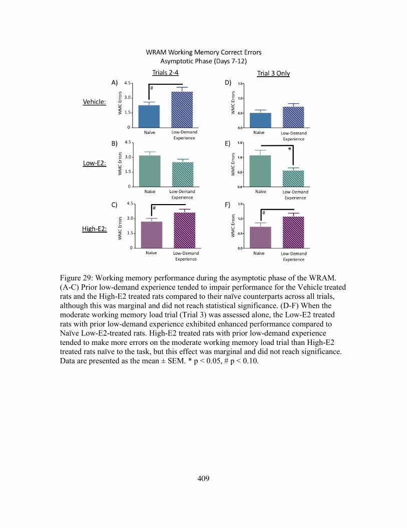
409
Figure 29: Working memory performance during the asymptotic phase of the WRAM. (A-C) Prior low-demand experience tended to impair performance for the Vehicle treated rats and the High-E2 treated rats compared to their naïve counterparts across all trials, although this was marginal and did not reach statistical significance. (D-F) When the moderate working memory load trial (Trial 3) was assessed alone, the Low-E2 treated rats with prior low-demand experience exhibited enhanced performance compared to Naïve Low-E2-treated rats. High-E2 treated rats with prior low-demand experience tended to make more errors on the moderate working memory load trial than High-E2 treated rats naïve to the task, but this effect was marginal and did not reach significance. Data are presented as the mean ± SEM. * p < 0.05, # p < 0.10.

410
Figure 30: Delayed memory retention on the WRAM. (A-F) Following a four-hour delay between trials 2 and 3, all subjects, regardless of treatment group and prior experience, made more WMC errors on Trial 3 after the delay compared to Trial 3 WMC performance on the last day of baseline testing (Day 12). Data are presented as the mean ± SEM. * p < 0.05, # p < 0.10.

411
Figure 31: Total errors committed on the first day of DMS testing. (A-C, E-F) Low-E2 and High-E2 treated rats with prior high-demand experience made fewer errors than their naïve counterparts on trials 3-6 of DMS. (D,G) The Vehicle treated rats with high-demand experience made fewer errors than naïve Vehicle treated rats only on trials 4-6, indicating that even with high-demand experience, Ovx rats without exogenous E2 treatment require more exposures to the task before exhibiting enhanced performance compared to rats naïve to the task. Data are presented as the mean ± SEM. * p < 0.05, # p < 0.10.

412
Figure 32: Total errors committed on Days 2-8 of DMS testing. (A-F) Low-E2 treated rats with prior high demand experience exhibited enhanced performance on trials 2-6 of the DMS across baseline testing compared to Low-E2 treated rats naïve to the task. The High-E2 treated rats with prior experience tended to make fewer errors than the High-E2 treated rats naïve to the task. Data are presented as the mean ± SEM. * p < 0.05, # p < 0.10.

413
Figure 33: Delayed memory retention on the DMS. (A-D) Following a four-hour delay between trials 1 and 2, Vehicle treated rats and Low-E2 treated rats did not exhibit an impairment following the delay, regardless of prior testing experience. (E-F) Both High-E2 treated rats naïve to the DMS and High-E2 treated rats with prior high-demand experience showed a significant increase in total errors committed prior to finding the platform on the post-delay trial. Data are presented as the mean ± SEM. * p < 0.05, # p < 0.10.

414
Figure 34: Body weights. Body weight (grams) increased for all subjects between Ovx and Pump Insertion time points. Following hormone treatment initiation, Ovx rats treated with Low-E2 or High-E2 decreased body weights compared to Ovx-Vehicle treated rats. This difference between Vehicle treated rats and E2 treated rats persisted through the end of the experiment. Data are presented as the mean ± SEM. * p < 0.05, # p < 0.10.

415
Figure 35: Uterine wet weights at euthanization. All groups were Ovx. The Low-E2 and High-E2 treated rats exhibited significantly heavier uterine wet weights at sacrifice compared to the Vehicle treated rats. Data are presented as the mean ± SEM. * p < 0.05, # p < 0.10.

416
Figure 36: Study 1 Water radial-arm maze. A) During Early Acquisition, each DRSP dose enhanced WMC performance compared to Vehicle on a high working memory load trial. B) This benefit was extended to WMI errors for DRSP-Low and DRSP-Medium doses compared to Vehicle. C) During the Asymptotic Phase, DRSP-Medium continued to enhance performance compared to Vehicle treatment for WMI errors on the maximum working memory load trial.

417
Figure 37: Study 1 Water radial-arm maze delay. Vehicle (A) and DRSP-Low (B) treated rats showed impaired delayed memory retention following a six hour delay between trials 2 and 3. DRSP-Medium (C) and DRSP-High (D) groups trended toward a delay-related impairment, but this effect did not reach significance in these groups.

418
Figure 38: Study 1 Morris water maze. Across Days 2-5 (A), the DRSP-Medium group showed enhanced performance compared to the Vehicle group (C). All groups localized to the target quadrant during the probe trial (E).

419
Figure 39: Study 1 Body weight and uterine weights. There were no treatment differences in body weight or uterine horn weights at sacrifice.

420
Figure 40: Study 1 Western blot protein analysis. There were no Treatment differences in IGF1-R expression, GAD65 expression, or GAD67 expression for any brain region evaluated.

421
Figure 41: Study 2 Water radial-arm maze. A) The DRSP group showed enhanced WMI performance on the high working memory load trial compared to Vehicle, EE-Low+DRSP, and EE-High+DRSP groups. B) EE-High+DRSP treatment attenuated EE-High impairment on the high working memory load trial during Late Acquisition. C) EE-Low+DRSP impaired memory on the maximum working memory load trial compared to EE-alone treatment during Late Acquisition.

422
Figure 42: Study 2 Water radial-arm maze delay. A) The Vehicle group tended to show impairments following a six-hour delay B,C, E) EE-Low, EE-High, and EE-Low+DRSP groups showed significantly impaired working memory performance following a six-hour delay. D,F) The DRSP group and the EE-High+DRSP group did not significantly increase errors committed following a six-hour delay.

423
Figure 43: Study 2 Morris water maze. A) There were no Treatment differences across MM Days 2-5. B) All groups localized to the target quadrant during the probe trial.

424
Figure 44: Open field task performance. The EE-High group showed increased locomotor activity compared to the Vehicle group and the EE-High+DRSP group showed increased locomotor activity compared to the DRSP group. EE-treated groups showed evidence of anxiolytic behavior.

425
Figure 45: Study 2 Body weight and uterine weight. A) EE-treated groups weighed significantly less than Vehicle groups. The DRSP group weighed more than EE-Low+DRSP and EE-High+DRSP groups, and tended to weigh more than the Vehicle group. Of note, two-group comparisons were not made between DRSP and EE-Low or EE-High groups. B) Uterine weights from rats treated with EE weighed significantly more at euthanasia. Uterine weights from the DRSP group weighed less than EE-Low+DRSP and EE-High+DRSP groups, but did not differ from Vehicle. Of note, two-group comparisons were not made between DRSP and EE-Low or EE-High groups.

426
Figure 46: Diagram of the surgical manipulations performed. Sham-treated rats had intact an uterus and ovaries, Ovx-treated rats had an intact uterus, Hysterectomy-treated rats had intact ovaries, and Ovx-Hysterectomy-treated rats had both the uterus and ovaries removed.

427
Figure 47: Study Timeline. Rats were tested on a battery of spatial working and reference memory tasks six weeks following surgical manipulations. One week following the last behavioral assay, subjects were euthanized.

428
Figure 48: Representative images from consecutive days of vaginal cytology monitoring. Rats in the Sham and Hysterectomy groups (i.e., rats with intact ovaries) showed normal 4-5 day estrous cycling when examined 2-3 weeks after surgery. Ovx and Ovx-Hysterectomy rats (i.e., rats that experienced surgical removal of the ovaries) showed mostly blank vaginal smear cytology two weeks after surgery, indicating successful removal of the ovaries.

429
Figure 49: Working memory correct errors during the early acquisition phase of the water radial-arm maze (days 1-4). A) There was a main effect of Surgery across all days and trials within the early acquisition block. B) There was a main effect of Surgery across Days 1-4 on the high working memory load trials (Trials 3+4). C) There was a Surgery x Trial interaction on high working memory load trials (Trials 3+4), with D) a main effect of Surgery evident on Trial 4 alone across Days 1-4. * = p < 0.05 ^ = p < 0.10

430
Figure 50: Working memory incorrect errors during the early acquisition phase of the water radial-arm maze (days 1-4). A) There was a Surgery x Trial interaction was present across Days 1-4 for WMI errors. B) There was a Surgery x Trial interaction on the high working memory load trials (Trials 3+4), with C) a main effect of Surgery on Trial 4 alone. * = p < 0.05

431
Figure 51: Working memory correct errors during the asymptotic phase of the water radial-arm maze (days 9-12). A marginal Surgery x Trial interaction was present for WMC errors. ^ = p < 0.10

432
Figure 52: Working memory incorrect errors during the asymptotic phase of the water radial-arm maze (days 9-12). A) A main effect of Surgery was evident across all days and trials within the asymptotic phase block. B) A marginal Surgery x Trial interaction was found across all days and trials in the asymptotic phase. C) A main effect of Surgery was shown for the high working memory load trials (Trials 3+4) for the asymptotic phase block. * = p < 0.05 ^ = p < 0.10

433
Figure 53: Water radial-arm maze delay data. A-D demonstrate working memory correct errors of each surgery group on Trial 3 of the last day of baseline testing (Day 12) compared to the number of working memory correct errors committed on Trial 3 of the delay day (first post-delay trial on Day 13). * = p < 0.05 ^ = p < 0.10

434
Figure 54: Weekly body weights were collected beginning on the date of surgery (baseline) and continued through euthanization. Body weights began to significantly diverge between rats with ovaries (i.e., Sham and Hysterectomy groups) versus rats without intact ovaries (i.e., Ovx and Ovx-Hysterectomy groups) beginning two weeks after surgical manipulation. * = p < 0.05

435
Figure 55: Uterine wet weights from rats that had an intact uterus throughout the experiment (Sham and Ovx groups). Uteri from Sham-operated rats weighed significantly more than those of the Ovx group, indicating successful removal of the ovaries in the Ovx group and a lack of ovarian hormone stimulation of the uterus. * = p < 0.05

436
Figure 56: Representative ovarian micrographs stained with hematoxylin and eosin Y-phloxine B from A) a sham-operated rat and B) a Hysterectomy-operated rat. Low magnification images are 3.45x magnification with a scale bar of 500 um. High magnification images are at 20x magnification with a scale bar of 100 um. Corpora lutea are designated by orange diamonds. Primordial follicles are denoted with a green arrow. Secondary follicles are marked with blue stars.

437
Figure 57: Healthy ovarian follicle count estimates from groups that had intact ovaries throughout the experiment (Sham and Hysterectomy groups). Primordial, primary, and secondary follicles did not differ between groups. Hysterectomy-operated rats had marginally fewer antral follicles compared to Sham-operated rats. Corpora lutea counts did not differ between groups. ^ = p < 0.10

438
Figure 58: Circulating serum ovarian hormone levels from blood collected at euthanization. A) Sham-operated rats had more 17b-estradiol compared to Ovx and Ovx-Hysterectomy groups, and marginally more 17b-estradiol compared to the Hysterectomy group. B) Ovx and Ovx-Hysterectomy groups had non-detectable levels of androstenedione at euthanization. Hysterectomy-operated rats had less circulating androstenedione compared to Sham-operated rats. C) Ovx and Ovx-Hysterectomy groups had less progesterone than Sham and Hysterectomy groups. Hysterectomy-operated rats had more progesterone present than Sham-operated rats. D) Ovx and Ovx-Hysterectomy groups had undetectable levels of AMH at euthanization. AMH levels in Hysterectomy and Sham rats did not differ from each other. E) Both groups without ovaries had increased LH levels compared to Sham and Hysterectomy groups, indicating successful ovary removal. LH levels did not differ between Sham and Hysterectomy groups. F) Both groups without ovaries had increased FSH levels compared to Sham and Hysterectomy groups, indicating successful ovary removal. The Hysterectomy group had increased FSH levels compared to the Sham group.

439
Figure 59: WRAM Learning Phase (Adult Cohort; 7 mo old, tested 6 weeks after surgery). A) WMC errors across trials B) WMI errors across trials C) Hysterectomy rats made more WMC errors on the moderate working memory load trial, Trial 3, compared to Ovx-Hysterectomy rats. D) Hysterectomy rats made more WMI errors on the maximum working memory load trial, Trial 4, compared to Sham rats. Ovx-Hysterectomy rats tended to make fewer errors than Hysterectomy rats. E) There were no group differences for WMC errors on Trial 4. F) There were no group differences for WMI errors on Trial 4. ** p < 0.01 * p < 0.05 ^ p < 0.10

440
Figure 60: WRAM Asymptotic Phase (Adult Cohort; 7 mo old, tested 6 weeks after surgery). There were no group differences for any planned comparison during the Asymptotic phase of testing. A) WMC errors across trials B) WMI errors across trials C) Trial 3 WMC errors D) Trial 3 WMI errors E) Trial 4 WMC errors F) Trial 4 WMI errors

441
Figure 61: WRAM Delayed Memory Retention (Adult Cohort; 7 mo old, tested 6 weeks after surgery). Sham rats did not show a significant delay-induced working memory impairment (A). Ovx rats (B), Hysterectomy rats (C), and Ovx-Hysterectomy rats (D) committed more WMC errors on Trial 3 following a six-hour delay compared to the last day of baseline performance on Trial 3. ** p < 0.01 * p < 0.05 ^ p < 0.10

442
Figure 62: Morris Water Maze (Adult Cohort; 7 mo old, tested 6 weeks after surgery). A) There were no group differences in any planned comparison across days. B) Each Surgery group spent a greater proportion of total distance swimming in the Target (NE) Quadrant compared to the Opposite (SW) Quadrant during the probe trial, indicating spatial localization to the platform location. **** p < 0.0001

443
Figure 63: Peripheral Measures (Adult Cohort; 7 mo old, tested 6 weeks after surgery). A) Body weights were tracked weekly across the experiment. At euthanasia, ovary-intact groups (Sham, Hysterectomy) weighed less than Ovx groups (Ovx, Ovx-Hysterectomy). B) Uterine wet weight was obtained at euthanasia; Sham rats had increased uterine weights compared to Ovx rats. C) Ovary wet weights obtained at euthanasia did not differ between Sham and Hysterectomy rats at this time point. **** p < 0.0001 ** p < 0.01 * p < 0.05

444
Figure 64: Ovarian Follicle Counts Estimates (Adult Cohort; 7 mo old, tested 6 weeks after surgery). There were no differences in Primordial Follicles (A), Primary Follicles (B), Secondary Follicles (C), Antral Follicles (D), or Corpora Lutea Counts (E) at this time point.

445
Figure 65: Serum Hormone Levels (Adult Cohort; 7 mo old, tested 6 weeks after surgery). A) Progesterone levels were significantly decreased in Ovx groups (Ovx, Ovx-Hysterectomy) when compared to ovary-intact groups (Sham, Hysterectomy). B) Androstenedione levels did not differ at this time point between Sham and Hysterectomy groups. C) E2 levels did not differ at this time point between Sham and Hysterectomy groups. ** p < 0.01.

446
Figure 66: WRAM Learning Phase (Middle-Aged Cohort; 12 mo old, tested 7 months after surgery). A) WMC errors across trials B) WMI errors across trials C) Hysterectomy rats made more WMC errors on the moderate working memory load trial, Trial 3, compared to Ovx and Ovx-Hysterectomy rats. Sham rats tended to make fewer errors than Hysterectomy rats. D) Hysterectomy rats made more WMI errors on the maximum working memory load trial, Trial 4, compared to Sham rats and Ovx rats. E) There were no group differences for WMC errors on Trial 4. F) Hysterectomy rats made more WMI errors on Trial 4 compared to Ovx-Hysterectomy rats. ** p < 0.01 * p < 0.05 ^ p < 0.10

447
Figure 67: WRAM Asymptotic Phase (Middle-Aged Cohort; 12 mo old, tested 7 months after surgery). There were no group differences for any planned comparison during the Asymptotic phase of testing. A) WMC errors across trials B) WMI errors across trials C) Trial 3 WMC errors D) Trial 3 WMI errors E) Trial 4 WMC errors F) Trial 4 WMI errors

448
Figure 68: WRAM Delayed Memory Retention (Middle-Aged Cohort; 12 mo old, tested 7 months after surgery). A) Sham rats tended to make more WMC errors on Trial 3 following a six-hour delay compared to performance on the last day of baseline testing. B) Ovx rats did not significantly increase in errors following a six-hour delay. C) Hysterectomy rats tended to make more WMC errors on Trial 3 following a six-hour delay compared to performance on the last day of baseline testing. D) Ovx-Hysterectomy rats made more WMC errors on Trial 3 following a six-hour delay compared to performance on the last day of baseline testing. * p < 0.05 ^ p < 0.10

449
Figure 69: Morris Water Maze (Middle-Aged Cohort; 12 mo old, tested 7 months after surgery). A) There were no group differences in any planned comparison across days. B) Each Surgery group spent a greater proportion of total distance swimming in the Target (NE) Quadrant compared to the Opposite (SW) Quadrant during the probe trial, indicating spatial localization to the platform location. **** p < 0.0001

450
Figure 70: Peripheral Measures (Middle-Aged Cohort; 12 mo old, tested 7 months after surgery). A) Body weights were tracked weekly across the experiment. At euthanasia, ovary-intact groups (Sham, Hysterectomy) weighed less than Ovx groups (Ovx, Ovx-Hysterectomy). The Ovx group tended to weigh less than the Ovx-Hysterectomy group. B) Uterine wet weight was obtained at euthanasia; Sham rats had increased uterine weights compared to Ovx rats. C) Ovary wet weights obtained at euthanasia did not differ between Sham and Hysterectomy rats at this time point. **** p < 0.0001 *** p < 0.001 ^ p < 0.10

451
Figure 71: Ovarian Follicle Counts (Middle-Aged Cohort; 12 mo old, tested 7 months after surgery). There were no differences in Primordial Follicles (A), Primary Follicles (B), Secondary Follicles (C), Antral Follicles (D), or Corpora Lutea Counts (E) at this time point.

452
Figure 72: Serum Hormone Levels (Middle-Aged Cohort; 12 mo old, tested 7 months after surgery). A) Progesterone levels were significantly decreased in Ovx groups (Ovx, Ovx-Hysterectomy) when compared to ovary-intact groups (Sham, Hysterectomy). B) Androstenedione levels did not differ at this time point between Sham and Hysterectomy groups. C) E2 levels did not differ at this time point between Sham and Hysterectomy groups. ** p < 0.01 * p < 0.05.

453
Figure 73: WRAM Learning Phase (Aged Cohort; 18 mo old, tested 12 months after surgery). A) WMC errors across trials B) WMI errors across trials C) There were no group differences for WMC errors on Trial 3 for any planned comparison. D) There were no group differences for WMI errors on Trial 3 for any planned comparison. E) Hysterectomy rats tended to make more WMC errors on Trial 4 when compared to Sham rats and compared to Ovx rats. F) Hysterectomy rats made more WMI errors on Trial 4 compared to Sham rats and compared to Ovx rats; Ovx-Hysterectomy rats tended to make fewer WMI errors than Hysterectomy rats on Trial 4 at this time point. ** p < 0.01 * p < 0.05 ^ p < 0.10

454
Figure 74: WRAM Asymptotic Phase (Aged Cohort; 18 mo old, tested 12 months after surgery). A) WMC errors across trials B) WMI errors across trials C) There were no group differences for WMC errors on Trial 3 for any planned comparison. D) There were no group differences for WMI errors on Trial 3 for any planned comparison. E) Hysterectomy rats tended to make fewer WMC errors on Trial 4 compared to Ovx rats, although this effect did not reach statistical significance. F) There were no group differences for WMI errors on Trial 4 for any planned comparison. ^ p < 0.10

455
Figure 75: WRAM Delayed Memory Retention (Aged Cohort; 18 mo old, tested 12 months after surgery). A) Sham rats made more WMC errors on Trial 3 following a six-hour delay compared to performance on the last day of baseline testing. Ovx rats (B), Hysterectomy rats (C), and Ovx-Hysterectomy rats (D) did not exhibit a significant increase in WMC errors following a six-hour delay compared to performance on the last day of baseline testing within each group. ** p < 0.01

456
Figure 76: Morris Water Maze (Aged Cohort; 18 mo old, tested 12 months after surgery). A) There were no group differences in any planned comparison across days. B) Each Surgery group spent a greater proportion of total distance swimming in the Target (NE) Quadrant compared to the Opposite (SW) Quadrant during the probe trial, indicating spatial localization to the platform location. *** p < 0.001 ** p < 0.01 * p < 0.05

457
Figure 77: Peripheral Measures (Aged Cohort; 18 mo old, tested 12 months after surgery). A) Body weights were tracked weekly across the experiment. At euthanasia, Sham rats weighed less than Ovx rats. B) Uterine wet weight was obtained at euthanasia; Sham rats had increased uterine weights compared to Ovx rats. C) Ovary wet weights obtained at euthanasia tended to be increased in Hysterectomy rats compared to Sham rats, after correcting for body weight. **** p < 0.0001 * p < 0.05 ^ p < 0.10

458
Figure 78: Ovarian Follicle Counts (Aged Cohort; 18 mo old, tested 12 months after surgery). There were no differences in Primordial Follicles (A), Primary Follicles (B), Antral Follicles (D), or Corpora Lutea Counts (E) at this time point. Hysterectomy rats tended to have fewer Secondary Follicles compared to Sham rats, although this effect did not reach statistical significance (C). ^ p < 0.10

459
Figure 79: Serum Hormone Levels (Aged Cohort; 18 mo old, tested 12 months after surgery). A) Progesterone levels were significantly decreased Ovx rats when compared to Sham rats and compared to Hysterectomy rats. Ovx-Hysterectomy rats tended to have lower Progesterone levels compared to Hysterectomy rats. B) Androstenedione levels did not differ at this time point between Sham and Hysterectomy groups. C) E2 levels did not differ at this time point between Sham and Hysterectomy groups. **** p < 0.0001 *** p < 0.001 ^ p < 0.10.

460
Figure 80: FosB and ΔFosB Expression in the Entorhinal Cortex. A) The Adult cohort did not exhibit changes in FosB or B) ΔFosB expression in the Entorhinal Cortex. C) Middle-Aged Ovx rats had less FosB expression compared to Sham rats and compared to Hysterectomy rats. Ovx-Hysterectomy rats had less FosB expression compared to Hysterectomy rats and marginally less FosB expression compared to Sham rats. D) Middle-Aged Hysterectomy rats tended to exhibit greater ΔFosB expression compared to Ovx rats and Ovx-Hysterectomy rats. E) Aged rats did not exhibit altered FosB expression. F) Aged Ovx rats had less ΔFosB expression than Sham rats.

461
Figure 81: Age Effects on FosB and ΔFosB Expression in the Frontal Cortex. There was no significant age-related change in FosB expression (A, C, E, G) or ΔFosB expression for any surgery group (B, D, F, H).

462
Figure 82: Age Effects on FosB and ΔFosB Expression in the Dorsal Hippocampus. There were no age-related changes in FosB expression with age in any surgery group (A, C, E, G). However, all subjects, regardless of surgery type, exhibited an decrease in ΔFosB expression in the Aged time point, when rats were 18 months old (B, D, F, H).

463
Figure 83: Age Effects on FosB and ΔFosB Expression in the Entorhinal Cortex. A) Sham rats showed a transient increase in FosB expression in Middle Age. B) Sham rats had more ΔFosB expression in the Middle-Aged cohort and the Aged cohort compared to the Adult cohort. C) Ovx rats did not exhibit changes in FosB expression across the three cohorts in the Entorhinal Cortex. D) ΔFosB expression tended to increase in Middle-Aged Ovx rats compared to Adult Ovx rats. E) Hysterectomy rats tended to exhibit greater FosB expression in the Middle-Aged cohort compared to the Adult cohort. However, FosB expression was lower in the Aged cohort than the Middle-Aged cohort. F) Adult Hysterectomy rats had lower ΔFosB expression compared to the Middle-Aged cohort and tended to have lower ΔFosB expression compared to the Aged cohort. G) Ovx-Hysterectomy rats did not exhibit changes in FosB expression across the three cohorts. H) Ovx-Hysterectomy rats did not exhibit changes in ΔFosB expression across the three cohorts.

464
Figure 84: Age and Follicular Depletion Hormone Correlation Summary. Young-Vehicle rats (8-12 months of age) exhibited positive correlations between serum estrone, androstenedione, and progesterone levels, as well as ovarian follicle reserve estimates with ChAT-IR neurons in the vertical/diagonal bands within the basal forebrain, such that higher circulating levels of these hormones were associated more ChAT-IR cells in this brain region. Furthermore, higher 17b-estradiol levels were associated with more GAD65 and GAD67 expression in the Ventral Hippocampus. Age-matched counterparts that underwent VCD-induced follicular depletion at 6 months of age did not exhibit these correlations; however, there was a negative association between estrone levels and Entorhinal Cortex GAD67 expression, where higher estrone levels were associated with less GAD67 expression. Middle-Aged-VCD rats (14-18 months of age) that underwent VCD-induced follicular depletion at 12 months of age exhibited a positive correlation between androstenedione levels and ChAT-IR neurons in the medial septum, but Vehicle-treated, age-matched counterparts did not exhibit any associations between circulating hormone levels and brain measures.

465
Figure 85: Age and Follicular Depletion Behavior Correlation Summary. Young-Vehicle rats (8-12 months old) exhibited a positive correlation between Entorhinal Cortex GAD65 expression and MM Swim Distance, such that higher GAD65 expression was associated with a longer swim distance to platform, indicating poorer performance. Conversely, Young-VCD rats showed a negative association between serum 17b-estradiol levels and working memory errors on the WRAM, where higher 17b-estradiol was associated with fewer working memory errors on the maximum working memory load trial, an indication of better performance. There were no associations between behavior measures and physiological measures in Middle-Aged rats (14-18 months old), regardless of follicular depletion status.

466
Figure 86: Hysterectomy Aging Ovarian Follicle Age Effect. Adult rats had more primordial (A), primary (B), secondary (C), and antral (D) follicles compared to Middle-Aged and Aged rats. Secondary follicles also significantly decreased from Middle-Age to Aged time points (C). Corpora lutea counts did not significantly differ with age.

467
Figure 87: Hysterectomy Aging Serum Hormone Age Effect. A) Progesterone levels did not significantly change with age B) Androstenedione levels were increased in Aged rats compared to Adult and Middle-Aged time points C) Adult rats had higher serum 17b-estradiol levels compared to Aged rats.

468
APPENDIX A
TINTO DE VERANO

469
TINTO DE VERANO
Ingredients
1 bottle red wine 1 bottle lemon Fanta or Squirt soda Garnish: Cinnamon and lemon twist
Instructions Pour 8 oz of red wine into a glass. Combine with citrus-flavored soda to taste. Sprinkle
cinnamon on top. Add a lemon twist for garnish, if desired. Serve chilled or with ice.
Enjoy!
Ingredientes
1 botella de vino tinto 1 botella de refresco con sabor cítrico Adorno: Canela y cáscara de limón
Instrucciones
Sírvase una copa de vino tinto (250 mL). Combine con el refresco cítrico a su gusto.
Espolvoree la canela encima. Añada una cáscara de limón si desea. Sírvase frío o con
hielo. ¡Disfrute!
Copyright © 2022 FDOKUMEN