Managing Menopause Managing Menopause
84
Volume 36, Number 9 • volume 36, numéro 9 September • septembre 2014 Supplement 2 • supplément 2 Publications mailing agreement #40026233. Return undeliverable Canadian copies and change of address notifications to SOGC Subscriptions Services, 780 Echo Dr. Ottawa, Ontario K1S 5R7. Journal of Obstetrics and Gynaecology Canada The official voice of reproductive health care in Canada Le porte-parole officiel des soins génésiques au Canada Journal d’obstétrique et gynécologie du Canada Abstract S1 Chapter 1: Assessment and Risk Management of Menopausal Women S6 Chapter 2: Cardiovascular Disease S16 Chapter 3: Menopausal Hormone Therapy and Breast Cancer S23 Chapter 4: Vasomotor Symptoms S31 Chapter 5: Urogenital Health S35 Chapter 6: Prescription Therapeutic Agents S42 Chapter 7: Ongoing Management of Menopausal Women and Those With Special Considerations S51 Chapter 8: Sexuality and Menopause S59 Chapter 9: Complementary and Alternative Medicine (CAM) S74 Managing Menopause Managing Menopause guide.medlive.cn
-
Upload
khangminh22 -
Category
Documents
-
view
1 -
download
0
Transcript of Managing Menopause Managing Menopause

Volume 36, Number 9 • volume 36, numéro 9 September • septembre 2014 Supplement 2 • supplément 2
Publications mailing agreement #40026233. Return undeliverable Canadian copies and change of address notifications to SOGC Subscriptions Services, 780 Echo Dr. Ottawa, Ontario K1S 5R7.
Journal of Obstetrics and Gynaecology CanadaThe official voice of reproductive health care in Canada
Le porte-parole officiel des soins génésiques au CanadaJournal d’obstétrique et gynécologie du Canada
Abstract . . . . . . . . . . . . . . . . . . . . . . . . . . . . . . . . .S1
Chapter 1:Assessment and Risk Management of Menopausal Women . . . . . . . . . . . . . . . . S6
Chapter 2:Cardiovascular Disease . . . . . . . . . . . . . . S16
Chapter 3:Menopausal Hormone Therapy and Breast Cancer . . . . . . . . . . . . . . . . . . . . S23
Chapter 4:Vasomotor Symptoms . . . . . . . . . . . . . . . . S31
Chapter 5:Urogenital Health . . . . . . . . . . . . . . . . . . . . . S35
Chapter 6:Prescription Therapeutic Agents . . . . . . S42
Chapter 7:Ongoing Management of Menopausal Women and Those With Special Considerations . . . . . . . . . . S51
Chapter 8:Sexuality and Menopause . . . . . . . . . . . . S59
Chapter 9:Complementary and Alternative Medicine (CAM) . . . . . . . . . . . . . . . . . . . . . . S74
ManagingMenopauseManaging
Menopause
guide.medlive.cn

Editor-in-Chief / Rédacteur en chef Timothy Rowe
CPL Editor / Rédactrice PPPVyta Senikas
Translator / TraducteurMartin Pothier
Assistant Editor / Rédactrice adjointeJane Fairbanks
Editorial Assistant / Adjointe à la rédactionDaphne Sams
Editorial Office / Bureau de la rédactionJournal of Obstetrics and Gynaecology Canada Room D 405ABC Women's Hospital4500 Oak StreetVancouver BC V6H [email protected]: (604) 875-2424 ext. 5668Fax: (604) 875-2590http://www.jogc.com
The Journal of Obstetrics and Gynaecology Canada (JOGC) is owned by the Society of Obstetricians and Gynaecologists of Canada (SOGC), published by the Canadian Psychiatric Association (CPA), and printed by The Lowe-Martin Group, Ottawa, ON.
Le Journal d’obstétrique et gynécologie du Canada (JOGC), qui relève de la Société des obstétriciens et gynécologues du Canada (SOGC), est publié par l’Association des psychiatres du Canada (APC), et imprimé par The Lowe-Martin Group, Ottawa (Ontario).
Publications Mail Agreement no. 40026233. Return undeliverable Canadian copies and change of address notices to SOGC, JOGC Subscription Service,780 Echo Dr., Ottawa ON K1S 5R7. USPS #021-912. USPS periodical postage paid at Champlain, NY, and additional locations. Return other undeliverable copies to International Media Services, 100 Walnut St., #3, PO Box 1518, Champlain NY 12919-1518.
Numéro de convention poste-publications 40026233. Retourner toutes les copies canadiennes non livrées et les avis de changement d’adresse à la SOGC, Service de l’abonnement au JOGC, 780, promenade Echo, Ottawa (Ontario), K1S 5R7. Numéro USPS 021-912. Frais postaux USPS au tarif des périodiques payés à Champlain (NY) et autres bureaux de poste. Retourner les autres copies non livrées à International Media Services, 100 Walnut St., #3, PO Box 1518, Champlain (NY) 12919-1518.
ISSN 1701-2163 The cover image was obtained through the: Society of Obstetricians and Gynaecologists of Canada (SOGC)
guide.medlive.cn

SEPTEMBER JOGC SEPTEMBRE 2014 l S1
SOGC CLINICAL PRACTICE GUIDELINE
Abstract
Objective: To provide updated guidelines for health care providers on the management of menopause in asymptomatic healthy women as well as in women presenting with vasomotor or urogenital symptoms and on considerations related to cardiovascular disease, breast cancer, urogynaecology, and sexuality .
Outcomes: Lifestyle interventions, prescription medications, and complementary and alternative therapies are presented according to their efficacy in the treatment of menopausal symptoms. Counselling and therapeutic strategies for sexuality concerns in the peri- and postmenopausal years are reviewed . Approaches to the identification and evaluation of women at high risk of osteoporosis, along with options for prevention and treatment, are presented in the companion osteoporosis guideline .
Evidence: Published literature was retrieved through searches of PubMed and The Cochrane Library in August and September 2012 with the use of appropriate controlled vocabulary (e .g ., hormone therapy, menopause, cardiovascular diseases, and sexual function) and key words (e .g ., hormone therapy, perimenopause, heart disease, and sexuality) . Results were restricted to clinical practice guidelines, systematic reviews, randomized control trials/controlled clinical trials, and observational studies . Results were limited to publication dates of 2009 onwards and to material in English or French . Searches were updated on a regular basis and incorporated in the guideline until January 5, 2013. Grey (unpublished) literature was identified through searching the websites of health technology assessment and health technology assessment-related agencies, national and international medical specialty societies, and clinical practice guideline collections .
No. 311, September 2014 (Replaces No. 222, January 2009)
SOGC CLINICAL PRACTICE GUIDELINE
Managing Menopause
This document reflects emerging clinical and scientific advances on the date issued and is subject to change. The information should not be construed as dictating an exclusive course of treatment or procedure to be followed. Local institutions can dictate amendments to these opinions. They should be well documented if modified at the local level. None of these contents may be reproduced in any form without prior written permission of the SOGC.
This clinical practice guideline has been prepared by the Menopause and Osteoporosis Working Group, reviewed by the Clinical Practice Gynaecology and Family Physician Advisory Committees, and approved by the Executive and Council of the Society of Obstetricians and Gynaecologists of Canada.
PRINCIPAL AUTHORS
Robert Reid, MD, Kingston ON
Beth L . Abramson, MD, Toronto ON
Jennifer Blake, MD, Toronto ON
Sophie Desindes, MD, Sherbrooke QC
Sylvie Dodin, MD, Quebec QC
Shawna Johnston, MD, Kingston ON
Timothy Rowe, MB BS, Vancouver BC
Namrita Sodhi, MD, Toronto ON
Penny Wilks, ND, Dundas ON
Wendy Wolfman, MD, Toronto ON
MENOPAUSE AND OSTEOPOROSIS WORKING GROUP
Michel Fortier, MD (Co-Chair), Quebec QC
Robert Reid, MD (Co-Chair), Kingston ON
Beth L . Abramson, MD, Toronto ON
Jennifer Blake, MD, Toronto ON
Sophie Desindes, MD, Sherbrooke QC
Sylvie Dodin, MD, Quebec QC
Lisa Graves, MD, Toronto ON
Bing Guthrie, MD, Yellowknife NT
Aliya Khan, MD, Hamilton ON
Shawna Johnston, MD, Kingston ON
Timothy Rowe, MB BS, Vancouver BC
Namrita Sodhi, MD, Toronto ON
Penny Wilks, ND, Dundas ON
Wendy Wolfman, MD, Toronto ON
The literature searches and bibliographic support for this guideline were undertaken by Becky Skidmore, Medical Research Analyst, Society of Obstetricians and Gynaecologists of Canada .
Acknowledgement: Claudio N . Soares, MD, PhD, Toronto ON
Disclosure statements have been received from all contributors .
Key Words: Menopause, estrogen, vasomotor symptoms, urogenital symptoms, mood, memory, cardiovascular diseases, breast cancer, lifestyle, nutrition, exercise, estrogen therapy, complementary therapies, progestin, androgen, menopausal hormone therapy, hormones, estrogen, testosterone, menopause, depression, antidepressants, sexualityJ Obstet Gynaecol Can 2014;36(9 eSuppl A):S1–S80
guide.medlive.cn

S2 l SEPTEMBER JOGC SEPTEMBRE 2014
Managing Menopause
Values: The quality of the evidence in this document was rated using the criteria described by the Report of the Canadian Task Force on Preventive Health Care (Table 1) .
SUMMARY STATEMENTS AND RECOMMENDATIONS
Chapter 1: Assessment and Risk Management of Menopausal Women
Recommendations for Patients
1 . Women aged 51 to 70 should consume 7 servings of vegetables and fruits, 6 of grain products, 3 of milk and alternatives, and 2 of meat and alterna tives daily . (III-A)
2 . A diet low in sodium and simple sugars, with substitution of unsaturated fats for saturated and trans fats, as well as increased consumption of fruits, vegetables, and fibre, is recommended. (I-A)
3 . Routine vitamin D supplementation and calcium intake for all Canadian adults year round is recommended . (I-A)
4 . Achieving and maintaining a healthy weight throughout life is recommended . (I-A)
5 . Women aged 18 to 64 should accumulate at least 150 minutes of moderate to vigorous aerobic physical activity per week in bouts of 10 minutes or more . (I-A)
Recommendations for Health Care Providers1. A waist circumference ≥ 88 cm (35 in) for women is associated
with an increased risk of health problems such as diabetes, heart disease, and hypertension and should be part of the initial assessment to identify risk . (II-2A)
2 . Tobacco-use status should be updated for all patients on a regular basis, (I-A) health care providers should clearly advise patients to quit, (I-C) the willingness of patients to begin treatment to achieve abstinence (quitting) should be assessed, (I-C) and every tobacco user who expresses the willingness to begin treatment to quit should be offered assistance . (I-A)
3 . Blood pressure should be assessed and controlled as women go through menopause . (II-2B) If the systolic blood pressure is ≥ 140 mmHg and/or the diastolic blood pressure is ≥ 90 mmHg, a specific visit should be scheduled for the assessment of hypertension . (III-A)
4. Women ≥ 50 years of age or postmenopausal and those with addi-tional risk factors, such as current cigarette smoking, diabetes, and arterial hypertension, should have lipid-profile screening done. (II-2A)
5 . A cardiovascular risk assessment using the Framingham Risk Score should be completed every 3 to 5 years for women aged 50 to 75 . (II-2A)
6 . A history of past pregnancy complications (preeclampsia, gestational hypertension, gestational diabetes, placental abruption, idiopathic preterm delivery, and/or fetal growth restriction) should be elicited since it can often predict an increased risk for premature cardiovascular disease and cardiovascular death and may inform decisions about the need for screening . (II-2B)
Chapter 2: Cardiovascular Disease
Recommendations1 . Health care providers should not initiate hormone therapy for the
sole purpose of preventing cardiovascular disease (coronary artery disease and stroke) in older postmenopausal women since there are no data to support this indication for hormone therapy . (I-A)
2 . The risk of venous thromboembolism increases with age and obesity, in carriers of a factor V Leiden mutation, and in women with a history of deep vein thrombosis . Transdermal therapy is associated with a lower risk of deep vein thrombosis than oral therapy and should be considered only if the benefits outweigh the risks . (III-C) Health care providers should abstain from prescribing oral hormone therapy for women at high risk of venous thromboembolism . (I-A)
3 . Health care providers should initiate other evidence-based therapies and interventions to effectively reduce the risk of cardiovascular disease events in women with or without vascular disease . (I-A)
4 . Risk factors for stroke (obesity, hypertension, elevated cholesterol levels, diabetes, and cigarette smoking) should be addressed in all postmenopausal women . (I-A)
5 . If prescribing hormone therapy to older postmenopausal women, health care providers should address cardiovascular risk factors; low- or ultralow-dose estrogen therapy is preferred . (I-B)
6 . Health care providers may prescribe hormone therapy to diabetic women for the relief of menopausal symptoms . (I-A)
Chapter 3: Menopausal Hormone Therapy and Breast Cancer
Recommendations 1 . Health care providers should periodically review the risks and
benefits of prescribing hormone therapy to a menopausal woman in light of the association between duration of use and breast cancer risk . (I-A)
ABBREVIATIONSAI aromatase inhibitorCAD coronary artery diseaseCAM complementary and alternative medicineCEE conjugated equine estrogensCHD coronary heart disease CVD cardiovascular diseaseDVT deep vein thrombosisEPT combined estrogen/progestin therapyET estrogen therapyHABITS Hormonal replacement therapy after breast cancer —
Is it safe?HERS Heart and Estrogen/progestin Replacement StudyHR hazard ratioHSDD hypoactive sexual desire disorderHT hormone therapyIMT intima–media thicknessMPA medroxyprogesterone acetateNAMS North American Menopause SocietyNHP natural health productPOF premature ovarian failureRR relative riskSERM selective estrogen-receptor modulatorSHBG sex-hormone-binding globulinSNRI serotonin–norepinephrine reuptake inhibitorSSRI selective serotonin reuptake inhibitorUTI urinary tract infectionVMS vasomotor symptomsVTE venous thromboembolismWHI Women’s Health Initiative
guide.medlive.cn

SEPTEMBER JOGC SEPTEMBRE 2014 l S3
ABSTRACT
2 . Health care providers may prescribe hormone therapy for menopausal symptoms in women at increased risk of breast cancer with appropriate counselling and surveillance . (I-A)
3 . Health care providers should clearly discuss the uncer tainty of risks associated with systemic hormone therapy after a diagnosis of breast cancer in women seeking treatment for distressing symptoms (vasomotor symptoms or vulvovaginal atrophy) . (I-B)
Chapter 4: Vasomotor Symptoms
Recommendations
1. Lifestyle modifications, including reducing core body temperature, regular exercise, weight management, smoking cessation, and avoidance of known triggers such as hot drinks and alcohol, may be recommended to reduce mild vasomotor symptoms . (I-C)
2 . Health care providers should offer hormone therapy, estrogen alone or combined with a progestin, as the most effective therapy for the medical management of menopausal symptoms . (I-A)
3 . Progestins alone or low-dose oral contraceptives can be offered as alternatives for the relief of menopausal symptoms during the menopausal transition . (I-A)
4 . Non-hormonal prescription therapies, including certain antidepressant agents, gabapentin, and clonidine, may afford some relief from hot flashes but have their own side effects. These alternatives can be considered when hormone therapy is contraindicated or not desired . (I-B)
5. There is limited evidence of benefit for most complementary and alternative approaches to the management of hot flashes. Without good evidence for effectiveness, and in the face of minimal data on safety, these approaches should not be recommended . Women should be advised that, until January 2004, most natural health products were introduced into Canada as “food products” and did not fall under the regulatory requirements for pharmaceutical products . As such,
most have not been rigorously tested for the treatment of moderate to severe hot flashes, and many lack evidence of efficacy and safety. (I-B)
6 . Estrogen therapy can be offered to women who have undergone surgical menopause for the treatment of endometriosis . (I-A)
Chapter 5: Urogenital Health
Recommendations
1 . Conjugated estrogen cream, an intravaginal sustained-release estradiol ring, and low-dose estradiol vaginal tablets are recommended as effective treatment for vaginal atrophy . (I-A)
2 . Routine progestin co-therapy is not required for endometrial protection in women receiving vaginal estrogen therapy in an appropriate dose . (III-C)
3 . Vaginal lubricants may be recommended for subjective symptom improvement of dyspareunia . (II-2B)
4 . Because systemic absorption of vaginal estrogen is minimal, its use is not contraindicated in women with contraindications to systemic estrogen therapy, including recent stroke and thromboembolic disease. (III-C) However, there are currently insufficient data to recommend its use in women with breast cancer who are receiving aromatase inhibitors (where the goal of adjuvant therapy is a complete absence of estrogen at the tissue level) . Its use in this circumstance needs to be dictated by quality-of-life concerns after discussion of possible risks . (III-C)
5 . Systemic estrogen therapy should not be recommended for the treatment of postmenopausal urge or stress urinary incontinence given the lack of evidence of therapeutic benefit. (I-A) Vaginal estrogen may, however, be recommended, particularly for the management of urinary urge incontinence . (II-1A)
6 . As part of the management of stress incontinence, women should be encouraged to try non-surgical options, including weight loss (in obese women). (I-A) Pelvic floor physiotherapy, with or without biofeedback, (II-1B) weighted vaginal cones, (II-2B) functional
Table 1. Key to evidence statements and grading of recommendations, using the ranking of the Canadian Task Force on Preventive Health CareQuality of evidence assessment* Classification of recommendations†
I: Evidence obtained from at least one properly randomized controlled trial
A . There is good evidence to recommend the clinical preventive action
II-1: Evidence from well-designed controlled trials without randomization
B . There is fair evidence to recommend the clinical preventive action
II-2: Evidence from well-designed cohort (prospective or retrospective) or case–control studies, preferably from more than one centre or research group
C. The existing evidence is conflicting and does not allow to make a recommendation for or against use of the clinical preventive action; however, other factors may influence decision-making
II-3: Evidence obtained from comparisons between times or places with or without the intervention . Dramatic results in uncontrolled experiments (such as the results of treatment with penicillin in the 1940s) could also be included in this category
D . There is fair evidence to recommend against the clinical preventive action
E . There is good evidence to recommend against the clinical preventive action
III: Opinions of respected authorities, based on clinical experience, descriptive studies, or reports of expert committees
L. There is insufficient evidence (in quantity or quality) to make a recommendation; however, other factors may influence decision-making
*The quality of evidence reported in these guidelines has been adapted from The Evaluation of Evidence criteria described in the Canadian Task Force on Preventive Health Care .
†Recommendations included in these guidelines have been adapted from the Classification of Recommendations criteria described in the Canadian Task Force on Preventive Health Care .
Woolf SH, Battista RN, Angerson GM, Logan AG, Eel W . Canadian Task Force on Preventive Health Care . New grades for recommendations from the Canadian Task Force on Preventive Health Care . CMAJ 2003;169:207–8 .
guide.medlive.cn

S4 l SEPTEMBER JOGC SEPTEMBRE 2014
Managing Menopause
electrical stimulation, (I-B) and/or intravaginal pessaries (II-2B) can also be recommended .
7. Behavioural modification, (II-2B) functional electrical stimulation, (II-1B) and antimuscarinic therapy (I-A) are recommended for the treat ment of urge urinary incontinence .
8 . Vaginal estrogen therapy can be recommended for the prevention of recurrent urinary tract infections in postmenopausal women . (I-B)
Chapter 6: Prescription Therapeutic Agents
No recommendations
Chapter 7: Ongoing Management of the Menopausal Woman and Those With Special Considerations
Recommendations
1 . Any unexpected vaginal bleeding that occurs after 12 months of amenorrhea is considered postmenopausal bleeding and should be investigated . (I-A)
2 . Cyclic (at least 12 days per month) or continuous progestogen therapy should be added to estrogen therapy if women have an intact uterus; physicians should monitor adherence to the progestogen therapy . (I-A)
3 . Hormone therapy should be offered to women with premature ovarian failure or early menopause, (I-A) and its use until the natural age of menopause should be recommended . (III-B)
4 . Estrogen therapy can be offered to women who have undergone surgical menopause for the treatment of endometriosis . (I-A)
Chapter 8: Sexuality and Menopause
Summary Statements
1 . Sexuality is multifactorial, biopsychological, and affected by psychological, relationship, physical, social, and cultural factors, as well as aging and hormonal decline . (II-2)
2 . Although desire, arousal, orgasm, and satisfaction decline with meno-pause and age, the potential for sexual satisfaction still exists . (II-2)
3 . Decreased desire is the most common sexual problem in middle-aged women, occurring in up to 40% . However, only 12% of menopausal women are personally distressed by the problem . (II-2)
4 . As women age, their sexual function is affected by the presence or absence of a partner and the partner’s health and sexual function . (II-2)
5 . Surgically menopausal women have a higher prevalence of decreased libido and distress than naturally menopausal women . (II-2)
6 . Satisfying sexual contact improves quality of life as women age . (II-2)
7 . Medical and psychological illnesses and their treatment can affect sexuality . (II-2)
8 . Women may be reluctant to discuss their sexuality with physicians . (II-2)
Recommendations1 . Health care providers should acknowledge that aging women are
sexual and have sexual needs but may be unwilling to initiate a discussion about problems . (III-A)
2 . Health care providers should be sensitive to changes in sexuality in women as they age or illnesses develop . (III-A)
3 . Women and their partners should be educated about the changes affecting sexuality that occur as women age . (III-A)
4 . If women have decreased sexual desire and are not distressed, no therapy is necessary . (III-B)
FEMALE SEXUAL DYSFUNCTIONSSummary Statements1 . Determinants of sexual function involve central and peripheral
mechanisms . (II-2)
2 . Both testosterone and estrogen have effects on sexual function . (I)
3 . The serum testosterone level is not a useful marker for the diagnosis of sexual dysfunction . (II-1)
4 . Estrogen’s primary action is on maintenance of vaginal and vulvar health . (II-2)
Recommendations1 . Vulvovaginal atrophy should be addressed in all middle-aged women
who complain of sexual dysfunction . (I-A)
2 . Serum androgen measurements should not be used in the assessment of female sexual dysfunction . (I-A)
EVALUATION AND TREATMENTSummary Statements1 . Taking a brief sexual history is part of the evaluation of the
menopausal woman . (III)
2 . Female dysfunction can be categorized into desire, arousal, pain, and orgasm problems . These categories often overlap . (II-2)
3 . Low desire with distress is most common in mid-life women . (II-2)
4 . Vaginal atrophy occurs in 50% of women within 3 years of menopause and is a common cause of sexual pain in menopausal women . (II-1)
5 . Sexual pain results in a cascade of detrimental sexual symptoms . (II-1)
6 . The treatment of sexual dysfunctions involves a multifaceted approach addressing medical, psychological, and relationship issues . (III)
7 . Transdermal testosterone therapy has been shown to increase desire, arousal, and frequency of satisfactory sexual events and to decrease personal distress for women with surgical and also natural menopause, but there are no approved products for this indication in Canada . (I)
Recommendations1 . Health care providers should include a short sexual screening
history as part of a medical history of menopausal women . Interventions should be undertaken only if the patient is distressed about the problem . (III-A)
2 . The patient’s problem should be categorized according to desire, arousal, pain, or orgasm problems in order to facilitate treatment and triage care . (III-A)
3 . Vaginal estrogen therapy should be prescribed for postmenopausal women with vulvovaginal atrophy and sexual dysfunction . (I-A)
4 . For women with decreased sexual desire the current best options include management of vaginal atrophy, addressing treatable contributing factors, and sexual counselling . (I-A)
5 . For women with signs or symptoms of vulvovaginal atrophy who cannot use estrogens, vaginal dilators, lubricants, and moisturizers should be offered . (III-B)
6. Clinicians should endorse the benefits of alternative forms of sexual contact for patients unable to have penetration . (III-A)
guide.medlive.cn
SEPTEMBER JOGC SEPTEMBRE 2014 l S5
ABSTRACT
SPECIAL CLINICAL SITUATIONS
Summary Statements
1 . Sexual dysfunction is common in depressed patients and those taking selective serotonin reuptake inhibitors . (I)
2 . Premature loss of ovarian function may be attended by sexual dysfunction related to loss of both ovarian estrogen and androgen production at a time of life when sexual activity is normally heightened . (II-1)
3 . Survivors of breast cancer using aromatase inhibitors have more sexual dysfunction due to vulvovaginal atrophy than do women using tamoxifen or control subjects . (II-1)
Recommendations
1 . Patients using selective serotonin reuptake inhibitors should be educated about the effects of these medications on sexuality and informed that these effects are reversible when the medications are stopped . (III-B)
2 . Patients with premature ovarian failure should be asked about their sexual health . (III-B)
3 . Patients with breast cancer using aromatase inhibitors should be advised that these medications may have sexual effects . (II-2B) The decision to use intravaginal estrogen therapy for severe vulvovaginal atrophy in such women needs to be based on quality-of-life
considerations and should be made only after a discussion of the uncertain effects on breast cancer recurrence . (III-I)
Chapter 9: Complementary and Alternative Medicine
Summary Statement
1 . Health Canada’s Licensed Natural Health Products Database lists products approved for use in women with menopausal symptoms that have been evaluated for safety, efficacy, and quality. (III)
Recommendation
1. Health care providers may offer identified complementary and alternative medicine with demonstrated efficacy for mild menopausal symptoms . (I-B)
This document’s Abstract was previously published in:
J Obstet Gynaecol Can 2014;35(9):830–833
guide.medlive.cn

S6 l SEPTEMBER JOGC SEPTEMBRE 2014
CHAPTER 1
Assessment and Risk Management of Menopausal Women
Menopause is an important milestone and may be one of the first times a woman seeks medical advice
around issues of long-term health promotion and disease prevention. Women typically begin to experience menopausal symptoms between 40 and 58 years of age, spending at least one third of their lives after menopause.1 The 3 main causes of illness and disability in developed countries for postmenopausal women are CVD, cancer, and osteoporosis-associated fractures.2 As outlined in the following chapters of this update to the Canadian Consensus Conference on Menopause3 and the Canadian Consensus Conference on Osteoporosis,4 many of the risk factors for the conditions prevalent among older women are modifiable through changes in lifestyle.
DIET AND MENOPAUSE
Healthy eating can prevent or reduce certain conditions that may develop during and after menopause, including obesity, type 2 diabetes, heart disease, certain types of cancer, and osteoporosis. All perimenopausal women should be reminded of healthy eating and should use menopause as an opportunity to make healthy changes.
Canada’s Food GuideSince 1942, Canada’s Food Guide has provided advice on food selection and nutritional health. The latest version of Eating Well with Canada’s Food Guide5 offers information on the amount and types of food recom mended, according to age and sex, and emphasizes the importance of combining regular physical activity with healthy eating. The guide encourages Canadians to focus on vegetables, fruits, and whole grains, to include milk, meat, and their alternatives, and to limit foods that are high in calories, fat (especially trans fats), sugar, and salt. According to Canada’s Food Guide, women ages 51 to 70 should consume 7 servings of vegetables and fruits, 6 of grain prod ucts, 3 of milk and alternatives, and 2 of meat and alterna tives daily. Within each food group, specific recommendations exist. For vegetables and fruits it is recommended that Canadians eat at least 1 dark green and 1 orange vegetable each day,
prepare vegetables and fruits with little or no added fat, sugar, or salt, and have vegetables and fruits more often than juice. For grain products it is recommended that at least half of the daily grain products be whole grain and that grain-product choices be low in fat, sugar, and salt. For milk and alternatives, drinking skim, 1%, or 2% milk each day and selecting lower-fat milk alternatives is recommended. For meat and alternatives, alternatives such as beans, lentils, and tofu, eating at least 2 food-guide servings of fish each week, and selecting lean meat and alternatives prepared with little or no added fat or salt are recommended.
My Food Guide, an interactive component on Health Canada’s Eating Well with Canada’s Food Guide website, helps users per sonalize dietary information in 9 steps according to age, sex, and food preferences. Also on the website is My Food Guide Servings Tracker, a printable tool for Canadians at different ages to track daily food choices and compare them with the recommendations in the Food Guide. Another tool linked to Health Canada’s website is the Eating and Activity Tracker (at http://www.eatracker.ca), developed by the Dietitians of Canada to help people check food and activity choices, analyze recipes, and plan meals; the tool provides guidance as users make healthy changes in both eating and physical activity.
Diet and a woman’s risk of heart diseaseObservational studies have shown a relationship between serum cholesterol levels and CVD,6 and dietary measures to lower these levels are an important part of disease prevention.7 According to the Canadian Cholesterol Guidelines8 a diet low in sodium and simple sugars, with substitution of unsaturated fats for saturated and trans fats, as well as increased consumption of fruits, vegetables, and fibre is recommended. Evidence from the Nurses’ Health Study suggests that replacing dietary saturated fats and trans fatty acids with non-hydrogenated, monounsaturated, and polyunsaturated fats may be more effective in reducing CVD risk than reducing overall fat intake in women.9 The adequate daily intake for sodium in healthy Canadians 51
guide.medlive.cn

SEPTEMBER JOGC SEPTEMBRE 2014 l S7
CHAPTER 1: Assessment and Risk Management of Menopausal Women
CHAPTER 1to 70 years of age should be 1300 mg; the upper limit is 2300 mg, which is equivalent to 1 level teaspoon of table salt.10 Caloric restriction to achieve and maintain ideal body weight is also advised.8 Interestingly, the dietary content (percentages of protein, carbohydrate, and fat) required to maintain a healthy weight does not appear to matter as long as caloric intake is reduced.8,11 For individuals with hypertriglyceridemia, a reduction in the intake of alcohol and refined carbohydrates, in conjunction with increased consumption of omega-3 and omega-6 polyunsaturated fats, is indicated.8 Potential dietary sources of these fats include cold-water fish (salmon, tuna, and halibut), flax seeds, and flaxseed oil. Other dietary strategies to reduce CVD risk include increasing the intake of flavonoids12,13
(found in vegetables, fruits, and tea), dietary folate14 (found in vegetables, fruits, and grains), and soy products15 (sources of isoflavones). Although a recent publication questioned whether calcium supplements might increase the risk for coronary heart disease,16 further analysis of the WHI data does not support such an association.17
Diet and bone health Vitamin D and calcium are essential to preventing osteoporosis and may reduce the risk of other health conditions, such as diabetes and immune system disorders. Although exposure to sunlight provides vitamin D, Canadians are at risk of seasonal vitamin D deficiency because winter sunlight in northern latitudes above 35° does not contain enough ultraviolet B for vitamin D production.18,19 Supplementation is necessary to obtain adequate levels, as dietary intake has minimal impact. Osteoporosis Canada18 recommends routine vitamin D supplementation for all Canadian adults year round: healthy adults at low risk for vitamin D deficiency (those under age 50, without osteoporosis or conditions affecting vitamin D absorption or action) require 400 to 1000 IU daily, whereas those over 50 and younger adults at high risk (with osteoporosis, multiple fractures, or conditions affecting vitamin D absorption) require at least 800 to 1000 IU daily; for people who need added supplementation to reach optimal vitamin D levels, doses up to the current “tolerable upper intake level” of 2000 IU are safely taken without medical supervision.
Calcium in combination with vitamin D significantly reduces the occurrence of fractures.20,21
For women ages 19 to 50 Osteoporosis Canada recommends 1000 mg of calcium intake daily, whereas for women over the age of 50 the recommendation is 1200 mg daily.22 Although the tolerable upper limit for daily calcium intake from all sources (diet and supplements) is 2500 mg, calcium supplements exceeding 1200 mg daily often cause
gastrointestinal symptoms, such as constipation, which limits compliance. Osteoporosis Canada suggests that yogurt, cheese, calcium-fortified beverages, puddings, and custards are all adequate calcium sources. For those intolerant to dairy products, calcium-fortified soy, almond, and rice beverages, calcium-fortified orange juice, and canned salmon or canned sardines are good alternatives.
Diet and a woman’s risk of cancerIt has been estimated that 30% to 40% of all cancer deaths each year are linked to diet and physical activity, including being overweight or obese, while another third are caused by tobacco products.23 Although it is not clear exactly how excess body fat, consuming too many calories, and lack of physical activity raise cancer risk, the link to cancers, such as breast (among women who have gone through menopause),24 colon and rectum, endometrium, esophagus, and kidney is undeniable. The Canadian Cancer Society25 recommends that everyone achieve and maintain a healthy weight throughout life by following Canada’s Food Guide on healthy eating. Eating regular meals, cutting back on portions, filling half your plate with vegetables, a quarter with grain products, and a quarter with meat or alternatives, using smaller dishes, and limiting processed meat and red meat are among their recommendations.
EXERCISE AND MENOPAUSE
Regular exercise is a simple and effective way to improve both physical and mental health in menopausal women. Among the many benefits of exercise are improvements in serum lipid levels and weight and protection from CVD, osteoporosis, diabetes, and breast cancer.24 Women who exercise regularly report lower levels of stress, lighter menstrual periods, and fewer menopausal symptoms, including night sweats and hot flashes.
The Canadian Society for Exercise Physiology26 recommends that women ages 18 to 64 accumulate at least 150 minutes of moderate to vigorous aerobic physical activity per week in bouts of 10 minutes or more. It also recommends adding muscle- and bone-strengthening activities using major muscle groups at least 2 days per week. Exercise regimens should be tailored to a woman’s age, ability, and individual preference. A sedentary woman should be advised to start slowly and progress gradually. Osteoporosis Canada27 recommends a minimum of 20 to 30 minutes of weight-bearing exercise, such as walking, dancing, step aerobics, or running, at moderate to vigorous intensity, on most days to improve heart health and bone strength. Strength training with free weights, machines, or exercise bands, or by using body weight as resistance, is
guide.medlive.cn

S8 l SEPTEMBER JOGC SEPTEMBRE 2014
Managing Menopause
recommended 2 to 3 days per week (2 to 3 sets of 8 to 12 repetitions) to improve muscle and bone strength, posture, and mobility.27 Balance training, with such activities as tai chi and yoga, 2 to 3 days per week can also improve mobility and balance, leading to fewer falls and reduced fracture risk.27 Posture training, including safe movements and awareness of position and posture should be practised at all times to reduce the risk of back injuries, falls, and fractures.27
LIFESTYLE MODIFICATION, RISK ASSESSMENT, AND CARDIOVASCULAR HEALTH
Menopause should be seen as an opportunity for health care providers to assess and modify cardiovascular risk. Despite overall health care improvements, risk of heart disease in women continues to be underestimated. CVD remains the leading cause of death and an important contributor to ill ness and disability among women: half of all postmenopausal women will have CVD, and one third will die from it. Eighty percent of all CVD is preventable, which is significant considering that CVD costs Canadians $22 billion annually; the early identification and management of cardiovascular risks has the potential to prevent CVD. Health-behaviour interventions remain a cornerstone of chronic disease prevention, including CVD prevention, in women and should be highlighted during health care visits (Appendix).
The INTERHEART study,28 which examined modifiable risk factors across many populations, determined that for women 94% of CVD risk could be attributed to modifiable factors. Factors identified in this study as contributing substantially to increased CVD risk include diabetes mellitus, hyperten sion, abdominal obesity, current smoking, and psychosocial stress. Each of these risks can be reduced through appropri ate choices or interventions, or both.
Past pregnancy complications and risk for CVDThe development of common complications in pregnancy, namely pre-eclampsia, gestational hypertension, gestational diabetes, placental abruption, idiopathic preterm delivery, and/or fetal growth restriction, has been shown to predict a woman’s risk of premature CVD and CVD-related death.29 The 2011 Update for the American Heart Association’s Evidence-Based Guidelines for the Prevention of CVD in Women, now identifies these complications of pregnancy as relevant in the determination of CVD risk.30
Hypertension diagnosis, assessment, and follow-upBlood pressure in women generally increases after menopause. Menopause-related hormonal changes can
lead to weight gain and make the blood pressure more reactive to salt in the diet, contributing to the pressure changes seen after menopause.31 The blood pressure should be assessed in women at all appropriate visits in order to screen for hypertension, assess cardiovascular risk, and monitor antihypertensive treatment if applicable. Reversible risks for hypertension include obesity, poor dietary habits, high sodium intake, sedentary lifestyle, and high alcohol consumption. Close attention to these factors should occur when menopausal and postmenopausal women are being assessed. If the systolic pressure is ≥ 140 mmHg or the diastolic pressure is ≥ 90 mmHg, or both, a specific visit should be scheduled for the assessment of hypertension according to the Canadian Hypertension Education Program.32 If the pressures are high-normal (130 to 139 mmHg and 85 to 89 mmHg, respectively), annual follow-up is recommended. At the initial visit for the assessment of hypertension, if the pressures are ≥ 140 or ≥ 90 mmHg, or both, at least 2 more readings should be taken during the same visit using a validated device; the first reading should be discarded and the latter 2 averaged. At visit 2 for the assessment of hypertension, patients with macrovascular target organ damage, diabetes mellitus, or chronic kidney disease (glomerular filtration rate < 60 mL/min per 1.73 m2) can be considered hypertensive if the pressures are ≥ 140 and/or ≥ 90 mmHg. Patients without macrovascular target organ damage, diabetes mellitus, or chronic kidney disease can be considered hypertensive if the pressures are ≥ 180 and/or ≥ 110 mmHg. Patients without macrovascular target organ damage, diabetes mellitus, or chronic kidney disease but with lower pressures should undergo further evaluation by means of office manual measurements, ambulatory monitoring, or home measurement. With office manual measurements, patients can be considered hypertensive if the pressures are ≥ 160 or ≥ 100 mmHg averaged across the first 3 visits or if they average ≥ 140 or ≥ 90 mmHg across 5 visits. Hypertensive patients receiving advice on lifestyle modification alone should be seen by the health care provider at 3- to 6-month intervals. Shorter intervals (every 1 or 2 months) are needed for patients with higher pressures.
Dyslipidemia and cardiovascular risk assessment The 2012 Canadian Cardiovascular Society Dyslipidemia Guidelines33 recommend that women ≥ 50 years of age or postmenopausal and those with additional risk factors such as current cigarette smoking, diabetes, and arterial hypertension have a full lipid profile screening every 1 to 3 years. A cardiovascular risk assessment using the Framingham Risk Score should be completed every 3 to 5 years for women ages 50 to 75. If there is a family
guide.medlive.cn

SEPTEMBER JOGC SEPTEMBRE 2014 l S9
CHAPTER 1: Assessment and Risk Management of Menopausal Women
history of premature CVD (i.e., in a first-degree relative < 55 years for men and < 65 years for women) the age parameters should be modified. A risk assessment may also be completed whenever a patient’s expected risk status changes. Younger individuals with more than 1 risk factor for premature CVD may also benefit from a risk assessment to encourage them to improve their lifestyle. The Framingham Risk Score provides a reasonable estimate of the 10-year risk of a major cardiovascular event for a large portion of the Canadian population. However, it does not account for family history of premature CAD, which increases the risk 1.7-fold in women. Despite limitations, assessing the total CVD risk improves the management of blood pressure and blood lipids. The Reynolds Risk Score (http://www.reynoldsriskscore.org), a tool that takes into account both the result of the high-sensitivity C-reactive protein test and family history, may be used as an alternative to the Framingham Risk Score.
In low-risk women, pharmacotherapy should be considered if the LDL cholesterol value is ≥ 5.0 mmol/L or if there is evidence of genetic dyslipidemia (e.g., familial hypercholesterolemia). For intermediate-risk women, treatment should be considered with an LDL cholesterol value ≥ 3.5 mmol/L. Treatment should be considered in all high-risk women, regardless of LDL level, with target LDL cholesterol value of ≤ 2.0 mmol/L or a decrease of 50% or more for optimal risk reduction.
Premenopausal women at riskPolycystic ovary syndrome is an endocrinopathy frequently encountered in women of reproductive age.34 Not only does this disorder affect the quality of life of women during their reproductive years, but it also contributes to illness and death by the time of menopause.34 A cohort of women with this syndrome who were followed for many years after wedge resection revealed that they had a later menopause and an increased prevalence of diabetes (16%) and hypertension (40%).34
It is important for all physicians involved in the care of women with the above conditions to emphasize lifestyle changes and assess cardiac risk every 3 to 5 years with the Framingham Risk Score. Although this score tends to be low in premenopausal women, attention to future risk and modification of lifestyle are especially important for those with polycystic ovary syndrome and gestational diabetes mellitus. Important aspects of cardiovascular risk screening include regular assessment of blood pressure, waist circumference and BMI, lipid profile, fasting glucose and HgA1c levels, and a composite scoring system such as the Framingham risk assessment.
Body weight Health Canada’s Canadian Guidelines for Body Weight Classification in Adults35 is a valuable tool in assessing the risk of health problems associated with being overweight or underweight. This classification system uses waist circumference and BMI.
As women go through menopause their body fat distribution can change, causing an increase in waist circumference. This measure is an indicator of abdominal fat. Excess fat around the waist and upper body (“apple” body shape) is associated with greater health risk than fat located more in the hip and thigh area (“pear” body shape). A waist circumference ≥ 88 cm (35 in) in a woman is associated with an increased risk of health problems, such as diabetes, heart disease, and hypertension.35
Adults with a high BMI (≥ 25 kg/m2; overweight or obese) have a high percentage of body fat, which is associated with increased risk of health problems, such as diabetes, heart disease, hypertension, gallbladder disease, and some forms of cancer. A low BMI (< 18.5 kg/m2; underweight) is associated with health problems, such as osteoporosis, undernutrition, and eating disorders.35
Weight classification can be used as an initial assessment tool to identify women at increased relative risk for disease and death. However, there is considerable variability among women in the risk associated with a specific waist circumference or BMI.35,36 For this reason, the estimation of a woman’s health risk should not be based on these measures alone; these measures should be components of a more comprehensive risk assessment. This assessment could also include, depending on age and other factors, information on the presence of other risk factors, such as hypertension, dyslipidemia, family history of disease, and individual weight history (i.e., patterns of weight gain and loss). In addition, individual health behaviours, such as tobacco use, eating habits, and physical activity patterns require assessment, as do weight-related psychological and social factors.35
Alcohol use and smokingAlcohol use and smoking are both risk factors for many chronic diseases. For Canadians that choose to drink, the Canadian Centre on Substance Abuse37 recommends at most 10 drinks a week for women, with no more than 2 drinks a day. Non-drinking days every week are advised to avoid a habit. It is recommended that women not consume more than 3 drinks on any single occasion. For these guidelines, a drink refers to a 341-mL (12-oz) bottle of 5% alcohol content (beer, cider, or cooler), a 142-mL (5-oz) glass of wine of 12% alcohol content, or a 43-mL (1.5-oz) serving of drink of 40% distilled alcohol content (rye, gin, rum, etc.).
guide.medlive.cn

S10 l SEPTEMBER JOGC SEPTEMBRE 2014
Managing Menopause
Cessation of tobacco use is highly encouraged. The Canadian Action Network for the Advancement, Dissemination and Adoption of Practice-informed Tobacco Treatment38 has put forth smoking cessation guidelines for Canadians. The network recommends that all health care providers update the status of tobacco use for all patients on a regular basis, that they clearly advise patients to quit, that they assess the willingness of patients to begin treatment to achieve abstinence, and that they offer assistance to every tobacco user who expresses the willingness to begin treatment to quit.
Stroke and menopauseStroke is also a leading cause of disability and death among women, especially postmenopausal women. Risk factors for stroke, which are also similar for other forms of vascular disease, include obesity, hyperlipidemia, hypertension, smoking, and diabetes. These risk factors are common among North American women as they enter menopause, and certain segments of the population, such as those of African heritage, are more likely to manifest these risk factors. Risk factors for stroke should be addressed in all menopausal women.
The mainstay for CVD prevention in all women will remain a focus on lifelong patterns of healthy living. This incorporates a balanced, heart-healthy diet, moderate exercise, maintenance of a healthy body weight, limited con-sumption of alcohol, avoidance of smoking, and attention to treatment of known risk factors, such as hypertension, hyperlipidemia, and diabetes mellitus. Identification, evaluation, and treatment of modifiable risk factors are essential in postmenopausal women for CVD prevention.
MOOD AND DEPRESSION
The mid-life years are, for most women, a transitional period with little or no significant impact on psychological well-being. Recent evidence suggests, however, that for some women this time in life represents a period of increased vulnerability for the development of depressive symptoms or a major depressive episode (new-onset or recurrent). Aside from a history of depression, various factors appear to influence or mediate the risk for depression during mid-life years: the presence and severity of VMS (hot flashes, night sweats), the occurrence of stressful life events, sleep problems, and, most importantly, a history of reproductive-related mood sensitivity; that is, premenstrual dysphoria, postpartum depression, or mood symptoms during pregnancy.
A thorough reproductive history is imperative to detect those at high risk for mood disorders and anxiety during mid-life years. To first assess the current presence of
mood symptoms, clinicians may rely on brief, standardized screening tools or use simple questions that have shown high screening sensitivity, such as “Have you been down or depressed, most of the time, for the past 2 weeks?” or “Have you lost your interest in life or pleasure in engaging in your usual activities over the past 2 weeks?”
Although antidepressants remain the treatment of choice for major depression at any time in life, it is important to tailor the treatment strategy to address the multiple symptom domains in depressed midlife women. Regular exercise and a balanced diet may reduce or prevent some of the bothersome symptoms. Evidence-based psychotherapy and hormonal and non-hormonal treatments have a place in the treatment armamentarium to ultimately reduce the overall burden and functional impairment associated with depression in this population.
ADDITIONAL BENEFITS OF LIFESTYLE MODIFICATION
The benefits of a healthy lifestyle extend well beyond opti-mizing cardiovascular health. It has been suggested that a significant increase in verbal memory scores can occur after caloric restriction.39 For best preservation of memory and cognition, women should be advised about the importance of good overall health, including good cardio vascular health, exercise,40 avoidance of excessive alcohol consumption, and measures to reduce the risk of diabetes and hypertension, as well as maintenance of an active mind.
Lifestyle modifications are also essential in the prevention and treatment of osteoporosis. In addition to dietary factors and physical activity, as discussed above, excessive alcohol consumption and tobacco use increase one’s risk for osteoporosis.41 Regular consumption of more than 2 alcoholic drinks a day increases this risk, possibly because alcohol can interfere with the body’s ability to absorb calcium. The exact role of tobacco in osteoporosis is not clearly understood; however, evidence does suggest that tobacco use contributes to weak bones.
Urinary-incontinence risk factors may also be modi fied with lifestyle changes. Those identified include obe sity, amount and type of fluid intake, and smoking. For obese women (mean baseline BMI 38.3 kg/m2), even a reduction in BMI of as little as 5% can result in significant subjective improvement in urine loss.42 The effect of BMI and weight gain was assessed in 30 000 women with new-onset urinary incontinence in the Nurses’ Health Study II.43 Increasingly higher BMI was related to increasing odds of incontinence developing (P for trend < 0.001). The increases were similar for all incontinence types. The odds
guide.medlive.cn

SEPTEMBER JOGC SEPTEMBRE 2014 l S11
CHAPTER 1: Assessment and Risk Management of Menopausal Women
of incontinence also increased with increasing adult weight gain (P for trend < 0.001): the OR for at least weekly incon-tinence developing was 1.44 (95% CI 1.05 to 1.97) among women who had gained 5.1 to 10 kg since early adulthood and 4.04 (95% CI 2.93 to 5.56) among women who had gained more than 30 kg compared with women who had maintained their weight within 2 kg. In the same popula-tion, physical activity was associated with a significant reduction in the risk of urinary incontinence developing. The results appeared to be somewhat stronger for stress uri nary incontinence than for urge incontinence.44
ROLE OF HEALTH CARE PROVIDERS
Health promotion and disease prevention provide the foundation for the comprehensive management of women’s health and are critical strategies for the responsible allocation of limited health care resources. Health care providers must assess cardiovascular risk with the Framingham Risk Score in all postmenopausal women. In addition, there is evidence that a healthy lifestyle leads
to better quality of life and that discussion with health care providers increases the likelihood that a patient will make a healthy change. Family physicians, in addition to obstetricians and gynaecologists, are integral in this process. Screening for risk factors and identification of women at increased risk are critical first steps. Providing advice, encouragement, and support, as well as trusted educational resources (Table 1.1), are funda mental adjuncts to any other medical advice that may be appropriate. An individualized approach to comprehensive care, based on the identified benefits and risks combined with regular reassessment and re-evaluation, will ensure that a woman’s changing needs are met.
RISK ASSESSMENT AND POSTMENOPAUSAL HT
A practical risk-assessment tool for menopausal women can be found at the end of this chapter. Women with moderate to severe menopausal symptoms who are considering HT will benefit from risk assessment for VTE.45–47 Readers are referred to Chapter 4 for details on HT.
Table 1.1 Selected resourcesTopic Organization and details Website*
Breast cancer risk US National Cancer Institute: Breast Cancer Risk Assessment Tool
http://www .cancer .gov/bcrisktool
Disease risk and prevention Siteman Cancer Center, Washington University School of Medicine: Your Disease Risk (health tool, originally developed at the Harvard Center for Cancer Prevention, which covers cancer, diabetes, heart disease, osteoporosis, and stroke)
http://www .yourdiseaserisk .wustl .edu
Exercise Public Health Agency of Canada: physical activity guide http://www .phac-aspc .gc .ca/hp-ps/hl- mvs/pa-ap/index-eng .php
Heart disease and stroke Heart and Stroke Foundation of Canada: information on heart disease, stroke, nutrition, physical activity, smoking cessation, and stress reduction
http://www .hsf .cahttp://www .thehearttruth .ca
Body and Health: heart disease risk calculator based on the Framingham study
http://bodyandhealth .canada .com/health_tools .asp?t=17&text_id=2704
Menopause Society of Obstetricians and Gynaecologists of Canada: clinical practice guidelines, consensus conference reports, and educational material for consumers
http://www .sogc .org http://www .menopauseandu .ca
Nutrition Health Canada: Eating Well with Canada’s Food Guide http://www .healthcanada .gc .ca/foodguide
Dietitians of Canada: EATracker (Eating and Activity Tracker)
http://www .dietitians .ca/Your-Health/Assess-Yourself .aspx
Osteoporosis Osteoporosis Canada: information on diagnosis, prevention, and treatment
http://www .osteoporosis .ca
Sexual health Society of Obstetricians and Gynaecologists of Canada: news and information on sexual-health issues, including a section for women over 50 years of age
http://www .sexualityandu .ca
Weight control US National Heart, Lung, and Blood Institute: Aim for a Healthy Weight (information from the Obesity Education Initiative for patients and the public and for health professionals)
http://www .nhlbi .nih .gov/health/public/heart/obesity/lose_wt/index .htm
*Last accessed March 13, 2014 .
guide.medlive.cn

S12 l SEPTEMBER JOGC SEPTEMBRE 2014
Managing Menopause
Recommendations For Patients1. Women ages 51 to 70 should consume 7 servings of
vegetables and fruits, 6 of grain prod ucts, 3 of milk and alternatives, and 2 of meat and alterna tives. (III-A)
2. A diet low in sodium and simple sugars, with substitution of unsaturated fats for saturated and trans fats, as well as increased consumption of fruits, vegetables, and fibre, is recommended. (I-A)
3. Routine vitamin D supplementation and calcium intake for all Canadian adults year round is recommended. (I-A)
4. Achieving and maintaining a healthy weight throughout life is recommended. (I-A)
5. Women ages 18 to 64 should accumulate at least 150 minutes of moderate to vigorous aerobic physical activity per week in bouts of 10 minutes or more. (I-A)
Recommendations For Health Care Providers1. A waist circumference ≥ 88 cm (35 in) for
women is associated with an increased risk of health problems, such as diabetes, heart disease, and hypertension and should be part of the initial assessment to identify risk. (II-2A)
2. Tobacco-use status should be updated for all patients on a regular basis, (I-A), health care providers should clearly advise patients t quit, (I-C) the willingness of patients to begin treatment to achieve abstinence (quitting) should be assessed, (I-C) and every tobacco user who expresses the willingness to begin treatment to quit should be offered assistance. (I-A)
3. Blood pressure should be assessed and controlled as women go through menopause. (II-2B) If the systolic blood pressure is ≥ 140 mmHg and/or the diastolic blood pressure is ≥ 90 mmHg, a specific visit should be scheduled for the assessment of hypertension. (III-A)
4. Women ≥ 50 years of age or postmenopausal and those with additional risk factors, such as current cigarette smoking, diabetes, and arterial hypertension, should have lipid-profile screening done. (II-2A)
5. A cardiovascular risk assessment using the Framingham Risk Score should be completed every 3 to 5 years for women ages 50 to 75. (II-2A)
6. A history of past pregnancy complications (pre-eclampsia, gestational hypertension, gestational diabetes, placental abruption, idiopathic preterm delivery, and/or fetal growth restriction) should be elicited since it can often predict an increased risk for premature cardiovascular disease and cardiovascular death and may inform decisions about the need for screening. (II-2B)
REFERENCES
1. National Institutes of Health. National Institutes of Health State-of-the-Science Conference statement: management of menopause-related symptoms. Ann Intern Med 2005;142(12 pt 1):1003–13.
2. Women’s Health Initiative [website]. Bethesda, Maryland: Women’s Health Initiative; 2010. Available at: http://www.nhlbi.nih.gov/whi. Accessed 2014 July 8.
3. Bélisle S, Blake J; SOGC Menopause Guidelines Committee. Canadian Consensus Conference on Menopause, 2006 update. SOGC Clinical Practice Guidelines, No. 171, February 2006. J Obstet Gynaecol Can 2006;28:S1–94.
4. Brown JP, Fortier M; SOGC Osteoporosis Guidelines Committee. Canadian Consensus Conference on Osteoporosis, 2006 update. SOGC Clinical Practice Guidelines, No. 172, February 2006. J Obstet Gynaecol Can 2006;28(2 Suppl 1):S95–112.
5. Health Canada. Eating well with Canada’s food guide. Ottawa: Health Canada; 2011. Available at: http://www.healthcanada.gc.ca/foodguide. Accessed 2014 Mar 11.
6. Kannel WB. Metabolic risk factors for coronary heart disease in women: perspective from the Framingham Study. Am Heart J 1987;114:413–9.
7. Hu FB, Manson JE, Willett WC. Types of dietary fat and risk of coronary heart disease: a critical review. J Am Coll Nutr 2001;20:5–19.
8. Genest J, McPherson R, Frohlich J, Anderson T, Campbell N, Carpentier A, et al. 2009 Canadian Cardiovascular Society/Canadian guidelines for the diagnosis and treatment of dyslipidemia and prevention of cardiovascular disease in the adult — 2009 recommendations. Can J Cardiol 2009;25:567–79.
9. Prior JC, Nielsen JD, Hitchcock CL, Williams LA, Vigna YM, Dean CB. Medroxyprogesterone and conjugated oestrogen are equivalent for hot flushes: a 1-year randomized double-blind trial following premenopausal ovariectomy. Clin Sci (Lond) 2007;112:517–25.
10. Health Canada. Sodium in Canada [website]. Ottawa: Health Canada; 2012. Available at: http://www.hc-sc.gc.ca/fn-an/nutrition/sodium/index-eng.php. Accessed 2014 March 11.
11. Sacks FM, Bray GA, Carey VJ, Smith SR, Ryan DH, Anton SD, et al. Comparison of weight-loss diets with different compositions of fat, protein, and carbohydrates. N Engl J Med 2009;360:859–73.
12. Knekt P, Jarvinen R, Reunanen A, Maatela J. Flavonoid intake and coronary mortality in Finland: a cohort study. BMJ 1996;312:478–81.
13. Geleijnse JM, Launer LJ, Hofman A, Pols HA, Witteman JC. Tea flavonoids may protect against atherosclerosis: the Rotterdam Study. Arch Intern Med 1999;159:2170–4.
14. Rimm EB, Willett WC, Hu FB, Sampson L, Colditz GA, Manson JE, et al. Folate and vitamin B6 from diet and supplements in relation to risk of coronary heart disease among women. JAMA 1998;279:359–64.
15. Jenkins DJ, Kendall CW, Jackson CJ, Connelly PW, Parker T, Faulkner D, et al. Effects of high- and low-isoflavone soyfoods on blood lipids, oxidized LDL, homocysteine, and blood pressure in hyperlipidemic men and women. Am J Clin Nutr 2002;76:365–72.
16. Bolland MJ, Grey A, Avenell A, Gamble GD, Reid IR. Calcium supplements with or without vitamin D and risk of cardiovascular events: reanalysis of the Women’s Health Initiative limited access dataset and meta-analysis. BMJ 2011;342:d2040.
17. Shufelt CL, Merz CN, Prentice RL, Pettinger MB, Rossouw JE, Aroda VR, et al. Hormone therapy dose, formulation, route of delivery, and risk of cardiovascular events in women: findings from the Women’s Health Initiative Observational Study. Menopause 2013;21:260–266.
guide.medlive.cn

SEPTEMBER JOGC SEPTEMBRE 2014 l S13
CHAPTER 1: Assessment and Risk Management of Menopausal Women
18. Hanley DA, Cranney A, Jones G, Whiting S, Leslie WD, Cole D, et al; Guidelines Committee of the Scientific Advisory Council of Osteoporosis Canada. Vitamin D in adult health and disease: a review and guideline statement from Osteoporosis Canada. CMAJ 2010;182:E610–8. Epub 2010 Jul 12.
19. Holick MF. Sunlight and vitamin D for bone health and prevention of autoimmune diseases, cancers, and cardiovascular disease. Am J Clin Nutr 2004;80(Suppl):1678S–88S.
20. Storm D, Eslin R, Porter ES, Musgrave K, Vereault D, Patton C, et al. Calcium supplementation prevents seasonal bone loss and changes in biochemical markers of bone turnover in elderly New England women: a randomized placebo-controlled trial. J Clin Endocrinol Metab 1998;83:3817–25.
21. Dawson-Hughes B, Harris SS, Krall EA, Dallal GE. Effect of calcium and vitamin D supplementation on bone density in men and women 65 years of age or older. N Engl J Med 1997;337:670–6.
22. Osteoporosis Canada. Calcium: an important nutrient that builds stronger bones [website]. Toronto: Osteoporasis Canada; 2014. Available at: http://www.osteoporosis.ca/index.php/ci_id/5535/la_id/1.htm. Accessed 2014 Mar 11.
23. World Cancer Research Fund/American Institute for Cancer Research. Food, Nutrition, Physical Activity, and the Prevention of Cancer: a Global Perspective. Washington, DC: American Institute for Cancer Research; 2007:31–40.
24. Sprague BL, Trentham-Dietz A, Egan KM, Titus-Ernstoff L, Hampton JM, Newcomb PA. Proportion of invasive breast cancer attributable to risk factors modifiable after menopause. Am J Epidemiol 2008;168:404–11. Epub 2008 Jun 13.
25. Canadian Cancer Society. Nutrition and fitness [website]. Toronto: Canadian Cancer Society 2014. Available at: http://www.cancer.ca/en/ prevention-and-screening/live-well/nutrition-and-fitness/?region=on. Accessed 2014 Mar 11.
26. Canadian Society for Exercise Physiology. Canadian physical activity guidelines and Canadian sedentary behaviour guidelines [website]. Ottawa: Canadian Society for Exercise Physiology; 2014. Available at: http://www.csep.ca/english/view.asp?x=949. Accessed 2014 Mar 11.
27. Osteoporosis Canada. Exercise for healthy bones [website]. Toronto: Osteoporasis Canada; 2014. Available at: http://www.osteoporosis.ca/index.php/ci_id/5523/la_id/1.htm. Accessed 2014 Mar 11.
28. Yusuf S, Hawken S, Ounpuu S, Dans T, Avezum A, Lanas F, et al; INTERHEART Study Investigators. Effect of potentially modifiable risk factors associated with myocardial infarction in 52 countries (the INTERHEART study): case–control study. Lancet 2004;364:937–52.
29. Smith GN, Pudwell J, Roddy M. The Maternal Health Clinic: a new window of opportunity for early heart disease risk screening and intervention for women with pregnancy complications. J Obsetet Gynaecol Can 2013;35:831–9.
30. Mosca L, Benjamin EJ, Berra K, Bezanson JL, Dolor RJ, Lloyd-Jones D, et al. Effectiveness-based guidelines for the prevention of cardiovascular disease in women — 2011 update. J Am Coll Cardiol 2011;57:1404–23.
31. Pruthi S, Mayo Clinic. Is there a connection between menopause and high blood pressure? [website]. Scottsdale, Arizona: Mayo Clinic; 2013. Available at: http://www.mayoclinic.com/health/menopause-and- high-blood-pressure/AN01463. Accessed 2014 Mar 11.
32. Hypertension Canada. Canadian Hypertension Education Program [website]. Markham, Ontario: Hypertension Canada; 2014. Available at: http://www.hypertension.ca/en/chep/publications-and-archive. Accessed 2014 Mar 11.
33. Anderson TJ, Grégoire J, Hegele RA, Couture P, Mancini GB, McPherson R, et al. 2012 update of the Canadian Cardiovascular Society guidelines for the diagnosis and treatment of dyslipidemia for the prevention of cardiovascular disease in the adult. Can J Cardiol 2013;29:151–67.
34. Carmina E, Lobo RA. Polycystic ovary syndrome (PCOS): arguably the most common endocrinopathy is associated with significant morbidity in women. J Clin Endocrinol Metab 1999;84:1897–9.
35. Health Canada. Canadian Guidelines for Body Weight Classification in Adults [website]. Ottawa: Health Canada; 2014. Available at: http://www.hc-sc.gc.ca/fn-an/nutrition/weights-poids/guide-ld-adult/qa-qr-pub-eng.php. Accessed 2014 Mar 11.
36. NHLBI Obesity Education Initiative Expert Panel on the Identification, Evaluation, and Treatment of Obesity in Adults (US). Clinical Guidelines on the Identification, Evaluation, and Treatment of Overweight and Obesity in Adults: The Evidence Report. Bethesda: National Heart, Lung, and Blood Institute; 1998. Available at: http://www.ncbi.nlm.nih.gov/books/NBK2003. Accessed 2014 Mar 11.
37. Canadian Centre on Substance Abuse. Canada’s Low-Risk Alcohol Drinking Guidelines. Ottawa: CCSA, 2013. Available at: http://www.ccsa.ca/Resource%20Library/2012-Canada- Low-Risk-Alcohol-Drinking-Guidelines-Brochure-en.pdf. Accessed 2014 Mar 11.
38. Canadian Action Network for the Advancement, Dissemination and Adoption of Practice-Informed Tobacco Treatment. Smoking Cessation Knowledge Exchange Network & Clinical Practice Guideline [website]. Toronto: Centre for Addiction and Mental Health; 2012. Available at: https://www.nicotinedependenceclinic.com/English/CANADAPTT/Pages/Home.aspx. Accessed 2014 Mar 12.
39. Witte AV, Fobker M, Gellner R, Knecht S, Flöela A. Caloric restriction improves memory in elderly humans. Proc Natl Acad Sci U S A 2009;106:1255–60.
40. Pines A, Berry EM. Exercise in the menopause — an update. Climacteric 2007;10(Suppl 2):42–6.
41. FRAX: WHO fracture assessment tool [website]. Sheffield, England: World Health Organization Collaborating Centre for Metabolic Bone Diseases, 2012. Available at: http://www.shef.ac.uk/FRAX. Accessed 2014 Mar 12.
42. Subak LL, Johnson C, Whitcomb E, Boban D, Saxton J, Brown JS. Does weight loss improve incontinence in moderately obese women? Int Urogynecol J Pelvic Floor Dysfunct 2002;13:40–3.
43. Townsend MK, Danforth KN, Rosner B, Curhan GC, Resnick NM, Grodstein F. Body mass index, weight gain, and incident urinary incontinence in middle-aged women. Obstet Gynecol 2007;110(2 Pt 1):346–53.
44. Danforth KN, Shah AD, Townsend MK, Lifford KL, Curhan GC, Resnick NM, et al. Physical activity and urinary incontinence among healthy, older women. Obstet Gynecol 2007;109:721–7.
45. Olié V, Canonico M, Scarabin PY. Postmenopausal hormone therapy and venous thromboembolism. Thromb Res 2011;127(Suppl 3):S26–9.
46. Olié V, Plu-Bureau G, Conard J, Horellou MH, Canonico M, Scarabin PY. Hormone therapy and recurrence of venous thromboembolism among postmenopausal women. Menopause 2011;18:488–93.
47. Laliberté F, Dea K, Duh MS, Kahler KH, Rolli M, Lefebvre P. Does the route of administration for estrogen hormone therapy impact the risk of venous thromboembolism? Estradiol transdermal system versus oral estrogen-only hormone therapy. Menopause 2011;18:1052–9.
guide.medlive.cn

S14 l SEPTEMBER JOGC SEPTEMBRE 2014
Managing Menopause
Name Blood pressure Total cholesterol Fasting glucose
Date Waist circumference HDL-C LDL-C 1. Calculate 10-year cardiovascular risk
Framingham Risk Assessment:
Age 30–34 35–39 40–44 45–49 50–54 55–59 60–64 65–69 70–74 75+ Points
Risk points 0 2 4 5 7 8 9 10 11 12 < >
Total cholesterol level (mmol/L) < 4 .1 4 .1–5 .2 5 .2–6 .2 6 .2–7 .2 > 7 .2 Points
0 1 3 4 5 < >
Smoker No Yes Points
0 3 < >
Diabetic No Yes Points
0 4 < >
HDL-C level (mmol/L) > 1 .6 1 .3–1 .6 1 .2–1 .3 0 .9–1 .2 < 0 .9 Points
−2 −1 0 1 2 < >
Systolic BP (mmHg) < 120 120–129 130–139 140–149 150–159 160+ Points
Untreated −3 0 1 2 4 5 < >
Treated −1 2 3 5 6 7
Total points Age TC Smoking Diabetes HDL-C BP < >
Factor
Points
10-year risk
Total risk points
10-year risk (%)
Total risk points
10-year risk (%)
21+ > 30 9 5 .3
20 27 .5 8 4 .5
19 24 .8 7 3 .9
18 21 .5 6 3 .3
17 18 .51 5 2 .8
16 15 .9 4 2 .4
15 13 .7 3 2 .0
14 11 .7 2 1 .7
13 10 .0 1 1 .5
12 8 .6 0 1 .2
11 7 .3 −1 1 .0
10 6 .3 −2 or less < 1
*From 2012 Dyslipidemia Guidelines: Anderson TJ, Grégoire J, Hegele RA, Couture P, Mancini GB, McPherson R, et al . 2012 update of the Canadian Cardiovascular Society guidelines for the diagnosis and treatment of dyslipidemia for the prevention of cardiovascular disease in the adult . Can J Cardiol 2013;29:151–67 .
APPENDIX. MENOPAUSE LIFESTYLE AND RISK ASSESSMENT TOOL
Patients with established coronary or peripheral artery disease, and most patients with diabetes, are automatically considered at high risk . A family history is considered to double the 10-year risk for women . An elevated level of high-sensitivity C-reactive protein increases risk category .
High risk*
Moderate risk*
Low risk*
continued
guide.medlive.cn
SEPTEMBER JOGC SEPTEMBRE 2014 l S15
CHAPTER 1: Assessment and Risk Management of Menopausal Women
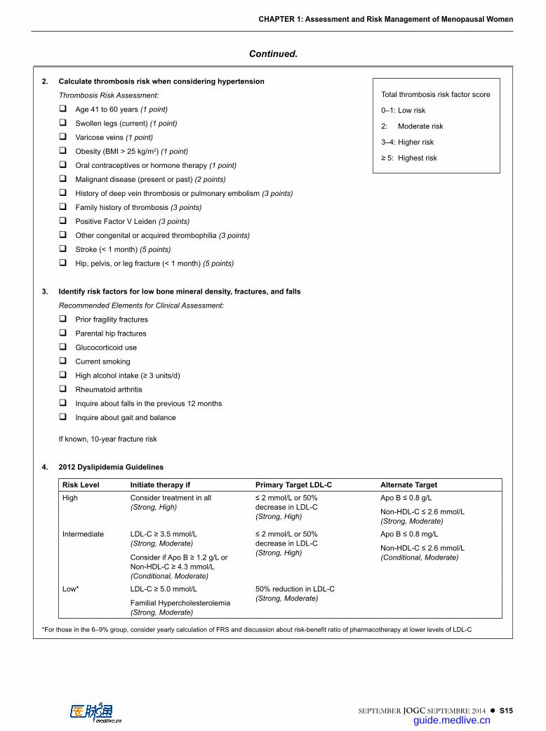
2. Calculate thrombosis risk when considering hypertension
Thrombosis Risk Assessment:
Age 41 to 60 years (1 point)
Swollen legs (current) (1 point)
Varicose veins (1 point)
Obesity (BMI > 25 kg/m2) (1 point)
Oral contraceptives or hormone therapy (1 point)
Malignant disease (present or past) (2 points)
History of deep vein thrombosis or pulmonary embolism (3 points)
Family history of thrombosis (3 points)
Positive Factor V Leiden (3 points)
Other congenital or acquired thrombophilia (3 points)
Stroke (< 1 month) (5 points)
Hip, pelvis, or leg fracture (< 1 month) (5 points)
3. Identify risk factors for low bone mineral density, fractures, and falls
Recommended Elements for Clinical Assessment:
Prior fragility fractures
Parental hip fractures
Glucocorticoid use
Current smoking
High alcohol intake (≥ 3 units/d)
Rheumatoid arthritis
Inquire about falls in the previous 12 months
Inquire about gait and balance
If known, 10-year fracture risk
4. 2012 Dyslipidemia Guidelines
Risk Level Initiate therapy if Primary Target LDL-C Alternate TargetHigh Consider treatment in all
(Strong, High)≤ 2 mmol/L or 50% decrease in LDL-C (Strong, High)
Apo B ≤ 0.8 g/L
Non-HDL-C ≤ 2.6 mmol/L (Strong, Moderate)
Intermediate LDL-C ≥ 3.5 mmol/L (Strong, Moderate)
Consider if Apo B ≥ 1.2 g/L or Non-HDL-C ≥ 4.3 mmol/L (Conditional, Moderate)
≤ 2 mmol/L or 50% decrease in LDL-C (Strong, High)
Apo B ≤ 0.8 mg/L
Non-HDL-C ≤ 2.6 mmol/L (Conditional, Moderate)
Low* LDL-C ≥ 5.0 mmol/L
Familial Hypercholesterolemia (Strong, Moderate)
50% reduction in LDL-C (Strong, Moderate)
*For those in the 6–9% group, consider yearly calculation of FRS and discussion about risk-benefit ratio of pharmacotherapy at lower levels of LDL-C
Continued.
Total thrombosis risk factor score
0–1: Low risk
2: Moderate risk
3–4: Higher risk
≥ 5: Highest risk
guide.medlive.cn

S16 l SEPTEMBER JOGC SEPTEMBRE 2014
CHAPTER 2
Cardiovascular Disease
Consensus is emerging from the controversy and confusion that has occupied the past decade regarding
the effects of postmenopausal HT on CVD. Since the publication of the SOGC’s Canadian Consensus Conference on Meno pause in 2006,1 several publications have shed additional light on this subject.
The areas of agreement can be summarized as follows.1. Menopausal EPT is indicated for relief of
symptoms, but it is not indicated for primary or secondary prevention of CVD; the evidence supports aggressive identification and modification of risk factors as the most effective means of reducing cardiovascular risk.
2. Women who initiate EPT 10 or more years after menopause are at increased risk for adverse cardiac events.
3. Women who initiate EPT shortly after menopause are, in general, at low risk for events in the subsequent few years. Studies have been reassuring regarding safety in this age group.
4. With respect to stroke, increased risk has been identified in all age groups using standard formulations of HT; however, the incidence in young women is extremely low. There is increasing evidence to suggest that lower doses of estrogen, either oral or transdermal, are associated with a lower or no increase in risk.
5. Venous thrombotic events in otherwise healthy women increase in incidence with age and obesity. HT increases the risk; events are associated more with oral than with transdermal preparations and more with EPT than with ET.
6. Women on EPT are reported to have more adverse cardiovascular events than women on ET. Progestogens may differ with respect to cardiovascular risk.
7. There is an emerging literature on the use of a SERM rather than a progestin to protect the uterus from hyperplasia. To date, these agents do not appear to be associated with cardiovascular risk.
Reduction of modifiable risk factors is the most effective strategy for prevention of CVD. The INTERHEART study, a global case–control study examining modifiable risk factors across many populations, determined that for women 94% of CVD risk could be attributed to modifiable factors.2 Factors identified in that study included diabetes mellitus (OR 2.37), hyperten sion (OR 1.91), abdominal obesity (OR 1.62), current smoking (OR 2.87), and psychosocial stress (OR 2.67). Women at pre-existing risk because of elevated Framingham scores or pre-existing metabolic syndrome appear to be at elevated risk of cardiovascular events when on HT, adverse events arising in the first years of use.
Reproductive hormones do have important beneficial effects on risk markers of CVD; however, the outcomes that guide treatment decisions must be confirmed cardiovascular events. The sys temic effects on lipids, hemostasis, and carbohydrate metabolism are well known.3
HT has no role in reduc ing future risks of cardiovascular events in women with established CAD. The HERS secondary prevention trial demonstrated no benefit and an increased risk of early adverse cardiac events in women with known CVD.4 Other research has confirmed that HT fails to delay the progression of disease.5–7
The data on the role of HT for primary prevention of CVD have been the primary reason for the ongoing debate. Whereas data from a variety of sources (epidemiologic stud ies, observational studies, and clinical trials examining sur rogate endpoints) suggested a possible cardioprotective role for estrogen,8,9 the WHI cast doubt on the value of HT in this situation. The first publication from the WHI reported that EPT increased the risk of myocardial infarction and stroke.10 The subsequently published findings showed no statistically significant overall increase in the incidence of coronary events or death among users of the combination of CEE and MPA (EPT).11 There was a significant eleva tion in the incidence of cardiovascular events in EPT users compared with women receiving a placebo in the first year of therapy but not thereafter. The estrogen-only arm of the trial demon strated no evidence of coronary artery benefit or risk (HR 0.63; 95% CI
guide.medlive.cn

SEPTEMBER JOGC SEPTEMBRE 2014 l S17
CHAPTER 2: Cardiovascular Disease
CHAPTER 20.36 to 1.08).12,13 Subsequent subgroup analysis demon-strated a reduction in the total mortality rate in the age group 50 to 59 years (HR 0.70; 95% CI 0.51 to 0.96).14
Observational studies are at risk of confounding. Women who seek HT are better educated and of higher socioeconomic status; thus, they have greater access to other health care resources, from which they may receive treatment for other cardiovascular risk factors, such as dia-betes, hypertension, and hypercholesterolemia.15,16 Those who seek HT are more likely to adhere to other wellness advice: they tend to be leaner, to exercise more often, and to consume more alcohol, which by itself affords a degree of cardioprotection. Women who become sick with other con ditions are more likely to stop HT, so that there appear to be more deaths in non-users or past users than in current users.
Because of the potential for bias in observational studies, RCT data are important to clarify the observation of cardioprotective benefits for HT when started early in postmenopausal women.
Conclusions about the role of HT for primary cardioprevention based on the WHI findings have been challenged because of the greater ages (an average of 63 years) of the participants and the time since loss of ovarian estrogen production (an average of 13 years).17 Time since menopause has been shown to correlate with extent of subclinical atherosclerosis as determined by carotid-wall IMT in populations of women with natural and surgical menopause.18 WHI subsamples were weighted heavily in favour of the inclusion of marginalized and disadvantaged women, and many of the modifiable risks for CVD identified in the INTERHEART study were present in such women. With close to 70% of women in the WHI over the age of 60 years at enrolment, it seems likely that a substantial proportion of the WHI population would have had subclinical CVD. The early increase in the inci dence of cardiac events reported in the EPT arm of the WHI, with no overall difference in the cardiovascular mortality rate, is similar to the effect of HT started in older women in the HERS secondary prevention trial.4 In the EPT arm of the WHI the RR for CAD was 1.68 in the first 2 years after the start of HT, 1.25 at 2 to 5 years, and 0.66 beyond 5 years.
Lobo19 looked at data from 2 clinical trials in which all adverse events were recorded for 4065 young, healthy postmenopausal women started on HT and found no increase in the incidence of either myocardial infarction or stroke in the year after initiation of therapy. These women were not followed for long enough to deter mine whether there might be longer-term benefit or risk.
A “critical-window” or “critical-timing” hypothesis was advanced as a way to try to explain how the use of HT at the onset of menopause could be cardioprotective whereas later initiation could cause adverse coronary events as seen in the WHI.20–23 This the ory suggests that the prothrombotic or plaque-destabilizing effects of HT in women with established CAD may account for an initial increase in the incidence of coronary artery events in older women but that the healthy coronary arteries of younger women benefit from the anti-atherogenic effects of estrogen. Salpeter et al.24,25 performed a meta-analysis of RCTs to assess the effect of HT for at least 6 months on the incidence of CHD events including myocardial infarction and death in younger and older postmenopausal women. They found that HT significantly reduced the incidence of CHD events when initiated in younger (OR 0.68; 95% CI 0.48 to 0.96) but not older (OR 1.03; 95% CI 0.91 to 1.16) menopausal women. The cardiac event rate for younger women seen in this meta-analysis paralleled that seen in the observational Nurses’ Health Study, which followed a cohort of 120 000 women below the age of 55 years. After adjustment for potential confounding variables, such as age, cardiovascular risk fac-tors, and socioeconomic status, HT use was found to be associated with a 40% reduction in the incidence of CHD events.8 As with the HERS4 and WHI10 trials, initiation of HT in older women was associated with an increase in the incidence of adverse CHD events in the first year only.
In addition to the well-publicized RCT, the WHI included an observational arm, which reported lower rates of cardiac events in 17 503 current users of EPT (62% had used EPT for more than 5 years at enrolment) than in 35 551 age-matched control subjects (OR 0.71).26
Grodstein et al.27 re-examined the observational data from the Nurses’ Health Study to determine the effect of differ-ent ages at initiation of HT on the incidence of cardiac events. For women beginning HT near the onset of meno-pause, both ET alone (RR 0.66; 95% CI 0.54 to 0.80) and EPT (RR 0.72; 95% CI 0.56 to 0.92) were associated with a significantly reduced risk of CHD. No sig nificant benefit was observed in women starting HT beyond age 60 or more than 10 years after menopause.
Rossouw et al.14 performed a secondary analysis of the WHI data to determine the impact of years since menopause and age at the time of HT initiation on cardiovascular outcomes. The HR for adverse cardiovascular outcomes was 0.76 in women starting HT less than 10 years after menopause, 1.10 for women starting 10 to 20 years since menopause, and 1.28 for women starting more than 20 years after meno pause (P for trend = 0.02). The HR for
guide.medlive.cn
S18 l SEPTEMBER JOGC SEPTEMBRE 2014
Managing Menopause
total mortality among the women aged 50 to 59 years who were randomly assigned to HT was significantly reduced, at 0.76 (95% CI 0.51 to 0.96).
Ideally this critical-timing hypothesis would be tested in an RCT designed for that specific purpose rather than through post-hoc and subgroup analysis of the data from other tri als.28 Depypere et al.29 estimated the numbers of women needed in any RCT designed to assess possible cardioprotective benefits of HT in newly menopausal women. To detect a 30% difference in women 50 to 54 years old approximately 35 000 women would be required, twice as many as were enrolled in the EPT trial arm. To detect a 10% difference close to 350 000 women would be required. Such a large trial would not be feasible.
The Danish Osteoporosis Study reported on 1006 women who were randomly assigned to cyclic EPT or no treatment (there was no placebo arm) early in menopause and followed for 10 years with a 6-year extension. At the time of analysis 15 of the 504 women in the treated groups had experienced cardiovascular events versus 26 of the 502 control subjects (HR 0.48; 95% CI 0.26 to 0.87, P = 0.015). The primary endpoint was a composite of death, admission to hospital for heart failure, and myocardial infarction. After 10 years of intervention, 16 women in the treatment group had experienced the primary composite endpoint compared with 33 in the control group (HR 0.48; 95% CI 0.26 to 0.87, P = 0.015). There was no increased risk of cancer in general, breast cancer in particular, or stroke.30
Although clinical outcomes are the preferred study endpoint, data on clinically relevant indicators are helpful when a clinical trial is not feasible. Carotid-wall IMT has been followed as an early marker of atherosclerotic disease.9,31,32
The Kronos Early Estrogen Prevention Study (KEEPS) is a multicentre, 5-year clinical trial to evaluate the effectiveness of 0.45 mg/d of CEE, 50 g/wk of transdermal estradiol (both in combi nation with cyclic oral micronized progesterone, 200 mg/d for 12 days each month), and placebo in preventing the pro gression of carotid-wall IMT and the accrual of coronary-artery calcium in women aged 42 to 58 years who are within 36 months of their final menstrual period.33 The investigators have presented preliminary data (http://www.menopause.org/annual-meetings/2012- meeting/keeps-report), unpublished at the time of writing, that do not show any evidence of a difference in IMT progression between the treatment and control groups.
Calcium scores for the coronary arteries, generated by means of electron-beam computed tomography, have been shown to correlate with plaque burden as assessed pathologically
and to have significant predictive value for subsequent cardiac events in symptomatic and asymptomatic adults.34,35 A recent meta-analysis of predictive utility concluded that this score is an independent predictor of subsequent CAD events.36 Most of the data on these scores come from studies on men; data for women must be interpreted with caution at this time.37 With these caveats in mind, it is of interest to consider recent studies of these scores as a surrogate endpoint for CAD in women using or not using HT.38–41 Each study demonstrated evidence of reduced subclinical vascular disease among women who were compliant with HT. The WHI investigators41 performed a substudy on 1064 women aged 50 to 59 years in the estrogen-only arm of the WHI. Coronary-artery calcium scores were significantly lower among the women randomly assigned to ET than among the women assigned to placebo after a mean of 7.4 years of treatment. In women who remained at least 80% adherent to the treatment protocol the OR for a high score in users compared with non-users was 0.39 (P = 0.004).
The National Institute on Aging’s Early versus Late Inter-vention Trial with Estradiol (ELITE) is designed to test the hypothesis that 17β-estradiol therapy will reduce the pro gression of early atherosclerosis if initiated soon after menopause, when the vascular endothelium is relatively healthy, versus later, when the endothelium has lost its responsiveness to estrogen.
Considering all these studies, healthy recently menopausal women who are considering HT for relief of symptoms should be reassured that there does not appear to be significant cardiovascular risk; some argue that there is benefit. However, most women will be using HT for a limited time: a survey of hormone use in the United States before the WHI results were reported revealed that only 3% of women using EPT and only 10% using estrogen alone stayed on their HT for more than 5 years.42 Therefore, the key cardiovascular preventive advice will remain the reduction of modifiable risk factors.
PREMATURE LOSS OF OVARIAN FUNCTION AND CVD
Large numbers of women continue to face early loss of ovarian function because of either surgical oophorectomy or chemotherapy-associated ovarian failure. Several studies have suggested that women have a greater risk for CAD after bilateral oophorectomy.43–48 An increased risk of stroke has been found in women with surgical premature menopause.49 WHI investigators reported that oophorectomized women who subsequently received ET had less coronary-artery calcium accumulation than
guide.medlive.cn

SEPTEMBER JOGC SEPTEMBRE 2014 l S19
CHAPTER 2: Cardiovascular Disease
those who did not receive ET, and they concluded that “the findings are consistent with the thesis that estrogen deficiency associated with bilateral oophorectomy is related to an increased burden of calcified plaque in the coronary arteries that can be countered by the use of HT”.50 This finding supports the need for ET after premature loss of ovar ian function at least until the natural age of menopause if estrogen is not contraindicated for other reasons.
STROKE
Risk factors for stroke (obesity, hypertension, smoking, and diabetes) are common among North American women as they enter menopause. Certain segments of the population are more likely to have these risk factors. Seventy-three percent of women entering the WHI trial were classified as being in the Framingham medium-risk (36%) or high-risk (37%) category for stroke.51 Among the various racial and ethnic groups, black women had the highest risk of stroke (HR 2.52; 95% CI 1.05 to 6.08).
Studies of HT (predominantly with estrogen) have pro-vided inconsistent evidence about the effects on the risk of stroke.8,19,52 In the WISDOM trial53 there was no excess incidence of cerebrovascular accidents among 2196 women randomly assigned to EPT compared with 2189 randomly assigned to placebo therapy, with an average of 1 year of follow-up. A meta-analysis of RCTs performed before the WISDOM trial found an HR of 1.30 (95% CI 1.13 to 1.47) for total stroke.54 Dose of estrogen, use of a progestin, and route of administration have all been studied as potential contributors to these inconsistent findings.
In a case–control study using data from the General Practice Research Database (GPRD), lower doses of transdermal estrogen (50 μg/d of estradiol or less) were not significantly associated with stroke (RR 0.81; 95% CI 0.62 to 1.05), but doses greater than 50 μg/d were associated with an increased risk of stroke (RR 1.89; 95% CI 1.15 to 3.11).55 Whereas in the Nurses’ Health Study lower doses of oral estrogens (< 0.625 mg daily of CEE) were not associated with any increase in risk,8 in the GPRD study an increased risk was observed with both standard and low-dose CEE therapy (RR 1.35; 95% CI 1.16 to 1.58).55
The absolute risk of ischemic stroke due to HT in younger menopausal women is low, but the health consequences can be severe. The additional risk conferred by the use of HT was found to be 8/10 000 woman-years in the EPT arm of the WHI51 and 13/10 000 woman-years in the estrogen arm.56 Risk factors for stroke should be assessed and addressed in all menopausal women and particularly in those seeking HT for distressing VMS.
DIABETES AND METABOLIC SYNDROME
The results of large RCTs have suggested that HT reduces the incidence of new-onset diabetes mellitus. Women receiving active treatment in the EPT arm of the WHI had an annualized incidence of diabetes requiring treatment of 0.61% versus 0.76% in placebo-treated women. This trans-lated into a 21% reduction (HR 0.79; 95% CI 0.67 to 0.93) in incident-treated diabetes, or 15 fewer cases per 10 000 women per year of therapy.57 A similar risk reduction was noted in the HERS trial (HR 0.65; 95% CI 0.48 to 0.89).58 In the estrogen arm of the WHI there was a 12% reduction (HR 0.88; 95% CI 0.77 to 1.01) in incident diabetes, or 14 fewer cases per 10 000 women per year of therapy. It is unclear whether the mechanism for this benefit is through lesser centripetal weight gain or reduced insulin resistance in women receiving combined EPT or some other factor.
A meta-analysis of 107 trials examining components of the metabolic syndrome concluded that HT reduced abdomi-nal obesity, insulin resistance, the incidence of new-onset diabetes, lipid levels, and blood pressure in women without diabetes and reduced insulin resistance and fasting glucose levels in women with diabetes.59
There is inadequate evidence to recommend HT solely to prevent or ameliorate diabetes.
VENOUS THROMBOEMBOLISM
The risk of DVT has been discussed in the NAMS 2012 Consensus Position Statement.60 The risk of DVT roughly doubles with each decade of aging. Women with obesity, prior history of DVT, and factor V Leiden gene mutations are at increased risk of venous thrombosis.61–62 Women who have underlying prothrombotic disorders of factor V and factor VIII appear to be at particularly high risk: the combination of oral EPT and an underlying coagulation disorder carries 17 times the risk of DVT.63 Although the highest risks are in women who are carriers of the factor V Leiden gene defect, screening is not recommended for this condition owing to low cost-effectiveness. Calculations suggest that screening of 795 women would be required to prevent 1 episode of VTE in 5 years.64
The risk of DVT appears to be higher with EPT than with ET. Douketis et al.65 studied 1168 women with suspected DVT and found the risk of thrombosis not to be significantly elevated in women on ET but to be significantly elevated in those on EPT (OR 2.70; 95% CI 1.44 to 5.07).
Oral HT results in an increased risk of VTE that is greatest in the first year after the start of therapy.66 In the WHI the HR was 4.0 in year 1 and fell to 1.04 by year 6.61,64
guide.medlive.cn

S20 l SEPTEMBER JOGC SEPTEMBRE 2014
Managing Menopause
In the WHI, compared with women aged 50 to 59 years, those aged 60 to 69 years had a doubled risk of DVT (HR 2.03; 95% CI 1.43 to 2.88), and those aged 70 to 79 years had an almost quadruple risk (HR 3.72; 95% CI 2.57 to 5.36).64 A large population study revealed that the absolute incidence is 2 to 3 per 10 000 for women aged 50 to 54 years and increases to 20 to 30 per 10 000 at age 80.67
In the WHI, being overweight doubled the risk of DVT (HR 1.96; 95% CI 1.33 to 2.88) and obesity tripled it (HR 3.09; 95% CI 2.13 to 4.49).64 The overall risk was lower with ET alone (HR 1.32; 95% CI 0.99 to 1.75) than with EPT (HR 2.06; 95% CI 1.57 to 2.70). The risk attributable to HT was not synergistic with the risk factors of obesity and advancing age.61
There is increasing and consistent evidence that the risk of thrombosis is associated more with oral than with transdermal delivery. The European Menopause and Andropause Society in its 2011 position statement noted that a personal history of DVT, or a strong family history, is a contraindication to oral HT but that consideration can be given to transdermal therapy in those circumstances.68 The Estrogen and Thromboembolism Risk (ESTHER) Study, a multicentre case–control evaluation of the risk of thromboembolism in postmenopausal users of estrogen, reported more risk associated with oral than with trans dermal ET.69 The prospective E3N cohort study of 80 308 postmenopausal women found that the risk of thromboembolism was increased with oral but not transdermal therapy and was most increased in women using EPT involving norpregnane progestins.70
Differences in lipid and coagula tion responses to oral and transdermal HT have led to the suggestion that the route be selected on the basis of individual risk profile.71,72
Recommendations1. Health care providers should not initiate hormone
therapy for the sole purpose of preventing cardiovascular disease (coronary artery disease and stroke) in older postmenopausal women since there are no data to support this indication for hormone therapy. (I-A)
2. The risk of venous thromboembolism increases with age and obesity, in carriers of a factor V Leiden mutation, and in women with a history of deep vein thrombosis. Transdermal therapy is associated with a lower risk of deep vein thrombosis than oral therapy and should be considered only if the benefits overcome the risks. (III-C) Health care providers should abstain from prescribing oral hormone therapy for women at high risk of venous thromboembolism. (I-A)
3. Health care providers should initiate other evidence-based therapies and interventions to effectively reduce the risk of cardiovascular events in women with or without vascular disease. (I-A)
4. Risk factors for stroke (obesity, hypertension, elevated cholesterol levels, diabetes, and cigarette smoking) should be addressed in all postmenopausal women. (I-A)
5. If prescribing hormone therapy to older postmenopausal women, health care providers should address cardiovascular risk factors; low- or ultralow-dose estrogen therapy is preferred. (I-B)
6. Health care providers may prescribe hormone therapy to diabetic women for the relief of menopausal symptoms. (I-A)
REFERENCES
1. Bélisle S, Blake J; SOGC Menopause Guidelines Committee. Canadian Consensus Conference on Menopause, 2006 update. SOGC Clinical Practice Guidelines, No. 171, February 2006. J Obstet Gynaecol Can 2006;28:S1–94.
2. Yusuf S, Hawken S, Ounpuu S, Dans T, Avezum A, Lanas F, et al; INTERHEART Study Investigators. Effect of potentially modifiable risk factors associated with myocardial infarction in 52 countries (the INTERHEART study): case–control study. Lancet 2004;364:937–52.
3. Langer RD, Pradhan AD, Lewis CE, Manson JE, Rossouw JE, Hendrix SL, et al. Baseline associations between postmenopausal hormone therapy and inflammatory, haemostatic, and lipid biomarkers of coronary heart disease. The Women’s Health Initiative Observational Study. Thromb Haemost 2005;93:1108–16.
4. Hulley S, Grady D, Bush T, Furberg C, Herrington D, Riggs B, et al. Randomized trial of estrogen plus progestin for secondary prevention of coronary heart disease in postmenopausal women; Heart and Estrogen/progestin Replacement Study (HERS) Research Group. JAMA 1998;280:605–13.
5. Hodis HN, Mack WJ, Azen SP, Lobo RA, Shoupe D, Mahrer PR, et al; Women’s Estrogen–Progestin Lipid-Lowering Hormone Atherosclerosis Regression Trial Research Group. Hormone therapy and the progression of coronary-artery atherosclerosis in postmenopausal women. N Engl J Med 2003;349:535–45.
6. Nair GV, Herrington DM. The ERA trial: findings and implications for the future. Climacteric 2000;3:227–32.
7. Clarke SC, Kelleher J, Lloyd-Jones H, Slack M, Schofiel PM. A study of hormone replacement therapy in postmenopausal women with ischaemic heart disease: the Papworth HRT Atherosclerosis Study. BJOG 2002;109:1056–62.
8. Grodstein F, Manson JE, Colditz GA, Willett WC, Speizer FE, Stampfer MJ. A prospective observational study of postmenopausal hormone therapy and primary prevention of cardiovascular disease. Ann Intern Med 2000;133:933–41.
9. Hodis HN, Mack WJ, Lobo RA, Shoupe D, Sevanian A, Mahrer PR, et al; Estrogen in the Prevention of Atherosclerosis Trial Research Group. Estrogen in the prevention of atherosclerosis. A randomized, double-blind, placebo-controlled trial. Ann Intern Med 2001;135:939–53.
guide.medlive.cn

SEPTEMBER JOGC SEPTEMBRE 2014 l S21
CHAPTER 2: Cardiovascular Disease
10. Rossouw JE, Anderson GL, Prentice RL, LaCroix AZ, Kooperberg C, Stefanick ML, et al; Writing Group for the Women’s Health Initiative Investigators. Risks and benefits of estrogen plus progestin in healthy postmenopausal women: principal results from the Women’s Health Initiative randomized controlled trial. JAMA 2002;288:321–33.
11. Manson JE, Hsia J, Johnson KC, Rossouw JE, Assaf AR, Lasser NL, et al; Women’s Health Initiative Investigators. Estrogen plus progestin and the risk of coronary heart disease. N Engl J Med 2003;349:523–34.
12. Anderson GL, Limacher M, Assaf AR, Bassford T, Beresford SA, Black H, et al; Women’s Health Initiative Steering Committee. Effects of conjugated equine estrogen in postmenopausal women with hysterectomy: the Women’s Health Initiative randomized controlled trial. JAMA 2004;291:1701–12.
13. Hsia J, Langer RD, Manson JE, Kuller L, Johnson KC, Hendrix SL, et al; Women’s Health Initiative Investigators. Conjugated equine estrogens and coronary heart disease: the Women’s Health Initiative. Arch Intern Med 2006;166:357–65.
14. Rossouw J, Prentice RL, Manson JE, Wu LL, Barad D, Barnabei VM, et al. Postmenopausal hormone therapy and risk of cardiovascular disease by age and years since menopause. JAMA 2007;297:1465–77.
15. Barrett-Connor E. Postmenopausal estrogen and prevention bias. Ann Intern Med 1991;115:455–6.
16. Matthews KA, Kuller LH, Wing RR, Meilahn EN, Plantinga P. Prior to use of estrogen replacement therapy, are users healthier than nonusers? Am J Epidemiol 1996;143:971–8.
17. Naftolin F, Taylor HS, Karas R, Brinton E, Newman I, Clarkson TB, et al; Women’s Health Initiative. The Women’s Health Initiative could not have detected cardioprotective effects of starting hormone therapy during the menopausal transition. Fertil Steril 2004;81:1498–501.
18. Mack WJ, Slater CC, Xiang M, Shoupe D, Lobo RA, Hodis HN. Elevated subclinical atherosclerosis associated with oophorectomy is related to time since menopause rather than type of menopause. Fertil Steril 2004;82:391–7.
19. Lobo RA. Evaluation of cardiovascular event rates with hormone therapy in healthy, early menopausal women: results from two large clinical trials. Arch Intern Med 2004;164:482–4.
20. Clarkson TB, Anthony MS, Klein KP. Hormone replacement therapy and coronary artery atherosclerosis: the monkey model. Br J Obstet Gynaecol 1996;103(Suppl 13):53–7; discussion 57–8.
21. Phillips LS, Langer RD. Postmenopausal hormone therapy: critical reappraisal and a unified hypothesis. Fertil Steril 2005;83:558–66.
22. Dubey RK, Imthurn B, Barton M, Jackson EK. Vascular consequences of menopause and hormone therapy: importance of timing of treatment and type of estrogen. Cardiovasc Res 2005;66:295–306.
23. Cann JA, Register TC, Adams MR, St Clair RW, Espeland MA, Williams JK. Timing of estrogen replacement influences atherosclerosis progression and plaque leukocyte populations in ApoE-/-mice. Atherosclerosis 2008;201:43–52. Epub 2008 Feb 21.
24. Salpeter SR, Walsh JM, Greyber E, Salpeter EE. Coronary heart disease events associated with hormone therapy in younger and older women. A meta-analysis. J Gen Intern Med 2006;21:363–6.
25. Salpeter SR, Walsh JME, Greyber E, Ormiston TM, Salpeter EE. Mortality associated with hormone replacement therapy in younger and older women: a meta-analysis. J Gen Intern Med 2004;19:791–804.
26. Prentice RL, Langer R, Stefanick ML. Combined postmenopausal hormone therapy and cardiovascular disease: toward resolving the discrepancy between observational studies and the Women’s Health Initiative clinical trial. Am J Epidemiol 2005;162:404–14.
27. Grodstein F, Manson JE, Stampfer MJ. Hormone therapy and coronary heart disease: the role of time since menopause and age at hormone initiation. J Womens Health 2006;15:35–44.
28. Barrett-Connor E. Hormones and heart disease in women: the timing hypothesis. Am J Epidemiol 2007;166:506–10.
29. Depypere HT, Tummers P, De Bacquer D, De Backer G, Do M, Dhont M. Number of women needed in a prospective trial to prove potential cardiovascular benefit of hormone replacement therapy. Climacteric 2007;10:238–43.
30. Schierbeck LL, Rejnmark L, Tofteng CL, Stilgren L, Eiken P, Mosekilde L, et al. Effect of hormone replacement therapy on cardiovascular events in recently postmenopausal women: randomised trial. BMJ 2012;345:e6409.
31. Espeland MA, Applegate WB, Furberg CD, Lefkowitz DS, Rice L, Hunninghake D. Estrogen replacement therapy and progression of intimal–medial thickness in the carotid arteries of postmenopausal women. Am J Epidemiol 1995;142:1011–9.
32. Dwyer KM, Nordstrom CK, Bairey Merz CN, Dwyer JH. Carotid wall thickness and years since bilateral oophorectomy: the Los Angeles Atherosclerosis Study. Am J Epidemiol 2002;156:438–44.
33. Harman SM, Brinton EA, Cedars M, Lobo R, Manson JE, Merriam GR, et al. KEEPS: the Kronos Early Estrogen Prevention Study. Climacteric 2005;8(1):3–12.
34. Wexler L, Brundage B, Crouse J, Detrano R, Fuster V, Maddahi J, et al. Coronary artery calcification: pathophysiology, epidemiology, imaging methods, and clinical implications. A statement for health professionals from the American Heart Association Writing Group. Circulation 1996;94:1175–92.
35. Arad Y, Spadaro LA, Goodman K, Lledo-Perez A, Sherman S, Lerner G, et al. Predictive value of electron beam computed tomography of the coronary arteries: 19 month follow-up of 1173 asymptomatic subjects. Circulation 1996;93:1951–3.
36. Pletcher MJ, Tice JA, Pignone M, Browner WS. Using the coronary artery calcium score to predict coronary heart disease events: a systematic review and meta-analysis. Arch Intern Med 2004;164:1285–92.
37. Greenland P, Bonow RO, Brundage BH, Budoff MJ, Eisenberg MJ, Grundy SM, et al. ACCF/AHA 2007 clinical expert consensus document on coronary artery calcium scoring by computed tomography in global cardiovascular risk assessment and in evaluation of patients with chest pain: a report of the American College of Cardiology Foundation Clinical Expert Consensus Task Force. Circulation 2007;115:402–26.
38. Christian RC, Harrington S, Edwards WD, Oberg AL, Fitzpatrick LA. Estrogen status correlates with the calcium content of coronary atherosclerotic plaques in women. J Clin Endocrinol Metab 2002;87:1062–7.
39. Akhrass F, Evans AT, Wang Y, Rich S, Kannan CR, Fogelfeld L, et al. Hormone replacement therapy is associated with less coronary atherosclerosis in postmenopausal women. J Clin Endocrinol Metab 2003;88:5611–4.
40. Barrett-Connor E, Laughlin GA. Hormone therapy and coronary artery calcification: the Rancho Bernardo Study. Menopause 2005;12(1):40–8.
41. Manson JE, Allison MA, Rossouw JE, Carr JJ, Langer RD, Hsia J, et al; WHI and WHI-CACS Investigators. Estrogen therapy and coronary artery calcification. N Engl J Med 2007;356:2591–602.
42. Brett KM, Reuben CA. Prevalence of estrogen or estrogen–progestin hormone therapy use. Obstet Gynecol 2003;102:1240–9.
43. Oliver MF, Boyd GS. Effect of bilateral ovariectomy on coronary-artery disease and serum-lipid levels. Lancet 1959;2: 690–4.
44. Rivin AU, Dimitroff SP. The incidence and severity of atherosclerosis in estrogen-treated males and in females with a hypoestrogenic or hyperestrogenic state. Circulation 1954;9:533–9.
guide.medlive.cn
S22 l SEPTEMBER JOGC SEPTEMBRE 2014
Managing Menopause
45. Robinson RW, Higano N, Cohen WD. Increased incidence of coronary heart disease in women castrated prior to menopause. Arch Intern Med 1959;104:908–13.
46. Wuest JH Jr, Dry TJ, Edwards JE. The degree of coronary atherosclerosis in bilaterally oophorectomized women. Circulation 1953;7:801–9.
47. Parrish HM, Carr CA, Hall DG, King TM. Time interval from castration in premenopausal women to development of excessive coronary atherosclerosis. Am J Obstet Gynecol 1967;99:155–62.
48. Rosenberg L, Hennekens CH, Rosner B, Belanger C, Rothman KJ, Speizer FE. Early menopause and the risk of myocardial infarction. Am J Obstet Gynecol 1981;139:47–51.
49. Rocca WA, Grossardt BR, Miller VM, Shuster LT, Brown RD Jr. Premature menopause or early menopause and risk of ischemic stroke. Menopause 2012;19:272–7.
50. Allison MA, Manson JE, Langer RD, Carr JJ, Rossouw JE, Pettinger MB, et al. Oophorectomy, hormone therapy, and subclinical coronary artery disease in women with hysterectomy: the Women’s Health Initiative coronary artery calcium study. Menopause 2008;15:639–47.
51. Wassertheil-Smoller S, Hendrix SL, Limacher M, Heiss G, Kooperberg C, Baird A, et al; WHI Investigators. Effect of estrogen plus progestin on stroke in postmenopausal women: the Women’s Health Initiative: a randomized trial. JAMA 2003;289:2673–84.
52. Teede HJ. Sex hormones and the cardiovascular system: effects on arterial function in women. Clin Exp Pharmacol Physiol 2007;34:672–6.
53. Vickers MR, MacLennan AH, Lawton B, Ford D, Martin J, Meredith SK, et al; WISDOM group. Main morbidities recorded in the Women’s International Study of Long Duration Oestrogen after Menopause (WISDOM): a randomised controlled trial of hormone replacement therapy in postmenopausal women. BMJ 2007;335:239. Epub 2007 Jul 11.
54. Brass LM. Hormone replacement therapy and stroke: clinical trials review. Stroke 2004;35(11 Suppl 1):2644–753.
55. Renoux C, Dell’Aniello S, Garbe E, Suissa S. Transdermal and oral hormone replacement therapy and the risk of stroke: a nested case-control study. BMJ 2010;340:c2519.
56. Hendrix SL, Wassertheil-Smoller S, Johnson KC, Howard BV, Kooperberg C, Rossouw JE, et al; WHI Investigators. Effects of conjugated equine estrogen on stroke in the Women’s Health Initiative. Circulation 2006;113:2425–34.
57. Margolis KL, Bonds DE, Rodabough RJ, Tinker L, Phillips LS, Allen C, et al; for the Women’s Health Initiative Investigators. Effect of oestrogen plus progestin on the incidence of diabetes in postmenopausal women: results from the Women’s Health Initiative Hormone Trial. Diabetologia 2004;47:1175–87. Epub 2004 Jul 14.
58. Kanaya AM, Herrington D, Vittinghoff E, Lin F, Grady D, Bittner V, et al; Heart and Estrogen/progestin Replacement Study. Glycemic effects of postmenopausal hormone therapy: the Heart and Estrogen/progestin Replacement Study. A randomized, double-blind, placebo-controlled trial. Ann Intern Med 2003;138:1–9.
59. Salpeter SR, Walsh JM, Ormiston TM, Greyber E, Buckley NS, Salpeter EE. Meta-analysis: effect of hormone-replacement therapy on components of the metabolic syndrome in postmenopausal women. Diabetes Obes Metab 2006;8:538–54.
60. North American Menopause Society. The 2012 hormone therapy position statement of The North American Menopause Society. Menopause 2012;19(3):257–71.
61. Curb JD, Prentice RL, Bray PF, Langer RD, Van Horn L, Barnabei VM, et al. Venous thrombosis and conjugated equine estrogen in women without a uterus. Arch Intern Med 2006;166:772–80.
62. Høibraaten E, Qvigstad E, Arnesen H, Larsen S, Wickstrøm E, Sandset PM. Increased risk of recurrent venous thromboembolism during hormone replacement therapy: results of the randomized, double-blind, placebo-controlled Estrogen in Venous Thromboembolism Trial (EVTET). Thromb Haemost 2000;84:961–7.
63. Douketis JD, Julian JA, Crowther MA, Kearon C, Bates SM, Barone M, et al. The effect of prothrombotic blood abnormalities on risk of deep vein thrombosis in users of hormone replacement therapy: a prospective case–control study. Clin Appl Thromb Hemost 2011;17:E106-13. Epub 2010 Dec 15.
64. Cushman M, Kuller LH, Prentice R, Rodabough RJ, Psaty BM, Stafford RS, et al; Women’s Health Initiative Investigators. Estrogen plus progestin and risk of venous thrombosis. JAMA 2004;292:1573–80.
65. Douketis JD, Julian JA, Kearon C, Anderson DR, Crowther MA, Bates SM, et al. Does the type of hormone replacement therapy influence the risk of deep vein thrombosis? A prospective case–control study. J Thromb Haemost 2005;3:943–8.
66. Miller J, Chan BKS, Nelson HD. Postmenopausal estrogen replacement and risk for venous thromboembolism: a systematic review and meta-analysis for the US Preventive Services Task Force. Ann Intern Med 2002;136:680–90.
67. Silverstein MD, Heit JA, Mohr DN, Petterson TM, O’Fallon WM, Melton LJ 3rd. Trends in the incidence of deep vein thrombosis and pulmonary embolism: a 25-year population-based study. Arch Intern Med 1998;158:585–93.
68. Tremollieres F, Brincat M, Erel CT, Gambacciani M, Lambrinoudaki I, Moen MH, et al. European Menopause and Andropause Society. EMAS position statement: Managing menopausal women with a personal or family history of VTE. Maturitas 2011;69:195–8. Epub 2011 Apr 12.
69. Canonico M, Oger E, Plu-Bureau G, Conard J, Meyer G, Lévesque H, et al. Hormone therapy and venous thromboembolism among postmenopausal women. Impact of the route of estrogen administration and progestogens: the ESTHER Study. Circulation 2007;115:840–5.
70. Canonico M, Fournier A, Carcaillon L, Olié V, Plu-Bureau G, Oger E, et al. Postmenopausal hormone therapy and risk of idiopathic venous thromboembolism: results from the E3N cohort study Arterioscler Thromb Vasc Biol 2010;30:340–5.
71. Mueck AO, Genazzani AR, Samsioe G, Vukovic-Wysocki I, Seeger H. Low dose continuous combinations of hormone therapy and biochemical surrogate markers for vascular tone and inflammation: transdermal versus oral application. Menopause 2007;14:978–84.
72. Clarkson TB, Karas RH. Do the cardiovascular disease risks and benefits of oral versus transdermal estrogen therapy differ between perimenopausal and postmenopausal women? Menopause 2007;14:963–7.
guide.medlive.cn
SEPTEMBER JOGC SEPTEMBRE 2014 l S23
CHAPTER 3
Menopausal Hormone Therapy and Breast Cancer
Breast cancer accounts for almost 25% of all cancers diagnosed in women (1.4 million cases worldwide
in 2008), and it is clearly the greatest concern for women considering menopausal HT to ease their transition into menopause. The cumulative incidence of breast cancer among women in Europe and North America is about 2.7% by age 55, about 5.0% by age 65, and about 7.7% by age 75.1
Breast cancers arise either from the endothelial cells lining the excretory ducts of the breast (ductal carcinoma, accounting for 80% of invasive breast cancers) or from the glandular tissue itself (lobular carcinoma, accounting for 5% to 10% of invasive breast cancers). Ductal carcinoma in situ refers to a ductal cancer that has not extended beyond the basement membrane; the frequency of this diagnosis has increased by 200% to 300% with the introduction of screening mammography, and this condition now accounts for 20% of all cancers detected by mammography.2
Female sex and advancing age are the most prevalent risk factors for breast cancer. Epidemiologic data indicate that exposure to ovarian hormones is linked to the risk of breast cancer. Longer exposure (early menarche, late menopause) increases breast cancer risk, whereas shorter exposure (oophorectomy before menopause) decreases the risk.3 Other factors that may influence lifetime breast cancer risk include dietary and environmental exposures (incidence twice as high in developed countries), age at first birth, breastfeeding, personal and family history of breast disease, and modifiable factors, such as postmenopausal weight gain, exercise, alcohol exposure, and menopausal HT. In considering risk factors it is important to remember that some risk factors are small in magnitude but of high prevalence (such as drinking alcohol and postmenopausal obesity), whereas others are of greater magnitude but of low prevalence (BRCA gene mutations).
The incidence of breast cancer ranges from 72 per 100 000 women in developed countries to 29 per 100 000 in less developed countries. The 5-year survival rate is between 85% and 90% in developed countries but between 40% and 50% in less developed countries.4 Although such
survival differences can be attributed in part to early-detection programs and access to treatment, other factors likely play an important role. Such factors include lead-time bias (detection of cancer at an earlier stage with screening compared with no screening, so that post-treatment survival seems longer despite the fact that death subsequently occurs at the same age) and overdiagnosis of cancer (although ductal carcinoma in situ detected by mammography is typically treated as cancer, up to 50% of cases would never progress to cancer).5
Survival from breast cancer is also dependent upon other prognostic factors. Survival is better in women aged 40 to 69 years at diagnosis who have a lower tumour grade (well-differentiated), absence of comorbidities (such as CVD, diabetes, and other cancers), favourable genetic profile, absence of overexpression of human epidermal growth factor receptor type 2, and positive estrogen/progesterone receptor status.6 The introduction of highly effective adjuvant therapies, such as tamoxifen and AIs, has decreased recurrence rates and improved survival in the past decade.6
DATA ON RISK FROM MAJOR STUDIES
An association between menopausal HT and breast cancer was suggested by data from the Nurses’ Health Study in 1995.7 Established in 1976, this cohort of 121 700 female nurses aged 30 to 55 at study entry was followed prospectively until 1992. Menopausal HT was found to confer an increased risk of breast cancer in women using estrogen alone (RR 1.32; 95% CI 1.14 to 1.54) and in women using combined estrogen and progestin (RR 1.41; 95% CI 1.15 to 1.75).
In 1997 the Collaborative Group on Hormonal Factors in Breast Cancer8 reported, from a meta-analysis of 51 epidemiologic studies involving 52 705 women with breast cancer and 108 411 women without breast cancer, that women who had used menopausal HT for more than 5 years had an increased risk of breast cancer (RR 1.35; 95% CI 1.21 to 1.49) and that this increased risk disappeared by 5 years after cessation of HT.
guide.medlive.cn

S24 l SEPTEMBER JOGC SEPTEMBRE 2014
Managing Menopause
The WHI comprised 2 large randomized clinical trials evaluating, among other things, the risk of breast cancer in women using menopausal HT. One trial randomly assigned 10 000 women to receive either CEE or placebo, and the second trial randomly assigned 12 000 women with an intact uterus to receive a combination of CEE and MPA. The women using combined menopausal HT for the first time were reported to have no increase in breast cancer incidence during the 5.6 years before the study was terminated. Other women in the trial who had past exposure to menopausal HT before study entry had an increase in breast cancer incidence that became apparent only in the 5th year of the trial (RR 1.25; 95% CI 1.07 to 1.46). Overall the incidence of breast cancer in EPT users was 38 per 10 000 women compared with 30 per 10 000 in the placebo arm, amounting to 8 additional breast cancers per 10 000 EPT users per year. Women receiving combined HT were more likely to have abnormal mammograms, and those in whom breast cancer developed were more likely to have positive lymph nodes and more likely to die from breast cancer (1.96; 95% CI 1.00 to 4.04) than women receiving placebo in whom breast cancer developed. In contrast, the women assigned to CEE had a significant decrease in breast cancer risk (RR 0.77; 95% CI 0.62 to 0.95).9–12
Other research supports the fact that the effect of estrogen alone on breast cancer is small and is usually undetectable with short-term exposure.13,14 A Finnish study using the national medical reimbursement register found that estradiol therapy for more than 4 years resulted in 2 to 3 extra cases of breast cancer per 1000 women followed for 10 years.13 As in the WHI, no increase in the risk of breast cancer was observed among the women who used estrogen for less than 5 years (standardized incidence ratio for < 5 years 0.93; 95% CI 0.80 to 1.04). Beyond 5 years, sys temic estradiol therapy was associated with an increased risk (standardized incidence ratio 1.44; 95% CI 1.29 to 1.59). Zhang et al.14 conducted a prospective cohort analysis with data from the Harvard Women’s Health Study and reported that consistent current users of CEE com-pared with “never users” showed no significant increase in breast cancer risk after a mean of 10 years of follow-up (HR 1.13; 95% CI 0.77 to 1.64). Similarly, Li et al.,15 in a population-based case–control study, found no increase in the risk of breast cancer in women who had used unop-posed estrogen for up to 25 years.
To explain the paradoxic decrease in breast cancer risk in women using estrogen alone in the WHI, several investigators have examined the risk according to the “gap time” between natural menopause and initiation of HT.16–18 Their results suggest that a longer gap may have conferred
some protection (because reintroduction of estrogen after a period of deprivation might induce tumour cell death19) and that women initiating HT at menopause and remaining on it for longer periods would be at increased risk.
The Million Women Study recruited 1 084 110 women between 1996 and 2001 from those invited by the British National Health Service Breast Screening Programme to have screening mammography every 3 years; about half had ever used postmenopausal HT.20 The study data were recorded from questionnaires returned before mammogra-phy, and the women were followed to determine cancer incidence and death rates. The study is noteworthy for its large numbers and adjustments for the well-recognized fac tors associated with risk of breast cancer. Data on breast cancer were analyzed for 828 923 women. No increase in risk of breast cancer was found in past users of any hor mone preparation, regardless of time since discontinua tion, from less than 5 years to 10 or more years, and regardless of duration of use. Current HT use was reported to increase the RR of incident breast cancer in estrogen-only users to 1.3 and in EPT users to 2.0. The finding of a greater risk with EPT than with estrogen alone is consistent with the WHI findings.
The most surprising findings in the Million Women Study were the timelines reported from HT initiation until breast cancer detection and death from breast cancer: a mean of 1.2 years from recruitment to diagnosis and 2.4 years from recruitment to death.20 An understanding of tumour growth rates based on the concept of tumour doubling time sug gests that for a breast cancer each doubling would take 50 to 100 days21 and that 30 to 35 doublings are required for a tumour size of 1 cm.22 In other words, 5 to 10 years is required for a cancerous breast cell to grow to a tumour of detectable size. For a variety of methodologic issues relating to data collection and analysis, the Million Women Study has been criticized by other epidemiologists.23–25
The European Prospective Investigation into Cancer and Nutrition (EPIC study),26 a prospective cohort study following 133 744 postmenopausal women, found an increased risk of breast cancer in women receiving estrogen alone (RR 1.42; 95% CI 1.23 to 1.64) and a slightly greater risk in women receiving EPT (RR 1.77; 95% CI 1.40 to 2.24). As well, continuous combined regimens carried a greater risk than cyclic regimens (RR 1.43; 95% CI 1.19 to 1.72).
Two meta-analyses subsequent to the WHI, looking at data from both cohort and controlled trials, provided strong statis tical evidence that EPT carries a significant risk for breast cancer that is greater than the risk attributable to ET alone.27,28
guide.medlive.cn

SEPTEMBER JOGC SEPTEMBRE 2014 l S25
CHAPTER 3: Menopausal Hormone Therapy and Breast Cancer
MENOPAUSAL HT AND MAMMOGRAPHIC BREAST DENSITY
Increased breast density has been found to be an independ ent risk factor for breast cancer.29,30 Women receiving postmenopausal HT in the WHI were found to have increased breast density and a greater frequency of abnor mal mammograms compared with those receiving placebo.12 Even though breast density can be increased by the use of estrogen with a progestin,31 it has never been shown that an acquired increase in density, as in HT, increases breast cancer risk.32,33
Estrogen alone and low-dose or transdermal combination therapy appear to have less impact on breast density.34,35 There is conflicting epidemiologic evidence as to whether transdermal ET may be associated with a lesser risk of breast cancer.28,36 There is no clinical-trial evi dence of a decreased risk of breast cancer in women using transdermal ET.13,37
Two large prospective studies examined the effect of HT on the diagnostic accuracy of screening mammography; neither found an adverse effect of HT.38,39 Other studies have indicated a 15% to 20% decrease in mammographic sensitivity in hormone users who have dense breasts.40–43 The WHI reported more recalls due to false-positive results in HT users.10,44 Women receiving EPT had an 11% greater risk of an abnormal mammogram after 5 years (P < 0.001). Biopsy in women receiving combined HT was less likely to yield a diagnosis of cancer even though breast cancers were slightly more common in that group. After discontinuation of combined HT, the adverse effect on mammography persisted for at least 12 months.45
There remains no consensus on whether cancers detected in women using HT are more or less advanced. The WHI had contradictory findings: among users of HT, invasive cancers were larger and more advanced at diagnosis, whereas in situ cancers were no more advanced compared with the tumours of women not using HT.46
THE EFFECT OF PROGESTINS
The risk of breast cancer appears to be greater with EPT than with ET alone. There is as yet insufficient evi dence to support progesterone over various progestins, but there is both clinical and basic science evidence accu mulating to suggest that there may indeed be clinically important differences between progestogens with respect to the breast. Progestins are currently class-labelled according to their effect on the endometrium. In addition to exerting less influence on the proliferation of breast cancer cells in preclinical studies47 and on the proliferation of breast
epithelium in a small randomized trial in primates,48 transdermal estradiol/progesterone had less impact on markers of cell proliferation in breast biopsy specimens from normal postmenopausal women.49 The E3N cohort study in France, following 80 377 women for 12 years, found that breast cancer risk varied according to the progestogen used.50 The incidence of breast cancer was not increased in users of estrogen and progesterone (OR 1.00; 95% CI 0.83 to 1.22) but was increased in users of estrogen with a variety of other progestogens (OR 1.69; 95% CI 1.50 to 1.91).
DOES MENOPAUSAL HT CAUSE BREAST CANCER?
Detailed study of the Collaborative Reanalysis and the two WHI studies using causal principles established that none of these studies met rigorous epidemiologic criteria for establishing cause and effect between menopausal HT and breast cancer.51–53 Although estrogen and progestogens have been targeted as responsible for breast cancer, there is in fact considerable debate as to whether the apparent associations between HT and breast cancer are due to the facilitated detection of pre-existing small carcinomas because of more rapid growth under HT stimulation or to de novo development of malignant breast tumours brought about by an increased frequency of initiating mutations.23 Autopsy studies have identified a “reservoir” of small occult, undiagnosed breast cancers in up to 15.6% of women, which has led researchers to consider the possibility that menopausal HT may be promoting pre-existing tumours rather than initiating tumours de novo.54
There is no question that estrogen and progesterone have a role in the cell divi sion and replication that leads to the development of mature breast tissue. And, although epidemiologic and basic-sci ence data suggest that endogenous estrogen is potentially carcinogenic,55 proof for humans is lacking. Studies that report the rapid appearance of breast cancers after initiation of HT lend support to the hypothesis that HT is speeding up the growth and detection of pre-existing tumours.22,56 Similarly, the rapid disappearance of risk for breast cancer within 5 years after cessation of menopausal HT is inconsistent with the possibility that this treatment induces new cancers that require up to 10 years for detection.8,9
Many countries have seen a decline in breast cancer incidence in the past decade, and it seemed apparent to some that this must be the result of reduced use of menopausal HT after publication of the WHI findings. The downturn in breast cancer incidence actually began before the first WHI publication, and it followed an
guide.medlive.cn
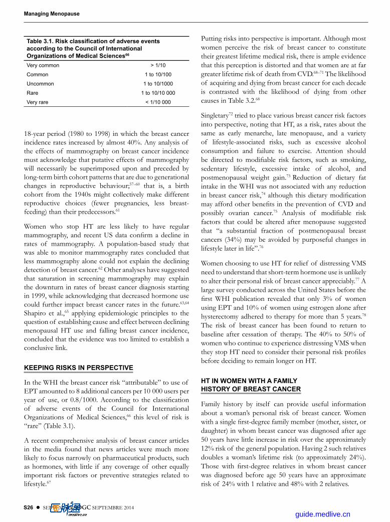
S26 l SEPTEMBER JOGC SEPTEMBRE 2014
Managing Menopause
18-year period (1980 to 1998) in which the breast cancer incidence rates increased by almost 40%. Any analysis of the effects of mammography on breast cancer incidence must acknowledge that putative effects of mammography will necessarily be superimposed upon and preceded by long-term birth cohort patterns that are due to generational changes in reproductive behaviour;57–60 that is, a birth cohort from the 1940s might collectively make different reproductive choices (fewer pregnancies, less breast-feeding) than their predecessors.61
Women who stop HT are less likely to have regular mammography, and recent US data confirm a decline in rates of mammography. A population-based study that was able to monitor mammography rates con cluded that less mammography alone could not explain the declining detection of breast cancer.62 Other analyses have suggested that saturation in screening mammography may explain the downturn in rates of breast cancer diagnosis starting in 1999, while acknowledging that decreased hormone use could further impact breast cancer rates in the future.63,64 Shapiro et al.,65 applying epidemiologic principles to the question of establishing cause and effect between declining menopausal HT use and falling breast cancer incidence, concluded that the evidence was too limited to establish a conclusive link.
KEEPING RISKS IN PERSPECTIVE
In the WHI the breast cancer risk “attributable” to use of EPT amounted to 8 additional cancers per 10 000 users per year of use, or 0.8/1000. According to the classification of adverse events of the Council for International Organizations of Medical Sci ences,66 this level of risk is “rare” (Table 3.1).
A recent compre hensive analysis of breast cancer articles in the media found that news articles were much more likely to focus narrowly on pharmaceutical products, such as hormones, with little if any coverage of other equally important risk factors or pre ventive strategies related to lifestyle.67
Putting risks into perspective is important. Although most women perceive the risk of breast cancer to constitute their greatest lifetime medical risk, there is ample evidence that this perception is distorted and that women are at far greater life time risk of death from CVD.68–71 The likelihood of acquiring and dying from breast cancer for each decade is contrasted with the likelihood of dying from other causes in Table 3.2.68
Singletary72 tried to place various breast cancer risk factors into perspective, noting that HT, as a risk, rates about the same as early menarche, late menopause, and a variety of lifestyle-associated risks, such as excessive alcohol con sumption and failure to exercise. Attention should be directed to modifiable risk factors, such as smoking, seden tary lifestyle, excessive intake of alcohol, and postmenopausal weight gain.73 Reduction of dietary fat intake in the WHI was not associated with any reduction in breast cancer risk,74 although this dietary modification may afford other benefits in the prevention of CVD and possibly ovarian cancer.75 Analysis of modifiable risk factors that could be altered after menopause suggested that “a substantial fraction of postmenopausal breast cancers (34%) may be avoided by purposeful changes in lifestyle later in life”.76
Women choosing to use HT for relief of distressing VMS need to understand that short-term hor mone use is unlikely to alter their personal risk of breast cancer appreciably.77 A large survey conducted across the United States before the first WHI publication revealed that only 3% of women using EPT and 10% of women using estrogen alone after hysterectomy adhered to therapy for more than 5 years.78 The risk of breast cancer has been found to return to baseline after cessation of therapy. The 40% to 50% of women who continue to experience distressing VMS when they stop HT need to consider their per sonal risk profiles before deciding to remain longer on HT.
HT IN WOMEN WITH A FAMILY HISTORY OF BREAST CANCER
Family history by itself can provide useful information about a woman’s personal risk of breast cancer. Women with a single first-degree family member (mother, sister, or daughter) in whom breast cancer was diagnosed after age 50 years have little increase in risk over the approxi mately 12% risk of the general population. Having 2 such relatives doubles a woman’s lifetime risk (to approximately 24%). Those with first-degree relatives in whom breast cancer was diagnosed before age 50 years have an approximate risk of 24% with 1 relative and 48% with 2 relatives.
Table 3.1. Risk classification of adverse events according to the Council of International Organizations of Medical Sciences66
Very common > 1/10
Common 1 to 10/100
Uncommon 1 to 10/1000
Rare 1 to 10/10 000
Very rare < 1/10 000
guide.medlive.cn
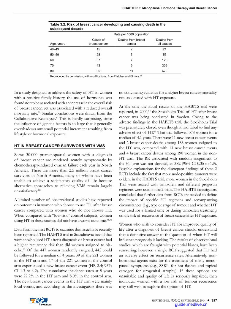
SEPTEMBER JOGC SEPTEMBRE 2014 l S27
CHAPTER 3: Menopausal Hormone Therapy and Breast Cancer
In a study designed to address the safety of HT in women with a positive family history, the use of hormones was found not to be associated with an increase in the overall risk of breast cancer, yet was associated with a reduced overall mortality rate.79 Similar conclusions were drawn from the Collaborative Reanalysis.8 This is hardly surprising, since the influence of genetic factors is so large that it gener ally overshadows any small potential increment resulting from lifestyle or hormonal exposure.
HT IN BREAST CANCER SURVIVORS WITH VMS
Some 30 000 premenopausal women with a diagnosis of breast cancer are rendered acutely symptomatic by chemotherapy-induced ovarian failure each year in North America. There are more than 2.5 million breast cancer sur vivors in North America, many of whom have been unable to achieve a satisfactory quality of life because alternative approaches to relieving VMS remain largely unsatisfactory.80
A limited number of observational studies have reported on outcomes in women who choose to use HT after breast cancer compared with women who do not choose HT. When compared with “low-risk” control subjects, women using HT in these studies did not have a worse outcome.81,82
Data from the first RCTs to examine this issue have recently been reported. The HABITS trial in Scandinavia found that women who used HT after a diagnosis of breast cancer had a higher recurrence risk than did women assigned to pla-cebo.83 Of the 447 women randomly assigned, 442 could be followed for a median of 4 years: 39 of the 221 women in the HT arm and 17 of the 221 women in the control arm experienced a new breast cancer event (HR 2.4; 95% CI 1.3 to 4.2). The cumulative incidence rates at 5 years were 22.2% in the HT arm and 8.0% in the control arm. The new breast cancer events in the HT arm were mainly local events, and according to the investigators there was
no con vincing evidence for a higher breast cancer mortality rate associated with HT exposure.
At the time the initial results of the HABITS trial were reported, in 2004,84 the Stockholm Trial of HT after breast cancer was being conducted in Sweden. Owing to the adverse findings in the HABITS trial, the Stockholm Trial was prematurely closed, even though it had failed to find any adverse effect of HT.85 That trial followed 378 women for a median of 4.1 years. There were 11 new breast cancer events and 2 breast cancer deaths among 188 women assigned to the HT arm, compared with 13 new breast cancer events and 4 breast cancer deaths among 190 women in the non-HT arm. The RR associ ated with random assignment to the HT arm was not elevated, at 0.82 (95% CI 0.35 to 1.9). Possible explanations for the discrepant findings of these 2 RCTs include the fact that more node-positive tumours were evident in the HABITS trial, more women in the Stockholm Trial were treated with tamoxifen, and different progestin regimens were used in the 2 trials. The HABITS investigators con cluded that further data from RCTs are needed to define the impact of specific HT regimens and accompanying circum stances (e.g., type or stage of tumour and whether HT was used for a lim ited time or during tamoxifen treatment) on the risk of recurrence of breast cancer after HT exposure.
Women who wish to consider HT for improved quality of life after a diagnosis of breast cancer should understand that a definitive answer to the question of when HT will influ ence prognosis is lacking. The results of observational studies, which are fraught with potential biases, have been reassuring; however, a single RCT suggested that HT had an adverse effect on recurrence rates. Alternatively, non-hormonal agents exist for the treatment of many meno-pausal symptoms (e.g., SSRIs for hot flashes and topical estrogen for urogenital atrophy). If these options are unsuit able and quality of life is seriously impaired, then individual women with a low risk of tumour recurrence may still wish to explore the option of HT.
Table 3.2. Risk of breast cancer developing and causing death in the subsequent decade
Rate per 1000 population
Age, years
Cases of breast cancer
Deaths from breast cancer
Deaths from all causes
40–49 15 2 21
50–59 28 5 55
60 37 7 126
70 43 9 309
80 35 11 670Reproduced by permission, with modifications, from Fletcher and Elmore.68
guide.medlive.cn
S28 l SEPTEMBER JOGC SEPTEMBRE 2014
Managing Menopause
SERMS AND BREAST CANCER
Raloxifene has been approved in the United States for both the treatment and the prevention of osteoporosis. In the pivotal osteoporosis prevention trial MORE, women assigned to raloxifene rather than to placebo demonstrated a 72% reduction in the incidence rate of invasive breast cancer at 4 years.86 The MORE trial was not designed to measure the reduction in breast cancer incidence in women at increased risk, so in 1999 the National Surgical Adjuvant Breast and Bowel Project initiated the STAR trial. In this study, postmenopausal women aged at least 35 years and at increased risk for breast cancer were randomly assigned to receive either tamoxifen or raloxifene for 5 years and were required to complete follow-up examinations for at least 7 years. The STAR trial found that raloxifene was as effective as tamoxifen in reducing the risk of invasive breast cancer and was associated with a lower risk of thromboembolic events and cataracts but a higher, though not statistically signifi cant, risk of non-invasive breast cancer.87 The risk of other cancers, fractures, ischemic heart disease, and stroke was similar for the 2 drugs.88 Raloxifene has now been approved in the United States for use in preventing breast cancer in women at high risk.
Recommendations1. Health care providers should periodically review the
risks and benefits of prescribing hormone therapy to a menopausal woman in light of the association between duration of use and breast cancer risk. (I-A)
2. Health care providers may prescribe hormone therapy for menopausal symptoms in women at increased risk of breast cancer with appropriate counselling and surveillance. (I-A)
3. Health care providers should clearly discuss the uncer tainty of risks associated with systemic hormone therapy after a diagnosis of breast cancer in women seeking treatment for distressing symptoms (vasomotor symptoms or vulvovaginal atrophy). (I-B)
REFERENCES
1. Key TJ, Verkasalo PK, Banks E. Epidemiology of breast cancer. Lancet Oncol 2001;2:133–40.
2. American Cancer Society. Cancer facts & figures. Atlanta, Georgia: American Cancer Society; 2008. Available at http://www.cancer.org/research/cancerfactsstatistics/cancerfactsfigures2008/index. Accessed 2013 Aug 12.
3. Feinlieb M. Breast cancer and artificial menopause: a cohort study. J Natl Cancer Inst 1968;41:315–29.
4. Youlden DR, Cramb SM, Dunn NA, Muller JM, Pyke CM, Baade PD. The descriptive epidemiology of female breast cancer: an international comparison of screening, incidence, survival and mortality. Cancer Epidemiol 2012;36:237–48.
5. Welch HG, Woloshin S, Schwartz LM. The sea of uncertainty surrounding ductal carcinoma in situ—the price of screening mammography. J Natl Cancer Inst 2008;100:228–9.
6. Peto R, Davies C, Godwin J, Gray R, Pan HC, Clarke M, et al. Comparisons between different polychemotherapy regimens for early breast cancer: meta-analyses of long-term outcome among 100,000 women in 123 randomised trials. Lancet 2012;379:432–44.
7. Colditz GA, Hankiinson SE, Hunter DJ, Willett WC, Manson JE, Stampfer MJ, et al. The use of estrogens and progestins and the risk of breast cancer in postmenopausal women. N Engl J Med 1995;332:1589–93.
8. Collaborative Group on Hormonal Factors in Breast Cancer. Breast cancer and hormone replacement therapy: a collaborative reanalysis of data from 51 epidemiological studies of 52,705 women with breast cancer and 108,411 women without breast cancer. Lancet 1997;350:1047–59.
9. LaCroix AZ, Chlebowski RT, Manson JE, Aragaki AK, Johnson KC, Martin L, et al. Health outcomes after stopping conjugated equine estrogens among postmenopausal women with prior hysterectomy: a randomized controlled trial. JAMA 2011;305:1305–14.
10. Stefanick ML, Anderson GL, Margolis KL, Hendrix SL, Rodabough RJ, Paskett ED, et al; WHI Investigators. Effects of conjugated equine estrogens on breast cancer and mammography screening in postmenopausal women with hysterectomy. JAMA 2006;295:1647–57.
11. McTiernan A, Chlebowski RT, Martin C, Peck JD, Aragaki A, Pisano ED, et al. Conjugated equine estrogen influence on mammographic density in postmenopausal women in a substudy of the Women’s Health Initiative randomized trial. J Clin Oncol 2009; 27:6135–43.
12. McTiernan A, Martin CF, Peck JD, Aragaki AK, Chlebowski RT, Pisano ED, et al; Women’s Health Initiative Mammogram Density Study Investigators. Estrogen-plus-progestin use and mammographic density in postmenopausal women: Women’s Health Initiative Randomized Trial. J Natl Cancer Inst 2005;97:1366–76.
13. Lyytinen H, Pukkala E, Ylikorkala O. Breast cancer risk in postmenopausal women using estrogen-only therapy. Obstet Gynecol 2006;108:1354–60.
14. Zhang SM, Manson JE, Rexrode KM, Cook NR, Buring JE, Lee IM. Use of oral conjugated estrogen alone and risk of breast cancer. Am J Epidemiol 2006;165:524–9.
15. Li CI, Malone KE, Porter PL, Weiss NS, Tang MT, Cushing-Haugen KL, et al. Relationship between long durations and different regimens of hormone therapy and risk of breast cancer. JAMA 2003;289:3254–63.
16. Chlebowski RT, Anderson GL. Changing concepts: menopausal hormone therapy and breast cancer. J Natl Cancer Inst 2012;104:517–27.
17. Fournier A, Mesrine S, Boutron-Ruault MC, Clavel-Chapelon C. Estrogen–progestagen menopausal hormone therapy and breast cancer: Does delay from menopause onset to treatment initiation influence risks? J Clin Oncol 2009;27:5138–43.
18. Beral V, Reeves G, Bull D, Green J; for the Million Women Study Collaborators. Breast cancer risk in relation to the interval between menopause and starting hormone therapy. J Natl Cancer Inst 2011;103:296–305.
19. Jordan VC, Ford LG. Paradoxical clinical effect of estrogen on breast cancer risk: a new biology of estrogen induced apoptosis. Cancer Prev Res 2011;4:633–7.
20. Beral V; for the Million Women Study Collaborators. Breast cancer and hormone-replacement therapy in the Million Women Study. Lancet 2003;362:419–27.
21. Spratt JS, Meyer JS, Spratt JA. Rates of growth of human solid neoplasms: part I. J Surg Oncol 1995;60:137–46.
guide.medlive.cn

SEPTEMBER JOGC SEPTEMBRE 2014 l S29
CHAPTER 3: Menopausal Hormone Therapy and Breast Cancer
22. Dietel M, Lewis MA, Shapiro S. Hormone replacement therapy: pathobiological aspects of hormone-sensitive cancers in women relevant to epidemiological studies on HRT: a mini-review. Hum Reprod 2005;20:2052–60.
23. Speroff L. Postmenopausal hormone therapy and the risk of breast cancer: a contrary thought. Menopause 2008;15:393–400.
24. Whitehead M, Farmer R. The Million Women Study: a critique. Endocrine 2004;24:187–93.
25. Shapiro S. The Million Women Study: potential biases do not allow uncritical acceptance of the data. Climacteric 2004;7:3–7.
26. Bakken K, Fournier A, Lund E, Waaseth M, Dumeaux V, Clavel-Chapelon F, et al .Menopausal hormone therapy and breast cancer risk: impact of different treatments. The European Prospective Investigation into Cancer and Nutrition. (EPIC Study). Int J Cancer 2011;128:144–56.
27. Reeves GK, Beral V, Green J, Gathani T, Bull D; for the Million Women Study Collaborators. Hormonal therapy for menopause and breast-cancer risk by histological type: a cohort study and meta-analysis. Lancet Oncol 2006;7:910–8.
28. Opatrny L, Dell’Aniello S, Assouline S, Suissa S. Hormone replacement therapy use and variations in the risk of breast cancer. BJOG 2008;115:169–75.
29. Boyd NF, Martin LJ, Yaffe MJ, Minkin S. Mammographic density: a hormonally responsive risk factor for breast cancer. J Br Menopause Soc 2006;12:186–93.
30. Tamimi RM, Byrne C, Colditz GA, Hankinson SE. Endogenous hormone levels, mammographic density, and subsequent risk of breast cancer in postmenopausal women. J Natl Cancer Inst 2007;99:1178–87.
31. Conner P. Breast response to menopausal hormone therapy—aspects on proliferation, apoptosis and mammographic density. Ann Med 2007;39:28–41.
32. McCormack VA, dos Santos Silva I. Breast density and parenchymal patterns as markers of breast cancer risk: a meta-analysis. Cancer Epidemiol Biomarkers Prev 2006;15:1159–69.
33. Nachtigall LE. Breast density as a clinical entity: Is it a marker for breast cancer? Menopause 2007;14:345–6.
34. Harvey J, Scheurer C, Kawakami FT, Quebe-Fehling E, de Palacios PI, Ragavan VV. Hormone replacement therapy and breast density changes. Climacteric 2005;8:185–92.
35. Grady D, Vittinghoff E, Lin F, Hanes V, Ensrud K, Habel LA, et al. Effect of ultra-low-dose transdermal estradiol on breast density in postmenopausal women. Menopause 2007;14(3 Pt 1):391–6.
36. Corrao G, Zambon A, Conti V, Nicotra F, La Vecchia C, Fornari C, et al. Menopause hormone replacement therapy and cancer risk: an Italian record linkage investigation. Ann Oncol 2008;19:150–5.
37. Kerlikowske K, Cook AJ, Buist DS, Cummings SR, Vachon C, Vacek P, et al. Breast cancer risk by breast density, menopause, and postmenopausal hormone therapy use. J Clin Oncol 2010;28:3830–7.
38. Moy L, Slanetz PJ, Yeh ED, Moore RH, Rafferty EA, Kopans DB. Hormone replacement therapy rarely complicates or alters interpretation on screening mammography: a prospective analysis [abstract]. Radiology 2000;217:446.
39. Carney PA, Miglioretti DL, Yankaskas BC, Kerlikowske K, Rosenberg R, Rutter CM, et al. Individual and combined effects of age, breast density, and hormone replacement therapy use on the accuracy of screening mammography. Ann Intern Med 2003;138:168–75.
40. Laya MB, Larson EB, Taplin SH, White E. Effect of estrogen replacement therapy on the specificity and sensitivity of screening mammography. J Natl Cancer Inst 1996;8:643–9.
41. Rosenberg RD, Hunt WC, Williamson MR, Gilliland FD, Wiest PW, Kelsey CA, et al. Effects of age, breast density, ethnicity, and estrogen replacement therapy on screening mammographic sensitivity and cancer stage at diagnosis: review of 183,134 screening mammograms in Albuquerque, New Mexico. Radiology 1998;209:511–8.
42. Litherland JC, Stallard S, Hole D, Cordiner C. The effect of hormone replacement therapy on the sensitivity of screening mammograms. Clin Radiol 1999;54:285–8.
43. Kavanagh AM, Mitchell H, Giles GG. Hormone replacement therapy and accuracy of mammographic screening. Lancet 2000;355:270–4.
44. Chlebowski RT, Hendrix SL, Langer RD, Stefanick ML, Gass M, Lane D; WHI Investigators. Influence of estrogen plus progestin on breast cancer and mammography in healthy postmenopausal women: the Women’s Health Initiative Randomized Trial. JAMA 2003;289:3243–53.
45. Chlebowski RT, Anderson G, Pettinger M, Lane D, Langer RD, et al; for the Women’s Health Initiative Investigators. Estrogen plus progestin and breast cancer detection by means of mammography and breast biopsy. Arch Intern Med 2008;168:370–7.
46. Anderson GL, Chlebowski RT, Rossouw JE, Rodabough RJ, McTiernan A, Margolis KL, et al. Prior hormone therapy and breast cancer risk in the Women’s Health Initiative randomised trial of estrogen plus progestin. Maturitas 2006;55:103–15. Epub 2006 Jul 11.
47. Seeger H, Wallweiner D, Mueck AO. The effect of progesterone and synthetic progestins on serum and estradiol-stimultaed proliferation of human breast cancer cells. Horm Metab Res 2003;35:76–80.
48. Wood CE, Register TC, Lees CJ, Chen H, Kimrey S, Cline JM. Effects of estradiol with micronized progesterone or medroxyprogesterone acetate on risk markers for breast cancer in postmenopausal monkeys. Breast Cancer Res Treat 2007;101:125–34.
49. Murkes D, Lalitkumar PGL, Leifland K, Lundström E, Söderqvist G. Percutaneous estradiol/oral micronized progesterone has less-adverse effects and different gene regulations than oral conjugated equine estrogens/medroxyprogesterone acetate in the breasts of healthy women in vivo. Gynecol Endocrinol 2012;28(Suppl 2):12–15. Epub 2012 July 27.
50. Fournier A, Berrino F, Clavel-Chapelon F. Unequal risks for breast cancer associated with different hormone replacement therapies: results from the E3N cohort study. Breast Cancer Res Treat 2008;107:103–11.
51. Shapiro S, Farmer RD, Mueck AO, Seaman H, Stevenson JC. Does hormone replacement therapy cause breast cancer? An application of causal principles to three studies: part 3. The Women’s Health Initiative: unopposed estrogen. J Fam Plann Reprod Health Care 2011;37:225–30.
52. Shapiro S, Farmer RD, Mueck AO, Seaman H, Stevenson JC. Does hormone replacement therapy cause breast cancer? An application of causal principles to three studies: part 2. The Women’s Health Initiative: estrogen plus progestogen. J Fam Plann Reprod Health Care 2011;37:165–72.
53. Shapiro S, Farmer RD, Seaman H, Stevenson JC, Mueck AO. Does hormone replacement therapy cause breast cancer? An application of causal principles to three studies: part 1. The Collaborative Reanalysis. J Fam Plann Reprod Health Care 2011;37:103–9.
54. Santen RJ, Heitjan DF. Modelling of growth kinetics of occult breast tumours: role in interpretation of studies of prevention and menopausal hormone therapy. Cancer Epidemiol Biomarkers Prev 2012;21:1038–48.
55. Russo J, Russo IH. The role of estrogen in the initiation of breast cancer. J Steroid Biochem Mol Biol 2006;102(1–5):89–96.
56. Wren BG. The origin of breast cancer. Menopause 2007;14:1060–8.
57. Jemal A, Ward E, Thun MJ. Recent trends in breast cancer incidence rates by age and tumor characteristics among U.S. women. Breast Cancer Res 2007;9:R28.
guide.medlive.cn

S30 l SEPTEMBER JOGC SEPTEMBRE 2014
Managing Menopause
58. Glass AG, Lacey JV Jr, Carreon JD, Hoover RN. Breast cancer incidence, 1980–2006: combined roles of menopausal hormone therapy, screening mammography, and estrogen receptor status. J Natl Cancer Inst 2007;99:1152–61. Epub 2007 Jul 24.
59. Tarone RE, Chu KC. Implications of birth cohort patterns in interpreting trends in breast cancer rates. J Natl Cancer Inst 1992;84:1402–10.
60. Blot WJ, Devesa SS, Fraumeni JF Jr. Declining breast cancer mortality among young American women. J Natl Cancer Inst 1987;78:451–4.
61. Collaborative Group on Hormonal Factors and Breast Cancer. Breast cancer and breast feeding: collaborative reanalysis of data from 47 epidemiologic studies in 30 countries including 50, 302 women with breast cancer and 96, 973 women without the disease. Lancet 2002;360:187–95.
62. Kerlikowske K, Miglioretti DL, Buist DSM, Walker R, Carney PA. Declines in invasive breast cancer and use of postmenopausal hormone therapy in a screening mammography population. J Natl Cancer Inst 2007;99:1335–9.
63. Li CI, Daling JR. Changes in breast cancer incidence rates in the United States by histologic subtype and race/ethnicity, 1995 to 2004. Cancer Epidemiol Biomarkers Prev 2007;16:2773–80.
64. Gompel A, Plu-Bureau G. Is the decrease in breast cancer incidence related to a decrease in postmenopausal hormone therapy? Ann N Y Acad Sci 2010;1205:268–76.
65. Shapiro S, Farmer RDT, Stevenson JC, Burger HG, Mueck AO, Gompel A. Does hormone replacement therapy (HRT) cause breast cancer? An application of causal principles to three studies: part 5. Trends in breast cancer incidence in relation to the use of HRT. J Fam Plann Reprod Health Care 2013;39:80–8.
66. Council for International Organizations of Medical Sciences. Guidelines for preparing core clinical-safety information on drugs. 2nd ed. Geneva: CIOMS; 1999.
67. Atkin CK, Smith SW, McFeters C, Ferguson V. A comprehensive analysis of breast cancer news coverage in leading media outlets focusing on environmental risks and prevention. J Health Commun 2008;13:3–19.
68. Fletcher SW, Elmore JG. Mammographic screening for breast cancer. N Engl J Med 2003;348:1672–80.
69. Giordano SH, Hortobagyi GN. Time to remove the subspecialty blinders: breast cancer does not exist in isolation. J Natl Cancer Inst 2008;100:230–1.
70. Chapman JA, Meng D, Shepherd L, Parulekar W, Ingle JN, Muss HB, et al. Competing causes of death from a randomized trial of extended adjuvant endocrine therapy for breast cancer. J Natl Cancer Inst 2008;100:252–60.
71. Bluming AZ, Tavaris C. What are the real risks for breast cancer? Climacteric 2012;15:133–5.
72. Singletary SE. Rating the risk factors for breast cancer. Ann Surg 2003;4:474–82.
73. Reeves GK, Pirie K, Beral V, Green J, Spencer E, Bull D. Million Women Study Collaboration. Cancer incidence and mortality in relation to body mass index in the Million Women Study: cohort study. BMJ 2007;335:1134. Epub 2007 Nov 6.
74. Prentice RL, Caan B, Chlebowski RT, Patterson R, Kuller LH, Ockene JK, et al. Low-fat dietary pattern and risk of invasive breast cancer: the Women’s Health Initiative Randomized Controlled Dietary Modification Trial. JAMA 2006;295:629–42.
75. Prentice RL, Thomson CA, Caan B, Hubbell FA, Anderson GL, Beresford SA, et al. Low-fat dietary pattern and cancer incidence in the Women’s Health Initiative Dietary Modification Randomized Controlled Trial. J Natl Cancer Inst 2007;99:1534–43.
76. Sprague BL, Trentham-Dietz A, Egan KM, Titus-Ernstoff L, Hampton JM, Newcomb PA. Proportion of invasive breast cancer attributable to risk factors modifiable after menopause. Am J Epidemiol 2008;168:404–11.
77. Collins J. Hormones and breast cancer: Should practice be changed? Obstet Gynecol 2006;108:1352–3.
78. Brett KM, Reuben CA. Prevalence of estrogen or estrogen–progestin hormone therapy use. Obstet Gynecol 2003;102:1240–9.
79. Sellars TA, Mink PJ, Cerhan JR, Zheng W, Anderson KE, Kushi LH, et al. The role of hormone replacement therapy in the risk for breast cancer and total mortality in women with a family history of breast cancer. Ann Intern Med 1997;127:973–80.
80. Speroff L. Alternative therapies for postmenopausal women. Int J Fertil Womens Med 2005;50:101–4.
81. O’Meara ES, Rossing MA, Daling JR, Elmore JG, Barlow WE, Weiss NS. Hormone replacement therapy after a diagnosis of breast cancer in relation to recurrence and mortality. J Natl Cancer Inst 2001;93:754–62.
82. Col NF, Hirota LK, Orr RK, Erban JK, Wong JB, Lau J. Hormone replacement therapy after breast cancer: a systematic review and quantitative assessment of risk. J Clin Oncol 2001;19:2357–63.
83. Holmberg L, Iversen OE, Rudenstam CM, Hammar M, Kumpulainen E, Jaskiewicz J, et al; HABITS Study Group. Increased risk of recurrence after hormone replacement therapy in breast cancer survivors. J Natl Cancer Inst 2008;100:475–82.
84. Holmberg L, Anderson H; HABITS steering and data monitoring committees. HABITS (hormonal replacement therapy after breast cancer—Is it safe?), a randomised comparison: trial stopped. Lancet 2004;363:453–5.
85. von Schoultz E, Rutqvist LE; Stockholm Breast Cancer Study Group. Menopausal hormone therapy after breast cancer: the Stockholm Randomized Trial. J Natl Cancer Inst 2005;97:533–5.
86. Barrett-Connor E, Mosca L, Collins P, Geiger MJ, Grady D, Kornitzer M, et al; Raloxifene Use for The Heart (RUTH) Trial Investigators. Effects of raloxifene on cardiovascular events and breast cancer in postmenopausal women. N Engl J Med 2006;355:125–37.
87. Stefanick ML. Risk–benefit profiles of raloxifene for women. N Engl J Med 2006;355:190–2.
88. Vogel VG, Costantino JP, Wickerham DL, Cronin WM, Cecchini RS, Atkins JN, et al; National Surgical Adjuvant Breast and Bowel Project (NSABP). Effects of tamoxifen vs raloxifene on the risk of developing invasive breast cancer and other disease outcomes: the NSABP Study of Tamoxifen and Raloxifene (STAR) P-2 trial. JAMA 2006;295:2727–41.
guide.medlive.cn

SEPTEMBER JOGC SEPTEMBRE 2014 l S31
CHAPTER 4
Vasomotor Symptoms
VMS affect 60% to 80% of women entering menopause.1 Hot flashes are common in the
perimenopausal transition, when ovarian activity may be intermittent, and they have also been documented during the luteal and menstrual phases of the cycle in women with premenstrual dysphoric disorder.2 After menopause, it is important to be alert to atypical features or to a lack of response to effective therapy, which might indicate an alternative cause of the symptoms. The differential diagnosis includes hyperthyroidism, anxiety, panic attack, hypertension, emotional flushing, neurologic flushing, carcinoid, physical deconditioning, tumours, spinal cord injury, and reactions to food, drugs, and alcohol.3 In menopausal women hot flashes are thought to result from a disturbance of the temperature-regulating mechanism in the hypothalamus that is due to low estrogen levels after prior estrogen priming.
Although most postmenopausal women (60%) experience hot flashes for less than 7 years, up to 15% report that hot flashes persist for 15 years or more.4 The symptoms that can accompany hot flashes (including sweating, palpitations, apprehension, and anxiety) contribute to the woman’s discomfort, inconvenience, and distress, particularly when these episodes occur very frequently. They can be a significant contributor to sleep disturbance. VMS adversely affect quality of life for 20% to 25% of women, primarily owing to the physical discomfort and social embarrassment that they evoke, although night sweats and sleep disturbance are also reported to have a negative impact.5–7
A recent excellent review addresses the scientific basis for hot flashes, the strength of evidence suggesting associations between VMS and CVD, breast cancer, and osteoporosis, and treatment options.8
Normally the body maintains an optimal temperature for metabolic activity through vasodilatation and sweating when overheated and shivering when cold. Postmenopausal women are thought to have narrowing of this “thermoneutral zone” such that small changes in temperature can evoke the regulatory response of sweating or shivering.9 Risk factors for hot flashes include obesity,
limited physical activity, and cigarette smoking and, along with a variety of known triggers (alcohol, warm ambient environment, hot drinks), form the basis for certain lifestyle recommendations to reduce VMS. The prevalence of these symptoms differs according to ethnicity. Compared with Caucasian women, VMS are reported less frequently by Japanese and Chinese women but more frequently by African-American women.10
TREATMENT OPTIONS
Multiple forms of treatment have been used to relieve hot flashes, including lifestyle modifications, non-prescription drugs, and prescription drugs. Prescription drugs may be grouped into hormonal and non-hormonal agents. Multiple placebo-controlled trials have shown a reduction of about 25% to 30% in the frequency of hot flashes within 4 weeks of placebo treatment. Moreover, a Cochrane review of ET has shown that placebo may cause a reduction of about 50%.11 These results highlight the importance of applying appropriate scientific scrutiny to anecdotal reports and uncontrolled trials that claim efficacy for treatment of hot flashes.
Since the report about risks associated with HT from the WHI in 2002, many physicians have abandoned the prescription of HT for VMS in favour of recommending lifestyle changes and cooling devices. Unfortunately, many women find that these approaches afford little relief and have turned to unproven and often untested complementary and alternative therapies. The scientific evidence, however, remains clear that the single most effective agent for treating VMS remains estrogen or, for women with an intact uterus, estrogen plus progestin. Both oral and transdermal routes of administration of estrogen are effective against VMS.
The most recent Cochrane review of RCTs of menopausal HT for the treatment of VMS concluded that HT is highly effective, with reductions in both frequency and severity in the order of 75%.11 The dropout rate was higher in the placebo arms for lack of effectiveness, but there was no difference between the treatment and placebo arms in the rate of dropout because of adverse effects.
guide.medlive.cn

S32 l SEPTEMBER JOGC SEPTEMBRE 2014
Managing Menopause
Progestogens alone may be considered as an alternative for treating hot flashes if the benefit–risk profile is acceptable to the woman. The question of whether a progestin alone increases the risk of breast cancer is unanswered. MPA has been shown in several trials to relieve hot flashes in healthy women as well as in women with breast or endometrial cancer.12–14 Both intramuscular (150 mg) and oral forms (20 mg/d) have demonstrated efficacy. Micronized progesterone, 300 mg, is superior to placebo but possibly less effective than estrogen for in the treatment of VMS.15
Non-hormonal options that have shown some efficacy for relief of VMS include clonidine,16,17 SNRIs18 or their active metabolites, such as desvenlafaxine succinate,19 gabapentin,20 and pregabalin.21 None are as effective as estrogen, and the response rate among women is variable. These options can be offered to women with disruptive VMS for whom estrogen is contraindicated or unacceptable.
Two placebo-controlled trials of clonidine showed a reduction in the severity of hot flashes over placebo but were unable to demonstrate a statistically significant reduction in frequency.16,17 Sleep disturbance was reported as an unwanted side effect.
Recent systematic reviews of SSRIs and SNRIs have found these classes of medication to be more effective than placebo in reducing VMS. Successful placebo-controlled trials have been reported with paroxetine, fluoxetine, sertraline, venlafaxine, desvenlafaxine, and citalopram. These agents may be used together with HT and can be offered to women in menopause with coexisting depression.22–24 In Canada since 2002 there has been an inverse relationship between the numbers of prescriptions for HT and SSRIs, those for HT declining and those for SSRIs rising, which led the investigators to conclude that “the simultaneous increase in prescriptions of serotonergic antidepressants suggests that antidepressants are being prescribed for symptoms (psychological, physical) previously controlled with the use of HRT”.25
Antidepressant agents, though moderately effective for the treatment of VMS, are not without significant side effects26,27 and afford none of the other health benefits seen with HT that directly impact quality of life (e.g., prevention of urogenital atrophy and osteoporosis).28 Recent reports suggest that rates of osteoporotic fractures have started to increase since 2002.29,30
A recent meta-analysis of gabapentin for the treatment of VMS analyzed the experience of 901 women in 7 trials conducted between 2002 and 2008, including 1 Canadian
trial.31 Reductions in hot-flash severity and frequency were between 20% and 30%. Doses were between 900 and 2400 mg, and treatment was associated with dizziness, unsteadiness, somnolence, and fatigue.32 Useful strategies to reduce the risk of such side effects include slowly titrating the dose over 12 days or more, using a lower dose, or prescribing the drug for bedtime.33
Pregabalin, 75 mg twice a day, reduced the hot-flash score (frequency times mean severity) by 65% compared with 50% for a placebo.21 Combined treatment with gabapentin and an antidepressant was not superior to treatment with either treatment alone.34
Herbal remedies have become a popular alternative in North America,35 yet few of those promoted for VMS treatment have met the rigorous testing criteria required of pharmaceutical products by the US Food and Drug Administration. The current regulatory requirement for pharmaceutical products with purported benefit for hot flashes is that participants in clinical trials must experience on average 7 hot flashes per day, or 50 per week. Most reported studies of herbal products have been open-label and conducted in women with as few as 1 or 2 hot flashes per day. Recent reports caution about potential adverse safety profiles of marketed herbal products, and cautions have appeared about interactions of NHPs with pharmaceutical and anesthetic agents.36–39 Canadian legislation in January 2004 removed NHPs from the food category and placed them in a special drug category to allow regulation of manufacturing, labelling, and indications for use.40 To date, little appears to have been accomplished in the regulation of NHPs in Canada. Several recent systematic reviews have examined options for treatment of moderate to severe VMS.41–45 None of these found any single complementary therapy to have proven efficacy for moderate to severe hot flashes, and the most recent review41 concluded by stating that “although individual trials suggest benefits from certain therapies, data are insufficient to support the effectiveness of any complementary and alternative therapy in this review for the management of menopausal symptoms.” A direct head-to-head comparison of HT with black cohosh, soy, or multibotanicals showed only HT to have an effect greater than that of placebo.46
In light of the evidence supporting the effectiveness of HT for treatment of VMS and the apparent underuse owing to fear and uncertainty about the safety of menopausal HT, a dozen leading North American medical organizations collaborated to produce the following clear statement about HT:47
guide.medlive.cn

SEPTEMBER JOGC SEPTEMBRE 2014 l S33
CHAPTER 4: Vasomotor Symptoms
Leading medical societies devoted to the care of menopausal women agree . . . there is no question that hormone therapy has an important role in managing symptoms for women during the menopause transition and in early menopause.
A tissue-selective estrogen compound consisting of the SERM bazedoxifene combined with an oral conjugated estrogen has recently been shown to suppress VMS, and prevent postmenopausal bone loss while having a favourable safety profile with respect to breast and endometrium.48 This combination will obviate the need for progestin cotherapy in women using systemic estrogen, both simplifying therapy and avoiding progestin-associated adverse effects.
Recommendations1. Lifestyle modifications, including reducing
core body temperature, regular exercise, weight management, smoking cessation, and avoidance of known triggers such as hot drinks and alcohol, may be recommended to reduce mild vasomotor symptoms. (I-C)
2. Health care providers should offer hormone therapy, estrogen alone or combined with a progestin, as the most effective agents for the medical management of menopausal symptoms. (I-A)
3. Progestins alone or low-dose oral contraceptives can be offered as alternatives for the relief of menopausal symptoms during the menopausal transition. (I-A)
4. Non-hormonal prescription drugs, including certain antidepressant agents, gabapentin, and clonidine, may afford some relief from hot flashes but have their own side effects. These alternatives can be considered when hormone therapy is contraindicated or not desired. (I-B)
5. There is limited evidence of benefit for most complementary and alternative approaches to the management of hot flashes. Without good evidence for effectiveness, and in the face of minimal data on safety, these approaches should not be recommended. Women should be advised that, until January 2004, most natural health products were introduced into Canada as “food products” and did not fall under the regulatory requirements for pharmaceutical products. As such, most have not been rigorously tested for the treatment of moderate to severe hot flashes, and many lack evidence of efficacy and safety. (I-B)
6. Estrogen therapy can be offered to women who have undergone surgical menopause for the treatment of endometriosis. (I-A)
REFERENCES
1. Freeman EW, Sherif K. Prevalence of hot flushes and night sweats around the world: a systematic review. Climacteric 2007;10:197–214.
2. Hahn PM, Wong J, Reid RL. Menopausal-like hot flashes reported in women of reproductive age. Fertil Steril 1998;70:913–8.
3. Mohyi D, Tabassi K, Simon J. Differential diagnosis of hot flashes. Maturitas 1997;27:203–14.
4. Kronenberg F. Hot flashes: epidemiology and physiology. Ann N Y Acad Sci 1990;592:52–86.
5. Hunter M, Rendall M. Biopsychosociocultural perspectives on menopause. Best Pract Res Clin Obstet Gynaecol 2007;21:261–74.
6. Whiteman MK, Staropoli CA, Lnagenberg PW, McCarter RJ, Kjerulff KH, Flaws JA. Smoking, body mass and hot flashes in midlife women. Obstet Gynecol 2003;101:264–72.
7. Utian WH. Psychosocial and socioeconomic burden of vasomotor symptoms in menopause: a comprehensive review. Health Qual Life Outcomes 2005;3:47.
8. Archer DF, Sturdee DW, Baber R, de Villiers TJ, Pines A, Freedman RR, et al. Menopausal hot flushes and night sweats: where are we now? Climacteric 2011;14:515–528. Epub 2011 Aug 18.
9. Freedman RR. Hot flashes: behavioral treatments, mechanisms, and relation to sleep. Am J Med 2005;118:124S–130S.
10. Gold EB, Colvin A, Avis N, Bromberger J, Greendale GA, Powell L, et al. Longitudinal analysis of the association between vasomotor symptoms and race/ethnicity across the menopausal transition: study of women’s health across the nation. Am J Public Health 2006;96:1226–35. Epub 2006 May 30.
11. Maclennan AH, Broadbent JL, Lester S, Moore V. Oral oestrogen and combined oestrogen/progestogen therapy versus placebo for hot flushes. Cochrane Database Syst Rev 2004;(4):CD002978.
12. Schiff I, Tulchinski D, Cramer D, Ryan KJ. Oral medroxyprogesterone in the treatment of postmenopausal symptoms. JAMA 1980;244:1443–45.
13. Bullock JL, Massey FM, Gambrell RD. Use of medroxypregesterone acetate to prevent menopausal symptoms. Obstet Gynecol. 1975;46:165–8.
14. Prior JC, Nielsen JD, Hitchcock CL, Williams LA, Vigna YM, Dean CB. Medroxyprogesterone and conjugated oestrogen are equivalent for hot flushes: a 1-year randomized double-blind trial following premenopausal ovariectomy. Clin Sci (Lond) 2007;112:517–25.
15. Hitchcock CL, Prior JC. Oral micronized progesterone for vasomotor symptoms—a placebo-controlled randomized trial in healthy postmenopausal women. Menopause 2012;19:886–93.
16. Pandya KJ, Raubertas RF, Flynn PJ, Hynes HE, Rosenbluth RJ, Kirshner JJ, et al. Oral clonidine in postmenopausal patients with breast cancer experiencing tamoxifen-induced hot flashes: a University of Rochester Cancer Center Community Clinical Oncology Program study. Ann Intern Med 2000;132:788–93.
17. Goldberg RM, Loprinzi CL, O’Fallon JR, Veeder MH, Miser AW, Mailliard JA, et al. Transdermal clonidine for ameliorating tamoxifen-induced hot flashes. J Clin Oncol 1994;12:155–8.
18. Rapkin AJ. Vasomotor symptoms in menopause: physiologic condition and central nervous system approaches to treatment. Am J Obstet Gynecol 2007;196:97–106.
19. Speroff L, Gass M, Constantine G, Olivier S; Study 315 Investigators. Efficacy and tolerability of desvenlafaxine succinate treatment for menopausal vasomotor symptoms: a randomized controlled trial. Obstet Gynecol 2008;111:77–87.
guide.medlive.cn

S34 l SEPTEMBER JOGC SEPTEMBRE 2014
Managing Menopause
20. Reddy SY, Warner H, Guttuso T Jr, Messing S, DiGrazio W, Thornburg L, et al. Gabapentin, estrogen, and placebo for treating hot flushes: a randomized controlled trial. Obstet Gynecol 2006;108:41–8.
21. Loprinzi CL, Qin R, Baclueva EP, Flynn KA, Rowland KM Jr, Graham DL, et al. Phase III, randomized, double-blind, placebo-controlled evaluation of pregabalin for alleviating hot flashes. J Clin Oncol 2010;28:641–47. Epub 2009 Nov 9.
22. Hall E, Frey BN, Soares CN. Non-hormonal treatment strategies for vasomotor symptoms: a critical review. Drugs 2011;71:287–304.
23. Nelson HD, Vesco KK, Haney E, Fu R, Nedrow A, Miller J, et al. Nonhormonal therapies for menopausal hot flashes: systematic review and meta-analysis. JAMA 2006;295:2057–71.
24. Thacker HL. Assessing risks and benefits of nonhormonal treatments for vasomotor symptoms in perimenopausal and postmenopausal women. J Womens Health 2011;20:1007–16.
25. McIntyre RS, Konarski JZ, Grigoriadis S, Fan NC, Mancini DA, Fulton KA, et al. Hormone replacement therapy and antidepressant prescription patterns: a reciprocal relationship. CMAJ 2005;172:57–9.
26. Pachman DR, Morgenthaler TI, Loprizini CL. Hot flashes and antidepressant agents: uneasy bedfellows. Menopause 2012;19:839–42.
27. Moret C, Isaac M, Briley M. Problems associated with long-term treatment with selective serotonin reuptake inhibitors. J Psychopharmacol 2009;23:967–74. Epub 2008 Jul 17.
28. Pines A, Sturdee DW, MacLennan AH. Quality of life and the role of menopausal hormone therapy. Climacteric 2012;15:213–16.
29. Islam S, Liu Q, Chines A, Helzner E. Trend in incidence of osteoporosis-related fractures among 40- to 69-year-old women: analysis of a large insurance claims database, 2000–2005. Menopause 2009;16:77–83.
30. Karim R, Dell RM, Greene DF. Hip fracture in postmenopausal women after cessation of hormone therapy: results from a prospective study in a large health management organization. Menopause 2011;18:1172–7.
31. Butt DA, Lock M, Lewis JE, Ross S, Moineddin R. Gabapentin for the treatment of menopausal hot flashes: a randomized controlled trial. Menopause 2008;15:310–18.
32. Toulis KA, Tzellos T, Kouvelas D, Goulis DG. Gabapentin for the treatment of hot flashes in women with natural or tamoxifen-induced menopause: a systematic review and meta-analysis. Clin Ther 2009;31:221–35.
33. Reddy SY, Warner H, Guttuso T Jr, Messing S, DiGrazio W, Thornburg L, et al. Gabapentin, estrogen, and placebo for treating hot flushes: a randomized controlled trial. Obstet Gynecol 2006;108:41–48.
34. Loprinzi CL, Kugler JW, Barton DL, Dueck AC, Tschetter LK, Nelimark RA, et al. Phase III trial of gabapentin alone or in conjunction with an antidepressant in the management of hot flashes in women who have inadequate control with an antidepressant alone: NCCTG N03C5. J Clin Oncol 2007;25:308–12. Epub 2006 Dec 4.
35. Brett KM, Keenan NL. Complementary and alternative medicine use among midlife women for reasons including menopause in the United States: 2002. Menopause 2007;14: 300–7.
36. Ang-Lee MK, Moss J, Yuan CS. Herbal medicines and perioperative care. JAMA 2001;286:208–16.
37. Mills E, Wu P, Johnston BC, Gallicano K, Clarke M, Guyatt G. Natural health product—drug interactions: a systematic review of clinical trials. Ther Drug Monit 2005;27:549–7.
38. Tsai HH, Lin HW, Pickard AS, Tsai HY, Mahady GB. Evaluation of documented drug interactions and contraindications associated with herbs and dietary supplements: a systematic literature review. Int J Clin Pract 2012;66:1056–78.
39. Singh SR, Levine MA. Potential interactions between pharmaceuticals and natural health products in Canada. J Clin Pharmacol 2007;47:249–58.
40. Moss K, Boon H, Ballantyne P, Kachan N. New Canadian natural health product regulations: a qualitative study of how CAM practitioners perceive they will be impacted. BMC Complement Altern Med 2006;6:18. Epub 2006 May 10.
41. Speroff L. Alternative therapies for postmenopausal women. Int J Fertil Womens Med 2005;50:101–4.
42. Kronenberg F, Fugh-Berman A. Complementary and alternative medicine for menopausal symptoms: a review of randomized, controlled trials. Ann Intern Med 2002;137:805–13.
43. Huntley AL, Ernst E. A systematic review of herbal medicinal products for the treatment of menopausal symptoms. Menopause 2003;10:465–76.
44. Nedrow A, Miller J, Walker M, Nygren P, Huffman LH, Nelson HD. Complementary and alternative therapies for the management of menopause-related symptoms: a systematic evidence review. Arch Intern Med 2006;166:1453–65.
45. Nelson HD, Vesco KK, Haney E, Fu R, Nedrow A, Miller J, et al. Nonhormonal therapies for menopausal hot flashes: systematic review and meta-analysis. JAMA 2006;295:2057–71.
46. Newton KM, Reed SD, LaCroix AZ, Grothaus LC, Ehrlich K, Guiltinan J. Treatment of vasomotor symptoms of menopause with black cohosh, multibotanicals, soy, hormone therapy, or placebo: a randomized trial. Ann Intern Med 2006;145:869–79.
47. Stuenkel CA, Gass MLS, Manson JAE, Lobo RA, Pal L, Rebar RW, et al. A decade after the Women’s Health Initiative—the experts do agree. Menopause 2012;19:846–7.
48. Mirkin S, Pickar JH. Management of osteoporosis and menopausal symptoms: focus on bazedoxifene/conjugated estrogen combination. Int J Womens Health 2013;5:465–75. Epub 2013 Aug 7.
guide.medlive.cn

SEPTEMBER JOGC SEPTEMBRE 2014 l S35
CHAPTER 5
Urogenital Health
The vagina, lower urinary tract, and pelvic floor have the same embryologic origin, and therefore all contain
estrogen receptors and undergo atrophy in the estrogen-deficient state of menopause. The concept of urogenital aging encompasses the altered structure and function of the urogenital tissues under the combined influence of estrogen loss from menopause and tissue aging. Although tissue aging is both insidious and inevitable, the effect of estrogen loss on the urogenital tissues is relatively rapid and, at least to some extent, reversible with ET.
CONSEQUENCES OF ESTROGEN LOSS FOR UROGENITAL TISSUES
Significant physiologic changes occur to the female genital anatomy during menopause because of estrogen loss. The vulva loses most of its collagen and adipose tissue in response to estrogen loss. Oriba and Maibach1 showed that when lipids in the stratum corneum are lost the barrier function they provide is lost, and the vulvar tissue loses its ability to retain water; it becomes flattened and thin. Glan dular secretions also diminish. The prepuce of the clitoris atrophies, exposing the gland to irritation from clothing, prolonged sitting, and sexual contact. The vaginal surface becomes thinner, less elastic, and more friable. The production of secre tions is reduced, and during sexual stimulation the production of fluid is delayed. Estrogen loss alters the urethral and vaginal flora, resulting in a less acidic (more basic) tissue pH. This allows enteric colonization of the urethra and vagina, predisposing both to infection. Because of the high number of estrogen receptors in the lower urinary tract, atrophy secondary to estrogen loss has been assumed (without clear evidence) to be a factor contributing to the development of urinary incontinence and irritative lower urinary tract symptoms, such as urgency, frequency, and dysuria.
The most common symptoms of vaginal atrophy include vaginal dryness, dyspareunia, vulvar pruritus, and vulvar burning or pain.2 In the WHI trial cohorts, vaginal dryness was reported by 27.0% of participants, irritation or itching by 18.6%, vaginal discharge by 11.1%, and dysuria by 5.2%.3 A population-based study has shown that these symptoms
present early after the menopausal transition, not years later, as previously thought: although only 3% of premenopausal women reported vaginal dryness, Dennerstein et al.4 noted vaginal dryness in 21% of women within 1 year of menopause and 47% by 3 years after the menopausal transition. Smokers have more vaginal dryness than non-smokers.5 In a cohort of 76 postmenopausal survivors of breast cancer, 71% reported vaginal dryness.6 Although no good prevalence data exist, vaginal dryness may be even more problematic for women with breast cancer who are receiving AIs because of the marked hypoestrogenic state induced by these agents.7,8 In all groups, women with atrophic vaginal symptoms experience decreased quality of life; almost one third of women with vaginal dryness have concomitantly lost interest in sex.9 In breast-cancer survivors, symptoms of vaginal atrophy have a negative effect on quality of life and may even affect adherence to cancer treatment.10
Despite the high prevalence of these symptoms and their negative effect on quality of life, women across all cultures remain reluctant to seek medical help. Although this may be due to sociocultural factors including patient embarrassment and complacency, a recent publication has highlighted that, in addition, postmenopausal women across the world have a remarkably poor understanding of vaginal atrophy. Nappi and Kokot-Kierepa 11 conducted an anonymous survey of 3520 women aged 55 to 65 years across the world. Almost half of the total survey population (45%) reported troublesome vaginal symptoms, though only 4% attributed their symptoms to vaginal atrophy. Shockingly, of the 500 Canadians participating in the survey, 52% were unaware of the effects of local ET, perhaps in part because 59% claimed that their health care provider had never raised the subject of vaginal health.
VAGINAL ATROPHY
Non-hormonal treatment Locally applied lubricants can alleviate dryness and discomfort but do not reverse the histologic changes associated with urogeni tal aging.12,13 A gel containing hyaluronic acid (Hyalfem; Triton Pharma, Concord,
guide.medlive.cn

S36 l SEPTEMBER JOGC SEPTEMBRE 2014
Managing Menopause
Ontario) is now available as a vaginal moisturizer, although no scientifically robust data exist to support its efficacy. Dietary phytoestrogens have little trophic effect on the vaginal mucosa.14 Sparse data exist to support the concept that the phytoestrogen genistein, vaginally adminstered in gel form, might lessen vaginal symptoms and provide cytomorphologic improvements in menopausal women.15 Oral supplements with black cohosh16 and dong quai17 afford no measurable therapeutic benefit over placebo. Although oral vitamin D therapy (calcitriol, 0.5 μg/d) did not provide symptom benefit in a small cohort of 60 postmenopausal women, physical and cytologic examination showed less atrophy in users than in non-users, suggesting a possible objective benefit of vitamin supplementation.18 Polycarbophil gel (Replens; Columbia Laboratories, Boston, Massachusetts, USA) may lessen vaginal dryness and even improve tissue elasticity, although it is not clear if the subjective benefit is only a lubricant placebo response that is not durable.19 Importantly, neither lubricants nor polycarbophil gel would be expected to yield vaginal cytomorphologic or pH improvements or reduce the lower urinary tract symptoms associated with urogenital atrophy such as dysuria and urinary urgency.
Hormonal treatmentBecause urogenital aging is at least in part related to estrogen deficiency, ET is the mainstay of effective therapy. Estrogen improves blood supply to the urogenital tissues, inducing normal mucosal proliferation and lubrication and restoring a lactobacilli-predominant flora and thus an acidic tissue pH. Estrogen administration can be either systemic (oral or transdermal) or vaginal (local), but the vaginal route is more efficacious both objectively and subjectively.20 Up to 40% of women receiving systemic therapy do not get an adequate effect of estrogen on the vaginal mucosa.21
A comprehensive meta-analysis concluded that ET, particularly vaginal ET, is highly effective for alleviating the symptoms of vaginal atrophy and for reversing atrophic cytomorphologic changes.22 Vaginal ET significantly reduces the risk of UTI in menopausal women23,24 and prolongs the interval between infections.25 Its effectiveness for managing other symptoms of urogenital aging, including urinary urge incontinence, frequency, and nocturia, is less clear, perhaps because the latter conditions have a complex, multifactorial etiology that is less clearly related to estrogen deficiency than is vaginal atrophy.
Currently multiple delivery vehicles for vaginal estrogen are available in Canada, including CEE cream (Premarin Vaginal Cream; Pfizer Canada, Kirkland, Quebec), a low-dose estradiol-releasing ring manufactured from silicone elastomer (Estring; Pfizer), and a micronized estradiol
tablet (Vagifem 10; Novo Nordisk Canada, Mississauga, Ontario). Generic equivalents are not yet available. All are considered equally effective.22 Patient preference may vary.26
Estrogen is readily absorbed from the vagina,27 and systemic effects will be avoided only if the dose and formulation are controlled for this purpose. Very low doses are needed in the vagina to reverse atrophic change, so systemic absorption can be limited. That said, when the vaginal mucosa is most atrophic is also when it is most permeable, so minor absorption may occur at the beginning of treatment until the mucosa matures and becomes less permeable. This “spill-over effect” has been estimated to be transient over the first 7 to 14 days;28 the serum estradiol concentration returns to pre-treatment levels thereafter and remains low on serial assay over 12 weeks, which suggests that there is no long-term accumulation of estradiol with vaginal therapy.29 The “spill-over effect” is further limited to less than 24 hours (i.e., after a single initial dose) when an ultra-low-dose vaginal tablet containing only 10 μg of estradiol is used, which indicates that there is minute (and insignificant) systemic absorption with this dose and yet equal therapeutic effect.29 Many studies of vaginal estrogen (all formulations) have shown no evidence of endometrial proliferation after 6 to 24 months of use; therefore, in general, concomitant progestogen therapy or endometrial surveillance is not recommended in asymptomatic (non-bleeding) women.22,30,31 After conducting their population-based case–control study of 789 cases of endometrial cancer, Weiderpass et al.32 concluded that vaginal low-potency ET did not increase the risk of endometrial hyperplasia over the population baseline.
Conclusions with respect to the use of vaginal ET in breast cancer patients are less clear. A recent trial recommended caution in daily use of Vagifem 25 (each tablet providing 25 μg of estradiol) in breast cancer survivors who are concomitantly receiving AIs in view of the authors’ observation of a significant rise in serum estradiol levels after 14 days of treatment.33 Their study, however, included only 7 women, there was high intersubject variability with the radioimmunoassays for estradiol, and measurements were conducted for only 2 weeks (during the “spill-over” interval), so it is impossible to draw broad conclusions from this work. Furthermore, the FSH and LH levels did not change in the study participants. The new dosing for this product is only 10 μg per tablet; hence, negligible absorption would be expected.
Two large cohort studies demon strated no difference in the outcome of breast cancer for women choosing to receive vaginal ET. Dew et al.34 followed 1472 women
guide.medlive.cn

SEPTEMBER JOGC SEPTEMBRE 2014 l S37
CHAPTER 5: Urogenital Health
with breast cancer, of whom 69 (4.7%) used vaginal ET. Adjusting for factors affecting breast-cancer prognosis, the subjects who used vaginal ET had a lower relative risk of disease recurrence than non-users (HR 0.57; 95% CI 0.20 to 1.58, P = 0.28). Le Ray and colleagues35 followed a cohort of 13 479 women with breast cancer; most (10 806) received tamoxifen, and another 2673 received AIs. Vaginal estrogen use was reported in only 271 women. The mean duration of follow-up was 3.5 years. As with the previous study, vaginal ET was not associated with an increased risk of cancer recurrence (RR 0.78; 95 % CI 0.48 to 1.25), although this conclusion was isolated to tamoxifen-treated participants, as no recurrences were identified among those using AIs. Although these cohort data are reassuring, both studies were limited by the short duration of follow-up. Until better long-term data are available, it would seem prudent to avoid any vaginal ET in patients using AIs when the goal of therapy is an absolute absence of systemic estrogen.36
It has been suggested that testosterone cream might be used in women taking AIs,37 but efficacy and safety data are very limited, and concern remains about aromatization of testosterone to estrogen in these women.38 Although the SERMs tamoxifen and raloxifene have a neutral effect on vulvovaginal tissues,39 two novel SERMs, ospemifene40 and lasofoxifene,41 have been shown to act as agonists in vaginal tissues and thus might be considered useful therapeutic options, though at present neither is approved for clinical use.
DHEA is an androgen but has estrogenic activity after peripheral conversion, so its use in the treatment of menopausal vaginal atrophy has been proposed. Intravaginal DHEA has been shown to improve vaginal cytologic findings and decrease vaginal pH without increasing baseline levels of serum estradiol.42 Limited available data suggest that sexual function (at least sexual desire/interest, arousal, and orgasm) might also improve with vaginal DHEA therapy.43
URINARY INCONTINENCE
Epidemiologic studies have reported the prevalence of female urinary incontinence to vary widely, from 14.1% to 68.8%, and to increase with age.44 The costs of urinary incontinence are enormous, not only in terms of lost personal freedom and diminished quality of life but also in terms of expendi tures on the sanitary products needed to deal with acciden tal leakage.45
It has long been speculated that estrogen loss promotes urinary incontinence. Multiple biologically plausible
arguments include decreased blood flow to urethral tissues, causing sphincter fibrosis and loss of resistance, and urethral mucosal thinning that compromises the ability of the urethra to form a continent mucosal seal through coaptation. Using Doppler imaging, Jarmy-Di Bella et al.46 found that systemic HT (unopposed or combined) for 3 months improved periurethral vessel number and blood flow. Similar to atrophy of the vagina, bladder muscle fibrosis from estrogen deprivation would be expected to lower functional capacity and promote urgency and/or urge incontinence and dysuria. ET would then be expected to favour urinary continence in menopausal women. Supporting clinical evidence includes the observation that an estradiol-releasing vaginal ring and oral oxybutynin provide similar subjective improvement and reduction in voiding frequency in menopausal women with urge urinary incontinence.47
Surprisingly, however, investigation to date has failed to identify any measurable benefit from systemic ET for stress incontinence and perhaps even harm. The 4th International Consultation on Incontinence gave estrogen a D grade, indicating that no recommendation was possible for the use of estrogen for stress incontinence owing to conflicting and inadequate evidence.48 In the HERS trial, secondary analysis showed no difference in frequency or nocturia between treatment groups.49 Perhaps the broadest conclusions can be drawn from the most recent 2013 update of the Cochrane meta-analysis.50 Thirty-four trials collectively including 19 676 incontinent women, of whom 9599 received ET in any form, were reviewed. Of the hormone-treated women, 1464 received vaginal ET. Sample sizes in the studies ranged from 16 to 16 117 women, and there was wide heterogeneity in the type, dose, and duration of HT as well as in the length of follow-up, which limited the robustness of the conclusions. Only 6 trials evaluated the effect on incontinence of systemic (exclusively oral) ET, and these included the large subgroup from the WHI trial (which notably drew its conclusions regarding urinary incontinence from secondary analysis only). Overall, systemic ET worsened urinary incontinence relative to placebo (RR 1.32; 95% CI 1.17 to 1.48). All the treated women had had a hysterectomy, and perhaps this was a confounding factor, although even among the study participants who had an intact uterus and received EPT an increased risk of incontinence was observed (RR 1.11; 95% CI 1.04 to 1.18), which suggested that the observed treatment effect in both arms was related to hormones. Interestingly, unlike systemic ET, the same Cochrane Review identified modest evidence that vaginal ET lessened incontinence relative to placebo (RR
guide.medlive.cn

S38 l SEPTEMBER JOGC SEPTEMBRE 2014
Managing Menopause
0.74; 95% CI 0.64 to 0.86) and significantly reduced the number of daily voids, urinary frequency, and urinary urgency.
It is becoming increasingly clear that systemic ET has no real role in the management of menopausal urinary incontinence, whereas vaginal ET, which results in higher tissue levels of hormone in the urogenital tract, may offer some measurable benefit. Further research is needed to accurately define the value of vaginal ET for incontinence, although the collective evidence to date suggests that it has a benefit, particularly for urge incontinence.
It would appear that if estrogen deprivation indeed plays a role in the development or worsening of urinary incontinence, it is likely much less important than other risk factors. Botlero et al.51 surveyed over 500 community-dwelling women and then used logistic regression analysis to determine risk factors for the development of urinary incontinence. For stress incontinence the only significant associations were with obesity (P < 0.001) and parity (P = 0.019) and, importantly, not with menopausal status, even after adjustment for systemic estrogen use. The only risk association for urge incontinence was with advancing age (P = 0.002); one might argue that menopause is embedded in aging in this case, but analysis did not determine menopause itself to be an independent risk factor for urge incontinence. For mixed stress and urge incontinence, obesity was again identified as a significant risk factor, as was hysterectomy (P = 0.021), but not menopause. As further evidence, in a longitudinal analysis52 for 6 years of American women aged 42 to 52 years, all continent at baseline, menopause was independently associated with only monthly (but not weekly) urinary incontinence; other factors, including worsening anxiety, high baseline BMI, weight gain, and new-onset diabetes were associated with more frequent (weekly) incontinence. Specifically, higher BMI and weight gain were associated with new-onset stress incontinence, whereas worsening anxiety was associated with new-onset urge incontinence. The development of diabetes was uniquely associated with an increase of approximately 50% in the HR for the development of any incontinence.
As multiple authors have observed, body habitus is clearly an important risk factor for urinary incontinence. For obese women (mean baseline BMI 38.3 kg/m2), a reduction in BMI of as little as 5% can result in significant subjective lessening of urine loss.53 The effect of BMI and weight gain were assessed in 30 000 women with new-onset urinary incontinence in the Nurses’ Health Study II.54 Increasingly higher BMI was related to increasing odds of
incontinence developing (P for trend < 0.001). The OR for at least monthly incontinence devel oping was 2.11 (95% CI 1.84 to 2.42) among the women with a BMI of 35 kg/m2 or greater compared with lean women (those with a BMI of 21 to 22.9 kg/m2). The increases were similar for all incontinence types. The odds of incontinence also increased with increasing adult weight gain (P < 0.001): the OR for at least weekly incon tinence developing was 1.44 (95% CI 1.05 to 1.97) among women who had gained 5.1 to 10 kg since early adulthood and 4.04 (95% CI 2.93 to 5.56) among women who had gained more than 30 kg compared with women who had maintained their weight within 2 kg.
In addition to weight loss, management options for stress incontinence include physiotherapy, including pelvic floor (Kegel) exercises with or without biofeedback, weighted vaginal cones, and functional electrical stimulation.55 Objective response rates vary, but improvement and cure have been reported in as many as 60% of women 3 months after completion of therapy.56,57
Patient motivation and dedicated expert staffing are imperative for success. Several anti-incontinence devices, including vaginal supportive pessaries, have been used for the treatment of stress incontinence. These devices have moderate success in appropriately selected patients, although the rate of long-term compliance is discouraging.58 Farrell et al.59 reported cure or improvement in 59 of 100 women with urinary incontinence with or without prolapse at a mean of 11 months after initial pessary fitting, which highlights the useful role of these devices. Vaginal atrophy should be managed with vaginal ET before initial pessary fitting and during ongoing pessary use to prevent complications, such as mucosal ulceration or infection. A discussion of surgical options for stress incontinence is beyond the scope of this guideline.
Urinary urge incontinence is most often reported in concert with a constellation of other symptoms, including urgency, frequency, and nocturia, and collectively this is referred to as the overactive bladder syndrome. Although vaginal ET may have a role in the management of this syndrome, anticholinergic (specifically antimuscarinic) medications are the mainstay of pharmacotherapy for this condition. Objective evidence for their efficacy is derived from short-term phase III randomized drug trials. A Cochrane Review60 of 5 of the 6 antimuscarinic drugs available on the Canadian market concluded that these drugs lead to statistically significant and comparatively equal reductions in symptoms. The number needed to treat for clinical improvement or cure was 7. On average, patients taking antimuscarinic drugs had 4 fewer leakage episodes and 5
guide.medlive.cn

SEPTEMBER JOGC SEPTEMBRE 2014 l S39
CHAPTER 5: Urogenital Health
fewer voids per week when compared with patients taking a placebo.
Behavioural management protocols (bladder training) and functional electrical stimulation are also statistically effective treatments for overactive bladder syndrome.61 Overall, urge incontinence can be managed with bladder training, functional electric stimulation, or antimuscarinic therapy: a 2006 Cochrane Review62 including 1770 participants in 13 trials was not able to distinguish one treatment from another on the basis of efficacy. Combination therapy (i.e., behavioural management with antimuscarinic therapy) does not have a clear advantage over a single therapy alone.63
UTINARY TRACT INFECTION
Systemic ET significantly decreases the vaginal pH but not the incidence of recurrent UTI in menopausal women. As with urinary incontinence, vaginal ET may, however, offer some clinical benefit. Raz and Stamm23 conducted a double-blind, placebo-controlled trial comparing a low-potency vaginal estriol cream with placebo in a cohort of 93 women with recurrent UTI. They observed a dramatic reduction in UTI incidence in the treatment group (0.5 episodes per year) relative to the placebo arm (5.9 episodes per year). A significant reduction in vaginal pH and reappearance of lactobacilli was seen in the estriol users. In another study, 45% of women using an estradiol ring remained infection-free for 36 weeks as compared with 20% of placebo-ring users.25 However, conflicting results exist. Vaginal estriol therapy was shown to be less effective than oral antibiotic therapy with nitrofurantoin in the prevention of bacteriuria in menopausal women.23 No difference in UTI frequency was seen in the HERS cohorts that used HT as opposed to placebo; multivariate analysis identified other significant risk factors, including diabetes, poor health, and urge incontinence.46 The literature supporting the efficacy of vaginal ET for the prevention of recurrent UTI in menopause is clearly not robust, but almost certainly there is at least a minor preventive role for vaginal ET in promoting urogenital health that makes infection less likely.
Recommondations1. Conjugated estrogen cream, an intravaginal
sustained-release estradiol ring, and low-dose estradiol vaginal tablets are recommended as effective treatment for vaginal atrophy. (I-A)
2. Routine progestin co-therapy is not required for endometrial protection in women receiving vaginal estrogen therapy in an appropriate dose. (III-C)
3. Vaginal lubricants may be recommended for subjective symptom improvement of dyspareunia. (II-2B)
4. Because systemic absorption of vaginal estrogen is minimal, its use is not contraindicated in women with contraindications to systemic estrogen therapy, including recent stroke and thromboembolic disease. (III-C) However, there are currently insufficient data to recommend its use in women with breast cancer who are receiving aromatase inhibitors (where the goal of adjuvant therapy is a complete absence of estrogen at the tissue level). Its use in this circumstance needs to be dictated by quality-of-life concerns after discussion of possible risks. (III-C)
5. Systemic estrogen therapy should not be recommended for the treatment of postmenopausal urge or stress urinary incontinence given the lack of evidence of therapeutic benefit. (I-A) Vaginal estrogen may, however, be recommended, particularly for the management of urinary urge incontinence. (II-1A)
6. As part of the management of stress incontinence, women should be encouraged to try non-surgical options, including weight loss (in obese women). (I-A) Pelvic floor physiotherapy, with or without biofeedback, (II-1B) weighted vaginal cones, (II-2B) functional electrical stimulation, (I-B) and/or intravaginal pessaries (II-2B) can also be recommended.
7. Behavioural modification, (II-2B) functional electrical stimulation, (II-1B) and antimuscarinic therapy (I-A) are recommended for the treat ment of urge urinary incontinence.
8. Vaginal estrogen therapy can be recommended for the prevention of recurrent urinary tract infections in postmenopausal women. (I-B)
REFERENCES
1. Oriba HA, Maibach HI. Vulvar transepidermal water loss (TEWL) decay curves. Effect of occlusion, delipidation, and age. Acta Derm Venereol 1989;69:461–5.
2. Iosif CS, Bekassy Z. Prevalence of genito-urinary symptoms in the late menopause. Acta Obstet Gynecol Scand 1984;63:257–60.
3. Pastore LM, Carter RA, Hulka BS, Wells E. Self-reported urogenital symptoms in postmenopausal women: Women’s Health Initiative. Maturitas 2004;49:292–303.
4. Dennerstein L, Dudley EC, Hopper JL, Guthrie JR, Burger HG. A prospective population-based study of menopausal symptoms. Obstet Gynecol 2000;96:351–8.
5. Kalogeraki A, Tamiolakis D, Relakis K, Karvelas K, Froudarakis G, Hassan E, et al. Cigarette smoking and vaginal atrophy in postmenopausal women. In Vivo 1996;10:597–600.
guide.medlive.cn

S40 l SEPTEMBER JOGC SEPTEMBRE 2014
Managing Menopause
6. Ganz PA, Greendale GA, Petersen L, Zibecchi L, Kahn B, Belinet TR. Managing menopausal symptoms in breast cancer survivors: results of a randomized controlled trial. J Natl Cancer Inst 2000;92:1054–64.
7. Moegele M, Buchholz S, Seitz S, Ortmann O. Vaginal estrogen therapy in postmenopausal breast cancer patients treated with aromatase inhibitors. Arch Gynecol Obstet 2012;285:1397–1402. Epub 2012 Jan 3.
8. Baumgart J, Nilsson K, Stravreus-Evers A, Kask K, Villman K, Lindman H, et al. Urogenital disorders in women with adjuvant endocrine therapy after early breast cancer. Am J Obstet Gynecol 2011;204:26.e1–7. Epub 2010 Oct 14.
9. Stenberg A, Heimer G, Ulmsten U, Cnattingius S. Prevalence of genitourinary and other climacteric symptoms in 61-year-old women. Maturitas 1996;24:31–6.
10. Hickey M, Saunders C, Partridge A, Santoro N, Joffe H, Stearns V. Practical clinical guidelines for assessing and managing menopausal symptoms after breast cancer. Ann Oncol 2008;19:1669–80. Epub 2008 Jun 2.
11. Nappi RE, Kokot-Kierepa M. Vaginal health: insights, views and attitudes (VIVA) — results from an international survey. Climacteric 2012;15:36–44.
12. Nachtigall LE. Comparative study: replens versus local estrogen in menopausal women. Fertil Steril 1994;61:178–80.
13. Bygdeman M, Swahn ML. Replens versus dienoestrol cream in the symptomatic treatment of vaginal atrophy in postmenopausal women. Maturitas 1996;23:259–63.
14. Nikander E, Rutanen EM, Neiminen P, Wahlstrom T, Ylikorkala O, Tiitinen A. Lack of effect of isoflavonoids on the vagina and endometrium in postmenopausal women. Fertil Steril 2005;83:137–42.
15. Le Donne M, Caruso C, Mancuso A, Costa G, Iemmo R, Pizzimenti G, et al. The effect of vaginally administered genistein in comparison with hyaluronic acid on atrophic epithelium in postmenopause. Arch Obstet Gynecol 2011;283:1319–23. Epub 2010 Jun 25.
16. Leach MJ, Moore V. Black cohosh (Cimicifuga spp.) for menopausal symptoms. Cochrane Database Syst Rev 2012;(9):CD007244.
17. Hirata JD, Swiersz LM, Zell B, Small R, Ettinger B. Does dong quai have estrogenic effects in postmenopausal women? A double-blind, placebo-controlled trial. Fertil Steril 1997; 68:981–6.
18. Yildirim B, Kaleli B, Duzcan E, Topuz O. The effects of postmenopausal vitamin D treatment on vaginal atrophy. Maturitas 2004;49:334–7.
19. Biglia N, Peano E, Sgandurra P, Moggio G, Panuccio E, Migliardi M, et al. Low-dose vaginal estrogens or vaginal moisturizer in breast cancer survivors with urogenital atrophy: a preliminary study. Gynecol Endocrinol 2010;26:404–12.
20. Cardozo LD, Bachmann G, McClish D, Fonda D, Birgerson L. Meta-analysis of estrogen therapy in the management of urogenital atrophy in postmenopausal women: second report of the hormone and urogenital therapy committee. Obstet Gynecol 1998;92:722–7.
21. Johnston SL, Farrell SA; SOGC Clinical Practice Gynaecology and Urogynaecology Joint Committee. The detection and management of vaginal atrophy. SOGC Clinical Practice Guidelines, No. 145, May 2004. Int J Gynaecol Obstet 2005;88:222–8.
22. Suckling JA, Kennedy R, Lethaby A, Roberts H. Local oestrogen for vaginal atrophy in postmenopausal women. Cochrane Database Syst Rev 2006;(4)CD001500.pub2.
23. Raz R, Stamm W. A controlled trial of intravaginal estriol in postmenopausal women with recurrent urinary tract infections. N Engl J Med 1993;329:753–6.
24. Raz R. Hormone replacement therapy or prophylaxis in postmenopausal women with recurrent urinary tract infection. J Infect Dis 2001;183(Suppl 1):74–6.
25. Eriksen B. A randomized, open, parallel-group study on the preventive effect of an estradiol-releasing vaginal ring (Estring) on recurrent urinary tract infections in postmenopausal women. Am J Obstet Gynecol 1999;180:1072–9.
26. Ayton R, Darling G, Murkies A, Farrell E, Weisberg E, Selinus I, et al. A comparative study of safety and efficacy of continuous low dose oestradiol released from a vaginal ring compared with conjugated equine oestrogen vaginal cream in the treatment of postmenopausal urogenital atrophy. Br J Obstet Gynaecol 1996;103:351–8.
27. Rigg LA, Milanes B, Villanueva B, Yen SS. Efficacy of intravaginal and intranasal administration of micronized estradiol-17beta. J Clin Endocrinol Metab 1997;45:1261–4.
28. Pschera H, Hjerpe A, Carlstrom K. Influence of the maturity of the vaginal epithelium upon the absorption of vaginally administered estradiol-17 beta and progesterone in postmenopausal women. Gynecol Obstet Invest 1989;27:204–7.
29. Eugster-Hausmann M, Waitzinger J, Lehnick D. Minimized estradiol absorption with ultra-low-dose 10 microg 17beta-estradiol vaginal tablets. Climacteric 2010;13:219-227.
30. Rioux JE, Devlin MC, Gelfand MM, Steinberg WM, Hepburn DS. 17-β-estradiol vaginal tablets versus conjugated equine estrogen cream to relieve menopausal atrophic vaginitis. Menopause 2000;7:156–61.
31. Mettler L, Olsen PG. Long-term treatment of atrophic vaginitis with low-dose oestradiol vaginal tablets. Maturitas 1991;14:23–31.
32. Weiderpass E, Baron JA, Adami HO, Magnusson C, Lindgren A, Bergström R, et al. Low-potency oestrogen and risk of endometrial cancer: a case–control study. Lancet 1999;353:1824–8.
33. Kendall A, Dowsett M, Folkerd E, Smith I. Caution: vaginal estradiol appears to be contraindicated in postmenopausal women on adjuvant aromatase inhibitors. Ann Oncol 2006;17:584–7.
34. Dew JE, Wren BG, Eden JA. A cohort study of topical vaginal estrogen therapy in women previously treated for breast cancer. Climacteric 2003;6:45–52.
35. Le Ray I, Dell’Aniello S, Bonnetain F, Azoulay L, Suissa S. Local estrogen therapy and risk of breast cancer recurrence among hormone-treated patients: a nested case–control study. Breast Cancer Res Treat 2012;135:603–9. Epub 2012 Aug 19.
36. North American Menopause Society. The role of local vaginal estrogen for treatment of vaginal atrophy in postmenopausal women: 2007 position statement of the North American Menopause Society. Menopause 2007;14(3 Pt 1):355–69.
37. Witherby S, Johnson J, Demers L, Mount S, Littenberg B, Maclean CD, et al. Topical testosterone for breast cancer patients with vaginal atrophy related to aromatase inhibitors: a phase I/II study. Oncologist 2011;16:424–31. Epub 2011 Mar 8.
38. Derzko C, Elliot S, Lam W. Management of sexual dysfunction in postmenopausal breast cancer patients taking adjuvant aromatase inhibitor therapy. Curr Oncol 2007;14 (Suppl 1):S20–40.
39. Shelley W, Draper MW, Krishnan V, Wong M, Jaffe RB. Selective estrogen receptor modulators: an update on recent clinical findings. Obstet Gynecol Surv 2008;63:163–81.
40. Simon JA, Lin VH, Radovich C, Bachmann GA; Ospemifene Study Group. One-year long-term safety extension study of ospemifene for the treatment of vulvar and vaginal atrophy in postmenopausal women with a uterus. Menopause 2013;20:418–27.
guide.medlive.cn

SEPTEMBER JOGC SEPTEMBRE 2014 l S41
CHAPTER 5: Urogenital Health
41. Goldstein SR, Cummings SR, Eastell R, Ensrud K, Tan O, Bradshaw K, et al. Vaginal effects of lasofoxifene: 3-year results from the PEARL Trial. Menopause 2008;15:1228–9.
42. Labrie F, Cusan L, Gomez JL, Côté I, Bérubé R, Bélanger P, et al. Effect of intravaginal DHEA on serum DHEA and eleven of its metabolites in postmenopausal women. J Steroid Biochem Mol Biol 2008;111:178–94. Epub 2008 Jun 12.
43. Labrie F, Archer D, Bouchard C, Fortier M, Cusan L, Gomez JL, et al. High internal consistency and efficiency of intravaginal DHEA for vaginal atrophy. Gynecol Endocrinol 2010;26:524–32.
44. Cerruto MA, D’Elia C, Aloisi A, Fabrello M, Artibani W. Prevalence, incidence and obstetric factors’ impact on female urinary incontinence in Europe: a systematic review. Urol Int 2013;90:1–9. Epub 2012 Aug 3.
45. Subak LL, Brown JS, Kraus SR, Brubaker L, Lin F, Richter HE, et al; Diagnostic Aspects of Incontinence Study Group. The “costs” of urinary incontinence for women. Obstet Gynecol 2006;107:908–16.
46. Jarmy-Di Bella ZI, Girao MJ, Di Bella V, Sartori MG, Szejnfeld J, Baracat EC, et al. Hormonal influences on periurethral vessels in postmenopausal incontinent women using Doppler velocimetry analysis. Maturitas 2007;56:297–302.
47. Nelken RS, Ozel BZ, Leegant AR, Felix JC, Mishell DR Jr. Randomized trial of estradiol vaginal ring versus oral oxybutynin for the treatment of overactive bladder. Menopause 2011;18:962–6.
48. Abrams P, Andersson KE, Birder L, Brubaker L, Cardozo L, Chapple C, et al. Fourth International Consultation on Incontinence. Recommendations of the International Scientific Committee: evaluation and treatment of urinary incontinence, pelvic organ prolapse, and fecal incontinence. Neurourol Urodyn 2010;29:213–40.
49. Brown JS, Vittinghoff E, Kanaya AM, Agarwal SK, Hulley S, Foxman B; Heart and Estrogen/Progestin Replacement Study Research Group. Urinary tract infections in postmenopausal women: effect of hormone therapy and risk factors. Obstet Gynecol 2001;98:1045–52.
50. Cody JD, Jacobs ML, Richardson K, Moehrer B, Hextall A. Oestrogen therapy for urinary incontinence in post-menopausal women. Cochrane Database Syst Rev 2012;10:CD001405.
51. Botlero R, Davis SR, Urquhart DM, Shortreed S, Bell RJ. Age-specific prevalence of, and factors associated with, different types of urinary incontinence in community-dwelling Australian women assessed with a validated questionnaire. Maturitas 2009;62:134–9. Epub 2009 Jan 31.
52. Waetjen LE, Ye J, Feng WY, Johnson WO, Greendale GA, Sampselle CM, et al; Study of Women’s Health Across the Nation (SWAN). Association between menopausal transition stages and developing urinary incontinence. Obstet Gynecol 2009;114:989–98.
53. Subak LL, Johnson C, Whitcomb E, Boban D, Saxton J, Brown JS. Does weight loss improve incontinence in moderately obese women? Int Urogynecol J Pelvic Floor Dysfunct 2002;13:40–3.
54. Townsend MK, Danforth KN, Rosner B, Curhan GC, Resnick NM, Grodstein F. Body mass index, weight gain, and incident urinary incontinence in middle-aged women. Obstet Gynecol 2007;110(2 Pt 1):346–53.
55. Johnston S. Urogenital concerns. In: Canadian Consensus Conference on Menopause, 2006 update. SOGC Clinical Practice Guidelines, No. 171, February 2006. J Obstet Gynaecol Can 2006;28:S33–42.
56. Peattie AB, Plevnik S, Stanton SL. Vaginal cones: a conservative method of treating genuine stress incontinence. Br J Obstet Gynaecol 1988;95:1049–53.
57. Sand PK, Richardson DA, Staskin DR, Swift SE, Appell RA, Whitmore KE, et al. Pelvic floor electrical stimulation in the treatment of genuine stress incontinence: a multicenter, placebo-controlled trial. Am J Obstet Gynecol 1995;173:72–9.
58. Donnelly MJ, Powell-Morgan S, Olsen AL, Nygaard IE. Vaginal pessaries for the management of stress and mixed urinary incontinence. Int Urogynecol J Pelvic Floor Dysfunct 2004;15:302–7.
59. Farrell SA, Singh B, Aldakhil L. Continence pessaries in the management of urinary incontinence in women. J Obstet Gynaecol Can 2004;26:113–17.
60. Nabi G, Cody JD, Ellis G, Hay-Smith J, Herbison GP. Anticholinergic drugs versus placebo for overactive bladder syndrome in adults. Cochrane Database Syst Rev 2009;(1):CD003781.
61. Robert M, Ross S; SOGC Urogynaecology Committee. Conservative management of urinary incontinence. J Obstet Gynaecol Can 2006;28:1113–8.
62. Alhasso AA, McKinlay J, Patrick K, Stewart L. Anticholinergic drugs versus non-drug active therapies for overactive bladder syndrome in adults. Cochrane Database Syst Rev 2006;(2):CD003193.
63. Geoffrion R. Treatments for overactive bladder: focus on pharmacotherapy. J Obstet Gynaecol Can 2012;34:1092–1101.
guide.medlive.cn

S42 l SEPTEMBER JOGC SEPTEMBRE 2014
CHAPTER 6
Prescription Therapeutic Agents
In November 1929, clinicians at Montreal General Hospital began the first clinical trial of emmenin, an
“ovary stimulating hormone” extracted from human placenta and given by mouth.1 Emmenin was subsequently extracted from the urine of pregnant women, but the low yield prompted a search for another source of compounds with estrogenic activity. The urine of stallions proved to be such a source, but the stallions persisted in kicking over the collecting buckets. Subsequently, pregnant mares’ urine was found to be a generous source of a product with high estrogenic activity that was later named conjugated equine estrogens.2 The commercial CEE product Premarin was first produced as an oral estrogenic agent in 1939 and was introduced to the Canadian market in 1941 and to the United States in 1942.2 It has remained commercially available for treatment of menopausal symptoms ever since.
The development of other estrogenic compounds has permitted alternatives in managing menopausal symptoms, including oral, transdermal, vaginal, subcutaneous, and intramuscular forms of therapy. Each of these options has advantages and disadvantages for women; there is no “single best” option for all.
From the introduction of Premarin through to the 1970s, postmenopausal women were mostly given “estrogen replacement therapy” (with estrogen alone) for reducing VMS, atrophic vaginitis, and osteoporosis. By the late 1970s it became clear that a link between unopposed ET and endometrial hyperplasia and carcinoma was real, and “hormone replacement therapy” (including cyclic or continuous use of a progestin with ET to reduce the risk of endometrial hyperplasia) became the clinical standard.3 After 2000, the term “hormone replacement therapy” was gradually replaced by “menopausal hormone therapy” to reduce the perception that such treatment in postmenopausal women was intended to restore hormone activity to premenopausal levels.4
Because of increasing concern about the safety of HT in menopausal women, there has been a parallel increase in interest in non-hormonal treatments for menopausal symptoms, particularly VMS.5 Nevertheless, ET (with or
without progestin therapy) remains the most effective treatment for VMS associated with menopause.5 HT for postmenopausal women is now considered appropriate only for the management of menopause-related symptoms and is not indicated primarily for the reduction or prevention of disease.
Every woman should be informed of the potential benefits and adverse reactions associated with each treatment option she considers. It is the responsibility of the health care professional to provide the most current information.
ESTROGENS
To distinguish between different estrogens, the terminology of “natural” versus “synthetic” is often used; however, this can be confusing or misleading. Some have used the term “natural” to refer to the source of the preparation (i.e., plant or animal), and others have used the term to refer to the chemical structure (i.e., identical to human estrogens); but the only truly “natural” estrogens are those produced and secreted within a woman’s body (i.e., estrone, estradiol, and estriol). The critical determinant of an estrogen preparation’s usefulness is not its origin but its biologic effectiveness.
The term “bioidentical” is often used in concert with “natural” by compounding pharmacies in describing different estrogens, with the implication that such preparations are safer and more effective than “artificial” or “synthetic” estrogens.6 However, all estrogen preparations used in therapy, regardless of source or structure, will have undergone a process of chemical extraction and stabilization. From this perspective, all forms of estrogen used in therapy are “synthetic”.
The most potent naturally occurring estrogen is 17β-estradiol; next most potent are estrone and estriol.7
Estrone and estradiol are not readily absorbed by the gastrointestinal tract. Rapid conversion of estradiol to estrone occurs in the intestinal mucosa; further metabolism and conjugation occur in the liver, with glucuronidation of up to 30% of the initial oral dose occurring in the “first
guide.medlive.cn
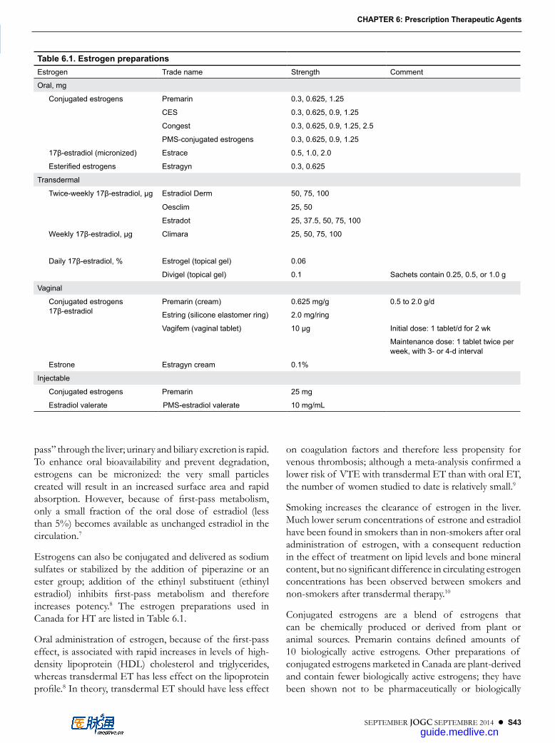
SEPTEMBER JOGC SEPTEMBRE 2014 l S43
CHAPTER 6: Prescription Therapeutic Agents
CHAPTER 6
pass” through the liver; urinary and biliary excretion is rapid. To enhance oral bioavailability and prevent degradation, estrogens can be micronized: the very small particles created will result in an increased surface area and rapid absorption. However, because of first-pass metabolism, only a small fraction of the oral dose of estradiol (less than 5%) becomes available as unchanged estradiol in the circulation.7
Estrogens can also be conjugated and delivered as sodium sulfates or stabilized by the addition of piperazine or an ester group; addition of the ethinyl substituent (ethinyl estradiol) inhibits first-pass metabolism and therefore increases potency.8 The estrogen preparations used in Canada for HT are listed in Table 6.1.
Oral administration of estrogen, because of the first-pass effect, is associated with rapid increases in levels of high-density lipoprotein (HDL) cholesterol and triglycerides, whereas transdermal ET has less effect on the lipoprotein profile.8 In theory, transdermal ET should have less effect
on coagulation factors and therefore less propensity for venous thrombosis; although a meta-analysis confirmed a lower risk of VTE with transdermal ET than with oral ET, the number of women studied to date is relatively small.9
Smoking increases the clearance of estrogen in the liver. Much lower serum concentrations of estrone and estradiol have been found in smokers than in non-smokers after oral administration of estrogen, with a consequent reduction in the effect of treatment on lipid levels and bone mineral content, but no significant difference in circulating estrogen concentrations has been observed between smokers and non-smokers after transdermal therapy.10
Conjugated estrogens are a blend of estrogens that can be chemically produced or derived from plant or animal sources. Premarin contains defined amounts of 10 biologically active estrogens. Other preparations of conjugated estrogens marketed in Canada are plant-derived and contain fewer biologically active estrogens; they have been shown not to be pharmaceutically or biologically
Table 6.1. Estrogen preparationsEstrogen Trade name Strength Comment
Oral, mg
Conjugated estrogens Premarin 0 .3, 0 .625, 1 .25
CES 0 .3, 0 .625, 0 .9, 1 .25
Congest 0 .3, 0 .625, 0 .9, 1 .25, 2 .5
PMS-conjugated estrogens 0 .3, 0 .625, 0 .9, 1 .25
17β-estradiol (micronized) Estrace 0 .5, 1 .0, 2 .0
Esterified estrogens Estragyn 0 .3, 0 .625
Transdermal
Twice-weekly 17β-estradiol, µg Estradiol Derm 50, 75, 100
Oesclim 25, 50
Estradot 25, 37 .5, 50, 75, 100
Weekly 17β-estradiol, µg Climara 25, 50, 75, 100
Daily 17β-estradiol, % Estrogel (topical gel) 0 .06
Divigel (topical gel) 0 .1 Sachets contain 0 .25, 0 .5, or 1 .0 g
Vaginal
Conjugated estrogens 17β-estradiol
Premarin (cream)
Estring (silicone elastomer ring)
0 .625 mg/g
2 .0 mg/ring
0 .5 to 2 .0 g/d
Vagifem (vaginal tablet) 10 µg Initial dose: 1 tablet/d for 2 wk
Maintenance dose: 1 tablet twice per week, with 3- or 4-d interval
Estrone Estragyn cream 0 .1%
Injectable
Conjugated estrogens Premarin 25 mg
Estradiol valerate PMS-estradiol valerate 10 mg/mL
guide.medlive.cn
S44 l SEPTEMBER JOGC SEPTEMBRE 2014
Managing Menopause
equivalent to Premarin,11 and the effects associated with the use of CEE in clinical trials may, therefore, not be attributed unconditionally to other conjugated estrogens. Preparations of CEE are available for oral, vaginal, and injectable use (Table 6.1).
Estradiol is available in oral, transdermal, and injectable delivery systems for systemic use and in delivery systems for local vaginal therapy. To be adequately absorbed when taken orally, estradiol must be micronized. Once absorbed, it is mostly converted to estrone; by contrast, transdermal application avoids hepatic first-pass metabolism, resulting in sustained concentrations of estradiol.8 Transdermal delivery systems in Canada are exclusively matrix patches, which contain an adhesive matrix in which the estradiol is dissolved. Depending on the system, patches must be changed once or twice weekly. Estradiol is also available in a gel formulation that is applied to the skin daily. This product is absorbed into the skin in 1 to 2 minutes, and serum concentrations reach steady state after the third daily administration. For local vaginal therapy, estradiol is available in the form of slow-release tablets or a silicone elastomer ring that releases estradiol in very low dose over 3 months.
The preparation of esterified estrogens available in Canada for oral therapy contains estrone sulfate and equilin sulfate.
PROGESTOGENS
Progestogens are not generally used as stand-alone treatment for menopausal symptoms, but they can be used to control VMS in women with contraindications to the use of estrogen. More usually, progestogens are used in combination with estrogen. The use of estrogen alone is associated with endometrial hyperplasia and cancer, and the addition of a progestogen to ET has been shown to reduce, but not eliminate, this risk in a dose- and duration-dependent fashion.12 Maximum protective effects are obtained with continuous progestogen use, and the risk increases with less than 16 days of progestogen per month.13
Three classes of progestogens are used in HT:
1. 17α-hydroxyprogesterone derivatives (including MPA, megestrol, and progesterone),
2. 19-nortestosterone derivatives (norethindrone, norethindrone acetate, and levonorgestrel), and
3. the spironolactone derivative drospirenone.
The progestogen preparations available for single-agent therapy in Canada are listed in Table 6.2. For postmenopausal use, drospirenone is currently available only in a combination
preparation with estradiol (Table 6.3), and levonorgestrel is only available in the Mirena intrauterine system.
The 17α-hydroxyprogesterone derivatives primarily exhibit progestational activity, although there are some notable differences between these agents. For example, micronized progesterone does not appear to antagonize the positive effects of CEE on HDL cholesterol levels, whereas MPA attenuates the estrogen-induced lipid effects.14 Additionally, differences in bleeding patterns may occur. Full secretory transformation of the endometrium occurs with the use of MPA, whereas daily doses of less than 300 mg of micronized progesterone have only antimitotic effects, which may result in less menstrual bleeding.15
The 19-nortestosterone derivatives have varying estrogenic, anti-estrogenic, progestogenic, and androgenic properties. These agents produce full secretory transformation of the endometrium, similar to the effect of MPA.16 Drospirenone is a unique progestogen whose biochemical and pharmacologic profile most closely resembles that of progesterone.17
THERAPEUTIC HORMONAL REGIMENS
Several therapeutic regimens for HT in postmenopausal women are used in Canada. In general, women should be offered a regimen containing both estrogen and progestogen unless they have had a hysterectomy; in that case, they do not need the endometrial protection provided by a progestogen.
Cyclic estrogen/progestogen regimensAlthough estrogen and progestogen have traditionally been used in a cyclic manner, with a hormone-free interval of 5 to 7 days at the end of each month, the rationale for this hormone-free interval is unclear; many women have reported the return of distressing symptoms. Theoretically, eliminating the hormone-free interval may be prudent to maintain adequate estrogen concentrations in the blood and to provide maximal symptomatic relief.
Cyclic EPT generally involves continuous estrogen use with 12 to 14 days of progestin use per month. Whitehead et al.18 demonstrated the critical importance of the duration of progestin therapy in stabilizing the endometrium and reducing the risk of hyperplasia. With longer therapy, continuous administration of a progestogen is more effective in preventing endometrial hyperplasia than is cyclic administration, and monthly cyclic use of progestogen is more effective than long-cycle use, in which the progestogen is administered every 3 months.19 However, cyclic withdrawal of the progestin may be beneficial in the breast: it has been
guide.medlive.cn

SEPTEMBER JOGC SEPTEMBRE 2014 l S45
CHAPTER 6: Prescription Therapeutic Agents
Table 6.2. Progestogen preparationsProgestogen Trade name Strength Comparable oral dose*
Oral, mg
Medroxyprogesterone acetate Apo-medroxy 2 .5, 5, 10, 100 5 .0
Dom-medroxyprogesterone 2 .5, 5, 10
Medroxy 2 .5 2 .5
Medroxy 5 5
PMS-medroxyprogesterone 2 .5, 5, 10
Provera 2 .5, 5, 10
Provera Pak 5 5 (14 tablets)
Provera Pak 10 10 (10 tablets)
Teva-medroxyprogesterone 2 .5, 5, 10
Megestrol Megestrol 40, 160 5 .0
Megace OS 40/mL (liquid)
Norethindrone Micronor 0 .35 0 .7 to 1 .0
Norethindrone acetate Norlutate 5 1 .0
Progesterone (micronized) Prometrium 100 200
Intravaginal, mg
Progesterone Crinone 8% (gel) 90
Endometrin (insert) 100
Injectable, mg/mL
Medroxyprogesterone acetate Depo-Provera 50 (5 mL) 150 (1 mL)
Medroxyprogesterone acetate injectable suspension
150 (1 mL)
Progesterone Progesterone Injection 50 (10 mL)
Intrauterine, mg
Levonorgestrel Mirena Intrauterine System 52 per IUS
*The Comparable oral dose for the various progestins is expected to have a similar effect on endometrium .6
Table 6.3. Combination productsCombination Trade name Strengths
Oral
Conjugated estrogens (CE) and medroxyprogesterone acetate (MPA) Premplus 0 .625 mg CE + 2 .5 mg MPA (2 tablets)
0 .625 mg CE + 5 mg MPA (2 tablets)
Premplus cycle 0 .625 mg CE (single tablet)
0 .625 mg CE + 10 mg MPA (2 tablets)
17β-estradiol (E2) and drospirenone (DRSP) Angeliq 1 mg E2 + 1 mg DRSP
17β-estradiol (E2) and norethindrone acetate (NETA) Activelle 1 mg E2 + 0 .5 mg NETA
ActivelleLD 0 .5 mg E2 + 0 .1 mg NETA
Transdermal
17β-estradiol (E2) and levonorgestrel (LNG) Climara Pro 45 μg E2 + 15 μg LNG
17β-estradiol (E2) and norethindrone acetate (NETA) Estalis 140/50 50 μg E2 + 140 μg NETA
Estalis 250/50 50 μg E2 + 250 μg NETA
guide.medlive.cn

S46 l SEPTEMBER JOGC SEPTEMBRE 2014
Managing Menopause
shown that withdrawal of progestin, but not continuous therapy, induces apoptosis in normal breast tissue.20
Continuous combined regimensAn alternative to the cyclic administration of a progestogen in postmenopausal HT is continuous daily treatment with both an estrogen and a progestogen. This method was developed to avoid the withdrawal bleeding associated with cyclic regimens. The most comprehensive data are derived from the WHI combined therapy study, which used 0.625 mg of CEE with 2.5 mg MPA per day.21 Other progestogens can be used. During the first 3 to 6 months 40% of women receiving this therapy have irregular breakthrough bleeding.22 However, most patients become amenorrheic by 12 months of use; after 12 months, irregular bleeding is more common in women receiving cyclic therapy than in those receiving continuous combined therapy.19
The levonorgestrel-releasing intrauterine system Mirena is currently indicated for contraception and the treatment of idiopathic menorrhagia. The device can be left in situ for 5 years and, as with other continuous progestin use, breakthrough bleeding may occur during the first months of use.6 The device may be used in postmenopausal women, in combination with systemic ET, and it appears to be superior to cyclic MPA therapy for endometrial protection.23
In theory, combined oral contraceptives (containing ethinyl estradiol with a progestin) can also be used in postmenopausal women. However, the daily dose of ethinyl estradiol in oral contraceptive preparations (at least 20 µg) is several times higher than the minimum dose required for symptom relief and bone benefit (5 µg).6 For this reason, use of combined oral contraceptive preparations in postmenopausal women should not be encouraged.
Commercial preparations containing both estrogen and a progestogen are shown in Table 6.3.
Individualizing therapy Estradiol therapy can have unpleasant side effects in some women. The use of transdermal estradiol 75-µg patches in a phase III clinical trial resulted in headache for 17% of women, nausea for 5.3%, and breast pain for 10.7%.24 These side effects may be dose-related and may respond to a reduction in dose, but they may also resolve with continued therapy at the same dose. Because they vary between preparations and routes of administration, side effects may also improve with a change in the preparation used.
Estrogen doses may be titrated to achieve control of symptoms. In general, therapy should begin with a low dose. Persisting VMS or vaginal dryness may indicate a
need to increase the dose or to change the preparation or route of administration, whereas breast tenderness or leukorrhea may require a reduction in dose.
Adverse reactions to progestogens are more frequent when the progestogens are given with ET.25 Side effects of progestogens include alterations in mood, breast tenderness, and bloating. Switching from one progestogen formulation to another may reduce these symptoms.6 Cyclic progestogen-associated side effects may be reduced or eliminated by switching to a continuous combined regimen.
Like estrogens, each progestogen preparation has a different side-effect profile. Synthetic progestins may have significant side effects, including fatigue, fluid retention, lipid level alterations, and dysphoria.26 Micronized progesterone has fewer side effects than synthetic progestins and is generally well tolerated.26 The micronized progesterone formulation Prometrium used to contain peanut oil but now contains sunflower oil and is no longer contraindicated in women allergic to peanuts.
Oral versus transdermal therapyOral and transdermal HT are each first-line options for systemic therapy, but transdermal therapy is more expensive. On the basis of current evidence, the following groups of women requiring HT should preferentially be offered transdermal therapy:6
1. women at high risk for VTE,2. women with malabsorption,3. women with spontaneous or estrogen-induced
hypertriglyceridemia, and4. and obese women with metabolic syndrome.
In each of these groups, there is evidence for adverse effects of oral ET.8,9 In addition, there is evidence suggesting an advantage in using transdermal rather than oral ET in women who smoke,10 women who have hypertension,27 and women with sexual dysfunction.28
Estrogen-only therapy The use of systemic estrogen alone is recommended only in women who do not have a uterus. The role of estrogen in causing endometrial proliferation and potentially endometrial hyperplasia has been well documented.3 Because of intolerable side effects or abnormal bleeding some women will choose to take estrogen unopposed by a progestogen. Because of the associated risk of endometrial hyperplasia and endometrial cancer, such women should have an endometrial biopsy at least annually. If women who have used estrogen alone subsequently switch to EPT, endometrial surveillance should continue because the
guide.medlive.cn
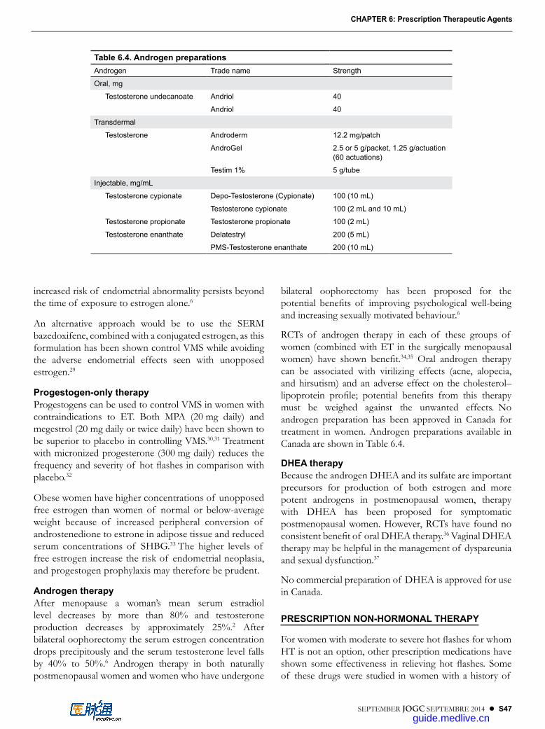
SEPTEMBER JOGC SEPTEMBRE 2014 l S47
CHAPTER 6: Prescription Therapeutic Agents
increased risk of endometrial abnormality persists beyond the time of exposure to estrogen alone.6
An alternative approach would be to use the SERM bazedoxifene, combined with a conjugated estrogen, as this formulation has been shown control VMS while avoiding the adverse endometrial effects seen with unopposed estrogen.29
Progestogen-only therapyProgestogens can be used to control VMS in women with contraindications to ET. Both MPA (20 mg daily) and megestrol (20 mg daily or twice daily) have been shown to be superior to placebo in controlling VMS.30,31 Treatment with micronized progesterone (300 mg daily) reduces the frequency and severity of hot flashes in comparison with placebo.32
Obese women have higher concentrations of unopposed free estrogen than women of normal or below-average weight because of increased peripheral conversion of androstenedione to estrone in adipose tissue and reduced serum concentrations of SHBG.33 The higher levels of free estrogen increase the risk of endometrial neoplasia, and progestogen prophylaxis may therefore be prudent.
Androgen therapyAfter menopause a woman’s mean serum estradiol level decreases by more than 80% and testosterone production decreases by approximately 25%.2 After bilateral oophorectomy the serum estrogen concentration drops precipitously and the serum testosterone level falls by 40% to 50%.6 Androgen therapy in both naturally postmenopausal women and women who have undergone
bilateral oophorectomy has been proposed for the potential benefits of improving psychological well-being and increasing sexually motivated behaviour.6
RCTs of androgen therapy in each of these groups of women (combined with ET in the surgically menopausal women) have shown benefit.34,35 Oral androgen therapy can be associated with virilizing effects (acne, alopecia, and hirsutism) and an adverse effect on the cholesterol–lipoprotein profile; potential benefits from this therapy must be weighed against the unwanted effects. No androgen preparation has been approved in Canada for treatment in women. Androgen preparations available in Canada are shown in Table 6.4.
DHEA therapyBecause the androgen DHEA and its sulfate are important precursors for production of both estrogen and more potent androgens in postmenopausal women, therapy with DHEA has been proposed for symptomatic postmenopausal women. However, RCTs have found no consistent benefit of oral DHEA therapy.36 Vaginal DHEA therapy may be helpful in the management of dyspareunia and sexual dysfunction.37
No commercial preparation of DHEA is approved for use in Canada.
PRESCRIPTION NON-HORMONAL THERAPY
For women with moderate to severe hot flashes for whom HT is not an option, other prescription medications have shown some effectiveness in relieving hot flashes. Some of these drugs were studied in women with a history of
Table 6.4. Androgen preparationsAndrogen Trade name Strength
Oral, mg
Testosterone undecanoate Andriol 40
Andriol 40
Transdermal
Testosterone Androderm 12 .2 mg/patch
AndroGel 2 .5 or 5 g/packet, 1 .25 g/actuation (60 actuations)
Testim 1% 5 g/tube
Injectable, mg/mL
Testosterone cypionate Depo-Testosterone (Cypionate) 100 (10 mL)
Testosterone cypionate 100 (2 mL and 10 mL)
Testosterone propionate Testosterone propionate 100 (2 mL)
Testosterone enanthate Delatestryl 200 (5 mL)
PMS-Testosterone enanthate 200 (10 mL)
guide.medlive.cn

S48 l SEPTEMBER JOGC SEPTEMBRE 2014
Managing Menopause
breast cancer. Data obtained from cancer survivors can reasonably be extrapolated to other populations of women.
In Canada, Dixarit (clonidine) and Bellergal Spacetabs (belladonna, ergotamine tartrate, and phenobarbital) are the only non-hormonal preparations approved for the treatment of VMS.
AntidepressantsNewer antidepressant drugs have become the most promising class of non-hormonal agents for treatment of VMS. These agents affect the release and reuptake of neurotransmitters, principally serotonin and norepinephrine, at multiples sites in the central nervous system.
Venlafaxine was the first antidepressant drug to undergo clinical investigation for the treatment of VMS. This agent inhibits both serotonin and norepinephrine reuptake. In an RCT of venlafaxine treatment of 191 women after breast cancer treatment, 37.5, 75, or 150 mg daily resulted in a significant reduction of hot flashes compared with placebo.38 Significant improvement compared with placebo (51% vs. 15% reduction) was also found in an RCT of 80 healthy postmenopausal women.39 The women treated with venlafaxine experienced adverse effects more frequently than did those treated with placebo.
For VMS the dose of venlafaxine should begin at 37.5 mg per day and increase to 75 mg/d after 1 week if the hot flashes are not optimally reduced. There is no reason to use a higher dose, because side effects increase without additional benefit.
Treatment with desvenlafaxine has also been shown in RCTs to offer more relief of moderate to severe VMS (> 50 hot flashes per week) than placebo.40 The minimum effective dose is 100 mg daily.
Paroxetine and fluoxetine, both SSRIs, have demonstrated efficacy in the treatment of VMS. RCTs have shown a 50% to 60% decrease in hot flashes with controlled-release paroxetine, 12.5 mg or 25 mg/d, and with fluoxetine, 20 mg/d.41,42 However, in an RCT comparing fluoxetine with citalopram and placebo, after 6 months of treatment the reduction in hot flashes was similar in each group (62%, 64%, and 58%, respectively).43
GabapentinGabapentin is a γ-aminobutyric acid analogue that is used to treat a variety of neurologic disorders, including epilepsy and neuropathic pain. Its specific mechanism of action is unclear. Several RCTs followed initial observations that gabapentin was associated with reduced VMS in women using it for neurologic conditions. A study of gabapentin in 420 women
with breast cancer found after 8 weeks of treatment that hot-flash severity was reduced by 15% in the placebo group, by 31% in the women who took gabapentin, 300 mg/d, and by 46% in the women who took gabapentin, 900 mg/d.44 A randomized crossover study comparing the use of venlafaxine with gabapentin in women after treatment for breast cancer (4 weeks of treatment with each) showed a 66% reduction in hot-flash scores with each agent; however, 68% of the women preferred venlafaxine to gabapentin because of gabapentin’s side effects (chiefly dizziness and increased appetite).45
ClonidineClonidine has been used for many years as an antihypertensive agent. It is a centrally acting α-adrenergic receptor agonist with potential to reduce hot flashes. An 8-week RCT found that oral administration of clonidine, 0.1 mg/d, marginally reduced hot flashes in 194 women (by 38% vs. 30% for placebo).46 However, clonidine was associated with more side effects than placebo, including dizziness, dry mouth, drowsiness, and constipation.
BellergalBellergal is a compound containing phenobarbital, ergotamine, and belladonna that has been in clinical use for many years. Clinical trials of this combination for VMS showed a small benefit favouring it over placebo.47 However, this combination is associated with several adverse effects, including dry mouth, constipation, and sedation.
CONTRAINDICATIONS TO HT
EstrogenFollowing are contraindications to estrogen use.
1. Unexplained vaginal bleeding 2. Acute liver dysfunction3. Estrogen-dependent cancer (endometrial, breast)4. Coronary heart disease5. Previous stroke6. Active thromboembolic disease
Caution and careful consideration should be applied in recommending ET for women after treatment for breast or endometrial cancer, but a family history of these conditions is not a contraindication.
ProgestogenFollowing are contraindications to progestogen use.
1. Unexplained vaginal bleeding2. Breast cancer3. Peanut allergy (micronized progesterone therapy
only)
guide.medlive.cn

SEPTEMBER JOGC SEPTEMBRE 2014 l S49
CHAPTER 6: Prescription Therapeutic Agents
As with ET, the list of contraindications to progestogen therapy is evolving. Generally agreed-upon contraindications to progestogen use are a known allergy to a specific preparation and known or suspected breast cancer. Caution should be applied in recommending progestogen use to women with chronically impaired liver function. There appears to be no reason to withhold progestogen treatment from women with thrombovascular disease.
AndrogenThe use of androgen is contraindicated in women with extensive cardiac, hepatic, or renal disease.48
DRUG INTERACTIONS
Many drugs and environmental agents (such as cigarette smoke) can induce or inhibit the enzymes involved in metabolism of both estrogen and progestogens and therefore have the capacity to alter clearance of the hormones from the circulation. Prescribers should use a reliable source, such as a pharmacist or drug interaction reference, to ensure that drugs prescribed for women already receiving HT will not produce a change in hormone activity.
CUSTOM-COMPOUNDED (“BIO-IDENTICAL”) HT
The term “bio-identical” HT has no agreed-upon definition but is most commonly taken to refer to custom-compounded combinations of hormones, usually estrogens, prepared by a compounding pharmacy. Because these combinations are not prepared under regulatory supervision, their quality and composition may vary significantly.49 The safety and effectiveness of such preparations have not been assessed in the way that preparations approved by regulatory bodies must be assessed; in the absence of such assessment, the compounded preparations are often inappropriately deemed “safe” and may be advertised as such. Consumers and prescribers should be aware that the content of custom-compounded HT in Canada is unregulated and its safety unknown.
REFERENCES
1. Campbell AD, Collip JB. The clinical use of the ovary stimulating hormone of the placenta. Can Med Assoc J 1930;22:219–20.
2. Kinch RA. Dr. J.B. Collip and the development of Premarin. J Obstet Gynaecol Can 2008;30:244–5.
3. Thom MH, Studd JW. Oestrogens and endometrial hyperplasia. Br J Hosp Med 1980;23:506–13.
4. The North American Menopause Society. Menopause glossary [website]. Mayfield Heights, Ohio: NAMS; 2014. Available at: http://www.menopause.org/for-women/menopause-glossary. Accessed 2013 Jan 4.
5. Hall E, Frey BN, Soares CN. Non-hormonal treatment strategies for vasomotor symptoms: a critical review. Drugs 2011:71:287–304.
6. Fritz MA, Speroff L. Clinical Gynecologic Endocrinology and Infertility. 8th ed. Philadelphia: Lippincott Williams & Wilkins; 2011.
7. Faigle JW, Schenkel L. Pharmacokinetics of estrogens and progestogens. In: Fraser IS, Jansen RPS, Lobo RA, Whitehead MI, editors. Estrogens and Progestogens in Clinical Practice. London: Churchill Livingstone; 1998. p. 273–94.
8. Levin ER, Hammes SR. Estrogens and progestins. In: Brunton LL, Chabner BA, Knollmann BC, editors. Goodman & Gilman’s The Pharmacological Basis of Therapeutics, 12th ed. New York: McGraw-Hill; 2011. Available at: http://accessmedicine.mhmedical.com.ezproxy.library.ubc.ca/content.aspx?bookid=374&Sectionid=41266249. Accessed 2014 Mar 15.
9. Canonico M, Plu-Bureau G, Lowe GD, Scarabin PY. Hormone replacement therapy and risk of venous thromboembolism in postmenopausal women: systematic review and meta-analysis. BMJ 2008;336:1227–31.
10. Geisler J, Omsjo IH, Helle SI, Ekse D, Silsand T, Lonning PE. Plasma oestrogen fractions in postmenopausal women receiving hormone replacement therapy: influence of route of administration and cigarette smoking. J Endocrinol 1999;162:265–70.
11. Bhavnani BR, Nisker JA, Martin J, Aletebi F, Watson L, Milne JK. Comparison of pharmacokinetics of a conjugated equine estrogen preparation (Premarin) and a synthetic mixture of estrogens (C.E.S.) in postmenopausal women. J Soc Gynecol Invest 2000;7:175–83.
12. Beresford SA, Weiss NS, Voigt LF, McKnight B. Risk of endometrial cancer in relation to use of estrogen combined with cyclic progestin therapy in women. Lancet 1997;349:458–61.
13. Weiderpass E, Adami HO, Baron JA, Magnusson C, Bergstrom R, Lindgren A, et al. Risk of endometrial cancer following estrogen replacement with or without progestins. J Natl Cancer Inst 1999;91:1131–7.
14. The Postmenopausal Estrogen/Progestin Interventions (PEPI) Trial Investigators. Effects of estrogen or estrogen/progestin regimens on heart disease risk factors in postmenopausal women: the Postmenopausal Estrogen/Progestin Interventions (PEPI) Trial. J Am Med Assoc 1995;273:199–208.
15. Moyer DL, de Lignieres B, Driguez P, Pez JP. Prevention of endometrial hyperplasia by progesterone during long-term estradiol replacement: influence of bleeding pattern and secretory changes. Fertil Steril 1993;59:992–7.
16. Padwick ML, Pryse-Davies J, Whitehead MI. A simple method for determining the optimal dosage of progestin in postmenopausal women receiving estrogens. N Eng J Med 1986;315:930–4.
17. Krattenmacher R. Drospirenone: pharmacology and pharmacokinetics of a unique progestogen. Contraception 2000;62:29–38.
18. Whitehead MI, Townsend PT, Pryse-Davies J, Ryder T, Lane G, Siddle NC, et al. Effects of various types and dosages of progesterones on the postmenopausal endometrium. J Reprod Med 1982;27;539–48.
19. Lethaby A, Suckling J, Barlow D, Farquhar CM, Jepson RG, Roberts H. Hormone replacement therapy in postmenopausal women: endometrial hyperplasia and irregular bleeding. Cochrane Database Syst Rev 2004;(3):CD000402.
20. Desreux J, Kebers F, Noël A, Francart D, Van Cauwenberge H, Heinen V, et al. Effects of a progestogen on normal human breast epithelial cell apoptosis in vitro and in vivo. Breast 2003;12:142–9.
21. Rossouw JE, Anderson GL, Prentice RL, LaCroix AZ, Kooperberg C, Stefanick ML, et al; Writing Group for the Women’s Health Initiative Investigators. Risks and benefits of estrogen plus progestin in healthy postmenopausal women: principal results from the Women’s Health Initiative randomized controlled trial. JAMA 2002;288:321–33.
guide.medlive.cn

S50 l SEPTEMBER JOGC SEPTEMBRE 2014
Managing Menopause
22. Archer DF, Pickar JH, Bottiglioni F. Bleeding patterns in postmenopausal women taking continuous combined or sequential regimens of conjugated estrogens with medroxyprogesterone acetate. Menopause Study Group. Obstet Gynecol 1994;83:686–92.
23. Sturridge F, Guillebaud J. A risk–benefit assessment of the levonorgestrel-releasing intrauterine system. Drug Saf 1996;15:430–40.
24. Archer DF, Pickar JH, MacAllister DC, Warren MP. Transdermal estradiol gel for the treatment of symptomatic postmenopausal women. Menopause 2012;19:622–9.
25. Prior JC, Alojado N, McKay DW, Vigna YM. No adverse effects of medroxyprogesterone treatment without estrogen in postmenopausal women: double-blind, placebo-controlled, crossover trial. Obstet Gynecol 1994;83:24–8.
26. Goletiani NV, Keith DR, Gorsky SJ. Progesterone: review of safety for clinical studies. Exp Clin Psychopharmacol 2007;15:427–44.
27. Vongpatanasin W, Tuncel M, Mansour Y, Arbique D, Victor RG. Transdermal estrogen replacement therapy decreases sympathetic activity in postmenopausal women. Circulation 2001;103:2903–8.
28. Shifren JL, Desindes S, McIlwain M, Doros G, Mazer NA. A randomized, open-label, crossover study comparing the effects of oral versus transdermal estrogen therapy on serum androgens, thyroid hormones, and adrenal hormones in naturally menopausal women. Menopause 2007;14:985–94.
29. Mirkin S, Pickar JH. Management of osteoporosis amd menopausal symptoms: focus on bazedoxifene/conjugated estrogen combination. Int J Womens Health 2013;5:465–75. Epub 2013 Aug 7.
30. Schiff I, Tulchinsky D, Cramer D, Ryan K. Oral medroxyprogesterone in the treatment of postmenopausal symptoms. J Am Med Assoc 1980;244:1443–5.
31. Goodwin JW, Green SJ, Moinpour CM, Bearden JD, Giguere JK, Jiang CS, et al. Phase III randomized placebo-controlled trial of two doses of megestrol acetate as treatment for menopausal symptoms in women with breast cancer: Southwest Oncology Group Study 9626. J Clin Oncol 2008;26:1650–6.
32. Hitchcock CL, Prior JC. Oral micronized progesterone for vasomotor symptoms: a placebo-controlled randomized trial in healthy postmenopausal women. Menopause 2012;19:886–93.
33. MacDonald PC, Siiteri PK. The relationship between the extraglandular production of estrone and the occurrence of endometrial neoplasia. Gynecol Oncol 1974;2:259–63.
34. Braunstein GD, Sundwall DA, Katz M, Shifren JL, Buster JE, Simon JA, et al. Safety and efficacy of a testosterone patch for the treatment of hypoactive sexual desire disorder in surgically menopausal women: a randomized, placebo-controlled trial. Arch Intern Med 2005;165:1571–2.
35. Panay N, Al-Azzawi F, Bouchard C, Davis SR, Eden J, Lodhi I, et al. Testosterone treatment of HSDD in naturally menopausal women: the ADORE study. Climacteric 2010;13:121–31.
36. Davis SR, Panjari M, Stanczyk FZ. DHEA replacement for postmenopausal women. J Clin Endocrinol Metab 2011;96:1542–53.
37. Labrie F, Archer DF, Bouchard C, Fortier M, Cusan L, Gomez JL, et al. Intravaginal dehydroepiandrosterone (prasterone), a highly efficient treatment of dyspareunia. Climacteric 2011;14:282–8.
38. Loprinzi CL, Kugler JW, Sloan JA, Mailliard JA, LaVasseur BI, Barton DL, et al. Venlafaxine in management of hot flashes in survivors of breast cancer: a randomized controlled trial. Lancet 2000;356:2059–63.
39. Evans ML, Pritts E, Vittinghoff E, McClish K, Morgan KS, Jaffe RB. Management of postmenopausal hot flushes with venlafaxine hydrochloride: a randomized, controlled trial. Obstet Gynecol 2005;105:161–6.
40. Hall E, Frey BN, Soares CN. Non-hormonal treatment strategies for vasomotor symptoms: a critical review. Drugs 2011;71:287–304.
41. Loprinzi CL, Sloan JA, Perez EA, Quella SK, Stella PJ, Mailliard JA, et al. Phase III evaluation of fluoxetine for treatment of hot flashes. J Clin Oncol 2002;20:1578–83.
42. Stearns V, Beebe KL, Iyengar M, Dube E. Paroxetine controlled release in the treatment of menopausal hot flashes: a randomized controlled trial. J Am Med Assoc 2003;289:2827–34.
43. Suvanto-Luukkonen E, Koivunen R, Sundstrom H, Bloigu R, Karjalainen E, Haiva-Mallinen L, et al. Citalopram and fluoxetine in the treatment of postmenopausal symptoms: a prospective, randomized, 9-month, placebo-controlled, double-blind study. Menopause 2005;12:18–26.
44. Pandya KJ, Morrow GR, Roscoe JA, Zhao H, Hickok JT, Pajon E, et al. Gabapentin for hot flashes in 420 women with breast cancer: a randomised double-blind placebo-controlled trial. Lancet 2005;366:818–24.
45. Bordeleau L, Pritchard KI, Loprinzi CL, Ennis M, Jugovic O, Warr D, et al. Multicenter, randomized, cross-over clinical trial of venlafaxine versus gabapentin for the management of hot flashes in breast cancer survivors. J Clin Oncol 2010;28:5147–52.
46. Pandya KJ, Raubertas RF, Flynn PJ, Hynes HE, Rosenbluth RJ, Kirshner JJ, et al. Oral clonidine in postmenopausal patients with breast cancer experiencing tamoxifen-induced hot flashes: a University of Rochester Cancer Center Community Clinical Oncology Program study. Ann Intern Med 2000;132:788–93.
47. Shanafelt TD, Barton LB, Adjei AA, Loprinzi CL. Pathophysiology and treatment of hot flashes. Mayo Clin Proc 2002;77:1207–18.
48. Basaria S, Dobs AS. Safety and adverse effects of androgens: how to counsel patients. Mayo Clin Proc 2004;79(4 Suppl):S25–S32.
49. U.S. Food and Drug Administration. Menopausal hormone therapy and “bio-identical” hormones. In: Compounded menopausal hormone therapy questions and answers. Silverspring, Maryland: USA FDA; 2013. Available at: http://www.fda.gov/Drugs/GuidanceComplianceRegulatoryInformation/PharmacyCompounding/ucm183088.htm. Accessed 2014 Feb 26.
guide.medlive.cn

SEPTEMBER JOGC SEPTEMBRE 2014 l S51
CHAPTER 7
Ongoing Management of Menopausal Women and Those With Special Considerations
The goal of this chapter is to give practical tips to health care providers for the management of symptomatic women
during and after the menopausal transition, concerning mostly the initiation and follow-up of HT. The reader may refer to previous chapters for more extensive literature review.
DEFINITIONS
Menopause is caused by the permanent cessation of menstruation resulting from the loss of ovarian follicular activity. Natural post-menopause is recognized to have occurred after 12 consecutive months of amenorrhea for which there is no other obvious pathological or physiological cause. Menopause is the final menstrual period, which is known with certainty only in retrospect 1 year or more after the event. An adequate independent biologic marker for the event does not exist, and there is no place for performing serial measurements of the serum concentration of estradiol or FSH in an attempt to specify whether the final menstrual period has passed.1 The term perimenopause is used to define the period immediately before menopause (when the endocrinologic, biologic, and clinical features of approaching menopause commence) until the end of the first year after menopause. The STRAW (Stages of Reproductive Aging Workshop) system used a change of 7 or more days of the menstrual cycle to indicate perimenopause (Figure 7.1).2 Again, there is no biologic marker. The serum estradiol and FSH measurements are extremely variable during that period of intense hormonal variation. During perimenopause there are periods of hyperestrogenism followed by periods of hypoestrogenism and frequent periods of anovulation.3 “Menopausal transition” refers to the time before the final menstrual period when variability in the menstrual cycle is usually increased. Menopause that occurs at an age less than 2 standard deviations below the mean age of menopause in the reference population is called premature menopause. In practice, without reliable estimates of the distribution of age at natural menopause in developing countries, the age of 40 years is used frequently as an arbitrary cut-off point, below which menopause is said to be premature. Premature menopause may be spontaneous, a manifestation of an
autoimmune disorder, or induced by disease, medication, irradiation, or surgery. It may also simply be menopause occurring at the outer limits of the normal curve. POF is characterized by amenorrhea and consistently high serum FSH levels in women under age 40. Presumptive autoimmune ovarian failure is not absolute: spontaneous remissions may be experienced by 5% to 25% of all women with presumed POF.4
ASSESSMENT AND MANAGEMENT OF PERIMENOPAUSAL WOMEN
Perimenopausal women may have multiple complaints related to anovulation and fluctuation of ovarian follicular activity. They often present with abnormal uterine bleeding, which should be investigated and managed appropriately.5 Perimenopausal women who have frequent menstrual bleeding and bothersome VMS may benefit from combined hormonal contraception. Two studies of perimenopausal women have shown a reduction of VMS with use of an oral contraceptive containing 0.02 or 0.03 mg of ethinyl estradiol.6,7 Birth control strategies should always be assessed. Even though the fertility rate is very low during menopausal transition, it is not zero. A pregnancy at that time is often unwelcome and has more risks for the mother and the fetus. Contraindications to the use of combined hormonal contraception include a history of thromboembolic events, CVD, migraine, hormone-sensitive carcinoma, jaundice, and liver disease. Smokers over age 35 should not use oral contraceptives.8 Perimenopausal women may also take advantage of several non-contraceptive benefits when using these agents. For a review of oral contraceptive use during menopausal transition, please refer to the Canadian contraception consensus.9 If women have long periods of amenorrhea (more than 3 to 4 months) and bothersome menopausal symptoms, HT is a good choice. A cyclic regimen with 12 to 14 days of progestogen use is often preferred because of the higher risk of uterine bleeding in women starting HT soon after their last menstrual period (late perimenopause or early postmenopause).10
guide.medlive.cn

S52 l SEPTEMBER JOGC SEPTEMBRE 2014
Managing Menopause
ASSESSMENT OF POSTMENOPAUSAL WOMEN
Unexpected vaginal bleedingUnexpected vaginal bleeding in postmenopausal women should be investigated, as endometrial cancer will be the cause in approximately 10%, or 1% to 25% depending upon risk factors.11,12 The most common cause of postmenopausal vaginal bleeding is atrophy of the vaginal mucosa or the endometrium. In recently postmenopausal women, endometrial hyperplasia, polyps, or submucosal fibroids are also common causes.13 Evaluation should include careful history-taking, assessment of risk factors, and a complete pelvic examination to find the bleeding site. The primary goal in the diagnostic evaluation is to exclude malignant disease. To exclude endometrial cancer, office endometrial biopsy is the first choice, but transvaginal ultrasonography with
measurement of the thickness of the endometrial stripe may be used initially to evaluate the endometrium. Dilatation and fractional curettage will be indicated in specific conditions.14–16
Hysteroscopy or sonohysterography is the next appropriate diagnostic procedure to evaluate the uterine cavity.
Hypoestrogenism symptomsThe most frequent complaints of postmenopausal women are VMS and sleep disturbance. They may also suffer from psychological problems, such as anxiety and mood symptoms.17 Major depression should be excluded if mood symptoms are significant. When quality of life is reduced by such symptoms, pharmacologic treatment should be offered. The most effective treatment for menopausal symptoms is HT. For women who cannot or do not wish to use HT, non-hormonal alternatives are detailed in Chapter 6.
Stage -5 -4 -3b -3a -2 -1 +1a +1b +1c +2Reproductive Menopausal Transition Post-menopause
Terminology Early Peak Late Early Late Early LatePeri-menopause
Duration Variable Variable 1-3 years 2 years (1+1) 3-6 years Remaining lifespan
Principal criteriaMenstrual cycle Variable to
regularRegular Regular Subtle
changes in flow/strength
Variable length:
Persistent ≥ 7-day
difference in length of consecutive
cycles
Interval of amenorrhea of ≥ 60 days
Supportive criteriaEndocrine
FSH Low Variable* Variable > 25 IU/L* Variable Stabilizes
AMH Low Low Low Low Low Very lowInhibin B Low Low Low Low Very low
Antral follicle count
Low Low Low Low Very low Very low
Descriptive characteristicsSymptoms Vasomotor
symptoms Likely
Vasomotor symptoms Most likely
Increasing symptoms
of urogenital atrophy
Figure 7.1. The Stages of Reproductive Aging Workshop + 10 staging system for reproductive aging in women
Menarche FMP (0)
FMP: final menstrual period; AMH: anti-Müllerian hormone*Blood drawn on cycle days 2 to 5
Elevated•Approximate expected level based on assays using current international pituitary standardReproduced with permission of the Endocrine Society from Harlow SD et al .2
guide.medlive.cn

SEPTEMBER JOGC SEPTEMBRE 2014 l S53
CHAPTER 7: Ongoing Management of Menopausal Women and Those With Special Considerations
MANAGEMENT OF POSTMENOPAUSAL WOMEN
As discussed in previous chapters, women must be encouraged to start or maintain non-pharmacologic strategies to prevent chronic disease: exercise, proper nutrition, stress management, and adequate intake of calcium and vitamin D. When menopausal symptoms do not significantly alter the quality of life, non-pharmacologic strategies are the first treatment choice; these are mostly lifestyle modifications. HT should not be prescribed if the only objective is the prevention of chronic disease.
Before prescribing HT the care provider should review the benefits and risks (Table 7.1), as well as contraindications (Table 7.2), with the woman. This review must be individualized and take into account the woman’s particular profile of risks and benefits.
Hormonal regimens Women with an intact uterus must have a combined regimen: an estrogen with a progestogen. The estrogen component is given continuously, and the progestogen component may be given cyclically (12 or more days a month) or continuously.18,19 Each regimen is reviewed in Chapter 6. We strongly disagree with the use of estrogen alone in a woman with an intact uterus.
Women with severe side effects of oral progestogen therapy may try a progestin intrauterine device. The levonorgestrel-releasing intrauterine system is currently indicated for contraception. The device can be left in
situ for 5 years. As with other continuous progestin use, breakthrough bleeding may occur during the first months of use. Current research suggests potential for use of such a device in postmenopausal women in combination with systemic estrogen administration.10
For women who choose an estrogen-only regimen, baseline and annual endometrial monitoring is mandatory. Infrequently, women without a uterus but with a history of severe endometriosis may benefit from a continuous combined regimen to prevent recurrence of the disease.
Bleeding with HTA cyclic regimen is often preferred in a recently postmenopausal woman or a late-perimenopausal woman because of the higher risk of abnormal uterine bleeding with a continuous combined regimen. Monthly withdrawal bleeding occurs in 80% to 90% of women on a cyclic regimen after the last dose of progestogen or during the last days of taking it.10,19 After 1 or 2 years women may choose to switch to a continuous combined regimen to stop bleeding or may stay on the same regimen if the bleeding is not bothersome. Women on a cyclic regimen with unscheduled or abnormal bleeding persisting after 6 months of initiation of the cyclic HT must be evaluated to exclude endometrial cancer.
Although the goal of a continuous regimen is to induce amenorrhea, data from the Menopause Study Group10 demonstrated that the prevalence of bleeding is related to the number of years since menopause. Women who were
Table 7.1. Benefits and risks of HTBenefits Risks
Reduction of VMSReduction of sleep problemsReduction of mood or anxiety problemsReduction of aches and painsOsteoporosis prevention and treatmentReversal of vulvar and vaginal atrophy (local ET if such atrophy is the only indication for therapy)
Thromboembolic eventsStrokeBreast cancer (after 4 to 5 years of EPT)Coronary heart disease (for women older than 60 and those who are more than 10 years after menopause)Endometrial hyperplasia and cancer (with estrogen-only regimens)
Table 7.2. Contraindications to HTContraindications to estrogens Contraindications to progestogens
Unexplained vaginal bleeding Acute liver dysfunctionEstrogen-dependent cancer (endometrial and breast cancer)Coronary heart diseasePrevious strokeActive thromboembolic disease
Unexplained vaginal bleeding Breast cancerPeanut allergy (prescribe micronized progesterone only)
guide.medlive.cn

S54 l SEPTEMBER JOGC SEPTEMBRE 2014
Managing Menopause
more than 3 years past menopause were less likely to have any bleeding during the first year of HT than women less than 2 years past menopause.10, 20 Forty percent of women receiving this therapy have irregular breakthrough bleeding during the first three to six months. The majority of patients who persist with the medication become amenorrheic by 12 months of use.21 Women on continuous HT who still have bleeding after six months of therapy must have evaluation of the endometrium.
Women on continuous HT who have uterine bleeding after a period of amenorrhea and women on cyclic HT in whom abnormal or unscheduled bleeding develops after a period of regular and normal withdrawal bleeding should undergo evaluation of the endometrium. If no pathological condition is found in the endometrium and the endometrial sampling shows proliferative endometrium, the dose of progestogen should be increased. If the endometrial sample shows atrophy, the dose of progestogen may be lowered or, if the lowest dose is being used in continuous HT, the number of days per month of progestogen use may be reduced (often to 21 days per month). If the uterine bleeding is persistent, a more thorough evaluation should be performed, including diagnostic and/or operative hysteroscopy.
Lack of effectiveness of HTAt the initiation of HT, the goals of therapy and the woman’s expectations related to HT should be reviewed. Adequate relief is often a reduction of symptoms and a better quality of life. Usually women do not seek complete resolution of symptoms. When lower doses of HT are used, the delay before adequate relief may be up to six weeks. It is therefore important to advise women to be prepared to wait 6 to 8 weeks before modifying the regimen. In the rare case in which a woman does not report an adequate response to HT, if the woman was using low-dose estrogen the standard dose should then be prescribed. It is important to review the application technique for a transdermal gel. If a woman does not respond to a standard doses of estrogen, the route of administration may be changed. Those using oral ET may have a 6- to 8-week trial of transdermal ET. If the response is again inadequate, the serum estradiol level may be measured: it should be between 200 and 400 pmol/L. Very rarely, a woman with normal menopause (not premature or due to POF) will need a higher dose of estrogen. It is essential that other potential causes of VMS (hyperthyroidism, underlying malignant disease, infection, use of SSRIs) be considered when hot flashes persist despite presumably adequate doses of estrogen.
When the only complaint is lack of efficacy of systemic treatment for vaginal atrophy, adding a vaginal preparation of estrogen is the first choice.
Side effects of HTCommon complaints in women on ET include breast tenderness, nausea, headache, and bloating. These side effects are often dose-related and may resolve with continued use or a decrease in dose. Because the side effects vary among currently available estrogen preparations, substituting another preparation for a poorly tolerated one is a reasonable strategy.
Side effects of progestins include alterations in mood, breast tenderness, and bloating. Switching to another progestin formulation may reduce these symptoms. Cyclic progestin-associated side effects may be reduced or eliminated by switching to a continuous combined regimen. Like estrogens, each progestin preparation has a different side-effect profile. For example, micronized progesterone can cause sedation and should therefore be administered at bedtime.
Duration of HTWomen receiving HT must be evaluated annually, with the risk–benefit profile as well as the woman’s expectations reviewed. Women using combined estrogen–progestogen HT should be informed of the increased risk of breast cancer after 4 to 5 years of HT. Women using ET do not have an increased breast cancer risk for at least 8 years. There is no clear recommended duration of therapy, and each woman should decide when it is time to stop HT. The lowest effective doses must be used. Women using standard doses should be advised to lower the dose after a few years. In women with persistent menopausal symptoms, extended use may be required. One quarter of women who discontinue HT are still symptomatic, with hot flashes or sleep disturbance, or feel that their quality of life was better with therapy than without. Some older women will choose to restart HT. Experts and data on surrogate CVD markers suggest that after 6 months without HT a woman may be considered a new user. Older postmenopausal women (more than 60 years old and/or more than 10 years postmenopausal) have greater cardiovascular risks associated with HT. Women planning HT discontinuation should be advised to quickly consult their health care provider if bothersome symptoms return.
HT cessationMany women have no trouble stopping HT and are able to do so without assistance from their health care provider. However, for others—in particular, women with severe hot flashes before the state of therapy—stopping ET can be quite difficult.22 Approximately 50% of women have a recurrence of VMS after discontinuing HT, independent of age and duration of use.23 In one RCT, tapering the dose of HT for 1 month and abruptly discontinuing HT had a similar impact on VMS.24 Women may choose
guide.medlive.cn

SEPTEMBER JOGC SEPTEMBRE 2014 l S55
CHAPTER 7: Ongoing Management of Menopausal Women and Those With Special Considerations
abrupt discontinuation or tapering of the dose. Those who experience bothersome symptoms after stopping abruptly may benefit from a second attempt using tapering. In patients with a history of severe VMS at baseline, very gradual tapering of the dose (over a few months) is probably preferable.
SPECIFIC MEDICAL CONSIDERATIONS
Premature or early menopauseSuch attention has been paid to the risks and benefits of HT in older postmenopausal women, and to changes in the paradigm for HT in women at physiologic menopause, that there has been confusion about the health and management issues for women undergoing an early onset of menopause. Women may experience premature menopause (before the age of 40) because of POF or through damage to ovarian function. Similarly, women may experience an earlier menopause (at age 40 to 45) through normal distribution around the mean or because of ovarian damage. These conditions require specific assessment and management.
Owing to the risk of health consequences, such as osteoporosis and sexual dysfunction, long-term HT should be offered to all women with POF.25–27 In younger women, higher doses of estrogen than are usually required in older postmenopausal women may be needed to relieve symptoms.28 Oral contraceptives may be used to achieve symptom relief. Women with POF appear to be at risk of premature CVD. In women with a normal karyotype, new research shows that there is an early onset of endothelial dysfunction associated with POF that is reversed by HT within 6 months of treatment.29 Many women with POF will have fertility concerns. In the absence of spontaneous remission, oocyte donation for in vitro fertilization offers the best potential for conception.
Induced menopause, whether from surgery, ionizing radiation, or chemotherapy, is unlike natural menopause in that the ovarian sources of androgen and estrogen are removed prematurely and simultaneously. Compared with natural menopause, induced menopause may have different physiologic effects on the rate of loss of bone mass and on the rates of atherosclerosis, vulvovaginal atrophy, and libido.
Women with POF or premature menopause receiving HT will be exposed to lower monthly estrogen levels than if they had spontaneous menstrual cycles. Recommendations to use HT for symptoms only and for the shortest duration possible in the lowest dosage are not applicable. HT must be offered until the usual age of menopause and thereafter as the result of a discussion between the woman and her physician.
Cancer With the exception of meningioma, breast, and widespread endometrial cancer, there is no biologic evidence that HT may increase the risk of recurrence.30 Thrombosis risk should be assessed in women who have had cancer, as some cancers are known to increase the risk of venous thromboembolic disease.
Endometriosis Combined HT in standard doses does not appear to cause regrowth of endometriotic tissue in postmenopausal women or in women receiving estrogen–progestin “add-back” therapy after medical oophorectomy with GnRH analogues.31 A small subgroup of women may experience recurring pain and other symptoms during unopposed ET, particularly if there is residual disease after definitive surgery.
In a prospective randomized trial of HT among women with endometriosis who underwent bilateral salpingo-oophorectomy there was no recurrence at 45 months among those who did not receive HT, whereas there was recurrence in 0.9% per year among those receiving HT. Among those with more than 3 cm of peritoneal involvement detected at the initial operation there was recurrence in 2.4% per year, and among those with incomplete surgery there was recurrence in 22.2%.32
In the absence of evidence from randomized studies, symptomatic endometriosis or large residual volumes of endometriotic tissue may be indications for progestin therapy after hysterectomy, either as part of a continuous combined regimen or as progestin-only therapy.33
There are case reports of endometrial cancer developing in residual endometriotic tissue in women receiving unopposed ET as well as in obese women with high levels of endogenous estrogen after abdominal hysterectomy and bilateral salpingo-oophorectomy for endometriosis.33–39
Women with a history of endometriosis can be offered HT, with the lowest effective dose of estrogen, for menopausal symptoms. There are no convincing data to support the routine use of EPT rather than ET for women with a history of endometriosis, nor is there evidence that use of progestin-only therapy or the withholding of estrogen for 6 months after definitive surgery will reduce the risk of recurrence or malignant disease. These options remain a matter of clinical judgement and informed choice.
Fibroids Although uterine fibroids do not constitute a contraindication to HT, both estrogen and progestin can influence fibroid growth. The doses in conventional HT regimens are usually
guide.medlive.cn

S56 l SEPTEMBER JOGC SEPTEMBRE 2014
Managing Menopause
not sufficient to cause enlargement of fibroids. A systematic review that included 5 RCTs found that postmenopausal HT caused myoma growth, but typically without symptoms.40 HT use for more than 5 years was associated in 1 RCT with a 1.7 times greater risk of subsequent leiomyoma only among those with a low BMI.41 Thus, the presence of leiomyomas is not a contraindication to postmenopausal HT, nor is HT associated with development of new symptomatic fibroids in most women.
However, rapid growth or abnormal bleeding from a pre-existing submucous fibroid requires investigation and possibly surgical intervention.
Migraine Various internal and external factors may trigger migraine headache, and genetic predisposition may set a lower threshold for these triggers.42 In women, fluctuating or falling levels of estrogen appear to be a trigger.43 Some women have a history of menstrual migraine,42,43 and migraine incidence may increase in the menopausal years.44 In perimenopause some women seem to be more susceptible to fluctuating hormone levels in the menstrual cycle.45
A population-based study examined the relationship between migraine and HT in postmenopausal women. The OR for migraine was 1.42 (95% CI 1.24 to 1.62) for women who were current users of HT compared with women who had never used HT.46 Although the influence of HT on migraine varies with the individual woman, most evidence suggests a worsening of headaches with use of HT.47 Conversely, there appears to be a decrease in the risk of migraine without aura in postmenopausal women.48 A continuous combined regimen, with its constant daily hormone doses, is better tolerated than a cyclic regimen.49 Transdermal ET may similarly afford more steady-state dosing and thus less provocation of migraine headache.50,51 Women with a history of atypical migraines provoked by oral contraceptive therapy will be concerned about the risk of either provoking headaches with systemic HT or causing a permanent neurologic abnormality. There are few relevant studies for guidance.
If neurologic symptoms or signs develop or worsen with use of HT, it is advisable to withdraw treatment and seek neurologic consultation. If it is determined that this is an atypical migraine, reintroduction of HT at a lower dose may be attempted if warranted by the potential benefits. Informed consent for treatment is essential.
Systemic lupus erythematosus A modest decrease in disease activity is seen in systemic lupus erythematosus after natural menopause.52 Data from
the Nurses’ Health Study show that postmenopausal HT is associated with a doubled risk of this disease developing.53 Among women with pre-existing disease (inactive or stable active) an RCT found that 12 months of HT (conjugated estrogen plus MPA for 12 days per month) was associated with a small risk of increasing the natural flare rate but that most of these flares were mild to moderate; HT did not significantly increase the risk of severe flares compared with placebo.54
HT may also exacerbate the prothrombotic tendency that exists in patients with antiphospholipid antibody syndrome.55 At present, most authors recommend that HT be used with caution in patients with active disease. Patients with inactive or stable moderate disease who are at low risk for thrombosis may benefit from HT without a change in disease activity.56 A 1-year RCT of transdermal ET and oral MPA prevented bone loss at the lumbar spine and femur in postmenopausal women with lupus, with no increase in disease activity.57
Rheumatoid arthritis HT has not been shown to prevent the development of rheumatoid arthritis in postmenopausal women.58, 59 Similarly, data from double-blind RCTs have shown no convincing effect of HT on the clinical course or disease markers.60,61 A prospective cohort study showed that estrogen and other female reproductive risk factors are not strongly associated with the development of rheumatoid arthritis in elderly women.62 Women with this disease have diminished bone mass for several reasons, including use of corticosteroids and immobility. They also have an increased risk of fracture. These risks are further exacerbated by postmenopausal osteoporosis. Other treatment options for osteoporosis should be strongly considered in these patients.
Recommendations1. Any unexpected vaginal bleeding that occurs
after 12 months of amenorrhea is considered postmenopausal bleeding and should be investigated. (I-A)
2. Cyclic (at least 12 days per month) or continuous progestogen therapy should be added to estrogen therapy if women have an intact uterus; physicians should monitor adherence to the progestogen therapy. (I-A)
3. Hormone therapy should be offered to women with premature ovarian failure or early menopause, (I-A) and its use until the natural age of menopause should be recommended. (III-B)
4. Estrogen therapy can be offered to women who have undergone surgical menopause for the treatment of endometriosis. (I-A)
guide.medlive.cn

SEPTEMBER JOGC SEPTEMBRE 2014 l S57
CHAPTER 7: Ongoing Management of Menopausal Women and Those With Special Considerations
REFERENCES
1. Taffe JR, Dennerstein L. Menstrual patterns leading to the final menstrual period. Menopause 2002;9:32.
2. Harlow SD, Gass M, Hall JE, Lobo R, Maki P, Rebar RW, et al; STRAW + 10 Collaborative Group. Executive summary of the Stages of Reproductive Aging Workshop + 10: Addressing the unfinished agenda of staging reproductive aging. J Clin Endocrinol Metab 2012;97:1159–68. Epub 2012 Feb 16.
3. Burger HG, Hale GE, Dennerstein L, Robertson DM. Cycle and hormone changes during perimenopause: the key role of ovarian function. Menopause 2008;15(4 Pt 1):603–12.
4. Nelson LM, Covington SN, Rebar RW. An update: spontaneous premature ovarian failure is not an early menopause. Fertil Steril 2005;83:1327–32.
5. Singh S, Best C, Dunn S, Leyland N, Wolfman WL; SOGC Clinical Practice – Gynaecology Committee. Abnormal uterine bleeding in pre-menopausal women. SOGC Clinical Practice Guidelines, No. 292, May 2013. J Obstet Gynaecol Can 2013;35:S1–32.
6. Casper RF, Dodin S, Reid RL; Study Investigators. The effect of 20 micrograms ethinyl estradiol/1 mg norethindrone acetate (Minestrin), a low-dose oral contraceptive, on vaginal bleeding patterns, hot flashes, and quality of life in symptomatic perimenopausal women. Menopause 1997;4:139–47.
7. Shargil AA. Hormone replacement therapy in perimenopausal women with a triphasic contraceptive compound: a three-year prospective study. Int J Fertil 1985;30:15–28.
8. Kaunitz AM. Oral contraceptive use in perimenopause. Am J Obstet Gynecol 2001;185:S32–37.
9. Black A, Francoeur D, Rowe T, Collins J, Miller D; SOGC Contraception Guidelines Committee. Canadian contraception consensus (part 3 of 3). SOGC Clinical Practice Guidelines, No. 143, April 2004. J Obstet Gynaecol Can 2004;26:347–87.
10. Archer DF, Pickar JH, Bottiglioni F. Bleeding patterns in postmenopausal women taking continuous combined or sequential regimens of conjugated estrogens with medroxyprogesterone acetate. Obstet Gynecol 1994;83:686–92.
11. Iatrakis G, Diakakis I, Kourounis G, Sakellaropoulos G, Rammos G, Ladopoulos J, et al. Postmenopausal uterine bleeding. Clin Exp Obstet Gynecol 1997;24:157.
12. Feldman S, Cook EF, Harlow BL, Berkowitz RS. Predicting endometrial cancer among older women who present with abnormal vaginal bleeding. Gynecol Oncol 1995;56:376–81.
13. Burbos N, Musonda P, Giarenis I, Shiner AM, Giamougiannis P, Morris E, et al. Age-related differential diagnosis of vaginal bleeding in postmenopausal women: a series of 3047 symptomatic postmenopausal women. Menopause Int 2010;16:5–8.
14. Brand A, Dubuc-Lissoir J, Ehlen RG, Plante M. Diagnosis of endometrial cancer in women with abnormal vaginal bleeding. SOGC Clinical Practice Guidelines, No. 86, February 2000. J Soc Obstet Gynaecol Can 2000;22:102–4.
15. American College of Obstetricians and Gynecologists. The role of transvaginal ultrasonography in the evaluation of postmenopausal bleeding. ACOG Committee Opinion No. 440. Obstet Gynecol 2009;114:409–11.
16. Goldstein RB, Bree RL, Benson CB, Benacerraf BR, Bloss JD, Carlos R, et al. Evaluation of the woman with postmenopausal bleeding: Society of Radiologists in Ultrasound-Sponsored Consensus Conference statement. J Ultrasound Med 2001;20:1025–36.
17. Bromberger JT, Meyer PM, Kravitz HM, Sommer B, Cordal A, Powell L, et al. Psychologic distress and natural menopause: a multiethnic community study. Am J Public Health 2001;91:1435–42.
18. Moyer DL, de Lignieres B, Driguez P, Pez JP. Prevention of endometrial hyperplasia by progesterone during long-term estradiol replacement: influence of bleeding pattern and secretory changes. Fertil Steril 1993;59:992–7.
19. Whitehead MI, Townsend PT, Pryse-Davies J, Ryder T, Lane G, Siddle NC, et al. Effects of various types and dosages of progesterones on the postmenopausal endometrium. J Reprod Med 1982;27:539–48.
20. Sturridge F, Guillebaud J. A risk–benefit assessment of the levonorgestrel-releasing intrauterine system. Drug Saf 1996;15:430–40.
21. Udoff L, Langenberg P, Adashi EY. Combined continuous hormone replacement therapy: a critical review. Obstet Gynecol 1995;86:306-16.
22. Ockene JK, Barad DH, Cochrane BB, Larson JC, Gass M, Wassertheil-Smoller S, et al. Symptom experience after discontinuing use of estrogen plus progestin. JAMA 2005;294:183–93.
23. Brunner RL, Aragaki A, Barnabei V, Cochrane BB, Gass M, Hendrix S, et al. Menopausal symptom experience before and after stopping estrogen therapy in the Women’s Health Initiative randomized, placebo-controlled trial. Menopause 2010;17: 946–54.
24. Haimov-Kochman R, Barak-Glantz E, Arbel R, Leefsma M, Brzezinski A, Milwidsky A, et al. Gradual discontinuationof hormone therapy does not prevent the reappearance of climacteric symptoms: a randomized prospective study. Menopause 2006;13:370–6.
25. Nelson LM, Covington SN, Rebar RW. An update: spontaneous premature ovarian failure is not an early menopause. Fertil Steril 2005;83:1327–32.
26. Anasti JN. Premature ovarian failure: an update. Fertil Steril 1998;70:1–15.
27. Eastell R. Management of osteoporosis due to ovarian failure. Med Pediatr Oncol 2003;41:222–7.
28. Rebar RW, Connelly HV. Clinical features of young women with hypergonadotropic amenorrhea. Fertil Steril 1990;53:804–8.
29. Kalantaridou SN, Naka KK, Papanikolaou E, Kazakos N, Kraviariti M, Calis KA, et al. Impaired endothelial function in young women with premature ovarian failure: normalization with hormone therapy. J Clin Endocrinol Metab 2004;89:3907–13.
30. Biglia N, Gadducci A, Ponzone R, Roagna R, Sismondi P. Hormone replacement therapy in cancer survivors. Maturitas 2004;48:333–46.
31. Pierce SJ, Gazvani MR, Farquharson RG. Long-term use of gonadotropin-releasing hormone analogs and hormone replacement therapy in the management of endometriosis: a randomized trial with a 6-year follow-up. Fertil Steril 2000;74:964–8.
32. Matorras R, Elorriaga MA, Pijoan JI, Ramon O, Rodriguez-Escudero F. Recurrence of endometriosis in women with bilateral adnexectomy (with or without total hysterectomy) who received hormone replacement therapy. Fertil Steril 2002;77:303–8.
33. Rattanachaiyanont M, Tanmahasamut P, Angsuwatthana S, Techatraisa K, Inthawiwat S, Leerasiri P. Hormonal replacement therapy in surgical menopause with underlying endometriosis. J Med Assoc Thai 2003;86:702–7.
34. Reimnitz C, Brand E, Nieberg RK, Hacker NF. Malignancy arising in endometriosis associated with unopposed estrogen replacement. Obstet Gynecol 1988;71(3 Pt 2):444–7.
35. Heaps JM, Nieberg RK, Berek JS. Malignant neoplasm arising in endometriosis. Obstet Gynecol 1990;75:1023–8.
36. Soliman NF, Evans AJ. Malignancy arising in residual endometriosis following hysterectomy and hormone replacement therapy. J Br Menopause Soc 2004;10:123–4.
37. Areia A, Sousa V, Frutuoso C, Dias I, Martins MI, de Oliveira CF. Endometrioid adenocarcinoma arising in endometriosis foci six years after estrogen replacement therapy: a case report. Eur Gynaecol Oncol 2004;25:255–6.
guide.medlive.cn

S58 l SEPTEMBER JOGC SEPTEMBRE 2014
Managing Menopause
38. Zanetta GM, Webb MJ, Li H, Keeney GL. Hyperestrogenism: a relevant risk factor for the development of cancer from endometriosis. Gynecol Oncol 2000;79:18–22.
39. Petersen VC, Underwood JC, Wells M, Shepherd NA. Primary endometrioid adenocarcinoma of the large intestine arising in colorectal endometriosis. Histopathology 2002;40:171–6.
40. Ang WC, Farrell E, Vollenhoven B. Effect of hormone replacement therapies and selective estrogen receptor modulators in postmenopausal women with uterine leiomyomas: a literature review. Climacteric 2001;4:284–92.
41. Reed SD, Cushing-Haugen KL, Daling JR, Scholes D, Schwartz SM. Postmenopausal estrogen and progestogen therapy and the risk of uterine leiomyomas. Menopause 2004;11:214–22.
42. Fettes I. Migraine in the menopause. Neurology 1999;53(4 Suppl 1):S29–33.
43. Silberstein S, Merriam G. Sex hormones and headache 1999 (menstrual migraine). Neurology 1999;53(4 Suppl 1):S3–13.
44. Neri I, Granella F, Nappi R, Manzoni GC, Facchinetti F, Genazzani AR. Characteristics of headache at menopause: a clinico-epidemiologic study. Maturitas 1993;17:31–7.
45. MacGregor EA. Menstruation, sex hormones, and migraine. Neurol Clin 1997;15:125–41.
46. Misakian AL, Langer RD, Bensenor IM, Cook NR, Manson JE, Buring Rexrode KM. Postmenopausal hormone therapy and migraine headache. J Womens Health (Larchmt) 2003;12:1027–36.
47. Bousser M-G. Migraine, female hormones, and stroke. Cephalalgia 1999;19:75–9.
48. Mattsson P. Hormonal factors in migraine: a population-based study of women aged 40 to 74 years. Headache 2003;43:27–35.
49. Facchinetti F, Nappi RE, Tirelli A, Polatti F, Nappi G, Sances G. Hormone supplementation differently affects migraine in postmenopausal women. Headache 2002;42:924–9.
50. Samsioe G. The role of ERT/HRT. Best Pract Res Clin Obstet Gynaecol 2002;16:371–81.
51. Nappi RE, Cagnacci A, Granella F, Piccinini F, Polatti F, Facchinetti F. Course of primary headaches during hormone replacement therapy. Maturitas 2001;38:157.
52. Sanchez-Guerrero J, Villegas A, Mendoza-Fuentes A, Romero-Diaz J, Moreno-Coutino G, Cravioto MC. Disease activity during the premenopausal and postmenopausal periods in women with systemic lupus erythematosus. Am J Med 2001;111:464–8.
53. Sanchez-Guerrero J, Liang MH, Karlson EW, Hunter DJ, Colditz GA. Postmenopausal estrogen therapy and the risk for developing systemic lupus erythematosus. Ann Intern Med 1995;122:430–3.
54. Buyon JP, Petri MA, Kim MY, Kalunian KC, Grossman J, Hahn BH, et al. The effect of combined estrogen and progesterone hormone replacement therapy on disease activity in systemic lupus erythematosus: a randomized trial. Ann Intern Med 2005;142(12 Pt 1):953–62.
55. Buyon JP. Hormone replacement therapy in postmenopausal women with systemic lupus erythematosus. J Am Med Womens Assoc 1998;53:13–7.
56. Askanase AD. Estrogen therapy in systemic lupus erythematosus. Treat Endocrinol 2004;3:19–26.
57. Bhattoa HP, Bettembuk P, Balogh A, Szegedi G, Kiss E. The effect of 1-year transdermal estrogen replacement therapy on bone mineral density and biochemical markers of bone turnover in osteopenic postmenopausal systemic lupus erythematosus patients: a randomized, double-blind, placebo-controlled trial. Osteoporos Int 2004;15:396–404.
58. Van Vollenhoven RF, McGuire JL. Estrogen, progesterone, and testosterone: can they be used to treat autoimmune disease? Cleve Clin J Med 1994;61:276–84.
59. Hernandez-Avila M, Liang MH, Willet WC, Stampfer MJ, Colditz GA, Rosner B, et al. Exogenous sex hormones and the risk of rheumatoid arthritis. Arthritis Rheum 1990;33:947–53.
60. Bijlsma JW, Van den Brink HR. Estrogens and rheumatoid arthritis. Am J Reprod Med 1992;28:231–4.
61. MacDonald AG, Murphy EA, Capell HA, Bankowska UZ, Ralston SH. Effects of hormone replacement therapy in rheumatoid arthritis: a double blind placebo-controlled study. Ann Rheum Dis 1994;53:54–7.
62. Merlino LA, Cerhan JR, Criswell LA, Mikuls TR, Saag KG. Estrogen and other female reproductive risk factors are not strongly associated with the development of rheumatoid arthritis in elderly women. Semin Arthritis Rheum 2003;33:72–82.
guide.medlive.cn
SEPTEMBER JOGC SEPTEMBRE 2014 l S59
CHAPTER 8
Sexuality and Menopause
In gynaecologic practice many peri- and postmenopausal women present to the physician with new complaints of
changes in sexual desire or pain with intercourse that affects their relationships and quality of life.1,2 Postmenopausal women are interested in sex for physical pleasure, intimacy, and expression of love, as well as to please their partner.3 Maintenance of sexuality is important for the well-being of women as they navigate the emotional and physical challenges of menopause.2,4 The most commonly reported sexual problems in mid-life women are loss of libido and dyspareunia.2,5–10 Although 52% of naturally menopausal women versus 27% of premenopausal women report low sexual desire,5 only 10% to 15% of women are distressed by their sexual problems.8 As well, up to 50% of women report vaginal discomfort within 3 years of menopause.11
“The quality and diversity of sexual functioning is multifactorial, complex and individual”.4 Sexuality is strongly influenced by aging as well as previous sexual functioning and sexual experiences, including abuse. The presence of a partner and his or her sexual function, the woman’s culture, mental and physical health, stress,12 socioeconomic status, use of medications and recreational drugs, urinary incontinence,13 and changes in hormonal function14,15 all affect sexual function. Studies have shown the benefits of sexual satisfaction on emotional well-being, quality of life,2,16 higher sense of purpose in life, interpersonal relationships,17 and marital happiness.18
Women are less likely to participate in sexual activity with a partner as they age because of a lack of a partner owing to death or divorce, declining sexual interest of the woman and her partner, an increase in health problems and vaginal dryness, and potentially the ability of the male partner. With each menopausal decade, however, there is less emotional distress about this decline.3,8 After menopause, physical changes of the vulva and vagina occur because of loss of estrogen and testosterone, which also affect sexual relationships. Besides pain due to shrinkage and lack of lubrication, excitement takes longer to achieve, and the intensity of orgasm is reduced. Women may feel less attractive with aging, weight gain, and the result of operations. In addition, with the current emphasis
on youthful sexuality, the partner may view his or her partner as less attractive.3 Masturbation is a normal part of sexual expression in older women and may constitute an important sexual outlet for women who do or do not have a partner. Education about these normal changes in sexuality may alleviate some concerns. Clinical intervention is merited only when a woman is distressed about some aspect of her sexual functioning.
STUDIES OF SEXUALITY IN MENOPAUSAL WOMEN
Multiple epidemiologic studies have supported the deterioration of sexuality with menopause12,14 and aging5–8 and correlated this decline with a number of factors. These studies have confirmed the importance of sexual activity in quality of life.
The SWAN (Study of Women’s Health Across the Nation) study,5,6 a prospective cohort study in the United States following 3302 women (ages 42 to 52) for 6 years through the menopause, found that 78% of the women were sexually active at baseline and 71% at the end of the study, confirming some decline. Lack of a partner was the most common reason for not being sexually active. Pain, decreased desire, and lack of arousal increased over the transition, but, surprisingly, this was not accompanied by a decline in frequency of sexual activity, which suggests that women continue sexual activity for reasons other than pleasure. Masturbation increased in early perimenopause but declined after menopause. Health, psychological functioning, and importance of sex were related to all sexual-function outcomes. Age, race/ethnicity, marital status, change in relationship, and vaginal dryness were also associated with sexual functioning.
Nappi et al.19 evaluated sexual function according to the STRAW (Stages of Reproductive Aging Workshop) criteria, dividing women into early menopausal transition, late perimenopause, and early postmenopause and using the Female Sexual Function Index. They showed that overall function varied significantly with the stage of menopause, the total score being less in early postmenopause for sexual desire, arousal, orgasm, and pain compared with the early menopausal transition. These findings support an independent decline in sexual function due to menopause.
guide.medlive.cn

S60 l SEPTEMBER JOGC SEPTEMBRE 2014
Managing Menopause
The National Social Life, Health, and Aging Project7 in the United States confirmed a decrease in sexual activity with aging in a sample of 1500 women aged 57 to 85: 62% of women aged 57 to 64, 39% of women aged 65 to 74, and 17% of women aged 75 to 85 reported partnered activity in the past year. Over 50% of the sexually active women reported activity at least 2 to 3 times per month. Both 50% of sexually active men and 50% of sexually active women had at least 1 sexual problem, the most prevalent in women being low desire (reported by 43%), decreased vaginal lubrication (39%), and anorgasmia (34%). Poor physical health was more important than age as a cause of sexual dysfunction. Only 22% had discussed sex with a physician after menopause.
A survey of more than 31 000 women over 18 years of age (PRESIDE) confirmed this increase in sexual problems with aging.8 Older women had the greatest prevalence of any sexual problem but the least distress (Figure 8.1). Women at mid-life (ages 45 to 64) had the highest prevalence of distress on the Female Sexual Distress Scale (14.8% prevalence overall, 12.3% for sexual desire, 7.5% for arousal, and 5.7% for orgasm), but only 33% sought formal care.
High rates of female sexual dysfunction have been found among postmenopausal women in Asia,20 Latin America,21,22 and Europe.23,24 In a survey of 6 European countries, 34% of postmenopausal women reported reduced sex drive.23
In a secondary analysis of the 2 hormone arms of the WHI,25 factors found to be associated with sexual inactivity at 1 year
in 27 347 women ages 50 to 79 included poor health, lack of satisfaction with quality of life, depression, and loss of partner. Vaginal atrophy was associated with sexual inactivity at baseline. The strongest predictor of sexual activity at 1 year was baseline sexual activity. Adherent women on HT at 3 and 6 years were more likely to maintain sexual activity. Most participants (63.2%) were satisfied with their current amount of sexual activity, although of those dissatisfied 57% would have preferred more sexual activity.
Declining general health,6–8,26 more common as women age, and use of some medications, such as opioids and alcohol, have additional effects on sexuality.3 Medical conditions associated with HSDD include depression, endocrine problems such as thyroid conditions, anxiety, and urinary incontinence.13 Studies have also shown high correlations of sexual dysfunction in women with chronic illnesses, including cancer, CVD27 and diabetes.28
Sexual dysfunction commonly occurs with depression and other mental health disorders.3 Between 30% and 70% of women treated with SSRIs reported sexual dysfunction, including problems with libido, arousal, and orgasm in over 40%.29 The WHI observational study correlated sexual satisfaction among sexually active women aged 50 to 79 who had been sexually active with a partner in the preceding year and confirmed that lower mental health status and SSRI therapy were linked with sexual dissatisfaction.30
The prevalence of sexual problems is higher in the surgically menopausal woman than in the naturally menopausal woman.8,31,32 In the PRESIDE study the highest incidence of HSDD was in women who had undergone oophorectomy before age 45; the incidence was 20% to 26% among those aged 45 to 64 compared with 14.8% among all mid-aged women.8
The results of a questionnaire recently administered to healthy community-dwelling women reinforced the concept of sexual preservation in healthy older women: of the 921 respondents (average age 67) 50% were sexually active, with maintenance of arousal, lubrication, and orgasm into old age, although low libido occurred in one third.33
Moreover, in the STRIDE study,18 which evaluated menopause and its effect on quality of life, 68% of women aged 41 to 68 had partnered sexual activity. These women tended to be younger, married, and more educated, and to have less medical illness, more social support, and a lower BMI but more vaginal dryness. Sexual enjoyment was associated with physical activity, more social support, and fewer sleep problems. This study again supports the association with satisfying sexual relationships and better quality of life.
Reprinted with the permission of Wolters Kluwer Health, from Shifren JL, Monz BU, Russo PA, Segreti A, Johannes CB . Sexual problems and distress in United States women: prevalence and correlates . Obstet Gynecol 2008;112:970–8 .
Figure 8.1. Sexual problems and personal distress by age
90
80
70
60
50
40
30
20
10
018–44 45–64 64+
Age (years)
Pre
vale
nce
(%)
Desire, arousal ororganism problem
Personaldistress
guide.medlive.cn

SEPTEMBER JOGC SEPTEMBRE 2014 l S61
CHAPTER 8: Sexuality and Menopause
Davison et al.2 reported that the frequency of sexual activity was slightly lower for postmenopausal women satisfied with their sexual function than for premenopausal women satisfied with their sexual function. However, the frequency of sexual activity did not differ between pre- and postmenopausal women dissatisfied with their sexual function, each group averaging 5 sexual events per month, again showing that women will still participate for reasons other than sexual satisfaction. Satisfied premenopausal women had higher frequencies of sexual thoughts, interest, events, and initiation of activity than satisfied postmenopausal women. Of interest, hormone users had higher frequencies of sexual thoughts and sexual interest compared with non-users.
These recent studies support the multifactorial nature of sexual function, the preservation of and the importance and interest in sexuality in aging women, and the sensitivity of sexual function to multiple psychological, physical, and relationship factors. They also support an independent association of menopause with a decline in sexual desire and an increase in sexual pain. Satisfying sexual contact improves the quality of life as women age.
PHYSIOLOGIC ASPECTS OF SEXUAL RESPONSE
Female sexual response is multifaceted, with anatomic, psychological, physiologic, hormonal, and social–interpersonal components. Multiple systems participate.34 Sexual arousal involves neural, sensory, cognitive, hormonal, and genetic factors. The brain is primed by sex steroids.35 It is postulated that the dopaminergic system in the hypothalamus triggers other areas of the brain, including the limbic system, with connections to the hypothalamus, medial pre-optic area of the thalamus, amygdala, tegmentum, anterior cingulate cortex, and medial frontal cortex.3 Sexual arousal appears to be an interplay between the brain and local genital stimulation. With adequate blood flow, the corporal tissue of the clitoris, vestibular glands, and spongiosal tissue around the urethra become engorged. Pelvic nerve stimulation produces clitoral smooth-muscle relaxation and arterial smooth-muscle dilation and ultimately tumescence and protrusion of the clitoris.34 Many agents with vascular or smooth-muscle relaxation effects such as phosphodiesterase type 5 inhibitors, nitric oxide, local prostaglandins, and vasoactive intestinal peptide have been explored for treatment of female dysfunction.27
It has been postulated that regulation of sexual desire is a dynamic neuroendocrine process balanced between excitatory and inhibitory neurons. Excitatory neurotransmitters include dopamine (considered the main neurotransmitter that
mediates arousal), norepinephrine, estrogen, progesterone, and testosterone, whereas serotonin, prolactin, and opioids are inhibitory.36 Other centrally acting agents are α-melanocortin-stimulating hormone, a neuropeptide found in the paraventricular hypothalamus and limbic areas, and the promoter oxytocin. Decreased desire may be due to increased inhibitory activity of reward pathways or decreased excitatory factors.36 Sex steroids can bind to dopamine, oxytocin, opioids, γ-aminobutyric acid, and adrenergic receptors. The increased blood flow to the sexually responsive tissues and subsequent muscular relaxation of engorged tissues results from this interaction between central and peripheral stimulation of the neural, sympathetic, and parasympathetic systems. The norepinephrine system is involved with initiating autonomic excitement via increases in heart rate and blood pressure. An alteration in any aspect of these contributors could result in dysfunction. Neuroimaging methods are enabling visualization of the brain during arousal and orgasm and providing new information about the physiologic process.37
FEMALE SEXUAL DYSFUNCTIONS
“Women’s sexual dysfunction” is defined by the World Health Organization as “the various ways in which a woman is unable to participate in a sexual relationship as she would wish.”38 There are 2 widely recognized sources of medical classification, the Diagnostic and Statistical Manual of Mental Disorders (DSM),39 published by the American Psychiatric Association, and the International Statistical Classification of Diseases and Related Health Problems,38 published by the World Health Organization. The most recent classification developed by the American Foundation of Urological Disease, published in 2000,40 categorizes sexual disorders into 4 groups corresponding to the classic female sexual response cycle/sexual desire disorders, sexual arousal disorders, orgasmic disorders, and sexual pain disorders. Using diagnostic criteria, the DSM-5 (2013) divides female sexual problems into 3 groups: female orgasmic disorder, female sexual interest/arousal disorder, and genitopelvic pain/penetration disorder; sexual aversion disorder is eliminated.39 For diagnosis, the DSM further specifies that the relevant symptoms must have been present for at least 6 months and must have caused significant distress; in addition, the sexual dysfunction must not be potentially explained by the presence of significant stressors, medication, or other medical condition.39
HSDD is very prevalent and most common in mid-life women. The “dysfunction” may be “logical, adaptive and distressing in the contextual situation.”41 Women rarely present with discrete problems in a single phase of the sexual response cycle, and phases change with time.41
guide.medlive.cn

S62 l SEPTEMBER JOGC SEPTEMBRE 2014
Managing Menopause
Basson4 has suggested that women initiate or accept sexual activity for a variety of reasons, often to enhance emotional closeness with the partner. She postulated that for some women, sexual thinking and fantasizing may be absent initially, but with sexual stimulation in an appropriate context, subjective arousal/excitement and enjoyment, and thus sexual desire, may ensue. This cycle can be modulated by the closeness of the relationship, the woman’s emotional well-being, the sense of sexual self-confidence, and baseline sexual desire. Other factors, such as fatigue, depression, medications, body image, and reduced circulating levels of androgens, thyroxine, and cortisol, can modulate the reponse.4 Various sexual-response models are discussed in the August 2012 “Female sexual health consensus clinical guidelines” from the SOGC.3
Sexual function and androgensTestosterone is a normal female hormone produced in nanogram amounts in the body. The production of androgens (in order of increasing potency: DHEA, DHEA sulfate, androstenedione, testosterone, and dihydrotestosterone) declines slowly with age and not precipitously at menopause.42 DHEA sulfate arises primarily from the adrenal gland. The ovaries and adrenals contribute 50% each to the total testosterone and androstenedione levels. Postmenopausal androgens arise primarily from the adrenal gland as DHEA or DHEA sulfate and androstenedione, which is converted to testosterone and then estrogen or dihydrotestosterone in peripheral tissues. Labrie et al.43 found that 20% of DHEA after menopause arises from the ovary and that there is a 7.9-fold difference between low and high producers of DHEA, which could contribute to the variety of sexual issues after menopause.
For women in their 40s, DHEA, primarily arising from the adrenal gland, is found in levels half that of younger women. Testosterone circulates bound 66% to SHBG and loosely to albumin, with less than 2% to 3% free. Low levels of SHBG have been shown to correlate with metabolic syndrome and insulin resistance as well as with levels of growth hormone and glucocorticoids. SHBG levels are typically higher after menopause and in those taking estrogen and thyroxine orally; they increase less when obesity is present. Studies have not shown a correlation between serum levels of free testosterone, total or bioavailable testosterone, and sexual function.44
Therefore, there is currently no absolute measureable level of testosterone that reflects androgen insufficiency.45
As the ovary is the source of 50% of premenopausal androgens, surgical menopause results in measurably lower total and free testosterone levels, which may lead to
sexual symptoms or concerns.1,5,32,42 In some circumstances gynaecologic surgery including oophorectomy in premenopausal women may improve sexual function by eliminating fear of pregnancy, unwanted bleeding, dyspareunia, or severe mood disorder related to the menstrual cycle (premenstrual dysphoric disorder).3
Sexual function and estrogenEstrogen may indirectly affect sexual motivation via the insomnia, irritability, altered skin sensitivity, and possibly VMS of estrogen deficiency.4 “A direct effect of estrogen on sexual interest, arousal, and orgasmic response independent of treatment of menopausal symptoms”46 and vaginal atrophy is not absolutely supported. However, several studies using especially transdermal ET have supported benefits in some aspects of sexual function.47,48 A study of 438 Australian women transitioning to menopause showed that estrogen levels did have a direct effect on sexual function in the areas of response and freedom from dyspareunia. However, the estrogen effect was not as important as the prior level of sexual function, a change in partner, and the woman’s feelings for her partner. This study again demonstrates the powerful effects of psychosocial factors on sexual function.
Postmenopausal estradiol levels are reduced abruptly within 6 months of the last menstrual period by 90%, from 440 pmol/L to less than 75 pmol/L49; the principal postmenopausal estrogen, estrone, declines by 70%. About 50% of postmenopausal women have symptoms of vaginal dryness within 3 years of menopause that affect sexuality and quality of life.11 Symptoms are more common in women with estradiol levels less than 185 pmol/L. One study determined that there was an association between endogenous serum concentrations of estradiol and changes in sexual function after 3 years: among 345 women (average age 65), those with estradiol levels at baseline below 20 pmol/L had greater baseline discomfort and more decline in sexual enjoyment after 3 years than women with levels above 20 pmol/L.50
Estrogens have vasodilatory effects. They increase vaginal, clitoral, and urethral blood flow through nitric oxide synthase and vasoactive intestinal polypeptide. As estrogen levels fall, the previously thick stratified squamous epithelium in the vagina thins. The lactobacilli that are the predominant aerobic organisms in the vagina decrease in number, causing an increase in the vaginal pH. As collagen and elastin fibres fragment, the elasticity of the vagina decreases as the rugae and epithelial folds that allow distensibility are reduced. Pelvic support is also affected. There may be atrophy of the prepuce of the clitoris, a reduction in the plumpness of the labia majora
guide.medlive.cn

SEPTEMBER JOGC SEPTEMBRE 2014 l S63
CHAPTER 8: Sexuality and Menopause
and the size of the labia minora, shrinkage of the introitus, and shortening of the vagina. Vaginal secretions (which are primarily a transudate across the vaginal wall with contributions from cervical and Bartholin’s glands) are reduced, causing decreased lubrication. One of the first signs of change is tearing at the posterior fourchette with intercourse. Symptoms of pruritus, burning, infection, and dryness ensue. All these changes increase the likelihood of pain with sexual activity.51 Associated with any painful syndromes are secondary vaginismus, reduction in orgasms, motivation, and satisfaction, and reluctance to engage in sexual activities. The Menopause Epidemiology Study, a cross-sectional, population-based study in1480 women aged 40 to 65 in the United States, found that sexual dysfunction was 3.84 times more likely in women with vulvovaginal atrophy than in those without and that vulvovaginal atrophy had a global impact on desire, arousal, and orgasm, affecting sexual function in 50% of the women in the study.52
EVALUATION AND TREATMENT OF SEXUAL DYSFUNCTIONS
Assessment of sexual difficulties is optimized by interviewing both patient and partner (if possible) and obtaining a medical and gynaecologic, sexual, social, relationship, and medication history. The nature of the sexual problem, onset, relationship to menopause or other health issues, and presence or nature of pain, as well as the orgasmic history, should be eludicated. A complete and targeted physical examination should be performed.53 Certain vulvar conditions, such as contact dermatitis, vulvar dystrophies, and lichen sclerosis should be differentiated from vulvovaginal atrophy, if necessary with culture or vulvar biopsy. It is recommended that adequate time be reserved to address the patient’s issues.3 Information, education, and readings can be provided. Specific suggestions can then be made, as well as referrals for sexual counselling as necessary if the problem is beyond the scope of the primary care physician.3,53
FEMALE SEXUAL INTEREST/AROUSAL DISORDER
The main issue for women with subjective and combined arousal disorders is lack of subjective arousal from any physical or non-physical stimuli. As Basson4 notes, psychological factors that trigger negative sexual memories may reduce the woman’s arousability. These include abuse or anticipated negative outcome from the partner’s sexual dysfunction, which is more common as men age. Referral may be required. Treating a male partner’s erectile dysfunction can result in reversal of the woman’s complaints.54 Re-introduction of penile intercourse after a
period of abstinence may cause a postmenopausal woman new vulvar pain. Relationship problems, a long history of sexual dysfunction, and infidelity may be beyond the scope of the primary physician.
The fundamentals of treatment for all sexual complaints are education and elimination of contributing conditions if possible, as well as relief of any vaginal atrophy causing the cascade of pain and avoidance of physical contact. Options to explore include changes to a stressful lifestyle, identification and treatment of comorbid conditions such as depression, individual therapy, and couples sexual counselling. Recreational drug use, medical illnesses, and concomitant use of medications thought to influence sexual function should be addressed. Some studies support the use of bupropion as a preferable antidepressant when there are sexuality problems.3,55 Weight loss and exercise to improve body image and general well-being may be helpful. Lifestyle changes, such as setting aside time for sex, “date time”, stress reduction, and relaxation techniques, such as yoga, addressing sleep problems, exercise, and improving communication with the partner may also be helpful. Specific techniques, such as sensate focusing and a thorough review of counseling options are discussed in the 2012 “Female sexual health consensus clinical guidelines” from the SOGC.3 Cognitive behavioural therapy and mindfulness are discussed by Basson56 in a recent publication.
Although HSDD remains an important concern, especially for mid-life women, there are no approved medical therapies for this condition in Canada. The transdermal testosterone patch has been approved for surgically menopausal women using systemic estrogen in the European Union. The lack of approved androgen supplements in the United States has been responsible for the “off-label” use of testosterone products by millions of women in that country. Pharmaceutical agents investigated but not yet approved for HSDD include flibanseran (a 5-hydroxytryptamine 1A agonist and 2A antagonist), gepirone (a 5-hydroxytryptamine 1A agonist), and bremelanotide (a synthetic analogue of α-melanocyte-stimulating hormone and an activator of receptors MC3-R and MC4-R in the central nervous system).57
Estrogens work primarily at a local level to decrease vaginal dryness and dyspareunia. A cascade of events leading to reduction in orgasm and decreased sexual satisfaction and relationship problems58 may ensue from vulvovaginal atrophy. With prolonged lack of estrogen, progressive dilators may have to be used before intercourse is successful. Systemic HT is preferred if symptoms such as hot flashes and vulvar symptoms coexist.
guide.medlive.cn

S64 l SEPTEMBER JOGC SEPTEMBRE 2014
Managing Menopause
Estrogen restores the health of vulvovaginal tissue and vaginal mucosa. Local formulations were found to be more effective than placebo in a Cochrane meta-analysis of 19 trials.59 Low vaginal doses of estrogen do not stimulate the endometrium and, therefore, additional progestin is not required.60 All modalities have been shown to be effective and safe. Vaginal cream (CEE, 0.625 mg/g) can be used cyclically (0.5 to 2 g daily for 21 days per month) or continuously (0.5 g twice weekly). Estrone (1 mg estrone per gram), 2 to 4 g/d for 3 weeks and 1 week off, is also available. The lowest dose that controls symptoms is recommended. Vaginal estradiol tablets in 10-µg doses can be used daily for 2 weeks and then twice weekly. An estradiol ring (7.5 µg/d) can be inserted every 3 months.51
Vaginal treatment can be continued indefinitely, although endometrial safety data do not go beyond 1 year. Any unanticipated bleeding should be evaluated.59
Gast et al.61 evaluated 285 women aged 45 to 65 that had been randomly assigned to either low-dose vaginal tablets of CEE (0.45 mg) and MPA (1.5 mg) for 6 cycles of 28 days with 1 g of CEE vaginal cream for the first 6 weeks or placebo tablet and cream. Compared with the placebo users, the EPT users had a significant decrease in dyspareunia, which was associated with improvement in sexual interest as well as frequency and pleasure of orgasm, without effect on coital frequency, and significant improvement in receptivity/initiation and relationship satisfaction.
Some recent studies, such as the WHI trials and SWAN, have suggested that women on systemic ET may have longer persistence of sexual activity.6,25 Whether this benefit is solely due to vulvovaginal improvement has
not been established. Many of the studies trying to distinguish estrogen’s systemic and indirect vulvovaginal effects involved oral ET, which increases SHBG levels and therefore reduces free testosterone levels.
In 2012 NAMS46 stated that an effect of ET on sexual interest, arousal, and orgasmic response independent of its role in treating menopausal symptoms was not supported by current evidence. HT was not recommended as the sole treatment of other problems of sexual function, including diminished libido. Further research on the central effects of estrogen is needed.
Androgens elicit actions through binding and activation of the androgen receptor, which regulates target gene expression, resulting in normal female sexual function. As well, androgens serve as precursors for estrogen biosynthesis and are synergistic with estrogen for bone and muscle maintenance.62 There is no discrete level of testosterone that correlates with sexual dysfunction.45 A Cochrane review of 35 trials with a total of 4768 participants concluded that the addition of testosterone to HT improves sexual function in postmenopausal women.63
Both testosterone and DHEA are estrogen precursors that produce combined estrogen and androgenic action. However, a trial of testosterone gel and AIs in postmenopausal women with HSDD (low sexual interest) showed no difference in outcome compared with testosterone alone, which suggests that testosterone does not improve libido through its breakdown to estrogen.64
Although there are data for efficacy of oral therapy with methyltestosterone and testosterone undecanoate and
Table 8.1. Studies of transdermal androgen therapy in the treatment of women with sexual dysfunctionYear of report
Authors
No . of subjects, treatment duration
Treatment
Indication
Results
2005 Buster et al .66 (DB RCT) 533, 24 wk T patch, 300 μg BSO with E patch and HSDD
↑ SD and SSA ↓ PD
2005 Braunstein et al .67 (RCT) 447, 24 wk Oral E + T patch, 150, 300, 450 μg
HSDD with BSO ↑ SD and SSA ↓ PD
2005 Simon et al .68 (RCT) 562, 24 wk T patch HSDD with BSO on E oral or transdermal (26–70 yr)
↑ SD and SSA ↓ PD
2006 Shifren et al .70 (INTIMATE) 483, 24 wk T patch, 300 μg HSDD on oral E + P, 300 μg ↑ SD and SSA ↓ PD
2008 Davis et al .72 (APHRODITE)
814, 24 wk (safety for 52 wk)
Patch,150 and 300 μg HSDD without E before and after menopause
↑ SD and SSA ↓ PD with 300 μg 4 cases of breast cancer
2010 Panay et al .73 (ADORE) 272, 24 wk 300 μg/d HSDD when naturally menopausal
↑ SD and SSA ↓ PD
DB: double-blind; T: testosterone; BSO: bilateral salpingo-oophorectomy; E: estrogen; SD: sexual desire; SSA: satisfying sexual activity; PD: personal distress
guide.medlive.cn

SEPTEMBER JOGC SEPTEMBRE 2014 l S65
CHAPTER 8: Sexuality and Menopause
intramuscular therapy with testosterone, these modalities are currently not recommended because of the attainment of supraphysiologic levels and other detrimental effects.48
Prospective double-blind RCTs of the 300-µg testosterone patch have shown improvement in HSDD, including increased number of satisfying sexual events, libido, and arousal in surgically and naturally menopausal women with and without ET or EPT (Table 8.1).65–73 Testosterone cream and gels have also shown benefit in small RCTs.74,75 Several large medical societies such as the Endocrine Society76 and the International Menopause Society58 have recognized the efficacy of transdermal testosterone therapy.
When used in low doses up to 4 years, testosterone has been shown to be safe without significant cardiovascular, endometrial, hepatic, or behavioral risks.72,77–80 The most troublesome side effects were slight hair growth and acne.
Tibolone has also been shown to improve HSDD. It is not available in Canada and therefore is not a therapeutic option currently for Canadian women.81
Oral DHEA has not been shown to improve sexual function82 except in women with adrenal insufficiency. Local vaginal DHEA has been shown in a prospective RCT in 216 women with vaginal atrophy to improve desire/interest and arousal and to decrease pain with sexual activity when used in doses of 1.0% over 4 months.83
In Canada, there are currently no approved testosterone options for women with HSDD. A thorough history and physical examination should be conducted to identify other treatable causes of sexual dysfunction. Any modifiable factors should be addressed, by counselling or lifestyle modification, before a testosterone prescription is considered.
Contraindications to testosterone therapy include severe acne and hirsutism, androgenic alopecia, SHBG levels below the lower limit of normal, and high baseline free testosterone levels.
Masculinizing effects, such as clitoral hypertrophy, personality change, hirsutism, alopecia, and deepening of voice are rare with low-dose transdermal testosterone therapy.76,77 Patients must be counselled that, although current information supports the safety of low-dose transdermal testosterone therapy for up to 4 years, this use is off-label in Canada. Therapy should be monitored with assessment of the levels of total testosterone and SHBG at baseline and every 3 months, with the aim of having the serum testosterone level no higher than in young women (2.8 nml/L) in order to avoid androgenic side effects. If the SHBG level is higher than normal (160 pmol/L or greater)
the therapy may be less effective because more of the administered testosterone will be inactivated by binding to SHBG. Patients should be advised that effects may not be noticed for 3 months. If there are no benefits noticed after 6 months, the therapeutic trial should be discontinued or alterations made to dosages with monitoring. A summary of current information on the safety of testosterone is reviewed in 2 recent articles.77,78
Sexual pain (genitopelvic pain/penetration disorder)The history, physical examination, and laboratory tests, including cultures and biopsy if necessary, should provide diagnostic possibilities for vulvar pain. Visible lesions, ulcers, and other organic disorders should be managed first, as recommended in the thorough review in the August 2012 issue of the Journal of Obstetrics and Gynaecology Canada.3 For instance, lichen sclerosis et atrophicus is a common vulvar condition that can appear after menopause. It is treated with potent steroids and sometimes immunomodulators. Shrinkage of the introitus with this disorder can cause dyspareunia. Local estrogen treatment can be a useful adjuvant in symptomatic postmenopausal women to treat any coexisting atrophy.3
The most efficacious treatment for vulvovaginal atrophy is local estrogen therapy. Severe atrophy may require local estrogen therapy combined with vaginal dilators. Significant clinical response may be delayed for several months. Low-dose systemic HT when used for VMS may be inadequate to resolve vulvovaginal issues, and local therapy may still be required. For women who cannot use vaginal ET, lubricants before and during sex and moisturizers for longer relief are available. Regular intercourse or stimulation does promote vaginal lubrication.84
Lamont3 recommends a 3-pronged therapeutic approach to sexual pain: local therapy, desensitization of sensory nerves, and cognitive therapy. This approach includes pelvic-floor physical therapy, relaxation exercises, and biofeedback for secondary vaginismus, yoga, and lesion-specific treatment, such as with steroid creams, antibiotics, and fungal creams. Vestibulitis can be treated with local analgesics and gabapentin. Vulvodynia and vaginismus can be treated with analgesics and neuropathic medications such as amitriptyline and gabapentin used systemically or locally.48 Patients may need to be referred to a pelvic pain clinic. As Basson4 notes, “chronic” dyspareunia can lead to loss of arousal and desire at any stage in the sexual experience if the pain persists. Individual and couple therapy and lifestyle changes are important in the management of pain disorders.48 Couples should be advised that rewarding sexual interactions that do not involve penetration are an option in some situations.
guide.medlive.cn

S66 l SEPTEMBER JOGC SEPTEMBRE 2014
Managing Menopause
Arousal disorderThis is now considered part of female sexual interest/arousal disorder.39 A recent study analyzed combined HSDD and arousal/lubrication problems and showed that in postmenopausal women both surgical menopause and SSRI treatment were associated with arousal problems. Of the 174 postmenopausal women with HSDD, 58% had decreased arousal, 57% poor lubrication, 49% a combination of problems, and 34.5% neither problem.85
There are no specific agents approved for arousal disorder. Treatment options involve HT, clitoral therapy devices, vibrators, physiotherapy for the pelvic floor, lifestyle changes, and psychosexual therapy. Transdermal testosterone treatment has been shown to significantly improve arousal. If lack of genital arousal is present, sildenafil, a phosphodiesterase type 5 inhibitor, has been shown to have positive effects.86,87 The vacuum clitoral device has been shown to improve sensation and satisfaction.88
Orgasmic disorderThere are no specific approved agents for treating orgasmic disorder in women with adequate arousal. If orgasm can occur with self-stimulation, then therapy should be directed towards interpersonal issues and reduction of anxiety if present.4 Locally administered estrogens can be offered. Transdermal testosterone therapy has been shown to improve orgasm in prospective RCTs.64–75 Sildenafil has been shown to be effective for the treatment of women with sexual orgasm disorder without concomitant low desire.89 One study examined the efficacy of a flexible dose of sildenafil (50 to 100 mg) for premenopausal women with new-onset sexual dysfunction related to use of an SSRI. Improvement was seen for achievement of orgasm with no benefit over placebo for sexual desire or arousal.90 Medications, such as SSRIs, should have dose adjustments or other medications, such as bupropion, should be substituted if possible.
Women with genital arousal disorder associated with neurologic disease or autonomic nerve damage may benefit from a phosphodiesterase type 5 inhibitor.91
SPECIAL CLINICAL SITUATIONS
Depression and SSRIsMajor depressive disorder may occur for the first time around the time of menopause in women never previously depressed.92 In a study of 1555 women over the age of 55, 18.2% were found to be depressed.93 Women who have previously been depressed in times of hormonal fluctuation are particularly vulnerable. Women with previous depression have a 5 times higher risk in the
transition compared with premenopausal women.92,94 Studies have shown that women with a lifetime history of depression have earlier menopause.95
SSRIs are commonly prescribed to treat depression. Secondary sexual dysfunction occurs in 35% to 70% as a side effect that varies according to the SSRI and dosage.96 Female sexual distress is also associated with higher depression scores. A cross-sectional study in the United States found that 40% of women with desire, arousal, or orgasm problems had concurrent depression.97 Bupropion (a norepinephrine–dopamine reuptake inhibitor) and duloxetine (an SNRI) have caused significantly less sexual dysfunction than SSRIs in short-term studies, and reboxetine (an SNRI) has caused significantly less sexual dysfunction than SSRIs in both short- and longer-term studies.98 SSRI-related sexual dysfunction may not be a significant problem for the patient until she is feeling better, and then it may be a cause of non-compliance with medication.
Identifying the issue and reassuring patients that effects on sexual function and orgasm may be a reversible effect of medication is important. There are limited trials showing benefit of switching medications. One study evaluating the addition of bupropion to an SSRI showed improvement of sexual function.99 Evidence-based data to address this common problem are currently lacking, leaving clinicians with challenges in management.
Premature ovarian insufficiency or POFThe loss of gonadal function in women younger than 40 years, which is multifactorial and occurs in less than 1% of the population, is associated with psychological distress, feelings of loss and bereavement, anger, sadness, blame, shame, anxiety, depression, low self-esteem, and problems with sexual function.100,101 Women may feel less feminine, older, and sexually unattractive. A study by Van der Stege et al.102 showed that 81 women with POF had poorer general well-being and sexual function and were less satisfied with their sex lives than 68 women with normal ovarian function (41% vs. 32%). In another study of 58 women with POF and 58 women with normal ovarian function (average age 39), sexual dysfunction occurred in 62% and 38%, respectively.103 Arousal, lubrication, orgasm, satisfaction, and pain scores were significantly different in the 2 groups. POF increased a women’s likelihood of having sexual dysfunction 3-fold.
The recommended therapy for these women would be individualized with any combination of counselling, antidepressant therapy, HT, and androgen therapy.
guide.medlive.cn

SEPTEMBER JOGC SEPTEMBRE 2014 l S67
CHAPTER 8: Sexuality and Menopause
Breast cancerCancer prevalence increases with age: 75% of women with breast cancer in developing countries are postmenopausal.104 Women with cancer may be faced with induced menopause, surgical disfigurement, premature ovarian loss as the result of chemotherapy, decreased desire, painful intercourse, and depression as the consequence of treatment. Often changes in sexuality are a delayed concern after recovery from the initial event. The quality of the previous relationship is the most accurate predictor of the quality of future sexual experiences.105,106
Since 80% of breast cancers are hormonally sensitive,104
endocrine therapy to reduce endogenous estrogens via AIs or SERMs is the mainstay of treatment and is increasingly being used as a preventive measure for high-risk individuals.107
In a recent prospective cohort study of 1684 women recruited within 12 months of the diagnosis of invasive breast cancer the Menopause-Specific Quality of Life questionnaire was administered 1 and 2 years after diagnosis. Of the 1011 women whose data were analyzed, 70% experienced sexual function problems and 77% reported VMS. Sexual problems were related to the use of AIs and body image issues.108
Supporting the more serious ramifications of AI therapy compared with tamoxifen therapy are the results of a recent study evaluating sexual dysfunction in breast cancer survivors using these agents:109 42% of those using an AI were dissatisfied with their sex life, 50% reported low sexual interest, 74% had insufficient lubrication, and 56.5% had dyspareunia. The women using tamoxifen resembled the control subjects apart from having a greater prevalence of dyspareunia (31.3%).
As the number of breast cancer survivors continues to increase, the detrimental effects of therapy on sexuality, relationships, and quality of life need to be recognized. Unfortunately, a study with 7 patients found that serum levels of estrogen rose initially when some women on AI therapy used a 25-µg vaginal estrogen tablet to treat vaginal atrophy,110 and in another study LH and FSH were suppressed with a low-dose vaginal estriol formulation,111 suggesting systemic absorption. Therefore, many clinicians are reluctant to prescribe these medications. Quality of life must be balanced against stage of disease and risk of recurrence in the management of sexual dysfunction after breast cancer treatment. An integrative approach, with education and support, is recommended.112 Early evaluation of sexual problems and treatment with counselling, moisturizers, lubricants, and physical therapies
such as dilator use is currently the recommended approach. In the near future SERMs that provide vaginal moisture without breast stimulation may be an option.
CONCLUSION
Sexual function declines with age. Sexuality is an important aspect of general health for menopausal and aging women. Sexual function is biopsychological and affected by multiple factors, including age, psychological and physical health, presence of a partner and quality of the relationship, socioeconomic status, cultural background, previous sexual experiences, the partner and his or her health, medications, and declining levels of hormones. Current sexual dysfunction can be categorized according to the sexual-response cycle into disorders of desire, arousal, orgasm, and pain. Decreased desire increases in mid-life women and has the aspect of personal distress in its definition. Decreased libido is the most common sexual complaint in mid-life, but painful intercourse due to new vulvovaginal atrophy is also very common. Current treatment options (Table 8.2) should be directed to the underlying cause. Sexual concerns of menopausal women may overlap diagnostic categories. For most menopausal women, maintenance of vulvovaginal health with lubricants, moisturizers, and local estrogen therapy is of paramount importance. If a menopausal woman continues to complain of a distressing loss of libido after other causes have been ruled out and/or addressed, it is appropriate to consider a trial of testosterone therapy. Education and counselling about the physiologic changes that occur with aging are also important for our menopausal patients.
Summary Statements1. Sexuality is multifactorial, biopsychological, and
affected by psychological, relationship, physical, social, and cultural factors, as well as aging and hormonal decline. (II-2)
2. Although desire, arousal, orgasm, and satisfaction decline with menopause and age, the potential for sexual satisfaction still exists. (II-2)
3. Decreased desire is the most common sexual problem in mid-age women, occurring in up to 40%. However, only 12% of menopausal women are personally distressed by the problem. (II-2)
4. The presence of the partner and his or her health and function affect sexual function as women age. (II-2)
5. Surgically menopausal women have a higher prevalence of decreased libido and distress than naturally menopausal women. (II-2)
6. Satisfying sexual contact improves quality of life as women age. (II-2)
guide.medlive.cn
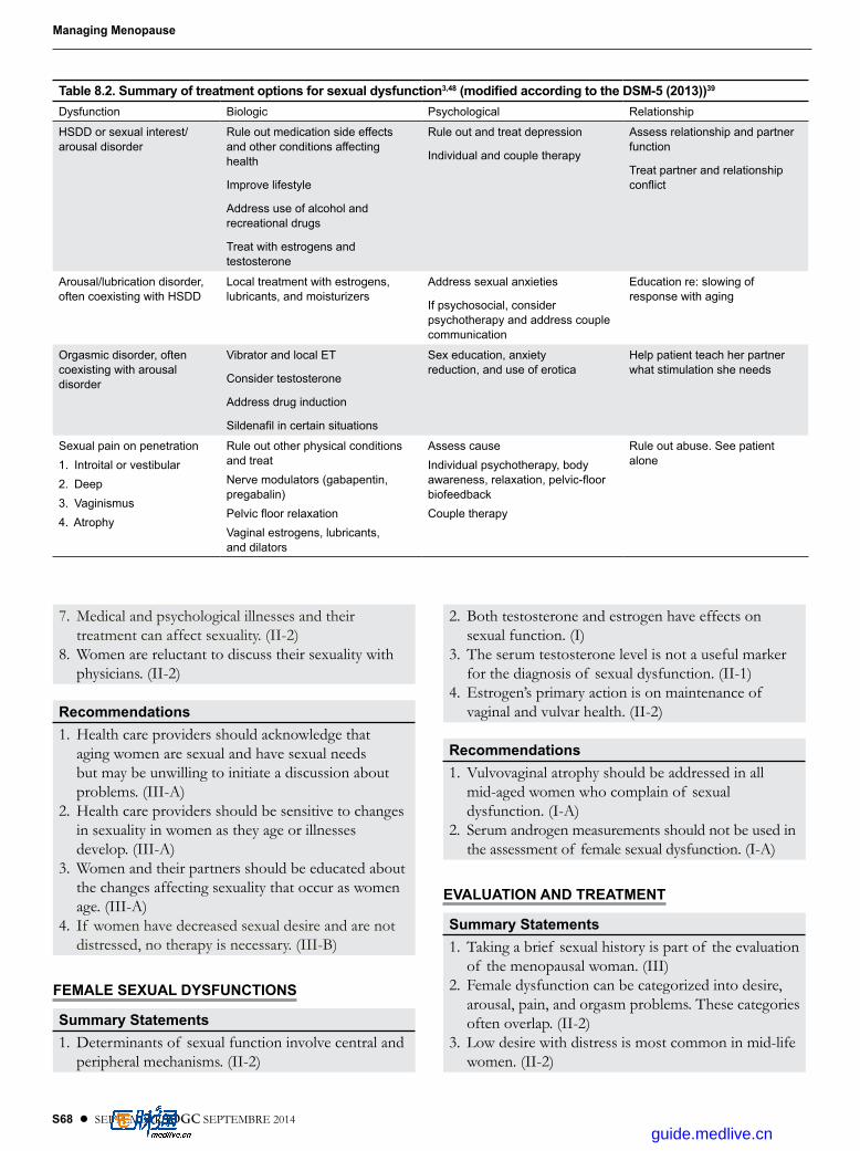
S68 l SEPTEMBER JOGC SEPTEMBRE 2014
Managing Menopause
7. Medical and psychological illnesses and their treatment can affect sexuality. (II-2)
8. Women are reluctant to discuss their sexuality with physicians. (II-2)
Recommendations1. Health care providers should acknowledge that
aging women are sexual and have sexual needs but may be unwilling to initiate a discussion about problems. (III-A)
2. Health care providers should be sensitive to changes in sexuality in women as they age or illnesses develop. (III-A)
3. Women and their partners should be educated about the changes affecting sexuality that occur as women age. (III-A)
4. If women have decreased sexual desire and are not distressed, no therapy is necessary. (III-B)
FEMALE SEXUAL DYSFUNCTIONS
Summary Statements1. Determinants of sexual function involve central and
peripheral mechanisms. (II-2)
2. Both testosterone and estrogen have effects on sexual function. (I)
3. The serum testosterone level is not a useful marker for the diagnosis of sexual dysfunction. (II-1)
4. Estrogen’s primary action is on maintenance of vaginal and vulvar health. (II-2)
Recommendations1. Vulvovaginal atrophy should be addressed in all
mid-aged women who complain of sexual dysfunction. (I-A)
2. Serum androgen measurements should not be used in the assessment of female sexual dysfunction. (I-A)
EVALUATION AND TREATMENT
Summary Statements1. Taking a brief sexual history is part of the evaluation
of the menopausal woman. (III)2. Female dysfunction can be categorized into desire,
arousal, pain, and orgasm problems. These categories often overlap. (II-2)
3. Low desire with distress is most common in mid-life women. (II-2)
Table 8.2. Summary of treatment options for sexual dysfunction3,48 (modified according to the DSM-5 (2013))39
Dysfunction Biologic Psychological Relationship
HSDD or sexual interest/ arousal disorder
Rule out medication side effects and other conditions affecting health
Improve lifestyle
Address use of alcohol and recreational drugs
Treat with estrogens and testosterone
Rule out and treat depression
Individual and couple therapy
Assess relationship and partner function
Treat partner and relationship conflict
Arousal/lubrication disorder, often coexisting with HSDD
Local treatment with estrogens, lubricants, and moisturizers
Address sexual anxieties
If psychosocial, consider psychotherapy and address couple communication
Education re: slowing of response with aging
Orgasmic disorder, often coexisting with arousal disorder
Vibrator and local ET
Consider testosterone
Address drug induction
Sildenafil in certain situations
Sex education, anxiety reduction, and use of erotica
Help patient teach her partner what stimulation she needs
Sexual pain on penetration1 . Introital or vestibular2 . Deep3 . Vaginismus4 . Atrophy
Rule out other physical conditions and treatNerve modulators (gabapentin, pregabalin)Pelvic floor relaxationVaginal estrogens, lubricants, and dilators
Assess causeIndividual psychotherapy, body awareness, relaxation, pelvic-floor biofeedbackCouple therapy
Rule out abuse . See patient alone
guide.medlive.cn

SEPTEMBER JOGC SEPTEMBRE 2014 l S69
CHAPTER 8: Sexuality and Menopause
4. Vaginal atrophy occurs in 50% of women within 3 years of menopause and is a common cause of sexual pain in menopausal women. (II-1)
5. Sexual pain results in a cascade of detrimental sexual symptoms. (II-1)
6. The treatment of sexual dysfunctions involves a multifaceted approach addressing medical, psychological, and relationship issues. (III)
7. Transdermal testosterone therapy has been shown to increase desire, arousal, and frequency of satisfactory sexual events and to decrease personal distress for women with surgical and also natural menopause, but there are no approved products for this indication in Canada. (I)
Recommendations1. Health care providers should include a short sexual
screening history as part of a medical history of menopausal women. Interventions should be undertaken only if the patient is distressed about the problem. (III-A)
2. The patient’s problem should be categorized according to desire, arousal, pain, or orgasm problems in order to facilitate treatment and triage care. (III-A)
3. Vaginal estrogen therapy should be prescribed for postmenopausal women with vulvovaginal atrophy and sexual dysfunction. (I-A)
4. For women with decreased sexual desire the current best options include management of vaginal atrophy, addressing treatable contributing factors, and sexual counselling. (I-A)
5. For women with signs or symptoms of vulvovaginal atrophy who cannot use estrogens, vaginal dilators, lubricants, and moisturizers should be offered. (III-B)
6. Clinicians should endorse the benefits of alternative forms of sexual contact for patients unable to have penetration. (III-A)
SPECIAL CLINICAL SITUATIONS
Summary Statements1. Sexual dysfunction is common in depressed patients
and those taking selective serotonin reuptake inhibitors. (I)
2. Premature loss of ovarian function may be attended by sexual dysfunction related to loss of both ovarian estrogen and androgen production at a time of life when sexual activity is normally heightened. (II-1)
3. Survivors of breast cancer using aromatase inhibitors have more sexual dysfunction due to vulvovaginal atrophy than do women using tamoxifen or control subjects. (II-1)
Recommendations1. Patients using selective serotonin reuptake inhibitors
should be educated about the effects of these medications on sexuality and informed that these effects are reversible when the medications are stopped. (III-B)
2. Patients with premature ovarian failure should have discussions about their sexual health. (III-B)
3. Patients with breast cancer using aromatase inhibitors should be advised that these medications may have sexual effects. (II-2B) The decision to use intravaginal estrogen therapy for severe vulvovaginal atrophy in such women needs to be based on quality-of-life considerations and should be made only after a discussion of the uncertain effects on breast cancer recurrence. (III-I)
REFERENCES
1. Leiblum S, Koochaki P, Rodenberg C, Barton I, Rosen R. Hypoactive sexual desire disorder in postmenopausal women. US results from the Women’s International Study of Health on Sexuality (WISHeS). Menopause 2006;13:46–56.
2. Davison SL, Bell RJ, LaChina M, Holden SL, Davis SR. The relationship between self-reported sexual satisfaction and general well-being in women. J Sex Med 2009;6:2690–7.
3. Lamont J. Female sexual health consensus clinical guidelines. J Obstet Gynaecol Can 2012;34(Suppl 2):S1–60.
4. Basson R. Sexual concerns — menopause and sexual function. In: Bélisle S, Blake J; SOGC Menopause Guidelines Committee. Canadian Consensus Conference on Menopause, 2006 update. SOGC Clinical Practice Guidelines, No. 171, February 2006. J Obstet Gynaecol Can 2006;28:S43–52.
5. West SL, D’Aloisio AA, Agans RP, Kalsbeek WD, Borissov NN, Thorp JM. Prevalence of low sexual desire and hypoactive sexual desire disorder in a nationally representative sample of US women. Arch Intern Med 2008;168:1441–9.
6. Avis NE, Brockwell S, Randolph JF Jr, Shen S, Cain VS, Ory M, et al. Longitudinal changes in sexual functioning as women transition through menopause; results from the study of Women’s Health Across the Nation. Menopause 2009;16:442–52.
7. Lindau ST, Schumm P, Laumann EO, Levinson W, Muircheartaigh CA, Waite LJ. A study of sexuality and health among older adults in the United States. N Engl J Med 2007;357:762–74.
8. Shifren JL, Monz BU, Russo PA, Segreti A, Johannes CB. Sexual problems and distress in United States women: prevalence and correlates. Obstet Gynecol 2008;112:970–8.
9. Shifren JL. Increasing our understanding of women’s sexuality at midlife and beyond. Menopause 2011;18:1149–51.
guide.medlive.cn

S70 l SEPTEMBER JOGC SEPTEMBRE 2014
Managing Menopause
10. Nappi RE, Davis SR. The use of hormone therapy for the maintenance of urogynecological and sexual health post WHI. Climacteric 2012;15:267–74.
11. Sturdee DW, Panay N; International Menopause Society Writing Group. Recommendations for the management of postmenopausal vaginal atrophy. Climacteric 2010;13:509–22. Epub 2010 Sep 30.
12. Laumann EO, Waite LJ. Sexual dysfunction among older adults: prevalence and risk factors from a nationally representative US probability sample of men and women 57–85 years of age. J Sex Med 2008;5:2300–11.
13. Salonia A, Zanni G, Nappi RE, Briganti A, Dehò F, Fabbri F, et al. Sexual dysfunction is common in women with lower urinary tract symptoms and urinary incontinence: results of a cross-sectional study. Eur Urol 2004;45:642–8.
14. Dennerstein L, Lehert P, Berger H. The relative effect of hormones and relationship factors on sexual function of women through the natural menopausal transition. Fertil Steril 2005;84:174–80.
15. Gracia CR, Freeman EW, Sammel MD, Lin H, Mogul M. Hormones and sexuality during transition to menopause. Obstet Gynecol 2007;109:831–40.
16. Davison SL, Bell RJ, LaChina M, Holden SL, Davis SR. Sexual function in well women: stratification by sexual satisfaction, hormone use, and menopause status. J Sex Med 2008;5:1214–22.
17. Prairie BA, Scheier MF, Matthews KA, Chang CC, Hess R. A higher sense of purpose in life is associated with sexual enjoyment in midlife women. Menopause 2011;18:39–844.
18. Hess R, Conroy MB, Ness R, Bryce CL, Dillon S, Chang CC, et al. Association of lifestyle and relationship factors with sexual functioning of women during midlife. J Sex Med 2009;6:1358–68.
19. Nappi RE, Albani F, Santamaria V, Tonani S, Magri F, Martini E, et al. Hormonal and psycho-relational aspects of sexual function during menopausal transition and at early menopause. Maturitas 2010;67:78–83. Epub 2010 Jun 16.
20. Laumann EO, Paik A, Glasser DB, Kang JH, Wang T, Levinson B, et al. A cross-national study of subjective sexual well-being among older women and men: findings from the Global Study of Sexual Attitude and Behaviors. Arch Sex Behav 2006;2:145–61. Epub 2006 Apr 26.
21. Chedraui P, Perez-Lopez FR, San Miguel G, Avila C. Assessment of sexuality among middle-aged women using the Female Sexual Function Index. Climacteric 2009;12:213–21.
22. Blümel JE, Chedraui P, Baron G, Belzares E, Bencosme A, Calle A, et al. Sexual dysfunction in middle-aged women: a multicenter Latin American study using the Female Sexual Function Index. Menopause 2009;16:1139–48.
23. Nappi RE, Nijland EA. Women’s perception of sexuality around the menopause: outcomes of a European telephone survey. Eur J Obstet Gynecol Reprod Biol 2008;137:10–6. Epub 2007 Jan 4.
24. Graziottin A. Prevalence and evaluation of sexual health problems—HSDD in Europe. J Sex Med 2007;4:211–9.
25. Gass ML, Cochrane BB, Larson JC, Manson JE, Barnabei VM, Brzyski RG, et al. Patterns and predictors of sexual activity among women in the hormone therapy trials of the Women’s Health Initiative. Menopause 2011;18:1160–71.
26. Basson R, Rees P, Wang R, Montejo AL, Incrocci L. Sexual function in chronic illness. J Sex Med 2010;7:374–88.
27. Allahdadi KJ, Tostes RC, Webb RC. Female sexual dysfunction: therapeutic options and experimental challenges. Cardiovasc Hematol Agents Med Chem 2009;7:260–9.
28. Copeland KL, Brown JS, Creasman JM, Van Den Eeden SK, Subak LL, Thom DH, et al. Diabetes mellitus and sexual function in middle-aged and older women. Obstet Gynecol 2012;120 (2 Pt 1):331–40.
29. Serretti A, Chiesa A. Treatment-emergent sexual dysfunction related to antidepressants: a meta-analysis. J Clin Psychopharmacol 2009;29:259–66.
30. McCall-Hosenfeld JS, Jaramillo SA, Legault C, Freund KM, Cochrane BB, Manson JE, et al. Correlates of sexual satisfaction among sexually active postmenopausal women in the Women’s Health Initiative—observational study. J Gen Intern Med 2008;23:2000–9. Epub 2008 Oct 7.
31. Castelo-Branco C, Palacios S, Combalia J, Ferrer M, Traveria G. Risk of hypoactive sexual desire disorder and associated factors in a cohort of oophorectomized women. Climacteric 2009;12:525–32.
32. Shifren JL, Avis NE. Surgical menopause: effects on psychological well-being and sexuality. Menopause 2007;14:586–91.
33. Trompeter S, Bettencourt R, Barrett-Connor E. Sexual activity and satisfaction in healthy community-dwelling older women. Am J Med 2012;125:37–43.
34. Buster J, Kingsberg S, Kilpatrick C. Female sexual dysfunction [practice bulletin]. Ob-Gyn 2011;119:996–1003.
35. Stahl S. Targeting circuits of sexual desire as a treatment strategy for hypoactive sexual desire disorder. J Clin Psychiatry 2010;71:821–2.
36. Pfaus JG. Pathways of sexual desire. J Sex Med 2009;6:1506–33.
37. Salonia A, Giraldi A, Chivers ML, Georgiadis JR, Levin R, Maravilla KR, et al. Physiology of women’s sexual function; basic knowledge and new findings. J Sex Med 2010;8:2637–60.
38. World Health Organization. International Statistical Classification of Diseases and Related Health Problems, 10th rev. Geneva: World Health Organization; 1992.
39. American Psychiatric Association. Diagnostic and Statistical Manual of Mental Disorders, 5th ed, text rev, Washington, DC: American Psychiatric Association; 2013.
40. Basson R, Berman J, Burnett A, Derogatis L, Ferguson D, Fourcroy J, et al. Report of the International Consensus Development Conference on Female Sexual Dysfunction: definitions and classifications. J Urol 2000;163:888–93.
41. Basson R. Is it time to move on from “hypoactive sexual desire disorder”? Menopause 2010;17:1097–8.
42. Davison SL, Bell R, Donath S, Montalto JG, Davis SR. Androgen levels in adult females: changes with age, menopause, and oophorectomy. J Clin Endocrinol Metab 2005;90:3847–953.
43. Labrie F, Marel C, Bulser J. Wide distribution of the serum dehydroepiandrosterone and sex steroid levels in postmenopausal women: Role of the ovary? Menopause 2011;18:30–43.
44. Davis SR, Burger HG. The role of androgen therapy. Best Pract Res Clin Endocrinol Metab 2003;17:165–75.
45. Basson R, Brotto L, Petkau A, Fernand L. Role of androgens in women’s sexual dysfunction. Menopause 2010;17:962–71.
46. 2012 Hormone Therapy Position Statement of the North American Menopause Society. Menopause 2012;19:257–71.
47. Nappi RE, Polatti F. The use of estrogen therapy in women’s sexual function. J Sex Med 2009;6:603–16.
48. Al-Azzawi F, Bitzer J, Brandenburg U, Castelo-Branco C, Graziottin A, Kenemans P, et al. Therapeutic options for postmenopausal female sexual dysfunction. Climacteric 2010;13:103–20.
49. Archer D. Efficacy and tolerability of local estrogen therapy for urogenital atrophy. Menopause 2010;17:194–203.
50. Modelska K, Litwach S, Ewing SK, Yaffe K. Endogenous estrogen levels affect sexual function in elderly post-menopausal women. Maturitas 2004;49:124–33.
guide.medlive.cn

SEPTEMBER JOGC SEPTEMBRE 2014 l S71
CHAPTER 8: Sexuality and Menopause
51. Tan O, Bradshaw K, Carr B. Management of vulvovaginal atrophy-related sexual dysfunction in postmenopausal women: an up-to-date review. Menopause 2012;19:109–17.
52. Levine KB, Williams RE, Hartmann KE. Vulvovaginal atrophy is strongly associated with female sexual dysfunction among sexually active postmenopausal women. Menopause 2008;15:661–67.
53. Sarrel P. Sexual dysfunction: treat or refer. Obstet Gynecol 2005;106:834–9.
54. Martin-Morales A, Graziottin A, Jaoude GB, Debruyne F, Buvat J, Beneke M, et al. Improvement in sexual quality of life of the female partner following vardenafil treatment of men with erectile dysfunction: a randomized, double-blind, placebo-controlled study. J Sex Med 2011;8:2831–40.
55. Thase ME, Clayton AH, Haight BR, Thompson AH, Modell JG, Johnston JA. A double-blind comparison between bupropion XL and venlafaxine XR: sexual functioning, antidepressant efficacy, and tolerability. J Clin Psychopharmacol 2006;26:482–8.
56. Basson R. Sexuality and sexual disorders. Clinical Updates in Women’s Health Care. Washington, DC; American College of Obstetricians and Gynecologists 2014:50–53.
57. Kingsberg S. The Hypoactive Sexual Desire Disorder Registry to characterize the natural history and outcomes of women with hypoactive sexual desire disorder. Menopause 2012;19:379–81.
58. Sturdee DW, Pines A; International Menopause Society Writing Group. Updated IMS recommendations on postmenopausal hormone therapy and preventive strategies for midlife health. Climacteric 2011;14:302–20.
59. Suckling J, Lethaby A, Kennedy R. Local oestrogen for vaginal atrophy in postmenopausal women (review). Cochrane Database Syst Rev 2006;(4):CD001500.
60. The North American Menopause Society. The role of local vaginal estrogen for treatment of vaginal atrophy in postmenopausal women: 2007 position statement of The North American Menopause Society. Menopause 2007;14:355–69.
61. Gast MJ, Freedman MA, Vieweg AJ, De Melo NR, Girao MJ, Zinaman MJ; The Dyspareunia Study Group. A randomized study of low-dose conjugated estrogens on sexual function and quality of life in postmenopausal women. Menopause 2009;16:247–56.
62. Panay N, Fenton A. The role of testosterone in women. Climacteric 2009;12:185–7.
63. Somboonporn W, Davis S, Seif MW, Bell R. Testosterone for peri- and postmenopausal women. Cochrane Database Syst Rev 2005;(4):CD004509.
64. Davis SR, Goldstat R, Papalia MA, Shah S, Kulkani J, Donath J, et al. Effects of aromatase inhibitors on sexual function and well-being in postmenopausal women treated with testosterone; a randomized, placebo-controlled trial. Menopause 2006;13:37–45.
65. Shifren JL, Braunstein GD, Simon JA, Casson PR, Buster JE, Redmond GP, et al. Transdermal testosterone treatment in women with impaired sexual function after oophorectomy. N Engl J Med 2000;343:682–8.
66. Buster JE, Kingsberg SA, Aguirre O, Brown C, Breaux JG, Buch A, et al. Testosterone patch for low sexual desire in surgically menopausal women: a randomized trial. Obstet Gynecol 2005;105 (5 Pt 1):944–52.
67. Braunstein GD, Sundwall DA, Katz M, Shifren JL, Buster JE, Simon JA, et al. Safety and efficacy of a testosterone patch for the treatment of hypoactive sexual desire disorder in surgically menopausal women: a randomized, placebo-controlled trial. Arch Intern Med 2005;165:1582–9.
68. Simon J, Braunstein G, Nachtigall L, Utian W, Katz M, Miller S, et al. Testosterone patch increases sexual activity and desire in surgically menopausal women with hypoactive sexual desire disorder. J Clin Endocrinol Metab 2005;90:5226–33. Epub 2005 Jul 12.
69. Davis SR, van der Mooren MJ, van Lunsen RH, Lopes P, Ribot C, Rees M, et al. Efficacy and safety of a testosterone patch for the treatment of hypoactive sexual desire disorder in surgically menopausal women: a randomized placebo-controlled trial. Menopause 2006;13:387–96.
70. Shifren JL, Davis SR, Moreau M, Waldbaum A, Bouchard C, DeRogatis L, et al. Testosterone patch for the treatment of hypoactive sexual desire disorder in naturally menopausal women: results from the INTIMATE NM1 study. Menopause 2006;13:770–9.
71. Kingsberg S, Shifren J, Wekselman K, Rodenberg C, Koochaki P, De Rogatis L. Evaluation of the clinical relevance of benefits associated with transdermal testosterone treatment in postmenopausal women with hypoactive sexual desire disorder. J Sex Med 2007;4:1001–8.
72. Davis SR, Moreau M, Kroll R, Bouchard C, Panay N, Gass M, et al; APHRODITE Study Team. Testosterone for low libido in menopausal women not taking estrogen. N Engl J Med 2008;359:2005–17.
73. Panay N, Al-Azzawi F, Bouchard C, Davis SR, Eden J, Lodhi I, et al. Testosterone treatment of HSDD in naturally menopausal women: the ADORE study. Climacteric 2010;13:121–31.
74. El-Hage G, Eden JA, Manga RZ. A double-blind, randomized, placebo-controlled trial of the effect of testosterone cream on the sexual motivation of menopausal hysterectomized women with hypoactive sexual desire disorder. Climacteric 2007;10:335–43.
75. Nathost-Boos J, Floter A, Jarkander-Rolff M, Carlstrom K, von Schoultz B. Treatment with percutaneous testosterone gel in postmenopausal women with decreased libido: effects on sexuality and psychological general well-being. Maturitas 2006;53:11–8.
76. Sante RJ, Allred DC, Ardoin SP, Archer DF, Boyd N, Braunstein GD, et al. Postmenopausal hormone therapy: an Endocrine Society scientific statement. J Clin Endocrinol Metab 2010;95(7 Suppl 1):S1–66.
77. Al-Imari L, Wolfman WL. The safety of testosterone therapy in women. J Obstet Gynaecol Can 2012;34:859–65.
78. Davis SR, Braunstein GD. Efficacy and safety of testosterone in HSDD. J Sex Med 2012;9:1134–48.
79. Nachtigall L, Casson P, Lucas J, Schofield V, Melson C, Simon JA. Safety and tolerability of testosterone patch therapy for up to 4 years in surgically menopausal women receiving oral or transdermal oestrogen. Gynecol Endocrinol 2011;27:39–48.
80. van Staa TP, Sprafka JM. Study of adverse outcomes in women using testosterone therapy. Maturitas 2009;62:76–80. Epub 2008 Dec 24.
81. Nijland EA, Weijmar Schultz WC, Nathorst-Boös M, Helmond FA, Van Lunsen RH, Palacios S, et al. Tibolone and transdermal E2/NETA for the treatment of female sexual dysfunction in naturally menopausal women: results of a randomized active-controlled trial. J Sex Med 2008;5:646–56.
82. Panjari M, Davis SR. DHEA for postmenopausal women: a review of the evidence. Maturitas 2010;66:172–9.
83. Labrie F, Archer D, Bouchard C, Fortier M, Cusan L, Gomez JL, et al. Effect of intravaginal dehydroepiandrosterone (Prasterone) on libido and sexual dysfunction in postmenopausal women. Menopause 2009;16:923–31.
84. Management of symptomatic vulvovaginal atrophy: 2013 position statement of The North Amerian Menopause Society. Menopause 2013;20:888–902.
85. Maserejian NN, Shifren J, Parish SJ, Segraves RT, Huang L, Rosen RC. Sexual arousal and lubrication problems in women with clinically diagnosed hypoactive sexual desire disorder: preliminary findings from the hypoactive sexual desire disorder registry for women. J Sex Marital Ther 2012;38:41–62.
86. Berman JR, Berman LA, Lin H, Flaherty E, Lahey N, Goldstein I, et al. Effect of sildenafil on subjective and physiologic parameters of the female sexual response in women with sexual arousal disorder. J Sex Marital Ther 2001;27:411–20.
guide.medlive.cn

S72 l SEPTEMBER JOGC SEPTEMBRE 2014
Managing Menopause
87. Basson R, McInnes R, Smith MD, Hodgson G, Koppiker N. Efficacy and safety of sildenafil citrate in women with sexual dysfunction associated with female sexual arousal disorder. J Womens Health Gend Based Med 2002;11:367–77.
88. Billups KL, Berman L, Berman J, Metz ME, Glennon ME, Goldstein I. A new non-pharmacological vacuum therapy for female sexual dysfunction. J Sex Marital Ther 2001;27:435–41.
89. Basson R, Brotto LA. Sexual psychophysiology and effects of sildenafil citrate in estrogenised women with acquired genital arousal disorder and impaired orgasm: a randomised controlled trail. BJOG 2003;110:1014–24.
90. Nurnberg HG, Hensley PL, Heiman JR, Croft HA, Debattista C, Paine S. Sildenafil treatment of women with antidepressant-associated sexual dysfunction: a randomized controlled trial. JAMA 2008;300:395–404.
91. Sipski MI, Rosen RC, Alexander LJ, Hamer RM. Sildenafil effects on sexual and cardiovascular reponses in women with spinal cord injury. Urology 2000;55:812–5.
92. Freeman EW. Associations of depression with the transition to menopause. Menopause 2010;17:823–7.
93. Murell SA, Himmelfarb S, Wright K. Prevalence of depression and its correlates in older adults. Am J Epidemiol 1983;117:173–85.
94. Clayton AH, Ninan PT. Depression or menopause? Presentation and management of major depressive disorder in perimenopausal and postmenopausal women. Prim Care Companion J Clin Psychiatry 2010;12(1):PCC.08r00747.
95. Harlow BL, Wise LA, Otto MW, Soares CN, Cohne LS. Depression and its influence on reproductive endocrine and menstrual cycle markers associated with perimenopause: The Harvard Study of Moods and Cycles. Arch Gen Psychiatry 2003;60:29–36.
96. Werneke U, Northey S, Bhugra D. Antidepressants and sexual dysfunction. Acta Psychiatr Scand 2006;114:385–97.
97. Johannes CB, Clayton AH, Odom DM, Rosen RC, Russo PA, Shifren JL, et al. Distressing sexual problems in United States women revisited: prevalence after accounting for depression. J Clin Psychiatry 2009;70:1698-706.
98. Fooladi E, Bell RJ, Davis SR. Management strategies in SSRI-associated sexual dysfunction in women at midlife. Climacteric 2012;15:306–16.
99. Clayton AH, Warnock JK, Kornstein SG, Pinkerton R, Sheldon-Keller A, McGarvey EL. A placebo-controlled trial of bupropion SR as an antidote for selective serotonin reuptake inhibitor-induced sexual dysfunction. J Clin Psychiatry 2004;65:62–7.
100. Kingsberg S. The impact of premature ovarian insufficiency on sexual function. Menopause 2011;18:246–7.
101. Kalantaridou SN, Vanderhoof VH, Calis KA, Corrigan EC, Troendle JF, Nelson LM. Sexual function in young women with spontaneous 46XX primary ovarian insufficiency. Fertil Steril 2008;90:1805–11.
102. Van der Stege JG, Groen H, van Zadelhoff SJ, Lambalk CB, Braat DD, van Kasteren YM, et al. Decreased androgen concentrations and diminished general and sexual well-being in women with premature ovarian failure. Menopause 2008;15:23–31.
103. de Almeida DM, Benetti-Pinto CL, Makuch MY. Sexual function of women with premature ovarian failure. Menopause 2011;18:262–6.
104. Anderson WF, Chatterjee N, Ershler WB, Brawley OW. Estrogen receptor breast cancer phenotypes in the Surveillance, Epidemiology, and End Results database. Breast Cancer Res Treat 2002;76:27–36.
105. American College of Obstetricians and Gynecologists, Committee on Practice Bulletins—Gynecology. Management of gynecologic issues in women with breast cancer. Practice Bulletin No. 126, March 2012. Obstet Gynecol 2012;119:666–82.
106. Gilbert E, Ussher JM, Perz J. Sexuality after breast cancer: a review. Maturitas 2010;66:397–407.
107. Bradford A. Sexual outcomes of aromatase inhibitor therapy in women with breast cancer: time for intervention. Menopause 2013;20:128–9.
108. Panjari M, Bell RJ, Davis SR. Sexual function after breast cancer. J Sex Med 2011;8:294–302.
109. Baumgart J, Nilsson K, Stavreus Evers A, Kunovac Kallak T, Sundström Poromaa I. Sexual dysfunction in women on adjuvant endocrine therapy after breast cancer. Menopause 2013;20:162–8.
110. Kendall A, Dowsett M, Folkerd E, Smith I. Caution: vaginal estradiol appears to be contraindicated in postmenopausal women on adjuvant aromatase inhibitors. Ann Oncol 2006;17:584–7. Epub 2006 Jan 27.
111. Pfeiler G, Glatz C, Konigsberg R, Geisendorfer T, Fink-Retter A, Kubista E, et al. Vaginal estriol to overcome side-effects of aromatase inhibitors in breast cancer patients. Climacteric 2011;14:339–44.
112. Bober SL, Varela VS. Sexuality in adult cancer survivors: challenges and intervention. J Clin Oncol 2012;30:3712–19.
113. Hatzichristou D, Rosen RC, Derogatis LR, Low WY, Meuleman EJ, Sadovsky R, et al. Recommendations for the clinical evaluation of men and women with sexual dysfunction. J Sex Med 2010;7(1 Pt 2):337–48.
guide.medlive.cn

SEPTEMBER JOGC SEPTEMBRE 2014 l S73
CHAPTER 8: Sexuality and Menopause
Brief sexual symptom checklist for women
Please answer the following questions about your overall sexual function .
1. Are you satisfied with your sexual function?
Yes No
2. How long have you been dissatisfied with your sexual function?
3 . The problems with your sexual function are (mark one or more)
Little or no interest in sex
Decreased genital sensation (feeling)
Decreased vaginal lubrication (dryness)
Problem reaching orgasm
Pain during sex
Other
4 . Which problem is most bothersome? (circle)
1 2 3 4 5 6
5 . Would you like to talk about it with your doctor?
Yes No
APPENDIX.
Reprinted by permission, with modifications, from Hatzichristou et al.113
guide.medlive.cn

S74 l SEPTEMBER JOGC SEPTEMBRE 2014
CHAPTER 9
Complementary and Alternative Medicine (CAM)
Women between 45 and 64 years old are significant users of CAM, including natural products.1 The
US National Center for Complementary and Alternative Medicine2 (NCCAM) has defined CAM as “a group of diverse medical and health care systems, practices, and products that are not generally considered to be part of conventional medicine”. NCCAM classifies CAM into 5 main groups: alternative medical systems, mind–body interventions, biologically based therapies including NHPs, manipulative and body-based methods, and energy therapies.
There is a strong consumer-driven need for substantial information regarding CAM, and health care providers should be prepared to answer patients’ enquiries and guide them into using products for which there are efficacy and safety data.
More than half of peri- and postmenopausal women use some type of CAM, including dietary and herbal therapies, stress management, acupuncture, and massage therapy, for menopausal symptoms that include hot flashes, joint pain, sleep problems, forgetfulness, mood difficulties, and fatigue.3,4 The challenge for health care providers is to incorporate evidence-based non-prescription therapeutics in a shared decision-making process as consumer demand increases for alternative treatment of their menopausal symptoms.
There is no question that the modification of diet and lifestyle can positively influence mid-life health. There are clear benefits of therapies involving dietary and lifestyle changes. Factors, such as obesity, diet, exercise, and cigarette smoking affect the general health of all menopausal women. A diet low in saturated and trans-unsaturated fats and rich in vegetables, fruits, fish, and whole grains is associated with a better health outcome.5 Cigarette smoking is a strong independent risk factor for CVD, stroke, peripheral vascular disease, osteoporosis, and certain cancers.6 Weight-bearing exercise enhances well-being, promotes balance and agility, and has positive effects on femoral bone density, cardiovascular function, and weight-gain prevention.7,8 These are standard recommendations for a healthy lifestyle
and prevention of heart disease and cancer. The general principles of healthy living can have a positive effect on night sweats and hot flashes in menopausal women, as cigarette smoking, alcohol drinking, and a higher BMI have been shown to increase the relative risk of hot flashes.9 The menopausal transitional weight gain can be minimized with moderate caloric restriction combined with a modest increase in exercise.10 The challenge is that despite the clear benefits of dietary and lifestyle modifications, adherence rates are often low.
NATURAL HEALTH PRODUCTS
Canadian regulationsThe Natural Health Products Regulations (NHPR) came into force January 1, 2004, and are administered by the Natural Health Products Directorate (NHPD), a division of the Health Products and Food Branch of Health Canada. The NHPR are intended “to ensure that all Canadians have ready access to natural health products that are safe, effective, and of high quality, while respecting the freedom of choice and philosophical and cultural diversity”. The NHPR define NHPs as a subset of drugs. Before 2004, products now captured under the definition of an NHP fell loosely into the category of foods or drugs, but many products were sold on the Canadian market non-compliantly. NHPs include the following: vitamin supplements, including synthetic duplicates; mineral supplements; plant or a plant material, an alga, a bacterium, a fungus, or a non-human animal material, including extracts, isolates, and synthetic duplicates of these substances; essential fatty acids, including synthetic duplicates; amino acids, including synthetic duplicates; traditional medicines, such as traditional Chinese medicines; homeopathic medicines; robotics; and enzymes.
NHPs require pre-market approval from NHPD before being marketed in Canada. An approved NHP will have an 8-digit natural product number on the primary label. In order to be granted this number by the NHPD, a sponsor must provide evidence of safety and efficacy of the product
guide.medlive.cn

SEPTEMBER JOGC SEPTEMBRE 2014 l S75
CHAPTER 9: Complementary and Alternative Medicine (CAM)
CHAPTER 9
under the labelled conditions of use. In addition, the NHPR set requirements for adverse-event reporting, clinical trials, manufacturing, packaging, labelling, importing, and distributing NHPs. Practitioners can check the status of approved products as well as the approved claims using the Licensed Natural Health Products Database (http://www.hc-sc.gc.ca/dhp-mps/prodnatur/applications/ licen-prod/lnhpd-bdpsnh-eng.php).
Adverse-reaction reporting is an important part of ongoing safety assessment and risk management. Health care providers and patients can report any adverse reactions using the forms found in the Compendium of Pharmaceuticals and Specialties, published by the Canadian Pharmacists Association (http://www.pharmacists.ca), or by accessing MedEffect Canada (http://www.hc-sc.gc.ca/dhp-mps/medeff/report-declaration/index-eng.php).
Manufacturing, packaging, labelling, and importing of NHPs are licensable activities. Approved site licence holders can be found at http://www.hc-sc.gc.ca/dhp-mps/prodnatur/applications/licen-site-exploit/sl-list-le-eng.php.
Consumers are advised to purchase only those brands with a natural product number on the primary label to ensure that they are using products that are of high quality and that have been reviewed with regard to the product’s formulation, labelling, claims, and instructions for safe use, including the potential for drug interactions. Tables 9.1 and 9.2 list selected general references and recommended websites for researching NHPs, and Table 9.3 summarizes the evidence-based data on NHPs.
PhytoestrogensThe NHP most studied for menopause conditions, phytoestrogens are plant-derived compounds that may
be considered SERMs, as they have both estrogenic and antiestrogenic properties. Phytoestrogens can be divided into 3 principal groups: isoflavones, lignans, and coumestans. Isoflavones are present in high concentrations in soybeans, soy products, such as tofu, and red clover. Flaxseed is the principal source of lignans.11
The most studied phytoestrogens in the context of menopause are isoflavones. Despite the fact that at least 3 systematic reviews evaluating the efficacy of isoflavones on menopausal symptoms have been published in the last few years, their results are still non-conclusive.12–14 In fact, lack of comparability of the source of isoflavones (red clover, soy), lack of control for other dietary sources of phytoestrogens, lack of identification of potential modifiers of the effect of isoflavones (such as genetic factors and individual differences in phytoestrogen metabolism and ability to produce equol11), and methodologic flaws could limit interpretation of the results of these meta-analyses. The authors of the systematic review published in 200913 did subgroup analysis according to menopausal status (peri- or postmenopausal), severity of symptoms, and type and dosage of phytoestrogens and concluded that phytoestrogens could be an alternative form of treatment in early-menopausal women with mild to moderate symptoms, genistein being the most effective compound. The 2011 NAMS report15 reached the same conclusion: “Soy-based isoflavones are modestly effective in relieving menopausal symptoms; supplements providing higher proportions of genistein or increased in S(Y)-equol may provide more benefits.” Health Canada has published a monograph on soybean extracts and isolate-containing NHPs (accessible from its alphabetic listing of monographs [http://webprod.hc-sc.gc.ca/nhpid-bdipsn/monosReq.do?lang=eng ] in the Natural Health Products Ingredients Database) and has approved products for claims related to
Table 9.1. Selected reference textsBarnes J, Anderson LA, Phillipson JD . Herbal medicines, 2nd ed . London: The Pharmaceutical Press; 2002 .
Boon H, Smith M . The complete natural medicine guide to the 50 most common medicinal herbs . Toronto: Robert Rose; 2003 .
Brinker F . Herb contraindications and drug interactions, 3rd ed . Sandy, Oregon: Eclectic Medical Publications; 2001 . (online updates available)
Chandler F, editor . Herbs: everyday reference for health professionals . Ottawa: Canadian Pharmacists Association and Canadian Medical Association; 2000 .
European Scientific Cooperative on Phytotherapy. ESCOP modules, 2nd ed. New York: ESCOP, George Thieme Verlag and Thieme New York; 2003 .
Jellin JM, Batz F, Hitchens K . Pharmacist’s letter/prescriber’s letter: natural medicines comprehensive database . Stockton, California: Therapeutic Research Faculty; 2003 . (available as textbook, as CD, and online)
World Health Organization . WHO monographs on selected medicinal plants . Geneva: World Health Organization; 1999 . (volumes 2 and 3 available as well)
guide.medlive.cn
S76 l SEPTEMBER JOGC SEPTEMBRE 2014
Managing Menopause
helping reduce loss of bone mineral density when used in conjunction with adequate amounts of calcium and vitamin D and that it may reduce severe and frequent menopausal symptoms. A number of publications exist on red clover isoflavones that also indicate a modest effect in relieving menopausal symptoms, particularly when used at a dose of about 80 mg/d.16–23 Products containing isoflavones sourced from soy and red clover and approved by Health Canada can be found by searching the Licensed Natural Health Products Database.
Three of the four studies evaluating the effect of flaxseed on menopausal symptoms reported no benefit compared with placebo.24–27
Although 2 systematic review results suggested a protective effect of isoflavones on bone density, a 2-year clinical trial found that isoflavone extract (200 mg once daily) was not superior to placebo in reducing bone loss or bone turnover in menopausal women.28–30 A 1-year clinical trial did not show any effect of flaxseed, 40 g once daily, on femoral or lumbar bone mineral density.24
The effect of phytoestrogen subclasses, including isoflavone extracts and isoflavone food sources, on cardiovascular risk factors was the subject of a meta-analysis that was not limited to a menopausal population.31 The analysis showed that long-term use of soy proteins significantly decreased diastolic blood pressure and levels of LDL cholesterol but
Table 9.2. Recommended websitesOrganization Website*
Natural Health Products Directorate, Health Canada http://hc-sc .gc .ca/dhp-mps/prodnatur/index-eng .php
Many links provide additional information on NHPs, including the Licensed Natural Health Products Database .
American Botanical Council http://www .herbalgram .org
An online resource for herbal news and information .
Cochrane Consumer Network http://www .cochrane .org/consumers/homepage .htm
Informs about consumer involvement in the Cochrane Collaboration .
European Scientific Cooperative on Phytotherapy (ESCOP) http://www .escop .com
An organization that aims to advance the scientific status of phytomedicines.
Memorial Sloan Kettering Cancer Center, New York http://www .mskcc .org
Provides information for oncologists and health care professionals, including a clinical summary for herbs, botanicals, and other products, with details about constituents, adverse effects, interactions, and potential benefits or problems . Also provides evaluations of alternative or unproved cancer therapies, as well as products for sexual dysfunction .
National Center for Complementary and Alternative Medicine, US National Institutes of Health
http://nccam .nih .gov
Explores complementary and alternative healing practices in the context of rigorous science .
The Richard and Hinda Rosenthal Center for Complementary and Alternative Medicine
http://nyp .org/services/complementary .html
Promotes an inclusive medical system by using scientific inquiry to ensure that the valuable health practices of other cultures are better understood and integrated with Western medical practices .
*Last accessed March 2014 .
Table 9.3. Menopausal symptoms and NHPs: summary of main evidence-based findingsNHP Type of evidence Main findings
Phytoestrogens
Isoflavones 3 systematic reviews12–14 Results still not conclusive . Supplements providing higher proportions of genistein or increased equol may provide more benefit.15
Flaxseed 4 RCTs No benefit compared with placebo.24–27
Black cohosh 2 systematic reviews40,41 No significant effect compared with placebo or with HT on frequency or intensity of hot flashes and quality of life.
St . John’s wort 1 clinical trial46 Significantly improved menopause-specific quality of life and reduced sleep difficulties. No significant effect on number and intensity of hot flashes.
guide.medlive.cn

SEPTEMBER JOGC SEPTEMBRE 2014 l S77
CHAPTER 9: Complementary and Alternative Medicine (CAM)
had no effect on the HDL level. Soy foods were associated with a significant improvement in systolic and diastolic blood pressure. Isoflavone extracts were associated with a significant decrease in only systolic blood pressure. The results of another meta-analysis evaluating the effect of flaxseed interventions on the lipid profile were inconclusive: a decrease in the LDL cholesterol level alone was observed only in hypercholesterolemic women.32
Whether phytoestrogens are associated with hormone-dependent cancer is still a subject of great research and intense debate. Indeed, the negative impact of phytoestrogens on breast cancer is almost exclusively supported by in-vitro or animal-model studies.33 A recent systematic review with meta-analysis, including 14 observational studies, found that soya isoflavone intake was inversely associated with the risk of breast cancer (RR 0.89; 95% CI 0.79 to 0.99). However, this protective effect was observed only among Asian participants (RR 0.76; 95% CI 0.65 to 0.86) and not in Western populations (RR 0.97; 95% CI 0.87 to 1.06).34 The LACE prospective study35 examined soya intake and breast cancer recurrence according to tamoxifen status among nearly 2000 women followed for 6 years. The results suggested that soy intake at levels comparable to those in Asian population is associated with a lower risk of recurrence among women who have been treated with tamoxifen. A recent meta-analysis that included 11 prospective studies and 6 case–control studies found an inverse relation between lignan intake and breast cancer in postmenopause women.36 Finally, a large meta-analysis that included 174 clinical trials concluded that phytoestrogens have a short-term, safe profile and that the risks of vaginal bleeding, endometrial hyperplasia, endometrial cancer, and breast cancer were not significantly increased among phytoestrogen users.37 However, the mean duration of follow-up was only 6.2 months.
In conclusion, we cannot expect to have a clinical trial powerful enough to definitively answer the question of breast cancer and phytoestrogens. Our counselling has to be based on the available literature. In the light of the current data, phytoestrogens could be safe for menopausal women with breast cancer, but we should encourage women who seek information about the use of phytoestrogens to integrate them preferentially in their diet.
Black cohoshBlack cohosh (Cimicifuga racemosa) is is a tall perennial plant in the buttercup family that grows in forests of Eastern Canada and the United States and was registered for the first time in the 19th century in the US pharmacopoeia. It has been used since the early 1940s in Germany as a natural agent to relieve premenstrual syndrome, menstrual cramps, and menopausal symptoms.38,39 Two 2012 systematic review
examined whether black cohosh has an evidence-based impact on menopausal symptoms. The Cochrane review, which included 16 clinical trials, failed to find any significant effect of black cohosh compared with placebo or with HT on frequency and intensity of hot flashes and quality of life.40 The second systematic review reached the same conclusion except for a significant effect of a combination of black cohosh and St. John’s wort.41 Several case reports of liver injury in patients using black cohosh have been published. However, Naser et al.42 found no evidence of black cohosh hepatotoxicity in their meta-analysis of RCTs.
Health Canada has issued a monograph for black cohosh (accessible from its alphabetic listing of monographs [http://webprod.hc-sc.gc.ca/nhpid-bdipsn/monosReq.do?lang=eng ] in the Natural Health Products Ingredients Database), and products are on the market with the claim that they can help to relieve symptoms associated with menopause. Approved products can be found in Health Canada’s Licensed Natural Health Products Database.
St. John’s wortSt. John’s wort (Hypericum perforatum) has been shown to be effective for mild to moderate depression in several meta-analyses.43–45 Only 1 clinical trial has been done among symptomatic women. Compared with placebo, St. John’s wort (300 mg three times a day) significantly improved menopause-specific quality of life and reduced sleep difficulties in symptomatic menopausal women. Nevertheless, the differences between the 2 groups in number and intensity of hot flashes were not significant.46 A combination of St. John’s wort and black cohosh demonstrated a significant positive effect on menopausal symptoms.32 St. John’s sort interacts with many medications, but a large-scale surveillance study of 14 245 patients indicated that the frequency of side effects was 10 times lower with St. John’s wort than with synthetic antidepressants.47 Health Canada has issued monographs for St. John’s wort (accessible from its alphabetic listing of monographs [http://webprod.hc-sc.gc.ca/nhpid-bdipsn/monosReq.do? lang=eng] in the Natural Health Products Ingredients Database), and products are on the market with claims to promote healthy mood balance and to help relieve sleep disturbances associated with mood imbalance. Approved products can be found in Health Canada’s Licensed Natural Health Products Database.
Other NHPsThere are no evidence-based data to support any clinically significant effect of dong quai, ginseng, evening primrose oil, wild yam (Dioscorea villosa), ginkgo, or Chinese herbal formulas compared with placebo for reducing menopausal symptoms.48,49
guide.medlive.cn

S78 l SEPTEMBER JOGC SEPTEMBRE 2014
Managing Menopause
OTHER CAM
There are other CAM approaches to handling menopausal symptoms that do not involve NHPs. Lifestyle modification, including practices that lower core body temperature, such as using a fan, dressing in layers, and consuming cold food and drinks, may temporarily help with night sweats and flushing.
Acupuncture is one of the most attractive types of CAM proposed for many medical indications. Acupuncture is defined as the practice of inserting a needle or needles into certain points in the body for therapeutic purposes.50 These points can also be stimulated by manual pressure (acupressure), small electric currents applied through the inserted needles (electro-acupuncture), and lasers. Two more recent systematic reviews found insufficient evidence to determine whether acupuncture is an effective treatment of VMS.51,52 The number, size, and quality of RCTs are still too low to permit firm conclusions.
Mind–body techniques, including yoga, relaxation, meditation, hypnosis, and tai chi, have been tested for pain and other chronic medical conditions in several clinical trials, but the paucity of data regarding menopausal symptoms does not permit any conclusion. Nevertheless, tai chi was associated with a positive effect on bone density53 and balance, as well as a reduction in the frequency of falls, in elderly women.54
Although NAMS recommends exercise for menopausal symptoms, there are no evidence-based data to support this recommendation. In fact, the positive results of a systematic review including only 1 small clinical trial cannot be used for firm conclusions; the methodologic flaws of the other studies were too important for inclusion of those studies.55
CONCLUSION
Although standard scientific research in the area of CAM is growing, the current evidence-based data lead to the firm conclusion that NHPs and other forms of CAM are less effective than HT for treating menopausal symptoms. The placebo effect may explain around 30% of the improvement with CAM. Consumers who decide to use NHPs should be encouraged to use products that have been reviewed by Health Canada and, as with pharmaceuticals, to report adverse reactions. In a shared decision-making process, good updated awareness of evidence-based CAM allows the health care provider to discuss with the woman safe, non-pharmaceutical choices for treatment of menopausal symptoms.
Summary Statement1. Health Canada’s Licensed Natural Health Products
Database lists products approved for use in women with menopausal symptoms that have been evaluated for safety, efficacy, and quality. (III)
Recommendation1. Health care providers may offer identified
complementary and alternative medicine with demonstrated efficacy for mild menopausal symptoms. (I-B)
REFERENCES
1. Lunny CA, Fraser SN. The use of complementary and alternative medicines among a sample of Canadian menopausal-aged women. J Midwifery Womens Health 2010;55:335–43.
2. Complementary, alternative, or integrative health: What’s in a name? Bethasda, Maryland: NCCAM; 2008. Available at: http://nccam.nih.gov/health/whatiscam. (Accessed 2013 Jan 31).
3. Kupferer EM, Dormire SL, Becker H. Complementary and alternative medicine use for vasomotor symptoms among women who have discontinued hormone therapy. J Obstet Gynecol Neonatal Nurs 2009;38:50–9.
4. Gold EB, Bair Y, Zhang G, Utts J, Greendale GA, Upchurch D, et al. Cross-sectional analysis of specific complementary and alternative medicine (CAM) use by racial/ethnic group and menopausal status: the Study of Women’s Health Across the Nation (SWAN). Menopause 2007;14:612–23.
5. Lin JS, O’Connor E, Whitlock EP, Beil TL, Zuber SP, Perdue LA, et al. Behavioral counseling to promote physical activity and a healthful diet to prevent cardiovascular disease in adults: update of the evidence for the U.S. Preventive Services Task Force. Evidence Synthesis no. 79. AHRQ publication no. 11-05149-EF-1. Rockville, Maryland: US Agency for Healthcare Research and Quality; 2010:24–6. Available at http://www.ncbi.nlm.nih.gov/books/NBK51030. Accessed 2014 June 2.
6. Rees M, Stevenson J. Primary prevention of coronary heart disease in women. Menopause Int 2008;14:40–5.
7. Stehr MD, von Lengerke T. Preventing weight gain through exercise and physical activity in the elderly: a systematic review. Maturitas 2012;72:13–22.
8. Martyn-St James M, Carroll S. Meta-analysis of walking for preservation of bone mineral density in postmenopausal women. Bone 2008;43:521–31.
9. Herber-Gast GC, Mishra GD, van der Schouw YT, Brown WJ, Dobson AJ. Risk factors for night sweats and hot flushes in midlife: results from a prospective cohort study. Menopause 2013;20:953–9.
10. Sternfeld B, Dugan S. Physical activity and health during the menopausal transition. Obstet Gynecol Clin North Am 2011;38:537–66.
11. Pilsakova L, Riecansky I, Jagla F. The physiological actions of isoflavone phytoestrogens. Physiol Res 2010;59:651–64.
12. Eden JA. Phytoestrogens for menopausal symptoms: a review. Maturitas 2012;72:157–9.
guide.medlive.cn

SEPTEMBER JOGC SEPTEMBRE 2014 l S79
CHAPTER 9: Complementary and Alternative Medicine (CAM)
13. Jacobs A, Wegewitz U, Sommerfeld C, Grossklaus R, Lampen A. Efficacy of isoflavones in relieving vasomotor menopausal symptoms: a systematic review. Mol Nutr Food Res 2009;53:1084–97.
14. Tempfer CB, Bentz EK, Leodolter S, Tscherne G, Reuss F, Cross HS, et al. Phytoestrogens in clinical practice: a review of the literature. Fertil Steril 2007;87:1243–9. Epub 2007 May 9.
15. The role of soy isoflavones in menopausal health: report of The North American Menopause Society/Wulf H. Utian Translational Science Symposium in Chicago, IL (October 2010). Menopause 2011;18:732–53.
16. Baber RJ, Templeman C, Morton T, Kelly GE, West L. Randomized placebo-controlled trial of an isoflavone supplement and menopausal symptoms in women. Climacteric 1999;2:85–92.
17. Coon JT, Pittler MH, Ernst E. Trifolium pratense isoflavones in the treatment of menopausal hot flushes: a systematic review and meta-analysis. Phytomedicine 2007;14:153–9. Epub 2007 Jan 18.
18. Jeri AR. The use of an isoflavone supplement to relieve hot flushes. Female Patient 2002;27:35–7.
19. Nachtigall LB, Fenichel R, La Grega L, Lee WW, Nachtigall L. The effects of isoflavone derived from red clover on vasomotor symptoms, endometrial thickness, and reproductive hormone concentrations in menopausal women [abstr]. Presented at 81st Annual Meeting of the Endocrine Society, June 12–15, 1999, San Diego, California.
20. Knight DC, Howes JB, Eden JA. The effect of Promensil, an isoflavone extract, on menopausal symptoms. Climacteric 1999;2:79–84.
21. van de Weijer PH, Barentsen R. Isoflavones from red clover (Promensil) significantly reduce menopausal hot flush symptoms compared with placebo. Maturitas 2002;42:187–93.
22. Lipovac M, Chedraui P, Gruenhut C, Gocan A, Kurz C, Neuber B, et al. The effect of red clover isoflavone supplementation over vasomotor and menopausal symptoms in postmenopausal women. Gynecol Endocrinol 2012;28:203–7. Epub 2011 Aug 26.
23. Tice JA, Ettinger B, Ensrud K, Wallace R, Blackwell T, Cummings SR. Phytoestrogen supplements for the treatment of hot flashes: the Isoflavone Clover Extract (ICE) Study: a randomized controlled trial. JAMA 2003;290:207–14.
24. Dodin S, Lemay A, Jacques H, Legare F, Forest JC, Masse B. The effects of flaxseed dietary supplement on lipid profile, bone mineral density, and symptoms in menopausal women: a randomized, double-blind, wheat germ placebo-controlled clinical trial. J Clin Endocrinol Metab 2005;90:1390–7.
25. Lewis JE, Nickell LA, Thompson LU, Szalai JP, Kiss A, Hilditch JR. A randomized controlled trial of the effect of dietary soy and flaxseed muffins on quality of life and hot flashes during menopause. Menopause 2006;13:631–42.
26. Simbalista RL, Sauerbronn AV, Aldrighi JM, Areas JA. Consumption of a flaxseed-rich food is not more effective than a placebo in alleviating the climacteric symptoms of postmenopausal women. J Nutr 2010;140:293–7.
27. Lucas EA, Wild RD, Hammond LJ, Khalil DA, Juma S, Daggy BP, et al. Flaxseed improves lipid profile without altering biomarkers of bone metabolism in postmenopausal women. J Clin Endocrinol Metab 2002;87:1527–32.
28. Ma DF, Qin LQ, Wang PY, Katoh R. Soy isoflavone intake increases bone mineral density in the spine of menopausal women: meta-analysis of randomized controlled trials. Clin Nutr 2008;27:57–64.
29. Coxam V. Phyto-oestrogens and bone health. Proc Nutr Soc 2008;67:184–95.
30. Levis S, Strickman-Stein N, Ganjei-Azar P, Xu P, Doerge DR, Krischer J. Soy isoflavones in the prevention of menopausal bone loss and menopausal symptoms: a randomized, double-blind trial. Arch Intern Med 2011;171:1363–9.
31. Hooper L, Kroon PA, Rimm EB, Cohn JS, Harvey I, Le Cornu KA, et al. Flavonoids, flavonoid-rich foods, and cardiovascular risk: a meta-analysis of randomized controlled trials. Am J Clin Nutr 2008;88:38–50.
32. Pan A, Yu D, Demark-Wahnefried W, Franco OH, Lin X. Meta-analysis of the effects of flaxseed interventions on blood lipids. Am J Clin Nutr 2009;90:288–97.
33. Patisaul HB, Jefferson W. The pros and cons of phytoestrogens. Front Neuroendocrinol 2010;31:400–19. Epub 2010 Mar 27.
34. Dong JY, Qin LQ. Soy isoflavones consumption and risk of breast cancer incidence or recurrence: a meta-analysis of prospective studies. Breast Cancer Res Treat 2011;125:315–23.
35. Guha N, Kwan ML, Quesenberry CP Jr, Weltzien EK, Castillo AL, Caan BJ. Soy isoflavones and risk of cancer recurrence in a cohort of breast cancer survivors: the Life After Cancer Epidemiology study. Breast Cancer Res Treat 2009;118:395–405.
36. Buck K, Zaineddin AK, Vrieling A, Linseisen J, Chang-Claude J. Meta-analyses of lignans and enterolignans in relation to breast cancer risk. Am J Clin Nutr 2010;92:141–53.
37. Tempfer CB, Froese G, Heinze G, Bentz EK, Hefler LA, Huber JC. Side effects of phytoestrogens: a meta-analysis of randomized trials. Am J Med 2009;122:939–46.e9.
38. Foster S. Black cohosh: a literature review. HerbalGram 1999;45:35–50.
39. Beuscher N. Cimicifuga racemosa L.-Die Traubensilberkerze - Portrait einer Arzneipflanze. Z Phytother 1995:301–10.
40. Leach MJ, Moore V. Black cohosh (Cimicifuga spp.) for menopausal symptoms. Cochrane Database Syst Rev 2012;9:CD007244.
41. Laakmann E, Grajecki D, Doege K, zu Eulenburg C, Buhling KJ. Efficacy of Cimicifuga racemosa, Hypericum perforatum and Agnus castus in the treatment of climacteric complaints: a systematic review. Gynecol Endocrinol 2012;28:703–9.
42. Naser B, Schnitker J, Minkin MJ, de Arriba SG, Nolte KU, Osmers R. Suspected black cohosh hepatotoxicity: no evidence by meta-analysis of randomized controlled clinical trials for isopropanolic black cohosh extract. Menopause 2011;18:366–75.
43. Whiskey E, Werneke U, Taylor D. A systematic review and meta-analysis of Hypericum perforatum in depression: a comprehensive clinical review. Int Clin Psychopharmacol 2001;16:239–52.
44. Linde K, Mulrow C, Berner M, Egger M. St John’s Wort for depression. Cochrane Database Syst Rev 2005;(2):CD000448.
45. Roder C, Schaefer M, Leucht S. [Meta-analysis of effectiveness and tolerability of treatment of mild to moderate depression with St. John’s wort]. Fortschr Neurol Psychiatr 2004;72:330–43.
46. Al-Akoum M, Maunsell E, Verreault R, Provencher L, Otis H, Dodin S. Effects of Hypericum perforatum (St. John’s wort) on hot flashes and quality of life in perimenopausal women: a randomized pilot trial. Menopause 2009;16:307–14.
47. Schulz V. Safety of St. John’s wort extract compared to synthetic antidepressants. Phytomedicine 2006;13:199–204.
48. Wang CC, Cheng KF, Lo WM, Law C, Li L, Leung PC, et al. A randomized, double-blind, multiple-dose escalation study of a Chinese herbal medicine preparation (dang gui buxue tang) for moderate to severe menopausal symptoms and quality of life in postmenopausal women. Menopause 2013;20:223–31.
guide.medlive.cn

S80 l SEPTEMBER JOGC SEPTEMBRE 2014
Managing Menopause
49. Borrelli F, Ernst E. Alternative and complementary therapies for the menopause. Maturitas 2010;66:333–43.
50. Nasir LS. Acupuncture. Prim Care 2002;29:393–405.
51. Dodin S, Blanchet C, Marc I, Ernst E, Wu T, Vaillancourt C, et al. Acupuncture for menopausal hot flushes. Cochrane Database Syst Rev 2013;7:CD007410.
52. Ernst E, Lee MS, Choi TY. Acupuncture in obstetrics and gynecology: an overview of systematic reviews. Am J Chin Med 2011;39:423–31.
53. Wayne PM, Kiel DP, Krebs DE, Davis RB, Savetsky-German J, Connelly M, et al. The effects of tai chi on bone mineral density in postmenopausal women: a systematic review. Arch Phys Med Rehabil 2007;88:673–80.
54. Low S, Ang LW, Goh KS, Chew SK. A systematic review of the effectiveness of tai chi on fall reduction among the elderly. Arch Gerontol Geriatr 2009;48:325–31. Epub 2008 Apr 16.
55. Daley AJ, Stokes-Lampard HJ, Macarthur C. Exercise to reduce vasomotor and other menopausal symptoms: a review. Maturitas 2009;63:176–80.
guide.medlive.cn

National Office / Bureau nationalCHIEF EXECUTIVE OFFICER / DIRECTRICE GÉNÉRALEJennifer Blake, MD, FRCSC, MSc – OttawaThe Society of Obstetricians and Gynaecologists of Canada / La Société des obstétriciens et gynécologues du Canada 780 Echo Drive Ottawa, Ontario K1S 5R7tel: (613) 730-4192 or 1-800-561-2416 fax: (613) 730-4314www.sogc.orgPublished for the Society of Obstetricians and Gynaecologists of Canada by the Canadian Psychiatric Association / Publié pour la Société des obstétriciens et gynécologues du Canada par l’Association des psychiatres du Canada141 Laurier Avenue West, Suite 701, Ottawa, ON K1P 5J3Director, Scientific Publications/ Directrice, Publications scientifiques Virginia St-DenisPeriodicals Production Manager / Gestionnaire, production des periodiquesSmita HamzehDesktop Publisher / Micro-éditriceElizabeth PayneProofreader / Correctrice d’épreuvesCandace TaylorOnline publishing / Publication en ligneLinda KolleshMarketing and advertising sales / Marketing et publicité Classified advertising / Annonces classées Reprints / Tirés à partKeith Communications Inc.Marg Churchill Executive Vice-President1464 Cornwall Road, Unit #8, Second FloorOakville, ON L6J 7W5Phone: 905-849-7777 or 800-661-5004Fax: [email protected] is indexed by the National Library of Medicine in Index Medicus and its online counterpart MEDLINE and included in NLM’s PubMed system.Le JOGC est répertorié par la National Library of Medicine dans Index Medicus et son équivalent en ligne, MEDLINE. Il est également inclus dans le système PubMed de la NLM.All prescription drug advertisements have been cleared by the Pharmaceutical Advertising Advisory Board.
Toutes les annonces de médicaments prescrits ont été approuvées par le Conseil consultatif de publicité pharmaceutique
guide.medlive.cn