Dendrimers in drug delivery and targeting: Drug-dendrimer interactions and toxicity issues
Upload
trismegistosCategory
view
1download
0
Journal of
Pharmacy and Pharmacology
JPP 2001, 53: 1447–1456# 2001 The AuthorsReceived February 27, 2001Accepted July 17, 2001ISSN 0022-3573 In-vitro nasal drug delivery studies: comparison of
derivatised, fibrillar and polymerised collagen matrix-
based human nasal primary culture systems for nasal
drug delivery studies
Remigius Uchenna Agu, Mark Jorissen, Tom Willems, Patrick Augustijns,
Renaat Kinget and Norbert Verbeke
Abstract
The aim of this study was to establish a collagen matrix-based nasal primary culture system for
drug delivery studies. Nasal epithelial cells were cultured on derivatised (Cellagen membrane
CD-24), polymerised (Vitrogen gel) and fibrillar (Vitrogen film) collagen substrata. Cell
morphology was assessed by microscopy. The cells were further characterised by measurement
of ciliary beat frequency (CBF), transepithelial resistance (TER), permeation of sodium
fluorescein, mitochondrial dehydrogenase (MDH) activity and lactate dehydrogenase (LDH)
release upon cell exposure to sodium tauro-24, 25 dihydrofusidate (STDHF). Among the three
collagen substrata investigated, the best epithelial differentiated phenotype (monolayer with
columnar/cuboidal morphology) occurred in cells grown on Cellagen membrane CD-24
between day 4 and day 11. Cell culture reproducibility was better with Cellagen membrane CD-
24 (90%) in comparison with Vitrogen gel (70%) and Vitrogen film (! 10%). TER was higher
in cells grown on Vitrogen gel than on Cellagen membrane CD-24 and Vitrogen film. The
apparent permeability coefficient (Papp ¬10−7cm s−1) of sodium fluorescein in these conditions
was 0.45³0.08 (Vitrogen gel) and 1.91³0.00 (Cellagen membrane CD-24). Except for LDH
release, CBF and cell viability were comparable for all the substrata. Based on MDH activity, LDH
release, CBF, TER and permeation studies, Cellagen membrane CD-24- and Vitrogen gel-based
cells were concluded to be functionally suitable for in-vitro nasal drug studies. Vitrogen film-
based cultures may be limited to metabolism and cilio-toxicity studies.
Introduction
Many research groups are actively involved in developing and validating nasal cell
culture systems to serve as alternatives to animal investigations for nasal drug
delivery studies. We have recently demonstrated the relevance of primary cultures
of human nasal epithelium for in-vitro nasal toxicological investigations (Agu et al
1999, 2000; Ugwoke et al 2000a, b). Efforts to develop and characterise nasal
culture systems for drug metabolism and permeation studies are still in their
infancy. This emphasises the need for development of new culture systems or
improvement of already existing ones.
Specialised epithelial functions such as barrier formation and vectorial transport
of solutes depend on formation of tight junctions that separate the plasma
Laboratorium voorFarmacotechnologie enBiofarmacie, K. U. Leuven,Campus Gasthuisberg O&N,Herestraat 49, B-3000 Leuven,Belgium
Remigius Uchenna Agu, PatrickAugustijns, Renaat Kinget,Norbert Verbeke
Laboratorium voorExperimenteleOtorhinolaryngologie, K. U.Leuven, Campus GasthuisbergO&N, Herestraat 49, B-3000Leuven, Belgium
Mark Jorissen, Tom Willems
Correspondence: N. Verbeke,Laboratorium voorFarmacotechnologie enBiofarmacie, K. U. Leuven,Campus Gasthuisberg O&N,Herestraat 49, B-3000 Leuven,Belgium. E-mail :Norbert.verbeke!farm.kuleuven.ac.be
Funding andacknowledgements: R. U. Aguacknowledges K. U. Leuven InterFaculty Council forDevelopmental Co-operation forreceiving a scholarship. We aregrateful to Prof. J. H.Widdicombe and Drs M. Weberand P. Annaert for usefulsuggestions.
1447

1448 Remigius Uchenna Agu et al
Table 1 Summary of culture conditions and physical characteristics of collagen preparations investigated.
Parameters for
comparison
Collagen substrataa
Fibrillar collagen (Vitrogen film) Polymerised collagen (Vitrogen
gel)
Derivatised collagen (Cellagen
membrane CD-24)
Method of collagen
preparation
Polymerisationdrying of 3 mg
mL−1 solution (100 µL cm−2)
Polymerisation of 3 mg mL−1
solution (100 µL cm−2)
Patented derivatisationstabilisation
method
Visual appearance
when wet
Thin, non-pliable film coated to
filter membrane
Thick hydrated soft gel coated on
filter membrane
Hydrated, thin, pliable stabilised
film
TEM appearance Collapsed structure due to drying Thickness varies widely across the
entire surface
Flat and interwoven with very few
surface defects
SEM appearance Coils of several fibrils in random
orientations without alignment
and with loose strands on the
surface
More coils of several fibrils in
random orientations without
alignment and with more loose
strands on the surface than in
fibrillar film
Uniform layer of collagen with few
fibrils (on the surface). The fibrils
are colinear with long
uninterrupted longitudinal sections
Culture medium DMEM-F12 supplemented with
Ultroser G (2%)choleratoxin
(10 ng mL−1)streptomycin
(50 µg mL−1)penicillin (50 IU
mL−1)
DMEM-F12 supplemented with
Ultroser G (2%)choleratoxin
(10 ng mL−1)streptomycin
(50 µg mL−1)penicillin (50 IU
mL−1)
DMEM-F12 supplemented with
Ultroser G (2%)choleratoxin
(10 ng mL−1)streptomycin (50 µg
mL−1)penicillin (50 IU mL−1)
Culture method Air–liquid interface Air–liquid interface Air–liquid interface
Seeding density 106 cells cm−2 106 cells cm−2 106 cells cm−2
Incubation condition 95% O2®5% CO2 95% O2®5% CO2 95% O2®5% CO2
Days of cell morphology
assessment
Day 7 and Day 12 Day 7 and Day 12 Day 7 and Day 12
TEM, transmission electron microscopy; SEM, scanning electron microscopy. aDescription of physical characteristics based on microscopy
conducted in our laboratory, Grinnell & Bennett (1982), Macklis et al (1984) and product information provided by ICN Biomedicals, Belgium.
membrane into apical and basolateral membranes. Such
differentiation is expectedly dependent on the cell-sup-
portmatrices(e.g.collagen,laminin,extracellularmatrix)
(Yankaskas et al 1985; Baeza-Squiban et al 1994).
Homologous cell–cell interaction, presence of soluble
factors (nutrients, Ca2+, O2CO2), matrix interaction,
polarity and shape of the cells are also important vari-
ables that determine the extent of epithelial cell dif-
ferentiation in-vitro (Folkman & Moscona 1978; Strom
& Michalopoulos 1982; Shannon et al 1987). These
variables are reasonably influenced by the architecture
of the cell-support matrices. Baeza-Squiban et al (1994)
reported that dried collagen film induced cell monolayer
formation in cultured rabbit tracheal epithelium, while
collagen gel caused cell multi-layering. Similarly,
floating hydrated collagen gel after cell attachment
resulted in formation of pseudostratified and pre-
dominantly ciliated nasal epithelium (Hanamure et al
1994).
To use nasal epithelial cells cultured on collagen for
routine drug delivery studies, additional improvement
with respect to cell culture reproducibility, reduction of
media components, improvement of time to attain con-
fluence and better epithelial differentiation are import-
ant considerations (Schmidt et al 1998).
The objective of this study was to establish a collagen
matrix-based human nasal primary culture system for
in-vitro drug delivery studies using a simple culture
medium. Based on literature information Vitrogen gel,
Vitrogen film and Cellagen membrane CD-24 were
selected for this study. These collagen substrata are
chemically identical (type I collagen), but physically
different. The physical differences stem from the method
of collagen gel, film and membrane preparation.
The various collagen substrata were classified ac-
cording to their physical characteristics and method of
preparation as follows: derivatised collagen (Cellagen
membrane CD-24), polymerised collagen (Vitrogen gel)
and fibrillar collagen (Vitrogen film). The physical
characteristics and the cell culture conditions investi-
gated are summarised in Table 1. The influence of these
collagen substrata on human nasal epithelial cell differ-
entiation, function and culture reproducibility was
investigated and compared. The implications of these
attributes for the selection of optimal collagen prep-
aration to grow nasal epithelial cells for in-vitro drug
1449In-vitro nasal drug delivery studies
delivery studies (permeability, metabolism, mucosal and
cilio-toxicity) were demonstrated.
Materials and Methods
Chemicals
Dimethyl sulfoxide (DMSO) and sodium tauro-24, 25
dihydrofusidate (STDHF) were obtained from Riedel-
Haen (Seelze, Germany) and Leo Pharmaceuticals
(Copenhagen, Denmark), respectively. Sodium bicar-
bonate (S8875), Triton X-100 (T6878), NADH (N8129),
pyruvic acid (P2256) and (3-[4,5-dimethylthiazol-2-yl]-
2,5-diphenyl tetrazolinium bromide (MTT)) were sup-
plied by Sigma (St Louis, MO). DMEM F12 11,
phosphate buffered saline, Hanks’ balanced salt
(HBBS), Ultroser G and fetal calf serum (FCS) were
obtained from Life Technologies (Paisley, UK). Vitro-
gen solution was provided by Cohesion (Palo Alto,
CA). Transwell polycarbonate inserts, Millicel-CM in-
serts and Cellagen membrane CD-24 were provided by
Corning (NY), Millipore (Bedford, MA), and ICN
Biomedical (Costa Mensa, CA), respectively.
Cell culture procedure
The cell culture protocol used for this study was based
on the selection, modification and combination of op-
timal conditions of the cell culture methods described
for human nasal and tracheal epithelia (Jorissen et al
1989; Yamaya et al 1992; Blank et al 1995).
Ultrastructurally normal human nasal epithelial tis-
sues obtained from 8 patients undergoing elective sur-
gery were dissociated enzymatically overnight at 4°Cusing0.1%pronase (Sigma, StLouis,MO).Thepronase
was competitively inhibited with 10% FCS and the cells
were filtered through 60 or 70-µm nylon mesh or poly-
propylene filters (Pall, Portsmouth, UK) to remove cell
debris and to obtain a homogenous cell suspension.
Subsequently, the cells were washed three times in
DMEM-F12 11 supplemented with FCS 5%, strep-
tomycin 50 µg mL−1 and penicillin 50 IU mL−1, before
counting with a Coulter Multisizer counter (Northwell,
UK). Incubation of the cells on plastic for 1 h reduced
fibroblast contamination. Cells were then plated at a
density of 5.0¬105 to 1.0¬106 cells cm−2 to grow on
various supports. Millicel-CM inserts (0.45 µm pore
size, 0.6 cm2 area; Millipore, Bedford, MA) and
Transwell polycarbonate inserts (0.4 µm pore size, 1 cm2
area; Corning, NY) were either not coated or coated
with Vitrogen gel and Vitrogen film. Cells were also
seededonCellagenmembranes,CD-24 (effective growth
surface area 0.785 cm2, 4000 MW cut off; ICN Bio-
medicals, Costa Mensa, CA). Vitrogen gel and film were
cross-linked with 25% ammonia vapour for 30 min.
Drying of Vitrogen gel to form Vitrogen film was done
under laminar workstation at room temperature over-
night. Before cell plating, the substratawere conditioned
by incubation with DMEM-F12 supplemented with 5%
FCS (0.5–1.0 mL, added to the basolateral compart-
ment) for 30–60 min at 37°C. Cells were maintained in
this medium for 24 h to allow maximum attachment.
Thereafter, the medium was changed to DMEM-F12
11 supplemented with Ultroser G (2%), streptomycin
50 µg mL−1, penicillin 50 IU mL−1 and choleratoxin
10 ng mL−1. The medium was subsequently changed
every day and the culture maintained at air–liquid
interface until used for experiments (up to day 12). The
volume of medium added to the basolateral compart-
ment ranged from 0.3 to 0.5 mLdepending on the inserts
used to avoid the detrimental effect of high hydrostatic
pressure on cell attachment and proliferation. The cells
were carefully washed every two days to reduce the
accumulation of cell metabolic products. Cell culture
reproducibility was calculated as the number of inserts
developing adequate transepithelial electrical resistance
(& 150 Ω cm2 with respect to total inserts seeded with
cells). A total of 48 inserts (for each type of substratum)
was used to estimate the culture reproducibility.
Morphological studies
Ultrastructural organisation and the level of epithelial
cell differentiation were assessed using phase contrast
and scanning electronmicroscopy.The cells for scanning
electron microscopy were fixed in glutaraldehyde 3.0%
in 0.1 sodium cacodylate buffer pH 7.4 for 2 h and
were dehydrated in graded ethanol series. The dehy-
dration process was completed by a critical point drying
(E300-polaron)withCO2. Subsequently, specimenswere
mounted in aluminium stubs, sputter-coated with gold
(E5100-polaron), and viewed with a scanning electron
microscope (Philips XL20, The Netherlands).
Biochemical, functional and toxicologicalstudies
MTT assay
MTT assay was used to monitor the viability of the cells
grown on the various collagen substrata.
The MTT assay protocol used for this study was
adapted from the method described by Hovgaard &
Brondsted (1995), with some modifications. Briefly,
MTT (2 mg mL−1) was dissolved in DMEM-F12 11 on
the assay day. Subsequently, 1 mL of the solution was
added to the cells and incubated for 2 h at 37°C.

1450 Remigius Uchenna Agu et al
Formazan crystals were extracted from the cells using
1 mLDMSOona rotor (80 revmin−1) for 1 h.Formazan
absorbancy was measured using a multi-well spectro-
photometric scanner (Beckman, Irvine, CA) at exci-
tation and emission wavelengths of 540 and 650 nm,
respectively, using DMSO as a blank. The MTT assay
was conducted on days 7 and 12. The assay conducted
on day 1 with 1.0¬106 cells (number of cells seeded per
insert, n¯ 3) served as control to monitor the change in
cell viability with time. Mitochondrial dehydrogenase
(MDH) enzyme activity, as reflected by formazan for-
mation, was used to assess the absolute viability of the
cells (number of viable cells per square cm of insert)
(Wadell et al 1999).
Lactate dehydrogenase assay
The cells were treated with either STDHF (0.5%), an
absorption enhancer known to cause cellular damage
(positive control), or with PBS (negative control) for
30 min on days 7 and 12. LDH activity was assayed
using a kinetic method (Roesems et al 1997). To de-
termine the amount of lactate dehydrogenase (LDH)
inside the cells, Triton X-100 in PBS was used to disrupt
the cell following 1 h incubation. LDH activity in the
lysate or supernatant was assayed using a Beckman
DU-Spectrophotometer (Beckman, Irvine, CA) at a
wavelength of 390 nm. The LDH substrate consisted
(per 80 mL PBS) of pyruvic acid (sodium salt 18.32 mg,
P2256), NADH (disodium salt 21.28 mg, N8129),
NaHCO3 (31.76 mg, S8875). The rate of reduction of
pyruvic acid (within 4 min at 10–15-s intervals) due to
the presence of LDH was calculated using Soft Pac
kinetic module (Beckman, Irvine, CA). Only the linear
part of the curve with a minimum correlation coefficient
of 0.990 was used. The % LDHrelease was calculated as
the relative decrease in rate of absorbancy using equa-
tion 1:
% LDHrelease ¯(LDHsupernatant(LDHsupernatantLDHcell))¬100 (1)
Ciliary beat frequency (CBF) studies
The number of ciliated cells and their CBF were quali-
tatively and quantitatively assessed every day for a
period of 12 days.
The CBF of the cells was measured using com-
puterised microscope photometry as described by
Jorissen et al 1989. TheCBF of 15 different cells, selected
each day from three different inserts, was measured for
12 days to monitor the rate of CBF degeneration. The
CBF of cells measured a day after seeding (to allow
equilibration) served as control.
Measurement of epithelial cell resistance and
permeability
The development of transepithelial (TER) electrical
resistance was followed every 2 days for 12 days using
MillicelERS (Millipore, Bedford,MA).TheTERvalues
were obtained by subtracting blank filter and collagen
resistance from the cell resistance and multiplying the
result by the effective growth surface area. The forma-
tion of tight junctions was confirmed by investigating
the transport of sodium fluorescein. The transport was
performed by initially incubating the cells in transport
medium (HBBS, pH 7.4, supplemented with 25 m glu-
cose and HEPES 15 m) for 1 h at 37°C. Transport was
initiated by placing sodium fluorescein (1 mg mL−1,
0.25 mL) on the apical side and 0.75 mL of transport
medium in the basolateral compartment. The transport
of the dye to the basolateral compartment was moni-
tored every 10 min (collagen preparations without cells)
andat 15-min intervals (collagenpreparationswith cells)
for a period of 60 and 120 min, respectively. The amount
of sample withdrawn was immediately replaced with an
equivalent volume of transport medium. The concen-
tration of sodium fluorescein was determined spectro-
photometrically at 490520 nm wavelength using a mul-
tiple well scanner (Beckman, Irvine, CA). Permeation
studies were conducted with 7–9-day-old cultures.
The apparent permeability coefficient, Papp (cm s−1)
was calculated using equation 2:
Papp ¯ (dQdt)¬(VA)¬Co (2)
where, dQdt (µg s−1) is the steady rate of appearance of
sodium fluorescein to the basolateral side, Co (µg mL−1)
is the initial concentration in the apical chamber, A is
the effective growth surface area of the inserts (0.6 cm2
Millicell-CM; 0.785 cm2 Cellagen membrane CD-24).
Data presentation and statistical analysis
Unless stated otherwise, three different inserts were used
for each experiment and the results expressed as
mean³s.d., n¯ 3. Differences between control and
treated groups with respect to LDH release, MDH
activity andCBF were compared using one-way analysis
of variance. The permeation of sodium fluorescein
across collagen substrata with and without cells was
determined using Student’s t-test. P! 0.05 was con-
sidered significant.
Results and Discussion
Morphological and functional studies
We could observe human nasal epithelial cells with
1451In-vitro nasal drug delivery studies
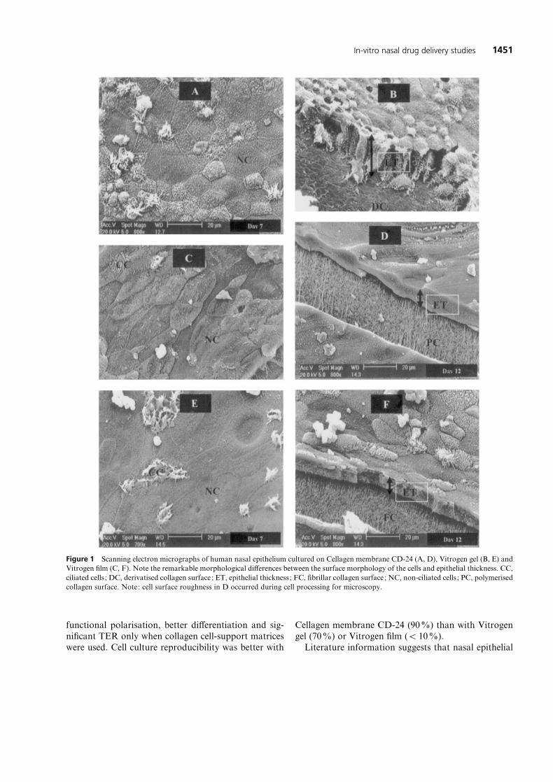
Figure 1 Scanning electron micrographs of human nasal epithelium cultured on Cellagen membrane CD-24 (A, D), Vitrogen gel (B, E) and
Vitrogen film (C, F). Note the remarkable morphological differences between the surface morphology of the cells and epithelial thickness. CC,
ciliated cells ; DC, derivatised collagen surface; ET, epithelial thickness ; FC, fibrillar collagen surface; NC, non-ciliated cells ; PC, polymerised
collagen surface. Note: cell surface roughness in D occurred during cell processing for microscopy.
functional polarisation, better differentiation and sig-
nificant TER only when collagen cell-support matrices
were used. Cell culture reproducibility was better with
Cellagen membrane CD-24 (90%) than with Vitrogen
gel (70%) or Vitrogen film (! 10%).
Literature information suggests that nasal epithelial

1452 Remigius Uchenna Agu et al
cells cultured on surfaces not coated with a biological
matrix (e.g. plastic) may multiply rapidly. Nevertheless,
the cells have the tendency to detach or become
squamous within a few days (Wasilenko & Marchok
1984). Cell detachment frequently occurs in such sur-
faces because nasal epithelial cells are predominantly
columnar cells, which do not have hemidesmosomes,
and attach to the basement membrane only by cell-
adhesion molecules (e.g. laminin and fibronectin)
(Mygind & Dahl 1998). Consequently, biological mat-
rices (e.g. collagen, laminin, extracellular matrix, etc.)
are important to obtain a stable, differentiated and
reproducible nasal culture system. However, Werner &
Kissel (1995) reported the development of a differen-
tiated nasal culture system consisting of highly dif-
ferentiated columnar-shaped ciliated, non-ciliated and
mucous-producing epithelial cells. Their observation is
surprising and difficult to explain given the context of
their culture condition (10% FCS, absence of a bio-
logical matrix and no preliminary fibroblast removal
before cell plating). Though serum may be an important
source of extracellular matrix (e.g. laminin), it contains
transforming growth factor β-1, which is implicated in
induction of squamous metaplasia in respiratory epi-
thelial cells in-vitro (Lechner et al 1984).
The morphological features of cells cultured on the
various collagen substrata are shown in Figure 1.Domes
(picture not shown) were frequently observed in cells
cultured on Cellagen membrane CD-24, but rarely on
Vitrogen gel and film. The best epithelial differentiated
phenotype (monolayer with columnarcuboidal ciliated
and non-ciliated cells) occurred in cells grown on Cel-
lagen CD-24 (Figures 1A and 1B). The cells had a
typical cobble stone appearance. In contrast, cells grown
on Vitrogen gel and film were squamous (Figures 1C
and 1E). For these substrata, cell cross-section revealed
multilayered cells (Figures 1D and 1F), with Vitrogen
gel-based cells beingmore densely packed in comparison
with Vitrogen film. Cell piling may result in a tortuous
network of aqueous pores, which may culminate in
greater resistance for molecules that traverse via these
pores.The epithelial thickness of cells grownonCellagen
membrane CD-24 was remarkably different from the
thicknesses in other collagen preparations (Cellagen
membrane CD-24, E 20 µm of monolayer ; Vitrogen
gel and film, E 10 µm of multi-layers ; Figures 1B, 1D
and 1F).
The collagen preparations we investigated are bio-
chemically identical (type I collagen), but structurally
they have a different scaffolding that may affect cell
shape and orientation to nutrient. While collagen fibres
in Vitrogen gel and film exist as loose coils with random
orientations, the fibres in Cellagen membrane CD-24
manifest as uniform layers of fibrils arranged in collinear
uninterrupted longitudinal sections. In addition, Vitro-
gen gel and film are hydrated non-flexible thick gel and
film, respectively, while Cellagen membrane CD-24,
whenwet, are hydrated thin pliable collagen films. When
used to grow epithelial cells at the air–liquid interface,
Cellagen membranes CD-24 may literally be described
as floating collagen films. Floating of collagen sub-
stratum after cell attachment is known to induce better
nasal epithelial cell differentiation (Hanamure et al
1994).
CBF studies
It was necessary to determine the culture period within
which the cells retained their ciliary beating at a physio-
logical level (7–20 Hz) because concurrent studies in-
volving drug transport, metabolism and mucosal or
ciliary toxicity is an advantage. The number of ciliated
cells in Cellagen membrane CD-24 and Vitrogen gel and
film was more than 10% up to day 6. Thereafter, ciliated
cells in all the culture conditions progressively dropped
to less than 10% and disappeared before day 21. The
CBF of the cells decreased significantly (P! 0.05) from
control (day 1) in all the collagen preparations after 7
days,withVitrogenfilm-grown cells showingmore rapid
decline in CBF. CBF of the cells decreased from 11.4³1.7 Hz to 4.6³0.9 Hz (Vitrogen gel), 10.6³2.1 Hz to
Figure 2 Ciliary beat frequency of human nasal epithelium (mean³s.d., n¯ 15 cells) cultured on Cellagen membrane CD-24 (D), Vitro-
gen gel (E) and Vitrogen film (*).

1453In-vitro nasal drug delivery studies
4.1³1.0 Hz (Cellagen membrane CD-24) and 12.5³1.8
to 3.7³0.4 Hz (Vitrogen film) on day 12 (Figure 2). The
percentage CBF decrease within this period for all the
substrata was 60–70%. However, within 7 days in
culture, the cells maintained a co-ordinated ciliary beat-
ing pattern, an indication of intercellular communi-
cation.
In all the collagen substrata investigated, the observed
CBF decrease agreed with literature information
(Rautiainen et al 1993; Agu et al 1999). The progressive
decline in the number and frequency of ciliated cells
may be explained by the loss of the cells ’ three-
dimensional geometry upon attachment (Jorissen et al
1989).Despite this decline, a sufficient number of ciliated
cells with CBF within physiological range (7–20 Hz)
could be seen in all the substrata.
Biochemical and toxicological studies
The MDH activity of 1.0¬106 cells (number of cells
seeded per insert) was determined before plating the
cells (day 1) to serve as a reference to monitor the
change in cell viability with culture duration. Trypan
blue exclusion assay routinely conducted after releasing
cells with pronase showed that the cells were more than
95% viable. Therefore, a statistically significant de-
crease in MDH activity on days 7 and 12, respectively
(relative to day 1), implied a decline in cell viability.
Days 7 and 12 were chosen for the study because the
optimal morphological and functional features of cells
grown on the three collagen substrata were observed
between days 4 and 11. The results of the MDH assay
are summarised in Table 2. There was a statistically
significant (P! 0.05) increase in the MDH activity of
Table 2 Absolute MDH activity of human nasal epithelium cultured on Cellagen membrane CD-24, Vitrogen gel and Vitrogen film.
Substrata Absolute MDH activity (absorbance cm−2) Change in MDH activity between culture days
(expressed as a ratio)
Day 1 Day 7 Day 12 Day 7:Day 1 Day 12:Day 1 Day 12:Day 7
Control
(1.0¬106 cells) 1.31³0.17a – – – – –
Cellagen
membrane CD-24 – 2.67³0.30* 4.78³0.56* 2.03W 3.64W 1.79WVitrogen gel – 5.58³0.31* 7.87³0.10* 4.25W 6.00W 1.41WVitrogen film – 4.68³0.17* 2.99³0.15* 3.57W 2.28W 0.64X
Absolute MDH activity is expressed as mean³s.d., n¯ 3. *P! 0.05 relative to control ; aMDH activity of 1.0¬106 cells (number of cells seeded
per insert) determined on day 1 to serve as reference. W, Increase in MDH activity ; X, Decrease in MDH activity. Increase in MDH activity
relates to cell multiplication and maintenance of viability.
cells grown on the various collagen supports on days 7
and 12.
Considering the fact that higher metabolic activity in
a particular collagen substratum may be due to the
presence of more cells per insert, the absolute viability
(formazan absorbancy based on number of viable cells
per cm2 of insert) was determined. The absolute viability
of cells grown on the various substrata on day 7 followed
the rank order Vitrogen gel"Vitrogen film"Cellagen
membrane CD-24. On day 12 the rank order changed to
become Vitrogen gel"Cellagen membrane"Vitrogen
film.
The higher absolute viability for cells cultured on
Vitrogen gel on day 7, in comparison with Cellagen
membrane and Vitrogen film, may be explained by the
extent of cell multi-layering (i.e. more cells per cm2) in
gel (Figures 1B, 1D and 1F). The higher MDH activity
of Cellagen membrane CD-24-based culture on day 12
in comparison with Vitrogen film might be due to better
epithelial cell differentiation or time-dependent loss of
viability in cells grown on Vitrogen film. Also, the
number of mitochondria per cell (which may vary from
cell to cell) or metabolic differences, as well as cell size,
may account for differences in cell viability (Saxton et al
1994). Given the fact that the viability of the cells grown
on the different collagen substrata were statistically
higher (P! 0.05) on days 7 and 12 (relative to day 1) the
cells were considered to retain their viability and thus be
suitable for metabolism studies.
To validate the suitability of the cells grown on the
different collagen substrata for assessment of mucosal
damage following exposure to pharmaceutical com-
pounds, LDH release by STDHF was measured as an
indexof cellmembranedamageand intracellular toxicity

1454 Remigius Uchenna Agu et al
Table 3 LDH release following exposure to STDHF.
Substrata LDH release
Day 7 Day 12
LDH rate medium LDH rate cell % LDH release LDH rate medium LDH rate cell % LDH release
Cellagen membrane
PBS (control) 0.0120³0.0044 0.1570³0.0079 6.9³3.4 0.0063³0.0062 0.1687³0.0420 4.4³5.8
0.5% STDHF 0.1520³0.0096 0.0530³0.0262 75.4³9.6* 0.1857³0.0468 0.0750³0.0134 70.8³2.5*
Vitrogen gel
PBS (control) 0.0035³0.0025 0.1235³0.0005 2.7³2.7 0.0012³0.0001 0.1760³0.0160 0.7³0.8
0.5% STDHF 0.0647³0.0200 0.0420³0.0193 62.5³7.9* 0.1160³0.0187 0.0667³0.0269 65.3³9.2*
Vitrogen film
PBS (control) 0.0010³0.0001 0.2240³0.0010 0.44³0.1 0.0190³0.0040 0.0985³0.0225 17.1³8.6
0.5% STDHF 0.3150³0.1449 0.0270³0.0094 91.4³2.6* 0.3257³0.1367 0.0250³0.0088 92.5³1.4*
LDH release is expressed as mean³s.d., n¯ 3. *P! 0.05 relative to control. PBS, phosphate buffered saline ; STDHF, sodium tauro-24,
25 dihydrofusidate.
using 7- and 12-day-old cultures. The results of the
LDH assay are summarised in Table 3. A statistically
significant difference (P! 0.05) was observed between
control cells (treated with PBS) and cells treated with
STDHF for 30 min in all the collagen substrata. In
terms of degree of LDH release, similar percentages of
release were seen for cells grown on Cellagen membrane
CD-24 and Vitrogen gel. The percentage LDH release
was much higher in cells cultured on Vitrogen film. In all
the culture conditions investigated, the percentage LDH
release did not vary significantly between day 7 and day
12. Thus the assay was not affected by the culture
duration (within the period investigated), an indication
that within this period the cells are suitable to study
mucosal or cytotoxicity of pharmaceutical compounds.
Cells cultured on Vitrogen film exhibited much higher
LDH release compared with other conditions. Conse-
quently, cells grown on this collagen preparation may
not be suitable for nasal mucosal toxicity studies due to
the possibility of overestimating the toxicity of drugs or
excipients.
Permeation studies
The development of electrical resistance over cell layers
cultured on Cellagen membrane CD-24 and Vitrogen
collagen gel and film is highlighted in Figure 3. The TER
for cells grown on Cellagen membrane CD-24 was
200–650 Ω cm2 (days 2–10). This value is within the
200–600 Ω cm2 range reported in literature for cultured
nasal epithelium (Werner & Kissel 1995; Leuba et al
1996). The relatively higher TER seen in Vitrogen gel
Figure 3 Development of TER (mean³s.d., n¯ 12 inserts) in
human nasal epithelium cultured on Cellagen membrane CD-24 (D),
Vitrogen gel (E) and Vitrogen film (*).
(1349³508 Ω cm2) also agreed with published data
(Yankaskas et al 1985; Yamaya et al 1992). The lower
TER observed in cells grown on Cellagen membrane
CD-24 in comparison with Vitrogen gel was not due to
the flow of outward current in the domes as cells without
domes had similar TER values. The higher TER values
in cells grown on Vitrogen gel can be linked to cell piling
(Figure 1F).
Given the insignificant TER, poor culture repro-
ducibility and leaky nature of cells cultured on Vitrogen

1455In-vitro nasal drug delivery studies
0 20 40 60 80 100 120 1400.0
0.5
1.0
1.5
2.0
2.5
3.0
3.5
4.0
Time (min)
Cu
mu
lati
ve a
mo
un
t tr
ansf
erre
d(í
g m
L–1) 0 20 40 60 80
Time (min)
0
10
20
30
40
50
60
70
Cu
mu
lati
ve a
mo
un
t tr
ansf
erre
d(í
g m
L–1)
Figure 4 Sodium fluorescein permeation across nasal epithelium cultured on Cellagen membrane CD-24 (D) and Vitrogen gel (E). Inset :
permeation of sodium fluorescein across collagen substrata without cells (V, Cellagen membrane; U, Vitrogen gel). The cells were cultured for
7 and 9 days for Cellagen membrane CD-24 and Vitrogen gel, respectively.
film, these cells were not used for permeation studies.
Results of the permeation studies are summarised in
Figure 4. The flux of sodium fluorescein, a paracellular
marker, through collagen preparations without cells
was linear, r2 ¯ 0.990³0.003, n¯ 3 (Cellagen mem-
brane) and 0.990³0.009, n¯ 3 (Vitrogen gel), indi-
cating that these collagen preparations did not form a
significant barrier to diffusion of sodium fluorescein.
The apparent permeability coefficients (Papp cm s−1) of
sodium fluorescein across the collagen preparations
were 40.70³2.30¬10−7 (Cellagen membrane CD-24)
and 61.70³6.00¬10−7 (Vitrogen gel). For Cellagen
membrane CD-24 and Vitrogen gel with cells grown on
them, the flux of fluorescein was statistically (P! 0.05)
reduced to 1.91³0.00¬10−7 cm s−1 (Cellagen membrane
CD-24) and 0.45³0.08¬10−7 cm s−1 (Vitrogen gel). For
both substrata the diffusion of sodium fluorescein was
% 2% of the amount added to the apical side within 2 h,
thus confirming the formationof tight junctions between
the cells.
The permeability coefficients of sodium fluorescein
across the cells were lower, though comparable, than
that of 5.86³0.48¬10−7 obtained for a Caco-2 model.
It is however, important to mention that the comparison
of the absolute Papp values obtained using different
experimental set-ups should be done with some reserv-
ation as some variables affect these values. Some of
the experimental factors that directly affect the absolute
values of Papp include pH gradients, additional diffusion
barriers (i.e. unstirred water layer, type of filter support),
analyte concentration, detection method, cell culture
variation (Caldwell et al 1998) and, possibly, the formula
used to calculate the Papp.
Conclusion
This studyhighlighted the fact that collagen is important
as a cell-support matrix to establish a stable, repro-
ducible and differentiated primary cell culture system
for nasal drug delivery studies. In terms of cell mor-
phology, Cellagen membrane induced better epithelial
cell differentiation than Vitrogen gel or film. For all the
collagen substrata investigated, cell morphology was
optimal between days 4 and 11. Therefore, the physical
structure of the collagenmatrix and timeof investigation
are important considerationswhenusing collagen-based

1456 Remigius Uchenna Agu et al
human nasal primary culture systems for in-vitro drug
delivery studies. Based on MDH activity, LDH release,
CBF, TER and permeation studies, human nasal epi-
thelial cells cultured on derivatised collagen (Cellagen
membrane CD-24) and polymerised collagen (Vitrogen
gel) were concluded to be functionally suitable for in-
vitro nasal drug delivery studies. Due to their leaky
nature and comparatively higher LDH release upon
exposure to STDHF, cells cultured on fibrillar collagen
(Vitrogen film) may be limited to metabolism and cilio-
toxicity studies. Based on better culture reproducibility
and epithelial cell differentiation, further characteris-
ation of the permeation profile of cells cultured on
Cellagen membrane CD-24, using compounds of dif-
ferent molecular weights and absorption characteristics,
is recommended.
References
Agu, R. U., Jorissen, M., Willems, T., Van den Mooter, G., Kinget,
R., Augustijns, P. (1999) The effects of pharmaceutical compounds
on ciliary beating in human nasal epithelial cells : a comparative
study of cell culture models. Pharm. Res. 16 : 1380–1385
Agu, R. U., Jorissen, M., Van den Mooter, G., Kinget, R., Augustijns,
P. (2000) Safety assessment of selected cyclodextrins – effect on
ciliary activity using a human cell suspension model exhibiting in
vitro ciliogenesis. Int. J. Pharm. 193 : 219–226
Baeza-Squiban, A., Boisvieux-Ulrich, E., Guilianelli, C., Houcine, O.,
Geraud, G., Guennou, C., Marano, F. (1994) Extracellular matrix-
dependent differentiation of rabbit tracheal epithelial cells in pri-
mary culture. In vitro Cell. Dev. Biol. 30A : 56–67
Blank, U., Clauss, W., Weber, W.-M. (1995) Effects of benzamil in
human cystic fibrosis airway epithelium. Cell Physiol. Biochem. 5 :
385–390
Caldwell, G. W., Easlick, S. M., Gunnet, J., Masucci, J. A., Demarest,
K. (1998) In vitro permeability of eight beta blockers through Caco-
2 monolayers utilizing liquid chromatographyelectrospray ion-
ization mass spectrometry. J. Mass Spectrom. 33 : 607–614
Folkman, J., Moscona, A. (1978) Role of cell shape in growth control.
Nature 273 : 345–349
Grinnell, F., Bennett, M. H. (1982) Ultrastructural studies of cell-
collagen interactions. In: Cunningham, L. W., Frederiksen, D. W.
(eds) Methods in enzymology. Vol. 82, Academic Press, New York,
pp 535–544
Hanamure, Y., Deguchi, K., Ohyama, M. (1994) Ciliogenesis and
mucus synthesis in cultured human respiratory epithelial cells. Ann.
Otol. Rhinol. Laryngol. 103 : 889–895
Hovgaard, L., Brondsted, H. (1995) Drug delivery studies in Caco-2
monolayers. IV. Absorption enhancer effects of cyclodextrins.
Pharm. Res. 9 : 1328–1332
Jorissen, M., Van der Schueren, B., Van der Berghe, H., Cassiman, J.
(1989) The preservation and regeneration of cilia of human nasal
epithelial cells cultured in vitro. Arch. Otorhinolaryngol. 246 : 308–
314
Lechner, J. F., Haugen, A., McClendon, I. A., Pettis, E. W. (1984)
Induction of squamous differentiation of normal human bronchial
epithelial cells by small amounts of serum. Differentiation 25 :
229–237
Leuba, D., De Ribaupiererre, Y., Kucera, P. (1996) Ion transport,
ciliary activity, and mechanosensitivity of sinusal mucosa: an in
vitro study. Am. J. Physiol. 271 : L349–L358
Macklis, J. D., Sidman, R. L., Shine, H. D. (1984) Cross-linked
collagen surface for cell culture that is stable, uniform and optically
superior to conventional surfaces. In vitro Cell Dev. Biol. 21 :
189–194
Mygind, N., Dahl, R. (1998) Anatomy, physiology and function of
the nasal cavities in health and disease. Adv. Drug Deliv. Rev. 29 :
3–12
Rautiainen, M., Matsume, S., Yoshiatsugu, M., Ohyama, M.(1993)
Degeneration of human respiratory cell ciliary beat in monolayer
cell culture. Eur. Arch. Otorhinol. 250 : 97–100
Roesems, G., Hoet, P. H. M., Demedts, M., Nemery, B. (1997) In
vitro toxicity of cobalt and hard metal dust in rat and human type
II pneumocytes. Pharmacol. Toxicol. 81 : 74–80
Saxton, R. E., Haghighat, S., Plant, D., Lufkin, R., Soundant, J.,
Castro, D. J. (1994) Dose response of human tumor cells to
rhodamine 123 and laser phototherapy. Laryngoscope 104 : 1013–
1018
Schmidt, M. C., Peter, H., Lang, S. R., Ditzinger, G., Merkle, H. P.
(1998) In vitro cell models to study nasal mucosal permeability and
metabolism. Adv. Drug Deliv. Rev. 29 : 51–79
Shannon, J. M., Mason, R. J., Jennings, S. (1987) Functional differ-
entiation of alveolar type II epithelial cells in vitro: effect of cell
shape, cell-matrix interactions and cell-cell interactions. Biochim.
Biophys. Acta 931 : 143–156
Strom, S. C., Michalopoulos, G. (1982) Collagen as a substrate for
cell growth and differentiation. In: Cunningham, L. W.,
Frederiksen,D. W. (eds) Methods in enzymology. Vol. 82,Academic
Press, New York, pp 544–555
Ugwoke, M. I., Agu, R. U., Jorissen, M., Augustijns, P., Sciot, R.,
Verbeke, N., Kinget, R. (2000a) Toxicological investigations of the
effectsof carboxymethylcellulose on ciliarybeat frequencyofhuman
nasal epithelial cells in primary suspension culture and in vivo on
rabbit nasal mucosa. Int. J. Pharm. 205 : 43–51
Ugwoke, M. I., Agu, R. U., Jorissen, M., Augustijns, P., Sciot, R.,
Verbeke, N., Kinget, R. (2000b) Nasal toxicological investigations
of carbopol 971P formulation of apomorphine: effect on ciliary
beat frequency of human nasal primary cell culture and in vivo on
rabbit nasal mucosa. Eur. J. Pharm. Sci. 9 : 387–396
Wadell, C., Bjo$ rk, E., Camber, O. (1999) Nasal drug delivery –
evaluation of an in vitro model using porcine nasal mucosa. Eur. J.
Pharm. Sci. 7 : 197–206
Wasilenko, W. J., Marchok A. C. (1984) Pyruvate regulation of
growth and differentiation in primary cultures of rat tracheal
epithelial cells. Exp. Cell. Res. 155 : 507–517
Werner, U., Kissel, T. (1995) Development of a human nasal epithelial
cell culture model and its suitability for transport and metabolism
studies under in vitro conditions. Pharm. Res. 12 : 565–571
Yamaya, M., Finkbeiner, W. E., Chun, S. Y., Widdicombe, J. H.
(1992) Differentiated structure and function of cultures of human
tracheal epithelium. Am. J. Physiol. 262 : L713–L724
Yankaskas, J. R., Cotton, C. U., Knowles, M. R., Gatzy, J. T.,
Boucher, R. C. (1985) Culture of human nasal epithelial cells on
collagen matrix supports. Am. Rev. Respir. Dis. 132 : 1281–1287
Copyright © 2022 FDOKUMEN