Bacterial sensor kinases: diversity in the recognition of environmental signals
24
Bacterial Sensor Kinases: Diversity in the Recognition of Environmental Signals Tino Krell, Jes ´ us Lacal, Andreas Busch, Hortencia Silva-Jim ´ enez, Mar´ ıa-Eugenia Guazzaroni, and Juan Luis Ramos Department of Environmental Protection, Estaci ´ on Experimental del Zaid´ ın, Consejo Superior de Investigaciones Cient´ ıficas, 18008 Granada, Spain; email: [email protected] Annu. Rev. Microbiol. 2010. 64:539–59 The Annual Review of Microbiology is online at micro.annualreviews.org This article’s doi: 10.1146/annurev.micro.112408.134054 Copyright c 2010 by Annual Reviews. All rights reserved 0066-4227/10/1013-0539$20.00 Key Words two-component system, bacterial signal transduction, transcriptional regulation, signal recognition Abstract Bacteria sense and respond to a wide range of physical and chemical signals. Central to sensing and responding to these signals are two- component systems, which have a sensor histidine kinase (SK) and a response regulator (RR) as basic components. Here we review the dif- ferent molecular mechanisms by which these signals are integrated and modulate the phosphorylation state of SKs. Apart from the basic mech- anism, which consists of signal recognition by the SK that leads to an alteration of its autokinase activity and subsequently a change in the RR phosphorylation state, a variety of alternative modes have evolved. The biochemical data available on SKs, particularly their molecular interactions with signals, nucleotides, and their cognate RRs, are also reviewed. 539 Annu. Rev. Microbiol. 2010.64:539-559. Downloaded from www.annualreviews.org by CSIC - Consejo Superior de Investigaciones Cientificas on 04/04/11. For personal use only.
Transcript of Bacterial sensor kinases: diversity in the recognition of environmental signals

MI64CH28-Ramos ARI 17 August 2010 15:42
Bacterial Sensor Kinases:Diversity in the Recognitionof Environmental SignalsTino Krell, Jesus Lacal, Andreas Busch,Hortencia Silva-Jimenez, Marıa-Eugenia Guazzaroni,and Juan Luis RamosDepartment of Environmental Protection, Estacion Experimental del Zaidın, ConsejoSuperior de Investigaciones Cientıficas, 18008 Granada, Spain; email: [email protected]
Annu. Rev. Microbiol. 2010. 64:539–59
The Annual Review of Microbiology is online atmicro.annualreviews.org
This article’s doi:10.1146/annurev.micro.112408.134054
Copyright c© 2010 by Annual Reviews.All rights reserved
0066-4227/10/1013-0539$20.00
Key Words
two-component system, bacterial signal transduction, transcriptionalregulation, signal recognition
Abstract
Bacteria sense and respond to a wide range of physical and chemicalsignals. Central to sensing and responding to these signals are two-component systems, which have a sensor histidine kinase (SK) and aresponse regulator (RR) as basic components. Here we review the dif-ferent molecular mechanisms by which these signals are integrated andmodulate the phosphorylation state of SKs. Apart from the basic mech-anism, which consists of signal recognition by the SK that leads to analteration of its autokinase activity and subsequently a change in theRR phosphorylation state, a variety of alternative modes have evolved.The biochemical data available on SKs, particularly their molecularinteractions with signals, nucleotides, and their cognate RRs, are alsoreviewed.
539
Ann
u. R
ev. M
icro
biol
. 201
0.64
:539
-559
. Dow
nloa
ded
from
ww
w.a
nnua
lrev
iew
s.or
gby
CSI
C -
Con
sejo
Sup
erio
r de
Inv
estig
acio
nes
Cie
ntif
icas
on
04/0
4/11
. For
per
sona
l use
onl
y.
MI64CH28-Ramos ARI 17 August 2010 15:42
Sensor kinase (SK):protein withautokinase activity ableto phosphorylate a RR
Response regulator(RR): exerts amodulatory responseas a function of itsphosphorylation state
Contents
INTRODUCTION . . . . . . . . . . . . . . . . . . 540RELEVANT BIOCHEMICAL AND
STRUCTURAL INFORMATIONON SENSOR KINASES . . . . . . . . . . 540Topology. . . . . . . . . . . . . . . . . . . . . . . . . . 540Oligomeric State . . . . . . . . . . . . . . . . . . . 541Mode of Autophosphorylation . . . . . . 541Structure . . . . . . . . . . . . . . . . . . . . . . . . . . 541Interaction with Signal Molecules . . . 542Interaction with Nucleotides . . . . . . . 542Catalytic Activities and Modes of
Transphosphorylation. . . . . . . . . . . 543Stability of Phosphorylated SKs . . . . 544Interaction Between Both
Components of a TCS . . . . . . . . . . 544Specificity of Interaction Between
Both Components of a TCS . . . . . 546MECHANISMS FOR THE
MODULATION OF THESENSOR KINASEPHOSPHORYLATION STATE . . 547Signal Sensing at Sensor Domains . . 547Signal Perception at
Transmembrane Regions . . . . . . . . 549Signal Perception at Accessory
Proteins . . . . . . . . . . . . . . . . . . . . . . . . 550Other Modes of Modulating the SK
Phosphorylation State . . . . . . . . . . . 552TWO-COMPONENT SYSTEMS
AS ELEMENTS ININTERKINGDOM SIGNALING. 553
INTRODUCTION
The environment of microbes is constantlychanging due to variations in physicochemi-cal parameters such as temperature, light, andoxygen tension. Due to the unicellular organi-zation of prokaryotes, environmental changes,whether biotic or abiotic, are rapid and dra-matic, whereas many eukaryotes can bufferthemselves against environmental changes be-cause of their multicellular organization. A widerange of molecular adaptational processes that
modulate cellular processes as a function of sig-nal molecules have evolved at transcriptional,translational, posttranslational, and postsyn-thetic levels. Central to these responses is themodulation of DNA transcription. Here we dis-cuss several mechanisms of bacterial response toenvironmental signals and concentrate on two-component regulatory systems (TCSs), whichare used by prokaryotes as the primary mech-anism to translate these rapid environmentalchanges into a regulatory readout. Genes en-coding TCSs are present in almost all bacteria,and genome analyses have shown that bacte-ria have on average 52 TCSs (19). The pro-totypical TCS consists of a membrane-boundsensor kinase (SK), which frequently respondsto extracytosolic signals, and a mobile cytosolicresponse regulator (RR), which in most casesinteracts with promoter regions to modulateDNA transcription.
Other RRs possess enzymatic activities orbind to other proteins. SKs can be phosphory-lated on a histidine and are able to transphos-phorylate their cognate RR at an aspartate. Thefunction of TCSs is based on the fact that theactivities of phosphorylated and unphospho-rylated RRs differ. The phosphorylation stateof the SK is the critical parameter that deter-mines transphosphorylation toward its cognateRR and thus the final modulatory readout (92).
The diversity in the nature of environmen-tal changes perceived by TCSs is enormous andis illustrated in Table 1. To respond to sucha wide variety of stimuli, different molecularmechanisms of signal recognition by SKs haveevolved and are reviewed here. In the initial partof this review, we summarize the biochemicaland structural data available on SKs.
RELEVANT BIOCHEMICAL ANDSTRUCTURAL INFORMATIONON SENSOR KINASES
Topology
The prototypical SK contains a periplasmicsensor domain and a cytosolic autokinase do-main linked by a transmembrane region. How-
540 Krell et al.
Ann
u. R
ev. M
icro
biol
. 201
0.64
:539
-559
. Dow
nloa
ded
from
ww
w.a
nnua
lrev
iew
s.or
gby
CSI
C -
Con
sejo
Sup
erio
r de
Inv
estig
acio
nes
Cie
ntif
icas
on
04/0
4/11
. For
per
sona
l use
onl
y.

MI64CH28-Ramos ARI 17 August 2010 15:42
Table 1 Selection of some two-component systems with their corresponding function and signal molecule
System Function Signal molecule Reference(s)ArcB/ArcA Sensing of oxygen and redox states Quinones reflecting the redox state 27, 59NarX/NarL Nitrate and nitrite respiration Nitrate, nitrite 51CitA/CitB Transport and anaerobic metabolism of citrate Citrate 5, 45CheA/CheY Chemotaxis Chemoattractants, e.g., serine and aspartate 57FixL/FixJ Nitrogen fixation O2, CO, NO 29LovK/LovR Bacterial cell attachment Blue light 76TodS/TodT Degradation of benzene derivatives Monoaromatic compounds 10, 49NtrB/NtrC Nitrogen utilization 2-ketoglutarate, glutamine 43KdpD/KdpE K+ supply K+ 23VanS/VanR Antibiotics Vancomycin 36EnvZ/OmpR Osmolarity ? 11KinB/Spo0F Sporulation ?, ATP as cosignal? 8BvgS/BvgA Virulence Temperature, sulfate ions, nicotinic acid 3LuxQ/LuxO Quorum sensing AI-2 67DesR/DesK Lipid modification Temperature 1
ever, variations of this standard type of SK arefrequent. This is highlighted in Figure 1, whichshows the topology of the major SKs discussedin this review. Approximately 83% of SKs con-tain transmembrane regions (19), whereas theremainder are located in the cytosol. The exis-tence of SK monomers comprising 1–10 trans-membrane regions is well documented (24), andsequence analysis reveals SKs with up to 20transmembrane regions (62).
Another characteristic feature of SKs is do-main duplication. Examples are TodS, whichcontains two autokinase domains (49); KinA,with three PAS domains (87); and CitA, whichhas a sensor domain in the periplasm and an-other in the cytosol (Figure 1) (86).
Oligomeric State
Structural studies and biochemical evidence in-dicate that SKs frequently function as dimers.For CheA, the dissociation constant for themonomer-dimer association was determined tobe between 0.2 and 0.4 μM. A dilute CheAsample containing only monomers was devoidof kinase activity, whereas increasing activitywas detected as the protein concentration wasraised, leading to protein dimerization. This
monomer-dimer equilibrium appears to be anintrinsic property of CheA because it is not in-fluenced by nucleotide binding, phosphoryla-tion, or binding to CheY (93).
Mode of Autophosphorylation
It has long been believed that the universalmode of SK autophosphorylation occurs intrans. This implies that the ATP bound to onemonomer transfers its γ-phosphoryl group tothe histidine of the other monomer. Using dif-ferent experimental approaches, investigatorshave documented this mode of phosphorylationfor CheA (94, 106), EnvZ (12), and NRII (69).However, evidence for autophosphorylation incis has recently been obtained for a SK fromThermotoga maritima (13).
Structure
There is a wealth of structural informationavailable on individual domains that form partof SKs. However, structural information onfull-length SKs is still limited. A 3.8 A electrondensity map has been recently reported for theSK/RR complex ThkA/TrrA (107). The high-resolution structures of the individual domains
www.annualreviews.org • Bacterial Sensor Kinases 541
Ann
u. R
ev. M
icro
biol
. 201
0.64
:539
-559
. Dow
nloa
ded
from
ww
w.a
nnua
lrev
iew
s.or
gby
CSI
C -
Con
sejo
Sup
erio
r de
Inv
estig
acio
nes
Cie
ntif
icas
on
04/0
4/11
. For
per
sona
l use
onl
y.

MI64CH28-Ramos ARI 17 August 2010 15:42
PAS Autokinase PAS Autokinase RRR
PAS-A PAS-B PAS-C Autokinase
PAS Autokinase
Autokinase
Autokinase
PAS Autokinase
Autokinase RRR HPT PAS
PAS HAMP Autokinase
PAS PAS Autokinase
HAMP Autokinase ?
Autokinase RRR LuxQ-per
Autokinase HPT CheY- binding CheW
TodS
ArcB
FixL
KinA
CitA
BceS/VanS
PhoQ
LuxQ
CheA
DesK
EnvZ
NtrB
PAS GAF PHY Autokinase BphP4
IM Cytoplasm
Figure 1Domain organization and topology of selected sensor kinases based on domainannotation by SMART and complemented with structural information.Abbreviations: PAS, PER-ARNT-SIM; HAMP, domain found in histidinekinases, adenylyl cyclases, methyl binding proteins, and phosphatases;RRR, response regulator receiver; HPT, histidine containing phosphotransfer;IM, inner membrane; GAF, GAF domain; PHY, phytochrome.
were placed into this map, and the resultingstructure provides insight into the interactionbetween the SK PAS domain and its catalyticand ATP binding (CA) domain. Both domains,which are part of the same polypeptide, inter-act with each other by creating an interdomainβ-sheet.
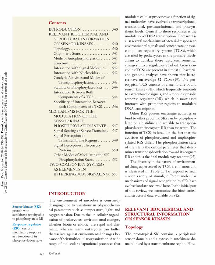
PAS domains are by far the most frequentsensor domains found in SKs and several struc-tures of such domains have been solved. Thestructure of the CitA PAS domain has beensolved in the absence and presence of citrate(Figure 2a) (77, 81). The domain consists ofa five-stranded β-sheet and five α-helices. Thedomain forms a central cavity to which citrate
is bound. A comparison of the structures in thepresence and absence of ligand shows that cit-rate binding causes a considerable contractionof the domain, which was proposed to be themolecular switch that activates transmembranesignaling (81).
The structure of the cytoplasmic fractionof a SK is shown in Figure 2b (60). Eachmonomer consists of a long α-hairpin, termedthe dimerization and histidine phosphotransfer(DHp) domain, that harbors the phospho-accepting histidine and the globular CAdomain. The ensemble of the DHp and CAdomains is called the autokinase domain. Thedimerization interface formed by the two DHpdomains results in a four-helix bundle. Thisstructure was obtained in complex with anADP derivative, and ATP was modeled byadding the γ-phosphate. In this model thedistance between the γ-phosphate oxygenand the nitrogen of the phospho-acceptinghistidine was 4.5 A, which is appropriate for thekinase transition state. According to structureand mutagenesis studies, the DHp and CA do-mains are loosely associated. Marina et al. (60)proposed that the CA domain is released fromthe DHp domain for autophosphorylation andretained for phosphotransferase activities.
Interaction with Signal Molecules
For many TCSs, the physiological signal thatmodulates the action of TCS is unknown. Sig-nal molecules have been identified for relativelyfew systems, and SK binding affinities for someof them are listed in Table 2. The affinitiesranged from submicromolar, such as the bind-ing of toluene to TodS, to millimolar values.The difference in affinity reflects the physiolog-ical need to trigger a response at a given signalconcentration. Busch et al. (10) provided evi-dence for some molecules that bind to TodSbut do not induce a regulatory output.
Interaction with Nucleotides
SKs use ATP as a nucleotide. A SK thatbinds GTP specifically has recently been
542 Krell et al.
Ann
u. R
ev. M
icro
biol
. 201
0.64
:539
-559
. Dow
nloa
ded
from
ww
w.a
nnua
lrev
iew
s.or
gby
CSI
C -
Con
sejo
Sup
erio
r de
Inv
estig
acio
nes
Cie
ntif
icas
on
04/0
4/11
. For
per
sona
l use
onl
y.

MI64CH28-Ramos ARI 17 August 2010 15:42
a
C
N
b c Spo0B
Spo0F
Aspartate
Histidine
Histidine
ADP
Citrate
α-helixβ-strand
Figure 2The structure of sensor kinase (SK) domains. (a) Structure of the PAS sensor domain of CitA in complex with citrate (77). (b) Thecytosolic part of a SK from Thermotoga maritima. Each monomer is colored differently. Bound ADP and the phospho-acceptinghistidine are labeled (60). (c) Structure of Spo0B in complex with the receiver domain of its cognate response regulator Spo0F (112).The histidine and aspartate residues involved in phosphotransfer are highlighted.
described (80). Binding and kinetic data forthe interaction of different SKs with ATPand ADP are summarized in Table 3. Theaffinity of SKs for ATP appears to be relativelyconserved, with KM or KD values between100 and 200 μM. Another interesting featureis the binding of ADP with relevant affinity.In all cases, SKs are slow enzymes, withkcat values typically ranging between 1 and10 min−1.
Catalytic Activities and Modesof Transphosphorylation
SKs catalyze as many as three reactions:autophosphorylation, transphosphorylation tothe cognate RR, and in some cases dephos-phorylation towards RR. The last activity iswell documented in AbsA1/AbsA2 (82) andVanS/VanR (36).
Several modes of transphosphorylationbetween SKs and RRs have been described
Table 2 Affinity of sensor kinases for signal molecules
Kinase Signal molecule
Cellularcompartment of
sensing KD (μM) Method Reference(s)TodS Toluene Cytosol 0.69 ITC 10
O-xylene 0.58CitA Citrate Periplasm 5.5 ITC 45
Isocitrate 15PhoQ Mg2+ Periplasm 300 CD and fluorescence 53
Peptide C18G 2.8 2FixL O2 Cytosol 50 Spectroscopy 30, 85
CO 2.1NarX Nitrate Periplasm 35 Autophosphorylation stimulation 51
Nitrite 3,500 16
Abbreviations: CD, circular dichroism; ITC, isothermal titration calorimetry.
www.annualreviews.org • Bacterial Sensor Kinases 543
Ann
u. R
ev. M
icro
biol
. 201
0.64
:539
-559
. Dow
nloa
ded
from
ww
w.a
nnua
lrev
iew
s.or
gby
CSI
C -
Con
sejo
Sup
erio
r de
Inv
estig
acio
nes
Cie
ntif
icas
on
04/0
4/11
. For
per
sona
l use
onl
y.

MI64CH28-Ramos ARI 17 August 2010 15:42
Table 3 The interaction of sensor kinases with nucleotides
Sensor kinaseKD ATP
(μM)KM ATP
(μM)KD ADP
(μM) kcat ATP (min−1) ReferenceFixL 104 – – 1.5 (deoxy state) 84
1.1 (carbonmonoxy) 85CheA – 309 – 14 54
260/1,100a – 90/330a 7 90
– 274 180 1.6 93
– 300 42 – 97EnvZ – 218 – – 47PrrB – 170 – – 20DcuS 160 – – – 40
aDifferent affinity for each monomer of the CheA dimer.
Phosphorelay:molecular mechanismby which phosphorylgroups are transferredto additional sites onthe SK prior tophosphorylation of theRR
(Figure 3). The prototypical mode consists ofa direct phosphotransfer between the SK au-tokinase domain and the RR receiver domain.However, more sophisticated hybrid systemsexist that involve multiple phosphotransferreactions, termed phosphorelay, of which theso-called tripartite kinases are well charac-terized (Figure 3). Phosphorelay involves anautokinase domain, an internal RR receiver,and subsequently an HPT domain prior to thephosphorylation of the RR (48). More recently,TodS was shown to operate by a phosphorelaymechanism composed of two autokinase do-mains (Figure 3) that have similar autokinaseactivities but of which only the N-terminaldomain is stimulated by signal binding (9).However, in all cases the chemistry of transfer isconserved and consists of His → Asp → His →Asp.
Another type of phosphorelay represents theKinA/Spo0F/Spo0B/Spo0A system (Figure 3),which consists of four individual proteins (8).A phosphorelay offers the possibility of regu-lating the individual phosphotransfer reactionsas a function of additional environmental stim-uli. As a consequence, several input signals maydefine the regulatory output, and the resultingfine-tuning of the regulatory response mightrepresent the physiological reason for the ex-istence of phosphorelays.
Stability of Phosphorylated SKs
Table 4 shows the half-life of phosphorylationfor several SKs. Half-lives of full-length pro-teins are rather conserved and range from 30to 90 min. The range of stability of phosphory-lated SKs appears to be narrower than that ob-served for the stability of phosphorylated RRs.The upper and lower limits of the half-lives ofphosphorylated RRs are 2 s for CheB (88) anddays for Spo0A (55).
Interaction Between BothComponents of a TCS
Structural studies have also provided insightinto the interaction between SKs and RRs.Figure 2c shows the structure of Spo0B boundto the receiver domain of its RR, Spo0F (112).Sequence analysis revealed that Spo0B has noautokinase domain and that it lacks autokinaseactivity (8). However, its structure is strikinglysimilar to that of autokinase domains and hasa histidine residue from which the phosphorylgroup is transferred to Spo0F. Therefore,the molecular mechanism of phosphotransferbetween Spo0B and Spo0F is assumed to cor-respond to the mechanism for the autokinasedomain RR phosphotransfer. Two monomersof SpoF bind to the SpoB dimer. Theassociation of the two molecules brings the
544 Krell et al.
Ann
u. R
ev. M
icro
biol
. 201
0.64
:539
-559
. Dow
nloa
ded
from
ww
w.a
nnua
lrev
iew
s.or
gby
CSI
C -
Con
sejo
Sup
erio
r de
Inv
estig
acio
nes
Cie
ntif
icas
on
04/0
4/11
. For
per
sona
l use
onl
y.

MI64CH28-Ramos ARI 17 August 2010 15:42
ArcB/ArcAPhosphorelay with autokinase and HPTdomain
TodS/TodTPhosphorelay with twoautokinase domains
HPTHis DNA-b
P P P
P P+
P
TodS
?
PhoQ/PhoPDirect phosphorylation
PAS
PAS
PAS1
PAS-A PAS-B
PAS2
AKHis
AK
AK1
His
AKHis
His
AK2His
ATP ADP
ATP
ATP
ADP ATP ADP
ATP ADP
ATP ADP
Tm1 Tm2
Tm1 Tm2
Asp RRR
Asp RRR
Asp RRR
Asp RRR
DNA-bAsp RRR
DNA-bAsp RRRAsp RRR
P
KinA/Spo0F/Spo0B/Spo0APhosphorelay withmultiple proteins
KinA Spo0B
? ?
His Spo0B
P P P
PhoQ PhoP
ArcB ArcA
TodT
Spo0F Spo0A
HAMP DNA-b
Signal:Antimicrobial
peptides; Mg2+
Signal:Quinones;redox state
Signal:Toluene, etc.
PAS-C
Figure 3Different types of phosphoryl transfer observed in two-component regulator systems. Abbreviations: AK, autokinase domain; DNA-b,DNA-binding domain; Tm, transmembrane region; HPT, histidine containing phosphotransfer domain; RRR, response regulatorreceiver domain.
histidine residue in close alignment with theRR aspartate, which is necessary for phospho-transfer. The authors modeled a phosphatein between both amino acids to mimic thetransition state. Distances of 2.45 A wereobserved for the Nε-P and Oδ-P interactions.
Furthermore, bound Mg2+ is found in theactive site, and its interaction with aspartateand the transition state phosphate is thoughtto facilitate phosphotransfer.
The interactions between SKs and RRsare summarized in Table 5. Strikingly, most
Table 4 Stabilities of the phosphorylation at sensor kinases
Sensor kinase Half-life (min) pH ReferenceTodS 70 7.5 49EnvZ 70–80 7.5 47RegB 34 7.6 75AbsA2 69 8.0 82FrzE 90 6.9 65
www.annualreviews.org • Bacterial Sensor Kinases 545
Ann
u. R
ev. M
icro
biol
. 201
0.64
:539
-559
. Dow
nloa
ded
from
ww
w.a
nnua
lrev
iew
s.or
gby
CSI
C -
Con
sejo
Sup
erio
r de
Inv
estig
acio
nes
Cie
ntif
icas
on
04/0
4/11
. For
per
sona
l use
onl
y.

MI64CH28-Ramos ARI 17 August 2010 15:42
Table 5 The interaction between the two components of two-component systems
TCS KD (μM) Method ReferenceFixL/FixJ 4 Fluorescence 84EvgS/EvgA 1.2a Surface plasmon resonance 74EnvZ/OmpR >1 μMb Native PAGE 110
0.52b Fluorescence anisotropy 641.2b 11f
CheA/CheY 0.9–1.3c Fluorescence 892.0d and 1.2e ITC 56
PhoQ/PhoP 0.12b Resonant mirror biosensor technology 14YPD1/SLN1 1.4 Quench-flow 41
aHPT domain of EvgS.bCytosolic part of EnvZ.cP2 domain of CheA.dFull-length CheA.eCheA fragment comprising domains P1 and P2.f His-tagged OmpR was mixed at different ratios with EnvZ; free OmpR and the EnvZ-OmpR complex were isolated byaffinity chromatography and submitted to SDS-PAGE.Abbreviations: ITC, isothermal titration calorimetry; PAGE, polyacrylamide gel electrophoresis; TCS, two-componentsystem.
Cross-talk:interaction betweennoncognate SKs andRRs
interactions are characterized by an affinityof ∼1 μM, indicative of certain conservationamong proteins. In general, the interaction be-tween proteins is characterized by high affin-ity, and an average KD of 17 nM for a protein-protein interaction was determined (91). Thisimplies that the average affinity between the twocomponents of a TCS is far below the affinitygenerally observed between proteins.
This raises the question whether both com-ponents of a TCS are associated in the cell ornot. Cai & Inouye (11) have determined thecellular concentration of EnvZ and OmpR tobe 0.18 and 6 μM, respectively. Combiningthe cellular concentration and the affinity ofboth proteins, the authors concluded that ap-proximately 85% of EnvZ molecules in the cellare associated with OmpR. These data are thusconsistent with the notion that SKs and RRs areloosely associated.
Specificity of Interaction BetweenBoth Components of a TCS
Bacteria contain a large number of TCSs,which raises the question of the specificity of
their interaction. This issue has been addressedin Escherichia coli, for which 30 SKs and 32 RRwere predicted. Yamamoto et al. (108) purifiedthe kinase domains of 27 SKs as well as all RRs.Almost all SKs showed autophosphorylationactivity and 24 proteins showed transphos-phorylation activity toward their cognate RR.Subsequent studies were aimed at evaluatingthe cross-talk between noncognate partners.
Interestingly, there were two differentmechanisms of cross-talk. In the first modethere was direct cross-talk, represented byRRs that were phosphorylated by noncog-nate SKs. Among 692 SK-RR noncognatepairs, transphosphorylation occurred in only 22noncognate pairs, indicative of a high degreeof specificity. The second mode of cross-talkis represented by the enhancement of SK de-phosphorylation by noncognate RRs that werenot phosphorylated. This phenomenon was ob-served for nine RRs that altered the phospho-rylation state of 12 SKs.
Another study of the specificity of themolecular interaction revealed that SKshave a strong kinetic preference for thecognate RR (83). This is exemplified by
546 Krell et al.
Ann
u. R
ev. M
icro
biol
. 201
0.64
:539
-559
. Dow
nloa
ded
from
ww
w.a
nnua
lrev
iew
s.or
gby
CSI
C -
Con
sejo
Sup
erio
r de
Inv
estig
acio
nes
Cie
ntif
icas
on
04/0
4/11
. For
per
sona
l use
onl
y.

MI64CH28-Ramos ARI 17 August 2010 15:42
Agonist: signalrecognition at SK thattriggers a modulatoryresponse
Antagonist: signalrecognition at SK thatdoes not trigger amodulatory response
transphosphorylation studies between EnvZand OmpR. After 60 min of transphosphoryla-tion, EnvZ phosphorylated 16 RRs. However,after only 10 s of transphosphorylation, EnvZexclusively phosphorylated its cognate partner,OmpR.
A central question is whether cross-talk ful-fills a biological function or whether it is animperfection in molecular recognition. Severalexamples of physiologically relevant cross-talklike that between RetS and GacS have beenreported (31). The different molecular mech-anisms which have evolved to optimize SK-RRspecificity are reviewed in Reference 50.
MECHANISMS FOR THEMODULATION OF THESENSOR KINASEPHOSPHORYLATION STATE
In order to respond to the enormous diversityof environmental signals, a wide range of signalperception modes have evolved that can be clas-sified into three groups: signal sensing at sensordomains, signal sensing at transmembrane re-gions, and signal sensing at auxiliary proteins.
Signal Sensing at Sensor Domains
Ulrich et al. (100) have shown that 14 differenttypes of sensor domains are present in SKs, ofwhich PAS domains are most frequently found.PAS domains are ubiquitously present in allkingdoms of life, where they sense a wide rangeof stimuli (98). They form a conserved α/β-fold, with almost no conservation at sequencelevel (102), frequently impeding domain an-notation. This is exemplified by the CitA andPhoQ sensor domains (17, 77) in which onlythe 3D structure, but not sequence annotation,revealed the PAS fold.
PAS domains incorporate signals by sev-eral mechanisms. The most frequent are(a) signal binding to the PAS domain cav-ity, (b) signal perception by cofactor-containingPAS domains, (c) signal binding at the PASdomain-membrane interface, and (d ) signal-
mediated modulation of inter-PAS domaindisulfide bonds.
Signal binding to the PAS domain cavity.For a number of SKs, PAS domains contain acavity that was shown to be the signal bindingsite. Signal recognition occurred with differentdegrees of specificity, which is illustratedby CitA and TodS. The TCS TodS/TodTcontrols the expression of genes responsiblefor the conversion of benzene, toluene, andethylbenzene into Krebs cycle intermediatesby the toluene dioxygenase pathway (49, 66). Invivo gene expression studies have demonstratedthat TodS-mediated gene expression occurredin response to many mono- and biaromaticcompounds, most of which are not pathwaysubstrates (49). In vitro studies revealed thatthese compounds bind to the N-terminal PASdomain of TodS (10). Surprisingly, the effectorprofile in vitro was wider than that obtained invivo. Ligands that induced gene expression invivo (agonists) increased the autophosphoryla-tion of TodS in vitro, whereas ligands that didnot show in vivo activity (antagonists) failedto alter TodS autophosphorylation in vitro(10). There is thus plasticity in the molecularrecognition of signal molecules by TodS.
The CitA/CitB TCS regulates the ex-pression of the citrate fermentation genesunder anoxic conditions (5). Microcalorimetricanalysis revealed that citrate binds with highspecificity to the CitA PAS domain. Very closestructural analogs of citrate, such as isocitrateor tricarballylate, did not bind (45). Reineltet al. (77) reported the structure of the PASsensor domain of CitA, which contained citratebound to its cavity (Figure 2a). The structuralbasis for the specific recognition of citrate is anextensive hydrogen bonding network betweenpolar residues and the three carboxyl groups ofcitrate.
Signal perception by cofactor-containingsensor domains. A primary function of PASdomains was identified in mediating the re-sponse to changes in internal oxygen con-centration, redox potential, and light (98).
www.annualreviews.org • Bacterial Sensor Kinases 547
Ann
u. R
ev. M
icro
biol
. 201
0.64
:539
-559
. Dow
nloa
ded
from
ww
w.a
nnua
lrev
iew
s.or
gby
CSI
C -
Con
sejo
Sup
erio
r de
Inv
estig
acio
nes
Cie
ntif
icas
on
04/0
4/11
. For
per
sona
l use
onl
y.

MI64CH28-Ramos ARI 17 August 2010 15:42
Frequently, the mechanism to mediate such re-sponse is based on PAS domains containingcofactors (FAD, FMN, heme) bound to theircentral cavity. The oxygen-sensing SK FixL(Figure 1) is the best-studied example of signalperception at a heme-containing PAS domain.The PAS domain of FixL contains a ferrousheme molecule, and oxygen binding was shownto occur at the distal heme site. The molecularconsequences of oxygen binding are well estab-lished. The oxygen binding site is surroundedby several hydrophobic residues (Ile209 andIle210) obstructing the access of oxygen.Oxygen binding displaces these amino acids,which in turn triggers conformational changesleading to kinase inactivation.
Structures of the FixL PAS domain in theunliganded and the oxygen-bound state havebeen solved. Their superposition reveals thatoxygen binding triggers a displacement of aloop in the proximity of the heme called the FGloop. These structural changes were proposedto cause kinase inactivation, and it was specu-lated that the kinase domain contacts the PASdomain at the site of the FG loop (32). Morerecent studies provide evidence that the stretchof amino acids from 206 to 220, comprising theFG loop, is essential for the initiation of thesignaling cascade (28).
Several SKs have been identified which em-ploy a FAD-containing PAS domain to sensethe cellular redox potential. The most studiedof these proteins is NifL, which has two PASdomains of which only one harbors FAD. Inthe absence of oxygen, the FAD is reduced andNifA activates transcription of the nif-operon-encoding proteins needed for nitrogen fixation.Following the oxidation of FAD, NifL bindsto NifA, which in turn prevents transcriptionalstimulation. However, the electron donor forthe oxidation of FAD is unknown (61). Thestructure of the PAS domain in complex withFAD has been reported (46). It is similar to PASdomains that contain heme or FMN, but themolecular mechanism of redox sensing remainsunknown.
The primary signal for the action of NifLis the redox potential. In the case of MmoS,
another SK with a FAD-containing PAS do-main, the primary signal is hypothesized to becopper. It was proposed that changes in the cop-per concentration alter the cellular redox po-tential, which is then sensed by MmoS. Thestructure of this domain is similar to that ofNifL (99).
PhoQ: signal binding to the sensor domain–membrane interface. There is increasing ev-idence that suggests that signal perception bythe SK PhoQ (Figure 1) occurs by a differ-ent mechanism. Initial studies have shown thatPhoQ/PhoP-mediated transcriptional activa-tion is repressed by millimolar concentrationsof bivalent cations (25). It was subsequentlyshown that high concentrations of cations donot alter PhoQ autophosphorylation but stim-ulate its phosphatase activity, consequently re-ducing the concentration of PhoP-P, whichin turn causes transcriptional inactivation (15).The structure of the PhoQ PAS domain in com-plex with Ca2+ has been solved (17), and itsclosest structural homolog is the CitA PAS do-main (77). Surprisingly, the Ca2+ binding siteof PhoQ is formed by a surface-exposed patchof acidic amino acids that faces the membrane,which is also negatively charged. It was pro-posed that the binding of ions to this negativelycharged patch at PhoQ neutralizes the electro-static repulsion between the protein surface andthe membrane (17).
Cationic antimicrobial peptides activatePhoQ autokinase activity and consequentlyupregulate gene expression (2). Available dataindicate that positive cations and cationic pep-tides bind to the same site at the PhoQ sensordomain (2, 17). PhoQ binds the cationic pep-tide C18G with an affinity ∼100-fold higher (2)than that of Mg2+ (53). These data are the basisfor the PhoQ activation model by which PhoQphosphorylation is activated by antimicrobialpeptides and repressed by divalent metals (2). Inthe absence of antimicrobial peptides but in thepresence of bivalent cations, the PhoQ sensordomain is attached to the membrane surface.This protein-membrane interaction is medi-ated by metal ions that are sandwiched between
548 Krell et al.
Ann
u. R
ev. M
icro
biol
. 201
0.64
:539
-559
. Dow
nloa
ded
from
ww
w.a
nnua
lrev
iew
s.or
gby
CSI
C -
Con
sejo
Sup
erio
r de
Inv
estig
acio
nes
Cie
ntif
icas
on
04/0
4/11
. For
per
sona
l use
onl
y.

MI64CH28-Ramos ARI 17 August 2010 15:42
Quinones:compounds with afully conjugated cyclicdione structure
the membrane and protein. Antimicrobial pep-tides are thought to displace bound metalions, which releases the sensor domain fromthe membrane. This process might promote aconformational change that is signaled acrossthe membrane, stimulating kinase activity.Although this model explains the biochemicaldata available, concerns on the physiologicalrelevance of the model have been raised (33).
Signal-mediated modulation of kinaseoligomeric state by disulfide bond forma-tion. The ArcA/ArcB TCS allows facultativeanaerobic bacteria to sense various respiratorygrowth conditions and adapt their gene expres-sion accordingly (79). ArcB (Figure 1) is thebest-studied member of the so-called tripartiteSKs, which use an intramolecular phosphorelaymechanism (Figure 2). Under aerobic condi-tions, the kinase is inactive and ArcB dephos-phorylates ArcA. The primary signals that si-lence ArcB autokinase activity under aerobicconditions are quinone electron carriers suchas ubiquitin (27).
The molecular mechanism underlying thismodulation of ArcB autokinase activity hasbeen identified. The ArcB PAS domain con-tains two cysteine residues, C180 and C241.Maximal kinase activity of ArcB is observedwhen both cysteines are reduced (59). Theformation of a disulfide bond linking C180of two monomers reduces the kinase activityto 15%. The formation of a second disulfidebond, linking the two C241 residues, com-pletely abolishes kinase activity. Furthermore,the authors demonstrate in vivo that disulfidebond formation causes dimerization of the oth-erwise monomeric ArcB. Disulfide bond for-mation is specifically catalyzed by quinones,such as ubiquinone, which as stated above werefound to be signal molecules in vivo. In sum-mary, the presence or absence of oxygen in thecell modulates the redox state of the quinonepool, which determines the formation of in-tramolecular disulfide bonds between two PASdomains. The resulting protein dimerizationreduced ArcB kinase activity.
Further support for a kinase activationmechanism involving disulfide bond formationcomes from RegB, another SK that senses theredox state. Exposure of RegB to the oxidiz-ing condition has led to the introduction of anintermolecular disulfide bond involving a con-served cysteine. Analogous to ArcB, disulfidebond formation increases the oligomeric stateof the protein. In the case of RegB, the forma-tion of tetramers from dimers was found. Thisalteration of the oligomeric state inactivated thekinase activity of RegB (95). Ubiquinone alsomodulates the activity of BvgS, EvgS, and CikA(4, 38), but it remains to be established whetherthis modulation is based on disulfide bondformation.
Signal Perception atTransmembrane Regions
The examples given above illustrate differentmechanisms by which signals are sensed by sen-sor domains. However, other modes of signalsensing exist such as via a signal interaction withSK transmembrane regions or via accessory sig-nal binding proteins.
Temperature-sensing kinases. The discov-ery of SKs that lack sensor domains implies thatsignal perception occurs in a sensor-domain-independent manner, of which DesK is a goodexample. This protein forms a TCS with DesR,which regulates the expression of the des gene,encoding a �5-lipid desaturase, as a functionof the ambient temperature (1). Evidence sug-gests that DesK has dual kinase and phosphataseactivity toward DesR. At 37◦C, DesK actsprimarily as a phosphatase that dephosphory-lates DesR, resulting in decreased transcription.Lowering the growth temperature gives rise tothe kinase-dominant state of DesK. DesK isan unusually small SK (Figure 1) but containsfive transmembrane regions. Membrane lipidsat low temperatures are in the gel phase state,and at increased temperatures they are in a liq-uid crystalline form. This temperature-inducedchange in the lipid state was proposed to alterthe conformation of the transmembrane partsof DesK, which sense it as a stimulus that is then
www.annualreviews.org • Bacterial Sensor Kinases 549
Ann
u. R
ev. M
icro
biol
. 201
0.64
:539
-559
. Dow
nloa
ded
from
ww
w.a
nnua
lrev
iew
s.or
gby
CSI
C -
Con
sejo
Sup
erio
r de
Inv
estig
acio
nes
Cie
ntif
icas
on
04/0
4/11
. For
per
sona
l use
onl
y.

MI64CH28-Ramos ARI 17 August 2010 15:42
transmitted to the autokinase domain. The pri-mary signal sensed by the transmembrane re-gions is thus the fluidity of membrane lipids,which varies with temperature (22).
The mode of signal perception describedfor DesK appears to be common for othertemperature-sensing kinases, and parallels ex-ist to CorS, which controls the synthesis of thephytotoxin coronatine in response to tempera-ture. The topology of CorS is similar to that ofDesK except that it contains six transmembraneregions. Deletion of these regions abolished thetemperature sensitivity of CorS (6).
Sensor kinases responding to antibiotics.Several cases indicate that antibiotics might besensed by the membrane part of some SKs.Mascher et al. (63) reported on the SKs BceS,YvqE, and YvcQ, which are involved in defenseagainst the antibiotic bacitracin. The SKs havean autokinase domain and two transmembraneregions separated only by 3 (BceS), 12 (YvqE),and 16 (YvcQ) amino acids, which representsthe part of the protein that is exposed to theperiplasm. The cytosolic part of these proteinslacks sensor domains and the authors proposethat bacitracin acts as a signal by binding to thetwo transmembrane helices.
However, it is uncertain whether antibioticsinteract with the transmembrane regions orwith the short periplasmic loop that connectsthem. Support for the latter mode comes fromthe SK VanS, which is similar in size and topol-ogy to the SKs involved in bacitracin resistance.VanS homologs differ in their antibiotic speci-ficity, and Hutchings et al. (36) noted that thetwo transmembrane regions were highly con-served, whereas the periplasmic linker showed ahigh degree of variation. Because both proteinsrecognize different antibiotics, the authors sug-gest that the periplasmic segments connectingthe transmembrane helices and not the trans-membrane helices themselves are involved inantibiotic binding. However, unequivocal evi-dence on the mode of binding requires addi-tional studies.
Further support for this mode of antibioticbinding comes from work by Li et al. (58) on
the SK ApsS. The authors demonstrate thatantisera against the periplasmic loop preventedupregulation of the target genes, suggestingthat antibody binding at the periplasmicloop prevented antibiotics from interactingwith ApsS. This is further supported by theobservation that three of the nine amino acidsin the periplasmic loop carry a negative chargethought to be important for the recognition ofcationic antibiotics.
Taken together, data on VanS and ApsS sug-gest that antibiotics bind to the short periplas-mic loops. Although these loops are short, SKsare typically dimers and loops of two monomerscould constitute the binding site. These data donot exclude SKs that recognize signal moleculesthrough binding at the transmembrane regions.
The case of EnvZ. The EnvZ/OmpR systemis the most-studied TCS. It regulates the ex-pression of outer membrane porins in responseto changes in osmolarity. However, neither theexact signal molecule nor the mode by whichsignals are sensed has been identified. EnvZhas a periplasmic domain that was assumed tobe the site of signal sensing (Figure 1), but datawere obtained that suggest that this domainis not involved in signal perception. TheEnvZ homolog in Xenorhabdus nematophiluslacks this periplasmic domain but its cytosolicpart shares 57% sequence identity with theE. coli protein. Surprisingly, the X. nematophilusEnvZ complements the envZ mutant of E. coli(96). This complemented mutant respondsto environmental osmolarity and regulatesthe phosphorylation state of its cognate RR.Furthermore, the replacement of the EnvZperiplasmic domain with the correspondingdomain of PhoQ did not significantly impactprotein function (52). These data mightindicate that the periplasmic domain of EnvZis not involved in signal sensing.
Signal Perception atAccessory Proteins
The examples detailed so far involve a directinteraction between the signal and a SK. Other
550 Krell et al.
Ann
u. R
ev. M
icro
biol
. 201
0.64
:539
-559
. Dow
nloa
ded
from
ww
w.a
nnua
lrev
iew
s.or
gby
CSI
C -
Con
sejo
Sup
erio
r de
Inv
estig
acio
nes
Cie
ntif
icas
on
04/0
4/11
. For
per
sona
l use
onl
y.

MI64CH28-Ramos ARI 17 August 2010 15:42
Accessory protein:protein that recognizessignals and transmitsthe resulting molecularstimuli to the SK
mechanisms have evolved in which the signalbinds to accessory proteins, which then trans-mit the stimulus to the SK. TCSs that make useof accessory proteins are frequently referred toas three-component systems. In contrast to SKsand RRs, which can be easily identified by se-quence analysis, the accessory proteins belongto many different families and their associa-tion with a TCS is in many cases the result ofan experimental approach designed to identifyaccessory proteins. Three-component systemswere considered exceptions, but the increasingnumber of reports on three-component systemsindicates that such systems are more frequentthan initially anticipated.
MCP: transmembrane accessory proteins.Methyl-accepting chemotaxis proteins (MCPs)are the predominant chemoreceptors in bacte-ria (113). In general, MCPs are transmembranereceptors that contain a periplasmic ligandbinding domain (LBD) and a cytosolic signalingdomain, which interacts with the cytosolic SKCheA. Signal binding at the periplasmic LBDof the MCP generates a stimulus that is trans-mitted across the membrane where it modulatesCheA autophosphorylation (Figure 1).
The best-studied MCP is the Tar chemore-ceptor. The protein has a topology similar tothat of prototypical SKs. It has two transmem-brane regions and a LBD that forms a four-helix bundle. The molecular mechanism oftransmembrane signal transduction for the Tarchemoreceptor is well established (72). Struc-tural studies show that aspartate binding at theLBD causes a shift in the final α-helix of theLBD. Because this helix extends into the mem-brane, forming a transmembrane region, ligandbinding appears to involve a piston-type shift ofthis transmembrane helix that is thought to bethe stimulus that alters CheA autophosphory-lation (72).
Evidence was presented that suggests thatthe molecular mechanism of MCP transmem-brane signaling is similar to that of SKs.Yang et al. (109) solved the structure ofthe N-terminal fragment of the BphP4 SK(Figure 1) containing the PAS, GAF and PHY
sensor domains. However, modeling of the en-tire SK showed that the N-terminal helix of theautokinase domain extends into the PHY do-main, forming a signaling helix. Signaling wasproposed to involve movements of this helixsimilar to those seen in Tar.
Further support for the notion that SKtransmembrane signaling is similar to that ofMCPs comes from the structure of the SKNarX sensor domain. The authors show thatligand (nitrate) binding induces piston-type dis-placement between the N- and C-terminal he-lices of the periplasmic domain that was pro-posed to trigger transmembrane signaling (16).
The similarity in the signal transductionmechanism observed in MCPs and transmem-brane SKs is furthermore confirmed by the gen-eration of chimeric proteins (termed Taz) inwhich the cytoplasmic domain of Tar was re-placed with that of EnvZ (111). As mentionedabove, the EnvZ/OmpR system regulates outermembrane porin expression. Proof of successfultransmembrane signaling was obtained by β-galactosidase measurements of Taz-containingstrains that show an induction of porin expres-sion in response to aspartate.
PII: a cytosolic accessory protein. The func-tion of MCPs consists in the stimulus transmis-sion across the membrane. Other TCSs withaccessory proteins are entirely located in the cy-tosol; the NtrB/NtrC/PII system is an example.The bifunctional SK NtrB has autophospho-rylation and dephosphorylation activity towardits RR NtrC (68). NtrB has a PAS domain forwhich no role in signal sensing has been iden-tified (Figure 1).
The activity of NtrB is regulated by the smallprotein PII. Binding of PII to NtrB slightly in-hibited autokinase activity but dramatically in-creased phosphatase activity toward NtrC (43).The capacity of PII to bind to NtrB is regu-lated by nitrogen and carbon signals. The sens-ing of carbon and nitrogen signals by PII oc-curs in two different manners. Carbon signals,such as 2-ketoglutarate, regulate the activity ofPII directly by binding to it. PII saturated with2-ketoglutarate is unable to bind to NtrB and
www.annualreviews.org • Bacterial Sensor Kinases 551
Ann
u. R
ev. M
icro
biol
. 201
0.64
:539
-559
. Dow
nloa
ded
from
ww
w.a
nnua
lrev
iew
s.or
gby
CSI
C -
Con
sejo
Sup
erio
r de
Inv
estig
acio
nes
Cie
ntif
icas
on
04/0
4/11
. For
per
sona
l use
onl
y.

MI64CH28-Ramos ARI 17 August 2010 15:42
Autoinducer-2(AI-2): a furanosylborate diester
thus unable to stimulate dephosphorylation(44).
In contrast, PII-mediated NtrB regulationin response to nitrogen signals is indirect. Thenitrogen signal glutamine is sensed by thebifunctional uridyltransferase/uridyl-removingenzyme. At low glutamine concentrations thisenzyme uridylates PII, whereas it deuridylatesPII at high glutamine concentrations (42). Atlow glutamine concentrations, PII is thus uridy-lated and does not bind to NtrB.
PII homologs appear to be extremely con-served and widely distributed in bacteria, ar-chaea, and phototrophic eukaryotes. For exam-ple, E. coli PII shares around 60% sequenceidentity with proteins in the eukaryote Por-phyra purpurea and the archaeon Methanococcusaeolicus.
LuxP: a periplasmic accessory protein. Thecentral protein of the Lux system is the trans-membrane SK LuxQ (Figure 1). At low celldensity, when concentrations of the quorum-sensing signal AI-2 are low, LuxQ has signifi-cant autokinase activity. The phosphoryl groupis then shuttled sequentially to the LuxQ re-ceiver domain, to the phosphotransferase pro-tein LuxU, and finally to the transcriptionalregulator LuxO (104).
However, AI-2 binding does not occur at theperiplasmic domain of LuxQ but at the LuxPsensor protein, which forms a complex withthe periplasmic domain of LuxQ. AI-2 bindingat LuxP causes a major conformational changewithin LuxP, which in turn stabilizes a qua-ternary arrangement in which two LuxP/LuxQmonomers are asymmetrically associated. It wasproposed that formation of this asymmetricstructure is responsible for repressing the ki-nase activity of LuxQ, triggering the transitioninto quorum-sensing mode (67).
Evidence for other accessory proteins.There is an increasing number of examplesof TCS which employ membrane-associatedaccessory proteins. For most of these acces-sory proteins a role in signal sensing has beenproposed but not confirmed. All these acces-
sory proteins are present in the extracytoplas-mic space, anchored to the membrane by alipidation or a transmembrane region. Exam-ples for such systems include SenSR/HbpS(71), CseBC/CseA (34), CpxRA/NlpE (73) andMtrAB/LpqB (34).
Other Modes of Modulatingthe SK Phosphorylation State
The principal mechanism by which the SKphosphorylation state is modulated is thesignal-molecule-mediated alteration of autok-inase or phosphatase activity. However, alter-native modes exist that modulate the SK phos-phorylation state.
FixT-competition for phosphorylation withRR. Garnerone et al. (26) demonstrated thatFixL autophosphorylation is inhibited in thepresence of FixT, which was termed a pro-tein kinase inhibitor. The authors proposed thatFixT inhibited either the production or theaccumulation of phosphorylated FixL. Whenaligning the sequence of the FixT homologin Caulobacter crescentus with RR receiver do-mains, Crosson et al. (21) discovered sequencehomology and FixT was shown to possess allthe residues necessary to remove a phosphorylgroup from a SK. The authors proposed thatthe cognate RR of FixL, FixJ, and FixT com-pete for phosphoryl groups at FixL. However,experimental evidence supporting this hypoth-esis is not available.
KipI/Sda inhibitors of autokinase activity.The KinA-Spo0F phosphorelay system regu-lates sporulation in Bacillus subtilis. The pres-ence of the 240-residue protein KipI and the46-residue protein Sda reduces the accumula-tion of the phosphorylated SK (78, 103). In con-trast to FixT, the mechanism of action of KipIand Sda is based on the reduction of KinA au-tophosphorylation by preventing the phospho-transfer from ATP to His405. In addition, KipIand Sda do not interfere with the phosphotrans-fer to the RR and do not dephosphorylate theSK (78, 103).
552 Krell et al.
Ann
u. R
ev. M
icro
biol
. 201
0.64
:539
-559
. Dow
nloa
ded
from
ww
w.a
nnua
lrev
iew
s.or
gby
CSI
C -
Con
sejo
Sup
erio
r de
Inv
estig
acio
nes
Cie
ntif
icas
on
04/0
4/11
. For
per
sona
l use
onl
y.

MI64CH28-Ramos ARI 17 August 2010 15:42
Structural studies have shown that Sda bindsto the KinA DHp domain but at a certain dis-tance from His405 (105). KipI is a two-domainprotein and its C-terminal domain, which is ap-proximately three times the size of Sda, alsobinds to the DHp domain (39). Both bindingsites overlap, but due to its larger size, the KipIsite extends farther toward the His405 with-out covering it. The binding of both inhibitorscauses a conformational signal that results in themovement of the CA domain. Whitten et al.(105) suggest an indirect mechanism of inhi-bition for Sda by which its binding stabilizesKinA dimerization, which is an allosteric signalinhibiting KinA autophosphorylation.
FixT, KipI, and Sda share no significant se-quence similarities. These proteins appear tohave evolved to modulate SK phosphorylationby two different mechanisms. FixT homologsare exclusively found in nitrogen-fixing bacte-ria, whereas KipI homologs are found in a va-riety of bacteria with different lifestyles.
CpxP: an inhibitor that binds to the sen-sor domain. The Cpx system consists of themembrane-anchored SK CpxA, the RR CpxR,and the periplasmic protein CpxP. CpxP actsas a feedback inhibitor of the Cpx system. It isthought that under nonstress conditions, CpxPinteracts with the sensing domain of CpxA,thereby inhibiting its autokinase activity (7).Upon stress, CpxA repression by CpxP is re-lieved by proteolytic degradation of CpxP byDegP, and the Cpx system is subsequently ac-tivated (37). SK inhibition by protein bindingto the sensor domain is yet another molecularmechanism in the control of its phosphoryla-tion state.
SixA: a sensor kinase phosphatase. The ac-tion of specific phosphatases appears to beanother mode for the modulation of the SKphosphorylation state. An example is the SixAphosphatase, which acts on ArcB. Libraryscreening of transformants resulted in the iden-tification of a clone lacking ArcB/ArcA signal-ing activity. The lack of activity is due to thepresence of the sixA gene (70). Subsequent stud-
ies showed that SixA specifically dephospho-rylates the phospho-accepting histidine in theArcB HPT domain (Figure 1). SixA was dis-covered because the experimental strategy (70)was aimed at identifying proteins that interferewith the ArcB/ArcA system. This might indi-cate that other phosphatases modulate TCS,which is supported by the fact that SixA ho-mologs with more than 60% sequence identityare found in many bacteria.
TWO-COMPONENT SYSTEMS ASELEMENTS IN INTERKINGDOMSIGNALING
The examples cited above show how TCSs helporganisms adapt to the environment. Membersof the environment include organisms of thesame species and their recognition is medi-ated by quorum sensing. Frequently, a signifi-cant part of the environment consists of organ-isms that belong to another species, and thereis also evidence that TCSs play a role in me-diating signaling between species of differentkingdoms.
Enterohemorrhagic E. coli (EHEC) arefood-borne pathogens that cause major out-breaks of bloody diarrhea worldwide. EHECsense three different signals to activate viru-lence genes: the quorum bacterial signal AI-3,produced by bacteria in the human microbialflora, and adrenaline and epinephrine, whichare produced by the host (86). Recognitionof these three signals is relevant for the ex-pression of virulence determinants (101). Therole of adrenaline and epinephrine signaling inbacterial pathogenesis received support whenit was discovered that both hormones inducethe expression of flagella and the type III se-cretion system in E. coli. The agonistic signalsare sensed by the SK QseC, which triggers acomplex regulatory cascade. Upon the directbinding of AI-3, adrenaline, or epinephrine,QseC increases its phosphorylation state (18).Activated QseC phosphorylates the QseB RR,which activates the expression of motility genesof type III secretion system systems. Thesedata suggest that QseC has a pivotal role in
www.annualreviews.org • Bacterial Sensor Kinases 553
Ann
u. R
ev. M
icro
biol
. 201
0.64
:539
-559
. Dow
nloa
ded
from
ww
w.a
nnua
lrev
iew
s.or
gby
CSI
C -
Con
sejo
Sup
erio
r de
Inv
estig
acio
nes
Cie
ntif
icas
on
04/0
4/11
. For
per
sona
l use
onl
y.

MI64CH28-Ramos ARI 17 August 2010 15:42
pathogenesis and interkingdom signaling (35).For some bacteria the environment is repre-sented by a host and in that sense QseC is an
example of a receptor that recognizes bacterialsignals and host signals and therefore mediatesbacteria-host interactions.
SUMMARY POINTS
1. SKs bind ATP with a relatively conserved affinity of approximately 200 μM.
2. SKs recognize their cognate RRs with an affinity that is below the average affinity of aprotein-protein interaction.
3. Signal recognition by SK PAS domains is accomplished by a variety of different mecha-nisms.
4. There is an increasing number of reports on TCSs that use accessory proteins for signalrecognition.
5. Accessory proteins can be located in the cytosol, transmembrane, or periplasm.
6. Future research needs to include the resolution of a full-length SK structure and theidentification of the signal molecules that are recognized by SKs.
DISCLOSURE STATEMENT
The authors are not aware of any affiliations, memberships, funding, or financial holdings thatmight be perceived as affecting the objectivity of this review.
ACKNOWLEDGMENTS
We acknowledge EDFR financial support through grants from the Junta de Andalucıa (grantsCVI1912, CVI-3010, and P09-RNM-4509) and the EDFR Consolider Ingenio project fromthe Spanish Ministry of Science and Innovation (CSD2007-00005). We also thank the BBVAFoundation for a research grant.
LITERATURE CITED
1. Discovery andcharacterization of a SKthat responds totemperature.
1. Aguilar PS, Hernandez-Arriaga AM, Cybulski LE, Erazo AC, de Mendoza D. 2001. Molecularbasis of thermosensing: a two-component signal transduction thermometer in Bacillus subtilis.EMBO J. 20:1681–91
2. Bader MW, Sanowar S, Daley ME, Schneider AR, Cho U, et al. 2005. Recognition of antimicrobialpeptides by a bacterial sensor kinase. Cell 122:461–72
3. Beier D, Gross R. 2006. Regulation of bacterial virulence by two-component systems. Curr. Opin.Microbiol. 9:143–52
4. Bock A, Gross R. 2002. The unorthodox histidine kinases BvgS and EvgS are responsive to the oxidationstatus of a quinone electron carrier. Eur. J. Biochem. 269:3479–84
5. Bott M, Meyer M, Dimroth P. 1995. Regulation of anaerobic citrate metabolism in Klebsiella pneumoniae.Mol. Microbiol. 18:533–46
6. Braun Y, Smirnova AV, Weingart H, Schenk A, Ullrich MS. 2007. A temperature-sensing histidinekinase: function, genetics, and membrane topology. Methods Enzymol. 423:222–49
7. Buelow DR, Raivio TL. 2005. Cpx signal transduction is influenced by a conserved N-terminal domainin the novel inhibitor CpxP and the periplasmic protease DegP. J. Bacteriol. 187:6622–30
8. Burbulys D, Trach KA, Hoch JA. 1991. Initiation of sporulation in Bacillus subtilis is controlled by amulticomponent phosphorelay. Cell 64:545–52
554 Krell et al.
Ann
u. R
ev. M
icro
biol
. 201
0.64
:539
-559
. Dow
nloa
ded
from
ww
w.a
nnua
lrev
iew
s.or
gby
CSI
C -
Con
sejo
Sup
erio
r de
Inv
estig
acio
nes
Cie
ntif
icas
on
04/0
4/11
. For
per
sona
l use
onl
y.

MI64CH28-Ramos ARI 17 August 2010 15:42
9. Busch A, Guazzaroni ME, Lacal J, Ramos JL, Krell T. 2009. The sensor kinase TodS operates by amultiple-step phosphorelay mechanism involving two autokinase domains. J. Biol. Chem. 284:10353–60 10. Provides evidence
for the existence ofagonistic andantagonistic signalmolecules.
10. Busch A, Lacal J, Martos A, Ramos JL, Krell T. 2007. Bacterial sensor kinase TodS interactswith agonistic and antagonistic signals. Proc. Natl. Acad. Sci. USA 104:13774–79
11. Determination ofcellular concentrationof EnvZ and OmpR.
11. Cai SJ, Inouye M. 2002. EnvZ-OmpR interaction and osmoregulation in Escherichia coli. J. Biol.
Chem. 277:24155–6112. Cai SJ, Inouye M. 2003. Spontaneous subunit exchange and biochemical evidence for trans-
autophosphorylation in a dimer of Escherichia coli histidine kinase (EnvZ). J. Mol. Biol. 329:495–50313. Casino P, Rubio V, Marina A. 2009. Structural insight into partner specificity and phosphoryl transfer
in two-component signal transduction. Cell 139:325–3614. Castelli ME, Cauerhff A, Amongero M, Soncini FC, Vescovi EG. 2003. The H box-harboring domain is
key to the function of the Salmonella enterica PhoQ Mg2+-sensor in the recognition of its partner PhoP.J. Biol. Chem. 278:23579–85
15. Castelli ME, Garcia-Vescovi E, Soncini FC. 2000. The phosphatase activity is the target for Mg2+
regulation of the sensor protein PhoQ in Salmonella. J. Biol. Chem. 275:22948–5416. Provides clues onthe interaction betweenthe sensor andautokinase domain.
16. Cheung J, Hendrickson WA. 2009. Structural analysis of ligand stimulation of the histidinekinase NarX. Structure 7:190–201
17. Cho US, Bader MW, Amaya MF, Daley ME, Klevit RE, et al. 2006. Metal bridges between the PhoQsensor domain and the membrane regulate transmembrane signaling. J. Mol. Biol. 356:1193–206
18. Clarke MB, Hughes DT, Zhu C, Boedeker EC, Sperandio V. 2006. The QseC sensor kinase: a bacterialadrenergic receptor. Proc. Natl. Acad. Sci. USA 103:10420–25
19. Cock PJ, Whitworth DE. 2007. Evolution of prokaryotic two-component system signaling pathways:gene fusions and fissions. Mol. Biol. Evol. 24:2355–57
20. Comolli JC, Carl AJ, Hall C, Donohue T. 2002. Transcriptional activation of the Rhodobacter sphaeroidescytochrome c(2) gene P2 promoter by the response regulator PrrA. J. Bacteriol. 184:390–99
21. Crosson S, McGrath PT, Stephens C, McAdams HH, Shapiro L. 2005. Conserved modular design of anoxygen sensory/signaling network with species-specific output. Proc. Natl. Acad. Sci. USA 102:8018–23
22. Cybulski LE, del Solar G, Craig PO, Espinosa M, de Mendoza D. 2004. Bacillus subtilis DesR functionsas a phosphorylation-activated switch to control membrane lipid fluidity. J. Biol. Chem. 279:39340–47
23. Epstein W. 2003. The roles and regulation of potassium in bacteria. Prog. Nucleic Acid Res. Mol. Biol.75:293–320
24. Fabret C, Feher VA, Hoch JA. 1999. Two-component signal transduction in Bacillus subtilis: How oneorganism sees its world. J. Bacteriol. 181:1975–83
25. Garcia-Vescovi E, Soncini FC, Groisman EA. 1996. Mg2+ as an extracellular signal: environmentalregulation of Salmonella virulence. Cell 84:165–74
26. Garnerone AM, Cabanes D, Foussard M, Boistard P, Batut J. 1999. Inhibition of the FixL sensor kinaseby the FixT protein in Sinorhizobium meliloti. J. Biol. Chem. 274:32500–6
27. Georgelis D, Kwon O, Lin ECC. 2001. Quinones as the redox signal for the Arc two-component systemof bacteria. Science 292:2314–16
28. Gilles-Gonzalez MA, Caceres AI, Sousa EH, Tomchick DR, Brautigam C, et al. 2006. A proximalarginine R206 participates in switching of the Bradyrhizobium japonicum FixL oxygen sensor. J. Mol. Biol.360:80–89
29. Gilles-Gonzalez MA, Gonzalez G. 2005. Heme-based sensors: defining characteristics, recent develop-ments, and regulatory hypotheses. J. Inorg. Biochem. 99:1–22
30. Gilles-Gonzalez MA, Gonzalez G, Perutz MF, Kiger L, Marden MC, Poyart C. 1994. Heme-basedsensors, exemplified by the kinase FixL, are a new class of heme protein with distinctive ligand bindingand autoxidation. Biochemistry 33:8067–73
31. Goodman AL, Merighi M, Hyodo M, Ventre I, Filloux A, et al. 2009. Direct interaction betweensensor kinase proteins mediates acute and chronic disease phenotypes in a bacterial pathogen. Genes Dev.23:249–59
32. Gong W, Hao B, Chan MK. 2000. New mechanistic insights from structural studies of the oxygen-sensing domain of Bradyrhizobium japonicum FixL. Biochemistry 39:3955–62
www.annualreviews.org • Bacterial Sensor Kinases 555
Ann
u. R
ev. M
icro
biol
. 201
0.64
:539
-559
. Dow
nloa
ded
from
ww
w.a
nnua
lrev
iew
s.or
gby
CSI
C -
Con
sejo
Sup
erio
r de
Inv
estig
acio
nes
Cie
ntif
icas
on
04/0
4/11
. For
per
sona
l use
onl
y.

MI64CH28-Ramos ARI 17 August 2010 15:42
33. Groisman EA, Mouslim C. 2006. Sensing by bacterial regulatory systems in host and non-host environ-ments. Nat. Rev. Microbiol. 4:705–9
34. Hoskisson PA, Hutchings MI. 2006. MtrAB-LpqB: a conserved three-component system in actinobac-teria? Trends Microbiol. 14:444–49
35. Hughes DT, Sperandio V. 2009. Inter-kingdom signalling: communication between bacteria and theirhosts. Nat. Rev. Microbiol. 6:111–20
36. Hutchings MI, Hong HJ, Buttner MJ. 2006. The vancomycin resistance VanRS two-component signaltransduction system of Streptomyces coelicolor. Mol. Microbiol. 59:923–35
37. Isaac DD, Pinkner JS, Hultgren SJ, Silhavy TJ. 2005. The extracytoplasmic adaptor protein CpxP isdegraded with substrate by DegP. Proc. Natl. Acad. Sci. USA 102:17775–79
38. Ivleva N, Gao T, LiWang AC, Golden SS. 2006. Quinone sensing by the circadian input kinase of thecyanobacterial circadian clock. Proc. Natl. Acad. Sci. USA 103:17468–73
39. Jacques DA, Langley DB, Jeffries CM, Cunningham KA, Burkholder WF, et al. 2008. Histidine kinaseregulation by a cyclophilin-like inhibitor. J. Mol. Biol. 384:422–35
40. Janausch IG, Garcia-Moreno I, Unden G. 2002. Function of DcuS from Escherichia coli as a fumarate-stimulated histidine protein kinase in vitro. J. Biol. Chem. 277:39809–14
41. Janiak-Spens F, Cook PF, West AH. 2005. Kinetic analysis of YPD1-dependent phosphotransfer reac-tions in the yeast osmoregulatory phosphorelay system. Biochemistry 44:377–86
42. Jiang P, Peliska JA, Ninfa AJ. 1998. Enzymological characterization of the signal-transducinguridylyltransferase/uridylyl-removing enzyme (EC 2.7.7.59) of Escherichia coli and its interaction withthe PII protein. Biochemistry 37:12782–94
43. Jiang P, Ninfa AJ. 1999. Regulation of autophosphorylation of Escherichia coli nitrogen regulator II bythe PII signal transduction protein. J. Bacteriol. 181:1906–11
44. Jiang P, Peliska A, Ninfa AJ. 1998. Reconstitution of the signal-transduction bicyclic cascade responsiblefor the regulation of ntr gene transcription in Escherichia coli. Biochemistry 37:12795–801
45. Kaspar S, Perozzo R, Reinelt S, Meyer M, Pfister K, et al. 1999. The periplasmic domain of the histidineautokinase CitA functions as a highly specific citrate receptor. Mol. Microbiol. 33:858–72
46. Key J, Hefti M, Purcell EB, Moffat K. 2007. Structure of the redox sensor domain of Azotobacter vinelandiiNifL at atomic resolution: signaling, dimerization, and mechanism. Biochemistry 46:3614–23
47. Kenney LJ. 1997. Kinase activity of EnvZ, an osmoregulatory signal transducing protein of Escherichiacoli. Arch. Biochem. Biophys. 346:303–11
48. Kwon O, Georgellis D, Lin EC. 2000. Phosphorelay as the sole physiological route of signal transmissionby the Arc two-component system of Escherichia coli. J. Bacteriol. 182:3858–62
49. Lacal J, Busch A, Guazzaroni ME, Krell T, Ramos JL. 2006. The TodS-TodT two-component regulatorysystem recognizes a wide range of effectors and works with DNA-bending proteins. Proc. Natl. Acad. Sci.USA 103:8191–96
50. Laub MT, Goulian M. 2007. Specificity in two-component signal transduction pathways. Annu. Rev.Genet. 41:121–45
51. Lee AI, Delgado A, Gunsalus RP. 1999. Signal-dependent phosphorylation of the membrane-boundNarX two-component sensor-transmitter protein of Escherichia coli: Nitrate elicits a superior anion ligandresponse compared to nitrite. J. Bacteriol. 181:5309–16
52. Leonardo MR, Forst S. 1996. Re-examination of the role of the periplasmic domain of EnvZ in sensingof osmolarity signals in Escherichia coli. Mol. Microbiol. 22:405–13
53. Lesley JA, Waldburger CD. 2001. Comparison of the Pseudomonas aeruginosa and Escherichia coli PhoQsensor domains: evidence for distinct mechanisms of signal detection. J. Biol. Chem. 276:30827–33
54. Levit MN, Liu Y, Stock JB. 1999. Mechanism of CheA protein kinase activation in receptor signalingcomplexes. Biochemistry 38:6651–58
55. Lewis RJ, Brannigan JA, Muchova K, Barak I, Wilkinson AJ. 1999. Phosphorylated aspartate in thestructure of a response regulator protein. J. Mol. Biol. 294:9–15
56. Li J, Swanson RV, Simon MI, Weis RM. 1995. The response regulators CheB and CheY exhibit com-petitive binding to the kinase CheA. Biochemistry 34:14626–36
57. Li M, Hazelbauer GL. 2004. Cellular stoichiometry of the components of the chemotaxis signalingcomplex. J. Bacteriol. 186:3687–94
556 Krell et al.
Ann
u. R
ev. M
icro
biol
. 201
0.64
:539
-559
. Dow
nloa
ded
from
ww
w.a
nnua
lrev
iew
s.or
gby
CSI
C -
Con
sejo
Sup
erio
r de
Inv
estig
acio
nes
Cie
ntif
icas
on
04/0
4/11
. For
per
sona
l use
onl
y.

MI64CH28-Ramos ARI 17 August 2010 15:42
58. Li M, Lai Y, Villaruz AE, Cha DJ, Sturdevant DE, et al. 2007. Gram-positive three-component antimi-crobial peptide-sensing system. Proc. Natl. Acad. Sci. USA 104:9469–74
59. Describes amechanism by whichdisulfide-bond-mediated changes in theSK oligomeric statemodulate kinaseactivity.
59. Malpica R, Franco B, Rodrıguez C, Kwon O, Georgellis D. 2004. Identification of a quinone-sensitive redox switch in the ArcB sensor kinase. Proc. Natl. Acad. Sci. USA 101:13318–23
60. Marina A, Waldburger CD, Hendrickson WA. 2005. Structure of the entire cytoplasmic portion of asensor histidine-kinase protein. EMBO J. 24:4247–59
61. Martınez-Argudo I, Little R, Shearer N, Johnson P, Dixon R. 2004. The NifL-NifA system: a multido-main transcriptional regulatory complex that integrates environmental signals. J. Bacteriol. 186:601–10
62. Mascher T, Helmann JD, Unden G. 2006. Stimulus perception in bacterial signal-transducing histidinekinases. Microbiol. Mol. Biol. Rev. 70:910–38
63. Mascher T, Margulis NG, Wang T, Ye RW, Helmann JD. 2003. Cell wall stress responses in Bacillussubtilis: the regulatory network of the bacitracin stimulon. Mol. Microbiol. 50:1591–604
64. Mattison K, Kenney LJ. 2002. Phosphorylation alters the interaction of the response regulator OmpRwith its sensor kinase EnvZ. J. Biol. Chem. 277:11143–48
65. McCleary WR, Zusman DR. 1990. Purification and characterization of the Myxococcus xanthus FrzEprotein shows that it has autophosphorylation activity. J. Bacteriol. 172:6661–68
66. Mosqueda G, Ramos-Gonzalez MI, Ramos JL. 1999. Toluene metabolism by the solvent-tolerant Pseu-domonas putida DOT-T1 strain, and its role in solvent impermeabilization. Gene 232:69–76
67. Neiditch MB, Federle MJ, Pompeani AJ, Kelly RC, Swem DL, et al. 2006. Ligand-induced asymmetryin histidine sensor kinase complex regulates quorum sensing. Cell 126:1095–108
68. Ninfa AJ, Magasanik B. 1986. Covalent modification of the glnG product, NRI, by the glnL product,NRII, regulates the transcription of the glnALG operon in Escherichia coli. Proc. Natl. Acad. Sci. USA83:5909–13
69. Ninfa EG, Atkinson MR, Kamberov ES, Ninfa AJ. 1993. Mechanism of autophosphorylation of Es-cherichia coli nitrogen regulator II (NRII or NtrB): trans-phosphorylation between subunits. J. Bacteriol.175:7024–32
70. Ogino T, Matsubara M, Kato N, Nakamura Y, Mizuno T. 1998. An Escherichia coli protein that exhibitsphosphohistidine phosphatase activity towards the HPt domain of the ArcB sensor involved in themultistep His-Asp phosphorelay. Mol. Microbiol. 27:573–85
71. Ortiz de Orue Lucana D, Groves MR. 2009. The three-component signalling system HbpS-SenS-SenRas an example of a redox sensing pathway in bacteria. Amino Acids 37:479–86
72. Ottemann KM, Xiao W, Shin YK, Koshland DE Jr. 1999. A piston model for transmembrane signalingof the aspartate receptor. Science 285:1751–54
73. Otto K, Silhavy TJ. 2002. Surface sensing and adhesion of Escherichia coli controlled by the Cpx-signalingpathway. Proc. Natl. Acad Sci. USA 99:2287–92
74. Perraud AL, Rippe K, Bantscheff M, Glocker M, Lucassen M, et al. 2000. Dimerization of signallingmodules of the EvgAS and BvgAS phosphorelay systems. Biochim. Biophys. Acta. 1478:341–54
75. Potter CA, Ward A, Laguri C, Williamson MP, Henderson PJ, et al. 2002. Expression, purification andcharacterisation of full-length histidine protein kinase RegB from Rhodobacter sphaeroides. J. Mol. Biol.320:201–13
76. Purcell EB, Siegal-Gaskins D, Rawling DC, Fiebig A, Crosson S. 2007. A photosensory two-componentsystem regulates bacterial cell attachment. Proc. Natl. Acad. Sci. USA 104:18241–46
77. Reinelt S, Hofmann E, Gerharz T, Bott M, Madden DR. 2003. The structure of the periplasmic ligand-binding domain of the sensor kinase CitA reveals the first extracellular PAS domain. J. Biol. Chem.278:39189–96
78. Rowland SL, Burkholder WF, Cunningham KA, Maciejewski MW, Grossman AD, et al. 2004. Structureand mechanism of action of Sda, an inhibitor of the histidine kinases that regulate initiation of sporulationin Bacillus subtilis. Mol. Cell. 13:689–701
79. Sawers G. 1999. The aerobic/anaerobic interface. Curr. Opin. Microbiol. 2:181–8780. Scaramozzino F, White A, Perego M, Hoch JA. 2009. A unique GTP-dependent sporulation sensor
histidine kinase in Bacillus anthracis. J. Bacteriol. 191:687–9281. Sevvana M, Vijayan V, Zweckstetter M, Reinelt S, Madden DR, et al. 2008. A ligand-induced switch in
the periplasmic domain of sensor histidine kinase CitA. J. Mol. Biol. 377:512–23
www.annualreviews.org • Bacterial Sensor Kinases 557
Ann
u. R
ev. M
icro
biol
. 201
0.64
:539
-559
. Dow
nloa
ded
from
ww
w.a
nnua
lrev
iew
s.or
gby
CSI
C -
Con
sejo
Sup
erio
r de
Inv
estig
acio
nes
Cie
ntif
icas
on
04/0
4/11
. For
per
sona
l use
onl
y.

MI64CH28-Ramos ARI 17 August 2010 15:42
82. Sheeler NL, MacMillan SV, Nodwell JR. 2005. Biochemical activities of the absA two-component systemof Streptomyces coelicolor. J. Bacteriol. 187:687–96
83. Skerker JM, Prasol MS, Perchuk BS, Biondi EG, Laub MT. 2005. Two-component signal transductionpathways regulating growth and cell cycle progression in a bacterium: a system-level analysis. PLoS Biol.3:e334
84. Sousa EH, Gonzalez G, Gilles-Gonzalez MA. 2005. Oxygen blocks the reaction of the FixL-FixJ complexwith ATP but does not influence binding of FixJ or ATP to FixL. Biochemistry 44:15359–65
85. Sousa EH, Tuckerman JR, Gonzalez G, Gilles-Gonzalez MA. 2007. A memory of oxygen bindingexplains the dose response of the heme-based sensor FixL. Biochemistry 46:6249–57
86. Sperandio V, Torres AG, Jarvis B, Nataro JP, Kaper JB. 2003. Bacteria-host communication: the languageof hormones. Proc. Natl. Acad. Sci. USA 100:8951–56
87. Stephenson K, Hoch JA. 2001. PAS-A domain of phosphorelay sensor kinase A: a catalytic ATP-bindingdomain involved in the initiation of development in Bacillus subtilis. Proc. Natl. Acad. Sci. USA 98:15251–56
88. Stewart RC. 1993. Activating and inhibitory mutations in the regulatory domain of CheB, themethylesterase in bacterial chemotaxis. J. Biol. Chem. 268:1921–30
89. Stewart RC, VanBruggen R. 2004. Phosphorylation and binding interactions of CheY studied by use ofBadan-labeled protein. Biochemistry 43:8766–77
90. Stewart RC, VanBruggen R, Ellefson DD, Wolfe AJ. 1998. TNP-ATP and TNP-ADP as probes ofthe nucleotide binding site of CheA, the histidine protein kinase in the chemotaxis signal transductionpathway of Escherichia coli. Biochemistry 37:12269–79
91. Stites WE. 1997. Protein-protein interactions: interface structure, binding thermodynamics, and muta-tional analysis. Chem. Rev. 97:1233–50
92. Stock AM, Robinson VL, Goudreau PN. 2000. Two-component signal transduction. Annu. Rev. Biochem.69:183–215
93. Surette MG, Levit M, Liu Y, Lukat G, Ninfa EG, et al. 1996. Dimerization is required for the activityof the protein histidine kinase CheA that mediates signal transduction in bacterial chemotaxis. J. Biol.Chem. 271:939–45
94. Swanson RV, Bourret RB, Simon MI. 1993. Intermolecular complementation of the kinase activity ofCheA. Mol. Microbiol. 8:435–41
95. Swem LR, Kraft BJ, Swem DL, Setterdahl AT, Masuda S, et al. 2003. Signal transduction by the globalregulator RegB is mediated by a redox-active cysteine. EMBO J. 22:4699–708
96. Tabatabai N, Forst S. 1995. Molecular analysis of the two-component genes, ompR and envZ, in thesymbiotic bacterium Xenorhabdus nematophilus. Mol. Microbiol. 117:643–52
97. Tawa P, Steart RC. 1994. Kinetics of CheA autophosphorylation and dephosphorylation reactions.Biochemistry 33:7917–24
98. Taylor BL, Zhulin IB. 1999. PAS domains: internal sensors of oxygen, redox potential, and light. Micro-biol. Mol. Biol. Rev. 63:479–506
99. Ukaegbu UE, Rosenzweig AC. 2009. Structure of the redox sensor domain of Methylococcus capsulatus(Bath) MmoS. Biochemistry 48:2207–15
100. Ulrich E, Koonin EV, Zhulin IB. 2005. One-component systems dominate signal transduction inprokaryotes. Trends Microbiol. 13:52–56
101. Vlisidou I, Lyte M, van Diemen PM, Hawes P, Monaghan P, et al. 2004. The neuroendocrine stresshormone norepinephrine augments Escherichia coli O157:H7-induced enteritis and adherence in a bovineligated ileal loop model of infection. Infect. Immun. 72:5446–51
102. Vreede J, Van Der Horst MA, Hellingwerf KJ, Crielaard W, van Aalten DM. 2003. PAS domains.Common structure and common flexibility. J. Biol. Chem. 278:18434–39
103. Wang L, Grau R, Perego M, Hoch JA. 1997. A novel histidine kinase inhibitor regulating developmentin Bacillus subtilis. Genes Dev. 11:2569–79
104. Waters CM, Bassler BL. 2005. Quorum sensing: cell-to-cell communication in bacteria. Annu. Rev. Cell.Dev. Biol. 21:319–46
105. Whitten AE, Jacques DA, Hammouda B, Hanley T, King GF, et al. 2007. The structure of the KinA-Sdacomplex suggests an allosteric mechanism of histidine kinase inhibition. J. Mol. Biol. 368:407–20
558 Krell et al.
Ann
u. R
ev. M
icro
biol
. 201
0.64
:539
-559
. Dow
nloa
ded
from
ww
w.a
nnua
lrev
iew
s.or
gby
CSI
C -
Con
sejo
Sup
erio
r de
Inv
estig
acio
nes
Cie
ntif
icas
on
04/0
4/11
. For
per
sona
l use
onl
y.

MI64CH28-Ramos ARI 17 August 2010 15:42
106. Wolfe AJ, Stewart RC. 1993. The short form of the CheA protein restores kinase activity and chemotacticability to kinase-deficient mutants. Proc. Natl. Acad. Sci. USA 90:1518–22
107. Yamada S, Sugimoto H, Kobayashi M, Ohno A, Nakamura H, Shiro Y. 2009. Structure of PAS-linkedhistidine kinase and the response regulator complex. Structure 17:1333–44
108. A comprehensiveassessment of thespecificity of two-component systems inE. coli
108. Yamamoto K, Hirao K, Oshima T, Aiba H, Utsumi R, et al. 2005. Functional characterizationin vitro of all two-component signal transduction systems from Escherichia coli. J. Biol. Chem.
280:1448–56109. Yang X, Kuk J, Moffat K. 2008. Crystal structure of Pseudomonas aeruginosa bacteriophytochrome: pho-
toconversion and signal transduction. Proc. Natl. Acad. Sci. USA 105:14715–20110. Yoshida T, Cai SJ, Inouye M. 2002. Interaction of EnvZ, a sensory histidine kinase, with phosphorylated
OmpR, the cognate response regulator. Mol. Microbiol. 46:1283–94111. Yoshida T, Phadtare S, Inouye M. 2007. The design and development of Tar-EnvZ chimeric receptors.
Methods Enzymol. 423:166–83112. Zapf J, Sen U, Madhusudan, Hoch JA, Varughese KI. 2000. A transient interaction between two phos-
phorelay proteins trapped in a crystal lattice reveals the mechanism of molecular recognition and phos-photransfer in signal transduction. Structure 8:851–62
113. Zhulin IB. 2001. The superfamily of chemotaxis transducers: from physiology to genomics and back.Adv. Microb. Physiol. 45:157–98
www.annualreviews.org • Bacterial Sensor Kinases 559
Ann
u. R
ev. M
icro
biol
. 201
0.64
:539
-559
. Dow
nloa
ded
from
ww
w.a
nnua
lrev
iew
s.or
gby
CSI
C -
Con
sejo
Sup
erio
r de
Inv
estig
acio
nes
Cie
ntif
icas
on
04/0
4/11
. For
per
sona
l use
onl
y.

AR-MI64-FM ARI 4 August 2010 22:12
Annual Review ofMicrobiology
Volume 64, 2010 Contents
Conversations with a PsychiatristL. Nicholas Ornston � � � � � � � � � � � � � � � � � � � � � � � � � � � � � � � � � � � � � � � � � � � � � � � � � � � � � � � � � � � � � � � � � � � � � � � � � � � � 1
Vaccines to Prevent Infections by OncovirusesJohn T. Schiller and Douglas R. Lowy � � � � � � � � � � � � � � � � � � � � � � � � � � � � � � � � � � � � � � � � � � � � � � � � � � � � � � �23
TonB-Dependent Transporters: Regulation, Structure, and FunctionNicholas Noinaj, Maude Guillier, Travis J. Barnard, and Susan K. Buchanan � � � � � � � � � � �43
Genomes in Conflict: Maintaining Genome Integrity During VirusInfectionMatthew D. Weitzman, Caroline E. Lilley, and Mira S. Chaurushiya � � � � � � � � � � � � � � � � � � � �61
DNA Viruses: The Really Big Ones (Giruses)James L. Van Etten, Leslie C. Lane, and David D. Dunigan � � � � � � � � � � � � � � � � � � � � � � � � � � � � � �83
Signaling Mechanisms of HAMP Domains in Chemoreceptors andSensor KinasesJohn S. Parkinson � � � � � � � � � � � � � � � � � � � � � � � � � � � � � � � � � � � � � � � � � � � � � � � � � � � � � � � � � � � � � � � � � � � � � � � � � � � 101
Viruses, microRNAs, and Host InteractionsRebecca L. Skalsky and Bryan R. Cullen � � � � � � � � � � � � � � � � � � � � � � � � � � � � � � � � � � � � � � � � � � � � � � � � � � � 123
Basis of Virulence in Community-Associated Methicillin-ResistantStaphylococcus aureusMichael Otto � � � � � � � � � � � � � � � � � � � � � � � � � � � � � � � � � � � � � � � � � � � � � � � � � � � � � � � � � � � � � � � � � � � � � � � � � � � � � � � � � 143
Biological Functions and Biogenesis of Secreted Bacterial OuterMembrane VesiclesAdam Kulp and Meta J. Kuehn � � � � � � � � � � � � � � � � � � � � � � � � � � � � � � � � � � � � � � � � � � � � � � � � � � � � � � � � � � � � 163
Structure, Function, and Evolution of Linear Replicons in BorreliaGeorge Chaconas and Kerri Kobryn � � � � � � � � � � � � � � � � � � � � � � � � � � � � � � � � � � � � � � � � � � � � � � � � � � � � � � � � 185
Intracellular Lifestyles and Immune Evasion Strategies ofUropathogenic Escherichia coliDavid A. Hunstad and Sheryl S. Justice � � � � � � � � � � � � � � � � � � � � � � � � � � � � � � � � � � � � � � � � � � � � � � � � � � � 203
Bacterial Shape: Two-Dimensional Questions and PossibilitiesKevin D. Young � � � � � � � � � � � � � � � � � � � � � � � � � � � � � � � � � � � � � � � � � � � � � � � � � � � � � � � � � � � � � � � � � � � � � � � � � � � � � � 223
vi
Ann
u. R
ev. M
icro
biol
. 201
0.64
:539
-559
. Dow
nloa
ded
from
ww
w.a
nnua
lrev
iew
s.or
gby
CSI
C -
Con
sejo
Sup
erio
r de
Inv
estig
acio
nes
Cie
ntif
icas
on
04/0
4/11
. For
per
sona
l use
onl
y.

AR-MI64-FM ARI 4 August 2010 22:12
Organelle-Like Membrane Compartmentalization of Positive-StrandRNA Virus Replication FactoriesJohan A. den Boon and Paul Ahlquist � � � � � � � � � � � � � � � � � � � � � � � � � � � � � � � � � � � � � � � � � � � � � � � � � � � � � � 241
Noise and Robustness in Prokaryotic Regulatory NetworksRafael Silva-Rocha and Vıctor de Lorenzo � � � � � � � � � � � � � � � � � � � � � � � � � � � � � � � � � � � � � � � � � � � � � � � � � 257
Genetic Diversity among Offspring from Archived Salmonella entericassp. enterica Serovar Typhimurium (Demerec Collection): In Searchof Survival StrategiesAbraham Eisenstark � � � � � � � � � � � � � � � � � � � � � � � � � � � � � � � � � � � � � � � � � � � � � � � � � � � � � � � � � � � � � � � � � � � � � � � � � 277
Letting Sleeping dos Lie: Does Dormancy Play a Role in Tuberculosis?Michael C. Chao and Eric J. Rubin � � � � � � � � � � � � � � � � � � � � � � � � � � � � � � � � � � � � � � � � � � � � � � � � � � � � � � � � 293
Mechanosensitive Channels in MicrobesChing Kung, Boris Martinac, and Sergei Sukharev � � � � � � � � � � � � � � � � � � � � � � � � � � � � � � � � � � � � � � 313
Mycobacteriophages: Genes and GenomesGraham F. Hatfull � � � � � � � � � � � � � � � � � � � � � � � � � � � � � � � � � � � � � � � � � � � � � � � � � � � � � � � � � � � � � � � � � � � � � � � � � � 331
Persister CellsKim Lewis � � � � � � � � � � � � � � � � � � � � � � � � � � � � � � � � � � � � � � � � � � � � � � � � � � � � � � � � � � � � � � � � � � � � � � � � � � � � � � � � � � � � 357
Use of Fluorescence Microscopy to Study Intracellular Signaling inBacteriaDavid Kentner and Victor Sourjik � � � � � � � � � � � � � � � � � � � � � � � � � � � � � � � � � � � � � � � � � � � � � � � � � � � � � � � � � 373
Bacterial MicrocompartmentsCheryl A. Kerfeld, Sabine Heinhorst, and Gordon C. Cannon � � � � � � � � � � � � � � � � � � � � � � � � � � � � 391
Mitochondrion-Related Organelles in Eukaryotic ProtistsApril M. Shiflett and Patricia J. Johnson � � � � � � � � � � � � � � � � � � � � � � � � � � � � � � � � � � � � � � � � � � � � � � � � � � 409
Stealth and Opportunism: Alternative Lifestyles of Species in theFungal Genus PneumocystisMelanie T. Cushion and James R. Stringer � � � � � � � � � � � � � � � � � � � � � � � � � � � � � � � � � � � � � � � � � � � � � � � � 431
How to Make a Living by Exhaling MethaneJames G. Ferry � � � � � � � � � � � � � � � � � � � � � � � � � � � � � � � � � � � � � � � � � � � � � � � � � � � � � � � � � � � � � � � � � � � � � � � � � � � � � � 453
CRISPR/Cas System and Its Role in Phage-Bacteria InteractionsHelene Deveau, Josiane E. Garneau, and Sylvain Moineau � � � � � � � � � � � � � � � � � � � � � � � � � � � � � � 475
Molecular Insights into Burkholderia pseudomallei and Burkholderiamallei PathogenesisEdouard E. Galyov, Paul J. Brett, and David DeShazer � � � � � � � � � � � � � � � � � � � � � � � � � � � � � � � � � 495
Unique Centipede Mechanism of Mycoplasma GlidingMakoto Miyata � � � � � � � � � � � � � � � � � � � � � � � � � � � � � � � � � � � � � � � � � � � � � � � � � � � � � � � � � � � � � � � � � � � � � � � � � � � � � � 519
Contents vii
Ann
u. R
ev. M
icro
biol
. 201
0.64
:539
-559
. Dow
nloa
ded
from
ww
w.a
nnua
lrev
iew
s.or
gby
CSI
C -
Con
sejo
Sup
erio
r de
Inv
estig
acio
nes
Cie
ntif
icas
on
04/0
4/11
. For
per
sona
l use
onl
y.

AR-MI64-FM ARI 4 August 2010 22:12
Bacterial Sensor Kinases: Diversity in the Recognition ofEnvironmental SignalsTino Krell, Jesus Lacal, Andreas Busch, Hortencia Silva-Jimenez,Marıa-Eugenia Guazzaroni, and Juan Luis Ramos � � � � � � � � � � � � � � � � � � � � � � � � � � � � � � � � � � � � � 539
Iron-Oxidizing Bacteria: An Environmental and Genomic PerspectiveDavid Emerson, Emily J. Fleming, and Joyce M. McBeth � � � � � � � � � � � � � � � � � � � � � � � � � � � � � � � 561
Fungi, Hidden in Soil or Up in the Air: Light Makes a DifferenceJulio Rodriguez-Romero, Maren Hedtke, Christian Kastner, Sylvia Muller,and Reinhard Fischer � � � � � � � � � � � � � � � � � � � � � � � � � � � � � � � � � � � � � � � � � � � � � � � � � � � � � � � � � � � � � � � � � � � � � � � 585
Index
Cumulative Index of Contributing Authors, Volumes 60–64 � � � � � � � � � � � � � � � � � � � � � � � � � � � 611
Errata
An online log of corrections to Annual Review of Microbiology articles may be found athttp://micro.annualreviews.org/
viii Contents
Ann
u. R
ev. M
icro
biol
. 201
0.64
:539
-559
. Dow
nloa
ded
from
ww
w.a
nnua
lrev
iew
s.or
gby
CSI
C -
Con
sejo
Sup
erio
r de
Inv
estig
acio
nes
Cie
ntif
icas
on
04/0
4/11
. For
per
sona
l use
onl
y.