Tyrosine Phosphorylation of Tau by the Src Family Kinases Lck and Fyn

REVIEW
Structural Modes of Stabilization of PermissivePhosphorylation Sites in Protein Kinases: DistinctStrategies in Ser/Thr and Tyr Kinases
A. Krupa1, G. Preethi2 and N. Srinivasan1*
1Molecular Biophysics UnitIndian Institute of ScienceBangalore 560012, India
2Department of BiotechnologyBharathiar UniversityCoimbatore 641043, India
Protein kinases phosphorylate several cellular proteins providing controlmechanisms for various signalling processes. Their activity is impeded ina number of ways and restored by alteration in their structural propertiesleading to a catalytically active state. Most protein kinases are subjectedto positive and negative regulation by phosphorylation of Ser/Thr/Tyrresidues at specific sites within and outside the catalytic core.
The current review describes the analysis on 3D structures of proteinkinases that revealed features distinct to active states of Ser/Thr and Tyrkinases. The nature and extent of interactions among well-conserved resi-dues surrounding the permissive phosphorylation sites differ among thetwo classes of enzymes. The network of interactions of highly conservedArg preceding the catalytic base that mediates stabilization of the acti-vation segment exemplifies such diverse interactions in the two groupsof kinases.
The N-terminal and the C-terminal lobes of various groups of proteinkinases further show variations in their extent of coupling as suggestedfrom the extent of interactions between key functional residues in acti-vation segment and the N-terminal aC-helix. We observe higher similarityin the conformations of ATP bound to active forms of protein kinasescompared to ATP conformations in the inactive forms of kinases.
The extent of structural variations accompanying phosphorylation ofprotein kinases is widely varied. The comparison of their crystal struc-tures and the distinct features observed are hoped to aid in the under-standing of mechanisms underlying the control of the catalytic activity ofdistinct subgroups of protein kinases.
q 2004 Elsevier Ltd. All rights reserved.
Keywords: protein kinase; phosphorylation; regulation; stabilization ofactive site; active and inactive state*Corresponding author
Introduction
Phosphorylation of intra-cellular proteins byprotein kinases serve as molecular switches ofvarious cellular processes including metabolism,gene expression, cell division, motility anddifferentiation.1–3 Toggling between distinct acti-
vation states endowed with unique conformationalfeatures brings about the tight regulation of theseenzymes.4 Multiple modes of regulation of proteinkinases have enabled perception of specific signalsleading to selective activation of downstreamsignalling cascades.5,6
Critical structural features of catalytically activestates of all protein kinases carrying out phosphotransfer are very similar.4,7 Phosphorylation on theactivation segment, a key regulatory element, actsas a switch for the catalytic activity of proteinkinases belonging to distinct subgroups of Ser/Thr and Tyr kinase families.8 Protein kinases regu-lated by such phosphorylation form a networkof residue contacts at the active site that are well
0022-2836/$ - see front matter q 2004 Elsevier Ltd. All rights reserved.
E-mail address of the corresponding author:[email protected]
Abbreviations used: PKA, protein kinase A; PKB,protein kinase B; GS, glycine-serine; GSK-3, glycogensynthase kinase-3; GSK, glycogen synthase kinase; CK2,casein kinase-2; CSK, C-terminal Src kinase; HCK,hemapoietic cell kinase.
doi:10.1016/j.jmb.2004.04.043 J. Mol. Biol. (2004) 339, 1025–1039

conserved in their “on” or maximally active state.The key residues involved in stabilization of thecatalytic site as described above are invariantacross different subgroups of kinases with similarmechanism of regulation.5 The focus of this reviewis to provide our current understanding on theextent of conservation of the nature of residue con-tacts in kinases at the activation loop as revealedfrom the survey of available 3D structures in activeand inactive states. We describe the variations inthe residue contacts observed across Ser/Thr andTyr kinase families. Further, the extension of thesecontacts to residues conserved specifically in Ser/Thr and Tyr kinases and their implications in alter-nate modes of stabilization of activation segmentare discussed. Apart from the positive regulatoryphosphorylation sites within the catalytic core, theinhibitory phosphorylation sites and their residueenvironment have been surveyed to explore theconservation of their interaction network. Thevarying residue environment of these phospho-sites as exemplified by cyclin dependent kinasesprovides insight into their mechanism of inhi-bition. Analysis of other critical functional featuresincluding the Lys-Glu salt-bridge dyad and nucleo-tide binding of the protein kinases in active andinactive states, reveal differences that provide aplatform for further investigations on their role incatalysis and ligand binding.
The 3D structure of kinase catalytic core
The first 3D structure of a protein kinase, proteinkinase A9 (PKA) as determined by X-ray crystal-lography revealed the basic bilobal scaffold thathas now been observed in all the protein kinase
structures (Figure 1) solved to date. The N-terminallobe of the kinase fold comprises of an anti-parallelb-sheet made of five b-strands and a single a-helixreferred to as aC-helix. The C-terminal lobe islarger and is mainly composed of a-helices. Thenucleotide binding and the substrate-bindingpocket are located in the cleft between the twolobes. The phosphate groups of ATP are positionedfor phospho transfer by their interactions with con-served residues in the N and C-terminal lobes.These include a glycine-rich loop characterized by“GXGXXG” (where X represents any amino acid)motif between the b1 and b2 strands, a Lys residuelocalized by a salt bridge formed by a Lys-Glu pair(K72† and E91) and Mg2þ ions. Conserved Asn(N171) and Asp (D184) further coordinate themetal ions. The catalytic loop situated in theC-terminal lobe contains the aspartate (D166)referred to as catalytic base that facilitates extractionof proton from the hydroxyl side-chains of phos-pho-sites of the substrates. The activation segmentof 20–30 residues in length caps the C-terminallobe. This segment forms a part of the substrate-binding pocket and shows high structural variationin the active and inactive kinase structures.
Phosphorylation of the activation segment ofprotein kinases
The common catalytic scaffold shared by theeukaryotic protein kinases switches between twoextreme conformations of on and off states ofhigh and least activity, respectively, in a number
Figure 1. The 3D fold of catalyticcore of Ser/Thr or Tyr kinase withtheir key functional elements isshown. This Figure as well asFigures 3, 4, 6, 7, 9, and 11, aregenerated using SETOR.37
†Unless otherwise specified residue numbering as inthe crystal structure of PKA9 is followed.
1026 Review: Phosphorylation Sites in Kinases

of ways. The transitions between two distinct func-tional states are mediated by a variety of strategiesin different kinases such as phosphorylation,and interactions of additional domains withinthe protein kinases and/or by binding to otherregulatory proteins.4
Phosphorylation of the activation segment is aconserved feature in the regulation of most proteinkinases belonging to the group of “RD” kinases.5
The RD kinases contain an Arg preceding the cata-lytic aspartate in the catalytic loop. Phosphoryl-ation at the conserved Ser/Thr in the activationsegment serves as a nucleation centre, neutralizinga cluster of positively charged residues includingArg of the RD motif.5 Such electrostatic interactionsare crucial for the relative orientation of the “DFG”motif of the activation segment and the other cata-lytic and substrate binding residues to facilitatephosphotransfer. The conformation of the acti-vation segment is controlled by the phos-phorylation, which promotes secondary changesincluding re-orientation between the lobes, reliefof autoinhibition and the correct disposition ofcatalytic residues. The basic residues that interactwith the phospho-amino acid on the activation seg-ment are highly conserved across various kinasesand are referred to as contact residues (Figures2(a) and (b) and 3) in the subsequent discussion.
Protein kinases in the off state adopt distinctconformation characteristic to their inherent modeof regulation. The nature of interactions betweenphospho-amino acid and the contact residuesobserved for various protein kinases in their activeand inactive states has been described earlier.5
These interactions are well conserved in the onstate of most protein kinases consistent with thenotion of a unique conformation and functionalfeatures of the protein kinases in their active states.Crystal structures of protein kinases in on statealso reveal distinct pattern of interaction confinedto either Ser/Thr kinases or tyrosine kinases.These differences at the active site are discussedbelow.
Distinct strategies of stabilization of theactivation segment
The crystal structures of protein kinases of theRD group requiring phosphorylation at the acti-vation segment and available in both active andinactive states are listed in Table 1. Comparison ofthe 3D structures of Ser/Thr and tyrosine kinasesin on state, reveals a striking difference in thenature of contacts of the phospho-amino acid inthe activation segment with the highly conservedArg of the RD motif. Ion pair between the Argresidue (in RD motif) and the phospho-amino acidthat forms a network of electrostatic interactionswith other contact residues (Figures 2(a) and 3),are observed in the 3D structures of PKA, PKB,CDK and MAP kinase. The Arg in RD doublet(RD-Arg) is absolutely conserved in all the tyrosinekinases known to date. The phospho-amino acid
in the activation loop of the tyrosine kinases, how-ever, lacks a direct interaction with the RD-Arg. Itshould be noted that among the four residuesequivalent to the contact residues in the Ser/Thrkinases mentioned above, only two are conservedas positively charged residues (RD-Arg and theArg from the activation segment) in the tyrosinekinases. The contact residues from the N-terminallobe are either apolar or uncharged and lack inter-actions with the phospho-amino acid (Figures 2(b)and 4(a) and (b)). The mode of stabilization ofactivation segment in the tyrosine kinases to reachout for the substrate phospho-sites thereforeseems to differ from that of Ser/Thr kinases.Water mediated interaction of the RD-Arg with
the phospho-amino acid, has been observed in thecrystal structure of LCK.10 A novel cation–p (aro-matic ring) interaction11,12 between the RD-R andthe aromatic side-chain of well-conserved Phe(Phe1186 in IRK) has been identified as a uniquefeature (Figure 4(a) and (b)) among the tyrosinekinases. The critical parameters that define such acontact include the inter-atomic distance betweenthe CG, CD, NE, CZ, NH1, or NH2 atoms of argi-nine and any of the six carbon atoms of the aro-matic rings to be less than 4.6 A.13 Such aromatic-cation interactions are characterized by the parallelalignment of the aromatic ring to the plane of theguanidium group. The Phe residue located in theloop joining the activation segment to the aF-helixfurther interacts with various hydrophobic resi-dues including Met1176 and Leu1171 (p þ 1 andp þ 3 substrate-binding pocket) located in theactivation segment of IRK. This Phe is hence likelyto be an important mediator in channeling the con-formational changes occurring at the activationsegment to the catalytic loop via the RD-Arg.Similar interactions are observed between theequivalent phenylalanine and the hydrophobicresidues in the activation segment of the othertyrosine kinases (Figure 4(b)). In the receptorkinase, Ret a M918T mutation leads to multipleendocrine neoplasia.14 The mutated residue isequivalent to M1176 of IRK and is involved in theformation of p þ 3 substrate-binding pocket ofIRK.15 The apolar contacts of M1176 with the Pheare likely to be directed by the cation–p inter-actions between the planar ring of Phe and theRD-Arg. The influence of the aromatic-cation inter-action between the RD-Arg and the conserved Pheresidue in activation loop of tyrosine kinases onsubstrate binding needs to be further explored.The extended network of interactions involvingthe RD-Arg of the catalytic loop, the highly con-served phenylalanine (F1186) and the hydrophobicresidues (M1176, L1171) can therefore be regardedas novel “local signal integration motifs”8 uniqueto tyrosine kinases. Occurrence of such interactionsin both active and inactive tyrosine kinases asevident from their crystal structures suggests apre-formed substrate-binding site, which is, how-ever, restricted to substrate access by the activationsegment that wraps around the catalytic site in
Review: Phosphorylation Sites in Kinases 1027

inactive tyrosine kinases. The cation–p interactionis also observed between two other highly con-served residues of tyrosine kinases, non-RD Argin the catalytic loop (R1136 in IRK) and a Trp resi-due (W1175 in IRK) in the activation segment. Ananalogous interaction between the Lys (K168 inPKA) and Thr (T201) is found in Ser/Thr kinasesonly in their active state (Figure 3). The positivelycharged amino acid residues in the catalytic loopof the tyrosine kinases therefore seek an alternatemode of stabilization mediated through cation–pinteractions in contrast to the electrostatic andpolar interactions observed for equivalent residuesin the active state of Ser/Thr kinases. The crystalstructures of receptor tyrosine kinases (IRK andIGFR) in their active state15,16 also reveal contactsof the phospho-amino acid with two basic residuesfrom the activation segment. One of these residuesis equivalent to the contact residue (R1155 in IRK)that forms a part of the phospho-amino acid net-
work conserved in all the protein kinases describedin the previous sections. The other basic residue(R1164 in IRK) is well conserved in tyrosine kinasesand has very distinct conformations in the activeand inactive structures.
Summary of the differences between Ser/Thrand Tyr kinases in the permissive phospho-aminoacid network is schematically represented inFigure 5. From the above discussion it is evidentthat Ser/Thr kinase and Tyr kinases exhibit a fewunique features in their phospho-amino acidnetwork. The amino acid residues (T201 of PKA,W1175 of IRK) involved in the interactions withthe positively charged residues (K168 of PKA andR1136 of IRK) in the catalytic loop of Ser/Thr andTyr kinases are specifically conserved within thegroup of Ser/Thr and Tyr kinases. These distinctmodes of interactions provide a structural basisfor the conservation of residues specific to Ser/Thr and tyrosine kinase family.17
Figure 2 (legend opposite)
1028 Review: Phosphorylation Sites in Kinases

Figure 2. Multiple sequence alignment of Ser/Thr and Tyr kinases showing key functional residues. Sequences of (a)Ser/Thr kinases and (b) tyrosine kinases, with 3D structures and discussed in the text, are shown. Numbers shown inparentheses in the alignment indicate the number of residues inserted in the corresponding region of the alignment.Color coding used in the alignment is: conserved residues, shaded gray; catalytic residues, black and bold; contact resi-dues, green; other residues interacting with contact residues and specifically conserved in Ser/Thr or tyrosine kinases,red.
Figure 3. The phospho-aminoacid network at the activation seg-ment of protein kinase B (1o6l), atypical RD kinase, is shown. Thephospho-amino acid is colored red,and the contact residues are shownin green. Similar color-coding isused to represent them in sub-sequent Figures. The conservedresidues, lysine of the catalytic loopand the threonine (discussed in thetext) that are in contact in the phos-pho-amino acid in on state areshown in blue.
Review: Phosphorylation Sites in Kinases 1029
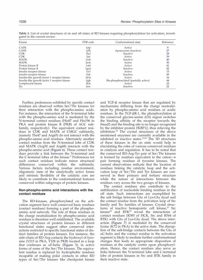
Further, preferences exhibited by specific contactresidues are observed within Ser/Thr kinases fortheir interaction with the phospho-amino acids.For instance, the interaction of the N-terminal lobewith the phospho-amino acid is mediated by theN-terminal contact residues (His87 and His196 inPKA and protein kinase B (PKB) of AGC sub-family, respectively). The equivalent contact resi-dues in CDK and MAPK of CMGC subfamily,(namely Thr47 and Arg65) do not interact with thephospho-amino acid residues. Alternately anothercontact residue from the N-terminal lobe of CDKand MAPK (Arg50 and Arg68) interacts with thephospho-amino acid (Figure 6). These contact resi-dues provide a link between the N-terminal andthe C-terminal lobes of the kinase.5 Preferences forsuch contact residues indicate minor structuraldifferences conserved within the subfamily.Various factors including residue environment,oligomeric state of the catalytically active formsand intrinsic flexibility of the catalytic core arelikely to contribute to the conformational featuresconserved within subgroups of protein kinases.
Non-phospho-amino acid interactions with thecontact residues
The RD-kinases, phosphorylated on the acti-vation segment have well conserved basic residues(contact residues) forming a part of the phospho-amino acid network. The role of these residues inthe charge neutralization by phospho-amino acidresidues is therefore well established. The availablecrystal structures of protein kinases in differentfunctional states suggest other conserved inter-actions restricted to specific functional states of dis-tinct families of protein kinases. For instance, theside-chain of RD-Arg contacts the hydroxyl of tyro-sine (Y215 in PKA, Y328 in PKB) located in a loopthat continues as aF-helix (Figure 3), in activeforms of some of the Ser/Thr kinases. The equiva-lent residue is replaced by hydrophobic residuesincapable of making polar contacts in other RDtypes of Ser/Thr kinases like checkpoint kinase
and TGF-b receptor kinase that are regulated bymechanisms differing from the charge neutraliz-ation by phospho-amino acid residues or acidicresidues. In the TGF-bR-1, the phosphorylation atthe conserved glycine-serine (GS) region switchesthe binding affinity of the receptor towards theSmad2 and the binding site is no longer recognizedby the inhibitor protein FKBP12, thus relieving theinhibition.18 The crystal structures of the abovementioned enzymes are currently available in theinhibited or inactive states.19,20 The 3D structuresof these kinases in the on state would help inelucidating the roles of various conserved residuesin catalysis and regulation. It has to be noted thatthe conserved RD-Arg-Tyr pair of Ser/Thr kinasesis formed by residues equivalent to the cation–ppair forming residues of tyrosine kinases. Thecurrent observations indicate that the location ofresidues linking the catalytic loop and the acti-vation loop of Ser/Thr and Tyr kinases are con-served in their primary and tertiary structurewhile the nature of interactions between theresidues vary across the two groups of kinases.
The contact residues also contribute to thestabilization of nucleotide binding residues in theoff state. Such interactions are exemplified bythe salt bridge between Glu of Lys-Glu dyad andthe contact residue from the activation loop of Srcfamily and Tec families of kinases. Crystal struc-tures of inactive hemapoietic cell kinase,21 Srckinase22 and BTK23 reveal the interactions ofcontact residues (R385 of HCK, Src and R544 ofBTK) with Glu of Lys-Glu dyad. The above inter-action (Figure 7) is mediated by the conservedlysine (K72 in PKA) in the active state. The disrup-tion of the salt-bridge contacts between the Glu ofaC-helix and the contact residue in the activationsegment is likely to mediate further conformationalchanges that leads to appropriate disposition ofresidues at the catalytic centre upon phosphoryl-ation. Hence, the contact residues also serve aslinks between the N-terminal lobe and C-terminallobe of protein kinases in Src and BTK family intheir inactive state.
Table 1. List of crystal structures of on and off states of RD kinases requiring phosphorylation for activation, investi-gated in the current review
Kinase PDB code Conformational state Reference
CAPK 1atp Active 9CAPK 1j3h Apoenzyme (inactive) 27CDK 1hcl Inactive 38CDK 1jst Active 39MAPK 1erk Inactive 40MAPK 2erk Active 30Protein kinase B 1o6l Active 41Protein kinase B 1gzn Inactive 42Insulin receptor kinase 1ir3 Active 15Insulin receptor kinase 1irk Inactive 43Insulin-like growth factor 1 receptor kinase 1k3a Active 16Insulin-like growth factor 1 receptor kinase 1jqh Bis-phosphorylated (partially active) 44Lymphocyte kinase 3lck Active 10Src 2src Inactive 22
1030 Review: Phosphorylation Sites in Kinases

Distinct modes of ATP binding in the on andoff states of Ser/Thr protein kinases
The crystal structures of substrate and ATPbound ternary complexes of various Ser/Thrprotein kinases including PKA, CDK24,25 and phos-phorylase kinase26 have indicated a common bind-ing mode of the substrate peptides in their active
conformations. The backbone of the substrate pep-tides adopts an extended conformation to alignthe hydroxyl group suitably at the catalytic site.An analysis on the conformation of the co-sub-
strate ATP, bound to active and inactive states ofprotein kinases, by pair-wise superposition of ATPatoms reveals distinct features. Average deviationsof atoms in base, ribose and phosphate groups
Figure 4. The phospho-aminoacid in the activation segment of(a) IRK and (b) LCK and its inter-actions with the contact residues inthe on state are shown. Aromaticresidues involved in cation–p inter-actions with RD-Arg and otherbasic residues discussed in the textare shown in blue.
Review: Phosphorylation Sites in Kinases 1031

arrived at from all pairwise superpositions of ATPmolecules in kinases are represented in Figure 8.ATP displays similar conformation in the onstate of Ser/Thr protein kinases as indicated by
the lower deviation of their adenine ring, riboseand phosphate groups compared to ATP moleculesin the inactive states of kinases (Figure 8). Thisfeature is consistent with appropriate disposition
Figure 6. Phospho threonine(pT 183) at the activation segmentof active MAPK and its interactionswith specific contact residues areshown. Arg65 (blue) equivalent toHis196 of PKB (Figure 3) is driftedaway from the catalytic site andlacks interactions with pT183.
Figure 5. Schematic representation of the interactions of phospho-amino acid in the activation segment of (a) Ser/Thr and (b) Tyr kinases. PKA (a) and IRK (b) have been chosen as examples to illustrate the phospho-amino acid net-work in the two groups of protein kinases. Distinct nature of interactions with RD-Arg, characterized by electrostaticinteractions in Ser/Thr kinases and cation–p (aromatic) interactions in tyrosine kinases, stabilize the activation seg-ment. Unlike in Ser/Thr kinases, N-terminal helix (aC-helix) of tyrosine kinases lacks interactions with the phospho-amino acid. The arrows (blue) indicate the residues involved in cation–p (aromatic) interactions in tyrosine kinases.
1032 Review: Phosphorylation Sites in Kinases

Figure 7. 3D structure of Srckinase (2Src) in an inhibited state.The interaction between residuesinvolved in Lys-Glu dyad (red) inthe active state and residues fromthe activation segment (blue) isshown.
Figure 8. Extent of deviation of conformation of ATP bound to active and inactive states of protein kinases. The aver-age deviation (A) of equivalent atoms of ATP observed for adenine, ribose and phosphate groups among active forms(red) and between active and inactive forms (dark green) is shown.
Review: Phosphorylation Sites in Kinases 1033

of the gamma phosphate groups for inline phos-pho-transfer.
ATP molecules bound to inactive forms of pro-tein kinases show wide variations in the confor-mation of their chemical groups. Although theadenine ring and the ribose groups of ATP deviateleast across active and inactive states, deviationsgreater than 1 A are observed in the equivalentatoms of the phosphate groups of the ATP. Thebeta and gamma phosphate groups are highlymobile with equivalent atoms deviating by greaterthan 3 A.
These variations in the conformation of ATP arelikely to arise form the large conformationalchanges accompanying the key residues involvedin ATP binding and coordination with the metalions (Mg2þ, Mn2þ) during transition into differentstates. ATP binding residues of the kinase arelocalized in distinct regions of the fold thatincludes the Gly-loop, hinge region, catalytic loopand the activation segment. In CDK the key resi-dues comprising the ATP-binding pocket includeGly13, Thr14, Val18 of Gly loop; Lys33 of Lys-Gludyad; Phe80, Glu81, Phe82 and Leu83 of the hingeregion; Asn132 of the catalytic loop and Asp145 ofthe activation segment. These residues vary intheir extent of interactions with ATP in their activeand inactive states. While some of these residues(G13, T14, F80, D145) are involved in ATP bindingonly in the active state, other residues (F82, L83)interact with ATP in the inactive state. The remain-ing residues participate in ATP binding in bothstates of activity.
High mobility of phosphate groups enables theirco-ordination with the metal ions that are dis-placed due to the variation in the positions ofresidue ligands (N132, D145) located in the cata-lytic and activation loop between the two confor-mational states of kinases.
The atoms of ATP bound to active form ofkinases in general can be well superimposed.Large variation is, however, observed in the con-formation of ribose and phosphate groups of ATPbound to phospho-CDK/cyclin complex in com-parison with ATP bound to active states of otherSer/Thr kinases. Accommodation of ATP in alteredconformations within the structurally similarcatalytically active scaffold without hinderingphospho-transfer therefore seems to be influencedby the binding of cyclin to CDK.
The conformations of ATP bound to Ser/Thrkinases and tyrosine kinases in the on state alsovaries to a large extent. The RTKs lack the for-mation of a correct Lys-Glu ion-pair in their activestates as described in a later section. The natureof stabilization of the activation segment of tyro-sine kinases in the on state is also distinct fromthat of Ser/Thr kinases as described earlier. Highsequence divergence is observed in the nucleotidebinding pockets of Ser/Thr and Tyr kinases. Thesedistinct functional and structural features of Tyrkinases are hence likely to influence the mode ofATP binding in the tyrosine kinases. The different
features appear to play an important role in thedistinct nature of co-ordination of phosphategroups of ATP with the conserved lysine andmetal ions.
Influence of phosphorylation and ATP bindingon closed state of protein kinases
Appropriate spatial orientation of the ATPbinding residues and the relative disposition ofcatalytic residues in the activation and catalyticloop are characteristic to the closed state ofprotein kinases. The aC-helix is a key structuralelement in the N-terminal lobe mediating theconformational changes occurring at the catalyticcenter through an ion pair (Lys72-Glu91 in PKA)that bridges phosphate groups of ATP. The chargeneutralization of the basic cluster of residues in theactive site5 is another critical feature in RD kinasesregulated by phosphorylation as described earlier.
The crystal structure of the apo-enzyme of phos-phorylated PKA27 reveals the electrostatic inter-actions of the phospho-amino acid similar to itsternary complex.
However, the Lys and Glu residues are spatiallydistant, incapable of salt bridge formation andcoordination with a- and b-phosphate groups ofATP. Phosphorylation of the activation segment inPKA therefore appears to alter the active sitelocally, with least influence on the formation ofion pair in the N-terminal lobe.
In contrast to the Ser/Thr kinases, the receptortryosine kinases lack the Lys-Glu ion-pair inter-action in their active states as observed in the 3Dstructures of insulin receptor15 and IGF receptorkinases.16 However, the phospho-amino acid inthe activation segment is capable of interactingwith the basic residues in its vicinity in a uniqueway as described in the previous sections.
In the conformationally restrained state ofc-Abl28,29 the Lys-Glu ion pair interaction isretained. However, the residues at the catalyticsite are not aligned for phospho transfer, as thecontact residues are not drawn towards Tyr412,which is phosphorylated for complete activationof the kinase.
The formation of Lys-Glu ion pair and confor-mational changes at the active site accompanyingthe activation process are hence suggested to varyacross different protein kinases. The mechanismsof acquisition of a closed, functionally competentstate in protein kinases is therefore suggested tovary based on the extent of reorganization of theactive site necessary to initiate phospho transfer.
Interactions of “secondary” phospho-aminoacid residues in protein kinases
The phosphorylation occurs at multiple sites onprotein kinases providing additional controls ontheir activity. In MAP kinases of CMGC subfamily,the successive dual phosphorylation of the Thrand Tyr residues in TXY motif of the activation
1034 Review: Phosphorylation Sites in Kinases

segment results in local and global conformationalchanges30 realigning the positively charged contactresidues close to the phospho threonine (pT183).The second dianionic site unique to MAPK (Figure9(a)) created by the phosphorylation of Tyr residuealso interacts with two basic residues (Arg189,Arg192). Glycogen synthase kinase-3 (GSK-3),another member of CMGC subfamily is usuallykept active in cells and is known to phosphorylatea wide range of cellular proteins.31 PKB mediatedphosphorylation on the N-terminal serine residueof GSK-3b leads to its inactivation. GSK-3b isunique in its choice of substrates with higherpreferences for phosphorylated (primed) sub-strates. The phosphorylation on the activationsegment for regulation of this enzyme is non-obligatory. Analogous interactions are observed inthe 3D structure32 of GSK between the phospho-
tyrosine (pY) 416 (only phospho-amino acid) inthe activation segment and Arg220 and Arg223(Figure 9(b)). The basic residues hence are likelyto play a major role in accommodating the phos-pho-amino acid on the primed substrates similarto their contacts with pY416 in the absence of thesubstrate.The four main groups of CMGC kinases (CDK,
MAPK, glycogen synthase kinase (GSK) and caseinkinase-2 (CK2)) show high conservation of basicresidues in equivalent positions with the exceptionof CDK. An apolar residue (Leu166) replaces theresidue equivalent to the Arg189 (MAPK) in CDKwhile the other basic residue is well conserved.However, the role of these residues in CDK thatpossess mechanisms of activation distinct fromMAPK and GSK is unclear. CK2 is a constitutivelyactive kinase and no phosphorylation at the
Figure 9. The interactions of secondary phospho-amino acid residues in the active and inactive states of proteinkinases. (a) Active state of MAPK, (b) phospho-glycogen synthase kinase (GSK), (c) inactive state of c-Src kinase(inactive) and (d) inactive state of hemapoietic cell kinase (HCK).
Review: Phosphorylation Sites in Kinases 1035
catalytic subunit is so far documented for itsregulation. It has to be noted that the Sky1p33 ofSaccharomyces cerevisiae (a non-RD kinase) is theonly other protein kinase in addition to the RDkinases of the CMGC group, with basic residuesconserved at equivalent positions.
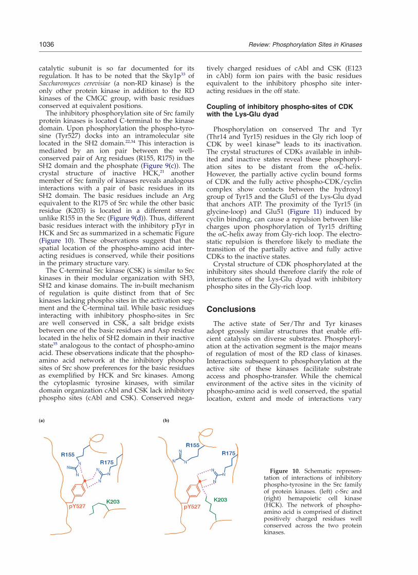
The inhibitory phosphorylation site of Src familyprotein kinases is located C-terminal to the kinasedomain. Upon phosphorylation the phospho-tyro-sine (Tyr527) docks into an intramolecular sitelocated in the SH2 domain.22,34 This interaction ismediated by an ion pair between the well-conserved pair of Arg residues (R155, R175) in theSH2 domain and the phosphate (Figure 9(c)). Thecrystal structure of inactive HCK,21 anothermember of Src family of kinases reveals analogousinteractions with a pair of basic residues in itsSH2 domain. The basic residues include an Argequivalent to the R175 of Src while the other basicresidue (K203) is located in a different strandunlike R155 in the Src (Figure 9(d)). Thus, differentbasic residues interact with the inhibitory pTyr inHCK and Src as summarized in a schematic Figure(Figure 10). These observations suggest that thespatial location of the phospho-amino acid inter-acting residues is conserved, while their positionsin the primary structure vary.
The C-terminal Src kinase (CSK) is similar to Srckinases in their modular organization with SH3,SH2 and kinase domains. The in-built mechanismof regulation is quite distinct from that of Srckinases lacking phospho sites in the activation seg-ment and the C-terminal tail. While basic residuesinteracting with inhibitory phospho-sites in Srcare well conserved in CSK, a salt bridge existsbetween one of the basic residues and Asp residuelocated in the helix of SH2 domain in their inactivestate35 analogous to the contact of phospho-aminoacid. These observations indicate that the phospho-amino acid network at the inhibitory phosphosites of Src show preferences for the basic residuesas exemplified by HCK and Src kinases. Amongthe cytoplasmic tyrosine kinases, with similardomain organization cAbl and CSK lack inhibitoryphospho sites (cAbl and CSK). Conserved nega-
tively charged residues of cAbl and CSK (E123in cAbl) form ion pairs with the basic residuesequivalent to the inhibitory phospho site inter-acting residues in the off state.
Coupling of inhibitory phospho-sites of CDKwith the Lys-Glu dyad
Phosphorylation on conserved Thr and Tyr(Thr14 and Tyr15) residues in the Gly rich loop ofCDK by wee1 kinase36 leads to its inactivation.The crystal structures of CDKs available in inhib-ited and inactive states reveal these phosphoryl-ation sites to be distant from the aC-helix.However, the partially active cyclin bound formsof CDK and the fully active phospho-CDK/cyclincomplex show contacts between the hydroxylgroup of Tyr15 and the Glu51 of the Lys-Glu dyadthat anchors ATP. The proximity of the Tyr15 (inglycine-loop) and Glu51 (Figure 11) induced bycyclin binding, can cause a repulsion between likecharges upon phosphorylation of Tyr15 driftingthe aC-helix away from Gly-rich loop. The electro-static repulsion is therefore likely to mediate thetransition of the partially active and fully activeCDKs to the inactive states.
Crystal structure of CDK phosphorylated at theinhibitory sites should therefore clarify the role ofinteractions of the Lys-Glu dyad with inhibitoryphospho sites in the Gly-rich loop.
Conclusions
The active state of Ser/Thr and Tyr kinasesadopt grossly similar structures that enable effi-cient catalysis on diverse substrates. Phosphoryl-ation at the activation segment is the major meansof regulation of most of the RD class of kinases.Interactions subsequent to phosphorylation at theactive site of these kinases facilitate substrateaccess and phospho-transfer. While the chemicalenvironment of the active sites in the vicinity ofphospho-amino acid is well conserved, the spatiallocation, extent and mode of interactions vary
Figure 10. Schematic represen-tation of interactions of inhibitoryphospho-tyrosine in the Src familyof protein kinases. (left) c-Src and(right) hemapoietic cell kinase(HCK). The network of phospho-amino acid is comprised of distinctpositively charged residues wellconserved across the two proteinkinases.
1036 Review: Phosphorylation Sites in Kinases

across Ser/Thr and Tyr kinases. The Arg of theRD motif stabilizes the activation segment byelectrostatic interaction with the phospho-aminoacid in most Ser/Thr kinases in their on state. Incontrast in the Tyr kinase the RD-Arg lacks theconserved interaction with the phospho-aminoacid and alternately makes cation–p interactionsin on and off states with phenylalanine well con-served in known Tyr kinases. The phenylalaninefurther extends its interaction to the substrate-binding pocket and is hence likely to be importantfor the formation of substrate binding sites.
Crystal structures of Ser/Thr kinases in theactive state also reveal conservation of polar inter-actions of RD-R with conserved tyrosine that isuniquely conserved among RD kinases regulatedby negative charge or phospho-amino acid in theactivation segments. While the role of such inter-actions is currently unclear, it is likely to be anotherfeature of the active site of protein kinases specifi-cally conserved in the on state.
The significance of the stabilizing interactions ofthe contact residues with the phospho-amino acidin the active state of kinases is well established.Ion pair between the contact residue and the Lys-Glu dyad in the off-state of Src kinases and BTKsuggest coupling of the activation segment andaC-helix, which communicates the changes to thecatalytic center subsequent to phosphorylation.
Preferences for interactions of the phospho-amino acid with specific contact residues inaC-helix, are observed within the group of Ser/Thr kinases as exemplified by AGC and CMGCfamily of kinases. The variation in orientation of
aC-helix in distinct functional states of these sub-groups of kinases and the intrinsic flexibilityassociated with the catalytic core for such move-ments, appear to be critical for the choice ofresidues in the phospho-amino acid network.Inhibitory phosphorylation sites of Src family of
kinases also create a cluster of positively chargedresidues, which are usually exposed in the onstates. Distinct set of basic residues, however,participate in such interactions in the c-Src andHCK family of kinases.The tyrosine in the Gly-rich loop of CDKs is
proximal to Glu of Lys-Glu dyad in the active andpartially active structures. Phosphorylation of theTyr in the cyclin bound forms of CDKs leading toinhibition of CDK is hence suggested to resultfrom the repulsion between Glu and the dianioniccharge.The available 3D structures of protein kinases
have enabled the identification of a subset ofunique features associated with the active stateof Ser/Thr and tyrosine kinases despite manycommon structural features and mechanisms ofphospho transfer. The participation of residuesconserved specifically in the two groups of kinasesin unique interactions provides a structural basisfor their conservation. While interactions of thephospho-amino acid residues located at varioussites in the 3D-scaffold of the protein kinases withpositively charged residues is observed, the extentof neutralization by the dianionic charge and thespatial location of the positively charged residuesmay vary.ATP bound to Ser/Thr kinases in the on state,
Figure 11. 3D structure of cyclindependent kinase showing inter-actions between Tyr15 (red), theinhibitory phosphorylation siteand conserved Glu51 (red) of Lys(green)-Glu dyad in cyclin bound-phosphorylated complex (1JST).
Review: Phosphorylation Sites in Kinases 1037
in general, adopt similar conformation of adenine,ribose and phosphate groups. The conformation ofATP varies between active and inactive forms ofATP, with the phosphate groups showing the largedeviation in their relative orientation. Variationsare, however, observed in active states of otherprotein kinases (CDK) which might be partiallyinfluenced by additional factors like the inter-actions of the kinases with regulatory proteins.
The vast number of crystal structures of RD-kinases in distinct conformational states haveaided in the identification of conserved and variedfunctional features at their active sites subsequentto phosphorylation. The number of 3D structurescurrently available for protein kinases regulatedby other modes of regulation in on and off statesis limited. The emergence of crystal structures ofnon-RD kinases in their on state will enhance thecurrent understanding of the factors stabilizingtheir active sites.
Acknowledgements
This research is supported by the WellcomeTrust, London, in the form of Senior Fellowship inBiomedical Sciences to N.S. A.K. is supported byCouncil of Scientific and Industrial Research,New Delhi. G.P. is a visiting student trainee to theIndian Institute of Science.
References
1. Schenk, P. W. & Snaar-Jagalska, B. E. (1999). Signalperception and transduction: the role of proteinkinases. Biochim. Biophys. Acta, 1449, 1–24.
2. Cohen, P. (2000). The regulation of protein functionby multisite phosphorylation—a 25-year update.Trends Biochem. Sci. 25, 596–601.
3. Hunter, T. (2000). Signaling—2000 and beyond. Cell,100, 113–127.
4. Huse, M. & Kuriyan, J. (2002). The conformationalplasticity of protein kinases. Cell, 109, 275–282.
5. Johnson, L. N., Noble, M. E. & Owen, D. J. (1996).Active and inactive protein kinases: structural basisfor regulation. Cell, 85, 149–158.
6. Kobe, B. & Kemp, B. E. (1999). Active site-directedprotein regulation. Nature, 402, 373–376.
7. Johnson, L. N., Lowe, E. D., Noble, M. E. & Owen,D. J. (1998). The eleventh datta lecture. The structuralbasis for substrate recognition and control by proteinkinases. FEBS Letters, 430, 1–11.
8. Johnson, L. N. & Lewis, R. J. (2001). Structural basisfor control by phosphorylation. Chem. Rev. 101,2209–2242.
9. Knighton, D. R., Zheng, J. H., Ten Eyck, L. F., Ash-ford, V. A., Xuong, N. H., Taylor, S. S. & Sowadski,J. M. (1991). Crystal structure of the catalytic subunitof cyclic adenosine monophosphate-dependentprotein kinase. Science, 253, 407–414.
10. Yamaguchi, H. & Hendrickson, W. A. (1996). Struc-tural basis for activation of human lymphocyte
kinase LCK upon tyrosine phosphorylation. Nature,384, 484–489.
11. Singh, J. & Thornton, J. M. (1990). SIRIUS. An auto-mated method for the analysis of the preferred pack-ing arrangements between protein groups. J. Mol.Biol. 211, 595–615.
12. Flocco, M. M. & Mowbray, S. L. (1994). Planar stack-ing interactions of arginine and aromatic side-chainsin proteins. J. Mol. Biol. 235, 709–717.
13. Momany, F. A., Carruthers, L. M., McGuire, R. F. &Scheraga, H. A. (1974). Intermolecular potentialsand applications to the packing configurations andlattice energies in crystals of hydrocarbons, car-boxylic acids, amines and amides. J. Phys. Chem. 78,1595–1620.
14. Hofstra, R. M., Landsvater, R. M., Ceccherini, I.,Stulp, R. P., Stelwagen, T., Luo, Y., Pasini, B.,Hoppener, J. W., van Amstel, H. K. & Romeo, G.(1994). A mutation in the RET protooncogene asso-ciated with multiple endocrine neoplasia type 2Band sporadic medullary thyroid carcinoma. Nature,367, 375–376.
15. Hubbard, S. R. (1997). Crystal structure of the acti-vated insulin receptor tyrosine kinase in complexwith peptide substrate and ATP analog. EMBO J. 16,5572–5581.
16. Favelyukis, S., Till, J. H., Hubbard, S. R. & Miller,W. T. (2001). Structure and autoregulation of theinsulin-like growth factor 1 receptor kinase. NatureStruct. Biol. 8, 1058–1063.
17. Singh, J. (1994). Comparison of conservation withinand between the Ser/Thr and Tyr protein kinasefamily: proposed model for the catalytic domain ofthe epidermal growth factor receptor. Protein Eng. 7,849–858.
18. Huse, M., Muir, T. W., Xu, L., Chen, Y. G., Kuriyan, J.& Massague, J. (2001). The TGF beta receptor acti-vation process: an inhibitor- to substrate-bindingswitch. Mol. Cell, 8, 671–682.
19. Huse, M., Chen, Y. G., Massague, J. & Kuriyan, J.(1999). Crystal structure of the cytoplasmic domainof the type I TGF beta receptor in complex withFKBP12. Cell, 96, 425–436.
20. Chen, P., Luo, C., Deng, Y., Ryan, K., Register, J.,Margosiak, S., Tempczyk-Russell, A., Nguyen, B.,Myers, P., Lundgren, K., Kan, C. C. & O’Connor,P. M. (2000). The 1.7 A crystal structure of humancell cycle checkpoint kinase Chk1: implications forChk1 regulation. Cell, 100, 681–692.
21. Sicheri, F., Moarefi, I. & Kuriyan, J. (1997). Crystalstructure of the Src family tyrosine kinase Hck.Nature, 385, 602–609.
22. Xu, W., Doshi, A., Lei, M., Eck, M. J. & Harrison, S. C.(1999). Crystal structures of c-Src reveal features ofits autoinhibitory mechanism. Mol. Cell, 3, 629–638.
23. Mao, C., Zhou, M. & Uckun, F. M. (2001). Crystalstructure of Bruton’s tyrosine kinase domainsuggests a novel pathway for activation and pro-vides insights into the molecular basis of X-linkedagammaglobulinemia. J. Biol. Chem. 276,41435–41443.
24. Brown, N. R., Noble, M. E., Endicott, J. A. & Johnson,L. N. (1999). The structural basis for specificity ofsubstrate and recruitment peptides for cyclin-dependent kinases. Nature Cell Biol. 1, 438–443.
25. Brown, N. R., Noble, M. E., Lawrie, A. M., Morris,M. C., Tunnah, P., Divita, G., Johnson, L. N. &Endicott, J. A. (1999). Effects of phosphorylation of
1038 Review: Phosphorylation Sites in Kinases

threonine 160 on cyclin-dependent kinase 2 structureand activity. J. Biol. Chem. 274, 8746–8756.
26. Lowe, E. D., Noble, M. E., Skamnaki, V. T.,Oikonomakos, N. G., Owen, D. J. & Johnson, L. N.(1997). The crystal structure of a phosphorylasekinase peptide substrate complex: kinase substraterecognition. EMBO J. 16, 6646–6658.
27. Akamine, P., Madhusudan, Wu, J., Xuong, N. H., TenEyck, L. F. & Taylor, S. S. (2003). Dynamic features ofcAMP-dependent protein kinase revealed by apo-enzyme crystal structure. J. Mol. Biol. 327, 159–171.
28. Hantschel, O., Nagar, B., Guettler, S., Kretzschmar, J.,Dorey, K., Kuriyan, J. & Superti-Furga, G. (2003). Amyristoyl/phosphotyrosine switch regulates c-Abl.Cell, 112, 845–857.
29. Nagar, B., Hantschel, O., Young, M. A., Scheffzek, K.,Veach, D., Bornmann, W., Clarkson, B., Superti-Furga, G. & Kuriyan, J. (2003). Structural basis forthe autoinhibition of c-Abl tyrosine kinase. Cell, 112,859–871.
30. Canagarajah, B. J., Khokhlatchev, A., Cobb, M. H. &Goldsmith, E. J. (1997). Activation mechanism of theMAP kinase ERK2 by dual phosphorylation. Cell,90, 859–869.
31. Welsh, G. I., Miyamoto, S., Price, N. T., Safer, B. &Proud, C. G. (1996). T-cell activation leads to rapidstimulation of translation initiation factor eIF2B andinactivation of glycogen synthase kinase-3. J. Biol.Chem. 271, 11410–11413.
32. Bax, B., Carter, P. S., Lewis, C., Guy, A. R., Bridges,A., Tanner, R., Pettman, G., Mannix, C., Culbert,A. A., Brown, M. J., Smith, D. G. & Reith, A. D.(2001). The structure of phosphorylated GSK-3betacomplexed with a peptide, FRATtide, that inhibitsbeta-catenin phosphorylation. Structure (Camb), 9,1143–1152.
33. Nolen, B., Ngo, J., Chakrabarti, S., Vu, D., Adams,J. A. & Ghosh, G. (2003). Nucleotide-induced confor-mational changes in the Saccharomyces cerevisiae SRprotein kinase, Sky1p, revealed by X-ray crystallo-graphy. Biochemistry, 42, 9575–9585.
34. Young, M. A., Gonfloni, S., Superti-Furga, G., Roux,B. & Kuriyan, J. (2001). Dynamic coupling betweenthe SH2 and SH3 domains of c-Src and Hckunderlies their inactivation by C-terminal tyrosinephosphorylation. Cell, 105, 115–126.
35. Ogawa, A., Takayama, Y., Sakai, H., Chong, K. T.,Takeuchi, S., Nakagawa, A., Nada, S., Okada, M. &Tsukihara, T. (2002). Structure of the carboxyl-terminal Src kinase, Csk. J. Biol. Chem. 277,14351–14354.
36. Russell, P. & Nurse, P. (1987). Negative regulation ofmitosis by wee1 þ , a gene encoding a protein kinasehomolog. Cell, 49, 559–567.
37. Evans, S. V. (1993). SETOR: hardware-lighted three-dimensional solid model representations of macro-molecules. J. Mol. Graph. 11, 134–138. see also pp.127–128.
38. Schulze-Gahmen, U., Brandsen, J., Jones, H. D.,Morgan, D. O., Meijer, L., Vesely, J. & Kim, S. H.(1995). Multiple modes of ligand recognition: crystalstructures of cyclin-dependent protein kinase 2 incomplex with ATP and two inhibitors, olomoucineand isopentenyladenine. Proteins: Struct. Funct.Genet. 22, 378–391.
39. Russo, A. A., Jeffrey, P. D. & Pavletich, N. P. (1996).Structural basis of cyclindependent kinase activationby phosphorylation. Nature Struct. Biol. 3, 696–700.
40. Zhang, F., Strand, A., Robbins, D., Cobb, M. H. &Goldsmith, E. J. (1994). Atomic structure of the MAPkinase ERK2 at 2.3 A resolution. Nature, 367,704–711.
41. Yang, J., Cron, P., Good, V. M., Thompson, V.,Hemmings, B. A. & Barford, D. (2002). Crystal struc-ture of an activated Akt/protein kinase B ternarycomplex with GSK3-peptide and AMP-PNP. NatureStruct. Biol. 9, 940–944.
42. Yang, J., Cron, P., Thompson, V., Good, V. M., Hess,D., Hemmings, B. A. & Barford, D. (2002). Molecularmechanism for the regulation of protein kinase B/Akt by hydrophobic motif phosphorylation. Mol.Cell, 9, 1227–1240.
43. Hubbard, S. R., Wei, L., Ellis, L. & Hendrickson, W. A.(1994). Crystal structure of the tyrosine kinasedomain of the human insulin receptor. Nature, 372,746–754.
44. Pautsch, A., Zoephel, A., Ahorn, H., Spevak, W.,Hauptmann, R. & Nar, H. (2001). Crystal structureof bisphosphorylated IGF-1 receptor kinase: insightinto domain movements upon kinase activation.Structure (Camb), 9, 955–965.
Edited by J. Thornton
(Received 11 February 2004; received in revised form 21 April 2004; accepted 22 April 2004)
Review: Phosphorylation Sites in Kinases 1039
Copyright © 2022 FDOKUMEN