Presence of multiple acid phosphatases activity in seedlings of cucumber, radish and rocket salad

Yalak et al. Journal of Translational Medicine 2014, 12:165http://www.translational-medicine.com/content/12/1/165
COMMENTARY Open Access
Ecto-protein kinases and phosphatases: anemerging field for translational medicineGarif Yalak1, Yigal H Ehrlich2 and Bjorn R Olsen1*
Abstract
Progress in translational research has led to effective new treatments of a large number of diseases. Despite thisprogress, diseases including cancer and cardiovascular disorders still are at the top in death statistics and disorderssuch as osteoporosis and osteoarthritis represent an increasing disease burden in the aging population. Novelstrategies in research are needed more than ever to overcome such diseases. The growing field of extracellularprotein phosphorylation provides excellent opportunities to make major discoveries of disease mechanisms thatcan lead to novel therapies. Reversible phosphorylation/dephosphorylation of sites in the extracellular domains ofmatrix, cell-surface and trans-membrane proteins is emerging as a critical regulatory mechanism in health anddisease. Moreover, a new concept is emerging from studies of extracellular protein phosphorylation: in cells whereATP is stored within secretory vesicles and released by exocytosis upon cell-stimulation, phosphorylation of extracellularproteins can operate as a messenger operating uniquely in signaling pathways responsible for long-term cellularadaptation. Here, we highlight new concepts that arise from this research, and discuss translation of the findingsinto clinical applications such as development of diagnostic disease markers and next-generation drugs.
Keywords: Ecto-protein kinase, Ecto-protein phosphatase, Phosphorylation, Disease marker, Extracellular-drug targets
IntroductionProtein phosphorylation, a reversible posttranslationalmodification carried out by the enzymes protein kinasesand phosphatases, is arguably one of the most criticalregulatory biochemical processes operating in physio-logical systems. The covalent attachment of a phosphategroup by a protein kinase and detachment by a proteinphosphatase is rapid and energy efficient, making pro-tein phosphorylation one of the most common post-translational modifications in cells [1]. It is thus notsurprising that intracellular protein kinases have emergedas one of the most important drug targets, with some 20drugs on the market and hundreds in clinical trials [2].It has been estimated that 30% of all human proteins
are phosphorylated at one or another stage during theirlifecycle [3]; however this percentage may be an underesti-mation since it does not include extracellular proteins. Allpreconditions for protein kinases to be active also exist inthe extracellular environment, such as sufficiently high
* Correspondence: [email protected] of Developmental Biology, Harvard School of Dental Medicine,188 Longwood Avenue, Boston, MA 02115, USAFull list of author information is available at the end of the article
© 2014 Yalak et al.; licensee BioMed Central LtCommons Attribution License (http://creativecreproduction in any medium, provided the orDedication waiver (http://creativecommons.orunless otherwise stated.
concentrations of extracellular ATP and a growing bodyof evidence is documenting the existence and activity ofthese enzymes in extracellular spaces (Figure 1; [4-7]). Infact, evidence that many prominent kinases and phospha-tases are located extracellularly has been obtained in-vivoas well as in-vitro including FAM20C [7], PKA [8,9], PKC[10,11], CKII [12], alkaline phosphatase [13], tartrate-resistant acid phosphatase (TRAP) [14] and the PTENphosphatase [15] (Table 1). A recent review provides anin-depth mining of the extensive literature in the field, aswell as of high-throughput mass spectrometry data, de-scribing known phosphorylated proteins in the extracellu-lar matrix in many different types of healthy and diseasedtissues and animal species [4]. Major components of theextracellular matrix and cell surface proteins, includingfibronectin [16], vitronectin [17], osteopontin [18], col-lagens [19], fibrinogen [20], laminin [21], CD36 [22], β-amyloid precursor protein (APP) [23], T-cell-receptorcomplex (TCR) [24] and many more, have been reported tobe phosphorylated in vitro and in vivo [4]. Evidence that thisenzymatic regulatory activity may be important in extracellularmatrix biology and pathology, cancer growth, tissue engineer-ing, regenerative medicine, immune response, cardiovascular
d. This is an Open Access article distributed under the terms of the Creativeommons.org/licenses/by/4.0), which permits unrestricted use, distribution, andiginal work is properly credited. The Creative Commons Public Domaing/publicdomain/zero/1.0/) applies to the data made available in this article,

Figure 1 Potential scenarios of extracellular protein phosphorylation. Potential scenarios of extracellular protein phosphorylation are shown.Extracellular ATP can enter the extracellular matrix (ECM) through vesicle secretion or cell lysis [4]. Extracellular proteins or the extracellular domains ofcell surface and trans-membrane proteins may be phosphorylated during biosynthesis and then exported, or phosphorylated after release orappearance at the cell surface by exo- and ecto-kinases, respectively.
Yalak et al. Journal of Translational Medicine 2014, 12:165 Page 2 of 6http://www.translational-medicine.com/content/12/1/165
and neuronal biology is growing [4]. These recent find-ings suggest that phosphorylation of extracellular matrixproteins provides an important basis for identifyingnovel biomarkers and for design of new therapeuticdrugs. In fact, a clinical application based on extracellu-lar protein-phosphorylation has already been patented[25], and recent studies propose the involvement ofextracellular protein kinases and phosphatases in diseasessuch as cancer, Alzheimer’s, cardiovascular disorders andothers. In this review, we highlight the potential of thisresearch area as an emerging new field in translationalmedicine and for developing novel medical applications.
Small differences can have huge impactBiomolecules such as insulin or interferon serve as im-portant drugs today. Seven of the top 10 biopharmaceuti-cals in 2012 were indeed biomolecular drugs, according to
Table 1 Reported prominent extracellular protein kinasesand phosphatases
Kinase Sample Reference
PKA serum from cancer patient [8,9]
PKC human platelets, hippocampalneurons
[10,26]
CKII human prostatic cancer cell line [12]
FAM20C HEK293T [7]
Alkalinephosphatase
human serum [13]
PTEN primary human breast tumor [15]
TRAP human serum [14]
FiercePharma statistics. These include extracellular pro-teins. For example, recombinant BMP-2, an FDA ap-proved drug, is routinely used to induce bone formationin bone defects [27]. Recombinant BMP-7 is also FDA ap-proved as an alternative to autografts [28]. The design andproduction of such drugs need to be precise. Even smalldifferences in structure can have a huge impact on theirperformance. The phosphorylation state of these proteinsis one of these critical factors. These drugs are producedas recombinant proteins; therefore their phosphorylationstate may vary from batch to batch. Our analyses showthat both BMP-2 and BMP-7 can occur in phosphorylatedstates in human biosamples; however when they are usedin the clinic, this modification is largely ignored. Paying at-tention to their state of phosphorylation may have signifi-cant consequences for their effective activities. Similareffects should be considered when growing cells in cultureon surfaces coated with extracellular proteins; a standardmethod used today in basic research as well as in transla-tional science. The phosphorylation state of matrix pro-teins can vary from batch to batch of purified orcommercially available protein substrates for cell cul-ture. Such variations seem minor, but they can causesignificant differences in cell behavior. For example,while the phosphorylated form of the extracellularprotein vitronectin promotes cell adhesion, the non-phosphorylated form inhibits cell adhesion [29]. Simi-larly, the phosphorylation state of extracellular proteinswas reported to regulate the binding affinity as well asspecificity in protein activity. For example, phosphoryl-ation of CD36 by PKC at the position T92 leads to
Yalak et al. Journal of Translational Medicine 2014, 12:165 Page 3 of 6http://www.translational-medicine.com/content/12/1/165
stronger binding to collagen, but results in loss of throm-bospondin binding. The effect is reversible and suggests aregulated mechanism [22]. Finally, MMP2 enzyme activitywas shown to be regulated by extracellular PKC phos-phorylation, in that dephosphorylation leads to increasedactivity, and phosphorylation inhibits the activity [30,31].In all translational studies and clinical developmentswhere extracellular proteins are involved, their phos-phorylation state might have substantial effects on theoutcomes and needs to be taken into account.
Impact on diseasesThe activity of extracellular protein kinases has been de-scribed in the context of several physiological processes.These include leukocyte and macrophage adhesion andmigration [32,33], fertilization [34], platelet function[35,36], blood coagulation [37], complement systemfunction [38], receptor specificity and sensitivity [29],neuronal development and adaptation [39], synapticplasticity and memory formation [40], (for a review see[4]). The involvement of extracellular protein kinaseshas also been reported for a number of diseases (Table 2).For example, in Alzheimer’s phosphorylation of amyloid-β-peptides by ecto-PKA at the cell surface and in the cere-brospinal fluid promotes the formation of toxic aggregatesleading to increased aggregation and decreased clearance[41]. Phosphorylated amyloid-β-peptides have been de-tected in the brains of Alzheimer’s disease patients andthis may offer new ways to target the disease. In prostateor breast cancer patients, enhanced kinase levels and ac-tivities are found in sera. It has been shown that tumorcells secrete cAMP-dependent PKA; however, the functionof the secreted form is unknown [8,9,42]. Analysis ofserum from a large number of Schizophrenia patients re-vealed 72 phosphoproteins and phosphorylation-specificchanges in 59 of these proteins compared with samplesfrom matched healthy controls [43].
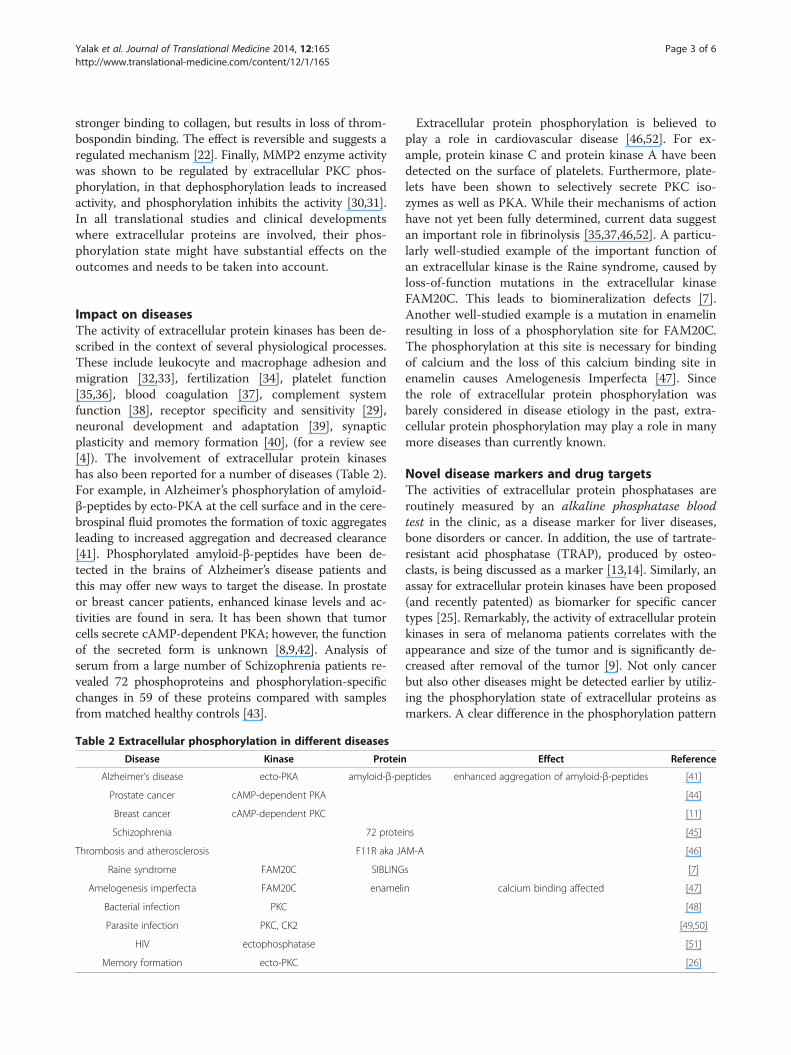
Table 2 Extracellular phosphorylation in different diseases
Disease Kinase Protein
Alzheimer’s disease ecto-PKA amyloid-β-pe
Prostate cancer cAMP-dependent PKA
Breast cancer cAMP-dependent PKC
Schizophrenia 72 prote
Thrombosis and atherosclerosis F11R aka JA
Raine syndrome FAM20C SIBLING
Amelogenesis imperfecta FAM20C enamel
Bacterial infection PKC
Parasite infection PKC, CK2
HIV ectophosphatase
Memory formation ecto-PKC
Extracellular protein phosphorylation is believed toplay a role in cardiovascular disease [46,52]. For ex-ample, protein kinase C and protein kinase A have beendetected on the surface of platelets. Furthermore, plate-lets have been shown to selectively secrete PKC iso-zymes as well as PKA. While their mechanisms of actionhave not yet been fully determined, current data suggestan important role in fibrinolysis [35,37,46,52]. A particu-larly well-studied example of the important function ofan extracellular kinase is the Raine syndrome, caused byloss-of-function mutations in the extracellular kinaseFAM20C. This leads to biomineralization defects [7].Another well-studied example is a mutation in enamelinresulting in loss of a phosphorylation site for FAM20C.The phosphorylation at this site is necessary for bindingof calcium and the loss of this calcium binding site inenamelin causes Amelogenesis Imperfecta [47]. Sincethe role of extracellular protein phosphorylation wasbarely considered in disease etiology in the past, extra-cellular protein phosphorylation may play a role in manymore diseases than currently known.
Novel disease markers and drug targetsThe activities of extracellular protein phosphatases areroutinely measured by an alkaline phosphatase bloodtest in the clinic, as a disease marker for liver diseases,bone disorders or cancer. In addition, the use of tartrate-resistant acid phosphatase (TRAP), produced by osteo-clasts, is being discussed as a marker [13,14]. Similarly, anassay for extracellular protein kinases have been proposed(and recently patented) as biomarker for specific cancertypes [25]. Remarkably, the activity of extracellular proteinkinases in sera of melanoma patients correlates with theappearance and size of the tumor and is significantly de-creased after removal of the tumor [9]. Not only cancerbut also other diseases might be detected earlier by utiliz-ing the phosphorylation state of extracellular proteins asmarkers. A clear difference in the phosphorylation pattern
Effect Reference
ptides enhanced aggregation of amyloid-β-peptides [41]
[44]
[11]
ins [45]
M-A [46]
s [7]
in calcium binding affected [47]
[48]
[49,50]
[51]
[26]

Yalak et al. Journal of Translational Medicine 2014, 12:165 Page 4 of 6http://www.translational-medicine.com/content/12/1/165
of serum proteins in sera of schizophrenia patients wasmentioned above [45]. Further studies detected 502 serumproteins to be phosphorylated in sera of 80 healthy indi-viduals and discussed the phosphorylation pattern as po-tential disease marker [53-55]. A large number of studiesdescribing the activity of extracellular kinases in parasitesand bacteria open the door to the discovery of novelmethods for detecting and treating infections [48,49]. Fur-ther progress in this area could lead to novel and/or morereliable biomarker assays in clinical studies of cancer andother diseases and new therapies [25].The location of extracellular protein kinases provides a
unique advantage for drug development. Potential inhib-itors or activators of extracellular kinases and phospha-tases can be designed purposefully as lipid insolublemolecules that are unable to penetrate into cells throughthe plasma membrane. This may provide greater selectivityand fewer side effects, as it will prevent interaction of thesedrugs with intracellular kinases [56]. An excellent exampleof this kind of new drug would be a water-soluble,membrane-impermeable peptide or peptido-mimetic basedon the sequence and tertiary structure of the region sur-rounding a critical phosphorylated amino-acid residue lo-cated in the extracellular domain of a specific proteinsubstrate of an ecto- or exo-protein kinase [56].Analyses of drug-target interactions are crucial during
the drug design phase. Protein phosphorylation is knownto regulate protein-protein interaction in many cases in-side the cell [57]. During the design of potential drugsfor targeting extracellular or cell surface proteins, thephosphorylation state of the proteins also needs to betaken into account. An example of how important thephosphorylation state of a drug may be is demonstratedby the Multiple Sclerosis drug Fingolimod (FTY720); itsphosphorylation state is believed to be regulated by theecto-phosphatase LPP3 in vivo [58].
A novel concept revealed by extracellular proteinphosphorylationIn several cell types ATP is stored within secretory vesiclesand released by exocytosis upon cell-stimulation. Repeti-tive, high frequency stimulation of these cells producesunusually high concentrations of ATP in the extracellularspace, and particularly in synaptic-clefts between neurons.Such events do not occur usually during the course ofroutine cellular communications. Instead, they representunique biochemical signals responsible for triggeringadaptive processes with long-lasting consequences [59]. Inthe next step, extracellular protein kinase activity that isdependent on these higher ATP concentrations producesa phosphorylation-dependent event required for the in-duction of long-term adaptive changes [39,56,59,60].Platelet and neuronal activities have been most studied inregard to this aspect of extracellular phosphorylation.
Massive stimulation of localized platelets produces highconcentrations of extracellular ATP during blood coagula-tion and atherosclerotic plaque formation, and extracellu-lar protein kinase activities have been implicated in both[56]. High frequency stimulation of neurons in the hippo-campus induces a memory-related adaptive process calledlong term potentiation (LTP), involving high ATP concen-trations in the synaptic-cleft [60]. LTP is a physiologicalmeasure of memory formation in the brain that requiresan extracellular protein kinase C activity [26]. Similarly,the same concept suggests that high levels of extracellularATP produced by neurons during seizures may triggerextracellular phosphorylation activity involved in the eti-ology of Epileply.
Conclusions and future outlookRecent advances in the research of reversible extracellularprotein phosphorylation activity provide unique opportun-ities for translational medicine to design novel drugs andidentify new disease markers. While the mechanisms ofaction of extracellular protein kinases and phosphatasesare being uncovered and complete knowledge of theirphysiological roles requires further investigation, clinicalstudies already show their potential as novel biomarkersand drug targets in diseases such as cancer [44], neuronal[5,39,41,56] and cardiovascular disorders [10,33,46,52].Initial studies and patents show encouraging results re-garding the performance of such biomarkers compared toconventional markers [25]. The fact that secreted alkalinephosphatase is routinely used as a biomarker in clinicstoday suggests that other secreted protein kinases andphosphatases will be used as novel markers in the future[61]. A large number of extracellular proteins have beenreported in phosphorylated states as a result of interac-tions with yet unknown extracellular protein kinases. It isthus expected that many more active kinases will be dis-covered in the extracellular environment. Finally, moredirected studies may uncover the involvement of extracel-lular phosphorylation in many more diseases than thosethat are currently known and open up new avenues fornovel therapies.
AbbreviationsTRAP: Tartrate-resistant acid phosphatase; PKA: Protein kinase A; PKC: Proteinkinase C; ATP: Adenosine triphosphate; APP: β-amyloid precursor protein;LTP: Long term potentiation; TCR: T-cell-receptor complex; BMP: Bonemorphogenetic protein; FAM20C: Family with sequence similarity 20;CD36: Cluster of differentiation 36.
Competing interestsThe authors declare that they have no competing interests.
Authors’ contributionsAll authors have contributed equally. All authors performed article searches,drafted and revised the manuscript. All authors read and approved the finalmanuscript. Author BR Olsen provided financial support for publication.

Yalak et al. Journal of Translational Medicine 2014, 12:165 Page 5 of 6http://www.translational-medicine.com/content/12/1/165
AcknowledgmentsWe apologize to all research groups whose relevant publications could notbe cited here because of space limitations.
FundingThis work was supported by the Swiss National Science Foundation(Fellowship PBEZP3_145998 to GY).
Author details1Department of Developmental Biology, Harvard School of Dental Medicine,188 Longwood Avenue, Boston, MA 02115, USA. 2Program in Neuroscience,College of Staten Island (CSI), City University of New York, 2800 VictoryBoulevard, Staten Island, NY 10314, USA.
Received: 4 April 2014 Accepted: 29 May 2014Published: 12 June 2014
References1. Hunter T: Why nature chose phosphate to modify proteins. Philos Trans R
Soc Lond B Biol Sci 2012, 367:2513–2516.2. Cohen P, Alessi DR: Kinase drug discovery–what's next in the field? ACS
Chem Biol 2013, 8:96–104.3. Mann M, Ong SE, Gronborg M, Steen H, Jensen ON, Pandey A: Analysis of
protein phosphorylation using mass spectrometry: deciphering thephosphoproteome. Trends Biotechnol 2002, 20:261–268.
4. Yalak G, Vogel V: Extracellular phosphorylation and phosphorylatedproteins: not just curiosities but physiologically important. Sci Signal2012, 5:re7.
5. Ehrlich YH: Extracellular protein kinases. Science 1996, 271:278–279.6. Redegeld FA, Caldwell CC, Sitkovsky MV: Ecto-protein kinases: ecto-domain
phosphorylation as a novel target for pharmacological manipulation?Trends Pharmacol Sci 1999, 20:453–459.
7. Tagliabracci VS, Engel JL, Wen J, Wiley SE, Worby CA, Kinch LN, Xiao J,Grishin NV, Dixon JE: Secreted kinase phosphorylates extracellularproteins that regulate biomineralization. Science 2012, 336:1150–1153.
8. Wang H, Li M, Lin W, Wang W, Zhang Z, Rayburn ER, Lu J, Chen D, Yue X,Shen F, Jiang F, He J, Wei W, Zeng X, Zhang R: Extracellular activity ofcyclic AMP-dependent protein kinase as a biomarker for human cancerdetection: distribution characteristics in a normal population and cancerpatients. Cancer Epidemiol Biomarkers Prev 2007, 16:789–795.
9. Kita T, Goydos J, Reitman E, Ravatn R, Lin Y, Shih WC, Kikuchi Y, Chin KV:Extracellular cAMP-dependent protein kinase (ECPKA) in melanoma.Cancer Lett 2004, 208:187–191.
10. Hogan MV, Pawlowska Z, Yang HA, Kornecki E, Ehrlich YH: Surfacephosphorylation by ecto-protein kinase C in brain neurons: a target forAlzheimer's beta-amyloid peptides. J Neurochem 1995, 65:2022–2030.
11. Kang JH, Asai D, Toita R, Kitazaki H, Katayama Y: Plasma protein kinase C(PKC) alpha as a biomarker for the diagnosis of cancers. Carcinogenesis2009, 30:1927–1931.
12. Bohana-Kashtan O, Pinna LA, Fishelson Z: Extracellular phosphorylation ofC9 by protein kinase CK2 regulates complement-mediated lysis. Eur JImmunol 2005, 35:1939–1948. doi:10.1002/eji.200425716.
13. Wiwanitkit V: High serum alkaline phosphatase levels, a study in 181 Thaiadult hospitalized patients. BMC Fam Pract 2001, 2:2.
14. Halleen JM, Tiitinen SL, Ylipahkala H, Fagerlund KM, Vaananen HK:Tartrate-resistant acid phosphatase 5b (TRACP 5b) as a marker of boneresorption. Clinical laboratory 2006, 52:499–509.
15. Hopkins BD, Fine B, Steinbach N, Dendy M, Rapp Z, Shaw J, Pappas K, Yu JS,Hodakoski C, Mense S, Klein J, Pegno S, Sulis ML, Goldstein H, AmendolaraB, Lei L, Maurer M, Bruce J, Canoll P, Hibshoosh H, Parsons R: A secretedPTEN phosphatase that enters cells to alter signaling and survival.Science 2013, 341:399–402.
16. Imada S, Sugiyama Y, Imada M: Fibronectin phosphorylation byecto-protein kinase. Exp Cell Res 1988, 179:554–564.
17. Seger D, Gechtman Z, Shaltiel S: Phosphorylation of vitronectin by caseinkinase II. Identification of the sites and their promotion of cell adhesionand spreading. J Biol Chem 1998, 273:24805–24813.
18. Kazanecki CC, Uzwiak DJ, Denhardt DT: Control of osteopontin signalingand function by post-translational phosphorylation and protein folding.J Cell Biochem 2007, 102:912–924.
19. Zimina EP, Fritsch A, Schermer B, Bakulina AY, Bashkurov M, Benzing T,Bruckner-Tuderman L: Extracellular phosphorylation of collagen XVII byecto-casein kinase 2 inhibits ectodomain shedding. J Biol Chem 2007,282:22737–22746.
20. Ogata Y, Heppelmann CJ, Charlesworth MC, Madden BJ, Miller MN, Kalli KR,Cilby WA, Bergen HR 3rd, Saggese DA, Muddiman DC: Elevated levels ofphosphorylated fibrinogen-alpha-isoforms and differential expression ofother post-translationally modified proteins in the plasma of ovariancancer patients. J Proteome Res 2006, 5:3318–3325.
21. Koliakos G, Trachana V, Gaitatzi M, Dimitriadou A: Phosphorylation oflaminin-1 by protein kinase C. Mol Cells 2001, 11:179–185.
22. Chu LY, Silverstein RL: CD36 ectodomain phosphorylation blocksthrombospondin-1 binding: structure-function relationships andregulation by protein kinase C. Arterioscler Thromb Vasc Biol 2012,32:760–767.
23. Suzuki T, Ando K, Isohara T, Oishi M, Lim GS, Satoh Y, Wasco W, Tanzi RE,Nairn AC, Greengard P, Gandy SE, Kirino Y: Phosphorylation of Alzheimerbeta-amyloid precursor-like proteins. Biochemistry 1997, 36:4643–4649.
24. Apasov SG, Smith PT, Jelonek MT, Margulies DH, Sitkovsky MV:Phosphorylation of extracellular domains of T-lymphocyte surfaceproteins. Constitutive serine and threonine phosphorylation of theT cell antigen receptor ectodomains. J Biol Chem 1996, 271:25677–25683.
25. Puskas R, Held D: Measurement of PKA for cancer detection; 2013. Patent.https://www.google.com/patents/US8455200?dq=Puskas+R.+and+Held+D.&hl=en&sa=X&ei=UMeIU6yZA5OssQT0iIHICg&ved=0CEMQ6AEwAg.
26. Chen W, Wieraszko A, Hogan MV, Yang HA, Kornecki E, Ehrlich YH:Surface protein phosphorylation by ecto-protein kinase is required forthe maintenance of hippocampal long-term potentiation. Proc Natl AcadSci U S A 1996, 93:8688–8693.
27. Khan S, Lane J: The use of recombinant human bone morphogeneticprotein-2 (rhBMP-2) in orthopaedic applications. Expert Opin Biol Ther2004, 4(5):741–748. May.
28. Vaccaro AR, Lawrence JP, Patel T, Katz LD, Anderson DG, Fischgrund JS,Krop J, Fehlings MG, Wong D: The safety and efficacy of OP-1 (rhBMP-7)as a replacement for iliac crest autograft in posterolateral lumbararthrodesis: a long-term (>4 years) pivotal study. Spine (Phila Pa 1976)2008, 33:2850–2862.
29. Seger D, Seger R, Shaltiel S: The CK2 phosphorylation of vitronectin.Promotion of cell adhesion via the alpha (v) beta 3-phosphatidylinositol3-kinase pathway. J Biol Chem 2001, 276:16998–17006.
30. Jacob-Ferreira AL, Kondo MY, Baral PK, James MN, Holt A, Fan X, Schulz R:Phosphorylation Status of 72 kDa MMP-2 Determines Its Structure andActivity in Response to Peroxynitrite. PLoS One 2013, 8:e71794.
31. Sariahmetoglu M, Crawford BD, Leon H, Sawicka J, Li L, Ballermann BJ,Holmes C, Berthiaume LG, Holt A, Sawicki G, Schulz R: Regulation of matrixmetalloproteinase-2 (MMP-2) activity by phosphorylation. FASEB J 2007,21:2486–2495.
32. Florey O, Durgan J, Muller W: Phosphorylation of leukocyte PECAMand its association with detergent-resistant membranes regulatetransendothelial migration. J Immunol 2010, 185:1878–1886.
33. Weber GF, Zawaideh S, Hikita S, Kumar VA, Cantor H, Ashkar S:Phosphorylation-dependent interaction of osteopontin with its receptorsregulates macrophage migration and activation. J Leukoc Biol 2002,72:752–761.
34. Maiti A, Nath D, Dungdung SR, Majumder GC: Sperm ecto-protein kinaseand its protein substrate: novel regulators of membrane fusion duringacrosome reaction. J Cell Physiol 2009, 220:394–400.
35. Morgenstern E, Gnad U, Preissner KT, Dierichs R, Belleli A, Chestukhin A,Schvartz I, Shaltiel S: Localization of protein kinase A and vitronectin inresting platelets and their translocation onto fibrin fibers during clotformation. Eur J Cell Biol 2001, 80:87–98.
36. Korc-Grodzicki B, Tauber-Finkelstein M, Shaltiel S: Platelet stimulationreleases a cAMP-dependent protein kinase that specificallyphosphorylates a plasma protein. Proc Natl Acad Sci U S A 1988,85:7541–7545.
37. Hillen TJ, Aroor AR, Shukla SD: Selective secretion of protein kinase Cisozymes by thrombin-stimulated human platelets. Biochem Biophys ResCommun 2001, 280:259–264.
38. Paas Y, Bohana-Kashtan O, Fishelson Z: Phosphorylation of the complementcomponent, C9, by an ecto-protein kinase of human leukemic cells.Immunopharmacology 1999, 42:175–185.
Yalak et al. Journal of Translational Medicine 2014, 12:165 Page 6 of 6http://www.translational-medicine.com/content/12/1/165
39. Ehrlich YH, Hogan MV, Pawlowska Z, Naik U, Kornecki E: Ectoprotein kinasein the regulation of cellular responsiveness to extracellular ATP.Ann N Y Acad Sci 1990, 603:401–416.
40. Fujii S, Kuroda Y, Ito K, Kato H: Long-term potentiation induction–asynaptic catch mechanism released by extracellular phosphorylation.Neuroscience 2000, 96:259–266.
41. Kumar S, Rezaei-Ghaleh N, Terwel D, Thal DR, Richard M, Hoch M,Mc Donald JM, Wullner U, Glebov K, Heneka MT, Walsh DM, Zweckstetter M,Walter J: Extracellular phosphorylation of the amyloid beta-peptidepromotes formation of toxic aggregates during the pathogenesis ofAlzheimer's disease. EMBO J 2011, 30:2255–2265.
42. Babiker AA, Ronquist G, Nilsson B, Ekdahl KN: Overexpression ofecto-protein kinases in prostasomes of metastatic cell origin.Prostate 2006, 66:675–686.
43. Jaros JA, Martins-de-Souza D, Rahmoune H, Rothermundt M, Leweke FM,Guest PC, Bahn S: Protein phosphorylation patterns in serum fromschizophrenia patients and healthy controls. J Proteomics 2012, 76:43–55.Spec No.
44. Cho YS, Park YG, Lee YN, Kim MK, Bates S, Tan L, Cho-Chung YS: Extracellularprotein kinase A as a cancer biomarker: its expression by tumor cells andreversal by a myristate-lacking Calpha and RIIbeta subunit overexpression.Proc Natl Acad Sci U S A 2000, 97:835–840.
45. Jaros JA, Martins-de-Souza D, Rahmoune H, Schwarz E, Leweke FM, GuestPC, Bahn S: Differential phosphorylation of serum proteins reflectinginflammatory changes in schizophrenia patients. Eur Arch Psychiatry ClinNeurosci 2012, 262:453–455. doi:10.1007/s00406-011-0283-6.
46. Kedees MH, Babinska A, Swiatkowska M, Deitch J, Hussain MM, Ehrlich YH,Kornecki E: Expression of a recombinant protein of the platelet F11receptor (F11R) (JAM-1/JAM-A) in insect cells: F11R is naturallyphosphorylated in the extracellular domain. Platelets 2005, 16:99–109.
47. Chan HC, Mai L, Oikonomopoulou A, Chan HL, Richardson AS, Wang SK,Simmer JP, Hu JC: Altered enamelin phosphorylation site causesamelogenesis imperfecta. J Dent Res 2010, 89:695–699.
48. Crane JK, Vezina CM: Externalization of host cell protein kinase C duringenteropathogenic Escherichia coli infection. Cell Death Differ 2005,12:115–127.
49. Dutra PM, Vieira DP, Meyer-Fernandes JR, Silva-Neto MA, Lopes AH:Stimulation of Leishmania tropica protein kinase CK2 activities byplatelet-activating factor (PAF). Acta Trop 2009, 111:247–254.
50. Alvarez-Rueda N, Biron M, Le Pape P: Infectivity of Leishmania mexicana isassociated with differential expression of protein kinase C-like triggeredduring a cell-cell contact. PLoS One 2009, 4:e7581.
51. Portela MB, Kneipp LF, de Souza IP R, Holandino C, Alviano CS,Meyer-Fernandes JR, de Araujo Soares RM: Ectophosphatase activityin Candida albicans influences fungal adhesion: study betweenHIV-positive and HIV-negative isolates. Oral Dis 2010, 16:431–437.
52. Babinska A, Hogan MV, Sobocki T, Sobocka MB, Ehrlich YH, Kornecki E:Identification of ecto-PKC on surface of human platelets: role inmaintenance of latent fibrinogen receptors. Am J Physiol Heart Circ Physiol2000, 278:H2008–2019.
53. Carrascal M, Gay M, Ovelleiro D, Casas V, Gelpi E, Abian J: Characterizationof the human plasma phosphoproteome using linear ion trap massspectrometry and multiple search engines. J Proteome Res 2010, 9:876–884.
54. Zhou W, Ross MM, Tessitore A, Ornstein D, Vanmeter A, Liotta LA, PetricoinEF 3rd: An initial characterization of the serum phosphoproteome.J Proteome Res 2009, 8:5523–5531.
55. Jaros JA, Guest PC, Ramoune H, Rothermundt M, Leweke FM, Martins-de-Souza D, Bahn S: Clinical use of phosphorylated proteins in blood serumanalysed by immobilised metal ion affinity chromatography and massspectrometry. J Proteomics 2012, 76:36–42. Spec No.
56. Ehrlich YH, Kornecki E: Ecto-protein kinases as mediators for the action ofsecreted ATP in the brain. Prog Brain Res 1999, 120:411–426.
57. Watanabe N, Osada H: Phosphorylation-dependent protein-proteininteraction modules as potential molecular targets for cancer therapy.Curr Drug Targets 2012, 13:1654–1658.
58. Mechtcheriakova D, Wlachos A, Sobanov J, Bornancin F, Zlabinger G,Baumruker T, Billich A: FTY720-phosphate is dephosphorylated by lipidphosphate phosphatase 3. FEBS Lett 2007, 581:3063–3068.
59. Ehrlich YH, Kornecki E: Extracellular protein phosphorylation systems inthe regulation of cellular responsiveness. Prog Clin Biol Res 1987,249:193–204.
60. Wieraszko A, Ehrlich YH: On the role of extracellular ATP in the inductionof long-term potentiation in the hippocampus. J Neurochem 1994,63:1731–1738.
61. Yamada S, Tsuruya K, Yoshida H, Taniguchi M, Haruyama N, Tanaka S,Eriguchi M, Nakano T, Kitazono T: The clinical utility of serumtartrate-resistant acid phosphatase 5b in the assessment of boneresorption in patients on peritoneal dialysis. Clin Endocrinol (Oxf ) 2013,78:844–851.
doi:10.1186/1479-5876-12-165Cite this article as: Yalak et al.: Ecto-protein kinases and phosphatases:an emerging field for translational medicine. Journal of TranslationalMedicine 2014 12:165.
Submit your next manuscript to BioMed Centraland take full advantage of:
• Convenient online submission
• Thorough peer review
• No space constraints or color figure charges
• Immediate publication on acceptance
• Inclusion in PubMed, CAS, Scopus and Google Scholar
• Research which is freely available for redistribution
Submit your manuscript at www.biomedcentral.com/submit
Copyright © 2022 FDOKUMEN