Biochemical characterization of an ecto-ATP diphosphohydrolase activity in Candida parapsilosis and...
12
RESEARCH ARTICLE Biochemical characterization ofan ecto-ATP diphosphohydrolase activity in Candida parapsilosis and its possible role in adenosine acquisition and pathogenesis Tina Kiffer-Moreira 1,2 , Maria Ester Fernandes Sampaio 1,2 , Daniela S. Alviano 3 , Flavia Axelband 2,4 , Gabriele Vargas Cesar 3 , Daniela Cosentino-Gomes 1,2 , Marcio L. Rodrigues 3 , Leonardo Nimrichter 3 , Adalberto Vieyra 2,4 , Celuta S. Alviano 3 & Jos ´ e Roberto Meyer-Fernandes 1,2 1 Instituto de Bioqu´ ımica M ´ edica, Universidade Federal do Rio de Janeiro, Rio de Janeiro, Brazil; 2 Instituto Nacional de Cie ˆncia e Tecnologia de Biologia Estrutural e Bioimagem, Universidade Federal do Rio de Janeiro, Rio de Janeiro, Brazil; 3 Instituto de Microbiologia Prof. Paulo de G ´ oes, Universidade Federal do Rio de Janeiro, Rio de Janeiro, Brazil; and 4 Instituto de Biof´ ısica Carlos Chagas Filho, Universidade Federal do Rio de Janeiro, Rio de Janeiro, Brazil Correspondence: Jos ´ e Roberto Meyer- Fernandes, Instituto de Bioqu´ ımica M ´ edica, Universidade Federal do Rio de Janeiro (UFRJ), CCS, Bloco H, Cidade Universit ´ aria, Ilha do Funda ˜ o, 21541-590, Rio de Janeiro, Brazil. Tel.: 155 21 590 4548; fax: 155 21 270 8647; e-mail: [email protected] Received 4 January 2010; revised 15 April 2010; accepted 26 April 2010. Final version published online 24 June 2010. DOI:10.1111/j.1567-1364.2010.00641.x Editor: Terrance Cooper Keywords Candida parapsilosis; ecto-ATPase activity; adenosine acquisition; virulence. Abstract In this work, we describe the ability of intact cells of Candida parapsilosis to hydrolyze extracellular ATP. ATP hydrolysis was stimulated by MgCl 2 in a dose- dependent manner. The ecto-ATPase activity was increased in the presence of 5 mM MgCl 2 , with values of V max and apparent K m for Mg-ATP 2 increasing to 33.80 1.2 nmol Pi h 1 10 8 cells and 0.6 0.06 mM, respectively. Inhibitors of phosphatases, mitochondrial Mg 21 -ATPases and Na 1 -ATPases had no effect on the C. parapsilosis Mg 21 -stimulated ATPase activity, but extracellular impermeant compounds, 4,4 0 -diisothiocyanatostilbene-2,2 0 disulfonic acid and suramin, reduced enzyme activity in yeast living cells by 83.1% and 81.9%, respectively. ARL 67156 (6-N,N 0 -diethyl-D-b-g-dibromomethylene ATP), a nucleotide analo- gue, also inhibited the ecto-ATPase activity in a dose-dependent manner. ATP was the best substrate for the yeast Mg 21 -stimulated ecto-enzyme, but ADP, ITP, CTP, GTP and UTP were also hydrolyzed. A direct relationship between ecto-ATPase activity and adhesion to host cells was observed. In these assays, inhibition of enzyme activity resulted in decreased levels of yeast adhesion to epithelial cells. Based also on the differential expression of ecto-ATPase activities in the different isolates of C. parapsilosis, the possible role of this enzyme in fungal biology is discussed. Introduction Candida parapsilosis is a cause of serious nosocomial infec- tions and is the second most common Candida sp. isolated from bloodstream infections in many regions worldwide (San Miguel et al., 2005; Sarvikivi et al., 2005; Almirante et al., 2006; Trofa et al., 2008). The mechanisms by which C. parapsilosis evades host defenses and colonizes host tissues are poorly understood. The search for new structures representing virulence factors that will enhance our under- standing of the pathogenic steps of Candida infections is therefore extremely important. Cell–cell recognition and adherence are central processes to many fundamental areas of biology. The cell wall compo- sition is of primary importance during microbial adherence and infection establishment (Calderone et al., 1994). Cell surfaces contain enzymes whose catalytic site faces the extracellular environment (Jesus et al., 2002; Meyer- Fernandes, 2002; Gomes et al., 2006; Pinheiro et al., 2007; Peres-Sampaio et al., 2008). The activities of these enzymes, referred to as ecto-enzymes, can be measured using living cells (Goding, 2000; Meyer-Fernandes, 2002; Amazonas et al., 2009). This class of enzymes includes surface ATPases (ecto-ATPases), which are transmembrane glycoproteins that hydrolyze extracellular nucleoside tri- and/or dipho- sphates (Zimmermann, 2001; Meyer-Fernandes, 2002). Ecto-ATPases, which are also known as E-type ATPases, are divalent cation-dependent enzymes that are insensitive FEMS Yeast Res 10 (2010) 735–746 c 2010 Federation of European Microbiological Societies Published by Blackwell Publishing Ltd. All rights reserved YEAST RESEARCH
-
Upload
independent -
Category
Documents
-
view
2 -
download
0
Transcript of Biochemical characterization of an ecto-ATP diphosphohydrolase activity in Candida parapsilosis and...
R E S E A R C H A R T I C L E
Biochemical characterizationofan ecto-ATPdiphosphohydrolaseactivity inCandida parapsilosis and its possible role inadenosineacquisitionandpathogenesisTina Kiffer-Moreira1,2, Maria Ester Fernandes Sampaio1,2, Daniela S. Alviano3, Flavia Axelband2,4,Gabriele Vargas Cesar3, Daniela Cosentino-Gomes1,2, Marcio L. Rodrigues3, Leonardo Nimrichter3,Adalberto Vieyra2,4, Celuta S. Alviano3 & Jose Roberto Meyer-Fernandes1,2
1Instituto de Bioquımica Medica, Universidade Federal do Rio de Janeiro, Rio de Janeiro, Brazil; 2Instituto Nacional de Ciencia e Tecnologia de Biologia
Estrutural e Bioimagem, Universidade Federal do Rio de Janeiro, Rio de Janeiro, Brazil; 3Instituto de Microbiologia Prof. Paulo de Goes, Universidade
Federal do Rio de Janeiro, Rio de Janeiro, Brazil; and 4Instituto de Biofısica Carlos Chagas Filho, Universidade Federal do Rio de Janeiro, Rio de Janeiro,
Brazil
Correspondence: Jose Roberto Meyer-
Fernandes, Instituto de Bioquımica Medica,
Universidade Federal do Rio de Janeiro (UFRJ),
CCS, Bloco H, Cidade Universitaria, Ilha do
Fundao, 21541-590, Rio de Janeiro, Brazil.
Tel.: 155 21 590 4548; fax: 155 21 270
8647; e-mail: [email protected]
Received 4 January 2010; revised 15 April 2010;
accepted 26 April 2010.
Final version published online 24 June 2010.
DOI:10.1111/j.1567-1364.2010.00641.x
Editor: Terrance Cooper
Keywords
Candida parapsilosis; ecto-ATPase activity;
adenosine acquisition; virulence.
Abstract
In this work, we describe the ability of intact cells of Candida parapsilosis to
hydrolyze extracellular ATP. ATP hydrolysis was stimulated by MgCl2 in a dose-
dependent manner. The ecto-ATPase activity was increased in the presence of
5 mM MgCl2, with values of Vmax and apparent Km for Mg-ATP2� increasing to
33.80� 1.2 nmol Pi h�1 10�8 cells and 0.6� 0.06 mM, respectively. Inhibitors of
phosphatases, mitochondrial Mg21-ATPases and Na1-ATPases had no effect on
the C. parapsilosis Mg21-stimulated ATPase activity, but extracellular impermeant
compounds, 4,40-diisothiocyanatostilbene-2,2 0disulfonic acid and suramin,
reduced enzyme activity in yeast living cells by 83.1% and 81.9%, respectively.
ARL 67156 (6-N,N0-diethyl-D-b-g-dibromomethylene ATP), a nucleotide analo-
gue, also inhibited the ecto-ATPase activity in a dose-dependent manner. ATP was
the best substrate for the yeast Mg21-stimulated ecto-enzyme, but ADP, ITP, CTP,
GTP and UTP were also hydrolyzed. A direct relationship between ecto-ATPase
activity and adhesion to host cells was observed. In these assays, inhibition of
enzyme activity resulted in decreased levels of yeast adhesion to epithelial cells.
Based also on the differential expression of ecto-ATPase activities in the different
isolates of C. parapsilosis, the possible role of this enzyme in fungal biology is
discussed.
Introduction
Candida parapsilosis is a cause of serious nosocomial infec-
tions and is the second most common Candida sp. isolated
from bloodstream infections in many regions worldwide
(San Miguel et al., 2005; Sarvikivi et al., 2005; Almirante
et al., 2006; Trofa et al., 2008). The mechanisms by which
C. parapsilosis evades host defenses and colonizes host
tissues are poorly understood. The search for new structures
representing virulence factors that will enhance our under-
standing of the pathogenic steps of Candida infections is
therefore extremely important.
Cell–cell recognition and adherence are central processes
to many fundamental areas of biology. The cell wall compo-
sition is of primary importance during microbial adherence
and infection establishment (Calderone et al., 1994). Cell
surfaces contain enzymes whose catalytic site faces the
extracellular environment (Jesus et al., 2002; Meyer-
Fernandes, 2002; Gomes et al., 2006; Pinheiro et al., 2007;
Peres-Sampaio et al., 2008). The activities of these enzymes,
referred to as ecto-enzymes, can be measured using living
cells (Goding, 2000; Meyer-Fernandes, 2002; Amazonas
et al., 2009). This class of enzymes includes surface ATPases
(ecto-ATPases), which are transmembrane glycoproteins
that hydrolyze extracellular nucleoside tri- and/or dipho-
sphates (Zimmermann, 2001; Meyer-Fernandes, 2002).
Ecto-ATPases, which are also known as E-type ATPases,
are divalent cation-dependent enzymes that are insensitive
FEMS Yeast Res 10 (2010) 735–746 c� 2010 Federation of European Microbiological SocietiesPublished by Blackwell Publishing Ltd. All rights reserved
YEA
ST R
ESEA
RC
H
to inhibitors of P-type, F-type and V-type ATPases (Zim-
mermann, 2001; Meyer-Fernandes, 2002). E-type ATPases
have been described on the surface of several microorgan-
isms including protozoa (Meyer-Fernandes et al., 1997;
Barros et al., 2000; De Jesus et al., 2002; Sissons et al., 2004;
Leite et al., 2007; Matin & Khan, 2008; Santos et al., 2009),
bacteria (MacFarlane et al., 1994; Hopfe & Henrich, 2004;
Sansom et al., 2008a) and fungi, including Saccharomyces
cerevisiae (Zhong & Guidotti, 1999), Cryptococcus neofor-
mans (Junior et al., 2005) and Fonsecaea pedrosoi (Collopy-
Junior et al., 2006). Several hypotheses have been suggested
for the physiological role of these enzymes. They include: (1)
protection from the cytolytic effects of extracellular ATP
(Fillipini et al., 1990; Steinberg & Di Virgilio, 1991); (2)
termination of purinergic signaling (Weisman et al., 1996;
Westfall et al., 1997); (3) involvement in signal transduction
(Margolis et al., 1990; Dubyak & El-Moatassim, 1993); and
(4) involvement in cellular adhesion (Kirley, 1997; Bisaggio
et al., 2003; Pinheiro et al., 2006; Santos et al., 2009).
The characterization of the cell wall and other surface
components apparently have an express impact in the
development of new antifungal agents (Nimrichter et al.,
2005). In the present work, we characterized a surface
ATPase activity in C. parapsilosis as an Mg21-stimulated
ecto-enzyme. We found that C. parapsilosis isolates from the
oral cavity and bloodstream express different levels of
Mg21-stimulated ATPase activity at their surface. We also
show that the inhibition of enzyme activity resulted in
decreased levels of yeast adhesion to epithelial cells.
Materials and methods
Chemicals
All reagents were purchased from Sigma-Aldrich Co. (St.
Louis, MO) or from E. Merck (Darmstadt, Germany).
[g-32Pi]ATP was prepared as described by Glynn & Chappel
(1964). Distilled water was deionized using a MilliQ system
of resins (Millipore Corp., Bedford, MA) and was used in
the preparation of all solutions.
Microorganisms and growth conditions
Candida parapsilosis strain CCT3834 (ATCC 22019) was
kindly supplied by Dr Anibal Vercesi (Departamento de
Patologia Clınica, Universidade Federal de Campinas, Sao
Paulo, Brazil). Isolates RFO, obtained from the oral cavity of
a human patient, and H297, from the bloodstream of an
infected individual, were obtained from Faculdade de Odo-
tonlogia and from Hospital Universitario Clementino Fraga
Filho, Universidade Federal do Rio de Janeiro, Brazil,
respectively. Stock cultures were maintained on solid
brain–heart infusion at 37 1C. For measurements of enzyme
activity, all the strains of C. parapsilosis were cultivated for
48 h in a complex medium containing glycerol (2% v/v),
peptone (2% w/v; Bacto peptone; Becton Dickinson) and
yeast extract (1% w/v) at room temperature with contin-
uous shaking (Milani et al., 2001). Yeast cells were obtained
by centrifugation, and washed twice in a solution containing
116 mM NaCl, 5.4 mM KCl, 5.5 mM D-glucose and 50 mM
HEPES-Tris buffer (pH 7.2). Cell growth was estimated by
counting the number of yeast in a Neubauer chamber.
Except for those assays using different isolates (H297 and
RFO), experiments were performed using strain CCT3834.
Cellular viability was assessed, before and after incubations,
by Trypan blue dye exclusion. For Trypan staining, the cells
were incubated in the presence of 0.01% Trypan blue for
10 min in the buffer used in each experiment (Kneipp et al.,
2004). The viability was not affected under the conditions
used here.
Determination of ecto-ATPase activity
Intact cells (1.0� 108) were incubated for 1 h at 30 1C in
0.5 mL of a mixture containing, unless otherwise specified,
116 mM NaCl, 5.4 mM KCl, 5.5 mM D-glucose, 50 mM
HEPES-Tris buffer (pH 7.2) and 5 mM ATP in the absence
or in the presence of 5 mM MgCl2. The Mg21-stimulated
ecto-ATPase activity was calculated from the total activity
measured in the presence of 5 mM MgCl2 minus the basal
activity, which was measured in the absence of MgCl2. The
ATPase activity was determined by measuring the hydrolysis
of [g-32P]ATP (104 Bq nmol�1 ATP) (Meyer-Fernandes
et al., 2000). The experiments were started by the addition
of living cells and terminated by the addition of 1 mL of
a cold mixture containing 25% charcoal in 1 M HCl.
The tubes were then centrifuged at 1500 g for 10 min at
4 1C. Aliquots (0.5 mL) of the supernatants containing the
released 32Pi were transferred to scintillation vials contain-
ing 9 mL of scintillation fluid. The ATPase activity was
calculated by subtracting the nonspecific ATP hydrolysis
measured in the absence of cells from the total released 32Pi.
The hydrolysis of ATP was linear with time under the assay
conditions used and was found to be proportional to the cell
number. In the assays where other nucleotides were used,
the hydrolytic activities were measured under the same
experimental conditions described above, and Pi release
was assayed spectrophotometrically by measuring the re-
lease of Pi from the nonradioactive nucleotides (Lowry &
Lopes, 1946). The values obtained for ATPase activities
measured using both methods (i.e. spectrophotometry and
radioactivity) were the same. In the experiments where high
concentrations of Mn21, Ca21 and Sr21 were tested, the
possible formation of precipitates was checked as described
previously (Meyer-Fernandes & Vieyra, 1988). In the reac-
tion media containing 50 mM HEPES (pH 7.2), 116 mM
NaCl, 5.4 mM KCl, 5.5 mM D-glucose and 5 mM ATP, no
FEMS Yeast Res 10 (2010) 735–746c� 2010 Federation of European Microbiological SocietiesPublished by Blackwell Publishing Ltd. All rights reserved
736 T. Kiffer-Moreira et al.
phosphate precipitates were observed in the presence of
these cations under the conditions used.
Reverse-phase HPLC analysis of ATP hydrolysis
The hydrolysis of ATP and the generation of ADP, AMP and
adenosine was determined by incubating 1� 108 cells mL�1 in
a mixture containing 116 mM NaCl, 5.4 mM KCl, 5.5 mM
D-glucose, 5 mM MgCl2, 50 mM HEPES-Tris buffer (pH 7.2)
and 100mM ATP. After 5, 10, 15, 30, 45, 60 and 120 min, aliquots
of 300mL were taken and concentrated to dryness in a vacuum
concentrator. The dried aliquots were resuspended in 0.1 mL of
a solution containing 50 mM KH2PO4, 50 mM K2HPO4, 4 mM
tetra-n-butylammonium bromide (TBAB), 10% methanol, pH
6.0, and immediately injected into a C-18 reverse-phase column
(Rexcrom, 25 cm� 4.6 mm, Regis Technologies Inc., IL)
coupled to an LC10AS-HPLC model (Shimadzu) through a
50-mL loop. The solvent system was the solution described
above. The methodology used was modified from the original
protocol described by Kawamoto et al. (1998). The nucleotides
and adenosine were separated with a flow rate maintained at
1 mL min�1 (retention times, minimum: adenosine, 8.5� 0.1;
AMP, 7.5� 0.07; ADP, 10.8� 0.2; ATP, 15.9� 0.2) and detected
by UV spectroscopy at 254 nm. The amounts of remaining
nucleotides and adenosine were calculated using the peak ratio
area in the calibration graph constructed using standard con-
centrations of nucleotides and adenosine.
Interaction of C. parapsilosis with epithelial cells
Chinese hamster ovary (CHO) cells were purchased from
the Rio de Janeiro cell culture collection (BCRJ, UFRJ,
Brazil). The cells were grown in a 5% CO2 (37 1C) in
25-cm2 culture flasks containing Roswell Park Memorial
Institute (RPMI) medium 1640 (GibcoBRL) supplemented
with L-glutamine, 25 mM HEPES buffer and 10% fetal
bovine serum (FBS). The initial inoculum was 5�104 cells mL�1; this was subcultured every 2 days, and the
cells were maintained in log-phase growth. For adhesion
assays, animal cells were plated onto 24-well multidishes at a
density of 105 cells per well. They were then incubated at
37 1C for 24 h in a culture medium supplemented with 10%
FBS. Before interaction with animal cells, C. parapsilosis cells
(106) (strain CCT3834) were incubated for 30 min at room
temperature in 0.9% NaCl (control cells) or in the same
solution containing 0.5 mM 4,40-diisothiocyanostylbene-2,
20-disulfonic acid (DIDS), 0.5 mM suramin and 0.5 mM
ARL 67156. Yeast cells were then washed twice with 0.9%
saline, and finally rinsed in RPMI. Fungal cells were
suspended in the same medium and incubated with CHO
monolayers. The CHO cell–yeast ratio used was 1 : 10. After
the addition of fungi, the cells were incubated at 37 1C for
2 h, washed three times in phosphate-buffered saline to
remove nonadherent yeast cells, fixed in Bouin’s solution
and stained with Giemsa. The index of association between
C. parapsilosis and epithelial cells was determined using a
microscope at a magnification of 1000 (Zeiss Axioplan 2,
Germany). Representative images were taken at a magnifica-
tion of 400. The index of association between C. parapsilosis
and CHO cells was taken as the number of attached and
internalized yeast cells per host cell (total number of host
cell-associated yeast/total number of host cell). For each
experiment, 400 animal cells were counted.
Phylogenetic tree and sequence alignment
The phylogenetic tree was constructed with MEGA version
4.0 (Tamura et al., 2007) using the neighbor-joining
method. NTPDase protein sequences from Mus musculus
(GenBank accession nos NP_033978; NP_033979; NP_
848791; NP_080450; NP_001021385; NP_742115; NP_
444333; NP_082369), Homo sapiens (GenBank accession
nos NP_001767; NP_982293; NP_001239; NP_004892;
NP_001240; NP_001238; EAW49862; NP_001028285) try-
panosomatids, Trypanosoma cruzi (GenBank accession
no. AAS75599); Trypanosoma brucei (GenBank accession
no. AAZ13145); Leishmania major (GenBank accession no.
DAA04950) and fungi, Schizosaccharomyces pombe (Gen-
Bank accession no. CAB57847); S. cerevisiae (GenBank
accession no. EDN62971); Aspergillus fumigatus (Gen-
Bank accession no. XP_753547); and Candida albicans
(GenBank accession no. XP_715624) were retrived from the
NCBI protein database.
Protein sequence alignment and analysis were performed
using the CLUSTAL X version 2 (Larkin et al., 2007) and
GENEDOC (Nicholas et al., 1997) programs.
Statistical analysis
All experiments were performed in triplicate, with similar
results obtained in at least three separate cell suspensions.
The maximal velocity Vmax and Km for Mg-ATP2� were
calculated using a computerized nonlinear regression analy-
sis of the data to the Michaelis–Menten equation (SIGMA PLOT
7.0; Jandel Scientific Software). Statistical significances were
determined using Student’s t-test. Values of Po 0.05 were
considered as significant.
Results
Candida parapsilosis expresses an ecto-ATPasethat is stimulated by divalent cations
Initially, we compared the enzyme activity in the presence or
absence of divalent cations, potent modulators of ecto-
ATPases (Meyer-Fernandes, 2002). In the absence of diva-
lent metals, C. parapsilosis hydrolyzed ATP at a rate of
2.12� 0.07 nmol Pi h�1 10�8 cells. Our results revealed that
FEMS Yeast Res 10 (2010) 735–746 c� 2010 Federation of European Microbiological SocietiesPublished by Blackwell Publishing Ltd. All rights reserved
737Ecto-ATPase in Candida parapsilosis
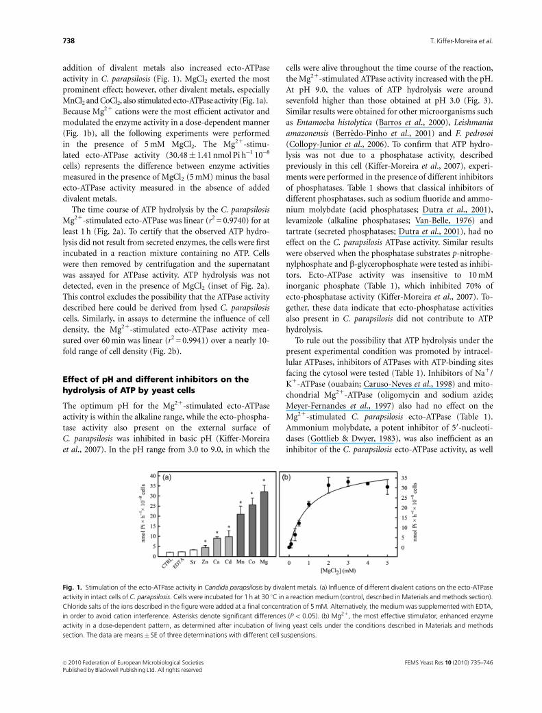
addition of divalent metals also increased ecto-ATPase
activity in C. parapsilosis (Fig. 1). MgCl2 exerted the most
prominent effect; however, other divalent metals, especially
MnCl2 and CoCl2, also stimulated ecto-ATPase activity (Fig. 1a).
Because Mg21 cations were the most efficient activator and
modulated the enzyme activity in a dose-dependent manner
(Fig. 1b), all the following experiments were performed
in the presence of 5 mM MgCl2. The Mg21-stimu-
lated ecto-ATPase activity (30.48� 1.41 nmol Pi h�1 10�8
cells) represents the difference between enzyme activities
measured in the presence of MgCl2 (5 mM) minus the basal
ecto-ATPase activity measured in the absence of added
divalent metals.
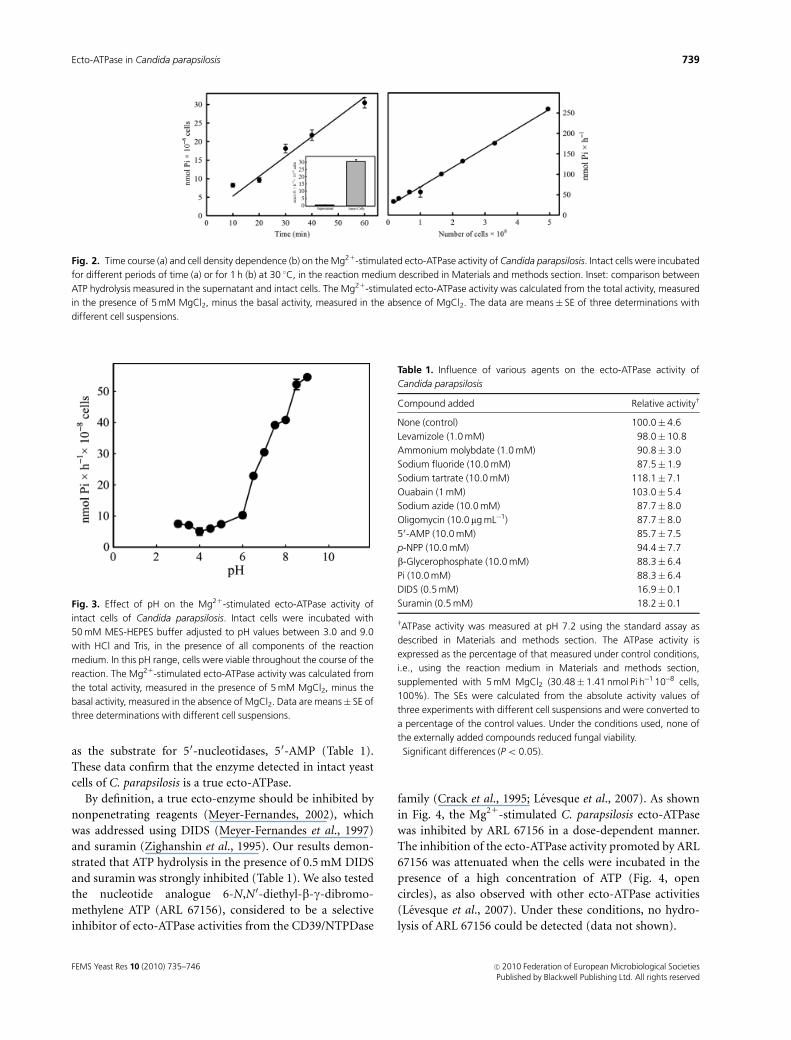
The time course of ATP hydrolysis by the C. parapsilosis
Mg21-stimulated ecto-ATPase was linear (r2 = 0.9740) for at
least 1 h (Fig. 2a). To certify that the observed ATP hydro-
lysis did not result from secreted enzymes, the cells were first
incubated in a reaction mixture containing no ATP. Cells
were then removed by centrifugation and the supernatant
was assayed for ATPase activity. ATP hydrolysis was not
detected, even in the presence of MgCl2 (inset of Fig. 2a).
This control excludes the possibility that the ATPase activity
described here could be derived from lysed C. parapsilosis
cells. Similarly, in assays to determine the influence of cell
density, the Mg21-stimulated ecto-ATPase activity mea-
sured over 60 min was linear (r2 = 0.9941) over a nearly 10-
fold range of cell density (Fig. 2b).
Effect of pH and different inhibitors on thehydrolysis of ATP by yeast cells
The optimum pH for the Mg21-stimulated ecto-ATPase
activity is within the alkaline range, while the ecto-phospha-
tase activity also present on the external surface of
C. parapsilosis was inhibited in basic pH (Kiffer-Moreira
et al., 2007). In the pH range from 3.0 to 9.0, in which the
cells were alive throughout the time course of the reaction,
the Mg21-stimulated ATPase activity increased with the pH.
At pH 9.0, the values of ATP hydrolysis were around
sevenfold higher than those obtained at pH 3.0 (Fig. 3).
Similar results were obtained for other microorganisms such
as Entamoeba histolytica (Barros et al., 2000), Leishmania
amazonensis (Berredo-Pinho et al., 2001) and F. pedrosoi
(Collopy-Junior et al., 2006). To confirm that ATP hydro-
lysis was not due to a phosphatase activity, described
previously in this cell (Kiffer-Moreira et al., 2007), experi-
ments were performed in the presence of different inhibitors
of phosphatases. Table 1 shows that classical inhibitors of
different phosphatases, such as sodium fluoride and ammo-
nium molybdate (acid phosphatases; Dutra et al., 2001),
levamizole (alkaline phosphatases; Van-Belle, 1976) and
tartrate (secreted phosphatases; Dutra et al., 2001), had no
effect on the C. parapsilosis ATPase activity. Similar results
were observed when the phosphatase substrates p-nitrophe-
nylphosphate and b-glycerophosphate were tested as inhibi-
tors. Ecto-ATPase activity was insensitive to 10 mM
inorganic phosphate (Table 1), which inhibited 70% of
ecto-phosphatase activity (Kiffer-Moreira et al., 2007). To-
gether, these data indicate that ecto-phosphatase activities
also present in C. parapsilosis did not contribute to ATP
hydrolysis.
To rule out the possibility that ATP hydrolysis under the
present experimental condition was promoted by intracel-
lular ATPases, inhibitors of ATPases with ATP-binding sites
facing the cytosol were tested (Table 1). Inhibitors of Na1/
K1-ATPase (ouabain; Caruso-Neves et al., 1998) and mito-
chondrial Mg21-ATPase (oligomycin and sodium azide;
Meyer-Fernandes et al., 1997) also had no effect on the
Mg21-stimulated C. parapsilosis ecto-ATPase (Table 1).
Ammonium molybdate, a potent inhibitor of 50-nucleoti-
dases (Gottlieb & Dwyer, 1983), was also inefficient as an
inhibitor of the C. parapsilosis ecto-ATPase activity, as well
Fig. 1. Stimulation of the ecto-ATPase activity in Candida parapsilosis by divalent metals. (a) Influence of different divalent cations on the ecto-ATPase
activity in intact cells of C. parapsilosis. Cells were incubated for 1 h at 30 1C in a reaction medium (control, described in Materials and methods section).
Chloride salts of the ions described in the figure were added at a final concentration of 5 mM. Alternatively, the medium was supplemented with EDTA,
in order to avoid cation interference. Asterisks denote significant differences (Po 0.05). (b) Mg21, the most effective stimulator, enhanced enzyme
activity in a dose-dependent pattern, as determined after incubation of living yeast cells under the conditions described in Materials and methods
section. The data are means� SE of three determinations with different cell suspensions.
FEMS Yeast Res 10 (2010) 735–746c� 2010 Federation of European Microbiological SocietiesPublished by Blackwell Publishing Ltd. All rights reserved
738 T. Kiffer-Moreira et al.
as the substrate for 50-nucleotidases, 50-AMP (Table 1).
These data confirm that the enzyme detected in intact yeast
cells of C. parapsilosis is a true ecto-ATPase.
By definition, a true ecto-enzyme should be inhibited by
nonpenetrating reagents (Meyer-Fernandes, 2002), which
was addressed using DIDS (Meyer-Fernandes et al., 1997)
and suramin (Zighanshin et al., 1995). Our results demon-
strated that ATP hydrolysis in the presence of 0.5 mM DIDS
and suramin was strongly inhibited (Table 1). We also tested
the nucleotide analogue 6-N,N0-diethyl-b-g-dibromo-
methylene ATP (ARL 67156), considered to be a selective
inhibitor of ecto-ATPase activities from the CD39/NTPDase
family (Crack et al., 1995; Levesque et al., 2007). As shown
in Fig. 4, the Mg21-stimulated C. parapsilosis ecto-ATPase
was inhibited by ARL 67156 in a dose-dependent manner.
The inhibition of the ecto-ATPase activity promoted by ARL
67156 was attenuated when the cells were incubated in the
presence of a high concentration of ATP (Fig. 4, open
circles), as also observed with other ecto-ATPase activities
(Levesque et al., 2007). Under these conditions, no hydro-
lysis of ARL 67156 could be detected (data not shown).
Fig. 2. Time course (a) and cell density dependence (b) on the Mg21-stimulated ecto-ATPase activity of Candida parapsilosis. Intact cells were incubated
for different periods of time (a) or for 1 h (b) at 30 1C, in the reaction medium described in Materials and methods section. Inset: comparison between
ATP hydrolysis measured in the supernatant and intact cells. The Mg21-stimulated ecto-ATPase activity was calculated from the total activity, measured
in the presence of 5 mM MgCl2, minus the basal activity, measured in the absence of MgCl2. The data are means� SE of three determinations with
different cell suspensions.
Fig. 3. Effect of pH on the Mg21-stimulated ecto-ATPase activity of
intact cells of Candida parapsilosis. Intact cells were incubated with
50 mM MES-HEPES buffer adjusted to pH values between 3.0 and 9.0
with HCl and Tris, in the presence of all components of the reaction
medium. In this pH range, cells were viable throughout the course of the
reaction. The Mg21-stimulated ecto-ATPase activity was calculated from
the total activity, measured in the presence of 5 mM MgCl2, minus the
basal activity, measured in the absence of MgCl2. Data are means� SE of
three determinations with different cell suspensions.
Table 1. Influence of various agents on the ecto-ATPase activity of
Candida parapsilosis
Compound added Relative activityw
None (control) 100.0� 4.6
Levamizole (1.0 mM) 98.0� 10.8
Ammonium molybdate (1.0 mM) 90.8� 3.0
Sodium fluoride (10.0 mM) 87.5� 1.9
Sodium tartrate (10.0 mM) 118.1� 7.1
Ouabain (1 mM) 103.0� 5.4
Sodium azide (10.0 mM) 87.7� 8.0
Oligomycin (10.0 mg mL�1) 87.7� 8.0
50-AMP (10.0 mM) 85.7� 7.5
p-NPP (10.0 mM) 94.4� 7.7
b-Glycerophosphate (10.0 mM) 88.3� 6.4
Pi (10.0 mM) 88.3� 6.4
DIDS (0.5 mM) 16.9� 0.1�
Suramin (0.5 mM) 18.2� 0.1�
wATPase activity was measured at pH 7.2 using the standard assay as
described in Materials and methods section. The ATPase activity is
expressed as the percentage of that measured under control conditions,
i.e., using the reaction medium in Materials and methods section,
supplemented with 5 mM MgCl2 (30.48� 1.41 nmol Pi h�1 10�8 cells,
100%). The SEs were calculated from the absolute activity values of
three experiments with different cell suspensions and were converted to
a percentage of the control values. Under the conditions used, none of
the externally added compounds reduced fungal viability.�Significant differences (Po 0.05).
FEMS Yeast Res 10 (2010) 735–746 c� 2010 Federation of European Microbiological SocietiesPublished by Blackwell Publishing Ltd. All rights reserved
739Ecto-ATPase in Candida parapsilosis
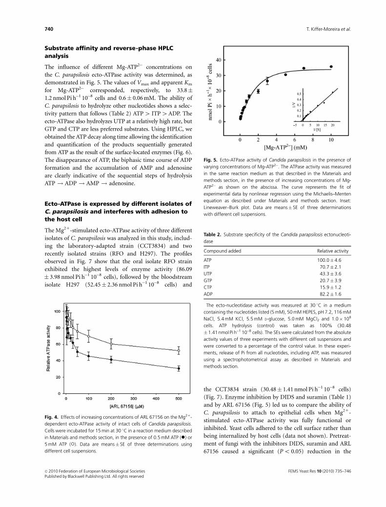
Substrate affinity and reverse-phase HPLCanalysis
The influence of different Mg-ATP2� concentrations on
the C. parapsilosis ecto-ATPase activity was determined, as
demonstrated in Fig. 5. The values of Vmax and apparent Km
for Mg-ATP2� corresponded, respectively, to 33.8�1.2 nmol Pi h�1 10�8 cells and 0.6� 0.06 mM. The ability of
C. parapsilosis to hydrolyze other nucleotides shows a selec-
tivity pattern that follows (Table 2) ATP4 ITP4ADP. The
ecto-ATPase also hydrolyzes UTP at a relatively high rate, but
GTP and CTP are less preferred substrates. Using HPLC, we
obtained the ATP decay along time allowing the identification
and quantification of the products sequentially generated
from ATP as the result of the surface-located enzymes (Fig. 6).
The disappearance of ATP, the biphasic time course of ADP
formation and the accumulation of AMP and adenosine
are clearly indicative of the sequential steps of hydrolysis
ATP ! ADP ! AMP ! adenosine.
Ecto-ATPase is expressed by different isolates ofC. parapsilosis and interferes with adhesion tothe host cell
The Mg21-stimulated ecto-ATPase activity of three different
isolates of C. parapsilosis was analyzed in this study, includ-
ing the laboratory-adapted strain (CCT3834) and two
recently isolated strains (RFO and H297). The profiles
observed in Fig. 7 show that the oral isolate RFO strain
exhibited the highest levels of enzyme activity (86.09
� 3.98 nmol Pi h�1 10�8 cells), followed by the bloodstream
isolate H297 (52.45� 2.36 nmol Pi h�1 10�8 cells) and
the CCT3834 strain (30.48� 1.41 nmol Pi h�1 10�8 cells)
(Fig. 7). Enzyme inhibition by DIDS and suramin (Table 1)
and by ARL 67156 (Fig. 5) led us to compare the ability of
C. parapsilosis to attach to epithelial cells when Mg21-
stimulated ecto-ATPase activity was fully functional or
inhibited. Yeast cells adhered to the cell surface rather than
being internalized by host cells (data not shown). Pretreat-
ment of fungi with the inhibitors DIDS, suramin and ARL
67156 caused a significant (Po 0.05) reduction in the
Fig. 4. Effects of increasing concentrations of ARL 67156 on the Mg21-
dependent ecto-ATPase activity of intact cells of Candida parapsilosis.
Cells were incubated for 15 min at 30 1C in a reaction medium described
in Materials and methods section, in the presence of 0.5 mM ATP (�) or
5 mM ATP (�). Data are means� SE of three determinations using
different cell suspensions.
Fig. 5. Ecto-ATPase activity of Candida parapsilosis in the presence of
varying concentrations of Mg-ATP2�. The ATPase activity was measured
in the same reaction medium as that described in the Materials and
methods section, in the presence of increasing concentrations of Mg-
ATP2� as shown on the abscissa. The curve represents the fit of
experimental data by nonlinear regression using the Michaelis–Menten
equation as described under Materials and methods section. Inset:
Lineweaver–Burk plot. Data are means� SE of three determinations
with different cell suspensions.
Table 2. Substrate specificity of the Candida parapsilosis ectonucleoti-
dase
Compound added Relative activity�
ATP 100.0� 4.6
ITP 70.7� 2.1
UTP 43.3� 3.6
GTP 20.7� 3.9
CTP 15.9� 1.2
ADP 82.2� 1.6
�The ecto-nucleotidase activity was measured at 30 1C in a medium
containing the nucleotides listed (5 mM), 50 mM HEPES, pH 7.2, 116 mM
NaCl, 5.4 mM KCl, 5.5 mM D-glucose, 5.0 mM MgCl2 and 1.0� 108
cells. ATP hydrolysis (control) was taken as 100% (30.48
�1.41 nmol Pi h�1 10�8 cells). The SEs were calculated from the absolute
activity values of three experiments with different cell suspensions and
were converted to a percentage of the control value. In these experi-
ments, release of Pi from all nucleotides, including ATP, was measured
using a spectrophotometrical assay as described in Materials and
methods section.
FEMS Yeast Res 10 (2010) 735–746c� 2010 Federation of European Microbiological SocietiesPublished by Blackwell Publishing Ltd. All rights reserved
740 T. Kiffer-Moreira et al.

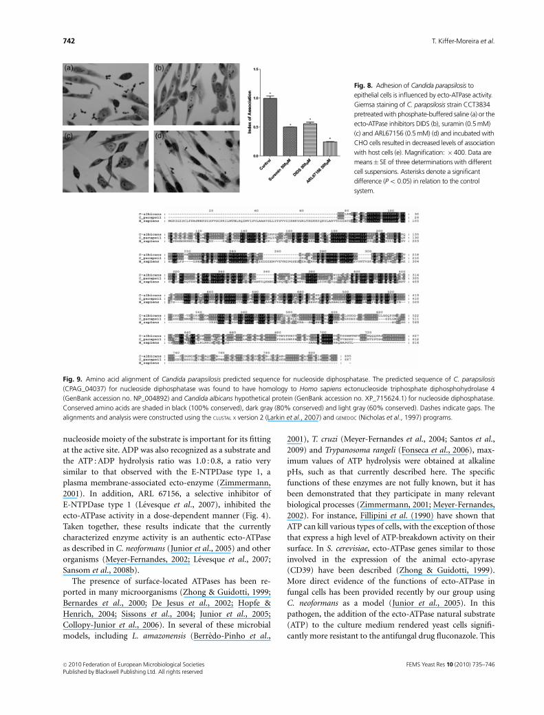
adhesion of the C. parapsilosis to host cells (Fig. 8), indicat-
ing that Mg21-stimulated ecto-ATPase did indeed influence
the interaction between yeast and epithelial cells.
In the C. albicans genome, the first sequenced Candida
genome available (Jones et al., 2004), we identified one gene
for nucleoside diphosphatase (NDPase/NTPase) in the
NCBI database. The gene CaO19.10432 encodes a hypothe-
tical protein (GenBank accession no. XP_715624.1) with
695 amino acids and significant similarity to human ecto-
nucleoside triphosphate diphosphohydrolase 4 (GenBank
accession no. NP_004892) (29% of identity, 49% of similar-
ity and an E value of 9e� 62). In C. parapsilosis, although
the genome has been sequenced recently (Butler et al., 2009)
by the Wellcome Trust Sanger Institute Pathogen Genomics
group (http://www.sanger.ac.uk/sequencing/Candida/para
psilosis/), most of the genes have not been completely
annotated yet. To verify the existence of nucleoside dipho-
sphatase sequences in the C. parapsilosis genome, we used
the gene sequence from C. albicans as a query, and then a
BLASTN search was performed for related sequences in the
Candida database on the Broad Institute server, for a
comparative analysis of genes and genomes across the
Candida clade (http://www.broadinstitute.org/annotation/
genome/candida_group/MultiHome.html). We observed at
the gene CPAG_04037 transcript product a conserved do-
main for CD39 family. The predicted C. parapsilosis protein
sequence for nucleoside diphosphatase shares 62% and 31%
identities and 74% and 49% similarities to the correspond-
ing C. albicans and human sequences, respectively. The
conserved amino acids are shown in the protein alignments
(Fig. 9).
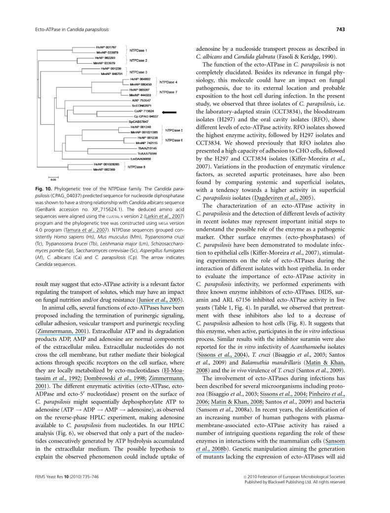
The phylogenetic relationship between NTPDase se-
quences from Candida sp., mammals, other fungi and
parasites such as trypanosomatids can be seen in Fig. 10.
The alignment of the eight well-known NTPDase groups of
proteins from H. sapiens (Hs) and M. musculus (Mm)
resulted in separate clades in the phylogenetic tree, whereas
the orthologues sequences from T. cruzi (Tc), T. brucei (Tb)
and L. major (Lm) formed a different clade. As expected, all
fungi species, S. pombe (Sp), S. cerevisiae (Sc), A. fumigatus
(Af), C. albicans and C. parapsilosis were also grouped in a
unique clade, suggesting homologous functions between the
proteins.
Discussion
In the present study, we aimed at the biochemical character-
ization of an ecto-ATPase activity in intact cells of
C. parapsilosis. This activity was stimulated by Mg21 and
Mn21, as demonstrated in S. cerevisiae, C. neoformans and
F. pedrosoi (Zhong & Guidotti, 1999; Junior et al., 2005;
Collopy-Junior et al., 2006). The surface localization of the
enzyme was supported by its sensitivity to the impermeant
reagents DIDS (Barbacci et al., 1996; Meyer-Fernandes et al.,
1997; Berredo-Pinho et al., 2001) and suramin. The Mg21-
stimulated enzyme activity cannot be ascribed to ecto-
phosphatases or 50-nucleotidases, because it was not affected
by inhibitors and substrates of these enzymes. The Mg21-
stimulated ecto-ATPase hydrolyzes ITP, GTP, CTP and UTP
(Table 2) with different selectivities indicating that the
Fig. 6. Time course of ATP hydrolysis in the presence of intact Candida
parapsilosis cells. The cells were incubated for each indicated period of
time at 30 1C in the presence of 116 mM NaCl, 5.4 mM KCl, 5.5 mM
D-glucose, 5 mM MgCl2, 50 mM HEPES-Tris Buffer (pH 7.2) and 100 mM
ATP. Filled circles: ATP concentration; open circles: ADP concentration,
filled triangle: AMP concentration; open triangle: adenosine concentra-
tion. Inset: amplified view of adenosine generation. Data are means� SE
of three determinations with different cell suspensions.
Fig. 7. Expression of Mg21-stimulated ecto-ATPase by different isolates
of Candida parapsilosis. Equivalent cell suspensions were incubated for
1 h at 30 1C in the same reaction medium described in Materials and
methods section. Enzyme activities in strains H297 and RFO were
significantly higher (�Po 0.05) than that observed in strain CCT3834.
The Mg21-stimulated ecto-ATPase activity was calculated from the total
activity, measured in the presence of 5 mM MgCl2, minus the basal
activity, measured in the absence of MgCl2. Data are means� SE of three
determinations with different cell suspensions.
FEMS Yeast Res 10 (2010) 735–746 c� 2010 Federation of European Microbiological SocietiesPublished by Blackwell Publishing Ltd. All rights reserved
741Ecto-ATPase in Candida parapsilosis
nucleoside moiety of the substrate is important for its fitting
at the active site. ADP was also recognized as a substrate and
the ATP : ADP hydrolysis ratio was 1.0 : 0.8, a ratio very
similar to that observed with the E-NTPDase type 1, a
plasma membrane-associated ecto-enzyme (Zimmermann,
2001). In addition, ARL 67156, a selective inhibitor of
E-NTPDase type 1 (Levesque et al., 2007), inhibited the
ecto-ATPase activity in a dose-dependent manner (Fig. 4).
Taken together, these results indicate that the currently
characterized enzyme activity is an authentic ecto-ATPase
as described in C. neoformans (Junior et al., 2005) and other
organisms (Meyer-Fernandes, 2002; Levesque et al., 2007;
Sansom et al., 2008b).
The presence of surface-located ATPases has been re-
ported in many microorganisms (Zhong & Guidotti, 1999;
Bernardes et al., 2000; De Jesus et al., 2002; Hopfe &
Henrich, 2004; Sissons et al., 2004; Junior et al., 2005;
Collopy-Junior et al., 2006). In several of these microbial
models, including L. amazonensis (Berredo-Pinho et al.,
2001), T. cruzi (Meyer-Fernandes et al., 2004; Santos et al.,
2009) and Trypanosoma rangeli (Fonseca et al., 2006), max-
imum values of ATP hydrolysis were obtained at alkaline
pHs, such as that currently described here. The specific
functions of these enzymes are not fully known, but it has
been demonstrated that they participate in many relevant
biological processes (Zimmermann, 2001; Meyer-Fernandes,
2002). For instance, Fillipini et al. (1990) have shown that
ATP can kill various types of cells, with the exception of those
that express a high level of ATP-breakdown activity on their
surface. In S. cerevisiae, ecto-ATPase genes similar to those
involved in the expression of the animal ecto-apyrase
(CD39) have been described (Zhong & Guidotti, 1999).
More direct evidence of the functions of ecto-ATPase in
fungal cells has been provided recently by our group using
C. neoformans as a model (Junior et al., 2005). In this
pathogen, the addition of the ecto-ATPase natural substrate
(ATP) to the culture medium rendered yeast cells signifi-
cantly more resistant to the antifungal drug fluconazole. This
Fig. 9. Amino acid alignment of Candida parapsilosis predicted sequence for nucleoside diphosphatase. The predicted sequence of C. parapsilosis
(CPAG_04037) for nucleoside diphosphatase was found to have homology to Homo sapiens ectonucleoside triphosphate diphosphohydrolase 4
(GenBank accession no. NP_004892) and Candida albicans hypothetical protein (GenBank accession no. XP_715624.1) for nucleoside diphosphatase.
Conserved amino acids are shaded in black (100% conserved), dark gray (80% conserved) and light gray (60% conserved). Dashes indicate gaps. The
alignments and analysis were constructed using the CLUSTAL X version 2 (Larkin et al., 2007) and GENEDOC (Nicholas et al., 1997) programs.
Fig. 8. Adhesion of Candida parapsilosis to
epithelial cells is influenced by ecto-ATPase activity.
Giemsa staining of C. parapsilosis strain CCT3834
pretreated with phosphate-buffered saline (a) or the
ecto-ATPase inhibitors DIDS (b), suramin (0.5 mM)
(c) and ARL67156 (0.5 mM) (d) and incubated with
CHO cells resulted in decreased levels of association
with host cells (e). Magnification: � 400. Data are
means� SE of three determinations with different
cell suspensions. Asterisks denote a significant
difference (Po 0.05) in relation to the control
system.
FEMS Yeast Res 10 (2010) 735–746c� 2010 Federation of European Microbiological SocietiesPublished by Blackwell Publishing Ltd. All rights reserved
742 T. Kiffer-Moreira et al.
result may suggest that ecto-ATPase activity is a relevant factor
regulating the transport of solutes, which may have an impact
on fungal nutrition and/or drug resistance (Junior et al., 2005).
In animal cells, several functions of ecto-ATPases have been
proposed including the termination of purinergic signaling,
cellular adhesion, vesicular transport and purinergic recycling
(Zimmermann, 2001). Extracellular ATP and its degradation
products ADP, AMP and adenosine are normal components
of the extracellular mileu. Extracellular nucleotides do not
cross the cell membrane, but rather mediate their biological
actions through specific receptors on the cell surface, where
they are locally metabolized by ecto-nucleotidases (El-Moa-
tassim et al., 1992; Dombrowski et al., 1998; Zimmermann,
2001). The different enzymatic activities (ecto-ATPase, ecto-
ADPase and ecto-50 nucleotidase) present on the surface of
C. parapsilosis might sequentially dephosphorylate ATP to
adenosine (ATP ! ADP ! AMP ! adenosine), as observed
on the reverse-phase HPLC experiment, making adenosine
available to C. parapsilosis from nucleotides. In our HPLC
analysis (Fig. 6), we observed that only a part of the nucleo-
tides consecutively generated by ATP hydrolysis accumulated
in the extracellular medium. The possible hypothesis to
explain the observed phenomenon could include uptake of
adenosine by a nucleoside transport process as described in
C. albicans and Candida glabrata (Fasoli & Keridge, 1990).
The function of the ecto-ATPase in C. parapsilosis is not
completely elucidated. Besides its relevance in fungal phy-
siology, this molecule could have an impact on fungal
pathogenesis, due to its external location and probable
exposition to the host cell during infection. In the present
study, we observed that three isolates of C. parapsilosis, i.e.
the laboratory-adapted strain (CCT3834), the bloodstream
isolates (H297) and the oral cavity isolates (RFO), show
different levels of ecto-ATPase activity. RFO isolates showed
the highest enzyme activity, followed by H297 isolates and
CCT3834. We showed previously that RFO isolates also
presented a high capacity of adhesion to CHO cells, followed
by the H297 and CCT3834 isolates (Kiffer-Moreira et al.,
2007). Variations in the production of enzymatic virulence
factors, as secreted aspartic proteinases, have also been
found by comparing systemic and superficial isolates,
with a tendency towards a higher activity in superficial
C. parapsilosis isolates (Dagdeviren et al., 2005).
The characterization of an ecto-ATPase activity in
C. parapsilosis and the detection of different levels of activity
in recent isolates may represent important initial steps to
understand the possible role of the enzyme as a pathogenic
marker. Other surface enzymes (ecto-phosphatases) of
C. parapsilosis have been demonstrated to modulate infec-
tion to epithelial cells (Kiffer-Moreira et al., 2007), stimulat-
ing experiments on the role of ecto-ATPases during the
interaction of different isolates with host epithelia. In order
to evaluate the importance of ecto-ATPase activity in
C. parapsilosis infectivity, we performed experiments with
three known enzyme inhibitors of ecto-ATPases. DIDS, sur-
amin and ARL 67156 inhibited ecto-ATPase activity in live
yeasts (Table 1, Fig. 4). In parallel, we observed that pretreat-
ment with these inhibitors also led to a decrease of
C. parapsilosis adhesion to host cells (Fig. 8). It suggests that
this enzyme, when active, participates in the in vitro infectious
process. Similar results with the inhibitor suramin were also
reported for the in vitro infectivity of Acanthamoeba isolates
(Sissons et al., 2004), T. cruzi (Bisaggio et al., 2003; Santos
et al., 2009) and Balamuthia mandrillaris (Matin & Khan,
2008) and the in vivo virulence of T. cruzi (Santos et al., 2009).
The involvement of ecto-ATPases during infections has
been described for several microorganisms including proto-
zoa (Bisaggio et al., 2003; Sissons et al., 2004; Pinheiro et al.,
2006; Matin & Khan, 2008; Santos et al., 2009) and bacteria
(Sansom et al., 2008a). In recent years, the identification of
an increasing number of human pathogens with plasma-
membrane-associated ecto-ATPase activity has raised a
number of intriguing questions regarding the role of these
enzymes in interactions with the mammalian cells (Sansom
et al., 2008b). Genetic manipulation aiming the generation
of mutants lacking the expression of ecto-ATPases will aid
Fig. 10. Phylogenetic tree of the NTPDase family. The Candida para-
psilosis (CPAG_04037) predicted sequence for nucleoside diphosphatase
was shown to have a strong relationship with Candida albicans sequence
(GenBank accession no. XP_715624.1). The deduced amino acid
sequences were aligned using the CLUSTAL X version 2 (Larkin et al., 2007)
program and the phylogenetic tree was constructed using MEGA version
4.0 program (Tamura et al., 2007). NTPDase sequences grouped con-
sistently Homo sapiens (Hs), Mus musculus (Mm), Trypanosoma cruzi
(Tc), Trypanosoma brucei (Tb), Leishmania major (Lm), Schizosaccharo-
myces pombe (Sp), Saccharomyces cerevisiae (Sc), Aspergillus fumigates
(Af), C. albicans (Ca) and C. parapsilosis (Cp). The arrow indicates
Candida sequences.
FEMS Yeast Res 10 (2010) 735–746 c� 2010 Federation of European Microbiological SocietiesPublished by Blackwell Publishing Ltd. All rights reserved
743Ecto-ATPase in Candida parapsilosis
the functional elucidation of these enzymes in C. parapsi-
losis, including its involvement in virulence.
Acknowledgements
The present work was supported by grants from the
Brazilian Agencies Conselho Nacional de Desenvolvimento
Cientıfico e Tecnologico (CNPq), Coordenacao de Aperfei-
coamento de Pessoal de Nıvel Superior (CAPES) and
Fundacao de Amparo a Pesquisa do Estado do Rio de
Janeiro (FAPERJ). We would like to acknowledge Dr Clau-
dio A. Masuda for helping with the identification of putative
E-NTPDases in the C. parapsilosis genome. We also thank
Fatima Regina de Vasconcelos Goulart for preparation of
fungal cultures and Fabiano Ferreira Esteves and Rosangela
Rosa de Araujo for technical assistance.
References
Almirante BD, Rodriguez D, Cuenca-Estrella M, Almela M,
Sanchez F, Ayats J, Alonso-Tarres C, Rodriguez-Tudela JL &
Pahissa A (2006) Epidemiology, risk factors and prognosis of
Candida parapsilosis blood-stream infections: case–control
population-based surveillance study of patients in Barcelona,
Spain, from 2002 to 2003. J Clin Microbiol 44: 1681–1685.
Amazonas JN, Cosentino-Gomes D, Werneck-Lacerda A,
Pinheiro AAD, Lanfredi-Rangel A, De Souza W & Meyer-
Fernandes JR (2009) Giardia lamblia: characterization of ecto-
phosphatase activities. Exp Parasitol 121: 15–21.
Barbacci E, Filippini A, De Cesaris P & Ziparo E (1996)
Identification and characterization of an ecto-ATPase activity
in rat Sertoli cells. Biochem Bioph Res Co 222: 273–279.
Barros FS, De Menezes LF, Pinheiro AA, Silva EF, Lopes AH, De
Souza W & Meyer-Fernandes JR (2000) Ectonucleotide
diphosphohydrolase activities in Entamoeba histolytica. Arch
Biochem Biophys 375: 304–314.
Bernardes CF, Meyer-Fernandes JR, Saad-Nehme J, Vannier-
Santos MA, Peres-Sampaio CE & Vercesi AE (2000) Effects of
4-40-diisothyocanatostilbene-2,20-dissuolfinic acid on
Trypanosoma cruzi proliferation and Ca21 homeostasis. Int J
Biochem Cell B 32: 519–527.
Berredo-Pinho M, Peres-Sampaio CE, Chrispim PP, Belmont-
Firpo R, Lemos AP, Martiny A, Vannier-Santos MA & Meyer-
Fernandes JR (2001) A Mg-dependent ecto-ATPase in
Leishmania amazonensis and its possible role in adenosine
acquisition and virulence. Arch Biochem Biophys 391: 16–24.
Bisaggio DFR, Peres-Sampaio CE, Meyer-Fernandes JR & Souto-
Padron T (2003) Ecto-ATPase activity on the surface of
Trypanosoma cruzi and its possible role in the parasite–host
cell interaction. Parasitol Res 91: 273–282.
Butler G, Rasmussen MD, Lin MF et al. (2009) Evolution of
pathogenicity and sexual reproduction in eight Candida
genomes. Nature 459: 657–662.
Calderone R, Diamond R, Senet JM, Warmingdon J, Filler S &
Edwards JE (1994) Host cell fungal interactions. J Med Vet
Mycol 32: 151–168.
Caruso-Neves C, Meyer-Fernandes JR, Saad-Nehme J & Lopes
AG (1998) Osmotic modulation of the ouabain-sensitive
(Na11K1) ATPase from Malpighian tubules of Rhodnius
prolixus. Z Naturforsch 53: 911–917.
Collopy-Junior I, Kneipp LF, da Silva FC, Rodrigues ML, Alviano
CS & Meyer-Fernandes JR (2006) Characterization of an ecto-
ATPase activity in Fonsecaea pedrosoi. Arch Microbiol 185:
355–362.
Crack BE, Pollard CE, Beukers MW, Roberts SM, Hunt SF, Ingall
AH, McKechnie KC, IJzerman AP & Leff P (1995)
Pharmacological and biochemical analysis of FPL 67156, a
novel selective inhibitor of ecto-ATPase. Brit J Pharmacol 114:
475–481.
Dagdeviren M, Cerikcioglu N & Karavus M (2005) Acid
proteinase, phospholipase and adherence properties of
Candida parapsilosis strains isolated from clinical specimens of
hospitalised patients. Mycoses 48: 321–326.
De Jesus JB, de Sa Pinheiro AA, Lopes AH & Meyer-Fernandes JR
(2002) An ectonucleotide ATP-diphosphohydrolase activity in
Trichomonas vaginalis stimulated by galactose and its possible
role in virulence. Z Naturforsch C 57: 890–896.
Dombrowski KE, Ke Y, Brewer KA & Kapp JA (1998) Ecto-
ATPase: an activation marker necessary for effector cell
function. Immunol Rev 161: 111–118.
Dubyak GR & el-Moatassim C (1993) Signal transduction via P2-
purinergic receptors for extracellular ATP and other
nucleotides. Am J Physiol 265: C577–C606.
Dutra PML, Dias FA, Santos MAA, Rodrigues CO, Romeiro A,
Attias M, De Souza W, Lopes AHCS & Meyer-Fernandes JR
(2001) Secreted phosphatase activities in trypanosomatid
parasites of plants modulated by platelet-activating factor.
Phytopathology 91: 408–414.
El-Moatassim C, Dornand J & Mani J (1992) Extracellular ATP
and cell signaling. Biochim Biophys Acta 1134: 31–45.
Fasoli MO & Kerridge MD (1990) Uptake of pyrimidines and
their derivatives into Candida glabrata and Candida albicans.
J Gen Microbiol 136: 1475–1481.
Fillipini A, Taffs RE, Agui T & Sitkovsky MV (1990) Ecto-ATPase
activity in cytolytic T-lymphocytes. Protection from the
cytolytic effects of extracellular ATP. J Biol Chem 265: 334–340.
Fonseca FV, Fonseca de Souza AL, Mariano AC, Entringer PF,
Gondin KC & Meyer-Fernandes JR (2006) Trypanosoma
rangeli: characterization of an Mg-dependent ecto ATP-
diphosphohydrolase activity. Exp Parasitol 112: 76–84.
Glynn IM & Chappel JB (1964) A simple method for the
preparation of 32Pi-labelled adenosine triphosphate of high
specific activity. Biochem J 90: 147–149.
Goding JW (2000) Ecto-enzymes: physiology meets pathology.
J Leukocyte Biol 67: 285–311.
Gomes SA, Fonseca de Souza AL, Silva BA, Kiffer-Moreira T,
Santos-Mallet JR, Santos AL & Meyer-Fernandes JR (2006)
Trypanosoma rangeli: differential expression of cell surface
FEMS Yeast Res 10 (2010) 735–746c� 2010 Federation of European Microbiological SocietiesPublished by Blackwell Publishing Ltd. All rights reserved
744 T. Kiffer-Moreira et al.
polypeptides and ecto-phosphatase activity in short and long
epimastigote forms. Exp Parasitol 112: 253–262.
Gottlieb M & Dwyer DM (1983) Evidence for distinct 50- and
30-nucleotidase activities in the surface membrane fraction of
Leishmania donovani promastigotes. Mol Biochem Parasit 7:
303–317.
Hopfe M & Henrich B (2004) OppA, the substrate-binding
subunit of the oligopeptide permease, is the major Ecto-
ATPase of Mycoplasma hominis. J Bacteriol 186: 1021–1028.
Jesus JB, Lopes AHCS & Meyer-Fernandes JR (2002)
Characterization of an ecto-ATPase of Tritrichomonas foetus.
Vet Parasitol 103: 29–42.
Jones T, Federspiel NA, Chibana H et al. (2004) The diploid
genome sequence of Candida albicans. P Natl Acad Sci USA
101: 7329–7334.
Junior IC, Rodrigues ML, Alviano CS, Travassos LR & Meyer-
Fernandes JR (2005) Characterization of an ecto-ATPase
activity in Cryptococcus neoformans. FEMS Yeast Res 5:
899–907.
Kawamoto Y, Shinozuka K, Kunitomo M & Haginaka J (1998)
Determination of ATP and its metabolites released from rat
caudal artery by isocratic ion-pair reversed-phase high-
performance liquid chromatography. Anal Biochem 262:
33–38.
Kiffer-Moreira T, de Sa Pinheiro AA, Alviano WS, Barbosa FM,
Souto-Padron T, Nimrichter L, Rodrigues ML, Alviano CS &
Meyer-Fernandes JR (2007) An ectophosphatase activity in
Candida parapsilosis influences the interaction of fungi with
epithelial cells. FEMS Yeast Res 7: 621–628.
Kirley TL (1997) Complementary DNA cloning and sequencing
of the chicken muscle ecto-ATPase. Homology with the
lymphoid cell activation antigen CD39. J Biol Chem 272:
1076–1081.
Kneipp LF, Rodrigues ML, Holandino C, Esteves FF, Alviano CS,
Travassos LR & Meyer- Fernandes JR (2004) Ecto-phosphatase
activity in conidial forms of Fonsecaea pedrosoi is modulated
by exogenous phosphate and mediates fungal adhesion to
epithelial cells. Microbiology 150: 3355–3362.
Larkin MA, Blackshields G, Brown NP et al. (2007) Clustal W and
Clustal X version 2.0. Bioinformatics 23: 2947–2948.
Leite MS, Thomaz R, Fonseca FV, Panizzutti R, Vercesi AE &
Meyer-Fernandes JR (2007) Trypanosoma brucei brucei:
biochemical characterization of ecto-nucleoside triphosphate
diphosphohydrolase activities. Exp Parasitol 115: 315–323.
Levesque SA, Lavoie EG, Lecka J, Bigonnesse F & Sevigny J (2007)
Specificity of the ecto-ATPase inhibitor ARL 67156 on human
and mouse ectonucleotidases. Brit J Pharmacol 152: 141–150.
Lowry OH & Lopes J (1946) The determination of inorganic
phosphate in the presence of labile phosphate esters. J Biol
Chem 162: 421–428.
MacFarlane GD, Sampson DE, Clawson DJ, Clawson CC, Kelly
KL & Herzberg MC (1994) Evidence for an ecto-ATPase on the
cell wall of Streptococcus sanguis. Oral Microbiol Immun 9:
180–185.
Margolis RN, Schell MJ, Taylor SI & Hubbard AL (1990)
Hepatocyte plasma membrane ecto-ATPase (pp120/HA4) is a
substrate for tyrosine kinase activity of the insulin receptor.
Biochem Bioph Res Co 166: 562–566.
Matin A & Khan NA (2008) Demonstration and partial
characterization of ecto-ATPase in Balamuthia mandrilaris
and its possible role in the host–cell interactions. Lett Appl
Microbiol 47: 348–354.
Meyer-Fernandes JR (2002) Ecto-ATPases in protozoa parasites:
looking for a function. Parasitol Int 51: 229–303.
Meyer-Fernandes JR & Vieyra A (1988) Pyrophosphate formation
from acetyl phosphate and orthophosphate: evidence for
heterogeneous catalysis. Arch Biochem Biophys 266: 132–141.
Meyer-Fernandes JR, Dutra PML, Rodrigues CO, Saad-Nehme J
& Lopes AHCS (1997) Mg-dependent ecto-ATPase activity in
Leishmania tropica. Arch Biochem Biophys 341: 40–46.
Meyer-Fernandes JR, Lanz-Mendoza H, Gondim KC, Willott E &
Wells MA (2000) Ectonucleotide diphosphohydrolase
activities in hemocytes of larval Manduca sexta. Arch Biochem
Biophys 382: 152–159.
Meyer-Fernandes JR, Saad-Nehme J, Peres-Sampaio CE,
Belmont-Firpo R, Bisaggio DF, Do Couto LC, Fonseca de
Souza AL, Lopes AH & Souto-Padron T (2004) A Mg-
dependent ecto-ATPase is increased in the infective stages of
Trypanosoma cruzi. Parasitol Res 93: 41–50.
Milani G, Jarmuszkiewicz W, Sluse-Goffart CM, Schreiber AZ,
Vercesi AE & Sluse FE (2001) Respiratory chain network in
mitochondria of Candida parapsilosis: ADP/O appraisal of the
multiple electron pathways. FEBS Lett 508: 231–235.
Nicholas KB, Nicholas HB Jr & Deerfield DW II (1997) GeneDoc:
Analysis and visualization of genetic variation, Embnew News,
4: 14.
Nimrichter L, Rodrigues ML, Rodrigues EG & Travassos LR
(2005) The multitude of targets for the immune system and
drug therapy in the fungal cell wall. Microbes Infect 7: 789–798.
Peres-Sampaio CE, De Almeida-Amaral EE, Giarola NLL &
Meyer-Fernandes JR (2008) Leishmania amazonensis: effects of
heat shock on ecto-ATPase activity. Exp Parasitol 119:
135–143.
Pinheiro AADS, Amazonas JN, de Souza Barros F, De Menezes LF,
Batista EJ, Silva EF, De Souza W & Meyer-Fernandes JR (2007)
Entamoeba histolytica: an ecto-phosphatase activity regulated
by oxidation–reduction reactions. Exp Parasitol 115: 352–358.
Pinheiro CM, Martins-Duarte ES, Ferraro RB, Fonseca de Souza
AL, Gomes MT, Lopes AH, Vannier-Santos MA, Santos AL &
Meyer-Fernandes JR (2006) Leishmania amazonensis: bio-
logical and biochemical characterization of ecto-nucleoside
triphosphate diphosphohydrolase activities. Exp Parasitol 114:
16–25.
San Miguel LG, Cobo J, Otheo E, Sanchez-Sousa A, Abraira V &
Moreno S (2005) Secular trends of candidemia in a large
tertiary-care hospital from 1988 to 2000: emergence of
Candida parapsilosis. Infect Cont Hosp Ep 26: 548–552.
Sansom FM, Riedmaier P, Newton HJ et al. (2008a) Enzymatic
properties of an ecto-nucleotide triphosphate
FEMS Yeast Res 10 (2010) 735–746 c� 2010 Federation of European Microbiological SocietiesPublished by Blackwell Publishing Ltd. All rights reserved
745Ecto-ATPase in Candida parapsilosis
diphosphohydrolase from Legionella pneumophila; substrate
specificity and requirement for virulence. J Biol Chem 283:
12909–12918.
Sansom FM, Robson SC & Hartland EL (2008b) Possible effects
of microbial ecto-nucleotide triphosphate diphospho-
hydrolase on host–pathogen interactions. Microbiol Mol Biol R
72: 765–781.
Santos RF, Possa MAS, Bastos MS, Guedes PMM, Almeida MR,
De Marco R, Verjovski-Almeida S, Bahia MT & Fietto JLR
(2009) Influence of ecto-nucleoside triphosphate diphospho-
hydrolase activity on Trypanosoma cruzi infectivity and
virulence. PLoS Neglected Trop Diseases 3: e387.
Sarvikivi E, Lyyttikainen O, Soll DR, Pujol C, Pfaller MA,
Richardson M, Koukila-Kahkola P, Luukkainen P & Saxen H
(2005) Emergence of fluconazole resistance in a Candida
parapsilosis strain that caused infections in a neonatal intensive
care unit. J Clin Microbiol 43: 2729–2735.
Sissons J, Alsam J, Jayasekera S & Khan NA (2004) Ecto-ATPases
of clinical and non-clinical isolates of Acanthamoeba. Microb
Pathogenesis 37: 231–239.
Steinberg T & Di Virgilio F (1991) Cell-mediated cytotoxicity:
ATP as an effector and the role of target cells. Curr Opin
Immunol 3: 71–75.
Tamura K, Dudley J, Nei M & Kumar S (2007) MEGA4:
Molecular Evolutionary Genetics Analysis (MEGA) software
version 4.0. Mol Biol Evol 24: 1596–1599.
Trofa D, Gacser A & Nosanchuk JD (2008) Candida parapsilosis,
an emerging fungal pathogen. Clin Microbiol Rev 21:
606–625.
Van-Belle H (1976) Alkaline phosphatases I. Kinetics and
inhibition by levamisole of purified isoenzymes from humans.
Clin Chem 22: 972–976.
Weisman G, Turner TJ & Fedan JS (1996) Structure and function
of P2 purinoceptors. J Pharmacol Exp Ther 277: 1–9.
Westfall TD, Kennedy C & Sneddon P (1997) The ecto-ATPase
inhibitor ARL 67156 enhances parasympathetic
neurotransmission in the guinea-pig urinary bladder. Eur J
Pharmacol 329: 169–173.
Zhong X & Guidotti G (1999) A yeast Golgi E-type ATPase
with unusual membrane topology. J Biol Chem 274:
32704–32711.
Zighanshin AU, Ziganshina LE, King BE & Burnstock G (1995)
Characteristics of ecto-ATPase of Xenopus oocytes and the
inhibitory actions of suramin on ATP breakdown. Pflugers
Arch 429: 412–418.
Zimmermann H (2001) Ectonucleotidases: some recent
developments and a note on nomenclature. Drug Develop Res
52: 44–56.
FEMS Yeast Res 10 (2010) 735–746c� 2010 Federation of European Microbiological SocietiesPublished by Blackwell Publishing Ltd. All rights reserved
746 T. Kiffer-Moreira et al.