Protein Kinases and Phosphatases in the Control of Cell Fate
26
SAGE-Hindawi Access to Research Enzyme Research Volume 2011, Article ID 329098, 26 pages doi:10.4061/2011/329098 Review Article Protein Kinases and Phosphatases in the Control of Cell Fate Angela Bononi, Chiara Agnoletto, Elena De Marchi, Saverio Marchi, Simone Patergnani, Massimo Bonora, Carlotta Giorgi, Sonia Missiroli, Federica Poletti, Alessandro Rimessi, and Paolo Pinton Section of General Pathology, Department of Experimental and Diagnostic Medicine, Interdisciplinary Center for the Study of Inflammation (ICSI) and LTTA Center, University of Ferrara, 44100 Ferrara, Italy Correspondence should be addressed to Paolo Pinton, [email protected] Received 15 March 2011; Revised 6 May 2011; Accepted 8 June 2011 Academic Editor: Heung Chin Cheng Copyright © 2011 Angela Bononi et al. This is an open access article distributed under the Creative Commons Attribution License, which permits unrestricted use, distribution, and reproduction in any medium, provided the original work is properly cited. Protein phosphorylation controls many aspects of cell fate and is often deregulated in pathological conditions. Several recent findings have provided an intriguing insight into the spatial regulation of protein phosphorylation across different subcellular compartments and how this can be finely orchestrated by specific kinases and phosphatases. In this review, the focus will be placed on (i) the phosphoinositide 3-kinase (PI3K) pathway, specifically on the kinases Akt and mTOR and on the phosphatases PP2a and PTEN, and on (ii) the PKC family of serine/threonine kinases. We will look at general aspects of cell physiology controlled by these kinases and phosphatases, highlighting the signalling pathways that drive cell division, proliferation, and apoptosis. 1. Introduction Cells are exposed to several extracellular signals simultane- ously. Maintaining the fidelity of intracellular transduction systems, resulting in the amplification of specific biological responses, is crucial in eliciting the appropriate physiological response. The accurate selection of effector molecules is required, and these molecules must be finely regulated in their activation and deactivation, often by phosphorylation and dephosphorylation events. Protein phosphorylation is a reversible posttranslational modification that plays key roles in several physiological processes and is often deregulated in pathological conditions. The balance between activation and deactivation of signalling pathways is a delicate one, which is regulated not only through phosphorylation by kinases, but also through dephosphorylation events induced by a diverse range of phosphatases. The majority of oncogenes identified thus far encode protein kinases, and dysregulation in their activity is required for cancer initiation and maintenance. Intuitively, by counterbalancing the activity of kinases, phosphatases should primarily act as tumour suppressors [1, 2]. Approximately one-third of proteins encoded by the human genome are presumed to be phosphorylated during their life cycle, accounting for an estimated 100,000 different phosphorylation sites in a cellular proteome [3]. The structural changes imparted by the phosphorylation of specific residues afford exquisite mechanisms for the regulation of protein functions by modulating protein fold- ing, substrate affinity, stability, and activity. In many cases, phosphorylation results in switch-like changes in protein function, which can also bear to major modifications, that is, in the catalytic function of other enzymes, including kinases. Moreover, protein phosphorylation often leads to a structural change of the protein that can induce changes in interaction partners or subcellular localization. Phosphorylation acts as a molecular switch for many regulatory events in signalling pathways that drive cell division, proliferation, differentiation, and apoptosis. One of the main strategies for achieving the proper outcome is the compartmentalization of both protein kinases and phosphatases, to ensure an appropriate balance of protein phosphorylation [4]. The spatial distribution of kinases and phosphatases implies that a gradient of phosphorylated substrates exists across different subcellular compartments. This spatial separation not only regulates protein phos- phorylation but can also control the activity of other proteins, enzymes, and the transfer of other posttranslational
Transcript of Protein Kinases and Phosphatases in the Control of Cell Fate

SAGE-Hindawi Access to ResearchEnzyme ResearchVolume 2011, Article ID 329098, 26 pagesdoi:10.4061/2011/329098
Review Article
Protein Kinases and Phosphatases in the Control of Cell Fate
Angela Bononi, Chiara Agnoletto, Elena De Marchi, Saverio Marchi,Simone Patergnani, Massimo Bonora, Carlotta Giorgi, Sonia Missiroli,Federica Poletti, Alessandro Rimessi, and Paolo Pinton
Section of General Pathology, Department of Experimental and Diagnostic Medicine,Interdisciplinary Center for the Study of Inflammation (ICSI) and LTTA Center, University of Ferrara, 44100 Ferrara, Italy
Correspondence should be addressed to Paolo Pinton, [email protected]
Received 15 March 2011; Revised 6 May 2011; Accepted 8 June 2011
Academic Editor: Heung Chin Cheng
Copyright © 2011 Angela Bononi et al. This is an open access article distributed under the Creative Commons Attribution License,which permits unrestricted use, distribution, and reproduction in any medium, provided the original work is properly cited.
Protein phosphorylation controls many aspects of cell fate and is often deregulated in pathological conditions. Several recentfindings have provided an intriguing insight into the spatial regulation of protein phosphorylation across different subcellularcompartments and how this can be finely orchestrated by specific kinases and phosphatases. In this review, the focus will be placedon (i) the phosphoinositide 3-kinase (PI3K) pathway, specifically on the kinases Akt and mTOR and on the phosphatases PP2aand PTEN, and on (ii) the PKC family of serine/threonine kinases. We will look at general aspects of cell physiology controlled bythese kinases and phosphatases, highlighting the signalling pathways that drive cell division, proliferation, and apoptosis.
1. Introduction
Cells are exposed to several extracellular signals simultane-ously. Maintaining the fidelity of intracellular transductionsystems, resulting in the amplification of specific biologicalresponses, is crucial in eliciting the appropriate physiologicalresponse. The accurate selection of effector molecules isrequired, and these molecules must be finely regulated intheir activation and deactivation, often by phosphorylationand dephosphorylation events. Protein phosphorylation is areversible posttranslational modification that plays key rolesin several physiological processes and is often deregulated inpathological conditions. The balance between activation anddeactivation of signalling pathways is a delicate one, which isregulated not only through phosphorylation by kinases, butalso through dephosphorylation events induced by a diverserange of phosphatases. The majority of oncogenes identifiedthus far encode protein kinases, and dysregulation in theiractivity is required for cancer initiation and maintenance.Intuitively, by counterbalancing the activity of kinases,phosphatases should primarily act as tumour suppressors[1, 2]. Approximately one-third of proteins encoded bythe human genome are presumed to be phosphorylatedduring their life cycle, accounting for an estimated 100,000
different phosphorylation sites in a cellular proteome [3].The structural changes imparted by the phosphorylationof specific residues afford exquisite mechanisms for theregulation of protein functions by modulating protein fold-ing, substrate affinity, stability, and activity. In many cases,phosphorylation results in switch-like changes in proteinfunction, which can also bear to major modifications, thatis, in the catalytic function of other enzymes, includingkinases. Moreover, protein phosphorylation often leads to astructural change of the protein that can induce changes ininteraction partners or subcellular localization.
Phosphorylation acts as a molecular switch for manyregulatory events in signalling pathways that drive celldivision, proliferation, differentiation, and apoptosis. Oneof the main strategies for achieving the proper outcomeis the compartmentalization of both protein kinases andphosphatases, to ensure an appropriate balance of proteinphosphorylation [4]. The spatial distribution of kinasesand phosphatases implies that a gradient of phosphorylatedsubstrates exists across different subcellular compartments.This spatial separation not only regulates protein phos-phorylation but can also control the activity of otherproteins, enzymes, and the transfer of other posttranslational

2 Enzyme Research
modifications. The most important routes showing the fun-damental importance of subcellular localization of kinasesand phosphatases are perhaps the mitogen-activated protein-kinase- (MAPK-) mediated pathways, the phosphoinosi-tide 3-kinase (PI3K)/Akt/mammalian target of rapamycin(mTOR)-dependent signalling, as well as those involvingprotein kinase A (PKA) and protein kinase C (PKC), bothof which act as key transducers in many signalling cascades.These signalling pathways (MAPK, PI3K/Akt/mTOR, PKA,PKC) interact at numerous levels and at multiple intracel-lular sites to regulate many fundamental cellular processes.Crosstalk between signalling pathways is a common themein cell regulation, which usually depends on cell contextand plays an important role in fine-tuning the biologicalresponses. It is now well established that MAPK and PI3Kare two of the most predominant oncogenic routes, and theyare intimately linked together [5, 6]. The signal transductionensuing from these pathways is complicated by a remark-able number of interconnections [7] (Ras-MAPK pathwaymodulates PI3K pathway at multiple levels: MAPK—c-Jun—PTEN, Ras—PI3K, ERK—TSC2, RSK—TSC2, RSK—S6,RSK—eIF4B, and so does MKK4—JNK pathway throughthe activation of NF-κB; for a recent review see [8]). BothMAPK and PI3K pathways may result in the phosphorylationof many downstream targets and impose a role in theregulation of cell survival and proliferation. Overexpressionof these pathways in acute myeloid leukemias (AML) hasbeen associated with a worse prognosis than overexpressionof a single pathway [9], while activation of the MAPK(Raf/MEK/ERK) cascade is suppressed in some prostatecancer cell lines which express high levels of activated Akt[10, 11]. Hyperactivation of Akt has been found in cellularmodels of prostate cancer, such as LNCaP cells, as wellas in prostate cancer specimens, particularly in advancedstages of the disease [12]. It has been demonstrated that inprostate cancer, PI3K-Akt survival pathway could be affectedalso by PKC activation. Indeed, PKC promotes apoptosis inLNCaP cells through activation of p38 MAPK, and inhibitionof the Akt survival pathway [13]. Also PKA can act as acentral hub that interacts with a variety of other signalingpathways, not only mediating but also communicatingcAMP effects to Akt, PKC, MAPK and other pathways [14].Recent studies indicated that cAMP-dependent signallingis closely interwoven with the PI3K/Akt pathway [15].Akt is of tremendous importance for several neuronal keysignalling events, including cell differentiation, proliferation,and survival [16]. Neuronal survival and axonal regenerationmediated by PI3K-dependent Akt signalling were shown tobe induced by elevated cAMP levels [15, 17]. Finally, it isimportant to state that several of these major protein kinasesin the cell, in particular Akt, PKC, and ERK MAP kinases,are substrates for the protein phosphatase 2a (PP2a), whichappears to be the major phosphatase in eukaryotic cells thatdownregulates activated protein kinases. Thus, PP2a is likelyto play an important role in determining the activation ofprotein kinase cascades [18].
In the first part of this paper we will focus on thePI3K/Akt/mTOR pathway, specifically on the kinases Akt
and mTOR, along with the phosphatases PP2a and PTEN(phosphatase and tensin homolog deleted on chromosome10). The PI3K/Akt/mTOR signalling cascade is crucial tomany widely divergent physiological processes which includecell cycle progression, transcription, translation, differenti-ation, apoptosis, motility, and metabolism [19]. After thep53 pathway, the PI3K/Akt/mTOR signalling pathway isone of the most mutated pathways associated with humantumour and contributes to both cancer pathogenesis andtherapy resistance [20]. The binding of insulin, insulin-like growth factor-1 (IGF-1), and other growth factors toits related receptors can activate/phosphorylate PI3K, whichcatalyses the synthesis of the lipid phosphatidylinositol 3,4,5-trisphosphate (PIP3). These lipid products in turn interactwith proteins via their pleckstrin homology (PH) domain,which allows the recruitment of other signalling moleculesto the cell membrane [21]. Akt and phosphoinositide-dependent kinase-1 (PDK1) are PI3K’s main downstreameffectors: they contain a C-terminal PH domain, whichbinds the membrane-bound PIP3. Here, Akt is activatedthrough phosphorylation mediated by PDK1 [22] and bythe mammalian target of rapamycin complex 2 (mTORC2)[23]. In turn, activated Akt phosphorylates many targetproteins to regulate a broad range of cellular processes whichinclude cell survival, growth, proliferation, angiogenesis,metabolism, and migration. This pathway is activated inmany tumours as a result of amplification/overexpressionof PI3K (either PI3CA or the p85 subunit) [24], or of theamplification/overexpression/mutation of Akt [25]. More-over, a tight counterregulation by phosphatases has emergedas a crucial process to control PI3K/Akt/mTOR-dependentsignalling. Elevated Akt activation in human cancers canresult from its enhanced phosphorylation due to loss ofthe PTEN tumour suppressor [26] or PP2a, which is nowconsidered to be a tumour suppressor as well since itcan also dephosphorylate Akt and thus downregulate itsactivity [27]. Evidence accumulated over the past years hashighlighted the presence of different players of this intricatepathway in almost all the different subcellular compartments(namely, cytosol, plasma membrane, nucleus, mitochondria,endoplasmic reticulum (ER) and mitochondrial-associatedmembranes (MAMs)), where they can both modulate eachother and act on a plethora of different substrates (Figure 1).In the following sections, we will discuss these events andhow they are implicated in the control of cell fate.
In the final part, we will address more in depth thePKC family of serine/threonine kinases that, when activated,can be translocated from one intracellular compartmentto another, thus being able to affect a wide variety ofcellular processes. PKC is one of the most extensively studiedkinase families and has been implicated in cell proliferation,differentiation, apoptosis, tumour promotion, and neuronalactivity [28]. There are 12 isoforms of PKC termed con-ventional (PKCα, PKCβ and PKCγ), novel (PKCδ, PKCε,PKCη and PKCθ), atypical (PKCζ and PKCλ), and PKNand PKC-related (PKN1, PKN2 and PKN3) forms. Thereis strong evidence that this family of kinases can be relatedto the PI3K/Akt/mTOR signalling pathway at some levels.

Enzyme Research 3
Receptortyrosinekinase
PI3KPIP2PIP3PDK1
Active Akt
PT308
P S473mTOR
mLST8SIN1 Rictor
Pro
tor
mTOR
mLST8
RaptorPR
AS4
0
4E-BP1
p70S6K
Proteinsynthesis
P
P
P
ULK1PAtg13FIP200
eIF4
E
mRNA
HIF
Autophagy
Angiogenesis
PTEN
PIP3PIP2
“open” active
PTEN
“closed” inactive
P P P
Ck2
P
InactiveAkt
AktInactive
NEDD4-1UbbbbbbbbU
bUbbbbbbbbbUb
Degra
dation
Ub
Nuclear
import
Chromosomestability
IP3RP
PP2a
FOXOP
BaxPBad
P
Acinus
P Caspase-9P
Apoptosis
Cell cycle
PGSK3P
Glucosemetabolism
p53
DNArepair
MDM2Nucleus
ER
Mitochondria
P
TSC2
Rheb GTP
P
PAC
S-2
P
PP2a
Bcl-2
BaxP
PP2a
mTORC2
mTORC1
Growth factors, insulin, . . .
NF-κB
GSK3β
Figure 1: PI3K signalling pathway: The phosphatidylinositol 3-kinase (PI3K) signalling pathway begins with PI3K activation by receptortyrosine kinases after growth factors or insulin stimulation. PI3K activity phosphorylates and converts the lipid second messengerphosphatidylinositol 4,5-bisphosphate (PIP2—indicated by red phosphoinositide) into phosphatidylinositol 3,4,5-triphosphate (PIP3—indicated by green phosphoinositide), with consequent double phosphorylation/activation of Akt kinase. Akt promotes cell proliferationand survival by phosphorylation/inhibition of several proapoptotic targets (hold by the red dotted line), or by activation of mTOR complex1, an important regulator of several processes, such as autophagy, angiogenesis and protein synthesis. Protein phosphatase 2a (PP2a) familymembers are able to dephosphorylate/inhibit Akt, favouring apoptosis also by direct dephosphorylation of Bcl-2. The tumour suppressorphosphatase PTEN negatively regulates PI3K signalling by dephosphorylating PIP3, converting it back to PIP2. A mono-uniquitinated/activeform of PTEN is able to translocate into the nucleus, promoting DNA repair, cell cycle arrest and chromosome stability.
As such, a deeper understanding could yield improvementsnot only in how their mutations and dysregulation playcausal roles in human diseases, but may also provide insightsto develop agonists and antagonists for use in therapy. Inthis respect, it is well known that PKC belongs to the AGC(cAMP-dependent, cGMP-dependent and protein kinase C)family which includes Akt [29]. PKC and Akt share somecommon kinases (i.e., PDK1 and mTORC2) that performimportant regulatory functions [30–33]. Moreover, somePKC isoforms interact with Akt although their specific effecton Akt activity is isoform dependent [34, 35]; it has alsobeen demonstrated that high glucose induced Akt activation,
in a PKCβ-dependent manner [36]. In addition, it has beenfound that activation of PKCη leads to the activation of Aktand mTOR signalling pathway, promoting glioblastoma cellsproliferation [37]. PKC activity seems to be related also toPP2a and PTEN. PKCδ not only physically associates withthe PP2a catalytic subunit (PP2a/C) but also phosphorylatesthe phosphatase to increase its activity [38]; this is the casefor PKCα too, which seems to be the primary mediator ofthe ∼2-fold increase in PP2a activity observed in intestinalcells after PKC-signalling activation [39]. Regarding PTEN,suppression of its activity by transforming growth factor-β (TGF-β) has been shown to be specifically mediated by

4 Enzyme Research
PKCα. On the topic of PKCs, we will summarise their generalcharacteristics and role in different cell signalling pathways,as well as the result of their deregulation in cell fate.
For reasons of brevity, we will not discuss much furtherthe MAPK- and PKA-mediated pathways; interested readersshould refer to [40–44].
2. Akt Kinase
The serine/threonine kinase Akt constitutes an importantnode in diverse signalling cascades downstream of growthfactor receptor tyrosine kinases. Akt plays an essential rolein cell survival, growth, migration, proliferation, polarity,metabolism (lipid and glucose), cell cycle progression, mus-cle and cardiomyocyte contractility, angiogenesis, and self-renewal of stem cells. Altered Akt activity has been associatedwith cancer and other disease conditions, such as diabetesmellitus, neurodegenerative diseases, and muscle hypotrophy[45].
Among the downstream effectors of PI3Ks, Akt is themost important and best studied [46]. Three Akt isoformshave been identified in mice and humans [47]. These threeAkt proteins, although encoded by distinct genes localizedon different chromosomes, have approximately 80% aminoacid identity and similar domain structures. Each isoformpossesses an N-terminal PH domain of approximately 100amino acids, with a high similarity to PH domains foundin other signalling molecules that bind 3-phosphoinositides[48]. Biochemical analysis revealed that the PH domainof Akt binds to both PIP3 and phosphatidylinositol 4,5-bisphosphate (PIP2) with similar affinity [49]. Moreover, incontrast to PH domains of other proteins, the head groupof PIP3 is localized in a significantly different orientation inthe PH domain of Akt, and it lacks a specific tyrosine thatis conserved in PH domains of other proteins (e.g., DAPP1,GRP1, BTK) [50]. The kinase catalytic domain, located inthe central region of the protein, shows a high degree ofsimilarity to those found in PKA and PKC [51]: the relationwith PKA and PKC explains the other name of Akt, PKB.Also present in this region is a threonine residue (T308 inAkt1) whose phosphorylation is necessary for the activationof Akt. Following the kinase domain is a C-terminal tailof around 40 amino acids, containing a second regulatoryphosphorylation site (S473 in Akt1). This region possessesthe F-X-X-F/Y-S/T-Y/F hydrophobic motif (where X is anyamino acid) that is characteristic of the AGC kinase family. Inmammalian Akt isoforms, this motif is identical (FPQFSY)[52]. Phosphorylation at T308 and S473 occurs in responseto growth factors and other extracellular stimuli and isessential for maximal Akt activation [53].
The crystal structure of activated Akt was determinedin 2002 [54] and underlines the fundamental role of thehydrophobic motif as an allosteric regulator of the kinaseactivity: in fact, with Akt phosphorylated on threonine in thecatalytic loop but lacking its carboxyl-terminal hydrophobicmotif, the αB and C-helices in the aminoterminal lobe ofthe kinase domain and the activation loop are disordered[55]. Replacing the hydrophobic motif with a similar
sequence from the PKC-related kinase 2 (PRK2), containingan acidic residue instead of the Ser (called the PDK1-interacting fragment (PIF) sequence) [56], or mimickingthe phosphorylation in S473 through a substitution withaspartate leads to a stabilization of the kinase domainin active state. In other words, the molecular interactionbetween the phosphorylated hydrophobic motif and thegroove, formed by the αB and C-helices, stabilizes the wholecatalytic domain: this association is critical for the completeactivation of Akt.
As mentioned above, the activation of the PI3K signallingpathway by growth factor stimulation leads to recruitment ofAkt to the plasma membrane through binding to PIP3. Atthe plasma membrane, Akt can be phosphorylated on tworesidues, T308 and S473, by two different kinases, PDK1 [22,57] and the mTORC2 [23], respectively. Once activated, Akttranslocates to various subcellular compartments, includingthe Golgi, ER, mitochondria, and nucleus [58], where itphosphorylates substrates or interacts with other molecules(Figures 1 and 3). Consensus motif analysis indicates thatthere are potentially thousands of cellular substrates for Akt;about 50 of these have been characterized so far. Throughphosphorylation, Akt may either positively or negativelyaffect the function of these substrates, alter their subcellularlocalization, or modify their protein stabilities [45]. As aprotooncoprotein and the primary target of PI3K, Akt wasfirst characterized for its function in regulating cell survivaland cell proliferation, and its antiapoptotic activity is solvedthrough the inactivation of many proapoptotic factors.Constitutive activation of Akt leads to uncontrolled cellproliferation, inhibited apoptotic pathways, and strong cellcycle dysregulation, typical hallmarks of many human can-cers. Akt is able to directly or indirectly modulate apoptosis[59]. The direct effects are linked to phosphorylation eventsor interactions with cell death actors, whereas the indirectregulation of apoptosis is mediated through transcriptionalresponses to apoptotic stimuli.
Active Akt migrates to both the cytosol and the nucleus.Nevertheless, the relative contribution of Akt signalling at theplasma membrane, the cytosol, and the nucleus remains to beelucidated. Nuclear Akt may fulfil important antiapoptoticroles [60] (Figure 1). Indeed, Akt plays a crucial role indetermining cell fate by regulating fundamental transcrip-tional factors for the expression of pro- or anti-apoptoticmolecules, for example, YAP, CREB, FOXO proteins, andNF-κB, as well as the E3 ubiquitin ligase MDM2, a knownnegative regulator of the tumour suppressor p53 [61].Phosphorylation of CREB and NF-κB induce upregulationof antiapoptotic Bcl-2 family members, whereas phospho-rylation of FOXO family members leads to their nuclearexclusion and inactivation [62, 63], with a consequentdecreased transcriptional activity that is required for pro-moting apoptosis. Interestingly, Trotman et al. have shownthat the promyelocytic leukemia protein (PML) tumoursuppressor prevents cancer by inactivating phosphorylatedAkt (pAkt) inside the nucleus. PML specifically recruits theAkt phosphatase PP2a as well as pAkt into PML nuclearbodies. PML-null cells are impaired in PP2a phosphataseactivity towards Akt and thus, accumulate nuclear pAkt. As

Enzyme Research 5
a consequence, the progressive reduction in PML dose leadsto the inactivation of FOXO3a-mediated transcription of theproapoptotic Bim and the cell cycle inhibitor p27kip1 [64].
At the same time, Akt deliver antiapoptotic signals viadifferent proteins directly modulated by Akt phosphory-lation. Bad is one of the first discovered targets of Aktphosphorylation [65]. Bad is a proapoptotic member ofthe Bcl-2 family of proteins, able to bind Bcl-2 or Bcl-XL, blocking their antiapoptotic activities. Phosphorylationof Bad on S136 by Akt disrupts its interaction with Bcl-2/Bcl-XL, localized on the outer mitochondrial membrane,sequestering Bad in the cytosol, through the interaction with14-3-3 protein. In an analogous way, phosphorylation by Aktof proapoptotic Bax protein on S184 suppresses its translo-cation to mitochondria, preventing Bax conformationalchange, a typical event that occurs after apoptotic induction[66]. In addition, the caspase cascade is further inhibitedby Akt phosphorylation of procaspase 9 [67], inactivatedthrough phosphorylation in S196, a residue that, however, isnot conserved in other mammalian species [68]. Also Acinus,one of the most relevant proapoptotic factors, responsiblefor chromatin condensation, is phosphorylated by Akt inS422 and S573, with a consequent resistance of the proteinto the activator cleavage by caspase 3 [69]. Moreover, Aktphosphorylates the two isoforms, α and β, of the glycogensynthase kinase 3 (GSK-3) in S21 and S9, respectively,promoting their inactivation [70] and blocking its abilityto induce apoptosis in response to a wide range of stimuli(reviewed in [71]). Interestingly, GSK-3 β is a target of Aktphosphorylation/inactivation also inside mitochondria [72],but the role of this mitochondrial interaction remains to beclarified.
Akt is at the crossroads of several mitochondria-mediatedcell death pathways and exerts a major role in apoptosisdue to its networking with mitochondria in the regula-tion, and interconnection of metabolic pathways and cellsurvival. Notably, the mitochondrial import and exportmechanisms for Akt have yet to be investigated. Alsolinked to mitochondrial metabolism is the Akt activity onhexokinase. Hexokinase catalyzes the first step of glycolysis:in particular, hexokinases I and II are found to directlyinteract with mitochondria, and their expression is increasedin tumours [73], providing a putative explanation of theWarburg effect. In cancer cells, hypoxic conditions inducethe activation of the PI3K/Akt cell survival pathway [74]and the association of Akt with mitochondria [75]. Aktpromotes binding of hexokinase II to the mitochondrialvoltage-dependent anion channel (VDAC) [76]. The highaffinity of hexokinase II to VDAC allows it to selectivelyutilize intramitochondrial ATP to phosphorylate glucose anddirectly couple glycolysis to oxidative phosphorylation [77].By promoting the interaction hexokinase-VDAC, Akt seemsto maintain the “opening state” of the channel, protectingcells from apoptotic events through conservation of themitochondrial integrity [76]. Despite this, other studies showhow the hexokinase-VDAC binding mediates channel closurerather than VDAC opening, still maintaining the ability ofhexokinases to inhibit cell death [78, 79], especially reducing
the mitochondrial calcium (Ca2+) overload, a primary eventthat triggers cytochrome c release.
Connected to Ca2+-induced apoptosis, the inositol 1,4,5-trisphosphate receptor (IP3R), the main ER Ca2+-releasechannel, is phosphorylated by Akt due to the presence of aconsensus substrate motif that is conserved in all the threeIP3R isoforms. The consensus substrate motif (R-X-R-X-X-S/T) for Akt kinase is located in the C-terminal portionof the IP3R, and it is also conserved in IP3Rs cloned fromseveral different species, with the exception of Caenorhabditiselegans [80]. The IP3R is involved in Ca2+ mobilization fromintracellular stores, where channel activity is largely underthe control of IP3 binding. This receptor is also involvedin fundamental processes such as fertilization, mitosis, andapoptosis. In fact, there is general agreement in the literaturethat Ca2+ efflux from the ER and Ca2+ accumulation intomitochondria are linked to the effects of various apoptoticstimuli [81]. Akt, probably through its kinase activity,reduces Ca2+ release from the IP3R and protects the cellsfrom apoptosis induced by several Ca2+-mediated apoptoticstimuli [82, 83]. Moreover, a recent paper by our groupexplains how the protein PML functions at the MAMs andER levels to suppress Ca2+ transfer to the mitochondria.Knock-out cells for PML show a strong reduction of IP3RCa2+ release, due to a hyperactivation of Akt. PML is a neg-ative regulator of Akt, forming a macrocomplex composedby PML, Akt, the phosphatase PP2a, and IP3R type 3 atER/MAMs. In the absence of PML, PP2a cannot inhibit Akt,with a consequent increase in IP3R phosphorylation, limitedCa2+ release, and protection from apoptosis [84] (see alsobelow, PP2a section). Another protein regulated by Akt thatmediates interorganelle signalling and transmits apoptoticsignals from the ER to mitochondria is phosphofurin acidiccluster-sorting protein-2 (PACS-2). Akt phosphorylates thisprotein in S473, favouring the binding with 14-3-3, whichrepresses PACS-2 apoptotic activity [85].
One of the best-conserved functions of Akt is its rolein promoting cell growth. The predominant mechanismappears to be through activation of the mammalian targetof rapamycin complex 1 (mTORC1), which is regulatedby both nutrients and growth factor signalling. Akt hasbeen suggested to directly phosphorylate mTOR on S2448[86], but the role of this phosphorylation remains stillunclear. The mTORC1 activation by Akt seems due tothe inhibitory phosphorylation of the tuberous sclerosiscomplex 2 (TSC2, also known as tuberin), a tumoursuppressor, critical negative regulator of mTORC1 signalling[87]. This complex functions as a GTPase-activating protein(GAP) for the small G protein Ras homologue enriched inbrain (RHEB). The decreased GAP activity of the complexleads to accumulation of RHEB-GTP and activation ofmTORC1. However, it remains poorly understood how TSC2phosphorylation by Akt leads to decreased GAP activity[88]. Moreover, a second Akt substrate has been found tobe involved in mTORC1 regulation, the proline-rich Aktsubstrate of 40 kDa (PRAS40, also known as Akt1 substrate1). Akt was shown to directly phosphorylate PRAS40 onT246 [89], and this phosphorylation was fundamental for

6 Enzyme Research
14-3-3 binding. PRAS40 associates with mTORC1, nega-tively regulates it; therefore, phosphorylation by Akt andassociation with 14-3-3 are crucial for insulin to stimulatemTOR [90]. Akt regulation of mTORC1 through TSC2 andPRAS40 phosphorylation is also important for cell prolifer-ation, controlling the translation of proteins important forcell-cycle progression. Akt-mediated cell proliferation andoncogenic transformation has been shown to be dependenton mTORC1 activation [91], raising the possibility thatmTORC1 is the dominant Akt target in cells transformedwith mutant Ras [92].
Considering the strong connection between Akt andmTORC1 and the role of this signalling, frequently dysreg-ulated in cancers, in the next section, we will focus ourattention on the major component of the mTOR complex,the kinase mTOR.
3. mTOR Kinase
The PI3K pathway includes a number of critical effectorsthat are involved in basic cellular functions, such as cellgrowth control, the cell cycle and DNA damage checkpoints,and recombination or maintenance of telomere length.One of this family members is mTOR, now recognizedas a central regulator in a diverse array of vital cellularprocesses, including proliferation, growth, differentiation,and survival [93]. The physiological importance of mTORis undoubtedly demonstrated by the fact that the knock-out of mTOR in mice is primordially embryonic lethal[94], and the dysregulation of the mTOR pathway isassociated with increased transformation and oncogenesis[20].
Structurally, mTOR possesses in the N-terminusup to 20 tandem HEAT motifs, including a protein-protein interaction structure of two tandem antiparallelα-helices found in huntingtin, elongation factor 3, the Asubunit of protein PP2a, and TOR [95]. The C-terminusconsists of mutated FRAP-ataxia-telangiectasia (FAT), atransformation/transcription-domain-associated proteindomain, an FRB domain (FKPB12 (FK506-bindingprotein 12 kDa)-rapamycin binding), a catalytic kinasedomain containing also an ATP-binding site, a probableautoinhibitory or repressor domain, and an FATC (FATcarboxy-terminal) domain. The kinase domain is betweenthe FRB domain (which is C-terminal to the FAT domain)and the FATC domain, located at the C-terminus ofthe protein [96]. It is speculated that the HEAT repeatsserve to mediate protein-protein interactions, the FRBdomain is responsible to provide a docking site for theFKBP12/rapamycin complex, and FAT and FATC domainsmodulate mTOR kinase activity via unknown mechanisms[97]. The catalytic kinase domain in the C-terminus has ahigh similarity to the catalytic domain of PI3K, so mTORis considered a member of the PIKK (PI3K-related kinase)family, but there is no experimental evidence that it displayslipid kinase activity [98]. In mammalian cells, mTOR existsin two distinct complexes called complex 1 (mTORC1) andcomplex 2 (mTORC2).
mTORC1 is rapamycin-sensitive and consists of themTOR catalytic subunit, Raptor (regulatory associatedprotein of mTOR), mLST8 (also known as GβL), andPRAS40 [99]. Whereas the function of mLST8 is notreally clarified, Raptor regulates mTORC1 functioning asa scaffold for recruiting mTORC1 substrates. PRAS40 isphosphorylated by Akt at T246 releasing its inhibitory effectson mTORC1. mTORC1 is a master controller of proteinsynthesis, integrating signals from growth factors within thecontext of the energy, and nutritional conditions of the cell.Activated mTORC1 regulates protein synthesis by directlyphosphorylating 4E-BP1 (eukaryotic initiation factor 4Ebinding protein-1) and p70S6K (ribosomal p70S6 kinase),translation initiation factors that are important to cap-dependent mRNA translation, which increases the level ofmany proteins that are needed for cell cycle progression,proliferation, angiogenesis, and survival pathways [100].Many diverse signals (such as growth factors, amino acids,glucose, energy status, and different forms of stress) andpharmacological agents (such as rapamycin) regulate themTORC1 pathway.
In the other complex, mTORC2, mTOR also containsthe mLST8 protein and additionally interacts with a pro-tein called Sin1 (stress-activated protein kinase-interactingprotein), a second protein termed Protor (protein observedwith Rictor), and, instead of Raptor, Rictor (rapamycin-insensitive companion of mTOR) is present [101] (Figure 1).The interaction between Rictor and mTOR is not blockedby the drug rapamycin nor affected by nutrient levels,which are conditions known to regulate mTORC1. mTORC2modulates cell survival in response to growth factors byphosphorylating Akt on Ser473 which enhances subsequentAkt phosphorylation on Thr308 by PDK1 [23]. Anotherdownstream target of mTORC2 is serum/glucocorticoid-regulated kinase 1 (SGK1) [102]. Also the hydrophobicmotif phosphorylation of PKCα has been shown to bemediated by mTORC2 [103]. Moreover, mTORC2-mediatedphosphorylation of Akt and conventional PKC (cPKC) intheir C-terminal turn motif (TM) is crucial for their propercarboxyl-terminal folding, stability, and signalling [32, 33].
The mTOR signalling pathway is activated during a widevariety of cellular responses: it regulates growth by main-taining the appropriate balance between anabolic processes,such as macromolecular synthesis and nutrient storage, andcatabolic processes, like autophagy and the utilization ofenergy stores. Active mTOR enhances cell growth promotingprotein translation and increasing cell mass by controlling asubset of mRNAs that are thought to promote cell growthand proliferation.
Most of protein translation is modulated at the level ofinitiation, through the positioning of the ribosome at theAUG codon. Cellular mRNAs contain a cap structure attheir 5′ terminus [104], and it has been demonstrated thata strong repressor of cap-mediated translation is 4E-BP1[105]. mTOR phosphorylates and inactivate the translationinhibitor 4E-BP1, inducing its dissociation from the transla-tion initiation factor eIF4E, which can bind the cap structureat the 5′ termini of mRNAs, thereby allowing cap-dependenttranslation (in fact, dephosphorylated 4E-BP1 binds and

Enzyme Research 7
inhibits eIF4E) of proteins involved in cell proliferationand survival [106]. Moreover, mTOR is able to activate thep70S6K (through T389 phosphorylation) which activates the40S ribosomal protein S6 via phosphorylation at S240/244[107], a required process for translation and cell growth.mTOR is also able to monitor the regulation of ribosomebiogenesis, an aspect that fundamentally occurs during thetranslation of mRNAs of the ribosomal proteins and thesynthesis of ribosomal RNA (rRNA). Transcription of ribo-somal DNA (rDNA) and transfer RNA (tRNA) genes by RNApolymerase I (Pol I) and III (Pol III) is a major rate-limitingstep in the biogenesis of ribosomes. Nuclear localized mTORis involved in Pol-I- and Pol-III-mediated transcription ofrDNA and tRNA genes. It has been demonstrated that mTORis associated with the promoters of 45S rDNA and genes of 5SrDNA and tRNAs [108]. mTOR also regulates Pol-I and Pol-III-mediated transcription through phosphorylation of TIF-IA and upstream binding factor (UBF), two transcriptioninitiation factors of Pol I [109, 110]. The phosphorylationof TIF-IA in Ser44 is indispensable for its activity, whilephosphorylation in Ser199 inhibits TIF-IA. Indeed, it hasbeen demonstrated that inhibition of mTOR signalling inac-tivates TIF-IA by decreasing phosphorylation at Ser44 andenhancing phosphorylation at Ser199 [111]. Additionally,this effect not only regulates the activity of TIF-IA, but alsocontrols its intracellular localization. Treatment of cells withmTOR inhibitors causes translocation of a significant partof TIF-IA into the cytosol (from the nucleus) inhibiting theformation of the transcription-initiation complex [111].
The signalling components upstream and downstreamof mTOR are frequently altered in a wide variety of humantumours. Mutations in several tumour-suppressor genes(such as TSC1, TSC2, LKB1, PTEN, VHL, NF1, and PKD1)trigger the development of different diseases [100]. Toconfirm a primary role of mTOR in cancer development, itwas shown that the inhibition of this kinase is fundamentalfor blocking cell growth and motility in a number oftumour cell lines [112]. Different data provide evidencethat the mTOR pathways receive stimulatory signals fromRas, and, ultimately, these pathways drive tumorigenesisthrough the coordinated phosphorylation of proteins thatdirectly regulate protein synthesis, cell-cycle progression, andcancer development. It has been demonstrated that Ras isable to determine an ERK-dependent phosphorylation ofTSC2. This results in the suppression of its biochemicaland biological tumour-suppressive functions (the loss ofTSC2 is the typical aspect of the tumour syndrome tuberoussclerosis) and in the activation of S6K through the mTOR-dependent site T389. This shows a direct control of transla-tion through a mechanism that involves the TSC1/2-mTORpathway and Ras-MAPK (mitogen-activated protein kinase)signalling [113].
mTOR is also able to regulate the expression of thehypoxia-inducible factor-1 alpha (HIF-1α). Hypoxia occursin the majority of tumours, promoting angiogenesis, metas-tasis, and resistance to therapy. Responses to hypoxia areorchestrated in part through the activation of the hypoxia-inducible factor family of transcription factors (HIFs) [114].HIFs are heterodimeric transcription factors composed of an
α and a β subunit (whose levels are controlled by oxygentension [115]) and activates the transcription of 100–200genes involved in cellular metabolism and the adaptationof cells to hypoxic conditions. A number of interestingpotential connections have emerged between HIFs andmTOR, and there are clear examples of these pathwaysregulating each other and common downstream pathways.Perhaps the most firmly established is the ability of mTOR toinfluence HIF1A translation: mTORC1 signalling increasesHIF-1α protein levels by promoting mRNA translation fromits 5′UTR [116–118]. At the same time, hypoxia and lowoxygen levels inhibit mTOR signalling through multiplemechanisms. One of these determines a decrease in cellularATP levels with consequent activation of AMPK (AMP-activated protein kinase), which inhibits mTOR via TSC2and raptor phosphorylation [119].
Moreover, the mTOR pathway is the most studied path-way regulating mammalian autophagy. Autophagy, which ishighly conserved from yeast to humans, is a bulk degradationprocess involved in the clearance of long-lived proteins andorganelles. Dysfunction in the autophagy pathway has beenimplicated in an increasing number of human diseases,from infectious diseases to cancer and neurodegeneration.mTOR negatively regulates autophagy, and, downstream ofthis kinase, there are more than 30 different genes regulatingautophagy in yeast (known as the ATG genes), and many ofthese have mammalian orthologs [120]. mTORC1 regulatesnumerous proteins that are required for the executionof the autophagic program, including a macromolecularcomplex (FIP200,ULK1/Atg1) implicated in the initiationstep of autophagosome formation. mTORC1 is able tophosphorylate ULK1 (unc-51-like kinase 1), moderatelyreducing ULK1 kinase activity and, consequently, blockingautophagy [121] (Figure 1).
Despite the central role of mTOR in cell physiology,relatively little is known about its precise subcellular distribu-tion and the underlying functional significance. Essentially,mTOR is localized in the cytosol where it plays the importantfunction of maintaining an appropriate balance betweenanabolic and catabolic processes. However, following differ-ent stimuli, mTOR can be driven into other compartmentswhere it performs its activity (Figure 3). Previous studiesindicate that mTOR is present in both the cytoplasm[122] and nucleus [123, 124]. Biochemical characterizationin association with confocal microscopy identified a newlocalization of mTOR in the ER and the Golgi apparatus;specifically, mTOR is a peripheral ER membrane protein,tightly anchored to the ER/Golgi membranes [125]. ERand Golgi localization sequences of mTOR have also beenidentified [126], suggesting that anchoring to the ER/Golgi isimportant for mTOR signal transduction. ER and the Golgiapparatus are internal membrane structures constitutingmore than one-half of the total membranes in a cell. Thesecompartments have an important role in processing andpackaging macromolecules and sequestration of Ca2+, whichhas been involved in mechanisms capable of activating bothcell suicide programs as well as prosurvival mechanisms[127]. It is, therefore, possible to hypothesize that mTORcould play its role of amino acid sensor in the ER/Golgi

8 Enzyme Research
side. Moreover, the Rag GTPase family has been shownto be an amino acid-specific regulator of mTOR [128], acondition that promotes the translocation of the kinase to asurface of endomembrane compartments, identified recentlyas the lysosomal surface. In fact, a trimeric complex, namedRagulator, (encoded by the genes MAPKSP1, ROBLD3, andc11orf59) interacts with the Rag GTPase, localizing these andmTOR to lysosomes, a translocation necessary for mTORactivity [129].
Recent data suggest that mTOR signalling might alsoregulate mitochondrial function [130]. Cunningham et al.have shown that mTOR is necessary for the maintenance ofmitochondrial oxidative function. mTOR inhibition deter-mines lower oxygen consumption, mitochondrial membranepotential, and cellular ATP levels. Conversely, hyperacti-vation of mTORC1 increases mitochondrial DNA copynumber, as well as the expression of many genes encodingproteins involved in oxidative metabolism [131]. Interest-ingly, starting from the observation that mTOR regulatescell growth by sensing nutrients, ATP levels, and osmoticstress, and since energy metabolism is a critical aspect ofmitochondria, Desai and coworkers demonstrated a directinteraction of a large portion of mTOR with mitochondria,exactly at the level of the outer mitochondrial membrane. Asvalidation of this direct control of mitochondrial functionby mTOR, induced mitochondrial dysfunction results inmTOR-mediated growth regulation, suggesting the presenceof a critical crosstalk between mitochondrial activity andgrowth regulation mediated by mTOR [122]. Even if themechanism remains still unclear, mTOR might be localizedto mitochondria via FKBP38 (a member of the FK506-binding family of proteins), a mitochondrial protein thatshould bind to the FRB domain of mTOR [132]. Moreover,a recent study has identified mTOR in a complex with themitochondrial outermembrane proteins Bcl-XL and VDAC1and demonstrated that Bcl-XL, but not VDAC1, is a kinasesubstrate for mTOR in vitro; the employment of mTORinhibitors caused the dissociation of mTOR from Bcl-XL,modulating the mitochondria metabolism [133].
As reported above, mTOR is strongly involved in growth,survival, metabolism, and cancer development. A varietyof transformed or tumour cells with deregulated mTORsignalling have shown higher susceptibility to inhibitors ofmTOR than normal cells. The identification of mTOR asa potential target for anticancer therapeutics occurred afterthe discovery of the antineoplastic properties of the naturalproduct rapamycin. This lipophilic macrolide was isolatedfrom a strain of Streptomyces hygroscopicus indigenous toEaster Island (known as Rapa Nui) more than 20 years ago[134]. Rapamycin was initially developed as an antifungalagent. However, its major application quickly changedafter rapamycin was proven to have immunosuppressiveand antiproliferative properties. Upon entering the cells,rapamycin binds the small protein receptor called FKBP12.The rapamycin/FKBP12 complex specifically binds to mTORand potently interferes with its function, inhibiting sig-nals required for cell cycle progression, cell growth, andproliferation, leading to cell-cycle arrest in the G1 [97].This inhibition blocks the activation of two fundamental
downstream signalling effectors: p70S6K and 4E-BP1. Byinhibiting cell cycle progression, growth, and division,rapamycin (and its analogues, termed rapalogs) produce sev-eral immunosuppressive and antiproliferative effects. Thesemolecules were expected to become a breakthrough for thetreatment of different types of cancer. However, rapamycinand rapalogs demonstrated only a limited clinical efficacy.This is explained since the complex rapamycin/FKBP12 isable to inhibit mTORC1 only partially, and in additionmTORC2 is resistant to rapamycin. Moreover, there isanother possible outcome: feedback inhibition of PI3K sig-nalling may be relieved by rapamycin treatment, presumablyby inhibiting the downstream activation of S6K, which canserve as a negative regulator of PI3K signalling [135]. Inthis situation, inhibition of mTORC1 relieves a feedbackinhibition of PI3K and results in enhanced PI3K signalling toAkt. To bypass these issues, a new molecule directed at bothmTOR complexes simultaneously was reported recently. Thisinhibitor binds to the ATP-binding site in the mTOR catalyticdomain, inhibiting mTOR completely (both mTORC-1 and-2) and minimizing the feedback activation of PI3K-Aktsignalling [136].
Despite its humble beginning two decades ago, theprotein kinase mTOR is now recognized as a critical mediatorof the cellular response to many types of stress, includingDNA damage as well as drops in the levels of energy, glucose,amino acids, and oxygen. The physiological functions ofmTOR continue to expand, and it has been demonstratedthat mTOR is involved in a wide variety of human diseases.Recent data suggest that use of mTOR inhibitors resultin antitumor activity against several types of refractorymalignancies. It is hoped that a more detailed understandingof the mTOR signalling cascade will lead to new therapies formany different human disorders.
4. PP2a Phosphatases
PP2a is a family of serine-threonine phosphatases highlyconserved and ubiquitously expressed, implicated in thecontrol of diverse cellular processes through the negativeregulation of signalling pathways initiated by protein kinases.PP2a phosphatase activity has been linked to the regulationof the cell cycle, signal transduction, DNA replication,transcription, and translation [137, 138].
The structure of PP2a has been extensively studied. PP2ais a heterotrimer consisting of a catalytic subunit (the 36 kDaC subunit, PP2a/C), a structural subunit (the 65 kDa Asubunit, PP2a/A), and a regulatory subunit (the B subunit,PP2a/B, which can vary in size from 50 to 130 kDa). TheA and C subunits are ubiquitously expressed and form acatalytic complex (PP2a/AC) that interacts with at least threefamilies of regulatory subunits (B55, B56, and PR72/130)and tumour antigens (e.g., SV40 small T antigen). Thegeneration of a great diversity of PP2a holoenzymes is dueto the existence of several isoforms for each subunit andtheir combination. Numerous studies implicate specific rolesfor PP2a subunits in regulating physiological functions. Thevariable B-type subunits are expressed differentially by tissue

Enzyme Research 9
and temporally during differentiation and/or development.They exert a crucial role in the control of PP2a activity byregulating substrate selectivity and regulating the catalyticactivity in a wide range of biological processes [139]. Inaddition, there is evidence that the regulatory B subunitsmay target the catalytic complex to intracellular sites suchas microtubules, the nucleus, and the mitochondria (Figures1 and 3) [140, 141]. Some PP2a hetero-trimers displaya quite restricted subcellular distribution [142]. Nuclearlocalization signals have been identified experimentally inB56α, and B56ε, while nuclear export signals are presentin B56α, B56β and B56ε [143, 144]: as a consequence, B56proteins can be detected at centromeres (B56α colocalizedwith pericentrin, a centrosomal marker) from prophase tometaphase during mitosis [145], while during interphase,B56α, B56β, and B56ε undergo nuclear-cytoplasmic shut-tling, with a prevalent localization in the cytoplasm [139].Nuclear PP2a proteins containing different B56 isoformsexert a further function in maintaining the stability ofcohesin at centromeres. The accurate chromosome segre-gation during mitosis and meiosis depends on shugoshinproteins that associate with PP2a/B56, which is thoughtto dephosphorylate cohesin and thereby, protecting cohesindegradation and prophase chromosome dissociation [146].Although most regulatory subunits do not contain obvioustargeting motifs, their presence certainly influences the com-partmentalization of PP2a, such as focal adhesions [147] andthe microtubule cytoskeleton [142]. Moreover, ceramide, thecentral molecule of sphingolipid metabolism which generallymediates antiproliferative responses, such as cell growthinhibition, apoptosis induction, senescence modulation, ERstress responses, and/or autophagy, was found to promotethe upregulation of a regulatory subunit (B56α) and thuspromotes translocation of PP2a to the mitochondria. C2-ceramide, but not inactive C2-dihydroceramide, was foundto specifically activate a mitochondrial PP2a, which rapidlyand completely induced Bcl-2 dephosphorylation and corre-lated closely with ceramide-induced cell death [148, 149].
This explains partially the variety of specific physiologicalfunctions carried out by the enzyme and underlines thepotential complexity of PP2a’s role and its regulation [150]in several signalling pathways by regulating the activity ofprotein kinases, protein phosphatases, or their substrates[151]. Hence PP2a activity needs to be tightly regulated, andthis is achievable through enzyme modulation at differentlevels. Therefore, in addition to the heterooligomeric com-position of the PP2a complexes, which is the most importantone, PP2a can also undergo posttranslational modificationsand be controlled by its association with inhibitory proteins[150]. Specifically, the highly conserved C-terminal sequenceof the PP2a catalytic subunit contains tyrosine residues,including Y307, which can be phosphorylated, resulting ininactivation of the enzyme [152], and PP2a can reactivateitself by autodephosphorylation. At least two endogenousprotein inhibitors, designated I1PP2a (also known as pp32 orPHAP-1) and I2PP2a (also known as SET), were identifiedas noncompetitive inhibitors of PP2a activity [153]. Thebiological significance of I1PP2a is still mostly unknown.Conversely, I2PP2a is known to be phosphorylated on two
serine residues (most likely by PKC) and is required in chro-matin remodelling. The importance of its potent inhibitoryactivity towards PP2a is underlined in its involvement in aform of acute non-lymphocytic myeloid leukemia with t(6,9)and in BCR/ABL-driven leukemias (CML and Ph1-ALL),suggesting that disruption of normal PP2a regulation mayalso play a role in the pathogenesis of these leukemias [150].Moreover, protein-protein interactions between PP2a andother intracellular components contribute to the specificityof PP2a signalling: the selective association of PP2a withscaffolding proteins directs the phosphatase to specificsignalling modules [154].
Interaction of PP2a with other proteins might alsorecruit PP2a activity towards specific substrates in apoptoticsignalling. PP2a promotes cell survival by negatively regu-lating the PI3K/Akt pathway, by associating to and directlydephosphorylating Akt, with consequent inactivation of thekinase [155] (Figure 1). Trotman et al. have shown thatAkt and PP2a are corecruited into PML nuclear bodies andthat the inability to recruit PP2a to Akt in PML deficiencyresulted in an accumulation of nuclear phosphorylated Akt,which drives tumorigenesis [64]. Moreover, PML, Akt, andPP2a colocalize with the IP3R3 in high molecular weightcomplexes at the ER and MAMs, supporting a modelin which dephosphorylation of Akt at the MAMs mightoccur through PML-mediated recruitment of PP2a. Reducedcellular sensitivity to apoptotic stimuli was observed in cellswith high Akt activity, as a result of diminished Ca2+ fluxfrom the ER through the IP3R. This effect is highly specificto Ca2+-mediated apoptotic stimuli and explains a moredirect role of PP2a in regulating cell survival through changesin Ca2+ signalling in the ER, cytosol, and mitochondria[84]. Also in the sarcoplasmic reticulum of cardiac musclecells, PP2a has been shown to generate a macromolecularcomplex with the ryanodine receptor Ca2+-release channel,the FK506-binding protein FKBP12.6, PKA, PP-1, and themuscle A-kinase anchoring protein (AKAP), where it isprobably involved in the regulation of channel activity [156,157].
The PP2a proapoptotic function is also mediatedby direct dephosphorylation of specific proteins: PP2aboth inactivates the antiapoptotic Bcl-2 and activates theproapoptotic factor Bad (Figure 1). Dependent on the sur-vival stimuli and cell type, Bad becomes phosphorylated atS112, S136 and/or S155 by different prosurvival kinases suchas mitochondrial cAMP-dependent protein kinase (PKA)and cytosolic Akt, which cooperatively mediate binding ofBad to 14-3-3 proteins, causing cytoplasmic retention of Bad.This interferes with the ability of Bad to translocate to themitochondrial membrane and to inhibit the antiapoptoticBcl-2 protein. In the absence of survival stimuli, Bad isdephosphorylated and colocalises and binds with prosurvivalBcl-2 members at the mitochondrial membrane, leading toapoptotic cell death. Finally, the A/PR65 subunit of PP2a is asubstrate of caspase-3. Hence, partial or total loss (by furtherdegradation) of the A/PR65 subunit resulted in an increasein PP2a activity towards yet unidentified substrates, whichleads to the increase of the apoptotic response [138].

10 Enzyme Research
A direct involvement of PP2a in the regulation of the cellcycle has been extensively demonstrated. The distributionof the protein differs with cell-cycle phase, is markedlyaccumulated in the nucleus at the G1/S border and inS phase, regulating the G1- to S-transition [158]. PP2aactivity is also modulated by cell cycle-related events: indeed,the methylation levels of nuclear and cytoplasmic PP2avary during the G0/G1 and G1/S phases [154]. PP2a isrequired in the control of the Cdk1/cyclin B complex (alsoknown as maturation-promoting factor, MPF) [142]. In earlyG2, PP2a, containing a B56δ/B55α-targeting subunit [159],keeps MPF in the inactive precursor state by inhibiting theactivities of both CAK (Cdk-activating kinase) and Wee1kinase [156, 160]. Together, CAK, Wee1, and the dual-specificity kinase Myt1 switch off Cdk1/cyclin B [142]. PP2aalso inhibits complete phosphorylation of Cdc25 [156],which is a critical upstream regulator of Cdk1 [160]. Indeed,final activation of MPF occurs when the inhibitory Thr14and Tyr15 are dephosphorylated by the dual-specificityphosphatase Cdc25. PP2a is also positively implicated inthe exit from mitosis, mediating cyclin B destruction [156].Lastly, full mitotic exit requires the inhibition of bothPP2a/B55α and PP2a/B55δ complexes, which is obtainedby the action of the kinase Mast-I (Greatwall), a target ofCdk1/cyclin B. Once this system is active, the cell switchesto complete mitosis, implying that inhibition of proteinphosphatases is critically important for this process [156,159, 161].
Moreover, PP2a can dephosphorylate and inactivateMEK1 and ERK-family kinases, therefore, inhibiting mito-genic signals; this effect is obtained directly and indirectly,through the inactivation of PKCζ enzyme [162], which inturn stimulates the MAPK cascade through Raf-1 indepen-dent activation of MEK [150, 154]. Accordingly, PP2a exertsits anti-mitogenic function through the negative regulationof the eukaryotic initiation factor 4E (eIF4E) and eIF4Fassembly, which requires the MAPK Mnk1/2 signallingmediating eIF4E phosphorylation at S209. Inhibition of PP2acauses increased Mnks and eIF4E phosphorylation, resultingin the dissociation of eIF4E from 4E-BP1 and the subsequentincrease in eIF4F assembly, potentiating the expression ofcertain oncogenic proteins such as Mcl-1 and c-Myc [163](Figure 1). The SV40 small t-antigen too, a well-known PP2ainhibitor and a cell-transforming viral protein, is believedto transform cells in part by hyperactivation of the MAPKpathway as a consequence of PP2a inactivation [150].
PP2a can also dephosphorylate MDM2 at T216, and thisis thought to activate MDM2 resulting in the degradation ofp53, a proapoptotic transcription factor [164]. PP2a inhibitsthe mTOR pathway too, through direct inactivation of theS6 kinase, which is responsible for promoting cell growth[150]. Other PP2a substrates involved in prosurvival signalsare proteins integrated in the signal-dependent activation ofthe NF-κB pathway, which indeed regulates the expression ofmultiple genes involved in the control of cell growth, divi-sion, and survival. Specifically, PP2a dephosphorylates theNF-κB protein family members RelA [150]. PP2a interactswith IKK in the multicomponent protein kinase termed theIKK signalosome, facilitating the induction of IkB kinase
activity, targeted degradation of IkB, and release of NF-κBto its nuclear site of action following the stimulation with theproinflammatory cytokine TNF. The precise mechanism ofPP2a action on IKK remains unclear. However, given the keyrole that IKK plays in inflammation, the kinase/phosphataseinteraction described represents an attractive therapeutictarget for small molecule inhibitors [165].
By altering the functions of proteins involved in mito-genic and apoptotic signalling pathways, PP2a is consideredto be a tumour suppressor, and many observations supporta role for PP2a in tumorigenesis. Indeed, the dysfunctionof particular PP2a complexes regulates specific phosphory-lation events necessary for cancer initiation. Moreover, thedisruption of functional PP2a complexes also induces trans-formation. Mutations of both structural subunits contributewith low frequency to different subsets of human cancers bydistinct mechanisms [166]. In conclusion, characterizationof the specific physiological substrates and the regulationof PP2a holoenzyme assembly will be one of the mainrequirements to identify the in vivo function and regulationof PP2a in different signal transduction pathways.
5. PTEN Phosphatase
In 1997, two groups independently searching for tumoursuppressors on chromosome 10q23, a locus that is frequentlydeleted in a variety of human cancers [167], identified anew phosphatase termed PTEN (phosphatase and tensinhomolog deleted on chromosome 10), also known as MMAC(mutated in multiple advanced cancers) [168] or TEP1(transforming growth factor-β-regulated and epithelial-cellenriched phosphatase 1) [169]. Subsequently, PTEN somaticmutations were identified in almost all human tumourtypes, especially those of the brain (above all glioblastoma),prostate, and endometrium; moreover, mutation of at leastone allele occurs in one-third or more of breast, colon,and lung tumours [170]. Therefore, nowadays the PTENgene appears to be the second most frequently mutatedtumour-suppressor gene in human cancers after TP53 [171].Germline mutations in PTEN are associated with a wideand diverse clinical spectrum of inherited autosomal dom-inant disorders characterized by developmental disorders,neurological deficits, multiple hamartomas, and an increasedrisk of breast, thyroid, and endometrial cancers. Collectively,these are referred to as the PTEN hamartoma tumoursyndromes (PHTS), which include Cowden syndrome (CS),Lhermitte-Duclos disease, Bannayan-Riley-Ruvalcaba syn-drome, and Proteus and Proteus-like syndromes [172]. Someof the patients with these disorders also show macrocephaly,mental retardation, ataxia, and seizures. Recently, a subsetof autistic patients with macrocephaly were found to bearPTEN mutations [173]. Various mouse models in whichthe PTEN gene is deleted further demonstrate its crucialrole as a tumour suppressor in multiple tumour types, andthe consequences of its loss in human health and disease(for recent reviews, see [174, 175]). Notably, these studies

Enzyme Research 11
revealed the requirement of PTEN for embryonic develop-ment and provide evidence for its haploinsufficient tumour-suppressive activity (one functional allele is not enoughto sustain a wild-type condition) [176–180]. Today, it iswell documented that PTEN function affects diverse cellularprocesses such as cell-cycle progression, cell proliferation,apoptosis, ageing, DNA damage response, angiogenesis,muscle contractility, chemotaxis, cell polarity, and stem cellmaintenance.
As its name suggests, PTEN has been shown to bea non-redundant, evolutionarily conserved dual-specificityphosphatase that is capable of removing phosphates fromprotein and lipid substrates [181, 182]. PTEN harboursthe Cys-X5-Arg-Thr/Ser (CX5RT/S) phosphatase catalyticsignature and shares primary sequence similarity with mem-bers of the protein tyrosine phosphatase superfamily [183].This initially suggested that PTEN is a protein phosphataseand prompted the expectation that its function would beto suppress directly oncogenic tyrosine kinase signalling[184]. Indeed, early studies showed PTEN-dephosphorylatedserine, threonine, and tyrosine residues in peptide substratesin vitro [185] and focal adhesion kinase (FAK) in vivo[186]. Studies carried out so far have firmly established thatthe protein phosphatase activity of PTEN plays a role inthe regulation of cellular processes, especially cell migration[187, 188]. In addition, there are reports that PTEN candephosphorylate itself [189], the platelet-derived growthfactor receptor [190], and various studies have demonstratedthat the protein phosphatase activity of PTEN regulatesMAPK phosphorylation and cyclin D1 expression [191, 192].
Ironically, although PTEN represents the only proteintyrosine phosphatase that can unambiguously be termeda tumour suppressor, it is its lipid phosphatase functionthat has been shown to be crucial for maintaining tissuehomeostasis [181]. Specifically, the primary target of PTENin cancer is the lipid second messenger intermediate PIP3[182]. PTEN removes the phosphate from the three-positionof the inositol ring to generate PIP2, thereby, directlyantagonizing signalling through the PI3K/Akt pathway [8,26, 193, 194]. The loss of PTEN leads to constitutively highlevels of PIP3, which promotes the recruitment of a subsetof proteins that contain a pleckstrin homology domain tocellular membranes, including PDK1 and Akt kinase. Asdiscussed before, Akt is the major downstream effector ofPI3K signalling that can phosphorylate a wide array ofsubstrates and, thus, stimulates cell growth, proliferation,and survival (Figure 1) [45].
The obvious biological effect caused by a modest changeof PTEN expression level in mouse models also emphasizesthat PTEN function needs to be precisely controlled in a tem-porally and spatially specific manner. Yet, the function andregulation of PTEN has turned out to be amazingly complex.PTEN can be regulated through multiple mechanisms as wellas epigenetic effects: transcriptional modulation, posttran-scriptional mechanisms (also through coding-independentfunction of pseudogene PTENP1 mRNA, as recently discov-ered [195]), posttranslational modifications (including oxi-dation, acetylation, phosphorylation, and ubiquitination),binding partners, and subcellular localization—all these
potentially impact PTEN levels and/or function. Every oneof these mechanisms has been extensively reviewed elsewhere[196–198]; here, we will only provide a brief overview, withparticular emphasis on new perspectives that have emergedabout the importance of PTEN’s subcellular localization(Figures 1 and 3).
Human PTEN encompasses 403 amino acids and ischaracterized by five functional domains: a short N-terminalPIP2-binding domain, a phosphatase domain, a C2 domain,a C-terminal tail containing PEST (proline, glutamic acid,serine, threonine) sequences, and a PDZ (postsynapticdensity protein—Drosophila disc large tumour suppressor—zonula occludens 1 protein) interaction motif [26]. Themajority of the missense mutations found in human tumoursand CS occur in the phosphatase domain and affect thecatalytic activity of PTEN [199]. Nevertheless, mutations alsooccur in all other domains of PTEN, strongly suggestingthat these different domains are physiologically relevant toPTEN-related tumorigenesis.
PTEN’s amino acid sequence and tertiary structureexplain a great deal about PTEN’s unique enzymatic prop-erties. Detailed analysis of the PTEN crystal structure [200]revealed that its phosphatase domain contains all the highlyconserved residues of the signature motif CX5RT/S presentin the active sites of protein tyrosine phosphatases and dual-specificity phosphatases, required for protein phosphataseaction but, in addition, has two unique Lys residues thatmight interact with negatively charged PIPs. Moreover, thePTEN active site pocket is wider and slightly deeper thanother protein phosphatases, allowing both bulky PIP3 andsmaller phosphoaminoacids to be substrates. However, theshape of the active site pocket, electrostatic interactionswith positively charged sidechains, and hydrogen bondswith polar sidechains make PIP3 binding more favourable[201]. Interestingly, PTEN protein- and phosphoinositide-phosphatase activities can be uncoupled. For example,the C124S mutation (which is the catalytic site residue)inactivates both lipid and protein phosphatase activity [181]but the G129E mutation disrupts the lipid but not theprotein phosphatase activity of PTEN [182]. The presence ofthis mutation in patients with PHTS and sporadic humantumours clearly indicates that loss of the lipid phosphataseactivity is sufficient to cause these clinical phenotypes. Theunimpressive activity of PTEN towards phosphopeptidesubstrates, while consistent with the idea that it is primarilya lipid phosphatase, does not exclude the possibility thatPTEN is also a protein phosphatase, but one with an exquisitesubstrate specificity, as recently demonstrated [183].
Even though PTEN has multiple domains for membraneassociation, in most mammalian cell types it does notshow an obvious association with the plasma membrane.Instead, PTEN usually appears diffusely in the cytosol, with asomewhat variable nuclear component. The crystal structurerevealed that PTEN has a C2 domain that harbours basicresidues essential for phospholipid membrane binding invitro [202]. In addition, the C-terminal, apparently unstruc-tured and flexible tail of PTEN, contains a cluster of serineand threonine phosphorylation sites (S370, S380, T382,T383, and S385) which may regulate its stability, activity,

12 Enzyme Research
and recruitment to the membrane [203]. Multiple kinasesincluding casein kinase 2 (CK2) [204], GSK3β [205], PICT-1 [206], and Rock [207] are capable of phosphorylating thePTEN C-terminal tail. Vazquez et al. have proposed thatwhen phosphorylated PTEN assumes a closed conformation(due to interaction between the phosphorylation cluster ofthe C-terminal tail and the positively charged surface onthe C2 domain), this enhances its stability but opposesmembrane binding, thus keeping PTEN inactive in thecytoplasm (in terms of its lipid phosphatase activity) [208,209]. Dephosphorylation leads to a more active phosphatase,but also more unstable and subject to ubiquitin-mediatedproteasomal degradation (Figure 1). The identity of PTEN’sphosphatase is still unclear (and also how it might beactivated), but it has been proposed that PTEN could be itsown phosphatase, dephosphorylating itself [189].
More recently, infrared spectroscopy experiments havealso indicated that the PTEN N-terminal domain canbind specifically to PIP2; this is involved in PTEN mem-brane targeting and, in addition, causes a conformationalchange within the enzyme and its allosteric activation [210,211]. Single-molecule studies using total internal reflectionfluorescence microscopy (TIRF) have shown that PTENassociates stably but transiently (about 150 ms) with theplasma membrane [212], and localization of PTEN to theplasma membrane is presumably influenced by the localPIP2 and PIP3 concentrations [213]. As such, PTEN is an“interfacial enzyme”, which exists in a high-activity statewhen bound transiently at membrane surfaces containingits substrate and other acidic lipids, such as PIP3 andphosphatidylserine. This mechanism ensures that PTENfunctions in a spatially restricted manner and may explainits involvement in forming the gradients of PIP3, which arenecessary for generating and/or sustaining cell polarity dur-ing motility, in developing neurons and in epithelial tissues[214]. Moreover, other potential functional consequencesof this are suggested by experiments that identified poolsof PIP3 in the nuclear matrix and on the ER and otherendomembrane compartments [215, 216].
The identification of nuclear PIP2 and PIP3 [217, 218], aswell as other key components of the PI3K pathway includingPI3Ks, PDK1, and Akt, indicate that PTEN might alsofunction as a PIP3 lipid phosphatase in the nucleus [219].It is now well documented that a pool of PTEN proteinis located and functional within the nucleus; accumulatingevidence also suggests that the role of nuclear PTEN is notthe same as that of cytoplasmic PTEN. Importantly, lossof this nuclear PTEN population correlates with increasedtumorigenicity [220–222]. PTEN nuclear shuttling is medi-ated by ubiquitination; as described above, PTEN containsthe two so-called PEST motifs characteristic of short-livedproteins that are subject to ubiquitin-mediated degradationby the proteasome. In support of this, Wang et al. [223] haveidentified NEDD4-1 as an E3-ligase for PTEN. In additionto promoting polyubiquitination and, therefore degradationof PTEN, NEDD4-1 can also catalyse monoubiquitina-tion of PTEN, which was shown by Pandolfi’s laboratoryto promote PTEN nuclear import (Figure 1) [224]. Thesame group have also identified HAUSP as a critical and
essential enzyme for PTEN deubiquitination and nuclearexclusion, a mechanism which involved also the tumour-suppressor PML [225]. Nuclear PTEN affects a varietyof biological functions: chromosomal stability through aninteraction with the centromeric-binding protein CENP-C, which enhances centromere stability specifically andoverall genomic stability [226]; DNA-damage responsesthrough its ability to upregulate the transcription of Rad51,which leads to double-strand break repair [226]; cell-cycleregulation by inducing G0-G1 arrest, probably as a resultof cyclin D1 downregulation [227]. In addition, injectionof PTEN into isolated nuclei enhances apoptosis-inducedDNA fragmentation [228]. Recently, Song et al. have alsorevealed that nuclear PTEN interacts with the anaphase-promoting complex (APC) and enhances its stability andtumour-suppressive activity, in a phosphatase-independentmanner [229].
Notably, even if in cells the vast majority of PTEN appearsto exist as a monomer and usually only a small percentage ofcellular PTEN stably binds to a particular associating factor,PTEN can also be recruited to special plasma membranelocations through binding of its PDZ-interaction motifwith PDZ-containing membrane-anchored proteins [230]including MAGI-2 [231], PAR-3 [232], the microtubule-associated serine/threonine kinases MAST-1 and MAST-3 [233], and NHERF [234]. A recent proteomics studysuggests that there are additional PTEN-interacting proteins[235], remarkably PP2a, which attracted our attention and isalso addressed in this paper. Some PTEN-binding proteinsmight simply act as an adaptor protein to guide PTEN tospecific cellular compartments and subsequently to functionthere. Although the physiological relevance of many of theseinteractions needs to be validated, it is possible that someof these associating proteins can regulate PTEN function,including its enzymatic activity; conversely, it is also likelythat PTEN regulates the function of its binding partners[236].
Other interesting studies carried out by Liu et al., usingfluorescence recovery after photobleaching (FRAP) of GFP-PTEN, revealed that nuclear PTEN had a very rapid diffusionand appeared not to be tethered. Conversely, cytoplasmicPTEN diffused more slowly, suggesting that there aretransient interactions with immobile cytoplasmic structures[237]. The apparent tethering of PTEN to cytoplasmicstructures raises an intriguing question. Does tetheringenhance PTEN activity by placing PTEN close to substrate ordoes it prevent PTEN from acting at the plasma membrane?[201]. Data regarding PTEN’s subcellular localization couldlead to new insights, especially considering the recent work ofZhu et al., demonstrating a mitochondrial location of PTENand its crucial role as mediator of apoptosis [238].
It is possible that a subpopulation of cellular PTENis dedicated to (and sufficient for) a specific biologicalfunction and that there are specific regulatory mechanismsdefining this subpopulation. Research on these regulatorymechanisms should provide exciting avenues of investigationand may identify additional functions of PTEN that dependon its subcellular localization. These should help us furtherelucidate the importance of this protein in human health

Enzyme Research 13
and disease and—given the distinct localization of PTENin normal and cancerous cells—might identify potentialtherapeutic targets.
6. Protein Kinase C
PKC comprises a multigene family of related serine/ thre-onine kinases that involve a lot of signal transductionpathways, for example, cell proliferation, differentiation,apoptosis, and autophagy [239]. PKCs exhibit a high molec-ular heterogeneity, occurring in at least 10 different isoformsdiffering in biochemical properties and sensitivity to acti-vators. PKCs are lipid-sensitive enzymes that are activatedby growth factor receptors that stimulate phospholipase C(PLC), the enzyme that generates diacylglycerol (DAG) andinositol trisphosphate (IP3), with the latter being involvedin the mobilization of intracellular Ca2+. There are alsopharmacological activators of PKC, such as phorbol 12-myristate 13-acetate (PMA). PMA exerts its function byanchoring PKCs in their irreversibly active conformations tothe plasma membrane [240]. PKC isozymes are classified asCa2+-dependent, Ca2+-independent, and atypical, accordingto their sensitivity to Ca2+ and DAG. For their activation, theconventional PKC (cPKCs: α, βI, βII, γ) require additionalfree Ca2+ and DAG. The novel PKCs (nPKCs: δ, ε, η, θ)are activated by DAG; the atypical PKCs (aPKCs: ζ , λ/ι) areindependent of Ca2+ and DAG [241].
The PKC polypeptide consists of a C-terminal catalyticdomain and an N-terminal regulatory domain that areseparated by a flexible hinge region [242]. cPKCs containfour homologous domains (C1, C2, C3, and C4) interspacedby isozyme-unique variable domains (V1, V2, V3, V4, andV5). The C1 region is a putative membrane-binding domain,the C2 region appears to be related to the Ca2+ sensitivity ofthe enzyme, the C3 region contains the catalytic site and theATP-binding site, and the C4 region appears to be necessaryfor recognition of the substrate to be phosphorylated. InnPKCs, there is no C2 homologous domain (and thus, theyare insensitive to Ca2+); aPKCs lack both the C2 and one-half of the C1 homologous domain (and thus, they areinsensitive to both DAG and Ca2+) (Figure 2(a)) [243, 244].In PKCs there are three constitutively phosphorylated sitesin the kinase domain. These phosphorylations occur on theactivation loop site and two C-terminal sites (TM) [245]. Thematuration of PKCs occurs through ordered and constitutivephosphorylations that are very important for their stabilityand catalytic activity [246]. The first phosphorylation event(by PDK1) is controlled by the conformation of PKC, whichis in an open conformation; the pseudosubstrate is removedfrom the substrate-binding cavity, and the activation loopsite is free to be phosphorylated by PDK1 [247]. Thephosphorylation of the TM, which depends on mTORC2[245], stabilizes the structure of mature PKC by anchoringthe C-terminal tail on the upper lobe of the kinase [245, 248].
PKC isoforms are unique, not only with respect toprimary structure but also on the basis of expressionpatterns, subcellular localization, activation in vitro, and
responsiveness to extracellular signals. PKC isozymes arehomologous enzymes in which their subcellular localizationdetermines the functional specificity. The localization facili-tates crosstalk between different signalling intermediates, tar-geted substrate phosphorylation, and regulation of catalyticactivity [239]. PKCs are thought to reside in the cytoplasmin an inactive conformation and, after cell activation, theseproteins translocate to the plasma membrane, cytoplasmicorganelles, or nucleus (Figure 3) [249, 250]. Translocationof PKCα, β, δ, ε, and ζ to mitochondria, Golgi, nuclear,or perinuclear regions results in regulation of mitosis, cellsurvival pathways, and apoptosis [251]. In the cytosol,PKCs interact with several different proteins, includingreceptors for activated C kinase (RACKS), the product ofthe par-4 gene, zeta-interacting protein, lambda-interactingprotein, and AKAPs. PKCs are also involved in remodellingthe actin cytoskeleton, partly by phosphorylating specificPKC substrates such as the myristoylated alanine-rich PKCsubstrate (MARCKS) protein and pleckstrin. As such, PKCsinfluence many distinct aspects of cell function [252]. PKCtranslocation to the plasma membrane generally has beenconsidered the hallmark of activation (and frequently hasbeen used as a measure of PKC isoform activation incells). PKC activation in the plasma membrane results inserine phosphorylation and endocytosis of transmembraneproteins and receptors [251]. PKC isoforms also translocateto specialized membrane compartments such as lipid rafts orcaveolae [253]. A sizeable body of evidence collected over thelast 20 years has also shown PKC to be capable of translocat-ing to the nucleus. Furthermore, PKC isoforms are residentwithin the nucleus. Studies from independent laboratorieshave led to the identification of quite few nuclear proteinswhich are PKC substrates and to the characterization ofnuclear PKC-binding proteins which may be critical for fine-tuning PKC function in this cell microenvironment. Severallines of evidence suggest that nuclear PKC isozymes areinvolved in the regulation of biological processes as impor-tant as cell proliferation and differentiation, gene expression,neoplastic transformation, and apoptosis (interested readersshould refer to [249, 254]). As mentioned above, certainPKCs also accumulate in mitochondria. Nearly 15 years ago,the α and β isoforms of PKC were detected in a subsetof mitochondria in carp retinal Muller cells [255]. Theseimmunoelectron microscopy studies showed that the kinasewas localized in the inner membrane. Since then, severaldata have confirmed that PKC isoforms play a direct rolein regulating mitochondrial function. In particular, twoisoforms of nPKCs, PKCδ, and PKCε show opposite effectson apoptosis: activation of PKCδ induces and/or enhancesthe apoptotic events that occur during ischemia-reperfusionand malignant progression of cancer cells, whereas activationof PKCε inhibits and/or reduces these events (recentlyreviewed in [244]).
Mitochondria mediate diverse cellular functions includ-ing energy generation and intracellular signalling throughthe generation of ROS (reactive oxygen species) and regu-lation of intracellular Ca2+ signalling [244, 256]. Changesin the spatial distribution and concentration of Ca2+ in thecytoplasm constitute a very important intracellular signalling

14 Enzyme Research
C2C1 C3 C4
C2-like
C1-like
N
N
N
C
C
C
Pseudosubstrate
V1 V2 V3 V4 V5Conventional
Novel
Atypical
DAG/PMA-binding site Ca2+-binding site ATP-binding site Substrate-binding site
Regulatory domain Catalytic domain
(a)
PKCs
ApoptosisCell cycle Senescence
Principal biological effects
REDOXsignalingTumorigenesis
(b)
Figure 2: (a) Domain structure of protein kinase C (PKC) isoforms. All PKC family members comprise four conserved domains (C1–4)and five variable (V) domains. All isoforms contain a pseudosubstrate domain (PS) that maintains PKC in a catalytically inactive form.The C1 domains are the molecular sensors of phorbol 12-myristate 13-acetate (PMA)/diacylglycerol (DAG) in cPKC and nPKC isoforms.Atypical PKCs have only one C1 domain and are unable to bind DAG. The C2 domains function as calcium-dependent phospholipid bindingmodules in cPKCs, whereas nPKC C2 domains do not bind calcium, and aPKCs are lacking these domains. (b) PKCs signalling. The diagramsummarizes current knowledge of the PKCs signalling pathways. Full and further details are provided in the text.
pathway in cellular physiology, including cell proliferation[257]. Ca2+ is an activator of some PKCs, and at the sametime PKC-dependent phosphorylation reactions change thespatiotemporal pattern of cellular Ca2+ responses. PKCisoforms are involved in decoding high- and low-frequencyCa2+ spiking [258] and also in shaping the Ca2+ signals andCa2+ release from the ER after agonist stimulation [259, 260].In particular, PKCα is responsible for reduction of ER Ca2+
release, PKCζ is involved in the increase of mitochondrialCa2+ uptake, while PKCβ and PKCδ are involved in thereduction of mitochondrial Ca2+ uptake in HeLa cells
[260]. In addition, signalling by PKCs has been shown toencompass a remarkable complexity during redox stress,with differences among PKC isoforms also belonging to thesame subgroup [250]. Intracellular redox state is routinelysubjected to modifications during cell life, and redox stressmay be involved in the pathogenesis of several diseases [244].PKCs contain, both in the N-terminal regulatory domainand in the C-terminal catalytic domain, regions that aresusceptible to redox modifications. Redox stresses changethe phosphorylation state of PKC and in turn may regulatetheir activity; hence, PKCs are proposed to be activated in

Enzyme Research 15
NucleusProtein Function Ref.Akt Antiapoptotic roles, cell cycle regulation:
Regulates fundamental transcriptional factors for the expression of pro- or antiapoptotic molecules, for examples, YAP, CREB,
FOXO proteins, and NF-κB.
Regulates the E3 ubiquitin ligase MDM2, a known negative regulator of the tumour suppressor p53
Phosphorylation of acinus results in its resistance to caspase cleavage in the nucleus and the inhibition of acinus-dependent
chromatin condensation.
[61, 62, 63, 64]
mTOR Regulation of ribosome biogenesis:Phosphorylation of TIF-IA and UBF, two transcription initiation factors of Pol I
[109,110,111]
PP2a Chromosome stability and accurate chromosome segregation:Protecting cohesin degradation
Cell survival and growth:Akt and PP2a are corecruited into PML nuclear bodies. The inability to recruit PP2a to Akt in PML deficiency resulted in an accumulation of nuclear pAkt, which drives tumorigenesis.
Dephosphorylates and activates MDM2 resulting in the degradation of p53
Dephosphorylates the NF-κB protein family members RelA
Cell cycle regulation:Required in the control of the Cdk1/cyclin B complex
[64,146,150,154,156,164]
PTEN Directly antagonizes the PI3K-Akt pathway:PTEN might also acts as a PIP3 lipid phosphatase in the nucleus. Loss of nuclear PTEN correlates with increased tumorigenicity.
Chromosomal stability:Docking onto the centromeric-binding protein CENP-C.
DNA repair:Upregulates the transcription of Rad51.
Cell-cycle regulation:Induces G0-G1 arrestia result of cyclin D1 downregulation.
Interacts with the anaphase-promoting complex (APC) and enhances its stability and tumour-suppressive activity.
[219,220,221,222,226,227,229]
PKCs Cell proliferation and cell cycle:PKCα is antiproliferative and causes G1 cell-cycle arrest associated with ERK activation and inhibition of p21 and p27.
PKCδ is involved in proliferation defects affecting G1 and G2 phase and is capable of downregulating the expression of cyclinscausing cell cycle arrest.
PKC/PKCα can also induce cell cycle withdrawal and growth suppression through downregulation of cyclin D, mediated by activation of 4E-BP1.
PKC-mediated 4E-BP1 dephosphorylation is dependent on the activity of PP2a.
[263,264,265,266,267,268,269]
Endoplasmic reticulum
Protein Function Ref.Akt
Akt phosphorylates IP3Rs, significantly reducing their Ca2+
release activity.
[80,82,83]
mTOR ER and Golgi localization sequences of mTOR have been identified
[126]
PP2a In the sarcoplasmic reticulum of cardiac muscle cells, PP2a has been shown to generate a macromolecular complex with the ryanodine receptor Ca2+-release channel, the FK506- binding protein FKBP12.6, PKA, PP-1,and AKAP.
[156, 157]
MitochondriaProtein Function Ref.Akt Antiapoptotic roles:
Phosphorylates Bad allowing itssequestration in the cytoplasm.
Regulation of apoptosis and glucose metabolism:Phosphorylates/inactivates GSK-3 β
Promotes the interaction between hexokinase and VDAC.
[65, 72, 76]
mTOR Cell growth and survival:mTOR might be localized to mitochondria via FKBP38 or through complex with Bcl- XL and VDAC1.
mTOR inhibition determines lower: oxygen consumption, mitochondrial membrane potential, and cellular ATP levels. Conversely, hyperactivation of mTORC1 increases mitochondrial DNA copy number, as well as the expression of many genes encoding proteins involved in oxidative metabolism.
[130,131,132,133]
PP2a Apoptosis induction:Dephosphorylates and inactivates the antiapoptotic Bcl-2.
[148,149]
PTEN Apoptosis regulation:Mitochondrial PTEN has a crucial role as mediator of apoptosis.
[238]
PKCs Regulation of intracellularCa2+ signalling:
PKCα is responsible for reduction of ER Ca2+ release, PKCζ is involved in the increase of mitochondrial Ca2+ uptake, while PKCβ and PKCδ are involved in the reduction of mitochondrial Ca2+.
Apoptosis regulation:PKCδ induces apoptosis whereas activation of PKCε inhibits it.
[244,260]
MAMsProtein Function Ref.Akt
Akt is localized in a specific multi- protein complex with IP3R-3, PP2a, and PML, that is essential for Akt- and PP2a-dependent modulation of IP3R phosphorylation and in turn for IP3R-mediated Ca2+ release from ER.
Akt phosphorylates PACS-2 and represses its apoptotic activity.
[84, 85]
PP2a Apoptosis induction:Dephosphorylation of Akt at the MAMs occurs through PML-mediated recruitment of PP2a.
[84]
Protein Function Ref.Akt Activation:
Akt is recruited to the plasma membranethrough the specific binding of PIP3 to the PH domain and is activated through phosphorylation on T308 and S473, by two
respectively.
[22, 23,57]
mTOR Cell survival:
response to growth factors.
PTEN
through its lipid phosphatase activity converts PIP3 to PIP2.
Directly associates with FAK and can reduce its tyrosine phosphorylation.
[182,186]
PKCsPKC translocation to the plasma membrane generally has been considered the hallmark of activation.
Involved in serine phosphorylation and endocytosis of transmembrane proteins and receptors.
[251,253]
Protein Function Ref.Akt
mitochondria.
S9 respectively, promoting their inactivation.
[66, 67,70]
mTOR Autophagy, protein synthesis:
mTORC1 regulates numerous proteins that are required for the execution of the autophagic program.
mTORC2 phosphorylates SGK1 and cPKC.
[100,102,103,106,107,118,121]
PP2a
Inactivates Akt through directly association and dephosphorylation.
signals, through the inactivation of PKCζ enzyme.
Negative regulation of eIF4E.
Inhibits the mTOR pathway, through direct inactivation of the S6 kinase
Apoptosis induction:
Dephosphorylates and activates the proapoptotic factor Bad.
[162,163]
PTEN PTEN is phosphorylated and assumes a closed conformation; this enhances its stability but opposesmembrane binding.
208,209]
PKCs PKCs reside in the cytoplasm in an inactive conformation.
Partly by phosphorylating MARCKS and pleckstrin
mitochondrial accumulation.
[252,262]
Cytosol
Phosphorylation by Akt of proapoptotic Bax protein on S184 suppresses its translocation toAntiapoptotic roles:
Caspase cascade is further inhibited by Akt phosphorylation of procaspase 9.
Anti-apoptotic roles, cell cycle, and glucose metabolism regulation:Akt phosphorylates the two isoforms, α and β, of the glycogen synthase kinase 3 (GSK-3) in S21 and
Cell growth, translation, autophagy, and cell cycle:Activation of mTORC1.
mTORC1 directly phosphorylates and inactivates 4E-BP1 whereas activates p70S6K.
Cell growth and survival:
Cell growth and survival:
Dephosphorylates and inactivates MEK1 and ERK-family kinases, therefore inhibiting mitogenic
Life-span determination:
Remodelling the actin cytoskeleton:
PKCβ, activated by oxidative conditions in the cell, induces phosphorylation of p66Shc, and triggers its
Plasma membrane
different kinases, PDK-1 and the mTORC2,
mTORC2 phosphorylates Akt on Ser473 in
Directly antagonizes the PI3K/Akt pathway:
Cell migration:
Activation:
Antiapoptotic roles:
Antiapoptotic roles:
mTORC1 increases HIF-1-α protein levels by promoting mRNA translation.
Figure 3: Summary of Akt, mTOR, PP2a, PTEN, and PKC intracellular localization and functions: each table summarized the variousphysiological processes regulated in the different subcellular compartments. Full and further details are provided in the text.

16 Enzyme Research
oxidative stress conditions [261]. Recently, we demonstratedthat PKCβ, activated by oxidative conditions in the cell,induced phosphorylation of p66Shc (a protein that translatesoxidative damage into cell death, since it acts as producerof ROS within mitochondria) and triggered mitochondrialaccumulation of the protein after it is recognized by theprolyl isomerase Pin1 [262].
As mentioned previously, PKCs are also involved incellular proliferation, in particular during the G1 and theG2/M phases of cell cycle. PKCα is antiproliferative andcauses G1 cell-cycle arrest associated with ERK activationand inhibition of p21 and p27 in the intestinal epithelialcells [263]. PKCδ is involved in proliferation defects affectingG1 and G2 phases [264], and is capable of downregulatingthe expression of cyclins causing cell-cycle arrest in lungadenocarcinoma cells [265, 266]. PKC/PKCα can also inducecell cycle withdrawal and growth suppression in intestinalepithelial cells, an event associated with rapid downreg-ulation of cyclin D1 [267, 268], mediated by activationof 4E-BP1 and inhibition of cap-dependent translationinitiation [269]. PKC signalling has been shown to pro-mote dephosphorylation of Thr45 and Ser64 on 4E-BP1,residues directly involved in its association with eIF4E, andfurther studies have indicated that PKC-mediated 4E-BP1hypophosphorylation is dependent on the activity of PP2a[39].
The PKC family has been implicated in several patholo-gies as neoplastic transformation, ageing, diabetes, andangiogenesis. Several studies indicate that increased PKClevels can be found in malignant transformation in breast[270], lung, and gastric carcinomas [271] and in diseaseprogression. PKCs have distinct and opposing roles incancer development. For example, PKCε overexpression hasproliferative properties in rat fibroblasts [272] and has beenshown to be upregulated in various types of cancer [273],while PKCα and PKCδ are downregulated [274, 275]. Infact, PKCδ is antiproliferative, and these effects are tissue-dependent [266, 276]. The levels of PKCβII are relevant incolonic epithelial cell proliferation and progression to coloncarcinogenesis. Increased expression of PKCβII was observedin both aberrant crypt foci (ACF; preneoplastic lesions of thecolon) and colon tumours, as compared to normal colonicepithelium [244]. There is also a correlation between changesin PKC expression and disease progression.
In conclusion, PKCs are involved in a wide array of cellu-lar pathways (Figure 2(b)) and potentially represent a goodbiological target in the treatment of disease. For example,assuming that redox stresses change the phosphorylationstate and thus the activity of PKC, a potential therapeuticstrategy could be the modulation of PKC activity usingantioxidant agents to prevent ROS concentrations in cancer,since the formation of free radicals appears to have animportant role in tumour progression. There are severalcellular nonenzymatic antioxidants, such as glutathione,thiols, vitamins and metals, isoflavones, polyphenols andflavanoids. Antioxidants alone, however, are not specificand must necessarily be associated with drugs specific fordifferent isoforms [244].
7. Conclusions and Perspectives
This paper describes the functional relationship betweenprotein kinases and protein phosphatases in regulatingsignalling transduction pathways in eukaryotic cells, with aparticular focus on cell cycle and apoptosis. The ability tobalance a complex network of signal transduction pathwaysis a key element in homeostasis, enabling a cell to reactaccurately to external stimuli by proliferating, differen-tiating, or even dying. The balance between prosurvivaland proapoptotic signalling is very sophisticated and theintegration of the complex web of incoming signals willultimately determine cell proliferation or cell death. Oneof the main mechanisms by which a normal cell appro-priately and promptly transduces signals is the reversibleand dynamic process of protein phosphorylation [277].The multiple positive and negative interactions, and thesubsequent cellular responses are further modulated byregulated localization of different signalling molecules tokey intracellular locations, to facilitate exquisite control offunction. Disturbing of this complex equilibrium resultsin the deregulation of a plethora of cellular processes andsubsequent development of diseases. Indeed, the majorityof oncogenes identified thus far encode protein kinases,and dysregulation in their activity is required for cancerinitiation and maintenance. Although abundant evidencesupports the role of kinase oncogenes in cancer development,the study of protein phosphatases and their regulation hasonly recently become an expanding field of research aimedat shedding light on the importance of these proteins incancer. Specific protein phosphatases may also play a key rolein cancer progression by acting as important regulators ofkinase activation and the phosphorylation status of proteinsinvolved in signal transduction [166, 196].
Both protein phosphatases and protein kinases play arole in modulating cell responses, yielding a wide array ofpotential pharmaceutical targets in cancer research. Towardsthese proteins, it is possible to develop small molecules thatspecifically inhibit or activate any abnormal cell responsesthat lead to cell proliferation or cell death [266, 278]. Indeed,several targeted therapies focused on inhibiting particularkinases have now been approved for clinical use, whilean increasing number of studies are aimed at identifyingnew targets and strategies based on the counterbalancingactivation of phosphatases.
Abbreviations
4E-BP1: Eukaryotic initiation factor 4Ebinding protein-1
ACF: Aberrant crypt fociAcinus: Apoptotic chromatin
condensation inducer in thenucleus
AGC: cAMP-dependent,cGMP-dependent, and proteinkinase C
AKAP: A-kinase anchoring protein

Enzyme Research 17
AMPK: 5′ AMP-activated protein kinaseAPC: Anaphase-promoting complexAsp: AspartateATG: Autophagy-relatedATP: Adenosine triphosphateBad: Bcl-2-associated death promoterBax: Bcl-2-associated X proteinBTK: Bruton’s tyrosine kinaseBcl-2: B-cell lymphoma 2Ca2+: Calcium ionsCaMKIV: CaM-dependent kinase IVCdk: Cyclin-dependent kinasesCK2: Casein kinase 2CREB: cAMP response element-bindingDAG: DiacylglycerolDAPP1: Dual adapter for phosphotyrosine and
3-phosphotyrosine and 3-phosphoinositideeIF4: Eukaryotic initiation factor 4ER: Endoplasmic reticulumERK: Extracellular signal-regulated kinaseFAK: Focal adhesion kinaseFAT: FRAP-ataxia-telangiectasiaFATC: FAT carboxy-terminalFIP200: Focal adhesion kinase family interacting
protein of 200 kDFKBP12: FK506-binding protein 12 kDaFOXO: Forkhead box OFRAP: Fluorescence recovery after photobleachingFRB: FKPB12-rapamycin bindingGAP: GTPase-activating proteinGRP1: General receptor for phosphoinositides-1GSK-3: Glycogen synthase kinase 3HIF: Hypoxia-inducible factorHK: HexokinaseIGF-1: Insulin-like growth factor 1IP3: Inositol 1,4,5-trisphosphateIP3R: Inositol 1,4,5-trisphosphate receptorLCMT1: Leucine carboxyl methyltransferase 1MAMs: Mitochondrial-associated membranesMAPK: Mitogen-activated protein kinaseMARCKS: Myristoylated alanine-rich PKC substrateMAST 1/3: Microtubule-associated serine/threonine
kinases MAST-1 and MAST-3MDM2: Murine double minute 2MMAC: Mutated in multiple advanced cancersNF-κB: Nuclear factor kappa-light-chain-enhancer
of activated B cellsp70S6K: Ribosomal p70S6 kinasePACS-2: Phosphofurin acidic cluster sorting protein-2pAkt: Phosphorylated AktPDK1: Phosphoinositide-dependent kinase-1PDZ: Postsynaptic density protein—Drosophila
disc large tumour suppressor—zonulaoccludens 1 protein
PH: Pleckstrin homologyPI3K: Phosphoinositide 3-kinasePin1: Prolyl isomerasePIP2: Phosphatidylinositol 4,5-bisphosphatePIP3: Phosphatidylinositol 3,4,5-trisphosphate
PKA: Protein kinase APKB: Protein kinase BPKC: Protein kinase CaPKC: Atypical PKCcPKC: Conventional PKCnPKC: Novel PKCPLC: Phospholipase CPMA: Phorbol 12-myristate 13-acetatePML: Promyelocytic leukemia proteinPP1: Protein phosphatase 1PP2a: Protein phosphatase 2aPRAS40: Proline-rich Akt substrate of 40 kDaPRK2: PKC-related kinase 2PTEN: Phosphatase and tensin homolog deleted on
chromosome 10RACKS: Receptors for activated C kinaseRaptor: Regulatory associated protein of mTORRHEB: Ras homologue enriched in brainRictor: Rapamycin-insensitive companion of mTORROS: Reactive oxygen speciesSer: SerineSGK1: Serum/glucocorticoid regulated kinase 1Sin1: Stress-activated protein kinase-interacting
proteinSTAT: Signal transducer and activator of
transcriptionTEP1: Transforming growth factor-β-regulated and
epithelial-cell-enriched phosphatase 1Thr: ThreonineTM: Turn motifmTOR: Mammalian target of rapamycinmTORC1/2: Mammalian target of rapamycin complex 1/2TSC1/2: Tuberous sclerosis complex 1/2VDAC: Voltage-dependent anion channelYAP: Yes-associated protein.
Acknowledgments
A. Bononi was supported by a research fellowship, FISM—Fondazione Italiana Sclerosi Multipla—Cod, 2010/B/1; S.Patergnani was supported by a training fellowship, FISM—Fondazione Italiana Sclerosi Multipla—Cod. 2010/B/13;Telethon (GGP09128), local funds from the University ofFerrara, the Italian Ministry of Education, University andResearch (COFIN), the Italian Cystic Fibrosis ResearchFoundation, the Italian Association for Cancer Research(AIRC), and Italian Ministry of Health to P. Pinton.
References
[1] S. G. Julien, N. Dube, S. Hardy, and M. L. Tremblay, “Insidethe human cancer tyrosine phosphatome,” Nature ReviewsCancer, vol. 11, no. 1, pp. 35–49, 2011.
[2] R. R. Ruela-de-Sousa, K. C. Queiroz, M. P. Peppelenbosch,and G. M. Fuhler, “Reversible phosphorylation in haema-tological malignancies: potential role for protein tyrosinephosphatases in treatment?” Biochimica et Biophysica Acta,vol. 1806, no. 2, pp. 287–303, 2010.

18 Enzyme Research
[3] H. Zhang, X. Zha, Y. Tan et al., “Phosphoprotein analysisusing antibodies broadly reactive against phosphorylatedmotifs,” Journal of Biological Chemistry, vol. 277, no. 42, pp.39379–39387, 2002.
[4] A. L. Bauman and J. D. Scott, “Kinase- and phosphatase-anchoring proteins: Harnessing the dynamic duo,” NatureCell Biology, vol. 4, no. 8, pp. E203–E206, 2002.
[5] A. S. Dhillon, S. Hagan, O. Rath, and W. Kolch, “MAP kinasesignalling pathways in cancer,” Oncogene, vol. 26, no. 22, pp.3279–3290, 2007.
[6] J. A. Engelman, J. Luo, and L. C. Cantley, “The evolution ofphosphatidylinositol 3-kinases as regulators of growth andmetabolism,” Nature Reviews Genetics, vol. 7, no. 8, pp. 606–619, 2006.
[7] A. Carracedo, L. Ma, J. Teruya-Feldstein et al., “Inhibitionof mTORC1 leads to MAPK pathway activation through aPI3K-dependent feedback loop in human cancer,” Journal ofClinical Investigation, vol. 118, no. 9, pp. 3065–3074, 2008.
[8] A. Carracedo and P. P. Pandolfi, “The PTEN-PI3K pathway:of feedbacks and cross-talks,” Oncogene, vol. 27, no. 41, pp.5527–5541, 2008.
[9] S. M. Kornblau, M. Womble, H. Q. Yi et al., “Simultaneousactivation of multiple signal transduction pathways conferspoor prognosis in acute myelogenous leukemia,” Blood, vol.108, no. 7, pp. 2358–2365, 2006.
[10] J. A. McCubrey, L. S. Steelman, W. H. Chappell et al., “Rolesof the Raf/MEK/ERK pathway in cell growth, malignanttransformation and drug resistance,” Biochimica et BiophysicaActa, vol. 1773, no. 8, pp. 1263–1284, 2007.
[11] J. A. McCubrey, L. S. Steelman, S. L. Abrams et al., “Rolesof the RAF/MEK/ERK and PI3K/PTEN/AKT pathways inmalignant transformation and drug resistance,” Advances inEnzyme Regulation, vol. 46, no. 1, pp. 249–279, 2006.
[12] J. I. Kreisberg, S. N. Malik, T. J. Prihoda et al., “Phosphoryla-tion of Akt (Ser473) is an excellent predictor of poor clinicaloutcome in prostate cancer,” Cancer Research, vol. 64, no. 15,pp. 5232–5236, 2004.
[13] Y. Tanaka, M. V. Gavrielides, Y. Mitsuuchi, T. Fujii, and M. G.Kazanietz, “Protein kinase C promotes apoptosis in LNCaPprostate cancer cells through activation of p38 MAPK andinhibition of the Akt survival pathway,” Journal of BiologicalChemistry, vol. 278, no. 36, pp. 33753–33762, 2003.
[14] A. Robinson-White and C. A. Stratakis, “Protein kinase asignaling: “Cross-talk” with other pathways in endocrinecells,” Annals of the New York Academy of Sciences, vol. 968,pp. 256–270, 2002.
[15] Q. Cui and K. F. So, “Involvement of cAMP in neuronalsurvival and axonal regeneration,” Anatomical Science Inter-national, vol. 79, no. 4, pp. 209–212, 2004.
[16] Y. Kim, R. Seger, C. V. Suresh Babu, S. Y. Hwang, and Y. S.Yoo, “A positive role of the PI3-K/Akt signaling pathway inPC12 cell differentiation,” Molecules and Cells, vol. 18, no. 3,pp. 353–359, 2004.
[17] I. M. Nijholt, A. M. Dolga, A. Ostroveanu, P. G. M. Luiten,M. Schmidt, and U. L. M. Eisel, “Neuronal AKAP150 coor-dinates PKA and Epac-mediated PKB/Akt phosphorylation,”Cellular Signalling, vol. 20, no. 10, pp. 1715–1724, 2008.
[18] T. A. Millward, S. Zolnierowicz, and B. A. Hemmings, “Regu-lation of protein kinase cascades by protein phosphatase 2A,”Trends in Biochemical Sciences, vol. 24, no. 5, pp. 186–191,1999.
[19] T. L. Yuan and L. C. Cantley, “PI3K pathway alterations incancer: variations on a theme,” Oncogene, vol. 27, no. 41, pp.5497–5510, 2008.
[20] M. Cully, H. You, A. J. Levine, and T. W. Mak, “Beyond PTENmutations: the PI3K pathway as an integrator of multipleinputs during tumorigenesis,” Nature Reviews Cancer, vol. 6,no. 3, pp. 184–192, 2006.
[21] D. Lemmon, T. Y. Shiang, A. Hashmi, J. S. Ulbrecht, and P. R.Cavanagh, “The effect of insoles in therapeutic footwear—afinite element approach,” Journal of Biomechanics, vol. 30, no.6, pp. 615–620, 1997.
[22] D. R. Alessi, S. R. James, C. P. Downes et al., “Character-ization of a 3-phosphoinositide-dependent protein kinasewhich phosphorylates and activates protein kinase Bα,”Current Biology, vol. 7, no. 4, pp. 261–269, 1997.
[23] D. D. Sarbassov, D. A. Guertin, S. M. Ali, and D. M. Sabatini,“Phosphorylation and regulation of Akt/PKB by the rictor-mTOR complex,” Science, vol. 307, no. 5712, pp. 1098–1101,2005.
[24] Y. Samuels, Z. Wang, A. Bardelli et al., “High frequency ofmutations of the PIK3CA gene in human cancers,” Science,vol. 304, no. 5670, p. 554, 2004.
[25] T. F. Franke, “PI3K/Akt: getting it right matters,” Oncogene,vol. 27, no. 50, pp. 6473–6488, 2008.
[26] N. Chalhoub and S. J. Baker, “PTEN and the PI3-kinasepathway in cancer,” Annual Review of Pathology, vol. 4, pp.127–150, 2009.
[27] P. J. A. Eichhorn, M. P. Creyghton, and R. Bernards, “Proteinphosphatase 2A regulatory subunits and cancer,” Biochimicaet Biophysica Acta, vol. 1795, no. 1, pp. 1–15, 2009.
[28] E. M. Griner and M. G. Kazanietz, “Protein kinase C andother diacylglycerol effectors in cancer,” Nature ReviewsCancer, vol. 7, no. 4, pp. 281–294, 2007.
[29] L. R. Pearce, D. Komander, and D. R. Alessi, “The nuts andbolts of AGC protein kinases,” Nature Reviews Molecular CellBiology, vol. 11, no. 1, pp. 9–22, 2010.
[30] M. M. Chou, W. Hou, J. Johnson et al., “Regulation of proteinkinase C ζ by PI 3-kinase and PDK-1,” Current Biology, vol.8, no. 19, pp. 1069–1077, 1998.
[31] E. M. Dutil, A. Toker, and A. C. Newton, “Regulation of con-ventional protein kinase C isozymes by phosphoinositide-dependent kinase 1 (PDK-1),” Current Biology, vol. 8, no. 25,pp. 1366–1375, 1998.
[32] V. Facchinetti, W. Ouyang, H. Wei et al., “The mammaliantarget of rapamycin complex 2 controls folding and stabilityof Akt and protein kinase C,” EMBO Journal, vol. 27, no. 14,pp. 1932–1943, 2008.
[33] T. Ikenoue, K. Inoki, Q. Yang, X. Zhou, and K. L. Guan,“Essential function of TORC2 in PKC and Akt turn motifphosphorylation, maturation and signalling,” EMBO Journal,vol. 27, no. 14, pp. 1919–1931, 2008.
[34] Y. Kawakami, H. Nishimoto, J. Kitaura et al., “Protein kinaseC βII regulates Akt phosphorylation on Ser-473 in a celltype- and stimulus-specific fashion,” Journal of BiologicalChemistry, vol. 279, no. 46, pp. 47720–47725, 2004.
[35] D. Lu, J. Huang, and A. Basu, “Protein kinase Cε activatesprotein kinase B/Akt via DNA-PK to protect against tumornecrosis factor-α-induced cell death,” Journal of BiologicalChemistry, vol. 281, no. 32, pp. 22799–22807, 2006.
[36] D. Wu, F. Peng, B. Zhang et al., “PKC-β1 mediates glucose-induced akt activation and TGF-β1 upregulation in mesan-gial cells,” Journal of the American Society of Nephrology, vol.20, no. 3, pp. 554–566, 2009.
[37] S. E. Aeder, P. M. Martin, J. W. Soh, and I. M. Hussaini, “PKC-η mediates glioblastoma cell proliferation through the Aktand mTOR signaling pathways,” Oncogene, vol. 23, no. 56,pp. 9062–9069, 2004.
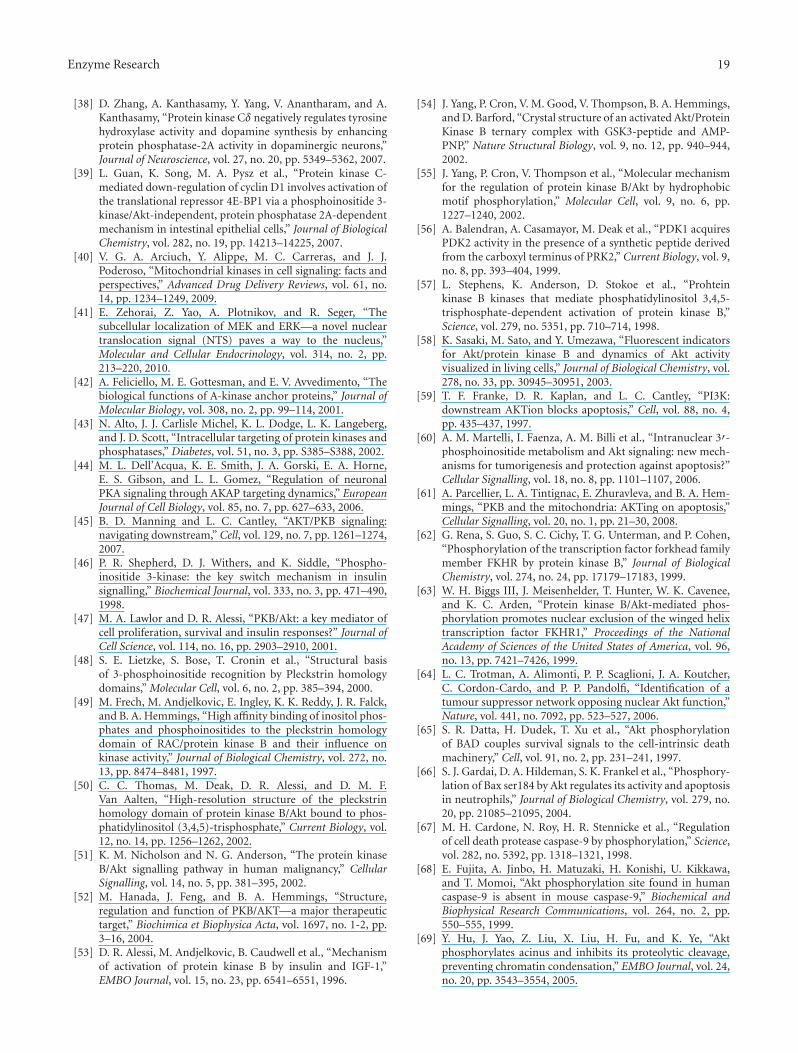
Enzyme Research 19
[38] D. Zhang, A. Kanthasamy, Y. Yang, V. Anantharam, and A.Kanthasamy, “Protein kinase Cδ negatively regulates tyrosinehydroxylase activity and dopamine synthesis by enhancingprotein phosphatase-2A activity in dopaminergic neurons,”Journal of Neuroscience, vol. 27, no. 20, pp. 5349–5362, 2007.
[39] L. Guan, K. Song, M. A. Pysz et al., “Protein kinase C-mediated down-regulation of cyclin D1 involves activation ofthe translational repressor 4E-BP1 via a phosphoinositide 3-kinase/Akt-independent, protein phosphatase 2A-dependentmechanism in intestinal epithelial cells,” Journal of BiologicalChemistry, vol. 282, no. 19, pp. 14213–14225, 2007.
[40] V. G. A. Arciuch, Y. Alippe, M. C. Carreras, and J. J.Poderoso, “Mitochondrial kinases in cell signaling: facts andperspectives,” Advanced Drug Delivery Reviews, vol. 61, no.14, pp. 1234–1249, 2009.
[41] E. Zehorai, Z. Yao, A. Plotnikov, and R. Seger, “Thesubcellular localization of MEK and ERK—a novel nucleartranslocation signal (NTS) paves a way to the nucleus,”Molecular and Cellular Endocrinology, vol. 314, no. 2, pp.213–220, 2010.
[42] A. Feliciello, M. E. Gottesman, and E. V. Avvedimento, “Thebiological functions of A-kinase anchor proteins,” Journal ofMolecular Biology, vol. 308, no. 2, pp. 99–114, 2001.
[43] N. Alto, J. J. Carlisle Michel, K. L. Dodge, L. K. Langeberg,and J. D. Scott, “Intracellular targeting of protein kinases andphosphatases,” Diabetes, vol. 51, no. 3, pp. S385–S388, 2002.
[44] M. L. Dell’Acqua, K. E. Smith, J. A. Gorski, E. A. Horne,E. S. Gibson, and L. L. Gomez, “Regulation of neuronalPKA signaling through AKAP targeting dynamics,” EuropeanJournal of Cell Biology, vol. 85, no. 7, pp. 627–633, 2006.
[45] B. D. Manning and L. C. Cantley, “AKT/PKB signaling:navigating downstream,” Cell, vol. 129, no. 7, pp. 1261–1274,2007.
[46] P. R. Shepherd, D. J. Withers, and K. Siddle, “Phospho-inositide 3-kinase: the key switch mechanism in insulinsignalling,” Biochemical Journal, vol. 333, no. 3, pp. 471–490,1998.
[47] M. A. Lawlor and D. R. Alessi, “PKB/Akt: a key mediator ofcell proliferation, survival and insulin responses?” Journal ofCell Science, vol. 114, no. 16, pp. 2903–2910, 2001.
[48] S. E. Lietzke, S. Bose, T. Cronin et al., “Structural basisof 3-phosphoinositide recognition by Pleckstrin homologydomains,” Molecular Cell, vol. 6, no. 2, pp. 385–394, 2000.
[49] M. Frech, M. Andjelkovic, E. Ingley, K. K. Reddy, J. R. Falck,and B. A. Hemmings, “High affinity binding of inositol phos-phates and phosphoinositides to the pleckstrin homologydomain of RAC/protein kinase B and their influence onkinase activity,” Journal of Biological Chemistry, vol. 272, no.13, pp. 8474–8481, 1997.
[50] C. C. Thomas, M. Deak, D. R. Alessi, and D. M. F.Van Aalten, “High-resolution structure of the pleckstrinhomology domain of protein kinase B/Akt bound to phos-phatidylinositol (3,4,5)-trisphosphate,” Current Biology, vol.12, no. 14, pp. 1256–1262, 2002.
[51] K. M. Nicholson and N. G. Anderson, “The protein kinaseB/Akt signalling pathway in human malignancy,” CellularSignalling, vol. 14, no. 5, pp. 381–395, 2002.
[52] M. Hanada, J. Feng, and B. A. Hemmings, “Structure,regulation and function of PKB/AKT—a major therapeutictarget,” Biochimica et Biophysica Acta, vol. 1697, no. 1-2, pp.3–16, 2004.
[53] D. R. Alessi, M. Andjelkovic, B. Caudwell et al., “Mechanismof activation of protein kinase B by insulin and IGF-1,”EMBO Journal, vol. 15, no. 23, pp. 6541–6551, 1996.
[54] J. Yang, P. Cron, V. M. Good, V. Thompson, B. A. Hemmings,and D. Barford, “Crystal structure of an activated Akt/ProteinKinase B ternary complex with GSK3-peptide and AMP-PNP,” Nature Structural Biology, vol. 9, no. 12, pp. 940–944,2002.
[55] J. Yang, P. Cron, V. Thompson et al., “Molecular mechanismfor the regulation of protein kinase B/Akt by hydrophobicmotif phosphorylation,” Molecular Cell, vol. 9, no. 6, pp.1227–1240, 2002.
[56] A. Balendran, A. Casamayor, M. Deak et al., “PDK1 acquiresPDK2 activity in the presence of a synthetic peptide derivedfrom the carboxyl terminus of PRK2,” Current Biology, vol. 9,no. 8, pp. 393–404, 1999.
[57] L. Stephens, K. Anderson, D. Stokoe et al., “Prohteinkinase B kinases that mediate phosphatidylinositol 3,4,5-trisphosphate-dependent activation of protein kinase B,”Science, vol. 279, no. 5351, pp. 710–714, 1998.
[58] K. Sasaki, M. Sato, and Y. Umezawa, “Fluorescent indicatorsfor Akt/protein kinase B and dynamics of Akt activityvisualized in living cells,” Journal of Biological Chemistry, vol.278, no. 33, pp. 30945–30951, 2003.
[59] T. F. Franke, D. R. Kaplan, and L. C. Cantley, “PI3K:downstream AKTion blocks apoptosis,” Cell, vol. 88, no. 4,pp. 435–437, 1997.
[60] A. M. Martelli, I. Faenza, A. M. Billi et al., “Intranuclear 3′-phosphoinositide metabolism and Akt signaling: new mech-anisms for tumorigenesis and protection against apoptosis?”Cellular Signalling, vol. 18, no. 8, pp. 1101–1107, 2006.
[61] A. Parcellier, L. A. Tintignac, E. Zhuravleva, and B. A. Hem-mings, “PKB and the mitochondria: AKTing on apoptosis,”Cellular Signalling, vol. 20, no. 1, pp. 21–30, 2008.
[62] G. Rena, S. Guo, S. C. Cichy, T. G. Unterman, and P. Cohen,“Phosphorylation of the transcription factor forkhead familymember FKHR by protein kinase B,” Journal of BiologicalChemistry, vol. 274, no. 24, pp. 17179–17183, 1999.
[63] W. H. Biggs III, J. Meisenhelder, T. Hunter, W. K. Cavenee,and K. C. Arden, “Protein kinase B/Akt-mediated phos-phorylation promotes nuclear exclusion of the winged helixtranscription factor FKHR1,” Proceedings of the NationalAcademy of Sciences of the United States of America, vol. 96,no. 13, pp. 7421–7426, 1999.
[64] L. C. Trotman, A. Alimonti, P. P. Scaglioni, J. A. Koutcher,C. Cordon-Cardo, and P. P. Pandolfi, “Identification of atumour suppressor network opposing nuclear Akt function,”Nature, vol. 441, no. 7092, pp. 523–527, 2006.
[65] S. R. Datta, H. Dudek, T. Xu et al., “Akt phosphorylationof BAD couples survival signals to the cell-intrinsic deathmachinery,” Cell, vol. 91, no. 2, pp. 231–241, 1997.
[66] S. J. Gardai, D. A. Hildeman, S. K. Frankel et al., “Phosphory-lation of Bax ser184 by Akt regulates its activity and apoptosisin neutrophils,” Journal of Biological Chemistry, vol. 279, no.20, pp. 21085–21095, 2004.
[67] M. H. Cardone, N. Roy, H. R. Stennicke et al., “Regulationof cell death protease caspase-9 by phosphorylation,” Science,vol. 282, no. 5392, pp. 1318–1321, 1998.
[68] E. Fujita, A. Jinbo, H. Matuzaki, H. Konishi, U. Kikkawa,and T. Momoi, “Akt phosphorylation site found in humancaspase-9 is absent in mouse caspase-9,” Biochemical andBiophysical Research Communications, vol. 264, no. 2, pp.550–555, 1999.
[69] Y. Hu, J. Yao, Z. Liu, X. Liu, H. Fu, and K. Ye, “Aktphosphorylates acinus and inhibits its proteolytic cleavage,preventing chromatin condensation,” EMBO Journal, vol. 24,no. 20, pp. 3543–3554, 2005.

20 Enzyme Research
[70] D. A. E. Cross, D. R. Alessi, P. Cohen, M. Andjelkovich, and B.A. Hemmings, “Inhibition of glycogen synthase kinase-3 byinsulin mediated by protein kinase B,” Nature, vol. 378, no.6559, pp. 785–789, 1995.
[71] J. E. Forde and T. C. Dale, “Glycogen synthase kinase 3: a keyregulator of cellular fate,” Cellular and Molecular Life Sciences,vol. 64, no. 15, pp. 1930–1944, 2007.
[72] G. N. Bijur and R. S. Jope, “Rapid accumulation of Aktin mitochondria following phosphatidylinositol 3-kinaseactivation,” Journal of Neurochemistry, vol. 87, no. 6, pp.1427–1435, 2003.
[73] R. Rossignol, R. Gilkerson, R. Aggeler, K. Yamagata, S. J.Remington, and R. A. Capaldi, “Energy substrate modulatesmitochondrial structure and oxidative capacity in cancercells,” Cancer Research, vol. 64, no. 3, pp. 985–993, 2004.
[74] M. Alvarez-Tejado, S. Naranjo-Suarez, C. Jimenez, A. C.Carrera, M. O. Landazuri, and L. Del Peso, “Hypoxia inducesthe activation of the phosphatidylinositol 3-kinase/Akt cellsurvival pathway in PC12 cells. Protective role in apoptosis,”Journal of Biological Chemistry, vol. 276, no. 25, pp. 22368–22374, 2001.
[75] H. Pelicano, R. H. Xu, M. Du et al., “Mitochondrialrespiration defects in cancer cells cause activation of Aktsurvival pathway through a redox-mediated mechanism,”Journal of Cell Biology, vol. 175, no. 6, pp. 913–923, 2006.
[76] K. Gottlob, N. Majewski, S. Kennedy, E. Kandel, R. B. Robey,and N. Hay, “Inhibition of early apoptotic events by Akt/PKBis dependent on the first committed step of glycolysis andmitochondrial hexokinase,” Genes and Development, vol. 15,no. 11, pp. 1406–1418, 2001.
[77] R. B. Robey and N. Hay, “Mitochondrial hexokinases, novelmediators of the antiapoptotic effects of growth factors andAkt,” Oncogene, vol. 25, no. 34, pp. 4683–4696, 2006.
[78] H. Azoulay-Zohar, A. Israelson, S. Abu-Hamad, and V.Shoshan-Barmatz, “In self-defence: hexokinase promotesvoltage-dependent anion channel closure and preventsmitochondria-mediated apoptotic cell death,” BiochemicalJournal, vol. 377, no. 2, pp. 347–355, 2004.
[79] H. Zaid, S. Abu-Hamad, A. Israelson, I. Nathan, and V.Shoshan-Barmatz, “The voltage-dependent anion channel-1modulates apoptotic cell death,” Cell Death and Differentia-tion, vol. 12, no. 7, pp. 751–760, 2005.
[80] M. T. Khan, L. Wagner, D. I. Yule, C. Bhanumathy, andS. K. Joseph, “Akt kinase phosphorylation of inositol 1,4,5-trisphosphate receptors,” Journal of Biological Chemistry, vol.281, no. 6, pp. 3731–3737, 2006.
[81] P. Pinton and R. Rizzuto, “Bcl-2 and Ca2+ homeostasis in theendoplasmic reticulum,” Cell Death and Differentiation, vol.13, no. 8, pp. 1409–1418, 2006.
[82] T. Szado, V. Vanderheyden, J. B. Parys et al., “Phosphory-lation of inositol 1,4,5-trisphosphate receptors by proteinkinase B/Akt inhibits Ca2+ release and apoptosis,” Proceed-ings of the National Academy of Sciences of the United States ofAmerica, vol. 105, no. 7, pp. 2427–2432, 2008.
[83] S. Marchi, A. Rimessi, C. Giorgi et al., “Akt kinase reducingendoplasmic reticulum Ca2+ release protects cells fromCa2+-dependent apoptotic stimuli,” Biochemical and Bio-physical Research Communications, vol. 375, no. 4, pp. 501–505, 2008.
[84] C. Giorgi, K. Ito, H. K. Lin et al., “PML regulates apoptosisat endoplasmic reticulum by modulating calcium release,”Science, vol. 330, no. 6008, pp. 1247–1251, 2010.
[85] J. E. Aslan, H. You, D. M. Williamson et al., “Akt and 14-3-3 control a PACS-2 homeostatic switch that integrates
membrane traffic with TRAIL-induced apoptosis,” MolecularCell, vol. 34, no. 4, pp. 497–509, 2009.
[86] P. H. Scott, G. J. Brunn, A. D. Kohn, R. A. Roth, and J. C.Lawrence, “Evidence of insulin-stimulated phosphorylationand activation of the mammalian target of rapamycin medi-ated by a protein kinase B signaling pathway,” Proceedingsof the National Academy of Sciences of the United States ofAmerica, vol. 95, no. 13, pp. 7772–7777, 1998.
[87] C. J. Potter, L. G. Pedraza, and T. Xu, “Akt regulates growthby directly phosphorylating Tsc2,” Nature Cell Biology, vol. 4,no. 9, pp. 658–665, 2002.
[88] B. D. Manning and L. C. Cantley, “Rheb fills a GAP betweenTSC and TOR,” Trends in Biochemical Sciences, vol. 28, no. 11,pp. 573–576, 2003.
[89] K. S. Kovacina, G. Y. Park, S. S. Bae et al., “Identificationof a proline-rich Akt substrate as a 14-3-3 binding partner,”Journal of Biological Chemistry, vol. 278, no. 12, pp. 10189–10194, 2003.
[90] E. V. Haar, S. I. Lee, S. Bandhakavi, T. J. Griffin, and D. H.Kim, “Insulin signalling to mTOR mediated by the Akt/PKBsubstrate PRAS40,” Nature Cell Biology, vol. 9, no. 3, pp. 316–323, 2007.
[91] J. E. Skeen, P. T. Bhaskar, C. C. Chen et al., “Akt deficiencyimpairs normal cell proliferation and suppresses oncogenesisin a p53-independent and mTORC1-dependent manner,”Cancer Cell, vol. 10, no. 4, pp. 269–280, 2006.
[92] N. Rosen and Q. B. She, “AKT and cancer—is it all mTOR?”Cancer Cell, vol. 10, no. 4, pp. 254–256, 2006.
[93] P. B. Dennis, A. Jaeschke, M. Saitoh, B. Fowler, S. C. Kozma,and G. Thomas, “Mammalian TOR: a homeostatic ATPsensor,” Science, vol. 294, no. 5544, pp. 1102–1105, 2001.
[94] M. Murakami, T. Ichisaka, M. Maeda et al., “mTOR is essen-tial for growth and proliferation in early mouse embryos andembryonic stem cells,” Molecular and Cellular Biology, vol. 24,no. 15, pp. 6710–6718, 2004.
[95] S. Huang and P. J. Houghton, “Targeting mTOR signaling forcancer therapy,” Current Opinion in Pharmacology, vol. 3, no.4, pp. 371–377, 2003.
[96] A. Janus, T. Robak, and P. Smolewski, “The mammaliantarget of the rapamycin (mTOR) kinase pathway: its role intumourigenesis and targeted antitumour therapy,” Cellularand Molecular Biology Letters, vol. 10, no. 3, pp. 479–498,2005.
[97] Q. Yang and K. L. Guan, “Expanding mTOR signaling,” CellResearch, vol. 17, no. 8, pp. 666–681, 2007.
[98] C. Cutler, J. H. Antin, and F. J. Giles, “Mammalian target ofrapamycin inhibition as therapy for hematologic malignan-cies,” Cancer, vol. 101, no. 6, p. 1478, 2004.
[99] S. Sengupta, T. R. Peterson, and D. M. Sabatini, “Regulationof the mTOR complex 1 pathway by nutrients, growthfactors, and stress,” Molecular Cell, vol. 40, no. 2, pp. 310–322, 2010.
[100] M. Rosner, M. Hanneder, N. Siegel, A. Valli, C. Fuchs, and M.Hengstschlager, “The mTOR pathway and its role in humangenetic diseases,” Mutation Research, vol. 659, no. 3, pp. 284–292, 2008.
[101] L. R. Pearce, X. Huang, J. Boudeau et al., “Identificationof Protor as a novel Rictor-binding component of mTORcomplex-2,” Biochemical Journal, vol. 405, no. 3, pp. 513–522,2007.
[102] J. M. Garcıa-Martınez and D. R. Alessi, “mTOR complex2 (mTORC2) controls hydrophobic motif phosphorylationand activation of serum- and glucocorticoid-inducedprotein

Enzyme Research 21
kinase 1 (SGK1),” Biochemical Journal, vol. 416, no. 3, pp.375–385, 2008.
[103] D. A. Guertin, D. M. Stevens, C. C. Thoreen et al., “Ablationin mice of the mTORC components raptor, rictor, or mLST8reveals that mTORC2 is required for signaling to Akt-FOXOand PKCalpha, but not S6K1,” Developmental Cell, vol. 11,no. 6, pp. 859–871, 2006.
[104] A. J. Shatkin, “mRNA cap binding proteins: essential factorsfor initiating translation,” Cell, vol. 40, no. 2, pp. 223–224,1985.
[105] A. Pause, G. J. Belsham, A. C. Gingras et al., “Insulin-dependent stimulation of protein synthesis by phosphoryla-tion of a regulator of 5’-cap function,” Nature, vol. 371, no.6500, pp. 762–767, 1994.
[106] L. Beretta, A. C. Gingras, Y. V. Svitkin, M. N. Hall, and N.Sonenberg, “Rapamycin blocks the phosphorylation of 4E-BP1 and inhibits cap-dependent initiation of translation,”EMBO Journal, vol. 15, no. 3, pp. 658–664, 1996.
[107] D. J. Price, J. R. Grove, V. Calvo, J. Avruch, and B. E.Bierer, “Rapamycin-induced inhibition of the 70-kilodaltonS6 protein kinase,” Science, vol. 257, no. 5072, pp. 973–977,1992.
[108] C. K. Tsang, H. Liu, and X. F. S. Zheng, “mTOR binds to thepromoters of RNA polymerase I- and III-transcribed genes,”Cell Cycle, vol. 9, no. 5, pp. 953–957, 2010.
[109] K. M. Hannan, Y. Brandenburger, A. Jenkins et al.,“mTOR-dependent regulation of ribosomal gene transcrip-tion requires S6K1 and is mediated by phosphorylation ofthe carboxy-terminal activation domain of the nucleolartranscription factor UBF,” Molecular and Cellular Biology, vol.23, no. 23, pp. 8862–8877, 2003.
[110] C. Mayer and I. Grummt, “Ribosome biogenesis and cellgrowth: mTOR coordinates transcription by all three classesof nuclear RNA polymerases,” Oncogene, vol. 25, no. 48, pp.6384–6391, 2006.
[111] C. Mayer, J. Zhao, X. Yuan, and I. Grummt, “mTOR-dependent activation of the transcription factor TIF-IAlinks rRNA synthesis to nutrient availability,” Genes andDevelopment, vol. 18, no. 4, pp. 423–434, 2004.
[112] M. Matsuo, S. Yamada, K. Koizumi, H. Sakurai, and I.Saiki, “Tumour-derived fibroblast growth factor-2 exertslymphangiogenic effects through Akt/mTOR/p70S6kinasepathway in rat lymphatic endothelial cells,” European Journalof Cancer, vol. 43, no. 11, pp. 1748–1754, 2007.
[113] L. Ma, Z. Chen, H. Erdjument-Bromage, P. Tempst, and P.P. Pandolfi, “Phosphorylation and functional inactivation ofTSC2 by Erk: implications for tuberous sclerosis and cancerpathogenesis,” Cell, vol. 121, no. 2, pp. 179–193, 2005.
[114] B. G. Wouters and M. Koritzinsky, “Hypoxia signallingthrough mTOR and the unfolded protein response in cancer,”Nature Reviews Cancer, vol. 8, no. 11, pp. 851–864, 2008.
[115] G. L. Semenza, “Hif-1 and human disease: one highlyinvolved factor,” Genes and Development, vol. 14, no. 16, pp.1983–1991, 2000.
[116] K. Duvel, J. L. Yecies, S. Menon et al., “Activation of ametabolic gene regulatory network downstream of mTORcomplex 1,” Molecular Cell, vol. 39, no. 2, pp. 171–183, 2010.
[117] E. Laughner, P. Taghavi, K. Chiles, P. C. Mahon, andG. L. Semenza, “HER2 (neu) signaling increases the rateof hypoxia-inducible factor 1α (HIF-1α) synthesis: novelmechanism for HIF-1-mediated vascular endothelial growthfactor expression,” Molecular and Cellular Biology, vol. 21, no.12, pp. 3995–4004, 2001.
[118] G. V. Thomas, C. Tran, I. K. Mellinghoff et al., “Hypoxia-inducible factor determines sensitivity to inhibitors of mTORin kidney cancer,” Nature Medicine, vol. 12, no. 1, pp. 122–127, 2006.
[119] A. Sofer, K. Lei, C. M. Johannessen, and L. W. Ellisen,“Regulation of mTOR and cell growth in response to energystress by REDD1,” Molecular and Cellular Biology, vol. 25, no.14, pp. 5834–5845, 2005.
[120] B. Ravikumar, S. Sarkar, J. E. Davies et al., “Regulation ofmammalian autophagy in physiology and pathophysiology,”Physiological Reviews, vol. 90, no. 4, pp. 1383–1435, 2010.
[121] N. Mizushima, “The role of the Atg1/ULK1 complex inautophagy regulation,” Current Opinion in Cell Biology, vol.22, no. 2, pp. 132–139, 2010.
[122] B. N. Desai, B. R. Myers, and S. L. Schreiber, “FKBP12-rapamycin-associated protein associates with mitochondriaand senses osmotic stress via mitochondrial dysfunction,”Proceedings of the National Academy of Sciences of the UnitedStates of America, vol. 99, no. 7, pp. 4319–4324, 2002.
[123] J. E. Kim and J. Chen, “Cytoplasmic-nuclear shuttlingof FKBP12-rapamycin-associated protein is involved inrapamycin-sensitive signaling and translation initiation,”Proceedings of the National Academy of Sciences of the UnitedStates of America, vol. 97, no. 26, pp. 14340–14345, 2000.
[124] X. Zhang, L. Shu, H. Hosoi, K. Gopal Murti, and P.J. Houghton, “Predominant nuclear localization of mam-malian target of rapamycin in normal and malignant cells inculture,” Journal of Biological Chemistry, vol. 277, no. 31, pp.28127–28134, 2002.
[125] R. M. Drenan, X. Liu, P. G. Bertram, and X. F. S. Zheng,“FKBP12-rapamycin-associated protein or mammalian tar-get of rapamycin (FRAP/mTOR) localization in the endo-plasmic reticulum and the Golgi apparatus,” Journal ofBiological Chemistry, vol. 279, no. 1, pp. 772–778, 2004.
[126] X. Liu and X. F. S. Zheng, “Endoplasmic reticulum and Golgilocalization sequences for mammalian target of rapamycin,”Molecular Biology of the Cell, vol. 18, no. 3, pp. 1073–1082,2007.
[127] D. Wlodkowic, J. Skommer, D. McGuinness, C. Hillier, and Z.Darzynkiewicz, “ER-Golgi network—a future target for anti-cancer therapy,” Leukemia Research, vol. 33, no. 11, pp. 1440–1447, 2009.
[128] Y. Sancak, T. R. Peterson, Y. D. Shaul et al., “The rag GTPasesbind raptor and mediate amino acid signaling to mTORC1,”Science, vol. 320, no. 5882, pp. 1496–1501, 2008.
[129] Y. Sancak, L. Bar-Peled, R. Zoncu, A. L. Markhard, S.Nada, and D. M. Sabatini, “Ragulator-rag complex targetsmTORC1 to the lysosomal surface and is necessary for itsactivation by amino acids,” Cell, vol. 141, no. 2, pp. 290–303,2010.
[130] S. M. Schieke, D. Phillips, J. P. McCoy Jr. et al., “Themammalian target of rapamycin (mTOR) pathway regulatesmitochondrial oxygen consumption and oxidative capacity,”Journal of Biological Chemistry, vol. 281, no. 37, pp. 27643–27652, 2006.
[131] J. T. Cunningham, J. T. Rodgers, D. H. Arlow, F. Vazquez, V.K. Mootha, and P. Puigserver, “mTOR controls mitochon-drial oxidative function through a YY1-PGC-1α transcrip-tional complex,” Nature, vol. 450, no. 7170, pp. 736–740,2007.
[132] X. Bai, D. Ma, A. Liu et al., “Rheb activates mTOR byantagonizing its endogenous inhibitor, FKBP38,” Science, vol.318, no. 5852, pp. 977–980, 2007.

22 Enzyme Research
[133] A. Ramanathan and S. L. Schreiber, “Direct control of mito-chondrial function by mTOR,” Proceedings of the NationalAcademy of Sciences of the United States of America, vol. 106,no. 52, pp. 22229–22232, 2009.
[134] C. Vezina, A. Kudelski, and S. N. Sehgal, “Rapamycin(AY 22,989), a new antifungal antibiotic. I. Taxonomy ofthe producing streptomycete and isolation of the activeprinciple,” Journal of Antibiotics, vol. 28, no. 10, pp. 721–726,1975.
[135] S. H. Um, F. Frigerio, M. Watanabe et al., “Absence ofS6K1 protects against age- and diet-induced obesity whileenhancing insulin sensitivity,” Nature, vol. 431, no. 7005, pp.200–205, 2004.
[136] M. E. Feldman, B. Apsel, A. Uotila et al., “Active-siteinhibitors of mTOR target rapamycin-resistant outputs ofmTORC1 and mTORC2,” PLoS Biology, vol. 7, no. 2, ArticleID e1000038, 2009.
[137] K. Lechward, O. S. Awotunde, W. Swiatek, and G. Muszynska,“Protein phosphatase 2A: variety of forms and diversity offunctions,” Acta Biochimica Polonica, vol. 48, no. 4, pp. 921–933, 2001.
[138] C. Van Hoof and J. Goris, “Phosphatases in apoptosis: to beor not to be, PP2A is in the heart of the question,” Biochimicaet Biophysica Acta, vol. 1640, no. 2-3, pp. 97–104, 2003.
[139] J. Yang and C. Phiel, “Functions of B56-containing PP2Asin major developmental and cancer signaling pathways,” LifeSciences, vol. 87, no. 23-26, pp. 659–666, 2010.
[140] Y. Shi, “Serine/threonine phosphatases: mechanism throughstructure,” Cell, vol. 139, no. 3, pp. 468–484, 2009.
[141] V. Janssens, S. Longin, and J. Goris, “PP2A holoenzymeassembly: in cauda venenum (the sting is in the tail),” Trendsin Biochemical Sciences, vol. 33, no. 3, pp. 113–121, 2008.
[142] C. M. Forester, J. Maddox, J. V. Louis, J. Goris, and D. M.Virshup, “Control of mitotic exit by PP2A regulation ofCdc25C and Cdk1,” Proceedings of the National Academy ofSciences of the United States of America, vol. 104, no. 50, pp.19867–19872, 2007.
[143] C. P. Flegg, M. Sharma, C. Medina-Palazon et al., “Nuclearexport and centrosome targeting of the protein phosphatase2A subunit B56α: role of B56α in nuclear export of thecatalytic subunit,” Journal of Biological Chemistry, vol. 285,no. 24, pp. 18144–18154, 2010.
[144] Z. Jin, J. Shi, A. Saraf et al., “The 48-kDa alternative trans-lation isoform of PP2A:B56ε is required for Wnt signalingduring midbrain-hindbrain boundary formation,” Journal ofBiological Chemistry, vol. 284, no. 11, pp. 7190–7200, 2009.
[145] C. G. Riedel, V. L. Katis, Y. Katou et al., “Protein phosphatase2A protects centromeric sister chromatid cohesion duringmeiosis I,” Nature, vol. 441, no. 1, pp. 53–61, 2006.
[146] Z. Xu, B. Cetin, M. Anger et al., “Structure and function ofthe PP2A-shugoshin interaction,” Molecular Cell, vol. 35, no.4, pp. 426–441, 2009.
[147] A. Ito, T. R. Kataoka, M. Watanabe et al., “A truncatedisoform of the PP2A B56 subunit promotes cell motilitythrough paxillin phosphorylation,” EMBO Journal, vol. 19,no. 4, pp. 562–571, 2000.
[148] P. P. Ruvolo, X. Deng, T. Ito, B. K. Carr, and W. S. May,“Ceramide induces Bcl2 dephosphorylation via a mechanisminvolving mitochondrial PP2A,” Journal of Biological Chem-istry, vol. 274, no. 29, pp. 20296–20300, 1999.
[149] P. P. Ruvolo, W. Clark, M. Mumby, F. Gao, and W. StratfordMay, “A functional role for the B56 α-subunit of proteinphosphatase 2A in ceramide-mediated regulation of Bcl2
phosphorylation status and function,” Journal of BiologicalChemistry, vol. 277, no. 25, pp. 22847–22852, 2002.
[150] D. Perrotti and P. Neviani, “Protein phosphatase 2A (PP2A),a drugable tumor suppressor in Ph1(+) leukemias,” Cancerand Metastasis Reviews, vol. 27, no. 2, pp. 159–168, 2008.
[151] V. Janssens and J. Goris, “Protein phosphatase 2A: a highlyregulated family of serine/threonine phosphatases implicatedin cell growth and signalling,” Biochemical Journal, vol. 353,no. 3, pp. 417–439, 2001.
[152] J. Chen, B. L. Martin, and D. L. Brautigan, “Regulation ofprotein serine-threonine phosphatase type-2A by tyrosinephosphorylation,” Science, vol. 257, no. 5074, pp. 1261–1264,1992.
[153] J. Westermarck and W. C. Hahn, “Multiple pathways reg-ulated by the tumor suppressor PP2A in transformation,”Trends in Molecular Medicine, vol. 14, no. 4, pp. 152–160,2008.
[154] E. Sontag, “Protein phosphatase 2A: the Trojan Horse ofcellular signaling,” Cellular Signalling, vol. 13, no. 1, pp. 7–16, 2001.
[155] Y. Liao and M.-C. Hung, “Physiological regulation of Aktactivity and stability,” American Journal of TranslationalResearch, vol. 2, no. 1, pp. 19–42, 2010.
[156] S. Mochida, S. L. Maslen, M. Skehel, and T. Hunt, “Greatwallphosphorylates an inhibitor of protein phosphatase 2A thatis essential for mitosis,” Science, vol. 330, no. 6011, pp. 1670–1673, 2010.
[157] S. O. Marx, S. Reiken, Y. Hisamatsu et al., “PKA phosphory-lation dissociates FKBP12.6 from the calcium release channel(ryanodine receptor): defective regulation in failing hearts,”Cell, vol. 101, no. 4, pp. 365–376, 2000.
[158] T. Y. Lee, T. Y. Lai, S. C. Lin et al., “The B56γ3 regulatorysubunit of protein phosphatase 2A (PP2A) regulates S phase-specific nuclear accumulation of PP2A and the G1 to Stransition,” Journal of Biological Chemistry, vol. 285, no. 28,pp. 21567–21580, 2010.
[159] E. Manchado, M. Guillamot, G. de Carcer et al., “Targetingmitotic exit leads to tumor regression in vivo: modulationby Cdk1, Mastl, and the PP2A/B55α,δ phosphatase,” CancerCell, vol. 18, no. 6, pp. 641–654, 2010.
[160] T. A. Potapova, S. Sivakumar, J. N. Flynn, R. Li, and G.J. Gorbsky, “Mitotic progression becomes irreversible inprometaphase and collapses when Wee1 and Cdc25 areinhibited,” Molecular Biology of the Cell, vol. 22, no. 8, pp.1191–1206, 2011.
[161] A. Gharbi-Ayachi, J. C. Labbe, A. Burgess et al., “Thesubstrate of Greatwall kinase, Arpp19, controls mitosis byinhibiting protein phosphatase 2A,” Science, vol. 330, no.6011, pp. 1673–1677, 2010.
[162] S. Aggarwal, T. Suzuki, W. L. Taylor, A. Bhargava, andR. K. Rao, “Contrasting effects of ERK on tight junctionintegrity in differentiated and under-differentiated Caco-2cell monolayers,” Biochemical Journal, vol. 433, no. 1, pp. 51–63, 2011.
[163] Y. Li, P. Yue, X. Deng et al., “Protein phosphatase 2A nega-tively regulates eukaryotic initiation factor 4E phosphoryla-tion and eif4F assembly through direct dephosphorylation ofmnk and eif4E,” Neoplasia, vol. 12, no. 10, pp. 848–855, 2010.
[164] K. Okamoto, H. Li, M. R. Jensen et al., “Cyclin G recruitsPP2A to dephosphorylate Mdm2,” Molecular Cell, vol. 9, no.4, pp. 761–771, 2002.
[165] A. E. Kray, R. S. Carter, K. N. Pennington et al., “Positiveregulation of IκB kinase signaling by protein serine/threonine

Enzyme Research 23
phosphatase 2A,” Journal of Biological Chemistry, vol. 280, no.43, pp. 35974–35982, 2005.
[166] A. A. Sablina and W. C. Hahn, “The role of PP2A A subunitsin tumor suppression,” Cell adhesion & migration, vol. 1, no.3, pp. 140–141, 2007.
[167] J. Li, C. Yen, D. Liaw et al., “PTEN, a putative proteintyrosine phosphatase gene mutated in human brain, breast,and prostate cancer,” Science, vol. 275, no. 5308, pp. 1943–1947, 1997.
[168] P. A. Steck, M. A. Pershouse, S. A. Jasser et al., “Identificationof a candidate tumour suppressor gene, MMAC1, at chromo-some 10q23.3 that is mutated in multiple advanced cancers,”Nature Genetics, vol. 15, no. 4, pp. 356–362, 1997.
[169] D. M. Li and H. Sun, “TEP1, encoded by a candidate tumorsuppressor locus, is a novel protein tyrosine phosphataseregulated by transforming growth factor β,” Cancer Research,vol. 57, no. 11, pp. 2124–2129, 1997.
[170] L. Simpson and R. Parsons, “PTEN: life as a tumor suppres-sor,” Experimental Cell Research, vol. 264, no. 1, pp. 29–41,2001.
[171] M. Keniry and R. Parsons, “The role of PTEN signalingperturbations in cancer and in targeted therapy,” Oncogene,vol. 27, no. 41, pp. 5477–5485, 2008.
[172] J. A. Hobert and C. Eng, “PTEN hamartoma tumor syn-drome: an overview,” Genetics in Medicine, vol. 11, no. 10,pp. 687–694, 2009.
[173] M. G. Butler, M. J. Dazouki, X. P. Zhou et al., “Subsetof individuals with autism spectrum disorders and extrememacrocephaly associated with germline PTEN tumour sup-pressor gene mutations,” Journal of Medical Genetics, vol. 42,no. 4, pp. 318–321, 2005.
[174] C. Nardella, A. Carracedo, L. Salmena, and P. P. Pandolfi,“Faithfull modeling of PTEN loss driven diseases in themouse,” Current topics in Microbiology and Immunology, vol.347, pp. 135–168, 2010.
[175] A. Carracedo, A. Alimonti, and P. P. Pandolfi, “PTEN level intumor suppression: how much is too little?” Cancer Research,vol. 71, no. 3, pp. 629–633, 2011.
[176] A. Di Cristofano, B. Pesce, C. Cordon-Cardo, and P. P.Pandolfi, “Pten is essential for embryonic development andtumour suppression,” Nature Genetics, vol. 19, no. 4, pp. 348–355, 1998.
[177] A. Suzuki, J. L. De La Pompa, V. Stambolic et al., “Highcancer susceptibility and embryonic lethality associated withmutation of the PTEN tumor suppressor gene in mice,”Current Biology, vol. 8, no. 21, pp. 1169–1178, 1998.
[178] K. Podsypanina, L. H. Ellenson, A. Nemes et al., “Mutationof Pten/Mmac1 in mice causes neoplasia in multiple organsystems,” Proceedings of the National Academy of Sciences ofthe United States of America, vol. 96, no. 4, pp. 1563–1568,1999.
[179] L. C. Trotman, M. Niki, Z. A. Dotan et al., “Pten dose dictatescancer progression in the prostate,” PLoS Biology, vol. 1, no.3, article E59, 2003.
[180] S. Wang, J. Gao, Q. Lei et al., “Prostate-specific deletion ofthe murine Pten tumor suppressor gene leads to metastaticprostate cancer,” Cancer Cell, vol. 4, no. 3, pp. 209–221, 2003.
[181] T. Maehama and J. E. Dixon, “The tumor suppressor,PTEN/MMAC1, dephosphorylates the lipid second mes-senger, phosphatidylinositol 3,4,5-trisphosphate,” Journal ofBiological Chemistry, vol. 273, no. 22, pp. 13375–13378, 1998.
[182] M. P. Myers, I. Pass, I. H. Batty et al., “The lipid phosphataseactivity of PTEN is critical for its tumor supressor function,”
Proceedings of the National Academy of Sciences of the UnitedStates of America, vol. 95, no. 23, pp. 13513–13518, 1998.
[183] Y. Liu and V. A. Bankaitis, “Phosphoinositide phosphatasesin cell biology and disease,” Progress in Lipid Research, vol.49, no. 3, pp. 201–217, 2010.
[184] N. R. Leslie, H. Maccario, L. Spinelli, and L. Davidson,“The significance of PTEN’s protein phosphatase activity,”Advances in Enzyme Regulation, vol. 49, no. 1, pp. 190–196,2009.
[185] M. P. Myers, J. P. Stolarov, C. Eng et al., “P-TEN, the tumorsuppressor from human chromosome 10q23, is a dual-specificity phosphatase,” Proceedings of the National Academyof Sciences of the United States of America, vol. 94, no. 17, pp.9052–9057, 1997.
[186] M. Tamura, J. Gu, K. Matsumoto, S. I. Aota, R. Parsons, andK. M. Yamada, “Inhibition of cell migration, spreading, andfocal adhesions by tumor suppressor PTEN,” Science, vol.280, no. 5369, pp. 1614–1617, 1998.
[187] N. Dey, H. E. Crosswell, D. Pradip et al., “The proteinphosphatase activity of PTEN regulates Src family kinasesand controls glioma migration,” Cancer Research, vol. 68, no.6, pp. 1862–1871, 2008.
[188] N. R. Leslie, X. Yang, C. P. Downes, and C. J. Weijer, “Theregulation of cell migration by PTEN,” Biochemical SocietyTransactions, vol. 33, no. 6, pp. 1507–1508, 2005.
[189] M. Raftopoulou, S. Etienne-Manneville, A. Self, S. Nicholls,and A. Hall, “Regulation of cell migration by the C2 domainof the tumor suppressor PTEN,” Science, vol. 303, no. 5661,pp. 1179–1181, 2004.
[190] L. Mahimainathan and G. Ghosh Choudhury, “Inactivationof platelet-derived growth factor receptor by the tumorsuppressor PTEN provides a novel mechanism of action ofthe phosphatase,” Journal of Biological Chemistry, vol. 279,no. 15, pp. 15258–15268, 2004.
[191] L. P. Weng, J. L. Brown, K. M. Baker, M. C. Ostrowski, and C.Eng, “PTEN blocks insulin-mediated ETS-2 phosphorylationthrough MAP kinase, independently of the phosphoinositide3-kinase pathway,” Human Molecular Genetics, vol. 11, no. 15,pp. 1687–1696, 2002.
[192] L. P. Weng, J. L. Brown, and C. Eng, “PTEN coordinatesG1 arrest by down-regulating cyclin D1 via its proteinphosphatase activity and up-regulating p27 via its lipidphosphatase acitvity in a breast cancer model,” HumanMolecular Genetics, vol. 10, no. 6, pp. 599–604, 2001.
[193] V. Stambolic, A. Suzuki, J. L. De la Pompa et al., “Negativeregulation of PKB/Akt-dependent cell survival by the tumorsuppressor PTEN,” Cell, vol. 95, no. 1, pp. 29–39, 1998.
[194] D. Haas-Kogan, N. Shalev, M. Wong, G. Mills, G. Yount, andD. Stokoe, “Protein kinase B (PKB/Akt) activity is elevated inglioblastoma cells due to mutation of the tumor suppressorPTEN/MMAC,” Current Biology, vol. 8, no. 21, pp. 1195–1198, 1998.
[195] L. Poliseno, L. Salmena, J. Zhang, B. Carver, W. J. Haveman,and P. P. Pandolfi, “A coding-independent function of geneand pseudogene mRNAs regulates tumour biology,” Nature,vol. 465, no. 7301, pp. 1033–1038, 2010.
[196] L. Salmena, A. Carracedo, and P. P. Pandolfi, “Tenets of PTENTumor Suppression,” Cell, vol. 133, no. 3, pp. 403–414, 2008.
[197] N. R. Leslie and M. Foti, “Non-genomic loss of PTEN func-tion in cancer: not in my genes,” Trends in PharmacologicalSciences, vol. 32, no. 3, pp. 131–140, 2011.
[198] T. Tamguney and D. Stokoe, “New insights into PTEN,”Journal of Cell Science, vol. 120, no. 23, pp. 4071–4079, 2007.

24 Enzyme Research
[199] C. Eng, “PTEN: one gene, Many syndromes,” HumanMutation, vol. 22, no. 3, pp. 183–198, 2003.
[200] J. O. Lee, H. Yang, M. M. Georgescu et al., “Crystalstructure of the PTEN tumor suppressor: implications forits phosphoinositide phosphatase activity and membraneassociation,” Cell, vol. 99, no. 3, pp. 323–334, 1999.
[201] A. Gericke, M. Munson, and A. H. Ross, “Regulation of thePTEN phosphatase,” Gene, vol. 374, no. 1-2, pp. 1–9, 2006.
[202] M. M. Georgescu, K. H. Kirsch, P. Kaloudis, H. Yang, N.P. Pavletich, and H. Hanafusa, “Stabilization and productivepositioning roles of the C2 domain of PTEN tumor suppres-sor,” Cancer Research, vol. 60, no. 24, pp. 7033–7038, 2000.
[203] F. Vazquez and P. Devreotes, “Regulation of PTEN functionas a PIP3 gatekeeper through membrane interaction,” CellCycle, vol. 5, no. 14, pp. 1523–1527, 2006.
[204] J. Torres and R. Pulido, “The tumor suppressor PTEN isphosphorylated by the protein kinase CK2 at its C terminus.Implications for PTEN stability to proteasome-mediateddegradation,” Journal of Biological Chemistry, vol. 276, no. 2,pp. 993–998, 2001.
[205] H. Maccario, N. M. Perera, L. Davidson, C. P. Downes, andN. R. Leslie, “PTEN is destabilized by phosphorylation onThr366,” Biochemical Journal, vol. 405, no. 3, pp. 439–444,2007.
[206] F. Okahara, H. Ikawa, Y. Kanaho, and T. Maehama, “Regula-tion of PTEN phosphorylation and stability by a tumor sup-pressor candidate protein,” Journal of Biological Chemistry,vol. 279, no. 44, pp. 45300–45303, 2004.
[207] S. Vemula, J. Shi, P. Hanneman, L. Wei, and R. Kapur,“ROCK1 functions as a suppressor of inflammatory cellmigration by regulating PTEN phosphorylation and stabil-ity,” Blood, vol. 115, no. 9, pp. 1785–1796, 2010.
[208] F. Vazquez, S. Ramaswamy, N. Nakamura, and W. R. Sellers,“Phosphorylation of the PTEN tail regulates protein stabilityand function,” Molecular and Cellular Biology, vol. 20, no. 14,pp. 5010–5018, 2000.
[209] F. Vazquez, S. R. Grossman, Y. Takahashi, M. V. Rokas, N.Nakamura, and W. R. Sellers, “Phosphorylation of the PTENtail acts as an inhibitory switch by preventing its recruitmentinto a protein complex,” Journal of Biological Chemistry, vol.276, no. 52, pp. 48627–48630, 2001.
[210] S. M. Walker, N. R. Leslie, N. M. Perera, I. H. Batty, andC. P. Downes, “The tumour-suppressor function of PTENrequires an N-terminal lipid-binding motif,” BiochemicalJournal, vol. 379, no. 2, pp. 301–307, 2004.
[211] R. E. Redfern, D. Redfern, M. L. M. Furgason, M. Munson,A. H. Ross, and A. Gericke, “PTEN phosphatase selectivelybinds phosphoinositides and undergoes structural changes,”Biochemistry, vol. 47, no. 7, pp. 2162–2171, 2008.
[212] F. Vazquez, S. Matsuoka, W. R. Sellers, T. Yanagida, M.Ueda, and P. N. Devreotes, “Tumor suppressor PTEN actsthrough dynamic interaction with the plasma membrane,”Proceedings of the National Academy of Sciences of the UnitedStates of America, vol. 103, no. 10, pp. 3633–3638, 2006.
[213] M. Iijima, Y. E. Huang, H. R. Luo, F. Vazquez, and P.N. Devreotes, “Novel mechanism of PTEN regulation byits phosphatidylinositol 4,5-bisphosphate binding motif iscritical for chemotaxis,” Journal of Biological Chemistry, vol.279, no. 16, pp. 16606–16613, 2004.
[214] N. R. Leslie, I. H. Batty, H. Maccario, L. Davidson, and C.P. Downes, “Understanding PTEN regulation: PIP2, polarityand protein stability,” Oncogene, vol. 27, no. 41, pp. 5464–5476, 2008.
[215] Y. Lindsay, D. McCoull, L. Davidson et al., “Localization ofagonist-sensitive PtdIns(3,4,5)P3 reveals a nuclear pool thatis insensitive to PTEN expression,” Journal of Cell Science, vol.119, no. 24, pp. 5160–5168, 2006.
[216] M. Sato, Y. Ueda, T. Takagi, and Y. Umezawa, “Productionof PtdInsP3 at endomembranes is triggered by receptorendocytosis,” Nature Cell Biology, vol. 5, no. 11, pp. 1016–1022, 2003.
[217] G. Mazzotti, N. Zini, E. Rizzi et al., “Immunocytochemicaldetection of phosphatidylinositol 4,5-bisphosphate localiza-tion sites within the nucleus,” Journal of Histochemistry andCytochemistry, vol. 43, no. 2, pp. 181–191, 1995.
[218] I. V. Boronenkov, J. C. Loijens, M. Umeda, and R. A.Anderson, “Phosphoinositide signaling pathways in nucleiare associated with nuclear speckles containing Pre-mRNAprocessing factors,” Molecular Biology of the Cell, vol. 9, no.12, pp. 3547–3560, 1998.
[219] Z. Lian and A. Di Cristofano, “Class reunion: PTEN joins thenuclear crew,” Oncogene, vol. 24, no. 50, pp. 7394–7400, 2005.
[220] O. Gimm, A. Perren, L. P. Weng et al., “Differential nuclearand cytoplasmic expression of PTEN in normal thyroidtissue, and benign and malignant epithelial thyroid tumors,”American Journal of Pathology, vol. 156, no. 5, pp. 1693–1700,2000.
[221] A. Perren, P. Komminoth, P. Saremaslani et al., “Muta-tion and expression analyses reveal differential subcellularcompartmentalization of PTEN in endocrine pancreatictumors compared to normal islet cells,” American Journal ofPathology, vol. 157, no. 4, pp. 1097–1103, 2000.
[222] X. P. Zhou, A. Loukola, R. Salovaara et al., “PTEN mutationalspectra, expression levels, and subcellular localization inmicrosatellite stable and unstable colorectal cancers,” Ameri-can Journal of Pathology, vol. 161, no. 2, pp. 439–447, 2002.
[223] X. Wang, L. C. Trotman, T. Koppie et al., “NEDD4-1 is aproto-oncogenic ubiquitin ligase for PTEN,” Cell, vol. 128,no. 1, pp. 129–139, 2007.
[224] L. C. Trotman, X. Wang, A. Alimonti et al., “Ubiquitinationregulates PTEN nuclear import and tumor suppression,” Cell,vol. 128, no. 1, pp. 141–156, 2007.
[225] M. S. Song, L. Salmena, A. Carracedo et al., “The deubiq-uitinylation and localization of PTEN are regulated by aHAUSP-PML network,” Nature, vol. 455, no. 7214, pp. 813–817, 2008.
[226] W. H. Shen, A. S. Balajee, J. Wang et al., “Essential role fornuclear PTEN in maintaining chromosomal integrity,” Cell,vol. 128, no. 1, pp. 157–170, 2007.
[227] S. M. Planchon, K. A. Waite, and C. Eng, “The nuclear affairsof PTEN,” Journal of Cell Science, vol. 121, no. 3, pp. 249–253,2008.
[228] J. Y. Ahn, R. Rong, X. Liu, and K. Ye, “PIKE/nuclear PI 3-kinase signaling mediates the antiapoptotic actions of NGF inthe nucleus,” EMBO Journal, vol. 23, no. 20, pp. 3995–4006,2004.
[229] M. S. Song, A. Carracedo, L. Salmena et al., “Nuclear PTENregulates the APC-CDH1 tumor-suppressive complex in aphosphatase-independent manner,” Cell, vol. 144, no. 2, pp.187–199, 2011.
[230] C. L. Bonifant, J. S. Kim, and T. Waldman, “NHERFs,NEP, MAGUKs, and more: interactions that regulate PTEN,”Journal of Cellular Biochemistry, vol. 102, no. 4, pp. 878–885,2007.
[231] T. Tolkacheva, M. Boddapati, A. Sanfiz, K. Tsuchida, A.C. Kimmelman, and A. M. L. Chan, “Regulation of PTEN

Enzyme Research 25
binding to MAGI-2 by two putative phosphorylation sites atthreonine 382 and 383,” Cancer Research, vol. 61, no. 13, pp.4985–4989, 2001.
[232] W. von Stein, A. Ramrath, A. Grimm, M. Muller-Borg, and A.Wodarz, “Direct association of Bazooka/PAR-3 with the lipidphosphatase PTEN reveals a link between the PAR/aPKCcomplex and phosphoinositide signaling,” Development, vol.132, no. 7, pp. 1675–1686, 2005.
[233] M. Valiente, A. Andres-Pons, B. Gomar et al., “Binding ofPTEN to specific PDZ domains contributes to PTEN proteinstability and phosphorylation by microtubule-associatedserine/threonine kinases,” Journal of Biological Chemistry,vol. 280, no. 32, pp. 28936–28943, 2005.
[234] Y. Takahashi, F. C. Morales, E. L. Kreimann, and M. M.Georgescu, “PTEN tumor suppressor associates with NHERFproteins to attenuate PDGF receptor signaling,” EMBOJournal, vol. 25, no. 4, pp. 910–920, 2006.
[235] D. K. Crockett, G. C. Fillmore, K. S. J. Elenitoba-Johnson,and M. S. Lim, “Analysis of phosphatase and tensin homologtumor suppressor interacting proteins by in vitro and in silicoproteomics,” Proteomics, vol. 5, no. 5, pp. 1250–1262, 2005.
[236] X. Wang and X. Jiang, “Post-translational regulation ofPTEN,” Oncogene, vol. 27, no. 41, pp. 5454–5463, 2008.
[237] F. Liu, S. Wagner, R. B. Campbell, J. A. Nickerson, C. A.Schiffer, and A. H. Ross, “PTEN enters the nucleus bydiffusion,” Journal of Cellular Biochemistry, vol. 96, no. 2, pp.221–234, 2005.
[238] Y. Zhu, P. Hoell, B. Ahlemeyer, and J. Krieglstein, “PTEN:a crucial mediator of mitochondria-dependent apoptosis,”Apoptosis, vol. 11, no. 2, pp. 197–207, 2006.
[239] E. C. Dempsey, A. C. Newton, D. Mochly-Rosen et al.,“Protein kinase C isozymes and the regulation of diverse cellresponses,” American Journal of Physiology, vol. 279, no. 3, pp.L429–L438, 2000.
[240] S. F. Steinberg, “Structural basis of protein kinase C isoformfunction,” Physiological Reviews, vol. 88, no. 4, pp. 1341–1378, 2008.
[241] F. Battaini and A. Pascale, “Protein kinase C signal trans-duction regulation in physiological and pathological aging,”Annals of the New York Academy of Sciences, vol. 1057, pp.177–192, 2005.
[242] S. Stabel and P. J. Parker, “Protein kinase C,” Pharmacologyand Therapeutics, vol. 51, no. 1, pp. 71–95, 1991.
[243] Y. Nishizuka, “Intracellular signaling by hydrolysis of phos-pholipids and activation of protein kinase C,” Science, vol.258, no. 5082, pp. 607–614, 1992.
[244] C. Giorgi, C. Agnoletto, C. Baldini et al., “Redox control ofprotein kinase C: cell-and disease-specific aspects,” Antiox-idants and Redox Signaling, vol. 13, no. 7, pp. 1051–1085,2010.
[245] A. C. Newton, “Protein kinase C: poised to signal,” AmericanJournal of Physiology, vol. 298, no. 3, pp. E395–E402, 2010.
[246] A. C. Newton, “Regulation of the ABC kinases by phosphory-lation: protein kinase C as a paradigm,” Biochemical Journal,vol. 370, no. 2, pp. 361–371, 2003.
[247] E. M. Dutil and A. C. Newton, “Dual role of pseudosub-strate in the coordinated regulation of protein kinase Cby phosphorylation and diacylglycerol,” Journal of BiologicalChemistry, vol. 275, no. 14, pp. 10697–10701, 2000.
[248] C. Hauge, T. L. Antal, D. Hirschberg et al., “Mechanism foractivation of the growth factor-activated AGC kinases by turnmotif phosphorylation,” EMBO Journal, vol. 26, no. 9, pp.2251–2261, 2007.
[249] A. M. Martelli, C. Evangelisti, M. Nyakern, and F. A. Manzoli,“Nuclear protein kinase C,” Biochimica et Biophysica Acta,vol. 1761, no. 5-6, pp. 542–551, 2006.
[250] A. Rimessi, R. Rizzuto, and P. Pinton, “Differential recruit-ment of PKC isoforms in HeLa cells during redox stress,” CellStress and Chaperones, vol. 12, no. 4, pp. 291–298, 2007.
[251] D. Breitkreutz, L. Braiman-Wiksman, N. Daum, M. F. Den-ning, and T. Tennenbaum, “Protein kinase C family: on thecrossroads of cell signaling in skin and tumor epithelium,”Journal of Cancer Research and Clinical Oncology, vol. 133,no. 11, pp. 793–808, 2007.
[252] A. Toker, “Signaling through protein kinase C,” Frontiers inBioscience, vol. 3, pp. D1134–1147, 1998.
[253] R. G. Parton and K. Simons, “The multiple faces of caveolae,”Nature Reviews Molecular Cell Biology, vol. 8, no. 3, pp. 185–194, 2007.
[254] A. M. Martelli, N. Sang, P. Borgatti, S. Capitani, and L.M. Neri, “Multiple biological responses activated by nuclearprotein kinase C,” Journal of Cellular Biochemistry, vol. 74,no. 4, pp. 499–521, 1999.
[255] E. Fernandez, N. Cuenca, M. Garcia, and J. De Juan, “Twotypes of mitochondria, are evidenced by protein kinase Cimmunoreactivity in the Muller cells of the carp retina,”Neuroscience Letters, vol. 183, no. 3, pp. 202–205, 1995.
[256] A. P. Halestrap, “Calcium, mitochondria and reperfusioninjury: a pore way to die,” Biochemical Society Transactions,vol. 34, no. 2, pp. 232–237, 2006.
[257] K. Machaca, “Ca2+ signaling, genes and the cell cycle,” CellCalcium, vol. 48, no. 5, pp. 243–250, 2010.
[258] E. Oancea and T. Meyer, “Protein kinase C as a molecularmachine for decoding calcium and diacylglycerol signals,”Cell, vol. 95, no. 3, pp. 307–318, 1998.
[259] M. Montero, C. D. Lobaton, S. Gutierrez-Fernandez, A.Moreno, and J. Alvarez, “Modulation of histamine-inducedCa2+ release by protein kinase C: effects on cytosolicand mitochondrial [Ca2+] PEAKS,” Journal of BiologicalChemistry, vol. 278, no. 50, pp. 49972–49979, 2003.
[260] P. Pinton, S. Leo, M. R. Wieckowski, G. Di Benedetto, andR. Rizzuto, “Long-term modulation of mitochondrial Ca2+signals by protein kinase C isozymes,” Journal of Cell Biology,vol. 165, no. 2, pp. 223–232, 2004.
[261] R. Gopalakrishna and S. Jaken, “Protein kinase C signalingand oxidative stress,” Free Radical Biology and Medicine, vol.28, no. 9, pp. 1349–1361, 2000.
[262] P. Pinton, A. Rimessi, S. Marchi et al., “Protein kinase C βand prolyl isomerase 1 regulate mitochondrial effects of thelife-span determinant p66Shc,” Science, vol. 315, no. 5812, pp.659–663, 2007.
[263] M. R. Frey, M. L. Saxon, X. Zhao, A. Rollins, S. S. Evans, andJ. D. Black, “Protein kinase C isozyme-mediated cell cyclearrest involves induction of p21(waf1/cip1) and p27(kip1)and hypophosphorylation of the retinoblastoma protein inintestinal epithelial cells,” Journal of Biological Chemistry, vol.272, no. 14, pp. 9424–9435, 1997.
[264] M. Nakagawa, J. L. Oliva, D. Kothapalli, A. Fournier, R.K. Assoian, and M. G. Kazanietz, “Phorbol ester-inducedG1 phase arrest selectively mediated by protein kinase Cδ-dependent induction of p21,” Journal of Biological Chemistry,vol. 280, no. 40, pp. 33926–33934, 2005.
[265] J. D. Black, “Protein kinase C-mediated regulation of the cellcycle,” Front Biosci, vol. 5, pp. D406–D423, 2000.
[266] A. S. Ali, S. Ali, B. F. El-Rayes, P. A. Philip, and F. H. Sarkar,“Exploitation of protein kinase C: a useful target for cancer

26 Enzyme Research
therapy,” Cancer Treatment Reviews, vol. 35, no. 1, pp. 1–8,2009.
[267] M. R. Frey, J. A. Clark, O. Leontieva, J. M. Uronis, A. R.Black, and J. D. Black, “Protein kinase C signaling mediates aprogram of cell cycle withdrawal in the intestinal epithelium,”Journal of Cell Biology, vol. 151, no. 4, pp. 763–777, 2000.
[268] J. A. Clark, A. R. Black, O. V. Leontieva et al., “Involvementof the ERK Signaling Cascade in Protein Kinase C-mediatedCell Cycle Arrest in Intestinal Epithelial Cells,” Journal ofBiological Chemistry, vol. 279, no. 10, pp. 9233–9247, 2004.
[269] A. A. Hizli, A. R. Black, M. A. Pysz, and J. D. Black,“Protein kinase C α signaling inhibits cyclin D1 translationin intestinal epithelial cells,” Journal of Biological Chemistry,vol. 281, no. 21, pp. 14596–14603, 2006.
[270] D. H. Solomon, C. A. O’Brian, and I. B. Weinstein,“N-α-tosyl-L-lysine chloromethyl ketone and N-α-tosyl-L-phenylalanine chloromethyl ketone inhibit protein kinase C,”FEBS Letters, vol. 190, no. 2, pp. 342–344, 1985.
[271] G. K. Schwartz, J. Jiang, D. Kelsen, and A. P. Albino, “Proteinkinase C: a novel target for inhibiting gastric cancer cellinvasion,” Journal of the National Cancer Institute, vol. 85, no.5, pp. 402–407, 1993.
[272] A. M. Cacace, M. Ueffing, A. Philipp, E. K. H. Han, W. Kolch,and I. B. Weinstein, “PKC epsilon functions as an oncogeneby enhancing activation of the Raf kinase,” Oncogene, vol. 13,no. 12, pp. 2517–2526, 1996.
[273] T. Watanabe, Y. Ono, Y. Taniyama et al., “Cell division arrestinduced by phorbol ester in CHO cells overexpressing proteinkinase C-δ subspecies,” Proceedings of the National Academyof Sciences of the United States of America, vol. 89, no. 21, pp.10159–10163, 1992.
[274] M. J. Humphries, K. H. Limesand, J. C. Schneider, K. I.Nakayama, S. M. Anderson, and M. E. Reyland, “Suppressionof apoptosis in the protein kinase Cδ null mouse in vivo,”Journal of Biological Chemistry, vol. 281, no. 14, pp. 9728–9737, 2006.
[275] H. Oster and M. Leitges, “Protein kinase C α but not PKCζsuppresses intestinal tumor formation in ApcMin/+ mice,”Cancer Research, vol. 66, no. 14, pp. 6955–6963, 2006.
[276] I. Heit, R. J. Wieser, T. Herget et al., “Involvement of proteinkinase Cδ in contact-dependent inhibition of growth inhuman and murine fibroblasts,” Oncogene, vol. 20, no. 37, pp.5143–5154, 2001.
[277] T. Hunter, “Protein kinases and phosphatases: the yin andyang of protein phosphorylation and signaling,” Cell, vol. 80,no. 2, pp. 225–236, 1995.
[278] B. T. Hennessy, D. L. Smith, P. T. Ram, Y. Lu, and G. B.Mills, “Exploiting the PI3K/AKT pathway for cancer drugdiscovery,” Nature Reviews Drug Discovery, vol. 4, no. 12, pp.988–1004, 2005.