A pH signaling mechanism involved in the spatial distribution of calcium and anion fluxes in...
15
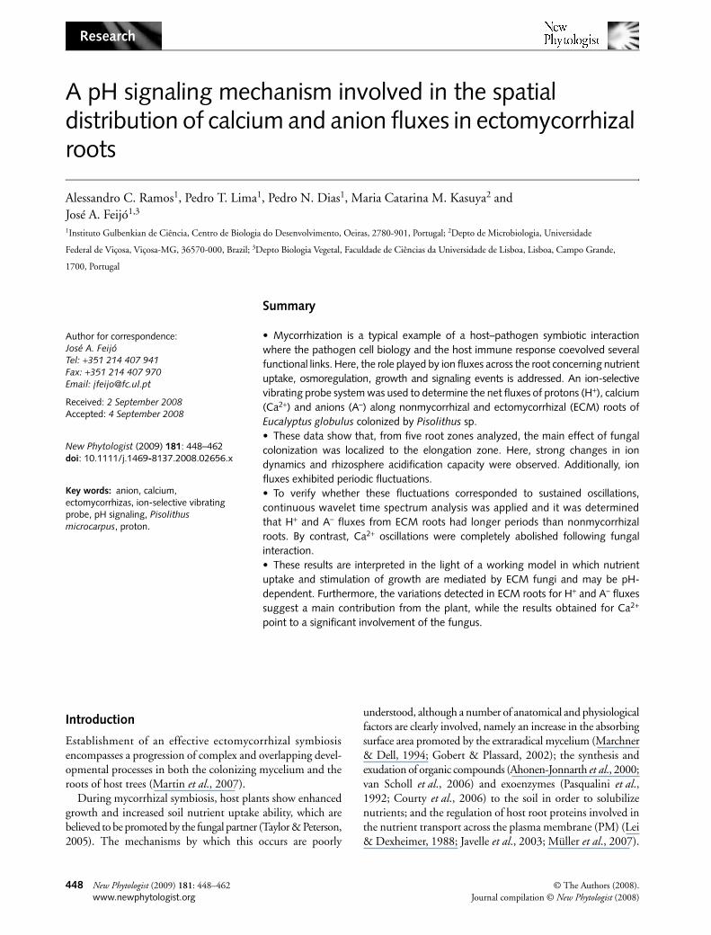
Research 448 New Phytologist (2009) 181: 448–462 © The Authors (2008). 448 www.newphytologist.org Journal compilation © New Phytologist (2008) Blackwell Publishing Ltd A pH signaling mechanism involved in the spatial distribution of calcium and anion fluxes in ectomycorrhizal roots Alessandro C. Ramos 1 , Pedro T. Lima 1 , Pedro N. Dias 1 , Maria Catarina M. Kasuya 2 and José A. Feijó 1,3 1 Instituto Gulbenkian de Ciência, Centro de Biologia do Desenvolvimento, Oeiras, 2780-901, Portugal; 2 Depto de Microbiologia, Universidade Federal de Viçosa, Viçosa-MG, 36570-000, Brazil; 3 Depto Biologia Vegetal, Faculdade de Ciências da Universidade de Lisboa, Lisboa, Campo Grande, 1700, Portugal Summary • Mycorrhization is a typical example of a host–pathogen symbiotic interaction where the pathogen cell biology and the host immune response coevolved several functional links. Here, the role played by ion fluxes across the root concerning nutrient uptake, osmoregulation, growth and signaling events is addressed. An ion-selective vibrating probe system was used to determine the net fluxes of protons (H + ), calcium (Ca 2+ ) and anions (A – ) along nonmycorrhizal and ectomycorrhizal (ECM) roots of Eucalyptus globulus colonized by Pisolithus sp. • These data show that, from five root zones analyzed, the main effect of fungal colonization was localized to the elongation zone. Here, strong changes in ion dynamics and rhizosphere acidification capacity were observed. Additionally, ion fluxes exhibited periodic fluctuations. • To verify whether these fluctuations corresponded to sustained oscillations, continuous wavelet time spectrum analysis was applied and it was determined that H + and A – fluxes from ECM roots had longer periods than nonmycorrhizal roots. By contrast, Ca 2+ oscillations were completely abolished following fungal interaction. • These results are interpreted in the light of a working model in which nutrient uptake and stimulation of growth are mediated by ECM fungi and may be pH- dependent. Furthermore, the variations detected in ECM roots for H + and A – fluxes suggest a main contribution from the plant, while the results obtained for Ca 2+ point to a significant involvement of the fungus. Author for correspondence: José A. Feijó Tel: +351 214 407 941 Fax: +351 214 407 970 Email: [email protected] Received: 2 September 2008 Accepted: 4 September 2008 New Phytologist (2009) 181: 448–462 doi: 10.1111/j.1469-8137.2008.02656.x Key words: anion, calcium, ectomycorrhizas, ion-selective vibrating probe, pH signaling, Pisolithus microcarpus, proton. Introduction Establishment of an effective ectomycorrhizal symbiosis encompasses a progression of complex and overlapping devel- opmental processes in both the colonizing mycelium and the roots of host trees (Martin et al., 2007). During mycorrhizal symbiosis, host plants show enhanced growth and increased soil nutrient uptake ability, which are believed to be promoted by the fungal partner (Taylor & Peterson, 2005). The mechanisms by which this occurs are poorly understood, although a number of anatomical and physiological factors are clearly involved, namely an increase in the absorbing surface area promoted by the extraradical mycelium (Marchner & Dell, 1994; Gobert & Plassard, 2002); the synthesis and exudation of organic compounds (Ahonen-Jonnarth et al., 2000; van Scholl et al., 2006) and exoenzymes (Pasqualini et al., 1992; Courty et al., 2006) to the soil in order to solubilize nutrients; and the regulation of host root proteins involved in the nutrient transport across the plasma membrane (PM) (Lei & Dexheimer, 1988; Javelle et al., 2003; Müller et al., 2007).
-
Upload
independent -
Category
Documents
-
view
3 -
download
0
Transcript of A pH signaling mechanism involved in the spatial distribution of calcium and anion fluxes in...
Research
448 New Phytologist (2009) 181: 448–462 © The Authors (2008).448 www.newphytologist.org Journal compilation © New Phytologist (2008)
Blackwell Publishing Ltd
A pH signaling mechanism involved in the spatial distribution of calcium and anion fluxes in ectomycorrhizal roots
Alessandro C. Ramos1, Pedro T. Lima1, Pedro N. Dias1, Maria Catarina M. Kasuya2 and José A. Feijó1,3
1Instituto Gulbenkian de Ciência, Centro de Biologia do Desenvolvimento, Oeiras, 2780-901, Portugal; 2Depto de Microbiologia, Universidade
Federal de Viçosa, Viçosa-MG, 36570-000, Brazil; 3Depto Biologia Vegetal, Faculdade de Ciências da Universidade de Lisboa, Lisboa, Campo Grande,
1700, Portugal
Summary
• Mycorrhization is a typical example of a host–pathogen symbiotic interactionwhere the pathogen cell biology and the host immune response coevolved severalfunctional links. Here, the role played by ion fluxes across the root concerning nutrientuptake, osmoregulation, growth and signaling events is addressed. An ion-selectivevibrating probe system was used to determine the net fluxes of protons (H+), calcium(Ca2+) and anions (A–) along nonmycorrhizal and ectomycorrhizal (ECM) roots ofEucalyptus globulus colonized by Pisolithus sp.• These data show that, from five root zones analyzed, the main effect of fungalcolonization was localized to the elongation zone. Here, strong changes in iondynamics and rhizosphere acidification capacity were observed. Additionally, ionfluxes exhibited periodic fluctuations.• To verify whether these fluctuations corresponded to sustained oscillations,continuous wavelet time spectrum analysis was applied and it was determinedthat H+ and A– fluxes from ECM roots had longer periods than nonmycorrhizalroots. By contrast, Ca2+ oscillations were completely abolished following fungalinteraction.• These results are interpreted in the light of a working model in which nutrientuptake and stimulation of growth are mediated by ECM fungi and may be pH-dependent. Furthermore, the variations detected in ECM roots for H+ and A– fluxessuggest a main contribution from the plant, while the results obtained for Ca2+
point to a significant involvement of the fungus.
Author for correspondence:José A. FeijóTel: +351 214 407 941Fax: +351 214 407 970Email: [email protected]
Received: 2 September 2008Accepted: 4 September 2008
New Phytologist (2009) 181: 448–462 doi: 10.1111/j.1469-8137.2008.02656.x
Key words: anion, calcium, ectomycorrhizas, ion-selective vibrating probe, pH signaling, Pisolithus microcarpus, proton.
Introduction
Establishment of an effective ectomycorrhizal symbiosisencompasses a progression of complex and overlapping devel-opmental processes in both the colonizing mycelium and theroots of host trees (Martin et al., 2007).
During mycorrhizal symbiosis, host plants show enhancedgrowth and increased soil nutrient uptake ability, which arebelieved to be promoted by the fungal partner (Taylor & Peterson,2005). The mechanisms by which this occurs are poorly
understood, although a number of anatomical and physiologicalfactors are clearly involved, namely an increase in the absorbingsurface area promoted by the extraradical mycelium (Marchner& Dell, 1994; Gobert & Plassard, 2002); the synthesis andexudation of organic compounds (Ahonen-Jonnarth et al., 2000;van Scholl et al., 2006) and exoenzymes (Pasqualini et al.,1992; Courty et al., 2006) to the soil in order to solubilizenutrients; and the regulation of host root proteins involved inthe nutrient transport across the plasma membrane (PM) (Lei& Dexheimer, 1988; Javelle et al., 2003; Müller et al., 2007).

Research 449
© The Authors (2008). New Phytologist (2009) 181: 448–462Journal compilation © New Phytologist (2008) www.newphytologist.org
Changing ion fluxes across the root plasma membraneimply alterations of transmembrane electrical potential, con-tributed by the electrogenic proton (H+) pumps, which inturn controls ion transport systems (Tazawa, 2003). High H+-ATPase activities were found in the PM of external hyphaeand sheaths of ectomycorrhizal (ECM) fungi (Lei & Dexheimer,1988). This enzyme was also found to be stimulated by externalanion concentrations (Churchill & Sze, 1984; Ullrich &Novacky, 1990) and inhibited by Ca2+ (Lino et al., 1998). Inthis context, an induction in the uptake has beendemonstrated for Pinus pinaster ECM roots (Gobert &Plassard, 2002; Plassard et al., 2002; Boukcim & Plassard,2003; Hawkins et al., 2008). This supports the notion thatH+ transport, PM H+-ATPase activity and root surface acidi-fication work together to promote uptake (Ullrich &Novacky, 1990; Glass et al., 1992; Forde, 2000). Positiveeffects on ion uptake during mycorrhizal symbiosis havebeen described for nitrogen, phosphate and some tracerelements such as copper and zinc (Marchner & Dell, 1994),although previous results for calcium have been limited anddifficult to interpret (Bücking et al., 2002). For example, in theroot cortex almost 100% of the cell wall calcium can beeasily exchanged for an external 44Ca label (Peterson &Enstone, 1996; Kuhn et al., 2000). Similarly, studies ofnutrient mobilization in ECM symbiosis have been performedby radioisotope coupling with laser microprobe mass analysis(LAMMA), energy-dispersive X-ray spectroscopy (EDXS)and secondary ion mass spectroscopy (SIMS) (Peterson &Enstone, 1996; Bücking & Heyser, 2000; Bücking et al., 2007).However, very few detailed studies aiming to determinethe regulation of ion dynamics in ECM symbiosis have beencarried out.
There is a profound effect of pH on several biologicalprocesses, including nutrient uptake, cell growth and plant–microbe interactions (Feijó et al., 1999; Felle, 2001; Michardet al., 2008). Recently, we showed that extracellular H+
fluxes are involved in both presymbiotic and symbioticdevelopment of arbuscular mycorrhizal symbiosis (Ramoset al., 2008a,b). By contrast, the possible impact of pHchanges was not yet established for ECM associations. Protonfluxes presumably generated by the PM H+-ATPase activitycan modify the root surface pH in ways that may trigger, forexample, modifications in the availability of free extracellularCa2+ or anion transport.
As a first step to test the role of ion fluxes in ECM associa-tions, we performed a systematic analysis of the different rootion fluxes in the presence and absence of fungal colonization.We measured these fluxes by means of ion-specific vibratingprobes. Major alterations were observed in the growing zoneof the root, and are compatible with the notion that pHmodulates nutrient uptake. Furthermore, the major alterationsdetected in ECM roots for H+ and A– seem to be associatedwith root-specific fluxes, while the results for Ca2+ suggest asignificant contribution of the fungus.
Materials and Methods
Biological material, inoculum production and in vitro synthesis of ectomycorrhizas
Three agar discs containing mycelium of the ECM gastero-mycete Pisolithus microcarpus isolate PT 90A were inoculatedonto Petri dishes containing 20 ml of modified MNM (Marx,1969) medium and incubated for 28 d at 28°C. From theresulting colonies, 9 mm agar discs were cut off from the edgeof actively growing colonies. Eucalyptus globulus Labill. seedswere superficially sterilized with 5% sodium hypochlorite (v/v)for 15 min, rinsed with five changes of sterile water, andplated on modified Clark solution at quarter-strength (Clark,1975) to which was added 2.9 µm thiamine-HCl and 1%sucrose in 0.5% (w/v) Phytagel (Sigma-Aldrich, Gillingham,UK). The use of Phytagel produced a clear and colorless medium,which is excellent for imaging and ion flux measurement withreduced electrical noise (Ramos et al., 2008a). After 7 d,aseptically germinated seedlings were placed on the edge of10-d-old ECM fungal mycelium grown on the same mediumused for seedlings. These were left for 15 d in a controlled-environment growth chamber, with 16 h of light (26°C,350 µmol m−2 s−1) and 8 h of dark, for ectomycorrhizaformation. ECM plants were later transferred to hydroponicconditions in the same solution and growth chamber settingsfor 10 d. Subsequently, ion fluxes measurements were performedin secondary roots of intact plants. In addition, pieces of rootsystem were washed and samples were subsequently collectedfor microscopic evaluation of mycorrhizal colonization, asdescribed by Brundrett et al. (1996).
Measurements of H+, Ca2+ and anion fluxes and currents using the ion-selective vibrating probe system
A detailed description of the experimental setup of the ion-selective vibrating probe technique utilized in this study hasbeen well described (Kochian et al., 1992; Feijó et al., 1999;Shipley & Feijó, 1999; Zonia et al., 2002; Kunkel et al., 2006;Ramos et al., 2008a). In short, E. globulus plants colonized ornot by ECM fungus P. microcarpus isolate PT 90A underhydroponic conditions, were placed in plastic Petri dishes(140 × 140 mm) filled with 30 ml of modified Clark solutionat quarter strength, excepted for Ca2+ measurements, where100 µm Ca2+ was used. Visual Minteq analysis was performedaccording to Parker et al. (1995) using the ion concentrationsof the modified Clark solution applied in this study.
We focused on secondary roots, as they are biologicallyand physiologically more significant than primary roots fornutrient supply to the plant. The volume occupied by second-ary roots in the soil can reach c. 30–40% more than primaryones. Readings were taken in five defined root zones of non-mycorrhizal (control) and mycelium-covered roots: apex (tip),meristematic (100–150 µm); elongation (300–800 µm); root
NO3−
NO3−

Research450
New Phytologist (2009) 181: 448–462 © The Authors (2008).www.newphytologist.org Journal compilation © New Phytologist (2008)
hairs (major presence of these structures); and finally maturezone (posterior to root hair zone).
Ion-specific vibrating microelectrodes were produced asdescribed by Feijó et al. (1999). Micropipettes were pulledfrom 1.5 mm borosilicate glass capillaries and treated withdimethyl dichlorosilane (Sigma-Aldrich). After silanization,they were backfilled with a 15–20 mm column of electrolyte(15 mm KCl and 40 mm KH2PO4, pH 6.0, for H+; 100 mmKCl for anions; 100 mm CaCl2 for Ca2+) and then front-loaded with a 20–25 µm column of the respective ion-selectiveliquid exchange cocktail (Fluka, Milwaukee, WI, USA). Weused Cl− electrodes to measure the anion fluxes given that thiselectrode has poor selectivity for Cl− under our experimentalconditions (Supporting information, Fig. S3a,b). Firstly themeasurement of chloride activity in the medium is slightlyaffected by the presence of other ions (Fig. S3a), but these changesshould be expressed below noise level within the microvoltrange usually measured on vibrating conditions for cellularfluxes. More importantly, the Cl− electrode calibration withdifferent anions showed that this electrode responds with aNernstian slope to chloride and nitrate, and while sub-Nernstianto sulfate and phosphate, also exhibits a significant responsewithin the concentrations used in this study (Fig. S3b). Lastbut not least, the background concentrations in the mediumof the individual anions span various orders of magnitude,likewise affecting the signal-to-noise (S/N) ratio measurementof the fluxes in a way that is inversely proportional to the con-centration. Taken together, these considerations make it almostimpossible to discriminate the individual activities of everysingle anion, and therefore we have opted to refer to these fluxesas reflecting the global ‘anionic’ concentration rather than Cl−
proper fluctuations. The final nutrient composition and bio-availability prediction (Ward et al., 2008) are displayed inTable 1. An Ag/AgCl wire electrode holder (World PrecisionInstruments, Sarasota, FL, USA) was inserted into the back ofthe microelectrode and established electrical contact with thebathing solution. The ground electrode was a dry reference(DRIREF-2, World Precision Instruments) that was insertedinto the sample bath. The microelectrodes were calibrated atthe beginning and end of each experiment using standardsolutions covering the experimental range of each ion, in orderto obtain a calibration line. Both the slope and intercept of thecalibration line were used to calculate the respective ion concen-tration from the mV values measured during the experiments.
Inhibition with vanadate ( ), gadolinium (GdCl3) and 4,4′-diisothiocyanatostilbene-2,2′-disulfonic acid (DIDS)
Inhibitor treatments were performed in Eucalyptus roots afterdetermination of each ion flux at the elongation zone (n = 5).The data acquisition was stopped and the respective inhibitors(Sigma-Aldrich) were added in the Petri dishes with thefollowing concentrations: plasma membrane H+-ATPase
VO43−
Tabl
e1
Ion
conc
entr
atio
ns a
nd p
redi
cted
bio
avai
labi
lity
(%)
in m
odifi
ed C
lark
nut
rient
sol
utio
ns u
sed
for
both
ect
omyc
orrh
izal
syn
thes
is a
nd io
n flu
x m
easu
rem
ents
Stre
ngth
H2P
O4
K+
Ca2+
Mg2+
Na+
Cl−
H3B
O3
Mn2+
Zn2+
Cu2+
(mM
)(µ
M)
Mod
ified
C
lark
sol
utio
n1F
0.06
90.
600
1.80
02.
560
0.60
00.
130
7.26
00.
900
0.50
019
.00
7.00
02.
000
0.50
00.
086
¼F
0.01
70.
150
0.45
00.
640
0.15
00.
032
1.81
50.
225
0.12
54.
750
1.75
00.
500
0.12
50.
0215
¼F
(Ca2+
)0.
017
0.15
00.
450
0.10
00.
150
0.03
20.
855
0.22
50.
125
4.75
01.
750
0.50
00.
125
0.02
15Pr
edic
ted
avai
labi
lity
(%)a
1F90
.50
78.0
399
.39
94.3
696
.50
99.6
499
.42
99.6
599
.39
99.9
695
.83
94.6
889
.33
60.9
2¼
F94
.86
90.3
699
.86
98.8
399
.40
99.9
299
.82
99.9
399
.81
99.9
799
.17
96.8
993
.68
78.6
9¼
F (C
a2+)
95.9
796
.10
99.9
198
.89
99.2
999
.94
99.9
499
.93
99.9
099
.97
99.1
598
.89
93.8
985
.14
The
free
una
ssoc
iate
d st
ate
of t
he io
n is
, in
mos
t ca
ses,
ass
umed
to
be t
he b
ioav
aila
ble
form
of
the
ion.
a The
pre
dict
ions
wer
e pe
rfor
med
by
Vis
ual M
inte
q v.
2.53
(Pa
rker
et
al.,
1995
; War
d et
al.,
2008
) an
d th
e an
alys
is u
sed
5µM
and
20
µM F
e-ED
TA f
or q
uart
er-s
tren
gth
(¼F)
and
ful
l-st
reng
th
(1F)
Cla
rk n
utrie
nt s
olut
ion,
resp
ectiv
ely.
SO42−
NO
3−N
H4+
MoO
42−

Research 451
© The Authors (2008). New Phytologist (2009) 181: 448–462Journal compilation © New Phytologist (2008) www.newphytologist.org
(100 µm orthovanadate), calcium channels (100 µm gadolinium)and chloride channels (50 µm DIDS). Five to 10 min later, abackground reference was taken and ion fluxes were againrecorded. Interference caused by the inhibitors was controlledfor by direct incubation with ionophore-loaded probes. Nosignificant interference of the inhibitors was found to occurfor H+ and anions. For Ca2+, the interference was morepronounced with high levels of gadolinum, which in thepresent study was used at lower concentrations (100 μm; Fig. S4).
Ion flux oscillation analysis
Frequency analyses were performed using AutoSignal v1.7(Systat Software, Inc.). For each set of flux oscillations to beanalyzed, a data trend removal was applied, consisting of alinear least-squares fit subtraction to remove the very low-frequency trend of the data. Two distinct methods were thenused to assess the frequency components of the oscillations:Fourier and Wavelet analyses. For Fourier analysis, a fastFourier transform Radix 2 algorithm was used, ensuring thateach data set was a continuous acquisition without breaks andwith a constant sampling rate. Peaks were detected by a localmaxima detection algorithm and considered relevant accordingto their significance levels (the higher the significance level,the less likely it is that a detected spectral signal will arise fromrandom noise). Significance levels are given in the Resultssection. For wavelet analysis, a continuous wavelet time–frequency spectrum was obtained with a noncomplex Morletwavelet (wave number, 12). A peak-type critical limit was usedinstead of the traditional confidence levels, as implemented inthe software.
Statistical analysis
All data were analyzed by one-way or two-way ANOVAs inorder to compare the mean values (considering ‘fungaltreatment’ and ‘root region’ as factors), which were validatedby convenient residual analyses and, when necessary, combinedwith Duncan’s test for multiple comparison. To compare thecontrol and fungal treatment (Table 2), we applied Student’st-test for two independent samples and calculated confidenceintervals for the mean difference, in order to guarantee aglobal 95% confidence level. The results are expressed asmeans with respective standard error, and the numbers ofrepetitions are given in each figure legend. All statisticalanalyses were conducted using the R program and the level ofsignificance was set up at 5% (Ihaka & Gentleman, 1996).
Results
ECM colonization effects on plant growth parameters
For ion flux analysis purposes (Fig. 1b), formation of ectomy-corrhizas was performed under in vitro conditions (Fig. 1a) in
order to produce E. globulus with a high degree of colonizationby P. microcarpus isolate 90A (Fig. 1c,d). During theexperiments, plants presented 78.3% of ECM root colonization(Table 2; Fig. 1d). In addition, significant and positive effectsof ECM colonization were found both on plant height and onshoot and root fresh weights (P < 0.05; Table 2). No changesin the number of root tips, at the time of the analysis, weredetected. A significant decrease in the length of root hairs wasfound in ECM roots (Table 2). Plant growth was stronglycorrelated with ionic fluxes as significant Pearson’s correlationcoefficients were found between H+ fluxes and plant growthparameters (0.78; P < 0.008), root surface pH (−0.82;P < 0.0001) and anion fluxes (−0.59; P < 0.002). Moreover,we also found significant correlation coefficients between rootsurface pH and plant growth parameters (−0.72; P < 0.0102).
H+ flux profile and root surface pH
A differential pattern of H+ fluxes was observed along thezones of eucalyptus roots (Fig. 2a). In both nonmycorrhizaland ECM roots, the apex, meristematic and elongation zoneswere characterized as domains of significant H+ efflux. Bycontrast, root hair and mature zones were characterized asdomains of H+ influx (Fig. 2a). A sixfold stimulation on H+
effluxes was observed at the elongation zone in the presence ofcolonizing P. microcarpus (P < 0.001). As expected, surfacepH values along the root system showed a pattern consistentwith the flux profile, and equally affected ECM colonization(Fig. 2b). The two domains described for H+ fluxes along theroots corresponded to patches of variable acidity, rangingfrom 5.56 in the meristematic region to 5.68 in the apex. InECM roots, significant acidification was observed in the apex,meristematic and, most notably, elongation regions. Thelowest pH value (< 5.4) was observed in the elongation zone.In root hairs and mature zones, pH values were found to be5.6 and 5.8, respectively. These results support an ECM-driven increase in overall H+ influx. All regions showed
Table 2 Average values of fungal and plant growth parameters analyzed in nonmycorrhizal (control) or mycorrhizal roots of Eucalyptus globulus colonized by Pisolithus microcarpus (ECM), 10 d after transplanting to hydroponic conditions (n = 35)
Parameter analyzed Control ECM
Fungal colonization (%) nd 78.3Plant heigth (cm) 14.38 17.61*Shoot fresh weight (mg per plant) 33.94 45.33**Root fresh weight (mg per plant) 12.2 15.75*Root hair length (µm) 386.52 152.39**Root tips (Nº) 12.00 17.00
Significantly differ by Student’s t-test (*P < 0.05; **P < 0.01; ***P < 0.001). For root tips, P = 0.051.nd, not determined.

Research452
New Phytologist (2009) 181: 448–462 © The Authors (2008).www.newphytologist.org Journal compilation © New Phytologist (2008)
significant differences in the surface pH after the establishmentof ECM. The global extracellular pH gradient increased byapprox. 0.12 pH units in the control (Me vs Mat) to 0.4 pHunits after ECM (Elong vs Mat) (Fig. 2b).
Ca2+ and anion flux profiles
Interestingly, the patterns of the Ca2+ and anion fluxes incontrol and ECM roots revealed a quite different scenario. Inall zones analyzed, the inoculation of eucalyptus plantsinduced an inhibition of the magnitude of Ca2+ fluxes(Fig. 3a). Furthermore, an inversion of flux direction (effluxto influx) was observed in the elongation zone. On the otherhand, a significant increase of anion influx was observedprimarily at the elongation zone (P < 0.001) and, to a lesserextent, at the root hair zone (P < 0.01, Fig. 3b). The resultsalso showed a significant inhibition of the anion influx at themeristematic zone (P < 0.01), but no significant changes wereobserved at the apex and mature zones (Fig. 3b).
Time-course change of external Ca2+ and anion concentrations
Analysis of the time-course changes in Ca2+ and anionconcentrations in the medium with nonmycorrhizal (control)and ECM roots after a 5 min exposure to the nutrientmedium is presented in Fig. 3(c) and (d). These results indicatethat ECM roots were more efficient than the control in takingup Ca2+ ions from the external medium (Fig. 3c). By contrast,control roots seem to take up anions less efficiently than ECM(control change is nonsignificant) (Fig. 3d). This correlateswell with the root surface pH values, since ECM roots showeda superior capacity to acidify the medium compared with thecontrol (Fig. 2b).
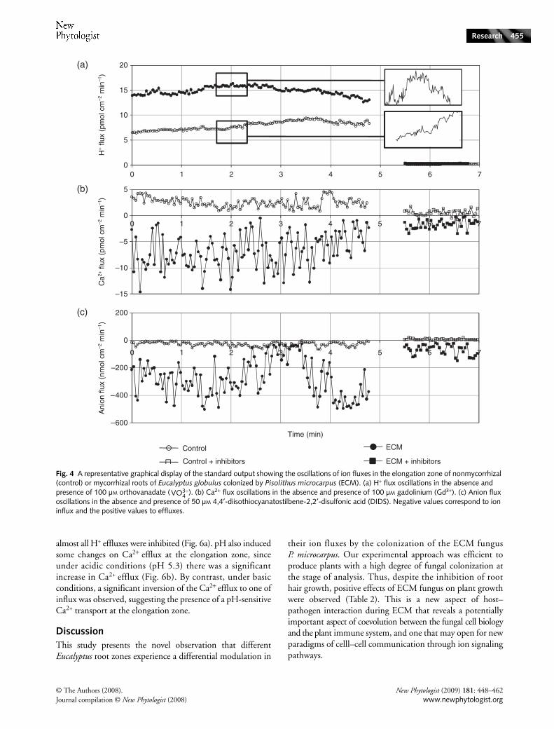
Pharmacological assays on H+, Ca2+ and anion fluxes
Highly significant changes in the ion fluxes were observed inthe root system of E. globulus in the presence of P. microcarpusECM fungus, notably in the elongation zone (Figs 2, 3). Wefurther investigated the various fluxes in this region bydetailed temporal analysis and pharmacological inference ofthe putative entities involved in their generation. All fluxesshowed a clear oscillatory behavior in the elongation zone,irrespective of the conditions assayed. Changes in the oscillatorycomponents of the ion fluxes were also induced by fungalcolonization, mainly in the case of H+ and anion fluxes(Fig. 4; wavelet spectral analysis in Fig. 5). The addition of100 µm orthovanadate, a P-type PM H+-ATPase inhibitor(Bowman, 1982; Bowman et al., 1983), strongly inhibited alleffluxes at the elongation zone (Fig. 4a). Ca2+ and anion fluxeswere differentially inhibited by 100 µm gadolinium and50 µm DIDS, respectively (Fig. 4b,c). Gadolinium (Gd3+) isa widely used inhibitor for Ca2+ channels (Yang & Sachs,1989; Hedrich et al., 1990; Klüsener et al., 1995; Caldwellet al., 1998; Antoine et al., 2000, 2001) and DIDS is a widelyused Cl− blocker (Schroeder et al., 1993; Zonia et al., 2001,2002; Messerli et al., 2004). Vanadate treatment led to analmost complete blockage of H+ fluxes (Fig. 4a, Table 3), andthe observed differences between nonmycorrhizal and ECMroots were not significant (P > 0.05), suggesting that all effluxesdetected were the result of the plasma membrane H+-ATPaseactivity. Considering the stronger values of H+ effluxes inECM roots and the presence of different H+-ATPase isoformsin the fungal hyphae, presumably with different sensitivitiesto vanadate, this degree of inhibition came as a surprise.Taken literally, one possible hypothesis is that the majorproportions of these fluxes are actually generated by the rootepidermis.
Fig. 1 (a) Ectomycorrhiza formation under in vitro germination conditions of Eucalyptus globulus seedlings and Pisolithus microcarpus before transplanting to hydroponic settings. The arrows show inoculum discs containing MNM medium and fungal mycelium. Bar, 9 mm. (b) Representation of a root apex during measurements with an ion-selective vibrating probe. Bar, 170 µm. (c) Representation of a lateral root (arrowhead) of E. globulus around P. microcarpus mycelium (arrow) under our experimental conditions. Bar, 450 µm. (d) Cross-section of E. globulus roots colonized by Pisolithus microcarpus. The arrow indicates the fungal colonization. Bar, 50 µm.

Research 453
© The Authors (2008). New Phytologist (2009) 181: 448–462Journal compilation © New Phytologist (2008) www.newphytologist.org
The Gd3+ inhibition of Ca2+ fluxes showed a more complexpattern than that of vanadate on H+ fluxes (Fig. 4b; Table 3).As previously mentioned, ECM reverses the efflux to influx inthe elongation zone, a result which is difficult to interpret, asit implies a shift in the balance of functional carriers for Ca2+,which are presumably derived from different equilibriumconditions. In the context of ECM, Gd3+ inhibits close to80% of the Ca2+ influx, a result consistent with the hypothesisthat the majority of Ca2+ is taken up via Gd3+-sensitivechannels, some of which could be the result of fungal Ca2+
channels. This conclusion is supported by the observationthat there is an almost total inhibition of Ca2+ channels innonmycorrhizal roots (Figs 4b, 5). However, it should bepointed out that inhibition of an efflux by Gd3+ is not astraightforward interpretable result, and calls for further study.
Anion influxes seem to be proportionally inhibited byDIDS in the same way in both control and ECM roots. Thissupports the notion that most anion fluxes are root-generated(Fig. 4c; Table 3). Furthermore, it was observed that all inhibitorsperformed more effectively in control conditions, supportingthe hypothesis that in ECM roots there is a greater variety ofion transporters, some of which are refractory to the broad-band inhibitors used.
Spectral analysis of the ion flux oscillations
As illustrated in the traces presented in Fig. 4, most of thecontinuous time-course measurements of fluxes showedcomponents that were suggestive of sustained periodicity. Tothe extent that the spectral properties of these temporalvariations could enlighten aspects of their regulation, weemployed continuous wavelet time–frequency spectrum coupledto Fourier analysis to dissect these properties further. In allcases analyzed, we found evidence for underlying oscillations,sometimes with one single component, and at other timeswith more than one component (Fig. 5a,c,e). More interestingly,they all showed some degree of modification upon colonizationof Eucalyptus roots with the ECM fungus P. microcarpus
Table 3 Percentage inhibition of H+, Ca2+ and anion fluxes after the pharmacological assays
Treatment
% inhibition
Vanadate Gadolinium DIDS
Nonmycorrhizal 97.72* 81.65* 100.00*Ectomycorrhizal 82.60 75.41 73.83
For H+ fluxes, orthovanadate was applied to the final concentration of 100 µM. For Ca2+ and anion fluxes, 100 µM gadolinium and 50 µM DIDS, respectively, were applied. n = 5.*At the same column, the mean values are significantly different by Student’s t-test at P < 0.01.
Fig. 2 Proton fluxes (a) and root surface pH (b) along nonmycorrhizal (control, open bars) and ectomycorrhizal (ECM) roots of Eucalyptus globulus colonized by Pisolithus microcarpus (ECM, closed bars). Apex, meristematic (Me), elongation (Elong), root hairs (RH) and mature (Mat) indicate the zones analyzed. Bars represent the mean values ± SE of five independent experiments (*statistical difference at P < 0.01). Negative values correspond to ion influx and positive values to effluxes. For H+ fluxes and surface pH, by two-way ANOVA combined with Duncan’s test, the results showed that there was significant interaction between fungal treatment and root zones (P < 0.0001). For H+ fluxes, we found no statistically significant difference with fungal inoculation at the meristematic zone. For pH data interpretation, bars followed by the same capital letter, in the same root region, are not significantly different by Duncan’s test at P < 0.05. Bars followed by the same lower-case letter, in different root regions, are not significantly different at P < 0.05 (n = 5).

Research454
New Phytologist (2009) 181: 448–462 © The Authors (2008).www.newphytologist.org Journal compilation © New Phytologist (2008)
(Fig. 5b,d,f ). Results shown in Fig. 5 reveal that, in thecontrol H+ flux oscillations, there is one dominant period ofc. 3.1 min, which lengthens to 5.3 min in the presence of theECM fungus (Fig. 5a,b). This broadening of the majorcomponents of the oscillations were confirmed by Fourieranalysis (P < 0.05; Fig. S1a,b). In addition, no significantoscillations were found in controls without a biologicalsample (Fig. S2d,e,f ).
By contrast, Ca2+ flux oscillations seem to show an opposingtrend after ECM (Fig. 5c,d). Firstly, they seem to have twomajor components in the control condition: a dominant oneof c. 5.3 min (P < 0.01) and a second of c. 1.5 min. However,both disappeared in the presence of the fungus (Fig. 5c,d),giving rise to a number of small periods at the borderline ofthe S/N ratio of the system. The Fourier analysis confirmedthese results and showed that some of the high frequenciesdetected by continuous wavelet time–frequency spectrumanalysis were not statistically significant at P < 0.05 (Fig. S1c,d).Finally, anion fluxes showed a third and different scenario.Control roots had at least one significant oscillation period ofc. 0.6 min, thus characterized as being very fast, together withothers considered nonsignificant by Fourier analysis at P < 0.05(Fig. 5e, S1e). In the presence of the ECM fungus, however,there was a drastic change of behavior, giving rise to a longer
period of c. 3.0 min (Fig. 5e,f, S1f ). Fourier analysis revealedthe same short period of 0.6 min in ECM roots as above thelevel of system noise, but with a much reduced significance.These results demonstrate that the ECM colonization changesthe H+ and anion flux oscillations, by increasing their periodsby approx. double and sixfold, respectively, while for Ca2+ fluxthe oscillations are completely disrupted in the presence of thefungus. In addition, all ion flux oscillations were fully inhibitedby the respective inhibitors such as orthovanadate, gadoliniumand DIDS (data not shown).
A dual effect of the external pH and Ca2+ concentration on extracellular ion fluxes
In systems showing prominent pH and Ca2+ dependency, thehomeostasis of these ions seems to be closely interrelated. Wetested whether this was also the case at the elongation zone, bygrowing E. globulus roots in medium with three different Ca2+
concentrations (0, 0.5, 1 mm) for 5 d, and analyzing the H+
fluxes and root surface pH after that period (Fig. 6). Theresults showed that an increase in Ca2+ availability provokeda significant inhibition on the H+ effluxes in the rootelongation zone (Fig. 6a). Likewise, the root surface pHincreased with the Ca2+ concentration. Also, at 0.5 mm Ca2+,
Fig. 3 Fluxes of calcium (a) and anions (b) along nonmycorrhizal (control, open bars) and ectomycorrhizal (ECM) roots of Eucalyptus globulus colonized by Pisolithus microcarpus (closed bars). Apex, meristematic (Me), elongation (Elong), root hairs (RH) and mature (Mat) refer to the root zones analyzed. Negative values correspond to ion influx and positive values to effluxes. Bars represent mean values ± SE of five independent experiments. (c, d) Fluctuations on external Ca2+ (c) and anion (d) concentrations in nonmycorrhizal (control, circles) and ECM (squares) roots. For uptake analysis, roots were exposed for 5 min to Clark solution containing 0.2 mM Ca2+ (c) and 1.5 mM anions (d). Scale bars represent the mean values ± standard error (n = 5). *Means significantly different by Student’s t-test at P < 0.001. For Ca2+ and anion fluxes, by two-way ANOVA combined with Duncan’s test, the results showed that there were significant interactions between fungal treatment and root zones (P < 0.0001). There were no significant effects of fungal inoculation for Ca2+ fluxes at the root hair zone, and for anion fluxes at the apex and mature zones. *, P < 0.01; **, P < 0.001.
Research 455
© The Authors (2008). New Phytologist (2009) 181: 448–462Journal compilation © New Phytologist (2008) www.newphytologist.org
almost all H+ effluxes were inhibited (Fig. 6a). pH also inducedsome changes on Ca2+ efflux at the elongation zone, sinceunder acidic conditions (pH 5.3) there was a significantincrease in Ca2+ efflux (Fig. 6b). By contrast, under basicconditions, a significant inversion of the Ca2+ efflux to one ofinflux was observed, suggesting the presence of a pH-sensitiveCa2+ transport at the elongation zone.
DiscussionThis study presents the novel observation that differentEucalyptus root zones experience a differential modulation in
their ion fluxes by the colonization of the ECM fungusP. microcarpus. Our experimental approach was efficient toproduce plants with a high degree of fungal colonization atthe stage of analysis. Thus, despite the inhibition of roothair growth, positive effects of ECM fungus on plant growthwere observed (Table 2). This is a new aspect of host–pathogen interaction during ECM that reveals a potentiallyimportant aspect of coevolution between the fungal cell biologyand the plant immune system, and one that may open for newparadigms of celll–cell communication through ion signalingpathways.
Fig. 4 A representative graphical display of the standard output showing the oscillations of ion fluxes in the elongation zone of nonmycorrhizal (control) or mycorrhizal roots of Eucalyptus globulus colonized by Pisolithus microcarpus (ECM). (a) H+ flux oscillations in the absence and presence of 100 µM orthovanadate ( ). (b) Ca2+ flux oscillations in the absence and presence of 100 µM gadolinium (Gd3+). (c) Anion flux oscillations in the absence and presence of 50 µM 4,4′-diisothiocyanatostilbene-2,2′-disulfonic acid (DIDS). Negative values correspond to ion influx and the positive values to effluxes.
VO43−

Research456
New Phytologist (2009) 181: 448–462 © The Authors (2008).www.newphytologist.org Journal compilation © New Phytologist (2008)
Fig. 5 Continuous wavelet time spectrum analyses of the H+ (a, b), Ca2+ (c, d) and anion flux oscillations in the elongation zone of nonmycorrhizal (a, c, e) and mycorrhizal roots (ECM) of Eucalyptus globulus colonized by Pisolithus microcarpus (b, d, f), as presented in Fig. 4. The frequencies are represented in min–1 and the periods in min. Wavelet analysis was coupled to Fourier analysis in order to dissect the frequency components, and shows the oscillatory pattern of the ion fluxes. No significant periods of ion fluxes were found in the medium without any biological sample (see also Fig. S2).

Research 457
© The Authors (2008). New Phytologist (2009) 181: 448–462Journal compilation © New Phytologist (2008) www.newphytologist.org
The control of root surface pH in ECM roots by extracellular H+ fluxes is linked to PM H+-ATPase activity
In comparison with control uninfected plants, the highestrates of H+ efflux and acidic surface pH were located at theelongation zones of ECM roots (Fig. 2). These effluxes aredependent on the PM H+-ATPase, as they were inhibited by100 µm orthovanadate (inhibitor of P-type plasma membraneH+-ATPase), a result which is conceptually sound since theelongation zone is a specialized growing zone (Winch &Pritchard, 1999). In fact, it has been shown that this zoneshows notably higher immunolocalization and higher activitylevels of PM H+-ATPase than the apical and meristematiczones (Jahn et al., 1998; Palmgren, 2001; see details inEnriquez-Arredondo et al., 2005). Using immunocytochemical
approaches, Lei & Dexheimer (1988) found strong PMH+-ATPase labeling in root cortical cells of Pinus sylvestris-Laccaria laccata, in external hyphae sheaths and Hartig nets.This localization supports the concept of a coupling mechanismbetween fungal and host H+ pumps in ECM roots (Fig. 2,Table 3). Indeed, it has been demonstrated that for arbuscularmycorrhizal associations, some host PM H+-ATPaseisoforms show increased activity and gene expression afterfungal colonization (Ferrol et al., 2002; Ramos et al., 2005;details in Rosewarne et al., 2007).
The H+ efflux mediated by the PM H+-ATPase is importantfor the regulation of cytoplasmic pH (Felle, 2001; Palmgren,2001; Tazawa, 2003) and the activation of cell wall-looseningenzymes and proteins through acidification of the apoplast(Hager, 2003). This effect is closely related to auxin-inducedcell growth as proposed by the ‘acid-growth theory’ by Rayle& Cleland (1992). This implies that enhanced H+ efflux inECM roots (Fig. 2a) results in an acidification of the apoplastic/external pH (Fig. 2b). Moloney et al. (1981) demonstratedthat pH changes in the apoplast are crucial for root growth,since acidic buffering conditions act as stimulators whilstneutral or basic pHs act as inhibitors. Our results clearly showthat when the H+ flux rate (Fig. 2a) and surface pH values(Fig. 2b) are combined, highly significant Pearson’s correlationcoefficients are obtained (−0.82; P < 0.0001). Other candi-dates that contribute to the control of extracellular H+ flux inECM are the presence of anions in the growth medium. Theseare reported to act as stimulators of the PM H+-ATPase(Churchill & Sze, 1984; Ullrich & Novacky, 1990; Glasset al., 1992; Forde, 2000; Garnett et al., 2001). This conceptis especially appealing taking into account the observed oscil-latory behavior (Figs 5, S1), where the ECM colonizationinduced changes in the flux oscillations, leading to theirincreased periods. For Ca2+ flux oscillation, ECM coloniza-tion abolished all significant periods observed in control roots(Figs 5b,c, S1). Combined with the reversion from efflux toinflux in the elongation zone, this result could be interpretedas showing that the fungus contributes to the majority of theCa2+ influx through specific channels. These different activitieswould produce intricate temporal patterns impossible tosynchronize on an organized oscillatory pattern. This beingthe case, the prediction would be that ectomycorrhizal plantsshould have an improved efficiency of Ca2+ uptake from thesoil, a result partly confirmed in Fig. 3(c).
Ca2+ efflux suppression and increase upon Ca2+ uptake in ECM roots
Calcium has a paradoxical effect on PM H+-ATPase, as it hasbeen reported to be an inhibitor via a Ca2+-dependentphosphorylation pathway (Lino et al., 1998; Tazawa, 2003)and an activator in guard cells (Assmann et al., 1985). Aninhibition of the PM Ca2+ influx channels in both animal(Yang & Sachs, 1989) and plant cells (Allen & Sanders, 1994;
Fig. 6 (a) Extracellular H+ fluxes (bars) and root surface pH values (squares) at the elongation zone of Eucalyptus globulus roots under three calcium concentrations (CaCl2). (b) Extracellular Ca2+ fluxes at the elongation zone with three different medium pH values. In this experiment, pH 5.7 was used as the control, since this value was used for all experiments of this work. The remaining pH values were obtained by growing roots for 2 d in the same medium used for ion flux analysis, to which was added 50 µM Tris-HCl, pH 5.3, or 50 µM Tris-base, pH 8.0. The negative values correspond to ion influx and the positive values to effluxes.

Research458
New Phytologist (2009) 181: 448–462 © The Authors (2008).www.newphytologist.org Journal compilation © New Phytologist (2008)
Klüsener et al., 1995; Knight et al., 1996; Antoine et al.,2000, 2001) occurs by the addition of extracellular Gd3+ in amicromolar range. Despite its use for detection of Ca2+
stretch-activated channels (Caldwell et al., 1998), Gd3+ islikely to inhibit other cationic channels as well, because ofits relatively broad effect. Our pharmacological analysissuggested that the Ca2+ influx in the elongation zone of ECMroots is the result of the activity of Gd3+-sensitive calciumchannels (Figs 3a, 4; Table 3). However, as the Ca2+ effluxes arelargely governed by the chemical potential gradient of Ca2+
generated by the PM Ca2+-ATPase, we hypothesized that thesuppression of effluxes in the control roots could represent anindirect dissipation of the Ca2+ gradient, as promoted byGd3+ treatment (Fig. 4, Table 3). In addition, similar fluxinhibition profiles were obtained by Nemchinov et al. (2008)in Nicotiana benthamiana leaves. The authors proposed amodel in which Gd3+-sensitive Ca2+ influxes and Ca2+ pumpsare involved in the signal transduction pathways of thehypersensitive response mechanisms (Nemchinov et al., 2008).As a passive Ca2+ efflux from the cell cytosol isthermodynamically improbable (Shabala & Newman, 2000),an active mechanism must be involved. Two possiblemechanisms of Ca2+ efflux might occur, one through Ca2+
release from the cell wall and the other by Ca2+ extrusion viathe PM Ca2+-ATPase (Lecourieux et al., 2006; Nemchinovet al., 2008). It remains to be determined which of these twomechanisms is responsible for this event to occur. Alternatively,an increase in the activity of Ca2+ influx in ECM roots couldreflect an increased cytosolic concentration of this ion. Indeed,it has recently been demonstrated that the exposure ofE. globulus root hairs to hypaphorine (an indole alkaloidsecreted by P. microcarpus) led to an elevation of cytoplasmicCa2+ concentration (Dauphin et al., 2007). Thus, hypaphorineled to a reduction of the Ca2+ gradient across the plasmamembrane, which was correlated with the arrested growth ofroot hairs (Béguiristain & Lapeyrie, 1997; Dauphin et al.,2007). These results seem similar to our own observations(Table 2), where root hair length was reduced in ECM roots.Recently, Martin et al. (2008) published the genome of theECM fungus Laccaria bicolor, in which numerous and diverseCa2+ channels are found to be encoded (see details at http://genome.jgi-psf.org/Lacbi1/Lacbi1.home.html). Accordingly, wefound ECM roots to have a higher uptake capacity of Ca2+
from the external medium (Fig. 3c). In itself this would notnecessarily lead to a major accumulation of Ca2+ in ECM ofwhole plants, but clearly suggests a higher potential for ionuptake and storage in the cell wall (Peterson & Enstone, 1996;Kuhn et al., 2000) promoted by the fungus. In ECMassociations, such as Suilus bovinus-Pinus sylvestris, an exposureto Ca2+ also led to an accumulation of this ion in the interfacialapoplast in between symbionts and in the fungal sheath(Bücking et al., 2002). Depending on the fungal species, Ca2+
can also accumulate as calcium oxalate in the fungal hyphae(Malajczuk & Cromack, 1982). In the light of this, calcium
dynamics in ECM interactions needs to be more carefullyinvestigated, not just using radioisotopes, but also by meansof an integration of techniques such as ion-selective vibratingprobes, patch-clamp and imaging analyses.
Activation of anion uptake by ECM fungus
It is well known that an increase in the root surfaceconcentration of H+ generates a proton-motive force, whichis necessary to drive the secondary transport of , SO4
2+,Cl−, Ca2+ and K+ (Portillo, 2000; Palmgren, 2001). Accordingly,we found that the changes in H+ efflux attributable to ECMfungal infection in the elongation zone were strictly correlatedto the root surface pH values (−0.82; P < 0.0001), and,significantly, correlations of root surface concentrations of H+
were found with both Ca2+ (−0.78, P < 0.001) and anionfluxes (0.66; P < 0.006). The correlation between Ca2+ andanions at the elongation zone (0.99, P < 0.001) raised thepossibility of an activation of anion influx by Ca2+, asdemonstrated in other cells (Hedrich et al., 1990). Since plantcells have adapted to low anion concentrations, anion uptakeis generally coupled to the electrochemical gradient generatedby the PM H+-ATPase activity (Evans et al., 1980;Zimmermann et al., 1994; Garnett et al., 2001). Consequently,ECM roots possess strong anion influxes and H+ effluxesprimarily at the elongation zone (Fig. 3b). Consistent withthis, we observed high H+-ATPase activity in this root zone.It has been reported that this enzyme is stimulated by anionsin plant (Churchill et al., 1983; Churchill & Sze, 1984;Zimmermann et al., 1994) and animal cell membranes(Vieira et al., 1995). The induction of uptake in P.pinaster ECM roots, even at low external concentrations, waspreviously shown by Gobert & Plassard (2002). The H+
efflux and consequent root surface acidification are necessaryfor the uptake mechanism to operate (Ullrich &Novacky, 1990; Glass et al., 1992; Forde, 2000), as this occursvia PM cotransporters (nH+/ ) (Crawford,1995). Thiswas already demonstrated for Eucalyptus nitens, where largeH+ effluxes were found in medium with . However,
fluxes were quantitatively linked to H+ fluxes (Garnett& Smethurst, 1999; Garnett et al., 2001, 2003). In addition,according to Garnett et al. (2003), negative correlationcoefficients can be obtained between and H+ fluxes.Nitrate is thus a strong candidate to be a component of theanion fluxes we observed, but unfortunately the technicallimitations of the electrodes used do not warrant a straight-forward conclusion in this respect (see the Materials andMethods section and Fig. S3a,b), with chloride probablyplaying also an important role.
In normal conditions, the maintenance of the electricalmembrane potential depends on the H+ efflux and influxes ofanions and potassium (Felle, 2001; Tazawa, 2003). In ECMsymbiosis, fungi have a high capacity to uptake potassium intheir external hyphae (Rygiewicz & Bledsoe, 1984). One
NO3−
NO3−
NO3−
NO3−
NO3−
NO3−
NO3−

Research 459
© The Authors (2008). New Phytologist (2009) 181: 448–462Journal compilation © New Phytologist (2008) www.newphytologist.org
possible molecular basis for this was recently discovered in thesame type of hyphae, where Corratgé et al. (2007) cloned theHcTrk1 transporter from Hebeloma cylindrosporum, and dem-onstrated it to encode for a single-file pore channel thatcotransports Na+-K+ into the hyphae.
Pharmacological analyses suggested the presence of anionchannels at the elongation zone, since the influxes were sensi-tive to DIDS. In guard cells, DIDS also inhibits anion uptake(Schroeder et al., 1993; Schwartz et al., 1995) similar to whatwas observed in this study (Table 3). Further studies should befocused on the proper discrimination of the specific anionsinvolved on the observed response at the elongation zone ofECM roots.
Concluding remarks
Based on our results, we propose a model for pH signaling inECM roots, which is directly linked to nutrient uptake andplant growth (Table 2, Fig. 7). ECM fungi induce positivemodulation of the H+ efflux rates and rhizosphere acidi-fication, mediated by PM H+-ATPase activities from bothhost and fungal partners. In turn, this stimulation triggers apH signal that modulates Ca2+ transport and, indirectly, anionuptake (Hedrich et al., 1990). This hypothesis is supported byour observation that external Ca2+ acts as a strong inhibitor ofthe H+ efflux and root surface acidification in the elongationzone of eucalypt roots. By contrast, Ca2+ fluxes were alsoaffected by the medium’s pH, as has previously been reportedin other plant cells (Foster, 1990). An increase in anion uptake
and lower concentrations of external Ca2+ will thus occur,which are reflected both in the promotion of plant growth andin PM H+-ATPase activity (Zimmermann et al., 1994). Thespectral analysis of the ion flux oscillations revealed itself to bean efficient parameter to compare biophysical effects of theECM fungus in the fast oscillation components. This analysiscan be used as an additional tool during the study of iondynamics using the ion-selective vibrating probe technique,on the assumption that shifts in the main components ofoscillations correspond to the activation/shift of a variety ofmolecular transporters.
Acknowledgements
We would like to acknowledge Prof. Michael Parkhouse andDr Mark Seldon for their critical review of and helpful sug-gestions about the manuscript. This work was supported by aFCT PostDoc fellowship (SFRH/BPD/21061/2004) to ACR.JAF’s laboratory is supported by FCT grants POCTI/BIA-BCM/61270/2004 and POCTI/BIA-BCM/60046/2004.
References
Ahonen-Jonnarth U, Van Hees PAW, Lundstrom US, Finlay RD. 2000. Organic acids produced by mycorrhizal Pinus sylvestris exposed to elevated aluminium and heavy metal concentrations. New Phytologist 146: 557–567.
Allen GJ, Sanders D. 1994. Two voltage-gated calcium release channels coreside in the vacuolar membrane of guard cells. Plant Cell 6: 685–694.
Fig. 7 Proposed model for the pH signaling mechanism in ectomycorrhizal (ECM) roots and the differential modulation of anion (A−) and calcium (Ca2+) uptake.

Research460
New Phytologist (2009) 181: 448–462 © The Authors (2008).www.newphytologist.org Journal compilation © New Phytologist (2008)
Antoine AF, Faure JE, Cordeiro S, Dumas C, Rougier M, Feijó JA. 2000. A calcium influx is triggered and propagates as a wavefront in the zygote after in vitro fertilization of flowering plants. Proceedings of the National Academy of Sciences, USA 97: 10643–10648.
Antoine AF, Faure JE, Dumas C, Feijó JA. 2001. Differential contributions of free cytosolic and extracellular fluxes of calcium to gamete fusion and egg activation in flowering plants. Nature-Cell Biology 3: 1120–1124.
Assmann SM, Simoncini L, Schroeder JI. 1985. Blue light activates electrogenic ion pumping in guard cell protoplasts of Vicia faba. Nature 318: 285–287.
Béguiristain T, Lapeyrie F. 1997. Host plant stimulates hypaphorine accumulation in Pisolithus tinctorius hyphae during ectomycorrhizal infection while excreted fungal hypaphorine controls root hair development. New Phytologist 136: 525–532.
Boukcim H, Plassard C. 2003. Juvenile nitrogen uptake capacities and root architecture of two open-pollinated families of Picea abies. Effects of nitrogen source and ectomycorrhizal symbiosis. Journal of Plant Physiology 10: 1211–1218.
Bowman BJ. 1982. Vanadate uptake in Neurospora crassa occurs via phosphate transport system II. Journal of Bacteriology 153: 286–291.
Bowman BJ, Allen KE, Slayman CW. 1983. Vanadate-resistant mutants of Neurospora crassa are deficient in a high-affinity phosphate transport system. Journal of Bacteriology 153: 292–296.
Brundrett M, Bougher NM, Dell B, Grove T, Malajczuck N. 1996. Working with mycorrhizas in forestry and agriculture. Camberra, Australia: Pirie Printers.
Bücking H, Hans R, Heyser W. 2007. The apoplast of ectomycorrhizal roots – site of nutrient uptake and nutrient exchange between the symbiotic partners. In: Sattelmacher B, Horst WJ, eds. The apoplast of higher plants: compartment of storage, transport and reactions. Dordrecht, the Netherlands: Springer-Verlag, 97–108.
Bücking H, Heyser W. 2000. Subcellular compartmentation of elements in nonmycorrhizal and mycorrhizal roots of Pinus sylvestris: an X-ray microanalytical study. II. The distribution of calcium, potassium and sodium. New Phytologist 145: 321–331.
Bücking H, Kuhn AJ, Schröder WH, Heyser W. 2002. The fungal sheath of ectomycorrhizal pine roots: an apoplastic barrier for the entry of calcium, magnesium, and potassium into the root cortex? Journal of Experimental Botany 53: 1659–1669.
Caldwell RA, Clemo HF, Baumgarten CM. 1998. Using gadolinium to identify stretch-activated channels: technical considerations. American Journal of Physiology – Cell Physiology 275: 619–621.
Churchill KA, Holaway B, Sze H. 1983. Separation of two types of electrogenic H+-pumping ATPases from oat roots. Plant Physiology 73: 921–928.
Churchill KA, Sze H. 1984. Anion-sensitive, H+ pumping ATPase of oat roots: direct effects of Cl−, and a disulfonic stilbene. Plant Physiology 76: 490–497.
Clark RB. 1975. Characterization of phosphatase of intact maize roots. Journal of Agricultural and Food Chemistry 23: 458–460.
Corratgé C, Zimmermann S, Lambilliotte R, Plassard C, Marmeisse R, Thibaud JB, Lacombe B, Sentenac H. 2007. Molecular and functional characterization of a Na+-K+ transporter from the Trk family in the ectomycorrhizal fungus Hebeloma cylindrosporum. Journal of Biological Chemistry. 282: 26057–26066.
Courty PE, Pouysegur R, Buee M, Garbaye J. 2006. Laccase and phosphatase activities of the dominant ectomycorrhizal types in a lowland oak forest. Soil Biology & Biochemistry 38: 1219–1222.
Crawford NM. 1995. Nitrate: nutrient and signal for plant growth. Plant Cell 7: 859–868.
Dauphin A, Gérard J, Lapeyrie F, Legué V. 2007. Fungal hypaphorine reduces growth and induces cytosolic calcium increase in root hairs of Eucalyptus globulus. Protoplasma 231: 83–88.
Enriquez-Arredondo C, Sanchez-Nieto S, Rendon-Huerta E, Gonzalez-Halphen D, Gavilanes-Ruiz M, Diaz-Pontones D. 2005. The plasma membrane H+-ATPase of maize embryos localizes in regions that are critical during the onset of germination. Plant Science 169: 11–19.
Evans ML, Mulkey TJ, Vesper MJ. 1980. Auxin action on proton influx in corn roots and its correlation with growth. Planta 148: 510–512.
Feijó JA, Sainhas J, Hackett GR, Kunkel JG, Hepler PK. 1999. Growing pollen tubes posses a constitutive alkaline band in the clear zone and a growth-dependent acidic tip. Journal of Cell Biology 144: 483–496.
Felle HH. 2001. pH: signal and messenger in plant cells. Plant Biology 3: 577–591.
Ferrol N, Pozo MJ, Antelo M, Azcón-Aguilar C. 2002. Arbuscular mycorrhizal symbiosis regulates plasma membrane H+-ATPase gene expression in tomato plants. Journal of Experimental Botany 53: 1683–1687.
Forde BG. 2000. Nitrate transporters in plants: structure, function and regulation. Biochimica et Biophysica Acta 1465: 219–235.
Foster JF. 1990. Influence of pH and plant nutrient status on ion fluxes between tomato plants and simulated acid mists. New Phytologist 116: 475–485.
Garnett TP, Shabala SN, Smethurst PJ, Newman IA. 2001. Kinetics of ammonium and nitrate uptake by eucalypt roots and associated proton fluxes measured using ion selective microelectrodes. Functional Plant Biology 30: 1165–1176.
Garnett TP, Shabala SN, Smethurst PJ, Newman IA. 2003. Simultaneous measurement of ammonium, nitrate and proton fluxes along the length of eucalypt roots. Plant and Soil 236: 55–62.
Garnett TP, Smethurst PJ. 1999. Ammonium and nitrate uptake by Eucalyptus nitens: effects of pH and temperature. Plant and Soil 214: 133–140.
Glass AD, Shaff JE, Kochian LV. 1992. Studies of the uptake of nitrate in barley. IV. Electrophysiology. Plant Physiology 99: 456–463.
Gobert A, Plassard C. 2002. Differential dependent patterns of uptake in Pinus pinaster, Rhizopogon roseolus and their ectomycorrhizal association. New Phytologist 154: 509–516.
Hager A. 2003. Role of the plasma membrane H+-ATPase in auxin induced elongation growth. Historical and new aspects. Journal of Plant Research 116: 483–505.
Hawkins BJ, Boukcim H, Plassard C. 2008. A comparison of ammonium, nitrate and proton net fluxes along seedling roots of Douglas-fir and lodgepole pine grown and measured with different inorganic nitrogen sources. Plant, Cell & Environment 31: 278–287.
Hedrich R, Busch H, Raschke K. 1990. Ca2+ and nucleotide dependent regulation of voltage dependent anion channels in the plasma membrane of guard cells. EMBO Journal 9: 3889–3892.
Ihaka R, Gentleman R. 1996. R: a language for data analysis and graphics. Journal of Computational and Graphical Statistics 5: 299–314.
Jahn T, Baluska F, Michalke W, Harper JF, Volkmann D. 1998. Plasma membrane H+-ATPase in the root apex: evidence for strong expression in xylem parenchyma and asymmetric localization within cortical and epidermal cells. Physiologia Plantarum 104: 311–316.
Javelle A, Andre B, Marini AM, Chalot M. 2003. High-affinity ammonium transporters and nitrogen sensing in mycorrhizas. Trends in Microbiology 11: 53–55.
Klüsener B, Boheim G, Liss H, Engelberth J, Weiler EW. 1995. Gadolinium-sensitive, voltage-dependent calcium release channels in the endoplasmic reticulum of a higher plant mechanoreceptor organ. EMBO Journal 14: 2708–2714.
Knight H, Trewavas AJ, Knight MR. 1996. Cold calcium signaling in Arabidopsis involves two cellular pools and a change in calcium signature after acclimation. Plant Cell 8: 489–50.
Kochian LV, Shaff JE, Kühtreiber WM, Jaffe LF. 1992. Use of an extracellular, ion-selective, vibrating microelectrodes system fort the
NO3−
NO3− NO3
−

Research 461
© The Authors (2008). New Phytologist (2009) 181: 448–462Journal compilation © New Phytologist (2008) www.newphytologist.org
quantification of K+, H+ and Ca2+ fluxes in maize suspension cells. Planta 188: 601–610.
Kuhn AJ, Schröder WH, Bauch J. 2000. The kinetics of calcium and magnesium entry into mycorrhizal spruce roots. Planta 210: 488–96.
Kunkel JG, Cordeiro S, Xu J, Shipley AM, Feijó JA. 2006. The use of noninvasive ion-selective microelectrode techniques for the study of plant development. In: Volkov V, ed. Plant electrophysiology – theory and methods. Berlin, Germany: Springer-Verlag, 109–137.
Lecourieux D, Ranjeva R, Pugin A. 2006. Calcium in plant defence-signalling pathways. New Phytologist 171: 249–69.
Lei J, Dexheimer J. 1988. Ultrastructural localization of ATPase activity in the Pinus sylvestris/Laccaria laccata ectomycorrhizal association. New Phytologist 108: 329–334.
Lino B, Baizabal-Aguirre VM, González de la Vara LE. 1998. The plasma membrane H+-ATPase from beet root is inhibited by a calcium-dependent phosphorylation. Planta 204: 352–359.
Malajczuk N, Cromack K Jr. 1982. Accumulation of calcium oxalate in the mantle of ectomycorrhizal roots of Pinus radiata and Eucalyptus marginata. New Phytologist 92: 527–531.
Marchner H, Dell B. 1994. Nutrient uptake in mycorhizal symbiosis. Plant and Soil 159: 89–102.
Martin F, Aerts A, Ahrén D, Brun A, Danchin EGJ, Duchaussoy F, Gibon J, Kohler A, Lindquist E, Pereda V et al. 2008. The genome of Laccaria bicolor provides insights into mycorrhizal symbiosis. Nature 452: 88–92.
Martin F, Kohler A, Duplessis S. 2007. Living in harmony in the wood underground: ectomycorrhizal genomics. Current Opinion in Plant Biology 10: 204–210.
Marx DH. 1969. The influence of ectotrophic mycorrhizal fungi on the resistance of pine roots to pathogenic fungi and soil bacteria. I. Antagonism of mycorrhizal fungi to root pathogenic fungi and soil bacteria. Phytopathology 59: 153–163.
Messerli MA, Smith PJS, Lewis RC, Robinson KR. 2004. Chloride fluxes in lily pollen tubes: a critical reevaluation. Plant Journal 40: 799–812.
Michard E, Dias P, Feijó JA. 2008. Tobacco pollen tubes as cellular models for ion dynamics: improved spatial and temporal resolution of extracellular flux and free cytosolic concentration of calcium and protons using pHluorin and YC3.1 CaMeleon. Sexual Plant Reproduction 21: 169–181.
Moloney MM, Elliott MC, Cleland RE. 1981. Acid growth effects in maize roots: evidence for a link between auxin economy and proton extrusion in the control of root growth. Planta 152: 285–291.
Müller T, Avolioa M, Olivia M, Benjdiab M, Rikirschb E, Kasarasa A, Fitza M, Chalotc M, Wipfa D. 2007. Nitrogen transport in the ectomycorrhiza association: the Hebeloma cylindrosporum-Pinus pinaster model. Phytochemistry 68: 41–51.
Nemchinov LG, Shabala L, Shabala S. 2008. Calcium efflux as a component of the hypersensitive response of Nicotiana benthamiana to Pseudomonas syringae. Plant and Cell Physiology 49: 40–46.
Palmgren MG. 2001. Plant plasma membrane H+-ATPases: powerhouses for nutrient uptake. Annual Review of Plant Physiology and Plant Molecular Biology 52: 817–845.
Parker DR, Chaney RL, Norvell WA. 1995. Chemical equilibrium models: applications to plant nutrition research. In: RH Loeppert, AP Schwab, S Goldberg, eds. Chemical equilibrium and reaction models. Madison, WI, USA: Soil Science Society of America, Inc, 163–200, 253–269.
Pasqualini S, Panara F, Antonielli M. 1992. Acid-phosphatase-activity in Pinus-pinea-Tuber-albidum ectomycorrhizal association. Canadian Journal of Botany 70: 1377–1383.
Peterson CA, Enstone DE. 1996. Functions of passage cells in the endodermis and exodermis of roots. Physiologia Plantarum 97: 592–598.
Plassard C, Guérin-Laguette A, Véry AA, Casarin V, Thibaud JB. 2002. Local measurements of nitrate and potassium fluxes along roots of maritime pine. Effects of ectomycorrhizal symbiosis. Plant, Cell & Environment 25: 75–84.
Portillo F. 2000. Regulation of plasma membrane H+-ATPase in fungi and plants. Biochimica et Biophysica Acta 1469: 31–42.
Ramos AC, Façanha AR, Feijó JA. 2008a. Proton (H+) flux signature for the presymbiotic development of the arbuscular mycorrhizal fungi. New Phytologist 178: 177–188.
Ramos AC, Façanha AR, Feijó JA. 2008b. Ion dynamics during the polarized growth of arbuscular mycorrhizal fungi: from presymbiosis to symbiosis. In: Varma A, ed. Mycorrhiza: structure function and biotechnology. Heidelberg, Germany: Springer-Verlag, 241–261.
Ramos AC, Martins MA, Façanha AR. 2005. ATPase and pyrophosphatase activities in corn root microsomes colonized with arbuscular mycorrhizal fungi. Brazilian Journal of Soil Science 29: 207–213.
Rayle DL, Cleland RE. 1992. The acid growth theory of auxin-induced cell elongation is alive and well. Plant Physiology 99: 1271–1274.
Rosewarne GM, Smith FA, Schachtman DP, Smith SE. 2007. Localization of proton-ATPase genes expressed in arbuscular mycorrhizal tomato plants. Mycorrhiza 17: 249–258.
Rygiewicz PT, Bledsoe CS. 1984. Mycorrhizal effects on potassium fluxes by northwest coniferous seedlings. Plant Physiology 76: 918–923.
van Scholl L, Hoffland E, van Breemen N. 2006. Organic anion exudation by ectomycorrhizal fungi and Pinus sylvestris in response to nutrient deficiencies. New Phytologist 170: 153–163.
Schroeder JI, Schmidt C, Sheaffer J. 1993. Identification of high-affinity slow anion channel blockers and evidence for stomatal regulation by slow anion channels in guard cells. Plant Cell 5: 1831–1841.
Schwartz A, Ilan N, Schwarz M, Scheaffer J, Assmann SM, Schroeder JI. 1995. Anion-channel blockers inhibit S-type anion channels and abscisic-acid responses in guard cells. Plant Physiology 109: 651–658.
Shabala S, Newman IA. 2000. Salinity effects on the activity of plasma membrane H+ and Ca2+ transporters in bean leaf mesophyll: masking role of the cell wall. Annals of Botany 85: 681–686.
Shipley AM, Feijó JA. 1999. The use of the vibrating probe technique to study steady extracellular currents during pollen germination and tube growth. In: Cresti M, Cai G, Moscatelli S, eds. Fertilization in higher plants: molecular and cytological aspects. Heidelberg, Germany: Springer-Verlag, 235–252.
Taylor JH, Peterson CA. 2005. Ectomycorrhizal impacts on nutrient uptake pathways in woody roots. New Forests 30: 203–214.
Tazawa M. 2003. Cell physiological aspects of the plasma membrane electrogenic H+ pump, Journal of Plant Research 116: 419–442.
Ullrich CI, Novacky AJ. 1990. Extra- and intracellular pH and membrane potential changes induced by K+, CI−, , uptake and fusicoccin in root hairs of Limnobium storoniferum. Plant Physiology 94: 1561–1567.
Vieira L, Slotki I, Cabantchik ZI. 1995. Chloride conductive pathways which support electrogenic H+ pumping by Leishmania major Promastigotes. Journal of Biological Chemistry 270: 5299–5304.
Ward JT, Lahner B, Yakubova E, Salt DE, Raghothama KG. 2008. The effect of iron on the primary root elongation of Arabidopsis during phosphate deficiency. Plant Physiology 147: 1181–1191.
Winch S, Pritchard J. 1999. Acid-induced cell wall loosening is confined to the accelerating region of the root growing zone. Journal of Experimental Botany 50: 1481–1487.
Yang X-C, Sachs F. 1989. Block of stretch-activated ion channels in Xenopus oocytes by gadolinium and calcium ions. Science. 243: 1068–1071.
Zimmermann S, Thomine S, Guern J, Barbier-Brygoo H. 1994. An anion current at the plasma membrane of tobacco protoplasts shows ATP-dependent voltage regulation and is modulated by auxin. Plant Journal 6: 707–716.
Zonia L, Cordeiro S, Feijó JA. 2001. Ion dynamics and the control of hydrodynamics in the regulation of pollen tube growth. Sexual Plant Reproduction 14: 111–116.
Zonia L, Cordeiro S, Tupy J, Feijó JA. 2002. Oscillatory chloride efflux at the pollen tube apical tip has a role in growth and osmoregulation and is linked to inositol polyphosphate signaling pathways. Plant Cell 14: 2233–2249.
H PO2 4−
NO3−

Research462
New Phytologist (2009) 181: 448–462 © The Authors (2008).www.newphytologist.org Journal compilation © New Phytologist (2008)
Supporting Information
Additional supporting information may be found in theonline version of this article.
Fig. S1 Fourier frequency spectrum analysis of H+ (a, b),Ca2+ (c, d) and anion flux oscillations in the elongation zoneof nonmycorrhizal (a, c, e) and mycorrhizal roots (ECM) ofEucalyptus globulus colonized by Pisolithus microcarpus (b, d, f ).
Fig. S2 H+, Ca2+ and Cl− (anion) fluxes (a, b, c) of the Clarksolution without any biological material (Reference) and theirrespective Fourier analysis (d, e, f ).
Fig. S3 (a) Interference on Cl− activity detection by theCl− electrode (control) by further anions ( , and
) present in quarter-strength Clark solution. (b) Calibra-tion of Cl− electrodes with different anions.
Fig. S4 Effect of different concentrations of gadolinium(Gd3+) on the sensitivity of Ca2+ electrodes (n = 5).
Please note: Wiley-Blackwell are not responsible for the contentor functionality of any supporting information supplied bythe authors. Any queries (other than missing material) shouldbe directed to the New Phytologist Central Office.
NO3− SO4
2−
PO42−
About New Phytologist
• New Phytologist is owned by a non-profit-making charitable trust dedicated to the promotion of plant science, facilitating projectsfrom symposia to open access for our Tansley reviews. Complete information is available at www.newphytologist.org.
• Regular papers, Letters, Research reviews, Rapid reports and both Modelling/Theory and Methods papers are encouraged.We are committed to rapid processing, from online submission through to publication ‘as-ready’ via Early View – our averagesubmission to decision time is just 29 days. Online-only colour is free, and essential print colour costs will be met if necessary.We also provide 25 offprints as well as a PDF for each article.
• For online summaries and ToC alerts, go to the website and click on ‘Journal online’. You can take out a personal subscription tothe journal for a fraction of the institutional price. Rates start at £139 in Europe/$259 in the USA & Canada for the online edition(click on ‘Subscribe’ at the website).
• If you have any questions, do get in touch with Central Office ([email protected]; tel +44 1524 594691) or, for a localcontact in North America, the US Office ([email protected]; tel +1 865 576 5261).