Soils, weathering zones, and landscapes in the upland loess ...
Upload
independentCategory
view
1download
0
Available online at www.sciencedirect.com
www.elsevier.com/locate/gca
Geochimica et Cosmochimica Acta 72 (2008) 2601–2618
Biotite weathering and nutrient uptake by ectomycorrhizal fungus,Suillus tomentosus, in liquid-culture experiments
Zsuzsanna Balogh-Brunstad a,*, C. Kent Keller a, J. Thomas Dickinson b,Forrest Stevens b, C.Y. Li c, Bernard T. Bormann c
a School of Earth and Environmental Sciences, Washington State University, P.O. Box 642812, Pullman, WA 99164-2812, USAb Department of Physics, Washington State University, P.O. Box 642814, Pullman, WA 99164-2814, USA
c Forest Service, PNW, 3200 SW Jefferson Way, Corvallis, OR 97331, USA
Received 23 January 2007; accepted in revised form 4 April 2008; available online 12 April 2008
Abstract
Ectomycorrhiza-forming fungi (EMF) alter the nutrient-acquisition capabilities of vascular plants, and may play an impor-tant role in mineral weathering and the partitioning of products of weathering in soils under nutrient-limited conditions. Inthis study, we isolated the weathering function of Suillus tomentosus in liquid-cultures with biotite micas incubated at roomtemperature. We hypothesized that the fungus would accelerate weathering by hyphal attachment to biotite surfaces andtransmission of nutrient cations via direct exchange into the fungal biomass. We combined a mass-balance approach withscanning electron microscopy (SEM) and atomic force microscopy (AFM) to estimate weathering rates and study dissolutionfeatures on biotite surfaces. Weathering of biotite flakes was about 2–3 orders of magnitude faster in shaken liquid-cultureswith fungus compared to shaken controls without fungus, but with added inorganic acids. Adding fungus in nonshaken cul-tures caused a higher dissolution rate than in inorganic pH controls without fungus, but it was not significantly faster thanorganic pH controls without fungus. The K+, Mg2+ and Fe2+ from biotite were preferentially partitioned into fungal biomassin the shaken cultures, while in the nonshaken cultures, K+ and Mg2+ was lost from biomass and Fe2+ bioaccumulated muchless. Fungal hyphae attached to biotite surfaces, but no significant surface changes were detected by SEM. When cultures wereshaken, the AFM images of basal planes appeared to be rougher and had abundant dissolution channels, but such channeldevelopment was minor in nonshaken conditions. Even under shaken conditions the channels only accounted for only 1/100of the total dissolution rate of 2.7 � 10�10 mol of biotite m�2 s�1. The results suggest that fungal weathering predominantlyoccurred not by attachment and direct transfer of nutrients via hyphae, but because of the acidification of the bulk liquid byorganic acids, fungal respiration (CO2), and complexation of cations which accelerated dissolution of biotite. Results furthersuggest that both carbohydrate source (abundant here) and a host with which nutrients are exchanged (missing here) may berequired for EMF to exert an important weathering effect in soils. Unsaturated conditions and physical dispersal of nutrient-rich minerals in soils may also confer a benefit for hyphal growth and attachment, and promote the attachment-mediatedweathering which has been observed elsewhere on soil mineral surfaces.� 2008 Elsevier Ltd. All rights reserved.
1. INTRODUCTION
Weathering of silicate minerals plays an important rolein soil formation and in chemical evolution of natural
0016-7037/$ - see front matter � 2008 Elsevier Ltd. All rights reserved.
doi:10.1016/j.gca.2008.04.003
* Corresponding author. Fax: +1 509 335 7816.E-mail address: [email protected] (Z. Balogh-Brunstad).
waters; it represents a major long-term sink for atmosphericCO2 and is also a source of nutrients to terrestrial ecosys-tems (Holland et al., 1986; Berner and Berner, 1996; Berner,2004). Thus quantifying mineral weathering rates andunderstanding the underlying processes are longstandingand ongoing concerns of geochemistry.
In recent years, there has been extensive interest inthe role of microbes in weathering, including the role of

2602 Z. Balogh-Brunstad et al. / Geochimica et Cosmochimica Acta 72 (2008) 2601–2618
ectomycorrhiza-forming fungi (EMF), which can live freelyin soils and also form symbiotic associations mostly withwoody plants (Smith and Read, 1997). Several laboratoryand field studies have suggested that EMF associated withectomycorrhizal plants are able to increase weathering ofsilicate minerals in the process of extracting P, K, Ca, Mgand Fe from soil minerals under nutrient limitations (Boyleand Voigt, 1973; Leyval and Berthelin, 1991; Paris et al.,1995, 1996; Jongmans et al., 1997; Wallander and Wick-man, 1999; Crawford et al., 2000; Blum et al., 2002; Yuanet al., 2004; Rosling et al., 2004). The EMF, along withassociated bacteria, increase the dissolution of soil mineralsby acidifying their microenvironments via excretion of pro-tons and organic acids, and via the formation of carbonicacid by respiration of CO2 (Burgstaller and Schinner,1993; Arvieu et al., 2003; Fomina et al., 2006; Wallander,2006). The EMF also exude complex-forming organic li-gands and extracellular polymers, and absorb and accumu-late cations, which decreases the saturation state of thoseelements locally (Welch and Vandevivere, 1994; Barkerand Banfield, 1996; Barker et al., 1997; Banfield et al.,1999; Gadd, 1999). A few studies have examined the roleof organic acids as cation-complexing agents (e.g. Pariset al., 1995, 1996) and found that the weathering powerof organic acids is 3–5 times higher than inorganic acidsat the same pH, which is partly caused by the complex-forming ability that lowers the saturation state of cationsin solutions (Banfield et al., 1999). Based on laboratorystudies, Drever (1994) argued that weathering rates are pri-marily dependent on pH and that biological production ofacids and complexing ligands only should have a small ef-fect on mineral weathering, not more than a factor oftwo. However, Jones et al. (2003) pointed out that bulkconcentration estimates of organic acids are not very usefulin determining the effect on mineral weathering, becausespatial variation of concentration is great around hyphaltips. In addition, the turnover rate of organic acids is highin soils (Van Hees et al., 2003).
The pH effect on biotite dissolution has been studiedwith inorganic acids, mostly in flow-through reactors (e.g.Acker and Bricker, 1992; Turpault and Trotignon, 1994;Kalinowski and Schweda, 1996; Malmstrom and Banwart,1997). These studies agreed that decreasing the pH belowneutral increases the dissolution rate of biotite, and thatincongruency changes with pH. Exchange of interlayerK+ is very rapid with decreasing pH and it becomes con-trolled by diffusion limitations (Malmstrom and Banwart,1997; Taylor et al., 2000). The dissolution of the octahedrallayer (Mg2+ and Fe2+) between pH 3 and 7 appears to be alinear function of pH (Acker and Bricker, 1992; Kalinowskiand Schweda, 1996). The tetrahedral layer (Al3+ and Si4+)does not dissolve appreciably above pH 4 and its dissolu-tion rate increases logarithmically with decreasing pH be-low that point (Acker and Bricker, 1992). The sequencesof element release were variable among the studies, but gen-erally Fe2+ was the slowest released element above pH 4(Malmstrom and Banwart, 1997) and below pH 4 iron oxi-dation limited the overall dissolution rate (Malmstrom andBanwart, 1997; Taylor et al., 2000). Single crystals of biotitedemonstrated selective dissolution, released more Fe2+ than
Mg2+, and defects in the structure increased the weatheringrate, but the edge area dominated the rate of dissolution(Turpault and Trotignon, 1994). However, Rufe and Hoch-ella (1999) found that octahedral cations were selectivelyleached, but all surfaces were reactive including the basalsurfaces of biotite. Incongruent dissolution of biotite overshort-time scales makes the choice of parameter for repre-senting rates of mineral dissolution difficult, and this con-tributes to variability in the estimates (Malmstrom andBanwart, 1997; Taylor et al., 2000).
Although the foregoing studies treat weathering as amineral–solution interaction process, there is another lineof research emphasizing the potential importance of local-ized microbial effects on mineral dissolution caused by sur-face attachment, reported recently e.g. as ‘‘rock-eatingfungi” (Jongmans et al., 1997; see also Barker et al., 1997;Banfield et al., 1999; Rosling et al., 2004). The underlyingidea here is that exchange of protons for base cations (Jen-ny, 1980) can occur efficiently at the attachment locus. In-deed bacterial and fungal attachment and biofilmformation on mineral surfaces has been shown in rhizo-spheric soil samples or in laboratory experiments (e.g. Ben-nett et al., 1996; Barker et al., 1998; Rosling, 2003;Calvaruso et al., 2006). Distinct weathering features (bacte-ria size etch pits and fungal-hyphae-size dissolution chan-nels) were detected, using scanning electron microscope(SEM), on surfaces of soil minerals in a sandbox experi-mental-ecosystem at Hubbard Brook (Bormann et al.,1987; Keller et al., 2006) and attributed to 15 years ofgrowth of red pine trees (Balogh et al., 2003, 2004; Balogh,2006). This indicated that the physical attachment of mi-crobes to mineral surfaces and localized weathering maybe important in the mechanisms of plant nutrient acquisi-tion. However, the role and importance of ectomycorrh-izae-initiated weathering of silicate minerals, and thesignificance of physical attachment in particular, is still un-der debate (Landeweert et al., 2001; Hoffland et al., 2004;Wallander and Hagerberg, 2004).
The foregoing findings motivated us to isolate and betterunderstand the role of physical attachment of ectomycor-rhizal fungal hyphae in chemical weathering and nutrientuptake processes. We were particularly interested in the ef-fects of such weathering on mineral surfaces and on solu-tion chemistry, and in isolating these effects from pHeffects. Our idea was that transport of weathered nutrientmass into biomass is maximized, while loss of these nutri-ents to drainage via soil solution is minimized, when ecto-mycorrhizal fungi transport mineral-derived nutrients tohigher plants (Marschner, 2002) via physical hyphal attach-ment to mineral surfaces. This idea was consistent with ourobservations of low soil–solution concentrations of somebase cations in the shallow rooting zone of red pine in theHubbard Brook sandbox study (Keller et al., 2006; Bal-ogh-Brunstad et al., 2008) where there was evidence ofabundant fungal hyphal surface attachment (Baloghet al., 2003, 2004).
We examined the weathering behavior of an ectomycor-rhizal fungus in batch liquid-culture experiments with biotiteflakes as the sole source of K+, Mg2+ and Fe2+ for fungalgrowth, while other nutrients were supplied in the solution.

Biotite weathering and nutrient uptake by ectomycorrhizal fungus 2603
With this approach, we eliminated variables associated withhost association, as well as complexities associated with soiland hydrological conditions. We hypothesized that the fungus
would weather biotite surfaces by physical attachment facili-
tating direct translocation of the cations into fungal tissue
and leaving localized dissolution channels on the surface. Wealso investigated an alternative hypothesis—that weatheringoccurs not by attachment but because of the chemistry of thebulk liquid—by including an experiment with slow agitation(to reduce any transport-limitation of dissolution) and con-trols with both inorganic and organic acids. Our specificquestions were: (a) How do fungal weathering rates compareto rates due to simple inorganic and organic acids? (b) Doesphysical attachment of hyphae to biotite surfaces affectweathering rates or the partitioning of nutrients into bio-mass? (c) What are the relative contributions of basal surfacesand edges to fungal weathering fluxes?
We performed mass balances by measuring pre- andpost-experimental concentrations of K+, Mg2+ and Fe2+
of the fungal biomass and the liquid medium. The changesin the closed system allowed us to estimate elementalweathering rates. We then sought attachment featuresmicroscopically, using SEM to characterize the pre- andpost-experimental basal and edge surfaces of the biotiteflakes, and AFM to study small-scale changes on basal sur-faces in three dimensions.
Biotite was selected for this study because it is an impor-tant source of K in many ecosystems (Velbel, 1984) and it isa common source of Mg and Fe in groundwater (Drever,1997). Furthermore, biotite has a perfect cleavage in onedirection and is thus easy to cleave to a microscopicallyvery smooth surface for initial experimental conditions.Suillus tomentosus, an ectomycorrhiza-forming fungus,was selected because it is able to grow in liquid-culture con-ditions and it has the ability to weather silicate minerals (seebelow). The SEM is a useful tool to characterize changes onsurfaces and it has excellent resolution in two lateral dimen-sions, but it is restricted in its height resolution to a coupleof 100s of nanometers and cannot quantify heights (Gold-stein et al., 1984). On the other hand, AFM is a powerfultool for detecting and measuring surface changes in threedimensions on the scale of atomic layers to microns (Binniget al., 1986; Hartman et al., 1990; Hochella et al., 1990), soit could be used to investigate small-scale and short-termweathering processes. The AFM method has been used tostudy mineral dissolution rates (e.g. Dove and Platt, 1996;Grantham et al., 1997; Teng et al., 2001), to examine bacte-rial–mineral interactions mostly on iron oxides and hydrox-ides (Maurice et al., 1996; Grantham and Dove, 1996;Grantham et al., 1997) and also to determine mineral struc-tures (Fenter et al., 2000; Kuwahara, 2001). In review of theliterature, we have not found AFM studies conducted onfungal weathering of silicate minerals.
2. MATERIALS AND METHODS
2.1. Mineral and fungus preparation
Biotite was obtained from Ward’s Natural Science Est.,Inc. (Rochester, NY) as classroom specimens. Composition
was determined with X-ray fluorescence spectrometer(XRF; Thermo Electron ARL Advant’XP+ sequential,Germany) following a recalculation method (Kleinand Hurlbut, 1993). The biotite formula is (K0.908Na0.079
Ca0.012P0.001)(Mg0.566Fe0.413Mn0.021)3((Al0.887Ti0.113)Si3O10)(OH)2. Biotite ‘‘books” were cut into 1-by-1 cm squareswith a dry diamond saw. The 1-cm2 books were handseparated and cleaved with adhesive tape to 100- to 200-lm-thick flakes, to emphasize basal surface area, and thesewere used in three liquid-culture experiments (see below).The geometric surface area of each flake was estimated tobe 2 cm2 as basal surface and about 0.06 cm2 as edgesurface.
Biotite edges were manufactured for a separate experi-ment to compare the contribution of edges and basal sur-faces to chemical weathering fluxes. About 1-mm-thickbiotite books were imbedded in epoxy and glass sheets wereglued to the basal surfaces to eliminate the exposure tosolutions. Then these ‘‘sandwiches” were cut with a dia-mond saw to smaller size exposing about 0.15 cm2 of totaledge geometric surface area. The exposed edge surfaceswere polished with 1/4 lm particle size diamond paste,which decreased the surface roughness.
Suillus tomentosus fruiting bodies were collected duringthe early summer of 2003 on the White Pine National Rec-reation Trail (St. Joe National Forest), about 15 milesNorth-East of Potlatch, ID (47�000N, 116�390W). Pure cul-tures were isolated at the microbiology laboratory of U.S.Forest Service, Corvallis, OR. Pure cultures were main-tained in the soil microbiology laboratory at WSU follow-ing Molina and Palmer (1982) and Heinonen-Tanski andHolopainen (1991) methods. The fungus was tested forweatherability with 0.25% ground biotite (<100 lm) onagar medium in a preliminary study (data not shown; Hen-derson and Duff, 1963). Halo zones produced around thefungal colonies indicating solubilization of minerals bythe fungus and therefore this species was selected for thisstudy (Crawford et al., 2000). Then fungal mats were grownfor 4–8 weeks to reach 5 cm diameter in size in ModifiedMelin–Norkrans (MMN) solution (Marx, 1969) as follows:10 g D-glucose L�1, 3 g malt extract L�1 (mg L�1)(NH4)2HPO4 (250), KH2PO4 (500), MgSO4�7H2O (150),CaCl2�2H2O (66), NaCl (25), Thiamine HCl (0.1), andSequestrene 10 ml (2 g L�1).
2.2. Fungal-weathering experiments
Four liquid-culture experiments were conducted todetermine the weathering action of S. tomentosus on biotiteflakes by methods modified from Crawford et al. (2000) andWelch et al. (2002). The fungal mats were washed in deion-ized water (DIW) and filtered with 0.45 lm filter paperthree times prior to each experiment, assuming that thehyphae would not wash through the filter. Biotite flakesand manufactured edges were sterilized in 95% ethanol thenrinsed with autoclaved DIW in a sterile hood. Previousliquid-culture (batch) studies of fungal dissolution of miner-als used 0.5–15% sugar content (e.g. Henderson and Duff,1963; Gadd, 1999; Crawford et al., 2000; Adeyemi andGadd, 2005) and these experiments showed that higher

2604 Z. Balogh-Brunstad et al. / Geochimica et Cosmochimica Acta 72 (2008) 2601–2618
sugar concentrations promote fungal production of organicacids. We selected an 8% D-glucose solution (Hendersonand Duff, 1963), because silicate-mineral weathering is aslow process (e.g. White and Brantley, 1995) and we werelooking for mineral surface changes on a short-time scale.The pH controls were prepared with inorganic or organiccompounds (see below) then all liquids were autoclaved.Treatments were replicated three times, incubated for12 weeks at room temperature. Ten milliliters of liquidand one biotite flake were sampled at 4, 6, 9 and 12 weeksin a sterile hood and the fungal biomass was harvested at12 weeks. After harvest the fungus was rinsed and filteredthe same way as prior to the experiment, and then oven-dried at 65 �C. D-Glucose was replenished after every liquidsampling to maintain the volume of the solution. The solu-tion pH was initially pH 6 and it dropped to pH 4 in thefungal treatment of a preliminary experiment (data notshown), which agreed with other studies (e.g. Burgstallerand Schinner, 1993; Burford et al., 2003; Adeyemi andGadd, 2005), so we selected pH controls in this range.The four experiments are shown in Table 1 and describedhere.
2.2.1. Shaken-inorganic
Following a standard microbiology approach, shaking(agitation) was applied to help supply oxygen to the fungus,to distribute mineral particles in the liquid, and to reducedevelopment of concentration gradients (e.g. Silvermanand Munoz, 1970; Watteau and Berthelin, 1990; Welchand Vandevivere, 1994; Vassilev et al., 1997; Crawfordet al., 2000; Li et al., 2000). The washed fungal mats weresuspended in 50 ml of 8% D-glucose solution in 250 ml Pyr-ex Erlenmeyer flasks containing five cleaved biotite flakesand were incubated on an elliptical shaker table at100 rpm. The flasks were covered with loosely fitted stain-less steel caps to keep out dust and allow air movement intothe cultures. The controls were shaken cultures with fungusbut without biotite, without fungus but with biotite, andwithout fungus and glucose but with biotite in pH-adjustedDIW (pH 4, 5, or 6). The pH of the pH-adjusted DIW con-trols was monitored every week and maintained with diluteHCl and NaOH.
2.2.2. Nonshaken-inorganic
The experiment was the same as shaken-inorganic(Table 1), but the flasks were incubated without shakingon a bench top to allow development of concentration gra-dients at liquid–microbe–mineral interfaces and to allowthe development of hyphae–mineral attachments (Lier-mann et al., 2000; Kalinowski et al., 2000).
2.2.3. Nonshaken-organic
The experiment was the same as nonshaken-inor-ganic, but with organic-acid controls instead of controlswith pH adjustment by HCl and NaOH (Table 1). Here,the pH was adjusted to pH 3, 4, 5, and 6 with oxalicacid (2.7–4.6 lmol/L) and citric acid (1.5–2.2 lmol/L),which are the most common acids exuded by EMF(e.g. Eckhardt, 1985; Gadd, 1999) and sodium acetate.These oxalic acid values were similar to concentrations
measured as produced by fungi in previous studies (Eck-hardt, 1985; Arvieu et al., 2003). However, the appliedcitric acid concentration was 10 times higher than thosein previous studies (Eckhardt, 1985; Gadd, 1999) in thelowest-pH controls. This was necessary to reach the de-sired pH values. The control without fungus but withbiotite was omitted and a pH 3 control was used instead(Table 1).
2.2.4. Edge
In this experiment each flask was prepared the same asin nonshaken-organic, but contained two manufacturedbiotite edges instead of five flakes. The controls were setwith the same organic buffers to pH 3 and 5 (Table 1).These were replicated twice and sampled after 6 and12 weeks of incubation at room temperature.
2.3. Analyses
The liquids were filtered through 0.2 lm nylon filtersand acidified to below pH 2 with HCl after collection.The elemental concentration of the clear supernatants wasdetermined with a Thermo Jarrell Ash, inductively coupledargon plasma optical emission spectrometer (ICP-OES),model 61. Solution pH was measured before acidificationof each sample. The dried pre- and post-experiment fungalbiomass was weighed and digested with concentrated hotnitric acid and hydrogen peroxide (SW-846 EPA, 1995),and then the diluted solutions were analyzed on the sameICP-OES as the liquid samples.
We used SEM (Hitachi, Ltd., Tokyo, Japan) and high-resolution field emission (FE) SEM (Zeiss Leo 982) to ana-lyze biotite flakes and edges. Prior to the analyses the min-eral samples were rinsed with DIW three times then freeze-dried in liquid nitrogen and lyophilized to preserve fungalstructures adhered to the surfaces. After drying the sampleswere mounted on stubs with a double sticky carbon tape.Flake edge surfaces were investigated with SEM after theywere gold coated with a sputter coater (Techniques Hum-mer V, Anatech, USA). Basal surface areas were examinedwith FE-SEM, at Pacific Northwest National Laboratory(PNNL), Environmental Microbiological Science Labora-tory (EMSL), Richland, WA, after they were carbon coatedwith a sputter coater (Edwards Sputter Coater S150B). Oneadditional set of samples were rinsed with DIW three timesthen placed under environmental scanning electron micros-copy (ESEM) (FEI-XL30ESEM-FEG) at PNNL, EMSL,Richland, WA, to check for artifacts caused by samplepreparation.
We used AFM (NanoScope III, Digital Instruments) toanalyze basal surfaces of biotite flakes and the manufac-tured edges. The samples were rinsed with DIW three timesthen gently blow-dried with a hand air drier. The operationof AFM has been described elsewhere (Binnig et al., 1986;Hartman et al., 1990; Hochella et al., 1990). On each sam-ple, 100-lm by 100-lm areas were scanned at 10 locationsand smaller scans were taken to image-specific featuresusing contact mode in air. Some samples with fungal hy-phal cover were imaged in air then the biotite surface wassoaked in DIW and gently wiped with a damp Kimwipe

Table 1Experimental design and treatments
Treatment ID Shaken (S) or non-shaken (NS) Abbreviations used in text Biotite (B) Glucose (G) Fungus (F) Suillus tomentosus Added acids Set pH to (#)
Inorganic (I) Organic (O)
Shaken-inorganic experiment+ B + G + F � I S 1BGF
p p p— N/A —
+ B + G � F � I S 1BGp p
— — —+ B � G � F + I4 S 1B4
p— —
p4
+ B � G � F + I5 S 1B5p
— —p
5+ B � G � F + I6 S 1B6
p— —
p6
�B + G + F � I S 1GF —p p
— —
Nonshaken-inorganic experiment+ B + G + F � I NS 2BGF
p p p— N/A —
+ B + G � F � I NS 2BGp p
— — —+ B � G � F + I4 NS 2B4
p— —
p4
+ B � G � F + I5 NS 2B5p
— —p
5+ B � G � F + I6 NS 2B6
p— —
p6
�B + G + F � I NS 2GF —p p
— —
Nonshaken-organic experiment+ B + G + F � O NS 3BGF
p p pN/A — —
+ B + G � F + O3 NS 3B3p p
—p
3+ B + G � F + O4 NS 3B4
p p—
p4
+ B + G � F + O5 NS 3B5p p
—p
5+ B + G � F + O6 NS 3B6
p p—
p6
�B + G + F � O NS 3GF —p p
— —
Edge experiment+ B + G + F � O + Ea NS 4BGF
p p pN/A — —
+ B + G � F + O3 + Ea NS 4B3p p
—p
3+ B + G-F+O5+Ea NS 4B5
p p—
p5
There were three replicates in each treatment except in the Edge experiment (two replicates per treatment).a Only manufactured biotite edges (E) were exposed to solution (see text).
Bio
titew
eatherin
gan
dn
utrien
tu
ptak
eb
yecto
myco
rrhizal
fun
gus
2605

2606 Z. Balogh-Brunstad et al. / Geochimica et Cosmochimica Acta 72 (2008) 2601–2618
to remove hyphae. The same surface region was imagedafter hyphal removal.
2.4. Calculations and statistics
Weathering rates were estimated by using a simple mass-balance approach for Fe2+, Mg2+ and K+. The sum of in-puts was subtracted from the sum of outputs for each ele-ment separately. The measured inputs and outputsincluded the elemental compositions and masses of liquidsand fungal biomass before and after the experiments. Thecontrol without biotite in glucose solution was used to cor-rect the other glucose based treatments for contaminationby the Pyrex Erlenmeyer flasks. Then we used the correctedvalues to estimate weathering fluxes of K+, Mg2+ and Fe2+
expressed in mol m�2 s�1 based on the estimated geometricsurface areas of biotite flakes and edges.
We also calculated the whole biotite-weathering fluxbased on its Fe2+ release (the slowest released element)from the structure, using the stoichiometry of our biotitespecimens. We expressed our values in mol of bio-tite m�2 s�1, again based on estimated geometric surfaceareas of biotite flakes and edges, so it would be comparableto reported biotite dissolution rates.
In the shaken-inorganic experiment, we estimatedweathering rates of biotite basal surfaces attributable tothe dissolution features which appeared on the surfaces.For all of the 100-lm by 100-lm AFM images taken oneach biotite flake in every replicate, we measured the geo-metric areas of dissolution channels with ImageJ softwareand the cross-sections of dissolution channels with Nano-Scope III. We combined the area and cross-section mea-surements to calculate the volume of lost biotite per unitarea of basal surface (lm3 lm�2). The average volume ofdissolution channels was used to estimate a dissolution rate(mol of biotite m�2 s�1) to compare with the rate based onFe2+ release.
The standard errors of input- and output-liquid and bio-mass compositions for each treatment were determinedconsidering replicate variability and analytical errors. Thesewere combined using standard methods to estimate weath-ering flux standard error (Dean and Voss, 1999; Balogh,2006). Significance of differences among treatment means,of concentration of cations, of changes in biomass and liq-uids and of weathering fluxes, were determined by pairedStudent t-tests at a = 0.05 using Excel program and onlinestatistics calculator based on Dean and Voss (1999). Thedominant source of experimental uncertainty was variabil-ity among replicates.
3. RESULTS
3.1. Chemistry
3.1.1. Liquid composition
Potassium, magnesium and iron were absent from theinitial liquid media. Their concentrations at 4, 6, 9 and12 weeks are shown in Fig. 1 and Table 2. The concentra-tion of Mg2+ was the largest in the fungal treatments ofall experiments with high variation and a slight, but not sig-
nificant (p > 0.05) increase from 4 to 12 weeks (Fig. 1a, d, g,and j). The Fe concentrations were not significantly differ-ent among treatments and among experiments (p > 0.1),but the concentration of Fe increased from 4 to 12 weeks(p = 0.04) in all treatments of the nonshaken experiments(Fig. 1b, e, h, and k). The concentration of K+ was veryhigh in the liquid phase with or without fungus in the sha-ken and nonshaken-inorganic experiments (Fig. 1c and f).The K+ concentrations of the inorganic pH controls werenot reliable in the shaken- and nonshaken-inorganic exper-iments, because a pH electrode leaked KCl. Therefore theseresults are not shown (Table 2 and Fig. 1c and f). Thisproblem did not affect the other treatments and experi-ments. In the nonshaken-organic and edge experimentsthe concentration of K+ was 4–80 times larger in the fungaltreatment than in the pH controls with high variabilitycaused by the difference in fungal development, the highmobility of K+and the high detection limits of K+ byICP-OES (Fig. 1i and l). Zero concentrations in the 3rdand 4th experiments mean that the values were beneaththe detection limit (Fig. 1).
Fungal treatments in all experiments produced acidityquickly; the pH dropped 2–3 units by the 4th week andeither continued to drop to pH 3 as in shaken-experiment,or stayed around pH 3 until the 12th week as in the non-shaken-inorganic and organic experiments (Fig. 2a–c).However, in the edge experiment the pH only dropped topH 4 by the 6th week and stayed at that value until the12th week (Fig. 2d). The dissolution of silicates is acid con-suming (Drever, 1997), and the pH of 1BG and 2BG con-trols without fungi were expected to increase in theshaken- and nonshaken-inorganic experiments. Contrarily,the pH dropped 0.3–0.6 units by the end of 12 weeks (Table2; Fig. 2a and b). This is probably due to a combination oflow-weathering rates around pH 6, and some acid produc-tion by glucose oxidation.
3.1.2. Fungal biomass composition
Dried biomass was analyzed before and after the exper-iments (Table 3). In general, the amount of dry fungal bio-mass increased with or without shaking by 2–3 times, butthe variability was high because fungal structures did notdevelop equally. This has been seen in other studies (e.g.Weed et al., 1969; Paris et al., 1995, 1996; Yuan et al.,2004). The original K+, Mg2+, and Fe2+ concentration ofthe fungal biomass was large, because the fungi were grownon medium with high-salt content to ensure healthy growth(Marx, 1969). In the nonshaken experiments, growth in aliquid initially lacking cations resulted in a substantial de-crease of tissue cation concentrations (Table 3) and smalldecreases of total amounts of K+ and Mg2+, but a small in-crease of Fe2+ in biomass (Table 4). In the shaken experi-ment, concentrations of all three cations increased infungal tissue, leading to corresponding increases in biomasstotals.
3.1.3. Chemical mass-balances
In the shaken-inorganic experiment, fungal addition(1BGF) significantly increased both Mg2+ and Fe2+ releaserates by 2–3 orders of magnitude (p = 0.02) compared to

Fig. 1. Magnesium, iron and potassium concentration (columns) changes over time in liquid phase of all four experiments (rows) (see Table2). Biotite was present in all treatments shown. The error bars show standard error.
Biotite weathering and nutrient uptake by ectomycorrhizal fungus 2607
inorganic acid pH (1B4, 1B5, and 1B6) and glucose (1BG)controls (Fig. 3a and b). In addition, 1BGF produced great-er Mg2+ and Fe2+weathering fluxes than the any othertreatments (Fig. 3). Fungal addition also increased weather-ing fluxes in the nonshaken-inorganic experiment (Fig. 3dand e), where the Mg2+ flux was somewhat higher andthe Fe2+ flux was significantly higher (p = 0.05) than theinorganic acid pH and glucose controls. Potassium fluxeswere similar with or without fungus and with or withoutshaking (Fig. 3c and f). When using organic acids as buffers,in the nonshaken-organic and the edge experiments, thefungal treatment did not significantly differ (p > 0.05) fromthe pH controls (Fig. 3g–l), that is, the fungus had similareffect on dissolution rates as the organic acids. In the pHcontrols of the shaken- and nonshaken-inorganic experi-ments, biotite dissolution was not stoichiometric and gener-ally 2–4 times more Mg2+ than Fe2+ was released tosolution based on the biotite Mg/Fe ratio. On the otherhand, the organic acid buffered pH controls maintainedclose to stoichiometric dissolution in the nonshaken-organ-ic and edge experiments. The mass-balance estimates showthat 2–3 times more Mg2+ than Fe2+ was found in the fun-gal treatments of all experiments (Fig. 3).
Changes of K+, Mg2+, and Fe2+ in the liquid phase andthe biomass of the BGF treatments are shown in Table 4.
The fungus accumulated a substantial amount of all threeelements in its tissue with shaking in 1BGF, and the cationsincreased in the liquid phase as well. The other three exper-iments without shaking behaved differently. In all threecases, K+ and Mg2+ accumulated in the liquid by leachingfrom the fungal tissue and from the biotite flakes. HoweverFe2+ accumulated in the fungus, or at least did not leachfrom the fungus, in all experiments (Table 4).
3.2. Scanning electron microscopy
Fungal hyphae attached to flake edges and basal sur-faces in all nonshaken-fungal treatments to various extents(Figs. 4–6). Flakes in the shaken experiment exhibitedrough edge surfaces with fewer adhered particles than theuntreated flakes (Fig. 4a and b), but hyphae attached tobiotite surfaces were not observed under shaken conditions.Extensive fungal hyphal cover and biofilm formation wasdetected on flake edges both in nonshaken-inorganic(Fig. 4c) and in nonshaken-organic (Fig. 4d) experiments.Hyphae also attached to the cleaved basal surfaces in thenonshaken experiments (Fig. 5a and b) and, in the latter,spore formations were observed on hyphal strands(Fig. 5b). In the edge experiment, small particles dissolvedfrom the surface of manufactured biotite edges and also

Table 2Cation concentration (lmol/L) and pH of the liquid mediums at 4, 6, 9, and 12 weeks
TreatmentID
Volume(ml)
InitialpH
4 weeks 6 weeks 9 weeks 12 weeks
K Mg Fe pH K Mg Fe pH K Mg Fe pH K Mg Fe pH
Shaken-inorganic experiment
1BGF 50 6 403 17.3 1.4 3.8 503 24.1 4.0 3.7 629 15.8 4.7 3.2 499 26.5 2.3 3.1(54) (2.0) (0.5) (0.33) (17) (1.0) (1.9) (0.32) (70) (5.6) (1.7) (0.61) (67) (7.1) (0.6) (0.52)
1BG 50 6 471 4.0 2.1 5.7 544 3.6 3.9 5.5 570 3.4 4.0 5.4 592 3.8 4.2 5.4(40) (1.2) (0.7) (0.11) (19) (1.5) (1.0) (0.28) (19) (0.3) (0.7) (0.32) (25) (1.6) (1.2) (0.28)
1B4 50 4 N/A 6.6 2.0 4.0 N/A 4.2 3.6 4.2 N/A 2.6 2.9 4.0 N/A 2.6 3.2 4.1(4.1) (0.8) (0.01) (1.0) (0.2) (0.05) (0.7) (0.9) (0.02) (1.1) (0.8) (0.02)
1B5 50 5 3.6 0.4 5.0 4.0 0.8 5.1 1.8 0.8 5.1 2.5 0.9 4.9(1.3) (0.1) (0.02) (2.1) (0.8) (0.03) (0.5) (0.3) (0.04) (0.3) (0.5) (0.13)
1B6 50 6 9.1 0.7 5.8 9.7 0.7 6.1 5.3 0.6 6.2 5.9 0.6 5.8(4.4) (0.6) (0.21) (3.8) (0.6) (0.21) (2.7) (0.5) (0.22) (2.4) (0.5) (0.09)
1GF 50 6 268 11.7 0.2 4.2 385 13.8 0.4 4.1 421 6.7 0.4 3.2 424 8.16 0.4 3.1(30) (3.2) (0.4) (0.07) (16) (1.0) (0.2) (0.07) (36) (2.1) (0.2) (0.41) (40) (1.6) (0.2) (0.32)
Nonshaken-inorganic experiment
2BGF 100 6 490 24.4 1.7 3.1 410 19.3 0.8 3.0 460 28.9 4.2 3.0 335 42.4 5.6 2.9(63) (15.5) (0.9) (0.61) (146) (9.1) (0.8) (0.46) (27) (20.0) (1.0) (0.42) (20) (23.4) (1.5) (0.35)
2BG 100 6 587 1.7 1.5 5.8 438 1.3 0.6 5.7 435 2.4 2.4 5.8 199 2.6 2.9 5.7(41) (0.0) (0.4) (0.24) (92) (0.7) (0.3) (0.22) (80) (0.9) (0.3) (0.15) (15) (0.0) (0.1) (0.16)
2B4 100 4 N/A 1.4 2.2 4.0 N/A 1.7 1.2 4.0 N/A 1.7 2.2 4.0 N/A 3.5 2.8 4.1(1.2) (0.4) (0.01) (0.3) (0.4) (0.01) (1.5) (0.2) (0.02) (0.9) (0.3) (0.02)
2B5 100 5 0.0 0.0 5.0 0.0 0.0 5.1 1.6 0.0 5.1 2.4 0.5 5.0(0.0) (0.0) (0.03) (0.0) (0.0) (0.06) (0.5) (0.0) (0.13) (0.2) (0.2) (0.16)
2B6 100 6 1.8 0.0 6.0 0.6 0.0 5.9 1.0 0.0 6.0 1.9 0.0 5.9(1.9) (0.0) (0.11) (1.1) (0.0) (0.09) (0.9) (0.0) (0.14) (1.6) (0.0) (0.19)
2GF 100 6 333 30.6 1.7 3.1 270 28.3 1.4 3.0 392 49.0 13.2 2.9 263 29.7 2.2 2.9(37) (1.2) (1.0) (0.69) (57) (4.5) (1.6) (0.64) (27) (5.4) (0.8) (0.71) (38) (3.9) (1.8) (0.76)
Nonshaken-organic experiment
3BGF 100 6 N/A 81 8.6 1.1 3.5 95 10.4 1.5 3.2 86 15.9 2.7 2.7(38) (6.0) (1.0) (1.02) (51) (6.9) (1.3) (0.94) (9) (1.5) (1.3) (0.18)
3B3 100 3 0.0 0.0 2.1 3.1 0.0 1.0 2.5 3.0 0.0 6.7 4.6 3.0(0.0) (0.0) (0.2) (0.05) (0.0) (0.9) (0.4) (0.04) (0.0) (1.0) (0.6) (0.03)
3B4 100 4 0.0 0.0 1.3 4.0 0.0 0.0 2.0 4.0 27 4.2 4.5 4.1(0.0) (0.0) (0.6) (0.01) (0.0) (0.0) (0.5) (0.01) (12) (1.0) (0.7) (0.01)
3B5 100 5 0.0 0.0 1.1 5.0 7.5 0.0 1.6 5.0 65 2.7 3.3 5.1(0.0) (0.0) (0.2) (0.00) (13) (0.0) (0.2) (0.01) (26) (0.8) (0.8) (0.01)
3B6 100 6 0.0 0.0 0.2 5.7 0.0 0.0 0.9 5.7 24 2.1 1.3 5.7(0.0) (0.0) (0.3) (0.02) (0.0) (0.0) (0.6) (0.02) (4) (0.8) (0.2) (0.02)
3GF 100 6 50.0 5.2 0.5 3.0 92 5.4 1.0 2.3 17 4.3 1.1 2.6(86) (4.3) (0.4) (0.97) (88) (6.1) (0.9) (0.19) (3) (0.9) (0.3) (0.21)
Edge experiment
4BGF 50 6 N/A 134 12.2 1.2 4.1 N/A 142 17.7 3.4 4.3(55) (1.9) (0.2) (0.16) (53) (1.8) (0.2) (0.23)
4B3 50 3 0.0 1.1 3.0 3.0 38 9.6 6.3 3.0(0.0) (1.6) (0.3) (0.01) (54) (4.0) (0.3) (0.01)
4B5 50 5 1.4 0.0 1.4 5.0 66 3.9 2.7 5.1(1.9) (0.0) (0.5) (0.00) (8.9) (0.9) (0.2) (0.00)
See Table 1 for details of treatments. Initially the liquid media did not contain cations and the initial pH is shown. Standard errors are inparentheses. N/A shows unusable data in the 1st and 2nd experiments and the absence of sampling in the 3rd and 4th experiments (see text fordetails).
2608 Z. Balogh-Brunstad et al. / Geochimica et Cosmochimica Acta 72 (2008) 2601–2618
large areas were covered with fungal hyphae (Fig. 6). Bio-tite basal surfaces did not show etch pits or channels detect-able by SEM or ESEM. The controls did not exhibit fungalattachments or biofilm covers on any surfaces in any of theexperiments, but SEM showed some large and irregularlyshaped dissolved areas on the basal surfaces in the 3B3and 3B4 controls of nonshaken-organic experiment, whichsupports the chemistry mass-balances.
3.3. Atomic force microscopy
Prior to the experiments, the freshly cleaved biotiteflakes were imaged to characterize initial conditions. Theseflakes showed smooth surfaces with small particles adheringto them and with some layer step edges (Fig. 7a and b).Large numbers of shallow dissolution channels were ob-served in 1BGF of the shaken-inorganic experiment

Fig. 2. pH variation through time in the liquid media. The fungusreduced and maintained low pH in all experiments. Standard erroris shown by the error bars; in some cases the error bars are smallerthan the symbols.
Biotite weathering and nutrient uptake by ectomycorrhizal fungus 2609
(Fig. 7c and d). Such distributions of channel features cov-ered up to 20% of the basal surface areas of 1BGF. Thechannel volume-based weathering rate of basal surfaces,assuming stoichiometric Fe loss from biotite, was two or-ders of magnitude smaller (3.7 � 10�12 mol of biotitem�2 s�1) than the total rate estimation using mass-balance(2.1 � 10�10 mol of biotite m�2 s�1). These results indicatedthe importance of the edges and layer step-dominated dis-solution on the basal surfaces in weathering processes.
Table 3Fungal biomass composition initially and after 12 weeks incubation
Experiment Biomass (g) K (lmol/g)
Initial 12 weeks Initial 12 w
1BGF 0.090 (0.012) 0.153 (0.042) 27.1 (12.3) 37.22BGF 0.100 (0.012) 0.234 (0.015) 24.4 (12.3) 5.783BGF 0.073 (0.028) 0.221 (0.036) 65.3 (35.7) 7.54BGF 0.073 (0.028) 0.141 (0.048) 65.3 (35.7) 11.66
Standard errors (in parentheses) represent three replicates except 4BGF
The nonshaken experiments did not have an extensivedissolution channel formation (<5% area) as the shaken-inorganic experiment, but some curved channels werefound on basal areas (Fig. 8a) and a few etch pits formedalong linear trends (Fig. 8b) in fungal treatments. Fungalhyphal attachments seen on basal surfaces with SEM(Fig. 5a and b) were also detected with AFM (Fig. 9a).The preliminary roughness measurements of AFM imagesshowed that overall basal surfaces became rougher com-pared to the initial freshly cleaved surfaces in these experi-ments (data not shown). Although the polished edges in theedge experiment showed extensive hyphal cover, we werenot able to detect surface morphology changes beneath thiscover. In general, we did not detect dissolution features inthe controls of these experiments with AFM, but the organ-ic acid-buffered 3B3 control in the nonshaken-organicexperiment showed some etch pits and long linear singlechannels. However, these features covered less than 1% ofthe total surface area and they were not observed in otherorganic or inorganic acid-buffered controls of the nonshak-en experiments.
After imaging the attached hyphae on a biotite surface(Fig. 9a), we removed the hyphae from the flake and thenimaged the same area again, but no dissolution featureswere seen where the hyphae had been (Fig. 9b). The samecrystallographic steps can be seen before and after removalof the hyphae, which shows that the biotite surface was notdamaged by the removal of hyphae. These images had 2 lm(with hyphae) and 400 nm (without hyphae) z-scales, whichare larger than those in other images, but zooming in onplaces where the hyphae had been, still revealed no tracesof dissolution channels. Many other attempts to find disso-lution channels under hyphae cover also failed.
4. DISCUSSION
Experimental treatments with fungi exhibited greaterwhole biotite weathering rates, releasing Mg2+ and Fe2+
more quickly than inorganic pH controls in both the sha-ken- and nonshaken-inorganic experiments. However, fun-gal treatments did not weather biotite significantly fasterthan organic pH controls. The shaken fungal culturesexhibited the highest Mg2+ and Fe2+ fluxes overall. We ex-plore several possible explanations.
4.1. Fungus-driven surface changes
Numerous, mostly straight channels formed on basalsurfaces of biotite flakes within 6–12 weeks in 1BGF ofthe shaken-inorganic experiment (Fig. 7c and d). Nonshak-
Mg (lmol/g) Fe (lmol/g)
eeks Initial 12 weeks Initial 12 weeks
(12.2) 12.5 (8.1) 35.5 (15.3) 3.6 (1.4) 17.5 (12.0)(1.1) 11.2 (8.1) 2.8 (1.9) 3.2 (1.4) 2.8 (1.8)
(1.1) 14.1 (12.1) 3.1 (0.9) 10.4 (2.0) 5.0 (1.2)(6.3) 14.1 (12.1) 4.9 (1.0) 10.4 (2.0) 7.0 (0.5)
(two replicates).

Table 4Total cation gains and losses in biomass and liquid media between initial state and after 12 weeks incubation with Suillus tomentosus in BGFtreatments of all experiments (see Table 1)
12 weeks K (lmol) Mg (lmol) Fe (lmol)
Biomass Liquid Biomass Liquid Biomass Liquid
1BGF 3.5 (3.4) 3.8 (3.4) 4.7 (3.8) 0.9 (0.4) 1.6 (1.1) 0.09 (0.03)2BGF �1.1 (0.5) 7.2 (1.0) �0.5 (0.5) 1.3 (2.4) 0.3 (0.2) 0.34 (0.15)3BGF �3.1 (0.3) 7.0 (1.2) �0.3 (0.1) 1.2 (0.2) 0.4 (0.2) 0.16 (0.03)4BGF �3.1 (1.4) 5.4 (3.0) �0.3 (0.4) 0.7 (0.2) 0.1 (0.2) 0.01 (0.03)
Standard errors are in parentheses.
2610 Z. Balogh-Brunstad et al. / Geochimica et Cosmochimica Acta 72 (2008) 2601–2618
en fungal cultures developed few dissolution channels,which paralleled the lower weathering rates relative to theshaken experiment (Figs. 3, 7, and 8). No flakes from anyof the controls showed these channels, indicating that thechannels did not form by abrasion with the flasks in theshaken cultures and suggesting that the fungus was respon-sible for dissolution channel formation. An organic acid3B3 control was prepared according to the nonshaken-or-ganic experiment and rerun with gentle shaking accordingto the shaken-inorganic experiment, and rapid channel for-mation did not occur. This confirmed that presence of thefungus was requisite for the shaken-experiment results, eventhough no hyphae were detected on basal biotite flakesurfaces so fungal effects occurred without prolongedcontact.
The lack of observable dissolution channels beneath hy-phae in the nonshaken experiments (Fig. 9) did not supportour hypothesis, which proposed the importance of dissolu-tion channel formation during fungal weathering becauseof direct attachment. Grantham and Dove (1996) foundbacterial imprints on iron-oxide coatings after the bacteriawere removed, but the bacteria-shaped pits were limited tothe coating thickness (Grantham et al., 1997). Iron-reduc-ing bacteria might be more effective in mass removal underliquid-culture conditions than fungus. The EMF mayweather by attachment when conditions favor this, as insoils with very low soil-water content, thus positively affect-ing plant growth under limiting conditions (Smith andRead, 1997). In fact, water and nutrient limitations in nat-ural soils have been suggested as favoring weathering by hy-phal attachment and contact-exchange processes (Banfieldet al., 1999; Rosling, 2003; Hoffland et al., 2004). Soil fungican be (a) hydrophobic and form linear structures, rhizo-morph-like cords in air on solid surfaces, but only mycelialpatches at the water–air interface; (b) hydrophilic and formextending hyphal mycelium or short strands and extensivelygrow into water sheaths (Unestam and Sun, 1995); or (c)adaptive to the environment and alter its morphologydepending on water levels (Smits et al., 2003). S. tomentosus
might be of type (c), because it formed on conifer roots andshowed typical hydrophobic structures, but it also grew wellunder submerged conditions of our experiments, exhibitingpellet and hyphal clump structures typical of liquid-cultureconditions (Cui et al., 1997; Li et al., 2000; Balogh, 2006)and fungal biomass doubled or tripled in all fungal treat-ments by 12 weeks (Table 3). Thus the fungal morphologiesdeveloped in our experiments were not unexpected wherewater and dissolved nutrients (such as C, N and cations
weathered from the biotite) were abundant, but there wasno physical support for hyphal growth.
The dissolution channel volume could not account forthe weathering, not even in the shaken experiment wherethe channels were more abundant (Fig. 7c and d) thanin the nonshaken experiments (Fig. 8a and b). The chan-nel-based dissolution rates were two orders of magnitudesmaller than the estimates based on mass-balance Fe re-lease, and therefore the biotite edges played a major rolein dissolution as discussed below (Turpault and Trotignon,1994; Bickmore, 2001). The fungus probably selectivelytook up nutrients (Paris et al., 1995; Yuan et al., 2004),which would weaken defects and inclusions in the crystallattice and cause them to dissolve faster than surroundingmaterial. These features tend to form along crystallograph-ically controlled lineations (Fanning et al., 1989; Putnis,1992) and this could initiate fast channel development(Fig. 7c and d). The significant difference between the disso-lution channels covered area in shaken and nonshaken fun-gal cultures might be caused by a formation of cationconcentration gradients at liquid–microbe–mineral inter-faces under nonshaken conditions (Kalinowski et al.,2000; Liermann et al., 2000). Shaking also apparently promotedsmaller particle dissolution (Fig. 4a and b), where the initialflake edges were covered with small adhered particles whichdisappeared after 12 weeks of shaking. These findings allsuggest that the fungus affected weathering not by attach-ment, but by altering the chemistry of the bulk solution.
4.2. Fungal effects on solution chemistry of weathering
All of the weathering rates reported here are in the rangeof the inorganic-acid weathering rates reported in literature(Table 5). This strongly suggests that acidity was an impor-tant driver of weathering in our work. The S. tomentosus
fungal mats acidified the liquid-culture medium veryquickly in all four experiments (Fig. 2). This acidity couldcome from several sources: (a) excretion of protons via pro-ton translocating ATPase, (b) cation nutrient uptake in ex-change for protons, (c) production of organic acids, and (d)production of carbonic acid as respired CO2 (Adams et al.,1992; Burgstaller and Schinner, 1993; Burford et al., 2003;Gadd, 1999, 2007). We expected to see uptake of cationsin fungal tissue as the biomass increased about 2- to 3-fold(dry weight) after 12 weeks of incubation (Table 3). In theshaken experiment, all cations (K+, Mg2+, and Fe2+) accu-mulated in fungal tissue after 12 weeks and also increased inliquid phase (Tables 3 and 4). In the experiments without

Fig. 3. Magnesium-, iron- and potassium-weathering fluxes after 12 weeks incubation in all four experiments (rows). Biotite was present in alltreatments shown. The pH controls were inorganic acids in the upper two rows and organic acids in the lower two rows. The fungussignificantly increased weathering fluxes of Mg2+ and Fe2+ in shaken and nonshaken-inorganic experiments (p < 0.001), but the organic acidpH controls were not significantly different from the fungus in the nonshaken-organic and edge experiments (p = 0.2). Standard error is shownby the error bars. Weathering flux of K+ could not be estimated for the inorganic pH controls (c and f) due to unusable K+ concentrationvalues, see Table 2. On graph (i) the flux of 3B3 is zero.
Biotite weathering and nutrient uptake by ectomycorrhizal fungus 2611
shaking, K+ and Mg2+ accumulated in the liquid but lea-ched from the fungal tissue. The gain of Fe2+ in these lattercases was small relative to the K+ and Mg2+ losses, so cat-ion uptake in exchange for protons could not contribute tothe pH decrease in the fungal cultures of the nonshakenexperiments (Table 4). The finding that biomass lost K+
and Mg2+ while it grew tissue in the nonshaken experimentswas different from previous in vitro studies, where weather-ing and elemental immobilization in fungal biomass oc-
curred together (Paris et al., 1995, 1996; Crawford et al.,2000; Yuan et al., 2004).
Excreted organic compounds probably contributed tothe acidity of the fungal liquid-cultures. Adeyemi and Gadd(2005) found that the presence of glucose promoted citric,malic and oxalic acids excretion by fungus, but the amountof these acids varied by fungal species and mineral medium.Laboratory experiments have demonstrated fungal degra-dation of aluminosilicates and silicates through organic

Fig. 4. SEM images of biotite flake edges (a) initially and (b–d) after 12 weeks incubation with fungus. (a) Initial biotite flake edge surface wascovered with small adhered particles and appeared rough. (b) Flake edge surface developed smoother surface in 1BGF. (c) Fungal hyphaecovered flake edge in 2BGF. (d) Biofilm covered flake edge in 3BGF.
Fig. 5. Fungal hyphae attachment to basal biotite surfaces was observed with SEM (a) in 2BGF and (b) in 3BGF. Note spore formation (b).
2612 Z. Balogh-Brunstad et al. / Geochimica et Cosmochimica Acta 72 (2008) 2601–2618
and inorganic acid production and metal-complex forma-tion (Eckhardt, 1985; Ehrlich and Rossi, 1990; Gadd,1999; Castro et al., 2000; Fomina et al., 2006). The similar-ity of Mg2+ and Fe2+ weathering fluxes between 2BGF,3BGF, 3B3, and 3B4 controls with oxalic and citric acidssuggest that the fungus indirectly influenced weatheringby exuding organic acids (possibly citric, oxalic, and malic)and lowering the pH of the solution. However, carbonic
acid produced acidity may also contribute to the loweredpH values of fungal treatments, because CO2 is releasedduring the respiration of fungi (Sterflinger, 2000).
Comparison of the inorganic and organic pH controlsand fungal treatments in experiment 2 and 3 shows thatpH is highly unlikely to be the only explanation for in-creased dissolution in the fungal cultures. The chelatingability of organic acids must be responsible for the some-
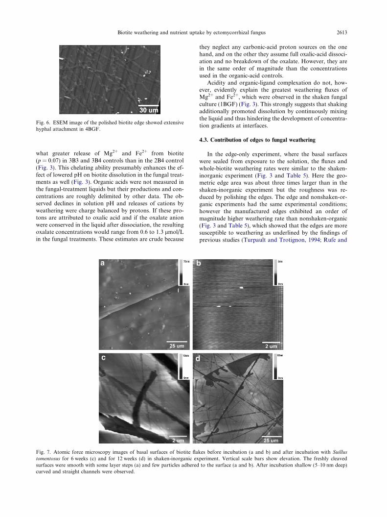
Fig. 6. ESEM image of the polished biotite edge showed extensivehyphal attachment in 4BGF.
Biotite weathering and nutrient uptake by ectomycorrhizal fungus 2613
what greater release of Mg2+ and Fe2+ from biotite(p = 0.07) in 3B3 and 3B4 controls than in the 2B4 control(Fig. 3). This chelating ability presumably enhances the ef-fect of lowered pH on biotite dissolution in the fungal treat-ments as well (Fig. 3). Organic acids were not measured inthe fungal-treatment liquids but their productions and con-centrations are roughly delimited by other data. The ob-served declines in solution pH and releases of cations byweathering were charge balanced by protons. If these pro-tons are attributed to oxalic acid and if the oxalate anionwere conserved in the liquid after dissociation, the resultingoxalate concentrations would range from 0.6 to 1.3 lmol/Lin the fungal treatments. These estimates are crude because
Fig. 7. Atomic force microscopy images of basal surfaces of biotite flatomentosus for 6 weeks (c) and for 12 weeks (d) in shaken-inorganic exsurfaces were smooth with some layer steps (a) and few particles adheredcurved and straight channels were observed.
they neglect any carbonic-acid proton sources on the onehand, and on the other they assume full oxalic-acid dissoci-ation and no breakdown of the oxalate. However, they arein the same order of magnitude than the concentrationsused in the organic-acid controls.
Acidity and organic-ligand complexation do not, how-ever, evidently explain the greatest weathering fluxes ofMg2+ and Fe2+, which were observed in the shaken fungalculture (1BGF) (Fig. 3). This strongly suggests that shakingadditionally promoted dissolution by continuously mixingthe liquid and thus hindering the development of concentra-tion gradients at interfaces.
4.3. Contribution of edges to fungal weathering
In the edge-only experiment, where the basal surfaceswere sealed from exposure to the solution, the fluxes andwhole-biotite weathering rates were similar to the shaken-inorganic experiment (Fig. 3 and Table 5). Here the geo-metric edge area was about three times larger than in theshaken-inorganic experiment but the roughness was re-duced by polishing the edges. The edge and nonshaken-or-ganic experiments had the same experimental conditions;however the manufactured edges exhibited an order ofmagnitude higher weathering rate than nonshaken-organic(Fig. 3 and Table 5), which showed that the edges are moresusceptible to weathering as underlined by the findings ofprevious studies (Turpault and Trotignon, 1994; Rufe and
kes before incubation (a and b) and after incubation with Suillus
periment. Vertical scale bars show elevation. The freshly cleavedto the surface (a and b). After incubation shallow (5–10 nm deep)

Fig. 8. AFM images of basal biotite surfaces after incubation with Suillus tomentosus in nonshaken experiments. (a) A few curved dissolutionchannel networks (1–3 lm wide and 10–30 nm deep) were found in both 2BGF and 3BGF after 12 weeks incubation. (b) Etch pit formationalong a lineation in 2BGF after 9 weeks incubation.
Fig. 9. The same basal biotite surface area was imaged with AFM in 3BGF before (a) and after (b) hyphal removal. The same steps can beseen before and after removal of hyphae, which shows that the biotite surface was not damaged by the removal. No dissolution channels weredetected beneath hyphae by this technique.
Table 5Dissolution rates of biotite reported in previous laboratory studies conducted with inorganic acids, and after 12-weeks’ incubation (this study)
Solution pH Acid Rate (mol m�2 s�1) Reference
2 Inorganic 8.2 ± 4.4 � 10�9a Bickmore (2001)1 Inorganic 6.1 ± 3.0 � 10�9a
1–4 Inorganic 3.4 � 10�11 Maslova et al. (2005)3 Inorganic 2.5 � 10�12 Taylor et al. (2000)2.8 Inorganic 7.9 � 10�12 Acker and Bricker (1992)2.8 Inorganic 4.0 to 6.3 � 10�10 Turpault and Trotignon (1994)2.8 Inorganic 4.0 to 7.9 � 10�11 Malmstrom and Banwart (1997)1–4 Inorganic 2.2 to 3.2 � 10�10 Kalinowski and Schweda (1996)
Dissolution rates of this study
2.8 Organic (by fungus) 2.1 ± 1.2 � 10�10 Shaken-inorganic (1BGF)3 Organic (by fungus) 6.0 ± 4.2 � 10�11 Nonshaken-inorganic (2BGF) and nonshaken-organic (3BGF)3 Organic (by fungus) 5.3 ± 2.8 � 10�10 Edge (4BGF)4 Inorganic 3.0 ± 1.7 � 10�11 Nonshaken-inorganic (2B4)3–4 Organic 6.2 ± 1.7 � 10�11 Nonshaken-organic (3B3, 3B4)3 and 5 Organic 7.6 ± 6.4 � 10�10a Edge (4B3 and 4B5)
a Normalized to geometric surface area of manufactured edges.
2614 Z. Balogh-Brunstad et al. / Geochimica et Cosmochimica Acta 72 (2008) 2601–2618
Hochella, 1999; Bickmore, 2001; Murakami et al., 2003).From a crystal physics point of view, relative to the smoothbasal cleavage surface, the edges of these crystals expose ex-tremely high densities of defects which are vulnerable to lo-
cal chemical attack; pit formation (nucleated atdislocations) and step dominated dissolution on cleavagesurfaces of single crystals such as calcite are classic exam-ples (Hillner et al., 1992a,b; Liang et al., 1996; Jordan

Biotite weathering and nutrient uptake by ectomycorrhizal fungus 2615
and Rammensee, 1998). Thus, the biotite edges would beexpected to weather at much higher rates than basal sur-faces. The possibility of whole biotite layers dissolving awayand leaving smooth surfaces behind was discounted, be-cause the size and thickness of the biotite booklets didnot change and the mass-balance chemistry did not supportsuch large dissolution rates.
Fungus attached extensively to the edge surfaces (Fig. 6)and to the flake edges as well (Fig. 4c). Despite their polish,the manufactured edges remained rough microscopicallywith scratch marks detectable with AFM. We were not ableto detect significant changes to the surfaces of the edgesafter 12-weeks’ incubation. Also, while fungal cover was de-tected, we did not see hyphae specifically grown into cracksand between layers. Thus our experiments did not showthat direct hyphal attachment to edges has any consequencefor weathering rates. This points toward the importance ofindirect fungal effects on the dissolution process, as dis-cussed above.
5. SUMMARY AND CONCLUSIONS
Estimated biotite-weathering rates by ectomycorrhizalfungus in liquid culture were within the range of literaturevalues for inorganic-acid weathering. Direct surface attach-ment of fungal hyphae did not prove to be important in li-quid-culture experimental conditions. The fungus did notgenerally mediate dissolution channel formation on basalsurfaces of biotite and did not transport nutrients via directhyphal contact-exchange process to tissue. In the shakenfungal treatment, channel formation was increased, but thiswas most likely caused by the combination of selective ele-mental uptake by fungus (weakening the biotite structure)and elimination of transport limitations to dissolution byshaking. Despite increase of fungal biomass in all experi-ments, K+, Mg2+, and Fe2+ preferentially partitioned intofungal biomass only in the shaken-inorganic experiment.However, a small amount of Fe2+ bioaccumulated in allnonshaken experiments. These findings did not supportour hypothesis that S. tomentosus directly weathers biotitesurfaces by strong attachment and by direct translocationof K+, Mg2+, and Fe2+ into fungal tissue leaving localizeddissolution channels behind on the basal surfaces.
Weathering fluxes in the nonshaken fungal treatmentsand organic-acid-buffered controls did not differ signifi-cantly. Our organic acid controls contained oxalic and citricacids, the most common exudates of ectomycorrhizal fungus.These results suggest that under these conditions, S. tomento-
sus indirectly weathers biotite surfaces by lowering the solu-tion pH and generating complex forming ligands, viaproduction of low molecular weight organic acids and by fun-gal respiration. This accelerated the dissolution of the min-eral independent of attachment and associated dissolutionfeatures. Even the relatively abundant dissolution channels,formed in the shaken experiment with fungus, could accountfor only 1/100 of the total weathering flux. This is consistentwith the results of edge-only experiments showing muchmore rapid weathering occurring at the edges of the flakes.The effect of dissolved organic acids seemed to have beenimportant in our experiments, and their concentrations
should be monitored in future work of this kind to furtherelucidate fungal-weathering processes.
Water was not limiting in our liquid-culture experi-ments. The saturated moisture potential and low-pH systemaffected hyphal morphology and may have eliminated muchof the contact-exchange advantage conferred by attachmentin natural systems where roots cannot uniformly acidifymineral surfaces. Under natural conditions of symbioticassociation, the hyphal system of EMF serves as conduitextension of plant roots and transports the required ele-ments for growth from minerals to plant parts (Marschner,2002). Thus plants function both as a source of carbohy-drate for the fungus and as a sink for cations weatheredby the fungus. The lack of such a chemical sink in ourexperiments may also have reduced the benefit of mineral-surface attachment.
ACKNOWLEDGMENTS
This work was supported by the NSF Grant EAR-03-12011 toC.K. Keller. We also acknowledge financial and logistical supportby the Department of Crop and Soil Sciences, Electron MicroscopyCenter and the School of Earth and Environmental Sciences atWashington State University; the Pacific Northwest National Lab-oratory—Environmental Microbiological Science Laboratory inRichland, WA; and the USDA Forest Service Research, Long-Term Ecosystem Productivity program at Corvallis, OR. We aregrateful for the help, advice and discussions of M. Fauci, R. Bol-ton, F.F. Foit, P.E. Rosenberg, B.W. Arey, A. Laskin, C. Davittand V. Lynch-Holm, to make the experiments and analytical workpossible. We also thank R.A. Gill and three anonymous reviewersfor useful comments on the manuscript.
REFERENCES
Acker J. G. and Bricker O. P. (1992) The influence of pH on biotitedissolution and alteration kinetics at low temperature. Geochim.
Cosmochim. Acta 56, 3073–3092.
Adams J. B., Palmer F. and Staley J. T. (1992) Rock weathering indeserts—mobilization and concentration of ferric iron bymicroorganisms. Geomicrobiol. J. 10, 99–114.
Adeyemi A. O. and Gadd G. M. (2005) Fungal degradation ofcalcium-, lead- and silicon-bearing minerals. BioMetals 18, 269–
281.
Arvieu J.-C., Leprince F. and Plassard C. (2003) Release of oxalateand protons by ectomycorrhizal fungi in response to P-deficiency and calcium carbonate in nutrient solution. Ann.
Forest Sci. 60, 209–215.
Balogh Zs. (2006) Chemical Hydrology of Vascular Plant Growth:
Role of Root–fungus Associations. Ph.D. Dissertation, Wash-ington State University.
Balogh-Brunstad Z., Keller C. K., Bormann B. T., O’Brien R.,Wang D. and Hawley G. (2008) Chemical weathering andchemical denudation dynamics through ecosystem developmentand disturbance. Global Biogeochem. Cycles 22, GB1007.
doi:10.1029/2007GB002957.
Balogh Zs., Keller C. K. and Dickinson J. T. (2003) Effect offungal–mineral interactions on chemical weathering and denu-dation processes—observations from experimental ecosystems.EOS (AGU Abstracts with Programs), 18 November 2003,Supplement, p. F245.
Balogh Zs., Keller C. K., Dickinson J. T., Wang D., Hawley G.and Coe T. (2004) Plant effect on chemical weathering and
2616 Z. Balogh-Brunstad et al. / Geochimica et Cosmochimica Acta 72 (2008) 2601–2618
denudation processes: experimental ecosystem studies. In Water
and Rock Interaction, vol. 2 (eds. R. B. Wanty and R. R. SealII). A.A. Balkema Publishers, Leiden, The Netherlands, pp.
1251–1254.
Banfield J. F., Barker W. W., Welch S. A. and Taunton A.(1999) Biological impact on mineral dissolution: applica-tion of the lichen model to understanding mineralweathering in the rhizosphere. Proc. Natl. Acad. Sci. USA
96, 3404–3411.
Barker W. W. and Banfield J. F. (1996) Biologically versusinorganically mediated weathering reactions: relationshipsbetween minerals and extracellular microbial polymers inlithobiontic communities. Chem. Geol. 132, 55–69.
Barker W. W., Welch S. A. and Banfield J. F. (1997) Biochemicalweathering of silicate minerals. In Geomicrobiology: Interac-
tions Between Microbes and Minerals, vol. 35 (eds. J. F. Banfield
and K. H. Nealson). Mineralogical Society of America,
Washington, DC, pp. 391–428.
Barker W. W., Welch S. A., Chu S. and Banfield J. F. (1998)Experimental observations of the effect of bacteria on alumi-nosilicate weathering. Am. Mineral. 83, 1551–1563.
Bennett P. C., Hiebert F. K. and Choi W. J. (1996) Microbialcolonization and weathering of silicates in a petroleum-contaminated groundwater. Chem. Geol. 132(1–4), 45–53.
Berner R. A. (2004) The Phanerozoic Carbon Cycle: CO2 and O2.Oxford University Press, New York.
Berner E. K. and Berner R. A. (1996) Global Environment: Water,
Air, and Geochemical cycles. Prentice Hall, New Jersey, p. 376.Bickmore B. R. (2001) Edge surface area normalized dissolution
rates of biotite and montmorillonite, In: Atomic Microscopy
Study of Clay Mineral Dissolution. Ph.D. Dissertation, VirginiaPolytechnic Institute and State University, pp. 49–76 (Chapter4).
Binnig G., Quate C. F. and Gerber C. (1986) Atomic forcemicroscope. Phys. Rev. Lett. 56, 930–933.
Blum J. D., Klaue A., Nezat C. A., Driscoll C. T., Johnson C. E.,Siccama T. G., Eagar C., Fahey T. J. and Likens G. E. (2002)Mycorrhizal weathering of apatite as an important calciumsource in base-poor forest ecosystems. Nature 417, 729–731.
Bormann F. H., Bowen W. B., Pierce R. S., Hamburg S. P., VoigtG. K., Ingersoll R. C. and Likens G. E. (1987) The HubbardBrook sandbox experiment. In Restoration Ecology (eds. R.Jordan, M. E. Gilpin and J. D. Aber). Cambridge University
Press, Cambridge, MA.
Boyle J. R. and Voigt G. K. (1973) Biological weathering of silicateminerals, implications for tree nutrition and soil genesis. Plant
Soil 38, 191–201.
Burford E. P., Fomina M. and Gadd G. M. (2003) Fungalinvolvement in bioweathering and biotransformation of rocksand minerals. Mineral. Mag. 67(6), 1127–1155.
Burgstaller W. and Schinner F. (1993) Leaching of metals withfungi. J. Biotechnol. 27, 91–116.
Calvaruso C., Turpault M.-P. and Frey-Klett P. (2006) Root-associated bacteria contribute to mineral weathering and tomineral nutrition in trees: a budgeting analysis. Appl. Environ.
Microbiol. 72(2), 1258–1266.
Castro I. M., Fietto J. L. R., Vieira R. X., Tropia M. J. M.,Campos L. M. M., Paniago E. B. and Brandao R. L. (2000)Bioleaching of zinc and nickel from silicates using Aspergillus
niger cultures. Hydrometallurgy 57, 39–49.
Crawford R. H., Floyed M. and Li C. Y. (2000) Degradation ofserpentine and muscovite rock minerals and immobilization ofcations by soil Penicillium spp. Phyton 40(2), 315–322.
Cui Y. Q., van der Lans R. G. J. M. and Luyben K. C. A. M.(1997) Effect of agitation intensities on fungal morphology ofsubmerged fermentation. Biotechnol. Bioeng. 55(5), 715–726.
Dean A. and Voss D. (1999) Design and Analysis of Experiments.Springer-Verlag, New York.
Dove P. M. and Platt F. M. (1996) Compatible real-time rates ofmineral dissolution by atomic force microscopy (AFM). Chem.
Geol. 127, 331–338.
Drever J. I. (1994) The effect of land plants on weathering rates ofsilicate minerals. Geochim. Cosmochim. Acta 58, 2325–2332.
Drever J. I. (1997) The Geochemistry of Natural Waters, 3rd ed.Prentice-Hall, New Jersey, p. 436.
Eckhardt F. E. W. (1985) Solubilization, transport, and depositionof mineral cations by microorganisms—efficient rock weath-ering agents. In The Chemistry of Weathering (ed. J. I. Drever).D. Reidel Publishing Company, Holland, pp. 161–173.
Ehrlich H. L. and Rossi G. (1990) Other bioleaching process. InMicrobial Mineral Recovery (eds. H. L. Ehrlich and C. L.Brierley). McGraw-Hill, New York, pp. 149–170.
Fanning D. S., Keramidas V. Z. and El-Desoky M. A. (1989)Micas. In: Minerals in Soil Environments (eds. J. B. Dixon andS. B. Weed). SSSA, Madison, Wisconsin, pp. 551–634.
Fenter P., Teng H., Geissbuhler P., Hanchar J. M., Nagy K. L. andSturchio N. C. (2000) Atomic-scale structure of orthoclase(001)–water interface measured with high resolution X-rayreflectivity. Geochim. Cosmochim. Acta 64, 3663–3673.
Fomina M., Burford E. P. and Gadd G. M. (2006) Fungaldissolution and transformation of minerals: significance fornutrient and metal mobility. In Fungi in Biogeochemical Cycles
(ed. G. M. Gadd). Cambridge University Press, Cambridge,
MA, pp. 236–266.
Gadd G. M. (1999) Fungal production of citric and oxalic acid:importance in metal speciation, physiology and biogeochemicalprocesses. Adv. Microb. Physiol. 41, 47–92.
Gadd G. M. (2007) Geomycology: biogeochemical transformationsof rocks, minerals, metals and radionuclides by fungi, biowea-thering and bioremediation. Mycol. Res. 111, 3–49.
Goldstein J. I., Newbury D. E., Echlin P., Joy D. C., Fiori C. andLifshin E. (1984) Scanning Electron Microscopy and X-ray
Microanalysis: A Text for Biologist, Material Scientist, and
Geologists. Plenum press, New York, pp. 123–203.Grantham M. C. and Dove P. M. (1996) Investigation of bacterial–
mineral interactions using Fluid Tapping ModeTM atomic forcemicroscopy. Geochim. Cosmochim. Acta 60, 2473–2480.
Grantham M. C., Dove P. M. and DiChristina T. J. (1997)Microbially catalyzed dissolution of iron and aluminum oxy-hydroxide mineral surface coatings. Geochim. Cosmochim. Acta
61, 4467–4477.
Hartman H., Sposito G., Yang A., Manne S., Gould S. A. C. andHansma P. K. (1990) Molecular scale imaging of clay mineralsurfaces with the atomic force microscope. Clays Clay Miner.
38, 337–342.
Heinonen-Tanski H. and Holopainen T. (1991) Maintenance ofectomycorrhizal fungi. In Techniques for Study of Mycorrhiza
Methods in Microbiology, vol. 23 (ed. Norris et al.). Academic
Press, New York, pp. 413–421.
Henderson M. E. K. and Duff R. B. (1963) The release of metallicand silicate ions from minerals, rocks, and soils by fungalactivity. J. Soil Sci. 14(2), 236–246.
Hillner P. E., Manne S., Gratz A. J. and Hansma P. K. (1992a)AFM images of dissolution and growth on a calcite crystal.Ultramicroscopy, 1387–1393.
Hillner P. E., Manne S., Gratz A. J. and Hansma P. K. (1992b)Atomic-scale imaging of calcite growth and dissolution in realtime. Geology 20, 359–362.
Hochella, Jr., M. F., Eggleston C. M., Elings V. B. and ThompsonM. S. (1990) Atomic structure and morphology of the albite(010) surface: an AFM and electron-diffraction study. Am.
Mineral. 75, 723–730.

Biotite weathering and nutrient uptake by ectomycorrhizal fungus 2617
Hoffland E., Kuyper T. W., Wallander H., Plassard C., Gorbush-ina A. A., Haselwandter K., Holmstrom S., Landeweert R.,Lundstrom U. S., Rosling A., Sen R., Smits M. M., van Hees P.A. W. and van Breemen N. (2004) The role of fungi inweathering. Front. Ecol. Environ. 2(5), 258–264.
Holland H. D., Lazar B. and McCaffrey M. (1986) Evolution of theatmosphere and oceans. Nature 320, 27–33.
Jenny H. (1980) The Soil Resource: Origin and Behavior. Springer-Verlag, New York.
Jones D. L., Dennis P. G., Owen A. G. and van Hees P. A. W.(2003) Organic acid behaviour in soils—misconceptions andknowledge gaps. Plant Soil 248, 31–41.
Jongmans A. G., Van Breemen N., Lundstrom U., van Hees P. A.W., Finlay R. D., Srinivasan M., Unestam T., Giesler R.,Melkerud P.-A. and Olsson M. (1997) Rock-eating fungi. Nature
389, 682–683.
Jordan G. and Rammensee W. (1998) Dissolution rates of calcite(101–4) obtained by scanning force microscopy: microtopog-raphy-based dissolution kinetics on surfaces with anisotropicstep velocities. Geochim. Cosmochim. Acta 62, 941–947.
Kalinowski B. E. and Schweda P. (1996) Kinetics of muscovite,phlogopite, and biotite dissolution and alteration at pH 1–4,room temperature. Geochim. Cosmochim. Acta 60, 367–385.
Kalinowski B. E., Liermann L. J., Givens S. and Brantley S. L.(2000) Rates of bacteria-promoted solubilization of Fe fromminerals: a review of problems and approaches. Chem. Geol.
169, 357–370.
Keller C. K., O’Brien R., Havig J. R., Smith J. L., Bormann B. T.and Wang D. (2006) Tree harvest in an experimental sandecosystem: hydrogeologic insight into nutrient cycling andsolute generation processes. Ecosystems 9, 634–646.
Klein C. and Hurlbut, Jr., C. S. (1993) Manual of Mineralogy, 21sted. John Wiley and Sons Inc., New York, pp. 221–249.
Kuwahara Y. (2001) Comparison of the surface structure of thetetrahedral sheets of muscovite and phlogopite by AFM. Phys.
Chem. Minerals 28, 1–8.
Landeweert R., Hoffland E., Finlay R. D., Kuyper T. W. and vanBreemen N. (2001) Linking plants to rocks: ectomycorrhizalfungi mobilize nutrients from minerals. Trends Ecol. Evol. 16(5),
248–254.
Leyval C. and Berthelin J. (1991) Weathering of mica by roots andrhizospheric microorganisms of pine. Soil Sci. Soc. Am. J. 55,
1009–1016.
Liang Y., Baer D. R., McCoy J. M., Amonette J. E. and LaFeminaJ. P. (1996) Dissolution kinetics at the calcite–water interface.Geochim. Cosmochim. Acta 60, 4883–4887.
Liermann L. J., Barnes A. S., Kalinowski B. E., Zhou X.and Brantley S. L. (2000) Microenvironments of pH inbiofilms grown an dissolving silicate surfaces. Chem. Geol.
171, 1–16.
Li Z. J., Shukla V., Fordyce A. P., Pedersen A. G., Wenger K. S.and Marten M. R. (2000) Fungal morphology and fragmenta-tion behavior in a fed-batch Aspergillus oryzae fermentation atthe production scale. Biotechnol. Bioeng. 70(3), 300–312.
Malmstrom M. and Banwart S. (1997) Biotite dissolution at 25 �C:the pH dependence of dissolution rate and stoichiometry.Geochim. Cosmochim. Acta 61, 2779–2799.
Marschner H. (2002) Mineral Nutrition of Higher Plants, 2nd ed.Academic Press, London.
Marx D. H. (1969) The influence of ectotrophic mycorrhizal fungion the resistance of pine roots to pathogenetic infection. I.Antagonism of mycorrhizal fungi to root pathogenic fungi andsoil bacteria. Phytopathology 59, 159–163.
Maslova M. V., Gerasimova L. G., Forsling W. and Makarov V.N. (2005) Mica behavior in aqueous solutions with pH 3–9.Geochem. Int. 43(7), 702–707.
Maurice P., Forsythe J., Hersman L. and Sposito G. (1996)Application of atomic-force microscopy to studies of microbialinteractions with hydrous Fe(III)-oxides. Chem. Geol. 132, 33–
43.
Molina R. and Palmer J. G. (1982) Isolation, maintenance andpure culture manipulation of ectomycorrhizal fungi. In Meth-
ods and Principles of Mycorrhizal Research (ed. N. C. Schenk).University of Florida, pp. 115–129.
Murakami T., Utsunomiya S., Yokoyama T. and Kasama T.(2003) Biotite dissolution processes and mechanisms in thelaboratory and in nature: early stage weathering environmentand vermiculization. Am. Mineral. 88, 377–386.
Paris F., Bonnaud P., Ranger J. and Lapeyrie F. (1995) In vitroweathering of phlogopite by ectomycorrhizal fungi. I. Effect ofK+ and Mg2+ deficiency on phyllosilicate evolution. Plant Soil
177, 191–201.
Paris F., Botton B. and Lapeyrie F. (1996) In vitro weathering ofphlogopite by ectomycorrhizal fungi. II. Effect of K+ and Mg2+
deficiency and N sources on accumulation of oxalate and H+.Plant Soil 179, 141–150.
Putnis A. (1992) Introduction to Mineral Sciences. CambridgeUniversity Press, Cambridge, MA, pp. 185–238.
Rosling A. (2003) Response of Ectomycorrhizal Fungi to Mineral
Substrates. Ph.D. Dissertation, Department of Forest Mycol-ogy and Pathology, Swedish University of Agricultural Sci-ences, Uppsala.
Rosling A., Lindahl B. D., Taylor A. F. S. and Finlay R. D. (2004)Mycelial growth and substrate acidification of ectomycorrhizalfungi in response to different minerals. FEMS Microbiol. Ecol.
47, 31–37.
Rufe E. and Hochella, Jr., M. F. (1999) Quantitative assessment ofreactive surface area of phlogopite during acid dissolution.Science 285, 874–876.
Silverman M. P. and Munoz E. F. (1970) Fungal attack on rock:solubilization and altered infrared spectra. Science 169, 985–987.
Smith S. E. and Read D. J. (1997) Mycorrhizal Symbioses, 2nd ed.Academic press, London.
Smits T. H. M., Wick L. Y., Harms H. and Keel C. (2003)Characterization of the surface hydrophobicity of filamentousfungi. Environ. Microbiol. 5(2), 85–91.
Sterflinger K. (2000) Fungi as geologic agents. Geomicrobiol. J. 17,
97–124.
SW-846 EPA Method 3050B (1995) Acid digestion of sediments,sludges, and soils In: Test Methods for Evaluating Solid Waste,3rd ed. U.S. EPA, Washington, DC.
Taylor A. S., Blum J. D., Lasaga A. C. and MacInnis I. N. (2000)Kinetics of dissolution and Sr release during biotite and phlogopiteweathering. Geochim. Cosmochim. Acta 64, 1191–1208.
Teng H. H., Fenter P., Cheng L. and Sturchio N. C. (2001)Resolving orthoclase dissolution processes with atomic forcemicroscopy and X-ray reflectivity. Geochim. Cosmochim. Acta
65, 3459–3474.
Turpault M.-P. and Trotignon L. (1994) The dissolution of biotitesingle crystals in dilute HNO3 at 24 �C: evidence of ananisotropic corrosion process of micas in acidic solutions.Geochim. Cosmochim. Acta 58, 2761–2775.
Unestam T. and Sun Y.-P. (1995) Extramatrical structures ofhydrophobic and hydrophilic ectomycorrhizal fungi. Mycor-
rhiza 5, 301–311.
Van Hees P. A. W., Jones D. L., Jentschke G. and Godbold D. L.(2003) Mobilization of aluminum, iron and silicon by Picea
abies and ectomycorrhizas in a forest soil. Eur. J. Soil Sci. 54,
101–111.
Vassilev N., Vassileva M. and Azcon R. (1997) Solubilization ofrock phosphate by immobilized Aspergillus niger. Bioresour.
Technol. 59, 1–4.

2618 Z. Balogh-Brunstad et al. / Geochimica et Cosmochimica Acta 72 (2008) 2601–2618
Velbel M. A. (1984) Weathering processes of rock-forming minerals.In: Short Course in Environmental Geochemistry (ed. M. E.Fleet). Mineralogical Association of Canada, pp. 67–111.
Wallander H. (2006) Mineral dissolution by ectomycorrhizal fungi.In Fungi in Biogeochemical Cycles (ed. G. M. Gadd). Cam-
bridge University Press, New York, NY, pp. 328–343.
Wallander H. and Hagerberg D. (2004) Do ectomycorrhizal fungihave a significant role in weathering of minerals in forest soil?Symbiosis 37, 249–257.
Wallander H. and Wickman T. (1999) Biotite and microcline aspotassium sources in ectomycorrhizal and non-ectomycorrhizalPinus sylvestris seedlings. Mycorrhiza 9, 25–32.
Watteau F. and Berthelin J. (1990) Iron solubilization by mycor-rhizal fungi producing siderophores. Symbiosis 9, 59–67.
Weed S. B., Davey C. B. and Cook M. G. (1969) Weathering ofmica by fungi. Soil Sci. Soc. Am. Proc. 33, 702–706.
Welch S. A. and Vandevivere P. (1994) Effect of microbial andother naturally occurring polymers on mineral dissolution.Geomicrobiol. J. 12, 227–238.
Welch S. A., Tauton A. E. and Banfield J. F. (2002) Effect ofmicroorganisms and microbial metabolites on apatite dissolu-tion. Geomicrobiol. J. 19, 343–367.
White A. F. and Brantley S. L. (1995) Chemical weathering rates of
silicate minerals. Reviews in Mineralogy. Mineralogical Societyof America, Washington, DC.
Yuan L., Huang J., Li X. and Christie P. (2004) Biologicalmobilization of potassium from clay minerals by ectomycor-rhizal fungi and eucalypt seedling roots. Plant Soil 262, 351–
361.
Associate editor: Roy A. Wogelius
Copyright © 2022 FDOKUMEN