
Aspects of silicate surface and bulk structure analysis using X-ray photoelectron spectroscopy (XPS)

Photoelectron Spectroscopy and Thermochemistry of the Peroxyformate Anion
Stephanie M. Villano, Nicole Eyet,† Scott W. Wren, G. Barney Ellison,Veronica M. Bierbaum,* and W. Carl Lineberger*JILA, UniVersity of Colorado and the National Institute of Standards and Technology and Department ofChemistry and Biochemistry, UniVersity of Colorado, Boulder, Colorado 80309-0440
ReceiVed: August 5, 2009; ReVised Manuscript ReceiVed: September 28, 2009
The 351.1 nm photoelectron spectrum of the peroxyformate anion has been measured. The photoelectronspectrum displays vibronic features in both the X2A′′ ground and A2A′ first excited states of the correspondingradical. Franck-Condon simulations of the spectrum show that the ion is formed exclusively in the trans-conformation. The electron affinity (EA) of the peroxyformyl radical was determined to be 2.493 ( 0.006eV, while the term energy splitting was found to be 0.783-0.020
+0.060 eV. Extended progressions in the C-OO (973( 20 cm-1) and O-O (1098 ( 20 cm-1) stretching modes are observed in the ground state of the radical.The fundamental frequency of the in-plane OCO bend was found to be 574 ( 35 cm-1. The gas-phase acidityof peroxyformic acid has been determined using an ion-molecule bracketing technique. On the basis of thesize of the trans- to cis- isomerization barrier, the measured acidity was assigned to the higher energy trans-conformer of the acid. The gas-phase acidity of the lower energy cis-conformer of peroxyformic acid wasfound from the measured acidity for the trans-form and a calculated energy correction: ∆aG298(cis-peroxyformicacid) ) 346.8 ( 3.3 kcal mol-1 and ∆aH298(cis-peroxyformic acid) ) 354.4 ( 3.3 kcal mol-1. Using a negativeion EA/acidity thermochemical cycle, the O-H bond dissociation energy (D0) values of the trans- and cis-conformers of peroxyformic acid to form the trans-radical were determined to be 94.0 ( 3.3 and 97.1 ( 3.3kcal mol-1, respectively. The heat of formation (∆fH298) of the trans-peroxyformyl radical was found to be-22.8 ( 3.5 kcal mol-1.
Introduction
The reaction of formyl radical with molecular oxygen hasreceived considerable attention, due to its prominent role intropospheric and hydrocarbon combustion processes.
A variety of techniques have been employed to determinethe room temperature reaction rate,1–13 and the formation of HO2
has been observed using laser magnetic resonance spectrosco-py.14 While there has been some controversy on the exacttemperature dependence6,9,10,13 as well as the effect of isotopicsubstitution,7,13 the consensus is that this reaction proceeds viaan activated peroxyformyl collision complex, [HC(O)OO]*.7,15–20
Hsu et al.15 have investigated the various product channels usingab initio molecular orbital and statistical calculations. Theseresults indicate that the vibrationally excited peroxyformylcomplex rapidly dissociates via a four-membered cyclic transi-tion state structure to form HO2 + CO. Direct formation of theseproducts via hydrogen abstraction was found to be negligibleat temperatures below 2000 K. At pressures below 2000 Torr,deactivation of the peroxyformyl radical by a third body collisionwas also found to be insignificant. The results of this study areconsistent with previous experimental observations, which foundthat the rate constant is pressure-independent over the range of5-1000 Torr.2,7,8
To date, the only experimental observation of the peroxy-formyl radical has been via matrix isolation IR-spectroscopy.In this technique, transient reaction intermediates, which readilydissociate in the gas phase, are stabilized through multiplecollisions when trapped in the matrix environment. In an earlystudy, this radical was produced by the photolysis of formal-dehyde in a solid oxygen matrix.17,18 The assignment of thespectral features, however, was complicated by the presence ofthe HO2 radical, which was likely present in the photolysis cage.More recently, the peroxyformyl radical has been studied bydischarge production and deposition of the formyl radical ontoa solid argon matrix followed by an association reaction withoxygen.19 Two vibrational transitions at 1821.5 and 957.3 cm-1
have been assigned to the CdO and C-OO stretching modes,respectively. The spectral assignments were confirmed by 18Oisotopic labeling and by electronic structure calculations.
In this work, we report the first observation of the isolatedperoxyformyl radical. This has been achieved through electronphotodetachment from the peroxyformyl anion. Analysis of thephotoelectron energy spectrum allows determinations of theelectron affinity, term energy splitting, and vibrational frequen-cies of the peroxyformyl radical and provides insight into themolecular structure of both the anion and radical species.Additionally, the gas-phase acidity of peroxyformic acid wasdetermined from ion-molecule bracketing experiments. Com-bining this gas-phase acidity with the peroxyformyl radicalelectron affinity in a negative ion thermochemical cycle21–24
allows the determination of the O-H bond dissociation energyin peroxyformic acid. Electronic structure calculations provideadditional insight into the structural and energetic aspects ofthe peroxy ion, the radical, and the parent acid. This paper
* Corresponding authors. E-mail: [email protected](V.M.B.); [email protected] (W.C.L.).
† Current address: Department of Chemistry, Saint Anselm College, 100Saint Anselm Dr. #1760, Manchester, New Hampshire 03102.
HCO + O2 f HO2 + CO (1)
J. Phys. Chem. A 2010, 114, 191–200 191
10.1021/jp907569w 2010 American Chemical SocietyPublished on Web 10/14/2009
represents a continuation of our studies on the thermochemistry,spectroscopy, and reactivity of peroxy compounds.25–32
Experimental Details
Negative Ion Photoelectron Spectroscopy. A detaileddescription of the anion photoelectron spectrometer that wasused in these studies has been provided elsewhere.28,33,34 Theperoxyformate anion [HC(O)OO-] was synthesized in a flowingafterglow source through the following series of ion-moleculereactions that occur sequentially downstream from a microwavedischarge source forming O- anions.
The final step is a gas-phase Baeyer-Villiger reaction,previously studied by DePuy and co-workers,35,36 in whichmethyl formate [HC(O)OCH3] or ethyl formate[HC(O)OCH2CH3] reacts with HOO-. The large number ofcollisions in the source region (∼0.5 Torr, 3-10 ms residencetime) ensures that the ions are thermalized to ∼298 K. Negativeions are gently extracted from the source region and passedthrough a Wien velocity filter for mass selection. The massselected ion beam is then crossed with a 351.1 nm (3.531 eV)line from a cw argon ion laser in an external build-up cavityproducing ∼100 W of circulating power. Photoelectrons thatare ejected into a small (4π/2000) solid angle perpendicular tothe plane formed by the photon and ion beams are analyzed bya hemispherical kinetic energy analyzer coupled to a positionsensitive detector. The photoelectron spectrum is a plot ofelectron kinetic energy (eKE) versus photoelectron counts. Theenergy scale is converted to electron binding energy (eBE) bysubtracting the electron kinetic energy from the photon energy.
The energy resolution ranges from 8 to 15 meV fwhm. Theabsolute energy scale is calibrated with the well-known electronaffinity (EA) of the sulfur atom37 and a small energy scalecompression factor (<1%), which is derived from the photo-electron spectra of reference ions S-, I-, and O- using the EAsof the corresponding atoms.33,37 Angular distribution measure-ments at a given electron kinetic energy (KE) were conductedby changing the angle (θ) between the electric field vector ofthe laser beam and the photoelectron collection axis. Theanisotropy parameter (), which characterizes the angulardistribution of the photoelectrons, is given by the expression38
where
In practice, we obtain the anisotropy parameter from mea-surements at two collection angles,
where I0° and I90° are the photoelectron counts at θ ) 0° and θ) 90°, respectively. In addition, spectra are collected at themagic angle (θ ) 54.7°), where the photoelectron intensityI(KE) is free from angular dependence.
While the electron energy resolution in these experiments is∼12 meV, the electron binding energy corresponding to anisolated transition can easily be determined to an accuracy of(5 meV (0.12 kcal mol-1). If that peak is associated with thetransition from the ground state of the anion to the ground stateof the neutral, the corresponding electron affinity can bedetermined with similar accuracy. The presence of unresolvedstructure reduces the accuracy of the experimental result, butthe Franck-Condon modeling of the full spectrum still allowsquantitative assessments of the experimental error in the bindingenergy measurements.
Flowing Afterglow-Selected Ion Flow Tube (FA-SIFT)Measurements. The gas-phase acidity (∆aG298) of peroxyformicacid was determined from an ion-molecule bracketing tech-nique39 using a tandem flowing afterglow-selected ion flow tubeinstrument, which has been described in detail elsewhere.40 Inthis method, the peroxyformate anion is allowed to react withseveral reference acids (AH) of known acidity. If rapid protontransfer is observed, then the reference compound is a strongeracid than peroxyformic acid [∆aG298(AH) <∆aG298(HC(O)OOH)]. If proton transfer is not observed, thenthe reference compound is a weaker acid than the peroxyformicacid [∆aG298(AH) > ∆aG298(HC(O)OOH)]. By examining theoccurrence or nonoccurrence of proton transfer with a series ofreference acids, an upper and lower bound can be placed onthe acidity of the peroxy acid. As a point of clarification, thegas-phase acidity is the Gibbs free-energy change for heterolyticbond cleavage, AH f A- + H+. Less energy is required toremove a proton from a stronger acid than from a weaker acid;thus, stronger acids have smaller free energy changes than doweaker acids. The enthalpy of deprotonation (∆aH298) can befound from eq 8, where ∆aS298, the entropy of deprotonation, isdetermined from electronic structure calculations.
Peroxyformate anions were formed in a flowing afterglowsource using the same synthesis scheme as in the abovephotodetachment experiments. Negative ions were extractedfrom the source region and focused into a quadrupole mass filter,which transmits ions with one specific mass-to-charge ratio. Themass selected ion beam was injected into a reaction flow tubewhereby the ions are thermalized to a ∼298 K rotational andvibrational Maxwell-Boltzmann distribution by collisions withHe buffer gas (∼0.5 Torr, 10 ms residence time). A measuredflow of the neutral reference acid was introduced into thereaction flow tube at various distances through a manifold ofinlets. The intensities of the reactant and product ions weremeasured using a triple quadrupole mass filter coupled to anelectron multiplier as a function of reaction time. Neutralreactant flow rates were measured by monitoring the pressurechange versus time in a calibrated volume system.
An experimental complication is that, despite injecting theperoxyformate anions with minimal energy, fragment ions [OH-,HOO-, and HC(O)O-] formed as a result of collision-induced
O- + CH4 f CH3 + OH- (2)
OH- + H2O2 f H2O + HOO- (3)
HOO- + HC(O)OR f HC(O)OO- + ROH (4)
dσdΩ
)σtot
4π[1 + (KE)P2(cos θ)] (5)
P2(cos θ) ) 3 cos2 θ - 12
and - 1 e e 2
(6)
(KE) )I0 - I90
1/2I0 + I90(7)
∆aH298(HC(O)OOH) ) ∆aG298(HC(O)OOH) +T∆aS298(HC(O)OOH) (8)
192 J. Phys. Chem. A, Vol. 114, No. 1, 2010 Villano et al.
dissociation are also present in the reaction flow tube. Sincethe basicity of some of these fragment ions is greater than thatof the parent anion, the deprotonated reference acid wasobserved even when it is less acidic than peroxyformic acid.Additionally, depletion of the parent peroxyformate anion wasalso generally observed, since it reacts by neutral O atom loss.In order to determine whether proton transfer occurs betweenthe peroxyformate anion and the reference acid, the intensitiesof all ions were monitored as a function of time and thecontribution from the fragment ions to the deprotonated refer-ence acid signal was removed.
Electronic Structure Calculations. Two different theoreticalmodels, both provided in the Gaussian 03 suite of programs,41
were used to evaluate structural and energetic aspects of theperoxyformate anion as well as the corresponding protonatedandradicalspecies.DFTcalculations[B3LYP/6-311++G(d,p)]42–44
were used to calculate optimized geometries, harmonic vibra-tional frequencies (unscaled), and the rotational constants. TheG3MP2B3 composite technique45 was used to investigatevarious thermodynamic properties of these species. In thistechnique, geometries and zero point energies are calculated atthe B3LYP/6-31G(d) level of theory followed by a series ofwell-defined ab initio single-point energy calculations. Thismethod has been tested on a variety of hydrocarbons, and theaverage deviation from experiment was reported to be 1.25 kcalmol-1.45 The accuracy of this method was independentlyevaluated in this work with a test set of molecules that werechosen on the basis of their similarity to the peroxy compoundstudied here [HOO-, CH3OO-, C2H5OO-, HC(O)O-, andCH3C(O)O-, as well as the corresponding protonated and radicalspecies]. The average deviation from experiment for a set ofcalculated electron affinities, gas-phase acidities, and bonddissociation enthalpies was found to be 1.5 kcal mol-1.46
Simulated photoelectron spectra were obtained by calculatingthe Franck-Condon (FC) factors at a vibrational temperatureof 298 K with the PESCAL program.47 These simulations arebased on the B3LYP/6-311++G(d,p)-optimized geometries andnormal modes for the anion and corresponding radical species,while electron binding energies are determined from theG3MP2B3 calculations.
Results and Discussion
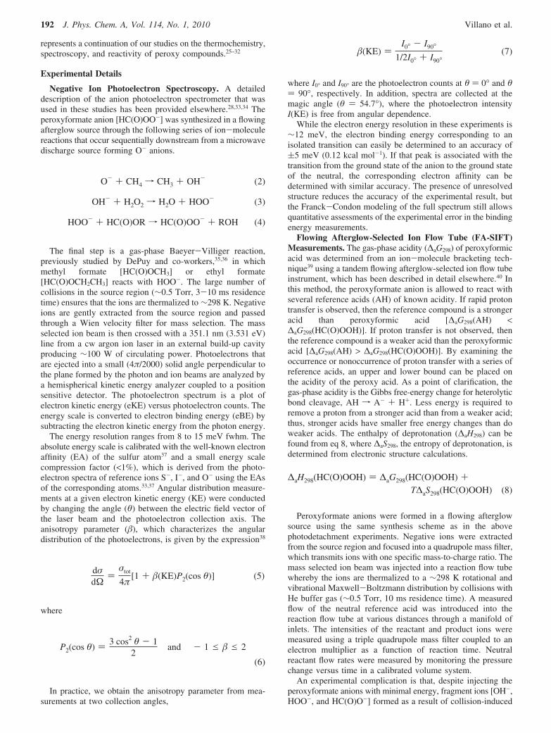
Photoelectron Spectra of the Peroxyformate Ion. The 351.1nm magic angle photoelectron spectrum of the m/z 61 ionproduced from the reaction of HOO- with ethyl formate isshown in Figure 1a. The same spectral profile is observed whenthe m/z 61 ion is produced from the reaction of HOO- withmethyl formate. The ion-molecule reactivity studies of DePuyand co-workers35,36 indicate that these reactions proceed via aBaeyer-Villiger reaction to produce the peroxyformate anionrather than the bicarbonate anion, which is the more stableisomer. While our results do not eliminate the possible formationof the latter species, the spectrum in Figure 1a must arise onlyfrom the peroxyformate anion. The basis for this assertion isthat the electron affinity of the bicarbonate radical is ∼3.9 eV,48
and hence, photodetachment of the bicarbonate anion is notenergetically possible with the ∼3.5 eV photon energy employedin this investigation. The photoelectron angular distribution wasmeasured and the anisotropy parameter () is negative acrossthe entire spectrum, consistent with photodetachment from aπ-type orbital.26,28 The most striking feature of the spectrum isthe series of intense peaks (b, e, g, h, i, j, k, and m), which arespaced by roughly 1000 cm-1. With the exception of peak b,the width of these peaks is ∼50 meV (fwhm), substantially
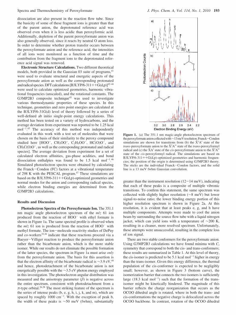
greater than the instrument resolution (12-14 meV), indicatingthat each of these peaks is a composite of multiple vibronictransitions. To confirm this statement, the same spectrum wascollected with slightly higher resolution (∼8 meV) but lowersignal-to-noise ratio; the lower binding energy portion of thishigher resolution spectrum is shown in Figure 2a. At thisresolution, it is evident that at least peaks e, g, and h havemultiple components. Attempts were made to cool the anionbeam by surrounding the source flow tube with a liquid nitrogenjacket, which can yield ions with a temperature of ∼200 K,resulting in a cleaner, more resolved spectrum. Unfortunately,these attempts were unsuccessful, resulting in the complete lossof ion signal.
There are two stable conformers for the peroxyformate anion.Using G3MP2B3 calculations we have found minima with Cs
symmetry that correspond to both the cis- and trans-conformers;these results are summarized in Table 1. At this level of theory,the cis-isomer is predicted to be 5.1 kcal mol-1 higher in energythan the trans-isomer. Given this energy difference, the thermalpopulation of the cis-conformer is expected to be negligiblysmall; however, as shown in Figure 3 (bottom curve), theisomerization barrier that connects the two isomers is sufficientlylarge (18.3 kcal mol-1) such that the formation of the trans-isomer might be kinetically hindered. The magnitude of thisbarrier reflects the charge reorganization that occurs as theOCOO dihedral angle is rotated, where in both the trans- andcis-conformations the negative charge is delocalized across theOCOO backbone. In contrast, rotation of the OCOO dihedral
Figure 1. (a) The 351.1 nm magic-angle photoelectron spectrum oftheperoxyformateanioncollectedwith∼13meVresolution.Franck-Condonsimulations are shown for transitions from (b) the X1A′ state of thetrans-peroxyformate anion to the X2A′′ state of the trans-peroxyformylradical and (c) the XA′ state of the cis-peroxyformate anion to the X2A′′state of the cis-peroxyformyl radical. The simulations are based onB3LYP/6-311++G(d,p)-optimized geometries and harmonic frequen-cies; the position of the origin is determined using G3MP2B3 theory.The sticks are the individual Franck-Condon factors, and the solidline is a 13 meV fwhm Gaussian convolution.
Spectra and Thermochemistry of Peroxyformate J. Phys. Chem. A, Vol. 114, No. 1, 2010 193
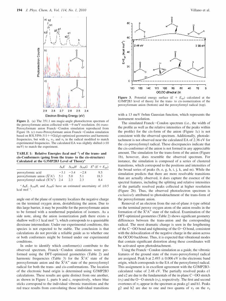
angle out of the plane of symmetry localizes the negative chargeon the terminal oxygen atom, destabilizing the anion. Due tothis large barrier, it may be possible for the peroxyformate anionto be formed with a nonthermal population of isomers. As aside note, along the anion isomerization path there exists ashallow well (<1 kcal mol-1), which corresponds to a quasistabledioxirane intermediate. Under our experimental conditions, thisspecies is not expected to be stable. The conclusion is thatcalculations do not provide a reliable guide as to whether oneor both conformers might be formed under our experimentalconditions.
In order to identify which conformer(s) contribute to theobserved spectrum, Franck-Condon simulations were per-formed using the DFT-optimized geometries (Table 2) andharmonic frequencies (Table 3) for the X1A′ state of theperoxyformate anion and the X2A′′ state of the peroxyformylradical for both the trans- and cis-conformations. The locationof the electronic band origin is determined using G3MP2B3calculations. These results are quite distinct from one another,as shown in Figure 1, parts b and c, respectively, where bluesticks correspond to the individual vibronic transitions and thered trace results from convoluting those individual transitions
with a 13 meV fwhm Gaussian function, which represents theinstrument resolution.
The simulated Franck-Condon spectrum (i.e., the width ofthe profile as well as the relative intensities of the peaks withinthe profile) for the cis-form of the anion (Figure 1c) is notconsistent with the observed spectrum. Additionally, photode-tachment is not observed near the calculated EA of 2.36 eV forthe cis-peroxyformyl radical. These discrepancies indicate thatthe cis-conformer of the anion is not formed in any appreciableamount. The simulation for the trans-form of the anion (Figure1b), however, does resemble the observed spectrum. Forinstance, the simulation is composed of a series of clusteredtransitions, which correspond to the positions and intensities ofthe broad series of peaks (b, e, g, h, i, j, k, and m). While thesimulation predicts that there are more resolvable transitionsthan are actually observed, it does capture the essence of thespectral features, including the splitting and relative intensitiesof the partially resolved peaks collected at higher resolution(Figure 2b). Thus, the observed photoelectron spectrum isexclusiVely attributed to photodetachment of the trans-form ofthe peroxyformate anion.
Removal of an electron from the out-of-plane π-type orbitalon the terminal peroxy oxygen atom of the anion results in theformation of the X2A′′ state of the radical. Examination of theDFT-optimized geometries (Table 2) shows significant geometrydifferences between the trans-anion and the correspondingradical. The most dramatic change is seen in the lengtheningof the C-OO bond and tightening of the O-O bond, consistentwith the delocalization of the negative charge in the anion acrossthe OCOO backbone. Thus, it is expected that vibrational modesthat contain significant distortion along these coordinates willbe activated upon photodetachment.
Using the Franck-Condon simulation as a guide, the vibronicfeatures of the ground state of the trans-peroxyformyl radicalare assigned. Peak b at 2.493 ( 0.006 eV is the electronic bandorigin, which corresponds to the EA of the peroxyformyl radical.This assignment is in excellent agreement with the G3MP2B3-calculated value of 2.48 eV. The partially resolved peaks e1and e2 are due to the fundamentals of the in-plane C-OO stretch(ν5) and the O-O stretch (ν4), respectively. The first and secondovertones of ν5 appear in the spectrum as peaks g1 and h1. Peaksg2 and h2 are due to one and two quanta of ν5 on the ν4
Figure 2. (a) The 351.1 nm magic-angle photoelectron spectrum ofthe peroxyformate anion collected with ∼9 meV resolution. (b) trans-Peroxyformate anion Franck-Condon simulation reproduced fromFigure 1b. (c) trans-Peroxyformate anion Franck-Condon simulationbased on B3LYP/6-311++G(d,p)-optimized geometries and harmonicfrequencies, but with ν4, ν5, and ν6 in the radical modified to matchexperimental frequencies. The calculated EA was slightly shifted (<10meV) to match the experiment.
TABLE 1: Relative Energies (kcal mol-1) of the trans- andcis-Conformers (going from the trans- to the cis-structure)Calculated at the G3MP2B3 Level of Theorya
∆0E ∆298H ∆298G Eq (E + Ezpt)
peroxyformic acid -3.1 -3.4 -2.8 9.5peroxyformate anion (X2A′) 5.1 5.0 5.1 18.3peroxyformyl radical (X2A′′) 2.4 2.3 2.3 8.4
a ∆0E, ∆298H, and ∆298G have an estimated uncertainty of (0.5kcal mol-1.
Figure 3. Potential energy surface (E + Ezpt) calculated at theG3MP2B3 level of theory for the trans- to cis-isomerization of theperoxyformate anion (bottom) and the peroxyformyl radical (top).
194 J. Phys. Chem. A, Vol. 114, No. 1, 2010 Villano et al.
fundamental (40150
1 and 40150
2, respectively), while peak h3 is dueto two quanta of ν4 on the ν5 fundamental (40
2501). The extended
progressions in these two modes enable a more accuratedetermination of fundamental vibrational frequencies, as wellas crude measurements of the anharmonicity of each mode.Using the expression for the energy differences betweenvibrational levels of an anharmonic oscillator,
we can estimate the harmonic frequencies (ω) and anharmonicityconstants () for these two modes; here, g(n)-g(0) is the energydifference between n and 0 quanta of a given mode.
The harmonic frequency and anharmonicity constant for theC-OO stretch (ν5) were found using the 50
n (n ) 0-3) and40
150n (n ) 0-2) progressions to be ω5 ) 973 ( 20 cm-1 and 5
) 4 ( 3 cm-1. The harmonic frequency and anharmonicity termfor the O-O stretch (ν4) were found using the 40
n501 (n ) 0-2)
progression to be ω4 )1098 ( 20 cm-1 and 4 )10 ( 5 cm-1.Using these parameters, the ν5 and ν4 fundamental frequencieswere determined to be 966 ( 20 and 1078 ( 20 cm-1,respectively. The intense, broad series of peaks primarily derivesits intensity from an extended progression in the ν5 C-OOstretch (50
n), along with two combination band progressions (40150
n
and 40250
n). At larger binding energies, higher order combinationbands (40
m50n) congest the spectrum. The low intensity peak d is
the fundamental of the ν6 OCO bend at 574 ( 35 cm-1, whilepeaks f and h are due to combination bands of one quanta ofthe ν6 OCO bend on the ν5 progression (50
n601, where n ) 1, 2).
Peaks a and c are due to excited anion vibrations. Peak a is the
fundamental of the anion ν6 OCO bend at 587 ( 30 cm-1, whilepeak c is a sequence band of one quanta of the anion ν6 OCObend with the radical ν5 C-OO stretch (50
1610). Since we were
unable to change the anion temperature, the assignment of theseanion hot bands is based solely on the spectral simulation.
Following the analysis of the X2A′′ state vibronic features, itappears that the discrepancy between the simulated and theexperimental spectrum largely stems from the overestimate ofthe O-O stretching frequency (see Table 3). In actuality, thefrequencies of O-O and C-OO stretches are closer to oneanother than predicted and, therefore, these two modes are onlypartially resolvable. Additionally, at higher binding energiesanharmonic effects are manifested and the harmonic simulationoverestimates the peak positions. Using the experimentallydetermined electron binding energy, frequencies, and anharmo-nicity constants in the Franck-Condon simulation (Figure 2c)greatly improves its quality; this improvement gives considerableconfidence that all of the major features in the trans-peroxy-formate anion shown in Figure 1a can be assigned to vibrationallevels of the ground state of the radical.
The sole exception to this claim is the narrow peak l at 3.27eV eBE, which is notably absent in the simulation in Figure1b. The narrowness of this peak (as well as the locationmentioned above) strongly suggest that it does not arise fromtransitions to the X2A′′ ground state of the peroxyformyl radical.Moreover, the photoelectron angular distribution associated withthis peak does not fit smoothly into the progression seen forthe other ground state vibrational peaks, but is less stronglypeaked perpendicular to the laser electric field. Inconsistenciessuch as these are often an indication that another electronic stateis appearing in the spectrum. Indeed, the position of peak l isconsistent with a vibronic transition to the A2A′ excited state
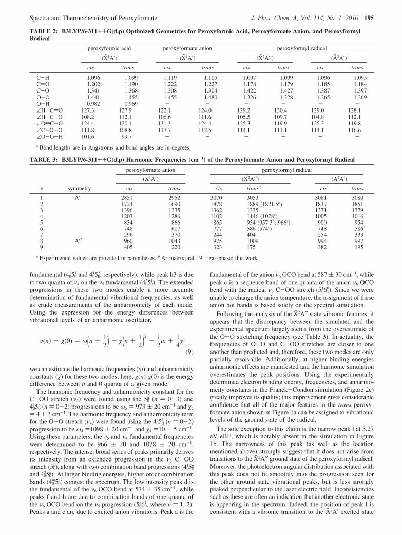
TABLE 2: B3LYP/6-311++G(d,p) Optimized Geometries for Peroxyformic Acid, Peroxyformate Anion, and PeroxyformylRadicala
peroxyformic acid peroxyformate anion peroxyformyl radical
(X1A′) (X1A′) (X2A′′) (A2A′)
cis trans cis trans cis trans cis trans
C-H 1.096 1.099 1.119 1.105 1.097 1.099 1.096 1.095CdO 1.202 1.190 1.222 1.227 1.178 1.179 1.185 1.184C-O 1.341 1.368 1.308 1.304 1.422 1.427 1.387 1.397O-O 1.441 1.455 1.455 1.480 1.326 1.328 1.365 1.369O-H 0.982 0.969 - - - - - -∠H-CdO 127.3 127.9 122.1 124.0 129.2 130.4 129.0 128.1∠H-C-O 108.2 112.1 106.6 111.6 105.5 109.7 104.8 112.1∠OdC-O 124.4 120.1 131.3 124.4 125.3 119.9 125.3 119.8∠C-O-O 111.8 108.8 117.7 112.5 114.1 111.1 114.1 116.6∠O-O-H 101.6 99.7 - - - - - -a Bond lengths are in Angstroms and bond angles are in degrees.
TABLE 3: B3LYP/6-311++G(d,p) Harmonic Frequencies (cm-1) of the Peroxyformate Anion and Peroxyformyl Radical
peroxyformate anion peroxyformyl radical
(X1A′) (X2A′′) (A2A′)ν symmetry cis trans cis transa cis trans
1 A′ 2851 2952 3070 3053 3081 30802 1724 1690 1878 1889 (1821.5b) 1837 18513 1396 1335 1362 1335 1371 13794 1203 1286 1102 1146 (1078c) 1005 10165 834 866 865 954 (957.3b; 966c) 900 9546 748 607 777 586 (574c) 748 5867 296 370 244 404 254 3338 A′′ 960 1043 975 1009 994 9979 405 220 323 175 382 195
a Experimental values are provided in parentheses. b Ar matrix; ref 19. c gas-phase: this work.
g(n) - g(0) ) ω(n + 12) - (n + 1
2)2- 1
2ω + 1
4
(9)
Spectra and Thermochemistry of Peroxyformate J. Phys. Chem. A, Vol. 114, No. 1, 2010 195
in the peroxyformyl radical. The G3MP2B3 calculations locatethis state 0.79 eV above the ground state. This electronic stateof the radical corresponds to removal of an electron from thein-plane p-orbital on the terminal peroxy oxygen atom ofthe anion. While only a small portion of this excited state profileis observed, the relative intensities predicted by Franck-Condonsimulations for the transition from the X1A′ state of the anionto the A2A′ state of the radical (provided in the SupportingInformation) indicate that this peak is either the electronic bandorigin or the fundamental of the COO bend (ν7), which has acalculated frequency of 333 cm-1 (41 meV). The latter appearsmore likely. Thus, we report that the A2A′ state of the trans-peroxyformyl radical lies 0.783-0.020
+0.060 eV above the ground state;the asymmetric error bar accounts for the possibility that peakl might correspond to the origin of the A2A′ state rather than toone quantum of the COO bend.
Gas-Phase Acidity Measurements. The gas-phase acidityof peroxyformic acid was determined through ion-moleculebracketing experiments. This method is employed when protontransfer equilibrium constant measurements are not feasible; inthis case, the neutral peroxyformic acid reagent is not availablein pure form. In the ion-molecule bracketing technique, theperoxyformate anion is allowed to react with a series ofreference acids and the occurrence or nonoccurrence of protontransfer sets a lower or upper bound on the unknown acidity,respectively. For these bracketing experiments, the peroxyfor-mate anion is formed using the same reaction scheme as in theabove photodetachment studies.
The gas-phase acidity of peroxyformic acid was bracketedagainst five reference acids: formic acid (338.6 ( 0.5 kcalmol-1), acetic acid (341.2 ( 0.5 kcal mol-1), hydrogen sulfide(344.89 ( 0.02 kcal mol-1), tert-butylthiol (346.7 ( 0.5 kcalmol-1), and isopropylthiol (347.6 ( 0.5 kcal mol-1).49 For thereactions of peroxyformate anion with tert-butylthiol andisopropylthiol, proton transfer does not occur; instead thesereactions proceed exclusively by neutral O atom loss to formthe formate anion. Proton transfer was the only pathwayobserved in the reactions of peroxyformate anion with formicacid and acetic acid. For the reaction with formic acid, protontransfer was confirmed by evaluating the reaction with formicacid-d1, DCO2H. In this case, an observed increase in m/z 46 isdue to proton transfer, whereas the absence of m/z 45 indicatesthat neutral O atom loss from the peroxyformate anion doesnot occur. The reaction with hydrogen sulfide primarily proceedsby O atom loss to form the formate ion. Only a minor amountof the proton transfer product, HS-, was observed, suggestingthat the proton transfer pathway is slightly endothermic. On thebasis of these results, the acidity of peroxyformic acid is foundto be ∆aG298 ) 344.0 ( 3.3 kcal mol-1, between that of aceticacid and tert-butylthiol, where the error bar spans the uncertaintyin the acidities of the upper and lower bound reference acids.
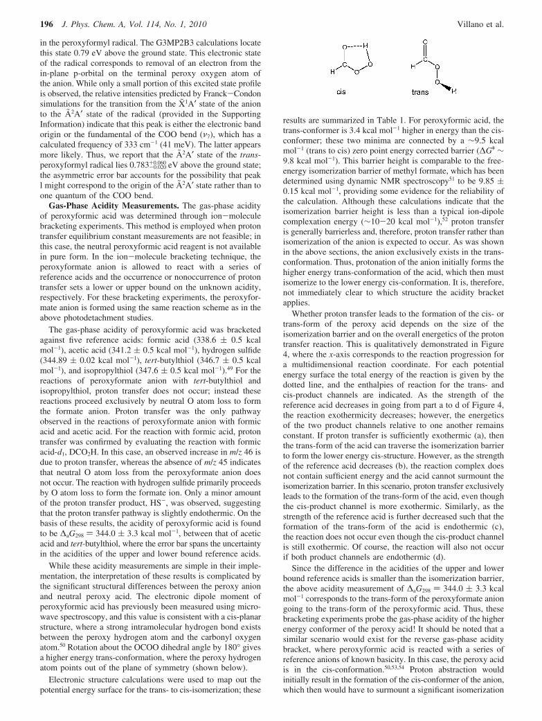
While these acidity measurements are simple in their imple-mentation, the interpretation of these results is complicated bythe significant structural differences between the peroxy anionand neutral peroxy acid. The electronic dipole moment ofperoxyformic acid has previously been measured using micro-wave spectroscopy, and this value is consistent with a cis-planarstructure, where a strong intramolecular hydrogen bond existsbetween the peroxy hydrogen atom and the carbonyl oxygenatom.50 Rotation about the OCOO dihedral angle by 180° givesa higher energy trans-conformation, where the peroxy hydrogenatom points out of the plane of symmetry (shown below).
Electronic structure calculations were used to map out thepotential energy surface for the trans- to cis-isomerization; these
results are summarized in Table 1. For peroxyformic acid, thetrans-conformer is 3.4 kcal mol-1 higher in energy than the cis-conformer; these two minima are connected by a ∼9.5 kcalmol-1 (trans to cis) zero point energy corrected barrier (∆Gq ∼9.8 kcal mol-1). This barrier height is comparable to the free-energy isomerization barrier of methyl formate, which has beendetermined using dynamic NMR spectroscopy51 to be 9.85 (0.15 kcal mol-1, providing some evidence for the reliability ofthe calculation. Although these calculations indicate that theisomerization barrier height is less than a typical ion-dipolecomplexation energy (∼10-20 kcal mol-1),52 proton transferis generally barrierless and, therefore, proton transfer rather thanisomerization of the anion is expected to occur. As was shownin the above sections, the anion exclusively exists in the trans-conformation. Thus, protonation of the anion initially forms thehigher energy trans-conformation of the acid, which then mustisomerize to the lower energy cis-conformation. It is, therefore,not immediately clear to which structure the acidity bracketapplies.
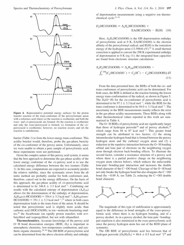
Whether proton transfer leads to the formation of the cis- ortrans-form of the peroxy acid depends on the size of theisomerization barrier and on the overall energetics of the protontransfer reaction. This is qualitatively demonstrated in Figure4, where the x-axis corresponds to the reaction progression fora multidimensional reaction coordinate. For each potentialenergy surface the total energy of the reaction is given by thedotted line, and the enthalpies of reaction for the trans- andcis-product channels are indicated. As the strength of thereference acid decreases in going from part a to d of Figure 4,the reaction exothermicity decreases; however, the energeticsof the two product channels relative to one another remainsconstant. If proton transfer is sufficiently exothermic (a), thenthe trans-form of the acid can traverse the isomerization barrierto form the lower energy cis-structure. However, as the strengthof the reference acid decreases (b), the reaction complex doesnot contain sufficient energy and the acid cannot surmount theisomerization barrier. In this scenario, proton transfer exclusivelyleads to the formation of the trans-form of the acid, even thoughthe cis-product channel is more exothermic. Similarly, as thestrength of the reference acid is further decreased such that theformation of the trans-form of the acid is endothermic (c),the reaction does not occur even though the cis-product channelis still exothermic. Of course, the reaction will also not occurif both product channels are endothermic (d).
Since the difference in the acidities of the upper and lowerbound reference acids is smaller than the isomerization barrier,the above acidity measurement of ∆aG298 ) 344.0 ( 3.3 kcalmol-1 corresponds to the trans-form of the peroxyformate aniongoing to the trans-form of the peroxyformic acid. Thus, thesebracketing experiments probe the gas-phase acidity of the higherenergy conformer of the peroxy acid! It should be noted that asimilar scenario would exist for the reverse gas-phase aciditybracket, where peroxyformic acid is reacted with a series ofreference anions of known basicity. In this case, the peroxy acidis in the cis-conformation.50,53,54 Proton abstraction wouldinitially result in the formation of the cis-conformer of the anion,which then would have to surmount a significant isomerization
196 J. Phys. Chem. A, Vol. 114, No. 1, 2010 Villano et al.
barrier (Table 1) to form the lower energy trans-conformer. Thisacidity bracket would, therefore, probe the gas-phase basicityof the cis-conformer of the peroxy anion. Unfortunately, sincewe were unable to obtain a pure sample of peroxyformic acid,these experiments were not performed.
Given the complex nature of this peroxy acid system, it seemsthat the best approach to determine the gas-phase acidity of thelower energy conformer of the cis-peroxy acid is to use thecalculated energy difference between the two isomers (Table1). In this case, computations are expected to accurately predictthe relative stability, since the systematic errors from the abinitio method are probably similar for both conformers and,therefore, cancel out in the energy difference. On the basis ofthis approach, the gas-phase acidity of cis-peroxyformic acidis determined to be 346.8 ( 3.3 kcal mol-1. Combining ourresults with the calculated entropy of deprotonation (∆aS298)allows for the determination of the enthalpy of deprotonation,∆aH298(cis-HC(O)OOH) ) 354.4 ( 3.3 kcal mol-1; ∆aH298(trans-HC(O)OOH) ) 351.1 ( 3.3 kcal mol-1,55 where in both casesdeprotonation leads to the trans-form of the anion. It should benoted that peroxyformic acid is slightly more acidic thancarbonic acid (HOC(O)OH); in ion-molecule bracketing stud-ies,48 the bicarbonate ion rapidly proton transfers with tert-butylthiol and isopropylthiol, but not with ethanethiol.
Thermochemistry. Accurate knowledge of the peroxy acidO-H bond dissociation energies (BDE, D0) is important inatmospheric chemistry, low-temperature combustion, and syn-thetic organic chemistry.56–58 The OH BDE of peroxyformic acidcan be determined from the above electron affinity and enthalpy
of deprotonation measurements using a negative ion thermo-chemical cycle.21–24
Here, ∆aHo(HC(O)OOH) is the OH deprotonation enthalpyof peroxyformic acid at 0 K, EA(HC(O)OO) is the electronaffinity of the peroxyformyl radical, and IE(H) is the ionizationenergy of the hydrogen atom (13.59844 eV).59 A small thermalcorrection is applied to convert the 298 K experimental enthalpyof deprotonation to 0 K (eq 11); the integrated heat capacitiesare found from electronic structure calculations.
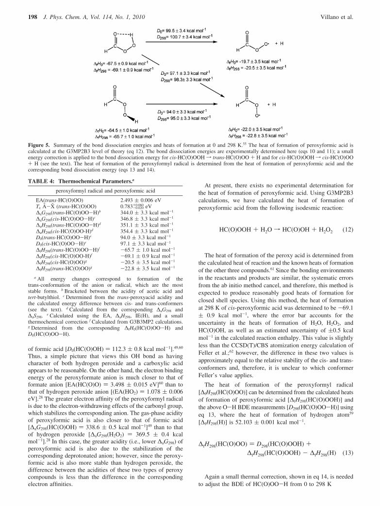
From the data presented here, the BDEs of both the cis- andtrans-conformers of peroxyformic acid can be determined. Forboth cases, the BDE is defined as the reaction forming the lowestenergy trans-conformation of the radical, as shown in Figure 5.The D0(O-H) for the cis-conformer of peroxyformic acid isdetermined to be 97.1 ( 3.3 kcal mol-1, while the BDE for thetrans-conformer is determined to be 94.0 ( 3.3 kcal mol-1. Theuncertainty in the BDE measurements mainly reflects the errorin the gas-phase acidity measurements. These BDEs along withother thermochemical values reported in this work are sum-marized in Table 4.
The O-H BDEs of peroxyformic acid are significantly largerthan those of hydrogen peroxide28 and of peroxy alcohols,26
which range from 84 to 87 kcal mol-1. This greater bondstrength can be attributed to two factors: (1) the strongintramolecular hydrogen bond that is formed between the peroxyhydrogen atom and the carbonyl oxygen atom and (2) areduction in the repulsive interaction between the O-H bondingorbital and lone pair of electrons on the neighboring oxygenatom through electron back-bonding effects. To illustrate thesecond factor, consider a resonance structure of a peroxy acidwhere there is a partial positive charge on the neighboringoxygen atom (shown below), which reduces the unfavorablelone-pair-bonding-pair interaction and increases the doublebond character of the C-OO bond. Cleavage of the O-H bondnot only breaks the hydrogen bond but also elongates the C-OObond (by ∼0.09 Å; see Table 2), reducing the C-OO doublebond character.
The magnitude of this type of stabilization is approximatelyequal to the difference in bond strengths of the trans-peroxy-formic acid, where there is no hydrogen bonding, and of aperoxy alcohol. As in a peroxy alcohol, the lone-pair-bonding-pair repulsion is also minimized in the trans-peroxyformic acidsince the peroxy hydrogen atom lies out of the ROO plane ofsymmetry.
The OH BDE of peroxyformic acid lies between that ofhydrogen peroxide [D0(H2O2) ) 86.6 ( 0.5 kcal mol-1]28 and
Figure 4. Representative potential energy surfaces for the protontransfer reaction of the trans-conformer of the peroxyformate anionwith a reference acid where (a) the reaction is exothermic and both thetrans- and cis-peroxyacids are formed; (b) the reaction is exothermicand only the trans-peroxyacid is formed; (c) formation of the cis-peroxyacid is exothermic; however, no reaction occurs; and (d) thereaction is endothermic.
Do(HC(O)OOH) ) ∆aHo(HC(O)OOH) +EA(HC(O)OO) - IE(H) (10)
∆aHo(HC(O)OOH) ) ∆aH298(HC(O)OOH) -
∫0298[Cp(HC(O)OO-) + Cp(H
+) - Cp(HC(O)OOH)] dT
(11)
Spectra and Thermochemistry of Peroxyformate J. Phys. Chem. A, Vol. 114, No. 1, 2010 197
of formic acid [D0(HC(O)OH) ) 112.3 ( 0.8 kcal mol-1].49,60
Thus, a simple picture that views this OH bond as havingcharacter of both hydrogen peroxide and a carboxylic acidappears to be reasonable. On the other hand, the electron bindingenergy of the peroxyformate anion is much closer to that offormate anion [EA(HC(O)O) ) 3.498 ( 0.015 eV]60 than tothat of hydrogen peroxide anion [(EA(HO2) ) 1.078 ( 0.006eV].28 The greater electron affinity of the peroxyformyl radicalis due to the electron-withdrawing effects of the carbonyl group,which stabilizes the corresponding anion. The gas-phase acidityof peroxyformic acid is also closer to that of formic acid[∆aG298(HC(O)OH) ) 338.6 ( 0.5 kcal mol-1]49 than to thatof hydrogen peroxide [∆aG298(H2O2) ) 369.5 ( 0.4 kcalmol-1].28 In this case, the greater acidity (i.e., lower ∆aG298) ofperoxyformic acid is also due to the stabilization of thecorresponding deprotonated anion; however, since the peroxy-formic acid is also more stable than hydrogen peroxide, thedifference between the acidities of these two types of peroxycompounds is less than the difference in the correspondingelectron affinities.
At present, there exists no experimental determination forthe heat of formation of peroxyformic acid. Using G3MP2B3calculations, we have calculated the heat of formation ofperoxyformic acid from the following isodesmic reaction:
The heat of formation of the peroxy acid is determined fromthe calculated heat of reaction and the known heats of formationof the other three compounds.61 Since the bonding environmentsin the reactants and products are similar, the systematic errorsfrom the ab initio method cancel, and therefore, this method isexpected to produce reasonably good heats of formation forclosed shell species. Using this method, the heat of formationat 298 K of cis-peroxyformic acid was determined to be -69.1( 0.9 kcal mol-1, where the error bar accounts for theuncertainty in the heats of formation of H2O, H2O2, andHC(O)OH, as well as an estimated uncertainty of (0.5 kcalmol-1 in the calculated reaction enthalpy. This value is slightlyless than the CCSD(T)/CBS atomization energy calculation ofFeller et al.;62 however, the difference in these two values isapproximately equal to the relative stability of the cis- and trans-conformers and, therefore, it is unclear to which conformerFeller’s value applies.
The heat of formation of the peroxyformyl radical[∆fH298(HC(O)OO)] can be determined from the calculated heatsof formation of peroxyformic acid [∆fH298(HC(O)OOH)] andthe above O-H BDE measurements [D298(HC(O)OO-H)] usingeq 13, where the heat of formation of hydrogen atom59
[∆fH298(H)] is 52.103 ( 0.001 kcal mol-1.
Again a small thermal correction, shown in eq 14, is neededto adjust the BDE of HC(O)OO-H from 0 to 298 K
Figure 5. Summary of the bond dissociation energies and heats of formation at 0 and 298 K.55 The heat of formation of peroxyformic acid iscalculated at the G3MP2B3 level of theory (eq 12). The bond dissociation energies are experimentally determined here (eqs 10 and 11); a smallenergy correction is applied to the bond dissociation energy for cis-HC(O)OOH f trans-HC(O)OO + H and for cis-HC(O)OOH f cis-HC(O)OO+ H (see the text). The heat of formation of the peroxyformyl radical is determined from the heat of formation of peroxyformic acid and thecorresponding bond dissociation energy (eqs 13 and 14).
TABLE 4: Thermochemical Parameters.a
peroxyformyl radical and peroxyformic acid
EA(trans-HC(O)OO) 2.493 ( 0.006 eVTe A-X (trans-HC(O)OO) 0.783-0.020
+0.060 eV∆aG298(trans-HC(O)OO-H)b 344.0 ( 3.3 kcal mol-1
∆aG298(cis-HC(O)OO-H)c 346.8 ( 3.3 kcal mol-1
∆aH298(trans-HC(O)OO-H)d 351.1 ( 3.3 kcal mol-1
∆aH298(cis-HC(O)OO-H)d 354.4 ( 3.3 kcal mol-1
D0(trans-HC(O)OO-H)e 94.0 ( 3.3 kcal mol-1
D0(cis-HC(O)OO-H)e 97.1 ( 3.3 kcal mol-1
∆fH298(trans-HC(O)OO-H)f -65.7 ( 1.0 kcal mol-1
∆fH298(cis-HC(O)OO-H)f -69.1 ( 0.9 kcal mol-1
∆fH298(cis-HC(O)OO)g -20.5 ( 3.5 kcal mol-1
∆fH298(trans-HC(O)OO)g -22.8 ( 3.5 kcal mol-1
a All energy changes correspond to formation of thetrans-conformation of the anion or radical, which are the moststable forms. b Bracketed between the acidity of acetic acid andtert-butylthiol. c Determined from the trans-peroxyacid acidity andthe calculated energy difference between cis- and trans-conformers(see the text). d Calculated from the corresponding ∆aG298 and∆aS298. e Calculated using the EA, ∆aH298, IE(H), and a smallthermochemical correction f Calculated from G3B3MP2 calculations.g Determined from the corresponding ∆fH0(HC(O)OO-H) andD0(HC(O)OO-H).
HC(O)OOH + H2O f HC(O)OH + H2O2 (12)
∆fH298(HC(O)OO) ) D298(HC(O)OOH) +∆fH298(HC(O)OOH) - ∆fH298(H) (13)
198 J. Phys. Chem. A, Vol. 114, No. 1, 2010 Villano et al.
The heat of formation of the trans-peroxyformyl radical isdetermined to be -22.8 ( 3.5 kcal mol-1. These values, alongwith the heats of formation of the higher energy conformers,are summarized in Figure 5.
In an effort to more fully characterize the thermochemistryof acyl peroxy compounds, we have recently performed a similarstudy on the peroxyacetate anion [CH3C(O)OO-]. This workwill be presented in a forthcoming paper.32
Conclusions
The 351.1 nm photoelectron spectrum of the trans-peroxy-formate anion was measured. On the basis of Franck-Condonsimulations, the observed spectral profile is exclusively attributedto the trans-form of the anion. The EA of the trans-peroxyformylradical was determined to be 2.493 ( 0.006 eV and the Te ofthe A2A′ state is 0.783-0.020
+0.060 eV. In the X2A′′ state of the trans-peroxyformyl radical, the harmonic frequency and anharmonicitycorrection are ω5 ) 973 ( 20 cm-1 and 5 ) 4 ( 3 cm-1 forthe ν5 C-OO stretch and ω4 ) 1098 ( 20 cm-1 and 4 ) 10( 5 cm-1 for the ν4 O-O stretch. The fundamental frequencyof the in-plane OCO bend is 574 ( 35 cm-1. The gas-phaseacidity of trans-peroxyformic acid was bracketed between theacidity of acetic acid and tert-butylthiol, ∆aG298(trans-peroxy-formic acid) ) 344.0 ( 3.3 kcal mol-1 and ∆aH298(trans-peroxyformic acid) ) 351.1 ( 3.3 kcal mol-1. The gas-phaseacidity of cis-peroxyformic acid was found from the trans-conformer acidity and a calculated energy correction to be∆aG298(cis-peroxyformic acid) ) 346.8 ( 3.3 kcal mol-1 and∆aH298(cis-peroxyformic acid) ) 354.4 ( 3.3 kcal mol-1. TheOH BDE values for both the cis- and trans-conformers weredetermined using a negative ion thermodynamic cycle to beD0(trans-peroxyformic acid) ) 94.0 ( 3.3 kcal mol-l andD0(cis-peroxyformic acid) ) 97.1 ( 3.3 kcal mol-1. Usingexperimental bond dissociation energy and the G3MP2B3 abinitio electronic structure derived heat of formation for peroxy-formic acid, we determine the heats of formation of the trans-peroxyformyl radical to be -22.8 ( 3.5 kcal mol-1.
Acknowledgment. We are grateful to Prof. John F. Stanton,Prof. Kent M. Ervin, and Prof. Charles H. DePuy for helpfuldiscussions and suggestions. Additionally, we acknowledge Ms.Kristen M. Volgelhuber for assisting in the photoelectronspectroscopy experiments. This work is supported by theAFOSR (FA9550-09-1-0046). In addition, W.C.L. acknowl-edges support from NSF (CHE0809391 and PHY0551010),V.M.B. acknowledges NSF (CHE-0647088), and G.B.E. ac-knowledges support from the DOE (DE-FG02-93ER14364) andfrom NSF (CHE-0848606). The computational results are basedupon work supported by the National Science Foundation underthe following NSF programs: Partnerships for AdvancedComputational Infrastructure, Distributed Terascale Facility(DTF), and Terascale Extensions: Enhancements to the Exten-sible Terascale Facility.
Supporting Information Available: The Franck-Condonsimulation for transitions from X1A′ state of the trans-peroxyformate anion to the A2A′ state of the trans-peroxyformylradical is provided in Figure S1. This material is available freeof charge via the Internet at http://pubs.acs.org.
References and Notes
(1) Washida, N.; Martinez, R. I.; Bayes, K. D. Z. Naturforsch., A: Phys.Sci. 1974, A 29, 251.
(2) Shibuya, K.; Ebata, T.; Obi, K.; Tanaka, I. J. Phys. Chem. 1977,81, 2292.
(3) Clark, J. H.; Moore, C. B.; Reilly, J. P. Int. J. Chem. Kinet. 1978,10, 427.
(4) Nadtochenko, V. A.; Sarkisov, O. M.; Vedeneev, V. I. Dokl. Akad.Nauk Sssr 1979, 244, 152.
(5) Gill, R. J.; Johnson, W. D.; Atkinson, G. H. Chem. Phys. 1981,58, 29.
(6) Veyret, B.; Lesclaux, R. J. Phys. Chem. 1981, 85, 1918.(7) Langford, A. O.; Moore, C. B. J. Chem. Phys. 1984, 80, 4211.(8) Temps, F.; Wagner, H. G. Ber. Bunsen-Ges. Phys. Chem. 1984,
88, 410.(9) Timonen, R. S.; Ratajczak, E.; Gutman, D. J. Phys. Chem. 1988,
92, 651.(10) Nesbitt, F. L.; Gleason, J. F.; Stief, L. J. J. Phys. Chem. A 1999,
103, 3038.(11) Ninomiya, Y.; Goto, M.; Hashimoto, S.; Kagawa, Y.; Yoshizawa,
K.; Kawasaki, M.; Wallington, T. J.; Hurley, M. D. J. Phys. Chem. A 2000,104, 7556.
(12) Yamasaki, K.; Sato, M.; Itakura, A.; Watanabe, A.; Kakuda, T.;Tokue, I. J. Phys. Chem. A 2000, 104, 6517.
(13) DeSain, J. D.; Jusinski, L. E.; Ho, A. D.; Taatjes, C. A. Chem.Phys. Lett. 2001, 347, 79.
(14) Radford, H. E.; Evenson, K. M.; Howard, C. J. J. Chem. Phys.1974, 60, 3178.
(15) Hsu, C. C.; Mebel, A. M.; Lin, M. C. J. Chem. Phys. 1996, 105,2346.
(16) Winter, N. W.; Goddard, W. A.; Bender, C. F. Chem. Phys. Lett.1975, 33, 25.
(17) Tso, T. L.; Diem, M.; Lee, E. K. C. Chem. Phys. Lett. 1982, 91,339.
(18) Tso, T. L.; Lee, E. K. C. J. Phys. Chem. 1984, 88, 5465.(19) Yang, R. J.; Yu, L. A.; Zeng, A. H.; Zhou, M. F. J. Phys. Chem.
A 2004, 108, 4228.(20) Yang, H. C.; Chen, H. L.; Ho, J. J. J. Mol. Struct. Theochem. 2006,
774, 35.(21) Berkowitz, J.; Ellison, G. B.; Gutman, D. J. Phys. Chem. 1994,
98, 2744.(22) Blanksby, S. J.; Ellison, G. B. Acc. Chem. Res. 2003, 36, 255.(23) Ervin, K. M.; Gronert, S.; Barlow, S. E.; Gilles, M. K.; Harrison,
A. G.; Bierbaum, V. M.; DePuy, C. H.; Lineberger, W. C.; Ellison, G. B.J. Am. Chem. Soc. 1990, 112, 5750.
(24) Wenthold, P. G.; Lineberger, W. C. Acc. Chem. Res. 1999, 32,597.
(25) Clifford, E. P.; Wenthold, P. G.; Gareyev, R.; Lineberger, W. C.;DePuy, C. H.; Bierbaum, V. M.; Ellison, G. B. J. Chem. Phys. 1998, 109,10293.
(26) Blanksby, S. J.; Ramond, T. M.; Davico, G. E.; Nimlos, M. R.;Kato, S.; Bierbaum, V. M.; Lineberger, W. C.; Ellison, G. B.; Okumura,M. J. Am. Chem. Soc. 2001, 123, 9585.
(27) Blanksby, S. J.; Ellison, G. B.; Bierbaum, V. M.; Kato, S. J. Am.Chem. Soc. 2002, 124, 3196.
(28) Ramond, T. M.; Blanksby, S. J.; Kato, S.; Bierbaum, V. M.; Davico,G. E.; Schwartz, R. L.; Lineberger, W. C.; Ellison, G. B. J. Phys. Chem. A2002, 106, 9641.
(29) Blanksby, S. J.; Kato, S.; Bierbaum, V. M.; Ellison, G. B. Aust.J. Chem. 2003, 56, 459.
(30) Blanksby, S. J.; Bierbaum, V. M.; Ellison, G. B.; Kato, S. Angew.Chem. 2007, 46, 4948.
(31) Kato, S.; Ellison, G. B.; Bierbaum, V. M.; Blanksby, S. J. J. Phys.Chem. A 2008, 112, 9516.
(32) Villano, S. M.; Eyet, N.; Wren, S. W.; Ellison, G. B.; Bierbaum,V. M.; Lineberger, W. C. Eur. J. Mass Spectrom. 2009, submitted.
(33) Ervin, K. M.; Lineberger, W. C. Photoelectron Spectroscopy ofNegative Ions. In AdVances in Gas Phase Ion Chemistry; Adams, N. G.,Babcock, L. M., Eds.; JAI Press: Greenwich, 1992; Vol. 1; pp 121.
(34) Ramond, T. M.; Davico, G. E.; Schwartz, R. L.; Lineberger, W. C.J. Chem. Phys. 2000, 112, 1158.
(35) DePuy, C. H.; Della, E. W.; Filley, J.; Grabowski, J. J.; Bierbaum,V. M. J. Am. Chem. Soc. 1983, 105, 2481.
(36) Bowie, J. H.; DePuy, C. H.; Sullivan, S. A.; Bierbaum, V. M. Can.J. Chem.-ReV. Can. Chim. 1986, 64, 1046.
(37) Hotop, H.; Lineberger, W. C. J. Phys. Chem. Ref. Data 1985, 14,731.
(38) Cooper, J.; Zare, R. N. J. Chem. Phys. 1968, 48, 942.(39) Ervin, K. M.; Ramond, T. M.; Davico, G. E.; Schwartz, R. L.;
Casey, S. M.; Lineberger, W. C. J. Phys. Chem. A 2001, 105, 10822.(40) Van Doren, J. M.; Barlow, S. E.; DePuy, C. H.; Bierbaum, V. M.
Int. J. Mass Spectrom. Ion Processes 1987, 81, 85.
D298(HC(O)OO-H) ) D0(HC(O)OO-H) +
∫0298[Cp(HC(O)OO) + Cp(H) - Cp(HC(O)OOH)] dT
(14)
Spectra and Thermochemistry of Peroxyformate J. Phys. Chem. A, Vol. 114, No. 1, 2010 199
(41) Frisch, M. J. Trucks, G. W.; Schlegel, H. B.; Scuseria, G. E.; Robb,M. A.; Cheeseman, J. R.; Montgomery, J. A.; Vreven, T.; Kudin, K. N.;Burant, J. C.; Millam, J. M.; Iyengar, S. S.; Tomasi, J.; Barone, V.;Mennucci, B.; Cossi, M.; Scalmani, G.; Rega, N.; Petersson, G. A.;Nakatsuji, H.; Hada, M.; Ehara, M.; Toyota, K.; Fukuda, R.; Hasegawa, J.;Ishida, M.; Nakajima, T.; Honda, Y.; Kitao, O.; Nakai, H.; Klene, M.; Li,X.; Knox, J. E.; Hratchian, H. P.; Cross, J. B.; Adamo, C.; Jaramillo, J.;Gomperts, R.; Stratmann, R. E.; Yazyev, O.; Austin, A. J.; Cammi, R.;Pomelli, C.; Ochterski, J. W.; Ayala, P. Y.; Morokuma, K.; Voth, G. A.;Salvador, P.; Dannenberg, J. J.; Zakrzewski, V. G.; Dapprich, S.; Daniels,A. D.; Strain, M. C.; Farkas, O.; Malick, D. K.; Rabuck, A. D.;Raghavachari, K.; Foresman, J. B.; Ortiz, J. V.; Cui, Q.; Baboul, A. G.;Clifford, S.; Cioslowski, J.; Stefanov, B. B.; Liu, G.; Liashenko, A.; Piskorz,P.; Komaromi, I.; Martin, R. L.; Fox, D. J.; Keith, T. Al-Laham, M. A.;Peng; C. Y., Nanayakkara, A.; Challacombe, M.; Gill, P. M. W.; Johnson,B.; Chen, W.; Wong, M. W.; Gonzalez, C.; Pople, J. A. Gaussian 03,revision B.05; Gaussian Corp.: Pittsburgh, PA, 2003.
(42) Becke, A. D. J. Chem. Phys. 1993, 98, 5648.(43) Krishnan, R.; Binkley, J. S.; Seeger, R.; Pople, J. A. J. Chem. Phys.
1980, 72, 650.(44) Lee, C. T.; Yang, W. T.; Parr, R. G. Phys. ReV. B 1988, 37, 785.(45) Baboul, A. G.; Curtiss, L. A.; Redfern, P. C.; Raghavachari, K.
J. Chem. Phys. 1999, 110, 7650.(46) G3MP2B3-calculated values. EA: HO2 1.061 eV; CH3OO 1.168 eV;
C2H5OO 1.175 eV; HC(O)O 3.575 eV; CH3C(O)O 3.296 eV. ∆aH0: H2O2 377.1kcal mol-1; CH3OOH 373.2 kcal mol-1; C2H5OOH 372.8 kcal mol-1;HC(O)OH 342.8 kcal mol-1; CH3C(O)OH 348.0 kcal mol-1. D0: H2O2 88.0kcal mol-1; CH3OOH 86.6 kcal mol-1; C2H5OOH 86.4 kcal mol-1; HC(O)OH113.1 kcal mol-1; CH3C(O)OH 110.4 kcal mol-1.
(47) Ervin, K. M. PESCAL; University of Nevada: Reno, 2003.(48) Squires, R. R. Int. J. Mass Spectrom. Ion Processes 1992, 117,
565.(49) Eyet, N.; Villano, S. M.; Bierbaum, V. M. Int. J. Mass Spectrom.
2009, 283, 26.(50) Oldani, M.; Ha, T. K.; Bauder, A. J. Am. Chem. Soc. 1983, 105,
360.
(51) Cain, D.; Pawar, D. M.; Stewart, M.; Billings, H.; Noe, E. A. J.Org. Chem. 2001, 66, 6092.
(52) DePuy, C. H. J. Org. Chem. 2002, 67, 2393.(53) Cugley, J. A.; Bossert, W.; Bauder, A.; Gunthard, H. H. Chem.
Phys. 1976, 16, 229.(54) Langley, C. H.; Noe, E. A. J. Mol. Struct. Theochem. 2004, 682,
215.(55) The 0.1 kcal mol-1 discrepancy with Table 1 is due to rounding.(56) Kirchner, F.; Stockwell, W. R. J. Geophys. Res. Atmos. 1996, 101,
21007.(57) Lightfoot, P. D.; Cox, R. A.; Crowley, J. N.; Destriau, M.; Hayman,
G. D.; Jenkin, M. E.; Moortgat, G. K.; Zabel, F. Atmos. EnViron. Part A1992, 26, 1805.
(58) Madronich, S.; Greenberg, J.; Paulson, S.; Orlando, J. J.; Tyndall,G. S. Organic Compounds. In Atmospheric Chemistry and Global Change;Brasseur, G. P., Orlando, J. J., Tyndall, G. S., Eds.; Oxford University Press:Oxford, 1999; pp 325.
(59) NIST Chemistry WebBook, NIST Standard Reference DatabaseNumber 69; Linstrom, P. J., Mallard, W. G., Eds.; National Institute ofStandards and Technology: Gaithersburg, MD, 2005.
(60) Kim, E. H.; Bradforth, S. E.; Arnold, D. W.; Metz, R. B.; Neumark,D. M. J. Chem. Phys. 1995, 103, 7801.
(61) ∆fH298(H2O) )-57.7978 ( 0.0096 kcal mol-1 [Cox, J. D.;Wagman, D. D.; Medvedev, V. A. CODATA Key Values for Thermodynam-ics; Hemisphere Publishing Corp.: New York, 1984; p 1]; ∆fH298(H2O2))-32.48 ( 0.05 kcal mol-1 [ Thermodynamic Properties of IndiVidualSubstances, 4th ed.;Gurvich, L. V., Veyts, I. V., Alcock, C. B., Eds.;Hemisphere: New York, 1989; Vol. 1]; ∆fH298(HCO2H) )-90.52 ( 0.73kcal mol-1 [average of five values:Gunthrie, J. P. J. Am. Chem. Soc. 1974,96, 3608. Lebedeva, N. D. Russ. J. Phys. Chem. 1964, 38, 1435. Sinke,G. C. J. Phys. Chem. 1959, 63, 2063].
(62) Feller, D.; Dixon, D. A.; Francisco, J. S. J. Phys. Chem. A 2003,107, 1604.
JP907569W
200 J. Phys. Chem. A, Vol. 114, No. 1, 2010 Villano et al.
Copyright © 2022 FDOKUMEN