Double-slit experiment with a polyatomic molecule: vibrationally resolved C 1s photoelectron spectra...
19
Double-slit experiment with a polyatomic molecule: vibrationally resolved C 1s photoelectron spectra of acetylene This article has been downloaded from IOPscience. Please scroll down to see the full text article. 2012 New J. Phys. 14 033012 (http://iopscience.iop.org/1367-2630/14/3/033012) Download details: IP Address: 195.221.0.6 The article was downloaded on 29/06/2013 at 14:00 Please note that terms and conditions apply. View the table of contents for this issue, or go to the journal homepage for more Home Search Collections Journals About Contact us My IOPscience
Transcript of Double-slit experiment with a polyatomic molecule: vibrationally resolved C 1s photoelectron spectra...

Double-slit experiment with a polyatomic molecule: vibrationally resolved C 1s photoelectron
spectra of acetylene
This article has been downloaded from IOPscience. Please scroll down to see the full text article.
2012 New J. Phys. 14 033012
(http://iopscience.iop.org/1367-2630/14/3/033012)
Download details:
IP Address: 195.221.0.6
The article was downloaded on 29/06/2013 at 14:00
Please note that terms and conditions apply.
View the table of contents for this issue, or go to the journal homepage for more
Home Search Collections Journals About Contact us My IOPscience
T h e o p e n – a c c e s s j o u r n a l f o r p h y s i c s
New Journal of Physics
Double-slit experiment with a polyatomic molecule:vibrationally resolved C 1s photoelectron spectraof acetylene
L Argenti1, T D Thomas2, E Plesiat1, X-J Liu3,4, C Miron4,T Lischke3, G Prumper3, K Sakai3, T Ouchi3, R Puttner5,V Sekushin5, T Tanaka6, M Hoshino6, H Tanaka6, P Decleva7,K Ueda3,9 and F Martın1,8,9
1 Departamento de Quımica, Modulo 13, Universidad Autonoma de Madrid,28049 Madrid, Spain2 Department of Chemistry, Oregon State University, Corvallis, OR 97331, USA3 Institute of Multidisciplinary Research for Advanced Materials,Tohoku University, Sendai 980-8577, Japan4 Synchrotron SOLEIL, L’Orme des Merisiers, Saint-Aubin, BP 48,91192 Gif-sur-Yvette Cedex, France5 Institut fur Experimentalphysik, Freie Universitat Berlin, Arnimallee 14,D-14195 Berlin, Germany6 Department of Physics, Sofia University, Chiyoda-ku, Tokyo 102-855, Japan7 Dipartimento di Scienze Chimiche, Universita di Trieste, 34127 Trieste, andCNR-IOM, Trieste, Italy8 Instituto Madrileno de Estudios Avanzados en Nanociencia(IMDEA-Nanociencia), Cantoblanco, 28049 Madrid, SpainE-mail: [email protected] and [email protected]
New Journal of Physics 14 (2012) 033012 (18pp)Received 5 December 2011Published 12 March 2012Online at http://www.njp.org/doi:10.1088/1367-2630/14/3/033012
Abstract. We report the first evidence for double-slit interferences in apolyatomic molecule, which we have observed in the experimental carbon 1sphotoelectron spectra of acetylene (or ethyne). The spectra have been measuredover the photon energy range of 310–930 eV and show prominent oscillationsin the intensity ratios σg(υ)/σu(υ) for the vibrational quantum numbersυ = 0, 1 and for the ratios σs(υ = 1)/σs(υ = 0) for the symmetry s = g, u.The experimental findings are in very good agreement with ab initio density
9 Authors to whom any correspondence should be addressed.
New Journal of Physics 14 (2012) 0330121367-2630/12/033012+18$33.00 © IOP Publishing Ltd and Deutsche Physikalische Gesellschaft

2
functional theory (DFT) calculations and are compatible with the Cohen–Fanomechanism of coherent emission from two equivalent atomic centers. Thisinterpretation is supported by the qualitative predictions of a simple model inwhich the effect of nuclear recoil is taken into account to the lowest order. Ourresults confirm the delocalized character of the core hole created in the primaryphotoionization event and demonstrate that intramolecular core-hole coherencecan survive the decoherent influence associated with the asymmetric nucleardegrees of freedom which are characteristic of polyatomic molecules.
Contents
1. Introduction 22. Experimental methods 53. Theoretical methods 74. Results and discussion 95. Summary and conclusions 15Acknowledgments 16References 16
1. Introduction
When a homonuclear diatomic molecule is photoionized from a delocalized orbital, thephotoelectron amplitudes originating from the vicinity of either one or the other of the twonuclei give rise to interference fringes, which remind us of Young’s double-slit experiment. Thisphenomenon was suggested by Cohen and Fano [1], almost half a century ago, as a tentativeexplanation of some unexpected ‘shoulders’ in the inner-shell photoionization cross sections ofN2 and O2, as a function of the photoelectron energy [2]. As it was originally conceived, theCohen–Fano model applies to the photoionization of H+
2 , where only the 1sσg molecular orbitalis occupied and is approximately written as
1sσg =1sa + 1sb√
2(1 + S), S = 〈1sa|1sb〉, (1)
where 1sa(b) is the 1s orbital centered in nucleus a (b). The dipole photoionization amplitude inplane wave approximation,A Ek , is then proportional to the sum of the photoionization amplitudesassociated with the two atomic centers, separated by a distance R, which results in constructiveand destructive interference fringes along specific directions with respect to the internuclear axis
A Ek = 〈Ek|ε · Ep|1sσg〉 ∝ 2(ε · Ek)〈 Ek|1s〉cos( Ek · ER/2)√
2(1 + S)e−i Ek· ERcm, (2)
where Ek is the momentum of the ejected electron, ε is the polarization direction of the incidentlight, Ep is the dipole operator, ER is the internuclear distance vector and ERcm is the center-of-massposition vector. Signatures of these interferences remain even when averaged over the molecularorientations as oscillations of the total cross-section as a function of the photon energy
σ =σH
1 + S
(1 +
sin k R
k R
), (3)
New Journal of Physics 14 (2012) 033012 (http://www.njp.org/)

3
where σH is the atomic photoionization cross section. Evidence of such a two-center coherentemission is thus expected in the photoelectron angular distribution in the molecular frame aswell as in the total cross section. Experimental demonstration of these effects, however, ischallenging due to the rapid decrease of the total cross section with the photoelectron energytogether with the relatively high energy that needs to be reached either to highlight interferencepatterns in the photoelectron angular distribution in the body-fixed frame, or to cover a fulloscillation period in the total cross section as a function of the photon energy.
As a consequence, several years passed before direct evidence for double-slit interferenceswas actually recorded. The first were observed in the ionization of H2 caused by the collisionwith positively charged ions [3, 4]. Signatures of this effect were later recognized also inelectron-impact ionization of D2 [5] and of H2 [6]. In order to disentangle the oscillations fromthe rapidly decreasing background, the observed cross sections had to be compared with somereference ones where the effect of the expected multi-center interferences is absent. For theinterpretation of (e,2e) cross sections [6], for example, H2 was compared with He, which is thenatural realization of the united-atom limit of the hydrogen molecule. In most cases, however,comparison is made with theoretical estimates of somewhat arbitrary atomic cross sections.A quantitative determination of these interferences depends very much on the particularreference used in such comparisons.
An unambiguous determination of double-slit interference was eventually accomplished bystudying double ionization of H2 with synchrotron radiation in association with a COLTRIMSdetection [7, 8]. In these latter experiments, the molecule is stripped of both electrons and allthe charged fragments are detected in coincidence, thus making it possible to reconstruct theangular distribution of the two electrons in the molecular frame. At extreme unequal energysharing the fast electron displays interference fringes in agreement with the predictions of amodified Cohen–Fano model that accounts for the interaction of the photoelectron with the twobare nuclear charges [9].
Interference effects arising in the photoionization of molecular hydrogen are now wellunderstood from a theoretical point of view [10–13]. In particular, it has been realized thatdouble-slit interferences are strongly affected by the vibrational motion of the nuclei. Thisaspect offers the opportunity for solving in an elegant and consistent way the problem ofthe rapid decrease of the cross section with the energy when, instead of the molecular-framephotoelectron angular distribution, only integral photoelectron cross sections are measured, i.e.by monitoring the ratios between vibrationally resolved photoelectron cross sections, as opposedto the unresolved total cross section itself. This is so because partial cross sections are subjectto a comparable decrease with the photon energy, but are unequally affected by the two-centerinterferences. High-resolution third-generation synchrotron facilities have made it possible toresolve the photoelectron peaks corresponding to different vibrational states of the parent ion,thus demonstrating unambiguously and conclusively the original Cohen–Fano prediction [14].
Cohen–Fano effects are visible whenever photoionization takes place from a well-defined molecular orbital that is significantly delocalized over two nuclei. In polyelectronichomonuclear diatomic molecules, e.g. N2, this is the case for some valence orbitals. Indeed,clear signatures of Young-type interference effects in the ratio between vibrationally resolvedcross sections in the photoionization of N2 from the valence shell were revealed bothexperimentally and theoretically [14, 15]. The strongest photoelectron signal at high energy,however, is due to core rather than to valence electrons. For core electrons, the situation is
New Journal of Physics 14 (2012) 033012 (http://www.njp.org/)

4
complicated by the circumstance that both the 1sσg and the 1sσu core orbitals are occupied:
1sσg/u =1sa ± 1sb√
2(1 ± S). (4)
This fact has two consequences. Firstly, the interference fringes from the two molecularorbitals are in phase opposition [16]. As a result, if the corresponding photoelectrons are notdisentangled, the interference pattern from one orbital cancels that from the other. Secondly,in contrast with the H2 case, the core-hole states of polyelectronic molecules are not stable;instead, they relax through Auger decay or x-ray fluorescence. For nitrogen and carbon, forexample, the characteristic lifetime is only a few femtoseconds, which corresponds to a naturallinewidth of ∼100 meV. To uncover double-slit interferences, the energy separation betweenthe σg and the σu orbitals must be larger than, or at least comparable to, their natural linewidths.This is the case for N2 [16–20], because the strong triple covalent bond that binds the two atomsbrings them close enough to induce a significant superposition between the two core orbitals,which results in an energy splitting between their symmetry-adapted combinations of as muchas ∼100 meV [21].
Given the wealth of evidence for double-slit interferences in nitrogen [16–20, 22], thequestion arises as to whether a similar phenomenology can be observed also in systemslarger than diatomics. The answer is not obvious, since polyatomic molecules feature severalvibrational modes, some of which are not totally symmetric. As a result, degenerate electroniclevels may be unstable and split, through vibronic coupling (the Renner–Teller effect in linearmolecules [23–27]), by spontaneously breaking the molecular symmetry. For closely energy-spaced core holes, the symmetry break results in a localization of the core hole [28, 29]. Theidea that core holes in polyatomic molecules should be regarded as being always localizedaltogether has been around for quite some time (e.g. [30–32]). For example, data available forC2H6 (ethane) and C2H4 (ethene) show that the energy separation between the σg and the σu
core holes is too small (650 meV [33–35]) to be compatible with delocalized core holes.Acetylene differs significantly from ethane and ethene, because its C–C bond length
is smaller, 1.203 Å [36] versus 1.536 and 1.337 Å, respectively, and comparable to that ofnitrogen, 1.112 Å. Thus, one could reasonably expect that double-slit interferences associatedwith delocalized core holes, i.e. large core-hole σu/σg energy splittings, should also exist foracetylene. However, this splitting [34] is comparable to the frequency of the stretching modesof the molecule and, as a result, the 26+
u (υ = 1) state of the antisymmetric carbon–hydrogenstretching mode is partly mixed with the 26+
g (υ = 0) state [37]. Still, the most prominent signalcorresponds to the excitation of the totally symmetric C–C normal mode, which suggests thatthis is the normal mode that one should ideally choose to observe double-slit interference effects.Nevertheless, the issue of core-hole localization versus delocalization in acetylene is still farfrom being settled. Indeed, measurements of the σu/σg ratio reported by Thomas et al [38] andHoshino et al [39] have shown no indications of double-slit interferences, likely due to the factthat the photon energy only extended up to 50–60 eV from the threshold. Furthermore, in thecontext of asymmetric molecular fragmentation, evidence of core-hole localization has beenreported [40] and similar considerations have also been expressed by Osipov et al [41].
In this paper, we present the first evidence of the Cohen–Fano interferences in vibrationallyresolved C1s photoionization of acetylene. As the 1σg and 1σu core-hole states are individuallyresolved, oscillations can be clearly seen both in the ratio of the same vibrational levels inthe two electronic channels (e.g. υ = 0 in the 1σu/1σg ratio) and in vibrational branching
New Journal of Physics 14 (2012) 033012 (http://www.njp.org/)

5
ratios υ = 1/υ = 0 in each separate electronic channel, 1σg and 1σu . Both ratios are freefrom the rapid decrease of the corresponding cross section with photoelectron energy. Theseresults demonstrate conclusively that, in the case of acetylene, (i) delocalized core holesare created following photoelectron emission, (ii) the interferences are not washed out bydecoherence effects induced by the possible vibronic coupling with asymmetric nucleardegrees of freedom and therefore (iii) Cohen–Fano interferences are not restricted to diatomicmolecules. The experimental values for the σg(υ)/σu(υ) (υ = 0, 1) ratios compare well withthe theoretical predictions of a first-principles density functional theory (DFT) calculation,which includes only the C–C symmetric stretching mode and which has already reproducedsuccessfully the vibrationally resolved photoionization cross sections of CO, N2 [14] andCH4 molecules [42]. The interpretation of the observed behavior in analogy to double-slitinterferences is confirmed by a comparison with the prediction of a simple model, in whichrecoil effects and intramolecular photoelectron scattering are included at the lowest order.Within this model, it is also apparent that the effect of the photoelectron amplitudes thatoriginate from the scattering on the two hydrogen atoms is negligible. In this respect, acetylenebears a close resemblance to the N2 molecule.
The paper is organized as follows. In sections 2 and 3, we describe the experimental andtheoretical methods used in this work. Then the results are presented and discussed in theframework of a simple model. Conclusions and future prospects are presented in section 5.
2. Experimental methods
Experiments have been carried out at three different facilities: Spring8 (Japan), SOLEIL(France) and ALS (USA). The results of the latter are restricted to low photoelectron energiesand have been published elsewhere [38], so they will not be described here.
The details of the SPring-8 experiment are similar to those described elsewhere[16, 43]. Briefly, we used the high-resolution soft x-ray photochemistry beam line 27SU [44](a figure-8 undulator) which provides linearly polarized radiation, whose polarization axis canbe selected either horizontal or vertical by changing just the undulator gap, without changingany other optics. In the present measurements, photoelectrons were detected at zero anglewith respect to the polarization vector. The heart of the electron spectroscopy apparatus is a20 cm radius hemispherical electron energy analyzer (Gammadata-Scienta SES-2002). In thepresent experiment, the analyzer bandwidth was set to ∼31 meV, whereas the monochromatorbandwidth was set to 1/10 000 of the photon energy. The overall bandwidth, i.e. a convolutionof the monochromator and analyzer bandwidths, was determined separately by measuring Xe5p photoelectrons at the same monochromator and analyzer settings.
In the SOLEIL experiment the data have been collected at the soft x-ray PLEIADESbeamline for isolated species spectroscopies [45]. This beamline, briefly described in [46–48],benefits from the improved spectral brightness of SOLEIL combined with innovative x-rayoptics and instrumentation. An 80 mm period Apple II undulator was used, making it possibleto cover the energy range of 35–1000 eV with any kind of polarization starting from 55 eV.The measurements were carried out using a wide-angle lens VG-Scienta R4000 electronspectrometer installed at a fixed position, with the electron detection axis perpendicular to thestorage ring plane. X-ray light polarization was set at the magic angle of 54.7◦ with respectto the electron detection axis. The gas pressure in the spectrometer vacuum chamber was keptconstant at about 8 × 10⁻⁶ mbar for all measurements.
New Journal of Physics 14 (2012) 033012 (http://www.njp.org/)
6
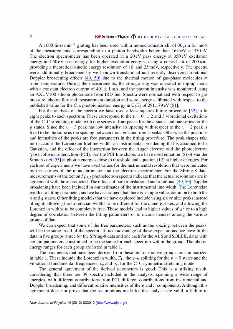
A 1600 lines mm−1 grating has been used with a monochromator slit of 30µm for mostof the measurements, corresponding to a photon bandwidth better than 16 meV at 350 eV.The electron spectrometer has been operated at a 20 eV pass energy at 350 eV excitationenergy and 50 eV pass energy for higher excitation energies using a curved slit of 200µm,providing a theoretical kinetic energy resolution of 10 and 25 meV, respectively. The spectrawere additionally broadened by well-known translational and recently discovered rotationalDoppler broadening effects [49, 50] due to the thermal motion of gas-phase molecules atroom temperature. During the measurements, the storage ring was operated in top-up modewith a constant electron current of 401 ± 1 mA, and the photon intensity was monitored usingan AXUV100 silicon photodiode from IRD Inc. Spectra were normalized with respect to gaspressure, photon flux and measurement duration and were energy calibrated with respect to thepublished value for the C1s photoionization energy in C2H2 of 291.179 eV [51].
For the analysis of the spectra we have used a least-squares fitting procedure [52] to fiteight peaks to each spectrum. These correspond to the v = 0, 1, 2 and 3 vibrational excitationsof the C–C stretching mode, with one series of four peaks for the u states and one series for theg states. Since the v = 3 peak has low intensity, its spacing with respect to the v = 2 peak isfixed to be the same as the spacing between the v = 2 and v = 1 peaks. Otherwise the positionsand intensities of the peaks are free parameters in the fitting procedure. The peak shapes takeinto account the Lorentzian lifetime width, an instrumental broadening that is assumed to beGaussian, and the effect of the interaction between the Auger electron and the photoelectron(post-collision interaction (PCI). For the PCI line shape, we have used equation (8) of van derStraten et al [53] at photon energies close to threshold and equation (12) at higher energies. Foreach set of experiments we have used values for the instrumental resolution that were indicatedby the settings of the monochromator and the electron spectrometer. For the SPring-8 data,measurements of the xenon 5p3/2 photoelectron spectra indicate that the actual resolutions are inagreement with those predicted. The effects of both translational and rotational [49, 50] Dopplerbroadening have been included in our estimates of the instrumental line width. The Lorentzianwidth is a fitting parameter, and we have assumed that there is a single value, common to both theu and g states. Other fitting models that we have explored include using six or nine peaks insteadof eight, allowing the Lorentzian widths to be different for the u and g states, and allowing theLorentzian widths to be completely free. These models lead to higher values of χ2 or to a highdegree of correlation between the fitting parameters or to inconsistencies among the variousgroups of data.
We can expect that some of the free parameters, such as the spacing between the peaks,will be the same in all of the spectra. To take advantage of these expectations, we have fit thedata in five groups (three for the SPring-8 data and one each for the ALS and SOLEIL data) withcertain parameters constrained to be the same for each spectrum within the group. The photonenergy ranges for each group are listed in table 1.
The parameters that have been derived from these fits for the five groups are summarizedin table 1. These include the Lorentzian width, 0L, the g–u splitting for the v = 0 states and thevibrational fundamental frequencies, υu and υg, for the C–C symmetric stretching mode.
The general agreement of the derived parameters is good. This is a striking result,considering that there are 39 spectra included in the analysis, spanning a wide range ofenergies, with different contributions from PCI, different contributions from instrumental andDoppler broadening, and different relative intensities of the g and u components. Although thisagreement does not prove that the assumptions made for the analysis are valid, a failure to
New Journal of Physics 14 (2012) 033012 (http://www.njp.org/)

7
Table 1. Parameters derived from fits. Values in parentheses are standarddeviations of the distribution.
Group hυ 0L g–u υu υg
(eV) (meV) (meV) (meV) (meV)
SP8-L 296–314 97.7 103.1 264.9 275.9SP8-M 316–350 98.6 102.8 264.3 277.1SP8-H 320–934 92.5 103.2 264.0 276.7ALS 310–340 101.8 102.4 265.0 276.8SOLEIL 350–500 107.8 102.0 265.0 278.2Average 100.0(6) 102.7(0.5) 264.6(0.5) 276.9(0.8)
find consistency among the results obtained from the different groups would certainly call intoquestion these assumptions.
The average value of the Lorentzian linewidth, 100 meV, is consistent with linewidths thathave been found for carbon 1s photoelectron spectra in other compounds [54]. The averagevalue of the g–u splitting, of 102.7 meV, is close to the value of 101.6 ± 0.8 meV given by Borveet al [37]. The two values of the C–C stretching frequency are slightly higher than those reportedby Borve et al, but agree with their result that the frequency for g vibration is slightly higherthan that for the u vibration.
3. Theoretical methods
The vibrationally resolved photoionization cross section is evaluated to first order ofperturbation theory within the Born–Oppenheimer and dipole approximations:
σα(v, v′, ω
)=
4π 2ω
3hca2
0
∑η
∑lη
∣∣Tαηlηvv′ (ε)∣∣2, (5)
where
Tαηlηvv′ (ε)=
∫χ∗
M,v (R) µαηlη (ε, R) χM+α ,v
′ (R) dR, (6)
µαηlη is the dipole–transition matrix element between the initial electronic state,ψ0, and the finalelectronic continuum state, ψαηlη , of C2H2, α denotes the electronic state of the residual ion, η isthe symmetry of the final state, χM,v is the initial vibrational state, χM+
α ,v′ is the final vibrational
state, ω is the photon energy and ε is the photoelectron energy.To evaluate the electronic continuum state ψαηlη , we have used an extension of DFT,
originally developed by Decleva and coworkers to treat molecular ionization at the molecule’sequilibrium position (see [55] and references therein). The method has been shown to provideaccurate photoionization cross sections for simple as well as for very complex molecules (see,e.g., [55]). Here the method has been extended to describe non-equilibrium geometries. Inparticular, we have considered 71 different geometries associated with the C–C symmetricstretching mode. They have been built by symmetrically varying the C–C distance while keepingthe C–H distance at its equilibrium value. A standard local density approximation (LDA)functional has been used to describe exchange and correlation effects.
New Journal of Physics 14 (2012) 033012 (http://www.njp.org/)

8
For each value of the C–C distance, we have followed the following procedure. Theinitial Kohn–Sham orbitals have been generated by running a standard preliminary LCAO-DFT calculation (LCAO stands for linear combination of atomic orbitals) with the programAmsterdam Density Functional (ADF) (see [56] and references therein) using an LDAfunctional in a double zeta-polarization (DZP) basis set. The resulting ground state densityis then used to build the Hamiltonian matrix in a new basis set of B-spline functions B and realspherical harmonics Y R:
ξ iαlα j (r, θ, φ)=
N ieq∑
k=1
1
rkB j(rk)
lα∑mα=−lα
bαlαmY Rlαm (θk, φk), (7)
where α gathers the indexes referring to a specific irreducible representation (IR), lα and mα
correspond to the usual angular momentum quantum numbers, i and k indicate, respectively, thei th non-equivalent expansion center and its kth equivalent subspecies, N i
eq denotes the numberof equivalent centers associated with i , the index j refers to the j th B-spline, r , θ and φ standfor the spherical coordinates in the molecular framework and bαlαm are the coefficients of thesymmetry adapted linear combination of real spherical harmonics. The non-equivalent centerscan be divided into two categories:
• The one-center expansion (OCE) that is unique (as its name implies) and located at achosen origin. Its radius is large (40 au in the present calculation), which allows us toproperly treat the long-range oscillatory behavior of the continuum wavefunction.
• The off-center expansions located at the center of each non-equivalent nucleus in order todescribe more accurately the sharpness of the bound-state wavefunctions. The expansion-interval is cut off and is generally quite small (0.5 au in the present calculation) in orderto avoid linear dependencies due to excessive overlap with other basis functions. It hasbeen observed that the introduction of the off-centers into the OCE B-spline improvesdramatically the convergence of the calculation for most molecules.
In the particular case of acetylene, the OCE is centered in the symmetry center of the moleculeand only two off-center expansions are required to take care of the carbon and hydrogen atoms.Hence, the radial part of the basis functions is defined by three sets of Nb one-dimensionalB-spline functions, built on two different radial intervals: [0; R0
max] for the OCE set {ξ 0αlα} and
[0; Rimax] for the off-centers set {ξ i
αlα}. The LCAO basis set and consequently the cost/accuracyof the calculation are completely defined by (i) the B-spline parameters k, R0
max, Rimax and Nb,
and (ii) the maximum value of the angular expansion L0max for the OCE and L i
max for each set ofnon-equivalent centers.
Since the off-center radius spheres cannot intersect, the resulting Hamiltonian matrix H ispartitioned into submatrices which are the diagonal elements H j j and the blocks H0 j and H j0
between the set {ξ 0αlα} and {ξ i
αlα}. The calculation of the non-diagonal block elements H0 j andH j0 represents the main computational effort because of the lack of analytical resolution. Tocarry out the integration, a numerical three-dimensional Gauss–Legendre scheme is employed.Also, a rotation of the coordinate framework of the main center of the spherical harmonics alongthe polar axis attached to the subsystems defined by the expansion centers leads to significantimprovement. This exploits local cylindrical symmetry reducing integration to two variables(r and ϕ).
New Journal of Physics 14 (2012) 033012 (http://www.njp.org/)

9
Figure 1. Photoelectron spectra of C2H2 taken at hυ = 534 eV. Circlesexperimental results. Full lines: fit of the experimental data. Peak numbersindicate the vibrational quantum number v′ of C2H+
2 . The spectrum has beennormalized to the value predicted by theory for the 26+
u (1sσu , v′= 0) peak. Inset:
potential energy curves of the ground state of C2H2 (16+g ) (black line) and the
26+g (1sσg) (purple line) and 26+
u (1sσu) (orange line) states of core-ionized C2H+2
along the C–C symmetric-stretching normal coordinate. The red arrow indicatesa vertical one-photon transition. The horizontal line in the upper potential energycurves indicates the dissociation limit and the shaded area below it the vibrationalstates in the 26+
g (1sσg) and 26+u (1sσu) states.
The bound states are extracted by a generalized diagonalization of HK S and continuumstates by block inverse iteration [57] for each given photoelectron energy. Electronic dipolematrix elements were computed for 200 photoelectron energies, in a basis of 400 B-splines, abox size of 40 au and a maximum L value of 20.
The vibrational wave functions result from the solution of the vibrational Schrodingerequation in a basis of B-splines by using the projection of the potential energy surface alongthe C–C symmetric stretching coordinate. We have evaluated the vibrational wave functionsby diagonalizing the vibrational Hamiltonian corresponding to this mode in a basis of 1000 B-splines within a box of 10 au. A similar method has been shown to provide accurate vibrationallyresolved photoionization cross sections for N2 and CO [14], as well as for CH4 [42].
It is important to stress here that the present description of the C–C symmetric stretchingvibrational mode is rather rudimentary, because in assuming that the C–H distance remainsfixed during the vibration for both the parent molecule and the remaining ion, we neglect amongother things the possible mixing between the two symmetric stretching modes associated withthe electronic excitation (the Dushinsky effect) [58].
4. Results and discussion
Figure 1 shows the photoelectron spectrum at a photon energy of 534 eV. As can be seen, C1s photoionization mainly leads to C2H+
2 ions in the ground vibrational state v = 0 of the C–Csymmetric stretching mode. This is because the equilibrium geometries of C2H2 and C2H+
2 arevery similar for this core-ionized electronic state. In other words, a vertical one-photon transition
New Journal of Physics 14 (2012) 033012 (http://www.njp.org/)

10
400 600 800 1000Photon energy (eV)
0.5
1
1.5
2
I(1σ
g)/I(
1σu)
v’=0
v’=1
Figure 2. Vibrationally resolved 1σg/1σu ratios in C 1s photoionization ofC2H2 for the C–C symmetric stretching mode of C2H+
2 . Squares and diamonds:experimental results. The error bars include both statistical and systematiccontributions. Full lines: theoretical results. Dashed lines: results from the simplemodel discussed in the text. No scaling has been applied to the calculated curves.
from the ground state of C2H2 necessarily leads to core ionized C2H+2 with a geometry close
to that of the ground state, so that the maximum Franck–Condon overlap corresponds to thev = 0 → v′
= 0 transition. For the same reason, the intensity of the observed peaks decreasesrapidly with the degree of vibrational excitation of the remaining C2H+
2 ion and, for all practicalpurposes, it is negligible beyond v′
= 3. Each v′ peak is formed by two components, whichcorrespond to C 1s ionization from the 1σg and 1σu molecular orbitals. The energy separationbetween these two components is 102.7 meV, in good agreement with the value of the 1σg–1σu
splitting reported in previous work. As can be inferred from figure 1, the high energy resolutionof the spectra allows us to clearly separate the two components.
The intensity in the photoelectron spectrum decreases very rapidly with photon energy.This rapid decrease, which also occurs in atoms, complicates the identification of interferenceeffects. As reported in previous works [12, 14], a very elegant way of removing this rapiddecrease from the analysis is to consider ratios of vibrational intensities (v-ratios), in whichsuch a behavior is no longer present because both the numerator and the denominator decreasewith a similar rate. An alternative way of removing this rapid decrease is to analyze the variationof the 1σg/1σu ratio with photon energy (see [16]).
Figures 2 and 3 show the measured and calculated 1σg/1σu and υ = 1/υ = 0 ratiosas functions of photon energy. As can be seen, both ratios exhibit pronounced oscillationssuperimposed on a slowly increasing background. The agreement between theory andexperiment is excellent. In particular, the theory reproduces quite well the differences between1σg/1σu ratios associated with different vibrational states of the remaining C2H+
2 ion, as wellas the phase opposition of the v-ratios in the 1σg and 1σu channels. Similar oscillations havebeen observed in 1σg/1σu ratios resulting from N 1s photoionization of N2 [16]. In that work,the oscillations in the 1σg/1σu ratios have been interpreted as resulting from Cohen–Fanointerferences. These interferences reflect a different phase in the 1σg and 1σu channels dueto the difference in sign of the corresponding linear combinations of 1s orbitals.
New Journal of Physics 14 (2012) 033012 (http://www.njp.org/)

11
400 600 800 1000Photon energy (eV)
0.45
0.5
0.55
0.6
I(v’
=1)
/I(v
’=0)
1σg
1σu
Figure 3. v′= 1/v′
= 0 ratios between vibrationally resolved photoionizationcross sections in C 1s photoionization of C2H2 for the C–C symmetric stretchingmode of C2H+
2 in the σg and σu channels. Squares and diamonds: experimentalresults. The error bars include both statistical and systematic contributions. Fulllines: theoretical results. Dashed lines: results from the simple model discussedin the text. The calculated curves have been rescaled so as to reproduce theexperimental Franck-Condon ratio. See text for more details.
The similarity of the present 1σg/1σu ratios to those found in N 1s photoionization of N2
suggests that the origin of the oscillations is the same. In order to ascertain that the observedfeatures are indeed to be interpreted as genuine Cohen–Fano oscillations, we have developeda simple model in which, in addition to two-center interference effects, we also account forintramolecular scattering. As shown in [42] for C 1s photoionization of CH4, the latter effect canlead to diffraction of the ejected electron and therefore to visible structures in the vibrationallyresolved cross-section ratios.
In the model, the integral vibrationally resolved photoionization cross section σ g/uυ
for a randomly oriented acetylene molecule is estimated within the single-active-electronapproximation as (atomic units, au, are used hereafter unless otherwise specified)
σ g/uυ =
1
4π
∫d�R
∫d�k 2
4π2k
cω
∣∣〈ψ Ek χg/uυ | ε · Ep |σg/u χ0〉
∣∣2 . (8)
Here χ0 is the wave function of the ground vibrational state of the neutral molecule and χ g/uυ is
the vibrational wave function for the ionized state (either g or u) in either the υ = 0 or υ = 1state of the C–C stretching vibrational mode. The initial core molecular orbital is approximatedwith a symmetry-adapted combination of the 1s core orbitals of the two carbon atoms as shownin equation (4). In momentum coordinates, this expression can be reformulated as
σg/u( Ep)= e−i Ep· ERCCei Ep· ER/2
± e−i Ep· ER/2
√2(1 ± S(R))
1s(p), (9)
where the 1s orbital is now centered on the origin; ERCC = ( ERC1 + ERC2)/2 indicates the center ofmass of the two carbons and ER = ERC2 − ERC1 is their relative position. The overlap S(R) turns
New Journal of Physics 14 (2012) 033012 (http://www.njp.org/)

12
out to be very small and will be neglected. The final electronic state is approximated with aplane wave:
〈 Er |ψ Ek 〉 = (2π)−3/2 exp(i Ek · Er), (10)
therefore the transition amplitude in (8) becomes
Ag/uυ = 〈ψ Ek χ
g/uυ | ε · Ep |σg/u χ0〉 =
ε · Ek√
21s(k) 〈χ g/u
υ | ei Ek· ER/2± e−i Ek· ER/2
|χ0 〉. (11)
(In the last expression we dropped the term exp[−i Ep · ERCC], related to the conservation ofthe total momentum of the CC moiety.) Based on these and other additional assumptions,several authors have already commented on the effect of the vibrational motion on Cohen–Fanooscillations [43, 59, 60]. As we will see below, the present model leads to qualitatively similarresults.
As in the first-principles calculations, we will consider only the C–C symmetric stretchingvibrational mode in which the C–H distance is kept constant:
ERC1 = − ER/2, ERC2 = ER/2, (12)
ERH1 = −(R/2 + RCH)R, ERH2 = (R/2 + RCH)R. (13)
To evaluate the vibrational transition amplitude in (11), we assume the harmonicapproximation and that the vibrational frequency in both final parent ions is the same as thatin the initial neutral molecule. In this context, we can express the internuclear distance in termsof the dimensionless vibrational normal coordinate of the neutral molecule,
R = R0 + (µω)−1/2 Q, (14)
while the final vibrational state χ g/uυ can be written as the translation by a quantity equal to
the shift in the equilibrium position, 1Rg/u0 = Rg/u
0 − R0, of the vibrational function χυ of theneutral molecule
χ g/uυ = e−i
√µω1Rg/u
0 P χυ = D
(√µω
21Rg/u
0
)χυ . (15)
In the last expression, P is the dimensionless momentum of the harmonic oscillator canonicallyconjugated to Q, [Q, P] = i , while the displacement operator D(α) is the generator ofvibrational coherent states (see, e.g., paragraphs 3.3–9 in [61])
D(α)= eαa†−α∗a, |α〉 = D(α)|0〉 =
∑υ
|υ〉 e−|α|2/2 α
υ
√υ!, (16)
D†(α)= D(−α), D(α)D(β)= D(α +β) e(αβ∗−βα∗)/2. (17)
In a similar way, the action of any exponential operator whose argument is linear in the normalcoordinate Q can also be expressed in terms of displacement operators:
eiK Q= D
(i
K√
2
). (18)
New Journal of Physics 14 (2012) 033012 (http://www.njp.org/)

13
By applying the relations listed above, after some passages, we are led to the expression
〈χ g/uυ |eip R
|χ0〉 = eip Rg/u0
⟨υ
∣∣∣∣−√µω2 1Rg/u0 + i
p√
2µω
⟩= e−
p2
4µω eip Rg/u0 e−
µω4 (1Rg/u
0 )2
×1
√υ!
(−
√µω
21Rg/u
0 + ip
√2µω
)υ, (19)
where Rg/u0 = (R0 + Rg/u
0 )/2. For p = 0, we recover the familiar Franck–Condon amplitude:
〈χ g/uυ |χ0〉 = e−
µω4 (1Rg/u
0 )2 1√υ!
(−
√µω
21Rg/u
0
)υ. (20)
For the vibrational mode under consideration, the factor µω is very large: µ' 6.5 amu '
12 000 au, ω ' 270 meV ' 0.01 au, hence µω ' 120. The momentum factor p can be assumedto be of the same order of magnitude as that of the final momentum k of the photoelectron.For photoelectron energies up to ∼600 eV, the factor exp(−p2/4µω) is thus very close to 1, sowe will ignore it altogether. To be consistent with this approximation, we also need to drop allthose terms arising from the expansion of the last power in equation (19) which are not linear inp/
√2µω. To conclude, as long as k2/4µω� 1, the following approximate expression can be
used:
〈χ g/uυ | eip R
|χ0 〉 ' 〈χ g/uυ |χ0〉 eip Rg/u
0
(1 − i
υ p
µω1Rg/u0
). (21)
In the latter linearized form, this expression is essentially equivalent to equation (18) in [43],where no assumptions on the form of the vibrational functions were made.
By using the relations (21) in (11) we finally obtain the expression of the differentialphotoionization amplitudes for an oriented molecule
Agυ '
ε · Ek√
21s(k) 〈χ g
υ |χ0〉
[2 cos( Ek ·
ERg0/2)+ υ
Ek · R
µω1Rg0
sin( Ek ·ERg0/2)
],
Auυ '
ε · Ek√
21s(k) 〈χu
υ |χ0〉
[2i sin( Ek ·
ERu0/2)− iυ
Ek · R
µω1Ru0
cos( Ek ·ERu0/2)
].
We can recover the familiar Cohen–Fano model from the latter expressions by retaining onlythe first term within parentheses. Note that in the present treatment the internuclear distancethat appears in the final formula is intermediate between that in the neutral and that in the parention. As the second term in parentheses shows, the model accounts for the sensitivity of theinterference amplitudes to the nuclear motion. This effect, which at this level is not visible inthe amplitude for the transition to the ground vibrational state, is a manifestation of the nuclearrecoil. Indeed, the factor exp(i Ek · ERi) can be seen as a boost by a momentum Ek on nucleus i .Nuclear recoil is therefore one of the mechanisms which mirror double-slit interferences tonon-Franck–Condon vibrational excitations and which thus permits us to monitor the latter bylooking at the σ g/u
1 /σg/u0 ratios.
After taking the square module and carrying out the angular integrations,
σ g/uυ =
1
4π
∫d�R
∫d�k 2
4π2k
cω
∣∣Ag/uυ
∣∣2 , (22)
New Journal of Physics 14 (2012) 033012 (http://www.njp.org/)

14
the cross sections in plane wave approximation are finally obtained:
σ g/uυ = 2
16π 3k3
3cω1s2(k)|〈χ g/u
1 |χ0〉|2
×
[1 ±
sin k Rg/u0
k Rg/u0
∓υk
µω1Rg/u0
cos(k Rg/u0 )
k Rg/u0
±υ
µω Rg/u0 1Rg/u
0
sin(k Rg/u0 )
k Rg/u0
](23)
In agreement with the previous approximations, in this last expression we dropped the termsquadratic in υ. The last term in parentheses amounts to a correction of the amplitude of theCohen–Fano oscillations. This term has not been considered in previous models. By neglectingthis term, we end up with an equation similar to equation (20) of Ueda et al [43]. Indeed, byborrowing part of their notation, one obtains an expression formally identical to equations (22)and (23) in [43],
σ g/uυ = 2
16π 3k3
3cω1s2(k)|〈χ g/u
1 |χ0〉|2[1 ±χ g,u
υ (k)], (24)
χ g/uυ (k)=
sin(k Rg/u0 +ψ g,u
υ (k))
cosψ g/uυ (k)k Rg/u
0
, tan ψ g/uυ = −
υk
µω1Rg/u0
. (25)
The recoil thus induces an energy-dependent phaseshiftψ g/uυ (k) in the Cohen–Fano oscillations.
Within the harmonic approximation adopted here, this phase shift is known analytically.A comparison with equation (23) in [43] leads to the following correspondence with theparametrization chosen by Ueda et al:
1Rυ = −υ
µω1Rg/u0
. (26)
What is indicated in [43] as an effective displacement 1Rυ of the carbon–carbon internucleardistance appears, within the harmonic approximation, to be related in a simple way to theparameters of the vibrational mode. On the basis of equation (14), we can in fact recognizein 1/
√µω the characteristic length Rω of the oscillator and write the formula
1RυRω
= −υRω
1Rg/u0
. (27)
The internuclear distances in the neutral are R0 = 2.273 au, RCH = 2.002 au. Theoreticalestimates of the C–C internuclear distance exist [37]: Rg
0 = 2.198 au, Ru0 = 2.185 au. With
these values, Rg0 = 2.236 au, Ru
0 = 2.230 au, 1Rg0 = −0.075 au and 1Ru
0 = −0.088 au. Infigure 3, the dashed lines show the υ = 1/υ = 0 intensity ratios computed with the completeformula (23). As can be seen from the figure, the model reproduces quite nicely the experimentaland theoretical results. In particular, the model accounts for the phase opposition of theoscillations in the gerade and ungerade cases. The oscillation periods are some 2–3% larger thanthe1k ∼ 2π/Rg/u
0 values which are expected on the basis of simple dimensional considerations.A closer inspection of these results shows that the two ratios oscillate around baselines that startat the corresponding Franck–Condon ratios and increase with the photoelectron energy. Thelatter is due to the momentum transferred to either nuclei as the photoelectron is ejected (recoil).Since the amplitudes for the photoelectron ejection from the two nuclei add coherently, themomentum transfer, and therefore the vibrational excitation, is affected by the same interference
New Journal of Physics 14 (2012) 033012 (http://www.njp.org/)

15
features observed in the total cross section. The largest discrepancy between the plane-waveprediction and the experiment is in the position of the baselines, i.e. in the Franck–Condonratios |〈χ
g/u1 |χ0〉/〈χ
g/u0 |χ0〉|
2. The latter, however, depend strongly on the precise value of thecontractions 1Rg/u
0 of the C–C bond length. It is possible that the theoretical estimates for theequilibrium positions in the parent ions given in [37] are not accurate enough. In fact, smalladjustments of the order of 10−2 Å to Rg/u
0 are sufficient to bring the theoretical curves on top ofthe experimental ones. A similar discrepancy between theory and experiment is observed alsoin the case of the first-principles DFT calculations.
In contrast to the good agreement found for the vibrational ratios, the σg/σu ratioscomputed in the plane wave approximation turn out to be almost in phase opposition withrespect to the measurements. The prediction of the model is shown in figure 2. In the case of theexcited final vibrational state υ = 1, the nuclear recoil induces a phase shift in the σg/σu ratiothat partly reduces the distance with the experimental points. This effect, however, is still toosmall to compensate for the error of the Cohen–Fano prediction. Moreover, at the present levelof approximation, the recoil does not have any effect on the υ = 0 cross section. A major causeof the observed discrepancy must therefore be sought in a different mechanism. The phase shiftdifference between the υ = 0 and the υ = 1 cases predicted by the recoil should presumablyshow up in the experiment as well. Unfortunately, the present experimental accuracy does notallow us to draw any conclusion in this sense.
We have extended the present model beyond the plane-wave approximation [62] byincluding the contributions of the carbon and hydrogen Coulomb potentials in the first Bornapproximation. The corresponding results barely differ from those shown in figures 2 and 3,thus lending further support to our interpretation of the observed oscillations as resulting fromtwo-center Cohen–Fano interferences.
5. Summary and conclusions
We have shown that vibrationally resolved C 1s photoelectron spectra of acetylene obtainedwith third-generation synchrotron radiation exhibit Cohen–Fano interferences similar to thosepreviously reported in diatomic molecules. The oscillations are unambiguously observed inboth the v and g/u ratios, for which no independent estimation for the rapidly decreasingcross section is required. The results obtained from first-principles calculations based ondensity functional theory are in excellent agreement with the experimental measurements. Thepredictions of experiment and theory are further supported by a simple model that basicallyfollows the premises of the original Cohen–Fano model developed for H+
2 . Although noquantitative agreement with either the experiment or the first-principles calculations is found,the qualitative features of the oscillations are correctly described: the overall period of theoscillations, the fact that the υ = 1/υ = 0 ratios for the gerade and ungerade cases are inphase opposition and the fact that there is no discernible influence of the nearby hydrogenatoms. These results confirm the delocalized character of the core hole created in the primaryphotoionization event and demonstrate that intramolecular core-hole coherence can survive thedecoherence introduced by the asymmetric nuclear degrees of freedom that are characteristic ofpolyatomic molecules.
The conclusions reported in this work as well as those reported in [42] for CH4 suggestthat vibrationally resolved photoelectron spectroscopy can be used to determine the structureof polyatomic molecules in different environments, e.g. adsorbed on surfaces [63]. In the
New Journal of Physics 14 (2012) 033012 (http://www.njp.org/)

16
latter case, photoelectron diffraction imaging makes use of holographic principles to extractinformation on the coordination of carbon atoms to a Si(100) surface. In a similar way,Cohen–Fano oscillations could give information on the C–C internuclear distance.
Finally, the ultrafast migration of holes in the valence shell has attracted considerableinterest in recent years (see, e.g., [64, 65] and references therein). It will certainly be worthextending the ultra-fast time-resolved investigation to the dynamics of coherently created core-holes, too.
Acknowledgments
We thank Mare Nostrum BSC, Cineca and CCC-UAM (Centro de Computacion Cientıfica,Universidad Autonoma de Madrid) for the allocation of computer time. This work wassupported by the MICINN project numbers FIS2010-15127, ACI2008-0777 and CSD 2007-00010 (Spain), the ERA-Chemistry project number PIM2010EEC-00751, the European COSTAction CM0702 and the Marie Curie ITN CORINF. The experiment at SPring-8 was carriedout with the approval of JASRI and was supported in part by grants in-aid for scientic researchprovided by the Japan Society for Promotion of Science. We thank the staff at SPring-8 for theirhelp during the experiment. At SOLEIL Synchrotron (France), the experiment was performedat the PLEIADES beamline during beamtime allotted under proposal number 20100163. We aregrateful to E Robert for technical assistance and to the SOLEIL staff for stable operation of theequipment and storage ring during the experiments.
References
[1] Cohen H D and Fano U 1966 Interference in the photo-ionization of molecules Phys. Rev. 150 30[2] Samson J A R and Cairns R A 1965 Total absorption cross sections of H2, N2 and O2 in the region 550–200 Å
J. Opt. Soc. Am. 55 1035[3] Stolterfoht N et al 2001 Evidence for interference effects in electron emission from H2 colliding with
60 MeV/u Kr34+ ions Phys. Rev. Lett. 87 023201[4] Misra D et al 2004 Interference effect in electron emission in heavy ion collisions with H2 detected by
comparison with the measured electron spectrum from atomic hydrogen Phys. Rev. Lett. 92 153201[5] Kamalou O et al 2005 Evidence for interference effects in both slow and fast electron emission from D2 by
energetic electron impact Phys. Rev. A 71 010702[6] Milne-Brownlie D et al 2006 Young-type interference in (e,2e) ionization of H2 Phys. Rev. Lett. 96 233201[7] Akoury D et al 2007 The simplest double slit: interference and entanglement in double photoionization of H2
Science 318 949[8] Kreidi K et al 2008 Interference in the collective electron momentum in double photoionization of H2 Phys.
Rev. Lett. 100 133005[9] Dıez Muino R et al 2002 Angular distributions of electrons photoemitted from core levels of oriented diatomic
molecules: multiple scattering theory in non-spherical potentials J. Phys. B: At. Mol. Opt. Phys. 35 L359[10] Fojon O et al 2004 Interference effects in H2 photoionization at high energies J. Phys. B: At. Mol. Opt. Phys.
37 3035[11] Fernandez J, Fojon O, Palacios A and Martın F 2007 Interferences from fast electron emission in molecular
photoionization Phys. Rev. Lett. 98 043005[12] Fernandez J, Fojon O and Martın F 2009 Double-slit, confinement and non-Franck–Condon effects in
photoionization of H2 at high photon energy Phys. Rev. A 79 023420
New Journal of Physics 14 (2012) 033012 (http://www.njp.org/)

17
[13] Fernandez J et al 2009 Two-center effects in one-photon single ionization of H+2 , H2 and Li+2 with circularly
polarized light Phys. Rev. A 79 043409[14] Canton S E et al 2011 Direct observation of Young’s double-slit interferences in vibrationally resolved
photoionization of diatomic molecules Proc. Natl Acad. Sci. USA 108 18 7302[15] Becker U 2011 Matter-wave interference made clear Nature 474 586[16] Semenov S K et al 2006 Interference modulation in the vibrationally resolved photoionization of the 1σ g
and 1σ u core levels of the N2 molecule J. Phys. B: At. Mol. Opt. Phys. 39 L261[17] Rolles D et al 2005 Isotope-induced partial localization of core electrons in the homonuclear molecule N2
Nature 437 7059–711[18] Schoffler M et al 2008 Ultrafast probing of core hole localization in N2 Science 320 920[19] Ueda K 2010 Which-pass information in the double-slit experiment of diatomic molecules J. Phys.: Conf.
Series 212 012033[20] Schoffler M et al 2011 Matter wave optics perspective at molecular photoionization: K -shell photoionization
and Auger decay of N2 New J. Phys. 13 095013[21] Hergenhahn U et al 2001 Symmetry-selective observation of the N 1s shape resonance in N2 J. Phys. Chem.
A 105 5704[22] Zimmermann B et al 2008 Localization and loss of coherence in molecular double-slit experiments Nat. Phys.
4 649[23] Adachi J, Kosugi N, Shigemasa E and Yagishita A 1995 Renner–Teller effect and Rydberg-valence mixing in
the N and O K-edge photoabsorption spectra of N2O J. Chem. Phys. 102 7369[24] Adachi J, Kosugi N, Shigemasa E and Yagishita A 1997 Renner–Teller splitting in the C 1s → π∗ excited
states of CS2, OCS and CO2 J. Chem. Phys. 107 4919[25] Miron C et al 2001 Nuclear motion driven by the Renner–Teller effect as observed in the resonant Auger
decay to the X 25 electronic ground state of N2O+ J. Chem. Phys. 115 864[26] Kimberg V, Kosugi N and Gel’mukhanov F 2009 Theoretical studies of angle-resolved ion yield spectra of
core-to-valence transitions of acetylene J. Chem. Phys. 130 114302[27] Miron C et al 2010 Vibrational scattering anisotropy generated by multichannel quantum interference Phys.
Rev. Lett. 105 093002[28] Gadea F X et al 1991 Vibronic coupling and core-hole localization in K-shell excitations of ethene Phys. Rev.
Lett. 66 883[29] Broer R and Nieuwpoort W C 1999 Hole localization and symmetry breaking J. Mol. Struct. 458 19[30] Snyder L C 1971 Core–electron binding energies and Slater atomic shielding constants J. Chem. Phys. 55 95[31] Domcke W and Cederbaum L S 1977 Vibronic coupling and symmetry breaking in core electron ionization
Chem. Phys. 25 189[32] Kivimaki A et al 1997 Vibrationally resolved O 1s photoelectron spectrum of CO2: vibronic coupling and
dynamic core-hole localization Phys. Rev. Lett. 79 998[33] Gunnelin K et al 1999 Bond-length-dependent core hole localization observed in simple hydrocarbons Phys.
Rev. Lett. 83 1315[34] Kempgens B et al 1997 Core level energy splitting in the C 1s photoelectron spectrum of C2H2 Phys. Rev.
Lett. 79 3617[35] Karlsen T et al 2001 Vibrational structure and vibronic coupling in the carbon 1s photoelectron spectra of
ethane and deuteroethane J. Phys. Chem. A 105 7700[36] Lievin J et al 2011 Comparison of the experimental, semi-experimental and ab initio equilibrium structures
of acetylene: influence of relativistic effects and of the diagonal Born–Oppenheimer corrections J. Chem.Phys. 134 064119
[37] Borve K et al 2000 Vibronic structure in the carbon 1s photoelectron spectra of HCCH and DCCD Phys. Rev.A 63 012506
[38] Thomas T D et al 1999 Photon energy dependence of the 1σu/1σg intensity ratio in carbon 1s photoelectronspectroscopy of ethyne Phys. Rev. Lett. 82 1120
New Journal of Physics 14 (2012) 033012 (http://www.njp.org/)

18
[39] Hoshino M et al 2006 Symmetry- and vibrationally-resolved C K-shell photoionization studies of C2H2
Chem. Phys. Lett. 421 256[40] Adachi J-I et al 2007 Photoelectron–photoion–photoion momentum spectroscopy as a direct probe of the
core-hole localization in C 1s photoionization of C2H2 J. Phys. B: At. Mol. Opt. Phys. 40 F285[41] Osipov T et al 2008 Fragmentation pathways for selected electronic states of the acetylene dication J. Phys.
B: At. Mol. Opt. Phys. 41 091001[42] Plesiat E et al 2012 Phys. Rev. A 85 023409[43] Ueda K et al 2006 Role of the recoil effect in two-center interference in x-ray photoionization Chem. Phys.
329 329[44] Ueda K 2003 High-resolution inner-shell spectroscopies of free atoms and molecules using soft-x-ray
beamlines at the third-generation synchrotron radiation sources J. Phys. B: At. Mol. Opt. Phys. 36 R1[45] Miron C et al 2009 PLEIADES: an ultra high resolution soft x-rays beamline for advanced spectroscopic
studies on diluted species www.synchrotron-soleil.fr/Recherche/LignesLumiere/PLEIADES[46] Travnikova O et al 2010 Circularly polarized x-rays: another probe of ultrafast molecular decay dynamics
Phys. Rev. Lett. 105 233001[47] Soderstrom J et al 2011 Angle-resolved electron spectroscopy of the resonant Auger decay in xenon with
meV energy resolution New J. Phys. 13 073014[48] Miron C et al 2011 Imaging molecular potentials using ultrahigh-resolution resonant photoemission Nat.
Phys. 8 135–8[49] Sun Y-P, Wang C-K and Gel’mukhanov F 2010 Rotational doppler effect in x-ray photoionization Phys. Rev.
A 82 052506[50] Thomas T D et al 2011 Experimental observation of rotational Doppler broadening in a molecular system
Phys. Rev. Lett. 106 193009[51] Myrseth V et al 2002 Adiabatic and vertical carbon 1s ionization energies in representative small molecules
J. Electron Spectrosc. Relat. Phenom. 122 57[52] Kukk E www.physics.utu.fi/en/research/material SUBSCRIPTNBscience/Fitting.html[53] van der Straten P, Morgenstern R and Niehaus A 1988 Angular dependent post-collision interaction in auger
processes Z. Phys. D 8 35[54] Carroll T X et al 2002 Carbon 1s photoelectron spectroscopy of CF4 and CO: search for chemical effects on
the carbon 1s hole-state lifetime J. Chem. Phys. 116 10221[55] Bachau H et al 2001 Application of B-splines in atomic and molecular physics Rep. Prog. Phys. 64 1815[56] Fonseca Guerra C, Snijders J G, te Velde G and Baerends E J 1998 Towards an order-N DFT method Theor.
Chem. Acc. 99 391[57] Venuti M, Stener M and Decleva P 1998 Valence photoionization of C6H6 by the B-spline one-centre
expansion density functional method Chem. Phys. 234 95[58] Duschinsky F 1937 On the interpretation of electronic spectra of polyatomic molecules Acta Physicochim.
URSS 7 551[59] Domcke W and Cederbaum L S 1978 Electronic recoil effects in high-energy photoelectron spectroscopy
J. Electron Spectrosc. Relat. Phenom. 13 161[60] Gel’mukhanov F et al 2007 Young’s double-slit experiment using two-center core-level photoemission:
photoelectron recoil effects J. Electron Spectrosc. Relat. Phenom. 158 265[61] Vourdas A 2006 Analytic representations in quantum mechanics J. Phys. A: Math. Gen. 39 R65[62] Argenti L, Plesiat E, Decleva P and Martın F in preparation[63] Xu S et al 2000 Photoelectron diffraction imaging for C2H2 and C2H4 chemisorbed on Si(100) reveals a new
bonding configuration Phys. Rev. Lett. 84 939[64] Lunnemann S, Kuleff A I and Cederbaum L S 2009 Ultrafast electron dynamics following outer-valence
ionization: the impact of low-lying relaxation satellite states J. Chem. Phys. 130 154305[65] Kuleff A I and Cederbaum L S 2011 Radiation generated by the ultrafast migration of a positive charge
following the ionization of a molecular system Phys. Rev. Lett. 106 053001
New Journal of Physics 14 (2012) 033012 (http://www.njp.org/)