Flow-induced Migration of Osteoclasts and Regulations of Calcium Signaling Pathways
30
Original Article Flow-induced Migration of Osteoclasts and Regulations 1 of Calcium Signaling Pathways 2 3 Chenglin Liu, Shuna Li, Baohua Ji*, Bo Huo* 4 5 Biomechanics and Biomaterials Laboratory, Department of Mechanics, 6 School of Aerospace Engineering, Beijing Institute of Technology, Beijing 7 100081, P. R. China 8 9 10 11 12 13 14 15 16 17 *Corresponding authors: Dr. Bo Huo ([email protected]); Dr. Baohua Ji 18 ([email protected]). Biomechanics and Biomaterials Laboratory, 19 Department of Mechanics, School of Aerospace Engineering, Beijing 20 Institute of Technology, No. 5 South Zhongguancun Street, Beijing 21 100081, P. R. China. 22 23 24
-
Upload
independent -
Category
Documents
-
view
2 -
download
0
Transcript of Flow-induced Migration of Osteoclasts and Regulations of Calcium Signaling Pathways

Original Article
Flow-induced Migration of Osteoclasts and Regulations 1
of Calcium Signaling Pathways 2
3
Chenglin Liu, Shuna Li, Baohua Ji*, Bo Huo* 4
5
Biomechanics and Biomaterials Laboratory, Department of Mechanics, 6
School of Aerospace Engineering, Beijing Institute of Technology, Beijing 7
100081, P. R. China 8
9
10
11
12
13
14
15
16
17
*Corresponding authors: Dr. Bo Huo ([email protected]); Dr. Baohua Ji 18
([email protected]). Biomechanics and Biomaterials Laboratory, 19
Department of Mechanics, School of Aerospace Engineering, Beijing 20
Institute of Technology, No. 5 South Zhongguancun Street, Beijing 21
100081, P. R. China. 22
23
24

2
Abstract 1
Osteoclasts are large multinucleate cells originating from the fusion of monocytes that 2
are differentiated from hematopoietic stem cells. Although activated osteoclasts 3
preferentially move to the area of microcrack by chemotaxis, whether such 4
mechanical cues as fluid shear stress (FSS) regulate the migration of osteoclasts 5
remains unknown. This study focuses on the effect of FSS on the migration of 6
RAW264.7 monocytes and differentiated osteoclasts, as well as the roles of calcium 7
signaling pathways in cell migration behaviors. We study five calcium signaling 8
pathways, namely, mechanosensitive cation-selective channels (MSCC), 9
phospholipase C (PLC), endoplasmic reticulum (ER), adenosine triphosphate (ATP), 10
and extracellular calcium. Results show that FSS induces the migration of RAW264.7 11
cells along flow direction, and the directionality, alignment along the flow direction, 12
and speed of cells are significantly enhanced with the increase in FSS levels. 13
Blocking the pathways of MSCC, ER, or extracellular calcium significantly reduces 14
the migration of RAW264.7 cells along the flow direction. However, the inhibition of 15
calcium signaling pathways does not affect the migration behaviors after inducing 16
RAW264.7 cells for 4 or 8 d with the conditioned medium, Therefore, this study 17
indicates that both undifferentiated monocytes and differentiated osteoclasts tend to 18
migrate along flow direction, furthermore the FSS-induced directional migration of the 19
monocytes is regulated by calcium signaling pathways, but that of differentiated 20
osteoclasts is unaffected. 21
Keywords: osteoclast, migration, fluid shear stress, calcium signaling, differentiation 22

3
1
1. Introduction 2
Bone metabolism is maintained by the collective effort of osteoclasts and 3
osteoblasts (Hadjidakis and Androulakis 2006). Osteoclasts are uniquely capable of 4
resorbing bone matrix, whereas osteoblasts are responsible for bone formation. 5
Mature osteoclasts are highly specialized multinucleated cells formed by the fusion of 6
marrow-derived mononuclear phagocytes (i.e., osteoclast precursors) that originated 7
from hematopoietic stem cells induced by some cytokines, such as the 8
macrophage-colony stimulating factor (M-CSF) and the receptor activator of NF-B 9
ligand (RANKL) (Baron, Neff et al. 1986; Teitelbaum 2000). Bone resorption starts 10
with the recruitment of mononucleated osteoclast precursors from circulation and 11
proceeds with the infiltration of the bone-lining cell layer to reach the bone surface 12
and resorb bone minerals (Baroukh, Cherruau et al. 2000). However, the mechanism 13
behind the migration of these osteoclast precursors toward resorption sites remains 14
unclear. 15
Previous studies have demonstrated that extracellular calcium ions (Ca2+) 16
(Boudot, Saidak et al. 2010), and some chemokines, e.g. CX3CL1 (Koizumi, Saitoh et 17
al. 2009) or blood-enriched lipid mediator sphingosine-1-phosphate (S1P) (Ishii, 18
Kikuta et al. 2010), regulate the localization and homing of osteoclasts or their 19
precursors(Koizumi, Saitoh et al. 2009; Ishii, Kikuta et al. 2010). However, the 20
functions of mechanical cues in this process are seldom considered. The bone matrix 21
becomes deformed when bone tissue is mechanically loaded, and the deformation 22
4
drives the interstitial fluid within the cavities to flow over the surfaces of the trabeculae 1
or lacunae, causing the osteoclasts or osteoblasts on the surfaces to suffer from fluid 2
shear stress (FSS) (Thompson, Rubin et al. 2012). Some in vivo (Johansson, Edlund 3
et al. 2009; Fahlgren, Bostrom et al. 2010) and in vitro (Liu, Li et al. 2007) studies 4
have shown that FSS can activate the differentiation of osteoclasts and further 5
regulate bone resorption. Our previous study has found that FSS can induce calcium 6
response in both osteoclasts and their precursors (Li, Hu et al. 2012; Li, Liu et al. 7
2014). Other mechanical stimulations, such as the single-cell indentation by a glass 8
pipette (Xia and Ferrier 1995; Xia and Ferrier 1996; Wiebe, Sims et al. 1999) or 9
whole-cell membrane stretching by changing osmotic pressure (Tsuzuki, Okabe et al. 10
2000), can also result in a significant intracellular calcium response in osteoclasts. 11
However, whether mechanical loading involves in the migration of osteoclasts and 12
what the molecular mechanism is remains unknown. 13
Calcium ions are among the most important secondary messengers in cells. 14
Previous studies have shown that intracellular calcium dynamics is closely related to 15
cell migration behaviors, such as an increase in calcium concentration from the front 16
to the rear of migrating cells at a relatively long timescale (Brundage, Fogarty et al. 17
1991; Schwab, Finsterwalder et al. 1997; Wei, Wang et al. 2009) and a calcium flicker 18
with front-to-rear polarization at a short timescale (Wei, Wang et al. 2009). Our recent 19
study has shown that the mechanosensitive, cation-selective channels (MSCC), 20
phospholipase C (PLC), and endoplasmic reticulum (ER) are the major signaling 21
pathways for mechanical stimulation-induced calcium response in RAW264.7 22

5
osteoclastic precursors (monocytes) (Li, Liu et al. 2014). In this study, we test the 1
hypothesis that calcium signaling pathways may be involved in FSS-induced cell 2
migration. 3
RAW264.7 monocytes were exposed to 1 and 10 dyne/cm2 FSS for 30 min, after 4
which the dynamic parameters of cell migration were analyzed. The results showed 5
that the RAW264.7 cells tended to migrate along the flow direction and that the 6
migration speed and orientation increased with FSS level. Three calcium pathways 7
(i.e., MSCC, ER, or extracellular calcium) had significant impact on their migration. 8
When one of the three is blocked, the migration speed along flow direction is 9
significantly reduced. The osteoclasts induced for 4 or 8 d also preferentially migrated 10
along the direction of fluid flow, but calcium pathways did not influence their migration. 11
12
2. Materials and Methods 13
2.1. Cell culture 14
The osteoblast-like MC3T3-E1 cell line was purchased from the American Type 15
Culture Collection (ATCC, Manassas, USA) and cultured in alpha modified eagle’s 16
medium (α-MEM, Hyclone, USA) supplemented with 10% fetal bovine serum (FBS, 17
Gibco, USA), 100 units/mL of penicillin (Sigma, St. Louis, USA), and 100 units/mL 18
streptomycin (Sigma, St. Louis, USA). The medium was changed every 48 h, and the 19
conditioned medium was collected every 24 h when cells reached 70% confluence. 20
The RAW264.7 cell line was purchased from the European Collection of Cell Cultures 21
(ECACC, Wiltshire, UK) and cultured in Dulbecco’s modified eagle’s medium (DMEM, 22

6
Hyclone, USA) supplemented with 10% FBS, 100 units/mL of penicillin, and 100 1
units/mL of streptomycin. As described in our previous study (Li, Hu et al. 2012), the 2
mononuclear RAW264.7 cells were induced to fuse or differentiate into 3
TRAP-positive multinucleated osteoclasts with a 1:1 mixture of the DMEM medium 4
and the conditioned medium from MC3T3-E1 cells. 5
6
2.2. FSS stimulation and calcium signaling pathways 7
The RAW264.7 monocytes or the induced osteoclasts were seeded on plastic 8
slides (NUNC, Roskilde, Denmark) at a density of 2×10⁴/cm2. The slides were 9
mounted on the custom-made parallel flow chamber after 24 h of cell seeding (Fig. 10
1C). An FSS level of 1 or 10 dyne/cm2 was adopted to stimulate the cells for 30 min, 11
during which time-lapse images were recorded at 30 s intervals by using a Leica DMI 12
6000B inverted microscope (Fig. 1A). These images were used to analyze cell 13
migration parameters. Similar to our previous study (Li, Liu et al. 2014), five pathways 14
of calcium signaling were investigated by incubating the slides with blocking reagents 15
before cells are exposed to FSS. In brief, 10 μM Gadolinium chloride (Gd3+, Sigma, St. 16
Louis, USA) was supplied as the MSCC blocker, the calcium stored in the ER was 17
depleted with 1 μM thapsigargin (TG, Sigma, St. Louis, USA), and 10 μM U73122 18
(Calbiochem, Darmstadt, Germany) was adopted to inhibit PLC. For the above 19
blocking tests, 10 min incubation of chemical reagents was applied. Extracellular 20
calcium was removed by using calcium-free DMEM (Gibco, Carlsbad, USA). 100 μM 21
suramin was used to block ATP binding with P2 purinergic receptors during the 30 22

7
min incubation. It should be noted that the blocking reagents were maintained during 1
FSS stimulation. 2
3
2.3. Cell migration assay 4
The geometric parameters of migrating cells were quantified by a program 5
developed with Matlab software. Cells that are already or going to be in contact with 6
other cells were excluded. Edge extraction and dilation were used to obtain the 7
general outline of a cell; the internal holes were filled, and the cell was treated with an 8
erosion process to remove the small regions (Fig. 1B). The centroid of a cell was 9
adopted as its position, around which a rectangular region twice the cell size was 10
defined to calculate the position of the migrating cell in the next image through digital 11
image correlation (DIC) (Bruck, McNeill et al. 1989). The cell trajectory path was 12
determined after its position has been identified. The location shift was calibrated by 13
using a fixed point in the field. 14
Cell migration is mainly analyzed by using the parameters shown in Fig. 1D. The 15
total distance that the cell traveled in time t is calculated by 𝑆𝑔 = ∑ 𝑆𝑖𝑛𝑖=1 , where 𝑆𝑖 is 16
the distance traveled by the cell in 1.5 min. Therefore, the general speed is 𝑆𝑔/𝑡. The 17
relative displacement 𝑆𝑙 is shown by the red arrow between the start and end points 18
of the cellular trajectory in Fig. 1D. The inclined angle 𝜃𝑝 between the cell trajectory 19
and the flow direction represent migration orientation. Cell speed along the flow 20
direction is 𝑆𝑑/𝑡 = 𝑆𝑙 × cos𝜃𝑝/𝑡, which indicates the capability of the cell to adapt to 21
the fluid flow. The directionality 𝑆𝑙/𝑆𝑔 shows the tendency of a cell to move along a 22

8
direct line. A directionally migrating cell registers migration speed and directionality 1
that are larger than the mean value plus standard deviation (SD), which indicates that 2
no flow is applied. 3
4
2.4. Statistical analysis 5
More than three slides (at least 60 cells) were tested for a specific induction time, 6
FSS level, and chemical blocking. All the results were expressed as mean ± SD. 7
Statistical differences between the mean values of different groups were determined 8
by using one-way ANOVA, and were considered significant when p<0.05 if not 9
specifically claimed. 10
11
3. Results 12
3.1. FSS stimulates RAW264.7 cells to migrate 13
The time-lapsed images of RAW264.7 cells recorded for 30 min with or without 1 14
and 10 dyne/cm2 FSS showed that the effect of FSS on cell migration is 15
dose-dependent (Fig. 2). When no flow was applied, cells migrated randomly. For 1 16
dyne/cm2 FSS, most cells migrated along the flow direction, and the inclined angle 𝜃𝑝 17
between the cell migration and fluid flow directions are uniformly distributed within the 18
range -90° to 90° (Fig. 2A). The average value of cos𝜃𝑝 was 0.47, which was 19
significantly higher than that in the no-flow group (Fig. 2B). For the 10 dyne/cm2 FSS 20
group, almost all cells migrated within a narrow ±30° range of 𝜃𝑝; more than 40% of 21
these cells were within the ±15° range (Fig. 2A). In addition, the value of cos𝜃𝑝 (0.70) 22

9
was significantly larger than that (0.47) under 1 dyne/cm2 FSS (Fig. 2B). When 1
excluding the randomly migrating cells, cos𝜃𝑝 was 0.77 for 10 dyne/cm2 FSS group 2
(Fig. 2C). 3
The directionalities for 1 and 10 dyne/cm2 FSS were 0.26 and 0.37, respectively, 4
both of which were significantly higher than that in the no-flow group (Fig. 2D). This 5
observation implies that increased FSS levels promote the cells to migrate along a 6
straight line rather than a curved trajectory. Meanwhile, 25% or 65% of directionally 7
migrating cells for 1 or 10 dyne/cm2 FSS, respectively, were also significantly more 8
than that in the static group (10%) (Fig. 2E). The migration speed of the no-flow group 9
was nearly zero, but the speed along the flow was increased to 0.09 and 0.29 μm/min 10
for 1 and 10 dyne/cm2 FSS, respectively (Fig. 2F). Figure 2G shows that the general 11
migration speed has no obvious correlation with directionality. 12
13
3.2. Calcium signaling pathways involved in the FSS-induced migration 14
of RAW264.7 cells 15
Five pathways, i.e., MSCC, PLC, ATP, ER, and extracellular calcium, were 16
chemically blocked by Gd3+, U73122, suramin, TG, and Ca2+-free medium, 17
respectively, to test whether intracellular calcium signaling pathways are involved in 18
the FSS-induced migration of RAW264.7 monocytes. The properties of cell migration 19
were also analyzed. For the control group with 10 dyne/cm2 FSS, 44% of the cells 20
migrated within ±15° relative to the flow direction, whereas 12.5% of the cells 21
migrated against the flow (Fig. 3A). However, for the Gd3+, TG, U73122, and 22

10
Ca2+-free groups, the cells that migrated within ±15° significantly decreased to 28.3%, 1
25.2%, 24.7%, and 33.3%, respectively, and those that migrated against flow 2
increased to 24.5%, 18.7%, 17.8%, and 28.4%, respectively. Inhibiting the ATP 3
pathway did not affect cell migration. 4
Figure 3B shows that when the MSCC, ER and extracellular calcium pathways 5
were inhibited, the cell speed along flow direction was significantly reduced to 0.17, 6
0.18 and 0.17 μm/min, respectively, compared with 0.29 μm/min for the control group. 7
Blocking the PLC or ATP pathway did not affect the speed. Interestingly, the general 8
speed along the trajectory was not significantly reduced when blocking either of these 9
pathways (Fig. 3C).The direction of cell migration along the flow, such as cos𝜃𝑝, was 10
only reduced by blocking MSCC pathway (Fig. 3D). Furthermore, the directionalities 11
of Gd3+ and Ca2+-free groups significantly decreased to 0.27 and 0.28, respectively, 12
compared with 0.37 for the control group (Fig. 3E). 13
14
3.3. Differentiated osteoclasts migrate under FSS but cannot be 15
regulated by calcium signaling pathways 16
After the RAW264.7 cells were induced to differentiate into osteocalsts, no 17
significant difference was observed (Fig. 4A). The speed of FSS-induced migration in 18
the FSS direction were 0.29, 0.31, and 0.36 μm/min for 0, 4, and 8 d induction, 19
respectively. Similarly, the general speed and migration orientation (cos𝜃𝑝) of cells 20
was also independent of induction time (Figs. 4B and 4C). Additionally, the 21
differentiated osteoclasts after 8 d induction exhibited higher directionality (0.46) than 22

11
RAW264.7 monocytes (0.37) but no significant difference was found (Fig. 4D). Figure 1
5 shows the effect of calcium signaling pathways on FSS-induced migration of 2
osteoclasts. No significant difference was detected for different calcium pathways for 3
RAW264.7 cells induced for 4 or 8 d. 4
5
4. Discussion 6
The present study demonstrated that FSS promoted the migration of RAW264.7 7
monocytes as well as differentiated osteoclasts induced by the conditioned medium 8
from osteoblasts (Figs. 2 and 4). Except for a few randomly migrating cells, most of 9
the cells migrated along the flow direction. The migration of pre-osteoclasts and 10
mature osteoclasts in FSS was dose-dependent, i.e. the speed along the flow 11
direction, inclined angle, and directionality were remarkably enhanced when FSS 12
level was increased from 1 dyne/cm2 to 10 dyne/cm2. The general migration speed 13
without fluid flow is 0.49 μm/min, close to the recent result indicating that the out 14
growth rate of osteoclast lamellipod is 0.7 μm/min (Wheal, Beach et al. 2014). 15
Similarly, an in vivo study on the migration of osteoclast precursor monocytes showed 16
that cells treated with vehicle, active vitamin D metabolite (1,25-D) and eldecalcitol 17
(ELD), respectively, had migration speeds of 1.9, 2.4 and 2.4 μm/min (Kikuta, 18
Kawamura et al. 2013). The flow-induced migration has been observed in other cell 19
types, e.g. FSS by blood flow increased the migration speed of vascular endothelial 20
cells (Shiu, Li et al. 2004; Chandran, Sasidharan et al. 2011). However, the FSS does 21
not always monotonically enhance cell migration along flow direction. For example, 22

12
when endothelial and smooth muscle cells were co-cultured, FSS delayed the onset 1
of dose-dependent migration (Kim, Park et al. 2013). In addition, although FSS of 2 2
dyne/cm2 to 10 dyne/cm2 can induce the migration of human mesenchymal stem cells 3
(hMSCs) with notably higher speed than the statically cultured hMSCs, FSS larger 4
than 20 dyne/cm2 markedly inhibited migration (Wang, Sun et al. 2014). Interestingly, 5
the breast cancer cells seeded at high density in 3D gel expectedly migrated against, 6
rather than with, the flow (Polacheck, Charest et al. 2011). To clarify the relations 7
between cell migration, cell type and mechanical loading may require more extensive 8
and quantitative experiments as well as theoretical analysis basing on the dynamics 9
of cell adhesion (Kong, Ji et al. 2010; He, Su et al. 2014; Zhong and Ji 2014). 10
The migration of osteoclast starts from the mononucleated precursors in the 11
capillaries of bone microcirculation system and ends at the periosteum close to bone 12
surfaces that encompass the trabecular and intramedullary cavities. The wall shear 13
stress of blood flow in postcapillary venule is approximately 3 dyne/cm2 (Sheikh, 14
Rainger et al. 2003). Therefore the physiological FSS level for preosteoblasts should 15
be around 3 dyne/cm2. When osteoclasts arrive at bone surface, they will experience 16
the shear stress from interstitial fluid flow, similar to osteoblasts. For osteocytes 17
located within lacunae, some theoretical analysis suggested that FSS was on the 18
order of 8-30 dyne/cm2 (Weinbaum, Cowin et al. 1994). Since the size of trabecular 19
cavities is much larger than that of lacunae-canaliculi system, the FSS level for 20
osteoclasts should be lower than that for osteocytes. In fact, the flow rate or FSS at 21
bone surface still remains unknown due to the difficulty of in vivo experimental 22
13
observation. In this study, we adopted 1 or 10 dyne/cm2 FSS, which is in the range 1
between the FSS in postcapillary venule and that in lacunae. 2
Bone resorption and subsequent bone formation usually occur around a 3
microdamage to secure skeletal integrity, which is called targeted remodeling 4
(Eriksen 2010). But it is still unclear how the mononuclear pre-osteoclasts in the 5
periosteum migrate to the resorption sites on bone surface (Baron, Neff et al. 1986; 6
Baroukh, Cherruau et al. 2000). Many researchers believed that the apoptotic 7
osteocytes near the microdamage released cytokines such as RANKL, 8
phosphatidylserine, ICAM-3 or CD31 to regulate the recruitment and differentiation of 9
osteoclasts (Cardoso, Herman et al. 2008; Al-Dujaili, Lau et al. 2011; Kennedy, 10
Herman et al. 2012). But the roles of mechanical cues on the bone resorption is 11
seldom studied. In fact, one study has demonstrated that osteocyte apoptosis may be 12
insufficient for repairing microdamage without mechanical stimulation (Waldorff, 13
Christenson et al. 2010). When the bone is loaded or unloaded, the interstitial fluid 14
permeates the lacunar–canalicular, trabecular, or medullary cavities (Gardinier, 15
Townend et al. 2010). According to fluid mechanics and the findings of this article, 16
fluid velocity may decrease quickly from bone surface to the inside of a microdamage 17
so that the pre-osteoclastic monocytes or osteoclasts may sink or pool in it (Fig. 6A). 18
There are usually two kinds of fatigue-induced microdamages, i.e. large crack with 10 19
to 100 μm in length, which is comparable with the size of osteoclasts (Li, Hu et al. 20
2012), and diffuse damage with submicron size. The previous studies have found that 21
bone resorption occurred at larger crack but not in diffuse damage (Bentolila, Boyce 22

14
et al. 1998; Herman, Cardoso et al. 2010; Wang, Ye et al. 2014). For the diffuse 1
damage, osteoclasts or monocytes may be driven away by fluid flow so as to inhibit 2
the bone resorption over there. Therefore our present result can provide an 3
alternative explanation for targeted bone remodeling. 4
The present study also showed that calcium signaling pathways, such as MSCC, 5
ER and extracellular calcium, involved in the FSS induced-migration of RAW264.7 6
cells (Fig. 3). It should be noted that the primary pathways are similar to those in our 7
previous study on FSS-induced [Ca2+]i response except PLC (Li, Hu et al. 2012). Our 8
current results indicated that when any of these pathways was blocked, the speed 9
along the flow direction was significantly reduced, and the general speed did not 10
change significantly (Fig. 3C), which suggest that fluid flow-induced intracellular 11
calcium response only regulates the migration direction of pre-osteoclasts rather than 12
their migration capability. Previous studies have shown that the blockade of [Ca2+]0 13
influx into the body of kidney cells slows down the migration to 47% in the control 14
(Schwab, Finsterwalder et al. 1997). The excellent discovery of calcium flicker in 15
migrating fibroblasts provides more evidence of the functions of MSCC, [Ca2+]0, and 16
[Ca2+]i (Wei, Wang et al. 2009), in which calcium flicker activity was dually coupled to 17
TRPM7, a stretch activated transient receptor potential ion channel, as well as 18
inositol-1,4,5-trisphosphate receptors on internal calcium stores. A recent study with 19
the isolated osteoclasts showed that the lamellipodia outgrowth occurs steadily at a 20
rate of ~0.7 μm/min (Wheal, Beach et al. 2013). The intracellular calcium elevation at 21
the uropod during migration initiated uropod retraction, whereas the dissipation of this 22

15
[Ca2+]i gradient by loading osteoclasts with the Ca2+ chelator BAPTA abolished 1
uropod retraction. Therefore, it is reasonable to deduce that fluid flow may induce the 2
gradient distribution of [Ca2+]i within an osteoclast, after which the local calcium 3
gradient regulates the dynamics of focal adhesion at the front or rear parts and further 4
controls the directional migration. More investigations need to be done to clarify the 5
relation between intracellular calcium gradient and osteoclast migration. 6
For RAW264.7 cells induced for 4 or 8 d, the present results (Fig. 5) showed that 7
the calcium signaling pathways were not involved in osteoclast migration, which is 8
significantly different from their undifferentiated states. Our previous study has shown 9
that under FSS stimulations, small osteoclasts possess more calcium responsive 10
peaks than large osteoclasts (Li, Hu et al. 2012). Increasing FSS enhances the 11
calcium oscillation of osteoclasts at early induction but impedes it at late induction. 12
Furthermore, the calcium responsive sensitivity of osteoclasts to FSS is reduced 13
along with the fusion of osteoclasts. Similarly, another study on RANKL-induced 14
calcium oscillation for osteoclasts with different nuclei has indicated that the capability 15
for calcium oscillation in the early differentiation is stronger than that in late 16
differentiation (Masuyama, Vriens et al. 2008). These results imply that the cells 17
become tardy in the calcium response to fluid flow stimulation at the terminal 18
differentiation stage of osteoclasts, thereby resulting in the halting of motile 19
osteoclasts so that they serve their function of resorbing bone minerals. 20
In conclusion, this study shows that fluid flow first activates the calcium signaling 21
pathways and then regulates the cellular migration along flow direction in 22

16
mononuclear osteoclasts (Fig. 6). If the pathways are inhibited, the cells lose the 1
capability to sense the flow direction and will migrate randomly. When the monocytes 2
fuse into multinucleated osteoclasts, calcium signaling pathways will not be involved 3
in flow-induced migration. 4
5
Acknowledgments 6
This work was supported by the National Natural Science Foundation of China 7
[11372043 (BH), 11025208, 11372042, and 11221202 (BJ)] and the Fundamental 8
Research Funds for the Central Universities [GZ2013015101 (BH)]. 9
10
Conflict of interest statement 11
Chenglin Liu, Shuna Li, Baohua Ji, and Bo Huo declare that they have no conflicts of 12
interest. 13
14
Ethical Standards 15
We state that no human studies were carried out by the authors for this article. 16
We state that no animal studies were carried out by the authors for this article. 17
18
19
20
21

17
References 1
Al-Dujaili, S. A., E. Lau, et al. (2011). "Apoptotic osteocytes regulate osteoclast precursor 2
recruitment and differentiation in vitro." J Cell Biochem 112(9): 2412-2423. 3
Baron, R., L. Neff, et al. (1986). "Kinetic and cytochemical identification of osteoclast 4
precursors and their differentiation into multinucleated osteoclasts." Am J Pathol 122(2): 5
363-378. 6
Baroukh, B., M. Cherruau, et al. (2000). "Osteoclasts differentiate from resident precursors in 7
an in vivo model of synchronized resorption: A temporal and spatial study in rats." Bone 8
27(5): 627-634. 9
Bentolila, V., T. M. Boyce, et al. (1998). "Intracortical remodeling in adult rat long bones 10
after fatigue loading." Bone 23(3): 275-281. 11
Boudot, C., Z. Saidak, et al. (2010). "Implication of the calcium sensing receptor and the 12
Phosphoinositide 3-kinase/Akt pathway in the extracellular calcium-mediated migration 13
of RAW 264.7 osteoclast precursor cells." Bone 46(5): 1416-1423. 14
Bruck, H. A., S. R. McNeill, et al. (1989). "DIGITAL IMAGE CORRELATION USING 15
NEWTON-RAPHSON METHOD OF PARTIAL-DIFFERENTIAL CORRECTION." 16
Experimental Mechanics 29(3): 261-267. 17
Brundage, R. A., K. E. Fogarty, et al. (1991). "Calcium gradients underlying polarization and 18
chemotaxis of eosinophils." Science 254(5032): 703-706. 19
Cardoso, L., B. C. Herman, et al. (2008). "Osteocyte Apoptosis Controls Activation of 20
Intracortical Resorption in Response to Bone Fatigue." J Bone Miner Res. 21
Chan, K. T., D. A. Bennin, et al. (2010). "Regulation of adhesion dynamics by 22

18
calpain-mediated proteolysis of focal adhesion kinase (FAK)." The Journal of Biological 1
Chemistry 285(15): 11418-11426. 2
Chandran, P., A. Sasidharan, et al. (2011). "Highly biocompatible TiO(2):Gd(3)(+) 3
nano-contrast agent with enhanced longitudinal relaxivity for targeted cancer imaging." 4
Nanoscale 3(10): 4150-4161. 5
Eriksen, E. F. (2010). "Cellular mechanisms of bone remodeling." Rev Endocr Metab Disord 6
11(4): 219-227. 7
Fahlgren, A., M. P. G. Bostrom, et al. (2010). "Fluid pressure and flow as a cause of bone 8
resorption." Acta Orthopaedica 81(4): 508-516. 9
Gardinier, J. D., C. W. Townend, et al. (2010). "In situ permeability measurement of the 10
mammalian lacunar-canalicular system." Bone 46(4): 1075-1081. 11
Hadjidakis, D. J. and Androulakis, II (2006). "Bone remodeling." Annals of the New York 12
Academy of Sciences 1092: 385-396. 13
He, S., Y. Su, et al. (2014). "Some basic questions on mechanosensing in cell-substrate 14
interation." Journal of the Mechanics and Physics of Solids. 15
Herman, B. C., L. Cardoso, et al. (2010). "Activation of bone remodeling after fatigue: 16
differential response to linear microcracks and diffuse damage." Bone 47(4): 766-772. 17
Ishii, M., J. Kikuta, et al. (2010). "Chemorepulsion by blood S1P regulates osteoclast 18
precursor mobilization and bone remodeling in vivo." J Exp Med 207(13): 2793-2798. 19
Johansson, L., U. Edlund, et al. (2009). "Bone Resorption Induced by Fluid Flow." Journal of 20
Biomechanical Engineering-Transactions of the Asme 131(9): -. 21
Kennedy, O. D., B. C. Herman, et al. (2012). "Activation of resorption in fatigue-loaded bone 22

19
involves both apoptosis and active pro-osteoclastogenic signaling by distinct osteocyte 1
populations." Bone 50(5): 1115-1122. 2
Kim, S. Y., S. H. Park, et al. (2013). "Mechanical stimulation and the presence of neighboring 3
cells greatly affect migration of human mesenchymal stem cells." Biotechnol Letters 4
35(11): 1817-1822. 5
Koizumi, K., Y. Saitoh, et al. (2009). "Role of CX3CL1/fractalkine in osteoclast 6
differentiation and bone resorption." J Immunol 183(12): 7825-7831. 7
Kong, D., B. H. Ji, et al. (2010). "Stabilizing to disruptive transition of focal adhesion 8
response to mechanical forces." Journal of Biomechanics 43(13): 2524-2529. 9
Li, P., M. Hu, et al. (2012). "Fluid flow-induced calcium response in early or late 10
differentiated osteoclasts." Annals of Biomedical Engineering 40(9): 1874-1883. 11
Li, P., C. Liu, et al. (2014). "Fluid flow-induced calcium response in osteoclasts: signaling 12
pathways." Ann Biomed Eng 42(6): 1250-1260. 13
Liu, Y., L. Li, et al. (2007). "Effects of fluid shear stress on bone resorption in rat 14
osteoclasts." Journal of Biomedical Engineering 24(3): 544-548. 15
Masuyama, R., J. Vriens, et al. (2008). "TRPV4-mediated calcium influx regulates terminal 16
differentiation of osteoclasts." Cell Metabolism 8(3): 257-265. 17
Polacheck, W. J., J. L. Charest, et al. (2011). "Interstitial flow influences direction of tumor 18
cell migration through competing mechanisms." Proceedings of the National Academy of 19
Sciences 108(27): 11115-11120. 20
Schwab, A., F. Finsterwalder, et al. (1997). "Intracellular Ca2+ distribution in migrating 21
transformed epithelial cells." Pflügers Archiv - European Journal of Physiology 434(1): 22

20
70-76. 1
Shiu, Y. T., S. Li, et al. (2004). "Rho mediates the shear-enhancement of endothelial cell 2
migration and traction force generation." Biophysical Journal 86(4): 2558-2565. 3
Teitelbaum, S. L. (2000). "Bone resorption by osteoclasts." Science 289(5484): 1504-1508. 4
Thompson, W. R., C. T. Rubin, et al. (2012). "Mechanical regulation of signaling pathways in 5
bone." Gene 503(2): 179-193. 6
Tsuzuki, T., K. Okabe, et al. (2000). "Osmotic membrane stretch increases cytosolic Ca(2+) 7
and inhibits bone resorption activity in rat osteoclasts." Japanese Journal of Physiology 8
50(1): 67-76. 9
Waldorff, E. I., K. B. Christenson, et al. (2010). "Microdamage repair and remodeling 10
requires mechanical loading." J Bone Miner Res 25(4): 734-745. 11
Wang, H., W. Sun, et al. (2014). "Polycystin-1 mediates mechanical strain-induced 12
osteoblastic mechanoresponses via potentiation of intracellular calcium and 13
Akt/beta-catenin pathway." PLoS One 9(3): e91730. 14
Wang, L., T. Ye, et al. (2014). "Repair of microdamage in osteonal cortical bone adjacent to 15
bone screw." PLoS One 9(2): e89343. 16
Wei, C., X. Wang, et al. (2009). "Calcium flickers steer cell migration." Nature 457(7231): 17
901-905. 18
Wheal, B. D., R. J. Beach, et al. (2013). "Subcellular elevation of cytosolic free calcium is 19
required for osteoclast migration." Journal of Bone and Mineral Research 29(3): 725-734. 20
Wiebe, S. H., S. M. Sims, et al. (1999). "Calcium signalling via multiple P2 purinoceptor 21
subtypes in rat osteoclasts." Cellular Physiology and Biochemistry 9(6): 323-337. 22

21
Xia, S. L. and J. Ferrier (1995). "Calcium signal induced by mechanical perturbation of 1
osteoclasts." Journal of Cellular Physiology 163(3): 493-501. 2
Xia, S. L. and J. Ferrier (1996). "Localized calcium signaling in multinucleated osteoclasts." 3
Journal of Cellular Physiology 167(1): 148-155. 4
Zhong, Y. and B. Ji (2014). "How do cells produce and regulate the driving force in the 5
process of migration?" The European Physical Journal Special Topics 223(7): 1373-1390. 6
7
8
9
10
11
12
13
14

22
Figure Captions 1
Fig. 1. Migration of RAW264.7 cells under shear flow. (A) Time-lapsed images of an 2
individual RAW264.7 cell exposed to 10 dyne/cm2 FSS; the arrow shows the flow 3
direction. (B) Image processing of a migrating cell; the red star indicates the centroid 4
of this cell adopted as its position. (C) Sketch map of a parallel-place flow chamber 5
system. (D) Definition of the migration parameters. 𝑆𝑖 is the distance of cell 6
movement in a given time interval, n is the number of time intervals, the general 7
distance of cell migration is denoted as 𝑆𝑔 = ∑ 𝑆𝑖𝑛𝑖=1 . The red arrow is the direct line 8
connecting the initial and end positions of a migration cell and 𝑆𝑙 and 𝜃𝑝 are the 9
line's length and angle relative to the flow direction, respectively. 10
11
Fig. 2. Regulation of FSS levels on RAW264.7 cell migration. (A) Rose diagrams of 12
cell migration at static state or when exposed to 1 and 10 dyne/cm2 FSS; the area of a 13
sector with 30° central angle indicates the percentage of cells migrating along the 14
corresponding orientation 𝜃𝑝 . (B) and (C), cos𝜃𝑝 of migrating and directionally 15
migrating cells. (D) Directionality of migrating cells. (E) Percentage of directionally 16
migrating cells. (F) The speed of cell migration along flow direction. (G) The general 17
speed of migrating cells versus the directionality. Data are shown as mean ± SD. 18
Each group has at least 60 cells from three independent experiments. p<0.05 (*). 19
20
Fig. 3. Effect of calcium signaling pathways on FSS-induced RAW264.7 cell migration. 21
(A) Rose diagrams of cell migration treated with different calcium signaling inhibitors; 22
23
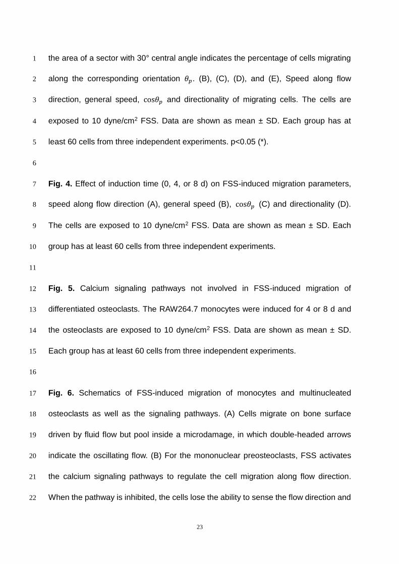
the area of a sector with 30° central angle indicates the percentage of cells migrating 1
along the corresponding orientation 𝜃𝑝. (B), (C), (D), and (E), Speed along flow 2
direction, general speed, cos𝜃𝑝 and directionality of migrating cells. The cells are 3
exposed to 10 dyne/cm2 FSS. Data are shown as mean ± SD. Each group has at 4
least 60 cells from three independent experiments. p<0.05 (*). 5
6
Fig. 4. Effect of induction time (0, 4, or 8 d) on FSS-induced migration parameters, 7
speed along flow direction (A), general speed (B), cos𝜃𝑝 (C) and directionality (D). 8
The cells are exposed to 10 dyne/cm2 FSS. Data are shown as mean ± SD. Each 9
group has at least 60 cells from three independent experiments. 10
11
Fig. 5. Calcium signaling pathways not involved in FSS-induced migration of 12
differentiated osteoclasts. The RAW264.7 monocytes were induced for 4 or 8 d and 13
the osteoclasts are exposed to 10 dyne/cm2 FSS. Data are shown as mean ± SD. 14
Each group has at least 60 cells from three independent experiments. 15
16
Fig. 6. Schematics of FSS-induced migration of monocytes and multinucleated 17
osteoclasts as well as the signaling pathways. (A) Cells migrate on bone surface 18
driven by fluid flow but pool inside a microdamage, in which double-headed arrows 19
indicate the oscillating flow. (B) For the mononuclear preosteoclasts, FSS activates 20
the calcium signaling pathways to regulate the cell migration along flow direction. 21
When the pathway is inhibited, the cells lose the ability to sense the flow direction and 22

24
will migrate randomly. (C) When the monocytes fuse into multinucleated osteoclasts, 1
calcium signaling pathways will not combine with the flow-induced migration although 2
FSS can still stimulate their migration. 3
4
5
6
7
8
9
10
11
12
13
14

25
Figure 1, C. Liu et al.

26
Figure 2, C. Liu et al.

27
Figure 3, C. Liu et al.

28
Figure 4, C. Liu et al.

29
Figure 5, C. Liu et al.

30
Figure 6, C. Liu et al.