Identification of PML Oncogenic Domains (PODs) in Human Megakaryocytes
9
Identification of PML Oncogenic Domains (PODs) in Human Megakaryocytes Arnaud Drouin, Alain Schmitt, Jean-Marc Masse ´, Anne-Marie Cieutat, Serge Fichelson, and Elisabeth M. Cramer 1 INSERM U. 474, Institut Cochin de Ge ´ne ´tique Mole ´culaire, Paris, France Megakaryocytes (Mks) are unique cells in the human body in that they carry a single and polyploid nucleus. It is therefore of interest to understand their nuclear ultrastructure. PML oncogenic domains (PODs) were described in several types of eukaryotic cells using human autoantibodies which recognize nuclear anti- gens with a specific speckled pattern (dots) in indirect immunofluorescence (IF). Two main antigens, PML and Sp 100, usually colocalize and concentrate in these nuclear subdomains. We investigated the presence of PODs using IF and immunoelectron microscopy (IEM) in cells from megakaryocytic lineage: the HEL cell line and human cultured Mks. Antibodies against PML, Sp100, and anti-nuclear dots were used in single and double labeling. PODs were identified in HEL cells and in human Mks, and their ultrastructure was charac- terized. We then used IF to quantify PODs within Mks and showed that their number increased proportion- ally to nuclear lobularity. In summary, we report the identification of PODs in human Mks at an ultrastruc- tural level and an increase in PODs number in parallel with Mk ploidy. We show that endomitosis not only leads to DNA increase but also to the multiplication of at least one of the associated nuclear structures. © 2001 Elsevier Science Key Words: nucleus; ultrastructure; megakaryocyte; endomitosis; PML oncogenic domains; human. INTRODUCTION Megakaryocytes (Mks), bone marrow precursors of platelets, are unique cells in humans since they become polyploid as nuclear DNA multiplies without cell divi- sion. Cell ploidy can then reach 32N to 64N without chromosome size increase or separate nuclear mem- brane formation. This physiological process, called en- domitosis, has been recently approached by several investigators [1, 2] but remains poorly understood. Electron microscopic examination has previously iden- tified distinct nuclear compartments in eukaryotic cells where specific proteins and nucleic acids may concen- trate [3–5]. Some of these nuclear structures were ini- tially described by electron microscopy [4] and further identified as autoantigenic targets in human autoim- mune diseases and called “multiple nuclear dots” or “dots” [5–7]. The cell growth repressor PML (promyelo- cytic leukemia protein) and the transcription factor Sp100 colocalize within these nuclear dots, which have therefore also been named PML nuclear bodies, or PML oncogenic domains (PODs) [4, 8 –11]. Since Mk nucleus is a unique model for the study of polyploidy, and because the presence of PODs has not so far been demonstrated within Mks, we found it noteworthy to determine whether PODs were present within the nu- clei of cells from the megakaryocytic lineage. We ana- lyzed PML and Sp100 nuclear distribution by immu- nofluorescence (IF) and immunoelectron microscopy (IEM) in the HEL cell line (derived from a human erythroleukemia with Mk differentiative potential) [12] and human Mks cultured from CD34 1 precursors. We also examined the evolution of the number of PODs during the endomitotic process and ploidy increase. MATERIALS AND METHODS HEL cell line and human MKs. The HEL cell line was grown and cultured as previously described [12]. Human cultured MKs were grown from circulating precursors. Blood CD34 1 cells were isolated from leukapheresis samples obtained from patients undergoing au- tologous peripheral blood stem cell transplantation (for IEM study) and cord blood (for IF study). CD34 1 cells were isolated using a magnetic cell-sorting system as previously described [13]. The cells were cultured for 13 days in a serum-free liquid medium containing thrombopoietin (TPO) (Rhu PEG-MGDF; Amgen, Thousand Oaks, CA) at a final concentration of 10 ng/mL. When CD34 1 cells were isolated from cord blood, they were cul- tured in the presence of TPO in serum-free media, as previously described [14]. All human cells were collected after informed consent was obtained from the patients, in accordance with the institutional guidelines of the Committee on Human Investigation. Antibodies. Human autoantibodies from the serum of one patient with systemic lupus erythematosus (SLE) was kindly provided by Dr. C. Andre ´ (Ho ˆpital H. Mondor, Creteil, France). This anti-dots 1 To whom reprint requests should be addressed at INSERUM U. 474, Maternite ´ de Port-Royal, 5 e `me e ´tage, Paris, France. Fax: 33 1 43 25 11 67. E-mail: [email protected]. 0014-4827/01 $35.00 277 © 2001 Elsevier Science All rights reserved. Experimental Cell Research 271, 277–285 (2001) doi:10.1006/excr.2001.5377, available online at http://www.idealibrary.com on
-
Upload
independent -
Category
Documents
-
view
1 -
download
0
Transcript of Identification of PML Oncogenic Domains (PODs) in Human Megakaryocytes

Experimental Cell Research 271, 277–285 (2001)doi:10.1006/excr.2001.5377, available online at http://www.idealibrary.com on
Identification of PML Oncogenic Domains (PODs)in Human Megakaryocytes
Arnaud Drouin, Alain Schmitt, Jean-Marc Masse, Anne-Marie Cieutat,Serge Fichelson, and Elisabeth M. Cramer1
INSERM U. 474, Institut Cochin de Genetique Moleculaire, Paris, France
Megakaryocytes (Mks) are unique cells in the humanbody in that they carry a single and polyploid nucleus.It is therefore of interest to understand their nuclearultrastructure. PML oncogenic domains (PODs) weredescribed in several types of eukaryotic cells usinghuman autoantibodies which recognize nuclear anti-gens with a specific speckled pattern (dots) in indirectimmunofluorescence (IF). Two main antigens, PMLand Sp 100, usually colocalize and concentrate in thesenuclear subdomains. We investigated the presence ofPODs using IF and immunoelectron microscopy (IEM)in cells from megakaryocytic lineage: the HEL cell lineand human cultured Mks. Antibodies against PML,Sp100, and anti-nuclear dots were used in single anddouble labeling. PODs were identified in HEL cells andin human Mks, and their ultrastructure was charac-terized. We then used IF to quantify PODs within Mksand showed that their number increased proportion-ally to nuclear lobularity. In summary, we report theidentification of PODs in human Mks at an ultrastruc-tural level and an increase in PODs number in parallelwith Mk ploidy. We show that endomitosis not onlyleads to DNA increase but also to the multiplication ofat least one of the associated nuclear structures. © 2001
Elsevier Science
Key Words: nucleus; ultrastructure; megakaryocyte;endomitosis; PML oncogenic domains; human.
INTRODUCTION
Megakaryocytes (Mks), bone marrow precursors ofplatelets, are unique cells in humans since they becomepolyploid as nuclear DNA multiplies without cell divi-sion. Cell ploidy can then reach 32N to 64N withoutchromosome size increase or separate nuclear mem-brane formation. This physiological process, called en-domitosis, has been recently approached by severalinvestigators [1, 2] but remains poorly understood.
1 To whom reprint requests should be addressed at INSERUM U.474, Maternite de Port-Royal, 5eme etage, Paris, France. Fax: 33 1 43
25 11 67. E-mail: [email protected].277
Electron microscopic examination has previously iden-tified distinct nuclear compartments in eukaryotic cellswhere specific proteins and nucleic acids may concen-trate [3–5]. Some of these nuclear structures were ini-tially described by electron microscopy [4] and furtheridentified as autoantigenic targets in human autoim-mune diseases and called “multiple nuclear dots” or“dots” [5–7]. The cell growth repressor PML (promyelo-cytic leukemia protein) and the transcription factorSp100 colocalize within these nuclear dots, which havetherefore also been named PML nuclear bodies, orPML oncogenic domains (PODs) [4, 8–11]. Since Mknucleus is a unique model for the study of polyploidy,and because the presence of PODs has not so far beendemonstrated within Mks, we found it noteworthy todetermine whether PODs were present within the nu-clei of cells from the megakaryocytic lineage. We ana-lyzed PML and Sp100 nuclear distribution by immu-nofluorescence (IF) and immunoelectron microscopy(IEM) in the HEL cell line (derived from a humanerythroleukemia with Mk differentiative potential)[12] and human Mks cultured from CD341 precursors.We also examined the evolution of the number of PODsduring the endomitotic process and ploidy increase.
MATERIALS AND METHODS
HEL cell line and human MKs. The HEL cell line was grown andcultured as previously described [12]. Human cultured MKs weregrown from circulating precursors. Blood CD341 cells were isolatedfrom leukapheresis samples obtained from patients undergoing au-tologous peripheral blood stem cell transplantation (for IEM study)and cord blood (for IF study). CD341 cells were isolated using amagnetic cell-sorting system as previously described [13]. The cellswere cultured for 13 days in a serum-free liquid medium containingthrombopoietin (TPO) (Rhu PEG-MGDF; Amgen, Thousand Oaks,CA) at a final concentration of 10 ng/mL.
When CD341 cells were isolated from cord blood, they were cul-tured in the presence of TPO in serum-free media, as previouslydescribed [14]. All human cells were collected after informed consentwas obtained from the patients, in accordance with the institutionalguidelines of the Committee on Human Investigation.
Antibodies. Human autoantibodies from the serum of one patientwith systemic lupus erythematosus (SLE) was kindly provided by
Dr. C. Andre (Hopital H. Mondor, Creteil, France). This anti-dots0014-4827/01 $35.00© 2001 Elsevier Science
All rights reserved.
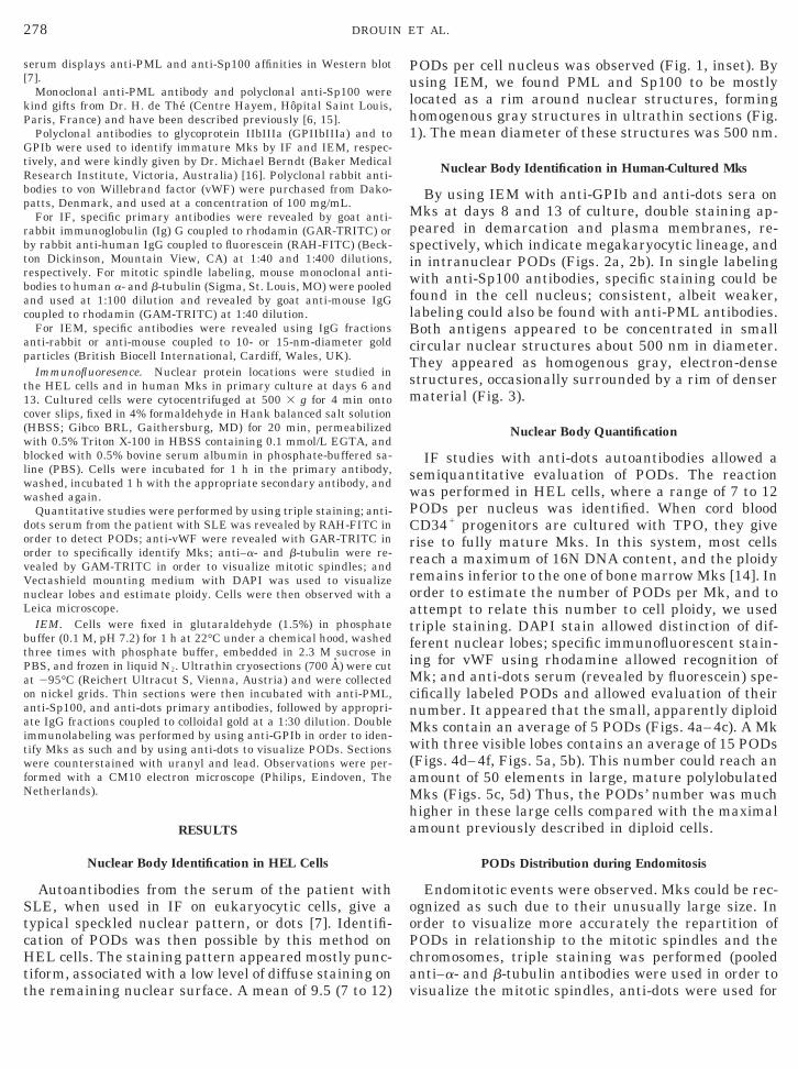
278 DROUIN ET AL.
serum displays anti-PML and anti-Sp100 affinities in Western blot[7].
Monoclonal anti-PML antibody and polyclonal anti-Sp100 werekind gifts from Dr. H. de The (Centre Hayem, Hopital Saint Louis,Paris, France) and have been described previously [6, 15].
Polyclonal antibodies to glycoprotein IIbIIIa (GPIIbIIIa) and toGPIb were used to identify immature Mks by IF and IEM, respec-tively, and were kindly given by Dr. Michael Berndt (Baker MedicalResearch Institute, Victoria, Australia) [16]. Polyclonal rabbit anti-bodies to von Willebrand factor (vWF) were purchased from Dako-patts, Denmark, and used at a concentration of 100 mg/mL.
For IF, specific primary antibodies were revealed by goat anti-rabbit immunoglobulin (Ig) G coupled to rhodamin (GAR-TRITC) orby rabbit anti-human IgG coupled to fluorescein (RAH-FITC) (Beck-ton Dickinson, Mountain View, CA) at 1:40 and 1:400 dilutions,respectively. For mitotic spindle labeling, mouse monoclonal anti-bodies to human a- and b-tubulin (Sigma, St. Louis, MO) were pooledand used at 1:100 dilution and revealed by goat anti-mouse IgGcoupled to rhodamin (GAM-TRITC) at 1:40 dilution.
For IEM, specific antibodies were revealed using IgG fractionsanti-rabbit or anti-mouse coupled to 10- or 15-nm-diameter goldparticles (British Biocell International, Cardiff, Wales, UK).
Immunofluoresence. Nuclear protein locations were studied inthe HEL cells and in human Mks in primary culture at days 6 and13. Cultured cells were cytocentrifuged at 500 3 g for 4 min ontocover slips, fixed in 4% formaldehyde in Hank balanced salt solution(HBSS; Gibco BRL, Gaithersburg, MD) for 20 min, permeabilizedwith 0.5% Triton X-100 in HBSS containing 0.1 mmol/L EGTA, andblocked with 0.5% bovine serum albumin in phosphate-buffered sa-line (PBS). Cells were incubated for 1 h in the primary antibody,washed, incubated 1 h with the appropriate secondary antibody, andwashed again.
Quantitative studies were performed by using triple staining; anti-dots serum from the patient with SLE was revealed by RAH-FITC inorder to detect PODs; anti-vWF were revealed with GAR-TRITC inorder to specifically identify Mks; anti–a- and b-tubulin were re-vealed by GAM-TRITC in order to visualize mitotic spindles; andVectashield mounting medium with DAPI was used to visualizenuclear lobes and estimate ploidy. Cells were then observed with aLeica microscope.
IEM. Cells were fixed in glutaraldehyde (1.5%) in phosphatebuffer (0.1 M, pH 7.2) for 1 h at 22°C under a chemical hood, washedthree times with phosphate buffer, embedded in 2.3 M sucrose inPBS, and frozen in liquid N2. Ultrathin cryosections (700 A) were cutat 295°C (Reichert Ultracut S, Vienna, Austria) and were collectedon nickel grids. Thin sections were then incubated with anti-PML,anti-Sp100, and anti-dots primary antibodies, followed by appropri-ate IgG fractions coupled to colloidal gold at a 1:30 dilution. Doubleimmunolabeling was performed by using anti-GPIb in order to iden-tify Mks as such and by using anti-dots to visualize PODs. Sectionswere counterstained with uranyl and lead. Observations were per-formed with a CM10 electron microscope (Philips, Eindoven, TheNetherlands).
RESULTS
Nuclear Body Identification in HEL Cells
Autoantibodies from the serum of the patient withSLE, when used in IF on eukaryocytic cells, give atypical speckled nuclear pattern, or dots [7]. Identifi-cation of PODs was then possible by this method onHEL cells. The staining pattern appeared mostly punc-tiform, associated with a low level of diffuse staining on
the remaining nuclear surface. A mean of 9.5 (7 to 12)PODs per cell nucleus was observed (Fig. 1, inset). Byusing IEM, we found PML and Sp100 to be mostlylocated as a rim around nuclear structures, forminghomogenous gray structures in ultrathin sections (Fig.1). The mean diameter of these structures was 500 nm.
Nuclear Body Identification in Human-Cultured Mks
By using IEM with anti-GPIb and anti-dots sera onMks at days 8 and 13 of culture, double staining ap-peared in demarcation and plasma membranes, re-spectively, which indicate megakaryocytic lineage, andin intranuclear PODs (Figs. 2a, 2b). In single labelingwith anti-Sp100 antibodies, specific staining could befound in the cell nucleus; consistent, albeit weaker,labeling could also be found with anti-PML antibodies.Both antigens appeared to be concentrated in smallcircular nuclear structures about 500 nm in diameter.They appeared as homogenous gray, electron-densestructures, occasionally surrounded by a rim of densermaterial (Fig. 3).
Nuclear Body Quantification
IF studies with anti-dots autoantibodies allowed asemiquantitative evaluation of PODs. The reactionwas performed in HEL cells, where a range of 7 to 12PODs per nucleus was identified. When cord bloodCD341 progenitors are cultured with TPO, they giverise to fully mature Mks. In this system, most cellsreach a maximum of 16N DNA content, and the ploidyremains inferior to the one of bone marrow Mks [14]. Inorder to estimate the number of PODs per Mk, and toattempt to relate this number to cell ploidy, we usedtriple staining. DAPI stain allowed distinction of dif-ferent nuclear lobes; specific immunofluorescent stain-ing for vWF using rhodamine allowed recognition ofMk; and anti-dots serum (revealed by fluorescein) spe-cifically labeled PODs and allowed evaluation of theirnumber. It appeared that the small, apparently diploidMks contain an average of 5 PODs (Figs. 4a–4c). A Mkwith three visible lobes contains an average of 15 PODs(Figs. 4d–4f, Figs. 5a, 5b). This number could reach anamount of 50 elements in large, mature polylobulatedMks (Figs. 5c, 5d) Thus, the PODs’ number was muchhigher in these large cells compared with the maximalamount previously described in diploid cells.
PODs Distribution during Endomitosis
Endomitotic events were observed. Mks could be rec-ognized as such due to their unusually large size. Inorder to visualize more accurately the repartition ofPODs in relationship to the mitotic spindles and thechromosomes, triple staining was performed (pooledanti–a- and b-tubulin antibodies were used in order to
visualize the mitotic spindles, anti-dots were used for
ifie
279PODs IN MEGAKARYOCYTES
PODs, and DAPI stain was used for chromatin) oncultured Mks. In most of them, anti-dots labeling wasdiffuse in the cytoplasm and PODs were undetectable;or fluorescent labeling appeared disorganized, withfaint clusters of fluorescence redistributed away at thecell periphery, replacing the classical speckled inter-phasic pattern. The clusters displayed no interactionwith the asters, spindle microtubules, or chromosomes(Fig. 6). In parallel, and as controls of the reaction,interphasic diploid cells and Mks displayed evidentnuclear bodies (Fig. 6c).
DISCUSSION
To our knowledge, this is the first report that dem-onstrates the existence of PODs in the nucleus of hu-man Mks cells. We used the HEL cell line, which is ableto undergo megakaryocytic differentiation and humancultured Mks [12]. A variety of autoantibodies from apatient with SLE and antibodies directed againstPODs components were utilized, followed by IF and
FIG. 1. EM view of HEL cells treated by autoantibodies from theas a rim of particles (arrowheads) surrounding a dense core, allowingMagnification, 61,0003. Inset: HEL cells stained with autoantibodiepattern of multinuclear dots. A range of 7 to 12 PODs can be quant
IEM analyses. Previous studies have reported PODs in
many eukaryotic cells, including hematopoietic cellsfrom the granulocytic and erythroid lineages [6, 17,18]. Occasional reports have mentioned the search forPODs in Mks, but the results concerning this cell typewere of secondary importance and did not lead to a firmconclusion [6, 18]. Mks are rare cells in human bonemarrow, which makes their study tedious, and thelarge size of their nucleus makes the identification ofvery small compartments by optical microscopy diffi-cult. Therefore, in our study, we used a culture systemwhere the Mk population is predominant, combinedwith the highly discriminative technique of IEM. Be-sides euchromatin, heterochromatin, and the nucleo-lus, other subnuclear compartments have been identi-fied within the eukaryotic cell nucleus [8]. Among themare nuclear bodies, which have been recognized byelectron microscopy and further characterized, thanksto their specific components (i.e., PML and Sp100 [10,11]). Initial functional approaches to the study of PODscame from acute promyelocytic leukemia (APL), a ma-
um of a patient with SLE and immunogold-specific labeling appearse ultrastructural identification of a POD. N, nucleus; Nu, nucleolus.nti-dots) from a patient with SLE by IF. The cell displays a typical
d in each cell nucleus.
serth
s (a
lignant hemopathy specifically associated with a t(15;

ag
280 DROUIN ET AL.
17) translocation [19]. This translocation fuses a nu-clear receptor for retinoic acid (RAR-a) with the PMLgene [20–22]. In APL, promyelocyte differentiation isblocked and abnormal promyelocytes accumulate. ThePODs of APL promyelocytes are disrupted, and PMLantigen is detected in the entire cell without specificlocation. All transretinoic acid treatment of APL pro-myelocytes in culture and in patients with APL inducesthe reconstitution of PODs, overcomes the differentia-tion blockage, and leads to apoptotic death of leukemicpromyelocytes [15, 23–27].
The PML protein is expressed in most tissues, in-cluding hematopoietic cells [6, 17]. PML protein ismainly nuclear, associated with the nuclear matrix[28], and localized in PODs. Transcription of the PMLgene is increased by interferons [3, 29], which aregrowth- and tumor-suppressive cytokines. In those ex-periments, the number of PODs is also increased. Thefunctions of PML protein are multiple and still underinvestigation. PML acts as a negative cell growth fac-tor and as a factor which promotes cell differentiation[30]. PML can improve both erythroid and myeloid
FIG. 2. IEM analysis of PODs in cultured Mks at day 13 (maturehuman antiserum (10 nm gold) to visualize PODs and anti-GPIb raba convoluted nucleus and characteristic organelles such as demarcahigher magnification view shows GPIb staining in demarcation memwithin the nucleus corresponding to a nuclear POD (arrowheads). M
differentiation [17]. Moreover, knockout animal model
PML2/2; mice display leucopenia with a decreasedcapacity for retinoid acid (RA)–dependent terminalgranulocytic maturation [31]. Thus, PML could medi-ate the RA growth-suppressive and cell-differentiation-promoting effects. In addition, PML clearly interfereswith cell cycle progression [17]; PML overexpressionregulates cdk2 and cyclin [30]; and PML protein inter-acts with the eukaryotic initiation factor 4E (eIF-4E),which regulates the cyclin D1 mRNA nucleocytoplas-mic transport, an interaction which interestingly re-quires intact PODs [32]. PML can also participate inthe upregulation of cell cycle inhibitor p21 [31]. Fi-nally, PML is implicated in genomic stability, as re-ported by Zhong et al. [33].
In contrast to the well-described role of PML protein,PODs have still elusive functions. PODs are multipro-tein complexes specifically assembled by PML protein.As PML, the PODs are associated with the nuclearmatrix. Numerous proteins were reported to constitutePODs. These proteins display intranuclear and nucleo-cytoplasmic shuttles [4, 10, 11]. Proteins which do notconstitute components of PODs can be specifically tar-
lls). Double immunogold labeling has been performed with anti-dotsantibodies (15 nm gold) to identify Mk. (a) This large cell possessesn membranes and secretory granules. Magnification, 83003. (b) Anes (dm) and a rim of labeling a small, dense, and round structure
nification, 35,0003.
cebittiobra
geted and accumulated at the vicinity of PODs. Accu-

281PODs IN MEGAKARYOCYTES
FIG. 3. IEM analysis of PODs in cultured Mk at day 13 (mature cells) detected with anti-dots serum shows various aspects of PODs. (a,b) The structure occasionally appears surrounded by a denser ring of chromatin. Magnification, (a) 48,0003; (b) 67,0003. (c, d) In all cases,the structure is distinct from the surrounding chromatin by its homogenous, moderately dense aspect, and the labeling is distributed at the
periphery of the organelle. Magnification, (c) 54,5003; (d) 44,0003.
large polylobulated Mk display a greater number of PODs (10 to 15) than the diploid cells.
282 DROUIN ET AL.
FIG. 5. Double fluorescent staining of human Mks in culture. Anti-dots antibodies coupled to FITC (a, c, e) labels the PODs, while DAPIstaining (b, f) reveals nuclear shape and allows estimation of the number of nuclear lobes, which increases with ploidy. (a, b) DAPI stainingreveals three lobes in this Mk, and the nucleus contains three times more PODs than a diploid cell. (c, d) Two adjacent highly polylobulatedMks (containing an average of four nuclear lobes) are identified as such by the specific red fluorescence for vWF, maximal in the paranuclear
FIG. 4. Human Mk in culture are triple stained. Anti-dots antibodies coupled to FITC show PODs (a, d); DAPI staining reveals nuclearshape and allows estimation of the number of nuclear lobes, which increases with ploidy (b, e). The cytoplasmic staining for vWF certifiesthe Mk lineage of cell (c, f). (a–c) A small Mk with a unique nuclear lobe, which is likely diploid, displays an average of five PODs. (d–f) The
regions. Immunostaining reveals an average number of 50 PODs per cell.

283PODs IN MEGAKARYOCYTES
mulation of nascent mRNAs was also reported inPODs, as a way of stockage that may regulate mRNAmaturation, mRNA nucleocytoplasmic transport, andprotein expression [34, 35]. In summary, PODs couldconstitute “nuclear dumps,” or nuclear storage for var-ious proteins and mRNA. PODs could then regulate thetraffic, activity, and expression of various proteins.PODs may participate to all the functions of PML: cellproliferation, cell cycle regulation, and genomic stabil-ity. In addition, since PODs are present in normalpromyelocytes and disorganized in leukemic promyelo-cytes, it would be of interest to investigate PML distri-bution and PODs structure in malignant Mks frompatients with megakaryoblastic leukemia and frommyeloproliferative syndromes.
Polyploidization results from endomitotic cycles thatoccur in Mk as cells stop proliferating and start differ-entiating. DNA accumulates in Mks without cell divi-sion, and the nuclei become polylobulated [2]. The part-ners of cell cycle machinery have unique expressionsand probably regulations during the endomitosis ofMks [2]. The evolution of nuclear structures duringendomitosis is poorly understood. Thus, we have exam-ined the number of PODs during the polyploidization ofMks. First, we have quantified PODs in HEL cells,which are mostly diploid cells. Then we followed theevolution of the number of PODs during cytoplasmicand nuclear growth of Mk (increased nuclear lobularityand polyploidization). We found that the more numer-ous the nuclear lobes were, the more numerous the
FIG. 6. Human Mk undergoing endomitosis, triple stained withfluorescent dyes. DAPI staining (a) shows an endomitosis with twoditinct groups of chromosomes. Anti-tubulin immunolabeling (b)shows spindle microtubules (green arrows), which radiate from eachaster to form a spherical conformation. Anti-dots serum (c) revealsthat PODs are disorganized, forming large aggregates of fluorescence(red arrows). PODs are excluded from the mitotic spindles and chro-mosome areas and redistribute at the periphery of the cell. As acontrol, an interphasic diploid cell exhibits three PODs (red arrow-heads).
PODs were. As the number of nuclear lobes in Mks was
reported to be roughly proportional to Mk ploidy [36],we deduce that the number of PODs is also propor-tional to Mk ploidy. Increased numbers of PODs werepreviously reported in cancerous cells but not in cellsfrom nontumoral hyperplastic tissues [37, 38]; this ob-servation could be explained by the fact that malignantcells are often multiploid, whereas hyperplastic normalcells are not. The increased PODs number could thenjust reflect multiploidy. Growth factors, erythropoietin(EPO), and granulocyte, macrophage colony stimula-tory factor (GMCSF) induce PML expression and accu-mulation of PODs, respectively, in erythroid and gran-ulocytic lineages cultured from human progenitorscells [17]. In the Mk lineage, TPO is the main regulatorof cell development; TPO acts at each stage of thedevelopment of Mks, proliferation, endomitosis, andmaturation [39]. TPO stimulates polyploidization incultured Mks [40, 41], and TPO 2/2 mice display Mkswith smaller size and ploidy [2, 3, 39, 42, 43]. Wesuggest that accumulation of PODs could be induced byTPO, since TPO and interferons both have EPO-likedomains, and it would be of interest to confirm thishypothesis.
When the number of PODs increases in the polyploidMk, PML, and PODs, associated proteins accumulateand then can assume an efficient cell growth controlduring polyploidization of Mks and differentiation con-trol of Mks, as in other hematopoietic lineages [44].Moreover, the level of the genomic protection role as-sumed by PML may thus be proportional to the DNAcontent of polyploid Mks.
In the majority of Mks undergoing endomitosis,PODs were disorganized and became undetectable,and the fluorescent labeling appeared to be redistrib-uted. They did not appear to interact either with thechromosomes or with the mitotic spindles. Thus, wefound in Mks during endomitosis the same intracellu-lar repartition and global disappearance of PODs, asdescribed previously in nonMks lineages [3, 45]. Fi-nally, a better understanding of PODs’ organization,composition, and subcellular interactions in Mks couldimprove our understanding of the endomitotic cell cy-cle.
The authors are grateful to Pr. Hugues de The (CNRS UPR 43,Centre Hayem, Hopital Saint Louis, Paris, France) for providinganti-PML and anti-Sp100 antibodies and to Dr. Chantal Andre (Ho-pital Henri Mondor, Creteil, France) for advice and for providingautoimmune serum from the patient with SLE. We are also gratefulto Najet Debili for providing the Mks used for electron microscopy.
REFERENCES
1. Cramer, E. M. (1999). Megakaryocyte structure and function.Curr. Opin. Hematol. 6, 354–361.
2. Zimmet, J., and Ravid, K. (2000). Polyploidy: Occurrence innature, mechanisms, and significance for the megakaryocyte–
platelet system. Exp. Hematol. 28, 3–16.
284 DROUIN ET AL.
3. Sterndorf, T., Grotzinger, T., Jensen, K., and Will, H. (1997).Nuclear dots, actors on many stages. Immunobiology 198, 307–331.
4. Lamond, A. I., and Earnshaw, W. C. (1998). Structure andfunction in the nucleus. Science 280, 547–553.
5. Matera, A. G. (1999). Nuclear bodies: Multifaceted subdomainsof the interchromatin space. Trends Cell. Biol. 9, 302–309.
6. Daniel, M. T., Koken, M., Romagne, O., Barbey, S, Bazarbachi,A., Stadler, M., Guillemin, M. C., Degos, L., Chomienne, C., andde The, H. (1993). PML protein expression in hematopoietic andacute promyelocytic leukemia cells. Blood 82, 1858–1867.
7. Andre, C., Guillemin, M. C., Zhu, J., Koken, M. H., Quignon, F.,Herve, L., Chelbi-Alix, M. K., Dhumeaux, D., Wang, Z. Y.,Degos, L., Chen, Z., and de The, H. (1996). The PML andPML/RARalpha domains: From autoimmunity to molecular on-cology and from retinoic acid to arsenic. Exp. Cell Res. 229,253–260.
8. Brasch, K., and Ochs, R. L. (1992). Nuclear bodies (NBs): anewly “rediscovered” organelle. Exp. Cell Res. 202, 211–223.
9. Doucas, V., and Evans, R. M. (1996). The PML nuclear com-partment and cancer. Biochim. Biophys. Acta 1288, M25–M29.
10. Hodges, M., Tissot, C., Howe, K., Grimwade, D., and Freemont,P. S. (1998). Structure, organization, and dynamics of promy-elocytic leukemia protein nuclear bodies. Am. J. Hum. Genet.63, 297–304.
11. Maul, G. G., Negorev, D., Bell, P., and Ishov, A. M. (2000).Properties and assembly mechanisms of ND10, PML bodies, orPODs. J. Struct. Biol. 129, 278–287.
12. Long, M. W., Heffner, C. H., Williams, J. L., Peters, C., andProchownik, E. V. (1990). Regulation of megakaryocyte poten-tial in human ertyholeukemia cells. J. Clin. Invest. 85, 1072–1084.
13. Cramer, E. M., Norol, F., Guichard, J., Breton-Gorius, J.,Vainchenker, W., Masse, J. M., and Debili, N. (1997). Ultra-structure of platelet formation by human megakaryocytes cul-tured with the Mpl ligand. Blood 89, 2336–2346.
14. Fichelson, S., Freyssinier, J. M., Picard, F., Fontenay-Roupie,M., Guesnu, M., Cherai, M., Gisselbrecht, S., and Porteu, F.(1999). Megakaryocyte growth and development factor–inducedproliferation and differentiation are regulated by the mitogen-activated protein kinase pathway in primitive cord blood hema-topoietic progenitors. Blood 94, 1601–1613.
15. Koken, M. H., Puvion-Dutilleul, F., Guillemin, M. C., Viron, A.,Linares-Cruz, G., Stuurman, N., de Jong, L., Szostecki, C.,Calvo, F., Chomienne, C., Degos, L., Puvion, E., and de The, H.(1994). The t(15;17) translocation alters a nuclear body in aretinoic acid–reversible fashion. EMBO J. 13, 1073–1083.
16. Youssefian, T., Masse, J. M., Rendu, F., Guichard, J., andCramer, E. M. (1997). Platelet and megakaryocyte dense gran-ule contain glycoproteins Ib and IIb–IIIa. Blood 89, 4049–4055.
17. Labbaye, C., Valtieri, M., Grignani, F., Puglisi, R., Luchetti, L.,Masella, B., Alcalay, M., Testa, U., and Peschle, C. (1999).Expression and role of PML gene in normal adult hematopoie-sis: Functional interaction between PML and Rb proteins inerythropoiesis. Oncogene 18, 3529–3540.
18. Flenghi, L., Fagioli, M., Tomassoni, L., Pileri, S., Gambacorta,M., Pacini, R., Grignani, F., Casini, T., Ferrucci, P. F., Martelli,M. F., Pelicci, P. G., and Falini, B. (1995). Characterization of anew monoclonal antibody (PG-M3) directed against the amino-terminal portion of the PML gene product: Immunocytochemi-cal evidence for high expression of PML proteins on activatedmacrophages, endothelial cells, and epithelia. Blood 85, 1871–
1880.19. Rowley, J. D., Golomb, H. M., and Dougherty, C. (1977). 15/17translocation, a consistent chromosomal change in acute pro-myelocytic leukaemia. Lancet 1 (8010), 549–550.
20. de The, H., Chomienne, C., Lanotte, M., Degos, L., and Dejean,A. (1990). The t(15;17) translocation of acute promyelocyticleukaemia fuses the retinoic acid receptor alpha gene to a noveltranscribed locus. Nature 347, 558–561.
21. Goddard, A. D., Borrow, J., Freemont, P. S., and Solomon, E.(1991). Characterization of a zinc finger gene disrupted by thet(15;17) in acute promyelocytic leukemia. Science 254, 1371–1374.
22. de The, H., Lavau, C., Marchio, A., Chomienne, C., Degos, L.,and Dejean, A. (1991). The PML-RAR alpha fusion mRNA gen-erated by the t(15;17) translocation in acute promyelocytic leu-kemia encodes a functionally altered RAR. Cell 66, 675–684.
23. Breitman, T. R., Collins, S. J., and Keene, B. R. (1981). Termi-nal differentiation of human promyelocytic leukemic cells inprimary culture in response to retinoic acid. Blood 57, 1000–1004.
24. Huang, M. E., Ye, Y. I., Chen, J. E., Chai, S. R., Lu, J. R., Zhoa,J. X., Gu, J., and Wang, Z. Y. (1988). Use of all trans retinoicacid in the treatment of acute promyelocytic leukemia. Blood72, 567–572.
25. Castaigne, S., Chomienne, C., Daniel, M. T., Ballerini, P.,Berger, R., Fenaux, P., and Degos, L. (1990). All-trans retinoicacid as a differentiation therapy for acute promyelocytic leuke-mia. I. Clinical results. Blood 76, 1704–1709.
26. Chomienne, C., Ballerini, P., Balitrand, N., Daniel, M. T., Fe-naux, P., Castaigne, S., and Degos, L. (1990). All-trans retinoicacid in acute promyelocytic leukemias. II. In vitro studies:Structure–function relationship. Blood 76, 1710–1717.
27. Weis, K., Rambaud, S., Lavau, C., Jansen, J., Carvalho, T.,Carmo-Fonseca, M., Lamond, A., and Dejean, A. (1994). Reti-noic acid regulates aberrant nuclear localization of PML-RARalpha in acute promyelocytic leukemia cells. Cell 76, 345–356.
28. Chang, K. S., Fan, Y. H., Andreeff, M., Liu, J., and Mu, Z. M.(1995). The PML gene encodes a phosphoprotein associatedwith the nuclear matrix. Blood 85, 3646–3653.
29. Lavau, C., Marchio, A., Fagioli, M., Jansen, J., Falini, B.,Lebon, P., Grosveld, F., Pandolfi, P. P., Pelicci, P. G., andDejean, A. (1995). The acute promyelocytic leukaemia-associ-ated PML gene is induced by interferon. Oncogene 11, 871–876.
30. Mu, Z. M., Le, X. F., Vallian, S., Glassman, A. B., and Chang,K. S. (1997). Stable overexpression of PML alteres regulation ofcell cycle progression in HeLa cells. Carcinogenesis 18, 2063–2069.
31. Wang, Z. G., Delva, L., Gaboli, M., Rivi, R., Giorgio, M., Cordon-Cardo, C., Grosveld, F., and Pandolfi, P. P. (1998). Role of PMLin cell growth and the retinoic acid pathway. Science 279,1547–1551.
32. Lai, H. K., and Borden, K. L. (2000). The promyelocytic leuke-mia (PML) protein suppresses cyclin D1 protein production byaltering the nuclear cytoplasmic distribution of cyclin D1mRNA. Oncogene 19, 1623–1634.
33. Zhong, S., Hu, P., Ye, T. Z., Stan, R., Ellis, N. A., and Pandolfi,P. P. (1999). A role for PML and the nuclear body in genomicstability. Oncogene 18, 7941–7947.
34. LaMorte, V. J., Dyck, J. A., Ochs, R. L., and Evans, R. M. (1998).Localization of nascent RNA and CREB binding protein withthe PML-containing nuclear body. Proc. Natl. Acad. Sci. USA95, 4991–4996.
35. Alcalay, M., Tomassoni, L., Colombo, E., Stoldt, S., Grignani,F., Fagioli, M., Szekely, L., Helin, K., and Pelicci, P. G. (1998).
The promyelocytic leukemia gene product (PML) forms stable
285PODs IN MEGAKARYOCYTES
complexes with the retinoblastoma protein. Mol. Cell. Biol. 18,1084–1093.
36. Harker, L. A. (1968). Kinetics of thrombopoiesis. J. Clin. Invest.47, 458–465.
37. Koken, M. H., Linares-Cruz, G., Quignon, F., Viron, A., Chelbi-Alix, M. K., Sobczak-Thepot, J., Juhlin, L., Degos, I., and deThe, H. (1995) The PML growth suppresor has an altered ex-pression in human oncogenesis. Oncogene 10, 1315–1324.
38. Cho, Y., Lee, I., Maul, G. G., and Yu, E. (1998). A novel nuclearsubstructure, ND10: Distribution in normal and neoplastic hu-man tissues. Int. J. Mol. Med. 1, 717–724.
39. Drouin, A., and Cramer, E. M. (2001). Production of platelets.In “Platelets in Thrombotic and Non-Thrombotic Disorders:Pathophysiology, Pharmacology and Therapeutics” (P. Gresele,C. P. Page, V. Fuster, and J. Vermyler, Eds.), Cambridge Uni-versity Press, New York, in press.
40. Debili, N., Wendling, F., Katz, A., Guichard, J., Breton-Gorius,J., Hunt, P., and Vainchenker, W. (1995). The Mpl-ligand or
thrombopoietin or megakaryocyte growth and differentiativefactor has both direct proliferative and differentiative activitieson human megakaryocyte progenitors. Blood 86, 2516–2525.
41. Vitrat, N., Cohen-Solal, K., Piques, C., Le Couedic, J. P., Norol,F., Larsen, A. K., Katz, A., Vainchenker, W., and Debili, N.(1998). Endomitosis of human megakaryocytes are due to abor-tive mitosis. Blood 91, 3711–3723.
42. de Sauvage, F. J., Carver-Moore, K., Luoh, S. M., Ryan, A.,Dowd, M., Eaton, D. L., and Moore, M. W. (1996). Physiologicalregulation of early and late stages of megakaryocytopoiesis bythrombopoietin. J. Exp. Med. 183, 651–656.
43. Gurney, A. L., and de Sauvage, F. J. (1996). Dissection of c-Mpland thrombopoietin function: Studies of knockout mice andreceptor signal transduction. Stem Cells 1, 116–123.
44. Topcu, Z., Mack, D. L., Hromas, R. A., and Borden, K. L. (1999).The promyelocytic leukemia protein PML interacts with theproline-rich homeodomain protein PRH: A RING may link he-matopoiesis and growth control. Oncogene 18, 7091–7100.
45. Ascoli, C. A., and Maul, G. G. (1991). Identification of a novel
nuclear domain. J. Cell. Biol. 112, 785–795.Received May 24, 2001Revised version received August 31, 2001Published online October 29, 2001