PML-RAR induces promyelocytic leukemias with high efficiency following retroviral gene transfer into...
28
doi:10.1182/blood-2001-11-0089 Prepublished online June 14, 2002; Francesco Contegno, Francesco Lo Coco, Eugenio Scanziani, Alberto Gobbi and Pier G Pelicci Insinga, Daniela Diverio, Patrizia Gasparini, Manuela Capillo, Emanuela Colombo, Cristian Matteucci, Saverio Minucci, Silvia Monestiroli, Sabrina Giavara, Simona Ronzoni, Francesco Marchesi, Alessandra retroviral gene transfer into purified murine hematopoietic progenitors PML-RAR induces promyelocytic leukemias with high efficiency following (795 articles) Oncogenes and Tumor Suppressors (4217 articles) Neoplasia Articles on similar topics can be found in the following Blood collections http://bloodjournal.hematologylibrary.org/site/misc/rights.xhtml#repub_requests Information about reproducing this article in parts or in its entirety may be found online at: http://bloodjournal.hematologylibrary.org/site/misc/rights.xhtml#reprints Information about ordering reprints may be found online at: http://bloodjournal.hematologylibrary.org/site/subscriptions/index.xhtml Information about subscriptions and ASH membership may be found online at: digital object identifier (DOIs) and date of initial publication. the indexed by PubMed from initial publication. Citations to Advance online articles must include final publication). Advance online articles are citable and establish publication priority; they are appeared in the paper journal (edited, typeset versions may be posted when available prior to Advance online articles have been peer reviewed and accepted for publication but have not yet Copyright 2011 by The American Society of Hematology; all rights reserved. 20036. the American Society of Hematology, 2021 L St, NW, Suite 900, Washington DC Blood (print ISSN 0006-4971, online ISSN 1528-0020), is published weekly by For personal use only. by guest on June 3, 2013. bloodjournal.hematologylibrary.org From
Transcript of PML-RAR induces promyelocytic leukemias with high efficiency following retroviral gene transfer into...
doi:10.1182/blood-2001-11-0089Prepublished online June 14, 2002;
Francesco Contegno, Francesco Lo Coco, Eugenio Scanziani, Alberto Gobbi and Pier G PelicciInsinga, Daniela Diverio, Patrizia Gasparini, Manuela Capillo, Emanuela Colombo, Cristian Matteucci, Saverio Minucci, Silvia Monestiroli, Sabrina Giavara, Simona Ronzoni, Francesco Marchesi, Alessandra retroviral gene transfer into purified murine hematopoietic progenitorsPML-RAR induces promyelocytic leukemias with high efficiency following
(795 articles)Oncogenes and Tumor Suppressors � (4217 articles)Neoplasia �
Articles on similar topics can be found in the following Blood collections
http://bloodjournal.hematologylibrary.org/site/misc/rights.xhtml#repub_requestsInformation about reproducing this article in parts or in its entirety may be found online at:
http://bloodjournal.hematologylibrary.org/site/misc/rights.xhtml#reprintsInformation about ordering reprints may be found online at:
http://bloodjournal.hematologylibrary.org/site/subscriptions/index.xhtmlInformation about subscriptions and ASH membership may be found online at:
digital object identifier (DOIs) and date of initial publication. theindexed by PubMed from initial publication. Citations to Advance online articles must include
final publication). Advance online articles are citable and establish publication priority; they areappeared in the paper journal (edited, typeset versions may be posted when available prior to Advance online articles have been peer reviewed and accepted for publication but have not yet
Copyright 2011 by The American Society of Hematology; all rights reserved.20036.the American Society of Hematology, 2021 L St, NW, Suite 900, Washington DC Blood (print ISSN 0006-4971, online ISSN 1528-0020), is published weekly by
For personal use only. by guest on June 3, 2013. bloodjournal.hematologylibrary.orgFrom
PML-RAR induces promyelocytic leukemias with high
efficiency following retroviral gene transfer into purified
murine hematopoietic progenitors
Short title: Leukemia by retroviral gene transfer of PML-RAR
º^% Saverio Minucci, ºSilvia Monestiroli, ºSabrina Giavara, ºSimona Ronzoni, Francesco
Marchesi*, ºAlessandra Insinga, Daniela Diverio$, Patrizia Gasparini#,Manuela Capillo#,
ºEmanuela Colombo, ºCristian Matteucci, ºFrancesco Contegno, Francesco Lo Coco$,
Eugenio Scanziani*,Alberto Gobbiº# and Pier Giuseppe Pelicciº# ^
ºEuropean Institute of Oncology, Department of Experimental Oncology, Milan,
20141 Italy.
%Department of Physiology and General Biochemistry, University of Milan,
20100 Milan, Italy.
$Department of Human Biotechnology and Hematology, University of Rome “La
Sapienza”, Rome,Italy.
*Department of Veterinary Pathology, Hygiene and Public Health, School of
Veterinary Medicine, University of Milan, 20133 Milan, Italy.
#IFOM-FIRC Institute of Molecular Oncology, Milan, 20100 Italy.
This study has been funded by grants from EEC, FIRC, AIRC, and Ministero della Sanitá to S.M.
and P.G.P.
^Corresponding Authors: Pier Giuseppe Pelicci, European Institute of Oncology, Department of
Experimental Oncology, Via Ripamonti 435 Milan, 20141 Italy. Phone: ++39-02-57489831; Fax:
++39-02-57489851; e-mail: [email protected]
, European Institute of Oncology, Department of Experimental Oncology, Via Ripamonti 435
Milan, 20141 Italy. Phone: ++39-02-57489831; Fax: ++39-02-57489851; e-mail: [email protected]
Total number of words: 204 (Abstract); 3867 (Manuscript)
Scientific heading: Neoplasia.
Copyright 2002 American Society of Hematology
Blood First Edition Paper, prepublished online June 14, 2002; DOI 10.1182/blood-2001-11-0089 For personal use only. by guest on June 3, 2013. bloodjournal.hematologylibrary.orgFrom
2
Abstract
Acute Promyelocytic Leukemia (APL) is associated with chromosomal
translocations resulting in fusion proteins of the retinoic acid receptor (RAR).
Here, we report a novel murine model system for APL, based on the
transduction of purified murine hematopoietic progenitors (lin-) using high-titer
retroviral vectors encoding PML-RAR, and the green fluorescent protein (GFP)
as a marker. PML-RAR-expressing lin- cells were impaired in their ability to
undergo terminal myeloid differentiation, and showed increased proliferative
potential in vitro. Inoculation of transduced lin- cells into syngeneic, irradiated
mice resulted in the development of retinoic acid-sensitive promyelocytic
leukemias at high frequency (more than 80%) and short latency (approximately 4
months). Morphological and immunophenotypic analysis revealed no gross
abnormalities of the pre-leukemic bone marrows. However, hematopoietic
progenitors from PML-RAR pre-leukemic mice showed a severe impairment in
their ability to undergo myeloid differentiation in vitro. This result, together with
the mono- or oligo-clonality of the leukemic blasts, supports a “multiple-hit”
model, where the fusion protein causes a “pre-leukemic” phase, and leukemia
occurs after additional genetic lesions. This model system faithfully reproduces
the main characteristics of human APL, and represents a versatile tool for the in
vitro and in vivo study of mechanisms of leukemogenesis, and the design of
protocols for differentiation treatment.
Corresponding Authors’ e-mail address: [email protected], [email protected]
For personal use only. by guest on June 3, 2013. bloodjournal.hematologylibrary.orgFrom
3
INTRODUCTION
Expression of the PML- RAR fusion protein –originated by the t(15;17)
chromosomal translocation- is associated with >95% cases of Acute
Promyelocytic Leukemia (APL)1. APL blasts are sensitive to the differentiative
action of pharmacological concentrations of Retinoic Acid (RA), that, in
association with standard chemotherapy, has become the elective treatment for
these patients, leading to long-term remissions2, 3.
Studies conducted in cell lines showed that PML-RAR is able to block
differentiation upon several differentiating stimuli, and that this effect is reverted
by RA treatment4. These results suggested that PML- RAR is responsible for both
the pathogenesis of the disease, and for its RA sensitivity. In vivo studies using
transgenic chicken and mice confirmed this hypothesis 5-13. Expression of PML-
RAR in mice under different myeloid-specific promoters gave two kind of results:
a) in the case of promoters for genes expressed during early myeloid-
promyelocytic stages of differentiation, such as cathepsin-G and MRP8, PML-
RAR induced slight alterations in myelopoiesis (“pre-leukemic syndrome”) in
100% of mice, and an APL-like leukemia at low penetrancy (15-20%) and long
latency (6-18 months)6, 8-10; b) in the case of promoters for late stages of myeloid
differentiation, such as CD11b, expression of PML-RAR provoked a modest
impairment in hematopoiesis, without induction of an overt leukemic syndrome7.
These results underscore the relevance of the proper targeting of PML-RAR
expression and suggest a “two-hit” model, where expression of the fusion protein
is not sufficient for leukemogenesis, but creates a cellular environment favorable
for the accumulation of additional genetic lesions12, 13.
We have developed an alternative strategy to express PML-RAR in the
hematopoietic compartment, by retroviral-based transduction of murine
hematopoietic progenitors and subsequent reinoculation of syngeneic mice. Our
results confirm and extend the observations obtained by the conventional
transgenic models, and offer a new, more versatile model system to study APL
pathogenesis.
For personal use only. by guest on June 3, 2013. bloodjournal.hematologylibrary.orgFrom
4
METHODS
Plasmids. The plasmids PINCO (empty vector, GFP expressing), PINCO- RAR,
and PINCO-PML-RAR (expressing GFP and PML-RAR) have been described
previously 14, 15.
Purification of lin- cells. Lin - cells were purified from the bone marrow of eight-
ten weeks old 129SvEv mice. After centrifugation through a Ficoll gradient,
mononucleate cells were enriched for progenitors by depletion of cells presenting
myeloid, erythroid, and lymphoid differentiation markers using commercially
available reagents (StemCell Technologies, Canada).
Transduction and sorting of GFP+ cells. Lin- cells were grown for 36 hours in
medium containing IL3, IL6,and SCF. The cells were then plated onto
Retronectin (Takara-Shuzo, Japan)-coated, non-tissue culture treated plates and
exposed to the supernatant of packaging, ecotropic Phoenix cells transiently
transfected with the indicated retroviral vectors. Transduced cells were sorted
using a Becton Dickinson FACS Vantage instrument. Purity of sorted cells was
in all experiments >98%.
Differentiation and survival assays. To analyze differentiation in vitro, sorted
cells or uninfected control lin- cells were plated (5,000 cells/plate) in
methylcellulose medium containing fetal bovine serum and the following
cytokines: IL3 (20ng/ml), IL6 (20ng/ml), SCF (100ng/ml), G-CSF (60ng/ml), and
GM-CSF (20ng/ml). 7-10 days after plating, cells were harvested and
characterized by analysis of surface myeloid markers (MAC1 and GR1). For in
vivo studies, cells from peripheral blood, bone marrow or spleen of healthy and
leukemic mice were analyzed for the presence of surface progenitor markers (C-
KIT, CD34 and SCA1) and myeloid markers ( GR1 and MAC1). Murine
antibodies for C-KIT, CD34, SCA1, GR1, MAC1 and their matched isotype
controls were purchased from PharMingen and used according to the
manufacturer’s instruction. Cells were analyzed by a Becton Dickinson FACScan,
using CELL QUEST software.
For personal use only. by guest on June 3, 2013. bloodjournal.hematologylibrary.orgFrom
5
In survival assays in vitro, cells were plated in methylcellulose medium as
described above. After 10 days, pooled colonies were re-seeded (10,000
cells/plate) in semisolid medium. The assay was repeated until no colonies
developed in control plates (usually, 2-3 platings), due to terminal differentiation
of the cells16.
Repopulation of lethally irradiated recipients. 129 SvEv wt mice were lethally
irradiated (9 Gy) and re-injected intravenously (i.v.) with 300,000 sorted cells plus
500.000 spleen cells (obtained from an untreated mouse); after 7 days, mice
were re-injected i.v. with 500.000 spleen cells. Mice re-injected with spleen cells
only survived slightly longer than untreated, irradiated mice (approx. 4 weeks vs
1-2 weeks), but eventually they all died due to impaired hematopoiesis.
The animals were checked periodically for clinical signs of disease, and for
presence of blast cells by May-Grünwald Giemsa staining of blood smears.
Morphologic analysis. Mice were euthanized by CO2 inhalation, and underwent
necropsy. Main organs were fixed in 10% buffered formalin and processed for
histopathological examination. Samples were embedded in paraffin, sectioned at
4µm and stained with Hematoxylin-Eosin. Blood smears, bone marrow smears
and spleen cells imprints were stained with May-Grunwald-Giemsa and Sudan
Black-B.
DNA, RNA, and protein analysis. DNA-PCR and RT-PCR from colonies
derived from methylcellulose plating, or from leukemic animals, were performed
according to standard techniques, using primers specific for the human PML-
RAR cDNA17. As a routinary control for DNA quality of all of our genomic
samples, we have used primers to amplify a genomic sub-region of the murine
p21 gene. In all of the samples shown in the figures, that test resulted positive.
For Southern blot analysis, genomic DNAs from spleen, liver or bone marrow of
leukemic mice (primary or secondary recipients) were digested with HindIII, run
on an agarose gel, and then blotted to nylon membranes (Amersham). The filters
were hybridized using an HindIII-EcoRI 3 kb fragment containing the entire PML-
RAR cDNA. 8 primary leukemic samples, and 12 secondary/tertiary leukemic
samples were analyzed in this study: four representative primary cases/two
For personal use only. by guest on June 3, 2013. bloodjournal.hematologylibrary.orgFrom
6
secondary cases are shown in figure 6. For protein analysis, immunoblotting and
immunofluorescence experiments were performed using antibodies specific for
the human and mouse PML18. Immunoprecipitations were performed using
antibodies against human PML, followed by analysis of the immunoprecipitates
by immunoblotting using anti-RAR antibodies.
FISH analysis. Metaphase spleen cells were obtained from leukemic mice as
described19. PINCO PML/RAR was labelled as described20. FISH analysis was
performed using Applied Spectral Imaging (ASI, Carlsbad, CA) and the software
SKY View 1.6.1.
RA treatment of secondary recipients. Mice with overt leukemias were
euthanized, and leukemic cells were harvested from the spleen. Leukemic cells
were re-injected i.v. (1x10⁷ cells/mouse) in non-irradiated, syngeneic recipient
mice. When the secondary recipient mice developed overt leukemia (in about 2-3
weeks), a 21-day-release pellet containing 5 mg RA or placebo (Innovative
Research of America, IRA, Sarasota, Florida) was implanted sub-cutaneously.
Absence/presence of blast cells, differentiation and recovery were evaluated on
peripheral blood smears stained with May-Grunwald-Giemsa or Sudan Black B.
All procedures involving animals were done in accordance with national and
international laws and policies.
RESULTS
Ectopic expression of PML-RAR blocks myeloid differentiation, and
increases proliferative potential of lin- cells. Our experimental strategy is
based on the following steps: i) enrichment of murine bone marrow
hematopoietic progenitors by depletion of cells expressing myeloid, lymphoid,
and erythroid differentiation markers (lin- cells); ii) infection of lin- cells with high-
titer retroviruses expressing PML-RAR -or wild-type RAR- from the 5’ viral LTR,
and the green fluorescent protein (GFP) from an internal promoter (CMV); iii)
separation of GFP+ cells by fluorescence activated cell sorter (FACS); and iv)
usage of GFP+ cells for in vitro differentiation assays (colony formation in
methylcellulose) or in vivo reconstitution of lethally irradiated, syngeneic mice
(Figure 1A). Western blot analysis showed expression of PML-RAR in the sorted
For personal use only. by guest on June 3, 2013. bloodjournal.hematologylibrary.orgFrom
7
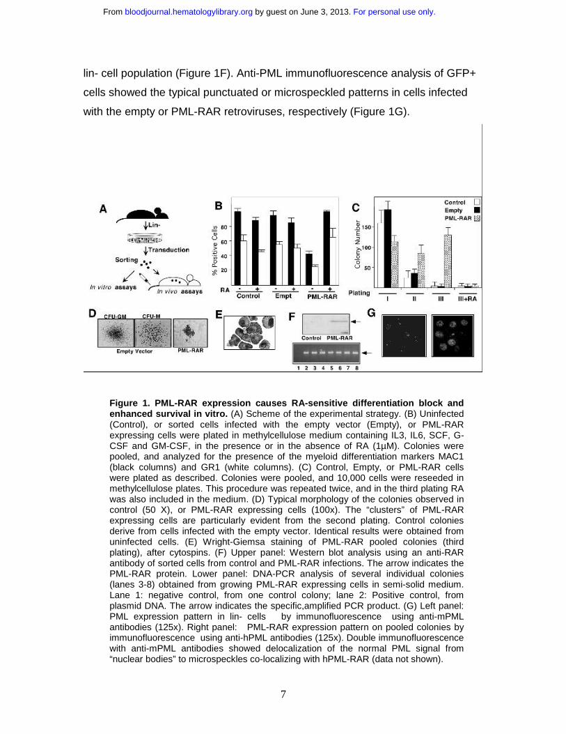
lin- cell population (Figure 1F). Anti-PML immunofluorescence analysis of GFP+
cells showed the typical punctuated or microspeckled patterns in cells infected
with the empty or PML-RAR retroviruses, respectively (Figure 1G).
Figure 1. PML-RAR expression causes RA-sensitive differentiation block and enhanced survival in vitro. (A) Scheme of the experimental strategy. (B) Uninfected (Control), or sorted cells infected with the empty vector (Empty), or PML-RAR expressing cells were plated in methylcellulose medium containing IL3, IL6, SCF, G-CSF and GM-CSF, in the presence or in the absence of RA (1µM). Colonies were pooled, and analyzed for the presence of the myeloid differentiation markers MAC1 (black columns) and GR1 (white columns). (C) Control, Empty, or PML-RAR cells were plated as described. Colonies were pooled, and 10,000 cells were reseeded in methylcellulose plates. This procedure was repeated twice, and in the third plating RA was also included in the medium. (D) Typical morphology of the colonies observed in control (50 X), or PML-RAR expressing cells (100x). The “clusters” of PML-RAR expressing cells are particularly evident from the second plating. Control colonies derive from cells infected with the empty vector. Identical results were obtained from uninfected cells. (E) Wright-Giemsa staining of PML-RAR pooled colonies (third plating), after cytospins. (F) Upper panel: Western blot analysis using an anti-RAR antibody of sorted cells from control and PML-RAR infections. The arrow indicates the PML-RAR protein. Lower panel: DNA-PCR analysis of several individual colonies (lanes 3-8) obtained from growing PML-RAR expressing cells in semi-solid medium. Lane 1: negative control, from one control colony; lane 2: Positive control, from plasmid DNA. The arrow indicates the specific,amplified PCR product. (G) Left panel: PML expression pattern in lin- cells by immunofluorescence using anti-mPML antibodies (125x). Right panel: PML-RAR expression pattern on pooled colonies by immunofluorescence using anti-hPML antibodies (125x). Double immunofluorescence with anti-mPML antibodies showed delocalization of the normal PML signal from “nuclear bodies” to microspeckles co-localizing with hPML-RAR (data not shown).
For personal use only. by guest on June 3, 2013. bloodjournal.hematologylibrary.orgFrom
8
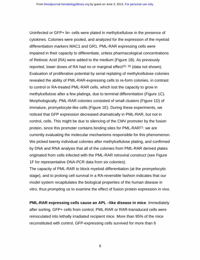
Uninfected or GFP+ lin- cells were plated in methylcellulose in the presence of
cytokines. Colonies were pooled, and analyzed for the expression of the myeloid
differentiation markers MAC1 and GR1. PML-RAR expressing cells were
impaired in their capacity to differentiate, unless pharmacological concentrations
of Retinoic Acid (RA) were added to the medium (Figure 1B). As previously
reported, lower doses of RA had no or marginal effect15, 16 (data not shown).
Evaluation of proliferative potential by serial replating of methylcellulose colonies
revealed the ability of PML-RAR-expressing cells to re-form colonies, in contrast
to control or RA-treated PML-RAR cells, which lost the capacity to grow in
methylcellulose after a few platings, due to terminal differentiation (Figure 1C).
Morphologically, PML-RAR colonies consisted of small clusters (Figure 1D) of
immature, promyelocyte-like cells (Figure 1E). During these experiments, we
noticed that GFP expression decreased dramatically in PML-RAR, but not in
control, cells. This might be due to silencing of the CMV promoter by the fusion
protein, since this promoter contains binding sites for PML-RAR21: we are
currently evaluating the molecular mechanisms responsible for this phenomenon.
We picked twenty individual colonies after methylcellulose plating, and confirmed
by DNA and RNA analysis that all of the colonies from PML-RAR derived plates
originated from cells infected with the PML-RAR retroviral construct (see Figure
1F for representative DNA-PCR data from six colonies).
The capacity of PML-RAR to block myeloid differentiation (at the promyelocytic
stage), and to prolong cell survival in a RA-reversible fashion indicates that our
model system recapitulates the biological properties of the human disease in
vitro, thus prompting us to examine the effect of fusion protein expression in vivo.
PML-RAR expressing cells cause an APL –like disease in mice. Immediately
after sorting, GFP+ cells from control, PML-RAR or RAR-transduced cells were
reinoculated into lethally irradiated recipient mice. More than 95% of the mice
reconstituted with control, GFP-expressing cells survived for more than 6
For personal use only. by guest on June 3, 2013. bloodjournal.hematologylibrary.orgFrom
9
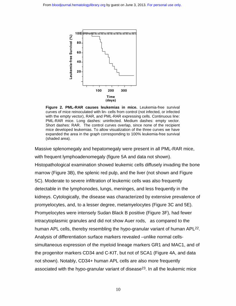
months, whereas the non-reinoculated mice died within 1-2 weeks after
irradiation (Table 1 and Figure 2).
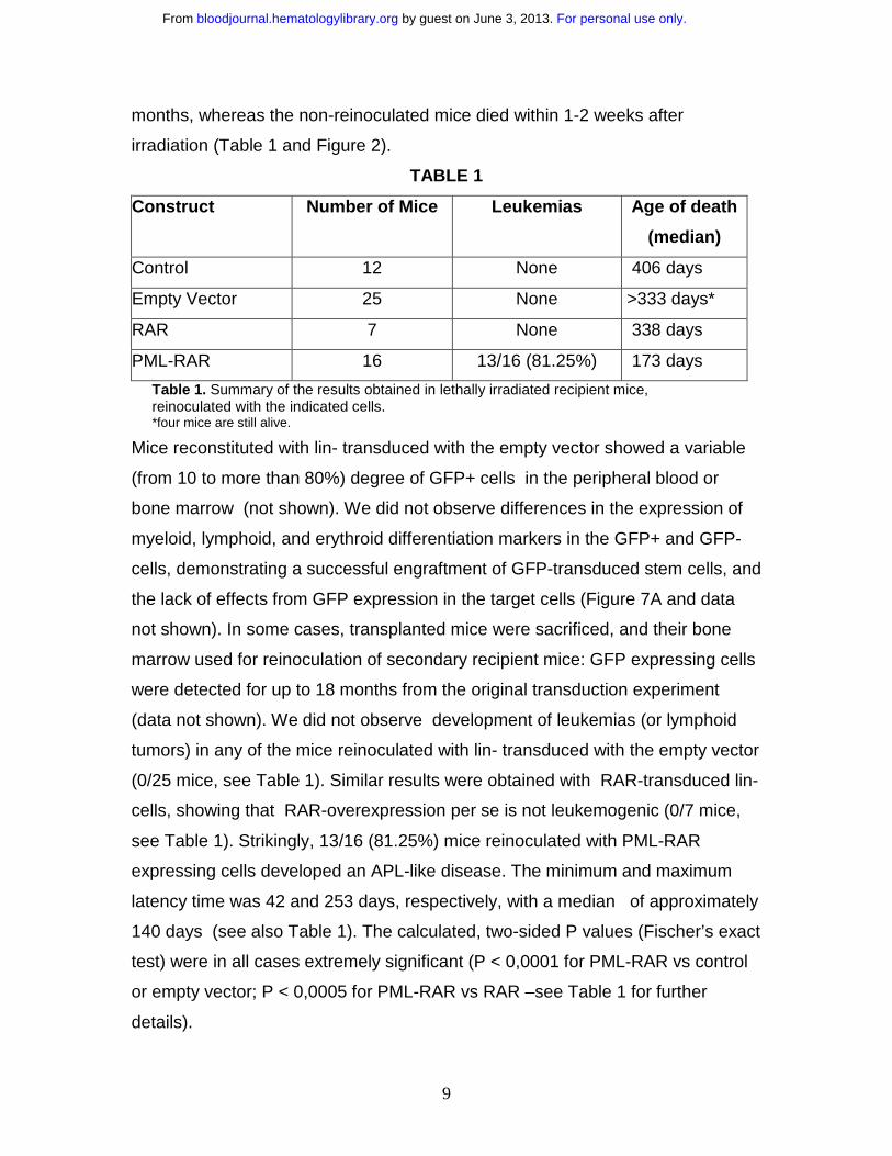
TABLE 1
Construct Number of Mice Leukemias Age of death
(median)
Control 12 None 406 days
Empty Vector 25 None >333 days*
RAR 7 None 338 days
PML-RAR 16 13/16 (81.25%) 173 days
Table 1. Summary of the results obtained in lethally irradiated recipient mice, reinoculated with the indicated cells.*four mice are still alive.
Mice reconstituted with lin- transduced with the empty vector showed a variable
(from 10 to more than 80%) degree of GFP+ cells in the peripheral blood or
bone marrow (not shown). We did not observe differences in the expression of
myeloid, lymphoid, and erythroid differentiation markers in the GFP+ and GFP-
cells, demonstrating a successful engraftment of GFP-transduced stem cells, and
the lack of effects from GFP expression in the target cells (Figure 7A and data
not shown). In some cases, transplanted mice were sacrificed, and their bone
marrow used for reinoculation of secondary recipient mice: GFP expressing cells
were detected for up to 18 months from the original transduction experiment
(data not shown). We did not observe development of leukemias (or lymphoid
tumors) in any of the mice reinoculated with lin- transduced with the empty vector
(0/25 mice, see Table 1). Similar results were obtained with RAR-transduced lin-
cells, showing that RAR-overexpression per se is not leukemogenic (0/7 mice,
see Table 1). Strikingly, 13/16 (81.25%) mice reinoculated with PML-RAR
expressing cells developed an APL-like disease. The minimum and maximum
latency time was 42 and 253 days, respectively, with a median of approximately
140 days (see also Table 1). The calculated, two-sided P values (Fischer’s exact
test) were in all cases extremely significant (P < 0,0001 for PML-RAR vs control
or empty vector; P < 0,0005 for PML-RAR vs RAR –see Table 1 for further
details).
For personal use only. by guest on June 3, 2013. bloodjournal.hematologylibrary.orgFrom
10
Figure 2. PML-RAR causes leukemias in mice. Leukemia-free survival curves of mice reinoculated with lin- cells from control (not infected, or infected with the empty vector), RAR, and PML-RAR expressing cells. Continuous line: PML-RAR mice. Long dashes: uninfected. Medium dashes: empty vector. Short dashes: RAR. The control curves overlap, since none of the recipient mice developed leukemias. To allow visualization of the three curves we have expanded the area in the graph corresponding to 100% leukemia-free survival (shaded area).
Massive splenomegaly and hepatomegaly were present in all PML-RAR mice,
with frequent lymphoadenomegaly (figure 5A and data not shown).
Histopathological examination showed leukemic cells diffusely invading the bone
marrow (Figure 3B), the splenic red pulp, and the liver (not shown and Figure
5C). Moderate to severe infiltration of leukemic cells was also frequently
detectable in the lymphonodes, lungs, meninges, and less frequently in the
kidneys. Cytologically, the disease was characterized by extensive prevalence of
promyelocytes, and, to a lesser degree, metamyelocytes (Figure 3C and 5E).
Promyelocytes were intensely Sudan Black B positive (Figure 3F), had fewer
intracytoplasmic granules and did not show Auer rods, as compared to the
human APL cells, thereby resembling the hypo-granular variant of human APL22.
Analysis of differentiation surface markers revealed –unlike normal cells-
simultaneous expression of the myeloid lineage markers GR1 and MAC1, and of
the progenitor markers CD34 and C-KIT, but not of SCA1 (Figure 4A, and data
not shown). Notably, CD34+ human APL cells are also more frequently
associated with the hypo-granular variant of disease23. In all the leukemic mice
For personal use only. by guest on June 3, 2013. bloodjournal.hematologylibrary.orgFrom
11
analysed, the leukemic cells expressed PML-RAR, as shown by
immunoprecipitation, RNA and immunofluorescence analyses (Figure 4B-C). In
contrast, leukemic cells were uniformly negative for GFP, reproducing in vivo the
phenotype observed in vitro (see below).
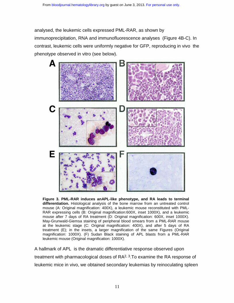
Figure 3. PML-RAR induces anAPL-like phenotype, and RA leads to terminal differentiation. Histological analysis of the bone marrow from an untreated control mouse (A: Original magnification: 400X), a leukemic mouse reconstituted with PML-RAR expressing cells (B: Original magnification:600X, inset 1000X), and a leukemic mouse after 7 days of RA treatment (D: Original magnification: 600X, inset 1000X). May-Grunwald-Giemsa staining of peripheral blood smears from a PML-RAR mouse at the leukemic stage (C: Original magnification: 400X), and after 5 days of RA treatment (E); in the insets, a larger magnification of the same Figures (Original magnification: 1000X). (F) Sudan Black staining of APL blasts from a PML-RAR leukemic mouse (Original magnification: 1000X).
A hallmark of APL is the dramatic differentiative response observed upon
treatment with pharmacological doses of RA2, 3.To examine the RA response of
leukemic mice in vivo, we obtained secondary leukemias by reinoculating spleen
For personal use only. by guest on June 3, 2013. bloodjournal.hematologylibrary.orgFrom
12
cell suspensions of leukemic mice (with 30-50% infiltration of leukemic cells) into
syngeneic, non-irradiated recipients.
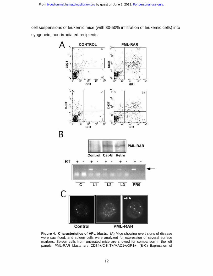
Figure 4. Characteristics of APL blasts. (A) Mice showing overt signs of disease were sacrificed, and spleen cells were analyzed for expression of several surface markers. Spleen cells from untreated mice are showed for comparison in the left panels. PML-RAR blasts are CD34+/C-KIT+/MAC1+/GR1+. (B-C) Expression of
For personal use only. by guest on June 3, 2013. bloodjournal.hematologylibrary.orgFrom
13
PML-RAR in APL blasts was confirmed by: (B) (Upper panel) Analysis of PML-RAR expression by immunoprecipitation with an anti-PML antibody, followed by Western blot using anti-RAR antibodies. Spleen cell extracts were prepared from control and leukemic animals obtained as outlined in Figure 1, or leukemic animals derived from Cathepsin G-PML-RAR transgenic mice8. (Lower panel) RT-PCR analysis of RNA obtained from the bone marrow of three independently derived leukemic mice (L1-L3). Reverse Transcriptase (RT) was omitted in the samples marked with the minus sign as control. Negative control (lanes “C”) is from a control mouse, positive control (lanes “PR9”) derives from U937 cells stably transfected with PML-RAR; the arrow indicates the specific, amplified PCR product. (C) Immunofluorescence of bone marrow cells from a leukemic mouse prior to –center panel-, or after 5 days of RA treatment –right panel-, using antibodies against mouse PML. Control, normal bone marrow cells.
The reinoculated mice developed (100% cases) secondary leukemias with
minimal latency (2 weeks), with characteristics identical to those observed in the
primary recipient mice (including transplantability to further recipients -data not
shown). Since the leukemic phenotype was observed sinchronously in the
secondary recipients, we could treat a cohort of mice that were at the same stage
of disease, and carrying the same leukemic clone(s). RA was administered by
subcutaneous implantation of a 21-day-release pellet containing 5 mg RA or
placebo. Mice were sacrificed at time intervals to monitor the response in the
various tissues, or maintained until death to monitor survival. Starting from the
3rd day of therapy, cells within the leukemic population revealed clear signs of
granulocytic differentiation, that was extensive by the 7th day of treatment
(Figure 3D-E and 5E). Immunofluorescence studies revealed that RA –as
previously shown- reverted almost completely the delocalized pattern of mouse
PML to the normal “nuclear bodies” appearance (Figure 4C: from 30% of cells
showing a micro-speckled pattern in spleen cells, to <2% upon RA treatment).
The leukemic infiltrates in the various organs appeared reduced, as shown by
their parallel reduction to normal size (Figure 5A, C-D). All of the mice implanted
with a subcutaneouos placebo died by progressive multifocal invasion of
parenchimal tissues by leukemic blasts, whereas RA treatment significantly
prolonged survival (P <0.0001), with no apparent signs of disease during
treatment, in >90% mice (Figure 5B). Identical results were obtained in five
independent experiments, using as donors three mice that developed APL in
three independent series of experiments (data not shown).
For personal use only. by guest on June 3, 2013. bloodjournal.hematologylibrary.orgFrom
14
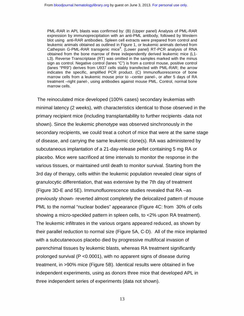
Figure 5. RA treatment induces differentiation of leukemic cells, and prolongs survival of leukemic mice. (A) Macroscopic appearance of spleens from two mice treated in the same experimental cohort with placebo (upper) or RA for 7 days (lower). (B) Survival curves of mice inoculated with spleen cells from one leukemic mouse (L3). RA treatment was started at day 12 after inoculation, when secondary leukemias were apparent in recipient mice (from analysis of peripheral blood smears). The RA pellet subcutaneously implanted has a duration of approximately three weeks. (C) Histological analysis of leukemic infiltrates in the liver of one leukemic mouse (upper panel), and of one leukemic mouse after 7 days of RA treatment (lower panel). (D) Extent of leukemic infiltration within the liver of mice treated with Placebo (n=4) or RA (n=4). For each mouse examined the leukemic cells infiltrating the liver were counted in 40 HPFs (magnification x630); for each animal the mean number of leukemic cells per HPF was calculated. The columns represent the mean of the values (obtained as described above). (E) Differential cell counts in the bone marrow of mice treated with Placebo (n=4) or RA (n=4). For each examined mouse, 500 myeloid cells were classified according to morphologic features.
In human APL, treatment with RA alone results in disease remission and
subsequent relapse in virtually all patients2,3. Likewise, suspension of RA
treatment in the leukemic mice resulted in relapse in 100% of mice (Figure 5B).
Taken together, these results show that our mouse model faithfully reproduce the
main biological and clinical features of human APL.
For personal use only. by guest on June 3, 2013. bloodjournal.hematologylibrary.orgFrom
15
APL in mice is a mono-, or oligo-clonal disease, that follows a pre-leukemic
state.
To evaluate the polyclonal or clonal origin of the mouse APL leukemias, we
performed Southern blotting analysis of integrated proviruses. A polyclonal origin
is consistent with the capacity of PML-RAR to directly transform target cells,
while a monoclonal, or oligoclonal origin suggests a two-hit model of
leukemogenesis, e.g. the requirement for additional genetic lesions6, 13. Genomic
DNAs from spleen/bone marrow of four primary and two secondary leukemic
mice were digested with the HindIII restriction enzyme (which cuts once in the
integrated retroviral sequence, allowing recognition of individual integration
events) and hybridized with a probe representative of the human PML-RAR
cDNA.
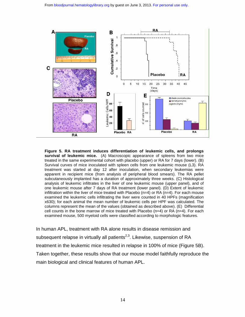
Figure 6. APL is a mono-, or oligo-clonal disease. Genomic DNA was prepared from leukemic mice (L1-L4, lanes 1, 3, 5 and 6), or from mice with secondary leukemias derived from reinjection of spleen cells from L1-L2 mice (lanes 2, 4). Samples were digested with HindIII, and then analyzed by Southern blot assay using the PML-RAR cDNA as a probe. (B) FISH analysis (using the proviral sequence as a probe) of metaphase-enriched nuclei of cells derived from the case “L1”. Arrowheads highlight the hybridization signals.
As observed in Figure 6A, all the primary leukemias showed a discrete number of
hybridizing DNA fragments (2 to 4 in the cases shown). Primary and secondary
leukemia samples revealed the same patterns of multiple hybridizing fragments
(compare lanes 1 and 2, 3 and 4 in Figure 6A). The presence of multiple
hybridizing fragments can be the consequence of a single, yet complex,
integration site, or of several integration sites in the leukemia initiating cell. To
For personal use only. by guest on June 3, 2013. bloodjournal.hematologylibrary.orgFrom
16
clarify this issue, we performed FISH analysis of leukemic metaphases using a
probe encompassing the entire integrated sequence. Strikingly, in the case
where two hybridizing fragments were observed, we detected two hybridizing
regions, confirming the monoclonality of the leukemia, and the presence of two
integration events (Figure 6B). Overall, these results support the mono-, or oligo-
clonal origin of the leukemic blasts, and the hypothesis that leukemia arose from
one PML-RAR expressing cell undergoing additional genetic lesions.
We next investigated whether expression of PML-RAR is sufficient to induce a
detectable phenotype, prior to the onset of an overt leukemia. In the transgenic
mice carrying PML-RAR under the MRP-8 or cathepsin-G promoters, a mild
defect in myeloid maturation was consistently seen in 100% of the mice, defined
as a “pre-leukemic” state and characterized by a slight decrease of MAC1 or
GR1 expression in the whole bone marrow6, 13. We did not, however, detect a
similar phenotype in our PML-RAR mice, as shown by the immunophenotypic
analysis of bone marrow cells at 30, 60, and 90 days after reinoculation (Figure
7A and data not shown). Since we could not follow directly the fate of PML-RAR
expressing cells, due to loss of GFP expression, we performed DNA-PCR
analysis on several colonies derived from methylcellulose platings of lin- cells
from the reinoculated mice to demonstrate that the pre-leukemic bone marrow
cells expressed the fusion protein. Analysis of differentiation markers was then
performed in those mice where >80% of colonies were positive for PML- RAR
cDNA. We conclude, therefore that the lack of a “pre-leukemic” condition is not
due to to the low number of transduced cells repopulating the recipient mouse
(Figure 7A-B). Slight variations in the expression of differentiation markers might
be however masked, in our case, by the complex kinetics of reconstitution of
normal hematopoiesis into lethally irradiated mice. We used, therefore, functional
assays to evaluate the existence of a preleukemic state. Whole bone marrow, or
the lin- population from reconstituted mice (control, GFP-only, and PML-RAR
expressing cells), were then analysed for their capacity to undergo myeloid
differentiation in vitro in semisolid medium. PML-RAR expressing cells showed a
strongly reduced capacity to differentiate in vitro, unless RA was added to the
For personal use only. by guest on June 3, 2013. bloodjournal.hematologylibrary.orgFrom

17
medium: similar results were obtained by analyzing cells derived from the MRP-8
and cathepsin-G PML-RAR transgenic mice (Figure 7C and data not shown).
Figure 7. PML-RAR expression induces a pre-leukemic state. (A) Differentiation marker analysis of bone marrow cells from recipient mice reconstituted with uninfected (Control), GFP-only (PINCO), and PML-RAR expressing lin- cells (PML-RAR). Four mice/group were analyzed 90 days after reconstitution. In the case of PINCO, the data are presented for both GFP+ and GFP- cells. Peripheral blood and spleen cell analysis gave comparable results (data not shown). In addition to MAC1, we analyzed the following markers: GR1, CD34, SCA1, TER119, CD3 with comparable results (data not shown). (B) Lin- cells were prepared from recipient
For personal use only. by guest on June 3, 2013. bloodjournal.hematologylibrary.orgFrom
18
mice reconstituted with PML-RAR expressing cells, 90 days after lethal irradiation. For DNA-PCR analysis, primers specific for human PML-RAR cDNA were used to amplify DNA extracted from colonies derived from methylcellulose plating . Lane 1, negative control; lane 2, positive control. Lanes 3-8, six colonies from one representative PML-RAR reconstituted mouse, used in the analysis of differentiation markers shown in (A). (C) In vitro differentiation analysis (MAC1) of pooled colonies derived as in (B).
These results suggest that –as in the transgenic mice- a “pre- leukemic” state
may be observed in our experimental system, and corresponds to an alteration in
the differentiation potential induced by the fusion protein in hematopoietic
progenitors.
DISCUSSION
We have presented here a novel model system to study APL in mice , based on
transplantation of transduced murine hematopoietic progenitors. This system
fully recapitulates the most relevant features of human APLs, e.g. a
differentiation block at the promyelocyte stage and RA-sensitivity. Leukemia is
obtained by the expression of PML-RAR (but not RAR) in lin- cells. Previously,
similar results have been obtained in chicken and mice model systems. In
chicken, retroviral-mediated expression of a gag-PML-RAR fusion protein
induced transformation of hematopoietic progenitors and caused leukemias .
Point-mutations harbored by the provirus, however, modified the intra-cellular
localization of PML-RAR, resulting in a disease not resembling human APL, and
that was not sensitive to RA treatment5. In transgenic mice, PML-RAR was
expressed under the control of myeloid specific promoters (MRP-8 or cathepsin
G)6-10. In those systems, RAR carrying a mutation in its ligand binding domain
was shown to be non-leukemogenic: the results presented here, using wild-type
RAR, further demonstrate that over-expression of RAR does not induce
leukemias 10. In the previous studies, PML-RAR leukemias developed after long
latency (6-18 months) and at low penetrance (10-30%). Simultaneous expression
of the product of the reciprocal translocation (RAR-PML) was shown to increase
frequency (to about 50%), without affecting the latency of the disease24. In our
experiments, instead, induction of a leukemic phenotype was observed in >80%
of the mice upon expression of PML-RAR, with decreased latency (median time
For personal use only. by guest on June 3, 2013. bloodjournal.hematologylibrary.orgFrom
19
of about 4 months) compared to the transgenic models. This discrepancy is not
due to different levels of PML-RAR, since direct comparison of fusion protein
expression revealed comparable levels among the different model systems
(catepsin-G, MRP8 transgenics and ours; Figure 4B). The long latency and low
frequency of leukemias in the transgenic mice suggested a “two-hit” model, that
is consistent with the clonal origin of leukemias in our model system. Differences
among the various model systems might, instead, be due to different targeting of
the PML-RAR cDNA13. Expression of PML-RAR in transgenic mice strictly
depend on the promoter used, as shown by the failure of late myeloid-stage
specific promoters (CD11b) to provoke leukemias7. It appears, therefore, that
PML-RAR acquires leukemogenic potential only if expressed at specific stages of
hematopoietic development. It is then possible that expression of PML-RAR (by
retroviral transduction) in the heterogeneous lin- population increases the
frequency of proper PML-RAR targeting, as compared to developmental
regulated promoters where the frequency of proper targeting might be lower
(MRP-8 and cathepsin-G), or close to zero (CD11b). Future experiments based
on the transduction and reinoculation of subsets of hematopoietic mouse
precursors should allow a better definition of the APL target cell.
Expression of PML-RAR is not sufficient to transform cells: in all model systems,
additional genetic/epigenetic events are required, which are favored by PML-
RAR expression25. The fusion protein provokes a so-called “pre-leukemic” state,
characterized, in vivo, by a mild impairment in myeloid differentiation. Lin- cells
from pre-leukemic animals are impaired in their ability to undergo terminal
differentiation in vitro, a phenotype that we also detected into freshly transduced,
PML-RAR expressing lin- cells, thereby offering the unique possibility to
investigate this phenomenon (and possibly the subsequent steps leading to
transformation) in vitro.
The possibility to study the same cell populations in vitro and in vivo is, indeed,
one of the greatest advantages of our experimental system. In vitro, we have
shown that PML-RAR blocks differentiation and extend survival of lin- cells: by
For personal use only. by guest on June 3, 2013. bloodjournal.hematologylibrary.orgFrom
20
structure-function analysis, it will be possible to dissect these two phenotypes,
and validate immediately the results in vivo.
Finally, RA sensitivity of primary and secondary leukemias, as well as of the pre-
leukemic state observed in vitro, shows that this system is also available for
pharmacological studies. Aberrant recruitment of histone deacetylases (HDACs)
is considered critical for the leukemogenic potential of PML-RAR and other AML
fusion proteins, and treatment with HDAC inhibitors is able to induce
differentiation in vitro26. Treatment of the leukemic mice with HDACi, or other
pharmacologically active compounds, may lead to new proposals for the therapy
of APL, and possibly other types of cancers.
ACKNOWLEDGEMENTS
We thank Tim Ley, Silvia Soddu, Jeff Medin, Mirco Fanelli and Francesco
Bertolini for discussions and help with the set up of some experimental
techniques.
S.Monestiroli and S. Giavara contributed equally to this work.
For personal use only. by guest on June 3, 2013. bloodjournal.hematologylibrary.orgFrom
21
REFERENCES
1. Melnick, A., and J. D. Licht. Deconstructing a disease: RARalpha, its
fusion partners, and their roles in the pathogenesis of acute promyelocytic
leukemia. Blood. 1999; 93:3167-215.
2. Fenaux, P., C. Chomienne, and L. Degos. All-trans retinoic acid and
chemotherapy in the treatment of acute promyelocytic leukemia. Semin Hematol.
2001; 38:13-25.
3. Warrell, R. P., Jr., H. de The, Z. Y. Wang, and L. Degos. Acute
promyelocytic leukemia. N Engl J Med. 1993; 329:177-89.
4. Grignani, F., P. F. Ferrucci, U. Testa, G. Talamo, M. Fagioli, M. Alcalay, A.
Mencarelli, F. Grignani, C. Peschle, I. Nicoletti, and et al. The acute
promyelocytic leukemia-specific PML-RAR alpha fusion protein inhibits
differentiation and promotes survival of myeloid precursor cells. Cell. 1993;
74:423-31.
5. Altabef, M., M. Garcia, C. Lavau, S. C. Bae, A. Dejean, and J. Samarut. A
retrovirus carrying the promyelocyte-retinoic acid receptor PML- RARalpha fusion
gene transforms haematopoietic progenitors in vitro and induces acute
leukaemias. Embo J. 1996; 15:2707-16.
6. Brown, D., S. Kogan, E. Lagasse, I. Weissman, M. Alcalay, P. G. Pelicci,
S. Atwater, and J. M. Bishop. A PMLRARalpha transgene initiates murine acute
promyelocytic leukemia. Proc Natl Acad Sci U S A. 1997; 94:2551-6.
7. Early, E., M. A. Moore, A. Kakizuka, K. Nason-Burchenal, P. Martin, R. M.
Evans, and E. Dmitrovsky. Transgenic expression of PML/RARalpha impairs
myelopoiesis. Proc Natl Acad Sci U S A. 1996; 93:7900-4.
8. Grisolano, J. L., R. L. Wesselschmidt, P. G. Pelicci, and T. J. Ley. Altered
myeloid development and acute leukemia in transgenic mice expressing PML-
RAR alpha under control of cathepsin G regulatory sequences. Blood. 1997;
89:376-87.
For personal use only. by guest on June 3, 2013. bloodjournal.hematologylibrary.orgFrom
22
9. He, L. Z., C. Tribioli, R. Rivi, D. Peruzzi, P. G. Pelicci, V. Soares, G.
Cattoretti, and P. P. Pandolfi. Acute leukemia with promyelocytic features in
PML/RARalpha transgenic mice. Proc Natl Acad Sci U S A. 1997; 94:5302-7.
10. Kogan, S. C., S. H. Hong, D. B. Shultz, M. L. Privalsky, and J. M. Bishop.
Leukemia initiated by PMLRARalpha: the PML domain plays a critical role while
retinoic acid-mediated transactivation is dispensable. Blood. 2000; 95:1541-50.
11. Lavau, C., J. M. Heard, O. Danos, and A. Dejean. Retroviral vectors for
the transduction of the PML-RARalpha fusion product of acute promyelocytic
leukemia. Exp Hematol. 1996; 24:544-51.
12. Pandolfi, P. P. In vivo analysis of the molecular genetics of acute
promyelocytic leukemia. Oncogene. 2001; 20:5726-35.
13. Westervelt, P., and T. J. Ley. Seed versus soil: the importance of the
target cell for transgenic models of human leukemias [see comments]. Blood.
1999; 93:2143-8.
14. Grignani, F., T. Kinsella, A. Mencarelli, M. Valtieri, D. Riganelli, L.
Lanfrancone, C. Peschle, G. P. Nolan, and P. G. Pelicci. High-efficiency gene
transfer and selection of human hematopoietic progenitor cells with a hybrid
EBV/retroviral vector expressing the green fluorescence protein. Cancer Res.
1998; 58:14-9.
15. Minucci, S., M. Maccarana, M. Cioce, P. De Luca, V. Gelmetti, S. Segalla,
L. Di Croce, S. Giavara, C. Matteucci, A. Gobbi, A. Bianchini, E. Colombo, I.
Schiavoni, G. Badaracco, X. Hu, M. A. Lazar, N. Landsberger, C. Nervi, and P.
G. Pelicci. Oligomerization of RAR and AML1 transcription factors as a novel
mechanism of oncogenic activation. Mol Cell. 2000; 5:811-20.
16. Du, C., R. L. Redner, M. P. Cooke, and C. Lavau. Overexpression of wild-
type retinoic acid receptor alpha (RARalpha) recapitulates retinoic acid-sensitive
transformation of primary myeloid progenitors by acute promyelocytic leukemia
RARalpha-fusion genes. Blood. 1999; 94:793-802.
17. Lo Coco, F., D. Diverio, B. Falini, A. Biondi, C. Nervi, and P. G. Pelicci.
Genetic diagnosis and molecular monitoring in the management of acute
promyelocytic leukemia. Blood. 1999; 94:12-22.
For personal use only. by guest on June 3, 2013. bloodjournal.hematologylibrary.orgFrom
23
18. Flenghi, L., M. Fagioli, L. Tomassoni, S. Pileri, M. Gambacorta, R. Pacini,
F. Grignani, T. Casini, P. F. Ferrucci, M. F. Martelli, and et al. Characterization of
a new monoclonal antibody (PG-M3) directed against the aminoterminal portion
of the PML gene product: immunocytochemical evidence for high expression of
PML proteins on activated macrophages, endothelial cells, and epithelia. Blood.
1995; 85:1871-80.
19. Le Beau, M. M., S. Bitts, E. M. Davis, and S. C. Kogan. Recurring
chromosomal abnormalities in leukemia in PML-RARA transgenic mice parallel
human acute promyelocytic leukemia. Blood. 2002; 99:2985-91.
20. Specchia, G., A. Cuneo, V. Liso, R. Contino, D. Pastore, E. Gentile, M.
Rocchi, and G. L. Castoldi. A novel translocation t(1;7)(p36;q34) in three patients
with acute myeloid leukaemia. Br J Haematol. 1999; 105:208-14.
21. Angulo, A., C. Suto, R. A. Heyman, and P. Ghazal. Characterization of the
sequences of the human cytomegalovirus enhancer that mediate differential
regulation by natural and synthetic retinoids. Mol Endocrinol. 1996; 10:781-93.
22. Neame, P. B., P. Soamboonsrup, B. Leber, R. F. Carter, L. Sunisloe, W.
Patterson, A. Orzel, S. Bates, and J. A. McBride. Morphology of acute
promyelocytic leukemia with cytogenetic or molecular evidence for the diagnosis:
characterization of additional microgranular variants. Am J Hematol. 1997;
56:131-42.
23. Foley, R., P. Soamboonsrup, R. F. Carter, A. Benger, R. Meyer, I. Walker,
Y. Wan, W. Patterson, A. Orzel, L. Sunisloe, B. Leber, and P. B. Neame. CD34-
positive acute promyelocytic leukemia is associated with leukocytosis,
microgranular/hypogranular morphology, expression of CD2 and bcr3 isoform.
Am J Hematol. 2001; 67:34-41.
24. Pollock, J. L., P. Westervelt, A. K. Kurichety, P. G. Pelicci, J. L. Grisolano,
and T. J. Ley. A bcr-3 isoform of RARalpha-PML potentiates the development of
PML- RARalpha-driven acute promyelocytic leukemia. Proc Natl Acad Sci U S A.
1999; 96:15103-8.
25. Zimonjic, D. B., J. L. Pollock, P. Westervelt, N. C. Popescu, and T. J. Ley.
Acquired, nonrandom chromosomal abnormalities associated with the
For personal use only. by guest on June 3, 2013. bloodjournal.hematologylibrary.orgFrom
24
development of acute promyelocytic leukemia in transgenic mice. Proc Natl Acad
Sci U S A. 2000; 97:13306-11.
26. Minucci, S., C. Nervi, F. Lo Coco, and P. G. Pelicci. Histone deacetylases:
a common molecular target for differentiation treatment of acute myeloid
leukemias? Oncogene. 2001; 20:3110-5.
For personal use only. by guest on June 3, 2013. bloodjournal.hematologylibrary.orgFrom

25
For personal use only. by guest on June 3, 2013. bloodjournal.hematologylibrary.orgFrom

26
For personal use only. by guest on June 3, 2013. bloodjournal.hematologylibrary.orgFrom

27
For personal use only. by guest on June 3, 2013. bloodjournal.hematologylibrary.orgFrom