Differential Requirement for Caspase 9 in Apoptotic Pathways In Vivo

Selective induction of apoptosis through the FADD/Caspase-8 pathway by a p53 C-terminal peptide in human pre-malignant and malignant cellsYin Li1, Yuehua Mao1, Ramon V. Rosal2, Richard D. Dinnen1, Ann C. Williams3, Paul W. Brandt-Rauf2 andRobert L. Fine1*1Experimental Therapeutics Program, Division of Medical Oncology, College of Physicians and Surgeons,Columbia University Medical Center, New York, NY, USA2Department of Environmental Health Sciences, Mailman School of Public Health, Columbia University Medical Center,New York, NY, USA3Department of Pathology and Microbiology, School of Medical Sciences, University of Bristol, Bristol, United Kingdom
A p53 C-terminal peptide (aa 361–382, p53p), fused at its C-terminus to the minimal carrier peptide of antennapedia (17 aa,Ant; p53p-Ant), induced rapid apoptosis in human cancer cells,via activation of the Fas pathway. We examined p53p-Ant mech-anism of action, toxicity in various human normal, non-malignant,pre-malignant and malignant cancer cells and investigated itsbiophysical characteristics. p53p-Ant selectively induced cell deathin only pre-malignant or malignant cells in a p53-dependent man-ner and was not toxic to normal and non-malignant cells. p53p-Ant was more toxic to the mutant p53 than wild-type p53 pheno-type in H1299 lung cancer cells stably expressing humantemperature-sensitive p53 mutant 143Ala. Surface plasmon reso-nance (BIACORE) analysis demonstrated that this peptide hadhigher binding affinity to mutant p53 as compared to wild-typep53. p53p-Ant induced-cell death had the classical morphologicalcharacteristics of apoptosis and had no features of necrosis. Themechanism of cell death by p53p-Ant was through the FADD/caspase-8-dependent pathway without the involvement of theTRAIL pathway, Bcl-2 family and cell cycle changes. Blocking Faswith antibody did not alter the peptide’s effect, suggesting that Fasitself did not interact with the peptide. Transfection with a dom-inant-negative FADD with a deleted N-terminus inhibited p53p-Ant-induced apoptosis. Its mechanism of action is related to theFADD-induced pathway without restoration of other p53 func-tions. p53p-Ant is a novel anticancer agent with unique selectivityfor human cancer cells and could be useful as a prototype for thedevelopment of new anti-cancer agents.© 2005 Wiley-Liss, Inc.
Key words: p53 C-terminal peptide; apoptosis; necrosis; BIACORE;TdT assay; FADD (Fas associated death domain)
The p53 tumor suppressor gene is one of the most commonlymutated genes found in human malignancies.1 More than 50% ofhuman tumors, including breast cancers, are associated with mis-sense mutations or deletions of p53 and most of the missensemutations map to the DNA-binding domain of the protein.2,3 p53protein functions in the transcription of growth inhibiting genes,apoptosis, cell cycle arrest and DNA repair.4–6 p53 is a sequence-specific transcription factor that transactivates a number of geneswhose products are involved in cell growth regulation. Theseinclude p21 (a cyclin-dependent kinase inhibitor known to arrestthe cell cycle mainly in G1/S phase), GADD45 for DNA repair andBax and Fas/APO-1 for apoptosis.7–11 p53 induced Fas-FADDbinding and transiently sensitized cells to Fas-induced apoptosis.12
Programmed cell death depends on conserved structural moi-eties that transmit and regulate the death signal. This is mostevident in death receptors of the tumor necrosis factor receptor(TNF-R) super-family. The intracellular regions of the Fas/CD95and TNFR1 death receptors have a moderately well conservedNH2-region of about 80 residues that is required for death signal-ling and has been called the ‘death domain’ (DD).13,14 Fas/CD95induces rapid apoptosis upon binding to Fas ligand through theautocrine/paracrine signalling pathway.15 When activated, the in-tracellular death domain in Fas/CD95 binds to FADD/MORT-1 atits C-terminus, which then recruits caspase-8 to FADD. A memberof the TNF ligand family identified more recently is TNF-related
apoptosis-inducing ligand (TRAIL). TRAIL shows high homologyto Fas Ligand and binds to the TRAIL receptor family. TRAIL-induced caspase activation shares many of same caspases involvedin Fas/CD95 and TNF-induced cell death mechanisms.16 FADD/MORT-1 is a cytosolic adaptor protein, which is critical for sig-nalling from Fas/APO-1 and other TNF-R family members.17 Twoprotein interaction domains have been identified in FADD/MORT-1. The C-terminal ‘death domain’ is needed for recruit-ment of FADD/MORT1 to ligated ‘death receptor’ and the N-terminal ‘death effector domain’ mediates oligomerization andactivation of caspase-8.18 Caspase-8 is the first protease activatedin the Fas/APO-1 signalling pathway, which initiates downstreamactivation of caspase-3, -6 and -7 and mitochondrial damage. Thisactivates poly (ADP-ribose) polymerase (PARP) and ICE familyproteases, triggering a cascade of apoptotic processes.19,20
Apoptosis involves a series of complex biochemical events andis regulated by several pathways, including members of the Bcl-2family. Bcl-2 is a negative regulator of apoptosis and its’ activityis modulated by homodimerization or heterodimerization associa-tion with promoters of apoptosis such as Bax and Bad. Bax can beregulated directly by p53 and induced in some cells undergoingp53-mediated apoptosis. Bcl-XL suppresses apoptosis, whereasBcl-Xs inhibits Bcl-2 mediated cell survival.21–23 BID is a novelpro-apoptotic Bcl-2 family protein that is activated by truncation tot-BID by caspase-8 in response to Fas death receptor signals.24
The sequence-specific DNA-binding activity of p53 seems to beregulated negatively by its C-terminal 30 aa segment (aa 363–393)and by a N-terminal proline-rich motif located between aa80–93.25,26 Synthetic peptides corresponding to the C-terminaldomain of p53, aa residues 363–393, bind directly in vitro towild-type (wt) p53. Binding studies with p53 proteins that containselected deletions suggest that binding of the p53 peptide aa363–393 to p53 protein requires the presence of both C-terminal aa363–393 and N-terminal aa 80–93 sequences in the p53 protein.26
Using conformational energy calculations to compute the lowestenergy conformations for the C- and N-terminal regulatory do-mains, we found that these 2 domains form a unique low energycomplex, suggesting that a direct interaction can potentially existbetween them.27
Previous studies demonstrated that the addition of a chemicallymodified p53 C-terminal peptide (aa 363–393) restored in vitrosequence-specific DNA binding function to mutant p53–273 (Argto His). Furthermore, intranuclear microinjection of this peptide
Grant sponsors include NIH NCI-RO1-CA82528, Herbert Pardes Clin-ical Scholar Award, Avon Foundation Scholar Award, Woman at Risk(WAR) Award and Vincent Loshiavo Memorial Fund to R.L.F. and NIHRO1-OH07590 to P.W.B.R.
*Correspondence to: College of Physicians and Surgeons, ColumbiaUniversity Medical Center, 650 West 168th Street, BB 20-05, New York,NY 10032. Fax: �001-212-305-7348. E-mail: [email protected]
Received 11 March 2004; Accepted after revision 20 October 2004DOI 10.1002/ijc.20838Published online 11 January 2005 in Wiley InterScience (www.
interscience.wiley.com).
Int. J. Cancer: 115, 55–64 (2005)© 2005 Wiley-Liss, Inc.
Publication of the International Union Against Cancer

into human colon cancer cells SW480 carrying an endogenousp53–273 His mutant restored transcriptional activation of a p53-responsive reporter construct.28 A p53 C-terminal peptide (aa361–382), fused at its C-terminus to the minimal Antennapedia(Ant) carrier sequence to facilitate cellular uptake, inhibitedgrowth and induced apoptosis of SW480 cells.29 Our previousstudies indicated that the C-terminal p53 peptide (aa 361–382)-Antennapedia peptide (p53p–Ant) induced selective and rapidapoptosis in human breast cancer cells associated with increasedFas and Fas ligand expression.27 The mechanism and componentsof the Fas pathway needed for apoptosis by p53p-Ant were notelucidated. In addition, using human null p53 lung cancer cellsH1299 and prostate cancer cells PC-3 stably transfected with thehuman p53 temperature-sensitive (ts) mutant 143Ala (Val-Ala) asa model, we found Fas-mediated apoptosis occurred only at the wtp53 phenotype at 32.5°C.30 The mechanism of the induction ofFas-mediated apoptosis by p53p-Ant, its effect upon other humancancer type cells, the differential binding of peptide in cancer cellswith mutant vs. wt p53 and its effects on normal cells are stillunknown. Thus, the mechanism and components of the Fas-me-diated apoptotic pathway involved needs to be defined moreclearly.
The BIACORE technology system, which is a biosensor systembased on surface plasmon resonance (SPR), has been appliedwidely in studies investigating simple interprotein, protein-DNAand protein-lipid interactions. It can monitor and quantitative thekinetics of binding and dissociation constants between moleculesin real-time and without labels.31–33 We utilized the BIACOREsystem to quantitate differential binding and dissociation constantsbetween the peptide and mutant and wt p53.
We investigated p53p-Ant’s effect on normal, non-malignant,pre-malignant and malignant human cells. We also used humanlung cancer cells H1299 expressing human p53 temperature sen-sitive (TS) mutant 143Ala to study its differential activity andbinding to mutant p53 and wt p53 phenotypes with the BIACOREtechnology. The type of cell death and its effect on p21, Bax,GADD45 and cell cycle and the involvement of caspases werefurther investigated. To better understand and define the mecha-nism of p53p-Ant-induced apoptosis, we studied the involvementof Fas, TRAIL and FADD molecules, including dominant negativeFADD.
Material and methodsPeptides
p53p, Ant, and p53p-Ant peptides were chemically synthesizedby Research Genetics (Huntsville, AL) as described previously.27
All peptides were HPLC purified to �95% pure. Peptide stocks(4 mM) were prepared in sterile distilled water and stored inaliquots at �80°C. Peptides include: p53p, N-GSRAHSSHLK-SKKGQSTSRHKK-C (22 aa, 361–382 of p53); Ant, KKWKM-RRNQFWVKVQRG (17 aa); p53p-AntCONT, N-KKGQST-SRKK-WKMRRNQFWVKVQRG-C (25 aa); and p53p-Ant,N-GSRAHSSHLKSKKGQSTSRHKK-WKMRRNQFWVKV-QRG-C (37 aa). The fused peptide has 37 aa because we elimi-nated KK from the NH2 terminus of the Ant sequence becausep53p has a KK at the C-terminus. This fused peptide is namedp53p-Ant.
Antibodies and reagentsAnti-p21 polyclonal antibody (clone: H-164) was obtained from
Santa Cruz (Santa Cruz, CA). Fluorescein isothiocyanate (FITC)-conjugated anti-human Fas/CD95 monoclonal antibody (clone:DX2) and phycoerythrin (PE)-conjugated mouse anti-humanmonoclonal TRAIL antibody (clone: RIK-2) were obtained fromPharmingen (San Diego, CA). Anti-human Fas/CD95 monoclonalantibody (clone: CH-11), antagonistic anti-Fas monoclonal anti-body (clone: ZB4) and anti-human FADD monoclonal antibody(clone: 1F7) were obtained from MBL International Corporation(MIC, Watertown, MA). Antagonistic anti-TRAIL monoclonal
antibody (clone: 2E5) was obtained from Alexis Biochemicals(San Diego, CA). Anti-�-tubulin monoclonal antibody was ob-tained from Sigma (St. Louis, MO). Caspase-3 inhibitor (Z-DEVD-FMK), caspase-8 inhibitor (Z-IETD-AFC) and caspase-9inhibitor (Z-LEHD-FMK) were obtained from MBL InternationalCorporation (MIC).
Cell lines and tissue cultureThe pre-malignant, human colorectal adenoma cell lines RG/C2
(mutant p53/Arg282Trp), BR/C1 (deletion of aa 262–266 of p53)and AA/C1 (wild-type p53) were derived in the laboratory of Drs.C. Paraskeva and A. Williams (University of Bristol, UK). Normalhuman colon cell line CCD-33Co (wild-type p53), non-malignanthuman breast cell line MCF10-2A (wild-type p53), malignanthuman breast cell lines MCF-7 (wild-type p53), MDA-MB-468(mutant p53), MDA-MB-231 (mutant p53) and MDA-MB-157(null p53) and human lung cancer cell line H1299 (null p53) wereobtained from ATCC. Normal human mammary epithelial cell lineHMEC (wild-type p53), non-malignant breast cell line MCF10F(wild-type p53) and its daughter cancer cell line MCF�5 (mutantp53) were kind gifts of Dr. T. Hei (Columbia University, NewYork, NY). MCF10F (wild-type p53) is a spontaneously derived,immortalized, non-malignant line derived from the normal humanbreast tissues of a female. MCF10F line was made malignant bygamma radiation in vitro and called MCF�5.34,35 CCD33Co,MDA-MB-468, MDA-MB-231, MCF�5, MCF-7, MDA-MB-157and H1299 cell lines were maintained in RPMI 1640 mediumsupplemented with 10% FCS, 4 mM L-glutamine and 100 �g/mLpenicillin/streptomycin. AA/C1, RG/C2, BR/C1, HMEC MCF10Fand MCF10-2A cell lines were maintained with a 1:1 mixture ofDMEM/F12 supplemented with 5% horse serum, 4 mM L-glu-tamine, 100 �g/mL penicillin/streptomycin, 20 mM HEPES,10 �g/mL insulin, 0.5 �g/mL hydrocortisone, 100 ng/mL choleratoxin and 20 ng/mL epidermal growth factor.
Plasmid and cell transfectantsPlasmids of pCMV and pCMV/p53-143 were a kind gift of
Dr. A. Deisseroth (Sidney Kimmel Cancer Institute, San Diego,CA). DNA fragment corresponding to the NH2 terminus deleteddominant-negative FADD (DN-FADD, aa 80–293) was amplifiedby RT-PCR mRNA from MDA-MB-468 cells with 5�-forwardingprimer containing BamHI site (5�-CCGGATCCGCCACCATGG-ACGACTTCGAGGCGGGG-3�) and 3�-reverse primer containingNotI site (5�-AGCGGCCGCTCAGGACGCTTCGGAGGT-3) andthen subcloned into a pEGFP-N2 vector (Clontech, Palo Alto,CA). The E1/E3-deleted adenovirus vector pAd/CMV/DN-FADDwas constructed and then propagated into 293A cells according tothe manufacturer’s instructions (Invitrogen, Carlsbad, CA). Thevirus particle titer was 3 � 108 pu/ml as determined by plaquetitration assay in 293A cells. pAd/CMV/DsRed (Invitrogen, Carls-bad, CA) was used as a control. For stable transfection in null p53H1299 cells, pCMV or pCMV/p53-143 was transfected by usingLipofectamine (GIBCO BRL, Grand Island, NY). The selection ofclones was carried out for 2 weeks with 800 �g/ml of G418(GIBCO BRL, Grand Island, NY). The insertion of the ts p53-143cDNA plasmid was determined by Western blot. For transienttransfection in MDA-MB-468 and H1299 cells, cells were infectedwith 10 multiplicity of infection (MOI) adenovirus containingpAd/CMV/DsRed (vector) or pAd/CMV/DN-FADD for 24 hr.
Western blotCells were untreated or treated with 30 �M p53p-Ant for 3 hr
and collected by centrifugation at 2,000g for 5 min. Cell lysateswere prepared in 1 ml lysis buffer (20 mM Tris CL [pH 7.6], 1 mMEDTA [pH 8.0], 150 mM NaCl, 1% Triton X-100, 10 �g/mlaprotinin, 1 mM benzamidine, 50 �g/ml leupeptin, 10 �g/mlPepstatin A, and 1 mM phenylmethylsulphonyl fluoride [PMSF])for 15 min on ice and centrifuged at 10,000g for 30 min. Equalamounts of lysates (40 �g) were boiled in SDS sample buffer andloaded on SDS-PAGE. After transferring, immunoreactive prod-
56 LI ET AL.
ucts were detected by ECL system (Amersham Pharmacia, Pisca-taway, NJ). �-Tubulin was used as a control.
Flow cytometric analysis for apoptosisCells were treated with 30 �M p53p-Ant for various time points
and all cells were collected by centrifugation at 2,000g for 5 min.The terminal deoxynucleotidyl transferase (TdT or TUNEL) assaywas carried out with a MEBSTAIN Apoptosis Kit Direct (MBL,Nagoya, Japan) according to the manufacturer’s instructions andanalyzed with a FACScater-Plus flow cytometer.
DAPI stainingCells were treated with various concentrations of p53p-Ant for
different periods of time. After collecting all cells by centrifuga-tion at 2,000g for 5 min, cells were washed with PBS, fixed in 4%paraformaldehyde for 30 min and then stained with 50 �g/ml of4�,6-diamidino-2-phenylindole (DAPI) at 4°C for 1 hr. Stainedcells were examined by using fluorescence microscopy. The num-ber of apoptotic cells with nuclear morphology of apoptosis wasscored in at least 400 cells in each sample in three independentexperiments, each in triplicate by a blinded reader.
Lactate dehydrogenase release assay for detection of necrosisCells were treated with 30 �M p53p-Ant for 1 hr. Lactate
dehydrogenase (LDH) released into the surrounding medium wasdetermined according to manufacturer’s instructions (Promega,Madison, WI). Results were expressed as a percentage of themaximum LDH release produced by same cell fractionation usingrepeated freeze-thawing cycles. p53(15)Ant (30 �M), which is ap53 N-terminal peptide (aa 12–26), was used as a positive controlfor necrosis. We had shown previously that this p53 N-terminalpeptide induced cell death only by necrosis without apoptosis.36,37
Cell surface Fas and TRAIL analysisCells were treated with 30 �M p53p-Ant at indicated time
periods. For cell surface Fas or TRAIL analysis, the fluoresceinisothiocyanate (FITC)-conjugated mouse anti-human monoclonalFas/CD95 antibody (clone: DX2) and the phycoerythrin (PE)-conjugated mouse anti-human monoclonal TRAIL antibody(clone: RIK-2) were used to determine cell surface Fas or TRAILreceptor expression, respectively, by measuring their fluore-scent intensity using FACS-Caliber according to manufacturer’sprotocol.
Surface plasmon resonance analysisBinding kinetics were determined by surface plasmon resonance
(SPR) using a BIAcore X biosensor system (BIAcore Inc., Pisca-taway, NJ). The BIACORE SPR system is sensitive to temperaturechanges. For every 1°C change in system temperature, there is aninverse change of 100 resonance units in the data set. Because theexperimental groups analyzed consisted of temperature alterations(32.5°C wt p53 and 37°C mutant p53), we took into account thistemperature effect and adjusted the binding data accordingly uponcomparison at the 2 different temperatures. p53p-Ant peptide wasimmobilized on research grade CM5 gold sensor chips (BIAcoreInc.) at a concentration of 50 �g/ml in 10 mM phosphate buffer,pH 6, using the amine coupling kit supplied by the manufacturer.Approximately 1,000 resonance units (RU) of p53p-Ant peptidewere immobilized under these conditions, where 1 RU correspondsto an immobilized protein concentration of �1 pg/mm2. Unreactedmoieties on the surface were blocked with ethanolamine. One oftwo channels on the instrument was used as a control, with noimmobilized peptide and monitored for any background signalcontribution. All measurements were carried out in PBS, whichcontained 10 mM phosphate, pH 7.4, 150 mM NaCl and 3.4 mMEDTA. Analyses were carried out separately on the same chip, at32.5°C (wt p53 form) and 37°C (mutant p53 form) and at flowrates of 5 or 10 �l/min for the determination of on and off rates andequilibrium binding. H1299 total cell lysate preparations weregenerally diluted 1:50 in PBS, and the instrumentation and celllysate prep temperatures were equilibrated for 1 hr at their respec-
tive temperatures to stabilize the 2 temperature-sensitive confor-mations. In all instances, total cell lysate protein concentrationswere calculated using the Bradford method, as well as measuringthe OD at 280 nm. Surfaces were regenerated to baseline with10 �l of 50 mM glycine, pH 2.5, in 10 mM PBS, followed by 5 �lof 4 mM cholate, pH 4.5, in the same buffer. BiaEvaluation 3.1software (BIAcore Inc.) was used to process the raw data using aglobal fitting model assuming a simple 1:1 bimolecular (Langmu-rie) binding model (A � B 7 AB), as well as obtaining kineticparameters. Excel 2002 (Microsoft, Redmond WA) and Canvas 8(Deneba Software, Miami, FL) software were used to display thedata. The temperature effects on the biosensor data was taken intoaccount and adjusted accordingly for the data obtained.
ResultsToxicity of p53 C-terminal peptide in tumor cell lines
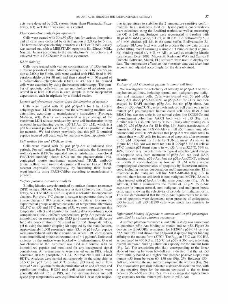
We investigated the selectivity of toxicity of p53p-Ant to vari-ous human cell lines, including normal, non-malignant, pre-malig-nant and malignant cells. Cells were treated with 50 �M p53palone, Ant alone, p53-AntCONT or p53p-Ant for 24 hr and thenassayed by DAPI staining. p53p-Ant, but not p53p alone, Antalone or p53p-AntCONT, selectively induced cell death only in themutant p53 pre-malignant human colon cell lines RG/C2 andBR/C1 but was not toxic in the normal colon line CCD33Co andpre-malignant colon line AA/C1 both with wt p53 (Fig. 1a).Similar results also obtained by TUNEL assay after treating cellswith 50 �M p53p-Ant for 18 hr (Fig. 1b). Stable transfectants ofhuman ts p53 mutant 143(Val-Ala) in null p53 human lung ade-nocarcinoma cells H1299 showed that p53p-Ant was more toxic tomutant than wt p53 cells for induction of apoptosis after exposureto 50 �M p53p-Ant for 18 hr by TUNEL assay. As shown inFigure 1c, p53p-Ant was more toxic to H1299/p53-143# 6 cells at37°C (mutant p53 form) than to its wt p53 form at 32.5°C, 76% vs.46%, respectively. To determine the typical morphological changeof apoptotic cells from treatment of p53p-Ant, we used DAPIstaining in our study. p53p-Ant, but not p53p-AntCONT, inducedcell death at concentrations as low as 10 �M with classicalmorphological characteristics of apoptosis by fluorescent micros-copy, including nuclear condensation and fragmentation after 12 hrtreatment in the malignant cell line MDA-MB-468 (Fig. 1d). Incontrast, there has no cell death in non-malignant MCF10-2A cellswhen treated with p53p-Ant by the same conditions (Fig. 1d). Inaddition, Table I summarizes the results of 30 �M p53p-Antexposure in human normal, non-malignant and malignant breastcells, again showing the selectivity of peptide for malignant cells.This also demonstrated that the p53p-Ant requirements for induc-tion of apoptosis were dependent upon presence of endogenousp53 because null p53 H1299 cells were much less sensitive topeptide.
Differential binding of peptide to mutant and wt p53 phenotypesquantified by surface plasmon resonance
A surface plasmon resonance (BIACORE) study was carried outto quantitate p53p-Ant binding to mutant and wt p53. Figure 2adepicts the BIACORE sensogram for H1299/ts p53–143 cells at32.5 and 37°C and shows that p53p-Ant displayed higher bindingaffinity to the mutant form (37°C). The Rmax at 37°C was 500 RU,as compared to 420 RU at 32.5°C (wt p53) at 360 sec, indicatingoverall increased binding saturation capacity for the mutant form(Fig. 2a). The association plot (ka), corresponding to the linearslope of binding between 60–360 sec, indicated that the wt p53form initially bound at a higher rate (steeper positive slope) thanmutant p53 form between 60–150 sec (Fig. 2b). Between 150–360 sec, however, the mutant form surpassed the wt form (Fig. 2b).The dissociation plot (kd) indicated a slower dissociation rate, witha less negative slope for the mutant compared to the wt formbetween 360–660 sec (Fig. 2c). This also suggested tighter bind-ing constants for the mutant p53 form to p53p-Ant.
57p53-ANT ACTS THROUGH THE FADD/CASPASE 8 PATHWAY
p53 C-terminal peptide-induced cell death:apoptosis vs. necrosis
To further delineate the form of cell death induced by p53p-Ant(apoptosis vs. necrosis), we used DAPI staining in time coursestudies in human breast lines. Cell death started as early as 3 hr andpeaked by 12 hr in the malignant mutant p53 cell line MDA-MB-
468 but it was not toxic to the non-malignant cell line MCF10-2A(Fig. 3a). Similar results also obtained by Trypan blue assays (datanot shown). To substantiate that the mechanism of cell death byp53p-Ant was apoptotic and not necrotic, we carried out an earlyLDH release assay in malignant breast cell lines MCF-7 (wt p53)and MDA-MB-468 (mutant p53). As shown in Figure 3b, when
FIGURE 1 – Selective induction of cell death by p53 C-terminal peptide in normal and pre-malignant human colon cells. (a) DAPI staining innormal human colon cells CDD-33Co (wt p53), pre-malignant colorectal adenoma cells AA/C1 (wt p53), RG/C2 (mutant p53) and BR/C1(mutant p53). Cells were treated with 50 �M p53p alone, Ant alone, p53-AntCONT or p53p-Ant for 24 hr and nuclear morphology was analyzedby DAPI staining. Number of apoptotic cells with nuclear morphology typical of apoptosis were scored in at least 400 cells in each sample byfluorescence microscopy. All microscopy experiments were carried out by a blinded reader to the actual groups. Data represent mean and SDof triplicate experiments. (b) TdT (TUNEL) assay in normal human colon cells CDD-33Co (wt p53), pre-malignant colorectal adenoma cellsAA/C1 (wt p53), RG/C2 (mutant p53) and BR/C1 (mutant p53). Cells were treated with 50 �M p53p-Ant for 18 hr. Apoptotic cells weredetermined by TdT assay and representative histograms show relative apoptotic cell numbers. (c) Effects of endogenous p53 status upon p53C-terminal peptide induced apoptosis in H1299 cells with stably transfected human ts p53 mutant 143Ala. Cells were pre-incubated for 16 hrat 37 or 32.5°C and then treated with 50 �M p53p-Ant for an additional 18 hr. Apoptotic cells were determined by TdT (TUNEL) assay andrepresentative histograms show relative apoptotic cell numbers. (d) Morphological analysis of apoptotic cells in MCF10-2A (wt p53) andMDA-MB-468 (mutant p53) cells. Cells were treated with 10 �M p53p-AntCONT or p53p-Ant for 12 hr and then nuclear morphology wasanalyzed by DAPI staining. The typical morphology in 400� magnification.
58 LI ET AL.

compared to the maximal LDH release (MLR), early LDH releaseinto the cytoplasm by 1 hr did not occur after exposure to 30 �Mp53p-Ant. The positive control for necrosis, 30 �M p53(15)Antthat is a p53 N-terminal peptide (aa 12–26) shown by us to induceonly necrosis without any evidence for apoptosis,36,37 was carriedout simultaneously. The N-terminal p53 peptide induced signifi-cant necrosis as evidenced by an early LDH release assay by 1 hr.Also, transmission electron microscopy was carried out at the 2-hrtime point and the infrastructure did not show plasma or nuclearmembrane perforations from 30 �M p53p-Ant, but the plasma andnuclear membranes displayed perforations after exposure to30 �M N-terminal p53(15)Ant (data not shown). These data indi-cated that p53p-Ant mechanism of cell death was purely apoptoticand not necrotic in these 2 cell lines.
Effects of p53 C-terminal peptide on protein targets of p53 andthe cell cycle
The effect of p53p-Ant on the expression of proteins induced byp53, including p21, GADD45 and Bax, and the cell cycle wereinvestigated to determine if other wt p53 functions were restoredby peptide. There was no increase in baseline p21, GADD45 andBax expression by 30 �M p53p-Ant treatment in a time coursestudy (0, 3, 6, 12, 24 and 48 hr) in malignant cell lines MDA-MB468 (mutant p53), MDA-MB231 (mutant p53) and MCF-7 (wtp53) (Figure 4 and data not shown). For the cell cycle study, thepattern of cell cycle G1 and G2 peaks did not change in MCF-7and MDA-MB-468 cells over a 24-hr time course (data notshown).
Effects of p53 C-terminal peptide on caspase activation andBcl-2 family proteins
To investigate the mechanism of downstream apoptotic induc-tion by p53p-Ant in the Fas signal transduction pathway, we testedits effect on caspase-3 and -8 activation in MDA-MB-468 cells andfound the highest caspase-3 and -8 activity appeared 2 hr aftertreatment with 30 �M p53p-Ant (data not shown). To furtherconfirm this finding, we used the specific caspase-3 inhibitor(Z-DEVD-FMK) and caspase-8 inhibitor (Z-IETD-FMK) at 4 �Mto pre-treat MDA-MB-468 cells for 1 hr and then exposed cells top53p-Ant for 6 hr. Both inhibitors were able to significantly blockp53p-Ant-induced apoptosis by TUNEL assays (Fig. 5). In con-trast, the caspase-9 inhibitor (Z-LEHD-FMK), which can block theintrinsic pathway of apoptosis induced by the Bcl-2 family ofproteins, did not block p53p-Ant-induced cell death. The resultsconfirmed that p53p-Ant-induced apoptosis was through the Faspathway.
To further determine whether the Bcl-2 family was involved inp53p-Ant-induced apoptosis, we examined the expression of sev-eral Bcl-2 family molecules: Bcl-2, Bax, Bcl-XL, Bak and BID byWestern blot analysis in malignant MDA-MB-468 cells treatedwith 30 �M p53p-Ant for 3 hr. There was no significant change inexpression levels of these molecules at 3 hr when apoptosis wasevident (data not shown). There were also no changes in the levelof heterodimers of Bax or Bcl-2 by immunoprecipitation (IP)assays (data not shown). These experiments further corroborated
that the intrinsic pathway of apoptosis (Bcl-2 family proteins) maynot be involved in p53p-Ant-induced apoptosis.
p53 C-terminal peptide-induced apoptosis and the Fas signaltransduction pathway
To further investigate the mechanism of p53p-Ant-induced celldeath through the Fas (extrinsic) apoptosis pathway, we assessedchanges in cell surface Fas and TRAIL expression. As shown inFigure 6a, MDA-MB-468 cells exhibited a 44% increase in thelevels of extracellular Fas protein within 60 min after 30 �Mp53p-Ant treatment by flow cytometry. There was no change inTRAIL expression by the same treatment and the controls, p53pand Ant alone, did not have any effect on levels of extracellularFas expression (data not shown). To determine whether p53p-Ant-induced apoptosis could involve an interaction between Fas andFas ligand, we pre-incubated the antagonistic anti-Fas antibodyZB4 at 10 �g/ml with MDA-MB-468 cells for 1 hr and thentreated with 30 �M p53p-Ant for 6 hr. As shown in Figure 6b,antagonistic anti-Fas antibody ZB4 did not block induction ofapoptosis by p53p-Ant. For controls, we exposed the cells to theagonistic anti-Fas antibody CH-11 at 50 ng/ml for 24 hr. The ZB4antagonistic antibody reduced CH-11-induced apoptosis from 34%to 8%. These data suggested the mechanism of p53p-Ant-inducedapoptosis was through an increased Fas expression signal trans-duction pathway independent of Fas–Fas ligand interaction. Inaddition, experiments with pre-incubation of cycloheximide andactinomycin D for 18 hr did not alter the above results, suggestingthat the increased Fas expression was from membrane re-distribu-tion to the extracellular milieu and not from new protein or mRNAFas synthesis, respectively (data not shown).
Expression of dominant–negative FADD and its effect on p53C-terminal peptide-induced cell death
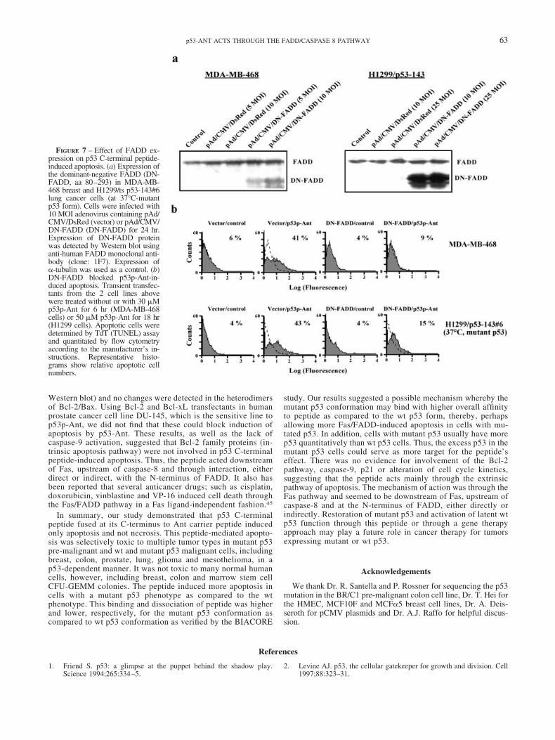
To determine whether FADD association to Fas after clusteringcould be pivotal for p53p-Ant-induced cell death, we modulatedFADD expression via an Ad5 adenovirus system. Breast MDA-MB-468 and lung H1299/p53-143#6 cells were transfected tran-siently with adenovirus containing control vector pAd/CMV/DsRed or pAd/CMV/DN-FADD (aa 80–293 of FADD) with 3different multiplicities of infection (MOI) for 24 hr. The expres-sion of dominant-negative FADD (DN-FADD) was detected byWestern blot as shown in Figure 7a. Transfectants of MDA-MB-468 were treated with 30 �M p53p-Ant for 6 hr and apoptotic cellswere detected by TdT assay. As shown in Figure 7b, DN-FADDexpression decreased p53p-Ant-induced apoptosis from 41% to9%. Similar results were also obtained in the H1299/p53-143#6cells at 37°C (mutant p53 form) exposed to 50 �M p53p-Ant for18 hr (43% to 15%). These results demonstrated that the N-terminus (aa 1–79) of FADD played a critical role for p53pC-terminal peptide-induced apoptosis in these human cancer cellsand the peptide interacted with FADD either directly or indirectlywithout the need for Fas or Fas ligand.
TABLE I – TOXICITY OF p53p-Ant CORRELATES WITH p53 STATUS AND MALIGNANCY INHUMAN BREAST CELL LINES
Cell line Cell type p53 statusApoptosis (%) by P1 staining
Control p53p-Ant(30 �M 6 hr)
HMEC Normal Wild-type 6 9MCF10-2A Non-malignant Wild-type 6 8MCF10F Non-malignant Wild-type 6 7MDA-MB-468 Malignant 273 Arg�His 6 65MDA-MB-231 Malignant 280 Arg�Lys 6 32MCF�5 Malignant 254 Ile�Asp 6 31MCF-7 Malignant Wild-type 6 30MDA-MB-157 Malignant Null 6 6
59p53-ANT ACTS THROUGH THE FADD/CASPASE 8 PATHWAY

Discussion
Our previous work showed that the p53 C-terminal peptide(p53p-Ant) was toxic to 3 malignant breast cells carrying eitherendogenous p53 mutations or overexpressed wt p53 but was not
toxic to malignant breast cells with null p53.27,38 In this study, weshowed that this peptide was not toxic to normal colon and normal,as well as, non-malignant breast cells with endogenous wt p53.The peptide selectively induced apoptosis in pre-malignant coloncells with mutant p53 (Fig. 1a, Table I). This is of particularinterest because of the paucity of agents that can kill pre-malignantcells. In addition, p53p-Ant was selectively cytotoxic to othertumor types such as prostate, lung, glioma, mesothelioma andcolon cancer cell lines in an apparent p53-dependent manner (datanot shown). Peptide at 30 �M had only a minor toxic effect onperipheral CD34 positive marrow stem cells in CFU-GEMM as-says (10% toxicity), which have normal levels of wt p53 (unpub-lished data).
FIGURE 2 – Surface plasmon resonance (SPR) binding assays (BIA-CORE) of p53p-Ant exposed to H1299/p53-143#6 whole cell lysates.Measurement of binding kinetics are shown between immobilizedp53p-Ant at 1 pg/mm2 and H1299/p53-143 transfectant cell lysates at32.5°C (wt p53 form; dotted line) and 37°C (mutant p53 form; solidline). Concentration of 10 nM p53p-Ant demonstrated differences inthe maximum binding capacity and association/dissociation constantsat the two temperatures. SPR were determined by using a BIAcoreXbiosensor system. (a) Sensogram. (b) Association slope. (c) Dissoci-ation slope.
FIGURE 3 – p53 C-terminal peptide-induced cell death: apoptosisvs. necrosis. (A) DAPI staining in human breast non-malignant cellsMCF10-2A (wt p53) and malignant cells MDA-MB-468 (mutantp53). Cells were treated with 30 �M p53p-Ant for the indicatedtimes and nuclear morphology was analyzed by DAPI staining.Number of apoptotic cells with nuclear morphology typical ofapoptosis were scored in at least 400 cells in each sample byfluorescence microscopy. All microscopy experiments were carriedout by a blinded reader to the actual groups. Data represent meanand SD of triplicate experiments. (b) LDH assay in human breastmalignant cells MDA-MB-468 (mutant p53) and MCF-7 (wt p53).Cells were untreated or treated with 30 �M p53p-Ant (C-terminalpeptide, aa 361–382) or p53(15)Ant (N-terminal peptide, aa 12–26)for 1 hr. LDH released into the surrounding media was determinedaccording to manufacturer’s instructions (Promega, Madison, WI).Results are expressed as a percentage of the maximum LDH re-lease, determined by cell fractionation by repeated freeze-thawing.The p53 N-terminal peptide, which only induced necrosis withoutapoptosis, was used as a positive control for necrosis.36,37 Datarepresent mean and SD of triplicate experiments.
60 LI ET AL.

To better understand the effects of p53 C-terminal peptide oncancer cells containing different p53 phenotypes, we used tsp53 mutant 143Ala as a model. The results showed p53p-Antwas significantly more toxic to the mutant p53 conformation (at37°C) than the wt p53 form (at 32.5°C) in H1299 cells stablytransfected with a human ts p53 mutant 143Ala (Fig. 1b). Thesurface plasmon resonance (SPR) data suggested a mechanismfor the differential cytotoxicity effects observed between mu-tant and wt forms with p53p-Ant. It may be attributed to
increased binding affinity of the p53p-Ant peptide to the mutantp53 form, as compared to the wt form. p53p-Ant initially boundcellular components at the mutant p53 temperature with greateraffinity than at the wt p53 temperature, as shown by the higherpositive slopes of association (Fig. 2b) and greater bindingsaturation levels indicated by the higher Rmax (Fig. 2a). Theseresults indicated that the mutant p53 form retains more boundcomplexes with p53 peptide during the binding interaction.More importantly, the dissociation slope for the mutant formexhibited a less negative slope as compared to the wt p53temperature, suggesting that the mutant p53 form binds peptidemore tightly, disassociating less than the wt p53 form (Fig. 2c).This could explain why there is more Fas related apoptosisinduced in cells with mutant p53 leading to increased restora-tion of mutant p53 ability for functional redistribution of Fas tothe membrane. We are currently studying the binding site(s) ofp53p-Ant on p53 by utilizing deletion mutants of p53 andBIACORE analysis.
We had reported that p53p-Ant activated the Fas/APO-1 signal-ing transduction pathway. This was associated with activation ofthe downstream initiator caspase-8 and effector caspase-3 pro-teases and cleavage of the caspase-3 substrate, PARP.27 In ourstudy, Fas antagonistic antibody ZB4 did not block apoptosisinduced by p53p-Ant peptide but did block Fas mediated apoptosisinduced by Fas agonistic antibody CH-11 (Fig. 6). Using anti-Fasantibodies in Western blot analysis, we still did not find increasedFas protein content after exposure to the p53-Ant peptide (data not
FIGURE 4 – Western blot for p21 expression in human breast malig-nant cells MDA-MB468 (mutant p53), MDA-MB231 (mutant p53)and MCF7 (wt p53). Cells were treated with 30 �M p53p-Ant for 0, 3or 6 hr. p21 expression was analyzed by Western blot using anti-p21polyclonal antibody (clone: H-164). Expression of �-tubulin was usedas a control.
FIGURE 5 – Effect of specific caspase-3 inhib-itor (Z-DEVD-FMK), caspase-8 inhibitor (Z-IETD-FMK) and caspase-9 inhibitor (Z-LEHD-FMK) on p53p-Ant-induced apoptosis in humanbreast malignant cells MDA-MB-468 (mutantp53). Cells were pre-treated with 4 �M inhibitorsfor 60 min and then treated with 30 �M p53p-Ant for 6 hr. Apoptotic cells were determined byTdT (TUNEL) assay and quantitated by flowcytometry according to the manufacturer’s in-structions. Representative histograms show rela-tive apoptotic cell numbers.
61p53-ANT ACTS THROUGH THE FADD/CASPASE 8 PATHWAY

shown). This suggested that p53p-Ant-induced apoptosis was notdependent on the interaction of Fas and Fas ligand but may befurther downstream of Fas such as FADD. DN-FADD (aa 80–293)clearly blocked p53 C-terminal peptide-induced cell death in 2different cell lines (Fig. 7b), suggesting that FADD played animportant role in this mechanism of cell death. We were not ableto detect direct Fas/FADD/p53p-Ant binding by IP studies inMDA-MB-468 cells (data not shown). In addition, TRAIL andTNF-R1 were not involved in p53-Ant-induced cell death (Fig. 6and data not shown).
p53p-Ant did not increase p53 target molecules such as p21,GADD45 and Bax or alter cell cycle characteristics (Fig. 4).These results suggested that p53p-Ant induced only an apopto-tic response via the extrinsic Fas/FADD pathway without alter-ing the intrinsic apoptosis pathway (Bax) or the cell cycle staticresponse, although the latter 2 are associated with the normalfunctioning p53 phenotype. Reasons for this dissociative effectare unknown but may be secondary to required conformationalchanges in p53 that may be different for inducing p21 andGADD45 and re-distribution of Fas to the outer membrane. Thepeptide’s selective induction of Fas-associated apoptosis with-
out p21 induction may provide an added advantage to p53peptide over whole p53 gene therapy approaches because p21induction, which can occur from the latter, has been reported toinduce drug resistance to cell cycle active agents via cell cyclestasis in several tumor cell types.39,40 In addition, whole p53gene transfer may hypothetically lead to increased toxicity innormal cells, such as increased sensitivity to chemotherapydrugs by increasing wt p53 levels.
Mitochondrial triggered cell death can occur through severalmechanisms including disruption of electron transport, releaseof proteins that in turn activate caspases and alteration ofcellular reduction-oxidation potentials. For example, activationof Fas leads to rapid inactivation of the electron transfer activityand release of cytochrome C from Bcl-2 in the mitochondrialmembrane can inhibit cytochrome C release during apopto-sis.41– 44 In addition, cytochrome C release from mitochondriaand cell death are mediated by BID when cleaved by caspase-8to t-BID, thus suggesting BID as a downstream component ofthe Fas pathway.24 MDA-MB-468 cells treated with p53p-Antshowed no significant changes in expression levels of Bcl-2family proteins (including BID, Bcl-2, Bax, Bcl-XL and Bak by
FIGURE 6 – Effect of p53p-Anton Fas and TRAIL-induced apo-ptosis pathways. (a) Cell surfaceFas and TRAIL analysis in MDA-MB-468 cells. Cells were treatedwith 30 �M p53p-Ant at indicatedtime periods. Cell surface Fas wasdetermined by FITC-conjugatedmouse anti-human monoclonal Fas/CD95 antibody (clone: DX2) andcell surface TRAIL was deter-mined by phycoerythrin (PE)-con-jugated mouse anti-human mono-clonal TRAIL antibody (clone:RIK-2) using FACS-Caliber ac-cording to manufacturer’s proto-col. (b) Effect of antagonistic anti-Fas antibody on p53p-Ant-inducedapoptosis in MDA-MB-468 cells.Cells were pre-treated with 10 �g/ml antagonistic anti-Fas antibodyZB4 for 1 hr and then treated with30 �M p53p-Ant for 6 hr or 50 ng/ml agonistic anti-Fas antibodyCH-11 for 24 hr. Apoptotic cellswere determined by TdT (TUNEL)assay and quantitated by flow cy-tometry according to the manufac-turer’s instructions. Representativehistograms show relative apoptoticcell numbers.
62 LI ET AL.
Western blot) and no changes were detected in the heterodimersof Bcl-2/Bax. Using Bcl-2 and Bcl-xL transfectants in humanprostate cancer cell line DU-145, which is the sensitive line top53p-Ant, we did not find that these could block induction ofapoptosis by p53-Ant. These results, as well as the lack ofcaspase-9 activation, suggested that Bcl-2 family proteins (in-trinsic apoptosis pathway) were not involved in p53 C-terminalpeptide-induced apoptosis. Thus, the peptide acted downstreamof Fas, upstream of caspase-8 and through interaction, eitherdirect or indirect, with the N-terminus of FADD. It also hasbeen reported that several anticancer drugs; such as cisplatin,doxorubicin, vinblastine and VP-16 induced cell death throughthe Fas/FADD pathway in a Fas ligand-independent fashion.45
In summary, our study demonstrated that p53 C-terminalpeptide fused at its C-terminus to Ant carrier peptide inducedonly apoptosis and not necrosis. This peptide-mediated apopto-sis was selectively toxic to multiple tumor types in mutant p53pre-malignant and wt and mutant p53 malignant cells, includingbreast, colon, prostate, lung, glioma and mesothelioma, in ap53-dependent manner. It was not toxic to many normal humancells, however, including breast, colon and marrow stem cellCFU-GEMM colonies. The peptide induced more apoptosis incells with a mutant p53 phenotype as compared to the wtphenotype. This binding and dissociation of peptide was higherand lower, respectively, for the mutant p53 conformation ascompared to wt p53 conformation as verified by the BIACORE
study. Our results suggested a possible mechanism whereby themutant p53 conformation may bind with higher overall affinityto peptide as compared to the wt p53 form, thereby, perhapsallowing more Fas/FADD-induced apoptosis in cells with mu-tated p53. In addition, cells with mutant p53 usually have morep53 quantitatively than wt p53 cells. Thus, the excess p53 in themutant p53 cells could serve as more target for the peptide’seffect. There was no evidence for involvement of the Bcl-2pathway, caspase-9, p21 or alteration of cell cycle kinetics,suggesting that the peptide acts mainly through the extrinsicpathway of apoptosis. The mechanism of action was through theFas pathway and seemed to be downstream of Fas, upstream ofcaspase-8 and at the N-terminus of FADD, either directly orindirectly. Restoration of mutant p53 and activation of latent wtp53 function through this peptide or through a gene therapyapproach may play a future role in cancer therapy for tumorsexpressing mutant or wt p53.
Acknowledgements
We thank Dr. R. Santella and P. Rossner for sequencing the p53mutation in the BR/C1 pre-malignant colon cell line, Dr. T. Hei forthe HMEC, MCF10F and MCF�5 breast cell lines, Dr. A. Deis-seroth for pCMV plasmids and Dr. A.J. Raffo for helpful discus-sion.
References
1. Friend S. p53: a glimpse at the puppet behind the shadow play.Science 1994;265:334–5.
2. Levine AJ. p53, the cellular gatekeeper for growth and division. Cell1997;88:323–31.
FIGURE 7 – Effect of FADD ex-pression on p53 C-terminal peptide-induced apoptosis. (a) Expression ofthe dominant-negative FADD (DN-FADD, aa 80–293) in MDA-MB-468 breast and H1299/ts p53-143#6lung cancer cells (at 37°C-mutantp53 form). Cells were infected with10 MOI adenovirus containing pAd/CMV/DsRed (vector) or pAd/CMV/DN-FADD (DN-FADD) for 24 hr.Expression of DN-FADD proteinwas detected by Western blot usinganti-human FADD monoclonal anti-body (clone: 1F7). Expression of�-tubulin was used as a control. (b)DN-FADD blocked p53p-Ant-in-duced apoptosis. Transient transfec-tants from the 2 cell lines abovewere treated without or with 30 �Mp53p-Ant for 6 hr (MDA-MB-468cells) or 50 �M p53p-Ant for 18 hr(H1299 cells). Apoptotic cells weredetermined by TdT (TUNEL) assayand quantitated by flow cytometryaccording to the manufacturer’s in-structions. Representative histo-grams show relative apoptotic cellnumbers.
63p53-ANT ACTS THROUGH THE FADD/CASPASE 8 PATHWAY
3. Cho Y, Gorina S, Jeffrey PD, Pavletich NP. Crystal structure of a p53tumor suppressor-DNA complex: understanding tumorigenic muta-tions. Science 1994;265:346–55.
4. Kastan MB, Onyekwere O, Sidransky D, Vogelstein B, Craig R.Participation of p53 protein in the cellular response to DNA damage.Cancer Res 1991;51:6304–11.
5. Livingstone LR, White A, Sprouse J, Livanos E, Jacks T, Tlsty TD.Altered cell cycle arrest and gene amplification potential accompanyloss of wild-type p53. Cell 1992;70:923–35.
6. Yin Y, Tainsky MA, Bischoff FZ, Strong LC, Wahl GM. Wild-typep53 restores cell cycle control and inhibits gene amplification in cellswith mutant p53 alleles. Cell 1992;70:937–48.
7. Ko LJ, Prives C. p53: puzzle and paradigm. Gene Dev 1996;10:1054–72.
8. El-Deiry WS, Tokino T, Velculescu VE, Levy DB, Parsons R, TrentJM, Lin D, Mercer WE, Kinzler KW, Vogelstein B. WAF1, a poten-tial mediator of p53 tumor suppression. Cell 1993;75:817–25.
9. Kastan MB, Zhan Q, El-Deiry WS, Carrier F, Jacks T, Walsh WV,Plunkett BS, Vogelstein B, Fornace AJ Jr. A mammalian cell cyclecheckpoint pathway utilizing p53 and GADD45 is defective in ataxia-telangiectasia. Cell 1992;71:587–97.
10. Oltvai ZN, Milliman CL, Korsmeyer SJ. Bcl-2 heterodimerizes invivo with a conserved homolog, Bax, that accelerates programmedcell death. Cell 1993;74:609–19.
11. Owen-Schaub LB, Zhang W, Cusack JC, Angelo LS, Santee SM,Fujiwara T, Roth JA, Deisseroth AB, Zhang WW. Wild-type humanp53 and a temperature-sensitive mutant induce Fas/APO-1 expression.Mol Cell Biol 1995;15:3032–40.
12. Bennett M, MacDonald K, Chan S-W, Luzio JP, Simari R, WeissbergP. Cell surface trafficking of Fas: a rapid mechanism of p53-mediatedapoptosis. Science 1998;282:290–4.
13. Itoh N, and Nagata SA. Novel protein domain required for apoptosis.J Biol Chem 1993;268:10932–7.
14. Tartaglia LA, Ayres TM, Wong GH, Goeddel DV. A novel domainwithin the 55 kd TNF receptor signals cell death. Cell 1993;74:845–53.
15. Suda T, Nagata S. Why do defects in the Fas-Fas ligand system causeauto immunity. J Allergy Clin Immunol 1997;100:97–101.
16. MacFarlane M. TRAIL-induced signalling and apoptosis. ToxicolLett 2003;139:89–97.
17. Chinnaiyan AM, O’Rourke K, Tewari M, Dixit VM. FADD, a noveldeath domain-containing protein, interacts with the death domain ofFas and initiates apoptosis. Cell 1995;81:505–12.
18. Strasser A, Newton K. FADD/MORT1, a signal transducer that canpromote cell death or cell growth. Int J Biochem Cell Biol 1999;31:533–7.
19. Cryns V, Yuan J. Proteases to die for. Genes Dev 1998;12:1551–70.20. Salvesen GS, Dixit VM. Caspases: intracellular signalling by prote-
olysis. Cell 1997;91:443–6.21. Farrow SN, Brown R. New members of the Bcl-2 family and their
protein partners. Curr Opin Genet Dev 1996;6:45–9.22. Kroemer G. The proto-oncogene Bcl-2 and its role in regulating
apoptosis. Nat Med 1997;3:614–20.23. Korsmeyer SL. Regulators of cell death. Trends Genet 1995;11:
101–5.24. Yin XM. Bid, a critical mediator for apoptosis induced by the acti-
vation of Fas/TNF-R1 death receptors in hepatocytes. J Mol Med2000;78:203–11.
25. Hupp TR, Lane DP. Allosteric activation of latent p53 tetramers. CurrBiol 1994;4:865–75.
26. Muller-Tiemann BF, Halazonetis TD, Elting JJ. Identification of anadditional negative regulatory region for p53 sequence-specific DNAbinding. Proc Natl Acad Sci USA 1998;95:6079–84.
27. Kim AL, Raffo AJ, Brandt-Rauf PW, Pincus MR, Monaco R, AbarzuaP, Fine RL. Conformational and molecular basis for induction ofapoptosis by a p53 C-terminal peptide in human cancer cells. J BiolChem 1999;274:34924–31.
28. Abarzua P, LoSardo JE, Gubler ML, Spathis R, Lu YA, Felix A, NeriA. Restoration of the transcription activation function to mutant p53in human cancer cells. Oncogene 1996;13:2477–82.
29. Selivanova G, Iotsova V, Okan I, Fritsche M, Strom M, Groner B,Grafstrom RC, Wiman KG. Restoration of the growth suppressionfunction of mutant p53 by a synthetic peptide derived from the p53C-terminal domain. Nat Med 1997;3:632–8.
30. Li Y, Raffo AJ, Lisa D, Mao Y, Tran A, Petrylak DP, Fine RL.Fas-mediated apoptosis is dependent on wild-type p53 status in hu-man cancer cells expressing a temperature-sensitive p53 mutant ala-nine-143. Cancer Res 2003;63:1527–33.
31. Fivash M, Towler EM, Fisher RJ. BIACORE for macromolecularinteraction. Curr Opin Biotechnol 1998;9:97–101.
32. Malmqvist M. BIACORE: an affinity biosensor system for character-ization of biomolecular interactions. Biochem Soc Trans 1999;27:335–40.
33. Rufer A, Neuenschwander PF, Sauer B. Analysis of Cre-loxP inter-action by surface plasmon resonance: influence of spermidine oncooperativity. Anal Biochem 2002;308:90–9.
34. Calaf GM, Hei TK. Establishment of a radiation- and estrogen-induced breast cancer model. Carcinogenesis 2000;21:769–76.
35. Roy D, Calaf G, Hei TK. Frequent allelic imbalance on chromosome6 and 17 correlate with radiation-induced neoplastic transformation ofhuman breast epithelial cells. Carcinogenesis 2001;22:1685–92.
36. Kanovsky M, Raffo A, Drew L, Rosal R, Do T, Friedman FK,Rubinstein P, Visser J, Robinson R, Brandt-Rauf PW, Michl J, FineRL, et al. Peptides from the amino terminal mdm-2-binding domain ofp53, designed from conformational analysis, are selectively cytotoxicto transformed cells. Proc Natl Acad Sci USA 2001;98:12438–43.
37. Do T, Ramon V, Rosal RV, Drew L, Raffo AJ, Michl J, Pincus MR,Friedman FK, Petrylak DP, Cassai N, Szmulewicz J, Sidhu G, et al.Preferential induction of necrosis in human breast cancer cells by ap53 peptide derived from the MDM2 binding site. Oncogene 2003;22:1431–44.
38. Li Y, Rosal RV, Brandt-Rauf PW, Fine RL. Correlation betweenhydrophobic properties and efficiency of carrier-mediated membranetransduction and apoptosis of a p53 C-terminal peptide. BiochemBiophys Res Commun 2002;298:439–49.
39. Waldman T, Lengauer C, Kinzler KW, Vogelstein B. Uncoupling ofS phase and mitosis induced by anticancer agents in cells lacking p21.Nature 1996;381:713–6.
40. Waldman T, Zhang YG, Dillehay L, Yu J, Kinzler K, Vogelstein B,Williams J. Cell-cycle arrest versus cell death in cancer therapy. NatMed 1997;3:1034–6.
41. Green DR, Reed JC. Mitochondria and apoptosis. Science 1998;281:1309–12.
42. Kluck RM, Bossy-Wetzel E, Green DR, Newmeyer DD. The releaseof cytochrome c from mitochondria: a primary site for Bcl-2 regula-tion of apoptosis. Science 1997;275:1132–6.
43. Yang J, Liu X, Bhalla K, Kim CN, Ibrado AM, Cai J, Peng TI, JonesDP, Wang X. Prevention of apoptosis by Bcl-2: release of cytochromec from mitochondria blocked. Science 1997;275:1129–32.
44. Zimmermann KC, Bonzon C, Green DR. The machinery of pro-grammed cell death. Pharmacol Ther 2001;92:57–70.
45. Micheau O, Solary E, Hammann A, Dimanche-Boitrel M. Fas ligand-independent, FADD-mediated activation of the Fas death pathway byanticancer drugs. J Biol Chem 1999;274:7987–92.
64 LI ET AL.
Copyright © 2022 FDOKUMEN