The deubiquitinase USP9X suppresses pancreatic ductal adenocarcinoma
Upload
dana-farberCategory
view
5download
0
OPEN
ORIGINAL ARTICLE
Oncogenic RIT1 mutations in lung adenocarcinomaAH Berger1,2, M Imielinski1,2,3,4, F Duke1, J Wala1,2,5, N Kaplan2, G-X Shi6, DA Andres6 and M Meyerson1,2,4
Lung adenocarcinoma is comprised of distinct mutational subtypes characterized by mutually exclusive oncogenic mutations inRTK/RAS pathway members KRAS, EGFR, BRAF and ERBB2, and translocations involving ALK, RET and ROS1. Identification of theseoncogenic events has transformed the treatment of lung adenocarcinoma via application of therapies targeted toward specificgenetic lesions in stratified patient populations. However, such mutations have been reported in only B55% of lungadenocarcinoma cases in the United States, suggesting other mechanisms of malignancy are involved in the remaining cases. Herewe report somatic mutations in the small GTPase gene RIT1 in B2% of lung adenocarcinoma cases that cluster in a hotspot nearthe switch II domain of the protein. RIT1 switch II domain mutations are mutually exclusive with all other known lungadenocarcinoma driver mutations. Ectopic expression of mutated RIT1 induces cellular transformation in vitro and in vivo, which canbe reversed by combined PI3K and MEK inhibition. These data identify RIT1 as a driver oncogene in a specific subset of lungadenocarcinomas and suggest PI3K and MEK inhibition as a potential therapeutic strategy in RIT1-mutated tumors.
Oncogene advance online publication, 27 January 2014; doi:10.1038/onc.2013.581
Keywords: lung adenocarcinoma; cancer genetics; RAS pathway; signal transduction; oncogene; GTPase
INTRODUCTIONLung adenocarcinomas contain characteristic, mutually exclusivemutations in receptor tyrosine kinase (RTK) and RAS pathwayoncogenes including frequent mutation of KRAS or EGFR in B30 or10% of US cases, respectively.1–13 Rarely, translocations are seeninvolving EML4 and ALK (1–4%), RET (1%) or ROS1 (2%)2,5 (TheCancer Genome Atlas, manuscript submitted). Additional rareoncogenic mutations are seen in HRAS, NRAS, BRAF, ERBB2, METand MAP2K1.1,2 Together these oncogenic events are found inB55% of lung adenocarcinomas in the US population.2 Theimportance of identifying such ‘driver’ mutations is exemplified bythe success of targeted therapies directed against these events—the prototypical example being the use of the small molecule EGFRinhibitors Iressa (gefitinib) and Tarceva (erlotinib) in EGFR-mutatedlung adenocarcinoma.14 Among patients with EGFR mutations,single-line EGFR-targeted therapy significantly extends progression-free survival compared with conventional chemotherapy.7,8 Patientswithout EGFR mutations, however, typically do not benefit fromtargeted therapy, underscoring the need to genotype patienttumor resections and/or biopsies to guide appropriate treatmentdecisions. Modeling this paradigm, clinical trials of the ALK inhibitorcrizotinib for treatment of tumors with EML4-ALK translocation wererapidly reported15 only 3 years after the identification of the EML4-ALK event.5 Therefore, continued identification of oncogenic drivergenes in the remaining B45% of ‘oncogene-negative’ lungadenocarcinoma cases may allow further patient stratification andtargeted therapies.
Recent genomic analyses of lung adenocarcinoma haveidentified novel mutations in diverse genes, but few additionaloncogenic mutations have been identified that displaymutual exclusivity with known lung adenocarcinoma driveroncogenes.1,16 Here we report the genomic and functional
characterization of somatic RIT1 mutations that we identify asnovel driver mutations in lung adenocarcinoma.
RESULTSTo identify rare oncogenic driver mutations, we and others in TheCancer Genome Atlas (TCGA) Research Network performed atargeted analysis of ‘oncogene-negative’ tumors that lack muta-tions in the known lung adenocarcinoma driver oncogenes KRAS,EGFR, BRAF, ERBB2, MAP2K1, MET, HRAS, NRAS and/or fusionsinvolving ALK, RET or ROS1 (TCGA, manuscript submitted). In 87‘oncogene-negative’ samples lacking mutations/fusions in theknown driver genes, RIT1 (Ras-like in all tissues) was among the topsignificantly mutated genes. 5/87 samples in this group harboredsomatic RIT1 mutations. In contrast, no mutations in RIT1 werefound in the 143 samples containing known driver oncogenicmutations (Po0.01 by Fisher’s exact test). Moreover, we identifiedfive additional RIT1-mutant samples in sequence data from anindependent cohort of 183 lung adenocarcinomas.1
In total, non-synonymous somatic mutations of RIT1 wereidentified in 10/413 (2.4%) lung adenocarcinomas subjected towhole exome sequencing (Figure 1a, Supplementary Table 1).Mutations in RIT1 consisted of missense mutations and small in-frame insertion/deletions. In all, 7/10 RIT1 mutations wereclustered near the switch II domain region (Figures 1a and b),including three recurrent p.M90I mutations. The switch II domainin RIT1 is homologous to the switch II region in RAS genes thatcontains glutamine 61 (Supplementary Figure 1), which is directlyinvolved in GTP hydrolysis and also a hotspot of mutation in RASgenes.17 This switch II domain hotspot is distinct from the P29hotspot in RAC genes recently reported in melanoma and othercancers18–20 (Supplementary Figure 1).
1Cancer Program, The Broad Institute of Harvard and M.I.T., 7 Cambridge Center, Cambridge, MA, USA; 2Department of Medical Oncology, Dana Farber Cancer Institute, Boston,MA, USA; 3Department of Pathology, Massachusetts General Hospital, Boston, MA, USA; 4Department of Pathology, Harvard Medical School, Boston, MA, USA; 5Harvard-MITDivision of Health Sciences and Technology, Massachusetts Institute of Technology, Cambridge, MA, USA and 6Department of Molecular and Cellular Biochemistry, University ofKentucky College of Medicine, Lexington, KY, USA. Correspondence: Dr M Meyerson, Department of Medical Oncology, Center for Cancer Genome Discovery, Dana-Farber CancerInstitute 450 Brookline Avenue, Boston, MA 02115, USA.E-mail: [email protected] 26 June 2013; revised 25 November 2013; accepted 3 December 2013
Oncogene (2014), 1–6& 2014 Macmillan Publishers Limited All rights reserved 0950-9232/14
www.nature.com/onc
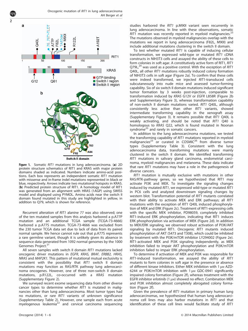
Recurrent alteration of RIT1 alanine 77 was also observed; oneof the ten mutated samples from this analysis harbored a p.A77Pmutation and an additional TCGA sample (TCGA-73-4666)harbored a p.A77S mutation. TCGA-73-4666 was excluded fromthe 230 tumor TCGA data set due to lack of data from its pairednormal sample. We hence cannot rule out that p.A77S representsa rare germline variant, though it is unlikely given its absence insequence data generated from 1092 normal genomes by the 1000Genomes Project.21
All seven samples with switch II domain RIT1 mutations lackedoncogenic driver mutations in EGFR, KRAS, BRAF, ERBB2, HRAS,NRAS and MAP2K1. This pattern of mutational mutual exclusivity isconsistent with the possibility that RIT1 switch II domainmutations may function as RAS/RTK pathway lung adenocarci-noma oncogenes. However, one of three non-switch II domainmutations, p.R122L, co-occurred with a KRAS mutation(Supplementary Figure 2).
We surveyed recent exome sequencing data from other diversecancer types to determine whether RIT1 is mutated in malig-nancies other than lung adenocarcinoma. Most tumor types hadno mutations, or rare RIT1 variants of unknown significance(Supplementary Table 2). However, one sample each from acutemyelogenous leukemia22 and cervical carcinoma sequencing
studies harbored the RIT1 p.M90I variant seen recurrently inlung adenocarcinoma. In line with these observations, somaticRIT1 mutation was recently reported in myeloid malignancies.23
The mutations observed in myeloid malignancies overlap with themutations we report in lung adenocarcinoma (F82L, M90I) andinclude additional mutations clustering in the switch II domain.
To test whether mutated RIT1 is capable of inducing cellulartransformation, we expressed wild-type or mutated RIT1 cDNAconstructs in NIH3T3 cells and assayed the ability of these cells toform colonies in soft agar. A constitutively active form of RIT1, RIT1Q79L,24 was used as a positive control. With the exception of RIT1Q40L, all other RIT1 mutations robustly induced colony formationof NIH3T3 cells in soft agar (Figure 2a). To confirm that these cellswere indeed transformed, we injected RIT1-transduced cellssubcutaneously into nude mice and assessed tumor-formingcapability. Six of six switch II domain mutations induced significanttumor formation by 3 weeks post-injection, comparable totransformation induced by KRAS G12V or EGFR L858R (Figure 2band Supplementary Figure 3), whereas transformation capabilityof non-switch II domain mutations varied. RIT1 Q40L, althoughconsistently less active than other RIT1 variants, showedintermediate transforming capability in the xenograft assay(Supplementary Figure 3). It remains possible that RIT1 Q40L isweakly activating, and should be noted that RIT1 Q40 ishomologous to KRAS Q22, which is found mutated in Noonansyndrome25 and rarely in somatic cancers.
In addition to the lung adenocarcinoma mutations, we testedthe transforming capability of RIT1 mutations reported in myeloidmalignancies23 or curated in COSMIC26 from diverse tumortypes (Supplementary Table 3). Consistent with the lungadenocarcinoma data, transforming mutations were mainlyclustered in the switch II domain. We identified transformingRIT1 mutations in salivary gland carcinoma, endometrial carci-noma, myeloid malignancies and melanoma. These data indicatethat rare RIT1 mutations may have a role in the pathogenesis ofdiverse cancers.
RIT1 mutation is mutually exclusive with mutations in otherRAS/RTK-pathway genes, so we hypothesized that RIT1 mayactivate PI3K and MEK. To investigate the signaling changesinduced by mutated RIT1, we expressed wild-type or mutated RIT1in PC6 cells and analyzed downstream signaling changes bywestern blot. Transformation potential of RIT1 mutants correlatedwith their ability to activate MEK and ERK pathways; all RIT1mutations with the exception of RIT1 Q40L induced phosphoryla-tion of MEK and ERK (Figure 2c). Treatment of RIT1-expressing cellswith the specific MEK inhibitor, PD98059, completely inhibitedRIT1-induced ERK phosphorylation, indicating that RIT1 inducesERK phosphorylation via activation of MEK (Figure 2d). In additionto MEK/ERK signaling, we observed robust activation of PI3K/AKTsignaling by mutated RIT1. Oncogenic RIT1 mutants inducedphosphorylation of AKT (S473 and T308), which could be inhibitedby treatment with the PI3K/mTOR inhibitor LY294002 (Figure 2e).RIT1-activated MEK and PI3K signaling independently, as MEKinhibition failed to impair AKT phosphorylation and PI3K/mTORinhibition failed to impair ERK phosphorylation.
To determine if activation of MEK and PI3K was responsible forRIT1-induced transformation, we assayed the ability of RIT1mutants to form colonies in soft agar in the presence or absenceof different kinase inhibitors. Either MEK inhibition with 1 mM AZD-6244 or PI3K/mTOR inhibition with 1 mM GDC-0941 significantlyimpaired colony formation (Figure 2f), whereas treatment with theEGFR inhibitor erlotinib (1 mM) showed no effect. Combination MEKand PI3K inhibition almost completely abrogated colony forma-tion (Figure 2f).
Given the prevalence of RIT1 mutation in primary human lungadenocarcinomas, we hypothesized that human lung adenocarci-noma cell lines may also harbor mutations in RIT1 and thatidentification of these cell lines would facilitate study of RIT1
Figure 1. Somatic RIT1 mutations in lung adenocarcinoma. (a) 2Dprotein structure schematics of RIT1 and KRAS with major proteindomains shaded as indicated. Numbers indicate amino-acid posi-tions. Each box represents an independent somatic RIT1 mutationwith missense and in-frame indel mutations represented in black orblue, respectively. Arrows indicate two mutational hotspots in KRAS.(b) Predicted protein structure of RIT1. A homology model of RIT1was generated from an alignment with HRAS (1AGP) using SWISSmodel and displayed using PYMOL. Amino acids near the switch IIdomain found mutated in this study are highlighted in yellow, inaddition to Q79, which is shown for reference.
Oncogenic mutation of RIT1 in lung adenocarcinomaAH Berger et al
2
Oncogene (2014) 1 – 6 & 2014 Macmillan Publishers Limited

function in human cancer pathogenesis. On the basis of thepattern of RIT1 mutation mutual-exclusivity with driver mutationsin primary samples, we focused our efforts on ‘oncogene-negative’human non-squamous non-small cell lung cancer cell lines. Wecurated the mutational status of all known lung adenocarcinomadriver genes from non-squamous non-small cell lung cancerlines in the Cancer Cell Line Encyclopedia.27 Among 35‘oncogene-negative’ cell lines, NCI-H2110 showed the highestlevel of RIT1 mRNA expression (Supplementary Figure 4 andSupplementary Table 4). Next, we sequenced the switch II domainof RIT1 in cDNA from 19 ‘oncogene-negative’ cell lines and 3‘oncogene-positive’ cell lines (Supplementary Table 5). In all, 1/19‘oncogene-negative’ and 0/3 ‘oncogene-positive cell lines’ had anon-synonymous change in RIT1; NCI-H2110 cells harbored thep.M90I mutation also seen in human primary lung adenocarcino-mas (Figure 3a).
To determine if RIT1-mediated PI3K and MEK activation areinvolved in RIT1-mutated human lung adenocarcinoma, weinterrogated the role of these pathways in NCI-H2110 cells.Using lentiviral delivery of RIT1-targeting (shRIT1-1, shRIT1-2 andshRIT1-3) and non-targeting (shLacZ) short hairpin RNA, weknocked down RIT1 levels in NCI-H2110 and NCI-H1299 cells, thelatter of which express low or no RIT1 (Figure 3b). Knockdown ofRIT1 in NCI-H2110 cells resulted in loss of AKT phosphorylationand a reduction in MEK and ERK phosphorylation, whereasexpression of the same hairpins in NCI-H1299 cells had noconsistent effect. Therefore, RIT1 regulates PI3K and MEK signalingin NCI-H2110 cells.
Given the potent ability of the PI3K/mTOR inhibitor GDC-0941to inhibit RIT1-induced cellular transformation (Figure 2f), wesought to determine whether GDC-0941 would impair the growthof RIT1-mutated human lung adenocarcinoma cells. NCI-H2110
Figure 2. Mutated RIT1 induces cellular transformation via activation of MEK and PI3K. (a) Soft agar transformation assay in NIH3T3 cells. Cellswere transduced with retrovirus to ectopically express wild-type (WT) or mutated RIT1 constructs or empty vector (control), then plated in softagar (Methods). Colonies were visualized at 14 days and quantified using CellProfiler. Top panel, data shown are meanþ s.e.m. of triplicatewells. Data shown are representative of at least three independent experiments. *Po0.05 by two-tailed t-test. Bottom panels, western blotshowing expression of RIT1 or vinculin (loading control). ‘INS’, T76_insTLDT. (b) Tumor growth of xenografts of NIH3T3 cells with or withoutexpression of RIT1. Data shown is meanþ s.e.m. of nine replicates per construct. *Po0.01 by two-tailed t-test. Data shown are representativeof at least two independent experiments per construct. (c) Western blot of PC6 cell lysates following transfection of wild-type or mutant RIT1or vector control (‘Emp’). Data shown is representative of at least three independent experiments. (d) Western blot of PC6 lysates followingtransfection of FLAG-RIT1 constructs or vector control (‘Emp’) in the presence or absence of 10 mM PD98059. Cells were serum starved for 5 hprior to lysis. (e) Western blot of PC6 cell lysates generated after transfection of FLAG-RIT1 mutant constructs in the presence or absence of10mM LY294002. Cells were serum starved for 5 h prior to lysis. (f ) Soft agar colony formation of NIH3T3 cells stably expressing RIT1 M90I orRIT1 Q79L in the presence or absence of 1 mM erlotinib, GDC-0941, AZD-6244 or GDC-0941/AZD-6244 or vehicle control (dimethylsulfoxide).5� 103 cells were suspended in a top agar solution together with each respective drug to a final concentration of 1mM in triplicate. After 15days, colonies were photographed and quantified using CellProfiler. *Po0.05.
Oncogenic mutation of RIT1 in lung adenocarcinomaAH Berger et al
3
& 2014 Macmillan Publishers Limited Oncogene (2014) 1 – 6

xenografts were generated via subcutaneous injection into theflanks of nude mice. After tumors were established, mice weredosed daily with 150 mg/kg GDC-0941 as previously described.28
GDC-0941 significantly impaired tumor growth (Figure 3c), result-ing in markedly smaller tumor masses in the GDC-0941-treatedgroup at the experimental end point (Figure 3d).
DISCUSSIONRIT1 encodes a RAS-family small GTPase24,29 with significant domainand sequence homology to KRAS, HRAS and NRAS (Figures 1a andb). Although RIT1 is ubiquitously expressed, expression of its closesthomolog, RIT2/RIN, is restricted to the nervous system.29 Like RASproteins, RIT1 possesses intrinsic GTP hydrolysis activity,24 which canbe inactivated by construction of a point mutation, Q79L, at theglutamine homologous to Q61 in RAS.24 Expression of Q79L RIT1 issufficient to transform NIH3T3 cells30 and induces activation of p38,ERK or AKT signaling pathways depending on cellular context.31,32
Interestingly, the codon for Q79 also provides the 50 splice site forintron 4–5 of RIT1; mutation of glutamine 79 to leucine would bepredicted to disrupt the splice site, which may make natural Q79Lmutation incompatible with RIT1 expression.
In PC6 cells, a derivative of the PC12 pheochromocytoma linecommonly used for study of RAS/MAPK signaling and neuronaldifferentiation,33,34 RIT1 activates MEK/ERK through direct bindingwith BRAF.31 Moreover, RIT1 activity is induced by stimulation withEGF or NGF in PC6 cells.31 Recently, studies in Rit knockout micedemonstrated that Rit promotes survival after oxidative stressthrough activation of a p38-AKT-BAD signaling cascade.35 Rit� /� cells are hypersensitive to apoptosis after exposure to
reactive oxygen species (ROS) whereas cells expressing Q79L RIT1are protected.35
The RIT1 switch II domain mutations identified here in B2% oflung adenocarcinomas define a new subset of lung adenocarci-noma. RIT1 switch II domain mutations are mutually exclusive withall other known lung adenocarcinoma oncogenes and rapidlyinduce transformation in vitro and in vivo. RIT1 mutation inducesactivation of PI3K and MEK signaling and these pathways arerequired for RIT1-mediated cellular transformation. These experi-mental and observational data indicate that RIT1 likely acts in theRTK/MAPK pathway to promote tumorigenesis. In agreement withthis notion is the recent identification of germline RIT1 mutationsin Noonan syndrome, a developmental disorder caused bymutations in RAS-pathway genes.36 In cancer, targeted inhibitionof PI3K and MEK should be explored as a possible therapeuticstrategy for patients with RIT1 mutations.
MATERIALS AND METHODSIdentification of mutationsSomatic RIT1 mutations were identified in two independent cohorts oflung adenocarcinoma sequence data from Imielinski et al.1 or The CancerGenome Atlas (http://cancergenome.nih.gov, manuscript submitted).
Construct generation and virus productionpDONR223-RIT1 was obtained from the Broad Institute’s ORF collection37
and confirmed to be wild-type by Sanger sequencing. Mutations wereintroduced into pDONR223-RIT1 by site-directed mutagenesis using theQuikchange II kit (Agilent Technologies, Santa Clara, CA, USA) according tothe manufacturer’s protocol. Primer sequences are available upon request.
Figure 3. Endogenous mutated RIT1 regulates MEK and PI3K in NCI-H2110 cells. (a) Sanger sequencing of RIT1 RT–PCR products generated fromNCI-H1993 or NCI-H2110 cell line cDNA. Numbering in black bold refers to amino-acid positions and colored letters refer to nucleotide sequence.An arrow indicates the position of a heterozygous p.M90I mutation. (b) Western blot of lysates from NCI-H1299 and NCI-H2110 followingexpression of shRNA hairpins targeting RIT1 (shRIT1-1, -2 and -3) or non-targeting hairpin control (shlacZ). (c) Tumor volume of NCI-H2110xenografts in nude mice. 2� 106 cells were injected subcutaneously into the flanks of nude mice. When tumors reached B100mm3, drugtreatment was initiated (day 0). Mice were treated daily with 150mg/kg GDC-0941 or vehicle control by oral gavage. *Po0.05. n¼ 9 tumors percondition. (d) Weight of tumors from NCI-H2110 xenografts shown in b. At day 18, animals were euthanized and tumors excised and weighed.
Oncogenic mutation of RIT1 in lung adenocarcinomaAH Berger et al
4
Oncogene (2014) 1 – 6 & 2014 Macmillan Publishers Limited

RIT1 mutations were then subcloned from pDONR223 into pBabe-puro forgeneration of retrovirus or p3xFlag-CMV10 (Sigma-Aldrich, St Louis, MO,USA) for transient transfection experiments. Retrovirus was generated by co-transfection of pBabe-puro and a pCL-Eco packaging vector into 293T cellsusing Fugene6 (Promega, Madison, WI, USA). Forty-eight hours post-transfection, virus was harvested and filtered through a 0.45mm syringe filterbefore addition to exponentially growing NIH3T3 cells. Forty-eight hourspost-infection, cells were selected with 2mg/ml puromycin for 2–3 daysbefore proceeding with downstream soft agar assays or other experiments.
Cell line sequencingWe considered ‘known’ adenocarcinoma oncogenes as previouslydefined,2 with the addition of the recently identified MET exon 14skipping variant.16,38 We curated the genotypes of these genes in 64 non-squamous cell lines (Supplementary Table 5) from previously generatedOncomap data.39,40 Nineteen cell lines lacking known lung adeno-carcinoma oncogenes and three control cell lines were selected for RIT1cDNA sequencing. Cell line culturing and cDNA preparation was aspreviously described.41 The switch II domain region of RIT1 was PCRamplified using the following primers: RIT1-c-ex3-F, TCATCAGCCACCGATTCC; RIT1-c-ex5-R, CGTCGGACTCGATAAATAAGC. PCR was performedusing the HotStarTaq mastermix (Qiagen, Valencia, CA, USA) with thefollowing thermocycling conditions: 5 min at 95 1C, 38 cycles of 15 s at94 1C, 1 min at 50 1C, 1 min at 72 1C and an additional extension time of10 min at 72 1C. PCR products were purified using the Qiaquick PCRpurification kit (Qiagen) and sequenced by Sanger sequencing (Genewiz,Cambridge, MA, USA) using the same primers as above. Seqman (DNAStar,Madison, WI, USA) was used to align sequencing traces and identify singlenucleotide polymorphisms. The p.M90I mutation identified in NCI-H2110cells was validated in an independent cDNA sample from an independentaliquot of cells obtained directly from ATCC.
Soft agar assayNIH3T3 cells were transduced with retrovirus generated with pBabe-puro-RIT1 plasmids or pBabe-puro empty vector. Forty-eight hours post-infection, cells were selected with 2 ug/ml puromycin. After 48 h ofselection, cells were split for seeding in a soft agar assay or lysis forverification of protein expression by western blotting. For the soft agarassay, 5� 103–5� 104 cells were suspended in 1 ml of 0.33% select agar inDMEM/FBS and plated on a bottom layer of 0.5% select agar in DMEM/FBSin six-well dishes. Each cell line/mutation was analyzed in triplicate.Colonies were photographed after 14–21 days and quantified usingCellProfiler.42 An anti-RIT1 antibody (#ab13322, Abcam, Cambridge, UK)and an anti-vinculin (loading control) antibody (Sigma-Aldrich) were usedto confirm RIT1 expression. For soft agar inhibitor experiments, AZD6244,erlotinib or GDC-0941 were purchased from Selleckchem, Houston, TX, USAand suspended in the top agar solution at a final concentration of 1 mM.Agar wells were hydrated weekly with media containing 1 mM drug.
Xenograft assaysXenografts were performed as previously described.43 For transformationassays, RIT1- or empty vector-transduced NIH3T3 cells, generated as above,were harvested by trypsinization, washed in PBS and resuspended at106 cells/ml in PBS. Two hundred microliters (2� 105 cells) were injectedinto each injection site, n¼ 9 sites per cell line, in 4–6-week-old female nu/nu mice (Jackson Laboratory, Bar Harbor, ME, USA). Cells were allowed toengraft for 1 week, then tumors were measured every 3 days using adigital caliper (VWR, Radnor, PA, USA). Tumor volume was calculated withthe formula 0.5� L�W2 where L is the longest diameter and W is thediameter perpendicular to L. NCI-H2110 studies, cells were prepared asabove and then resuspended at 107 cells/ml. Two hundred microliters(2� 106 cells) were injected per site into flanks of nu/nu mice. Tumorswere monitored until B100 mm3, at which time inhibitor dosing wasinitiated. GDC-0941 (Selleckchem) was resuspended at 20 mg/ml in 0.5%methylcellulose/0.2% Tween-80 and 150 mg/kg administered daily by oralgavage. All animal experiments were carried out in accordance with theDFCI Institutional Animal Care and Use Committee guidelines.
Antibodies and cell cultureMonolayers of PC6 cells were transfected with 2 mg of plasmid DNA perwell of a six-well plate as described previously.31 After a 60 h incubation,whole cell lysates were prepared in kinase lysis buffer, an equal amount of
protein from each lysate was resolved by 10% SDS–PAGE and subjected toimmunoblot analysis with the indicated antibody as described pre-viously.31 For inhibitor experiments, cells were pre-treated with 10 mM
PD98059 or LY294002 or dimethylsulfoxide for 30 min prior to transfection.60 h post-transfection, cells were serum-starved for 5 h in the presence ofthe inhibitor or dimethylsulfoxide before lysis in kinase buffer. Commercialantibodies were used to total- and phospho-specific MEK, ERK, AKT (CellSignaling, Danvers, MA, USA), Flag (Sigma-Aldrich) and RIT1 (Abcam).
Knockdown experimentsHairpins targeting RIT1 (shRIT1-1, -2 and -3) and a non-targeting hairpin(shLacZ) were obtained from The RNAi Consortium (TRC) in the pLKOlentiviral backbone. Virus was generated by co-transfection of pLKO, VSV-Gand D8.9 packaging vectors in 293T cells. Lentivirus was harvested inDMEMþ 30% FBS and filtered or frozen before application to exponentiallygrowing NCI-H1299 or NCI-H2110 cells. Cells were selected in 2 mg/mlpuromycin for 3 days and then expanded for 1–2 weeks before analysis.Hairpin TRC clone IDs and target sequences were as follows:
shLacZ,/TRCN0000072223, TGTTCGCATTATCCGAACCATshRIT1-1/TRCN0000047888, CGCTACTATATTGATGATGTTshRIT1-2/CGTCGAAGTTTCCATGAAGTT, CGTCGAAGTTTCCATGAAGTTshRIT1-3/TRCN0000047890, CGAGAATTCAGCTGTCCCTTT.
CONFLICT OF INTERESTMM is a founder and equity holder of Foundation Medicine, a for-profit company thatprovides next-generation sequencing diagnostic services.
ACKNOWLEDGEMENTSWe thank A. Brooks, P. Choi, H. Gannon, H. Greulich, L. De Waal, B. Kaplan, R. Liao,T. Sharifnia, J. Boehm and A. Koehler for critical advice and discussion. AHB issupported by a postdoctoral fellowship from the American Cancer Society. MI issupported by NCI Training Grant T32CA9216. JW is supported by NIH Training GrantT32HG002295. DAA is supported by NIH NS045103, KSCHIRT 12-1A, and a Universityof Kentucky Research Professorship. This work was supported by grants from theDepartment of Defense (#W81XWH-12-1-0269) National Cancer Institute(P01CA154303 and R01CA109038), Uniting Against Lung Cancer, the Lung CancerResearch Foundation and the American Lung Association to MM.
REFERENCES1 Imielinski M, Berger AH, Hammerman PS, Hernandez B, Pugh TJ, Hodis E et al.
Mapping the hallmarks of lung adenocarcinoma with massively parallel sequen-cing. Cell 2012; 150: 1107–1120.
2 Pao W, Hutchinson KE. Chipping away at the lung cancer genome. Nat Med 2012;18: 349–351.
3 Janne PA, Meyerson M. ROS1 rearrangements in lung cancer: a new genomicsubset of lung adenocarcinoma. J Clin Oncol 2012; 30: 878–879.
4 Rikova K, Guo A, Zeng Q, Possemato A, Yu J, Haack H et al. Global survey ofphosphotyrosine signaling identifies oncogenic kinases in lung cancer. Cell 2007;131: 1190–1203.
5 Soda M, Choi YL, Enomoto M, Takada S, Yamashita Y, Ishikawa S et al. Identifi-cation of the transforming EML4-ALK fusion gene in non-small-cell lung cancer.Nature 2007; 448: 561–566.
6 Ju YS, Lee WC, Shin JY, Lee S, Bleazard T, Won JK et al. A transforming KIF5B andRET gene fusion in lung adenocarcinoma revealed from whole-genome andtranscriptome sequencing. Genome Res 2012; 22: 436–445.
7 Mok TS, Wu YL, Thongprasert S, Yang CH, Chu DT, Saijo N et al. Gefitinib orcarboplatin-paclitaxel in pulmonary adenocarcinoma. N Engl J Med 2009; 361:947–957.
8 Mitsudomi T, Morita S, Yatabe Y, Negoro S, Okamoto I, Tsurutani J et al.Gefitinib versus cisplatin plus docetaxel in patients with non-small-cell lungcancer harbouring mutations of the epidermal growth factor receptor(WJTOG3405): an open label, randomised phase 3 trial. The Lancet Oncol 2010; 11:121–128.
9 Paez JG, Janne PA, Lee JC, Tracy S, Greulich H, Gabriel S et al. EGFR mutations inlung cancer: correlation with clinical response to gefitinib therapy. Science 2004;304: 1497–1500.
10 Pao W, Miller V, Zakowski M, Doherty J, Politi K, Sarkaria I et al. EGF receptor genemutations are common in lung cancers from ‘never smokers’ and are associatedwith sensitivity of tumors to gefitinib and erlotinib. Proc Natl Acad Sci USA 2004;101: 13306–13311.
Oncogenic mutation of RIT1 in lung adenocarcinomaAH Berger et al
5
& 2014 Macmillan Publishers Limited Oncogene (2014) 1 – 6
11 Lynch TJ, Bell DW, Sordella R, Gurubhagavatula S, Okimoto RA, Brannigan BWet al. Activating mutations in the epidermal growth factor receptor underlyingresponsiveness of non-small-cell lung cancer to gefitinib. N Engl J Med 2004; 350:2129–2139.
12 Pao W, Wang TY, Riely GJ, Miller VA, Pan Q, Ladanyi M et al. KRAS mutations andprimary resistance of lung adenocarcinomas to gefitinib or erlotinib. PLoS Med2005; 2: e17.
13 Riely GJ, Marks J, Pao W. KRAS mutations in non-small cell lung cancer. Proc AmThorac Soc 2009; 6: 201–205.
14 Pao W, Chmielecki J. Rational, biologically based treatment of EGFR-mutant non-small-cell lung cancer. Nat Rev Cancer 2010; 10: 760–774.
15 Kwak EL, Bang YJ, Camidge DR, Shaw AT, Solomon B, Maki RG et al. Anaplasticlymphoma kinase inhibition in non-small-cell lung cancer. N Engl J Med 2010; 363:1693–1703.
16 Seo JS, Ju YS, Lee WC, Shin JY, Lee JK, Bleazard T et al. The transcriptionallandscape and mutational profile of lung adenocarcinoma. Genome Res 2012; 22:2109–2119.
17 Scheffzek K, Ahmadian MR, Kabsch W, Wiesmuller L, Lautwein A, Schmitz F et al.The Ras-RasGAP complex: structural basis for GTPase activation and its loss inoncogenic Ras mutants. Science 1997; 277: 333–338.
18 Krauthammer M, Kong Y, Ha BH, Evans P, Bacchiocchi A, McCusker JP et al. Exomesequencing identifies recurrent somatic RAC1 mutations in melanoma. Nat Genet2012; 44: 1006–1014.
19 Hodis E, Watson IR, Kryukov GV, Arold ST, Imielinski M, Theurillat JP et al.A landscape of driver mutations in melanoma. Cell 2012; 150: 251–263.
20 Kawazu M, Ueno T, Kontani K, Ogita Y, Ando M, Fukumura K et al. Transformingmutations of RAC guanosine triphosphatases in human cancers. Proc Natl Acad SciUSA 2013; 110: 3029–3034.
21 Genomes Project CAbecasis GR, Auton A, Brooks LD, DePristo MA, Durbin RM et al.An integrated map of genetic variation from 1092 human genomes. Nature 2012;491: 56–65.
22 Atlas TCG. Genomic and Epigenomic Landscapes of Adult De Novo Acute MyeloidLeukemia. N Engl J Med 2013; 368: 2059–2074.
23 Gomez-Segui I, Makishima H, Jerez A, Yoshida K, Przychodzen B, Miyano S et al.Novel recurrent mutations in the RAS-like GTP-binding gene RIT1 in myeloidmalignancies. Leukemia 2013; 27: 1943–1946.
24 Shao H, Kadono-Okuda K, Finlin BS, Andres DA. Biochemical characterization ofthe Ras-related GTPases Rit and Rin. Arch Biochem Biophys 1999; 371: 207–219.
25 Zenker M, Lehmann K, Schulz AL, Barth H, Hansmann D, Koenig R et al. Expansionof the genotypic and phenotypic spectrum in patients with KRAS germlinemutations. J Med Genet 2007; 44: 131–135.
26 Forbes S, Clements J, Dawson E, Bamford S, Webb T, Dogan A et al. Cosmic 2005.Br J Cancer. 2006; 94: 318–322.
27 Barretina J, Caponigro G, Stransky N, Venkatesan K, Margolin AA, Kim S et al. TheCancer Cell Line Encyclopedia enables predictive modelling of anticancer drugsensitivity. Nature 2012; 483: 603–607.
28 O’Brien C, Wallin JJ, Sampath D, GuhaThakurta D, Savage H, Punnoose EA et al.Predictive biomarkers of sensitivity to the phosphatidylinositol 3’ kinase inhibitorGDC-0941 in breast cancer preclinical models. Clin Cancer Res 2010; 16: 3670–3683.
29 Lee CH, Della NG, Chew CE, Zack DJ. Rin, a neuron-specific and calmodulin-binding small G-protein, and Rit define a novel subfamily of ras proteins.J Neurosci 1996; 16: 6784–6794.
30 Rusyn EV, Reynolds ER, Shao H, Grana TM, Chan TO, Andres DA et al. Rit,a non-lipid-modified Ras-related protein, transforms NIH3T3 cells withoutactivating the ERK, JNK, p38 MAPK or PI3K/Akt pathways. Oncogene 2000; 19:4685–4694.
31 Shi GX, Andres DA. Rit contributes to nerve growth factor-induced neuronaldifferentiation via activation of B-Raf-extracellular signal-regulated kinaseand p38 mitogen-activated protein kinase cascades. Mol Cell Biol 2005; 25:830–846.
32 Shi GX, Rehmann H, Andres DA. A novel cyclic AMP-dependent Epac-Rit signalingpathway contributes to PACAP38-mediated neuronal differentiation. Mol Cell Biol2006; 26: 9136–9147.
33 Pittman RN, Wang S, DiBenedetto AJ, Mills JC. A system for characterizing cellularand molecular events in programmed neuronal cell death. J Neurosci 1993; 13:3669–3680.
34 Bar-Sagi D, Feramisco JR. Microinjection of the ras oncogene protein into PC12cells induces morphological differentiation. Cell 1985; 42: 841–848.
35 Cai W, Rudolph JL, Harrison SM, Jin L, Frantz AL, Harrison DA et al. An evolutio-narily conserved Rit GTPase-p38 MAPK signaling pathway mediates oxidativestress resistance. Mol Biol Cell 2011; 22: 3231–3241.
36 Aoki Y, Niihori T, Banjo T, Okamoto N, Mizuno S, Kurosawa K et al. Gain-of-functionmutations in RIT1 cause Noonan syndrome, a RAS/MAPK pathway syndrome. Am JHuman Genet 2013; 93: 173–180.
37 Yang X, Boehm JS, Yang X, Salehi-Ashtiani K, Hao T, Shen Y et al. A publicgenome-scale lentiviral expression library of human ORFs. Nat Methods 2011; 8:659–661.
38 Onozato R, Kosaka T, Kuwano H, Sekido Y, Yatabe Y, Mitsudomi T. Activation ofMET by gene amplification or by splice mutations deleting the juxtamembranedomain in primary resected lung cancers. J Thorac Oncol 2009; 4: 5–11.
39 Thomas RK, Baker AC, Debiasi RM, Winckler W, Laframboise T, Lin WM et al. High-throughput oncogene mutation profiling in human cancer. Nat Genet 2007; 39:347–351.
40 Sos ML, Michel K, Zander T, Weiss J, Frommolt P, Peifer M et al. Predicting drugsusceptibility of non-small cell lung cancers based on genetic lesions. J Clin Invest2009; 119: 1727–1740.
41 Koivunen JP, Mermel C, Zejnullahu K, Murphy C, Lifshits E, Holmes AJ et al.EML4-ALK fusion gene and efficacy of an ALK kinase inhibitor in lung cancer. ClinCancer Res 2008; 14: 4275–4283.
42 Lamprecht MR, Sabatini DM, Carpenter AE. CellProfiler: free, versatilesoftware for automated biological image analysis. BioTechniques 2007; 42:71–75.
43 Clark GJ, Cox AD, Graham SM, Der CJ. Biological assays for Ras transformation.Methods Enzymol 1995; 255: 395–412.
This work is licensed under a Creative Commons Attribution-NonCommercial-NoDerivs 3.0 Unported License. To view a copy of
this license, visit http://creativecommons.org/licenses/by-nc-nd/3.0/
Supplementary Information accompanies this paper on the Oncogene website (http://www.nature.com/onc)
Oncogenic mutation of RIT1 in lung adenocarcinomaAH Berger et al
6
Oncogene (2014) 1 – 6 & 2014 Macmillan Publishers Limited
Copyright © 2022 FDOKUMEN