Regulation of the antioncogenic Chk2 kinase by the oncogenic Wip1 phosphatase
11
Regulation of the antioncogenic Chk2 kinase by the oncogenic Wip1 phosphatase H Fujimoto 1,7 , N Onishi 1,7 , N Kato 1,7 , M Takekawa 2,3 , XZ Xu 1 , A Kosugi 4 , T Kondo 5 , M Imamura 6 , I Oishi 1 , A Yoda 1 and Y Minami* ,1 1 Department of Genome Sciences, Faculty of Medical Sciences, Graduate School of Medicine, Kobe University, 7-5-1, Kusunoki-cho, Chuo-ku, Kobe 650-0017, Japan 2 Division of Molecular Cell Signaling, Institute of Medical Science, The University of Tokyo, 4-6-1, Shirokanedai, Minato-ku, Tokyo 108-8639, Japan 3 PRESTO, Japan Science and Technology Corporation (JST), Kawaguchi, Saitama 332-0012, Japan 4 School of Allied Health Science, Faculty of Medicine, Osaka University, 1-7, Yamada-oka, Suita 565-0871, Japan 5 Departments of Gastroenterology and Hematology, Hokkaido University, Graduate School of Medicine, Kita 15 Nishi 7, Kita-ku, Sapporo, Hokkaido 060- 8638, Japan 6 Departments of Hematology and Oncology, Hokkaido University, Graduate School of Medicine, Kita 15 Nishi 7, Kita-ku, Sapporo, Hokkaido 060-8638, Japan 7 These authors contributed equally to this work * Corresponding author: Y Minami, Department of Genome Sciences, Faculty of Medical Sciences, Graduate School of Medicine, Kobe University, 7-5-1, Kusunoki-cho, Chuo-ku, Kobe 650-0017, Japan. Tel: þ 81-78-382-5560; Fax: þ 81-78-382-5579; E-mail: [email protected] Received 01.8.05; revised 26.9.05; accepted 27.9.05; published online 25.11.05 Edited by H Ichijo Abstract The antioncogenic Chk2 kinase plays a crucial role in DNA damage-induced cell-cycle checkpoint regulation. Here we show that Chk2 associates with the oncogenic protein Wip1 (wild-type p53-inducible phosphatase 1) (PPM1D), a p53- inducible protein phosphatase. Phosphorylation of Chk2 at threonine68 (Thr68), a critical event for Chk2 activation, which is normally induced by DNA damage or overexpression of Chk2, is inhibited by expression of wild-type (WT), but not a phosphatase-deficient mutant (D314A) of Wip1 in cultured cells. Furthermore, an in vitro phosphatase assay revealed that Wip1 (WT), but not Wip1 (D314A), dephosphorylates Thr68 on phosphorylated Chk2 in vitro, resulting in the inhibition of Chk2 kinase activity toward glutathione S-transferase-Cdc25C. Moreover, inhibition of Wip1 ex- pression by RNA interference results in abnormally sustained Thr68 phosphorylation of Chk2 and increased susceptibility of cells in response to DNA damage, indicating that Wip1 acts as a negative regulator of Chk2 in response to DNA damage. Cell Death and Differentiation (2006) 13, 1170–1180. doi:10.1038/sj.cdd.4401801; published online 25 November 2005 Keywords: cell-cycle checkpoint; DNA damage; Chk2 kinase; Wip1 phosphatase; phosphorylation Abbreviations: ATM, ataxia-telangiectasia mutated; FCS, foe- tal calf serum; PMSF, phenylmethyl sulphonyl fluoride; PP2C, type 2C protein phosphatases; Wip1, wild-type p53-inducible phosphatase 1 Introduction The Chk2 tumour suppressor protein is an evolutionarily conserved nuclear protein kinase that plays a crucial role in the response to DNA damage and helps to maintain genomic stability by regulating cell cycle checkpoints, DNA repair, and apoptosis. 1–8 Upon DNA damage, Chk2 is activated by phosphorylation of threonine68 (Thr68) by ATM (ataxia- telangiectasia mutated). 9–13 Activated Chk2 then phos- phorylates its downstream effectors, including the tumour suppressors p53, BRCA1, PML, E2F-1, and Cdc25 phospha- tases, 14–19 thereby regulating cellular responses following DNA damage. Although the molecular basis of Chk2 kinase activation and roles of Chk2 in checkpoint activation are rather well understood, it remains largely unknown as to how activated Chk2 is inhibited to release from checkpoint arrest or to prevent cells from undergoing Chk2-mediated apoptosis. In yeast cells, it has been recently reported that protein phosphatases play essential roles in inactivating cell-cycle checkpoint arrest induced by DNA damage. In Schizosac- charomyces pombe, the type I protein phosphatase Dis2 abrogates Chk1 phosphorylation and activation by depho- sphorylating the phosphorylated-Ser345 in Chk1, thereby allowing mitotic entry following repair of damaged DNA. 20 On the other hand, in Saccharomyces cerevisiae, the type 2C protein phosphatases (PP2C), Ptc2 and Ptc3, bind to Rad53, the S. cerevisiae orthologue of Chk2, and inactivate Rad53 presumably by dephosphorylating Rad53, leading to check- point inactivation upon a DNA double-strand break. 21 Thus, it can be assumed that protein phosphatases may mediate the termination of DNA damage-induced cell-cycle checkpoint arrest to restart cell cycle by dephosphorylating and inactivat- ing checkpoint kinases. Despite the high degree of conserva- tion between cell-cycle checkpoint in yeast and mammals, it remains unknown whether or not protein phosphatases are involved in the termination of DNA damage-induced cell-cycle checkpoint arrest in mammals. In mammals, the PP2C family of protein phosphatases consists of at least seven distinct isoforms, and have been implicated in stress response signalling. 22–24 Among mem- bers of the PP2C family, in particular, Wip1 (wild-type p53-inducible phosphatase 1) (PPM1D) possesses unique biological characteristics. Wip1 is induced by DNA damage in a p53-dependent manner, and inhibits ultraviolet (UV)- irradiation-induced p38 activation by dephosphorylating Thr180 in p38, thereby inhibiting the function of p53. 24,25 It has been reported that the Wip1 (PPM1D) gene is amplified or overexpressed in various human cancers, including breast Cell Death and Differentiation (2006) 13, 1170–1180 & 2006 Nature Publishing Group All rights reserved 1350-9047/06 $30.00 www.nature.com/cdd
-
Upload
keiopublichealth -
Category
Documents
-
view
0 -
download
0
Transcript of Regulation of the antioncogenic Chk2 kinase by the oncogenic Wip1 phosphatase
Regulation of the antioncogenic Chk2 kinase by theoncogenic Wip1 phosphatase
H Fujimoto1,7, N Onishi1,7, N Kato1,7, M Takekawa2,3, XZ Xu1,A Kosugi4, T Kondo5, M Imamura6, I Oishi1, A Yoda1 andY Minami*,1
1 Department of Genome Sciences, Faculty of Medical Sciences, GraduateSchool of Medicine, Kobe University, 7-5-1, Kusunoki-cho, Chuo-ku, Kobe650-0017, Japan
2 Division of Molecular Cell Signaling, Institute of Medical Science, TheUniversity of Tokyo, 4-6-1, Shirokanedai, Minato-ku, Tokyo 108-8639, Japan
3 PRESTO, Japan Science and Technology Corporation (JST), Kawaguchi,Saitama 332-0012, Japan
4 School of Allied Health Science, Faculty of Medicine, Osaka University, 1-7,Yamada-oka, Suita 565-0871, Japan
5 Departments of Gastroenterology and Hematology, Hokkaido University,Graduate School of Medicine, Kita 15 Nishi 7, Kita-ku, Sapporo, Hokkaido 060-8638, Japan
6 Departments of Hematology and Oncology, Hokkaido University, GraduateSchool of Medicine, Kita 15 Nishi 7, Kita-ku, Sapporo, Hokkaido 060-8638,Japan
7 These authors contributed equally to this work* Corresponding author: Y Minami, Department of Genome Sciences, Faculty of
Medical Sciences, Graduate School of Medicine, Kobe University, 7-5-1,Kusunoki-cho, Chuo-ku, Kobe 650-0017, Japan. Tel: þ 81-78-382-5560;Fax: þ 81-78-382-5579; E-mail: [email protected]
Received 01.8.05; revised 26.9.05; accepted 27.9.05; published online 25.11.05Edited by H Ichijo
AbstractThe antioncogenic Chk2 kinase plays a crucial role in DNAdamage-induced cell-cycle checkpoint regulation. Here weshow that Chk2 associates with the oncogenic protein Wip1(wild-type p53-inducible phosphatase 1) (PPM1D), a p53-inducible protein phosphatase. Phosphorylation of Chk2 atthreonine68 (Thr68), a critical event for Chk2 activation, whichis normally induced by DNA damage or overexpression ofChk2, is inhibited by expression of wild-type (WT), but not aphosphatase-deficient mutant (D314A) of Wip1 in culturedcells. Furthermore, an in vitro phosphatase assay revealedthat Wip1 (WT), but not Wip1 (D314A), dephosphorylatesThr68 on phosphorylated Chk2 in vitro, resulting in theinhibition of Chk2 kinase activity toward glutathioneS-transferase-Cdc25C. Moreover, inhibition of Wip1 ex-pression by RNA interference results in abnormallysustained Thr68 phosphorylation of Chk2 and increasedsusceptibility of cells in response to DNA damage, indicatingthat Wip1 acts as a negative regulator of Chk2 in response toDNA damage.Cell Death and Differentiation (2006) 13, 1170–1180.doi:10.1038/sj.cdd.4401801; published online 25 November 2005
Keywords: cell-cycle checkpoint; DNA damage; Chk2 kinase;
Wip1 phosphatase; phosphorylation
Abbreviations: ATM, ataxia-telangiectasia mutated; FCS, foe-
tal calf serum; PMSF, phenylmethyl sulphonyl fluoride; PP2C,
type 2C protein phosphatases; Wip1, wild-type p53-inducible
phosphatase 1
Introduction
The Chk2 tumour suppressor protein is an evolutionarilyconserved nuclear protein kinase that plays a crucial role inthe response to DNA damage and helps to maintain genomicstability by regulating cell cycle checkpoints, DNA repair, andapoptosis.1–8 Upon DNA damage, Chk2 is activated byphosphorylation of threonine68 (Thr68) by ATM (ataxia-telangiectasia mutated).9–13 Activated Chk2 then phos-phorylates its downstream effectors, including the tumoursuppressors p53, BRCA1, PML, E2F-1, and Cdc25 phospha-tases,14–19 thereby regulating cellular responses followingDNA damage. Although the molecular basis of Chk2 kinaseactivation and roles of Chk2 in checkpoint activation arerather well understood, it remains largely unknown as tohow activated Chk2 is inhibited to release from checkpointarrest or to prevent cells from undergoing Chk2-mediatedapoptosis.
In yeast cells, it has been recently reported that proteinphosphatases play essential roles in inactivating cell-cyclecheckpoint arrest induced by DNA damage. In Schizosac-charomyces pombe, the type I protein phosphatase Dis2abrogates Chk1 phosphorylation and activation by depho-sphorylating the phosphorylated-Ser345 in Chk1, therebyallowing mitotic entry following repair of damaged DNA.20 Onthe other hand, in Saccharomyces cerevisiae, the type 2Cprotein phosphatases (PP2C), Ptc2 and Ptc3, bind to Rad53,the S. cerevisiae orthologue of Chk2, and inactivate Rad53presumably by dephosphorylating Rad53, leading to check-point inactivation upon a DNA double-strand break.21 Thus, itcan be assumed that protein phosphatases may mediate thetermination of DNA damage-induced cell-cycle checkpointarrest to restart cell cycle by dephosphorylating and inactivat-ing checkpoint kinases. Despite the high degree of conserva-tion between cell-cycle checkpoint in yeast and mammals, itremains unknown whether or not protein phosphatases areinvolved in the termination of DNA damage-induced cell-cyclecheckpoint arrest in mammals.
In mammals, the PP2C family of protein phosphatasesconsists of at least seven distinct isoforms, and have beenimplicated in stress response signalling.22–24 Among mem-bers of the PP2C family, in particular, Wip1 (wild-typep53-inducible phosphatase 1) (PPM1D) possesses uniquebiological characteristics. Wip1 is induced by DNA damage ina p53-dependent manner, and inhibits ultraviolet (UV)-irradiation-induced p38 activation by dephosphorylatingThr180 in p38, thereby inhibiting the function of p53.24,25 Ithas been reported that the Wip1 (PPM1D) gene is amplified oroverexpressed in various human cancers, including breast
Cell Death and Differentiation (2006) 13, 1170–1180& 2006 Nature Publishing Group All rights reserved 1350-9047/06 $30.00
www.nature.com/cdd
cancers,26–29 and that overexpression of Wip1 (PPM1D)complements the oncogenes Ras, Myc, and Neu1 fortransformation of wild-type mouse embryonic fibroblasts(MEFs).26 More recently, it has been shown that Wip1associates with the nuclear isoform of uracil DNA glycosylase,UNG2, and that Wip1 suppresses DNA damage-induced baseexcision repair (BER) activity by dephosphorylating andinactivating UNG2.30
In this study, we show that Wip1 interacts with Chk2 bothphysically and functionally in the nuclei. Wip1 dephosphor-ylates Thr68 in activated Chk2 both in vitro and in vitro, andinactivates Chk2 kinase activity toward Cdc25C. Further-more, inhibition of Wip1 expression by RNA interference(RNAi) with Wip1 siRNA or p53 siRNA results in abnormallysustained Thr68 phosphorylation and kinase activation ofChk2 in response to DNA damage. We further show thatRNAi-mediated inhibition of Wip1 expression results inenhanced apoptosis following DNA damage, and that ectopicexpression of Wip1 suppresses Chk2-mediated apoptosis.Collectively, our in vitro and in vivo evidence suggests thatWip1 acts as a negative regulator of Chk2 during DNAdamage responses, and that Wip1 plays an essential role interminating DNA damage-induced cell-cycle checkpointactivation or in preventing cells from undergoing Chk2-mediated apoptosis in response to DNA damage.
Results and Discussion
Association of Chk2 with Wip1 in the nuclei
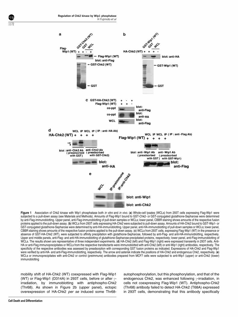
To better understand the roles and regulation of Chk2 duringthe DNA damage responses, we performed yeast two-hybridscreening using human Chk2 as a bait to identify potentialassociating molecule(s). From this screen, we identified Wip1,a p53-inducible oncogenic nuclear protein serine (S)/threo-nine (T) phosphatase24–28 (data not shown). To test whetherChk2 can associate with Wip1 in vitro, we performed pull-down analyses using glutathione S-transferase (GST)-Chk2(wild-type (WT)) or GST-Wip1 (WT) purified from Escherichiacoli. As shown in Figure 1a, Flag-tagged Wip1 (WT)expressed in 293T cells associated specifically with GST-Chk2 (WT), but not with GST. Conversely, haemagglutinin(HA)-tagged Chk2 (WT) expressed in 293T cells associatedspecifically with GST-Wip1 (WT), but not with GST(Figure 1b). These results indicate that Chk2 associates withWip1 in vitro. Since it has been reported previously that bothChk2 and Wip1 are localized in the nuclei,7,17,25,31 theirintracellular distribution was examined by immunofluores-cence in 293T cells coexpressing HA-tagged GST-Chk2(GST-HA-Chk2) and Flag-tagged Wip1 (Flag-Wip1) (seeMaterials and Methods). GST-HA-Chk2 and Flag-Wip1colocalized primarily in the nuclei (see SupplementaryInformation). To determine whether Chk2 can associate withWip1 in vivo, Flag-Wip1 (WT) was transiently expressed in293T cells, either alone or with GST-HA-Chk2 (WT). Cellswere harvested, and protein association was evaluated byco-precipitation with glutathione-Sepharose, followed byanti-Flag-immunoblotting. As shown in Figure 1c, in cellsexpressing both GST-HA-Chk2 and Flag-Wip1, glutathione-
Sepharose precipitation specifically co-precipitated Wip1,indicating that Chk2 associates with Wip1 in 293T cells.
We next examined whether endogenous Chk2 and Wip1can interact in intact cells. To this end, we generatedpolyclonal anti-Chk2 and anti-Wip1 antibodies raised againsthuman Chk2 peptides (amino acids (a.a.) 523–543) and GST-human Wip1 (a.a. 1–458), respectively (see Materials andMethods), and evaluated specificities for their antigenrecognition. As shown in Figure 1d (left panel), immuno-blotting with anti-Chk2 antibody clearly detected HA-Chk2(upper band) and endogenous Chk2 (lower band) in whole-cell lysates and HA-Chk2 in anti-HA immunoprecipitates from293T cells expressing HA-Chk2, and preadsorption of anti-Chk2 antibody with GST-Chk2 prior to immunoblottingresulted in a failure of the detection of HA-Chk2, confirmingthe specificity of anti-Chk2 antibody. Similarly, immuno-blotting with anti-Wip1 antibody specifically detected Flag-Wip1 (Figure 1d, right panel). Specificities of anti-Wip1 andanti-Chk2 antibodies were further confirmed by an RNAiexperiment where the bands detected by anti-Wip1 and anti-Chk2 antibodies were diminished in cells treated with Wip1siRNAs and Chk2 siRNAs, respectively (Figure 4, data notshown). We examined the association of endogenous Chk2and Wip1 in MCF7 cells that express Wip1 at a higher level. Asshown in Figure 1e, endogenous Chk2 and Wip1 associatedin MCF7 cells.
Effect of Wip1 on Thr68 phosphorylation of Chk2induced by DNA damage or overexpression ofChk2
It was shown that ectopic overexpression of Chk2 in 293Tcells results in its electrophoretic mobility shift (Figure 2a),13,32
indicating auto- or transphosphorylation. In fact, electrophore-tic mobility shift induced by overexpression of Chk2 wasabrogated following treatment of cell extracts with CIP or BAP(data not shown). To examine the functional significance ofthe association between Wip1 and Chk2, HA-Chk2 (WT) orHA-Chk2 (DK) was expressed in 293T cells along with eitherFlag-Wip1 (WT) or Flag-Wip1 (D314A). Chk2 electrophoreticmobilities were monitored by anti-HA-immunoblotting ofwhole-cell lysates. Overexpression of Chk2 (WT), but notChk2 (DK), resulted in decreased electrophoretic mobility(Figure 2a), indicating that Chk2 kinase activity is required forthis electrophoretic mobility shift. It had been shown pre-viously that phosphorylation of Chk2 on Thr68 is required forfull activation of Chk2 via auto- (trans- or cis)phosphoryla-tion.9–13,31–34 We therefore investigated the role of Thr68-phosphorylation in the electrophoretic mobility shift of Chk2.We found that a Chk2 mutant, the alanine68 mutant (HA-Chk2(T68A)), did not undergo the mobility shift when over-expressed (data not shown), indicating that Thr68 is required.Intriguingly, this mobility shift was abrogated by coexpressionof Wip1 (WT), but not Wip1 (D314A) (Figure 2a), suggestingthat Wip1 phosphatase activity can counteract Chk2 phos-phorylation either directly or indirectly.
It has been shown previously that g-irradiation inducesThr68-phosphorylation and modification (activation) ofChk2.9–13 Hence, we examined Thr68-phosphorylation and
Regulation of Chk2 kinase by Wip1 phosphataseH Fujimoto et al
1171
Cell Death and Differentiation
mobility shift of HA-Chk2 (WT) coexpressed with Flag-Wip1(WT) or Flag-Wip1 (D314A) in 293T cells, before or after g-irradiation, by immunoblotting with antiphospho-Chk2(Thr68). As shown in Figure 2b (upper panel), ectopicoverexpression of HA-Chk2 per se induced some Thr68-
autophosphorylation, but this phosphorylation, and that of theendogenous Chk2, was enhanced following g-irradiation, incells not coexpressing Flag-Wip1 (WT). Antiphospho-Chk2(Thr68) antibody failed to detect HA-Chk2 (T68A) expressedin 293T cells, demonstrating that this antibody specifically
Figure 1 Association of Chk2 kinase with Wip1 phosphatase both in vitro and in vivo. (a) Whole-cell lysates (WCLs) from 293T cells expressing Flag-Wip1 weresubjected to a pull-down assay (see Materials and Methods). Amounts of Flag-Wip1 bound to GST-Chk2- or GST-conjugated glutathione-Sepharose were determinedby anti-Flag-immunoblotting. Upper panel, anti-Flag-immunoblotting of pull-down samples or WCLs; lower panel, CBBR staining shows amounts of the respective fusionproteins applied to the pull-down assay. (b) WCLs from 293T cells expressing HA-Chk2 were subjected to pull-down assay. Amounts of HA-Chk2 bound to GST-Wip1- orGST-conjugated glutathione-Sepharose were determined by anti-HA-immunoblotting. Upper panel, anti-HA-immunoblotting of pull-down samples or WCLs; lower panel,CBBR staining shows amounts of the respective fusion proteins applied to the pull-down assay. (c) WCLs from 293T cells, expressing Flag-Wip1 (WT) in the presence orabsence of GST-HA-Chk2 (WT), were subjected to affinity precipitation with glutathione-Sepharose, followed by anti-Flag- and anti-HA-immunoblotting, respectively.Upper and middle panels, anti-Flag- and anti-HA-immunoblotting of glutathione-Sepharose-precipitated proteins, respectively; lower panel, anti-Flag-immunoblotting ofWCLs. The results shown are representative of three independent experiments. (d) HA-Chk2 (left) and Flag-Wip1 (right) were expressed transiently in 293T cells. Anti-HA or anti-Flag immunoprecipitates or WCLs from the respective transfectants were immunoblotted with anti-Chk2 (left) or anti-Wip1 (right) antibodies, respectively. Thespecificity of the respective antibodies was assessed by preadsorption with corresponding GST fusion proteins as indicated. Expressions of HA-Chk2 and Flag-Wip1were verified by anti-HA- and anti-Flag-immunoblotting, respectively. The arrow and asterisk indicate the positions of HA-Chk2 and endogenous Chk2, respectively. (e)WCLs or immunoprecipitates with anti-Chk2 or control (preimmune) antibodies prepared from MCF7 cells were subjected to anti-Wip1 (upper) or anti-Chk2 (lower)immunoblotting
Regulation of Chk2 kinase by Wip1 phosphataseH Fujimoto et al
1172
Cell Death and Differentiation

recognizes phosphorylated-Thr68 in Chk2 (data not shown).However, Thr68-phosphorylation and electrophoretic mobilityshift of HA-Chk2 (WT) were inhibited in cells coexpressingWip1 (WT), but not Wip1 (D314A) (Figure 2b), indicating thatthe phosphatase activity of Wip1 is required for the inhibitionof Chk2 Thr68-phosphorylation and mobility shift induced byoverexpression of Chk2 and/or g-irradiation, although it wasunclear whether Wip1 acted directly on Chk2. Since Wip1belongs to the PP2C family of protein phosphatases, weexamined whether or not other members of the PP2C familycan exhibit similar effects. Coexpression of human PP2Ca2,mouse PP2Cb, PP2Ce, or PP2Cz fails to inhibit Thr68-
phosphorylation and electrophoretic mobility shift of HA-Chk2 (WT) (Figure 2b, data not shown), indicating that theobserved effects are at least selective to Wip1 amongmembers of the PP2C family.
Effect of Wip1 on other phosphorylation sites inChk2 induced by DNA damage or overexpressionof Chk2
We next examined the effect of Wip1 (WT) on phosphorylationof several S and T residues other than Thr68 of Chk2, inducedupon ectopic overexpression of Chk2 and/or g-irradiation, byimmunoblotting with a series of phospho-Chk2 antibodies. Asshown in Figure 2c, ectopic overexpression of HA-Chk2 perse induced weak phosphorylation of Ser19, Ser33/35, andThr432 and strong (or apparent) phosphorylation of Thr387and Ser516 of Chk2, and these phosphorylations were furtherenhanced following g-irradiation. Phosphorylation of Ser19,Ser33/35, Thr387, and Thr432 of HA-Chk2 induced by itsoverexpression and/or g-irradiation was also inhibited in cellscoexpressing Wip1 (WT), but not Wip1 (D314A) (Figure 2c).The result presented in Figure 2b suggests that depho-sphorylation of phosphorylated-Thr68 in Chk2 by Wip1 mayinhibit subsequent phosphorylation of Chk2 at several S/Tresidues, or that Wip1 may dephosphorylate these phos-phorylated residues in Chk2 (see Figure 3a). We alsoexamined the effect of Wip1 (WT) on endogenous Chk2,and found that expression of Wip1 (WT), but not Wip1(D314A), in 293T cells also inhibited Thr68-phosphorylationand mobility shift of the endogenous Chk2 (Figure 2d).
Dephosphorylation of Ser19, Ser33/35, Thr68, andThr432, but not Thr387 and Ser516 in Chk2 by Wip1in vitro
We next addressed the question of whether or not Wip1 coulddephosphorylate Thr68 or Thr387 in Chk2. To this end, GST-Wip1 (WT) and GST-Wip1 (D314A) were expressed in E. coli
Figure 2 Inhibition of Chk2 Thr68-phosphorylation and electrophoretic mobilityshift by Wip1 (WT), but not Wip1 (D314A), before and after g-irradiation. (a) Wild-type (WT) or dead-kinase mutant (DK) of HA-Chk2 proteins were expressedtransiently in 293T cells along with either WT or a phosphatase-deficient mutant(D314A) of Flag-Wip1 proteins. WCLs from the respective transfectants weresubjected to anti-HA- (upper) or anti-Flag- (lower) immunoblotting, respectively.(b) HA-Chk2 (WT) proteins were expressed transiently in 293T cells along witheither Flag-Wip1 (WT), Flag-Wip1 (D314A), or Flag-PP2Ca2. Before or afterg-irradiation (10 Gy), WCLs were subjected to antiphospho-Chk2 (Thr68)-immunoblotting. The arrow and asterisk indicate the positions of HA-Chk2 andof endogenous Chk2, respectively. Upper, middle, and lower panels, anti-phospho-Chk2 (Thr68)-, anti-HA-, and anti-Flag-immunoblotting of WCLs,respectively. Flag-PP2Ca2 expressed in 293T cells is catalytically active, sinceit can dephosphorylate phosphorylated-p38 (data not shown). (c) HA-Chk2 (WT)proteins were expressed transiently in 293T cells along with either Flag-Wip1(WT) or Flag-Wip1 (D314A) proteins. Before or after g-irradiation (10 Gy), WCLswere subjected to antiphospho-Chk2 (Ser19, Ser33/35, Thr68, Thr387, Thr432,or Ser516)- or anti-HA-immunoblotting as indicated. WCLs were also subjectedto anti-Flag-immunoblotting. (d) Flag-Wip1 (WT) or Flag-Wip1 (D314A) wasexpressed transiently in 293T cells. Before or after g-irradiation (10 Gy), WCLsfrom the respective transfectants were subjected to antiphospho-Chk2 (Thr68)-immunoblotting. Upper, middle, and lower panels, antiphospho-Chk2 (Thr68)-,anti-Chk2-, and anti-Flag-immunoblotting of WCLs, respectively
Regulation of Chk2 kinase by Wip1 phosphataseH Fujimoto et al
1173
Cell Death and Differentiation
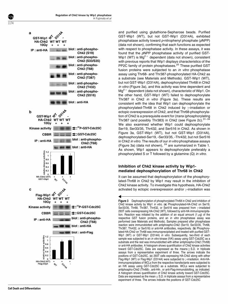
and purified using glutathione-Sepharose beads. PurifiedGST-Wip1 (WT), but not GST-Wip1 (D314A), exhibitedphosphatase activity toward p-nitrophenyl phosphate (pNPP)(data not shown), confirming that each functions as expectedwith respect to phosphatase activity. In these assays, it wasfound that the pNPP phosphatase activity of purified GST-Wip1 (WT) is Mg2þ dependent (data not shown), consistentwith previous reports that Wip1 displays characteristics of thePP2C family of protein phosphatases.25 These purified GSTfusion proteins were subjected to an in vitro phosphataseassay using Thr68- and Thr387-phosphorylated HA-Chk2 asa substrate (see Materials and Methods). GST-Wip1 (WT),but not GST-Wip1 (D314A), dephosphorylated Thr68 in Chk2in vitro (Figure 3a), and this activity was time dependent andMg2þ dependent (data not shown), characteristic of Wip1. Onthe other hand, GST-Wip1 (WT) failed to dephosphorylateThr387 in Chk2 in vitro (Figure 3a). These results areconsistent with the idea that Wip1 can dephosphorylate thephosphorylated-Thr68 in Chk2 induced by g-irradiation orectopic overexpression of Chk2, and that Thr68-phosphoryla-tion of Chk2 is a prerequisite event for (trans-)phosphorylatingThr387 (and possibly Thr383) in Chk2 (see Figure 2c).31–34
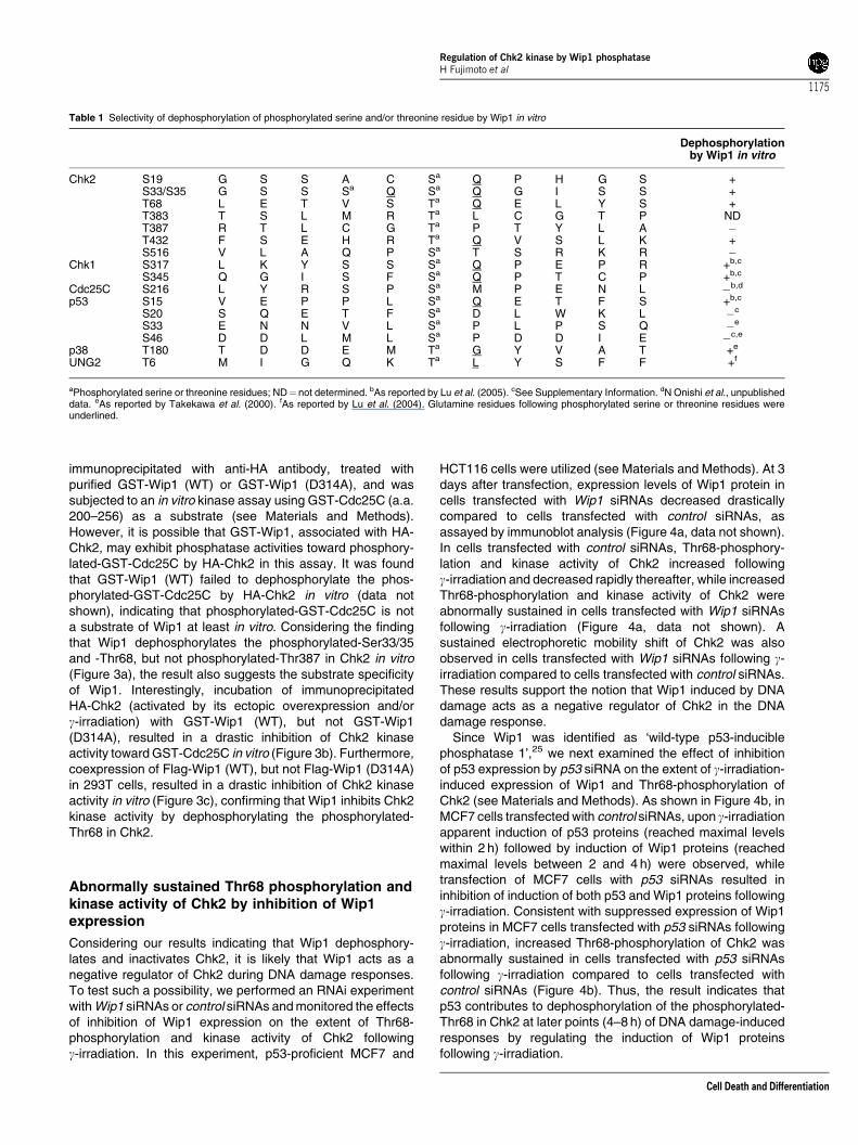
We also examined whether Wip1 could dephosphorylateSer19, Ser33/35, Thr432, and Ser516 in Chk2. As shown inFigure 3a, GST-Wip1 (WT), but not GST-Wip1 (D314A),dephosphorylated-Ser19, -Ser33/35, -Thr432, but not Ser516in Chk2 in vitro. The results of our in vitro phosphatase assays(Figure 3a) (data not shown), 24 are summarized in Table 1.As shown, Wip1 appears to dephosphorylate preferably aphosphorylated S or T followed by a glutamine (Q) in vitro.
Inhibition of Chk2 kinase activity by Wip1-mediated dephosphorylation of Thr68 in Chk2
It can be assumed that dephosphorylation of the phosphory-lated-Thr68 in Chk2 by Wip1 may result in the inhibition ofChk2 kinase activity. To investigate this hypothesis, HA-Chk2activated by ectopic overexpression and/or g-irradiation was
Figure 3 Dephosphorylation of phosphorylated-Thr68 in Chk2 and inhibition ofChk2 kinase activity by Wip1 in vitro. (a) Phosphorylated-HA-Chk2 on Ser19,Ser33/35, Thr68, Thr387, Thr432, or Ser516 was prepared from g-irradiated293T cells overexpressing HA-Chk2 (WT), followed by anti-HA immunoprecipita-tion. Reaction was initiated by the addition of an equal amount (1 mg) of therespective GST fusion proteins, and an in vitro phosphatase assay wasperformed (see Materials and Methods). Samples prepared after phosphatasereaction were immunoblotted with antiphospho-Chk2 (Ser19, Ser33/35, Thr68,Thr387, Thr432, or Ser516) or anti-HA antibodies, respectively. (b) Phosphory-lated-HA-Chk2 on Thr68 was immunoprecipitated and treated with purified GST-Wip1 (WT) or GST-Wip1 (D314A) in vitro. Subsequently, two-third of eachsample was subjected to an in vitro kinase (IVK) assay using GST-Cdc25C as asubstrate and the rest was immunoblotted with either antiphospho-Chk2 (Thr68)or anti-HA antibodies. A histogram shows quantification of Chk2 kinase activitiestoward GST-Cdc25C. Data are expressed as the means7S.D. in triplicateassays from a representative experiment of three. The arrows indicate thepositions of GST-Cdc25C. (c) 293T cells expressing HA-Chk2 along with eitherFlag-Wip1 (WT) or Flag-Wip1 (D314A) were subjected to g-irradiation. Anti-HA-immunoprecipitates of WCLs from the respective transfectants were subjected toan IVK assay using GST-Cdc25C as a substrate. WCLs were subjected toantiphospho-Chk2 (Thr68)-, anti-HA-, or anti-Flag-immunoblotting, as indicated.A histogram shows quantification of Chk2 kinase activity toward GST-Cdc25C.Data are expressed as the mean7S.D. in triplicate assays from a representativeexperiment of three. The arrows indicate the positions of GST-Cdc25C
Regulation of Chk2 kinase by Wip1 phosphataseH Fujimoto et al
1174
Cell Death and Differentiation
immunoprecipitated with anti-HA antibody, treated withpurified GST-Wip1 (WT) or GST-Wip1 (D314A), and wassubjected to an in vitro kinase assay using GST-Cdc25C (a.a.200–256) as a substrate (see Materials and Methods).However, it is possible that GST-Wip1, associated with HA-Chk2, may exhibit phosphatase activities toward phosphory-lated-GST-Cdc25C by HA-Chk2 in this assay. It was foundthat GST-Wip1 (WT) failed to dephosphorylate the phos-phorylated-GST-Cdc25C by HA-Chk2 in vitro (data notshown), indicating that phosphorylated-GST-Cdc25C is nota substrate of Wip1 at least in vitro. Considering the findingthat Wip1 dephosphorylates the phosphorylated-Ser33/35and -Thr68, but not phosphorylated-Thr387 in Chk2 in vitro(Figure 3a), the result also suggests the substrate specificityof Wip1. Interestingly, incubation of immunoprecipitatedHA-Chk2 (activated by its ectopic overexpression and/org-irradiation) with GST-Wip1 (WT), but not GST-Wip1(D314A), resulted in a drastic inhibition of Chk2 kinaseactivity toward GST-Cdc25C in vitro (Figure 3b). Furthermore,coexpression of Flag-Wip1 (WT), but not Flag-Wip1 (D314A)in 293T cells, resulted in a drastic inhibition of Chk2 kinaseactivity in vitro (Figure 3c), confirming that Wip1 inhibits Chk2kinase activity by dephosphorylating the phosphorylated-Thr68 in Chk2.
Abnormally sustained Thr68 phosphorylation andkinase activity of Chk2 by inhibition of Wip1expression
Considering our results indicating that Wip1 dephosphory-lates and inactivates Chk2, it is likely that Wip1 acts as anegative regulator of Chk2 during DNA damage responses.To test such a possibility, we performed an RNAi experimentwith Wip1 siRNAs or control siRNAs and monitored the effectsof inhibition of Wip1 expression on the extent of Thr68-phosphorylation and kinase activity of Chk2 followingg-irradiation. In this experiment, p53-proficient MCF7 and
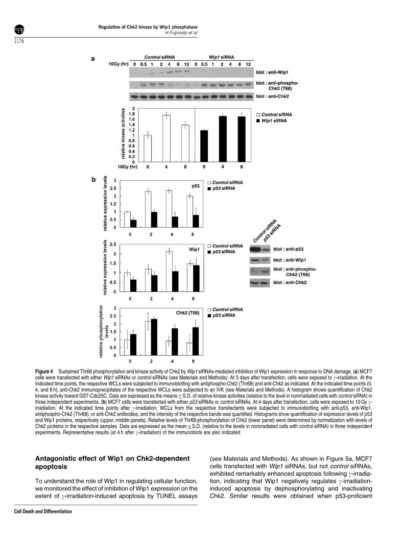
HCT116 cells were utilized (see Materials and Methods). At 3days after transfection, expression levels of Wip1 protein incells transfected with Wip1 siRNAs decreased drasticallycompared to cells transfected with control siRNAs, asassayed by immunoblot analysis (Figure 4a, data not shown).In cells transfected with control siRNAs, Thr68-phosphory-lation and kinase activity of Chk2 increased followingg-irradiation and decreased rapidly thereafter, while increasedThr68-phosphorylation and kinase activity of Chk2 wereabnormally sustained in cells transfected with Wip1 siRNAsfollowing g-irradiation (Figure 4a, data not shown). Asustained electrophoretic mobility shift of Chk2 was alsoobserved in cells transfected with Wip1 siRNAs following g-irradiation compared to cells transfected with control siRNAs.These results support the notion that Wip1 induced by DNAdamage acts as a negative regulator of Chk2 in the DNAdamage response.
Since Wip1 was identified as ‘wild-type p53-induciblephosphatase 1’,25 we next examined the effect of inhibitionof p53 expression by p53 siRNA on the extent of g-irradiation-induced expression of Wip1 and Thr68-phosphorylation ofChk2 (see Materials and Methods). As shown in Figure 4b, inMCF7 cells transfected with control siRNAs, upon g-irradiationapparent induction of p53 proteins (reached maximal levelswithin 2 h) followed by induction of Wip1 proteins (reachedmaximal levels between 2 and 4 h) were observed, whiletransfection of MCF7 cells with p53 siRNAs resulted ininhibition of induction of both p53 and Wip1 proteins followingg-irradiation. Consistent with suppressed expression of Wip1proteins in MCF7 cells transfected with p53 siRNAs followingg-irradiation, increased Thr68-phosphorylation of Chk2 wasabnormally sustained in cells transfected with p53 siRNAsfollowing g-irradiation compared to cells transfected withcontrol siRNAs (Figure 4b). Thus, the result indicates thatp53 contributes to dephosphorylation of the phosphorylated-Thr68 in Chk2 at later points (4–8 h) of DNA damage-inducedresponses by regulating the induction of Wip1 proteinsfollowing g-irradiation.
Table 1 Selectivity of dephosphorylation of phosphorylated serine and/or threonine residue by Wip1 in vitro
Dephosphorylationby Wip1 in vitro
Chk2 S19 G S S A C Sa Q P H G S +S33/S35 G S S Sa Q Sa Q G I S S +T68 L E T V S Ta Q E L Y S +T383 T S L M R Ta L C G T P NDT387 R T L C G Ta P T Y L A �T432 F S E H R Ta Q V S L K +S516 V L A Q P Sa T S R K R �
Chk1 S317 L K Y S S Sa Q P E P R +b,c
S345 Q G I S F Sa Q P T C P +b,c
Cdc25C S216 L Y R S P Sa M P E N L �b,d
p53 S15 V E P P L Sa Q E T F S +b,c
S20 S Q E T F Sa D L W K L �c
S33 E N N V L Sa P L P S Q �e
S46 D D L M L Sa P D D I E �c,e
p38 T180 T D D E M Ta G Y V A T +e
UNG2 T6 M I G Q K Ta L Y S F F +f
aPhosphorylated serine or threonine residues; ND¼ not determined. bAs reported by Lu et al. (2005). cSee Supplementary Information. dN Onishi et al., unpublisheddata. eAs reported by Takekawa et al. (2000). fAs reported by Lu et al. (2004). Glutamine residues following phosphorylated serine or threonine residues wereunderlined.
Regulation of Chk2 kinase by Wip1 phosphataseH Fujimoto et al
1175
Cell Death and Differentiation
Antagonistic effect of Wip1 on Chk2-dependentapoptosis
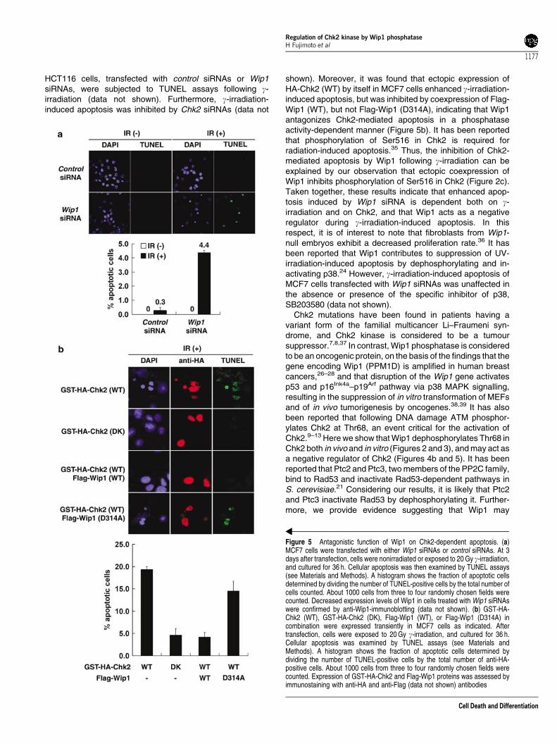
To understand the role of Wip1 in regulating cellular function,we monitored the effect of inhibition of Wip1 expression on theextent of g-irradiation-induced apoptosis by TUNEL assays
(see Materials and Methods). As shown in Figure 5a, MCF7cells transfected with Wip1 siRNAs, but not control siRNAs,exhibited remarkably enhanced apoptosis following g-irradia-tion, indicating that Wip1 negatively regulates g-irradiation-induced apoptosis by dephosphorylating and inactivatingChk2. Similar results were obtained when p53-proficient
Figure 4 Sustained Thr68 phosphorylation and kinase activity of Chk2 by Wip1 siRNAs-mediated inhibition of Wip1 expression in response to DNA damage. (a) MCF7cells were transfected with either Wip1 siRNAs or control siRNAs (see Materials and Methods). At 3 days after transfection, cells were exposed to g-irradiation. At theindicated time points, the respective WCLs were subjected to immunoblotting with antiphospho-Chk2 (Thr68) and anti-Chk2 as indicated. At the indicated time points (0,4, and 8 h), anti-Chk2 immunoprecipitates of the respective WCLs were subjected to an IVK (see Materials and Methods). A histogram shows quantification of Chk2kinase activity toward GST-Cdc25C. Data are expressed as the means7S.D. of relative kinase activities (relative to the level in nonirradiated cells with control siRNA) inthree independent experiments. (b) MCF7 cells were transfected with either p53 siRNAs or control siRNAs. At 4 days after transfection, cells were exposed to 10 Gy g-irradiation. At the indicated time points after g-irradiation, WCLs from the respective transfectants were subjected to immunoblotting with anti-p53, anti-Wip1,antiphospho-Chk2 (Thr68), or anti-Chk2 antibodies, and the intensity of the respective bands was quantified. Histograms show quantification of expression levels of p53and Wip1 proteins, respectively (upper, middle panels). Relative levels of Thr68-phosphorylation of Chk2 (lower panel) were determined by normalization with levels ofChk2 proteins in the respective samples. Data are expressed as the mean7S.D. (relative to the levels in nonirradiated cells with control siRNA) in three independentexperiments. Representative results (at 4 h after g-irradiation) of the immunoblots are also indicated
Regulation of Chk2 kinase by Wip1 phosphataseH Fujimoto et al
1176
Cell Death and Differentiation
HCT116 cells, transfected with control siRNAs or Wip1siRNAs, were subjected to TUNEL assays following g-irradiation (data not shown). Furthermore, g-irradiation-induced apoptosis was inhibited by Chk2 siRNAs (data not
shown). Moreover, it was found that ectopic expression ofHA-Chk2 (WT) by itself in MCF7 cells enhanced g-irradiation-induced apoptosis, but was inhibited by coexpression of Flag-Wip1 (WT), but not Flag-Wip1 (D314A), indicating that Wip1antagonizes Chk2-mediated apoptosis in a phosphataseactivity-dependent manner (Figure 5b). It has been reportedthat phosphorylation of Ser516 in Chk2 is required forradiation-induced apoptosis.35 Thus, the inhibition of Chk2-mediated apoptosis by Wip1 following g-irradiation can beexplained by our observation that ectopic coexpression ofWip1 inhibits phosphorylation of Ser516 in Chk2 (Figure 2c).Taken together, these results indicate that enhanced apop-tosis induced by Wip1 siRNA is dependent both on g-irradiation and on Chk2, and that Wip1 acts as a negativeregulator during g-irradiation-induced apoptosis. In thisrespect, it is of interest to note that fibroblasts from Wip1-null embryos exhibit a decreased proliferation rate.36 It hasbeen reported that Wip1 contributes to suppression of UV-irradiation-induced apoptosis by dephosphorylating and in-activating p38.24 However, g-irradiation-induced apoptosis ofMCF7 cells transfected with Wip1 siRNAs was unaffected inthe absence or presence of the specific inhibitor of p38,SB203580 (data not shown).
Chk2 mutations have been found in patients having avariant form of the familial multicancer Li–Fraumeni syn-drome, and Chk2 kinase is considered to be a tumoursuppressor.7,8,37 In contrast, Wip1 phosphatase is consideredto be an oncogenic protein, on the basis of the findings that thegene encoding Wip1 (PPM1D) is amplified in human breastcancers,26–28 and that disruption of the Wip1 gene activatesp53 and p16Ink4a–p19Arf pathway via p38 MAPK signalling,resulting in the suppression of in vitro transformation of MEFsand of in vivo tumorigenesis by oncogenes.38,39 It has alsobeen reported that following DNA damage ATM phosphor-ylates Chk2 at Thr68, an event critical for the activation ofChk2.9–13 Here we show that Wip1 dephosphorylates Thr68 inChk2 both in vivo and in vitro (Figures 2 and 3), and may act asa negative regulator of Chk2 (Figures 4b and 5). It has beenreported that Ptc2 and Ptc3, two members of the PP2C family,bind to Rad53 and inactivate Rad53-dependent pathways inS. cerevisiae.21 Considering our results, it is likely that Ptc2and Ptc3 inactivate Rad53 by dephosphorylating it. Further-more, we provide evidence suggesting that Wip1 may
Figure 5 Antagonistic function of Wip1 on Chk2-dependent apoptosis. (a)MCF7 cells were transfected with either Wip1 siRNAs or control siRNAs. At 3days after transfection, cells were nonirradiated or exposed to 20 Gy g-irradiation,and cultured for 36 h. Cellular apoptosis was then examined by TUNEL assays(see Materials and Methods). A histogram shows the fraction of apoptotic cellsdetermined by dividing the number of TUNEL-positive cells by the total number ofcells counted. About 1000 cells from three to four randomly chosen fields werecounted. Decreased expression levels of Wip1 in cells treated with Wip1 siRNAswere confirmed by anti-Wip1-immunoblotting (data not shown). (b) GST-HA-Chk2 (WT), GST-HA-Chk2 (DK), Flag-Wip1 (WT), or Flag-Wip1 (D314A) incombination were expressed transiently in MCF7 cells as indicated. Aftertransfection, cells were exposed to 20 Gy g-irradiation, and cultured for 36 h.Cellular apoptosis was examined by TUNEL assays (see Materials andMethods). A histogram shows the fraction of apoptotic cells determined bydividing the number of TUNEL-positive cells by the total number of anti-HA-positive cells. About 1000 cells from three to four randomly chosen fields werecounted. Expression of GST-HA-Chk2 and Flag-Wip1 proteins was assessed byimmunostaining with anti-HA and anti-Flag (data not shown) antibodies
Regulation of Chk2 kinase by Wip1 phosphataseH Fujimoto et al
1177
Cell Death and Differentiation
negatively regulate g-irradiation-induced apoptosis by depho-sphorylating and inactivating Chk2 (Figures 4 and 5).However, at present we cannot rule out the possibility thatWip1 may dephosphorylate and inactivate ATM or otherproteins, thereby inhibiting Chk2 function. Interestingly, it hasbeen recently reported that Wip1 dephosphorylates thenuclear form of uracil DNA glycosylase (UNG2) and Chk1,thereby suppressing BER and intra-S and G2/M checkpointregulation, respectively.30,40 In the case of UNG2 and p38,phosphorylation sites on these molecules, which can betargets for Wip1-mediated dephosphorylation, reveal con-sensus sequences ‘phospho-T-X-phospho-Tyr (Y) (or phos-pho-T-X-Y, X: optional a.a.)’.41 In this respect, it is important tonote that our in vitro evidence as well as previous report40
indicate the presence of additional target sequences for Wip1,which are ‘phospho-S-Q’ or ‘phospho-T-Q’, well-knownfingerprints of ATM kinase (see Table 1). Future study toidentify a substrate(s) for Wip1 in addition to phosphorylated-Chk2, -Chk1, -p38,24 and -UNG230 may contribute to ourunderstanding of the roles of Wip1 during DNA damage-induced cellular responses.
Materials and Methods
Plasmid constructions
The human Chk2 coding region was amplified by PCR using humanplacenta cDNA as a template, and subcloned into the mammalianexpression vector pcDNA3 (Invitrogen). The mutant Chk2 (T68A) wasgenerated by replacing Thr68 with Ala using the Transformer TM Site-Directed mutagenesis kit (Clontech). The kinase-dead mutant of Chk2(DK) replaces Lys249 with Arg and was created by PCR. A PCR-basedprocedure was employed to attach an HA tag at the C-terminal end ofChk2; to generate the expression vectors, pcDNA-Chk2 (WT)-HA, pcDNA-Chk2 (T68A)-HA, pcDNA-Chk2 (DK)-HA, and pEBG-Chk2 (encodingGST-HA-Chk2) were constructed to attach GST and HA tags at the N- andC-terminal ends of Chk2, respectively. pGEX-3X-Chk2, encoding GST-Chk2 fusion proteins, was constructed to attach GST tag at the N-terminalend of Chk2; pGEX-3X-Cdc25C, encoding GST-Cdc25C (a.a. 200–256 ofhuman Cdc25C), was constructed by subcloning of the PCR amplifiedCdc25C cDNA fragment into the pGEX-3X to generate GST-Cdc25C.pCMV-Wip1 (WT), and pCMV-Wip1 (D314A), encoding the wild-type andphosphatase inactive mutant of Wip1, respectively, were constructed asdescribed previously.24 A PCR-based procedure was employed to attacha FLAG-tag sequence at the C-terminal end of Wip1 to generate pcDNA-Wip1 (WT)-FLAG and pcDNA-Wip1 (D314A)-FLAG. pGEX-5X-Wip1 (WT)and pGEX-5X-Wip1 (D314A), encoding GST-Wip1 (WT) and GST-Wip1(D314A), respectively, were constructed as described previously.24
pcDNA-PP2Ca2 (WT)-FLAG, encoding the wild-type Flag-PP2Ca2, wasconstructed as described previously.23 pcDNA-Chk1-myc, encoding thewild-type Chk1 of mouse origin, was a gift from Noboru Motoyama(National Institute for Longevity Sciences, Japan).
Antibodies, cells, and DNA transfection
The following antibodies were used: mouse monoclonal antibody (MoAb)M2 (Eastman Kodak) recognizes the Flag peptide sequence(DYKDDDDK). Mouse MoAb 12CA5 (Boehringer Mannheim) and ratMoAb 3F10 (Roche) recognize the peptide sequence (YPYDVPDYA)derived from the human influenza HA protein. Mouse MoAb 9E10 (Santa
Cruz) recognize the peptide sequence (EQKLISEEDL) derived fromhuman c-myc protein. Rabbit polyclonal anti-Chk2 antibody was raisedagainst peptides corresponding to a.a. 523–543 of human Chk2. Rabbitpolyclonal anti-Wip1 antibody was raised against GST-Wip1 (a.a. 1–458).Mouse monoclonal anti-Wip1 (WC 10) antibody was from Trevigen. Mousemonoclonal anti-p53 (DO-1) antibody was from Santa Cruz. Rabbitpolyclonal anti-phospho-Chk1 (Ser317 and Ser345) antibodies, anti-phospho-Chk2 (Ser19, Ser33/35, Thr68, Thr387, Thr432, and Ser516)antibodies, and anti-phospho-p53 (Ser15, Ser20, and Ser46) antibodieswere purchased from Cell Signaling. Alexa Fluor 546 (goat anti-mouseIgG, red) and Alexa Fluor 488 (goat anti-rat IgG, green) were purchasedfrom Molecular Probes. SB203580, a specific inhibitor of p38 MAP kinase(p38a, p38b, and p38b2), was from Promega. HEK293T (293T), MCF7cells were maintained in Dulbecco’s modified Eagle’s medium (Nissui)supplemented with 10% (v/v) foetal calf serum (FCS). HCT116 cells weremaintained in McCoy’s 5a medium (GibcoBRL) supplemented with 10%(v/v) FCS. It should be noted that 293T cells bear p53 inactivated byadenovirus E1B and SV40 T-antigen, while MCF7 and HCT116 cells arep53-proficient breast adenocarcinoma and colorectal carcinoma cells,respectively. Transient cDNA transfection into cells was performed usingthe calcium phosphate method3 or Lipofectamine Reagent, PLUSReagent, and Lipofectamine 2000 Reagent (GibcoBRL) according tothe manufacturer’s instructions.
Preparation of cell lysates and co-(immuno)precipitation analysis
Transfected cells were solubilized with lysis buffer (50 mM Tris-HCl(pH 7.4), 0.5% (v/v) NP-40, 150 mM NaCl, 5 mM EDTA, 50 mM NaF, 1 mMNa3VO4, 1 mM phenylmethyl sulphonyl fluoride (PMSF), 10 mg/mlaprotinin), and cell lysates were prepared by centrifugation at12 000� g for 15 min at 41C. Subsequently, the cell lysates weresubjected to pull-down assay/immunoblot analysis or co-(immuno)precipitation/immunoblot analysis. For co-(immuno) precipitation analysis,the cell lysates were precleared with protein A–Sepharose (AmershamPharmacia Biotech) for 1 h at 41C. The precleared supernatants were thenco-(immuno) precipitated with glutathione-Sepharose beads (AmershamPharmacia Biotech) or with anti-HA antibody conjugated to protein A–Sepharose beads for 2 h at 41C. The resultant precipitates were washedfive times with lysis buffer, and eluted with Laemmli sample buffer.
Expression and purification of GST fusion proteins
The plasmids encoding the GST fusion proteins, GST-Chk2 (WT), GST-Wip1 (WT), GST-Wip1 (D314A), and GST-Cdc25C (a.a. 200–256), wereconstructed using the pGEX plasmids (Amersham Pharmacia Biotech).GST fusion proteins expressed in E. coli DH5a were extracted withphosphate-buffered saline (PBS) containing 1% (v/v) Triton X-100, 1 mMEDTA, 1 mM PMSF, 10 mg/ml leupeptin, and 10 mg/ml aprotinin, and wereisolated with glutathione-Sepharose beads. To further purify GST-Wip1(WT), GST-Wip1 (D314A), and GST-Cdc25C for phosphatase and kinaseassays, fusion proteins were eluted from beads by incubating with 25 mMglutathione (reduced), followed by dialysis prior to use.
Pull-down assay and immunoblot analysis
The cell lysates were precleared with glutathione-Sepharose for 1 h at41C. The precleared supernatant was then incubated with the respectiveGST fusion proteins conjugated with glutathione-Sepharose beads for 1 h
Regulation of Chk2 kinase by Wip1 phosphataseH Fujimoto et al
1178
Cell Death and Differentiation
at 41C, washed five times with lysis buffer, and eluted with Laemmlisample buffer. Precipitates either from the pull-down assay or whole-celllysates were separated by SDS-PAGE (10% PAG), and transferred ontoPVDF membrane filters (Immobilon P, Millipore). The membranes wereimmunoblotted with the respective antibodies, and bound antibodies werevisualized with HRP-conjugated antibodies against mouse or rabbit IgGs(Bio-Rad) using the chemiluminescence reagent (Renaissance, NEN).Quantification of the immunoblots was performed by densitometricscanning of the film using an Image Analysis system with NIH Image 1.62software.
In vitro phosphatase assay and kinase assays
To prepare phosphorylated-HA-Chk2 (WT), 293T cells were transfectedwith the expression vector encoding HA-Chk2 (WT). For in vitro kinaseassay, 24 h after transfection, cells were exposed to 10 Gy g-irradiation,and harvested 1 h later. Phosphorylated-HA-Chk2 (WT) was immunopre-cipitated with anti-HA antibody, washed five times with lysis buffer, andresuspended in 50ml phosphatase buffer (50 mM Tris-HCl (pH 7.5),30 mM MgCl2, 1 mg/ml bovine serum albumin, 0.05% 2-mercaptoethanol).The in vitro phosphatase reaction was initiated by addition of purified GST-Wip1 (WT) or GST-Wip1 (D314A), and allowed to incubate for 1 h at 301C.Samples were separated by SDS-PAGE (9% PAG), and transferred ontoPVDF membrane filters. Immunoblot analysis was performed with anti-phospho-Chk2 (Ser19, Ser33/35, Thr68, Thr387, Thr432, or Ser516)antibody to monitor the extent of Ser19, Ser33/35, Thr68, Thr387, Thr432,or Ser516-phosphorylation in Chk2 in the respective samples. Theamounts of phosphorylated HA-tagged Chk2 (WT) were assessed by anti-HA immunoblot analysis. Kinase activities of Chk2 after treatment withbacterial expressed GST-Wip1 (WT) or GST-Wip1 (D314A) in vitro weredetermined as follows: samples treated with either GST-Wip1 (WT) orGST-Wip1 (D314A) were washed once with kinase buffer (10 mMHEPES–NaOH (pH 7.5), 5 mM MgCl2, 2 mM DTT), resuspended in 30 mlof the kinase buffer containing 2mg GST-Cdc25C (a.a. 200–256), and15mCi of [g-32P]ATP (5000 Ci/mmol; Amersham), and incubated for30 min at 301C. The reactions were terminated by the addition of Laemmlisample buffer, separated by SDS-PAGE (12% PAG), and were subjectedto autoradiography. Kinase activities of Chk2 in cells coexpressing Flag-Wip1 (WT) or Flag-Wip1 (D314A) or in cells transfected with Wip1 siRNAsor control siRNAs (see below) were also determined similarly using GST-Cdc25C (a.a. 200–256) as a substrate following anti-HA or anti-Chk2immunoprecipitation, respectively.
siRNA
The siRNA duplexes were 21 bp including a 2-base deoxynucleotideoverhang synthesized by Dharmacon Research. The sequence of theWip1 siRNA oligo was UUGGCCUUGUGCCUACUAA. The sequence ofthe p53 siRNA oligo was GCAAUGGAUGAUUUGAUGC. The controlsiRNA oligo used (Scramble II Duplex) was GCGCGCUUUGUAGGAUUCG. Cells were transfected with siRNA duplexes using GeneSi-lencer (Gene Therapy Systems) following the manufacturer’s instructions.
TUNEL assays
Cells were transfected with the Wip1 siRNAs or control siRNAs or GST-HA-Chk2 or Flag-Wip1 as described above, subjected to g-irradiation(20 Gy), and cultured for 36 h. By using the MEBSTAIN Apoptosis Kit II(MBL), TUNEL assays were performed following the protocol recom-mended by the manufacturer. In brief, cells were washed with PBS, fixed
with 1% (w/v) formaldehyde in PBS for 15 min at 41C, and ethanol/aceticacid solution (2 : 1) for 5 min at �201C. DNA fragmentation was nick end-labelled with biotinylated dUTP, mediated by TdT for 1 h at 371C, andsubsequently stained with FITC-conjugated avidin. The nuclei werestained with DAPI or Hoechst. Samples were visualized using an invertedconfocal microscope (Zeiss).
Immunofluorescence
Cells grown on coverslips coated by rat-tail collagen were fixed in 4%paraformaldehyde/PBS for 15 min at room temperature and thenpermeabilized with PBS containing 0.1% Triton X-100 for 15 min at roomtemperature. After blocking in PBS with 10% FCS for 30 min, cells wereincubated with primary antibodies, anti-FLAG MoAb (M2, 1 : 500), and/oranti-HA monoclonal Ab (3F10, 1 : 200), in PBS/10% FCS for 30 min atroom temperature. Cell were washed twice with PBS and then incubatedwith secondary antibodies, Alexa Fluor 546 (goat anti-mouse IgG, 1 : 500)and/or Alexa Fluor 488 (goat anti-rat IgG, 1 : 200), in PBS/10% FCS atroom temperature for 30 min. After two washes in PBS, the cells weremounted with Pristine Mount (Research Genetics) and analysed with aninverted confocal microscope (Zeiss).
Acknowledgements
We thank M Lamphier, M Nishita, and M Takao for critically reading themanuscript, and N Motoyama for invaluable research tools. This work wassupported by a Grant-in-Aid for Scientific Research on Priority Areas fromthe Ministry of Education, Culture, Sports, Science and Technology,Japan, by the Yasuda Medical Research Foundation, Nippon BoehringerIngelheim, Co., Ltd, and Daiichi Pharmaceutical Co., Ltd.
References
1. Allen JB, Zhou Z, Siede W, Friedberg EC and Elledge SJ (1994) The SAD1/RAD53 protein kinase controls multiple checkpoints and DNA damage-inducedtranscription in yeast. Genes Dev. 8: 2401–2415
2. Murakami H and Okayama H (1995) A kinase from fission yeast responsible forblocking mitosis in S phase. Nature 374: 817–819
3. Oishi I, Sugiyama S, Otani H, Yamamura H, Nishida Y and Minami Y (1998) Anovel Drosophila nuclear protein serine/threonine kinase expressed in thegermline during its establishment. Mech. Dev. 71: 49–63
4. Oishi I, Iwai K, Kagohashi Y, Fujimoto H, Kariya K, Kataoka T, Sawa H, OkanoH, Otani H, Yamamura H and Minami Y (2001) Critical role of Caenorhabditiselegans homologs of Cds1 (Chk2)-related kinases in meiotic recombination.Mol. Cell. Biol. 21: 1329–1335
5. MacQueen AJ and Villeneuve AM (2001) Nuclear reorganization and homo-logous chromosome pairing during meiotic prophase require C. elegans chk-2.Genes Dev. 15: 1674–1687
6. Matsuoka S, Huang M and Elledge SJ (1998) Linkage of ATM to cell cycleregulation by the Chk2 protein kinase. Science 282: 1893–1897
7. Bartek J, Falck J and Lukas J (2001) CHK2 kinase – a busy messenger. Nat.Rev. Mol. Cell. Biol. 2: 877–886
8. Bartek J and Lukas J (2003) Chk1 and Chk2 kinases in checkpoint control andcancer. Cancer Cell 3: 421–429
9. Brown AL, Lee CH, Schwarz JK, Mitiku N, Piwnica-Worms H and Chung JH(1999) A human Cds1-related kinase that functions downstream of ATM proteinin the cellular response to DNA damage. Proc. Natl. Acad. Sci. USA 96:3745–3750
10. Chaturvedi P, Eng WK, Zhu Y, Mattern MR, Mishra R, Hurle MR, Zhang X,Annan RS, Lu Q, Faucette LF, Scott GF, Li X, Carr SA, Johnson RK, Winkler JDand Zhou BB (1999) Mammalian Chk2 is a downstream effector of the ATM-dependent DNA damage checkpoint pathway. Oncogene 18: 4047–4054
Regulation of Chk2 kinase by Wip1 phosphataseH Fujimoto et al
1179
Cell Death and Differentiation
11. Melchionna R, Chen XB, Blasina A and McGowan CH (2000) Threonine 68 isrequired for radiation-induced phosphorylation and activation of Cds1. Nat. CellBiol. 2: 762–765
12. Matsuoka S, Rotman G, Ogawa A, Shiloh Y, Tamai K and Elledge SJ (2000)Ataxia telangiectasia-mutated phosphorylates Chk2 in vivo and in vitro. Proc.Natl. Acad. Sci. USA 97: 10389–10394
13. Ahn JY, Schwarz JK, Piwnica-Worms H and Canman CE (2000) Threonine 68phosphorylation by ataxia telangiectasia mutated is required for efficientactivation of Chk2 in response to ionizing radiation. Cancer Res. 60:5934–5936
14. Blasina A, de Weyer IV, Laus MC, Luyten WH, Parker AE and McGowan CH(1999) A human homologue of the checkpoint kinase Cds1 directly inhibitsCdc25 phosphatase. Curr. Biol. 9: 1–10
15. Chehab NH, Malikzay A, Appel M and Halazonetis TD (2000) Chk2/hCds1functions as a DNA damage checkpoint in G(1) by stabilizing p53. Genes Dev.14: 278–288
16. Shieh SY, Ahn J, Tamai K, Taya Y and Prives C (2000) The human homologsof checkpoint kinases Chk1 and Cds1 (Chk2) phosphorylate p53 at multipleDNA damage-inducible sites. Genes Dev. 14: 289–300
17. Lee JS, Collins KM, Brown AL, Lee CH and Chung JH (2000) hCds1-mediatedphosphorylation of BRCA1 regulates the DNA damage response. Nature 404:201–204
18. Yang S, Kuo C, Bisi JE and Kim MK (2002) PML-dependent apoptosis afterDNA damage is regulated by the checkpoint kinase hCds1/Chk2. Nat. Cell Biol.4: 865–870
19. Stevens C, Smith L and La Thangue NB (2003) Chk2 activates E2F-1 inresponse to DNA damage. Nat. Cell Biol. 5: 401–409
20. den Elzen NR and O’Connell MJ (2004) Recovery from DNA damagecheckpoint arrest by PP1-mediated inhibition of Chk1. EMBO J. 23: 908–918
21. Leroy C, Lee SE, Vaze MB, Ochsenbien F, Guerois R, Haber JE andMarsolier-Kergoat MC (2003) PP2C phosphatases Ptc2 and Ptc3 arerequired for DNA checkpoint inactivation after a double-strand break. Mol.Cell 11: 827–835
22. Cohen P (1989) The structure and regulation of protein phosphatases. Annu.Rev. Biochem. 58: 453–508
23. Takekawa M, Maeda T and Saito H (1998) Protein phosphatase 2Calphainhibits the human stress-responsive p38 and JNK MAPK pathways. EMBO J.17: 4744–4752
24. Takekawa M, Adachi M, Nakahata A, Nakayama I, Itoh F, Tsukuda H, Taya Yand Imai K (2000) p53-inducible wip1 phosphatase mediates a negativefeedback regulation of p38 MAPK-p53 signaling in response to UV radiation.EMBO J. 19: 6517–6526
25. Fiscella M, Zhang H, Fan S, Sakaguchi K, Shen S, Mercer WE, Vande WoudeGF, O’Connor PM and Appella E (1997) Wip1, a novel human proteinphosphatase that is induced in response to ionizing radiation in a p53-dependent manner. Proc. Natl. Acad. Sci. USA 94: 6048–6053
26. Bulavin DV, Demidov ON, Saito S, Kauraniemi P, Phillips C, Amundson SA,Ambrosino C, Sauter G, Nebreda AR, Anderson CW, Kallioniemi A, Fornace Jr
AJ and Appella E (2002) Amplification of PPM1D in human tumors abrogatesp53 tumor-suppressor activity. Nat. Genet. 31: 210–215
27. Li J, Yang Y, Peng Y, Austin RJ, van Eyndhoven WG, Nguyen KC, Gabriele T,McCurrach ME, Marks JR, Hoey T, Lowe SW and Powers S (2002) Oncogenicproperties of PPM1D located within a breast cancer amplification epicenter at17q23. Nat. Genet. 31: 133–134
28. Saito-Ohara F, Imoto I, Inoue J, Hosoi H, Nakagawara A, Sugimoto T andInazawa J (2003) PPM1D is a potential target for 17q gain in neuroblastoma.Cancer Res. 63: 1876–1883
29. Hirasawa A, Saito-Ohara F, Inoue J, Aoki D, Susumu N, Yokoyama T, NozawaS, Inazawa J and Imoto I (2003) Association of 17q21–q24 gain in ovarian clearcell adenocarcinomas with poor prognosis and identification of PPM1D andAPPBP2 as likely amplification targets. Clin. Cancer Res. 9: 1995–2004
30. Lu X, Bocangel D, Nannenga B, Yamaguchi H, Appella E and Donehower LA(2004) The p53-induced oncogenic phosphatase PPM1D interacts with uracilDNA glycosylase and suppresses base excision repair. Mol. Cell 15: 621–634
31. Ward IM, Wu X and Chen J (2001) Threonine 68 of Chk2 is phosphorylated atsites of DNA strand breaks. J. Biol. Chem. 276: 47755–47758
32. Xu X, Tsvetkov LM and Stern DF (2002) Chk2 activation and phosphorylation-dependent oligomerization. Mol. Cell. Biol. 22: 4419–4432
33. Ahn JY, Li X, Davis HL and Canman CE (2002) Phosphorylation of threonine68 promotes oligomerization and autophosphorylation of the Chk2 proteinkinase via the forkhead-associated domain. J. Biol. Chem. 277: 19389–19395
34. Schwarz JK, Lovly CM and Piwnica-Worms H (2003) Regulation of the Chk2protein kinase by oligomerization-mediated cis- and trans-phosphorylation.Mol. Cancer Res. 1: 598–609
35. Wu X and Chen J (2003) Autophosphorylation of Chk2 at serine-516 is requiredfor radiation-induced apoptosis. J. Biol. Chem. 278: 36163–36168
36. Choi J, Nannenga B, Demidov ON, Bulavin DV, Cooney A, Brayton C, Zhang Y,Mbawuike IN, Bradley A, Appella E and Donehower LA (2002) Mice deficientfor the wild-type p53-induced phosphatase gene (Wip1) exhibit defects inreproductive organs, immune function, and cell cycle control. Mol. Cell. Biol. 22:1094–1105
37. Bell DW, Varley JM, Szydlo TE, Kang DH, Wahrer DC, Shannon KE,Lubratovich M, Verselis SJ, Isselbacher KJ, Fraumeni JF, Birch JM, Li FP,Garber JE and Haber DA (1999) Heterozygous germ line hCHK2 mutations inLi–Fraumeni syndrome. Science 286: 2528–2531
38. Bulavin DV, Phillips C, Nannenga B, Timofeev O, Donehower LA, AndersonCW, Appella E and Fornace Jr AJ (2004) Inactivation of the Wip1 phosphataseinhibits mammary tumorigenesis through p38 MAPK-mediated activation of thep16(Ink4a)–p19(Arf) pathway. Nat. Genet. 36: 343–350
39. Harrison M, Li J, Degenhardt Y, Hoey T and Powers S (2004) Wip1-deficientmice are resistant to common cancer genes. Trends Mol. Med. 10: 359–361
40. Lu X, Nannenga B and Donehower LA (2005) PPM1D dephosphorylates Chk1and p53 and abrogates cell cycle checkpoints. Genes Dev. 19: 1162–1174
41. Yamaguchi H, Minopoli G, Demidov ON, Chatterjee DK, Anderson CW, DurellSR and Appella E (2005) Substrate specificity of the human proteinphosphatase 2Cdelta, Wip1. Biochemistry 44: 5285–5294
Supplementary Information accompanies the paper on Cell Death and Differentiation website (http://www.nature.com/cdd)
Regulation of Chk2 kinase by Wip1 phosphataseH Fujimoto et al
1180
Cell Death and Differentiation