Role of Apoptosis Signal-Regulating Kinase 1 in Stress-Induced Neural Cell Apoptosis in Vivo
9
Neurobiology Role of Apoptosis Signal-Regulating Kinase 1 in Stress-Induced Neural Cell Apoptosis in Vivo Chikako Harada,* † Kazuaki Nakamura,* Kazuhiko Namekata,* Akinori Okumura,* Yoshinori Mitamura, ‡ Yoko Iizuka, § Kenji Kashiwagi, § Kazuhiko Yoshida, ¶ Shigeaki Ohno, ¶ Atsushi Matsuzawa, ** †† Kohichi Tanaka, †‡‡ Hidenori Ichijo, ** †† and Takayuki Harada* † From the Department of Molecular Neurobiology,* Tokyo Metropolitan Institute for Neuroscience, Fuchu, Tokyo; the Laboratory of Molecular Neuroscience, † School of Biomedical Science and Medical Research Institute, Tokyo Medical and Dental University, Tokyo; the Department of Ophthalmology, ‡ School of Medicine, Sapporo Medical University, Sapporo; the Department of Ophthalmology, § University of Yamanashi Faculty of Medicine, Yamanashi; the Department of Ophthalmology and Visual Sciences, ¶ Hokkaido University Graduate School of Medicine, Sapporo; the Laboratory of Cell Signaling, Graduate School of Pharmaceutical Sciences, University of Tokyo, Tokyo; Core Research for Evolutional Science and Technology,** Japan Science and Technology Corporation, Tokyo; Strategic Approach to Drug Discovery and Development in Pharmaceutical Sciences, †† Center of Excellence Program, Tokyo; and Precursory Research for Embryonic Science and Technology, ‡‡ Japan Science and Technology Corporation, Kawaguchi, Japan Apoptosis signal-regulating kinase 1 (ASK1) is a mito- gen-activated protein kinase kinase kinase that plays an important role in oxidative stress-induced apopto- sis. In the present study, we used ASK1 knockout (KO) mice to examine the possibility that ASK1 is involved in the neural cell apoptosis that occurs dur- ing retinal development and ischemic injury. ASK1 was expressed in retinal neurons , including retinal ganglion cells (RGCs) , but retinal structure and extent of cell death during development were normal in ASK1 KO mice. On the other hand , the strain was less suscep- tible to ischemic injury , and the number of surviving retinal neurons was significantly increased compared with that in wild-type mice. Interestingly , ischemia-in- duced phosphorylation of p38 mitogen-activated pro- tein kinase (p38) , which mediates RGC apoptosis , was almost completely suppressed in ASK1 KO mice. In such retinas , the numbers of cleaved caspase-3- and TUNEL-positive neurons were apparently decreased compared with those in wild-type mice. Furthermore , cultured RGCs from ASK1 KO mice were resistant to H 2 O 2 -induced apoptosis. Our findings suggest that ASK1 is involved in the neural cell apoptosis after various kinds of oxidative stress. Thus , inhibition of the ASK1- p38 pathway could be useful for the treatment of neu- rodegenerative diseases including glaucoma. (Am J Pathol 2006, 168:261–269; DOI: 10.2353/ajpath.2006.050765) Ischemic retinal injury is implicated in a number of patho- logical states, such as retinal artery occlusion, glaucoma, and diabetic retinopathy. 1–4 Thus, it is necessary to un- derstand the molecular regulation after ischemic injury. It has been proposed that free radicals are important me- diators of damage caused by retinal ischemia. 5,6 Reper- fusion injury after ischemia appears paradoxical, but oxygen-derived and other free radicals are principally formed when reduced compounds that accumulate dur- ing ischemia are reoxidized. 3 There is evidence that this free radical burst, produced during the early stage of reperfusion, overwhelms normal cellular antioxidant de- fense mechanisms, causing oxidative stress and a vari- ety of types of tissue injury. 7,8 However, the detailed molecular mechanism of oxidative stress-induced neural cell apoptosis remains to be elucidated. Apoptosis signal-regulating kinase 1 (ASK1) is a mito- gen-activated protein kinase (MAPK) kinase kinase, which is activated in response to various stimuli through distinct mechanisms. 9 Overexpression of wild-type or constitutively active ASK1 induces apoptosis in various cell types through mitochondria-dependent caspase ac- tivation. 10,11 On the other hand, oxidative stress and tu- Supported by grants from the Ministry of Education, Culture, Sports, Science, and Technology of Japan and the Ministry of Health, Labour, and Welfare of Japan. C.H. was supported by the Japan Society for the Promotion of Science for Young Scientists. C.H., K. Nakamura, and K. Namekata contributed equally to this work. Accepted for publication September 6, 2005. Address reprint requests to Takayuki Harada, M.D. Ph.D., Department of Molecular Neurobiology, Tokyo Metropolitan Institute for Neuroscience, 2-6 Musashidai, Fuchu, Tokyo 183-8526, Japan. E-mail: harada@ tmin.ac.jp. American Journal of Pathology, Vol. 168, No. 1, January 2006 Copyright © American Society for Investigative Pathology DOI: 10.2353/ajpath.2006.050765 261
Transcript of Role of Apoptosis Signal-Regulating Kinase 1 in Stress-Induced Neural Cell Apoptosis in Vivo
Neurobiology
Role of Apoptosis Signal-Regulating Kinase 1 inStress-Induced Neural Cell Apoptosis in Vivo
Chikako Harada,*† Kazuaki Nakamura,*Kazuhiko Namekata,* Akinori Okumura,*Yoshinori Mitamura,‡ Yoko Iizuka,§
Kenji Kashiwagi,§ Kazuhiko Yoshida,¶
Shigeaki Ohno,¶ Atsushi Matsuzawa,�**††
Kohichi Tanaka,†‡‡ Hidenori Ichijo,�**†† andTakayuki Harada*†
From the Department of Molecular Neurobiology,* Tokyo
Metropolitan Institute for Neuroscience, Fuchu, Tokyo; the
Laboratory of Molecular Neuroscience,† School of Biomedical
Science and Medical Research Institute, Tokyo Medical and
Dental University, Tokyo; the Department of Ophthalmology,‡
School of Medicine, Sapporo Medical University, Sapporo; the
Department of Ophthalmology,§ University of Yamanashi Faculty
of Medicine, Yamanashi; the Department of Ophthalmology and
Visual Sciences,¶ Hokkaido University Graduate School of
Medicine, Sapporo; the Laboratory of Cell Signaling,� Graduate
School of Pharmaceutical Sciences, University of Tokyo, Tokyo;
Core Research for Evolutional Science and Technology,** Japan
Science and Technology Corporation, Tokyo; Strategic Approach
to Drug Discovery and Development in Pharmaceutical
Sciences,†† Center of Excellence Program, Tokyo; and Precursory
Research for Embryonic Science and Technology,‡‡ Japan Science
and Technology Corporation, Kawaguchi, Japan
Apoptosis signal-regulating kinase 1 (ASK1) is a mito-gen-activated protein kinase kinase kinase that playsan important role in oxidative stress-induced apopto-sis. In the present study, we used ASK1 knockout(KO) mice to examine the possibility that ASK1 isinvolved in the neural cell apoptosis that occurs dur-ing retinal development and ischemic injury. ASK1was expressed in retinal neurons, including retinalganglion cells (RGCs), but retinal structure and extentof cell death during development were normal in ASK1KO mice. On the other hand, the strain was less suscep-tible to ischemic injury, and the number of survivingretinal neurons was significantly increased comparedwith that in wild-type mice. Interestingly, ischemia-in-duced phosphorylation of p38 mitogen-activated pro-tein kinase (p38), which mediates RGC apoptosis, wasalmost completely suppressed in ASK1 KO mice. Insuch retinas, the numbers of cleaved caspase-3- and
TUNEL-positive neurons were apparently decreasedcompared with those in wild-type mice. Furthermore,cultured RGCs from ASK1 KO mice were resistant toH2O2-induced apoptosis. Our findings suggest that ASK1is involved in the neural cell apoptosis after variouskinds of oxidative stress. Thus, inhibition of the ASK1-p38 pathway could be useful for the treatment of neu-rodegenerative diseases including glaucoma. (Am J
Pathol 2006, 168:261–269; DOI: 10.2353/ajpath.2006.050765)
Ischemic retinal injury is implicated in a number of patho-logical states, such as retinal artery occlusion, glaucoma,and diabetic retinopathy.1–4 Thus, it is necessary to un-derstand the molecular regulation after ischemic injury. Ithas been proposed that free radicals are important me-diators of damage caused by retinal ischemia.5,6 Reper-fusion injury after ischemia appears paradoxical, butoxygen-derived and other free radicals are principallyformed when reduced compounds that accumulate dur-ing ischemia are reoxidized.3 There is evidence that thisfree radical burst, produced during the early stage ofreperfusion, overwhelms normal cellular antioxidant de-fense mechanisms, causing oxidative stress and a vari-ety of types of tissue injury.7,8 However, the detailedmolecular mechanism of oxidative stress-induced neuralcell apoptosis remains to be elucidated.
Apoptosis signal-regulating kinase 1 (ASK1) is a mito-gen-activated protein kinase (MAPK) kinase kinase,which is activated in response to various stimuli throughdistinct mechanisms.9 Overexpression of wild-type orconstitutively active ASK1 induces apoptosis in variouscell types through mitochondria-dependent caspase ac-tivation.10,11 On the other hand, oxidative stress and tu-
Supported by grants from the Ministry of Education, Culture, Sports,Science, and Technology of Japan and the Ministry of Health, Labour,and Welfare of Japan. C.H. was supported by the Japan Society for thePromotion of Science for Young Scientists.
C.H., K. Nakamura, and K. Namekata contributed equally to this work.
Accepted for publication September 6, 2005.
Address reprint requests to Takayuki Harada, M.D. Ph.D., Departmentof Molecular Neurobiology, Tokyo Metropolitan Institute for Neuroscience,2-6 Musashidai, Fuchu, Tokyo 183-8526, Japan. E-mail: [email protected].
American Journal of Pathology, Vol. 168, No. 1, January 2006
Copyright © American Society for Investigative Pathology
DOI: 10.2353/ajpath.2006.050765
261
mor necrosis factor-induced apoptosis are suppressed inASK1-deficient cells.12 Recent studies have shown thatASK1 relays its apoptotic signals to the stress-activatedMAPK family members, c-Jun N-terminal kinase (JNK)and p38 MAPK (p38).13 p38 is activated and phosphor-ylated by environmental stress such as H2O2 and UV-Bradiation, as well as proinflammatory cytokines like inter-leukin-1 and tumor necrosis factor.14 Lipton and co-work-ers15,16 previously reported that axotomy of the opticnerve or intraocular injection of N-methyl-D-aspartate ac-tivates p38, which leads to neural cell apoptosis, espe-cially for retinal ganglion cells (RGCs). These resultssuggest the possibility that the ASK1-p38 pathway in-duces retinal death in various pathological conditions. Inthe present study, we attempted to elucidate a role ofASK1 during retinal development and ischemic injury byanalyzing ASK1 knockout (KO) mice. We provide evi-dence for a critical role of the ASK1-dependent cell deathpathway in the retina.
Materials and Methods
Animals
Experiments were performed using ASK1 KO mice12 andtheir littermates in accordance with the Association forResearch in Vision and Ophthalmology statement for theUse of Animals in Vision Research. Light intensity insidethe cages ranged from 100 to 200 lux under a 12-hourlight/dark cycle.
Histological Analysis
For developmental studies, mice were sacrificed at em-bryonic day 15 (E15), E17, postnatal day 0 (P0), P7, P14,and P90. For E15 and E17 mice, their heads were fixed in4% paraformaldehyde (PFA) in 0.1 mol/L phosphatebuffer (PB; pH 7.4) containing 0.5% picric acid for 2hours at 4°C and embedded in paraffin wax. They weresectioned transversely at 7-�m thickness, mounted, andstained with hematoxylin and eosin. For P0, P7, P14, andP90 mice, they were anesthetized with diethyletherand perfused transcardially with saline, followed by 4%PFA in 0.1 mol/L PB containing 0.5% picric acid at roomtemperature. The eyes were removed and postfixed inthe same fixative for 2 hours at 4°C and embedded inparaffin wax. The posterior part of the eyes was sectionedsaggitaly at 7-�m thickness, mounted, and stained withhematoxylin and eosin.
In Situ Hybridization
Sense and antisense cRNA probes corresponding to 431bases (nucleotides 283 to 713) of the human Ask1 cDNA(GenBank accession no. D84476) were generated. Thisregion is contained in the first coding exon of human Ask1gene, which is deleted in ASK1 KO mice.12 Nucleotides283 to 713 of the human Ask1 cDNA were amplified byPCR using human ASK1 HA cDNA in pcDNA3 as tem-plate. The following primers were designed to amplify this
region: sense primer, 5�-GACGAGGGCATCACTTTCTC-3�; and antisense primer, 5�-GCGTTGTAAAAACGGTC-CAG-3�. The PCR products were subcloned into pGEM-Teasy Vector (Promega, Madison, WI) according to man-ufacturer’s protocol. Probes were synthesized from thiscDNA template with SP6 (sense) or T7 (anti-sense) RNApolymerases (Roche, Basel, Switzerland) and digoxyge-nin-RNA labeling mix (Roche).
Mice were perfused transcardially with ice-cold 0.1mol/L phosphate buffered saline (PBS; pH 7.4) and thenwith 4% PFA in 0.07 mol/L PB. Eyes were immediatelyenucleated and immersed in the same fixative for 2 hoursat 4°C, followed by immersion in a sucrose solution (30%in PB) overnight at 4°C. Eyes were embedded in optimalcutting temperature compound (Tissue-Tek, Tokyo, Ja-pan) and frozen on dry ice. The posterior parts of theeyes were sectioned at 10-�m thickness, collected onMAS-coated slides (Matsunami, Osaka, Japan), and pro-cessed as previously reported.17,18 Briefly, the sectionswere washed with PBS and treated with 2 �g/ml Protein-ase K for 30 minutes at 37°C. They were fixed with 4%PFA in PB for 10 minutes, treated with 0.2 mol/L HCl for 10minutes, and then treated with 0.25% acetic anhydride in0.1 mol/L triethanol amine for 10 minutes. Hybridizationwas performed overnight at 50°C with 500 ng/ml probe in3� standard saline citrate, 50% formamide, 0.12 mol/LPB, 1� Denhardt’s solution, 125 �g/ml tRNA, 100 �g/mlsalmon sperm DNA, and 10% dextran sulfate. After wash-ing probe, the sections were incubated with an alkalinephosphatase-conjugated anti-digoxygenin antibody(Roche) diluted 1:1000 in 0.1 mol/L Tris-HCl buffer [pH7.4] containing 0.15 mol/L NaCl and 0.01% Tween-20.After washing, visible signal was detected with 5-bromo-4-chloro-3-indoxyl phosphate and nitro blue tetrazoliumchloride substrate system (DAKO, Carpinteria, CA).
Immunohistochemistry
Sections were prepared as described for histologicalanalysis section. They were treated with 0.3% H2O2 forblocking endogenous peroxidase. After washing in PBS,they were blocked with PBS containing 0.1% normalhorse serum and 0.4% Triton-X 100 for 1 hour at roomtemperature. They were incubated overnight at 4°C witha rabbit polyclonal antibody against active caspase-3(1:10,000; R&D Systems, Minneapolis, MN), active p38(1:100; Promega), a goat polyclonal antibody againstBrn3b (1.0 �g/ml; Santa Cruz Biotechnology, Santa Cruz,CA), a mouse monoclonal antibody against calretinin (1.0�g/ml; Chemicon, Temecula, CA), calbindin (2.0 �g/ml;Sigma, Saint Louis, MO), protein kinase C (2.0 �g/ml;Sigma), glutamine synthetase (1.0 �g/ml; Chemicon),and glial fibrillary acidic protein (5.0 �g/ml; Progen, Hei-delberg, Germany). The sections were then incubatedwith goat anti-rabbit or anti-mouse immunoglobulins con-jugated to peroxidase labeled-dextran polymer (DAKOEnVision; DAKO), or biotinylated rabbit anti-goat IgG(Nichirei, Tokyo, Japan) and peroxidase-conjugatedstreptavidin (Nichirei) for 30 minutes and visualized withdiaminobenzidine substrate kit (DAKO).
262 Harada et alAJP January 2006, Vol. 168, No. 1
Ischemic Retinal Injury
Ischemia was achieved, and the animals were treatedessentially as previously described.1,19–21 Briefly, we in-stilled sterile saline into the anterior chamber of the lefteye at 120 cm of H2O pressure for 20 minutes while theright eye served as a nonischemic control. Seven daysafter reperfusion, animals were sacrificed, and the pos-terior part of the eyes was sectioned sagittally at 7-�mthickness through the optic nerve, mounted, and stainedwith hematoxylin and eosin. Ischemic damage was quan-tified in three ways. First, the thickness of the inner retinallayer (IRL; between the internal limiting membrane andthe interface of the outer plexiform layer and the outernuclear layer) was measured. Second, in the same sec-tions, the number of neurons in the ganglion cell layer(GCL) was counted from one ora serrata through theoptic nerve to the other ora serrata. Third, RGCs wereretrogradely labeled from the superior colliculus (SC) withFluoro-Gold (FG; Fluorochrome, Englewood, CO)15,22
soon after ischemic injury. Each midbrain was exposed,and after removing the skull using a micro drill, 2 �l of 4%FG was injected into the SC to mark the RGCs by retro-grade axonal transport. Seven days after FG application,the eyes were enucleated and the retinas were detachedand prepared as flattened whole mounts in 4% PFA in0.1 mol/L PBS solution. GCL was examined in whole-mounted retinas with fluorescence microscopy to deter-mine the RGC density. Four standard areas (0.04 mm2) ofeach retina at the point of 0.1 mm from the optic disk wererandomly chosen, labeled cells were counted by observ-ers blinded to the identity of the mice, and the averagenumber of RGCs per square millimeter was calculated.For statistical analysis, six animals were used for eachwild-type (WT) and ASK1 KO group. The changes in RGCnumber after ischemia were expressed as a percentageof the WT control eyes.
Terminal Deoxynucleotidyl Transferase Biotin-dUTP Nick End Labeling (TUNEL) Staining
Paraffin sections were treated with 10 �g/ml Proteinase Kand then incubated in 0.26 U/�l terminal deoxynucleoti-dyl transferase in the supplied 1� buffer (Invitrogen,Carlsbad, CA) and 20 �mol/L biotinylated-16-dUTP(Roche) for 1 hour at 37°C. The sections were washedthree times in PBS and blocked for 30 minutes with 10%normal donkey serum in PBS. The sections were thenincubated with dichlorotriazinyl amino fluorescein-conju-gated streptavidin (Jackson Immunoresearch, WestGroove, PA) and diluted 1:500 in PBS for 30 minutes.
Quantification of Retinal Cell Apoptosis
Cell death was quantified by an ELISA (Roche) using acombination of antibodies recognizing histones andDNA, allowing the quantification of soluble nucleosomesin cell lysates.23 A mouse retina was homogenized in 100�l of PBS containing 1 mmol/L phenylmethylsulfonyl flu-oride and centrifuged at 15,000 � g for 10 minutes. A
portion of the supernatant was used to quantify proteinconcentration, and the rest was processed. Absorbancevalues ranged between 0.1 and 1.0 and were normalizedwith respect to the values obtained with control retinas.
Immunoblot
Whole retinas were quickly removed at 3, 6, or 24 hoursafter ischemic injury. Retinas were homogenized in 200�l of ice-cold 50 mmol/L Tris-HCl [pH 7.4] containing 150mmol/L NaCl, 10 mmol/L NaF, 1 mmol/L Na3VO4, and theprotease inhibitor cocktail (Roche). The protein concen-tration of the samples was determined by the Bio-Radprotein assay kit (Bio-Rad, Hercules, CA). Three micro-grams of the protein were separated on an 11% sodiumdodecyl sulfate-polyacrylamide gel and subsequentlytransferred to an Immobilon-P filter (Millipore, Billerica,MA). Membranes were incubated with anti-p38� (1:1000;Cell Signaling, Beverly, MA) or anti-phospho-p38� mono-clonal antibody (1:1000; Cell Signaling). They were thenincubated with a horseradish peroxidase-labeled anti-mouse IgG antibody (Amersham Bioscience, Piscat-away, NJ) and visualized using an ECL Western blottingsystem (Amersham Bioscience).
Isolation of RGCs
RGCs were isolated by using a previously describedtwo-step panning method.24–26 Briefly, P6 mice werekilled to obtain approximately 20 enucleated eyes foreach experiment. Isolated retinas were incubated in cal-cium- and magnesium-free Hanks’ balanced salt solutioncontaining 0.5 mg/ml papain, 0.24 mg/ml cysteine, and0.5 mmol/L EDTA for 10 minutes, and dissociated cellswere incubated with rabbit anti-mouse macrophage an-tibody (Inter-Cell Technologies, Hopewell, NJ) for 5 min-utes. Cell suspensions were treated for 30 minutes in100-mm petri dishes coated with goat antibody againstrabbit IgG heavy and light chains (Southern Biotechnol-ogy Associates, Birmingham, AL). Suspensions contain-ing the cells that did not adhere to the petri dish weretreated for 1 hour in 100-mm petri dishes coated withanti-mouse Thy 1.2 antibody (Serotec, Oxford, UK). Cellsthat adhered to the second petri dish were collected afterseparation by a 5-minute incubation with 0.05% trypsinand 0.53 mmol/L EDTA and were incubated in 96-wellplates for cell death assay or in 8-well chamber slides foridentifying the purity of isolated RGCs.
Culture plates or chamber slides were coated with 0.1mg/ml poly-D-lysine (Sigma, St. Louis, MO) for at least 4hours. They were then additionally coated overnight with5 �g/ml Engelbreth-Holm-Swarm-laminin (Sigma). Me-dium developed by Politi et al27 for monolayer culture ofmixed mouse retinal neurons, as modified for use in thisexperiment, consisted of Dulbecco’s modified Eagle’smedium with the additions of 1.6 �mol/L insulin, 40nmol/L progesterone, 60 nmol/L selenite, 125 nmol/Ltransferrin, 200 �mol/L putrescine, 100 nmol/L hydrocor-tisone, 5.2 �mol/L cytidine-5�-diphosphocholine, 2.9�mol/L cytidine-5�-diphosphoethanolamine, 80 ng/ml
Role of ASK1 in Ischemic Retina 263AJP January 2006, Vol. 168, No. 1
each of brain-derived neurotrophic factor and ciliary neu-rotrophic factor, 10 �mol/L forskolin, and 1% fetal bovineserum. Cells were incubated at 37°C in humidified 5%CO2 and 95% air.
Identification of Isolated RGCs
Mice were anesthetized by hypothermia, and RGCs wereretrogradely labeled with 2 �l of 4% FG at P3. After 3days, RGCs were purified and seeded in culture as de-scribed in the former section. In some experiments, weprepared whole-mounted retinas from FG-injected miceand determined whether injected FGs are really trans-ported into the RGCs in a retrograde manner. FG-labeledRGCs were observed to be uniformly distributed in retinaltissue (data not shown). Three days after preparation,cultured RGCs were fixed with 10% formalin in PBS la-beled with propidium iodide and assessed for double-labeling with FG.
Assessment of H2O2-Induced Cell Death inCultured RGCs
WT and ASK1 KO RGCs were seeded at density of 5 �104 cells/well and cultured with 0.1 ml of medium on a96-well culture plate. After 2 days, they were unstimu-lated (control), or stimulated with 2.5 mmol/L H2O2 for 16hours. After incubation, 50 �l of culture medium wascollected and analyzed using a lactate dehydrogenase(LDH) cytotoxic test kit (Wako, Osaka, Japan), to estimatecell damage. LDH activity after H2O2 stimulation wasnormalized by subtracting control values from unstimu-lated cells.
Statistics
Data are presented as means � SD except as noted.When statistical analyses are performed, Student’s t-testor Mann-Whitney’s U-test were used to estimate the sig-nificance of the results. Statistical significance was ac-cepted at P � 0.05.
Results
ASK1 Expression during Retinal Development
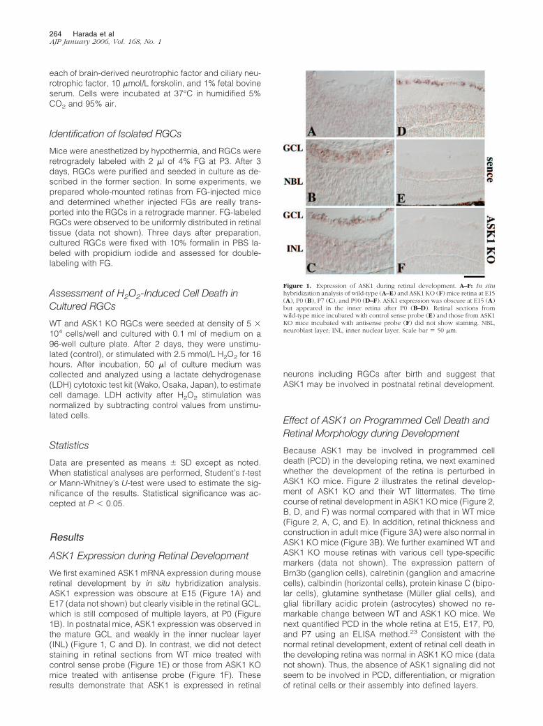
We first examined ASK1 mRNA expression during mouseretinal development by in situ hybridization analysis.ASK1 expression was obscure at E15 (Figure 1A) andE17 (data not shown) but clearly visible in the retinal GCL,which is still composed of multiple layers, at P0 (Figure1B). In postnatal mice, ASK1 expression was observed inthe mature GCL and weakly in the inner nuclear layer(INL) (Figure 1, C and D). In contrast, we did not detectstaining in retinal sections from WT mice treated withcontrol sense probe (Figure 1E) or those from ASK1 KOmice treated with antisense probe (Figure 1F). Theseresults demonstrate that ASK1 is expressed in retinal
neurons including RGCs after birth and suggest thatASK1 may be involved in postnatal retinal development.
Effect of ASK1 on Programmed Cell Death andRetinal Morphology during Development
Because ASK1 may be involved in programmed celldeath (PCD) in the developing retina, we next examinedwhether the development of the retina is perturbed inASK1 KO mice. Figure 2 illustrates the retinal develop-ment of ASK1 KO and their WT littermates. The timecourse of retinal development in ASK1 KO mice (Figure 2,B, D, and F) was normal compared with that in WT mice(Figure 2, A, C, and E). In addition, retinal thickness andconstruction in adult mice (Figure 3A) were also normal inASK1 KO mice (Figure 3B). We further examined WT andASK1 KO mouse retinas with various cell type-specificmarkers (data not shown). The expression pattern ofBrn3b (ganglion cells), calretinin (ganglion and amacrinecells), calbindin (horizontal cells), protein kinase C (bipo-lar cells), glutamine synthetase (Muller glial cells), andglial fibrillary acidic protein (astrocytes) showed no re-markable change between WT and ASK1 KO mice. Wenext quantified PCD in the whole retina at E15, E17, P0,and P7 using an ELISA method.23 Consistent with thenormal retinal development, extent of retinal cell death inthe developing retina was normal in ASK1 KO mice (datanot shown). Thus, the absence of ASK1 signaling did notseem to be involved in PCD, differentiation, or migrationof retinal cells or their assembly into defined layers.
Figure 1. Expression of ASK1 during retinal development. A–F: In situhybridization analysis of wild-type (A–E) and ASK1 KO (F) mice retina at E15(A), P0 (B), P7 (C), and P90 (D–F). ASK1 expression was obscure at E15 (A)but appeared in the inner retina after P0 (B–D). Retinal sections fromwild-type mice incubated with control sense probe (E) and those from ASK1KO mice incubated with antisense probe (F) did not show staining. NBL,neuroblast layer; INL, inner nuclear layer. Scale bar � 50 �m.
264 Harada et alAJP January 2006, Vol. 168, No. 1

Effect of ASK1 on Ischemia-Induced Neural CellDeath
Because ASK1 may mediate stress-induced neural cellapoptosis in adult retina, we next examined the effectof ischemic injury, which is severe oxidative stress,3,20
in WT and ASK1 KO mice. Histological evaluation dem-onstrated a significant decrease in ischemic damagein ASK1 KO mice (Figure 3D) compared with their WTlittermates (Figure 3C). The thickness of the IRL afterischemia was decreased to 65 � 2% (n � 6) in WT, but
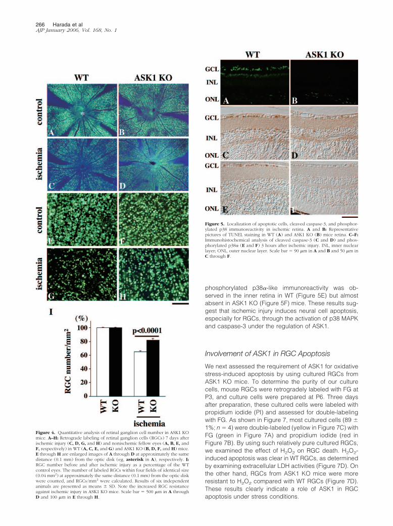
81 � 2% (n � 6) in ASK1 KO mice (Figure 3E). Second,the percentage of surviving cells in the GCL was alsoincreased in ASK1 KO (81 � 1%; n � 6) compared withWT mice (71 � 3%; n � 6) (Figure 3F). These resultssuggest that inner retinal neurons are less susceptibleto ischemic injury in ASK1 KO mice. Because GCL maycontain RGCs, displaced amacrine cells, and otherminor cell types,28 we next performed retrograde la-beling of RGCs with FG and tried to determine theeffect of ASK1 on RGC survival. Figure 4, A–H, showsthe representative results of RGC labeling in ASK1 KOmice and their WT littermates. Consistent with the re-sults of GCL counting (Figure 3F), the extent of isch-emia-induced RGC death seemed to be mild in ASK1KO mice (Figure 4, D and H) compared with theirlittermates (Figure 4, C and G, respectively). Quantita-tive analysis revealed that FG-labeled (surviving)RGCs after ischemia were significantly increased inASK1 KO mice (82 � 2%; n � 6) compared with theirWT littermates (65 � 1%; n � 6) (Figure 4I). Theseresults demonstrate that loss of endogenous ASK1 hasa protective effect on inner retinal neurons, especiallyfor RGCs, after ischemic injury.
Involvement of ASK1 Signaling in Ischemia-Induced Neuronal Apoptosis
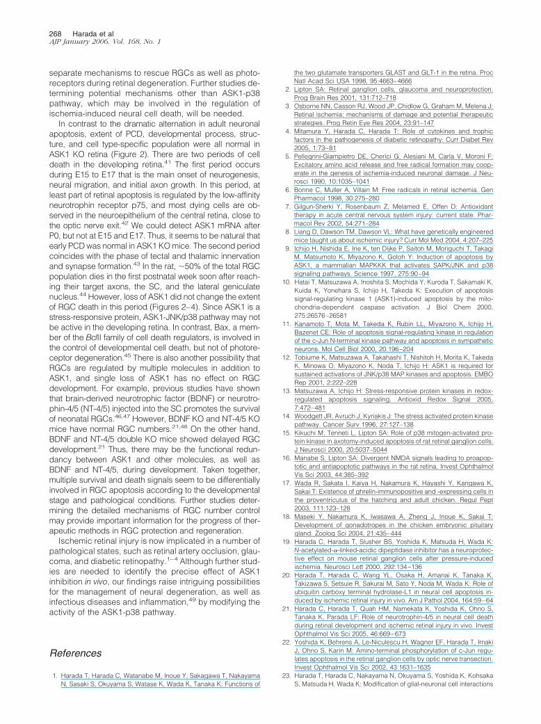
We next analyzed apoptotic cells in the retina by TUNELstaining 1 day after ischemic injury. Control animalsshowed practically no signals in both WT and ASK1 KOmice (data not shown). In ischemic retinas, many TUNEL-positive cells were observed in the inner retina in WT(Figure 5A) but not in ASK1 KO mice (Figure 5B). Wepreviously demonstrated that ischemia-induced retinalcell apoptosis in the inner retina is executed mainly bycaspase-3.20 In fact, immunohistochemical analysisshowed many cleaved caspase-3-positive cells in theinner retina in WT mice (Figure 5C), but the number wasapparently decreased in ASK1 KO mice (Figure 5D).Previous studies have suggested a possibility that ASK1could activate p38 MAPK pathway, leading to oxidativestress-induced RGC apoptosis.13,15,16 To determine thispossibility, we examined total and phosphorylated/acti-vated p38� immunoreactivity after ischemic injury in WTand ASK1 KO mice. Immunoblot analysis revealed thatthe levels of phosphorylated p38� in nonischemic controlretinas were very low in both strains (Figure 6A, toppanel). However, ischemic injury strongly induced phos-phorylation of p38� at 3, 6, and 24 hours after ischemicinjury in WT retina (Figure 6B). On the other hand, phos-phorylated p38� immunoreactivity at 3 hours was low andessentially disappeared by 24 hours after ischemia inASK1 KO mice (Figure 6, A and B). Nonetheless, totalp38� immunoreactivity (including both phosphorylatedand nonphosphorylated forms) did not change after isch-emia in both WT and ASK1 KO mice (Figure 6A, bottompanel). We also examined the localization of phosphory-lated p38� immunoreactivity in the mouse retina. In con-trol retinas, phosphorylated p38�-positive cells were ab-sent (data not shown). In contrast, 3 hours after ischemia,
Figure 2. Retinal development in ASK1 KO mice. A–F: Hematoxylin andeosin staining of retinal sections at E15 (A and B), P0 (C and D), and P14 (Eand F) in WT (A, C, and E) and ASK1 KO (B, D, and F) littermates. Asterisksin A and B indicate lens. NBL, neuroblast layer; INL, inner nuclear layer; ONL,outer nuclear layer. Scale bar � 50 �m.
Figure 3. Quantitative analysis of ischemic retinal injury in ASK1 KO mice.A–D: Hematoxylin and eosin staining of control (A and B) and ischemic (Cand D) retinal sections from WT (A and C) and ASK1 KO (B and D) mice. Eand F: Thickness of the IRL (E) and the number of cells in the GCL (F) beforeand after ischemic injury as a percentage of the WT control eyes. Results ofsix independent experiments are presented as means � SD. Note the de-creased neural cell loss in ASK1 KO mice. INL, inner nuclear layer; ONL,outer nuclear layer. Scale bar � 50 �m.
Role of ASK1 in Ischemic Retina 265AJP January 2006, Vol. 168, No. 1
phosphorylated p38�-like immunoreactivity was ob-served in the inner retina in WT (Figure 5E) but almostabsent in ASK1 KO (Figure 5F) mice. These results sug-gest that ischemic injury induces neural cell apoptosis,especially for RGCs, through the activation of p38 MAPKand caspase-3 under the regulation of ASK1.
Involvement of ASK1 in RGC Apoptosis
We next assessed the requirement of ASK1 for oxidativestress-induced apoptosis by using cultured RGCs fromASK1 KO mice. To determine the purity of our culturecells, mouse RGCs were retrogradely labeled with FG atP3, and culture cells were prepared at P6. Three daysafter preparation, these cultured cells were labeled withpropidium iodide (PI) and assessed for double-labelingwith FG. As shown in Figure 7, most cultured cells (89 �1%; n � 4) were double-labeled (yellow in Figure 7C) withFG (green in Figure 7A) and propidium iodide (red inFigure 7B). By using such relatively pure cultured RGCs,we examined the effect of H2O2 on RGC death. H2O2-induced apoptosis was clear in WT RGCs, as determinedby examining extracellular LDH activities (Figure 7D). Onthe other hand, RGCs from ASK1 KO mice were moreresistant to H2O2 compared with WT RGCs (Figure 7D).These results clearly indicate a role of ASK1 in RGCapoptosis under stress conditions.
Figure 4. Quantitative analysis of retinal ganglion cell number in ASK1 KOmice. A–H: Retrograde labeling of retinal ganglion cells (RGCs) 7 days afterischemic injury (C, D, G, and H) and nonischemic fellow eyes (A, B, E, andF, respectively) in WT (A, C, E, and G) and ASK1 KO (B, D, F, and H) mice.E through H are enlarged images of A through D at approximately the samedistance (0.1 mm) from the optic disk (eg, asterisk in A), respectively. I:RGC number before and after ischemic injury as a percentage of the WTcontrol eyes. The number of labeled RGCs within four fields of identical size(0.04 mm2) at approximately the same distance (0.1 mm) from the optic diskwere counted, and RGCs/mm2 were calculated. Results of six independentanimals are presented as means � SD. Note the increased RGC resistanceagainst ischemic injury in ASK1 KO mice. Scale bar � 500 �m in A throughD and 100 �m in E through H.
Figure 5. Localization of apoptotic cells, cleaved caspase-3, and phosphor-ylated p38 immunoreactivity in ischemic retina. A and B: Representativepictures of TUNEL staining in WT (A) and ASK1 KO (B) mice retina. C–F:Immunohistochemical analysis of cleaved caspase-3 (C and D) and phos-phorylated p38� (E and F) 3 hours after ischemic injury. INL, inner nuclearlayer; ONL, outer nuclear layer. Scale bar � 90 �m in A and B and 50 �m inC through F.
266 Harada et alAJP January 2006, Vol. 168, No. 1

Discussion
In the present study, we demonstrated that the ASK1MAPK kinase kinase signaling pathway is involved inneural cell apoptosis after ischemic retinal injury in vivo(Figures 3 and 4). In addition, we found that both p38MAPK and caspase-3 activation are clearly suppressedin ASK1 KO mice (Figures 5 and 6). Our results areconsistent with previous studies describing that activa-tion of p38 induces RGC apoptosis after axotomy of theoptic nerve or mediated by glutamate neurotoxicity.15,16
Ischemic injury is associated with excessive concentra-tions of glutamate, which results in overactivation of glu-tamate receptors and initiates a cascade of events thatleads to necrosis and/or apoptosis. Consistently, selec-tive inhibition of glutamate receptor antagonists2,3 orN-acetylated-�-linked-acidic dipeptidase, an enzyme re-sponsible for the hydrolysis of neuropeptide N-acetyl-aspartyl-glutamate to N-acetyl-aspartate and glutamate,protects neurons in animal models of stroke and ischemicretinal injury.19,29 Furthermore, we previously showedthat glutamate transporter KO mice are susceptible toischemic retinal injury.1 Therefore, as well as p38 inhibi-tion, appropriate control of glutamate concentrationand/or glutamate receptor activation could be usefulagainst ischemic injury.3,30
In addition to typical glutamate neurotoxicity, reactiveoxygen species, such as superoxide and H2O2, are im-portant mediators in damage caused by retinal isch-emia.5,6 Our present study demonstrated that H2O2-in-duced RGC death is attenuated in ASK1 KO mice in vitro(Figure 7). Under stress conditions, several stress pro-teins, such as ubiquitin, play important roles as heatshock- and stress-regulated proteins.31 However, recentstudies have shown that ubiquitin promotes either cellsurvival or apoptosis.32–35 In neural cells, excessive ubiq-uitin induction after ischemic injury may rather lead toneural cell apoptosis by the degradation of antiapoptoticproteins and suppression of the transcription of prosur-vival proteins.20 These results suggest a possibility thatmodulation of stress-responsive systems can become a“double-edged sword.” Although our present results sug-gest its usefulness, further investigations revealing theprecise effect of complete ASK1 inhibition in vivo will beneeded. On the other hand, ASK1 activity and conse-quent activation of downstream signaling molecules canbe negatively regulated by stimulating phosphoinosi-tide-3 kinase-Akt pathway. It was reported that Akt-me-diated phosphorylation of ASK1 inhibits its ability to acti-vate JNK/p38 and protects cells against H2O2-inducedapoptosis.36 Another possible factor that negatively reg-ulates ASK1 activity is thioredoxin (Trx).37 In resting cells,ASK1 constantly forms an inactive complex with Trx,whereas on stimulation, ASK1 is dissociated from Trx andactivated by conformational changes and covalent mod-ifications. Recent studies have suggested the possibilitythat Trx is a kind of neurotrophic factor released fromretinal pigmented epithelial cells and plays a crucial rolein maintaining photoreceptors from light damage.38 Be-cause other trophic factors such as ciliary neurotrophicfactor and glial cell line-derived neurotrophic factor sup-plied from Muller glial cells rescue neural cells,4,23,39,40
application of Trx, in combination with these trophic fac-tors, may stimulate multiple cellular targets and activate
Figure 6. Immunoblot analysis of p38� immunoreactivity in ischemic retina.A: Three micrograms of retinal protein were prepared from ischemic eyesand nontreated fellow eyes and separated in 11% sodium dodecyl sulfate-polyacrylamide gel followed by immunoblot analysis using anti-p38� andanti-phspho-p38� antibodies. Representative image of three independentexperiments are shown. B: Quantitative analysis of phosphorylated p38�immunoreactivity at 3, 6, and 24 hours after ischemic injury. Results of threeindependent experiments are presented as means � SD. Note the decreasedphosphorylation of p38� in ASK1 KO mice. *P � 0.01.
Figure 7. Quantitative analysis of H2O2-induced RGC death in ASK1 KOmice in vitro. A–C: Cultured RGCs were prepared from mouse retinas thathad been retrogradely labeled with FG. Most cultured cells (89 � 1%, n � 4)were double labeled (yellow in C) with FG (green in A) and propidiumiodide (PI; red in B), thus confirming the purity of our cultured RGCs. Scalebar � 100 �m. D: Extent of RGC death after stimulation with 2.5 mmol/LH2O2 was quantified by examining extracellular LDH activities. Note thedecrease of H2O2-induced death in RGCs from ASK1 KO mice. Results ofthree independent experiments are presented as means � SD. *P � 0.05.
Role of ASK1 in Ischemic Retina 267AJP January 2006, Vol. 168, No. 1
separate mechanisms to rescue RGCs as well as photo-receptors during retinal degeneration. Further studies de-termining potential mechanisms other than ASK1-p38pathway, which may be involved in the regulation ofischemia-induced neural cell death, will be needed.
In contrast to the dramatic alternation in adult neuronalapoptosis, extent of PCD, developmental process, struc-ture, and cell type-specific population were all normal inASK1 KO retina (Figure 2). There are two periods of celldeath in the developing retina.41 The first period occursduring E15 to E17 that is the main onset of neurogenesis,neural migration, and initial axon growth. In this period, atleast part of retinal apoptosis is regulated by the low-affinityneurotrophin receptor p75, and most dying cells are ob-served in the neuroepithelium of the central retina, close tothe optic nerve exit.42 We could detect ASK1 mRNA afterP0, but not at E15 and E17. Thus, it seems to be natural thatearly PCDwas normal in ASK1KOmice. The second periodcoincides with the phase of tectal and thalamic innervationand synapse formation.43 In the rat, �50% of the total RGCpopulation dies in the first postnatal week soon after reach-ing their target axons, the SC, and the lateral geniculatenucleus.44 However, loss of ASK1 did not change the extentof RGC death in this period (Figures 2–4). Since ASK1 is astress-responsive protein, ASK1-JNK/p38 pathway may notbe active in the developing retina. In contrast, Bax, a mem-ber of the BclII family of cell death regulators, is involved inthe control of developmental cell death, but not of photore-ceptor degeneration.45 There is also another possibility thatRGCs are regulated by multiple molecules in addition toASK1, and single loss of ASK1 has no effect on RGCdevelopment. For example, previous studies have shownthat brain-derived neurotrophic factor (BDNF) or neurotro-phin-4/5 (NT-4/5) injected into the SC promotes the survivalof neonatal RGCs.46,47 However, BDNF KO and NT-4/5 KOmice have normal RGC numbers.21,48 On the other hand,BDNF and NT-4/5 double KO mice showed delayed RGCdevelopment.21 Thus, there may be the functional redun-dancy between ASK1 and other molecules, as well asBDNF and NT-4/5, during development. Taken together,multiple survival and death signals seem to be differentiallyinvolved in RGC apoptosis according to the developmentalstage and pathological conditions. Further studies deter-mining the detailed mechanisms of RGC number controlmay provide important information for the progress of ther-apeutic methods in RGC protection and regeneration.
Ischemic retinal injury is now implicated in a number ofpathological states, such as retinal artery occlusion, glau-coma, and diabetic retinopathy.1–4 Although further stud-ies are needed to identify the precise effect of ASK1inhibition in vivo, our findings raise intriguing possibilitiesfor the management of neural degeneration, as well asinfectious diseases and inflammation,49 by modifying theactivity of the ASK1-p38 pathway.
References
1. Harada T, Harada C, Watanabe M, Inoue Y, Sakagawa T, NakayamaN, Sasaki S, Okuyama S, Watase K, Wada K, Tanaka K: Functions of
the two glutamate transporters GLAST and GLT-1 in the retina. ProcNatl Acad Sci USA 1998, 95:4663–4666
2. Lipton SA: Retinal ganglion cells, glaucoma and neuroprotection.Prog Brain Res 2001, 131:712–718
3. Osborne NN, Casson RJ, Wood JP, Chidlow G, Graham M, Melena J:Retinal ischemia: mechanisms of damage and potential therapeuticstrategies. Prog Retin Eye Res 2004, 23:91–147
4. Mitamura Y, Harada C, Harada T: Role of cytokines and trophicfactors in the pathogenesis of diabetic retinopathy. Curr Diabet Rev2005, 1:73–81
5. Pellegrini-Giampietro DE, Cherici G, Alesiani M, Carla V, Moroni F:Excitatory amino acid release and free radical formation may coop-erate in the genesis of ischemia-induced neuronal damage. J Neu-rosci 1990, 10:1035–1041
6. Bonne C, Muller A, Villain M: Free radicals in retinal ischemia. GenPharmacol 1998, 30:275–280
7. Gilgun-Sherki Y, Rosenbaum Z, Melamed E, Offen D: Antioxidanttherapy in acute central nervous system injury: current state. Phar-macol Rev 2002, 54:271–284
8. Liang D, Dawson TM, Dawson VL: What have genetically engineeredmice taught us about ischemic injury? Curr Mol Med 2004, 4:207–225
9. Ichijo H, Nishida E, Irie K, ten Dijke P, Saitoh M, Moriguchi T, TakagiM, Matsumoto K, Miyazono K, Gotoh Y: Induction of apoptosis byASK1, a mammalian MAPKKK that activates SAPK/JNK and p38signaling pathways. Science 1997, 275:90–94
10. Hatai T, Matsuzawa A, Inoshita S, Mochida Y, Kuroda T, Sakamaki K,Kuida K, Yonehara S, Ichijo H, Takeda K: Execution of apoptosissignal-regulating kinase 1 (ASK1)-induced apoptosis by the mito-chondria-dependent caspase activation. J Biol Chem 2000,275:26576–26581
11. Kanamoto T, Mota M, Takeda K, Rubin LL, Miyazono K, Ichijo H,Bazenet CE: Role of apoptosis signal-regulating kinase in regulationof the c-Jun N-terminal kinase pathway and apoptosis in sympatheticneurons. Mol Cell Biol 2000, 20:196–204
12. Tobiume K, Matsuzawa A, Takahashi T, Nishitoh H, Morita K, TakedaK, Minowa O, Miyazono K, Noda T, Ichijo H: ASK1 is required forsustained activations of JNK/p38 MAP kinases and apoptosis. EMBORep 2001, 2:222–228
13. Matsuzawa A, Ichijo H: Stress-responsive protein kinases in redox-regulated apoptosis signaling. Antioxid Redox Signal 2005,7:472–481
14. Woodgett JR, Avruch J, Kyriakis J: The stress activated protein kinasepathway. Cancer Surv 1996, 27:127–138
15. Kikuchi M, Tenneti L, Lipton SA: Role of p38 mitogen-activated pro-tein kinase in axotomy-induced apoptosis of rat retinal ganglion cells.J Neurosci 2000, 20:5037–5044
16. Manabe S, Lipton SA: Divergent NMDA signals leading to proapop-totic and antiapoptotic pathways in the rat retina. Invest OphthalmolVis Sci 2003, 44:385–392
17. Wada R, Sakata I, Kaiya H, Nakamura K, Hayashi Y, Kangawa K,Sakai T: Existence of ghrelin-immunopositive and -expressing cells inthe proventriculus of the hatching and adult chicken. Regul Pept2003, 111:123–128
18. Maseki Y, Nakamura K, Iwasawa A, Zheng J, Inoue K, Sakai T:Development of gonadotropes in the chicken embryonic pituitarygland. Zoolog Sci 2004, 21:435–444
19. Harada C, Harada T, Slusher BS, Yoshida K, Matsuda H, Wada K:N-acetylated-�-linked-acidic dipeptidase inhibitor has a neuroprotec-tive effect on mouse retinal ganglion cells after pressure-inducedischemia. Neurosci Lett 2000, 292:134–136
20. Harada T, Harada C, Wang YL, Osaka H, Amanai K, Tanaka K,Takizawa S, Setsuie R, Sakurai M, Sato Y, Noda M, Wada K: Role ofubiquitin carboxy terminal hydrolase-L1 in neural cell apoptosis in-duced by ischemic retinal injury in vivo. Am J Pathol 2004, 164:59–64
21. Harada C, Harada T, Quah HM, Namekata K, Yoshida K, Ohno S,Tanaka K, Parada LF: Role of neurotrophin-4/5 in neural cell deathduring retinal development and ischemic retinal injury in vivo. InvestOphthalmol Vis Sci 2005, 46:669–673
22. Yoshida K, Behrens A, Le-Niculescu H, Wagner EF, Harada T, ImakiJ, Ohno S, Karin M: Amino-terminal phosphorylation of c-Jun regu-lates apoptosis in the retinal ganglion cells by optic nerve transection.Invest Ophthalmol Vis Sci 2002, 43:1631–1635
23. Harada T, Harada C, Nakayama N, Okuyama S, Yoshida K, KohsakaS, Matsuda H, Wada K: Modification of glial-neuronal cell interactions
268 Harada et alAJP January 2006, Vol. 168, No. 1

prevents photoreceptor apoptosis during light-induced retinal degen-eration. Neuron 2000, 26:533–541
24. Barres BA, Silverstein BE, Corey DP, Chun LL: Immunological, mor-phological, and electrophysiological variation among retinal ganglioncells purified by panning. Neuron 1988, 1:791–803
25. Kashiwagi K, Iizuka Y, Araie M, Suzuki Y, Tsukahara S: Effects ofretinal glial cells on isolated rat retinal ganglion cells. Invest Ophthal-mol Vis Sci 2001, 42:2686–2694
26. Kashiwagi K, Iizuka Y, Mochizuki S, Tsumamoto Y, Mishima HK, AraieM, Suzuki Y, Tsukahara S: Differences in nitric oxide production: acomparison of retinal ganglion cells and retinal glial cells culturedunder hypoxic conditions. Brain Res Mol Brain Res 2003,112:126–134
27. Politi LE, Lehar M, Adler R: Development of neonatal mouse retinalneurons and photoreceptors in low density cell culture. Invest Oph-thalmol Vis Sci 1988, 29:534–543
28. Perry VH: Evidence for an amacrine cell system in the ganglion celllayer of the rat retina. Neuroscience 1981, 6:931–944
29. Slusher BS, Vornov JJ, Thomas AG, Hurn PD, Harukuni I, Bhardwaj A,Traystman RJ, Robinson MB, Britton P, Lu XC, Tortella FC, WozniakKM, Yudkoff M, Potter BM, Jackson PF: Selective inhibition of NAALA-Dase, which converts NAAG to glutamate, reduces ischemic braininjury. Nat Med 1999, 5:1396–1402
30. Tanaka K: Functions of glutamate transporters in the brain. NeurosciRes 2000, 37:15–19
31. Hershko A, Ciechanover A, Varshavsky A: The ubiquitin system. NatMed 2000, 6:1073–1081
32. Dimmeler S, Breitschopf K, Haendeler J, Zeiher AM: Dephosphory-lation targets Bcl-2 for ubiquitin-dependent degradation: a link be-tween the apoptosome and the proteasome pathway. J Exp Med1999, 189:1815–1822
33. Breitschopf K, Haendeler J, Malchow P, Zeiher AM, Dimmeler S:Posttranslational modification of Bcl-2 facilitates its proteasome-de-pendent degradation: molecular characterization of the involved sig-naling pathway. Mol Cell Biol 2000, 20:1886–1896
34. Daino H, Matsumura I, Takada K, Odajima J, Tanaka H, Ueda S,Shibayama H, Ikeda H, Hibi M, Machii T, Hirano T, Kanakura Y:Induction of apoptosis by extracellular ubiquitin in human hematopoi-etic cells: possible involvement of STAT3 degradation by proteasomepathway in interleukin 6-dependent hematopoietic cells. Blood 2000,95:2577–2585
35. Yang Y, Fang S, Jensen JP, Weissman AM, Ashwell JD: Ubiquitinprotein ligase activity of IAPs and their degradation in proteasomes inresponse to apoptotic stimuli. Science 2000, 288:874–877
36. Kim AH, Khursigara G, Sun X, Franke TF, Chao MV: Akt phosphory-lates and negatively regulates apoptosis signal-regulating kinase 1.Mol Cell Biol 2001, 21:893–901
37. Saitoh M, Nishitoh H, Fujii M, Takeda K, Tobiume K, Sawada Y,Kawabata M, Miyazono K, Ichijo H: Mammalian thioredoxin is a directinhibitor of apoptosis signal-regulating kinase (ASK) 1. EMBO J 1998,17:25596–25606
38. Tanito M, Kwon YW, Kondo N, Bai J, Masutani H, Nakamura H, FujiiJ, Ohira A, Yodoi J: Cytoprotective effects of geranylgeranylacetoneagainst retinal photooxidative damage. J Neurosci 2005,25:2396–2404
39. Harada T, Harada C, Kohsaka S, Wada E, Yoshida K, Ohno S,Mamada H, Tanaka K, Parada LF, Wada K: Microglia-Muller glia cellinteractions control neurotrophic factor production during light-in-duced retinal degeneration. J Neurosci 2002, 22:9228–9236
40. Harada C, Harada T, Quah HM, Maekawa F, Yoshida K, Ohno S,Wada K, Parada LF, Tanaka K: Potential role of glial cell line-derivedneurotrophic factor receptors in Muller glia cells during light-inducedretinal degeneration. Neuroscience 2003, 122:229–235
41. Bahr M: Live or let die: retinal ganglion cell death and survival duringdevelopment and in the lesioned adult CNS. Trends Neurosci 2000,23:483–490
42. Harada C, Harada T, Nakamura K, Sakai Y, Tanaka K, Parada LF:Effect of p75NTR on the regulation of naturally occurring cell deathand retinal ganglion cell number in the mouse eye. Dev Biol, in press
43. Cellerino A, Bahr M, Isenmann S: Apoptosis in the developing visualsystem. Cell Tissue Res 2000, 301:53–69
44. Perry VH, Henderson Z, Linden R: Postnatal changes in retinal gan-glion cell and optic axon populations in the pigmented rat. J CompNeurol 1983, 219:356–368
45. Mosinger-Ogilvie J, Deckwerth TL, Knudson CM, Korsmeyer SJ: Sup-pression of developmental retinal cell death but not of photoreceptordegeneration in Bax-deficient mice. Invest Ophthalmol Vis Sci 1998,39:1713–1720
46. Cui Q, Harvey AR: At least two mechanisms are involved in the deathof retinal ganglion cells following target ablation in neonatal rats.J Neurosci 1995, 15:8143–8155
47. Ma YT, Hsieh T, Forbes ME, Johnson JE, Frost DO: BDNF injectedinto the superior colliculus reduces developmental retinal ganglioncell death. J Neurosci 1998, 18:2097–2107
48. Cellerino A, Carroll P, Thoenen H, Barde YA: Reduced size of retinalganglion cell axons and hypomyelination in mice lacking brain-de-rived neurotrophic factor. Mol Cell Neurosci 1997, 9:397–408
49. Matsuzawa A, Saegusa K, Noguchi T, Sadamitsu C, Nishitoh H,Nagai S, Koyasu S, Matsumoto K, Takeda K, Ichijo H: ROS-depen-dent activation of the TRAF6-ASK1–p38 pathway is selectively re-quired for TLR4-mediated innate immunity. Nat Immunol 2005,6:587–592
Role of ASK1 in Ischemic Retina 269AJP January 2006, Vol. 168, No. 1