Critical function of endogenous XIAP in regulating caspase activation during sympathetic neuronal...
11
The Journal of Cell Biology The Rockefeller University Press, 0021-9525/2003/11/789/11 $8.00 The Journal of Cell Biology, Volume 163, Number 4, November 24, 2003 789–799 http://www.jcb.org/cgi/doi/10.1083/jcb.200307130 JCB Article 789 Critical function of endogenous XIAP in regulating caspase activation during sympathetic neuronal apoptosis Patrick Ryan Potts, 1 Shweta Singh, 1 Malia Knezek, 1 Craig B. Thompson, 3,4 and Mohanish Deshmukh 1,2 1 Neuroscience Center and 2 Department of Cell and Developmental Biology, University of North Carolina, Chapel Hill, NC 27599 3 Department of Cancer Biology and 4 Department of Pathology and Laboratory Medicine, Abramson Family Cancer Research Institute, University of Pennsylvania, Philadelphia, PA 19104 n sympathetic neurons, unlike most nonneuronal cells, growth factor withdrawal–induced apoptosis requires the development of competence in addition to cytochrome c release to activate caspases. Thus, although most non- neuronal cells die rapidly with cytosolic cytochrome c alone, sympathetic neurons are remarkably resistant unless they develop competence. We have identified endogenous X-linked inhibitor of apoptosis protein (XIAP) as the essential postcytochrome c regulator of caspase activation in these neurons. In contrast to wild-type neurons that are resistant to injection of cytochrome c, XIAP-deficient neurons died I rapidly with cytosolic cytochrome c alone. Surprisingly, the release of endogenous Smac was not sufficient to overcome the XIAP resistance in sympathetic neurons. In contrast, the neuronal competence pathway permitted cytochrome c to activate caspases by inducing a marked reduction in XIAP levels in these neurons. Thus, the removal of XIAP inhibition appears both necessary and sufficient for cytochrome c to activate caspases in sympathetic neurons. These data identify a critical function of endogenous XIAP in regulating apoptosis in mammalian cells. Introduction The ability of cells to induce apoptosis and die is important during development and is crucial for the maintenance of homeostasis (Vaux and Korsmeyer, 1999). Dysregulation of the apoptotic pathway is associated with many pathological conditions. For example, decreased apoptosis contributes to cancer progression (Green and Evan, 2002), whereas in- creased apoptosis leads to loss of neural cells during stroke, spinal cord injury and in many neurodegenerative diseases (Yuan and Yankner, 2000). Therefore, understanding how cells regulate apoptosis has important therapeutic implica- tions in many pathological conditions. The mechanism of neuronal apoptosis has been widely studied in sympathetic neurons that are dependent on NGF for survival. Deprivation of NGF induces an apoptotic neu- ronal death within 24–48 h after NGF removal in culture (Deckwerth and Johnson, 1993; Deshmukh and Johnson, 1997). This death is prevented by macromolecular synthesis inhibitors such as cycloheximide (Martin et al., 1988), is de- pendent on Bax (Deckwerth et al., 1996), and is executed by the caspase proteases (Deshmukh et al., 1996; Troy et al., 1996; McCarthy et al., 1997). Activation of caspases is a crucial point at which cells be- come committed to die during apoptosis (Deshmukh et al., 2000; Denault and Salvesen, 2002). In most mammalian cells, including sympathetic neurons, caspase activation is triggered by the release of cytochrome c from mitochondria to cytosol (Deshmukh and Johnson, 1998; Neame et al., 1998; Wang, 2001). Data from several cell-free studies indi- cate that cytochrome c released from the mitochondria binds to Apaf-1 and promotes its oligomerization. Procaspase-9 is then recruited to bind oligomerized Apaf-1 to form the apoptosome complex where caspase-9 becomes activated. Activated caspase-9 then cleaves and activates other caspases, P.R. Potts and S. Singh contributed equally to this work. The online version of this article includes supplemental material. Address correspondence to Mohanish Deshmukh, 7109E Neuroscience Research Building, Box 7250, 103 Mason Farm Rd., University of North Carolina, Chapel Hill, NC 27599. Tel.: (919) 843-6004. Fax: (919) 966-1050. email: [email protected] Key words: Smac; cytochrome c; nerve growth factor; IAP; neurons Abbreviations used in this paper: cIAP, cellular inhibitor of apopto- sis protein; CPTcAMP, 8-(4-chlorophenylthio)adenosine-3-5-cyclic monophosphate; GAPDH, glyceraldehyde-3-phosphate dehydrogenase; H 2 O 2 , hydrogen peroxide; IAP, inhibitor of apoptosis protein; XIAP, X-linked IAP. on August 1, 2016 jcb.rupress.org Downloaded from Published November 17, 2003 http://jcb.rupress.org/content/suppl/2003/11/11/jcb.200307130.DC1.html Supplemental Material can be found at:
-
Upload
independent -
Category
Documents
-
view
4 -
download
0
Transcript of Critical function of endogenous XIAP in regulating caspase activation during sympathetic neuronal...

The
Jour
nal o
f Cel
l Bio
logy
The Rockefeller University Press, 0021-9525/2003/11/789/11 $8.00The Journal of Cell Biology, Volume 163, Number 4, November 24, 2003 789–799http://www.jcb.org/cgi/doi/10.1083/jcb.200307130
JCB
Article
789
Critical function of endogenous XIAP in regulating caspase activation during sympathetic neuronal apoptosis
Patrick Ryan Potts,
1
Shweta Singh,
1
Malia Knezek,
1
Craig B. Thompson,
3,4
and Mohanish Deshmukh
1,2
1
Neuroscience Center and
2
Department of Cell and Developmental Biology, University of North Carolina, Chapel Hill, NC 27599
3
Department of Cancer Biology and
4
Department of Pathology and Laboratory Medicine, Abramson Family Cancer Research Institute, University of Pennsylvania, Philadelphia, PA 19104
n sympathetic neurons, unlike most nonneuronal cells,growth factor withdrawal–induced apoptosis requires thedevelopment of competence in addition to cytochrome
c
release to activate caspases. Thus, although most non-neuronal cells die rapidly with cytosolic cytochrome
c
alone, sympathetic neurons are remarkably resistant unlessthey develop competence. We have identified endogenousX-linked inhibitor of apoptosis protein (XIAP) as the essentialpostcytochrome
c
regulator of caspase activation in theseneurons. In contrast to wild-type neurons that are resistantto injection of cytochrome
c
, XIAP-deficient neurons died
I
rapidly with cytosolic cytochrome
c
alone. Surprisingly,the release of endogenous Smac was not sufficient toovercome the XIAP resistance in sympathetic neurons. Incontrast, the neuronal competence pathway permittedcytochrome
c
to activate caspases by inducing a markedreduction in XIAP levels in these neurons. Thus, the removalof XIAP inhibition appears both necessary and sufficient forcytochrome
c
to activate caspases in sympathetic neurons.These data identify a critical function of endogenous XIAPin regulating apoptosis in mammalian cells.
Introduction
The ability of cells to induce apoptosis and die is importantduring development and is crucial for the maintenance ofhomeostasis (Vaux and Korsmeyer, 1999). Dysregulation ofthe apoptotic pathway is associated with many pathologicalconditions. For example, decreased apoptosis contributes tocancer progression (Green and Evan, 2002), whereas in-creased apoptosis leads to loss of neural cells during stroke,spinal cord injury and in many neurodegenerative diseases(Yuan and Yankner, 2000). Therefore, understanding howcells regulate apoptosis has important therapeutic implica-tions in many pathological conditions.
The mechanism of neuronal apoptosis has been widelystudied in sympathetic neurons that are dependent on NGFfor survival. Deprivation of NGF induces an apoptotic neu-ronal death within 24–48 h after NGF removal in culture
(Deckwerth and Johnson, 1993; Deshmukh and Johnson,1997). This death is prevented by macromolecular synthesisinhibitors such as cycloheximide (Martin et al., 1988), is de-pendent on Bax (Deckwerth et al., 1996), and is executed bythe caspase proteases (Deshmukh et al., 1996; Troy et al.,1996; McCarthy et al., 1997).
Activation of caspases is a crucial point at which cells be-come committed to die during apoptosis (Deshmukh et al.,2000; Denault and Salvesen, 2002). In most mammaliancells, including sympathetic neurons, caspase activation istriggered by the release of cytochrome
c
from mitochondriato cytosol (Deshmukh and Johnson, 1998; Neame et al.,1998; Wang, 2001). Data from several cell-free studies indi-cate that cytochrome
c
released from the mitochondria bindsto Apaf-1 and promotes its oligomerization. Procaspase-9is then recruited to bind oligomerized Apaf-1 to form theapoptosome complex where caspase-9 becomes activated.Activated caspase-9 then cleaves and activates other caspases,
P.R. Potts and S. Singh contributed equally to this work.The online version of this article includes supplemental material.Address correspondence to Mohanish Deshmukh, 7109E NeuroscienceResearch Building, Box 7250, 103 Mason Farm Rd., University of NorthCarolina, Chapel Hill, NC 27599. Tel.: (919) 843-6004. Fax: (919)966-1050. email: [email protected] words: Smac; cytochrome
c
; nerve growth factor; IAP; neurons
Abbreviations used in this paper: cIAP, cellular inhibitor of apopto-sis protein; CPTcAMP, 8-(4-chlorophenylthio)adenosine-3
�
-5
�
-cyclicmonophosphate; GAPDH, glyceraldehyde-3-phosphate dehydrogenase;H
2
O
2
, hydrogen peroxide; IAP, inhibitor of apoptosis protein; XIAP,X-linked IAP.
on August 1, 2016
jcb.rupress.orgD
ownloaded from
Published November 17, 2003
http://jcb.rupress.org/content/suppl/2003/11/11/jcb.200307130.DC1.html Supplemental Material can be found at:
790 The Journal of Cell Biology
|
Volume 163, Number 4, 2003
such as caspase-3, to induce rapid apoptosis and cell death(Wang, 2001).
Members of the inhibitor of apoptosis proteins (IAPs) canregulate caspase activity by binding directly to activated cas-pases and inhibiting their function (Salvesen and Duckett,2002). The IAP family includes X-linked IAP (XIAP), cellu-lar IAP-1 (cIAP-1), cIAP-2, neuronal apoptosis inhibitoryprotein, Survivin, melanoma IAP, and Bruce, all of whichcontain one or more repeats of the characteristic baculovirusIAP repeat domain. Overexpression of IAPs blocks apoptosisin many cells (Deveraux et al., 1998; Duckett et al., 1998;Simons et al., 1999), including in sympathetic neurons(Wiese et al., 1999; Yu et al., 2003). The IAPs themselvescan potentially be regulated in cells by at least two mecha-nisms. First, mitochondrial proteins such as Smac/DIABLOand HtrA2/Omi, when translocated to the cytosol duringapoptosis, can bind to and inhibit multiple IAPs (Du et al.,2000; Verhagen et al., 2000; Suzuki et al., 2001a). Second,several IAPs including XIAP, cIAP-1, and cIAP-2 contain aRING finger domain that can function as an E3 ubiquitinligase and target themselves and other proteins for protea-some-mediated degradation (Yang et al., 2000; Suzuki et al.,2001b; MacFarlane et al., 2002). However, the mechanismby which degradation of IAPs is regulated in mammaliancells in not known.
In primary fibroblasts and many cell lines, cytosolic mi-croinjection of cytochrome
c
induces a rapid, caspase-depen-dent apoptotic death, thus indicating that cytosolic accumu-lation of cytochrome
c
alone is sufficient to activate caspasesin these nonneuronal cells (Li et al., 1997; Brustugun et al.,1998; Juin et al., 1999; Chang et al., 2000). In contrast, cy-tochrome
c
, although necessary, is not sufficient to inducecell death in sympathetic neurons. NGF-maintained sympa-thetic neurons are remarkably resistant to cytosolic microin-jection of cytochrome
c
, thus pointing to a stringent postcy-tochrome
c
regulation of caspase activation in these neurons(Deshmukh and Johnson, 1998; Neame et al., 1998). Im-portantly, NGF deprivation activates a novel pathway, calleddevelopment of competence, which is necessary to allow cy-tochrome
c
to activate caspases and induce cell death in sym-pathetic neurons (Deshmukh and Johnson, 1998). There-fore, NGF deprivation–induced apoptosis in sympatheticneurons requires the activation of two pathways: a cyto-chrome
c
release pathway that is dependent on protein syn-thesis and Bax function; and a development of the compe-tence pathway that requires neither protein synthesis norBax function (Deshmukh and Johnson, 1998).
Recent data suggests that the inability of cytosolic cyto-chrome
c
alone to induce death in NGF-maintained sympa-thetic neurons is due to a block in caspase activation by theIAPs. Microinjection of excess exogenous Smac that inhibitsIAPs overcomes this block and permits cytochrome
c
to in-duce a rapid, caspase-dependent death in these neurons(Deshmukh et al., 2002). Thus, the target of the compe-tence pathway is likely to be IAPs, although which IAP maybe important and the exact mechanism by which the compe-tence pathway permits cytochrome
c
to induce neuronalapoptosis is not known. Here, we identify XIAP as the criti-cal inhibitor of caspase activation in sympathetic neurons.We find that XIAP mRNA and protein are both selectively
decreased when neurons develop competence and becomepermissive for cytochrome
c
–mediated caspase activation.Importantly, although cytochrome
c
alone is incapable of in-ducing cell death in wild-type neurons, it is remarkably suf-ficient to induce a rapid apoptotic death in XIAP-deficientneurons. These data identify an essential function for endog-enous XIAP in regulating apoptosis in mammalian cells andindicate that removal of XIAP inhibition is both necessaryand sufficient for cytochrome
c
to activate caspases duringneuronal apoptosis.
Results
Removal of IAP inhibition is necessary for cytochrome
c
to induce apoptosis in sympathetic neurons
NGF deprivation–induced cytochrome
c
release and devel-opment of competence are both necessary for apoptosis insympathetic neurons. These two pathways are distinct fromeach other because whereas cycloheximide addition and Baxdeficiency block the cytochrome
c
release pathway, neitherone blocks the development of the competence pathway(Deshmukh and Johnson, 1998). Thus, whereas microinjec-tion of exogenous cytosolic cytochrome
c
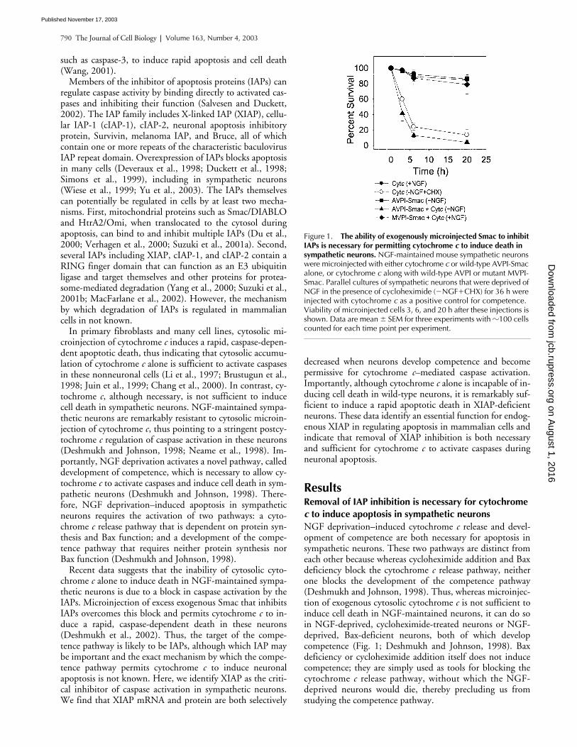
is not sufficient toinduce cell death in NGF-maintained neurons, it can do soin NGF-deprived, cycloheximide-treated neurons or NGF-deprived, Bax-deficient neurons, both of which developcompetence (Fig. 1; Deshmukh and Johnson, 1998). Baxdeficiency or cycloheximide addition itself does not inducecompetence; they are simply used as tools for blocking thecytochrome
c
release pathway, without which the NGF-deprived neurons would die, thereby precluding us fromstudying the competence pathway.
Figure 1. The ability of exogenously microinjected Smac to inhibit IAPs is necessary for permitting cytochrome c to induce death in sympathetic neurons. NGF-maintained mouse sympathetic neurons were microinjected with either cytochrome c or wild-type AVPI-Smac alone, or cytochrome c along with wild-type AVPI or mutant MVPI-Smac. Parallel cultures of sympathetic neurons that were deprived of NGF in the presence of cycloheximide (�NGF�CHX) for 36 h were injected with cytochrome c as a positive control for competence. Viability of microinjected cells 3, 6, and 20 h after these injections is shown. Data are mean � SEM for three experiments with �100 cells counted for each time point per experiment.
on August 1, 2016
jcb.rupress.orgD
ownloaded from
Published November 17, 2003
Neuronal apoptosis requires the removal of XIAP |
Potts et al. 791
The resistance of NGF-maintained neurons to cytosoliccytochrome
c
is overcome with microinjection of excess ma-ture Smac (Fig. 1; Deshmukh et al., 2002). To confirm thatexogenous Smac permitted cytochrome
c
to induce apopto-sis by inhibiting IAPs in these neurons, we examinedwhether a Smac mutant protein that cannot inhibit IAPs as aconsequence of a single, alanine-to-methionine amino acidchange in its mature NH
2
terminus was capable of permit-ting cytochrome
c
to induce death in sympathetic neurons.Unlike wild-type AVPI-Smac, the mutant MVPI-Smac can-not bind to IAPs and relieve their inhibition of caspases(Chai et al., 2000). We found that unlike wild-type AVPI-Smac, the mutant MVPI-Smac was incapable of cooperating
with cytochrome
c
to induce death in NGF-maintainedsympathetic neurons (Fig. 1). Less than 5% of cells that wereinjected with wild-type AVPI-Smac and cytochrome
c
wereviable 20 h after the injections, whereas almost 80% of cellsthat were injected with the mutant MVPI-Smac and cyto-chrome
c
were viable at that time (Fig. 1). Thus, cytochrome
c
was able to induce apoptosis in NGF-maintained sympa-thetic neurons only if the functions of one or more Smac-inhibitable IAPs were blocked in these neurons.
Levels of XIAP are reduced when sympathetic neurons develop competence
To identify the specific IAPs that regulated caspase activa-tion during sympathetic neuronal apoptosis, we first exam-ined which IAPs were expressed in sympathetic neurons.Western data show that NGF-maintained sympathetic neu-rons express multiple IAPs including XIAP, cIAP-1, andcIAP-2 (Fig. 2 a). Importantly, we examined whether thelevels of any IAPs were altered under conditions where neu-rons develop competence. Sympathetic neurons that are de-prived of NGF in the presence of cycloheximide for 24 h de-velop competence and become permissive for cytochrome
c
–mediated apoptosis (Fig. 1; Deshmukh and Johnson, 1998).Cycloheximide addition was used to block the cytochrome
c
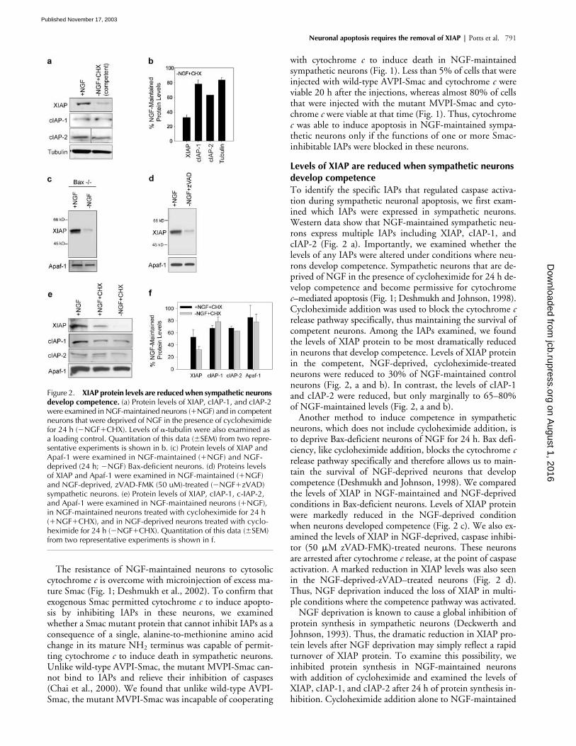
release pathway specifically, thus maintaining the survival ofcompetent neurons. Among the IAPs examined, we foundthe levels of XIAP protein to be most dramatically reducedin neurons that develop competence. Levels of XIAP proteinin the competent, NGF-deprived, cycloheximide-treatedneurons were reduced to 30% of NGF-maintained controlneurons (Fig. 2, a and b). In contrast, the levels of cIAP-1and cIAP-2 were reduced, but only marginally to 65–80%of NGF-maintained levels (Fig. 2, a and b).
Another method to induce competence in sympatheticneurons, which does not include cycloheximide addition, isto deprive Bax-deficient neurons of NGF for 24 h. Bax defi-ciency, like cycloheximide addition, blocks the cytochrome
c
release pathway specifically and therefore allows us to main-tain the survival of NGF-deprived neurons that developcompetence (Deshmukh and Johnson, 1998). We comparedthe levels of XIAP in NGF-maintained and NGF-deprivedconditions in Bax-deficient neurons. Levels of XIAP proteinwere markedly reduced in the NGF-deprived conditionwhen neurons developed competence (Fig. 2 c). We also ex-amined the levels of XIAP in NGF-deprived, caspase inhibi-tor (50
�
M zVAD-FMK)-treated neurons. These neuronsare arrested after cytochrome
c
release, at the point of caspaseactivation. A marked reduction in XIAP levels was also seenin the NGF-deprived-zVAD–treated neurons (Fig. 2 d).Thus, NGF deprivation induced the loss of XIAP in multi-ple conditions where the competence pathway was activated.
NGF deprivation is known to cause a global inhibition ofprotein synthesis in sympathetic neurons (Deckwerth andJohnson, 1993). Thus, the dramatic reduction in XIAP pro-tein levels after NGF deprivation may simply reflect a rapidturnover of XIAP protein. To examine this possibility, weinhibited protein synthesis in NGF-maintained neuronswith addition of cycloheximide and examined the levels ofXIAP, cIAP-1, and cIAP-2 after 24 h of protein synthesis in-hibition. Cycloheximide addition alone to NGF-maintained
Figure 2. XIAP protein levels are reduced when sympathetic neurons develop competence. (a) Protein levels of XIAP, cIAP-1, and cIAP-2 were examined in NGF-maintained neurons (�NGF) and in competent neurons that were deprived of NGF in the presence of cycloheximide for 24 h (�NGF�CHX). Levels of �-tubulin were also examined as a loading control. Quantitation of this data (�SEM) from two repre-sentative experiments is shown in b. (c) Protein levels of XIAP and Apaf-1 were examined in NGF-maintained (�NGF) and NGF-deprived (24 h; �NGF) Bax-deficient neurons. (d) Proteins levels of XIAP and Apaf-1 were examined in NGF-maintained (�NGF) and NGF-deprived, zVAD-FMK (50 uM)-treated (�NGF�zVAD) sympathetic neurons. (e) Protein levels of XIAP, cIAP-1, c-IAP-2, and Apaf-1 were examined in NGF-maintained neurons (�NGF), in NGF-maintained neurons treated with cycloheximide for 24 h (�NGF�CHX), and in NGF-deprived neurons treated with cyclo-heximide for 24 h (�NGF�CHX). Quantitation of this data (�SEM) from two representative experiments is shown in f.
on August 1, 2016
jcb.rupress.orgD
ownloaded from
Published November 17, 2003
792 The Journal of Cell Biology
|
Volume 163, Number 4, 2003
neurons reduced XIAP levels to
�
55% of the untreated,NGF-maintained control neurons (Fig. 2, e and f). Levels ofcIAP-1 and cIAP-2 were reduced to
�
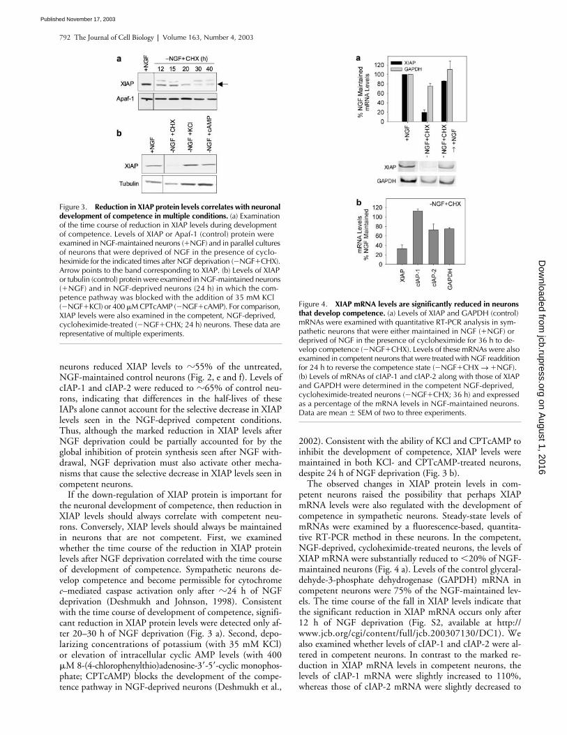
65% of control neu-rons, indicating that differences in the half-lives of theseIAPs alone cannot account for the selective decrease in XIAPlevels seen in the NGF-deprived competent conditions.Thus, although the marked reduction in XIAP levels afterNGF deprivation could be partially accounted for by theglobal inhibition of protein synthesis seen after NGF with-drawal, NGF deprivation must also activate other mecha-nisms that cause the selective decrease in XIAP levels seen incompetent neurons.
If the down-regulation of XIAP protein is important forthe neuronal development of competence, then reduction inXIAP levels should always correlate with competent neu-rons. Conversely, XIAP levels should always be maintainedin neurons that are not competent. First, we examinedwhether the time course of the reduction in XIAP proteinlevels after NGF deprivation correlated with the time courseof development of competence. Sympathetic neurons de-velop competence and become permissible for cytochrome
c
–mediated caspase activation only after
�
24 h of NGFdeprivation (Deshmukh and Johnson, 1998). Consistentwith the time course of development of competence, signifi-cant reduction in XIAP protein levels were detected only af-ter 20–30 h of NGF deprivation (Fig. 3 a). Second, depo-larizing concentrations of potassium (with 35 mM KCl)or elevation of intracellular cyclic AMP levels (with 400
�
M 8-(4-chlorophenylthio)adenosine-3
�
-5
�
-cyclic monophos-phate; CPTcAMP) blocks the development of the compe-tence pathway in NGF-deprived neurons (Deshmukh et al.,
2002). Consistent with the ability of KCl and CPTcAMP toinhibit the development of competence, XIAP levels weremaintained in both KCl- and CPTcAMP-treated neurons,despite 24 h of NGF deprivation (Fig. 3 b).
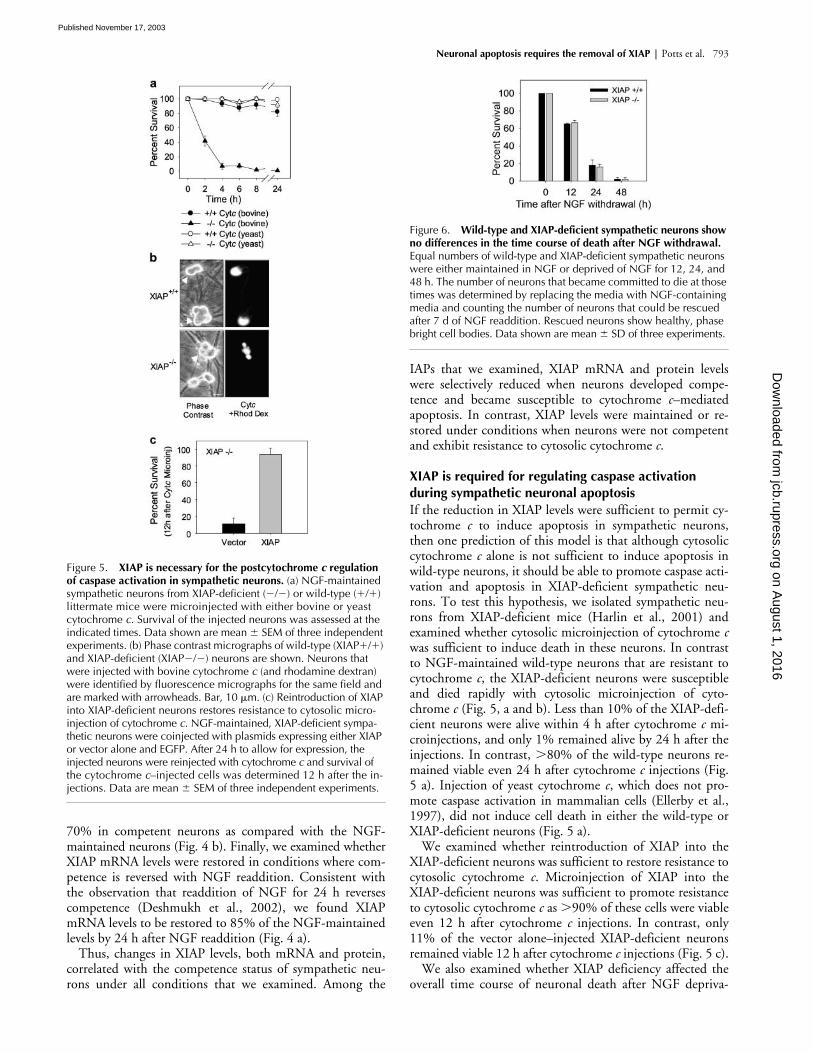
The observed changes in XIAP protein levels in com-petent neurons raised the possibility that perhaps XIAPmRNA levels were also regulated with the development ofcompetence in sympathetic neurons. Steady-state levels ofmRNAs were examined by a fluorescence-based, quantita-tive RT-PCR method in these neurons. In the competent,NGF-deprived, cycloheximide-treated neurons, the levels ofXIAP mRNA were substantially reduced to
�
20% of NGF-maintained neurons (Fig. 4 a). Levels of the control glyceral-dehyde-3-phosphate dehydrogenase (GAPDH) mRNA incompetent neurons were 75% of the NGF-maintained lev-els. The time course of the fall in XIAP levels indicate thatthe significant reduction in XIAP mRNA occurs only after12 h of NGF deprivation (Fig. S2, available at http://www.jcb.org/cgi/content/full/jcb.200307130/DC1). Wealso examined whether levels of cIAP-1 and cIAP-2 were al-tered in competent neurons. In contrast to the marked re-duction in XIAP mRNA levels in competent neurons, thelevels of cIAP-1 mRNA were slightly increased to 110%,whereas those of cIAP-2 mRNA were slightly decreased to
Figure 3. Reduction in XIAP protein levels correlates with neuronal development of competence in multiple conditions. (a) Examination of the time course of reduction in XIAP levels during development of competence. Levels of XIAP or Apaf-1 (control) protein were examined in NGF-maintained neurons (�NGF) and in parallel cultures of neurons that were deprived of NGF in the presence of cyclo-heximide for the indicated times after NGF deprivation (�NGF�CHX). Arrow points to the band corresponding to XIAP. (b) Levels of XIAP or tubulin (control) protein were examined in NGF-maintained neurons (�NGF) and in NGF-deprived neurons (24 h) in which the com-petence pathway was blocked with the addition of 35 mM KCl (�NGF�KCl) or 400 �M CPTcAMP (�NGF�cAMP). For comparison, XIAP levels were also examined in the competent, NGF-deprived, cycloheximide-treated (�NGF�CHX; 24 h) neurons. These data are representative of multiple experiments.
Figure 4. XIAP mRNA levels are significantly reduced in neurons that develop competence. (a) Levels of XIAP and GAPDH (control) mRNAs were examined with quantitative RT-PCR analysis in sym-pathetic neurons that were either maintained in NGF (�NGF) or deprived of NGF in the presence of cycloheximide for 36 h to de-velop competence (�NGF�CHX). Levels of these mRNAs were also examined in competent neurons that were treated with NGF readdition for 24 h to reverse the competence state (�NGF�CHX → �NGF). (b) Levels of mRNAs of cIAP-1 and cIAP-2 along with those of XIAP and GAPDH were determined in the competent NGF-deprived, cycloheximide-treated neurons (�NGF�CHX; 36 h) and expressed as a percentage of the mRNA levels in NGF-maintained neurons. Data are mean � SEM of two to three experiments.
on August 1, 2016
jcb.rupress.orgD
ownloaded from
Published November 17, 2003
Neuronal apoptosis requires the removal of XIAP | Potts et al. 793
70% in competent neurons as compared with the NGF-maintained neurons (Fig. 4 b). Finally, we examined whetherXIAP mRNA levels were restored in conditions where com-petence is reversed with NGF readdition. Consistent withthe observation that readdition of NGF for 24 h reversescompetence (Deshmukh et al., 2002), we found XIAPmRNA levels to be restored to 85% of the NGF-maintainedlevels by 24 h after NGF readdition (Fig. 4 a).
Thus, changes in XIAP levels, both mRNA and protein,correlated with the competence status of sympathetic neu-rons under all conditions that we examined. Among the
IAPs that we examined, XIAP mRNA and protein levelswere selectively reduced when neurons developed compe-tence and became susceptible to cytochrome c–mediatedapoptosis. In contrast, XIAP levels were maintained or re-stored under conditions when neurons were not competentand exhibit resistance to cytosolic cytochrome c.
XIAP is required for regulating caspase activation during sympathetic neuronal apoptosisIf the reduction in XIAP levels were sufficient to permit cy-tochrome c to induce apoptosis in sympathetic neurons,then one prediction of this model is that although cytosoliccytochrome c alone is not sufficient to induce apoptosis inwild-type neurons, it should be able to promote caspase acti-vation and apoptosis in XIAP-deficient sympathetic neu-rons. To test this hypothesis, we isolated sympathetic neu-rons from XIAP-deficient mice (Harlin et al., 2001) andexamined whether cytosolic microinjection of cytochrome cwas sufficient to induce death in these neurons. In contrastto NGF-maintained wild-type neurons that are resistant tocytochrome c, the XIAP-deficient neurons were susceptibleand died rapidly with cytosolic microinjection of cyto-chrome c (Fig. 5, a and b). Less than 10% of the XIAP-defi-cient neurons were alive within 4 h after cytochrome c mi-croinjections, and only 1% remained alive by 24 h after theinjections. In contrast, 80% of the wild-type neurons re-mained viable even 24 h after cytochrome c injections (Fig.5 a). Injection of yeast cytochrome c, which does not pro-mote caspase activation in mammalian cells (Ellerby et al.,1997), did not induce cell death in either the wild-type orXIAP-deficient neurons (Fig. 5 a).
We examined whether reintroduction of XIAP into theXIAP-deficient neurons was sufficient to restore resistance tocytosolic cytochrome c. Microinjection of XIAP into theXIAP-deficient neurons was sufficient to promote resistanceto cytosolic cytochrome c as 90% of these cells were viableeven 12 h after cytochrome c injections. In contrast, only11% of the vector alone–injected XIAP-deficient neuronsremained viable 12 h after cytochrome c injections (Fig. 5 c).
We also examined whether XIAP deficiency affected theoverall time course of neuronal death after NGF depriva-
Figure 5. XIAP is necessary for the postcytochrome c regulation of caspase activation in sympathetic neurons. (a) NGF-maintained sympathetic neurons from XIAP-deficient (�/�) or wild-type (�/�) littermate mice were microinjected with either bovine or yeast cytochrome c. Survival of the injected neurons was assessed at the indicated times. Data shown are mean � SEM of three independent experiments. (b) Phase contrast micrographs of wild-type (XIAP�/�) and XIAP-deficient (XIAP�/�) neurons are shown. Neurons that were injected with bovine cytochrome c (and rhodamine dextran) were identified by fluorescence micrographs for the same field and are marked with arrowheads. Bar, 10 �m. (c) Reintroduction of XIAP into XIAP-deficient neurons restores resistance to cytosolic micro-injection of cytochrome c. NGF-maintained, XIAP-deficient sympa-thetic neurons were coinjected with plasmids expressing either XIAP or vector alone and EGFP. After 24 h to allow for expression, the injected neurons were reinjected with cytochrome c and survival of the cytochrome c–injected cells was determined 12 h after the in-jections. Data are mean � SEM of three independent experiments.
Figure 6. Wild-type and XIAP-deficient sympathetic neurons show no differences in the time course of death after NGF withdrawal. Equal numbers of wild-type and XIAP-deficient sympathetic neurons were either maintained in NGF or deprived of NGF for 12, 24, and 48 h. The number of neurons that became committed to die at those times was determined by replacing the media with NGF-containing media and counting the number of neurons that could be rescued after 7 d of NGF readdition. Rescued neurons show healthy, phase bright cell bodies. Data shown are mean � SD of three experiments.
on August 1, 2016
jcb.rupress.orgD
ownloaded from
Published November 17, 2003

794 The Journal of Cell Biology | Volume 163, Number 4, 2003
Figure 7. Release of endogenous cytochrome c and Smac are not sufficient to overcome the XIAP-mediated postcytochrome c inhibition in NGF-maintained sympathetic neurons. (a) NGF-maintained neurons were either left untreated or treated with 3 mM H2O2 for 1 h. The status of cytochrome c and Smac in these neurons was examined 3 h after the H2O2 exposure by immunohistochemical techniques. Micrographs show that whereas untreated, NGF-maintained neurons show a punctate, mitochondrial pattern of staining for both cytochrome c (red) and Smac (green), H2O2 exposure induces the loss of both cytochrome c and Smac staining from the mitochondria in these neurons. Bizbenzimide staining shows the nuclei (blue). (b) Quantitation of the loss of mitochondrial cytochrome c and Smac from these neurons in response to the transient H2O2 treatment. Approximately 100 neurons were counted to determine the status of cytochrome c and Smac in untreated or H2O2-treated neurons. (c) Wild-type (XIAP�/�) or XIAP-deficient (XIAP�/�) sympathetic neurons were maintained in NGF and either left untreated or transiently exposed to H2O2 as described in panel a. The status of cytochrome c (red) and activated caspase-3 (green) in these neurons was examined by immunohistochemical techniques. Bizbenzimide staining shows the nuclei (blue). For untreated neurons, the staining patterns for both XIAP�/� and XIAP�/� conditions are identical but only the image for XIAP�/� is shown. The percentage of neurons showing activated caspase-3 immunostaining after transient H2O2 exposure in the wild-type (XIAP�/�) or XIAP-deficient (XIAP�/�) is quantitated in panel d. (e) Sympathetic neurons were either maintained in NGF (�NGF) or made competent by depriving them of NGF in the presence of cycloheximide for 24 h (�NGF�CHX). These neurons were either left untreated or treated with H2O2, and the status of cytochrome c (red) and activated caspase-3 (green) in these neurons was examined. For untreated neurons, the staining patterns for both �NGF and –NGF�CHX conditions are identical but only the image for �NGF�CHX is shown. The percentage of neurons showing activated caspase-3 immunostaining after transient H2O2 exposure in the NGF-maintained (�NGF) or competent, (�NGF�CHX) conditions is quantitated in panel f. Data shown in b, d, and f are mean � SD of three experiments. Bars: (a, c, and e) 10 �m.
on August 1, 2016
jcb.rupress.orgD
ownloaded from
Published November 17, 2003

Neuronal apoptosis requires the removal of XIAP | Potts et al. 795
tion. Cultures of sympathetic neurons from XIAP-deficientmice and their wild-type littermates were deprived of NGF,and their survival was assessed at 12, 24, and 48 h after NGFdeprivation. No differences were observed in the timecourses of death after NGF deprivation between the wild-type and XIAP-deficient neurons (Fig. 6). Thus, althoughremoval of XIAP made neurons vulnerable to cytosolic cyto-chrome c, XIAP deficiency alone did not change the overallkinetics of apoptosis after NGF withdrawal. These resultsare consistent with the idea that XIAP functions as a criticalsafety brake to protect against aberrant caspase activation ifcytochrome c is accidentally released from mitochondria.Whereas removal of XIAP was necessary for activating cas-pases in these neurons, the deletion of this safety mechanismby itself did not alter the kinetics of apoptosis after NGFdeprivation.
Hydrogen peroxide (H2O2)–induced release of endogenous cytochrome c and Smac are not sufficient to overcome XIAP in neuronsThe results thus far indicate that removal of XIAP is bothnecessary and sufficient to allow cytochrome c to activate cas-pases and apoptosis in sympathetic neurons. Although ourdata show that the NGF deprivation–induced competencepathway promoted a marked reduction in XIAP levels, an-other potential mechanism by which XIAP function could beinhibited in these neurons is by the mitochondrial release ofendogenous Smac. To examine whether endogenous Smacand cytochrome c are sufficient to overcome the XIAP inhibi-tion and permit caspase activation in these neurons, we in-duced the release of these mitochondrial proteins by treatingneurons with H2O2. H2O2 induces the release of mitochon-drial cytochrome c in sympathetic neurons (Kirkland et al.,2002). First, we optimized conditions and found that an ex-posure of 3 mM H2O2 for 1 h induced the release of mito-chondrial cytochrome c in 90% of the neurons by 3 h afterthe treatment (Fig. 7, a and b). Released cytochrome c is notdetected in the cytosol because once translocated from themitochondria, cytochrome c presumably gets degraded inthese neurons (Deshmukh and Johnson, 1998; Neame et al.,1998). Second, we examined whether endogenous Smac wasalso released along with cytochrome c under these conditions.All detectable Smac was also released from the mitochondriain 90% of neurons with H2O2 treatment (Fig. 7, a and b).Like cytochrome c, the released Smac is also presumably de-graded in the cytosol of these neurons. Thus, transient H2O2
treatment induced the loss of both cytochrome c and Smacfrom the mitochondria in sympathetic neurons.
Next, we examined whether the H2O2-induced release ofcytochrome c and Smac was sufficient to activate caspases inthese neurons. Untreated neurons showed the expectedpunctate, mitochondrial distribution of cytochrome c andno staining for activated caspase-3. Surprisingly, even withH2O2 treatment, despite the release of all detectable endoge-nous cytochrome c and Smac, virtually none of the neurons(�2%) exhibited any caspase-3 activation in wild-type neu-rons (Fig. 7, c and d).
The inability of endogenous cytochrome c and Smac,which were released from mitochondria by H2O2 treatment,to activate caspases could be because of their inability to
overcome XIAP inhibition in these neurons. To test this hy-pothesis, we examined whether the H2O2-induced release ofendogenous cytochrome c and Smac were sufficient to acti-vate caspases in XIAP-deficient neurons. In contrast to theresults in wild-type neurons, H2O2 treatment induced a ro-bust activation of caspase-3 in XIAP-deficient neurons in25% of the cells (Fig. 7, c and d). The number of cells thatshow caspase activation at any given time is not expected tobe very high because cells in which caspases become acti-vated undergo rapid apoptosis and die. Thus, H2O2-inducedrelease of endogenous cytochrome c and Smac, although notsufficient to overcome XIAP inhibition and permit caspaseactivation in wild-type sympathetic neurons, induced rapidcaspase activation in XIAP-deficient neurons.
To determine whether the down-regulation of XIAP in-duced by the competence pathway was sufficient to permitcaspase activation by endogenous cytochrome c and Smac, weexamined whether H2O2 exposure induced caspase activationin competent, NGF-deprived cycloheximide-treated neurons.Like in the XIAP-deficient neurons, H2O2 treatment inducedrobust caspase activation in competent sympathetic neurons(Fig. 7, e and f). The inability of endogenous Smac releasealone to overcome XIAP inhibition in NGF-maintained neu-rons underscores the importance of the competence pathwayin removing XIAP and permitting endogenous cytochrome cto activate caspases in sympathetic neurons.
DiscussionPrimary sympathetic neurons, unlike most mitotic cells, areremarkably resistant to cytosolic microinjection of cyto-chrome c. In this paper, we investigated the mechanism be-hind the stringent postcytochrome c regulation of caspaseactivation in sympathetic neurons. We report that the in-ability of cytochrome c to induce apoptosis in these neuronsis because of an essential function of endogenous XIAP inregulating caspase activation postcytochrome c. Our resultsare consistent with the model in which removal of XIAP isboth necessary and sufficient to allow cytochrome c–medi-ated caspase activation during sympathetic neuronal apopto-sis. These results are the first to identify a mammalian celltype in which apoptosis is regulated postcytochrome c by en-dogenous XIAP.
Down-regulation of XIAP during sympathetic neuronal apoptosisNGF deprivation–induced sympathetic neuronal apoptosisrequires activation of both the cytochrome c release and thedevelopment of competence pathways. We found that uponNGF deprivation, the competence pathway induced a dra-matic reduction in the levels of XIAP mRNA and protein insympathetic neurons (Figs. 2 and 3). These data provide in-sight into how the competence pathway is able to overcomethe neuronal resistance to cytosolic cytochrome c and permitcaspase activation and apoptosis in these neurons. Other sit-uations in which sympathetic neurons become competentand die with cytochrome c, such as after axotomy (Fletcheret al., 2000), may also induce similar mechanisms that re-move XIAP. Decreases in XIAP levels have also been ob-served in other neurons undergoing cell death in vivo (Gue-
on August 1, 2016
jcb.rupress.orgD
ownloaded from
Published November 17, 2003

796 The Journal of Cell Biology | Volume 163, Number 4, 2003
gan et al., 2001; Korhonen et al., 2001; Ishigaki et al., 2002;Perrelet et al., 2002).
We found no evidence of XIAP cleavage during the devel-opment of competence as no XIAP cleavage product wasfound to accumulate in NGF-deprived, competent neurons(Fig. S3 a, available at http://www.jcb.org/cgi/content/full/jcb.200307130/DC1). Also, in contrast to the caspase-medi-ated cleavage of DIAP1 seen in Drosophila (Ditzel et al.,2003), the loss of XIAP could not be blocked with caspaseinhibition in sympathetic neurons (Fig. 2 d). We also exam-ined the possibility that XIAP might be targeted for protea-some-mediated degradation in these neurons, as is seen dur-ing thymocyte apoptosis (Yang et al., 2000). However, noubiquitination of endogenous XIAP was detected in primarysympathetic neurons after NGF withdrawal (not depicted),and addition of the proteasome inhibitor lactacystin did notblock the reduction in XIAP levels seen in NGF-deprivedneurons (Fig. S3 b). A recent paper has identified the sites onXIAP that can be ubiquitinated in cells (Shin et al., 2003).The specific importance of the ubiquitin pathway in degrad-ing XIAP in neurons may require examining whether mu-tant XIAP that cannot be ubiquitinated are also targeted forremoval in NGF-deprived, competent sympathetic neurons.
Because NGF deprivation induces an overall reductionin neuronal metabolism (Deckwerth and Johnson, 1993),some decrease in the steady-state levels of all proteins is ex-pected in NGF-deprived sympathetic neurons. However, al-though levels of cIAP-1, cIAP-2, and Apaf-1 protein werereduced (by 20–35%) after NGF deprivation, these de-creases were small compared with the 70% decrease inXIAP levels observed under these conditions (Fig. 2). Im-portantly, total inhibition of protein synthesis with cyclo-heximide addition alone in NGF-maintained neurons in-duced a 45% reduction in XIAP levels (Fig. 2). Therefore,the turnover of XIAP protein after the fall in protein synthe-sis after NGF deprivation (Deckwerth and Johnson, 1993)could be one factor that contributes to the marked reductionin XIAP levels in NGF-deprived competent neurons. How-ever, cycloheximide addition alone to NGF-maintainedneurons is not sufficient to induce competence (Deshmukhand Johnson, 1998). The additional decrease in XIAP levels(25% above those seen with total inhibition of protein syn-thesis) that is seen in the NGF-deprived conditions, and ispresumably important for the development of competence,could be mediated by other posttranslational mechanismsinduced under the NGF deprivation conditions.
Essential function of endogenous XIAP in regulating caspase activation in sympathetic neuronsOur data show that whereas wild-type sympathetic neuronsare remarkably resistant to cytosolic cytochrome c, theXIAP-deficient neurons exhibited no resistance and diedrapidly with cytosolic cytochrome c (Figs. 5 and 7). Further-more, restoring XIAP expression in the XIAP knockout neu-rons restored the resistance to cytosolic cytochrome c inthese neurons (Fig. 5 c). These results identify XIAP as anessential postcytochrome c regulator of caspase activation insympathetic neurons.
Although XIAP deficiency made sympathetic neurons vul-nerable to cytosolic cytochrome c, it did not change the over-
all time course of apoptosis after NGF withdrawal (Fig. 6).Antisense-mediated depletion of XIAP also does not changethe time course of sympathetic neuronal death after NGFdeprivation (Troy et al., 2001). We have previously shownthat the time courses of cytochrome c release and develop-ment of competence (removal of XIAP) after NGF depriva-tion are very similar, with 50% of neurons having releasedcytochrome c and developed competence by 18–20 h afterNGF withdrawal (Deshmukh and Johnson, 1998). Thus, bythe time the neuron reaches the point of cytochrome c re-lease, it also removes XIAP, thereby permitting caspase acti-vation and apoptosis to occur in these neurons. Apoptosis inXIAP-deficient neurons, although no longer needing thecompetence pathway to remove XIAP, is still dependent onthe release of cytochrome c and therefore occurs with a timecourse that is indistinguishable from wild-type neurons.
Removal of XIAP during sympathetic neuronal apoptosis: Relative importance of the competence pathway versus mitochondrial release of SmacWe found that H2O2-induced mitochondrial release ofendogenous Smac and cytochrome c was not sufficient toovercome XIAP inhibition and activate caspases in NGF-maintained sympathetic neurons (Fig. 7). Thus, althoughexogenously microinjected, excess Smac is capable of remov-ing XIAP inhibition in these neurons (Fig. 1; Deshmukh etal., 2002), levels of endogenous Smac may simply be insuffi-cient to overcome XIAP in these cells. However, in compe-tent neurons, which have markedly decreased XIAP levels,and in XIAP-deficient neurons, the H2O2-induced release ofendogenous cytochrome c and Smac induced robust caspaseactivation (Fig. 7). Consistent with this observation, recentdata show that NGF-deprived Bax �/� competent neuronsare more susceptible to H2O2 exposure than NGF-main-tained sympathetic neurons (Kirkland et al., 2002). Theseresults suggest that the competence pathway, and not the re-lease of endogenous mitochondrial Smac, is likely to be thepredominant mechanism that removes XIAP and permitscaspase activation in sympathetic neurons.
This critical function of XIAP in sympathetic neurons ispresumably to be a “safety brake” that protects against anyaccidental caspase activation if cytochrome c is unexpectedlyreleased into the cytosol of these neurons. Arguably, for sucha safety mechanism to be effective, XIAP must also with-stand any inhibition from other proteins that may be re-leased along with cytochrome c, such as Smac, in such acci-dental situations. Such a safety brake would be particularlyimportant in postmitotic cells such as sympathetic neuronsthat are not replaceable and need to last for the lifetime ofthe organism. Similar postcytochrome c protection fromapoptosis is also seen in Xenopus eggs, presumably becauseensuring the survival of gametes is advantageous to the or-ganism (Tashker et al., 2002). In neurons, physiologicallyappropriate apoptotic stimuli such as NGF deprivationwould activate not only the cytochrome c release pathway,but also the competence pathway to ensure the removal ofthe XIAP brake and permit caspase activation.
The regulation of apoptosis in mammalian sympatheticneurons is conceptually similar to the “gas and brake” modelin Drosophila, where apoptosis requires both the removal of
on August 1, 2016
jcb.rupress.orgD
ownloaded from
Published November 17, 2003

Neuronal apoptosis requires the removal of XIAP | Potts et al. 797
IAPs and activation of caspases (Rodriguez et al., 2002).However, in Drosophila, activation of caspases appears to beconstitutive, as removal of DIAP1 alone results in increasedapoptosis and lethality (Wang et al., 1999). In contrast, inmammalian sympathetic neurons, caspase activation is de-pendent on both the removal of endogenous XIAP (thebrake) and the initiation of the cytochrome c release path-way (the gas). These results are consistent with the observa-tion that XIAP deficiency alone does not cause increasedapoptosis in mammalian cells under normal physiologicalconditions (Harlin et al., 2001). However, the XIAP-defi-cient neurons, like neuronal apoptosis inhibitory protein-deficient neurons (Holcik et al., 2000), may be predicted tobe more vulnerable if exposed to toxic stimuli that cause mi-tochondrial damage and cytochrome c release because theylack the postcytochrome c safety brake.
Materials and methodsReagentsAll reagents were purchased from Sigma-Aldrich or Fisher Scientific, un-less otherwise stated. Collagenase and trypsin were purchased from Wor-thington Biochemical Corporation and cycloheximide was purchased fromTocris Cookson. The pan-caspase inhibitor zVAD-FMK was purchasedfrom Enzyme Systems Products.
Sympathetic neuronal culturesSympathetic neurons were dissected from superior cervical ganglia of post-natal day zero to one mice (P0–P1) and maintained in culture as describedpreviously (Deshmukh et al., 2002). Cells were plated on collagen-coateddishes at a density of 30,000 cells per well for protein lysates, or 2,500cells per 35-mm dish for microinjection, survival counts, or immunofluo-rescence experiments. Sympathetic neurons were grown for 4–5 d in theNGF-containing media before treating them with experimental conditions.For NGF deprivation, cultures were rinsed twice with medium lackingNGF followed by addition of goat anti-NGF neutralizing antibody to thismedia. Other conditions required the addition of 1 �g/ml cycloheximide,400 �M CPTcAMP, or 35 mM KCl to anti-NGF–containing media. Sympa-thetic neurons from Bax-deficient mice were isolated as described previ-ously (Deckwerth et al., 1996).
Bacterial expression of recombinant proteinsA COOH-terminal His-tagged bacterial expression plasmid (pET15b) con-taining mature Smac cDNA (AVPI-Smac; corresponding to amino acids56–236 of the full-length Smac protein) was provided by C. Du (StowersInstitute, Kansas City, MO). The initiating Met in the “AVPI-Smac” proteinis removed during bacterial expression by the aminopeptidase activity be-cause of the presence of Ala as the second residue (Chai et al., 2000). Togenerate the “MVPI-Smac” protein, the first Ala residue was simply de-leted by site directed mutagenesis. Because the second residue now is Valinstead of Ala, the initiating Met remains intact, thus generating the MVPI-Smac protein. Primer sequences used for this site directed mutagene-sis were: forward 5�-AGAAGGAGATATACCATGGTTCCTATTGCAGAG-3� and reverse 5�-CTGTGCAATAGGAACCATGGTATATCTCCTTCT-3�.Escherichia coli BL21-(DE3) bacteria strain (Stratagene) were transformedwith either the AVPI-Smac– or MVPI-Smac–containing plasmids. Bacte-rial cultures were grown in Luria Bertani media until reaching a density ofOD600 0.6. Expression of protein was induced with 1 mM IPTG for 3 hat 37�C and cells were harvested by centrifugation. The bacterial pelletwas lysed by sonication and Ni-NTA agarose beads (QIAGEN) were usedto purify the His-tagged protein according to the manufacturer’s instruc-tions. The eluted fractions were analyzed by SDS-PAGE and fractionscontaining the desired protein were pooled and dialyzed overnight at 4�Cinto dialysis buffer (20 mM Hepes, 10 mM KCl, 1.5 mM MgCl2, 0.5 mMEDTA, 0.5 mM EGTA, and 1 mM DTT, pH 7.4) and stored in aliquots at�80�C until needed.
Microinjection and quantitation of cell survivalOur method for microinjecting sympathetic neurons with cytochrome chas been described previously (Deshmukh and Johnson, 1998). In brief,sympathetic neurons were microinjected using Femtotip II needles (Eppen-
dorf Inc.) and a Narashigi micromanipulator mounted on an inverted fluo-rescence microscope (Leica). The microinjection solution (100 mM KCland 10 mM KPi, pH 7.4) contained 4 mg/ml of rhodamine dextran dye tomark the injected cells. The concentration of bovine cytochrome c in-jected was 10–15 mg/ml, and that of mature Smac was 1 mg/ml. Immedi-ately after the injections, the number of viable cells injected was deter-mined by counting the number of rhodamine-positive cells that had intact,phase-bright cell bodies. This method of assessing neuronal survival corre-lates well with other cell survival assays such as trypan blue exclusion andstaining with calcien AM (Fig. S1, available at http://www.jcb.org/cgi/content/full/jcb.200307130/DC1). At various times after injections, thenumber of viable, injected neurons remaining was determined by usingthe same counting criteria and expressed as a percentage of the originalnumber of microinjected cells.
In experiments involving microinjection of the XIAP-expressing plasmid(pEBB-XIAP; a gift provided by C. Duckett, University of Michigan, Ann Ar-bor, MI), neurons were injected in the nucleus with 200 ng/�l of the plas-mid DNA along with 50 ng/�l EGFP-expressing DNA (CLONTECH Labora-tories, Inc.) in microinjection buffer. After 24 h to allow for expression,GFP-expressing cells were identified by fluorescence microscopy and rein-jected with cytochrome c (as described in the previous paragraph). The sur-vival of these double injected cells was assessed 12 h after cytochrome cinjections.
Western blot analysisSympathetic neuronal cultures were washed in PBS and harvested in RIPAbuffer (150 mM NaCl, 1% NP-40, 0.5% sodium deoxycholate, 0.1% SDS,50 mM Tris-HCl, pH 8.0, and protease inhibitor cocktail), and subjected toSDS-PAGE analysis as described previously (Deshmukh et al., 2002). Forquantitation, the Western blots were developed with ECL-plus reagents(Amersham Biosciences) and analyzed on a Typhoon fluorescent imager(Amersham Biosciences) using the ImageQuant software (Amersham Bio-sciences). The specific antibodies used in experiments are listed in the On-line supplemental materials and methods section, available at http://www.jcb.org/cgi/content/full/jcb.200307130/DC1.
Quantitation of mRNA expressionOur method of quantitative RT-PCR analysis is a modification of a previ-ously published protocol (Estus et al., 1994), where we substituted the ra-dioactivity-based detection method with a fluorescence-based detectiontechnique. In brief, parallel cultures of sympathetic neurons with equalcell numbers plated were grown under the appropriate conditions andtotal RNA was harvested using QIAGEN RNAeasy kit. RNA was con-verted to cDNA by Superscript II reverse transcriptase (Invitrogen) usingrandom hexamers (Invitrogen) as primer templates. Primer sets forGAPDH, XIAP, cIAP-1, and cIAP-2 (see Online supplemental material)were first analyzed for their linear range of amplification by varying thenumber of cycles and amount of input cDNA. The optimal cycle num-ber that yielded a detectable, linearly amplified product was found to be18 cycles for GAPDH and 22 cycles for XIAP, cIAP-1, and cIAP-2. PCRwas performed for the indicated number of cycles for each primer pairand the PCR products were separated on an 8% polyacrylamide nonde-naturing gel. The gel was stained with Sybr Green I (Molecular Probes)for 45 min on a rocker and the stained PCR products were visualized ona Typhoon fluorescence imager and quantitated using ImageQuant soft-ware.
Genotyping of XIAP �/� miceGenotyping was performed by digesting tail DNA overnight in genotypingbuffer (100 mM Tris-HCl, pH 8.5, 200 mM NaCl, 5 mM EDTA, 0.2% SDS,10 mg/ml proteinase K) at 55�C. DNA was precipitated by gently addingthree volumes of ice-cold ethanol to the supernatant and spooling at theinterface. The PCR primers used for genotyping are listed in the Onlinesupplemental materials and methods section.
Quantitation of neuronal survival after NGF deprivationSympathetic neuronal survival after 0, 12, 24, and 48 h of NGF depriva-tion was assessed by determining the percentage of cells that could be res-cued by adding back NGF to the cultures at those times. Equal numbers ofsympathetic neurons plated in multiple dishes were deprived of NGF forthe indicated times, rinsed three times, and incubated in fresh NGF-con-taining media. After 7 d of NGF readdition, the rescued neurons wereclearly identifiable with large and phase-bright cell bodies, whereas thenonrescued neurons atrophied and degenerated. All rescued cells in thedish were counted and expressed as a percent of the number of cells inthe 0 h, untreated condition.
on August 1, 2016
jcb.rupress.orgD
ownloaded from
Published November 17, 2003

798 The Journal of Cell Biology | Volume 163, Number 4, 2003
Immunofluorescence analysisImmunofluorescence analysis on sympathetic neuronal cultures was per-formed essentially as described previously (Deshmukh and Johnson,1998). The specific antibodies used in experiments are listed in the Onlinesupplemental materials and methods section.
Transient exposure of sympathetic neurons to H2O2 was done by treat-ment of neurons with 3 mM H2O2 for 1 h, with a change into fresh H2O2
containing media after 30 min. Cells were washed three times with NGF-containing (AM50) media and allowed to incubate at 37�C for three addi-tional hours in AM50 media. Cells were fixed and subjected to immunoflu-orescence analysis.
Online supplemental materialFig. S1 shows the assessment of neuronal survival after cytochrome c mi-croinjections using multiple techniques. Fig. S2 shows the time course ofdecrease in XIAP mRNA levels after NGF deprivation. Fig. S3 shows a full-length Western blot of extracts from NGF-maintained and NGF-deprived,cycloheximide-treated sympathetic neurons probed for XIAP. This figurealso shows the inability of lactacystin to block the decrease in XIAP levelsin the NGF-deprived, competent neurons. Online supplemental material isavailable at http://www.jcb.org/cgi/content/full/jcb.200307130/DC1.
We thank Colin Duckett for the XIAP plasmid and Chunying Du for theSmac plasmid. We also thank Eugene Johnson and members of the Desh-mukh laboratory for critical review of this manuscript.
This work was supported by National Institutes of Health grantNS42197 and the Burroughs Wellcome Fund New Investigator Award toM. Deshmukh.
Submitted: 21 July 2003Accepted: 8 October 2003
ReferencesBrustugun, O.T., K.E. Fladmark, S.O. Doskeland, S. Orrenius, and B. Zhivo-
tovsky. 1998. Apoptosis induced by microinjection of cytochrome c is cas-pase-dependent and is inhibited by Bcl-2. Cell Death Differ. 5:660–668.
Chai, J., C. Du, J.W. Wu, S. Kyin, X. Wang, and Y. Shi. 2000. Structural and bio-chemical basis of apoptotic activation by Smac/DIABLO. Nature. 406:855–862.
Chang, S.H., P.C. Phelps, I.K. Berezesky, M.J. Ebersberger, and B.F. Trump.2000. Studies on the mechanisms and kinetics of apoptosis induced by mi-croinjection of cytochrome c in rat kidney tubule epithelial cells (NRK-52E). Am. J. Pathol. 156:637–649.
Deckwerth, T.L., and E.M. Johnson, Jr. 1993. Temporal analysis of events associ-ated with programmed cell death (apoptosis) of sympathetic neurons de-prived of nerve growth factor. J. Cell Biol. 123:1207–1222.
Deckwerth, T.L., J.L. Elliott, C.M. Knudson, E.M. Johnson, Jr., W.D. Snider,and S.J. Korsmeyer. 1996. Bax is required for neuronal death after trophicfactor deprivation and during development. Neuron. 17:401–411.
Denault, J.B., and G.S. Salvesen. 2002. Caspases: keys in the ignition of cell death.Chem. Rev. 102:4489–4500.
Deshmukh, M., and E.M. Johnson, Jr. 1997. Programmed cell death in neurons:focus on the pathway of nerve growth factor deprivation-induced death ofsympathetic neurons. Mol. Pharmacol. 51:897–906.
Deshmukh, M., and E.M. Johnson, Jr. 1998. Evidence of a novel event duringneuronal death: development of competence-to-die in response to cytoplas-mic cytochrome c. Neuron. 21:695–705.
Deshmukh, M., J. Vasilakos, T.L. Deckwerth, P.A. Lampe, B.D. Shivers, andE.M. Johnson, Jr. 1996. Genetic and metabolic status of NGF-deprivedsympathetic neurons saved by an inhibitor of ICE-family proteases. J. CellBiol. 135:1341–1354.
Deshmukh, M., K. Kuida, and E.M. Johnson, Jr. 2000. Caspase inhibition extendsthe commitment to neuronal death beyond cytochrome c release to the pointof mitochondrial depolarization. J. Cell Biol. 150:131–143.
Deshmukh, M., C. Du, X. Wang, and E.M. Johnson, Jr. 2002. Exogenous smacinduces competence and permits caspase activation in sympathetic neurons.J. Neurosci. 22:8018–8027.
Deveraux, Q.L., N. Roy, H.R. Stennicke, T. Vanarsdale, Q. Zhou, S.M. Sriniva-sula, E.S. Alnemri, G.S. Salvesen, and J.C. Reed. 1998. IAPs block apoptoticevents induced by caspase-8 and cytochrome c by direct inhibition of dis-tinct caspases. EMBO J. 17:2215–2223.
Ditzel, M., R. Wilson, T. Tenev, A. Zachariou, A. Paul, E. Deas, and P. Meier.
2003. Degradation of DIAP1 by the N-end rule pathway is essential for reg-ulating apoptosis. Nat. Cell Biol. 5:467–473.
Du, C., M. Fang, Y. Li, L. Li, and X. Wang. 2000. Smac, a mitochondrial proteinthat promotes cytochrome c-dependent caspase activation by eliminatingIAP inhibition. Cell. 102:33–42.
Duckett, C.S., F. Li, Y. Wang, K.J. Tomaselli, C.B. Thompson, and R.C. Arm-strong. 1998. Human IAP-like protein regulates programmed cell deathdownstream of Bcl-xL and cytochrome c. Mol. Cell. Biol. 18:608–615.
Ellerby, H.M., S.J. Martin, L.M. Ellerby, S.S. Naiem, S. Rabizadeh, G.S. Salvesen,C.A. Casiano, N.R. Cashman, D.R. Green, and D.E. Bredesen. 1997. Es-tablishment of a cell-free system of neuronal apoptosis: comparison ofpremitochondrial, mitochondrial, and postmitochondrial phases. J. Neurosci.17:6165–6178.
Estus, S., W.J. Zaks, R.S. Freeman, M. Gruda, R. Bravo, and E.M. Johnson, Jr.1994. Altered gene expression in neurons during programmed cell death:identification of c-jun as necessary for neuronal apoptosis. J. Cell Biol. 127:1717–1727.
Fletcher, G.C., L. Xue, S.K. Passingham, and A.M. Tolkovsky. 2000. Death com-mitment point is advanced by axotomy in sympathetic neurons. J. Cell Biol.150:741–754.
Green, D.R., and G.I. Evan. 2002. A matter of life and death. Cancer Cell. 1:19–30.
Guegan, C., M. Vila, G. Rosoklija, A.P. Hays, and S. Przedborski. 2001. Recruit-ment of the mitochondrial-dependent apoptotic pathway in amyotrophiclateral sclerosis. J. Neurosci. 21:6569–6576.
Harlin, H., S.B. Reffey, C.S. Duckett, T. Lindsten, and C.B. Thompson. 2001.Characterization of XIAP-deficient mice. Mol. Cell. Biol. 21:3604–3608.
Holcik, M., C.S. Thompson, Z. Yaraghi, C.A. Lefebvre, A.E. MacKenzie, andR.G. Korneluk. 2000. The hippocampal neurons of neuronal apoptosis in-hibitory protein 1 (NAIP1)-deleted mice display increased vulnerability tokainic acid-induced injury. Proc. Natl. Acad. Sci. USA. 97:2286–2290.
Ishigaki, S., Y. Liang, M. Yamamoto, J. Niwa, Y. Ando, T. Yoshihara, H. Takeu-chi, M. Doyu, and G. Sobue. 2002. X-linked inhibitor of apoptosis proteinis involved in mutant SOD1-mediated neuronal degeneration. J. Neuro-chem. 82:576–584.
Juin, P., A.O. Hueber, T. Littlewood, and G. Evan. 1999. c-Myc-induced sensiti-zation to apoptosis is mediated through cytochrome c release. Genes Dev. 13:1367–1381.
Kirkland, R.A., J.A. Windelborn, J.M. Kasprzak, and J.L. Franklin. 2002. A Bax-induced pro-oxidant state is critical for cytochrome c release during pro-grammed neuronal death. J. Neurosci. 22:6480–6490.
Korhonen, L., N. Belluardo, and D. Lindholm. 2001. Regulation of X-chromo-some-linked inhibitor of apoptosis protein in kainic acid-induced neuronaldeath in the rat hippocampus. Mol. Cell. Neurosci. 17:364–372.
Li, F., A. Srinivasan, Y. Wang, R.C. Armstrong, K.J. Tomaselli, and L.C. Fritz.1997. Cell-specific induction of apoptosis by microinjection of cytochromec. Bcl-XL has activity independent of cytochrome c release. J. Biol. Chem.272:30299–30305.
MacFarlane, M., W. Merrison, S.B. Bratton, and G.M. Cohen. 2002. Proteasome-mediated degradation of Smac during apoptosis: XIAP promotes Smac ubiq-uitination in vitro. J. Biol. Chem. 277:36611–36616.
Martin, D.P., R.E. Schmidt, P.S. DiStefano, O.H. Lowry, J.G. Carter, and E.M.Johnson, Jr. 1988. Inhibitors of protein synthesis and RNA synthesis pre-vent neuronal death caused by nerve growth factor deprivation. J. Cell Biol.106:829–844.
McCarthy, M.J., L.L. Rubin, and K.L. Philpott. 1997. Involvement of caspases insympathetic neuron apoptosis. J. Cell Sci. 110:2165–2173.
Neame, S.J., L.L. Rubin, and K.L. Philpott. 1998. Blocking cytochrome c activitywithin intact neurons inhibits apoptosis. J. Cell Biol. 142:1583–1593.
Perrelet, D., A. Ferri, P. Liston, P. Muzzin, R.G. Korneluk, and A.C. Kato. 2002.IAPs are essential for GDNF-mediated neuroprotective effects in injuredmotor neurons in vivo. Nat. Cell Biol. 4:175–179.
Rodriguez, A., P. Chen, H. Oliver, and J.M. Abrams. 2002. Unrestrained caspase-dependent cell death caused by loss of Diap1 function requires the Drosoph-ila Apaf-1 homolog, Dark. EMBO J. 21:2189–2197.
Salvesen, G.S., and C.S. Duckett. 2002. IAP proteins: blocking the road to death’sdoor. Nat. Rev. Mol. Cell Biol. 3:401–410.
Shin, H., K. Okada, J.C. Wilkinson, K.M. Solomon, C.S. Duckett, J.C. Reed, andG.S. Salvesen. 2003. Identification of ubiquitination sites on the X-linkedinhibitor of apoptosis protein. Biochem. J. 373:965–971.
Simons, M., S. Beinroth, M. Gleichmann, P. Liston, R.G. Korneluk, A.E. Mac-Kenzie, M. Bahr, T. Klockgether, G.S. Robertson, M. Weller, and J.B. Schulz.
on August 1, 2016
jcb.rupress.orgD
ownloaded from
Published November 17, 2003
Neuronal apoptosis requires the removal of XIAP | Potts et al. 799
1999. Adenovirus-mediated gene transfer of inhibitors of apoptosis proteindelays apoptosis in cerebellar granule neurons. J. Neurochem. 72:292–301.
Suzuki, Y., Y. Imai, H. Nakayama, K. Takahashi, K. Takio, and R. Takahashi.2001a. A serine protease, HtrA2, is released from the mitochondria and in-teracts with XIAP, inducing cell death. Mol. Cell. 8:613–621.
Suzuki, Y., Y. Nakabayashi, and R. Takahashi. 2001b. Ubiquitin-protein ligase ac-tivity of X-linked inhibitor of apoptosis protein promotes proteasomal deg-radation of caspase-3 and enhances its anti-apoptotic effect in Fas-inducedcell death. Proc. Natl. Acad. Sci. USA. 98:8662–8667.
Tashker, J.S., M. Olson, and S. Kornbluth. 2002. Post-cytochrome c protectionfrom apoptosis conferred by a MAPK pathway in Xenopus egg extracts. Mol.Biol. Cell. 13:393–401.
Troy, C.M., L. Stefanis, A. Prochiantz, L.A. Greene, and M.L. Shelanski. 1996.The contrasting roles of ICE family proteases and interleukin-1� in apopto-sis induced by trophic factor withdrawal and by copper/zinc superoxide dis-mutase down-regulation. Proc. Natl. Acad. Sci. USA. 93:5635–5640.
Troy, C.M., S.A. Rabacchi, J.B. Hohl, J.M. Angelastro, L.A. Greene, and M.L.Shelanski. 2001. Death in the balance: alternative participation of the cas-pase-2 and -9 pathways in neuronal death induced by nerve growth factordeprivation. J. Neurosci. 21:5007–5016.
Vaux, D.L., and S.J. Korsmeyer. 1999. Cell death in development. Cell. 96:245–254.
Verhagen, A.M., P.G. Ekert, M. Pakusch, J. Silke, L.M. Connolly, G.E. Reid, R.L.Moritz, R.J. Simpson, and D.L. Vaux. 2000. Identification of DIABLO, amammalian protein that promotes apoptosis by binding to and antagonizingIAP proteins. Cell. 102:43–53.
Wang, S.L., C.J. Hawkins, S.J. Yoo, H.A. Muller, and B.A. Hay. 1999. The Dro-sophila caspase inhibitor DIAP1 is essential for cell survival and is negativelyregulated by HID. Cell. 98:453–463.
Wang, X. 2001. The expanding role of mitochondria in apoptosis. Genes Dev. 15:2922–2933.
Wiese, S., M.R. Digby, J.M. Gunnersen, R. Gotz, G. Pei, B. Holtmann, J. Lo-wenthal, and M. Sendtner. 1999. The anti-apoptotic protein ITA is essentialfor NGF-mediated survival of embryonic chick neurons. Nat. Neurosci.2:978–983.
Yang, Y., S.Y. Fang, J.P. Jensen, A.M. Weissman, and J.D. Ashwell. 2000. Ubiq-uitin protein ligase activity of IAPs and their degradation in proteasomes inresponse to apoptotic stimuli. Science. 288:874–877.
Yu, L.Y., L. Korhonen, R. Martinez, E. Jokitalo, Y. Chen, U. Arumae, and D.Lindholm. 2003. Regulation of sympathetic neuron and neuroblastoma celldeath by XIAP and its association with proteasomes in neural cells. Mol.Cell. Neurosci. 22:308–318.
Yuan, J., and B.A. Yankner. 2000. Apoptosis in the nervous system. Nature. 407:802–809.
on August 1, 2016
jcb.rupress.orgD
ownloaded from
Published November 17, 2003