Hepatic resection and transplantation for peripheral cholangiocarcinoma
17
Hepatic Resection and Transplantation for Peripheral Cholangiocarcinoma F. Adrian Casavilla, MD, FACS * , J. Wallis Marsh, MD, FACS * , Shunzaburo Iwatsuki, MD, PhD, FACS * , Satoru Todo, MD, FACS * , Randall G. Lee, MD † , Juan R. Madariaga, MD, FACS * , Antonio Pinna, MD * , Igor Dvorchik, PhD * , John J. Fung, MD, PhD, FACS * , and Thomas E. Starzl, MD, PhD, FACS * * Department of Surgery, the Thomas E. Starzl Transplantation Institute, University of Pittsburgh Medical Center, Pittsburgh, PA. † Department of Pathology, the Thomas E. Starzl Transplantation Institute, University of Pittsburgh Medical Center, Pittsburgh, PA. Abstract Background—Recent publications have questioned the role of orthotopic liver transplantation (OLT) in treating advanced or unresectable peripheral cholangiocarcinoma (Ch-Ca). Study Design—We reviewed our experience with Ch-Ca to determine survival rates, recurrence patterns, and risk factors in 54 patients who underwent either hepatic resection or OLT between 1981 and 1994. Liver transplantation was performed in patients with unresectable tumors (n = 12) and in those with advanced cirrhosis (n = 8). There were 33 women (61%) and 21 men (39%), with a mean age of 54.3 years. The median followup period was 6.8 years. Prognostic risk factors were analyzed by univariate and multivariate analyses. Results—Mortality within 30 days was 7.4%. Overall patient and tumor-free survival rates were 64% and 57% at 1 year, 34% and 34% at 3 years, and 26% and 27% at 5 years after operation. Thirty- two patients (59.3%) experienced tumor recurrence. Univariate analysis revealed that multiple tumors, bilobar tumor distribution, regional lymph node involvement, presence of metastasis, positive surgical margins, and advanced pTNM stages were significant negative predictors of both tumor-free and patient survival. Multivariate analysis revealed that positive margins, multiple tumors, and lymph node involvement were independently associated with poor prognosis. When patients with these three negative predictors were excluded, the patient survivals at 1, 3, and 5 years were 74%, 64%, and 62%, respectively. Conclusions—Both hepatic resection and OLT are effective therapies for Ch-Ca when the tumor can be removed with adequate margins, the lesion is singular, and lymph nodes are not involved. Peripheral or intrahepatic cholangiocarcinoma (Ch-Ca) is second (10–20%) only to hepatocellular carcinoma (70–80%) as the most common primary malignancy of the liver. Currently, non-surgical therapeutic modalities for Ch-Ca are ineffective. Hepatic resection (partial hepatectomy) has been the conventional form of potentially curative treatment (1–5); the possibility of extending extirpation to total hepatectomy (6–8) or upper-abdominal exenteration (9–11) followed by liver replacement has been controversial (12–16). © 1997 by the American College of Surgeons Correspondence address: F. Adrian Casavilla, MD, FACS, Department of Surgery, Thomas E. Starzl Transplantation Institute, Falk Clinic, 4th floor, 3601 Fifth Ave., Pittsburgh, PA 15213. NIH Public Access Author Manuscript J Am Coll Surg. Author manuscript; available in PMC 2010 October 21. Published in final edited form as: J Am Coll Surg. 1997 November ; 185(5): 429–436. NIH-PA Author Manuscript NIH-PA Author Manuscript NIH-PA Author Manuscript
-
Upload
independent -
Category
Documents
-
view
0 -
download
0
Transcript of Hepatic resection and transplantation for peripheral cholangiocarcinoma

Hepatic Resection and Transplantation for PeripheralCholangiocarcinoma
F. Adrian Casavilla, MD, FACS*, J. Wallis Marsh, MD, FACS*, Shunzaburo Iwatsuki, MD, PhD,FACS*, Satoru Todo, MD, FACS*, Randall G. Lee, MD†, Juan R. Madariaga, MD, FACS*,Antonio Pinna, MD*, Igor Dvorchik, PhD*, John J. Fung, MD, PhD, FACS*, and Thomas E.Starzl, MD, PhD, FACS**Department of Surgery, the Thomas E. Starzl Transplantation Institute, University of PittsburghMedical Center, Pittsburgh, PA.†Department of Pathology, the Thomas E. Starzl Transplantation Institute, University of PittsburghMedical Center, Pittsburgh, PA.
AbstractBackground—Recent publications have questioned the role of orthotopic liver transplantation(OLT) in treating advanced or unresectable peripheral cholangiocarcinoma (Ch-Ca).
Study Design—We reviewed our experience with Ch-Ca to determine survival rates, recurrencepatterns, and risk factors in 54 patients who underwent either hepatic resection or OLT between 1981and 1994. Liver transplantation was performed in patients with unresectable tumors (n = 12) and inthose with advanced cirrhosis (n = 8). There were 33 women (61%) and 21 men (39%), with a meanage of 54.3 years. The median followup period was 6.8 years. Prognostic risk factors were analyzedby univariate and multivariate analyses.
Results—Mortality within 30 days was 7.4%. Overall patient and tumor-free survival rates were64% and 57% at 1 year, 34% and 34% at 3 years, and 26% and 27% at 5 years after operation. Thirty-two patients (59.3%) experienced tumor recurrence. Univariate analysis revealed that multipletumors, bilobar tumor distribution, regional lymph node involvement, presence of metastasis,positive surgical margins, and advanced pTNM stages were significant negative predictors of bothtumor-free and patient survival. Multivariate analysis revealed that positive margins, multipletumors, and lymph node involvement were independently associated with poor prognosis. Whenpatients with these three negative predictors were excluded, the patient survivals at 1, 3, and 5 yearswere 74%, 64%, and 62%, respectively.
Conclusions—Both hepatic resection and OLT are effective therapies for Ch-Ca when the tumorcan be removed with adequate margins, the lesion is singular, and lymph nodes are not involved.
Peripheral or intrahepatic cholangiocarcinoma (Ch-Ca) is second (10–20%) only tohepatocellular carcinoma (70–80%) as the most common primary malignancy of the liver.Currently, non-surgical therapeutic modalities for Ch-Ca are ineffective. Hepatic resection(partial hepatectomy) has been the conventional form of potentially curative treatment (1–5);the possibility of extending extirpation to total hepatectomy (6–8) or upper-abdominalexenteration (9–11) followed by liver replacement has been controversial (12–16).
© 1997 by the American College of SurgeonsCorrespondence address: F. Adrian Casavilla, MD, FACS, Department of Surgery, Thomas E. Starzl Transplantation Institute, FalkClinic, 4th floor, 3601 Fifth Ave., Pittsburgh, PA 15213.
NIH Public AccessAuthor ManuscriptJ Am Coll Surg. Author manuscript; available in PMC 2010 October 21.
Published in final edited form as:J Am Coll Surg. 1997 November ; 185(5): 429–436.
NIH
-PA Author Manuscript
NIH
-PA Author Manuscript
NIH
-PA Author Manuscript

The objective of this study was to review our experience with Ch-Ca to determine survivalrates, recurrence patterns, and prognostic risk factors in patients who had undergone liverresection or hepatic transplantation.
MethodsDuring the 14-year period of 1981–1994, 54 patients with Ch-Ca underwent either hepaticresection (Hx) (n = 34) or orthotopic liver transplantation (OLT) (n = 20) at the University ofPittsburgh Medical Center. The median followup was 6.8 years (range, 1.3–14.9 years). Therewere 33 women (61%) and 21 men (39%); ages ranged from 28 to 75 years (mean ± SE, 54.3± 11.4 years).
Clinicopathologic characteristicsFor this study, Ch-Ca was defined as a carcinoma arising from the epithelium of the intrahepaticbile duct (intrahepatic or peripheral type). For patients in whom the anatomic classificationwas ambiguous, the presence of severe epithelial dysplasia or carcinoma in situ of theextrahepatic duct was taken as an indication of hilar Ch-Ca. These patients were not includedin the study.
The pathology reports and operative findings were used to determine the following: principaltumor size (diameter in cm), number of gross hepatic lesions (single or multiple), lobardistribution (unilobar or bilobar), vascular invasion (V0 = none; V1 = microscopic; V2, =macroscopic), lymphatic invasion (positive or negative), surgical margins (positive ornegative), distant metastases (present or absent), and cirrhosis (present or absent).
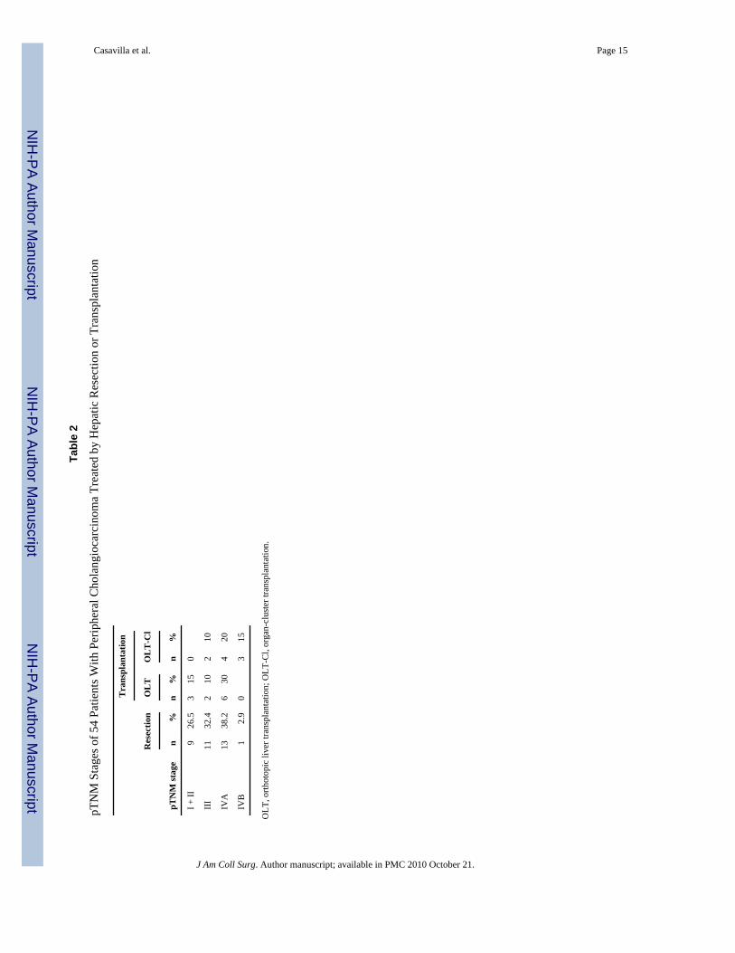
Tumor stage was defined according to the pTNM classification proposed by the InternationalUnion Against Cancer (17) and the American Joint Committee on Cancer (18) (Table 1). Therewere 12 patients with stage I or II tumors, 15 patients with stage III tumors, and 27 patientswith stage IV tumors (Table 2).
Surgical proceduresFor those with anatomically resectable tumors or for those without advanced, cirrhosis, partialhepatectomy (Hx) was the procedure of choice. Total hepatectomy with liver replacement(OLT) was performed when tumor extension or underlying cirrhosis precluded partialhepatectomy.
Thirty-four patients were treated with Hx: nine underwent left lobectomy, eight had a rightlobectomy, six had right trisegmentectomy, four underwent left trisegmentectomy, three hadan extended left lobectomy, two had an extended right lobectomy, and two underwent largenonanatomic resections. The surgical techniques of subtotal hepatectomy have been describedelsewhere (19–22).
Twenty patients were treated with OLT. Twelve had unresectable tumor(s) by conventionaltechniques of subtotal hepatectomy and had transplantation; eight had transplantation becauseof concomitant advanced cirrhosis. Because of highly unfavorable conditions such as lymphnode involvement or direct invasion of tumor into adjacent organs, an upper-abdominalexenteration and organ-cluster transplantation (OLT-Cl) were performed in 9 of the 20 patients(9,10). The surgical technique for OLT, including OLT-Cl, and the immunosuppression usedthereafter have been described elsewhere (9,23–25).
Casavilla et al. Page 2
J Am Coll Surg. Author manuscript; available in PMC 2010 October 21.
NIH
-PA Author Manuscript
NIH
-PA Author Manuscript
NIH
-PA Author Manuscript

Adjuvant therapyThirty-seven patients received adjuvant chemotherapy or radiation therapy before or after theoperation (Table 3). Because of the prolonged study period, these regimens were highlyvariable.
Statistical analysisThe results were summarized as of December 31, 1996. Cumulative overall survival and tumor-free survival rates were calculated by the method of Kaplan and Meier, with adjustment forthe type of operation (OLT versus Hx) (26). If a patient died at any time without tumorrecurrence, he or she was censored as tumor-free at that time for the Kaplan-Meier method.Alternatively, if a patient died with tumor recurrence, that patient was considered anuncensored observation for the tumor-free survival analysis. Potential risk factors examinedby univariate analysis were age, gender, tumor size, number of lesions, lobar involvement,vascular invasion, lymph node involvement, distant metastases, pTNM stage, surgical margins,presence or absence of associated cirrhosis of the liver, and adjuvant therapy. Values of p <0.05 were considered significant. The Cox proportional hazard regression model was used toassess the relative prognostic influence on survival.
ResultsPathologic characteristics
The principal tumor size had a median diameter of 8.3 cm (mean ± SE, 8.1 ± 4.2 cm), with arange of 0.8–22 cm. Twenty-six patients (48%) had a single tumor; the remaining 28 patients(52%) had multiple tumors. The tumors were unilobar in 27 patients; 14 had only right lobeinvolvement, and 13 had only left lobe involvement (Table 3).
Fifteen patients (28%) had no evidence of vascular invasion (V0), 32 patients (59%) hadmicroscopic invasion (V1), and 7 patients (13%) had gross vascular invasion (V2) of either theportal vein (4 patients) or the hepatic vein branches (3 patients).
Fourteen patients (26%) had regional lymph node involvement (N-1) and five (9%) hadregional metastatic disease (M-1) at the time of operation (Table 3). All lesions were resectedin continuity with the principal tumor.
The margins of resection were free of tumor (R0) in 40 patients. Margins were microscopicallypositive (R1) in 10 patients and grossly positive (R2) in the remaining 4 (Table 3). No grosstumor remained at the conclusion of the surgical procedure in any patient.
Eight patients in the OLT group had associated cirrhosis of the liver (40%). Of the 29 patientstested for hepatitis B virus, 3 had positive serology. None of the 20 patients tested was positivefor hepatitis C virus. One of 20 patients tested had an elevated serum titer of carcinoembryonicantigen; 3 of the 24 tested for serum alpha-fetoprotein had elevated levels.
The pTNM stages of the 54 patients are summarized in Tables 2 and 3. Twenty-seven patients(50%) presented with pTNM stage IV.
Survival rateThe overall patient survival rates of the 54 patients were 64% at 1 year, 34% at 3 years, and26% at 5 years after operation (Fig. 1). The tumor-free survival rates were 57% at 1 year, 34%at 3 years, and 27% at 5 years after operation (Fig. 1).
Casavilla et al. Page 3
J Am Coll Surg. Author manuscript; available in PMC 2010 October 21.
NIH
-PA Author Manuscript
NIH
-PA Author Manuscript
NIH
-PA Author Manuscript

The patient survival rates after Hx (60% at 1 year, 37% at 3 years, and 31 % at 5 years) weresimilar to those after OLT (70% at 1 year, 29% at 3 years, and 18% at 5 years), as shown inFigure 2. Tumor-free survival after Hx (50% at 1 year, 30% at 3 years, and 25% at 5 years)was also similar to that after OLT (67% at 1 year, 31% at 3 years, and 31% at 5 years), asshown in Figure 3. The higher rate of tumor-free survival compared with actuarial patientsurvival reflects a high death rate without recurrence among patients with Ch-Ca, regardlessof the type of surgical procedure.
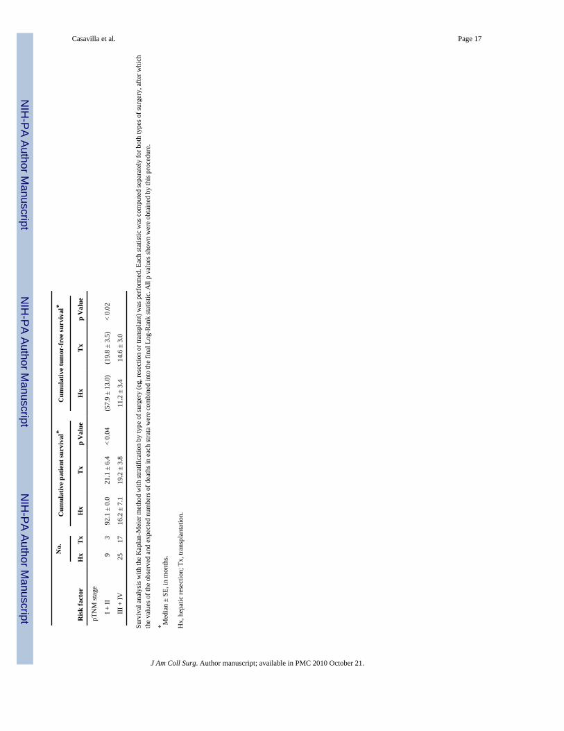
As expected, the pTNM stage significantly influenced both patient and tumor-free survival(Fig. 4; Table 3).
Causes of deathTwenty-four of the 34 patients (73.5%) in the Hx group and 16 of the 20 patients (80%) in theOLT group were dead at the time of followup (December 1996).
Four of the 54 patients died within 30 days after operation, giving an overall operative mortalityrate of 7.4%. The first patient died intraoperatively of an irreversible arrhythmia during Hx,and the second patient died of sepsis and liver failure 24 days after Hx. The third patient diedof massive upper gastrointestinal hemorrhage 6 days after OLT, and the fourth patient died ofrupture of a mycotic hepatic artery aneurysm 18 days after OLT.
An additional six patients died of causes unrelated to Ch-Ca. One patient died of renal andcardiac failure 19 months after Hx, the second died of an unknown sudden cause 6 years afterHx, and the third patient died of pancreatic cancer 14 years after Hx. The fourth patient diedof acute pulmonary tuberculosis 5 months after OLT, the fifth patient died of aspergillosis 14months after OLT, and the sixth died of recurrent hepatitis B 22 months after OLT.
A total of 30 patients died with recurrent Ch-Ca, 19 (55%) in the Hx group and 11 (56%) inthe OLT group. The higher 5-year tumor-free survival in the OLT group relative to the Hxgroup (31% and 25%, respectively) was due to the low rate of tumor recurrence among the 9OLT-Cl patients (Fig. 3).
Prognostic factorsThe influence of the 12 clinicopathologic factors on tumor-free and overall patient survivalwas examined by univariate analysis (Table 3). Multiple gross tumors, a bilobar tumordistribution, regional lymph node involvement, the presence of metastasis, positive surgicalmargins, and advanced (III and IV) pTNM stages were statistically significant negativeprognostic factors for both tumor-free and overall patient survival. The presence of vascularinvasion was a statistically significant factor reducing the tumor-free survival, but it did notinfluence the overall patient survival. Age, gender, size of principal tumor, associated cirrhosis,and adjuvant chemotherapy did not influence either tumor-free or patient survival.
Because positive metastasis turned out to be uniformly fatal (all five patients died within 1.3years) (p < 0.001), this prognostic factor was excluded from the multivariate analysis. Of thesix prognostic factors that reached statistical significance in the univariate analysis, themultivariate analysis revealed positive margins, multiple tumors, and lymph node involvementto be independently associated with a poor prognosis both for patient and for longterm (morethan one year) tumor-free survival. For patient survival, the relative hazards for positivemargins, multiple tumors, and lymph node involvement were 1.74, 1.63, and 1.60, respectively.For tumor-free survival, the relative hazards for positive margins, multiple tumors, and lymphnode involvement were 8.2, 2.0, and 2.65, respectively. These statistics show the hazard ofdeath or tumor recurrence at any given time point.
Casavilla et al. Page 4
J Am Coll Surg. Author manuscript; available in PMC 2010 October 21.
NIH
-PA Author Manuscript
NIH
-PA Author Manuscript
NIH
-PA Author Manuscript

There were 21 patients (15 in the Hx group and 6 in the OLT group) in whom none of thesethree negative predictors was present. The actuarial patient and tumor-free survival rates ofthese 21 patients (low-risk group) were 74% and 79% at 1 year, 64% and 62% at 3 years, and64% and 52% at 5 years, respectively (Fig. 5). Conversely, in the 33 patients (19 in the Hxgroup and 14 in OLT group) in whom at least one of these three negative predictors was present(high-risk group), patient and tumor-free survival rates were 58% and 43% at 1 year, 18% and13% at 3 years, and 9% and 13% at 5 years after operation (p < 0.0001) (Fig. 5).
Tumor recurrenceRecurrence of Ch-Ca was confirmed in 32 of the 54 patients (59%) during the study period:21 of the 34 patients (62%) in the Hx group and 11 of the 20 (55%) in the OLT group (2 patientsare currently alive with disease). Tumor recurrence was diagnosed within 1 year after operationin 21 patients (66%) and within 2 years in 29 patients (91%). Only one patient with recurrencehad it after 3 years; this patient was in the resection group and suffered recurrence at 4.3 years.Overall, there was no difference in time to recurrence between Hx and OLT.
The liver was the most common site of tumor recurrence (20 patients), followed by the lung(18 patients), the bones (7 patients), and other sites such as the peritoneum, adrenal gland, andkidney (5 patients).
Five-year survivorsOf the 39 patients with ≥ 5 years of followup, 9 (23%) survive: 6 of 21 (29%) in the Hx groupand 3 of 18 (17%) in the OLT group. Seven of these 9 patients had a single tumor; 6 had aunilobar tumor distribution, and 7, surprisingly, had a principal tumor size of > 5 cm. Four ofthe 9 patients had microscopic vascular invasion. Four of the 9 patients were classified aspTNM stage I or II, 3 patients as stage III, and 2 patients as stage IVA.
None of the 5-year survivors had positive margins, regional metastasis, lymph nodeinvolvement, or macroscopic vascular tumor invasion. All three patients in the OLT group whosurvived > 5 years were classified as pTNM stage III or IVA and underwent OLT-Cl. Two ofthe 5-year survivors (both in the Hx group) had tumor recurrence in the liver 2 and 4 yearsafter Hx and underwent repeat surgical resection.
DiscussionCholangiocarcinoma is a cancer arising from the epithelium of the biliary ducts. The bile ductlocated on the hepatic side of the first intrahepatic branch of the right and left hepatic duct isconsidered “intrahepatic” or “peripheral.” The remaining part of the bile duct is considered“extrahepatic,” including the hilum of the liver. In the strictest sense, then, Ch-Ca is a cancerarising from the epithelium of the intrahepatic duct or ductules (27–29). Practically speaking,differentiation of intrahepatic from extrahepatic bile duct cancer is often difficult anatomically,particularly in the hepatic hilum, and the histologic characteristics of the two are identical. Forthese reasons, cancers arising from the epithelium of the right and left hepatic ducts and thebifurcation are also called Ch-Ca of “hilar” or “central” type (Klatskin tumor) (30).
This article reports the prognostic risk factors and outcomes of 54 patients with Ch-Ca whounderwent aggressive surgical therapy at our institution. To our knowledge, this studyrepresents the largest series of patients with peripheral Ch-Ca who were treated with Hx orOLT at a single institution. These two groups of patients treated with two different surgicalprocedures are not strictly comparable because Hx was considered the surgical procedure ofchoice for resectable Ch-Ca, and OLT was performed when patients had either unresectabletumors or associated cirrhosis. It was surprising, then, to find that even though a significantly
Casavilla et al. Page 5
J Am Coll Surg. Author manuscript; available in PMC 2010 October 21.
NIH
-PA Author Manuscript
NIH
-PA Author Manuscript
NIH
-PA Author Manuscript

greater number of patients in the OLT group had advanced malignant disease than in the Hxgroup (65% versus 41%), as represented by stage IV disease (Table 2), the longterm tumor-free and patient survival rates were similar for the two groups. Although patient survival at 5years was greater in the Hx group, the 5-year tumor-free survival was greater in the OLT group(31% versus 25%), despite the more advanced disease. The dramatic decrease in survival forthe OLT group between 3 and 5 years was due to two deaths related to early recurrence.
This study confirmed the general opinion (13,14,31,32) that postoperative recurrence rates andsurvival of patients with Ch-Ca are determined by pTNM stage. The median survival after Hxor OLT for tumor stages I and II combined, III, and IV was 77, 18, and 17.5 months,respectively. The actuarial 1-, 3-, and 5-year patient survival rates were 90%, 70%, and 70%for stages I and II; 60%, 33.3%, and 25% for stage III; and 55.6%, 21.6%, and 13% for stageIV. These rates are consistent with previous reports of patients undergoing liver resection forCh-Ca (3,13,14). Unfortunately, an accurate analogy with published data for patients whounderwent OLT for Ch-Ca is extremely difficult to make because case material was small(12,14,31,32), results were not stratified according to specific stages (31–33), or the twodistinctive types of Ch-Ca (peripheral versus hilar) were combined in the analysis (12).
The actuarial survival of patients undergoing Hx or OLT for Ch-Ca, reported in this study, iscomparable to our results (34) using similar surgical treatment in patients with hepatocellularcarcinoma at equivalent stages. There were more 5-year survivors treated for Ch-Ca than thosetreated for hepatocellular carcinoma (23% versus 14%, respectively). Positive tumor marginsand lymph node metastasis were prognostic risk factors associated with significantly poorersurvival rates for both types of malignancies.
The results of this study suggest, in contrast to reports in the literature, that OLT should notbe eliminated in the treatment of Ch-Ca. In fact, based on our identification of a selected groupof patients found to have an “acceptable” longterm survival, an argument could be made forexpanding its role if there is a sufficient supply of donor organs. At least one group hasquestioned whether OLT-Cl is of any value for the treatment of hepatobiliary malignancies ingeneral (16). Although there is limited experience with the cluster procedure for the specificindication of Ch-Ca, our only patients with Ch-Ca in the OLT group who survived > 5 yearshad undergone OLT-Cl. Of nine patients who underwent OLT-Cl, only four had recurrentdisease during the study period.
Our multivariate analysis revealed that, in addition to metastatic disease, positive surgicalmargins, multiple gross tumors, and lymph node involvement were independent risk factorsfor poor survival after both Hx and OLT in patients with Ch-Ca. Conversely, the actuarial 1-,3-, and 5-year survival rates of patients with none of these three negative predictors weresignificantly higher than those of patients with at least one of these risk factors (74.2%, 66.5%,and 66.5% versus 58%, 18%, and 9%, respectively). Because we did not find any differencein time to recurrence between the Hx and OLT groups, our results do not support thehypothetical (and logical) contention that immunosuppression shortens the tumor-free intervalrate. The indications for OLT can reasonably include at least some appropriately staged patientswith Ch-Ca.
In conclusion, these data suggest that Hx and OLT are both defensible options for patients withCh-Ca who have single lesions, negative lymph nodes, and surgically resectable disease.Identification of risk factors, which predict prognosis, requires extensive preoperative andintraoperative evaluations. Because the number of patients undergoing surgical therapy for thisdisease is so small, a multiinstitutional study is needed to assess the relative efficacy of Hx,conventional OLT, or even upper-abdominal exenteration and OLT (OLT-Cl). Conceivably,
Casavilla et al. Page 6
J Am Coll Surg. Author manuscript; available in PMC 2010 October 21.
NIH
-PA Author Manuscript
NIH
-PA Author Manuscript
NIH
-PA Author Manuscript

the indications for conventional OLT or OLT-Cl should be broadened to include some of thepatients with advanced disease who are currently being treated with Hx.
References1. Kamiyama, Y.; Tobe, T. Treatment of primary liver cancer in Japan. In: Okuda, K.; Ishak, KG., editors.
Neoplasms of the Liver. Tokyo: Springer-Verlag; 1987. p. 375-380.2. Iwatsuki S, Sheahan DG, Starzl TE. The changing face of hepatic resection. Curr Probe Surg
1989;26:283–379.3. Chen MF, Jan YY, Wang CS, et al. Clinical experience in 20 hepatic resections for peripheral
cholangiocarcinoma. Cancer 1989;64:2226–2232. [PubMed: 2553242]4. Chu KM, Lai ECS, Al-Hadeedi S, et al. Intrahepatic cholangiocarcinoma. World J Surg 1997;21:301–
306. [PubMed: 9015175]5. Madariaga JR, Iwatsuki S, Todo S, et al. Liver resection for hilar and peripheral cholangiocarcinomas:
a study of 62 cases. Ann Surg. (in press).6. Starzl, TE. Hepatic malignancy. In: Starzl, TE., editor. Experience in Hepatic Transplantation.
Philadelphia: WB Saunders; 1969. p. 4-8.(with the assistance Putnam CW).7. Calne, RY. Indications and assessment for orthotopic liver transplantation. In: Calne, RY., editor. Liver
Transplantation. The Cambridge-King’s College Hospital Experience. London: Grune & Stratton;1983. p. 59-62.
8. Iwatsuki S, Gordon RD, Shaw BW, et al. Role of liver transplantation in cancer therapy. Ann Surg1985;202:401–407. [PubMed: 2996449]
9. Starzl TE, Todo S, Tzakis A, et al. Abdominal organ cluster transplantation for the treatment of upperabdominal malignancies. Ann Surg 1989;210:374–386. [PubMed: 2673085]
10. Tzakis A, Ricordi C, Alejandro R, et al. Pancreatic islet transplantation after upper abdominalexenteration and liver replacement. Lancet 1990;336:402–405. [PubMed: 1974944]
11. Alessiani M, Tzakis A, Todo S, et al. Assessment of five-year experience with abdominal organcluster transplantation. J Am Coll Surg 1995;180:1–9. [PubMed: 8000645]
12. Goldstein RM, Stone M, Tillery GW, et al. Is liver transplantation indicated for cholangiocarcinoma?Am J Surg 1993;166:768–772. [PubMed: 8273866]
13. Pichlmavr R, Lamesch P, Weimann A, et al. Surgical treatment of cholangiocellular carcinoma. WorldJ Surg 1995;19:83–88. [PubMed: 7740815]
14. Washburn WK, Lewis D, Jenkins RL. Aggressive surgical resection for cholangiocarcinoma. ArchSurg 1995;130:271–276.
15. Berdah SV, Delpero JR, Garcia S, et al. A Western surgical experience of peripheralcholangiocarcinoma. Br J Surg 1996;83:1517–1521. [PubMed: 9014664]
16. Knechtle SJ, Kalavoglu M, D’Alessandro AM, et al. Should abdominal cluster transplantation beabandoned? Transplant Proc 1993;25:1361–1363. [PubMed: 8442141]
17. Hermanek, P.; Sobin, LH., editors. TNM Classification of Malignant Tumors. 4th ed. Berlin: Springer-Verlag; 1987. p. 53-55.
18. Bearhs, OH.; Henson, DE.; Hutter, RVP.; Kennedy, BJ., editors. American Joint Committee onCancer. Manual for Staging of Cancer. 4th ed. Philadelphia: JB Lippincott; 1992. p. 89-100.
19. Starzl TE, Bell RH, Beart RW, Putnam CW. Hepatic trisegmentectomy and other liver resections.Surg Gynecol Obstet 1975;141:429–437. [PubMed: 1162576]
20. Starzl TE, Koep LJ, Weil R, et al. Right trisegmentectomy for hepatic neoplasm. Surg Gynecol Obstet1980;150:208–213. [PubMed: 7352313]
21. Starzl TE, Iwatsuki S, Shaw BW, et al. Left hepatic trisegmentectomy. Surg Gynecol Obstet1982;155:21–27. [PubMed: 6283687]
22. Blumgart, LH. Liver resection: liver and biliary tumors. In: Blumgart, LH., editor. Surgery of theLiver and Biliary Tract. Edinburgh: Churchill Livingstone; 1988. p. 1251-1280.
23. Starzl TE, Iwatsuki S, Van Thiel DH, et al. Evolution of liver transplantation. Hepatology 1982;2:614–636. [PubMed: 6749635]
Casavilla et al. Page 7
J Am Coll Surg. Author manuscript; available in PMC 2010 October 21.
NIH
-PA Author Manuscript
NIH
-PA Author Manuscript
NIH
-PA Author Manuscript

24. Iwatsuki S, Starzl TE, Todo S, et al. Experience in 1,000 liver transplants under cyclosporin-steroidtherapy: a surgical report. Transplant Proc 1988;20:498–504. [PubMed: 3279643]
25. Fung JJ, Eliasziw M, Todo S, et al. The Pittsburgh randomized trial of Tracolimus compared tocyclosporin for hepatic transplantation. J Am Coll Surg 1996;183:117–125. [PubMed: 8696542]
26. Fisher, LD.; Van Belle, G. A methodology for the health sciences. In: Barnett, V.; Bradley, RA.;Fisher, NI., et al., editors. Biostatistics. New York: J. Wiley and Sons, Inc; 1993. p. 807-808.
27. Edmondson HA, Steiner PE. Primary carcinoma of the liver. A study of 100 cases among 48,900necropsies. Cancer 1954;51:462–503. [PubMed: 13160935]
28. Altemeier WA, Gall EA, Zinniger MM, Hoxworth LH. Sclerosing carcinoma of the major intrahepaticducts. Arch Surg 1957;75:450–461.
29. Nakajima T, Kondo Y, Mivazaki M, Okui K. A histopathologic study of 102 cases of intrahepaticcholangiocarcinoma: histologic classification and modes of spreading. Hum Pathol 1988;19:1228–1234. [PubMed: 2844646]
30. Klatskin G. Adenocarcinoma of the hepatic duct at its bifurcation within the porta hepatis: an unusualtumor with distinctive clinical pathological features. Am J Med 1965;38:241–256. [PubMed:14256720]
31. Haug CE, Jenkins RL, Rohrer RJ, et al. Liver transplantation for primary hepatic cancer.Transplantation 1992;53:376–382. [PubMed: 1310823]
32. Olthoff KM, Millis LM, Rosove MH, et al. Is liver transplantation justified for treatment of hepaticmalignancies? Arch Surg 1990;125:1261–1268. [PubMed: 2171452]
33. O’Grady JG, Polson RJ, Rolles K, et al. Liver transplantation for malignant disease. Results in 93consecutive patients. Ann Surg 1988;207:373–379. [PubMed: 2451484]
34. Iwatsuki S, Starzl TE, Sheahan DG, et al. Hepatic resection versus transplantation for hepatocellularcarcinoma. Ann Surg 1991;214:221–229. [PubMed: 1656903]
Casavilla et al. Page 8
J Am Coll Surg. Author manuscript; available in PMC 2010 October 21.
NIH
-PA Author Manuscript
NIH
-PA Author Manuscript
NIH
-PA Author Manuscript
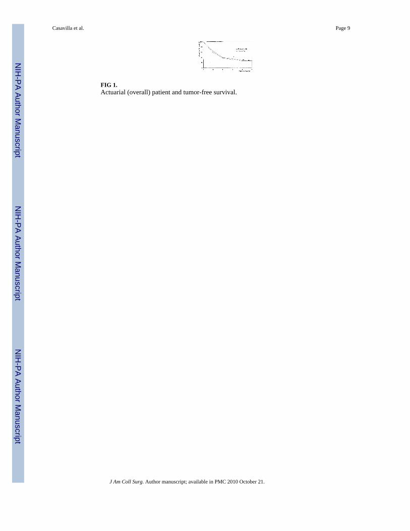
FIG 1.Actuarial (overall) patient and tumor-free survival.
Casavilla et al. Page 9
J Am Coll Surg. Author manuscript; available in PMC 2010 October 21.
NIH
-PA Author Manuscript
NIH
-PA Author Manuscript
NIH
-PA Author Manuscript

FIG 2.Actuarial patient survival. Hx, hepatic resection; OLT, orthotopic liver transplantation.
Casavilla et al. Page 10
J Am Coll Surg. Author manuscript; available in PMC 2010 October 21.
NIH
-PA Author Manuscript
NIH
-PA Author Manuscript
NIH
-PA Author Manuscript
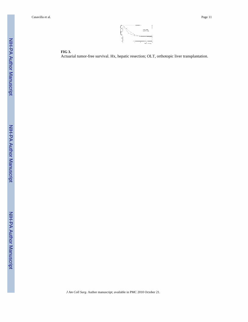
FIG 3.Actuarial tumor-free survival. Hx, hepatic resection; OLT, orthotopic liver transplantation.
Casavilla et al. Page 11
J Am Coll Surg. Author manuscript; available in PMC 2010 October 21.
NIH
-PA Author Manuscript
NIH
-PA Author Manuscript
NIH
-PA Author Manuscript

FIG 4.Patient and tumor-tree survival by TNM stage.
Casavilla et al. Page 12
J Am Coll Surg. Author manuscript; available in PMC 2010 October 21.
NIH
-PA Author Manuscript
NIH
-PA Author Manuscript
NIH
-PA Author Manuscript
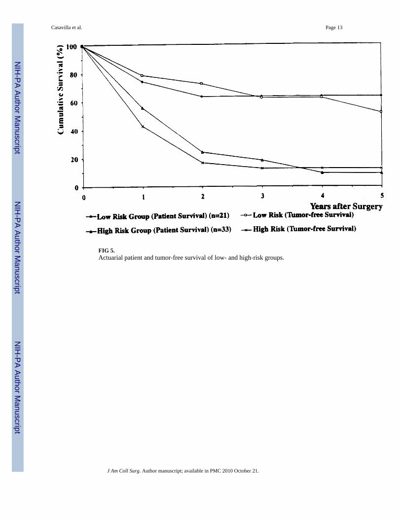
FIG 5.Actuarial patient and tumor-free survival of low- and high-risk groups.
Casavilla et al. Page 13
J Am Coll Surg. Author manuscript; available in PMC 2010 October 21.
NIH
-PA Author Manuscript
NIH
-PA Author Manuscript
NIH
-PA Author Manuscript

NIH
-PA Author Manuscript
NIH
-PA Author Manuscript
NIH
-PA Author Manuscript
Casavilla et al. Page 14
Table 1
pTNM Pathologic Classification
Stage I T1* N0 M0
Stage II T2† N0 M0
T1 N1‡ M0
Stage III T2 N1 M0
T3§ N0, N1 M0
Stage IVA T4∥ Any N M0
Stage IVB Any T Any N M1¶
*T1: solitary, ≤ 2 cm, without vascular invasion.
†T2: solitary, ≤ 2 cm, with vascular invasion. Multiple, one lobe, ≤ 2 cm, without vascular invasion. Solitary, > 2 cm, without vascular invasion.
‡N1: regional.
§T3: solitary, > 2 cm, with vascular invasion. Multiple, one lobe, > 2 cm, with or without vascular invasion.
∥T4: Multiple, more than one lobe. Invasion of major branch of portal or hepatic veins.
¶M1: Distant metastasis.
J Am Coll Surg. Author manuscript; available in PMC 2010 October 21.
NIH
-PA Author Manuscript
NIH
-PA Author Manuscript
NIH
-PA Author Manuscript
Casavilla et al. Page 15
Tabl
e 2
pTN
M S
tage
s of 5
4 Pa
tient
s With
Per
iphe
ral C
hola
ngio
carc
inom
a Tr
eate
d by
Hep
atic
Res
ectio
n or
Tra
nspl
anta
tion
Tra
nspl
anta
tion
Res
ectio
nO
LT
OL
T-C
l
pTN
M st
age
n%
n%
n%
I + II
926
.53
150
III
1132
.42
102
10
IVA
1338
.26
304
20
IVB
12.
90
315
OLT
, orth
otop
ic li
ver t
rans
plan
tatio
n; O
LT-C
l, or
gan-
clus
ter t
rans
plan
tatio
n.
J Am Coll Surg. Author manuscript; available in PMC 2010 October 21.

NIH
-PA Author Manuscript
NIH
-PA Author Manuscript
NIH
-PA Author Manuscript
Casavilla et al. Page 16
Tabl
e 3
Path
olog
ic C
hara
cter
istic
s and
Uni
varia
te A
naly
sis o
f Pro
gnos
tic R
isk
Fact
ors
No.
Cum
ulat
ive
patie
nt su
rviv
al*
Cum
ulat
ive
tum
or-fr
ee su
rviv
al*
Ris
k fa
ctor
Hx
Tx
Hx
Tx
p V
alue
Hx
Tx
p V
alue
Gen
der
F
emal
e23
1018
.2 ±
3.4
20.2
± 1
.3>
0.41
12.0
± 1
.214
.6 ±
2.0
> 0.
52
M
ale
2110
16.2
± 5
.911
.3 ±
4.0
23.4
± 1
7.2
32.1
± 1
6.7
Age
(y)
≤
60
1619
16.2
± 5
.520
.2 ±
4.5
> 0.
816.
8 ±
14.9
17.7
± 4
.1>
0.84
>
60
181
18.3
± 3
.117
.6 ±
0.0
12.9
± 1
.115
.6 ±
0.0
Che
mo/
rads
Y
es5
127.
2 ±
5.1
13.2
± 2
.9>
0.07
7.2
± 4.
932
.1 ±
9.1
> 0.
55
N
o29
818
.3 ±
2.9
20.9
± 5
.812
.9 ±
3.4
10.5
± 2
.1
Tum
or si
ze
≤
5 c
m10
716
.2 ±
44.
420
.9 ±
10.
0>
0.59
(73.
3 ±
28.3
)(1
8.7
± 3.
8)>
0.57
>
5 c
m24
1318
.1 ±
5.1
19.2
± 3
.412
.9 ±
3.4
15.6
± 4
.1
Tum
or
S
ingl
e19
777
.1 ±
59.
021
.1 ±
10.
3<
0.01
464
.5 ±
18
(68.
9 ±
10.4
)<
0.03
M
ultip
le15
1310
.1 ±
4.7
19.2
± 3
.49.
0 ±
4.9
13.1
± 3
.6
Dis
tribu
tion
U
nilo
bar
207
38.4
± 1
6.6
28.5
± 7
.7<
0.01
1(6
0.2
± 19
.5)
(68.
9 ±
10.4
)<
0.03
B
iloba
r14
138.
9 ±
1.8
17.6
± 4
.77.
2 ±
1.6
13.1
± 3
.1
Vas
cula
r inv
asio
n
V
010
577
.1 ±
57.
121
.1 ±
8.6
> 0.
08(4
4.3
± 12
.4)
(29.
7 ±
12.2
)<
0.02
V
1 + V
224
1516
.2 ±
5.3
19.2
± 3
.611
.2 ±
3.6
13.1
± 2
.7
Lym
ph n
odes
N
egat
ive
2814
22.4
± 1
1.4
20.2
± 1
.8<
0.00
115
.9 ±
8.4
32.1
± 1
6.6
< 0.
0001
P
ositi
ve6
66.
5 ±
2.2
6.6
± 8.
64.
2 ±
1.0
5.2
± 4.
0
Mar
gins
N
egat
ive
2416
38.4
± 2
8.1
19.2
± 2
.6<
0.00
0123
.4 ±
7.9
17.7
± 1
1.3
< 0.
0001
P
ositi
ve10
47.
2 ±
1.8
0.6
± 14
.35.
3 ±
1.2
1.5
± 0.
9
J Am Coll Surg. Author manuscript; available in PMC 2010 October 21.
NIH
-PA Author Manuscript
NIH
-PA Author Manuscript
NIH
-PA Author Manuscript
Casavilla et al. Page 17
No.
Cum
ulat
ive
patie
nt su
rviv
al*
Cum
ulat
ive
tum
or-fr
ee su
rviv
al*
Ris
k fa
ctor
Hx
Tx
Hx
Tx
p V
alue
Hx
Tx
p V
alue
pTN
M st
age
I
+ II
93
92.1
± 0
.021
.1 ±
6.4
< 0.
04(5
7.9
± 13
.0)
(19.
8 ±
3.5)
< 0.
02
II
I + IV
2517
16.2
± 7
.119
.2 ±
3.8
11.2
± 3
.414
.6 ±
3.0
Surv
ival
ana
lysi
s with
the
Kap
lan-
Mei
er m
etho
d w
ith st
ratif
icat
ion
by ty
pe o
f sur
gery
(eg,
rese
ctio
n or
tran
spla
nt) w
as p
erfo
rmed
. Eac
h st
atis
tic w
as c
ompu
ted
sepa
rate
ly fo
r bot
h ty
pes o
f sur
gery
, afte
r whi
chth
e va
lues
of t
he o
bser
ved
and
expe
cted
num
bers
of d
eath
s in
each
stra
ta w
ere
com
bine
d in
to th
e fin
al L
og-R
ank
stat
istic
. All
p va
lues
show
n w
ere
obta
ined
by
this
pro
cedu
re.
* Med
ian
± SE
, in
mon
ths.
Hx,
hep
atic
rese
ctio
n; T
x, tr
ansp
lant
atio
n.
J Am Coll Surg. Author manuscript; available in PMC 2010 October 21.