Ensemble Statistical Guidance or Statistical Guidance Ensemble
NEW TECHNOLOGY
Use of the Resection Map system as guidance during hepatectomy
Pablo Lamata • Felix Lamata • Valentin Sojar • Piotr Makowski •
Laurent Massoptier • Sergio Casciaro • Wajid Ali • Thomas Studeli •
Jerome Declerck • Ole Jackov Elle • Bjorn Edwin
Received: 29 April 2009 / Accepted: 14 August 2009 / Published online: 23 February 2010
� The Author(s) 2010. This article is published with open access at Springerlink.com
Abstract
Background The objective of this work is to evaluate a
new concept of intraoperative three-dimensional (3D)
visualization system to support hepatectomy. The Resec-
tion Map aims to provide accurate cartography for sur-
geons, who can therefore anticipate risks, increase their
confidence and achieve safer liver resection.
Methods In an experimental prospective cohort study, ten
consecutive patients admitted for hepatectomy to three
European hospitals were selected. Liver structures (portal
veins, hepatic veins, tumours and parenchyma) were seg-
mented from a recent computed tomography (CT) study of
each patient. The surgeon planned the resection preopera-
tively and read the Resection Map as reference guidance
during the procedure. Objective (amount of bleeding,
tumour resection margin and operating time) and sub-
jective parameters were retrieved after each case.
Results Three different surgeons operated on seven
patients with the navigation aid of the Resection Map.
Veins displayed in the Resection Map were identified
during the surgical procedure in 70.1% of cases, depending
mainly on size. Surgeons were able to track resection
progress and experienced improved orientation and
increased confidence during the procedure.
Conclusions The Resection Map is a pragmatic solution
to enhance the orientation and confidence of the surgeon.
Further studies are needed to demonstrate improvement in
patient safety.
Keywords Computer-assisted surgery � Hepatectomy �Instrumentation
Colorectal cancer is the third most common cancer in the
USA, with an estimated 150,000 new cases in 2008 [1]. Of
patients with colorectal cancer, 50% will develop liver
metastasis [2]. On the other hand, primary liver cancer,
which consists predominantly of hepatocellular carcinoma
(HCC), is the fifth most common cancer worldwide and the
third most common cause of cancer mortality [3]. Liver
resection is the treatment of choice in selected patients with
hepatic colorectal metastasis [4], even in recurrent cases
[5]. Hepatocellular carcinoma is potentially curable by
surgical resection, but surgery is the treatment of choice
only for patients with localized disease [6]. The critical
aspect in this procedure is to guarantee a safe margin
around the tumour (R0), what is often a trade-off with
P. Lamata � J. Declerck
Siemens Molecular Imaging, Oxford, UK
F. Lamata
Hospital Clınico Universitario, Zaragoza, Spain
V. Sojar
University Clinik Center, Ljubljana, Slovenia
P. Makowski � L. Massoptier � S. Casciaro
Institute of Clinical Physiology, Biomedical Engineering
Division, National Council of Research (CNR-IFC), 73100
Lecce, Italy
W. Ali � O. J. Elle � B. Edwin
The Interventional Centre, Oslo University Hospital, Oslo,
Norway
T. Studeli
Faculty of Industrial Design Engineering, Delft University
of Technology, Delft, The Netherlands
P. Lamata (&)
Computing Laboratory, University of Oxford, Wolfson Building,
Parks Road, Oxford OX1 3QD, UK
e-mail: [email protected]
123
Surg Endosc (2010) 24:2327–2337
DOI 10.1007/s00464-010-0915-3
remaining functional liver. Another key issue is reduction
of bleeding and control of main veins in the liver, one of
the major causes of complications. Liver resection is also
critical for living-donor programmes and other non-onco-
logic surgical procedures.
On the other hand, technical and computational advances
are constantly giving rise to new surgical concepts and
techniques. By extending the surgeon’s ability to plan and
carry out interventions more accurately and less invasively,
computer-aided surgical systems will address a crucial need
to reduce medical procedure costs, and improve clinical
outcome and thus the efficiency of health care delivery [7].
In this work, we focus on a system managing the organi-
sation, image processing and display of the complex interior
structures of the liver, to be used in resection procedures
(hepatectomies). It allows the surgeon to orient him/herself
by means of computer-generated visualizations of the
anatomy, target structures and planned resection paths.
Some research work in computer-aided surgical systems
for liver surgery is focussed on providing preoperative
planning support [7–12]. These systems perform image
segmentation and 3D reconstruction of liver, tumours and
vessel structures. Based on these 3D models, they offer
edition and calculation of anatomical and non-anatomical
resections with user-defined security margins. User inter-
action is generally through two-dimensional (2D) rendered
images, or even involving a 3D virtual environment [10].
Operation planning may be substantially improved by the
use of such systems [8]. Nevertheless, these systems do not
yet focus on presentation of information intraoperatively,
and the surgeon is forced to rely on his memory and ability
to translate the preoperative figures into the operative site
[8, 13]. There is still a need to effectively use the ana-
tomical information from the CT in the intraoperative suite
through an interactive virtual 3D model of the liver. As an
example in this direction, a case study using a 3D virtual
liver for navigation support has recently been reported [14].
On the other hand, recent contributions have been made to
provide intraoperative guidance in liver surgery. Orientation
in the liver anatomy is achieved in surgical routine using
ultrasound (US). This modality is limited by a restricted field
of view and decreasing practical utility as resection pro-
gresses, as it is difficult or impossible to image and interpret
the structures in the resection plane once resection has star-
ted. Three-dimensional reconstruction of a US volume and
an optical tracking system are the two main components of a
virtual navigation system used to guide the resection in open
surgery proposed by Beller et al. [15]. This system enhances
the accuracy of tumour resection margins, but requires
immobilisation of the liver and relative easy access to the
tumour location. Another alternative is the use of an optically
tracked C-Arm and laparoscope to enable an augmented
view of laparoscopic images [16]. This solution provides the
surgeon with advanced visual localization of hidden struc-
tures such as veins, but it produces radiation, has high cost
and is bulky and only available in specialised operating
rooms (OR). A third possibility is the use of laser-ranging
technology to acquire the intraoperative surface of organs,
and then to navigate in the registered preoperative CT vol-
ume. Preliminary results of such technology are still focus-
sed on calibration and technical validation issues [17]. In
general, all these new systems offer attractive guidance
possibilities, by means of tracking and intraoperative imag-
ing technologies. However, these technologies require deli-
cate handling, for example for the different calibration
procedures. In many cases they also introduce changes in the
workflow and ergonomic aspects that could be very disrup-
tive, for instance due to the need for a clear line of sight
between the tools and the optical tracking systems. There still
exists a need to efficiently enhance the safety of surgical
procedures by providing the surgeon with the localization of
critical structures during liver resection.
Our approach to address the clinical need for intraop-
erative navigation for safer liver resection is to present an
interactive 3D Resection Map to the surgeon: a system for
simplified and effective visualization of critical structures
and the preoperatively planned resection path. This concept
is somehow similar to the use of a navigation system while
driving a car, or to the use of context maps in computer
games [18, 19] but without the positioning information,
i.e., without knowing the corresponding location of the
tools in the map. Our strategy is to harness the rich pre-
operative planning information during the surgical proce-
dure through an intuitive cartography, and without the need
for any additional hardware or equipment. The system thus
relies on the surgeon’s capacity to perform a mental
alignment between the Resection Map and the operating
field. A detailed description of the design process and
concept of this system is described in previous work [20].
In the consecutive series of ten patients reported herein,
we prospectively validated the design and evaluated the
impact of the Resection Map on operative safety and sur-
geon confidence for both open and laparoscopic hepatec-
tomy. Special emphasis is given to evaluate the capacity of
the surgeon to perform the mental alignment between the
operating field and the Resection Map.
Methods
An explorative prospective cohort study was designed. Ten
consecutive patients admitted for hepatectomy to three
European hospitals from October to December 2008 were
selected. The only inclusion criterion for the patients was
the availability of a recent (less than 2 months) preopera-
tive CT study with good contrast quality in veins (sufficient
2328 Surg Endosc (2010) 24:2327–2337
123

for semi-automatic segmentation of veins). Three experi-
enced surgeons (having performed more than 25 hepatec-
tomies each) from the hospitals were enrolled in the study.
One of them had wide experience using a laparoscopic
approach for this procedure, whereas the other two had
only experience in open surgery.
A training and familiarization protocol was conducted in
each hospital. Surgeons and assistants first received an
interactive explanation of the Resection Map system. Then,
an expert technician provided intraoperative support to the
surgical team during the first surgery in each hospital. After
that, each surgical team worked independently with the
system.
The workflow for each case consisted of four steps after
patient selection: CT segmentation, resection planning,
intraoperative use of the Resection Map and retrieval of
evaluation metrics. In this study, intraoperative updating of
resection planning in the virtual 3D reconstructed liver was
not considered. The type of resection performed for each
case is described in the ‘‘Results’’ section.
CT segmentation
Segmentation was performed remotely with semi-auto-
matic tools by expert engineers and verified by clinicians.
CT studies were acquired with different machines and
contrast protocols depending on the hospital for each
patient (Table 1). Resulting image quality and resolution
was thus not homogeneous among the sample population.
This was an additional difficulty for a tool for segmentation
and 3D reconstruction of structures, because such tools
work better with known image quality and isotropic voxels
(i.e., with equal dimensions in all three directions). Thus,
an image decimation and interpolation process was adap-
ted, depending on the voxel dimensions, to each dataset to
obtain closer to isotropic dimensions.
A contrast-enhanced CT study was segmented into liver
parenchyma, tumours, portal vein system and hepatic vein
system by a two-stage semi-automatic process. First, liver
parenchyma and tumours were identified with the methods
described in previous work [21]. Secondly, the two vein
systems were extracted with an original and pragmatic
method that combines vessel enhancement filters and
region-growing algorithms as described below.
Cylindrical shapes (vessels) inside the liver volume
were intensified with vessel enhancement filters [22]. This
was done for seven scales, i.e., seven diameters of veins
(1.3–5 mm), producing seven enhanced images. The larg-
est separable regions were identified in the image of largest
scale, and a region growing algorithm was applied to the
other six images of lower scale connecting the remaining
smaller vessels (Fig. 1a). These regions, these vessel trees,
were finally manually grouped into the two main hepatic
and portal systems (Fig. 1b). If necessary, manual correc-
tion of the semi-automatic segmentation of veins was done.
CT segmentation time greatly depended on the size and
quality (resolution and contrast) of the image study. The
process, done with research prototype tools, took around
40 min (both human interaction and computer processing
time) for a study of average size and good quality, and up
to a few hours in cases of low contrast and resolution.
Preoperative resection planning and 3D model
reconstruction
Surgical planning was performed by the surgeons using
Resection Planner software designed specifically for this
study. This tool enables the surgeon to load the CT study,
review the segmentation results, define a resection plane
and automatically generate the 3D geometries that consti-
tute the cartography of the Resection Map (Fig. 2).
The Resection Planner software was developed on top of
the open-source platform ITK-Snap [4]. In this tool, the
resection plane is modelled as a smooth surface that can be
deformed and adapted to complex resection scenarios.
Mouse interactions are used to modify this plane in the
axial, coronal and sagittal views of the CT, and interactive
3D visualization is provided. Surgeons have the option to
‘‘undo/redo’’ any change, ‘‘save’’ the current plane or
‘‘restore’’ the plane previously saved in each study.
Surgeons checked the quality of the segmentation and
corrected the identification of tumours. Then they manually
defined the resection plane, taking into account the visible
structures in the venous phase of the CT study. The shape
Table 1 CT image acquisition
parameters of the seven
hepatectomy cases
n.a. not available in the DICOM
tag
Patient CT machine Voxel size (mm) Contrast
1 Siemens Sensation 16 0.77 x 0.77 x 3.85 n.a.
2 GE LightSpeed VCT 0.70 x 0.70 x 1.75 100 ml 300ultra
3 Toshiba Aquilon 0.78 x 0.78 x 0.78 n.a.
4 GE LightSpeed VCT 0.69 x 0.69 x 1.72 150 ml VISIP 320
5 Siemens Sensation 16 0.69 x 0.69 x 3.45 n.a.
6 GE LightSpeed VCT 0.73 x 0.73 x 1.82 200 ml VISIP 320
7 Siemens Sensation 16 0.82 x 0.82 x 4.1 n.a.
Surg Endosc (2010) 24:2327–2337 2329
123

and position of the resection plane were determined with
just the understanding of the vein systems and the position
of the tumour, without territory analysis as proposed in
other work [8]. Information from the arterial and bile duct
systems was not considered in this definition. The main
reason for this choice is that, compared with the venous
system, arteries and bile ducts add little significance for
predicting the remnant liver or navigation information
during resection [20], since these intrahepatic systems
mainly run in parallel to the portal system. A secondary
motive is that arteries and bile ducts segmentation and 3D
visualization increase the complexity of the process and the
need of CT scan time and radiation to the patient.
Intraoperative assistance by the Resection Map
Surgeons performed hepatectomies following their usual
standards and guidelines. The Resection Map was
displayed for them on a monitor as an additional source of
orientation and guidance. Surgeons read the information of
the Resection Map and interactively changed its visuali-
zation through voice commands to their assistants, who
controlled the tool with a computer mouse interface. The
interaction possibilities and contents of the Resection Map
were adapted to the different phases of the surgical pro-
cedure as described below.
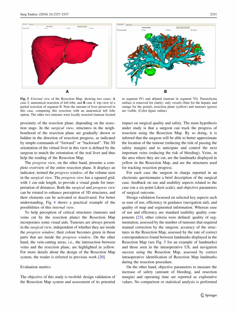
The Resection Map first shows an external view of the
virtual liver, which is used to review the case. Surgeons can
rotate this view, and activate or deactivate its elements
(resection plane, tumours, liver parenchyma, or veins;
Fig. 3). This view also helps the surgeon with orientation and
drawing of the cauterization line over the liver’s surface.
Once resection starts, the Resection Map can be changed
to show an internal view of the virtual liver. This view,
divided into a surgical view and a progress view, offers
interactive selection and visualization of a volume in the
Fig. 1 Manual grouping step in
vessel segmentation. A Result
of the automatic segmentation.
B Hepatic (blue) and portal
(orange) vein systems finally
generated by manual grouping.
(Color figure online)
Fig. 2 The Resection Planner software. A Revision of the segmen-
tation results: the tumour label (green) is made semitransparent in the
three slice views to check correct location and extension. Three-
dimensional reconstructions are shown in the bottom left window. B
Definition of the resection plane (red) of a complex case with a total
of six metastases (an additional segment I resection was done in this
case). Hepatic veins are coded in blue, and portal veins in orange.
(Color figure online)
2330 Surg Endosc (2010) 24:2327–2337
123
proximity of the resection plane, depending on the resec-
tion stage. In the surgical view, structures in the neigh-
bourhood of the resection plane are gradually shown or
hidden in the direction of resection progress, as indicated
by simple commands of ‘‘forward’’ or ‘‘backward’’. The 3D
orientation of the virtual liver in this view is defined by the
surgeon to match the orientation of the real liver and thus
help the reading of the Resection Map.
The progress view, on the other hand, presents a com-
plete overview of the entire resection plane. It displays an
indicator, termed the progress window, of the volume seen
in the surgical view. The progress view has a squared grid,
with 1 cm side length, to provide a visual guide for inter-
pretation of distances. Both the surgical and progress view
can be rotated to enhance perception of 3D structures, and
their elements can be activated or deactivated. For better
understanding, Fig. 4 shows a practical example of the
possibilities of this internal view.
To help perception of critical structures (tumours and
veins cut by the resection plane) the Resection Map
incorporates some visual aids. Tumours are always present
in the surgical view, independent of whether they are inside
the progress window; their colour becomes green in those
parts that are inside the progress window. On the other
hand, the vein-cutting areas, i.e., the intersection between
veins and the resection plane, are highlighted in yellow.
For more details about the design of the Resection Map
system, the reader is referred to previous work [20].
Evaluation metrics
The objective of this study is twofold: design validation of
the Resection Map system and assessment of its potential
impact on surgical quality and safety. The main hypothesis
under study is that a surgeon can track the progress of
resection using the Resection Map. By so doing, it is
inferred that the surgeon will be able to better approximate
the location of the tumour (reducing the risk of passing the
safety margin) and to anticipate and control the next
important veins (reducing the risk of bleeding). Veins, in
the area where they are cut, are the landmarks displayed in
yellow in the Resection Map, and are the structures used
for tracking resection progress.
For each case the surgeon in charge reported in an
electronic questionnaire a brief description of the surgical
case, feedback on use and usability aspects related to the
case (on a six-point Likert scale), and objective parameters
of surgical outcome.
Design validation focussed on selected key aspects such
as ease of use, efficiency in guidance (navigation aid), and
quality of map and segmented information. Whereas ease
of use and efficiency are standard usability quality com-
ponents [23], other criteria were defined: quality of seg-
mentation, assessed by the number of tumours that required
manual correction by the surgeon; accuracy of the struc-
tures in the Resection Map, assessed by the rate of correct
correspondences found between landmarks displayed in the
Resection Map (see Fig. 5 for an example of landmarks)
and those seen in the intraoperative US; and navigation
success using the Resection Map, assessed by correct
intraoperative identification of Resection Map landmarks
during the resection procedure.
On the other hand, objective parameters to measure the
increase of safety (amount of bleeding, and resection
margin) and operating time are reported as explorative
values. No comparison or statistical analysis is performed
Fig. 3 External view of the Resection Map, showing two cases: Acase 2: anatomical resection of left lobe, and B case 4: top view of a
partial resection of segment II. Note the amount of liver preserved in
this case, comparing this resection with an anatomical left lobe
option. The other two tumours were locally resected (tumour located
in segment IV) and ablated (tumour in segment VI). Parenchyma
surface is removed for clarity: only vessels (blue for the hepatic and
orange for the portal), resection plane (yellow) and tumours (green)
are visible. (Color figure online)
Surg Endosc (2010) 24:2327–2337 2331
123
with any control group in this exploratory cohort study,
since there are many interfering factors (type of resection,
surgeon, clinical condition of the patient etc.) that would
prevent statistical significance being reacted. As an addi-
tional metric, surgeons were asked to rank their sense of
surgical control and safety with the use of the Resection
Map (on a six-point Likert scale).
Results
Integration of the Resection Map in the different operating
rooms (OR) of the three hospitals was seamless. The sys-
tem only required a computer connected to an intraopera-
tive monitor. Each surgeon chose the most appropriate
location for this monitor in the OR, accordingly to the
organization of the operation (Fig. 6).
Seven out of ten planned hepatectomies were intraop-
eratively assisted by the Resection Map. The three non-
successful cases were caused by clinical and logistical
reasons: surgery cancelled due to new metastases covering
the whole liver (one case) or to a new disease condition
(one case), and lack of coordination in the new workflow
(one case).
Design validation
Preoperatively, 7.2% (1 of a total of 14) of tumours and
metastases were missed by the semi-automatic segmenta-
tion and corrected by the surgeons before the procedure.
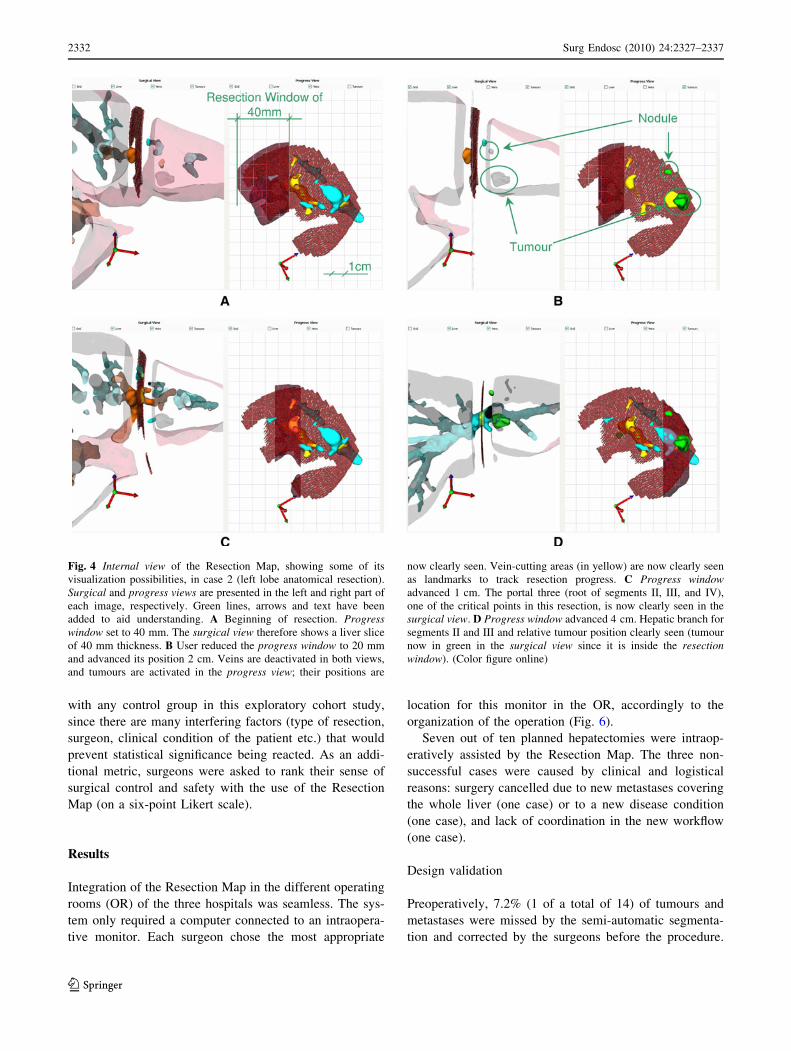
Fig. 4 Internal view of the Resection Map, showing some of its
visualization possibilities, in case 2 (left lobe anatomical resection).
Surgical and progress views are presented in the left and right part of
each image, respectively. Green lines, arrows and text have been
added to aid understanding. A Beginning of resection. Progresswindow set to 40 mm. The surgical view therefore shows a liver slice
of 40 mm thickness. B User reduced the progress window to 20 mm
and advanced its position 2 cm. Veins are deactivated in both views,
and tumours are activated in the progress view; their positions are
now clearly seen. Vein-cutting areas (in yellow) are now clearly seen
as landmarks to track resection progress. C Progress windowadvanced 1 cm. The portal three (root of segments II, III, and IV),
one of the critical points in this resection, is now clearly seen in the
surgical view. D Progress window advanced 4 cm. Hepatic branch for
segments II and III and relative tumour position clearly seen (tumour
now in green in the surgical view since it is inside the resectionwindow). (Color figure online)
2332 Surg Endosc (2010) 24:2327–2337
123

None of them required the boundaries of the tumour to be
refined. Surgeons planned the resection in about 15 min
and were satisfied with the result (question 1, Table 2).
Intraoperatively, interaction through voice commands with
an assistant was effective but had some limitations when
rotating views. Surgeons found it generally easy to read the
cartography of the Resection Map and find correspon-
dences (question 2, Table 2).
The information displayed in the Resection Map
intraoperatively was subjectively characterized by all
surgeons as accurate. As could be expected, 3D recon-
structed veins were easily identified on the intraoperative
ultrasound (US) monitor. Surgeons also noted the exces-
sive width of veins, which was a consequence of the
strategy followed for CT segmentation and 3D recon-
struction. The landmarks displayed in the Resection Map,
the points where veins intersect the resection plane, were
identified in 100% of cases when an US probe was
available (Table 3).
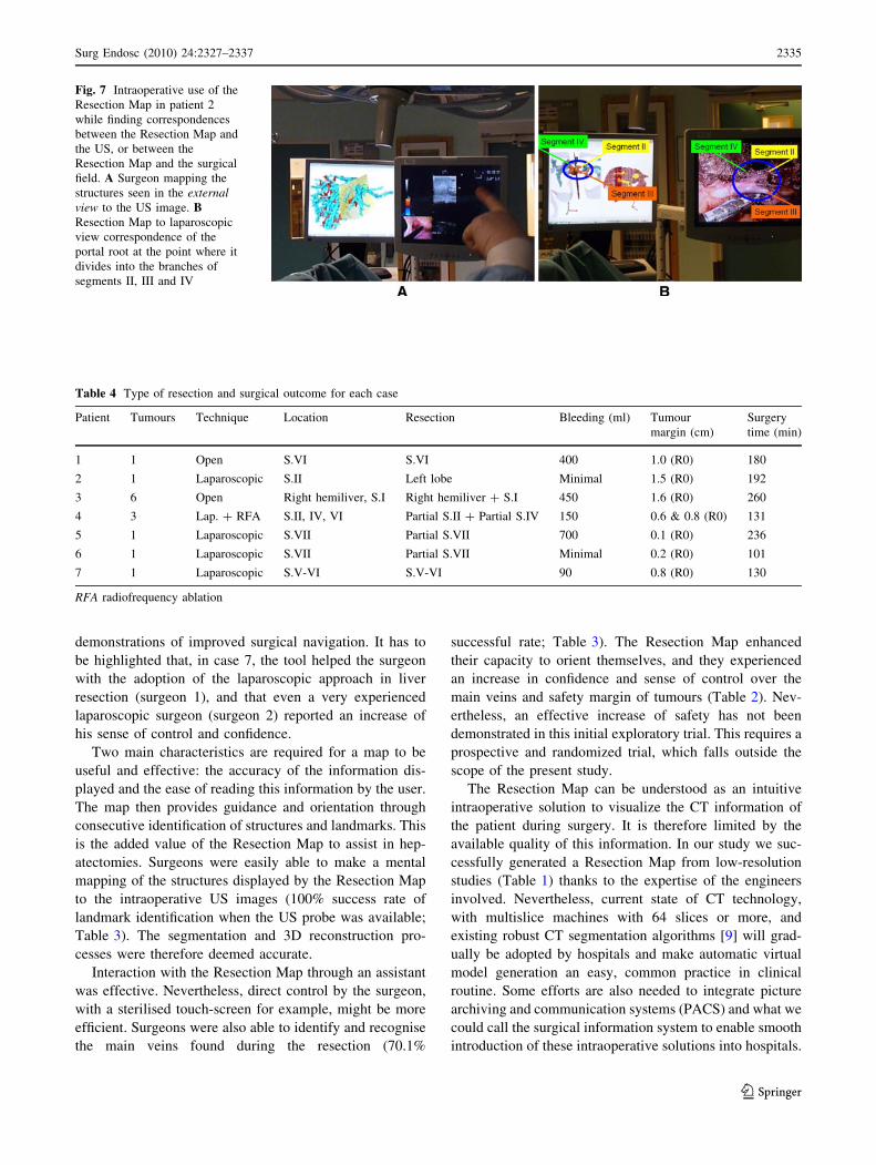
On the other hand, surgeons were able to identify these
yellow landmarks in the operating field in 70.1% of cases
(Table 3). Figure 7 shows an example of one of these
correspondences found in a laparoscopic procedure. The
main reason for a nonidentification of a vein in the oper-
ating field was size, small veins being difficult to visualise.
The use of the LigaSure Atlas (Valleylab, Boulder, CO,
USA), which does not preserve vessels during liver
resection, made vein identification difficult in the case of
patient 7 (Table 3). Finally, intraoperative update in plan-
ned resection path for case 6 led to changes in landmarks
(veins) to be found, which was also counted as non-iden-
tifications. Depth of vessel location during resection was
not a determining factor: the map helped the surgeons to
identify both superficial and deep vessels.
Fig. 5 Internal view of the
Resection Map of patient 1 with
manual annotations (circles and
numbers) of the intersections
between veins and resection
plane, the yellow landmarks to
identify the resection stage. The
progress view (right) shows an
overview of the resection plane
with all five yellow landmarks
and the progress window over
landmarks 1 and 2. The surgicalview (left) therefore focusses on
veins corresponding to
landmarks 1 and 2. (Color figure
online)
Fig. 6 The Resection Map in use in the three hospitals of the study
Surg Endosc (2010) 24:2327–2337 2333
123

Surgical outcome
Intraoperative bleeding, surgical time, resection margin
and a brief description of the resection done in each case
are presented in Table 4. Subjective perception of the
increase of safety and security is reported in Table 2.
Surgeons strongly agreed that the Resection Map increased
their confidence, and that they understood better the
resection case. In comparison, reduction of operative time
and increase of accuracy while drawing the cauterization
line were the aspects with the smallest subjective score (4,
‘‘slightly agree’’). Surgeons agreed that they could antici-
pate the location of and increase the control of veins.
Finally, they also agreed that use of the system increased
assurance of a good safety margin around tumours.
Discussion
Surgical demand exists for computer-aided surgical sys-
tems for both open and laparoscopic liver resections. Sur-
geons expect orientation and visualization support during
operations that allow for more accurate and secure execu-
tion of the planned operation, especially in non-anatomical
resections. The pragmatic solution of a Resection Map
fulfils part of this need without locating any additional
equipment in the OR, as proposed by other recent
approaches [15–17].
The scope of the present study was focussed on the
capacity of the surgeon to track resection progress by using
a Resection Map. In our experimental design, and with
limited resources (only 3 months of time), we tried to
maximize the number of experienced surgeons from dif-
ferent hospitals using the tool. Despite some lack of
coordination in one case, the integration of the new
workflow involving the Resection Map was seamless. The
number of surgical cases we finally managed to process
was small, but each of them provided several test cases of
vein identification, and all were deemed successful
Table 2 Design verification results (questions 1 and 2) and perceived increase of surgical quality and security (questions 3–9) in the seven
surgical cases, ranked on a Likert scale from 1 (strongly disagree) to 6 (strongly agree)
Question Surgical case Median
1 2 3 4 5 6 7
1. I was satisfied with the result of the Resection Planning
(right shape & position of the plane)
5 6 6 6 5 5 6 6
2. It was easy to find the correspondence between the veins
displayed on the Map (internal view) and the veins found
while resecting the liver
6 6 4 5 5 5 5 5
3. I understood better the case (position of the tumour,
resection strategy) thanks to the help of the Map
and the Preoperative Planning
6 6 3 6 4 5 6 6
4. I increased my accuracy in drawing the cauterization line
over the patient’s liver thanks to the help of the Map
5 4 3 4 5 4 6 4
5. I could anticipate the appearance of the next vein, and to
estimate the location of the tumours during the resection
6 5 5 5 4 4 5 5
6. I increased my control of veins, and reduced the amount
bleeding thanks to the help of the Map
6 4 3 4 5 5 6 5
7. I increased the guarantee of a good safety margin around
the tumours thanks to the help of the Map
6 4 5 4 4 5 6 5
8. The Map increased my confidence during the resection 6 6 4 6 5 5 6 6
9. The Map reduced the time of the resection 6 4 4 4 4 4 6 4
Table 3 Rate of correct identification by the surgeon of landmarks
(vein-cutting areas) displayed in the Resection Map
Patient Surgeon Landmark
identification in US
Landmark identification in
the operating field
1 1 n.a. 4/5
2 2 4/4 3/4
3 3 n.a. 5/7
4a 2 5/5 4/5
2/2 2/2
5 2 1/1 1/1
6 2 3/3 2/3
7 1 n.a. 1/4
Total 100% (15/15) 70.1% (22/31)
Landmarks were identified both in the US and in the operating field
n.a. US probe not available during surgerya Patient 4 had two local resections to remove two different tumours
2334 Surg Endosc (2010) 24:2327–2337
123
demonstrations of improved surgical navigation. It has to
be highlighted that, in case 7, the tool helped the surgeon
with the adoption of the laparoscopic approach in liver
resection (surgeon 1), and that even a very experienced
laparoscopic surgeon (surgeon 2) reported an increase of
his sense of control and confidence.
Two main characteristics are required for a map to be
useful and effective: the accuracy of the information dis-
played and the ease of reading this information by the user.
The map then provides guidance and orientation through
consecutive identification of structures and landmarks. This
is the added value of the Resection Map to assist in hep-
atectomies. Surgeons were easily able to make a mental
mapping of the structures displayed by the Resection Map
to the intraoperative US images (100% success rate of
landmark identification when the US probe was available;
Table 3). The segmentation and 3D reconstruction pro-
cesses were therefore deemed accurate.
Interaction with the Resection Map through an assistant
was effective. Nevertheless, direct control by the surgeon,
with a sterilised touch-screen for example, might be more
efficient. Surgeons were also able to identify and recognise
the main veins found during the resection (70.1%
successful rate; Table 3). The Resection Map enhanced
their capacity to orient themselves, and they experienced
an increase in confidence and sense of control over the
main veins and safety margin of tumours (Table 2). Nev-
ertheless, an effective increase of safety has not been
demonstrated in this initial exploratory trial. This requires a
prospective and randomized trial, which falls outside the
scope of the present study.
The Resection Map can be understood as an intuitive
intraoperative solution to visualize the CT information of
the patient during surgery. It is therefore limited by the
available quality of this information. In our study we suc-
cessfully generated a Resection Map from low-resolution
studies (Table 1) thanks to the expertise of the engineers
involved. Nevertheless, current state of CT technology,
with multislice machines with 64 slices or more, and
existing robust CT segmentation algorithms [9] will grad-
ually be adopted by hospitals and make automatic virtual
model generation an easy, common practice in clinical
routine. Some efforts are also needed to integrate picture
archiving and communication systems (PACS) and what we
could call the surgical information system to enable smooth
introduction of these intraoperative solutions into hospitals.
Fig. 7 Intraoperative use of the
Resection Map in patient 2
while finding correspondences
between the Resection Map and
the US, or between the
Resection Map and the surgical
field. A Surgeon mapping the
structures seen in the externalview to the US image. BResection Map to laparoscopic
view correspondence of the
portal root at the point where it
divides into the branches of
segments II, III and IV
Table 4 Type of resection and surgical outcome for each case
Patient Tumours Technique Location Resection Bleeding (ml) Tumour
margin (cm)
Surgery
time (min)
1 1 Open S.VI S.VI 400 1.0 (R0) 180
2 1 Laparoscopic S.II Left lobe Minimal 1.5 (R0) 192
3 6 Open Right hemiliver, S.I Right hemiliver ? S.I 450 1.6 (R0) 260
4 3 Lap. ? RFA S.II, IV, VI Partial S.II ? Partial S.IV 150 0.6 & 0.8 (R0) 131
5 1 Laparoscopic S.VII Partial S.VII 700 0.1 (R0) 236
6 1 Laparoscopic S.VII Partial S.VII Minimal 0.2 (R0) 101
7 1 Laparoscopic S.V-VI S.V-VI 90 0.8 (R0) 130
RFA radiofrequency ablation
Surg Endosc (2010) 24:2327–2337 2335
123

The Resection Map bridges the gap existing in current
liver planning systems [8–12] of effective translation of
preoperative analysis into intraoperative guidance. Plan-
ning systems can also be used in the OR [24], but they are
not designed to fulfil intraoperative requirements. The
Resection Map, on the other hand, is a virtual navigation
environment without the relative positioning of surgical
tools, which can be provided in an immobilised liver at the
cost of additional hardware and a more complex surgical
workflow [15–17]. There is no solution yet that copes with
intraoperative deformations or that is able to track and
adapt. A solution such as the Resection Map relies not on
additional hardware but on the surgeon’s capacity to read
and map the cartography displayed to them. We therefore
think that this is a pragmatic solution that solves part of the
intraoperative navigation need quite efficiently.
We believe that the Resection Map could be very
helpful for the education of inexperienced liver surgeons,
for the adoption of a laparoscopic approach and for com-
plex cases of an experienced surgeon. The tool could even
substitute some of the uses of intraoperative US, such as
the identification of key vessels that are going to be cut
during resection. Nevertheless, US will still be required for
verification of the position and size (and possible growth)
of known tumours, and the identification of new ones.
Future work will be directed towards full automation of
the Resection Map system, and towards comprehensive
study to measure the increase of safety during hepatecto-
mies and verify the hypothetical benefits of the tool stated
above. Technically, the tool should include the capability
to update the resection plane intraoperatively after finding
new metastases. Also, in order to enhance interactivity and
navigation experience, future research should address the
challenge of incorporating tool positioning information and
adapting to the deformations of the liver while minimizing
disturbance to the surgical workflow [25].
Conclusion
The Resection Map is a simple and pragmatic solution
conceived to enhance the safety of liver resection. Its
integration into the operating room was seamless, and
preliminary results showed a perceived increase in safety
and confidence of the surgeon. Other possible applications
of this technology are easier adoption of a laparoscopic
approach by surgical teams, easier implantation of a living-
donor programme and education of inexperienced surgeons.
Acknowledgements This work is part of the ARIS*ER Marie Curie
Research Training Network (MRTN-CT-2004-512400), which is
funded by the 6th Framework Programme of the European Commis-
sion. We especially acknowledge E. Samset for support and excellent
management and coordination, A. Jalote-Parmar for her contributions
at the early stage of this research, A. Serrablo for his enthusiasm and
engagement, and our partners and hospital staff for help and support
in the development and validation work.
Disclosures Authors Pablo Lamata, Felix Lamata, Valentin Sojar,
Piotr Makowski, MSc Laurent Massoptier, Sergio Casciaro, MSc
Wajid Ali, Thomas Studeli, Jerome Declerck, Ole Jackov Elle, and
Bjorn Edwin have no conflicts of interest or financial ties to disclose.
Open Access This article is distributed under the terms of the
Creative Commons Attribution Noncommercial License which per-
mits any noncommercial use, distribution, and reproduction in any
medium, provided the original author(s) and source are credited.
References
1. Jemal A, Siegel R, Ward E, Hao Y, Xu J, Murray T, Thun MJ
(2008) Cancer statistics, 2008. CA Cancer J Clin 58:71–96
2. Honore C, Detry O, Deroover A, Piront P, Polus M, Honore P,
Meurisse M (2008) When should we resect colorectal liver
metastases? Rev Med Liege 63:595–599
3. El-Serag HB, Rudolph KL (2007) Hepatocellular carcinoma:
epidemiology and molecular carcinogenesis. Gastroenterology
132:2557–2576
4. Grundmann RT, Hermanek P, Merkel S, Germer CT, Grundmann
RT, Hauss J, Henne-Bruns D, Herfarth K, Hermanek P, Hopt UT,
Junginger T, Klar E, Klempnauer J, Knapp WH, Kraus M, Lang
H, Link KH, Lohe F, Merkel S, Oldhafer KJ, Raab HR, Rau HG,
Reinacher-Schick A, Ricke J, Roder J, Schafer AO, Schlitt HJ,
Schon MR, Stippel D, Tannapfel A, Tatsch K, Vogl TJ (2008)
Diagnosis and treatment of colorectal liver metastases—work-
flow. Zentralbl Chir 133:267–284
5. Shaw IM, Rees M, Welsh FK, Bygrave S, John TG (2006) Repeat
hepatic resection for recurrent colorectal liver metastases is asso-
ciated with favourable long-term survival. Br J Surg 93:457–464
6. Bryant R, Laurent A, Tayar C, van Nhieu JT, Luciani A, Cherqui
D (2008) Liver resection for hepatocellular carcinoma. Surg
Oncol Clin North Am 17:607–633, ix
7. Marescaux J, Clement JM, Tassetti V, Koehl C, Cotin S, Russier
Y, Mutter D, Delingette H, Ayache N (1998) Virtual reality
applied to hepatic surgery simulation: the next revolution. Ann
Surg 228:627–634
8. Lang H, Radtke A, Hindennach M, Schroeder T, Fruhauf NR,
Malago M, Bourquain H, Peitgen HO, Oldhafer KJ, Broelsch CE
(2005) Impact of virtual tumor resection and computer-assisted
risk analysis on operation planning and intraoperative strategy in
major hepatic resection. Arch Surg 140:629–638
9. Soler L, Delingette H, Malandain G, Montagnat J, Ayache N,
Koehl C, Dourthe O, Malassagne B, Smith M, Mutter D,
Marescaux J (2001) Fully automatic anatomical, pathological,
and functional segmentation from CT scans for hepatic surgery.
Comput Aided Surg 6:131–142
10. Reitinger B, Bornik A, Beichel R, Schmalstieg D (2006) Liver
surgery planning using virtual reality. IEEE Comput Graph Appl
26:36–47
11. Meinzer H-P, Thorn M, Cardenas CE (2002) Computerized
planning of liver surgery—an overview. Comput Graph 26:569–
576
12. Sojar V, Stanisavljevic D, Hribernik M, Glusic M, Kreuh D,
Velkavrh U, Fius T (2004) Liver surgery training and planning in
3D virtual space. Proc CARS 2004 1268:390–394
13. Jalote-Parmar A, Pattynama P, de Ridder H, Goossens R,
Freudenthal A, Samset E (2007) Surgical workflow analysis:
2336 Surg Endosc (2010) 24:2327–2337
123

identifying user requirements for surgical information systems.
In: Pikaar R, Ernst K, Settels (eds) Diversity in ergonomics.
Elsevier, Oxford, pp 229–241
14. Mutter D, Dallemagne B, Bailey C, Soler L, Marescaux J (2009)
3D virtual reality and selective vascular control for laparoscopic
left hepatic lobectomy. Surg Endosc 23:432–435
15. Beller S, nerbein M, Eulenstein S, Lange T, Schlag PM (2007)
Feasibility of navigated resection of liver tumors using multi-
planar visualization of intraoperative 3-dimensional ultrasound
data. Ann Surg 246:288–294
16. Feuerstein M, Mussack T, Heining SM, Navab N (2008) Intra-
operative laparoscope augmentation for port placement and
resection planning in minimally invasive liver resection. IEEE
Trans Med Imag 27:355–369
17. Cash DM, Miga MI, Glasgow SC, Dawant BM, Clements LW,
Cao Z, Galloway RL, Chapman WC (2007) Concepts and pre-
liminary data toward the realization of image-guided liver sur-
gery. J Gastrointest Surg 11:844–859
18. Jalote-Parmar A, Pattynama PMT, Van der Plas AP, Heer P
(2007) Towards intuitive surgical interfaces: strategies from
video games. In: Casciaro S, Distante A (eds) New technology
frontiers in minimally invasive therapies. Lupiensis Biomedical
pp 101–111
19. Studeli T (2008) Surgical navigation during minimally invasive
procedures. In: Casciaro E, Samset E (eds) Minimally invasive
technologies and nanosystems for diagnosis and therapies.
Lupiensis Biomedical, Lecce, Italy, pp 177–186
20. Lamata P, Jalote-Parmar A, Lamata F, Declerck J (2008) The
Resection Map, a proposal for intraoperative hepatectomy guid-
ance. Int J CARS 3:299–306
21. Massoptier L, Casciaro S (2008) A new fully automatic and
robust algorithm for fast segmentation of liver tissue and tumors
from CT scans. Eur Radiol 18:1658–1665
22. Frangi AF, Niessen WJ, Hoogeveen RM, van WT, Viergever MA
(1999) Model-based quantitation of 3-D magnetic resonance
angiographic images. IEEE Trans Med Imag 18:946–956
23. Nielsen J (1995) Usability engineering. Morgan Kaufmann. San
Francisco, CA, USA
24. Grenacher L, Thorn M, Knaebel HP, Vetter M, Hassenpflug P,
Kraus T, Meinzer HP, Buchler MW, Kauffmann GW, Richter
GM (2005) The role of 3-D imaging and computer-based post-
processing for surgery of the liver and pancreas. Rofo 177:1219–
1226
25. Lamata P, Morvan T, Reimers M, Samset E, Declerck J (2009)
Addressing shading-based laparoscopic registration. In: Dossel O,
Schlegel WC (eds) WC 2009, IFMBE proceedings 25/VI, pp
189–192 (for further reference: http://www.springerlink.com/
content/j76732394677363w/?p=4693b05106ef4b109c37bf9e11
ae0fd8&pi=0)
Surg Endosc (2010) 24:2327–2337 2337
123
Copyright © 2022 FDOKUMEN