Insulin-like growth factor I (IGF-I) in a growth-enhanced transgenic (GH-overexpressing) bony fish,...
11
ORIGINAL PAPER Insulin-like growth factor I (IGF-I) in a growth-enhanced transgenic (GH-overexpressing) bony fish, the tilapia (Oreochromis niloticus): indication for a higher impact of autocrine/paracrine than of endocrine IGF-I Elisabeth Eppler Antje Caelers Natallia Shved Guylin Hwang Azizur M. Rahman Norman Maclean Ju ¨ rgen Zapf Manfred Reinecke Received: 1 December 2006 / Accepted: 19 March 2007 / Published online: 13 April 2007 Ó Springer Science+Business Media B.V. 2007 Abstract Several lines of growth hormone (GH)- overexpressing fish have been produced and analysed for growth and fertility parameters. However, only few data are available on the growth-promoting hormone insulin-like growth factor I (IGF-I) that mediates most effects of GH, and these are contradictory. Using quantitative real-time RT-PCR, radioimmunoassay, in situ hybridization, immunohistochemistry, and radiochromatography we investigated IGF-I and IGF binding proteins (IGFBPs) in an adult (17 months old) transgenic (GH-overexpressing) tilapia (Oreochromis niloticus). The transgenics showed an around 1.5-fold increase in length and an approximately 2.3-fold higher weight than the non-transgenics. Using radio- immunoassay, the serum IGF-I levels were lower (6.22 ± 0.75 ng/ml) in transgenic than in wild-type (15.01 ± 1.49 ng/ml) individuals (P = 0.0012). Radio- immunoassayable IGF-I in transgenic liver was 4.2- times higher than in wild-type (16.0 ± 2.21 vs. 3.83 ± 0.71 ng/g, P = 0.0017). No hepatocytes in wild-type but numerous hepatocytes in transgenic liver contained IGF-I-immunoreactivity. RT-PCR revealed a 1.4-times higher IGF-I mRNA expression in the liver of the transgenics (10.51 ± 0.82 vs. 7.3 ± 0.49 pg/mg total RNA, P = 0.0032). In correspondence, in situ hybridization showed more IGF-I mRNA containing hepatocytes in the transgenics. A twofold elevated IGF-I mRNA expression was determined in the skeletal muscle of transgenics (0.33 ± 0.02 vs. 0.16 ± 0.01 pg/mg total RNA, P < 0.0001). Both liver and serum of transgenics showed increased IGF-I binding. The increased IGFBP content in the liver may lead to retention of IGF-I, and/or the release of IGF-I into the circulation may be slower resulting in accu- mulation of IGF-I in the hepatocytes. Our results indicate that the enhanced growth of the transgenics likely is due to enhanced autocrine/paracrine action of IGF-I in extrahepatic sites, as shown here for skeletal muscle. Keywords IGF-I IGFBP Liver Serum Skeletal muscle Transgenic fish Introduction Insulin-like growth factor I (IGF-I) is a potent mitogenic hormone that induces growth and differ- entiation in a variety of target organs (Jones and Clemmons 1995; Reinecke and Collet 1998; Butler and LeRoith 2001). In mammals, IGF-I is mainly E. Eppler A. Caelers N. Shved J. Zapf M. Reinecke (&) Division of Neuroendocrinology, Institute of Anatomy, University of Zu ¨rich, Winterthurerstr 190, 8057 Zu ¨rich, Switzerland e-mail: [email protected] G. Hwang A. M. Rahman N. Maclean School of Biological Sciences, University of Southampton, Southampton, Hampshire SO16 7PX, UK 123 Transgenic Res (2007) 16:479–489 DOI 10.1007/s11248-007-9093-z
Transcript of Insulin-like growth factor I (IGF-I) in a growth-enhanced transgenic (GH-overexpressing) bony fish,...

ORIGINAL PAPER
Insulin-like growth factor I (IGF-I) in a growth-enhancedtransgenic (GH-overexpressing) bony fish, the tilapia(Oreochromis niloticus): indication for a higher impact ofautocrine/paracrine than of endocrine IGF-I
Elisabeth Eppler Æ Antje Caelers Æ Natallia Shved Æ Guylin Hwang ÆAzizur M. Rahman Æ Norman Maclean Æ Jurgen Zapf Æ Manfred Reinecke
Received: 1 December 2006 / Accepted: 19 March 2007 / Published online: 13 April 2007
� Springer Science+Business Media B.V. 2007
Abstract Several lines of growth hormone (GH)-
overexpressing fish have been produced and analysed
for growth and fertility parameters. However, only few
data are available on the growth-promoting hormone
insulin-like growth factor I (IGF-I) that mediates most
effects of GH, and these are contradictory. Using
quantitative real-time RT-PCR, radioimmunoassay,
in situ hybridization, immunohistochemistry, and
radiochromatography we investigated IGF-I and IGF
binding proteins (IGFBPs) in an adult (17 months old)
transgenic (GH-overexpressing) tilapia (Oreochromis
niloticus). The transgenics showed an around 1.5-fold
increase in length and an approximately 2.3-fold
higher weight than the non-transgenics. Using radio-
immunoassay, the serum IGF-I levels were lower
(6.22 ± 0.75 ng/ml) in transgenic than in wild-type
(15.01 ± 1.49 ng/ml) individuals (P = 0.0012). Radio-
immunoassayable IGF-I in transgenic liver was 4.2-
times higher than in wild-type (16.0 ± 2.21 vs.
3.83 ± 0.71 ng/g, P = 0.0017). No hepatocytes in
wild-type but numerous hepatocytes in transgenic liver
contained IGF-I-immunoreactivity. RT-PCR revealed
a 1.4-times higher IGF-I mRNA expression in the liver
of the transgenics (10.51 ± 0.82 vs. 7.3 ± 0.49 pg/mg
total RNA, P = 0.0032). In correspondence, in situ
hybridization showed more IGF-I mRNA containing
hepatocytes in the transgenics. A twofold elevated
IGF-I mRNA expression was determined in the
skeletal muscle of transgenics (0.33 ± 0.02 vs.
0.16 ± 0.01 pg/mg total RNA, P < 0.0001). Both liver
and serum of transgenics showed increased IGF-I
binding. The increased IGFBP content in the liver may
lead to retention of IGF-I, and/or the release of IGF-I
into the circulation may be slower resulting in accu-
mulation of IGF-I in the hepatocytes. Our results
indicate that the enhanced growth of the transgenics
likely is due to enhanced autocrine/paracrine action of
IGF-I in extrahepatic sites, as shown here for skeletal
muscle.
Keywords IGF-I � IGFBP � Liver � Serum � Skeletal
muscle � Transgenic fish
Introduction
Insulin-like growth factor I (IGF-I) is a potent
mitogenic hormone that induces growth and differ-
entiation in a variety of target organs (Jones and
Clemmons 1995; Reinecke and Collet 1998; Butler
and LeRoith 2001). In mammals, IGF-I is mainly
E. Eppler � A. Caelers � N. Shved � J. Zapf �M. Reinecke (&)
Division of Neuroendocrinology, Institute of Anatomy,
University of Zurich, Winterthurerstr 190, 8057 Zurich,
Switzerland
e-mail: [email protected]
G. Hwang � A. M. Rahman � N. Maclean
School of Biological Sciences, University of
Southampton, Southampton, Hampshire SO16 7PX, UK
123
Transgenic Res (2007) 16:479–489
DOI 10.1007/s11248-007-9093-z

produced in the liver which is the principal source of
endocrine IGF-I. The primary stimulus for the
synthesis and secretion of liver IGF-I is growth
hormone (GH) released from the anterior pituitary
(Reinecke et al. 2005).
Like in mammals, the major site of IGF-I gene
expression in bony fish is liver (Duan 1998; Reinecke
et al. 2005; Wood et al. 2005). High-affinity GH
binding sites have been characterized in salmon and
tilapia liver (Gray et al. 1992; Ng et al. 1992;
Shepherd et al. 1997). In accordance, GH injections
promoted liver IGF-I mRNA expression in salmonids
and tilapia (e.g. Duan et al. 1993a; Moriyama 1995;
Shamblott et al. 1995; Duguay et al. 1996; Shepherd
et al. 1997; Guillen et al. 1998; Kajimura et al. 2001;
Vong et al. 2003) and raised IGF-I plasma levels in
salmonids, seabream and tilapia (e.g. Moriyama
1995; Shamblott et al. 1995; Guillen et al. 1998;
Moriyama et al. 2000; Kajimura et al. 2001), which
was accompanied by an increase in growth (Guillen
et al. 1998). Similarly, GH also stimulates the
expression of IGF-I mRNA in primary hepatocyte
cultures of bony fish, such as salmonids (Duan et al.
1993a, b; Shamblott et al. 1995; Pierce et al. 2005)
and tilapia Oreochromis mossambicus (Schmid et al.
2000). In adult Coho salmon, continuous infusion of
bovine IGF-I increased growth rate and weight gain
(McCormick et al. 1992), and plasma IGF-I levels
and growth rate were significantly correlated in O.
mossambicus (Kajimura et al. 2001; Uchida et al.
2003).
Several groups have produced different lines of
transgenic fish (e.g. Devlin et al. 2000; Maclean et al.
2002; Rocha et al. 2004). Among the transgenic fish
those carrying exogenous GH gene constructs con-
stitute the majority. The obvious reason for this focus
of interest is to improve fish growth in aquacultural
food production (Zbikowska 2003). Mainly salmo-
nids (Du et al. 1992; Devlin et al. 1994, 2000;
Sundstrom et al. 2004), carp (Zhang et al. 1990),
tilapia (Martinez et al. 1996; Rahman et al. 1998),
and sea bream (Lu et al. 2002) have been used for
genetically induced growth enhancement. On the
other hand, GH-overexpressing fish are also consid-
ered as excellent models to study gene regulation and
development (Amsterdam and Becker 2005). How-
ever, investigations on transgenic fish have almost
exclusively dealt with growth parameters, fertility,
and body and organ integrity (Maclean et al. 2002;
Sundstrom et al. 2004).
The above referred studies demonstrate the stim-
ulating action of GH on IGF-I-mediated growth in
bony fish either by bolus injections of GH or by
adding GH to the medium of primary hepatocyte
cultures and, thus, deal with short-term effects of GH.
In contrast, only one study has dealt with IGF-I in
transgenic GH-overexpressing fish. In Coho salmon
Oncorhynchus kisutch IGF-I serum levels were
determined and gave contradictory results (Devlin
et al. 2000): in one experiment they were slightly
enhanced and in another slightly reduced. In order to
investigate whether the severe enhancement in body
growth in GH-overexpressing fish may be due to
endocrine (liver-derived) IGF-I and/or enhanced IGF-
I expression in skeletal muscle we used a transgenic
GH-overexpressing and age-matched wild-type tila-
pia (Oreochromis niloticus). The following parame-
ters were analysed: (1) the expression level of IGF-I
mRNA in liver and skeletal muscle using quantitative
real-time PCR, (2) the concentration of IGF-I peptide
in liver and serum by RIA, (3) the localization of
IGF-I in liver at the mRNA level by in situ hybrid-
ization and at the peptide level by immunohisto-
chemistry, and (4) the IGF-I binding proteins
(IGFBPs) in liver and serum by radiochromatogra-
phy.
Material and methods
Animals
The transgenic fish used in these experiments have
been previously produced from crosses between a
wild-type female O. niloticus and a G1 transgenic
male. This line of growth-enhanced tilapia (C86)
carries a single copy of a Chinook salmon Oncorhyn-
chus tshawytscha GH gene spliced to an ocean pout
Macrozoarces americanus antifreeze promoter (OP-
AFPcsGH) co-ligated with a carp beta actin/lacZ
reporter gene construct, integrated into the tilapia
genome (Rahman et al. 1998). Tilapia of the C86
strain and the non-transgenic siblings were bred at the
University of Southampton. The fish were kept in
fresh water tanks at 27 ± 18C under a 12 h/12 h light/
dark cycle and fed to satiation. Adult (17 months)
480 Transgenic Res (2007) 16:479–489
123

transgenic (TG) and wild-type (WT) fish were used
for the experiments. Fish were anaesthetized with 2-
phenoxy-ethanol (Sigma, St Louis, MO, USA) added
to water, measured in weight and length, and the
tissue samples excised. Principles of animal care and
specific national laws were followed.
Detection of transgene
The transgenic or non-transgenic state of the indi-
viduals was detected by RT-PCR followed up by
Southern Blotting. For PCR amplification an approx-
imately 3 mm · 2 mm fin clip was taken from the
caudal fin and immediately frozen in liquid nitrogen.
Genomic DNA was isolated. The standard procedures
for isolation and purification of DNA (Rahman and
Maclean 1992) and subsequent PCR and Southern
Blotting were performed as described before (Rah-
man et al. 1998).
Tissue sampling and extraction
Liver (n = 10 TG, n = 10 WT) and skeletal muscle
(n = 6 TG, n = 6 WT) specimens for RT-PCR were
excised and immediately transferred into 1.5 ml of
the RNA-preserving reagent RNAlaterTM (Ambion,
Austin, USA). The samples were kept at 48C to
promote inactivation of RNAses and later stored at
�208C until RNA isolation. Total RNA was extracted
using TRIzol reagent (GibcoBRL), treated with 1 U
of RQ1 RNAse-free DNase (Catalys AG, Wallisellen,
Switzerland), resuspended in DEPC-treated H2O and
stored at �808C.
Absolute quantification of liver and skeletal
muscle IGF-I gene expression by real-time PCR
Absolute quantification of liver and skeletal muscle
IGF-I mRNA was performed as already described
(Caelers et al. 2004). In brief, based on the gene
sequences of O. mossambicus IGF-I (Reinecke et al.
1997), and O. niloticus b-actin as a housekeeping
gene (Hwang et al. 2003), tilapia specific primers and
probes for real-time RT-PCR were designed with the
Primer Express software version 1.5 (PE Biosystems,
Foster City, CA, USA). To create templates for
in vitro transcription, a T7 phage polymerase pro-
moter gene sequence was added by primer extension
to the 50end of the antisense primers using conven-
tional RT-PCR of O. niloticus liver total RNA. For
cDNA synthesis, 5 mg RNA were annealed with 1 mM
poly(dT) primer (50-CCTGAATTCTAGAGCT-
CAT(dT17)-30) for 5 min at 708C. The RNA/primer
mix was incubated for 1 h at 378C with 10 mM
dNTPs and 100 U M-MLV-RTase (Promega) in 1·buffer. One microlitre aliquots of cDNA were added
to 50 ml PCR reaction using the Thermo-StartTM PCR
Master Mix (Abgene, NY, USA). Amplification
conditions were optimized for a Stratagene RoboCy-
cler Gradient 40: 1 cycle 10 min at 948C, 1 min at
608C, 2 min at 728C; 40 cycles 1 min at 948C, 1 min
at 608C, 2 min at 728C; final extension 10 min at
728C. PCR products including the T7 promoter gene
sequence were sequenced and visualized on a 2.5%
agarose gel. Standard cRNAs were generated by
in vitro transcription using the T7-MEGAshort-
scriptTM Kit (Ambion), analysed by photospectrom-
etry and UV-shadowing, and quantified by
photospectrometry and dot blot. Defined amounts at
tenfold dilutions were subjected to real-time PCR
using a one-Step-RT-PCR Mastermix (Applied Bio-
systems, Rotkreuz, Switzerland). RT-step (488C,
30 min) and denaturation step (958C, 10 min) were
performed followed by 40 cycles (958C, 15 s; 608C,
1 min) in a single tube using ABI PRISMTM 7700
Sequence Detection System Perkin Elmer (Applied
Biosystems). Standard curves were generated based
on the linear relationship between CT value and
logarithm of the starting amount. For measurements,
tenfold diluted defined amounts of standard cRNA
and 10 ng of total RNA were subjected in parallel to
real-time PCR under the same experimental settings.
To calculate absolute amounts the different lengths of
cRNA and mRNA were considered by a correction
factor determined by division of lengths of IGF-I
mRNA and cRNA (546 nt/70 nt = 7.8).
Peptide extraction of serum and liver
Blood (n = 5 TG, n = 5 WT) was taken with a 1 ml
heparinized syringe from the caudal vein and col-
lected in sterile 0.5 ml tubes. The blood was
centrifuged at 13,000 rpm for 15 min at 48C and
the serum removed and stored at –208C until use. 1 g
of liver (n = 5 TG, n = 5 WT) was homogenized with
5 ml 0.1 M sodium phosphate buffer (pH 7.0) and
centrifuged at 10,000g for 15 min at 48C. The
supernatant was removed with a Pasteur pipette,
Transgenic Res (2007) 16:479–489 481
123

deep-frozen immediately, and stored at -208C until
use. To dissociate IGF-I from the binding proteins,
acid–ethanol extraction was performed as already
described. In brief, 40 ml of serum and liver
homogenate, respectively, was thoroughly mixed
with 160 ml of an acid–ethanol extraction mix
(87.5% ethanol, 12.5% 2 M HCl, v/v) and incubated
for 30 min at room temperature. After neutralization
with 0.885 M Tris base, the extracts were centrifuged
at 13,000 rpm at 48C for 10 min. 50 ml of the
supernatant was used for the IGF-I radioimmunoas-
say.
Radioimmunoassay in serum and liver
Serum and liver IGF-I was measured using the Fish
IGF-I RIA kit for the tilapia O. mossambicus (GroPep
Pty Ltd., Adelaide, Australia) with recombinant
tilapia IGF-I as tracer and standard according to the
protocol of the manufacturer.
Radiochromatography for IGFBPs in serum and
liver
Radiochromatography was performed as described
earlier (Zapf et al. 1975, 1989). Briefly, 0.2 ml of
pooled serum or liver homogenate was diluted with
0.3 ml of Dulbecco’s phosphate-buffered saline, pH
7.4, containing NaN3 and 500 U of Trasylol (Bayer,
Germany), and incubated for 24 h at 48C with 125I-
labelled tilapia IGF-I (2.5 · 105 CPM, GroPep Ltd.).
The mixture was run through a Sephadex G-200
column (2.1 cm · 70 cm) preequilibrated with
Dulbecco’s buffered saline, pH 7.4, and the radioac-
tivity of the collected fractions (2.5 ml) was counted
in a gamma-counter.
Preparation of probes for in situ hybridization
Probes were prepared as described (Schmid et al. 1999;
Berishvili et al. 2006a, b). In brief, total RNA from
tilapia liver was extracted with the Ultraspec Extrac-
tion Kit (ams, Lugano, Switzerland). For cDNA
synthesis 5 mg RNA was annealed with 1 mM of a
poly(dT) primer (50 CCTGAATTCTAGAGCT-
CAT(dT17) 30) for 3 min at 708C. The RNA/primer
mix was incubated for 1 h at 378C with 15 mM dNTPs
and 10 U AMV-RTase (Pharmacia, Switzerland) in 1·
reaction buffer (50 mM Tris–HCl/pH 8.3, 40 mM KCl,
6 mM MgCl2). One microlitre cDNA was incubated
with 1 mM of sense (50-GTCTGTGGAGAGC-
G A G G C T T T - 3 0) a n d a n t i s e n s e ( 5 0-AACCTTGGGTGCTCTTGGCATG-30) primers
corresponding to the B- and E-domain, 200mM dNTPs,
and 1 U Taq-polymerase (Pharmacia) in 1· incubation
buffer (10 mM Tris–HCl/pH 8, 50 mM KCl, 1.5 mM
MgCl2, 0.001 % gelatine). The amplification program
was optimized for a Stratagene RoboCycler Gradient
40:1 cycle 10 min at 948C, 1 min at 598C, 2 min at
728C; 30 cycles 1 min at 948C, 1 min at 598C and 2 min
at 728C followed by final extension for 5 min at 728C.
PCR fragments were separated on a 2% agarose gel and
eluted by the Gel Extraction Kit QIAquick (Qiagen,
Switzerland). Thereafter, the PCR products were
cloned in a pCR-Script SK(+) cloning vector using a
kit (Stratagene, Heidelberg, Germany). Plasmids con-
taining the gene sequence fragments were sequenced
(Microsynth, Switzerland) and the sequences com-
pared to database. The plasmids containing the specific
inserts of IGF-I (207 bp) were used as templates for the
synthesis of digoxigenin (DIG)-labelled RNA probes.
Linearization was performed with EcoRI for T3- and
NotI for T7-polymerase-driven transcription. One
microgram of linearized plasmids was transcribed
in vitro in the presence of DIG-UTP from T3 and T7
promotors to obtain antisense and sense (negative
control) probes. Integrity of probes and efficiency of
labelling were confirmed by dot blot and gel electro-
phoresis including blotting and incubation with anti-
body. Specificity of the probes has been demonstrated
previously (Schmid et al. 1999; Berishvili et al. 2006a).
In situ hybridization
Liver preparations (n = 3 WT, n = 3 TG) were fixed
by immersion with 4% buffered formalin for 4 h at
room temperature. Specimens were dehydrated in
ascending series of ethanol and routinely embedded
in paraplast (588C). Four micrometre sections were
mounted on Super Frost Plus slides (Menzel-Glaser,
Germany) and dried overnight at 428C. After dewax-
ing and rehydration, the sections were postfixed with
4% paraformaldehyde and 0.1% glutaraldehyde in 1·PBS. In situ hybridization was performed as previ-
ously described (Schmid et al. 1999; Berishvili et al.
2006a, b). The following steps were carried out with
DEPC-treated solutions in a humified chamber. The
482 Transgenic Res (2007) 16:479–489
123

sections were digested with 0.02% proteinase K in
20 mM Tris–HCl/pH 7.4, 2 mM CaCl2 for 10 min at
378C. Thereafter, sections were treated with 1.5%
triethanolamine and 0.25% acetic anhydride for
10 min at room temperature and incubated with
50 ml prehybridization solution per section for 3–4 h
at 548C. Hybridization was performed overnight at
548C with 30 ml of hybridization buffer containing
10 ng of sense or antisense probe previously dena-
turated for 5 min at 858C. Slides were washed for
15 min at room temperature in 2· SSC, and for
30 min at the specific hybridization temperature at
descending concentrations of SSC (2·, 1·, 0.5·,
0.2·). The alkaline-phosphatase-coupled anti-DIG
antibody was diluted 1:4,000 in 1% blocking reagent
(Roche-Diagnostics) in buffer P1 and sections were
incubated for 1 h at room temperature in the dark.
After washing twice in P1 for 15 min, sections were
treated with buffer P3, 5 mM levamisole, and NBT/
BCIP stock solution (Roche Diagnostics). Colour
development was carried out overnight at room
temperature and stopped by rinse in tap water for
15 min. Sections were mounted with glycergel and
photographed with a Zeiss Axioscope using the
Axiovision software 3.1. (Zeiss, Zurich, Switzerland).
Immunohistochemical technique
Specimens for immunohistochemistry (n = 3 WT,
n = 3 TG) were immersed in Bouin’s solution without
acetic acid for 4 h at room temperature. Specimens
were dehydrated in ascending series of ethanol and
routinely embedded in paraplast (588C). Sections
were cut at 4 mm, mounted onto glass slides and dried
overnight at 428C. After dewaxing and rehydration
unspecific binding was reduced by treatment with
PBS (pH 7.4) containing 2% bovine serum albumine
for 30 min at room temperature. Thereafter, the
sections were incubated overnight with the rabbit
antiserum 116 raised against human IGF-I (1:400)
and washed repetitively in PBS. This antiserum
specifically also stains IGF-I in fish as shown by
absorption experiments (Reinecke et al. 1997; Sch-
mid et al. 1999; Berishvili et al. 2006a, b). The IGF-I
antiserum was detected by incubation with biotiny-
lated goat anti-rabbit IgG (Bioscience Products,
Emmenbrucke, Switzerland, 1:100) for 30 min at
room temperature. After repetitive rinsing in PBS, the
reactions were visualized with fluorescein-isothiocy-
anate-conjugated streptavidin (Bioscience Products,
1:100) for 30 min at room temperature in the dark.
Sections were mounted with glycergel and photo-
graphed with a Zeiss Axioscope using the Axiovision
software 3.1.
Statistical analysis
Statistical analysis of the data was performed with a
GraphPad Prism 4 program. This included a Mann–
Whitney-U-test with a significance level of 5%. All
data are expressed as means ± SEM.
Results
Fish size and weight
The 17-month old transgenic fish showed an approx-
imately 1.5-fold increase in head–tail length when
compared to their non-transgenic siblings (28.7 ± 8.3
vs. 18.5 ± 6.2 cm). The mean weights of the
transgenics were around 2.3-fold higher than those
of their non-transgenic siblings (415.6 ± 98 g and
184 ± 64 g, respectively).
Measurement of IGF-I peptide and mRNA
Using radioimmunoassay, the serum IGF-I levels
were lower (6.22 ± 0.75 ng/ml) in transgenic than in
wild-type (15.01 ± 1.49 ng/ml) individuals
(P = 0.0012) (Fig. 1a). As measured by the same
radioimmunoassay, the IGF-I concentration in liver
amounted to 16.0 ± 2.21 ng/g in transgenics and to
3.83 ± 0.71 ng/g in wild-type animals (P = 0.0017)
(Fig. 1b). Real-time PCR revealed a 1.4-times higher
IGF-I mRNA expression in the liver of the transgen-
ics (10.51 ± 0.82 vs. 7.3 ± 0.49 pg/mg total RNA,
P = 0.0032, Fig. 1c) and a twofold elevated IGF-I
mRNA expression in the skeletal muscle (0.33 ± 0.02
vs. 0.16 ± 0.01 pg/mg total RNA, P < 0.0001,
Fig. 1d).
Localization of IGF-I mRNA and peptide
In situ hybridization with antisense DIG-labelled
RNA probe specific for tilapia IGF-I revealed
positive responses in liver whereas the sense RNA
Transgenic Res (2007) 16:479–489 483
123

probe (negative control) showed no signals. In liver
from non-transgenic tilapia, IGF-I mRNA occurred in
numerous hepatocytes which were distributed in
clusters throughout the parenchyma (Fig. 2a). Liver
from transgenic individuals exhibited a higher degree
of labelled cells. This was especially pronounced
around the veins where numerous IGF-I mRNA
containing hepatocytes were present (Fig. 2b).
In the liver of wild-type tilapia no IGF-I immu-
noreactivity was observed in hepatocytes (Fig. 2c). In
contrast, numerous hepatocytes in transgenic liver
contained IGF-I-immunoreactivity (Fig. 2d).
Determination of IGFBPs in serum and liver
Radiochromatography of serum from wild-type and
transgenic tilapia gave two main radioactive peaks
around fraction 61 and fraction 85. In serum, the first
peak, an IGF-binding peak corresponding to a
molecular mass of 40–50 kDa, was about 1.6-fold
higher in the transgenic than in the wild-type fish
(Fig. 3a) as determined by planimetry. The second
peak representing unbound 125I-IGF-I tracer was
slightly higher for the wild-type serum, which is in
line with the observed decreased binding of the tracer
Fig. 1 (a, b) IGF-I peptide concentrations in serum (a) and
liver (b) were determined by a species-specific RIA. IGF-I
mRNA in liver (c) and muscle (d) were absolutely quantified
by real-time PCR. Columns give mean values of 5 (a, b), 10 (c)
and 6 (d) individuals and bars give SEM. WT wild-type, TGtransgenics. Significance levels in Mann–Whitney-U-test:
*P = 0.0032; **P = 0.0012, P = 0.0017, ***P < 0.0001
Fig. 2 IGF-I mRNA expression (a, b) and IGF-I immunore-
activity (c, d) in wild-type (a, c) and transgenic (b, d) tilapia
liver. In situ hybridization was performed with antisense DIG-
labelled RNA probe specific for tilapia IGF-I. Immunofluores-
cence used an IGF-I specific antiserum. In liver from wild-type
tilapia, IGF-I mRNA is detected in numerous hepatocytes
throughout the parenchyma (a) but no IGF-I peptide (c) is
found. In liver from transgenic individuals, numerous IGF-I
mRNA containing hepatocytes occur (b) and numerous
hepatocytes contain IGF-I-immunoreactivity (d). Bar (a, b)
100 mm, bar (c, d) 20 mm
484 Transgenic Res (2007) 16:479–489
123
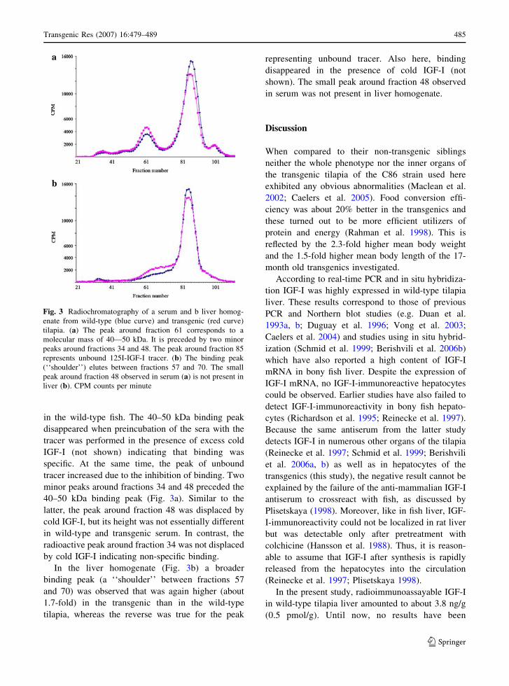
in the wild-type fish. The 40–50 kDa binding peak
disappeared when preincubation of the sera with the
tracer was performed in the presence of excess cold
IGF-I (not shown) indicating that binding was
specific. At the same time, the peak of unbound
tracer increased due to the inhibition of binding. Two
minor peaks around fractions 34 and 48 preceded the
40–50 kDa binding peak (Fig. 3a). Similar to the
latter, the peak around fraction 48 was displaced by
cold IGF-I, but its height was not essentially different
in wild-type and transgenic serum. In contrast, the
radioactive peak around fraction 34 was not displaced
by cold IGF-I indicating non-specific binding.
In the liver homogenate (Fig. 3b) a broader
binding peak (a ‘‘shoulder’’ between fractions 57
and 70) was observed that was again higher (about
1.7-fold) in the transgenic than in the wild-type
tilapia, whereas the reverse was true for the peak
representing unbound tracer. Also here, binding
disappeared in the presence of cold IGF-I (not
shown). The small peak around fraction 48 observed
in serum was not present in liver homogenate.
Discussion
When compared to their non-transgenic siblings
neither the whole phenotype nor the inner organs of
the transgenic tilapia of the C86 strain used here
exhibited any obvious abnormalities (Maclean et al.
2002; Caelers et al. 2005). Food conversion effi-
ciency was about 20% better in the transgenics and
these turned out to be more efficient utilizers of
protein and energy (Rahman et al. 1998). This is
reflected by the 2.3-fold higher mean body weight
and the 1.5-fold higher mean body length of the 17-
month old transgenics investigated.
According to real-time PCR and in situ hybridiza-
tion IGF-I was highly expressed in wild-type tilapia
liver. These results correspond to those of previous
PCR and Northern blot studies (e.g. Duan et al.
1993a, b; Duguay et al. 1996; Vong et al. 2003;
Caelers et al. 2004) and studies using in situ hybrid-
ization (Schmid et al. 1999; Berishvili et al. 2006b)
which have also reported a high content of IGF-I
mRNA in bony fish liver. Despite the expression of
IGF-I mRNA, no IGF-I-immunoreactive hepatocytes
could be observed. Earlier studies have also failed to
detect IGF-I-immunoreactivity in bony fish hepato-
cytes (Richardson et al. 1995; Reinecke et al. 1997).
Because the same antiserum from the latter study
detects IGF-I in numerous other organs of the tilapia
(Reinecke et al. 1997; Schmid et al. 1999; Berishvili
et al. 2006a, b) as well as in hepatocytes of the
transgenics (this study), the negative result cannot be
explained by the failure of the anti-mammalian IGF-I
antiserum to crossreact with fish, as discussed by
Plisetskaya (1998). Moreover, like in fish liver, IGF-
I-immunoreactivity could not be localized in rat liver
but was detectable only after pretreatment with
colchicine (Hansson et al. 1988). Thus, it is reason-
able to assume that IGF-I after synthesis is rapidly
released from the hepatocytes into the circulation
(Reinecke et al. 1997; Plisetskaya 1998).
In the present study, radioimmunoassayable IGF-I
in wild-type tilapia liver amounted to about 3.8 ng/g
(0.5 pmol/g). Until now, no results have been
Fig. 3 Radiochromatography of a serum and b liver homog-
enate from wild-type (blue curve) and transgenic (red curve)
tilapia. (a) The peak around fraction 61 corresponds to a
molecular mass of 40—50 kDa. It is preceded by two minor
peaks around fractions 34 and 48. The peak around fraction 85
represents unbound 125I-IGF-I tracer. (b) The binding peak
(‘‘shoulder’’) elutes between fractions 57 and 70. The small
peak around fraction 48 observed in serum (a) is not present in
liver (b). CPM counts per minute
Transgenic Res (2007) 16:479–489 485
123

reported for fish liver IGF-I measured with a species-
specific RIA. A significantly (about fourfold) higher
level of IGF-I was found in the liver of transgenics
(16.0 ng/g; 2.1 pmol/g). This is paralled by the
detection of IGF-I-immunoreactive hepatocytes in
transgenic liver which were not observed in wild-
type, as well as by the increase in IGF-I mRNA
expression as revealed by RT-PCR and in situ
hybridization. Because the latter results may also
indicate a higher release of IGF-I into the circulation
the level of IGF-I in serum was determined. In wild-
type individuals it amounted to 15 ng/ml (*2 pmol/
ml) whereas in the transgenics it was 6.2 ng/ml
(0.82 pmol/ml). Comparison of the serum IGF-I
levels published for diverse bony fish shows a broad
range. The serum IGF-I concentration in wild-type O.
niloticus as measured here (15 ng/ml) is lower than
that reported in two previous studies on another
tilapia species, O. mossambicus (150 ng/ml, Riley
et al. 2002; 120 ng/ml, Uchida et al. 2003), but
consistent with recent data on O. mossambicus
(*20 ng/ml, Fiess et al. 2007) obtained under similar
rearing conditions (288C, freshwater, feeding) as used
here, and on rainbow trout (11.5 ng/ml, Gabillard
et al. 2003), brown trout (42.2 ng/ml, Banos et al.
1999), Atlantic (53.1 ng/ml, Dyer et al. 2004) and
Coho salmon (7.5–14 ng/ml, Shimizu et al. 1999;
25.9 ng/ml, Shimizu et al. 2000; 45.2–85 ng/ml,
Moriyama 1995). In Coho salmon (Shimizu et al.
2003) and gilthead seabream (Mingarro et al. 2002),
plasma IGF-I levels changed with the seasons and
were higher in fed fish reared in a warm environment
(10–15 ng/ml) than in starved fish reared in the cold
(5–9 ng/ml, Larsen et al. 2001). In several salmonids,
liver IGF-I mRNA and/or plasma IGF-I levels were
related to environmental temperature (Gabillard et al.
2003; Larsen et al. 2001; Beckman et al. 1998). The
largely varying serum IGF-I levels in bony fish may,
thus, be due to species differences but may as well
reflect physiological parameters such as nutritional
status, seasons and environmental temperature (Rei-
necke and Collet 1998; Duan 1998; Reinecke et al.
2005).
As a first step towards identifying specific IGF-I
binding (IGF binding proteins) in tilapia serum and
liver we used incubation with 125I-IGF-I and
subsequent gel filtration at neutral pH (radiochroma-
tography). The main 125I-IGF-I binding peak of
tilapia serum eluted at the same position
(40–50 kDa) as in human serum (Zapf et al. 1975).
In human serum, this peak contains all six IGFBPs,
BP-1 to BP-6 (Zapf 1995). It is likely that the five
IGFBPs detected in fish serum so far (Duan and Xu
2005; Shimizu et al. 2005; Kelley et al. 2002, 2006)
are also located in this binding complex. Serum of the
transgenics bound more tracer than wild-type serum.
The height of the binding peak is determined by two
factors, the amount of binding protein and its IGF
content. Increased binding of 125I-IGF-I in the
transgenic serum could therefore be due to its lower
IGF-I concentration resulting in a greater residual
binding capacity or to a higher IGFBP concentration.
In agreement with the increased binding of 125I-IGF-I
in the serum of the transgenic (GH-overexpressing)
tilapia, short-term application of GH has been shown
to up-regulate the BP serum levels in Coho salmon
(Shimizu et al. 1999; Kelley et al. 1992), striped bass
(Siharath et al. 1995) and tilapia (O. mossambicus;
Park et al. 2000).
Whereas the radiochromatographic pattern of
human serum displays an IGF binding peak at
150 kDa (Zapf et al. 1975) consisting of a ternary
complex of IGFBP-3, IGF-I and -II, and an acid-
labile subunit (ALS, Baxter et al. 1989), the tested
tilapia serum showed only a minor binding peak in
the 150 kDa region. Whether this peak corresponds to
the ternary complex of mammalian serum is not clear.
However, no evidence for a ternary complex has so
far been obtained in fish serum (Kelley et al. 2006).
In contrast to serum, the protein-bound radioac-
tivity in tilapia liver homogenate eluted as a relatively
broad shoulder. It was shifted towards the peak of
non-bound tracer and, thus, apparently contained also
IGFBPs of smaller molecular weight or IGFBP
degradation products. The observed increased bind-
ing of tracer in transgenic liver is compatible with a
greater binding protein content than in wild-type liver
because the IGF-I concentration in the transgenic
liver was higher and binding capacity would therefore
be expected to be lower.
In GH-overexpressing Coho salmon, IGF-I serum
levels varied (Devlin et al. 2000): they were either
slightly enhanced or slightly reduced. Our finding
that the IGF-I serum levels in the growth-enhanced
transgenic tilapia were significantly by 60% lower
than in wild-type fish, despite increased IGF-I mRNA
expression and peptide levels in liver, was unex-
pected. This observation appears to be in contrast to
486 Transgenic Res (2007) 16:479–489
123

results obtained in O. mossambicus demonstrating
that body length and mass are positively correlated
with the plasma IGF-I concentration (Kajimura et al.
2001; Uchida et al. 2003). Moreover, a clear corre-
lation between individual serum IGF-I levels and
growth rates has been established in Coho salmon
(Pierce et al. 2001). How could the lower IGF-I
serum levels in the transgenic animals be explained?
The increased content of IGFBPs in the liver of the
transgenics may lead to retention of IGF-I by these
BPs. This is further supported by the finding that
IGF-I-immunoreactivity was present in hepatocytes
of transgenic but not of wild-type liver.
In addition then, the question arises why trans-
genic tilapia have an approximately 1.5-fold greater
body length and a 2.3-fold higher body weight than
the wild-type despite lower serum IGF-I. Recent
studies using the Cre/loxP recombination system to
delete the IGF-I gene exclusively in the liver of mice
(Sjogren et al. 1999; Yakar et al. 1999) underline the
potential importance of local IGF-I production for
growth. Despite deletion of liver IGF-I mRNA
expression and largely reduced levels of circulating
IGF-I, the animals did not show any obvious
impairment of postnatal body growth. Similarly, in
the transgenic tilapia, IGF-I in extrahepatic sites may
determine the growth rate. This has been shown here
to be true for skeletal muscle that is essentially
involved in growth. Enhanced growth therefore
seems to be caused by higher tissue levels of IGF-I
under the influence of the GH transgene and thus by
autocrine/paracrine actions of IGF-I.
Acknowledgements The authors are grateful to Ms Cornelia
Zwimpfer, Division of Endocrinology and Diabetes,
Department of Medicine, University Hospital Zurich, for
excellent support in the performance of radioimmunoassay
and radiochromatography. The study was supported by the
Swiss National Science Foundation (grant no. 111028) and the
Hartmann Muller-Foundation for Medical Research (grant no.
1115).
References
Amsterdam A, Becker TS (2005) Transgenes as screening tools
to probe and manipulate the zebrafish genome. Dev Dyn
234:255–268
Banos N, Planas JV, Gutierrez J, Navarro I (1999) Regulation
of plasma insulin-like growth factor-I levels in brown
trout (Salmo trutta). Comp Biochem Physiol C Pharmacol
Toxicol Endocrinol 124:33–40
Baxter RC, Martin JL, Beniac V (1989) High molecular weight
insulin-like growth factor binding protein complex. Puri-
fication and properties of the acid-labile subunit from
human serum. J Biol Chem 264:11843–11848
Beckman BR, Larsen DA, Moriyama S, Lee-Pawlak B, Dick-
hoff WW (1998) Insulin-like growth factor-I and envi-
ronmental modulation of growth during smoltification of
spring chinook salmon (Oncorhynchus tshawystscha).
Gen Comp Endocrinol 109:325–335
Berishvili G, D’Cotta H, Baroiller J-F, Segner H, Reinecke M
(2006a) Differential expression of IGF-I mRNA and
peptide in the male and female gonad during early
development of a bony fish, the tilapia Oreochromis nil-oticus. Gen Comp Endocrinol 146:204–210
Berishvili G, Shved N, Eppler E, Clota F, Baroiller JF, Rei-
necke M (2006b) Organ-specific expression of IGF-I
during early development of bony fish as revealed in the
tilapia, Oreochromis niloticus, by in situ hybridization and
immunohistochemistry: indication for the particular
importance of local IGF-I. Cell Tissue Res 325:287–301
Butler A, LeRoith D (2001) Minireview: tissue-specific versus
generalized gene targeting of the igf1 and igf1r genes and
their roles in insulin-like growth factor physiology.
Endocrinology 142:1685–1688
Caelers A, Berishvili G, Meli ML, Eppler E, Reinecke M
(2004) Establishment of a real-time RT-PCR for the
determination of absolute amounts of IGF-I and IGF-II
gene expression in liver and extrahepatic sites of the
tilapia. Gen Comp Endocrinol 137:196–204
Caelers A, Mclean N, Hwang G, Eppler E, Reinecke M (2005)
Expression of endogenous and exogenous growth hormone
(GH) messenger (m) RNA in a GH-transgenic tilapia (Or-eochromis niloticus). Transgenic Res 14:95–104
Devlin RH, Vesaki TY, Biagi CA, Donaldson EM, Swanson P,
Chan W-K (1994) Extraordinary salmon growth. Nature
371:209–210
Devlin RH, Swanson P, Clarke WC, Plisetskaya E, Dickhoff
W, Moriyama S, Yesaki TY, Hew C-L (2000) Seawater
adaptability and hormone levels in growth-enhanced
transgenic coho salmon, Oncorhynchus kisutch. Aqua-
culture 191:367–385
Du SJ, Gong ZY, Fletcher GL, Shears MA, King MJ, Idler DR,
Hew CL (1992) Growth enhancement in transgenic
Atlantic salmon by the use of an ‘‘all fish’’ chimeric
growth hormone gene construct. Biotechnology (NY)
10:176–181
Duan C (1998) Nutritional and developmental regulation of
insulin-like growth factors in fish. J Nutr 128(Suppl
2):306S–314S
Duan C, Xu Q (2005) Roles of insulin-like growth factor (IGF)
binding proteins in regulating IGF actions. Gen Comp
Endocrinol 142:44–52
Duan C, Duguay SJ, Plisetskaya EM (1993a) Insulin-like
growth factor (IGF-I) mRNA expression in coho salmon,
Oncorhynchus kisutch: tissue distribution and effects of
growth hormone/prolactin family proteins. Fish Physiol
Biochem 11:371–379
Duan C, Hanzawa N, Takeuchi Y, Hamada E, Miyachi S,
Hirano T (1993b) Use of primary cultures of salmon he-
patocytes for the study of hormonal regulation of IGF-I
expression in vitro. Zool Sci 10:473–480
Transgenic Res (2007) 16:479–489 487
123

Duguay SJ, Lai-Zhang J, Steiner DF, Funkenstein B, Chan SJ
(1996) Developmental and tissue-regulated expression of
IGF-I and IGF-II mRNAs in Sparus aurata. J Mol
Endocrinol 16:123–132
Dyer AR, Upton Z, Stone D, Thomas PM, Soole KL, Higgs N,
Quinn K, Carragher JF (2004) Development and valida-
tion of a radioimmunoassay for fish insulin-like growth
factor I (IGF-I) and the effect of aquaculture related
stressors on circulating IGF-I levels. Gen Comp Endo-
crinol 135:268–275
Fiess JC, Kunkel-Patterson A, Mathias L, Riley LG, Yancey
PH, Hirano T, Grau EG (2007) Effects of environmental
salinity and temperature on osmoregulatory ability, or-
ganic osmolytes, and plasma hormone profiles in the
Mozambique tilapia (Oreochromis mossambicus). Comp
Biochem Physiol A Mol Integr Physiol 146:252–264
Gabillard JC, Weil C, Rescan PY, Navarro I, Gutierrez J, Le
Bail PY (2003) Effects of environmental temperature on
IGF1, IGF2, and IGF type I receptor expression in rain-
bow trout (Oncorhynchus mykiss). Gen Comp Endocrinol
133:233–242
Gray ES, Kelley KM, Law S, Tsai R, Young G, Bern H (1992)
Regulation of hepatic growth hormone receptors in coho
salmon (Oncorhynchus kisutch). Gen Comp Endocrinol
88:243–252
Guillen II, Lleonart R, Agramonte A, Morales R, Morales A,
Hernandez CA, Vazquez MM, Diaz M, Herrera MT,
Alvarez-Lajonchere L, Hernandez O, de la Fuente J
(1998) Physiological changes in the juvenile euryhaline
teleost, the tilapia Oreochromis hornorum, injected with
E. coli-derived homologous growth hormone. J Mar
Biotechnol 6:142–151
Hansson H-A, Nilsson A, Isgaard J, Billig H, Isaksson O,
Skottner A, Andersson IK, Rozell B (1988) Immunohis-
tochemical localization of insulin-like growth factor I in
the adult rat. Histochemistry 89:403–410
Hwang GL, Rahman MA, Abdul Razak S, Sohm F, Farahmand
H, Smith A, Brooks C, Maclean N (2003) Isolation and
characterisation of tilapia beta-actin promoter and com-
parison of its activity with carp beta-actin promoter.
Biochim Biophys Acta 1625:11–18
Jones JI, Clemmons DR (1995) Insulin-like growth factors and
their binding proteins: biological actions. Endocr Rev
16:3–34
Kajimura S, Uchida K, Yada T, Riley LG, Byatt JC, Colloier
RJ, Aida K, Hirano T, Grau EG (2001) Stimulation of
insulin-like growth factor-I production by recombinant
bovine growth hormone in Mozambique tilapia, Ore-ochromis mossambicus. Fish Physiol Biochem 25:221–
230
Kelley KM, Siharath K, Bern HA (1992) Identification of
insulin-like growth factor-binding proteins in the circu-
lation of four teleost fish species. J Exp Zool 263:220–224
Kelley KM, Schmidt KE, Berg L, Sak K, Galima MM, Gil-
lespie C, Balogh L, Hawayek A, Reyes JA, Jamison M
(2002) Comparative endocrinology of the insulin-like
growth factor-binding protein. J Endocrinol 175:3–18
Kelley KM, Price TD, Galima MM, Sak K, Reyes JA, Zepeda
O, Hagstrom R, Tuan A, Truong TA, Lowe CG (2006)
Insulin-like growth factor binding proteins (IGFBPs) in
fish: beacons for (disrupted) growth endocrine physiology.
In: Reinecke M, Zaccone G, Kapoor BG (eds) Fish en-
docrinolgy. Science Publishers, Enfield, pp 167–195
Larsen DA, Beckman BR, Dickhoff WW (2001) The effect of
low temperature and fasting during the winter on meta-
bolic stores and endocrine physiology (insulin, insulin-
like growth factor-I, and thyroxine) of coho salmon, On-corhynchus kisutch. Gen Comp Endocrinol 123:308–323
Lu JK, Fu BH, Wu JL, Chen TT (2002) Production of trans-
genic silver sea bream (Sparus sarba) by different gene
transfer methods. Mar Biotechnol (NY) 4:328–337
Maclean N, Rahman MA, Sohm F, Hwang G, Iyengar A, Ayad
H, Smith A, Farahmand H (2002) Transgenic tilapia and
the tilapia genome. Gene 295:265–277
Martinez R, Estrada MP, Berlanga J, Guillen I, Hernandez O,
Cabrera E, Pimentel R, Morales R, Herrera F, Morales A,
Pina JC, Abad Z, Sanchez V, Melamed P, Lleonart R, de
la Fuente J (1996) Growth enhancement in transgenic
tilapia by ectopic expression of tilapia growth hormone.
Mol Mar Biol Biotechnol 5:62–70
McCormick SD, Kelley KM, Young G, Nishioka RS, Bern HA
(1992) Stimulation of coho salmon growth by insulin-like
growth factor I. Gen Comp Endocrinol 86:398–406
Mingarro M, Vega-Rubin de Celis S, Astola A, Pendon C,
Valdivia MM, Perez-Sanchez J (2002) Endocrine media-
tors of seasonal growth in gilthead sea bream (Sparusaurata): the growth hormone and somatolactin paradigm.
Gen Comp Endocrinol 128:102–111
Moriyama S (1995) Increased plasma insulin-like growth fac-
tor-I (IGF-I) following oral and intraperitoneal adminis-
tration of growth hormone to rainbow trout,
Oncorhynchus mykiss. Growth Regul 5:164–167
Moriyama S, Ayson FG, Kawauchi H (2000) Growth regula-
tion by insulin-like growth factor-I in fish. Biosci Bio-
technol Biochem 64:1553–1562
Ng TB, Leung TC, Cheng CHK, Woo NY (1992) Growth
hormone binding sites in tilapia (Oreochromis mossam-bicus) liver. Gen Comp Endocrinol 86:111–118
Park R, Shepherd BS, Nishioka RS, Grau EG, Bern HA (2000)
Effects of homologous pituitary hormone treatment on
serum insulin-like growth factor-binding proteins (IG-
FBPs) in hypophysectomized tilapia, Oreochromis mos-sambicus, with special reference to a novel 20-kDa
IGFBP. Gen Comp Endocrinol 117:404–412
Pierce AL, Beckman BR, Shearer KD, Larsen DA, Dickhoff
WW (2001) Effects of ration on somatotropic hormones
and growth in coho salmon. Comp Biochem Physiol B
Biochem Mol Biol 128:255–264
Pierce AL, Fukada H, Dickhoff WW (2005) Metabolic hormones
modulate the effect of growth hormone (GH) on insulin-like
growth factor-I (IGF-I) mRNA level in primary culture of
salmon hepatocytes. J Endocrinol 184:341–349
Plisetskaya EM (1998) Some of my not so favorite things about
insulin and insulin-like growth factors in fish. Comp
Biochem Physiol B Biochem Mol Biol 121:3–11
Rahman A, Maclean N (1992) Fish transgene expression by
direct injection into fish muscle. Mol Mar Biol Biotechnol
1:286–289
Rahman MA, Mak R, Ayad H, Smith A, Maclean N (1998)
Expression of a novel piscine growth hormone gene re-
sults in growth enhancement in transgenic tilapia (Ore-ochromis niloticus). Transgenic Res 7:357–369
488 Transgenic Res (2007) 16:479–489
123

Reinecke M, Collet C (1998) The phylogeny of the insulin-like
growth factors. Int Rev Cytol 183:1–94
Reinecke M, Schmid A, Ermatinger R, Loffing-Cueni D (1997)
Insulin-like growth factor I in the teleost Oreochromismossambicus, the tilapia: gene sequence, tissue expres-
sion, and cellular localization. Endocrinology 138:3613–
3619
Reinecke M, Bjornsson TB, Dickhoff WW, McCormick SD,
Navarro I, Power DM, Gutierrez J (2005) Growth hor-
mone and insulin-like growth factors in fish: where we are
and where to go. Gen Comp Endocrinol 142:20–24
Richardson NA, Anderson AJ, Rimmer MA, Sara VR (1995)
Localization of insulin-like growth factor-I immunoreac-
tivity in larval and juvenile barramundi (Lates calcarifer).
Gen Comp Endocrinol 100:282–292
Riley LG, Richman NH 3rd, Hirano T, Grau EG (2002)
Activation of the growth hormone/insulin-like growth
factor axis by treatment with 17 alpha-methyltestosterone
and seawater rearing in the tilapia, Oreochromis mos-sambicus. Gen Comp Endocrinol 127:285–292
Rocha A, Ruiz S, Estepa A, Coll JM (2004) Application of
inducible and targeted gene strategies to produce trans-
genic fish: a review. Mar Biotechnol (NY) 6:118–127
Schmid AC, Naef E, Kloas W, Reinecke M (1999) IGF-I and
IGF-II in the ovary of a bony fish Oreochromis mossam-bicus, the tilapia: In situ hybridization, immunohisto-
chemical localisation, Northern Blot and cDNA
sequences. Mol Cell Endocrinol 156:141–149
Schmid AC, Reinecke M, Kloas W (2000) Primary cultured
hepatocytes of the bony fish, Oreochromis mossambicus,
the tilapia: a valid tool for physiological studies on IGF-I
expression in liver. J Endocrinol 166:265–273
Shamblott MJ, Cheng CM, Bolt D, Chen TT (1995) Appear-
ance of insulin-like growth factor mRNA in the liver and
pyloric ceca of a teleost in response to exogenous growth
hormone. Proc Natl Acad Sci USA 92:6943–6946
Shepherd BS, Sakamoto T, Nishioka RS, Richman NH III,
Mori I, Madsen SS, Chen TT, Hirano T, Bern HA, Grau
EG (1997) Somatotrophic actions of homologous growth
hormone and prolactins in the euryhyaline teleost, the
tilapia, Oreochromis mossambicus. Proc Natl Acad Sci
USA 94:2068–2072
Shimizu M, Swanson P, Dickhoff WW (1999) Free and pro-
tein-bound insulin-like growth factor-I (IGF-I) and IGF-
binding proteins in plasma of coho salmon, Oncorhynchuskisutch. Gen Comp Endocrinol 115:398–405
Shimizu M, Swanson P, Fukuda H, Hara A, Dickhoff WW
(2000) Comparison of extraction methods and assay val-
idation for salmon insulin-like growth factor-I using
commercially available components. Gen Comp Endo-
crinol 119:26–36
Shimizu M, Hara A, Dickhoff WW (2003) Development of an
RIA for salmon 41 kDa IGF-binding protein. J Endocrinol
178:275–283
Shimizu M, Dickey JT, Fukada H, Dickhoff WW (2005) Sal-
mon serum 22 kDa insulin-like growth factor-binding
protein (IGFBP) is IGFBP-1. J Endocrinol 184:267–276
Siharath K, Nishioka RS, Madsen SS, Bern HA (1995) Regu-
lation of IGF-binding proteins by growth hormone in the
striped bass, Morone saxatilis. Mol Mar Biol Biotechnol
4:171–178
Sjogren K, Liu J-L, Blad K, Skrtic S, Vidal O, Wallenius V, Le
Roith D, Tornell J, Isaksson OPG, Jansson J-O, Ohlsson C
(1999) Liver-derived insulin-like growth factor I (IGF-I)
is the pricipal source of IGF-I in blood but is not required
for postnatal body growth in mice. Proc Natl Acad Sci
USA 96:7088–7092
Sundstrom LF, Lohmus M, Johnsson JI, Devlin RH (2004)
Growth hormone transgenic salmon pay for growth po-
tential with increased predation mortality. Proc Biol Sci
271(Suppl 5):S350–S352
Uchida K, Kajimura S, Riley LG, Hirano T, Aida K, Grau EG
(2003) Effects of fasting on growth hormone/insulin-like
growth factor I axis in the tilapia, Oreochromis mossam-bicus. Comp Biochem Physiol A Mol Integr Physiol
134:429–433
Vong QP, Chan KM, Cheng CH (2003) Quantification of
common carp (Cyprinus carpio) IGF-I and IGF-II mRNA
by real-time PCR: differential regulation of expression by
GH. J Endocrinol 178:513–521
Wood AW, Duan C, Bern HA (2005) Insulin-like growth factor
signaling in fish. Int Rev Cytol 243:215–285
Yakar S, Liu J-L, Stannard B, Butler A, Accili D, Sauer B,
LeRoith D (1999) Normal growth and development in theabsence of hepatic insulin-like growth factor I. Proc Natl
Acad Sci USA 96:7324–7329
Zapf J (1995) Physiological role of the insulin-like growth
factor binding proteins. Eur J Endocrinol 132:645–654
Zapf J, Waldvogel M, Froesch ER (1975) Binding of non-
suppressible insulin-like activity to human serum. Arch
Biochem Biophys 168:638–645
Zapf J, Hauri C, Waldvogel M, Futo E, Hasler H, Binz K,
Guler HP, Schmid C, Froesch ER (1989) Recombinant
human insulin-like growth factor I induces its own spe-
cific carrier protein in hypophysectomized and diabetic
rats. Proc Natl Acad Sci USA 86:3813–3817
Zbikowska HM (2003) Fish can be first—advances in fish
transgenesis for commercial applications. Transgenic Res
12:379–389
Zhang PJ, Hayat M, Joyce C, Gonzalez-Villasenor LI, Lin CM,
Dunham RA, Chen TT, Powers DA (1990) Gene transfer,
expression and inheritance of pRSV-rainbow trout-GH
cDNA in the common carp, Cyprinus carpio (Linnaeus).
Mol Reprod Dev 25:3–13
Transgenic Res (2007) 16:479–489 489
123