CXCL14 inhibits trophoblast outgrowth via a paracrine/autocrine manner during early pregnancy in...
10
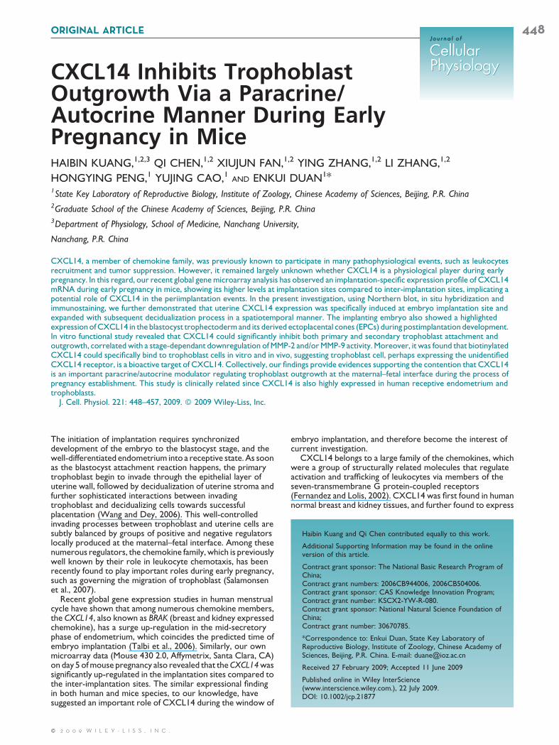
CXCL14 Inhibits Trophoblast Outgrowth Via a Paracrine/ Autocrine Manner During Early Pregnancy in Mice HAIBIN KUANG, 1,2,3 QI CHEN, 1,2 XIUJUN FAN, 1,2 YING ZHANG, 1,2 LI ZHANG, 1,2 HONGYING PENG, 1 YUJING CAO, 1 AND ENKUI DUAN 1 * 1 State Key Laboratory of Reproductive Biology, Institute of Zoology, Chinese Academy of Sciences, Beijing, P.R. China 2 Graduate School of the Chinese Academy of Sciences, Beijing, P.R. China 3 Department of Physiology, School of Medicine, Nanchang University, Nanchang, P.R. China CXCL14, a member of chemokine family, was previously known to participate in many pathophysiological events, such as leukocytes recruitment and tumor suppression. However, it remained largely unknown whether CXCL14 is a physiological player during early pregnancy. In this regard, our recent global gene microarray analysis has observed an implantation-specific expression profile of CXCL14 mRNA during early pregnancy in mice, showing its higher levels at implantation sites compared to inter-implantation sites, implicating a potential role of CXCL14 in the periimplantation events. In the present investigation, using Northern blot, in situ hybridization and immunostaining, we further demonstrated that uterine CXCL14 expression was specifically induced at embryo implantation site and expanded with subsequent decidualization process in a spatiotemporal manner. The implanting embryo also showed a highlighted expression of CXCL14 in the blastocyst trophectoderm and its derived ectoplacental cones (EPCs) during postimplantation development. In vitro functional study revealed that CXCL14 could significantly inhibit both primary and secondary trophoblast attachment and outgrowth, correlated with a stage-dependant downregulation of MMP-2 and/or MMP-9 activity. Moreover, it was found that biotinylated CXCL14 could specifically bind to trophoblast cells in vitro and in vivo, suggesting trophoblast cell, perhaps expressing the unidentified CXCL14 receptor, is a bioactive target of CXCL14. Collectively, our findings provide evidences supporting the contention that CXCL14 is an important paracrine/autocrine modulator regulating trophoblast outgrowth at the maternal–fetal interface during the process of pregnancy establishment. This study is clinically related since CXCL14 is also highly expressed in human receptive endometrium and trophoblasts. J. Cell. Physiol. 221: 448–457, 2009. ß 2009 Wiley-Liss, Inc. The initiation of implantation requires synchronized development of the embryo to the blastocyst stage, and the well-differentiated endometrium into a receptive state. As soon as the blastocyst attachment reaction happens, the primary trophoblast begin to invade through the epithelial layer of uterine wall, followed by decidualization of uterine stroma and further sophisticated interactions between invading trophoblast and decidualizing cells towards successful placentation (Wang and Dey, 2006). This well-controlled invading processes between trophoblast and uterine cells are subtly balanced by groups of positive and negative regulators locally produced at the maternal–fetal interface. Among these numerous regulators, the chemokine family, which is previously well known by their role in leukocyte chemotaxis, has been recently found to play important roles during early pregnancy, such as governing the migration of trophoblast (Salamonsen et al., 2007). Recent global gene expression studies in human menstrual cycle have shown that among numerous chemokine members, the CXCL14, also known as BRAK (breast and kidney expressed chemokine), has a surge up-regulation in the mid-secretory phase of endometrium, which coincides the predicted time of embryo implantation (Talbi et al., 2006). Similarly, our own microarray data (Mouse 430 2.0, Affymetrix, Santa Clara, CA) on day 5 of mouse pregnancy also revealed that the CXCL14 was significantly up-regulated in the implantation sites compared to the inter-implantation sites. The similar expressional finding in both human and mice species, to our knowledge, have suggested an important role of CXCL14 during the window of embryo implantation, and therefore become the interest of current investigation. CXCL14 belongs to a large family of the chemokines, which were a group of structurally related molecules that regulate activation and trafficking of leukocytes via members of the seven-transmembrane G protein-coupled receptors (Fernandez and Lolis, 2002). CXCL14 was first found in human normal breast and kidney tissues, and further found to express Haibin Kuang and Qi Chen contributed equally to this work. Additional Supporting Information may be found in the online version of this article. Contract grant sponsor: The National Basic Research Program of China; Contract grant numbers: 2006CB944006, 2006CB504006. Contract grant sponsor: CAS Knowledge Innovation Program; Contract grant number: KSCX2-YW-R-080. Contract grant sponsor: National Natural Science Foundation of China; Contract grant number: 30670785. *Correspondence to: Enkui Duan, State Key Laboratory of Reproductive Biology, Institute of Zoology, Chinese Academy of Sciences, Beijing, P.R. China. E-mail: [email protected] Received 27 February 2009; Accepted 11 June 2009 Published online in Wiley InterScience (www.interscience.wiley.com.), 22 July 2009. DOI: 10.1002/jcp.21877 ORIGINAL ARTICLE 448 Journal of Journal of Cellular Physiology Cellular Physiology ß 2009 WILEY-LISS, INC.
-
Upload
independent -
Category
Documents
-
view
1 -
download
0
Transcript of CXCL14 inhibits trophoblast outgrowth via a paracrine/autocrine manner during early pregnancy in...
ORIGINAL ARTICLE 448J o u r n a l o fJ o u r n a l o f
CellularPhysiologyCellularPhysiology
CXCL14 Inhibits TrophoblastOutgrowth Via a Paracrine/Autocrine Manner During EarlyPregnancy in Mice
HAIBIN KUANG,1,2,3 QI CHEN,1,2 XIUJUN FAN,1,2 YING ZHANG,1,2 LI ZHANG,1,2HONGYING PENG,1 YUJING CAO,1 AND ENKUI DUAN1*1State Key Laboratory of Reproductive Biology, Institute of Zoology, Chinese Academy of Sciences, Beijing, P.R. China2Graduate School of the Chinese Academy of Sciences, Beijing, P.R. China3Department of Physiology, School of Medicine, Nanchang University,
Nanchang, P.R. China
CXCL14, a member of chemokine family, was previously known to participate in many pathophysiological events, such as leukocytesrecruitment and tumor suppression. However, it remained largely unknown whether CXCL14 is a physiological player during earlypregnancy. In this regard, our recent global gene microarray analysis has observed an implantation-specific expression profile of CXCL14mRNA during early pregnancy in mice, showing its higher levels at implantation sites compared to inter-implantation sites, implicating apotential role of CXCL14 in the periimplantation events. In the present investigation, using Northern blot, in situ hybridization andimmunostaining, we further demonstrated that uterine CXCL14 expression was specifically induced at embryo implantation site andexpanded with subsequent decidualization process in a spatiotemporal manner. The implanting embryo also showed a highlightedexpression of CXCL14 in the blastocyst trophectoderm and its derived ectoplacental cones (EPCs) during postimplantation development.In vitro functional study revealed that CXCL14 could significantly inhibit both primary and secondary trophoblast attachment andoutgrowth, correlated with a stage-dependant downregulation of MMP-2 and/or MMP-9 activity. Moreover, it was found that biotinylatedCXCL14 could specifically bind to trophoblast cells in vitro and in vivo, suggesting trophoblast cell, perhaps expressing the unidentifiedCXCL14 receptor, is a bioactive target of CXCL14. Collectively, our findings provide evidences supporting the contention that CXCL14is an important paracrine/autocrine modulator regulating trophoblast outgrowth at the maternal–fetal interface during the process ofpregnancy establishment. This study is clinically related since CXCL14 is also highly expressed in human receptive endometrium andtrophoblasts.
J. Cell. Physiol. 221: 448–457, 2009. � 2009 Wiley-Liss, Inc.
Haibin Kuang and Qi Chen contributed equally to this work.
Additional Supporting Information may be found in the onlineversion of this article.
Contract grant sponsor: The National Basic Research Program ofChina;Contract grant numbers: 2006CB944006, 2006CB504006.Contract grant sponsor: CAS Knowledge Innovation Program;Contract grant number: KSCX2-YW-R-080.Contract grant sponsor: National Natural Science Foundation ofChina;Contract grant number: 30670785.
*Correspondence to: Enkui Duan, State Key Laboratory ofReproductive Biology, Institute of Zoology, Chinese Academy ofSciences, Beijing, P.R. China. E-mail: [email protected]
Received 27 February 2009; Accepted 11 June 2009
Published online in Wiley InterScience(www.interscience.wiley.com.), 22 July 2009.DOI: 10.1002/jcp.21877
The initiation of implantation requires synchronizeddevelopment of the embryo to the blastocyst stage, and thewell-differentiated endometrium into a receptive state. As soonas the blastocyst attachment reaction happens, the primarytrophoblast begin to invade through the epithelial layer ofuterine wall, followed by decidualization of uterine stroma andfurther sophisticated interactions between invadingtrophoblast and decidualizing cells towards successfulplacentation (Wang and Dey, 2006). This well-controlledinvading processes between trophoblast and uterine cells aresubtly balanced by groups of positive and negative regulatorslocally produced at the maternal–fetal interface. Among thesenumerous regulators, the chemokine family, which is previouslywell known by their role in leukocyte chemotaxis, has beenrecently found to play important roles during early pregnancy,such as governing the migration of trophoblast (Salamonsenet al., 2007).
Recent global gene expression studies in human menstrualcycle have shown that among numerous chemokine members,the CXCL14, also known as BRAK (breast and kidney expressedchemokine), has a surge up-regulation in the mid-secretoryphase of endometrium, which coincides the predicted time ofembryo implantation (Talbi et al., 2006). Similarly, our ownmicroarray data (Mouse 430 2.0, Affymetrix, Santa Clara, CA)on day 5 of mouse pregnancy also revealed that the CXCL14 wassignificantly up-regulated in the implantation sites compared tothe inter-implantation sites. The similar expressional findingin both human and mice species, to our knowledge, havesuggested an important role of CXCL14 during the window of
� 2 0 0 9 W I L E Y - L I S S , I N C .
embryo implantation, and therefore become the interest ofcurrent investigation.
CXCL14 belongs to a large family of the chemokines, whichwere a group of structurally related molecules that regulateactivation and trafficking of leukocytes via members of theseven-transmembrane G protein-coupled receptors(Fernandez and Lolis, 2002). CXCL14 was first found in humannormal breast and kidney tissues, and further found to express
C X C L 1 4 I N H I B I T S T R O P H O B L A S T O U T G R O W T H 449
in many other normal tissues, and also in some kinds ofmalignant tissues (Hromas et al., 1999; Frederick et al., 2000;Augsten et al., 2009; Pelicano et al., 2009). As a unique memberof chemokine family, CXCL14 is currently suggested as achemoattractant for dendritic cells (DC), activated monocytes,neutrophils, and natural killer cells (NK) under differentpathophysiological conditions (Kurth et al., 2001; Shellenbergeret al., 2004; Shurin et al., 2005; Starnes et al., 2006; Salogni et al.,2009). It has been also reported that CXCL14 was a potentmediators of neoangiogenesis and tumor growth, invasion,metastasis (Schwarze et al., 2005; Ozawa et al., 2006; Augstenet al., 2009), which shares many similarities with the process oftrophoblast invasion during early pregnancy (Fitzgerald et al.,2008). To date, the specific receptor of CXCL14 has not yetbeen found. Recently, targeted deletion of CXCL14 in mice hasresulted in compromised fertility (Meuter et al., 2007 andpersonal communication with authors). As the homozygouspairs were reported to produce reduced litter size and someknockout females did not produce newborns. However, theunderling mechanisms of this phenomenon are still unexplored.
The observed reproductive phenotype in CXCL14 null micecoupled with its potential role in cell migration has encouragedus to hypothesize that CXCL14 might play important roles introphoblast function during early pregnancy, since a lot ofpregnant failures were associated with abnormal trophoblastinvasion at periimplantation stage (Wilcox et al., 1999; Songet al., 2002). In this investigation, we showed a site-strict andcell-specific expression of CXCL14 within the implantation sitesat both maternal and embryonic compartment. Furtherfunctional study revealed CXCL14 as an inhibitory factoragainst trophoblast outgrowth through downregulating MMP-2and 9. Also, binding assays of CXCL14 were performed at thematernal–fetal interface to predict the potential site ofunidentified CXCL14 receptor.
Materials and MethodsAnimals and treatments
Kunming white strain mice (Experimental Animal Center, Instituteof Genetics and Development, Chinese Academy of Sciences) weremaintained in the animal facility of the State Key Laboratory ofReproductive Biology, Institute of Zoology, Chinese Academy ofSciences. The Guidelines for the Care and Use of Animals inResearch were followed. Mice were allowed free access to waterand food with a constant photoperiod (12L:12D). Adult femalemice (25–30 g, 7–8 weeks old) in estrus were mated with fertilemales of the same strain at room temperature (258C). Thefollowing morning of finding a vaginal plug was designated as day 1 ofpregnancy.
DNA microarray analyses
Implantation (IS) and inter-implantation (IIS) sites were monitoredby an intravenous injection of a blue dye and divided by sharpdissection at 0800–0900 h a.m. on day 5 (n¼ 10 mice). Uterinetissues were flash frozen and stored at �808C. Total RNA wasextracted with Trizol reagent (Invitrogen, Eugene, OR). cDNAand Biotinylated cRNA were prepared according to Affymetrixprotocols. Samples were hybridized overnight to high-densityMouse Genome 430 2.0 array (Affymetrix), at the Shanghai HujingBiotech Co., Ltd (Shanghai, China). Then the chips were scannedusing a Scanner3000, and the data were extracted using theAffymetrix GeneChip Operating Software version 1.2(Affymetrix). Each sample was hybridized on two chips for reducingthe false positive rate.
Northern blot analyses
Total uterine RNAs were extracted with Trizol reagent(Invitrogen), and were quantified by absorbance at 260 nm as well
JOURNAL OF CELLULAR PHYSIOLOGY
as by ethidium bromide staining after electrophoresis throughagarose gels. Samples of total RNA (20mg) were then blottedovernight to Hybond-N membrane by electrical transfer. ThecDNA probe (bases 442–1058 of Mus musculus CXCL14:GenBank Accession No. NM_019568) was labeled with 32P andhybridization was carried out overnight at 608C. The blot wassubjected to autoradiography at �708C using BioMax MS film(Amersham, UK). To determine the relative amounts of RNAtransferred to the membrane, blots were stripped and hybridizedwith a 32P-labeled 18S RNA probe.
RNA extraction and RT-PCR
Total RNA was extracted using Trizol Reagents and reversetranscribed in 25ml of reaction mixture containing 30 U avianmyeloblastosis virus reverse transcriptase (Promega, Madison,WI). The PCR was conducted in a total volume of 50ml for30 cycles of denaturation at 948C of 30 sec, annealing at 568C for30 sec, and extension at 728C for 45 sec, with a final extension stepof 10 min at 728C. The primers used in this study include CXCL14(5-GGG TCC AAG TGT AAG TGT TC-3; 5-GTA GTG CTG TGAACG GTC TC-3) and 18S (5-AAT CAG GGT TCG ATT CCG GA-3; 5-CCA AGA TCC AAC TAC GAG CT-3). The amplifiedproducts were analyzed by electrophoresis on 1% agarose gelsstained with ethidium bromide. Images of the RT-PCR agarose gelswere acquired with a High Performance CCD camera andquantification of the bands was performed by ChemiDocXRS(BioRAD, Hercules, CA). Band intensity of the genes determinedwas compared with the band intensity of 18S as an internal control,and the relative level was acquired.
Indirect immunofluorescence
Preimplantation embryos at different developmental stages wereflushed from the oviduct or uterus on days 2–4 of pregnancy. Allembryos were then fixed in 4% paraformaldehyde (30 min, roomtemperature). After fixation, embryos were washed three times inPBS and permeabilized in PBS containing 0.2% TritonX-100(12 min, room temperature). After rinsing three times in PBS,embryos were incubated in 5% BSA for 30 min at roomtemperature to block nonspecific binding of the antibodies. Then,embryos were incubated with anti-CXCL14mAb (R&D Systems,Minneapolis, MN) at 48C overnight followed by secondary antibody(fluorescein isothiocyanate-labeled anti-rat IgG) for 1 h at 378C.Nuclei were stained with 5mg/ml of propidium iodide (Sigma,St. Louis, MO) for 10 min. Embryos were viewed under alaser-scanning confocal microscope (Leica, Heidelberg, Germany).Rat preimmune IgG was used as a negative control.
In situ hybridization
In situ sense and antisense probe templates were prepared byamplifying a 351bp fragment of CXCL14 cDNA (GenBankaccession no: NM_019568, nucleotides 334–684), using sense andantisense primers modified with either T7 or SP6 sequences. Theresulting templates were then transcribed with thedigoxigenin-labeling kit (Roche Molecular Biomedicals, Mannheim,Germany) according to the manufacturer’s protocol. Afterdeparaffinization and proteinase K treatment, the tissue sectionswere prehybridized and then incubated in the hybridization buffercontaining 1mg/ml of digoxigenin-labeled sense or antisense BRAKcRNA probe overnight. After serial washing and RNase treatment,samples were blocked and incubated with alkaline phosphatase-conjugated antidigoxigenin antibody (Roche MolecularBiochemicals; 1:2,000 dilution) in the blocking buffer at roomtemperature for 1 h. After intense washing, signal was visualizedthrough the use of nitro blue tetrazolium and5-bromo-4-chloro-3-indolyl-phosphate (Promega) until the colordeveloped to the desired extent.
450 K U A N G E T A L .
Blastocyst attachment and outgrowth assays in vitro
Blastocyst attachment and outgrowth assays were performed asdescribed previously (Qin et al., 2005). In brief, blastocysts wereobtained by flushing the uterine horns with Ham’s F-12 medium onday 4 morning of pregnancy and transferred in 96-well platesprecoated with fibronectin (10mg/ml; Sigma), containing Ham’sF-12 (supplemented with 0.4% BSA) plus 0, 1, 10, 50, 100, 200,1,000 ng/ml of recombinant mouse CXCL14 (rmCXCL14) protein(R&D Systems). It was determined that the recombinant CXCL14 atthe concentration of 100 ng/ml has an optimal effect for blastocystattachment and outgrowth at 48 h after initiation of culture.
The blastocysts were then divided into four treatment groups:the medium only (control group, n¼ 40, four wells), the mediumplus 100 ng/ml of rmCXCL14 (rmCXCL14 group, n¼ 40, fourwells), the medium plus 30mg/ml of rat anti-mouse CXCL14 IgG(anti-CXCL14 group, n¼ 40, four wells), and medium plus 30mg/ml rat IgG (IgG group, n¼ 40, four wells). When primary gianttrophoblast cells were visible around the attachment site of theattached blastocysts, we designated the blastocysts as outgrowth.Blastocyst attachment was examined at 48 h of culture. Blastocystattachment was only examined once, because gentle pipetting wasrequired to determine whether the embryo would detach from thebottom of plates. The ratios of blastocysts with attachment andoutgrowth relative to the total number of embryos werecalculated.
Outgrowth assays of EPCs
Ectoplacental cones (EPCs) from the mice on day 8 of pregnancywere dissected out under sterile conditions and transferred to4-well (30 EPCs per well) plates precoated with Matrigel (8 mg/ml;BD). The EPCs were cultured in Ham’s F-12 (supplemented with0.4% BSA). Recombinant mouse CXCL14 (100 ng/ml), ratanti-mouse CXCL14 IgG (30mg/ml), or normal rat IgG wassupplemented to freshly isolated EPCs, respectively. Outgrowthassays in vitro were performed according to the methodsestablished by our group (Dai et al., 2003; Liu et al., 2008). Whensecondary trophoblast giant cells (sTGCs) were visible around theattachment site of the attached blastocysts, we designated theblastocysts as outgrowth. The percentage of EPCs outgrowth wasevaluated with outgrown EPCs to the number of total EPCs. Theoutgrowth area, which is occupied by sTGC, was recorded byinverted microscopy and measured with NIH ImageJ software(http://rsb.info.nih.gov/ij/).
Detection of CXCL14 binding sites
Recombinant CXCL14 protein was biotinylated by EZ-linkbiotinylation reagent (Pierce). Proteins were incubated with 1 ml of0.3 mg/ml EZ-link biotinylation reagent in PBS on ice for 2 h. Afterincubation, samples were dialyzed to remove the free biotin andmeasured their protein concentrations. Trophoblast giant cellsfrom EPCs and frozen tissues sectioned were fixed in 4%paraformaldehyde (30 min, room temperature). Sample wereblocked with Block Ace at room temperature for 1 h and thenincubated with biotinylated protein (100mg/ml) at roomtemperature for 1 h. After incubation, the samples were incubatedwith avidin-FITC (1:1,000, Santa Cruz Biotechnology, Inc.,Santa Cruz, CA) at room temperature for 1 h. Nuclei werecounterstained with 5mg/ml of propidium iodide (Sigma) for10 min. Samples were viewed under a fluorescence invertedmicroscope (Nikon, Tokyo, Japan). Nonlabeled CXCL14 andbiotin were used as negative controls.
Gelatin zymography
The conditioned medium of blastocysts and EPCs culture wascollected at 24 and 48 h. Protein content of conditioned media wasmeasured according to the method of Bradford, and 250 ng proteinmixed with 4� sample buffer (8% SDS (w/v), 0.04% Bromophenol
JOURNAL OF CELLULAR PHYSIOLOGY
Blue (w/v), 40% glycerol (v/v), and 0.25M Tris) and then subjectedto electrophoresis in a 10% polyacrylamine gel containing 0.5 mg/mlgelatin (Sigma). The gel was washed in 2.5% Triton X-100 and50 mM Tris/HCl, at pH 7.5 for 1 h to remove the SDS and incubatedfor 16–18 h in calcium assay buffer (50 mM Tris, 200 mM NaCl, and10 mM CaCl2, pH 7.5) at 378C. After staining with 0.2% CoomassieBrilliant Blue R250 in 50% methanol, and 10% acetic acid, the gelwas destained with 10% acetic acid. The lytic bands were quantifiedby computer-aided densitometry. Each experiment was repeatedthree times.
Statistical analysis
All experimental treatments were carried out in triplicate and eachexperiment was repeated at least three times. Values werepresented as means� SEM. Statistical analysis was performed usingone-way ANOVA followed by LSD’s post hoc test, and a value ofP< 0.05 was considered to be statistically significant. All statisticalanalyses were performed using SPSS 11.5.
ResultsCXCL14 expression is higher at implantation sitewith the onset of embryo implantation andongoing decidualization
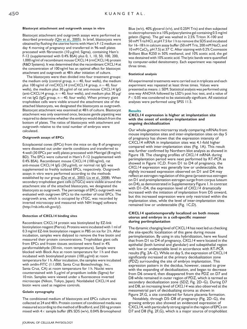
Our whole-genome microarray study comparing mRNAs frommouse implantation sites and inter-implantation sites on day 5of pregnancy has shown that the expression intensity ofCXCL14 mRNA in implantation sites was 4.1-fold highercompared with inter-implantation sites (Fig. 1A). This resultwas further confirmed by Northern blot analysis as showed inFigure 1B. The changing profiles of CXCL14 mRNA duringperiimplantation period were next performed by RT-PCR asshowed in Figure 1C,D. From D1 to D4 of pregnancy, theCXCL14 expression was generally low or undetectable, theslightly increased expression observed on D1 and D4 mayreflect an estrogen regulation of this gene (proestrous estrogenon D1 and preimplantation estrogen surge from corpus luteumon D4), as demonstrated in Supplementary Figure 1. In contrastwith D1–D4, the expression level of CXCL14 dramaticallyincreased with the initiation of implantation from D5 onward,but this increased expression were only restricted within theimplantation sites, while the level of inter-implantation sitesremained low or undetectable (Fig. 1C,D).
CXCL14 spatiotemporally localized on both mouseuterus and embryo in a cell-specific mannerduring periimplantation
The dynamic changing level of CXCL14 has next led us checkingthe site-specific localization of this gene during mouseperiimplantation. By using in situ hybridization, here we foundthat from D1 to D4 of pregnancy, CXCL14 were located in theepithelial (both luminal and glandular) and subepithelial regionwith low or undetectable level in accordance with RT-PCRresults (Fig. 2A–C). While on day 5, the expression of CXCL14significantly increased at the primary decidualization zone(PDZ) surrounding the site of embryo implantation. Thisexpression pattern in the decidua, however, ceased to growwith the expanding of decidualization, and began to decreasefrom D6 onward, then disappeared from the PDZ on D7 andD8 while remained in outer region of PDZ, which is termed assecondary decidualization zone (SDZ; Fig. 2D–G). During D7and D8, an increasing level of CXCL14 was also observed at themesometrial part of decidualizing stroma as shown inFigure 2F,G, a site considered for future placenta formation.
Notablely, through D5–D8 of pregnancy (Fig. 2D–G), thegrowing embryo also showed an evidenced expression ofCXCL14, with particularly strong signal in the region of EPCs onD7 and D8 (Fig. 2F,G), which is a major source of trophoblast
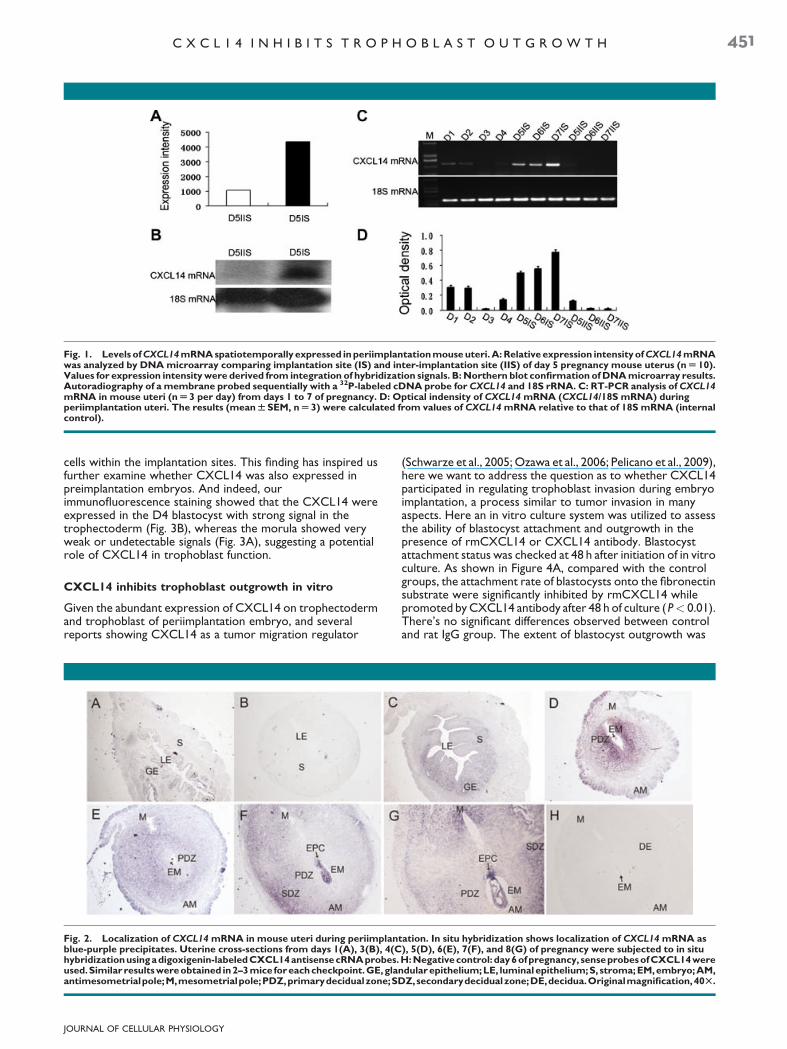
Fig. 1. Levels of CXCL14 mRNA spatiotemporally expressed in periimplantation mouse uteri. A: Relative expression intensity of CXCL14 mRNAwas analyzed by DNA microarray comparing implantation site (IS) and inter-implantation site (IIS) of day 5 pregnancy mouse uterus (n U 10).Values for expression intensity were derived from integration of hybridization signals. B: Northern blot confirmation of DNA microarray results.Autoradiography of a membrane probed sequentially with a 32P-labeled cDNA probe for CXCL14 and 18S rRNA. C: RT-PCR analysis of CXCL14mRNA in mouse uteri (n U 3 per day) from days 1 to 7 of pregnancy. D: Optical indensity of CXCL14 mRNA (CXCL14/18S mRNA) duringperiimplantation uteri. The results (mean W SEM, n U 3) were calculated from values of CXCL14 mRNA relative to that of 18S mRNA (internalcontrol).
C X C L 1 4 I N H I B I T S T R O P H O B L A S T O U T G R O W T H 451
cells within the implantation sites. This finding has inspired usfurther examine whether CXCL14 was also expressed inpreimplantation embryos. And indeed, ourimmunofluorescence staining showed that the CXCL14 wereexpressed in the D4 blastocyst with strong signal in thetrophectoderm (Fig. 3B), whereas the morula showed veryweak or undetectable signals (Fig. 3A), suggesting a potentialrole of CXCL14 in trophoblast function.
CXCL14 inhibits trophoblast outgrowth in vitro
Given the abundant expression of CXCL14 on trophectodermand trophoblast of periimplantation embryo, and severalreports showing CXCL14 as a tumor migration regulator
Fig. 2. Localization of CXCL14 mRNA in mouse uteri during periimplanblue-purple precipitates. Uterine cross-sections from days 1(A), 3(B), 4(Chybridizationusingadigoxigenin-labeled CXCL14 antisense cRNAprobes.used.Similar resultswereobtained in2–3mice foreachcheckpoint.GE,glaantimesometrialpole;M,mesometrialpole;PDZ,primarydecidualzone;SD
JOURNAL OF CELLULAR PHYSIOLOGY
(Schwarze et al., 2005; Ozawa et al., 2006; Pelicano et al., 2009),here we want to address the question as to whether CXCL14participated in regulating trophoblast invasion during embryoimplantation, a process similar to tumor invasion in manyaspects. Here an in vitro culture system was utilized to assessthe ability of blastocyst attachment and outgrowth in thepresence of rmCXCL14 or CXCL14 antibody. Blastocystattachment status was checked at 48 h after initiation of in vitroculture. As shown in Figure 4A, compared with the controlgroups, the attachment rate of blastocysts onto the fibronectinsubstrate were significantly inhibited by rmCXCL14 whilepromoted by CXCL14 antibody after 48 h of culture (P< 0.01).There’s no significant differences observed between controland rat IgG group. The extent of blastocyst outgrowth was
tation. In situ hybridization shows localization of CXCL14 mRNA as), 5(D), 6(E), 7(F), and 8(G) of pregnancy were subjected to in situH: Negative control: day 6ofpregnancy, senseprobesofCXCL14 werendular epithelium;LE, luminalepithelium;S, stroma;EM,embryo;AM,
Z,secondarydecidualzone;DE,decidua.Originalmagnification,40T.

Fig. 3. Immunofluorescence detection of CXCL14 in mouse preimplantation embryos. Green signal represents CXCL14 staining with FITC-conjugated secondary antibody and red signal indicates nuclear staining with PI. A: Morula. B: Blastocyst. Note the highlighted expression ofCXCL14proteinatthetrophectodermofblastocyst. ICM,innercellmass;TE,trophectoderm.C:Negativecontrol:primaryantibodiesreplacedbyrat IgG. Scale bars, 50mm.
452 K U A N G E T A L .
checked at three time points (24, 36, and 48 h). As shown inFigure 4B, after 36 and 48 h in vitro culture, an inhibitory effecton blastocyst outgrowth was evidently observed inrmCXCL14-treated groups, while a promontory effect wasobserved at 48 h when cultured with CXCL14 antibody(P< 0.01). We have further performed Ki67 staining ofblastocysts after cultured with control medium and mediumplus CXCL14, as to examine whether CXCL14 affect theproliferation ability of the blastocyst. As shown inSupplementary Figure 3. CXCL14 (100 and 200 ng/ml)treatment group does not show obvious differences in regardsto ki67 staining, also, the blastocysts seems morphologicallynormal compared with control group. These results suggestedthat the reduced outgrowth performance after CXCL14treatment is not due to reduced proliferation.
To further study the role of CXCL14 during trophoblast celloutgrowth and migration, we isolated EPCs from day 8 mousepregnant uteri, a structure predominantly composed of invasivesTGCs derived from blastocyst trophectoderm (Liu et al.,2008), EPCs were cultured in the presence of either
JOURNAL OF CELLULAR PHYSIOLOGY
rmCXCL14, CXCL14 antibody, or normal rat IgG. The resultsshowed that the trophoblastic outgrowth capacity weresignificantly reduced in the CXCL14-treated group, asdemonstrated by the outgrowth rate and outgrowth area ofEPCs, while the CXCL14 antibody shows an opposite effect asshown in Figure 5A–C. These results demonstrated thatCXCL14 has an inhibitory role on both primary and secondarytrophoblast invasion.
CXCL14 inhibits trophoblast outgrowth via MMP-2and 9 downregulation in vitro
To shed additional light on the mechanisms by which CXCL14regulates trophoblast outgrowth, we next examined whetherCXCL14 could regulate MMP-2 and 9 production of EPCs,which have been considered as two key factors regulatingtrophoblast invasion (Staun-Ram and Shalev, 2005). After24 and 48 h of in vitro culture, EPCs-conditioned medium wascollected and analyzed for collagenase activity by gelatinzymography. At 24 h checkpoint, no significant differences were
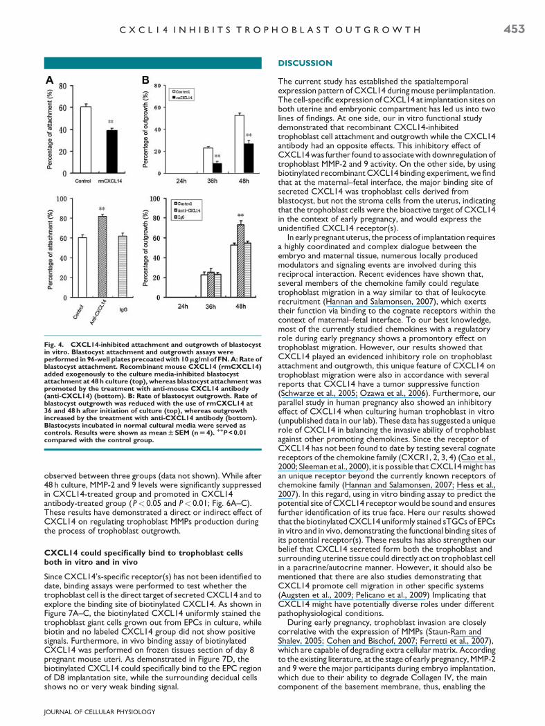
Fig. 4. CXCL14-inhibited attachment and outgrowth of blastocystin vitro. Blastocyst attachment and outgrowth assays wereperformed in 96-well plates precoated with 10mg/ml of FN. A: Rate ofblastocyst attachment. Recombinant mouse CXCL14 (rmCXCL14)added exogenously to the culture media-inhibited blastocystattachment at 48 h culture (top), whereas blastocyst attachment waspromoted by the treatment with anti-mouse CXCL14 antibody(anti-CXCL14) (bottom). B: Rate of blastocyst outgrowth. Rate ofblastocyst outgrowth was reduced with the use of rmCXCL14 at36 and 48 h after initiation of culture (top), whereas outgrowthincreased by the treatment with anti-CXCL14 antibody (bottom).Blastocysts incubated in normal cultural media were served ascontrols. Results were shown as mean W SEM (n U 4). MMP < 0.01compared with the control group.
C X C L 1 4 I N H I B I T S T R O P H O B L A S T O U T G R O W T H 453
observed between three groups (data not shown). While after48 h culture, MMP-2 and 9 levels were significantly suppressedin CXCL14-treated group and promoted in CXCL14antibody-treated group (P< 0.05 and P< 0.01; Fig. 6A–C).These results have demonstrated a direct or indirect effect ofCXCL14 on regulating trophoblast MMPs production duringthe process of trophoblast outgrowth.
CXCL14 could specifically bind to trophoblast cellsboth in vitro and in vivo
Since CXCL14’s-specific receptor(s) has not been identified todate, binding assays were performed to test whether thetrophoblast cell is the direct target of secreted CXCL14 and toexplore the binding site of biotinylated CXCL14. As shown inFigure 7A–C, the biotinylated CXCL14 uniformly stained thetrophoblast giant cells grown out from EPCs in culture, whilebiotin and no labeled CXCL14 group did not show positivesignals. Furthermore, in vivo binding assay of biotinylatedCXCL14 was performed on frozen tissues section of day 8pregnant mouse uteri. As demonstrated in Figure 7D, thebiotinylated CXCL14 could specifically bind to the EPC regionof D8 implantation site, while the surrounding decidual cellsshows no or very weak binding signal.
JOURNAL OF CELLULAR PHYSIOLOGY
DISCUSSION
The current study has established the spatialtemporalexpression pattern of CXCL14 during mouse periimplantation.The cell-specific expression of CXCL14 at implantation sites onboth uterine and embryonic compartment has led us into twolines of findings. At one side, our in vitro functional studydemonstrated that recombinant CXCL14-inhibitedtrophoblast cell attachment and outgrowth while the CXCL14antibody had an opposite effects. This inhibitory effect ofCXCL14 was further found to associate with downregulation oftrophoblast MMP-2 and 9 activity. On the other side, by usingbiotinylated recombinant CXCL14 binding experiment, we findthat at the maternal–fetal interface, the major binding site ofsecreted CXCL14 was trophoblast cells derived fromblastocyst, but not the stroma cells from the uterus, indicatingthat the trophoblast cells were the bioactive target of CXCL14in the context of early pregnancy, and would express theunidentified CXCL14 receptor(s).
In early pregnant uterus, the process of implantation requiresa highly coordinated and complex dialogue between theembryo and maternal tissue, numerous locally producedmodulators and signaling events are involved during thisreciprocal interaction. Recent evidences have shown that,several members of the chemokine family could regulatetrophoblast migration in a way similar to that of leukocyterecruitment (Hannan and Salamonsen, 2007), which exertstheir function via binding to the cognate receptors within thecontext of maternal–fetal interface. To our best knowledge,most of the currently studied chemokines with a regulatoryrole during early pregnancy shows a promontory effect ontrophoblast migration. However, our results showed thatCXCL14 played an evidenced inhibitory role on trophoblastattachment and outgrowth, this unique feature of CXCL14 ontrophoblast migration were also in accordance with severalreports that CXCL14 have a tumor suppressive function(Schwarze et al., 2005; Ozawa et al., 2006). Furthermore, ourparallel study in human pregnancy also showed an inhibitoryeffect of CXCL14 when culturing human trophoblast in vitro(unpublished data in our lab). These data has suggested a uniquerole of CXCL14 in balancing the invasive ability of trophoblastagainst other promoting chemokines. Since the receptor ofCXCL14 has not been found to date by testing several cognatereceptors of the chemokine family (CXCR1, 2, 3, 4) (Cao et al.,2000; Sleeman et al., 2000), it is possible that CXCL14 might hasan unique receptor beyond the currently known receptors ofchemokine family (Hannan and Salamonsen, 2007; Hess et al.,2007). In this regard, using in vitro binding assay to predict thepotential site of CXCL14 receptor would be sound and ensuresfurther identification of its true face. Here our results showedthat the biotinylated CXCL14 uniformly stained sTGCs of EPCsin vitro and in vivo, demonstrating the functional binding sites ofits potential receptor(s). These results has also strengthen ourbelief that CXCL14 secreted form both the trophoblast andsurrounding uterine tissue could directly act on trophoblast cellin a paracrine/autocrine manner. However, it should also bementioned that there are also studies demonstrating thatCXCL14 promote cell migration in other specific systems(Augsten et al., 2009; Pelicano et al., 2009) Implicating thatCXCL14 might have potentially diverse roles under differentpathophysiological conditions.
During early pregnancy, trophoblast invasion are closelycorrelative with the expression of MMPs (Staun-Ram andShalev, 2005; Cohen and Bischof, 2007; Ferretti et al., 2007),which are capable of degrading extra cellular matrix. Accordingto the existing literature, at the stage of early pregnancy, MMP-2and 9 were the major participants during embryo implantation,which due to their ability to degrade Collagen IV, the maincomponent of the basement membrane, thus, enabling the

Fig. 5. Effect of CXCL14 on EPCs outgrowth in vitro. Outgrowth assays of EPCs were performed in 4-well plates precoated with Matrigel(8 mg/ml). A: Outgrowth percentage of EPCs. rmCXCL14 (top) and anti-CXCL14 antibody (bottom) were added into the culture medium andcultured with EPCs for 24, 36, and 48 h, and outgrowth percentage of EPCs was recorded at each checkpoint. B: Outgrowth area of EPCs.Outgrowth area of EPCs was reduced with the treatment of rmCXCL14 at 48 h culture (top), whereas was promoted by the treatment with anti-CXCL14 (bottom). Data represented as fold change in the outgrowth area of EPCs (WSEM, n U 30) compared to controls. MP < 0.05, MMP < 0.01compared with the control group. C: Demonstrative photos showed morphologic observation of EPCs in the presence of rmCXCL14 oranti-CXCL14 antibody or rat IgG. Photos were taken under the light microscope after 24 and 48 h of culture. Original magnification, 40T.
454 K U A N G E T A L .
invasion of the trophoblast cells through the decidua and intothe maternal vasculature (Staun-Ram and Shalev, 2005). SinceCXCL14 could inhibit migration of both blastocyst (rich inprimary trophoblast giant cells) and EPCs outgrowth (rich insTGCs), which were previously reported to have differentpredominance in MMP-2 and 9 content (Hulboy et al., 1997).We tested the effect of CXCL14 on MMP-2, and 9 production
JOURNAL OF CELLULAR PHYSIOLOGY
in both targets. Our data indicated that CXCL14 coulddownregulate MMP-2 in the blastocyst (data not shown) whiledownregulate both MMP2, and 9 production in the EPCs. Theseresults are in correspondance with previously reportedrelationship between chemokine and MMPs secretion in othersystems (Cross and Woodroofe, 1999; Van and Libert, 2007).The relative abundance of MMP-2, and 9 in blastocyst and EPCs
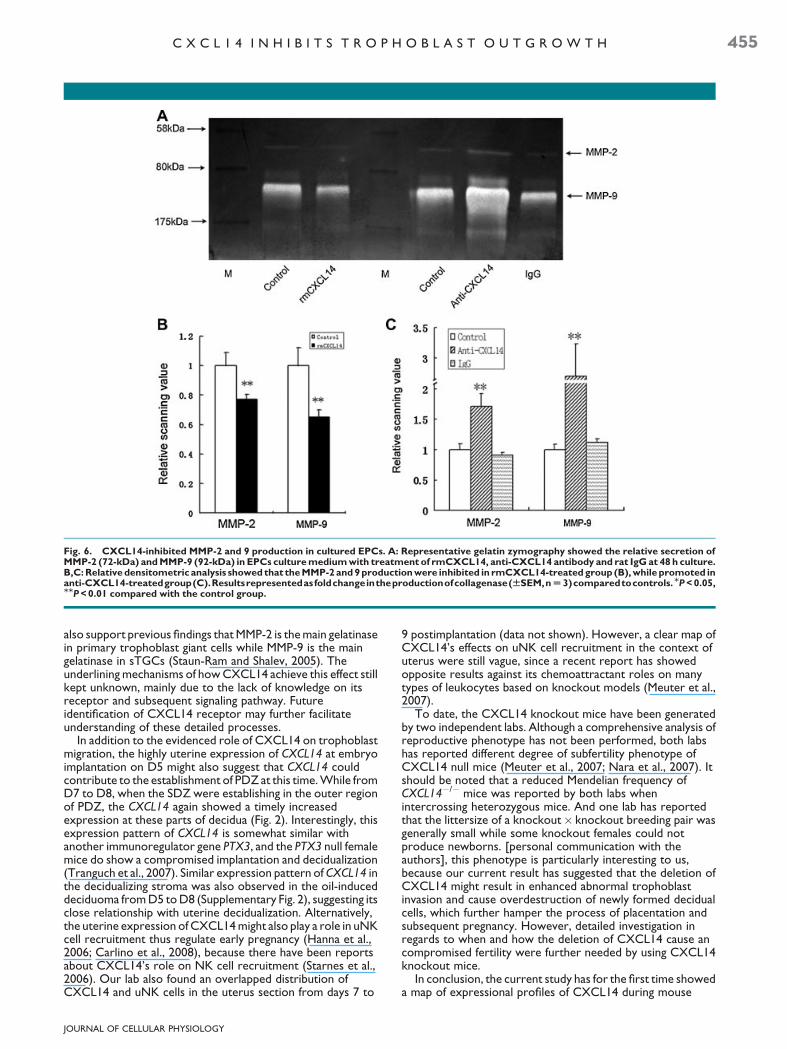
Fig. 6. CXCL14-inhibited MMP-2 and 9 production in cultured EPCs. A: Representative gelatin zymography showed the relative secretion ofMMP-2 (72-kDa) and MMP-9 (92-kDa) in EPCs culture medium with treatment of rmCXCL14, anti-CXCL14 antibody and rat IgG at 48 h culture.B,C: Relative densitometric analysis showed that the MMP-2 and 9 production were inhibited in rmCXCL14-treated group (B), while promoted inanti-CXCL14-treatedgroup(C).Resultsrepresentedasfoldchangeintheproductionofcollagenase(WSEM,n U 3)comparedtocontrols.MP < 0.05,MMP < 0.01 compared with the control group.
C X C L 1 4 I N H I B I T S T R O P H O B L A S T O U T G R O W T H 455
also support previous findings that MMP-2 is the main gelatinasein primary trophoblast giant cells while MMP-9 is the maingelatinase in sTGCs (Staun-Ram and Shalev, 2005). Theunderlining mechanisms of how CXCL14 achieve this effect stillkept unknown, mainly due to the lack of knowledge on itsreceptor and subsequent signaling pathway. Futureidentification of CXCL14 receptor may further facilitateunderstanding of these detailed processes.
In addition to the evidenced role of CXCL14 on trophoblastmigration, the highly uterine expression of CXCL14 at embryoimplantation on D5 might also suggest that CXCL14 couldcontribute to the establishment of PDZ at this time. While fromD7 to D8, when the SDZ were establishing in the outer regionof PDZ, the CXCL14 again showed a timely increasedexpression at these parts of decidua (Fig. 2). Interestingly, thisexpression pattern of CXCL14 is somewhat similar withanother immunoregulator gene PTX3, and the PTX3 null femalemice do show a compromised implantation and decidualization(Tranguch et al., 2007). Similar expression pattern of CXCL14 inthe decidualizing stroma was also observed in the oil-induceddeciduoma from D5 to D8 (Supplementary Fig. 2), suggesting itsclose relationship with uterine decidualization. Alternatively,the uterine expression of CXCL14 might also play a role in uNKcell recruitment thus regulate early pregnancy (Hanna et al.,2006; Carlino et al., 2008), because there have been reportsabout CXCL14’s role on NK cell recruitment (Starnes et al.,2006). Our lab also found an overlapped distribution ofCXCL14 and uNK cells in the uterus section from days 7 to
JOURNAL OF CELLULAR PHYSIOLOGY
9 postimplantation (data not shown). However, a clear map ofCXCL14’s effects on uNK cell recruitment in the context ofuterus were still vague, since a recent report has showedopposite results against its chemoattractant roles on manytypes of leukocytes based on knockout models (Meuter et al.,2007).
To date, the CXCL14 knockout mice have been generatedby two independent labs. Although a comprehensive analysis ofreproductive phenotype has not been performed, both labshas reported different degree of subfertility phenotype ofCXCL14 null mice (Meuter et al., 2007; Nara et al., 2007). Itshould be noted that a reduced Mendelian frequency ofCXCL14�/� mice was reported by both labs whenintercrossing heterozygous mice. And one lab has reportedthat the littersize of a knockout� knockout breeding pair wasgenerally small while some knockout females could notproduce newborns. [personal communication with theauthors], this phenotype is particularly interesting to us,because our current result has suggested that the deletion ofCXCL14 might result in enhanced abnormal trophoblastinvasion and cause overdestruction of newly formed decidualcells, which further hamper the process of placentation andsubsequent pregnancy. However, detailed investigation inregards to when and how the deletion of CXCL14 cause ancompromised fertility were further needed by using CXCL14knockout mice.
In conclusion, the current study has for the first time showeda map of expressional profiles of CXCL14 during mouse
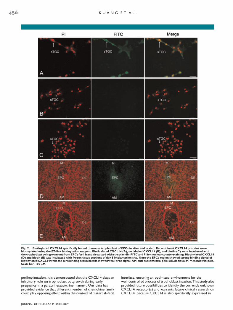
Fig. 7. Biotinylated CXCL14 specifically bound to mouse trophoblast of EPCs in vitro and in vivo. Recombinant CXCL14 proteins werebiotinylated using the EZ-link biotinylation reagent. Biotinylated CXCL14 (A), no labeled CXCL14 (B), and biotin (C) were incubated withthe trophoblast cells grown out from EPCs for 1 h and visualized with streptavidin-FITC and PI for nuclear counterstaining. Biotinylated CXCL14(D) and biotin (E) was incubated with frozen tissue sections of day 8 implantation site. Note the EPCs region showed strong binding signal ofbiotinylatedCXCL14whilethesurroundingdecidualcellsshowedweakornosignal.AM,anti-mesometrialpole;DE,decidua;M,mesometrialpole,Scale bar, 100mM.
456 K U A N G E T A L .
periimplantation. It is demonstrated that the CXCL14 plays aninhibitory role on trophoblast outgrowth during earlypregnancy in a paracrine/autocrine manner. Our data hasprovided evidence that different member of chemokine familycould play opposing effect within the context of maternal–fetal
JOURNAL OF CELLULAR PHYSIOLOGY
interface, ensuring an optimized environment for thewell-controlled process of trophoblast invasion. This study alsoprovided future possibilities to identify the currently unknownCXCL14 receptor(s) and warrants future clinical research onCXCL14, because CXCL14 is also specifically expressed in
C X C L 1 4 I N H I B I T S T R O P H O B L A S T O U T G R O W T H 457
human endometrium and trophoblast (Red-Horse et al., 2001,2004; Talbi et al., 2006).
Acknowledgments
We thank Dr. Haibin Wang at Institute of Zoology, ChineseAcademy of Sciences for proofreading of this manuscript.
Literature Cited
Augsten M, Hagglof C, Olsson E, Stolz C, Tsagozis P, Levchenko T, Frederick MJ, Borg A,Micke P, Egevad L, Ostman A. 2009. CXCL14 is an autocrine growth factor for fibroblastsand acts as a multi-modal stimulator of prostate tumor growth. Proc Natl Acad Sci USA106:3414–3419.
CaoX,Zhang W, Wan T, He L, Chen T, YuanZ,Ma S, Yu Y, Chen G. 2000. Molecularcloning andcharacterization of a novel CXC chemokine macrophage inflammatory protein-2 gammachemoattractant for human neutrophils and dendritic cells. J Immunol 165:2588–2595.
Carlino C, Stabile H, Morrone S, Bulla R, Soriani A, Agostinis C, Bossi F, Mocci C, Sarazani F,Tedesco F, Santoni A, Gismondi A. 2008. Recruitment of circulating NK cells throughdecidual tissues: A possible mechanism controlling NK cell accumulation in the uterusduring early pregnancy. Blood 114:3108–3115.
Cohen M, Bischof P. 2007. Factors regulating trophoblast invasion. Gynecol Obstet Invest64:126–130.
Cross AK, Woodroofe MN. 1999. Chemokine modulation of matrix metalloproteinase andTIMP production in adult rat brain microglia and a human microglial cell line in vitro. Glia28:183–189.
Dai B, Cao Y, Liu W, Li S, Yang Y, Chen D, Duan E. 2003. Dual roles of progesterone inembryo implantation in mouse. Endocrine 21:123–132.
Fernandez EJ, Lolis E. 2002. Structure, function, and inhibition of chemokines. Annu RevPharmacol Toxicol 42:469–499.
Ferretti C, Bruni L, Dangles-Marie V, Pecking AP, Bellet D. 2007. Molecular circuits shared byplacental and cancer cells, and their implications in the proliferative, invasive and migratorycapacities of trophoblasts. Hum Reprod Update 13:121–141.
Fitzgerald JS, Poehlmann TG, Schleussner E, Markert UR. 2008. Trophoblast invasion: Therole of intracellular cytokine signalling via signal transducer and activator of transcription 3(STAT3). Hum Reprod Update 14:335–344.
Frederick MJ, Henderson Y, Xu X, Deavers MT, Sahin AA, Wu H, Lewis DE, El-Naggar AK,Clayman GL. 2000. In vivo expression of the novel CXC chemokine BRAK in normal andcancerous human tissue. Am J Pathol 156:1937–1950.
Hanna J, Goldman-Wohl D, Hamani Y, Avraham I, Greenfield C, Natanson-Yaron S, Prus D,Cohen-Daniel L, Arnon TI, Manaster I, Gazit R, Yutkin V, Benharroch D, Porgador A,Keshet E, Yagel S, Mandelboim O. 2006. Decidual NK cells regulate key developmentalprocesses at the human fetal-maternal interface. Nat Med 12:1065–1074.
Hannan NJ, Salamonsen LA. 2007. Role of chemokines in the endometrium and in embryoimplantation. Curr Opin Obstet Gynecol 19:266–272.
Hess AP, Hamilton AE, Talbi S, Dosiou C, Nyegaard M, Nayak N, Genbecev-Krtolica O,Mavrogianis P, Ferrer K, Kruessel J, Fazleabas AT, Fisher SJ, Giudice LC. 2007. Decidualstromal cell response to paracrine signals from the trophoblast: Amplification of immuneand angiogenic modulators. Biol Reprod 76:102–117.
Hromas R, Broxmeyer HE, Kim C, Nakshatri H, Christopherson K II, Azam M, Hou YH. 1999.Cloning of BRAK, a novel divergent CXC chemokine preferentially expressed in normalversus malignant cells. Biochem Biophys Res Commun 255:703–706.
Hulboy DL, Rudolph LA, Matrisian LM. 1997. Matrix metalloproteinases as mediators ofreproductive function. Mol Hum Reprod 3:27–45.
Kurth I, Willimann K, Schaerli P, Hunziker T, Clark-Lewis I, Moser B. 2001. Monocyteselectivity and tissue localization suggests a role for breast and kidney-expressedchemokine (BRAK) in macrophage development. J Exp Med 194:855–861.
Liu Q, Zhang B, Zhao X, Zhang Y, Liu Y, Yan X. 2008. Blockade of adhesion molecule CD146causes pregnancy failure in mice. J Cell Physiol 215:621–626.
JOURNAL OF CELLULAR PHYSIOLOGY
Meuter S, Schaerli P, Roos RS, Brandau O, Bosl MR, von AU, Moser B. 2007. Murine CXCL14is dispensable for dendritic cell function and localization within peripheral tissues. Mol CellBiol 27:983–992.
Nara N, Nakayama Y, Okamoto S, Tamura H, Kiyono M, Muraoka M, Tanaka K, Taya C,Shitara H, Ishii R, Yonekawa H, Minokoshi Y, Hara T. 2007. Disruption of CXC motifchemokine ligand-14 in mice ameliorates obesity-induced insulin resistance. J Biol Chem282:30794–30803.
Ozawa S, Kato Y, Komori R, Maehata Y, Kubota E, Hata R. 2006. BRAK/CXCL14 expressionsuppresses tumor growth in vivo in human oral carcinoma cells. Biochem Biophys ResCommun 348:406–412.
Pelicano H, Lu W, Zhou Y, Zhang W, Chen Z, Hu Y, Huang P. 2009. Mitochondrialdysfunction and reactive oxygen species imbalance promote breast cancer cell motilitythrough a CXCL14-mediated mechanism. Cancer Res 69:2375–2383.
Qin J, Diaz-Cueto L, Schwarze JE, Takahashi Y, Imai M, Isuzugawa K, Yamamoto S, Chang KT,Gerton GL, Imakawa K. 2005. Effects of progranulin on blastocyst hatching and subsequentadhesion and outgrowth in the mouse. Biol Reprod 73:434–442.
Red-Horse K, Drake PM, Gunn MD, Fisher SJ. 2001. Chemokine ligand and receptorexpression in the pregnant uterus: Reciprocal patterns in complementary cell subsetssuggest functional roles. Am J Pathol 159:2199–2213.
Red-Horse K, Drake PM, Fisher SJ. 2004. Human pregnancy: The role of chemokine networksat the fetal-maternal interface. Expert Rev Mol Med 6:1–14.
Salamonsen LA, Hannan NJ, Dimitriadis E. 2007. Cytokines and chemokines during humanembryo implantation: Roles in implantation and early placentation. Semin Reprod Med25:437–444.
Salogni L, Musso T, Bosisio D, Mirolo M, Jala VR, Haribabu B, Locati M, Sozzani S. 2009. ActivinA induces dendritic cell migration through the polarized release of CXCL12 and CXCL14.Blood 5113:5848–5856.
Schwarze SR, Luo J, Isaacs WB, Jarrard DF. 2005. Modulation of CXCL14 (BRAK) expressionin prostate cancer. Prostate 64:67–74.
Shellenberger TD, Wang M, Gujrati M, Jayakumar A, Strieter RM, Burdick MD, Ioannides CG,Efferson CL, El-Naggar AK, Roberts D, Clayman GL, Frederick MJ. 2004. BRAK/CXCL14 isa potent inhibitor of angiogenesis and a chemotactic factor for immature dendritic cells.Cancer Res 64:8262–8270.
Shurin GV, Ferris RL, Tourkova IL, Perez L, Lokshin A, Balkir L, Collins B, Chatta GS, ShurinMR. 2005. Loss of new chemokine CXCL14 in tumor tissue is associated with lowinfiltration by dendritic cells (DC), while restoration of human CXCL14 expressionin tumor cells causes attraction of DC both in vitro and in vivo. J Immunol 174:5490–5498.
Sleeman MA, Fraser JK, Murison JG, Kelly SL, Prestidge RL, Palmer DJ, Watson JD, KumbleKD. 2000. B cell- and monocyte-activating chemokine (BMAC), a novel non-ELR alpha-chemokine. Int Immunol 12:677–689.
Song H, Lim H, Paria BC, Matsumoto H, Swift LL, Morrow J, Bonventre JV, Dey SK. 2002.Cytosolic phospholipase A2alpha is crucial [correction of A2alpha deficiency is crucial] for‘on-time’ embryo implantation that directs subsequent development. Development129:2879–2889.
Starnes T, Rasila KK, Robertson MJ, Brahmi Z, Dahl R, Christopherson K, Hromas R. 2006.The chemokine CXCL14 (BRAK) stimulates activated NK cell migration: Implications forthe downregulation of CXCL14 in malignancy. Exp Hematol 34:1101–1105.
Staun-Ram E, Shalev E. 2005. Human trophoblast function during the implantation process.Reprod Biol Endocrinol 3:56.
Talbi S, Hamilton AE, Vo KC, Tulac S, Overgaard MT, Dosiou C, Le SN, Nezhat CN, KempsonR, Lessey BA, Nayak NR, Giudice LC. 2006. Molecular phenotyping of humanendometrium distinguishes menstrual cycle phases and underlying biological processes innormo-ovulatory women. Endocrinology 147:1097–1121.
Tranguch S, Chakrabarty A, Guo Y, Wang H, Dey SK. 2007. Maternal pentraxin 3 deficiencycompromises implantation in mice. Biol Reprod 77:425–432.
Van LP, Libert C. 2007. Chemokine and cytokine processing by matrix metalloproteinasesand its effect on leukocyte migration and inflammation. J Leukoc Biol 82:1375–1381.
Wang H, Dey SK. 2006. Roadmap to embryo implantation: Clues from mouse models. NatRev Genet 7:185–199.
Wilcox AJ, Baird DD, Weinberg CR. 1999. Time of implantation of the conceptus and loss ofpregnancy. N Engl J Med 340:1796–1799.